












-
Polylactic acid (PLA) is one of the most widely used industrially produced biodegradable plastics[1]. As a biodegradable polymer, PLA is synthesized from renewable resources (e.g., corn) via bacterial fermentation of saccharified starch[2], with the molecular formula (C3H4O2)n. The fate of PLA is finally mineralized into biomass, carbon dioxide, and water upon entering the environment. PLA currently accounts for 24% of the global biodegradable polymer production capacity, and its output continues to rise as costs decrease[1].
Although metabolites of PLA are environmentally friendly, the improper accumulation in the environment poses a serious threat to soil health and inhibits crop growth and development[3,4]. Boots et al. reported that PLA particles not only reduce soil pH, but they also alter the distribution of water-stable aggregates and decrease the germination rate and shoot height of perennial ryegrass[5]. Chen et al. further showed that PLA affects microbial community interactions[6]. In addition, Yang et al. discovered that high concentrations of PLA exert physiological toxicity on plants, significantly inhibiting maize germination and root biomass, increasing Zn concentration in roots while reducing its translocation to shoots[7]. Therefore, ensuring the environmental safety of biodegradable plastics like PLA during their natural degradation or recycling process, without additional labor and resource inputs, is critical for crop health and sustainable agricultural development[8−11].
Addressing the soil health hazards posed by PLA requires effective degradation strategies, and fungi have emerged as promising biological agents for PLA biodegradation. Karamanlioglu et al. made significant breakthroughs by isolating indigenous fungi from PLA-contaminated environments and exploring their degradation mechanisms on PLA[12]. They also found that PLA drives the colonization of specific fungal populations, and that particular fungi, such as Thermomyces lanuginosus and Aspergillus fumigatus, significantly accelerated PLA degradation at 50 °C. When assessed via tensile strength loss, fungal degradation was more effective than abiotic hydrolysis; T. lanuginosus caused PLA to completely lose its tensile strength within 28–35 d[12]. Complementary metabolomic analyses by Okal et al. revealed the molecular mechanisms of fungal plastic degradation, showing that Lasiodiplodia iranensis activates various metabolic pathways and secretes hydrolytic and oxidative enzymes that synergistically act on the polymer structure[13]. Moreover, fungal biodegradation efficiency is affected by the chemical structure of plastics, the type of fungi, and reaction conditions such as environmental temperature[13−15].
The complete biodegradation of PLA follows the processes of colonization, depolymerization, and mineralization[16]. Recent research on PLA degradation is motivated by key challenges: its biodegradation in natural environments is slow and inefficient compared to controlled composting, due to limited microbial activity and unclear mechanisms[17−19]. Utilizing specific fungi offers a promising strategy for faster, more controllable biorecycling[14,20−22]. Furthermore, as conventional and biodegradable plastics coexist in soil environments, developing microbes capable of degrading both types is essential for effective, proactive remediation of plastic pollution. Consequently, screening functional fungi that efficiently degrade PLA under mild conditions is of great practical significance for developing reliable environmental remediation strategies.
Fusarium vanettenii has previously been associated with the degradation of synthetic polymers, including polyurethane and polyethylene terephthalate and has been reported to produce hydrolytic and oxidoreductive enzymes that may act on ester-containing substrates[12,21,23−26]. Since PLA is also a polyester, its ester bonds may represent potential targets for fungal-mediated deterioration. However, the ability of F. vanettenii to affect PLA under ambient conditions has not been clearly evaluated. Therefore, this study aimed to assess the short-term interaction between F. vanettenii and PLA films by examining fungal colonization, surface morphology, mass loss, and changes in Fourier transform infrared spectroscopy (FTIR) after 60 d of incubation. This work was designed as a preliminary in vitro assessment of PLA surface deterioration rather than a complete evaluation of PLA mineralization.
-
The F. vanettenii strain used in this study was isolated from soil heavily contaminated with polyurethane (PU) microplastics (100 μm). The soil was incubated in polyethylene pots under shaded garden conditions for 8 months. Subsequently, soil samples were plated onto chloramphenicol-supplemented malt extract agar (CMEA) and incubated at 28 °C for 14 d.
Fungal isolates were initially identified by sequencing the ITS and LSU regions. Genomic DNA was extracted using the E.Z.N.A.® Forensic DNA Kit (Omega Bio-Tek), and target regions were sequenced on a Sanger platform (Sangon Biotech, Shanghai, China). Sequences were assembled and aligned using Geneious v9.0.2.
Biodegradation potential was screened on a minimal medium containing 1% (w/v) Impranil, PEG-400, and xylose as carbon sources. The autoclave-sterilized medium was cooled to approximately 40 °C before separately adding filter-sterilized impranil and PEG-400 solutions. After solidification, plates were inoculated with fungal isolates. F. vanettenii (deposited as KUNCC: 23-13698) was selected for further study due to its dense mycelial growth and rapid colonization on this screening medium. The selected strain was maintained on potato dextrose agar (PDA, QDRS BIOTEC) for routine cultivation and species activation[26].
Medium preparation
-
PDA medium was used for subculture and activation of the fungal strain. It was prepared by dissolving 15.6 g of PDA powder in 400 mL of deionized water. The Czapek-Dox solid medium used for the PLA inoculation experiment contained yeast extract 5 g/L, NaNO3 3 g/L, KH2PO4 1 g/L, KCl 0.5 g/L, MgSO4·7H2O 0.5 g/L, FeSO4·7H2O 0.01 g/L, sucrose 30 g/L, and agar 15 g/L. Both media were sterilized by autoclaving at 121 °C for 20 min, cooled to approximately 50 °C, and poured aseptically into sterile plastic Petri dishes in a laminar flow cabinet.
Species activation
-
F. vanettenii was activated by inoculating it onto PDA plates at 28 °C for 5–7 d until mycelium overgrew the plate.
PLA film preparation
Solution preparation
-
In a fume hood, 24 g PLA particles were weighed, dispersed in 600 mL chloroform in a 1,000 mL flask, and transferred to a constant temperature shaking incubator (180 rpm, 28 °C) with continuous stirring for 7 h until PLA was completely dissolved to form a uniform and transparent solution.
Film casting
-
A 10 mL sample of the solution was sucked with a 10 mL glass glue head dropper and evenly added to a sterilized 90 mm glass Petri dish; a total of 20 glass Petri dishes were prepared. The Petri dishes were then placed in a fume hood for 24 h. After the chloroform was completely volatilized, PLA films with uniform thickness (about 0.1–0.2 mm) were formed.
Film cutting
-
The PLA film in each dish was cut into a square experimental film (4 cm × 4 cm) with the same specifications using a sterile scalpel for later use. Eventually, six intact and uniform PLA square films were used for the subsequent experiments.
Inoculation and degradation experiment
-
All inoculation steps were performed aseptically in a laminar flow cabinet. In the fungal treatment, a 5 mm × 5 mm mycelial plug was excised from the actively growing margin of a 5–7-d-old F. vanettenii colony grown on PDA. The plug, with minimal adherent agar, was transferred to the center of a fresh Czapek-Dox agar plate. A sterilized PLA film measuring 4 cm × 4 cm was then placed directly over the mycelial plug to ensure close contact between the fungus and the film surface.
In the abiotic control, PLA films of the same size were placed on Czapek-Dox agar plates without fungal inoculation. Three independent replicate plates were prepared for the fungal treatment and three for the abiotic control. All plates were incubated in the dark at 26 ± 2 °C for 60 d. Macroscopic observations were recorded after 30 and 60 d. After incubation, the PLA films were recovered for mass loss measurement, microscopy, SEM, and FTIR analysis[26]. Where films were cut for microscopy, subsamples were taken after final weighing to avoid affecting mass-loss calculations.
Experimental index determination
Optical microscopy and scanning electron microscopy (SEM)
-
The PLA film samples of the treatment group and the control group were cut into several pieces of appropriate size, placed on a glass slide, dropped with one drop of deionized water, and covered with a cover slip for observation.
After critical point drying and gold spraying, the surface morphology of the film fragments was observed by SEM (ZEISS Sigma 300), and the changes of cracks, holes, and roughness of the film surface were recorded to evaluate the degradation condition[13,26].
Fourier transform infrared spectroscopy (FTIR) analysis
-
The PLA film fragments of the treatment group and the control group were mixed with KBr and pressed into a tablet. The FTIR spectrometer (Bruker Vertex 70) was used to scan in the wave number range of 4,000–400 cm−1 to analyze the characteristic absorption peak changes of chemical functional groups before and after incubation to determine the chemical degradation of PLA by fungi[27].
Quality loss rate
-
Sample processing: after 60 d of incubation, the PLA films were removed and successively immersed in 2% (w/v) SDS solution and 75% (v/v) alcohol for 10 min to remove residual mycelia on the surface, rinsed three times with sterile deionized water, and finally blotted dry with sterile filter paper[26].
Mass-loss data were analyzed using GraphPad Prism 9.0. The initial and final masses of PLA films were recorded for each replicate and the percentage mass loss was calculated as follows:
$ {\rm{Mass}}\;{\rm{loss}}\;({\text{%}})=[({\rm W}_1-{\rm W}_2)/{\rm W}_1]\times 100 $ where, W1 is the initial mass of the PLA film and W2 is the final mass after 60 d of incubation[23]. Data are presented as mean ± standard deviation (SD), with n = 3 for each treatment. The percentage mass loss between the abiotic control and the F. vanettenii-inoculated treatment was compared using a t-test. Statistical significance was accepted at p < 0.05. Because of the small sample size, statistical results were interpreted cautiously and in combination with morphological and FTIR observations.
-
Macroscopic imaging revealed that, compared with the control group (Fig. 1j), the edges of the film began to curl (Fig. 1e). The area in direct contact with the fungal mycelium changed from transparent (Fig. 1a) to an opaque, milky white color, suggesting successful fungal colonization and surface transparency decrease of the film.
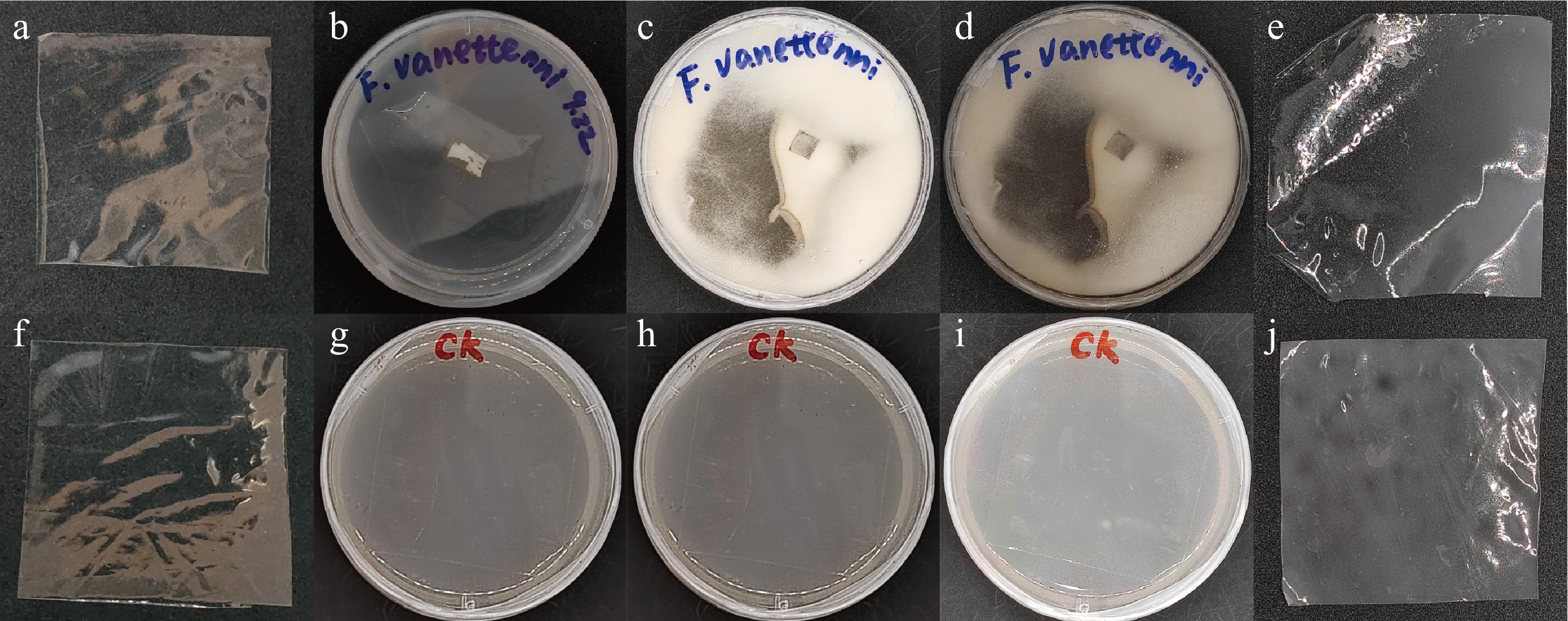
Figure 1.
Morphological changes of PLA films before and after fungal inoculation. (a), (f) Original PLA films. (b) Petri dish immediately after inoculation with F. vanettenii. (c) Petri dish after 1 month of incubation by F. vanettenii. (d) Petri dish after 2 months of incubation with F. vanettenii. (e) PLA film retrieved from (d). (g) Control Petri dish with agar medium only (without fungal inoculation) on day 0. (h) Control dish after 1 month of incubation. (i) Control dish after 2 months of incubation. (j) PLA film retrieved from (i).
Microscopic examination (Fig. 2a, b) showed residual mycelial fragments of F. vanettenii at the PLA film surface post-colonization. By comparing the scanning electron microscope (SEM) images of the samples before and after the experiment, it showed that the fungal hyphae induced the formation of pores (Fig. 2e) and cracks (Fig. 2f) in PLA films.
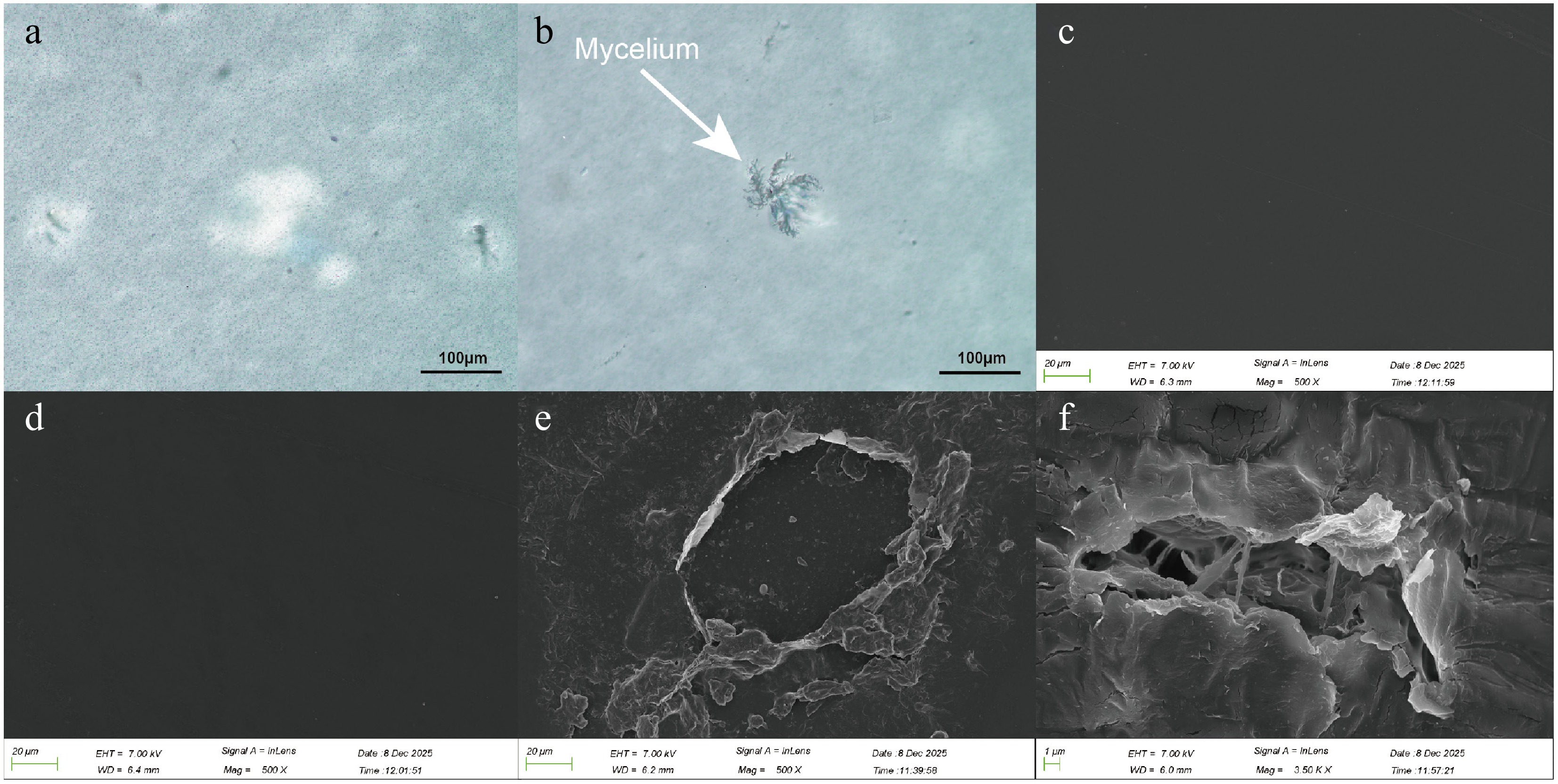
Figure 2.
Observation of the microscopic structure of PLA films. (a) Optical microscope image of the PLA film from the control group (CK). (b) Optical microscope image of the PLA film after 2 months of incubation by F. vanettenii. (c) SEM image of the PLA film surface before incubation. (d) SEM image of the PLA film surface from the control group (CK) after 2 months. (e), (f) SEM images of the PLA film surface after 2 months of incubation by F. vanettenii; (e) shows the formed pores, and (f) shows the generated cracks.
Mass loss and FTIR analysis of PLA films
-
After 60 d of incubation, the abiotic control films showed only slight mass loss, whereas the F. vanettenii-inoculated films showed a higher mean mass loss. The mean mass loss in the inoculated treatment was 5.32% ± 0.10%, compared with 0.44% ± 0.47% in the abiotic control (Fig. 3). This difference was statistically significant under the applied test (p = 0.0136). However, the absolute mass loss remained low, indicating that the fungal treatment induced only limited PLA mass reduction within the 60-d incubation period.
FTIR spectra of the PLA films before and after incubation showed the same major characteristic absorption bands, including the ester C=O stretching vibration at approximately 1,753–1,755 cm−1, C–H bending vibrations at approximately 1,452–1,454 cm−1 and 1,380–1,382 cm−1, and C–O–C stretching vibrations at approximately 1,086–1,088 cm−1. No major wavenumber shifts were detected among the initial PLA film, abiotic control, and F. vanettenii-inoculated treatment (Fig. 3). Slight changes in peak transmittance were observed in the inoculated films, particularly near 1,755, 1,184, and 1,086 cm−1. These changes may indicate minor alteration of ester-related functional groups; however, because no quantitative peak-ratio analysis was performed, the FTIR results should be interpreted as preliminary evidence of slight chemical alteration rather than definitive proof of extensive PLA depolymerization.
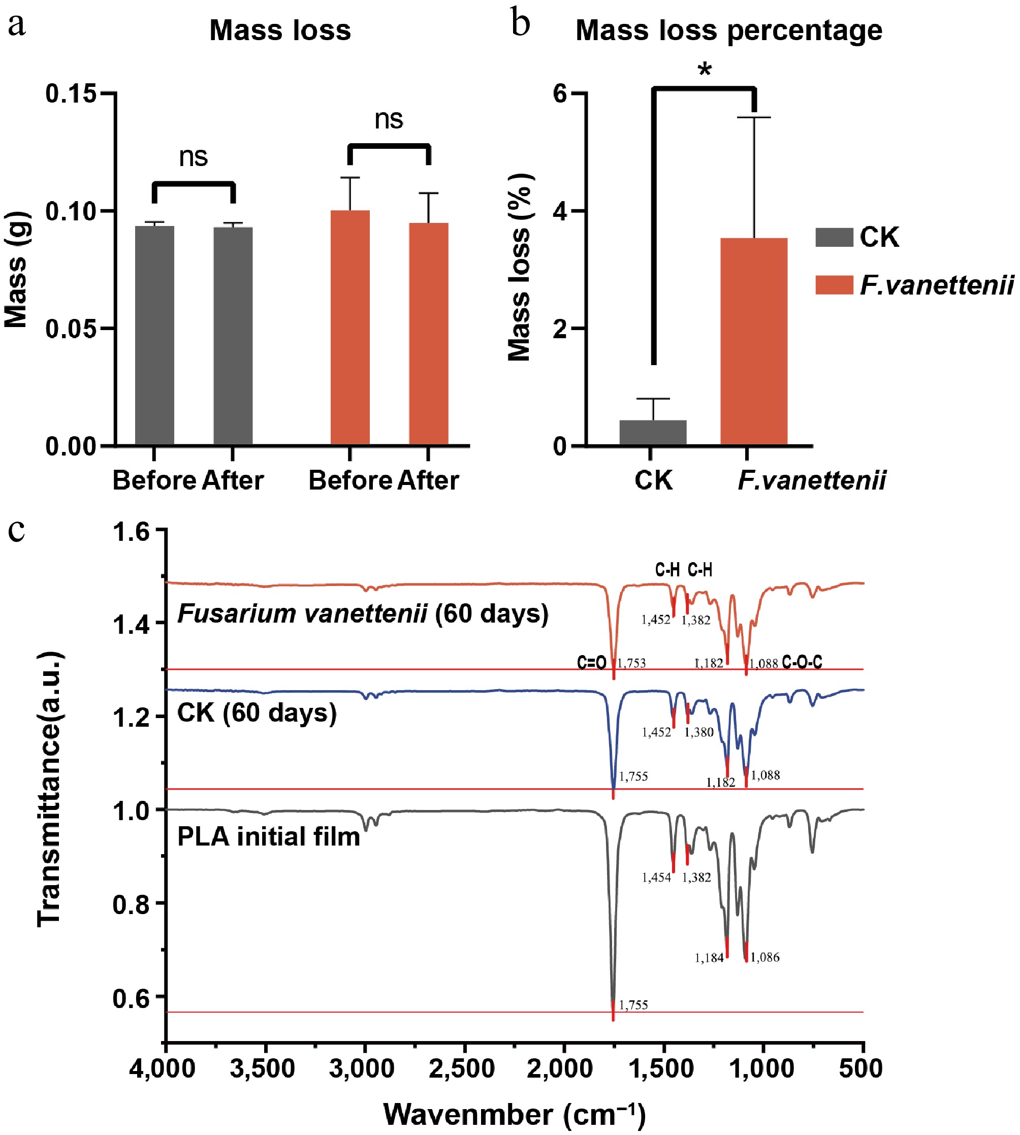
Figure 3.
Mass loss and chemical construction determination of PLA films. (a) Absolute mass of PLA films before and after cultivation. The data points represent individual repetitions. The points of the same PLA film before and after cultivation are connected by lines. The error bars represent the mean ± standard deviation (n = 3). (b) Mass loss percentage of PLA films after cultivation. The bar chart represents the group mean, and the error bars are the standard deviation (n = 3). (c) FTIR spectroscopy results. * Represents p < 0.05, ns represents not significant.
-
This study showed that F. vanettenii was able to colonize the surface of PLA films and induce visible physical deterioration within 60 d. Macroscopic observations showed reduced transparency and curling of the inoculated films, while SEM revealed pores, cracks, and increased surface roughness. These observations suggest that the fungus established close contact with the PLA surface and caused localized structural damage. Similar early-stage deterioration patterns have been reported for fungal interactions with polymeric materials[24,28], where hyphal adhesion, surface penetration, and mechanical pressure may contribute to changes in surface morphology.
However, these observations should be interpreted cautiously. The detected changes mainly demonstrate surface colonization and physical deterioration, not complete PLA biodegradation. Physical damage may increase the exposed surface area of the polymer and facilitate further hydrolytic or enzymatic attack[2,28], but additional evidence is required to confirm extensive polymer-chain cleavage or mineralization.
Limited mass loss and chemical alteration
-
The inoculated PLA films showed a mean mass loss of 5.32% ± 0.10%, which was higher than that of the abiotic control. This indicates that F. vanettenii contributed to measurable mass reduction under the tested conditions. Nevertheless, the absolute mass loss was low, suggesting that degradation was still at an early stage. The 60-d incubation period may have been sufficient for fungal colonization and surface deterioration but insufficient for substantial depolymerization or assimilation of PLA-derived carbon, as observed in other fungal biodegradation studies requiring longer timelines[25,29].
FTIR analysis further supports this cautious interpretation. The absence of major peak shifts indicates that the main chemical structure of PLA was largely retained after 60 d. Slight changes in peak transmittance may reflect early alteration of ester-related groups, but without quantitative FTIR peak-ratio analysis, molecular weight analysis or detection of degradation products, these spectral changes cannot be taken as conclusive evidence of extensive chemical degradation[28,30]. Therefore, the present results support limited early-stage chemical alteration rather than strong depolymerization.
Limitations and future directions
-
This study has several limitations. First, the experiment used only an abiotic control. Although this control allowed comparison with natural aging under the same incubation conditions, it did not fully separate fungal enzymatic effects from physical contact, moisture exposure, or biomass-associated effects. Future studies should include additional controls, such as heat-killed fungal biomass, enzyme-free biomass-contact controls, and medium-only controls. Second, the experiment was conducted for only 60 d and used a small number of replicates. Longer incubation periods and larger sample sizes are needed to better evaluate degradation kinetics. Third, the study relied mainly on mass loss, SEM, and FTIR. Future work should include molecular weight analysis, tensile strength testing, crystallinity analysis, enzyme assays, and detection of soluble degradation products to confirm polymer-chain cleavage and mineralization[2,12,31].
-
This study showed that the soil-derived filamentous fungus Fusarium vanettenii can colonize solution-cast PLA films and induce visible surface deterioration under laboratory conditions within 60 d. The inoculated PLA films showed pores, cracks, reduced transparency, and a low but measurable mass loss of 5.32% ± 0.10%. FTIR analysis showed no major shifts in the characteristic PLA peaks, although slight changes in peak transmittance were observed. These findings suggest that F. vanettenii can contribute to early-stage physical deterioration and limited chemical alteration of PLA films under the tested conditions.
However, the present data do not demonstrate complete PLA biodegradation, extensive depolymerization, or mineralization. Further studies using longer incubation periods, optimized culture conditions, additional controls, and quantitative chemical and mechanical analyses are needed to determine the true PLA-degrading capacity of F. vanettenii and its potential role in plastic bioremediation.
-
The authors confirm their contributions to this study as follows: conceptualization, investigation and methodology: Tang Y, Okal EJ; data curation, formal analysis, software, validation, visualization and writing − original draft: Tang Y; funding acquisition, project administration, resources and supervision: Gui H; writing − review and editing: Hu Y. All authors reviewed the results and approved the final version of the manuscript.
-
The datasets generated during and/or analyzed in the current study are available from the corresponding author on reasonable request.
-
The authors declare that they have no conflict of interest.
- Copyright: © 2026 by the author(s). Published by Maximum Academic Press, Fayetteville, GA. This article is an open access article distributed under Creative Commons Attribution License (CC BY 4.0), visit https://creativecommons.org/licenses/by/4.0/.
-
About this article
Cite this article
Tang Y, Okal EJ, Gui H, Hu Y. 2026. Deterioration of polylactic acid films by the filamentous fungus Fusarium vanettenii. Studies in Fungi 11: e026 doi: 10.48130/sif-0026-0026 

Deterioration of polylactic acid films by the filamentous fungus Fusarium vanettenii
- Received: 01 April 2026
- Revised: 03 May 2026
- Accepted: 08 May 2026
- Published online: 16 September 2026
Abstract: Polylactic acid (PLA) is considered a biodegradable polymer, but its deterioration under ambient environmental conditions is often slow and depends on microbial activity, material properties, and incubation conditions. This study evaluated the short-term ability of the filamentous fungus Fusarium vanettenii to colonize and deteriorate solution-cast PLA films under laboratory conditions. PLA films were incubated with F. vanettenii on Czapek-Dox medium at 26 ± 2 °C for 60 d. Scanning electron microscopy revealed fungal colonization and visible surface deterioration, including pores, cracks, and increased surface roughness. Mass-loss analysis showed a mean loss of 5.32% ± 0.10% in the inoculated treatment compared with 0.44% ± 0.47% in the abiotic control. Fourier transform infrared spectroscopy (FTIR) spectra showed no major shifts in the characteristic PLA peaks, although slight changes in peak transmittance were observed in the inoculated films. These results suggest that F. vanettenii can colonize PLA films and induce early-stage physical deterioration with limited chemical alteration under the tested conditions. However, the low mass loss and limited FTIR changes indicate that substantial depolymerization or mineralization was not achieved within 60 d. Further studies using longer incubation periods, optimized culture conditions, additional controls, and quantitative chemical analyses are needed to clarify the PLA-degrading capacity of this fungus.
-
Key words:
- Enzyme /
- FTIR /
- Fungi /
- Fusarium vanettenii /
- PLA /
- Plastic degradation /
- SEM