ARTICLE   Open Access    

The Association between Vitamin D and Hashimoto Thyroiditis: An Up-to-date Systematic Review and Meta-analysis

More Information
  • The objective of the surrent study was to summarize the up-to-date studies to investigate the relationship between vitamin D and Hashimoto thyroiditis (HT). An online search of English and Chinese databases was performed. The studies concerned the investigation of the relationship between vitamin D and HT including meta-analysis, meanwhile the heterogeneities were revealed by subgroup analysis. Fourty six elated studies containing 15,336 participants (HT: 6,138 versus control: 9,198) were included. HT patients had lower levels of 25(OH)D3 (standardised mean difference, −1.09; 95%CI: [−1.42, −0.75]; P < 0.01), and were more likely to be deficient in 25(OH)D3 (OR, 2.77; 95%CI, [1.88, 3.91]; P < 0.05). Obvious heterogeneities in the results of meta-analysis were down to the difference of detection methods and criteria of vitamin D insufficiency among studies. Vitamin D deficiency was colncluded to have a significant relation with HT.
  • The Trichoderma genus encompasses a wide-ranging collection of filamentous fungi, prevalent in various natural ecosystems[1]. Trichoderma species within this genus have earned acclaim for their exceptional capacity to inhabit plant roots, stimulate plant growth, and showcasing biocontrol attributes against a spectrum of fungal adversaries[2]. Employing tactics like mycoparasitism, antibiosis, competitive resource acquisition, and plant resistance induction, these species effectively manage fungal diseases[3]. Notably, they are increasingly utilized in agriculture as biofertilizers and biopesticides[1]. Trichoderma-based bio-fungicides, available in different formulations like wettable powders, granules, and flowable concentrates, offer a convenient application to seeds, seedlings, soil, and foliage[4,5]. Besides their disease-fighting properties, these bio-fungicides promote plant growth through various mechanisms such as phytohormone production, nutrient solubilization, and stress tolerance enhancement[3]. Recent progress in Trichoderma-based formulations has led to innovative materials, advanced nanotechnology strategies, and genetic engineering techniques aimed at boosting stability, shelf life, and efficacy[4]. Among these advancements, biochar has shown promise as an ideal carrier for Trichoderma formulations due to its high porosity, surface area, and soil stability maintenance abilities[6]. New research indicates that biochar can strengthen Trichoderma's biocontrol properties[7,8]. Experiments show that using Trichoderma bio-fungicides on soil blended with biochar is more effective in fungal disease control than on unamended earth[9]. Likewise, applying these bio-fungicides on biochar-coated seeds provides better resistance against fungal diseases in seedlings[7,10]. Such biotechnological advancements in Trichoderma-based formulations can promote sustainable agricultural practices by reducing reliance on chemical pesticides[11]. This, in turn, helps mitigate the ecological impact of agricultural activities and enhances food and feed safety[12]. By protecting plants from fungal diseases and improving soil fertility, Trichoderma-based bio-fungicides hold promise for enhancing crop yield[1]. Trichoderma formulations play a crucial role in minimizing harm to non-target organisms while maximizing the effectiveness of the active ingredient[13]. While Trichoderma is significant in ensuring agronomic safety, challenges in their formulation persist due to potential degradation of the biomass or bioactive metabolite caused by factors like exposure to air, light, and temperature[14]. Additionally, these products need to be easy to handle, apply, and produce[15,16]. To address this objective, the present study aims to offer a comprehensive examination of various technological advancements that enhance the efficiency of natural preparations. Distinguishing itself from typical literature reviews that predominantly delve into the biological attributes of metabolites, this review incorporates a bibliometric analysis of biopesticides and their formulations[17]. This analysis employs quantitative and statistical indicators to identify patterns related to the most critical pest issues, agriculture's susceptibility, sources of biological control, innovative methodologies, and the current status of Trichoderma formulations. The insights presented in this analysis significantly contribute to the bibliometric methodology, potentially promoting positive strides in the advancement of technology for Trichoderma formulation. Additionally, it offers valuable suggestions for researchers engaged in this field.

    Bibliometric analysis is a technique employed to scrutinize the characteristics and evolving patterns within academic literature using various mathematical and statistical methods[10]. Through this approach, we can quantitatively assess the overall state of the literature, collaborative relationships, research areas of interest, and the development trends in a specific research field[18]. A descriptive analysis of the corpus of published research pertaining to Trichoderma formulations was conducted. This analysis entailed the examination of co-occurring terms within the body of published articles, allowing for the elucidation of evolutionary trends in scientific themes[19]. The fundamental aim of this research is to conduct an exhaustive review of the existing body of literature on Trichoderma formulations and to project the areas of highest interest and potential for future investigation[20]. One of the primary objectives of bibliometric analysis is to assess the trends in research related to Trichoderma formulations, and to identify the most influential authors and institutions in the field of Trichoderma research. Determining the impact of research in terms of citations, patents, or applications in real-world scenarios.

    As a result, this study seeks to investigate the following research objectives: In the field of Trichoderma formulations, what are the key research themes and trends observed from 2016 to 2023 include:

    (1) How is research on Trichoderma formulations distributed geographically, and what regions exhibit the most active contributions to the field? (2) Can bibliometric analysis predict future trends and potential innovations in Trichoderma formulations research based on historical patterns? (3) The article follows a well organised structure[21]. Initially the research methodology adopted for the study is outlined. Subsequently, a well-organized article is crucial for effectively communicating research findings to the intended audience[22].

    Literature retrieval was performed online through the Science Citation Index Expanded (SCI-E) of the Web of Science Core Collection (WoSCC, Clarivate Analytics) from 2016 to 2023[21,22]. Scopus is a preferred data source for bibliometric analysis, and it provides comprehensive information and data from a multi-disciplinary field of literature[23]. To retrieve literature comprehensively and accurately on Trichoderma formulations, different search terms and retrieval strategies were assembled in this study. Finally, the optimal search items were set as follows: TS = ('Trichoderma formulation*') OR ('Bio formulation of Trichoderma*') OR ('Bio control') OR ('Antagonist') OR ('Rhizosphere fungus')[24]. The data range was set from 2016 to 2023, to collect all relevant publications. It is worth noting that as the Scopus database data network is constantly updated, the results may vary depending on the exact retrieval date.

    A detailed literature retrieval process was performed online through Science Citation Index Expanded (SCI-E) of the Web of Science Core Collection (WoSCC, Clarivate Analytics) from 2016 to 2023, and considered Scopus as a preferred data source for bibliometric analysis. Additionally, we outlined the search terms and retrieval strategies that are used in our study and set the range of data from 2016 to 2023 to collect all relevant publications. If we sum up our descriptions narrate on the following key points, like data sources, search terms, data range, constantly updated data base, and optimal search items[25]. The search strategy was designed to capture relevant literature on Trichoderma formulations. The search terms included Trichoderma formulation, bio-formulation of Trichoderma, biocontrol, antagonist and Rhizosphere fungus. The asterisks, in the search terms are used as wildcard characters to capture different word endings. The data range was set from 2016 to 2023 to collect all relevant publications within that timeframe. This was the period during which the literature retrieval was performed[25]. It is mentioned that as the Scopus database data network is constantly updated, and the results may vary depending on the exact retrieval date. This indicates that the study acknowledged the dynamic nature of the database and its potential impact on the results[26]. After assembling different search terms and retrieval strategies, the study determined the optimal search items, which were the selected search terms that would yield the most comprehensive and accurate results for the study's objectives. Overall, the present study took a systematic approach to literature retrieval, considering multiple data sources and employing a combination of search terms to ensure the retrieval of relevant publications on Trichoderma formulations. It is worth noting that as the Scopus database data network is constantly updated, to add upon, the results may vary depending on the exact retrieval date.

    To ensure the credibility of the research conclusions, this study gathered peer-reviewed English journal articles to summarize global research perspectives. It's important to mention that articles not aligned with this paper's purpose were manually omitted in the final phase of data collection[27]. For instance, some articles explored the relationship between plants and microorganisms on leaves. Eventually, a total of 287 articles that met all criteria were sourced from Scopus. These pieces represented almost all top-tier experimental studies on Trichoderma formulations from 2016 to 2023 worldwide. The variability among these articles could effectively indicate the trend of related research development. Therefore, these publications were prioritized for further analysis and assessment. Figure 1 illustrates the flowchart of the literature retrieved in this study[28].

    Figure 1.  Flow chart of literature review methodology.

    Table 1 presents the top 10 countries/regions, institutions, authors, and journals that published the most studies on Trichoderma formulations. As indicated in Table 1, China emerged as the country making the most significant contribution, with 1,231 publications, accounting for 20.33% of the total. India and Pakistan followed closely, ranking second and third, with 1,096 (18.10%) and 618 (10.20%) publications, respectively. Among the institutions, the Chinese Academy of Sciences held the top spot, boasting 160 (2.64%) publications. Following closely was the University of Agriculture, Faisalabad (144, 2.37%), and Nanjing Agricultural University (137, 2.26%). In the realm of scholarly contributions, prolific authors often set the tone for research trends. Identifying these influential scholars can shed light on the direction of the research field[29]. The leading author in the study of rhizosphere microorganisms was Wang Y, with 102 publications. Li Y, Zhang Y, and Hkan M were also highly prolific, each publishing nearly 90 studies. These works were predominantly featured in prominent journals in Ecology and Botany, such as Frontiers in Microbiology (3.73%), Frontiers in Plant Science (2.36%), and Plant and Soil (2.03%).

    Table 1.  Leading journals contributing to the existing body of knowledge in the field of formulation of Trichoderma.
    Sl. no. Name of journal No. of publication Citations
    1 Journal of Applied Microbiology 2 40
    2 Applied Microbiology and Biotechnology 2 16
    3 Indian Phytopathology 3 2
    4 Biological Control 2 59
    5 Crop Protection 2 30
    6 Frontiers in Microbiology 2 23
    7 Journal of Biological Control 2 1
    8 Medicinal Plants 2 5
     | Show Table
    DownLoad: CSV

    The analysis of scientific production on Trichoderma formulations demonstrated the trend of publications per year on Trichoderma formulation studies. It was observed that published research showed a significant increase of 71.24% over the last decade (2016–2023) (Fig. 1). The increasing trend is possibly related to economic support from government programs, since funding for innovative, sustainable, and ecological research is being considered to meet the demand for food and mitigate environmental pollution[30]. Figure 2 illustrates a co-citation map of authors collaborating in the field of Trichoderma formulation. The purpose of conducting this co-citation analysis is to visually portray the knowledge base of the specific area of review. The analysis identifies three distinct clusters, each represented by different colored nodes: blue, red, and green. The green cluster stands out as it is associated with Harman, who has the highest collaboration, working with nine researchers on Trichoderma formulation research. The red cluster, on the other hand, signifies the second-highest collaboration, led by Mukherjee et al.[31] with six researchers. Lastly, the pink cluster represents the lowest level of collaboration among researchers, with only two researchers working together in this area.

    Figure 2.  Co-citation analysis of cited authors as the unit of analysis in the field of Trichoderma formulation.

    Table 2 showcases the country–wise citation analysis, and the series presented here seems to have large variability in distribution. In terms of total citations of studies dedicated to Trichoderma formulations. Citations count of articles by country as a unit of analysis represents the popularity of a field of research in a particular region. India with 20 publications having 115 citations topped the list and, therefore, is the most impactful country contributing to the existing body of knowledge in the said domain followed by Italy with four publications having 69 citations and Brazil with three publications having 51 citations. From the viewpoint of the total number of publications, India holds first position, having 20 publications, followed by Italy having four publications.

    Table 2.  Leading countries contributing to the existing body of knowledge in the field of formulation of Trichoderma.
    Sl. no. Country No. of publication Citations
    1 Argentina 1 20
    2 Belgium 2 30
    3 Brazil 3 51
    4 China 2 82
    5 Croatia 1 30
    6 Finland 1 37
    7 India 20 115
    8 Italy 4 69
    9 New Zealand 2 21
    10 Portugal 1 67
    11 South korea 1 54
     | Show Table
    DownLoad: CSV

    The top researchers working in the field of Trichoderma formulation are presented in Table 2. The authors' citation count represents the recognition of their research work in a particular field of research. It is quite clear from the list of 13 author's citations that all the authors have at least 10 citations to their name based on the total citation count. Among the 13 authors Park et al.[32] has the highest number of citations of 57 followed by Herrera-Téllez et al.[33] having 47 and Hewedy et al.[34] having 44 citations. The analysis of bibliographic coupling in the Trichoderma formulation domain is represented in Fig. 3. This technique utilizes references from existing publications to elucidate the relevant literature[35]. For this study, five thematic clusters have been identified, labeled green, red, blue, yellow, and purple. Among these interconnected groups, India stands out as the country with the most extensive collaboration network, linked with 15 other countries. Given that India also holds the highest number of published documents (115), it was anticipated to be the central node in this cooperation network. The findings demonstrate the strong relationships between researchers and their respective institutional affiliations, emphasizing the scientific cooperation aimed at developing sustainable and ecologically sound strategies for crop protection in a competitive manner[36].

    Figure 3.  Bibliographic coupling of articles in the field of Trichoderma formulation.

    In Fig. 4, a co-occurrence analysis of keywords with a minimum threshold of five occurrences is displayed. The network illustrates the most frequently utilized terms within the 'Trichoderma formulation' research domain, capturing the essence of the article's core content. The prevalence of these keywords can be indicative of the research direction and content within this specific field[37]. This analysis allows for the identification of developmental trends within a field and a comprehensive understanding of its current research status[38]. The co-occurrence graph of keywords reveals a total of four co-occurrence clusters (Fig. 4), encompassing themes like biocontrol, formulation, Trichoderma, and shelf life. Each cluster is further examined below, providing an in-depth portrayal of the prominent topics within the Trichoderma formulations landscape during the research period. Annual publication number leading years contributing to the existing body of knowledge in the field of formulation of Trichoderma is presented in Table 3.

    Figure 4.  Co-occurrence analysis based upon keywords from articles in the field of Trichoderma formulation.
    Table 3.  Annual publication number leading years contributing to the existing body of knowledge in the field of formulation of Trichoderma.
    Sl. no. Year No. of publication
    1 2016 8
    2 2017 13
    3 2018 21
    4 2019 16
    5 2020 20
    6 2021 17
    7 2022 15
    8 2023 8
     | Show Table
    DownLoad: CSV

    The shift in annual publication counts serves as a vital benchmark for gauging the progress of a research field, lending insights into potential development trends[28]. Figure 4 provides a clear portrayal of the publication distribution in Trichoderma formulation from 2016 to 2023, illustrating a noticeable increase in annual article output. This surge suggests a heightened interest in the field over the past few years. Scientific research fields typically undergo a four-stage evolution[39] ; (1) the inception phase, characterized by the introduction of novel research areas or directions by notable scientists; (2) the expansion phase, where scientists gravitate toward the emerging research direction, leading to a proliferation of discussion topics; (3) the stabilization phase, marked by the amalgamation of new knowledge to form a distinct research context; and (4) the contraction phase, in which the number of new publications diminishes. Notably, the research on Trichoderma formulation seems to be currently in the expansion phase[40].

    The publication pattern reveals a growing research interest in Trichoderma formulations, a relatively new field that is attracting increasing enthusiasm among scholars. Notably, the top contributors to this area, as identified through VOS viewer analysis, include key individuals, organizations, sources, and countries. Park emerges as the leading author based on citation count, followed by Herrera-Téllez and Hewedy. China, Portugal, Italy, and South Korea are recognized as major contributors to research in this field, as reflected in their citation counts. Conversely, India leads in terms of document count, demonstrating a significant contribution to the literature.

    Co-citation and bibliographic coupling analyses have identified three distinct thematic clusters. In the co-citation analysis, these clusters relate to application methods, types of Trichoderma formulations, and their biocontrol efficacy. Additionally, insights from the bibliometric analysis of biopesticide formulations have facilitated the integration of methods and strategies aimed at enhancing the effectiveness of Trichoderma formulations.

    This paper conducts a bibliometric analysis to critically examine articles related to biological control, focusing specifically on those published in various indexed journals, and offers a comprehensive overview of the evolution of Trichoderma formulations over time. The primary objective of this research is to investigate and characterize the key literature on this topic, covering historical, current, and emerging developments in this dynamic field. Bibliometric techniques are employed to visualize the Trichoderma formulation landscape.

    To achieve this goal, the study analyses a dataset of articles obtained from the core collection databases of Scopus and Web of Science. Within the scope of this study, significant publications on Trichoderma formulations are meticulously reviewed to highlight the potential trajectory of Trichoderma's role in biological disease control in plants. The study identifies and examines the various developmental phases of Trichoderma formulations, offering a comprehensive analysis that can shape the future of this critical research area. Given the scarcity of comprehensive bibliometric studies on Trichoderma formulation research, this study seeks to fill this gap, making a significant contribution to the extensive readership interested in Trichoderma.

    This study has several limitations and challenges that must be considered by future researchers. First, the study relies on a single database, which could restrict the amount of available data. Additionally, the search criteria were limited to research articles, and only those with specific phrases in the title were included, which may not represent the complete dataset. However, Trichoderma's biological control mechanisms against plant fungal and nematode diseases involve various strategies, including competition, antibiosis, antagonism, and mycoparasitism. In addition to these, Trichoderma enhances plant growth and induces systemic resistance in plants, making it effective in controlling a wide range of plant fungal and nematode diseases[41]. Although biological control is effective, it generally requires time to become established in the environment, making it a slower process. Therefore, optimizing the formulation of Trichoderma-based products is essential to ensure their stability and efficacy. It is important to ensure that these formulations are compatible with other agricultural treatments, such as chemical fertilizers and pesticides, to maximize their overall effectiveness. Proper formulation can improve the shelf life, ease of application, and survival of Trichoderma under varying environmental conditions. Ongoing research is necessary to refine these formulations for broader application in integrated pest management programs[42]. This is a significant limitation as the analysis was restricted to articles published in journals, excluding other valuable sources like reviews, conferences, books, and book chapters. To overcome this limitation, future researchers should consider utilizing additional databases such as Scopus and Science Direct, which can provide more comprehensive data. This limitation may have impacted the study's ability to provide a comprehensive overview of the field.

    The authors confirm contribution to the paper as follows: conceptualization: Kumar V, Mishra KK, Panda SR; writing − original draft preparation: Kumar V, Wagh AK, Mishra KK; writing − review and editing: Panda SR, Kumar V, Wagh AK; supervision: Kumar V, Panda SR, All authors have read and agreed to the published version of the manuscript.

    The data that support the findings of this study are available on request from the corresponding authors.

  • The authors declare that they have no conflict of interest.

  • [1]

    Antonelli A, Ferrari SM, Corrado A, Di Domenicantonio A, Fallahi P. 2015. Autoimmune thyroid disorders. Autoimmunity Reviews 14:174−80

    doi: 10.1016/j.autrev.2014.10.016

    CrossRef   Google Scholar

    [2]

    von Hafe M, Neves JS, Vale C, Borges-Canha M, Leite-Moreira A. 2019. The impact of thyroid hormone dysfunction on ischemic heart disease. Endocrine Connections 8:R76−R90

    doi: 10.1530/EC-19-0096

    CrossRef   Google Scholar

    [3]

    Delitala AP, Scuteri A, Doria C. 2020. Thyroid hormone diseases and osteoporosis. Journal of Clinical Medicine 9:1034

    doi: 10.3390/jcm9041034

    CrossRef   Google Scholar

    [4]

    Biondi B, Kahaly GJ, Robertson RP. 2019. Thyroid dysfunction and diabetes mellitus: Two Closely associated disorders. Endocrine Reviews 40:789−824

    doi: 10.1210/er.2018-00163

    CrossRef   Google Scholar

    [5]

    Chen WH, Chen YK, Lin CL, Yeh JH, Kao CH. 2015. Hashimoto's thyroiditis, risk of coronary heart disease, and L-thyroxine treatment: a nationwide cohort study. The Journal of Clinical Endocrinology and Metabolism 100:109−14

    doi: 10.1210/jc.2014-2990

    CrossRef   Google Scholar

    [6]

    Liu CL, Cheng SP, Lin HW, Lai YL. 2014. Risk of thyroid cancer in patients with thyroiditis: a population-based cohort study. Annals of Surgical Oncology 21:843−49

    doi: 10.1245/s10434-013-3363-1

    CrossRef   Google Scholar

    [7]

    Hussein O, Abdelwahab K, Hamdy O, Awny S, Megahed NA, et al. 2020. Thyroid cancer associated with Hashimoto thyroiditis: similarities and differences in an endemic area. Journal of the Egyptian National Cancer Institute 32:7

    doi: 10.1186/s43046-020-0017-9

    CrossRef   Google Scholar

    [8]

    Vukovic R, Zeljkovic A, Bufan B, Spasojevic-Kalimanovska V, Milenkovic T, et al. 2019. Hashimoto thyroiditis and dyslipidemia in childhood: A review. Frontiers in Endocrinology 10:868

    doi: 10.3389/fendo.2019.00868

    CrossRef   Google Scholar

    [9]

    Verstuyf A, Carmeliet G, Bouillon R, Mathieu C. 2010. Vitamin D: a pleiotropic hormone. Kidney International 78:140−45

    doi: 10.1038/ki.2010.17

    CrossRef   Google Scholar

    [10]

    Krysiak R, Szkróbka W, Okopień B. 2019. The relationship between statin action on thyroid autoimmunity and vitamin D status: A pilot study. Experimental and Clinical Endocrinology & Diabetes 127:23−28

    doi: 10.1055/a-0669-9309

    CrossRef   Google Scholar

    [11]

    Nodehi M, Ajami A, Izad M, Asgarian Omran H, Chahardoli R, et al. 2019. Effects of vitamin D supplements on frequency of CD4+ T-cell subsets in women with Hashimoto's thyroiditis: a double-blind placebo-controlled study. European Journal of Clinical Nutrition 73:1236−43

    doi: 10.1038/s41430-019-0395-z

    CrossRef   Google Scholar

    [12]

    Jamka M, Ruchala M, Walkowiak J. 2019. Vitamin D and Hashimoto's disease. Polski merkuriusz lekarski 47:111−13

    Google Scholar

    [13]

    Effraimidis G, Badenhoop K, Tijssen JGP, Wiersinga WM. 2012. Vitamin D deficiency is not associated with early stages of thyroid autoimmunity. European Journal of Endocrinology 167:43−48

    doi: 10.1530/EJE-12-0048

    CrossRef   Google Scholar

    [14]

    Moher D, Liberati A, Tetzlaff J, Altman DG. 2009. Preferred reporting items for systematic reviews and meta-analyses: the PRISMA statement. BMJ 339:b2535

    doi: 10.1136/bmj.b2535

    CrossRef   Google Scholar

    [15]

    Meads C, Sutton A, Małysiak S, Kowalska M, Zapalska A, et al. 2013. Sentinel lymph node status in vulval cancer: systematic reviews of test accuracy and decision-analytic model-based economic evaluation. Health Technology Assessment 17:1−216

    doi: 10.3310/hta17600

    CrossRef   Google Scholar

    [16]

    Botelho IMB, Moura Neto A, Silva CA, Tambascia MA, Alegre SM, et al. 2018. Vitamin D in Hashimoto's thyroiditis and its relationship with thyroid function and inflammatory status. Endocrine Journal 65:1029−37

    doi: 10.1507/endocrj.EJ18-0166

    CrossRef   Google Scholar

    [17]

    Bozkurt NC, Karbek B, Ucan B, Sahin M, Cakal E, et al. 2013. The association between severity of vitamin D deficiency and Hashimoto's thyroiditis. Endocrine Practice 19:479−84

    doi: 10.4158/EP12376.OR

    CrossRef   Google Scholar

    [18]

    De Pergola G, Triggiani V, Bartolomeo N, Giagulli VA, Anelli M, et al. 2018. Low 25 hydroxyvitamin D levels are independently associated with autoimmune thyroiditis in a cohort of apparently healthy overweight and obese subjects. Endocrine, Metabolic & Immune Disorders Drug Targets 18:646−52

    doi: 10.2174/1871530318666180406163426

    CrossRef   Google Scholar

    [19]

    Ke W, Sun T, Zhang Y, He L, Wu Q, et al. 2017. 25-Hydroxyvitamin D serum level in Hashimoto's thyroiditis, but not Graves' disease is relatively deficient. Endocrine Journal 64:581−87

    doi: 10.1507/endocrj.EJ16-0547

    CrossRef   Google Scholar

    [20]

    Kim D. 2016. Low vitamin D status is associated with hypothyroid Hashimoto's thyroiditis. Hormones 15:385−93

    doi: 10.14310/horm.2002.1681

    CrossRef   Google Scholar

    [21]

    Kivity S, Agmon-Levin N, Zisappl M, Shapira Y, Nagy EV, et al. 2011. Vitamin D and autoimmune thyroid diseases. Cellular & Molecular Immunology 8:243−47

    doi: 10.1038/cmi.2010.73

    CrossRef   Google Scholar

    [22]

    Ma J, Wu D, Li C, Fan C, Chao N, et al. 2015. Lower serum 25-hydroxyvitamin D level is associated with 3 types of autoimmune thyroid diseases. Medicine 94:e1639

    doi: 10.1097/MD.0000000000001639

    CrossRef   Google Scholar

    [23]

    Maciejewski A, Wójcicka M, Roszak M, Losy J, Łącka K. 2015. Assessment of Vitamin D Level in Autoimmune Thyroiditis Patients and a Control Group in the Polish Population. Advances in Clinical and Experimental Medicine 24:801−6

    doi: 10.17219/acem/29183

    CrossRef   Google Scholar

    [24]

    Mansournia N, Mansournia MA, Saeedi S, Dehghan J. 2014. The association between serum 25OHD levels and hypothyroid Hashimoto’s thyroiditis. Journal of Endocrinological Investigation 37:473−76

    doi: 10.1007/s40618-014-0064-y

    CrossRef   Google Scholar

    [25]

    Sönmezgöz E, Ozer S, Yilmaz R, Önder Y, Bütün I, et al. 2016. Hypovitaminosis D in children with Hashimoto's thyroiditis. Revista Medica De Chile 144:611−16

    doi: 10.4067/S0034-98872016000500009

    CrossRef   Google Scholar

    [26]

    Tamer G, Arik S, Tamer I, Coksert D. 2011. Relative Vitamin D Insufficiency in Hashimoto's Thyroiditis. Thyroid 21:891−96

    doi: 10.1089/thy.2009.0200

    CrossRef   Google Scholar

    [27]

    Ucan B, Sahin M, Sayki Arslan M, Colak Bozkurt N, Kizilgul M, et al. 2016. Vitamin D treatment in patients with Hashimoto's thyroiditis may decrease the development of hypothyroidism. International Journal for Vitamin and Nutrition Research 86:9−17

    doi: 10.1024/0300-9831/a000269

    CrossRef   Google Scholar

    [28]

    Xu J, Zhu X, Sun H, Xu X, Xu S, et al. 2018. Low vitamin D levels are associated with cognitive impairment in patients with Hashimoto thyroiditis. BMC Endocrine Disorders 18:87

    doi: 10.1186/s12902-018-0314-7

    CrossRef   Google Scholar

    [29]

    Yasmeh J, Farpour F, Rizzo V, Kheradnam S, Sachmechi I. 2016. Hashimoto thyroiditis not associated with vitamin D deficiency. Endocrine Practice 22:809−13

    doi: 10.4158/EP15934.OR

    CrossRef   Google Scholar

    [30]

    Unal AD, Tarcin O, Parildar H, Cigerli O, Eroglu H, et al. 2014. Vitamin D deficiency is related to thyroid antibodies in autoimmune thyroiditis. Central-European Journal of Immunology 39:493−97

    doi: 10.5114/ceji.2014.47735

    CrossRef   Google Scholar

    [31]

    Evliyaoğlu O, Acar M, Özcabı B, Erginöz E, Bucak F, et al. 2015. Vitamin D deficiency and Hashimoto's thyroiditis in children and adolescents: a critical vitamin D level for this association? Journal of Clinical Research in Pediatric Endocrinology 7:128−33

    doi: 10.4274/jcrpe.2011

    CrossRef   Google Scholar

    [32]

    Chaudhary N, Kumar R, Sachdeva N, Dayal D. 2018. Vitamin D levels in children with Hashimoto's thyroiditis: Before and after L-thyroxine therapy. Thyroid Research and Practice 15:23

    doi: 10.4103/trp.trp_45_17

    CrossRef   Google Scholar

    [33]

    Camurdan OM, Döğer E, Bideci A, Celik N, Cinaz P. 2012. Vitamin D status in children with Hashimoto thyroiditis. Journal of Pediatric Endocrinology & Metabolism 25:467−70

    Google Scholar

    [34]

    Dellal FD, Niyazoglu M, Ademoglu E, Gorar S, Candan Z, et al. 2013. Evaluation of serum trace elements and vitamin levels in Hashimoto’s thyroiditis: single centre experience from Turkey. Open Journal of Endocrine and Metabolic Diseases 3:236−40

    doi: 10.4236/ojemd.2013.34031

    CrossRef   Google Scholar

    [35]

    Şıklar Z, Karataş D, Doğu F, Hacıhamdioğlu B, İkincioğulları A, et al. 2016. Regulatory T cells and vitamin D status in children with chronic autoimmune thyroiditis. Journal of Clinical Research in Pediatric Endocrinology 8:276−81

    doi: 10.4274/jcrpe.2766

    CrossRef   Google Scholar

    [36]

    Nalbant A, Gökosmanoğlu F, Cinemre H, Varım C, Kaya T, et al. 2017. The relation between serum vitamin D levels and Hashimoto thyroiditis in women. The Journal of the Kuwait Medical Association 49:223−26

    Google Scholar

    [37]

    Giovinazzo S, Vicchio TM, Certo R, Alibrandi A, Palmieri O, et al. 2017. Vitamin D receptor gene polymorphisms/haplotypes and serum 25(OH)D3 levels in Hashimoto's thyroiditis. Endocrine 55:599−606

    doi: 10.1007/s12020-016-0942-5

    CrossRef   Google Scholar

    [38]

    Guleryuz B, Akin F, Tunc Ata M, Mergen Dalyanoglu M, Turgut S. 2016. Vitamin-D Receptor (VDR) gene polymorphisms (TaqI, FokI) in Turkish patients with Hashimoto's thyroiditis: relationship to the levels of Vit-D and cytokines. Endocrine, Metabolic & Immune Disorders - Drug Targets 16:131−39

    doi: 10.2174/1871530316666160728092613

    CrossRef   Google Scholar

    [39]

    Perga S, Martire S, Montarolo F, Giordani I, Spadaro M, et al. 2018. The footprints of poly-autoimmunity: evidence for common biological factors involved in multiple sclerosis and Hashimoto's thyroiditis. Frontiers in Immunology 9:311

    doi: 10.3389/fimmu.2018.00311

    CrossRef   Google Scholar

    [40]

    Rezaee H, Najafipour F, Ranjdoust F, Sadra V, Hamishekar H, et al. 2017. Evaluation of serum vitamin D levels in patients with Hashimoto's thyroiditis. International Journal of Advanced Biotechnology and Research 2:890−95

    doi: 10.4274/turkderm.galenos.2020.66664

    CrossRef   Google Scholar

    [41]

    Prasad I, Kumari R, A S. 2016. Vitamin D evaluation in autoimmune thyroid diseases. International Journal of Contemporary Medical Research 12:3415−18

    Google Scholar

    [42]

    Chao G, Zhu Y, Fang L. 2020. Correlation between Hashimoto's thyroiditis-related thyroid hormone levels and 25-hydroxyvitamin D. Frontiers in Endocrinology 11:4

    doi: 10.3389/fendo.2020.00004

    CrossRef   Google Scholar

    [43]

    Ahi S, Dehdar MR, Hatami N. 2020. Vitamin D deficiency in non-autoimmune hypothyroidism: a case-control study. BMC Endocrine Disorders 20:41

    doi: 10.1186/s12902-020-0522-9

    CrossRef   Google Scholar

    [44]

    Feng Y, Qiu T, Chen H, Wei Y, Jiang X. et al. 2020. Association of serum IL-21 and vitamin D concentrations in Chinese children with autoimmune thyroid disease. Clin Chim Acta 507:194−98

    Google Scholar

    [45]

    Chen P, Chen G, Xu S, Li W, Liu C. 2015. Detection and significance of serum 25-hydroxyvitamin D in patients with Hashimoto thyroiditis. Jiangsu Medical Journal 41:675−77

    Google Scholar

    [46]

    Liu X, Zhang H. 2012. Correlation between serum level of 25-(OH)D3 and humoral immunity in patients with autoimmune thyroid diseases. The Journal of Practical Medicine 28:1455−57

    Google Scholar

    [47]

    Xiang Q, Zhang X, Li W, Guo Y, Bai Y, et al. 2017. Research on the relationship between 25-hydroxyvitamin D level and hashimoto's thyroiditis. Modern Journal of Integrated Traditional Chinese and Western Medicine 26:3877−79+95

    Google Scholar

    [48]

    Zhang W, An M. 2015. The association of serum 25(OH)D levels with thyroid hormones and autoantibodies in AITD patients. Zhejiang Clinical Medical Journal 2015:1067−68,69

    Google Scholar

    [49]

    Li N, Jiang Y, Yan S, Fang F, Gu L, et al. 2015. Correlation analysis between serum vitamin D and Hashimoto's thyroiditis. World Clinical Drugs 36:591−95

    Google Scholar

    [50]

    Cvek M, Kaličanin D, Barić A, Vuletić M, Gunjača I et al. 2021. Vitamin D and Hashimoto’s thyroiditis: observations from CROHT biobank. Nutrients 13:2793

    doi: 10.3390/nu13082793

    CrossRef   Google Scholar

    [51]

    Salem TM, Abdelmonem E, Fayad A. 2021. Hashimoto's thyroiditis, iron, and vitamin D deficiency among Egyptian female patients: associations and possible causalities. Hormones 20:833−36

    doi: 10.1007/s42000-021-00297-z

    CrossRef   Google Scholar

    [52]

    Hanna HWZ, Rizzo C, Abdel Halim RM, El Haddad HE, Salam R, et al. 2021. Vitamin D status in Hashimoto's thyroiditis and its association with vitamin D receptor genetic variants. The Journal of Steroid Biochemistry and Molecular Biology 212:105922

    doi: 10.1016/j.jsbmb.2021.105922

    CrossRef   Google Scholar

    [53]

    Głowinska-Olszewska B, Borysewicz-Sańczyk H, Sawicka B, Klonowska B, Charemska D, et al. 2020. Does Hashimoto's Thyroiditis increase the risk of cardiovascular disease in young type 1 diabetic patients? Frontiers in Endocrinology 11:431

    doi: 10.3389/fendo.2020.00431

    CrossRef   Google Scholar

    [54]

    Ahi S, Dehdar MR, Hatami N. 2020. Vitamin D deficiency in non-autoimmune hypothyroidism: a case-control study. BMC Endocrine Disorders 20:41

    doi: 10.1186/s12902-020-0522-9

    CrossRef   Google Scholar

    [55]

    Ren X, Wu H, Zhang T. 2021. Study on the changes of serum 25(OH)D concentration in patients with Type 2 Diabetes complicated with Hashimoto's thyroiditis and its correlation with clinical indicators. Journal of Medical Research 50:71−74

    Google Scholar

    [56]

    Huang S, Xue X, Li Y. 2018. Relevance of 25-hydroxyvitamin D and autoimmune thyroid diseases in children and adolescents. Modern Hospital 18:1792−94

    Google Scholar

    [57]

    Chi M, Liu J. 2020. Relationship analysis between 25-hydroxyvitamin D3 level and autoimmune thyroid disease in children and adolescents. Smart Healthcare 6:86−87

    Google Scholar

    [58]

    Yang Y, Qiu W, Zou H. 2021. Expression and correlation of CD4+ T lymphocytes and 25(OH)D in peripheral blood in patients with Hashimoto's thyroiditis. Chinese Journal of Practical Medicine 48:63−66

    Google Scholar

    [59]

    Ke W, Gu Y, Liu J, Jin X. 2021. The correlation between serum IL-17 and IL-35 and vitamin D in Hashimoto's thyroiditis. Lab Med 36:181−84

    Google Scholar

    [60]

    Wu H. 2019. Effects and significance of vitamin D on Hashimoto thyroiditis. Journal of New Medicine 29:279−280+283

    Google Scholar

    [61]

    Fu J, Li X, Zhang H, Xu Y, Zhu Y. 2021. Study on the relationship between vitamin D deficiency and Hashimoto's thyroiditis. Journal of Internal Medicine Concepts & Practice 16:27−31

    Google Scholar

    [62]

    Pearce EN, Farwell AP, Braverman LE. 2003. Thyroiditis. New England Journal of Medicine 348:2646−55

    doi: 10.1056/NEJMra021194

    CrossRef   Google Scholar

    [63]

    Dankers W, Colin EM, van Hamburg JP, Lubberts E. 2017. Vitamin D in autoimmunity: molecular mechanisms and therapeutic potential. Frontiers in Immunology 7:697

    doi: 10.3389/fimmu.2016.00697

    CrossRef   Google Scholar

    [64]

    Wang J, Lv S, Chen G, Gao C, He J, et al. 2015. Meta-analysis of the association between vitamin D and autoimmune thyroid disease. Nutrients 7:2485−98

    doi: 10.3390/nu7042485

    CrossRef   Google Scholar

    [65]

    Štefanić M, Tokić S. 2020. Serum 25-hydoxyvitamin D concentrations in relation to Hashimoto's thyroiditis: a systematic review, meta-analysis and meta-regression of observational studies. European Journal of Nutrition 59:859−72

    doi: 10.1007/s00394-019-01991-w

    CrossRef   Google Scholar

    [66]

    Boonstra A, Barrat FJ, Crain C, Heath VL, Savelkoul HF, et al. 2001. 1α,25-Dihydroxyvitamin d3 has a direct effect on naive CD4+ T cells to enhance the development of Th2 cells. Journal of Immunology 167:4974−80

    doi: 10.4049/jimmunol.167.9.4974

    CrossRef   Google Scholar

    [67]

    Baeke F, Takiishi T, Korf H, Gysemans C, Mathieu C. 2010. Vitamin D: modulator of the immune system. Current Opinion in Pharmacology 10:482−96

    doi: 10.1016/j.coph.2010.04.001

    CrossRef   Google Scholar

    [68]

    Fisher SA, Rahimzadeh M, Brierley C, Gration B, Doree C, et al. 2019. The role of vitamin D in increasing circulating T regulatory cell numbers and modulating T regulatory cell phenotypes in patients with inflammatory disease or in healthy volunteers: A systematic review. PLoS One 14:e0222313

    doi: 10.1371/journal.pone.0222313

    CrossRef   Google Scholar

    [69]

    Chahardoli R, Saboor-Yaraghi AA, Amouzegar A, Khalili D, Vakili AZ, et al. 2019. Can supplementation with vitamin D modify thyroid autoantibodies (Anti-TPO Ab, Anti-Tg Ab) and Thyroid profile (T3, T4, TSH) in Hashimoto's thyroiditis? A double blind, randomized clinical trial HormMetab Res 51:296−301

    doi: 10.1055/a-0856-1044

    CrossRef   Google Scholar

    [70]

    Vahabi AP, Aminorroaya A, Amini M, Momeni F, Feizi A, et al. 2017. Effect of Vitamin D deficiency treatment on thyroid function and autoimmunity markers in Hashimoto's thyroiditis: A double-blind randomized placebo-controlled clinical trial. Journal of Research in Medical Sciences 22:103

    doi: 10.4103/jrms.JRMS_1048_16

    CrossRef   Google Scholar

    [71]

    Kmiec P, Minkiewicz I, Rola R, Sworczak K, Zmijewski MA, et al. 2018. Vitamin D status including 3-epi-25(OH)D3 among adult patients with thyroid disorders during summer months. Endokrynologia Polska 69:653−60

    doi: 10.5603/EP.a2018.0065

    CrossRef   Google Scholar

  • Cite this article

    Liu Z, Feng L, He Y, Yuan S, Xu C. 2022. The Association between Vitamin D and Hashimoto Thyroiditis: An Up-to-date Systematic Review and Meta-analysis. Food Materials Research 2:9 doi: 10.48130/FMR-2022-0009
    Liu Z, Feng L, He Y, Yuan S, Xu C. 2022. The Association between Vitamin D and Hashimoto Thyroiditis: An Up-to-date Systematic Review and Meta-analysis. Food Materials Research 2:9 doi: 10.48130/FMR-2022-0009

Figures(8)  /  Tables(1)

Article Metrics

Article views(5621) PDF downloads(1068)

Other Articles By Authors

ARTICLE   Open Access    

The Association between Vitamin D and Hashimoto Thyroiditis: An Up-to-date Systematic Review and Meta-analysis

Food Materials Research  2 Article number: 9  (2022)  |  Cite this article

Abstract: The objective of the surrent study was to summarize the up-to-date studies to investigate the relationship between vitamin D and Hashimoto thyroiditis (HT). An online search of English and Chinese databases was performed. The studies concerned the investigation of the relationship between vitamin D and HT including meta-analysis, meanwhile the heterogeneities were revealed by subgroup analysis. Fourty six elated studies containing 15,336 participants (HT: 6,138 versus control: 9,198) were included. HT patients had lower levels of 25(OH)D3 (standardised mean difference, −1.09; 95%CI: [−1.42, −0.75]; P < 0.01), and were more likely to be deficient in 25(OH)D3 (OR, 2.77; 95%CI, [1.88, 3.91]; P < 0.05). Obvious heterogeneities in the results of meta-analysis were down to the difference of detection methods and criteria of vitamin D insufficiency among studies. Vitamin D deficiency was colncluded to have a significant relation with HT.

    • T and B lymphocyte mediated immune tolerance disorder led to abnormal auto-antibodies invasion in thyroid glands, which was the main mechanism of Hashimoto thyroiditis (HT). The increase of HT continued[1], and gradually became the most common cause of thyroid hormone insufficiency. Besides, it was suggested a link to diseases including ischemic heart disease, osteoporosis, diabetes, cardiovascular disease and cancer[26]. Papillary thyroid carcinoma was the most common form of cancer associated with HT[7]. Even the health of children was threatened by HT with higher risk of dyslipidemia and cardiovascular disease[8]. Along with the progress of HT, the secretion of thyroid hormone was significantly insufficient, and patients had to replenish with Euthyroxin throughout their life, but still suffered from higher risk of HT related complications. Therefore, effective methods for HT prevention were considered to be urgent research.

      Since Vitamin D receptors were shown to be present in the thyroid[9], Vitamin D was considered as HT prevention in resent studies. Krysiak et al.[10] suggested that Vitamin D combined with atorvastatin could improve thyroid autoimmunity. In addition, supplementation with cholecalciferol for HT patients was indicated to twist the balance of CD4+ T-cell subsets toward ameliorative composition[11]. Although the relationship between Vitamin D and HT seemed clear, it still needed further investigation[12], for there existed study reported they had no evident correlation[13]. These contradictory conclusions maybe due to the small sample size of the local population, but it was very difficult to conduct a high-quality epidemiological study with a large sample size. Therefore, to indicate whether vitamin D deficiency was really correlated to HT, which may affect clinical strategy whether we should use Vitamin D to prevent HT, evidence-based medical research tools such as systematic review and meta-analysis were necessary for a more reliable conclusion.

      We conducted a systematic review and meta-analysis of the domestic and foreign studies that investigated the relationship between Vitamin D and HT. Compatible data were pooled into meta-analysis to provide rigorous evidence-based medical reference for the prevention of HT.

    • The present systematic review and meta-analysis were performed under PRISMA guidelines[14]. An online bibliographic search was performed in Pubmed (for English), and China National Knowledge Infrastructure (CNKI) and Wanfang Databases (both for Chinese) by two investigators with key words of 'Vitamin D' and 'Hashimoto thyroiditis'. Studies were considered if they were in English or Chinese, and updated to 27 September 2021. If the abstract displayed investigation about the relationship between Vitamin D and HT, the full text was read in detail by at least two authors (Fig. 1).
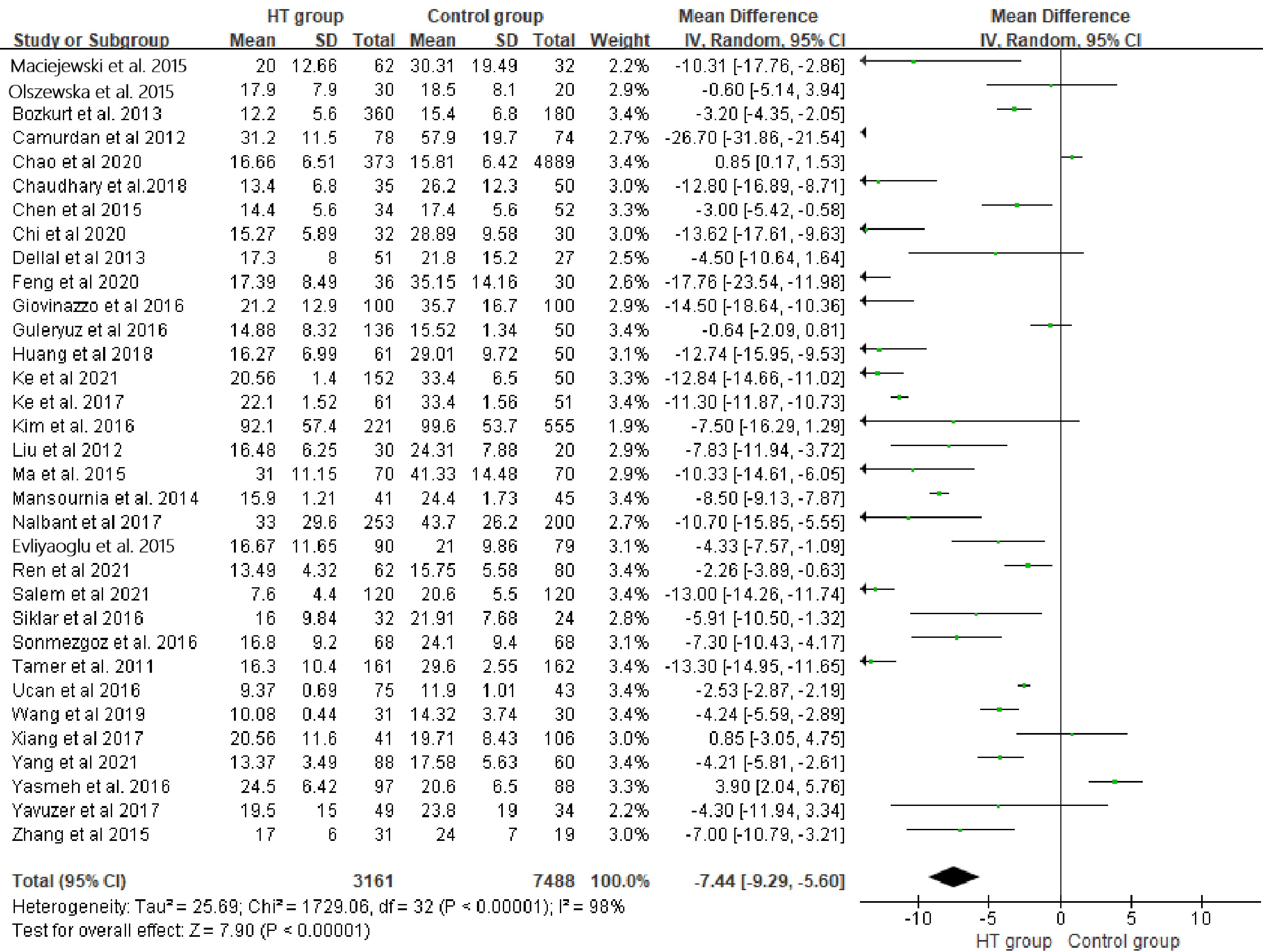
      Figure 1. 

      Forest plot of 25(OH)D3 level (random model).

    • Studies were finally included if they fulfilled the following criteria: (1) included the comparison of HT patient group with a healthy control group; (2) serum levels of 25(OH)D3 level and/or the quantity of patients with 25(OH)D3 insufficiency were reported; (3) written in English or Chinese; (4) with a quality score above or equal to 6 according to the coding manual for case-control studies[15] assessed by two authors respectively.

    • The following information was extracted from each study by two investigators independently: (1) the first author; (2) year of publication; (3) region; (4) sample size; (5) serum levels of 25(OH)D3; (6) cut-off of serum 25(OH)D3 insufficiency; (7) the quantity of patients with 25(OH)D3 insufficiency; and (8) quality score. After that, the extracted information was summarized and checked by another two authors.

    • RevMan 5.3 (the Cochrane Collaboration) was used to perform a meta-analysis on the data obtained. Firstly, Weighted mean differences (WMD) for continuous variable and Odds Ratios (OR) for binary variables were calculated. Subsequently, statistical heterogeneity was assessed with I2 test, and the main source of heterogeneity was revealed by subgroup analysis. Lastly, publication bias was evaluated by funnel plots. For statistical analysis above, 'P < 0.05' was considered a significant difference between groups.

    • Online search obtained 336 studies, in which 280 were excluded by abstract screening. Then, of the 56 remaining, 10 were excluded by full text in-detail evaluation; finally 46 studies with 15,336 individuals in total (6,138 HT patients and 9,198 healthy controls) were included into the present study for the systematic review[1661]. Characteristics of included studies are summarized in Table 1.

      Table 1.  Characteristics of included studies.

      Study
      (published year)
      RegionSample size (HT:C)25(OH)D3
      Assay method
      Serum 25(OH)D3 level
      (HT vs C)
      (ng/mL)
      Serum 25(OH)D3 insufficiency cut off (ng/mL)Number of 25(OH)D3
      insufficiency (HT:C)
      Quality
      score
      Maciejewski et al. 2015[23]Poland62/32ELISA
      8.00 ± 5.06 vs
      12.12 ± 7.80
      < 3061/277
      Ucan et al. 2016[27]Turkey
      75/43RIA
      9.37 ± 0.69 vs
      11.9 ± 1.01
      < 2075/369
      Bozkurt et al. 2013[12-17]]Turkey
      360/180CLS12.2 ± 5.6 vs
      15.4 ± 6.8
      < 10150/378
      Kim 2016[20]Korea221/555CLS36.84 ± 22.96 vs
      39.84 ± 21.48
      < 30108/2068
      Sonmezgoz et al. 2016[25]Turkey
      68/68CLS16.8 ± 9.2 vs
      24.1 ± 9.4
      < 3061/548
      De Pergola et al. 2018[18]Italy
      45/216CLS< 2031/1138
      Botelho et al. 2018[16]Brazil
      88/71CLS26.4 (7.6–48.2) vs
      28.6 (13–51.2)
      < 3061/397
      Ma et al. 2015[22]China70/70ELISA
      12.40 ± 4.46 vs
      16.53 ± 5.79
      < 3070/677
      Yasmeh et al. 2016[29]America97/88CLS24.5 ± 6.42 vs
      20.6 ± 6.5
      < 3066/747
      Xu et al. 2018[28]China194/200CPBA16.16 (13.72–18.76) vs
      23.32 (20.84–25.92)
      7
      Kivity et al. 2011[21]Israel
      28/98CLS< 1022/308
      Mansournia et al. 2014[24]Iran
      41/45SC15.9 ± 1.21 vs
      24.4 ± 1.73
      < 2034/248
      Tamer et al. 2011[26]Turkey
      161/162RIA
      16.3 ± 10.4 vs
      29.6 ± 2.55
      < 30148/1028
      Chaudhary et al. 2018[32]India
      35/50HPLC13.39 ± 6.8 vs
      26.16 ± 12.28
      < 2031/388
      Evliyaoğlu et al. 2015[31]Turkey
      90/79HPLC16.67 ± 11.65 vs
      20.99 ± 9.86
      < 2080/698
      Unal et al. 2014[30]Turkey
      254/124CLS17.05 (5.4−80) vs
      19.9 (9−122.7)
      < 20160/-7
      Ke et al. 2017[19]China
      61/51EBL22.10 ± 1.52 vs
      33.40 ± 1.56
      < 2034/127
      Camurdan et al. 2012[33]Turkey
      78/74HPLC31.2 ± 11.5 vs
      57.9 ± 19.7
      < 2069/247
      Dellal et al 2013[34]Turkey51/27RIA
      17.3 ± 8.0 vs
      21.8 ± 15.2
      6
      Siklar et al. 2016[35]Turkey32/24HPLC16.02 ± 9.84 vs
      21.91 ± 7.68
      < 2022/107
      Nalbant et al. 2017[36]Turkey253/200CLS33 ± 29.6 vs
      43.7 ± 26.2
      < 20161/1118
      Giovinazzo et al. 2017[37]Italy
      100/100HPLC21.2 ± 12.9 vs
      35.7 ± 16.7
      < 2070/187
      Guleryuz et al. 2016[38]Turkey
      136/50HPLC14.88 ± 8.23 vs
      15.52 ± 1.34
      6
      Perga et al. 2018[39]Italy
      55/59CLS< 2037/42
      Yavuzer et al. 2017Turkey
      49/34ELISA19.5 ± 15 vs
      23.8 ± 19
      6
      Priya et al. 2016India25/27ELISA14.3 (12.65−17.90)
      vs 26.2 (21.00−32.8)
      6
      Chao et al. 2020[42]China373/4889RIA
      16.66 ± 6.51 vs
      15.81 ± 6.42
      < 20363/47389
      Feng et al. 2020[44]China36/30ELISA17.39 ± 8.49 vs
      35.15 ± 14.16
      6
      Ahi et al. 2020[43]Iran633/200CLS13.22 (8.1−24.27) vs
      20.4 (11.2−29.6)
      7
      Liu and Zhang. 2012[46]China30/20RIA
      16.48 ± 6.25 vs
      24.31 ± 7.88
      7
      Xiang et al. 2017[47]China41/106CLS19.71 ± 8.43 vs
      20.56 ± 11.64
      < 3038/906
      Zhang et al. 2015[48]China31/19HPLC17 ± 6 vs
      24 ± 7
      6
      Chen et al. 2015[45]China34/52CLS14.4 ± 5.6 vs
      17.4 ± 5.6
      < 2029/377
      Li et al. 2015[49]China50/5621.19 (18.40−25.28) vs
      24.06 (18.94−33.90)
      < 3044/376
      Cvek et al. 2021[50]Croatian461/176CLS19.7 (14.4−25.2) vs
      17.3 (13.2−22.7)
      < 20127/657
      Salem et al. 2021[51]Egypt120/120ELISA7.6 ± 4.4 vs 20.6 ± 5.5< 10120/1127
      Hana et al. 2021[52]Egypt112/48HPLC10.1 (8.7−11.7) vs 12.0 (9.3−15.6)< 30101/406
      Olszewska et al. 2020[53]Italy30/2017.9 ± 7.9 vs 18.5 ± 8.16
      Rezaee et al. 2017[40]Iran51/45CLS6
      Ren et al. 2021[55]China62/8013.49 ± 4.32 vs 15.75 ± 5.85< 3060/766
      Huang et al. 2018[56]China61/50CLS16.27 ± 6.99 vs 29.01 ± 9.72< 206
      Chi et al. 2020[57]China32/30CLS15.27 ± 5.98 vs 28.89 ± 9.586
      Yang et al. 2021[58]China88/6013.37 ± 3.49 vs 17.58 ± 5.636
      Ke et al. 2021[59]China152/50CLS20.56 ± 1.4 vs 33.4 ± 6.5< 2090/67
      Wang et al. 2015[64]China31/30ELISA10.08 ± 0.44 vs 14.32 ± 3.746
      Fu et al. 2021[61]China334/30016.84 (11.81, 23.39) vs 16.66 (11.98, 22.13)< 30214/2097
      H: hashimoto thyroiditis group; C: Healthy control group; ELISA: Enzyme Linked Immunosorbent Assay; RIA: Radioimmunoassay; CLS: Chemiluminesent lmmunoassay Assay; CPBA: competitive protein binding assay; SC: Solid Chromatography, HPLC: High Performance Liquid Chromatography, EBL: Euglobulin lysis method, −: Non reported.
    • Meta-analysis included 33 studys with 3,161 patients in HT group and 7,488 healthy individuals in the control group for comparison. Random model indicated 25(OH)D3 levels of HT group were significantly lower than the control group (WMD: −7.44, 95%CI [−9.29, −5.60], P < 0.01). I2 test (98%) suggested significant heterogeneity in the meta-analysis (Fig. 1). The subgroup meta-analysis basing on 25(OH)D3 assays in a fixed model revealed similar results (WMDs: −0.55; 95%CI [−0.60, −0.49], P < 0.01), and its significant heterogeneity among subgroups represented by I2 = 98.3% suggested the difference of 25(OH)D3 assays was the main source of heterogeneity (Fig. 2,). Lastly, we separated the Chinese studies with 6,639 individuals (HT: 1,102 vs C: 5537) to perform another particle meta-analysis in random model. Result showed that, in the Chinese population, serum 25(OH)D3 level of HT patients was significantly lower than that of healthy individuals (WMD: −7.04, 95%CI [−10.37, −3.71], P < 0.01 ). Meanwhile, I2 = 98.0% also suggested a significant heterogeneity (Fig. 3).

      Figure 2. 

      Subgroup forest plot of 25(OH)D3 level(fixed model).
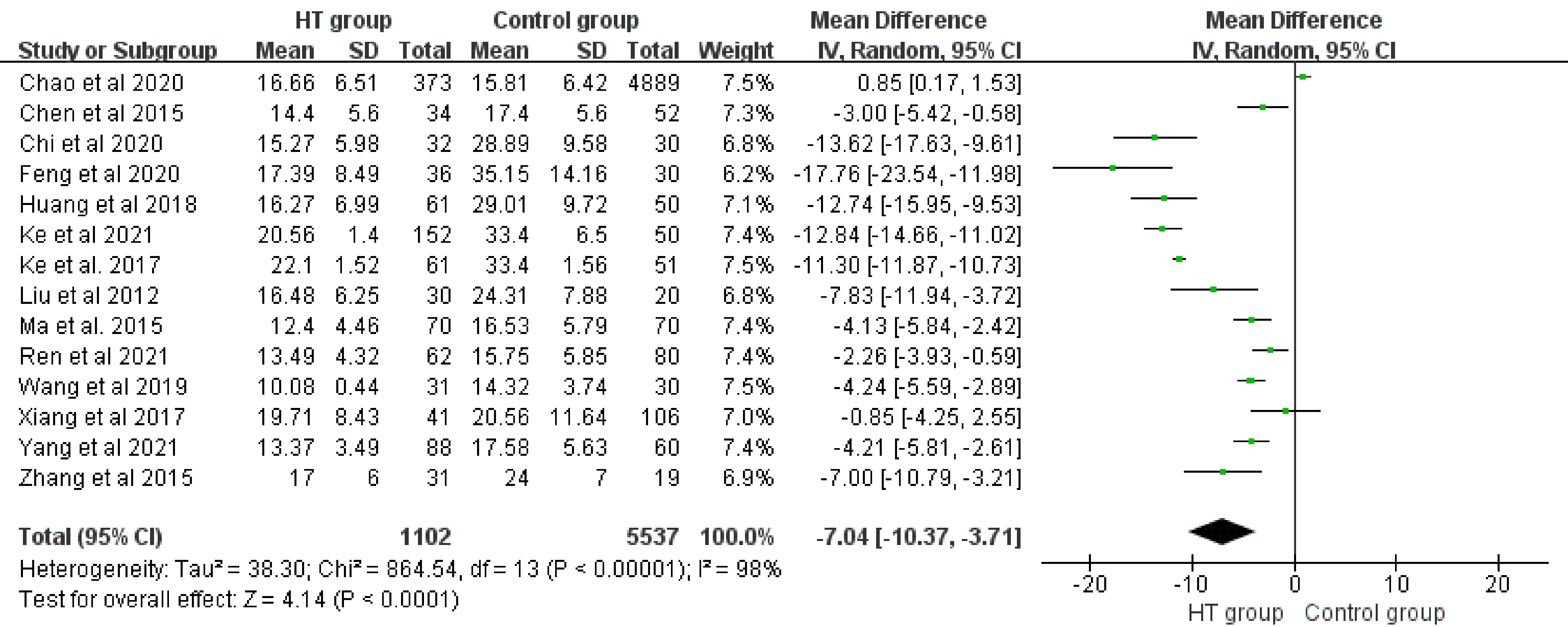
      Figure 3. 

      Forest plot of prevalence of Vitamin D insufficiency (random model).

    • A total of 29 studies comprising 11,795 individuals (HT: 3,709 vs C: 8,086) were pooled for OR of 25(OH)D3 insufficiency. Random model indicated HT patients had higher prevalence of Vitamin D insufficiency compared to healthy individuals (OR: 2.54, 95%CI [1.77, 3.63], P < 0.01). I2 test (86%) suggested significant heterogeneity in meta-analysis (Fig. 4). Subgroup meta-analysis in a fixed model based on different 25(OH)D3 insufficiency cut-off also revealed similar results as above (OR: 1.84; 95%CI [1.64, 2.07], P < 0.01). Meanwhile, I2 equaled to 93% suggested the main source of heterogeneity was from the different cut-off of 25(OH)D3 insufficiency (Fig. 5). Chinese studies with 1,373 individuals (HT: 700 vs C: 673) were separated to perform another particle meta-analysis in random model. Results displayed a trend that the HT population had a higher prevalence of 25(OH)D3 insufficiency compared to healthy individuals, but it was not statistically significant (P > 0.05), and meanwhile significant heterogeneity was indicated by I2 equal to 91% (Fig. 6).

      Figure 4. 

      Subgroup forest plot of prevalence of Vitamin D insufficiency (fixed model).
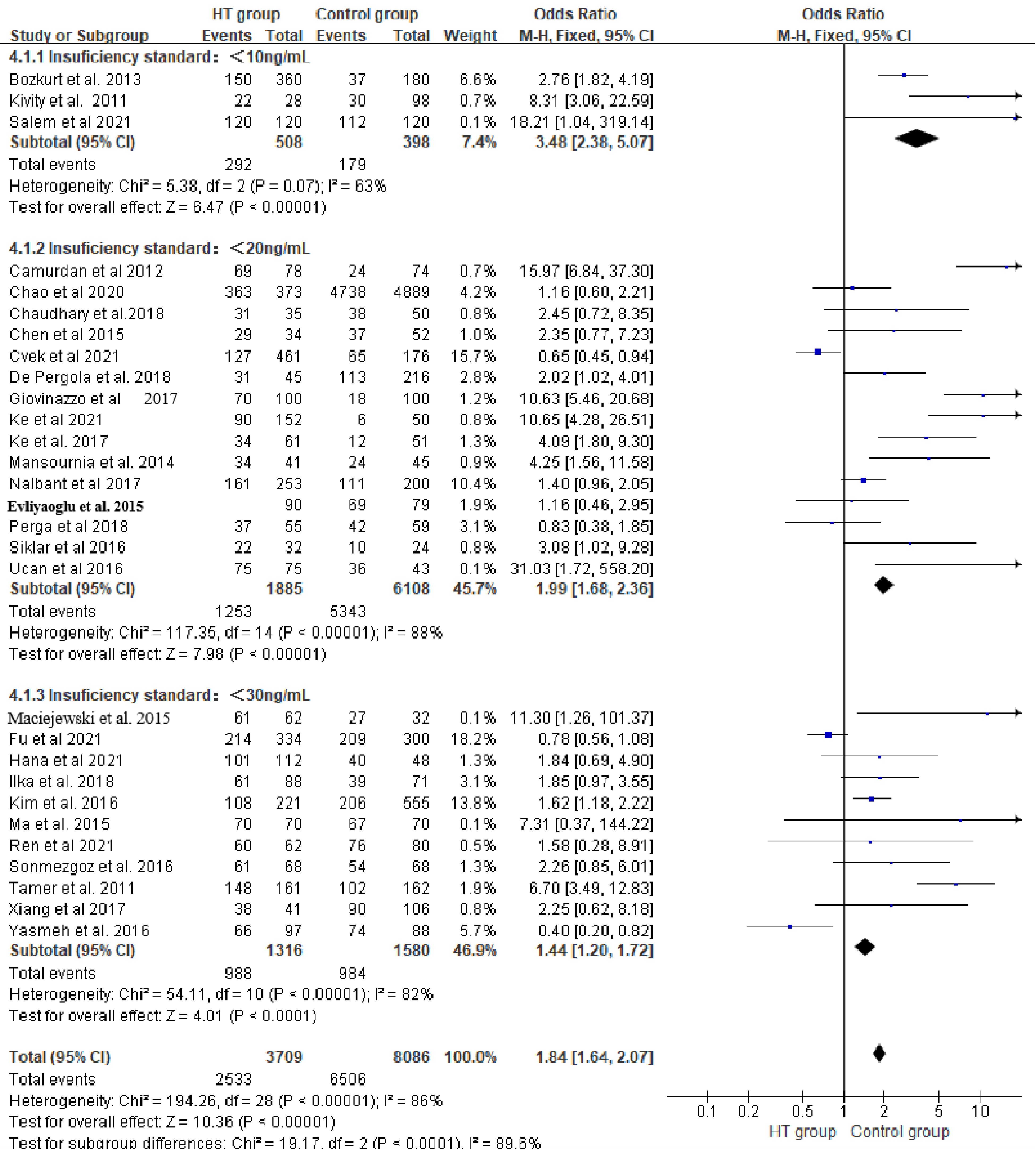
      Figure 5. 

      Forest plot of 25(OH)D3 level (random model, Chinese studies).
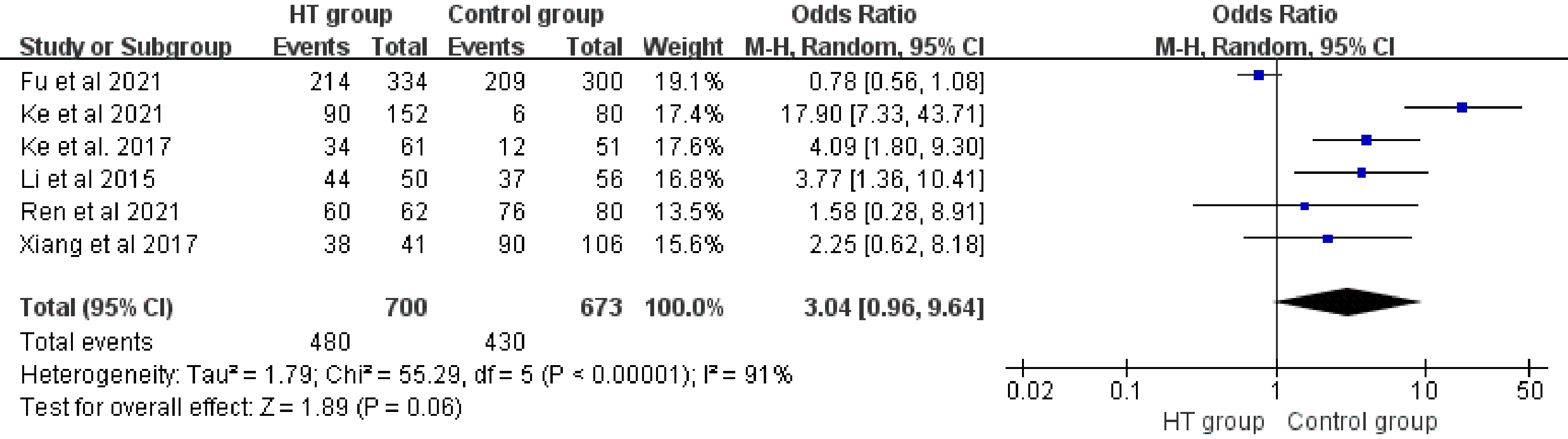
      Figure 6. 

      Forest plot of prevalence of Vitamin D insufficiency (random model, Chinese studies).

    • A funnel plot of serum 25(OH)D3 level in subgroup analysis exhibited that the included studies accumulated at the top of the funnel, which suggested that publication bias may exert little adverse effect on the confidence in the meta-analysis (Fig. 7). Similarly, results of the funnel plot suggested low risk of publication bias in prevalence of 25(OH)D3 insufficiency comparisons (Fig. 8).
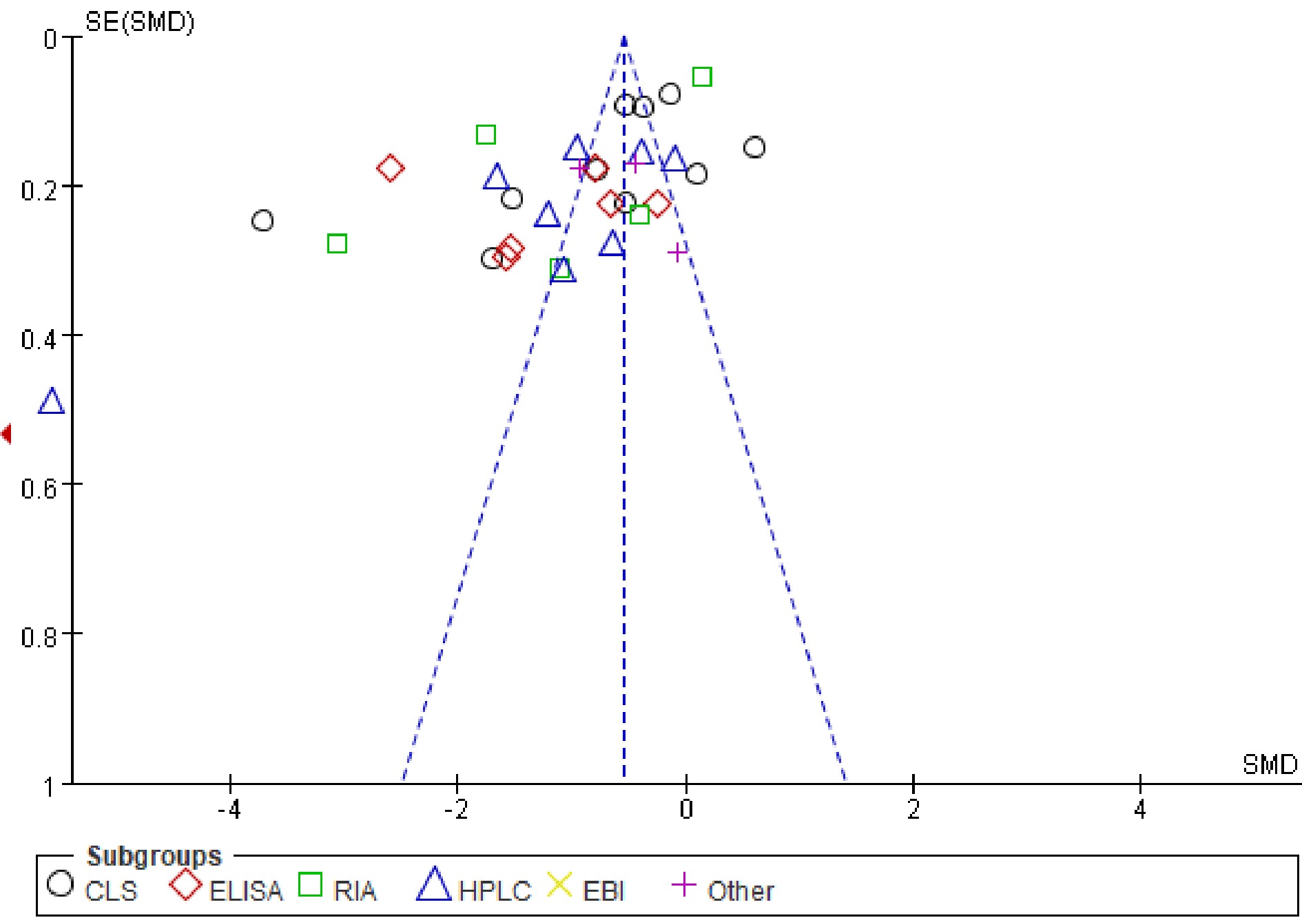
      Figure 7. 

      Funnel plot of 25(OH)D3 level.
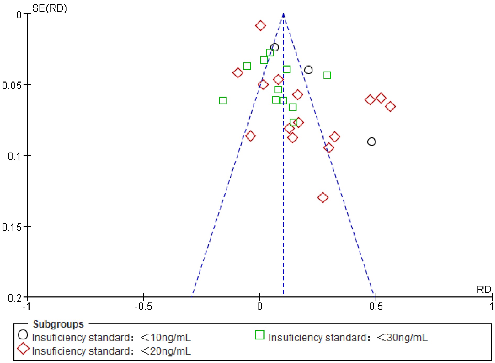
      Figure 8. 

      Funnel plot of quantity of individuals with 25(OH)D3 insufficiency.

    • The present study reinforced the close relationship between Vitamin D insufficiency and HT with methods of systematic review and meta-analysis. To our knowledge, the present systematic review summarized the most related studies to date; among them, Chinese studies, which may be ignored by other foreign researchers, were also included. Hence, we believe our conclusion produce more confident evidence for a relationship between Vitamin D insufficiency and HT.

      Although a series of related factors of HT have been revealed, the real etiology has so far not been clearly understood[62]. Vitamin D has been proved to closely relate to HT, for it plays a vital role in regulating inflammatory response and maintaining immune balance[63]. Multiple epidemiological studies suggested a close relationship between Vitamin D and HT; however, differences in quality, region and population may affect the conclusions. Therefore, high quality systematic review or meta-analysis was still needed to acquire more reliable evidence. Wang et al.[64] published a meta-analysis in 2015 to indicate the relationship between Vitamin D insufficiency and HT. Štefanić et al.[65] subsequently included more recent studies for meta-analysis and drew a similar conclusion, but their results seemed to weaken the relationship of Vitamin D insufficiency and HT compared with Wang et al.[64]. However, these two studied did not include enough recent Chinese studies which should not be ignored. This may not only decrease the confidence of the conclusions, but also weaken the reliability for Chinese researchers. To fulfill this deficiency, we included Chinese studies into the present systematic review and meta-analysis. As anticipated, the general results including serum 25(OH)D3 level and quantity of individuals with Vitamin D insufficiency displayed similar results to Wang et al.[64] and Štefanić et al.[65], which meant the relationship of Vitamin D insufficiency and HT also existed in the Chinese population. We next separated the Chinese studies to perform a particle meta-analysis. The particle result of serum 25(OH)D3 levels of Chinese HT patients were significantly lower than healthy individuals generally, but its difference was less (−7.05 vs −7.44). However, concerning the prevalence of Vitamin D insufficiency, Chinese HT patients were not likely to have more Vitamin D insufficiency cases compared to healthy individuals, suggesting the relationship between Vitamin D insufficiency and HT in the Chinese population may not be as strong as in the global population. Note that, the Chinese population in the present study was only a small part of the total, the negative result maybe due to the small sample size. Further studies with larger sample sizes and high quality investigating the prevalence of Vitamin D insufficiency in the HT population are necessary in China in the future.

      With regards to the studies included in the systematic review, most studies reported lower serum 25(OH)D3 levels and higher prevalence of Vitamin D insufficiency in HT patients compared to healthy individuals. The present study had drawn a similar conclusion, but the results contained significant heterogeneity. According to the systematic review, this heterogeneity may be due to the difference of 25(OH)D3 assays, thus we performed an analysis which separated studies with the same assay into several sub-groups. Results similarly indicated lower serum 25(OH)D3 levels in HT patients, and the most significant heterogeneity among sub-groups (I2 = 99.5%), which hinted that the heterogeneity was mainly caused by the difference in 25(OH)D3 assays. In parallel with lower serum 25(OH)D3 levels, HT patients were at higher risk of 25(OH)D3 insufficiency, indicated by our meta-analysis. Meanwhile, significant heterogeneity was indicated owing to the difference of serum 25(OH)D3 insufficiency criteria. With regard to the publication bias, we determined that heterogeneity would bring significant publication bias displayed by the funnel plot, but the results showed that the included studies accumulating at the top of the funnel; this suggested the publication bias exerted little influence on our results. However, our study also had limitations, as 25(OH)D3 level in a population could be affected by many other factors such as sunshine duration, season, area, economy, and education, which was not considered in our study. These factors may affect the conclusions of epidemiological research, and bring bias to the meta-analysis. Therefore, we can only indicate that vitamin D insufficiency was related to HT. Whether vitamin D insufficiency could lead to HT should be further investigated by biological research in the future.

      Until recently, further investigations focussed on the HT mechanism in which vitamin D was involved. As is well understood, T lymphocytes including Th1, Th2 and Th17 cells infiltrate the thyroid gland due to immunological disorders in HT patients. Vitamin D can inhibit the differentiation of Th1 cells, and the production of inflammatory cytokines such as TNF-α, INF-γ. It could also suppress inflammatory Th1, but induce anti-inflammatory Th2 which produced anti-inflammatory cytokines such as IL-4 and IL-5[66]. Furthermore, the Th17 cells with their production of IL-17A could also be inhibited by Vitamin D at the transcriptional level[67]. On the other hand, vitamin D could increase the proportion of Treg cells to exert immune regulation[68]. Taken together, vitamin D may have the potential to prevent HT. However, clinical studies have shown contradictory results: Chahardoli et al.[69] reported activated vitamin D supplementation can decrease TSH and TG-Ab antibodies levels in HT patients, but another study showed that activated vitamin D supplementation had no effect on improving HT[70]. To our knowledge, the studies mentioned above may ignore the vitamin D receptors polymorphism. Vitamin D receptors in the thyroid gland have single nucleotide polymorphism and most typical Apal, Bsml, Fokl and Taql single nucleotide variations have been shown to be closely related to autoimmune diseases[71]. Therefore, more prospective studies are needed to confirm the preventive effect of vitamin D on HT.

    • In conclusion, the present systematic review and meta-analysis strengthened the relationship between vitamin D insufficiency and HT. HT patients potentially had higher propensity for having lower serum 25(OH)D3 levels compared to healthy individuals. Clinical staff may have to carefully consider the possibility of vitamin D insufficiency in HT patients.

      • The authors declare that they have no conflict of interest.

      • Copyright: © 2023 by the author(s). Published by Maximum Academic Press on behalf of Nanjing Agricultural University. This article is an open access article distributed under Creative Commons Attribution License (CC BY 4.0), visit https://creativecommons.org/licenses/by/4.0/.
    Figure (8)  Table (1) References (71)
  • About this article
    Cite this article
    Liu Z, Feng L, He Y, Yuan S, Xu C. 2022. The Association between Vitamin D and Hashimoto Thyroiditis: An Up-to-date Systematic Review and Meta-analysis. Food Materials Research 2:9 doi: 10.48130/FMR-2022-0009
    Liu Z, Feng L, He Y, Yuan S, Xu C. 2022. The Association between Vitamin D and Hashimoto Thyroiditis: An Up-to-date Systematic Review and Meta-analysis. Food Materials Research 2:9 doi: 10.48130/FMR-2022-0009

Catalog

  • About this article

/

DownLoad:  Full-Size Img  PowerPoint
Return
Return