ARTICLE   Open Access    

Tissue-specific transcriptomics reveals a central role of CcNST1 in regulating the fruit lignification pattern in Camellia chekiangoleosa, a woody oil-crop

  • # These authors contributed equally: Chao Yan, Ziyan Nie

More Information
  • Fruit lignification is of significant economic importance because it affects the quality of fruit and the production of seed oil. The specified lignification pattern in Camellia chekiangoleosa fruits plays critical roles in its seed oil yield, but little is known about how this lignification process is regulated. Here, we report on a comprehensive tissue-specific transcriptomics analysis conducted for C. chekiangoleosa fruit. By mining the differentially expressed genes, we found that lignin biosynthesis and transcriptional regulation pathways were significantly enriched in the lignified tissues. The homolog of NST-like transcription factor, CcNST1, was highly expressed in lignified seed coat and endocarp tissues; transgenic analyses of CcNST1 in Arabidopsis and hybrid poplar revealed the enhanced lignification levels of various tissues. Gene expression analysis of the transgenic lines uncovered potential downstream genes involved in the regulation of lignin biosynthesis. This work provides a valuable gene expression resource and identified the pivotal role of CcNST1 in regulating the lignin biosynthesis underlying fruit lignification.
  • Ralstonia solanacearum, a soil-borne pathogen, is responsible for bacterial wilt (BW) disease affecting 200 plant species in over 50 families, including crucial Solanaceous crops like potato, tomato, pepper, eggplant, and tobacco, as well as model plants, such as Arabidopsis thaliana and Medicago truncatula[1]. As such, it is ranked as the world's second most impactful plant pathogenic bacteria in terms of scientific and economic significance[1]. After infecting the host, R. solanacearum enters the plant's vascular system, resulting in wilting and, ultimately, death, thus preparing it for the next transmission cycle[1]. Water is a vital condition for its survival, particularly in agricultural irrigation environments[2]. Remarkably, under sterile water conditions in labs, R. solanacearum can survive without a decrease in pathogenicity for over four years[2], indicating its robust survival capabilities and partly explaining its global dissemination.

    The extensive genetic diversity within R. solanacearum, stemming from factors such as geographical distribution, host range, and pathogenicity, led to the application of the term 'species complex' for this organism. This 'R. solanacearum species complex' (RSSC) consists of four phylotypes, each predominantly originating from different global regions[3]. Phylotype I largely originates from Asia, phylotype II from America, phylotype III from Africa and surrounding islands in the Indian Ocean, and phylotype IV from Indonesia, Japan, and Australia[3]. Recent studies have redefined these phylotypes into separate species, including R. solanacearum (phylotype II), R. pseudosolanacearum (phylotypes I and III), and an array of R. syzygii (phylotype IV)[4,5]. Furthermore, a new sub-clade, referred to as phylotype IIC, has been identified within phylotype II[6]. The phylotypes of RSSC are not related to the host preference, reflecting the genetic diversity and difficulty of prevention and control of bacterial wilt[7].

    The advent of high-throughput sequencing technology has accelerated our understanding of R. solanacearum's genetics. The first complete genome sequence of this pathogen was published in 2002[8]. Currently, the NCBI database archives a total of 325 R. solanacearum genomes, although the hosts for 58 of these strains remain unidentified (Supplemental Table S1). Significantly, the predominant hosts among the identified strains are Solanum lycopersicum (tomato), Solanum tuberosum (potato), and Capsicum annuum (pepper) (Supplemental Table S1). These plants have given rise to 70, 69, and 23 strains respectively, indicating their key role as hosts for R. solanacearum. This substantial genomic repository has streamlined the process of identifying genes related to pathogenicity and comprehending their pathogenic mechanisms. This wealth of information forms an essential foundation for the development of disease-resistant Solanaceous varieties.

    Various Solanaceous crops and their closely related species present a diverse set of plant accessions with varied degrees of disease resistance against R. solanacearum strains. For example, accessions of tomato (S. lycopersicum Hawaii 7996, TML46, CLN1463 and R3034), eggplant (Solanum melongena Ceylan SM 164, SM6, Surya, and AG91-25), pepper (C. annuum CA8 and MC4), potato (S. chacoense and S. sparsipilum), and tobacco (Nicotiana tabacum, N. benthamiana, and N. glutinosa) are resistant to BW[911]. The utilization of somatic hybridization has significantly enhanced BW resistance, especially in eggplant and potato cultivars (Table 1). Resistant somatic hybrids, generated through the fusion of different Solanum species, exhibit improved BW resistance, showcasing somatic hybridization's potential in enhancing resistance traits.

    Table 1.  Production of somatic hybrids through protoplast fusion for bacterial wilt resistance in Solanaceous crops.
    CropParentsReference
    EggplantS. melongena × S. aethiopicum[12]
    S. melongena × S. aethiopicum[13]
    S. integrifolium × S. violaceum[14]
    S. melongena × S. torvum[15]
    S. integrifolium × S. sanitwongsei[16]
    PotatoS. tuberosum × S. commersonii[18]
    S. tuberosum × S. phureja[22]
    S. tuberosum × S. commersonnii[19]
    S. tuberosum × S. stenotomum[23]
    S. tuberosum × S. chacoense[24]
    Eggplant to potatoS. melongena × S. tuberosum[26]
     | Show Table
    DownLoad: CSV

    Resistant somatic hybrids in eggplants were successfully created via electrical fusion between S. melongena and S. aethiopicum[12]. Furthermore, their hybrids exhibited enhanced resistance to BW and displayed superior fertility compared to conventional eggplant varieties[13]. In another notable development, a somatic hybrid line (27-14) was produced through protoplast fusion involving Solanum integrifolium, a commonly used rootstock in eggplant cultivation, and Solanum violaceum, a wild species exhibiting BW tolerance. This hybrid exhibited increased resistance to BW[14]. Similarly, hybrids derived from S. melongena and Solanum torvum demonstrated commendable resistance to BW[15]. In an innovative approach, hybrids originating from UV-irradiated cotyledonary protoplasts of S. integrifolium and iodoacetamide-treated protoplasts of S. sanitwongsei were not only vigorous and capable of bearing viable seeds but also exhibited a morphology intermediate to the parent species. This suggests their potential as eggplant rootstock candidates[16].

    In the pursuit of enhancing BW resistance in potatoes, breeders have identified and integrated resistance traits from various tuber-bearing Solanums into breeding programs. Despite the endosperm balance number incompatibility system restricting a direct cross between S. commersonii and S. tuberosum[17], innovative approaches such as somatic hybridization have enabled successful circumvention of this barrier. For instance, somatic hybrids between S. tuberosum and S. commersonii have overcome sexual incompatibilities, yielding disease-resistant lines compatible with S. tuberosum[18]. Moreover, resistant lines from their hybrids have been successfully bred with S. tuberosum varieties for amplified BW resistance[19]. Subsequent studies have undertaken an in-depth analysis of the behavior of S. commersonii chromosomes and the colonization patterns of R. solanacearum in the introgression lines[20,21]. In a similar vein, resistance traits from other Solanum species have been introduced into S. tuberosum through somatic hybridization. Notably, dihaploid potato and Solanum phureja hybrids have demonstrated increased BW resistance[22]. Remarkably, S. tuberosum and S. stenotomum hybrids have retained their parent's resistance level even after five years of in vitro maintenance[23]. Further, somatic hybrids originating from the protoplast fusion between S. tuberosum and S. chacoense exhibited enhanced BW resistance, marked by the presence of three specific S. chacoense simple sequence repeat (SSR) alleles linked to resistance[24,25]. An impressive achievement was the successful transfer of BW resistance from S. melongena cv. 508.3 to potato AC142-01 via interspecific symmetric protoplast fusion, resulting in resistant somatic hybrids dominant in potato parent nuclear genomes[26]. In the same lab, researchers also carried out asymmetric protoplast fusion and fused UV-treated protoplasts of the same resistant eggplant variety 508.3 with protoplasts of another susceptible potato variety to obtain 32 somatic hybrids, revealing the introgression of alien chromosome fragments and suggesting potential markers, emk03O04, emi04P17, and emd13E02a, associated with bacterial wilt resistance[27]. Later, researchers in the same lab successfully identified a gene, smPGH1, and seven BW-linked SSRs in somatic hybrids of potato and eggplant, providing valuable genetic resources for improving bacterial wilt resistance in cultivated potato through genome composition and transcriptome analysis[28].

    In conclusion, developing additional resistant germplasm is essential to meet the co-evolutionary challenges posed by the pathogen and the diverse demands for improved agronomic traits. Broadening the scope of BW resistance through hybrid creation between resistant and susceptible varieties offers a promising approach to safeguard Solanaceous crops. Furthermore, ploidy manipulation through sexual hybridization could present viable alternatives to surpass sexual barriers, as exemplified by S. commersonii. By doubling its chromosome number, a 4x line of S. commersonii was crossed with 2x genotypes, generating triploid progeny. These triploids, producing 2n eggs, were crossed with S. tuberosum in 3x × 4x configurations, yielding offspring with a near-pentaploid chromosome number[29]. Significantly, seven out of 26 near-pentaploid sexual hybrids between S. commersonii and cultivated S. tuberosum displayed S. commersonii-like resistance to BW, with notable inhibition of bacterial growth in the plant's aerial part[30]. Subsequent research efforts by various groups have also accomplished the successful transfer of resistance from S. commersonii to S. tuberosum[31,32]. The ongoing advancement of such resistant germplasm is indispensable for effectively combating the evolving challenges from pathogens and meeting the diverse demands for improved agronomic traits.

    Plants employ sophisticated systems, including pattern-recognition receptors (PRRs) and nucleotide-binding leucine-rich repeat domains (NLRs), to perceive and defend against diverse microbial molecules. Positioned on the plasma membrane, PRRs operate as extracellular receptors that discern pathogen-associated molecular patterns (PAMPs). Conversely, NLRs, which reside within the cell, act as intracellular receptors to sense proteins that pathogens directly inject into plant cells[33]. These mechanisms, broadly considered as invasion patterns, initiate a sequence of immune responses, strengthening the plant's defenses[34].

    In A. thaliana, various PRRs involved in bacterial recognition have been identified, including FLS2, EFR, XPS1, RLP1, RLP32, LYM1/3, and LORE[33]. However, recognition of the elongation factor Tu (EF-Tu) by the Receptor-Like Kinase (RLK) EFR is limited to the Brassicaceae family[35,36]. Intriguingly, Solanaceous plants, lacking EFR, are incapable of recognizing EF-Tu from R. solanacearum. Yet, transgenic introduction of Arabidopsis EFR in tomato and potato enhances disease resistance to R. solanacearum infection, demonstrating that interfamily transfer of PRRs can extend recognition of bacterial PAMPs, potentially providing durable disease resistance[31,35,3739]. Moreover, the recent discovery of an R. solanacearum csp22 peptide (csp22Rsol) has been found to initiate immune responses in N. benthamiana and tomato, but not in A. thaliana[40]. Remarkably, csp22Rsol treatment boosted resistance to BW in tomato. Even more, transgenic A. thaliana plants expressing the tomato csp22 receptor (SlCORE) acquired the ability to respond to csp22Rsol and developed greater resistance to R. solanacearum infection[40].

    The main virulence determinant of RSSC bacteria is the type III secretion system (T3SS), a 'molecular syringe' that allows the translocation of several type III effector proteins (T3Es) directly into the host cell[41]. Termed Ralstonia Injected Proteins (Rips), these T3Es can be detected by Nucleotide-binding Leucine-rich Repeat proteins (NLRs) as avirulence effectors, thereby triggering resistance to BW. In A. thaliana, extensive molecular studies have identified the major resistance gene RRS1, encoding a Toll/Interleukin-1 Receptor-Nucleotide Binding Site-Leucine-Rich Repeat (TIR-NBS-LRR) resistance protein that interacts directly with the avirulence effector PopP2[4244]. In addition, RRS1 requires the RD19 gene-encoded Cys protease to mediate resistance to the phylotype I strain GMI1000[45]. Moreover, the RRS1 gene collaborates with the RPS4 gene to enhance resistance to strains of both Pseudomonas and RSSC carrying AvrRps4 and PopP2 effectors, respectively[46,47].

    Within Solanaceous crops, the identification of intracellular receptors remains relatively limited. The major gene ERs1 has been discovered in eggplant via a map-based cloning method[48]. Utilizing a candidate gene approach, RE-bw, an intracellular receptor encompassing both NB-ACR and WRKY domains, was located within eggplant and proven to confer resistance against BW[49]. Using a multiplexed NbNLR-VIGS library, the RRS-Y (RESISTANCE TO RALSTONIA SOLANACEARUM RIPY) was identified in N. benthamiana[50]. Further, specific Rips have been detected as avirulence factors in Solanaceous plants, which can be recognized by known NLR proteins against other pathogens. As an example, RipB, a homolog of Xanthomonas XopQ, is recognized by the N. benthamiana NLR protein Roq1, signifying it as an avirulence factor in N. benthamiana[51]. Both RipE1 and RipBN can be recognized by Ptr1, thereby conferring resistance to BW in N. benthamiana and S. lycopersicoides, respectively[5254]. Additional effectors, including RipA5, RipH2, RipP1, RipP2, RipX, RipAA, RipAT, RipAV, RipAX2, and RipBI, have been observed to trigger cell death in Solanaceous plants[55]. Moreover, a comparative genomic analysis of R. solanacearum strains HA4-1 and HZAU091 led to the identification of four candidate avirulence effectors in HA4-1 that trigger immunity in wild potatoes[56,57]. As research advances, the discovery of further intracellular receptors can be optimistically anticipated.

    As a model pathogen for root and vascular diseases, R. solanacearum contains a great quantity of functionally characterized T3Es. The T3Es have evolved sufficiently to adapt to the plant immune system over a long period of natural evolution, making them indispensable molecular probes in plant immunity studies. A pan-effectome of 140 R. solanacearum strains has been created, comprising 102 known T3Es and 16 putative ones[58]. Recently, the novel T3E RS_T3E_Hyp9 was identified and renamed as RipBT, shown to promote R. solanacearum infection in potatoes[59]. Roughly half of these effectors have been characterized to varying degrees, with nine having identified host targets (Table 2[58]). For instance, RipAB targets TGA transcription factors to disrupt SA signaling and suppress plant immunity[60]. RipAC inhibits MAPK-mediated SGT1 phosphorylation and targets the plant E3 ubiquitin ligase PUB4 to repress immunity[61,62]. RipAC also targets a quantitative susceptibility factor BWS1 to regulate the SGT1-dependent immune response[63]. RipAK can inhibit host catalases and the oligomerization and enzymatic activity of pyruvate decarboxylases to promote disease[64,65]. RipAS diminishes the nucleolar accumulation of StTOPP6, contributing to virulence in potato[66]. RipAY degrades glutathione, inhibits the RipE1-triggered immune responses, and thus suppresses the immune response[6769]. RipD targets VAMP721/722 to promote disease[50]. RipI is a multifunctional effector that modulates plant metabolism and immunity. RipI enhances gamma-aminobutyric acid (GABA) accumulation by promoting calmodulin binding to glutamate decarboxylases (GADs), which contribute to virulence in tomato and Arabidopsis[70]. However, RipI also interacts with the transcription factor bHLH93, triggering a defense response in N. benthamiana[71]. RipTAL targets a 17-bp sequence upstream of arginine decarboxylase (ADC) genes, inhibiting the growth of R. solanacearum niche competitors in tomato[72,73]. Lastly, RipX suppresses the expression of mitochondrial atpA, inducing a defense response in N. benthamiana[74]. These identified host targets represent a wealth of genetic resources for breeding resistance to BW.

    Table 2.  Rips with identified host targets in Solanaceous crops.
    RipsAnonationTarget genesTarget plantsReferences
    RipAB(PopB), harboring proteinTGA transcription factorsSolanum tuberosum; Solanum lycopersicum; Arabidopsis thaliana.[60]
    RipAC(PopC), LRR domainSGT1; PUB4; BWS1Arabidopsis thaliana; Solanum lycopersicum; Nicotiana benthamiana[6163]
    RipAKCATs; PDCsNicotiana tabacum; Arabidopsis thaliana; Solanum lycopersicum.[64,65]
    RipASTOPP6Solanum tuberosum[66]
    RipAYTRX-hArabidopsis thaliana; Nicotiana benthamiana.[6769]
    RipBInosine-uridine nucleoside
    N-ribohydrolase
    Roq1Nicotiana benthamiana
    RipBNcysteine protease, AvrRPt2 familyPtr1Solanum lycopersicoides[52,53]
    RipDVAMP721/722Arabidopsis thaliana[66]
    RipE1Ptr1Nicotiana benthamiana[54]
    RipIbHLH93 transcription factor; plant calmodulin and GADsSolanum lycopersicum; Arabidopsis thaliana[70,71]
    RipP2AcetyltransferasebHLH94 transcription factorArabidopsis thaliana[44]
    RipTALTranscription Activator-Like proteinbHLH95 transcription factorSolanum lycopersicoides[72,73]
    RipX(PopA), HarpinbHLH96 transcription factorNicotiana benthamiana[74]
    RipYAnkyrin RepeatsbHLH97 transcription factorNicotiana benthamiana[50]
     | Show Table
    DownLoad: CSV

    Assisting in resistance breeding to R. solanacearum, marker-assisted selection (MAS) significantly improves breeding efficiency for oligogenic or polygenic resistance within Solanaceous crops. Notably, the Solanaceae family's genetic diversity offers multiple sources of BW resistance, extensively studied in tomatoes (Table 3). A case in point is the Hawaii 7996 tomato cultivar, which has demonstrated exceptional resistance against BW, achieving an impressive 97% survival rate across 12 field trials conducted in 11 countries spanning Asia, America, Australia, and the Indian Ocean region[75]. This resilience makes Hawaii 7996 a stable source of resistance and an ideal resistant parent for the creation of the interspecific population Hawaii7996 × Wva700, designed for resistance mapping studies. Pioneering research discovered a genetic locus on chromosome 12 exhibiting robust resistance against a certain R. solanacearum strain, which alongside another locus on chromosome 6, contributed significantly to the control of resistance[76]. The marker TG564 on chromosome 12 emerged as the primary association with resistance, accounting for a substantial proportion of the genetic variation[76]. Subsequent research pinpointed four quantitative trait loci (QTLs)-Bwr-3, Bwr-4, Bwr-6, and Bwr-8-that accounted for 3.2 to 29.8% of the phenotypic variation, with Bwr-6 and Bwr-3 persistently detected in both cool and hot seasons, while Bwr-4 and Bwr-8 were only detected during the hot season, implicating environmental factors in resistance manifestation[77]. The study underscored the importance of Bwr-6 and Bwr-3 in resistance to R. solanacearum race 3-phylotype II and suggested a potential overlap with resistance QTLs against other strains[77]. Further studies conducted on recombinant inbred lines identified Bwr-12 and Bwr-6 as principal contributors to resistance, wherein Bwr-12 controlling 17.9%−56.1% of total resistance variation, and Bwr-6 accounting for 11.5%−22.2% of phenotypic variation, with lines containing resistance alleles from both loci exhibiting the least disease incidence[78]. Thus, these findings reinforce the polygenic nature of tomato resistance to BW and the significance of Bwr-6 and Bwr-12 in conferring resistance.

    Table 3.  Quantitative trait loci (QTLs) and molecular markers linked to the resistance loci to bacterial wilt in Solanaceous crops.
    CropLocusChromosomeMarker IDMarker typeReference
    Tomato12TG564RFLP[76]
    Bwr-3, Bwr-4, Bwr-6 and Bwr-8RFLP[77]
    TSCARAAT/CGA,
    TSCARAAG/CAT
    SCAR[83]
    Bwr-6, Bwr-126, 12SSR[78]
    SCU176-534SCAR[79]
    Bwr-1212KHU-1SNP[80]
    Bwr-66SNP[80]
    SCU176-534SCAR[79]
    Bwr-6, near Bwr-126, 12RsR6-5, RsR12-1CAPS[81]
    Bwr-6.1, Bwr-6.3 and Bwr-126, 12SNP[82]
    Bwr-3Bwr3.2dCAPSSNP[84]
    EggplantERs19AFLP, SSR, and SRAP[48]
    EBWR9(ERs1), EBWR14, EBWR29, 5, 2SNP[86]
    3, 6SNP[87]
    emh21J12, emf01K16SSR[88]
    emb01D10, emh11I06,
    emh02E08, SSR–46
    SSR[89]
    PotatoqBWR-1, qBWR-2, qBWR-3,
    qBWR-4, and qBWR-5
    1, 3, 7, 10 and 11SNP[90]
    PBWR-6b6SNP[91]
    PBWR-6b6Rbw6-1allele-specific[92]
    PepperBw11CAMS451SSR[93]
    qRRs-10.110SNP[93]
    Bwr6w-7.2, Bwr6w-8.1, Bwr6w-9.1, Bwr6w-9.2, and Bwr6w-10.1, Bwr6w-5.1, Bwr6w-6.1,
    and Bwr6w-7.1
    5, 6, 7, 8, 9, 10C07_224926788-HRM, C08_134064617-HRM, C09_3486004-HRM, C10_232244800-HRM, C05_224016474-HRM, and C07_115436147-HRMHRM[95]
    TobaccoqBWR3a, qBWR-3b, qBWR-5a
    and qBWR-5b
    PT20275 and PT30229SSR[97]
    qBWR17aSSR[98]
     | Show Table
    DownLoad: CSV

    Beyond the aforementioned loci, the development of supplementary molecular markers has led to significant advancements. For instance, the SCAR marker (SCU176-534) was associated with BW resistance in the Hawaii 7996 line, as identified through bulked segregant analysis (BSA) and rapid amplified polymorphic DNA (RAPD) techniques[79]. This marker showed promise for accelerating the selection of resistant lines in breeding efforts involving Hawaii 7996. Separate investigations revealed 5,259 non-synonymous single nucleotide polymorphisms (SNPs) between seven BW-resistant tomato varieties and two susceptible counterparts, mainly located on chromosomes 6 and 12. Notably, the SNP marker KHU-1, located in gene Solyc12g009690.1, encoding a putative leucine-rich repeat (LRR) receptor-like protein and potentially linked to the Bwr-12 QTL, effectively differentiated resistant from susceptible tomato varieties[80]. Further developments include the creation of two CAPS markers, RsR6-5 and RsR12-1 on chromosomes 6 and 12, respectively. These markers proved effective in distinguishing between resistant and susceptible tomato varieties to BW[81]. A comprehensive study mapped a genetic chart using 1604 SNP markers, locating seven QTLs linked to BW resistance on chromosomes 6 and 12 within the 'Hawaii 7996' tomato line[82]. By phenotyping 80 BC3F3 near-isogenic lines (NILs), this study verified the specific effects of Bwr-6.1, Bwr-6.3, and Bwr-12 on disease severity after exposure to two different BW strains across two seasons[82]. In another study involving a cross between the resistant cultivar T51A and the susceptible cultivar T9230, a BSA applied to an F2 population identified two markers, TSCARAAT/CGA and TSCARAAG/CAT, using PCR-based amplified fragment length polymorphism (AFLP) techniques. These markers, converted into co-dominant SCAR markers, were found on the opposite side of TRSR-1[83]. Moreover, an analysis of resistance segregation in two populations and whole-genome sequence data from six BW-resistant and nine BW-susceptible tomato lines suggested possible roles of loci other than Bwr-6 and Bwr-12 in conferring resistance[84]. This investigation revealed 27,046 unique SNPs and 5,975 indels in the resistant lines, implicating 385 genes. Among these, a significant variant on chromosome 3, marked by Bwr3.2dCAPS in the Asc gene, was strongly associated with resistance[84].

    Eggplant, exhibiting potential resistance to all phylotypes of R. solanacearum (RSSC), serves as an intriguing subject for studying BW resistance. Recent research on the resistant breeding line AG91-25, derived from S. melongena and S. aethiopicum, yielded promising results[85]. In this context, a recombinant inbred lines (RILs) population, derived from a cross between AG91-25 and a susceptible parent (line MM738), was phenotyped with phylotype I strains[48]. Utilizing AFLP, SSR, and SRAP markers, researchers generated an intraspecific map with 119 molecular markers across 18 linkage groups. This led to the identification of a unique monogenic resistance locus, ERs1, in crop RSSC resistance[48]. When exposed to four additional RSSC strains representing phylotypes I, IIA, IIB, and III, this population showed one major phylotype-specific QTL, EBWR9 (which coincided with the previously identified ERs1), and two broad-spectrum QTLs, EBWR14 and EBWR2[86]. Notably, EBWR14 and EBWR2, located on chromosomes 2 and 5, offered partial resistance to strains of phylotypes I, IIA, III and strains of phylotypes IIA and III, respectively[86]. Additional studies on 123 doubled haploid lines, bred from a susceptible eggplant line (MM738) and a resistant counterpart (EG203), resulted in the mapping of 10 and three resistance QTLs for phylotypes I and III, respectively[87]. Interestingly, the most reliable QTLs were found on chromosomes 3 and 6, with the one on chromosome 6 resonating with the broad-spectrum resistance QTL Bwr-6 observed in tomatoes[87]. Screening of six elite eggplant genotypes in a field setting identified three—CARI-1, IIHR-7, and IIHR-500A—as resistant to BW. This led to the identification of two SSR markers, emh21J12 and emf01K16, associated with this resistance[88]. Subsequently, BSA was performed in two F2 populations exhibiting BW resistance, derived from crosses between resistant lines (CARI-1 and IIHR -7) and susceptible lines (Rampur Local and Arka Kushmakar IIHR-108). The SSR markers emb01D10, emh11I06, emh02E08, and SSR-46 co-segregated with resistant and susceptible genotypes of the two F2 populations and were linked to BW resistance loci[89].

    In the realm of potato research, a crossbreeding effort was undertaken between a resistant diploid line, 10-03-30, and a susceptible diploid line, F1-1. This yielded a diploid F1 population of 94 genotypes, a kind of two-way pseudo-testcross[90]. From this population, five QTLs (qBWR-1 to -5) were identified through QTL analysis. These QTLs were located on potato chromosomes 1, 3, 7, 10, and 11, accounting for 9.3%−18.4% of the phenotypic variance. Of particular note was that the alleles for qBWR-2 to -4 were resistant, whereas those for qBWR-1 and qBWR-5 were susceptible[90]. Subsequent investigation uncovered 10 resistance QTLs in an F1 population, which was derived from a cross between a resistant haploid line from Saikai 35 and a susceptible diploid line[91]. Among these, QTL PBWR-6b was the most effective, originating from the resistant parent and located on potato chromosome 6. In a later study, a resistance allele was identified and an allele-specific molecular marker (Rbw6-1) for PBWR-6b was developed[92]. This discovery marked a significant advance in our understanding of potato resistance to BW.

    In the domain of pepper research, Mimura et al. employed a double haploid mapping population derived from 'California Wonder' (susceptible) and 'LS2341' (resistant) to probe pepper's resistance to BW. This study successfully generated a linkage map encompassing 15 groups through the application of SSRs and AFLP. A significant QTL, Bw1, was discovered on pepper chromosome 1 (P1), accounting for 33% of the resistance attributed to 'LS2341'. This QTL was mapped using the SSR marker CAMS451[93]. More recently, Du et al. shed light on the dynamics of bioluminescent R. solanacearum colonization through an examination using reciprocal grafts of a resistant line (BVRC 1) and a susceptible line (BVRC 25). They pinpointed a key QTL (qRRs-10.1) on chromosome 10, hosting several resistance and defense-related genes, which plays a significant role in BW resistance[94]. Additionally, Lee et al. identified five QTLs (Bwr6w-7.2, Bwr6w-8.1, Bwr6w-9.1, Bwr6w-9.2, and Bwr6w-10.1) conferring resistance to a moderately pathogenic 'HS' isolate. In contrast, three QTLs (Bwr6w-5.1, Bwr6w-6.1, and Bwr6w-7.1) were found to resist a highly pathogenic 'HWA' isolate of R. solanacearum in two F2 populations derived from the highly resistant pepper cultivar 'Konesian hot'. Within the same study, six high-resolution melting (HRM) markers linked to these QTLs were also developed[95].

    In the realm of tobacco research, four QTL mapping studies have been conducted for tobacco bacterial wilt (TBW) resistance across bi-parental and diverse genetic populations, utilizing SSR and AFLP markers. An AFLP analysis identified a significant QTL linked with 15 markers, accounting for over 30% of the resistance variance, within the resistant variety W6 and susceptible variety Michinoku 1, thus yielding 117 useful DNA markers[96]. In an examination of the F2:3 and F2:4 progeny resulting from crosses between wilt-resistant breeding lines (Enshu and Yanyan97) and a susceptible line (TI448A), Qian et al. uncovered four QTLs (qBWR3a, qBWR-3b, qBWR-5a, and qBWR-5b) in linkage groups 3 and 5[97]. The closely linked markers PT20275 and PT30229, detected in both crosses, offer a valuable tool for the selection of resistant plants[97]. In another significant study, a major QTL (qBWR17a) was identified that accounted for 30% of the phenotypic variation, providing a noteworthy advantage for MAS in TBW resistance breeding[98]. A distinct investigation into 'K346' tobacco's resistance to BW associated three QTLs with resistance, explaining 50.3% of the observed variation[99]. Furthermore, a pioneering study identified 142 quantitative trait nucleotides (QTNs) that account for a substantial portion of the phenotypic variance for TBW resistance, with 38 of these QTNs being stable across varied environments and methodologies[100]. This research, marking the first identification of QTNs and superior alleles for the breeding of TBW-resistant tobacco varieties, also suggested the five most effective cross combinations for resistance and highlighted 52 potential candidate genes. These insights are invaluable for future studies in genetic architecture, marker-assisted selection, and functional genomics of TBW resistance, aiming to increase crop productivity. As a result, these discoveries offer instrumental tools for MAS in the breeding program to enhance resistance to BW in Solanaceous crops.

    R. solanacearum presents a considerable threat to Solanaceous crops, and the development of effective genetic control strategies remains a pressing priority. Emerging advancements in genomics, relating to both pathogens and host plants, offer the exciting potential to discover previously unrecognized resources for disease resistance in the near future. As such, intensifying our research efforts in remote hybridization and somatic cell fusion is critical, aiming to increase success rates and create a collection of Solanaceous germplasm with strong resistance to BW. The application of effectors could play a key role in implementing high-throughput methodologies for identifying BW resistance[101]. This strategy may also stimulate resistance screening in wild species, thereby enhancing the selection of disease-resistant materials and receptor identification. When identifying receptors in certain Solanaceous plants proves challenging, we could consider the development of resistant varieties by integrating resistance genes from A. thaliana and other Solanaceous plants. Moreover, genome-editing technologies present promising avenues for manipulating host target genes. The successful completion of whole-genome sequencing for key Solanaceous crops, including potato, tomato, eggplant, pepper, and tobacco (referenced at https://solgenomics.net), is set to expedite the cloning process for resistance genes against BW. Furthermore, the adoption of recent methodological advancements, such as Resistance gene enrichment sequencing (RenSeq)[102], could facilitate quicker receptor identification within Solanaceous crops. These combined efforts give rise to the promising future of R. solanacearum-resistant crop development, transforming it from a distant goal into an imminent reality.

    The authors confirm contribution to the paper as follows: study conception and design: study conception and design: Du J, Wang B, Chen H, Song B; data collection: Du J, Wang B, Huang M, Chen X, Nie L, Wang T; draft manuscript preparation: Du J, Wang B, Huang M, Chen X, Chen H, Song B. All authors reviewed the results and approved the final version of the manuscript.

    All data supporting the findings of this research are available within the paper and within its supplementary data.

    The work was partially supported by the National Natural Science Foundation of China (32201789) and the China Agriculture Research System of MOF and MARA (CARS-09).

  • The authors declare that they have no conflict of interest.

  • Supplemental Table S1 Primers used in this study.
    Supplemental Table S2 Identification of NAC domain transcription factors of Camellia chekiangoleosa  transcriptome based on BLAST search. In total, 15 transcripts were found to contain complete open reading frame (ORF).
    Supplemental Table S3 An overview of the RNA-seq statistics of Camellia chekiangoleosa tissues. Each tissue type includes three biological replicates. Q20 and Q30 represent base call accuracy of 99% and 99.9% respectively. FB, Floral Bud; EX, Exocarp; ME, Mesocarp; EN, Endocarp; SC, Seed Coat; SK, Seed Kernel.
    Supplemental Table S4 The statistics of Camellia chekiangoleosa transcriptome. The transcripts and unigenes are obtained by using the Trinity v2.11.0 software. Sequences less than 200 bp are removed.
    Supplemental Table S5 The identification of genes related to lignin biosynthesis and transcriptional regulation based on the information from Arabidopsis thaliana (TAIR v.11).
    Supplemental Fig. S1 The evaluation of the annotation of Camellia chekiangoleosa transcriptome. A, The distribution of unigenes that are annotated in the Gene Ontology (GO) database. B, The distribution of unigenes that are annotated in the eggNOG database. C, The distribution of unigenes that are annotated in the KEGG database.
    Supplemental Fig. S2 Morphological characterizations of the growth of Camellia chekiangoleosa fruits. A, The morphology of fruit at different timepoints of fruit growth. The lignin patterns at each corresponding stage are displayed below. Bar 2 cm. B, A growth curve of the C. chekiangoleosa fruits is presented by measuring of fruit weight, transverse diameter and vertical diameter. Transverse diameter1 and transverse diameter 2 are two independent measurements with angle around 90 degrees at each sampling point of fruits.
    Supplemental Fig. S3 The identification of NST homolog in Camellia chekiangoleosa. A, The alignment of protein sequences of CcNST1 and its homologs from diverse plant species. The red rectangle indicates the conserved NAM domain. The accession numbers of sequences:   Vitis vinifera: VV15G07370;  Manihot esculenta: ME03581G00220;  Ricinus communis: RC27964G00230;  Theobroma cacao:TC0001G33230; Gossypium raimondii: GR01G15080; Populus trichocarpa: PT14G10480; Citrus sinensis: CS00001G04980; Arabidopsis thaliana: AT2G46770. B, The expression profiles of NST-like transcripts that are identified from the C. chekiangoleosa transcriptome as described in Supple. Table 2. The candidate NST ortholog was underlined by red. C, The phylogenic tree of NST-like genes from Arabidopsis thaliana and C. chekiangoleaosa.
    Supplemental Fig. S4 The verification of transgenic Arabidopsis lines. A, The amplification of construct specific fragments of Arabidopsis genomic DNA. Wt, wild type; N4, N7, N10 are representative 35s:CcNST1 lines. B, The expression of CcNST1 in Arabidopsis lines. The wild type is not detectable (nd). Different letters (a, b) indicate significant difference by the Student’s test p < 0.05.
  • [1]

    Giovannoni JJ. 2004. Genetic regulation of fruit development and ripening. The Plant Cell 16:S170−S180

    doi: 10.1105/tpc.019158

    CrossRef   Google Scholar

    [2]

    Barakate A, Stephens J, Goldie A, Hunter WN, Marshall D, et al. 2011. Syringyl lignin is unaltered by severe sinapyl alcohol dehydrogenase suppression in tobacco. The Plant Cell 23:4492−506

    doi: 10.1105/tpc.111.089037

    CrossRef   Google Scholar

    [3]

    Hamann T. 2012. Plant cell wall integrity maintenance as an essential component of biotic stress response mechanisms. Frontiers in Plant Science 3:77

    doi: 10.3389/fpls.2012.00077

    CrossRef   Google Scholar

    [4]

    Novaes E, Kirst M, Chiang V, Winter-Sederoff H, Sederoff R. 2010. Lignin and biomass: a negative correlation for wood formation and lignin content in trees. Plant Physiology 154:555−61

    doi: 10.1104/pp.110.161281

    CrossRef   Google Scholar

    [5]

    Baucher M, Chabbert B, Pilate G, Van Doorsselaere J, Tollier MT, et al. 1996. Red xylem and higher lignin extractability by down-regulating a cinnamyl alcohol dehydrogenase in Poplar. Plant Physiology 112:1479−90

    doi: 10.1104/pp.112.4.1479

    CrossRef   Google Scholar

    [6]

    Boerjan W, Ralph J, Baucher M. 2003. Lignin biosynthesis. Annual Review of Plant Biology 54:519−46

    doi: 10.1146/annurev.arplant.54.031902.134938

    CrossRef   Google Scholar

    [7]

    Fraser CM, Chapple C. 2011. he phenylpropanoid pathway in Arabidopsis. The Arabidopsis Book 2011:e0152

    doi: 10.1199/tab.0152

    CrossRef   Google Scholar

    [8]

    Weng JK, Chapple C. 2010. The origin and evolution of lignin biosynthesis. New Phytologist 187:273−85

    doi: 10.1111/j.1469-8137.2010.03327.x

    CrossRef   Google Scholar

    [9]

    Zhong R, Lee C, Ye Z. 2010. Functional Characterization of Poplar Wood-Associated NAC Domain Transcription Factors. Plant Physiology 152:1044−55

    doi: 10.1104/pp.109.148270

    CrossRef   Google Scholar

    [10]

    Zhang J, Tuskan GA, Tschaplinski TJ, Muchero W, Chen JG. 2020. Transcriptional and post-transcriptional regulation of lignin biosynthesis pathway genes in Populus. Frontiers in Plant Science 11:652

    doi: 10.3389/fpls.2020.00652

    CrossRef   Google Scholar

    [11]

    Nakano Y, Yamaguchi M, Endo H, Rejab NA, Ohtani M. 2015. NAC-MYB-based transcriptional regulation of secondary cell wall biosynthesis in land plants. Frontiers in Plant Science 6:288

    doi: 10.3389/fpls.2015.00288

    CrossRef   Google Scholar

    [12]

    Zhou J, Lee C, Zhong R, Ye Z. 2009. MYB58 and MYB63 are transcriptional activators of the lignin biosynthetic pathway during secondary cell wall formation in Arabidopsis. The Plant Cell 21:248−66

    doi: 10.1105/tpc.108.063321

    CrossRef   Google Scholar

    [13]

    Tang X, Zhuang Y, Qi G, Wang D, Liu H, et al. 2015. Poplar PdMYB221 is involved in the direct and indirect regulation of secondary wall biosynthesis during wood formation. Scientific Reports 5:12240

    doi: 10.1038/srep12240

    CrossRef   Google Scholar

    [14]

    Patzlaff A, Mclnnis S, Courtenay A, Surman C, Newman LJ, et al. 2003. Characterisation of a pine MYB that regulates lignification. The Plant Journal 36:734−54

    doi: 10.1046/j.1365-313x.2003.01916.x

    CrossRef   Google Scholar

    [15]

    Zhong R, Ye Z. 2015. Secondary Cell Walls: Biosynthesis, Patterned Deposition and Transcriptional Regulation. Plant and Cell Physiology 56:195−14

    doi: 10.1093/pcp/pcu140

    CrossRef   Google Scholar

    [16]

    Mitsuda N, Iwase A, Yamamoto H, Yoshida M, Seki M, et al. 2007. NAC transcription factors, NST1 and NST3, are key regulators of the formation of secondary walls in woody tissues of Arabidopsis. The Plant Cell 19:270−280

    doi: 10.1105/tpc.106.047043

    CrossRef   Google Scholar

    [17]

    Kumar M, Campbell L, Turner S. 2016. Secondary cell walls: biosynthesis, patterned deposition and transcriptional regulation. Journal of Experimental Botany 67:515−31

    doi: 10.1093/jxb/erv533

    CrossRef   Google Scholar

    [18]

    Zhao Q, Dixon RA. 2011. Transcriptional networks for lignin biosynthesis: more complex than we thought. Trends in Plant Science 16:227−33

    doi: 10.1016/j.tplants.2010.12.005

    CrossRef   Google Scholar

    [19]

    Liljegren SJ, Roeder AHK, Kempin SA, Gremski K, Østergaard L, et al. 2004. Control of fruit patterning in Arabidopsis by INDEHISCENT. Cell 116:843−53

    doi: 10.1016/S0092-8674(04)00217-X

    CrossRef   Google Scholar

    [20]

    Liljegren SJ, Ditta GS, Eshed Y, Savidge B, Bowman JL, Yanofsky MF. 2000. SHATTERPROOF MADS-box genes control seed dispersal in Arabidopsis. Nature 404:766−70

    doi: 10.1038/35008089

    CrossRef   Google Scholar

    [21]

    Ferrándiz C, Fourquin C. 2014. Role of the FUL-SHP network in the evolution of fruit morphology and function. Journal of Experimental Botany 65:4505−13

    doi: 10.1093/jxb/ert479

    CrossRef   Google Scholar

    [22]

    Pabón-Mora N, Wong GKS, Ambrose BA. 2014. Evolution of fruit development genes in flowering plants. Frontiers in Plant Science 5:300

    doi: 10.3389/fpls.2014.00300

    CrossRef   Google Scholar

    [23]

    Garceau DC, Batson MK, Pan IL. 2017. Variations on a theme in fruit development: the PLE lineage of MADS-box genes in tomato (TAGL1) and other species. Planta 246:313−21

    doi: 10.1007/s00425-017-2725-5

    CrossRef   Google Scholar

    [24]

    Lyu T, Fan Z, Yang W, Yan C, Hu Z, et al. 2019. CjPLE, a PLENA-like gene, is a potential regulator of fruit development via activating the FRUITFUL homolog in Camellia. Journal of Experimental Botany 70:3153−64

    doi: 10.1093/jxb/erz142

    CrossRef   Google Scholar

    [25]

    Wang W, Zhang J, Ge H, Li S, Li X, et al. 2016. EjMYB8 transcriptionally regulates flesh lignification in loquat fruit. PloS One 11:e0154399

    doi: 10.1371/journal.pone.0154399

    CrossRef   Google Scholar

    [26]

    Jia N, Liu J, Tan P, Sun Y, Lv Y, et al. 2019. Citrus sinensis MYB Transcription Factor CsMYB85 Induce Fruit Juice Sac Lignification Through Interaction With Other CsMYB Transcription Factors. Frontiers in Plant Science 10:213

    doi: 10.3389/fpls.2019.00213

    CrossRef   Google Scholar

    [27]

    Xu Q, Wang W, Zeng J, Zhang J, Grierson D, et al. 2015. A NAC transcription factor, EjNAC1, affects lignification of loquat fruit by regulating lignin. Postharvest Biology and Technology 102:25−31

    doi: 10.1016/j.postharvbio.2015.02.002

    CrossRef   Google Scholar

    [28]

    Wang Q, Hu J, Yang T, Chang S. 2021. Anatomy and lignin deposition of stone cell in Camellia oleifera shell during the young stage. Protoplasma 258:361−70

    doi: 10.1007/s00709-020-01568-z

    CrossRef   Google Scholar

    [29]

    Lin P, Wang K, Wang Y, Hu Z, Yan C, et al. 2022. The genome of oil-Camellia and population genomics analysis provide insights into seed oil domestication. Genome Biology 23:14

    doi: 10.1186/s13059-021-02599-2

    CrossRef   Google Scholar

    [30]

    Shen T, Huang B, Xu M, Zhou P, Ni Z, et al. 2022. The reference genome of Camellia chekiangoleosa provides insights into Camellia evolution and tea oil biosynthesis. Horticulture Research 9:uhab083

    doi: 10.1093/hr/uhab083

    CrossRef   Google Scholar

    [31]

    Yan C, Lin P, Lyu T, Hu Z, Fan Z, et al. 2018. Unraveling the roles of regulatory genes during domestication of cultivated Camellia: evidence and insights from comparative and evolutionary genomics. Genes 9:488

    doi: 10.3390/genes9100488

    CrossRef   Google Scholar

    [32]

    Haas BJ, Papanicolaou A, Yassour M, Grabherr M, Blood PD, et al. 2013. De novo transcript sequence reconstruction from RNA-seq using the Trinity platform for reference generation and analysis. Nature Protocols 8:1494−512

    doi: 10.1038/nprot.2013.084

    CrossRef   Google Scholar

    [33]

    Love MI, Huber W, Anders S. 2014. Moderated estimation of fold change and dispersion for RNA-seq data with DESeq2. Genome Biology 15:550

    doi: 10.1186/s13059-014-0550-8

    CrossRef   Google Scholar

    [34]

    Livak KJ, Schmittgen TD. 2001. Analysis of relative gene expression data using real-time quantitative PCR and the 2−ΔΔCT Method. Methods 25:402−8

    doi: 10.1006/meth.2001.1262

    CrossRef   Google Scholar

    [35]

    Clough SJ, Bent AF. 1998. Floral dip: a simplified method for Agrobacterium-mediated transformation of Arabidopsis thaliana. The Plant Journal 16:735−43

    doi: 10.1046/j.1365-313x.1998.00343.x

    CrossRef   Google Scholar

    [36]

    Kumar S, Fladung M. 2001. Gene stability in transgenic aspen (Populus). II. Molecular characterization of variable expression of transgene in wild and hybrid aspen. Planta 213:731−40

    doi: 10.1007/s004250100535

    CrossRef   Google Scholar

    [37]

    Yin H, Gao P, Liu C, Yang J, Liu Z, et al. 2013. SUI-family genes encode phosphatidylserine synthases and regulate stem development in rice. Planta 237:15−27

    doi: 10.1007/s00425-012-1736-5

    CrossRef   Google Scholar

    [38]

    Chao Q, Gao Z, Zhang D, Zhao B, Dong F, et al. 2019. The developmental dynamics of the Populus stem transcriptome. Plant Biotechnology Journal 17:206−19

    doi: 10.1111/pbi.12958

    CrossRef   Google Scholar

    [39]

    Zhang S, Yang H, Ding L, Song Z, Ma H, et al. 2017. Tissue-specific transcriptomics reveals an important role of the unfolded protein response in maintaining fertility upon heat stress in Arabidopsis. The Plant Cell 29:1007−23

    doi: 10.1105/tpc.16.00916

    CrossRef   Google Scholar

    [40]

    Dardick C, Callahan AM. 2014. Evolution of the fruit endocarp: molecular mechanisms underlying adaptations in seed protection and dispersal strategies. Frontiers in Plant Science 5:284

    doi: 10.3389/fpls.2014.00284

    CrossRef   Google Scholar

    [41]

    Dardick CD, Callahan AM, Chiozzotto R, Schaffer RJ, Piagnani MC, et al. 2010. Stone formation in peach fruit exhibits spatial coordination of the lignin and flavonoid pathways and similarity to Arabidopsis dehiscence. BMC Biology 8:13

    doi: 10.1186/1741-7007-8-13

    CrossRef   Google Scholar

    [42]

    Su X, Zhao Y, Wang H, Li G, Cheng X, et al. 2019. Transcriptomic analysis of early fruit development in Chinese white pear (Pyrus bretschneideri Rehd. ) and functional identification of PbCCR1 in lignin biosynthesis. BMC Plant Biology 19:417

    doi: 10.1186/s12870-019-2046-x

    CrossRef   Google Scholar

    [43]

    Liu W, Zhang J, Jiao C, Yin X, Fei Z, et al. 2019. Transcriptome analysis provides insights into the regulation of metabolic processes during postharvest cold storage of loquat (Eriobotrya japonica) fruit. Horticulture Research 6:49

    doi: 10.1038/s41438-019-0131-9

    CrossRef   Google Scholar

  • Cite this article

    Yan C, Nie Z, Hu Z, Huang H, Ma X, et al. 2022. Tissue-specific transcriptomics reveals a central role of CcNST1 in regulating the fruit lignification pattern in Camellia chekiangoleosa, a woody oil-crop. Forestry Research 2:10 doi: 10.48130/FR-2022-0010
    Yan C, Nie Z, Hu Z, Huang H, Ma X, et al. 2022. Tissue-specific transcriptomics reveals a central role of CcNST1 in regulating the fruit lignification pattern in Camellia chekiangoleosa, a woody oil-crop. Forestry Research 2:10 doi: 10.48130/FR-2022-0010

Figures(6)

Article Metrics

Article views(6336) PDF downloads(641)

ARTICLE   Open Access    

Tissue-specific transcriptomics reveals a central role of CcNST1 in regulating the fruit lignification pattern in Camellia chekiangoleosa, a woody oil-crop

Forestry Research  2 Article number: 10  (2022)  |  Cite this article

Abstract: Fruit lignification is of significant economic importance because it affects the quality of fruit and the production of seed oil. The specified lignification pattern in Camellia chekiangoleosa fruits plays critical roles in its seed oil yield, but little is known about how this lignification process is regulated. Here, we report on a comprehensive tissue-specific transcriptomics analysis conducted for C. chekiangoleosa fruit. By mining the differentially expressed genes, we found that lignin biosynthesis and transcriptional regulation pathways were significantly enriched in the lignified tissues. The homolog of NST-like transcription factor, CcNST1, was highly expressed in lignified seed coat and endocarp tissues; transgenic analyses of CcNST1 in Arabidopsis and hybrid poplar revealed the enhanced lignification levels of various tissues. Gene expression analysis of the transgenic lines uncovered potential downstream genes involved in the regulation of lignin biosynthesis. This work provides a valuable gene expression resource and identified the pivotal role of CcNST1 in regulating the lignin biosynthesis underlying fruit lignification.

    • Lignification is a crucial process of fruit development that plays various roles in different types of fruits[1]. In angiosperms, the lignification of the pericarp is essential for seed protection and their dispersal. In the dry-fruit crop walnut, the extensive lignification of endocarp (known as the nutshell) is a key economic trait; impaired development of the nutshell often adversely affects the quality of walnut kernel, leading to malformed seed kernels. In fleshy fruits, the lignin process is mostly overlooked or considered negligible; however, unexpected lignification of fruit tissues could be caused by disease and stress conditions, which can affect the taste and quality of fruits[2].

      The accumulation of lignin leads to the lignification of fruit, which is essential for maintaining the integrity of the plant cell wall and resisting plant pathogens[3]. During the hardening process of the pericarp, lignin conjugates with the cellulose and hemicellulose network to provide rigidity and tensile strength to secondary walls, in a process similar to wood formation[4]. Lignin is a type of secondary metabolite derived from phenylpropane. After a series of biochemical reactions, including deamination, hydroxylation and methylation, phenylalanine is finally transformed into three types of lignin monomers[2]. These are then separately polymerized to form p-hydroxyphenyl lignin, guaiacyl lignin, and syringyl lignin[5]. The lignin biosynthesis pathway in plants has been extensively investigated. The enzymes involved in its two major processes — monolignol biosynthesis and monolignol polymerization — have been characterized through genetic and biochemical studies[6]. A cascade of conserved core enzymes is fundamental for the biosynthesis of lignin in diverse plant species[7,8].

      In recent years, several transcription factors (TFs) have been identified which control the plant lignification process via direct or indirect regulation of lignin biosynthesis genes. The transcription regulation network of lignin biosynthesis, which is primarily based upon studies of Arabidopsis thaliana and Populus trichocarpa, is highly complex, with extensive feedback among different types of transcription factors[9,10]. Various studies have shown that plant-specific NAM, ATAF and CUC (NAC) and Myeloblastosis (MYB) TFs play important roles in the regulation of secondary cell wall biosynthesis in A. thaliana and P. trichocarpa[11]. In A. thaliana, the genes MYB58 and MYB63 encoded transcriptional activators of the lignin biosynthetic pathway[12]. In P. trichocarpa, those of PtrMYB28, PtrMYB4, PtrMYB3 and PtrMYB20 found to activate lignin biosynthesis during the wood formation process[13,14]. The NAC SECONDARY WALL THICKENING PROMOTING FACTOR (NST) genes were demonstrated to function as master regulators for initiating the lignin biosynthesis during secondary cell wall formation[11]. In A. thaliana, the loss-of-function mutants of nst1 nst3/snd1, and nst1 nst2 nst3/snd1 impaired lignin biosynthesis in xylem and interfascicular fiber cells[15,16]. Further, NST1, NST2, and NST3 were found to directly target the expression of downstream TFs including MYB46, SND3, MYB103, and KNAT7[9,17].

      The formation pattern of fruit lignification requires the timely coordination of multiple types of TFs[18]. In A. thaliana, specific fruit lignification is indispensable for seed dispersal[19]. Three MADS-box TFs, namely FRUITFUL (FUL), SHATTERPROOF1 (SHP1) and SHATTERPROOF2 (SHP2), were shown to determine the formation of fruit dehiscence zone[20]. Later, the FUL-SHP regulatory module was found conserved across different plant species to control the expression of key genes involved in fruit development [2124]. In fruit crops, several key TFs involved in lignin biosynthesis have been identified and characterized. EjMYB8, a MYB family TF from loquat, activates the expression of lignin biosynthesis genes, including EjPAL1, Ej4CL1, and Ej4CL5, through direct binding to their promoters[25]. In citrus, overexpression of CsMYB85 significantly increases the expression of Cs4CL1, leading to a greater lignin content of fruits[26]. In pear, PpNAC187, a NST homolog, operates as an important regulator of stone cell formation that directly activates the expression of lignin biosynthesis genes[26]. In loquat, EjNAC1, the NAC type TF, are able to activate the lignin biosynthesis genes in response to temperature changes during the postharvest storage period[27].

      Oil-Camellia is an important woody edible crop predominately cultivated in China. Camellia-oil refers to a suite of species in the genus Camellia, such as C. oleifera, C. meiocarpa, and C. chekiangoleosa, whose main purpose for cultivation is to produce seed oil. Currently, C. oleifera is the main one cultivated for Camellia-oil production[28, 29]. Nevertheless, C. chekiangoleosa — closely-related to C. oleifera — is emerging as a favorable cultivation plant because of its high-quality oil; further, the oil content of its seed kernel is 5%−10% higher than that of C. oleifera[30]. Unlike C. oleifera, C. chekiangoleosa fruits have a very low level of lignin, which plays a prominent role in regulating the rate of fruit expansion, the size of seeds, and seed oil production[31]. In the present study, a tissue-specific transcriptome analysis of C. chekiangoleosa was conducted to elucidate its fruit lignification pattern. Through gene expression analysis and functional verification of transgenic A. thaliana and poplar, the NAC domain transcription factor, CcNST1, was revealed as a key regulator of fruit lignin biosynthesis. This work presents a genome-wide gene expression profile underlying the patterning of fruit lignification and characterizes the functions of CcNST1 in the regulation of fruit lignin biosynthesis.

    • The experiment materials of C. chekiangoleosa were obtained from the Research Institute of Subtropical Forestry of the Chinese Academy of Forestry (RISF, CAF; Hangzhou City, Zhejiang Province, China; 119°57′22'' N, 30°03′30'' E). The flower buds and different tissues of the fruits (exocarp, mesocarp, endocarp, seed coat, and seed kernel samples) were collected and placed into liquid nitrogen and stored at –80 °C before use. To distinguish the stages of fruit growth, the materials of C. chekiangoleosa were collected from the Jinhua ‘Dongfanghong’ Forest Farm (Jinhua City, Zhejiang Province, China; 119°30′12'' E, 29°1′55'' N). Arabidopsis thaliana (Columbia ecotype) was grown and maintained in a growth chamber under an 8-h light/16-h dark photoperiod at 21 °C and 40% humidity. The hybrid poplar ‘Nanlin895’ (Populus deltoides × P. euramericana cv. ‘Nanlin895’) was obtained from the Nanjing Forestry University (Nanjing, Jiangsu Province, China) and preserved as cuttings in the greenhouse of RISF. The transgenic poplar plants were initially grown in the growth chamber for about 2 months and then transferred into the greenhouse.

    • Total RNA of bud and each tissue was extracted using an RNAprep Pure Plant Kit (Tiangen, Beijing, China). The concentration and integrity of the total RNA were checked by a Nanodrop 2000 spectrophotometer (Thermo Fisher, CA, USA). The sequencing libraries were constructed using the TruSeq RNA library Prep Kit v2, after which transcriptome sequencing was carried out by an Illumina HiSeq4000 using the 2 × 150 bp sequencing pipeline. Both the library construction and sequencing were performed externally, by the Hangzhou LC-Bio Co., Ltd (Hangzhou, China). The raw reads were filtered to remove any low quality reads and adapter sequences, leaving only clean reads used for the assembly of unigenes of C. chekiangoleosa by Trinity v2.4.0[32]. All original sequencing reads were deposited into the National Center for Biotechnology Information (NCBI) SRA database, under Bioproject PRJNA565081. The transcriptome assembly of C. chekiangoleosa is available in the NCBI TSA database under accession number GISO00000000.

    • To identify the differentially expressed genes (DEGs), the expression levels of transcripts were calculated as reads per kilobase per million (RPKM). DESeq2 was used to identify DEGs according to two criteria: an absolute fold-change > 2 and FDR adjusted p-value < 0.05[33]. For real-time quantitative PCR (qRT-PCR) analysis, the total RNA was reverse-transcribed by the Prime Script RT reagent Kit (Takara, Dalian, China). The qRT-PCR was run on an ABI PRISM 7300 Real-Time PCR System (Foster City, CA, USA) which used SYBR Premix Ex Taq (Code No. RR420A, Takara, China); relative expression levels calculated according to the 2−ΔΔCᴛ method[34]. The gene-specific primers were designed in PrimerExpress 2.0 software (Supplemental Table S1), and three biological replicates, each with 2 or 3 technical replicates, were used to quantify gene expression.

    • Total RNA was reverse-transcribed by using a cDNA synthesis kit (Fermentas, Canada). To identify the homologs of NST-like genes, sequence alignments were performed using the NST1 (AT2g46770) protein sequence (BLASTp, e-value cutoff: E-15). Candidate transcripts were evaluated according to their sequences and expression profiles (Supplemental Table S2). Each full-length sequence was cloned by PCR amplification and then ligated to the T-vector pMDTM20 (Code No. 3270, Takara, Dalian, China) for its sequence verification. Then a CcNST1-Green Fluorescence Protein (GFP) fusion construct was obtained by cloning into the pEXT06/g vector (Cat. exv09, BIOGLE, Hangzhou, China), using specific corresponding primers (Supplemental Table S1). To construct the expression vector for poplar, the pEXT06/g-CchNST1-GFP plasmid was digested by BamHI and PstI and ligated into the pCambia2301 backbone.

    • The Agrobacterium tumefaciens strain C58 (pGV3101) harboring the constructs were transformed into A. thaliana by the floral-dip method[35]. Seeds of the ensuing T0 generation were collected and sown on 1/2 MS medium that contained 50 mg/L hygromycin. The independent T1 lines were verified by DNA amplification and gene expression analysis. The subcellular localization analysis was conducted using the root tips. The GFP signals were observed under a Zeiss LSM 800 confocal microscope. To observe the nucleus, each root sample was stain with 0.1 μg/mL DAPI (Sigma, Shanghai, China). The transgenic of hybrid poplar was performed according to the method as described by Kumar & Fladung [36].

    • Cross-sections of Arabidopsis and poplar tissues were prepared and stained with safranin and fast green, as previously described by Yin et al.[37]. The root and stem tissues were collected from the wild type and transgenic lines at ca. 38 d post-germination. Stem tissues were sampled between 0.5 and 1.0 cm in length to the basal area, and the mature zone of roots collected ca. 1 cm away from the root tip. For the analysis of poplar, its stem tissues were prepared using the middle part of the fourth internode. The lignin accumulation in fruits for different periods was observed by staining the cross section of fruits with phloroglucinol-hydrochloric acid[15].

    • In order to identify the genes involved in the lignification of fruits, we performed a detailed tissue-specific transcriptome analysis of C. chekiangoleosa. Six tissue types with three biological replicates were collected: floral bud (FB), exocarp (EX), mesocarp (ME), endocarp (EN), seed coat (SC), and seed kernel (SK), to determine the global expression patterning of genes (Fig. 1a). In the developmental stage of fruit expansion, both EN and SC were lignified (Fig. 1a) whereas EX and SK were not; a high level of lignified cells were present in SC and EN (Fig. 1a).
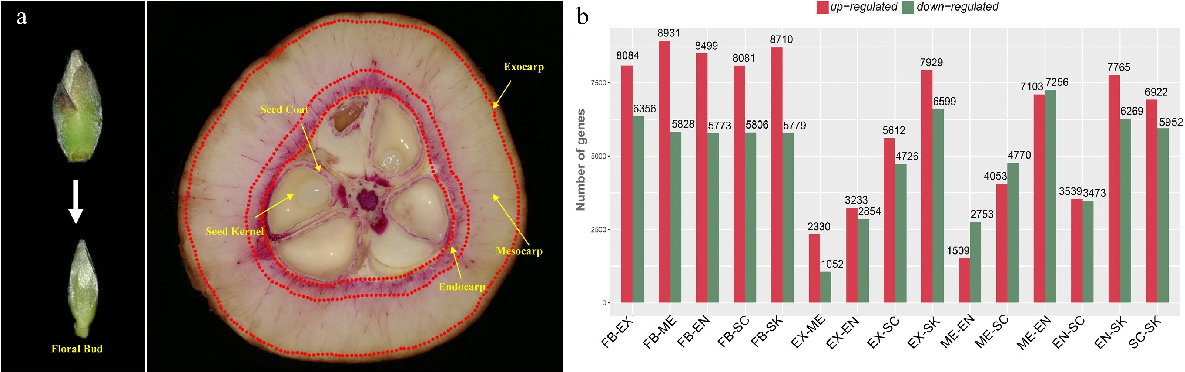
      Figure 1. 

      Tissue-specific transcriptomics in fruit of Camellia chekiangoleosa. (a) Morphology of tissues used for RNA sequencing. On the left is the incipient floral bud; its outside scale leaves are removed before a sample’s preparation. On the right are fruit tissue types at the stage of fruit enlargement; for each, three biological replicates were used for independent library construction and sequencing. FB, floral bud; EX, exocarp; ME, mesocarp; EN, endocarp; SC, seed coat; SK, seed kernel. The red-stained areas indicate the lignified tissues stained by phloroglucinol-hydrochloric acid. Yellow arrows indicate the tissues that were collected for sampling. Three biological replicates were used for library preparation and sequencing analysis. (b) Numbers of differentially expressed transcripts between tissue types. Red and green colors indicate the up-regulated and down-regulated genes, respectively, in each comparison.

      We obtained an average of ca. 79 million reads per RNA sequencing library for the de novo construction of the transcriptome (Supplemental Table S3). The assembled transcriptome consisted of 40,042 unigenes with a N50 value of 1,676 bp (Supplemental Table S4). The transcriptome assembly was annotated using multiple public databases; only those transcripts (unigenes) annotated in at least one database were retained for gene identification (Supplemental Fig. S1ac). Next, the transcriptome was used as a reference to identify the DEGs). We first obtained the expression levels based on the mapping of RNA sequencing reads and then designated the transcripts with > 2-fold change in expression (False Discovery Rate [FDR] corrected p-value < 0.05) as DEGs. Many DEGs were detected between each comparison of different tissue types; in particular, EX-ME and EX-EN displayed relatively fewer DEGs (Fig. 1b), which was consistent with their tissue homology.

    • To analyze the expression pattern of genes associated with lignin accumulation in C. chekiangoleosa, EN and SC tissues were selected (due to their high lignin levels); these were also used to distinguish the pertinent genes in the lignification process (Fig. 2a). There were 2,368 and 3,451 common DEGs in the EN-group and SC-group, respectively (Fig. 2a). Integrative analysis revealed 1,083 common DEGs by comparing these two groups (Fig. 2b); in further evaluating the expression patterns of these common DEGs, 568 of them were highly expressed in SC and EN (Fig. 2b). The functional annotation of these highly expressed DEGs revealed that the KEGG pathway 'phenylpropanoid pathway' was significantly enriched, suggesting an early initiation of the lignin biosynthesis (Fig. 2c & d). This result yielded a pool of potential genes likely involved in the lignification of fruits.
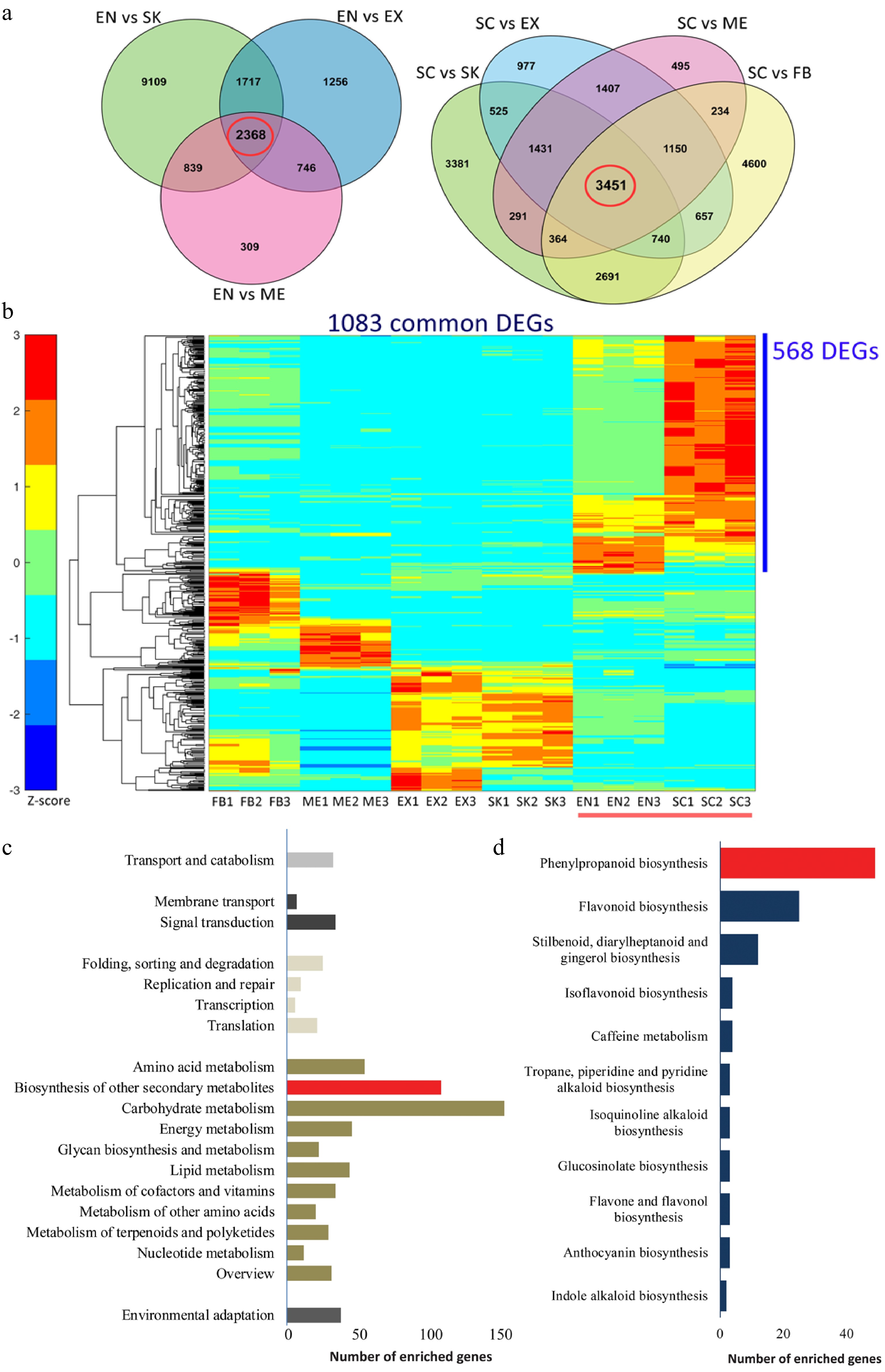
      Figure 2. 

      Functional characterization of differentially expressed genes (DEGs) that are involved in the lignification of the endocarp and seed coat of Camellia chekiangoleosa. (a) Venn diagrams of the DEGs in comparison to EN (left) and SC (right), which revealed 2348 and 3451 unigenes in the EN-group (left red circle) and SC-group (right red circle), respectively. (b) The EN-group and SC-group analysis yielded 1083 DEGs for gene expression analysis. The heatmap analysis of these 1083 genes identified clusters of them highly expressed in various tissue types. The red bar indicates the highly lignified EN and SC tissues. The blue bar indicates those genes highly expressed in EN and SC (568 DEGs); C, KEGG enrichment analysis of 568 DEGs that were highly expressed in EN and SC. D, Distribution of the number of genes that are enriched in 'Biosynthesis of other secondary metabolites'.

      Both the lignin biosynthesis pathway and its transcriptional regulation have been extensively studied[15,18]. Here, potential key genes involved in the regulation of lignin biosynthesis were identified based upon a sequence similarity analysis between C. chekiangoleosa and A. thaliana (Supplemental Table S5). Combined with the DEGs analysis, we screened out 15 lignin biosynthesis genes and six TF genes that could be involved in fruit lignification (Fig. 3a). The proposed biosynthesis and transcriptional regulation pathways were reconstructed to elucidate the lignification process in C. chekiangoleosa fruits (Fig. 3b). This revealed that different types of relevant transcription factors were possibly operating under a hierarchical regulatory network to induce lignin biosynthesis, among which the NAC transcription factor (NST ortholog) might direct a master switch given its intensive expression levels in both SC and EN tissue (Fig. 3b).

      Figure 3. 

      Tissue-specific expression analysis of lignin biosynthesis and transcriptional regulation gene in Camellia chekiangoleosa. (a) Heatmap of gene expression patterns for lignin-related genes that were identified based on sequence similarity. The gene symbols from Arabidopsis are used to indicate the potential homologs in C. chekiangoleosa. The red arrow indicates the NST homolog. Mean expression levels of transcripts are used for the expression analysis. (b) The key genes participating in lignin biosynthesis and its transcriptional regulation are presented according to known pathways identified in Arabidopsis. The master switch of secondary cell wall formation as regulated by NAC family TFs are highlighted in the red-dashed square.

    • To identify the key factors governing fruit lignification, we performed a gene expression analysis during the development of C. chekiangoleosa fruits by focusing on the establishment of lignified tissues (Fig. 4a). A growth curve of fruit development was derived based on the fruit size and lignification patterns (Supplemental Fig. S2). According to the levels of lignin, four critical stages of C. chekiangoleosa fruit development were discernible: stage 1, not lignified; stage 2, initiation of lignification; stage 3, fruit expansion and maintenance of lignification; stage 4, lignification completed (Supplemental Fig. S2; Fig. 4a). Next, the fruit pericarp (P) and seed-associated (S) tissues were dissected to verify the expression profiles of 20 candidate genes, including lignin biosynthesis and transcriptional regulators. The expression of these candidates agreed well with the transcriptomic results (Fig. 4b); notably, the CcNST1 displayed high correlations with the degree of lignification of both the endocarp and seed coat (Fig. 4b). To further verify the contribution from the NST-like gene in C. chekiangoleosa, we evaluated the transcriptome and identified 15 potential NST homologs with full-length ORF (open read frame) (Supplemental Table S2; Supplemental Fig. S3b). We cloned the full-length coding regions of CcNST1 and performed a phylogenetic analysis, which indicated that CcNST1 was an ortholog of the SND1/NST gene (Supplemental Fig. S3a & c). These results suggested that CcNST1 was an important regulator controlling the fruit lignification process in C. chekiangoleosa.
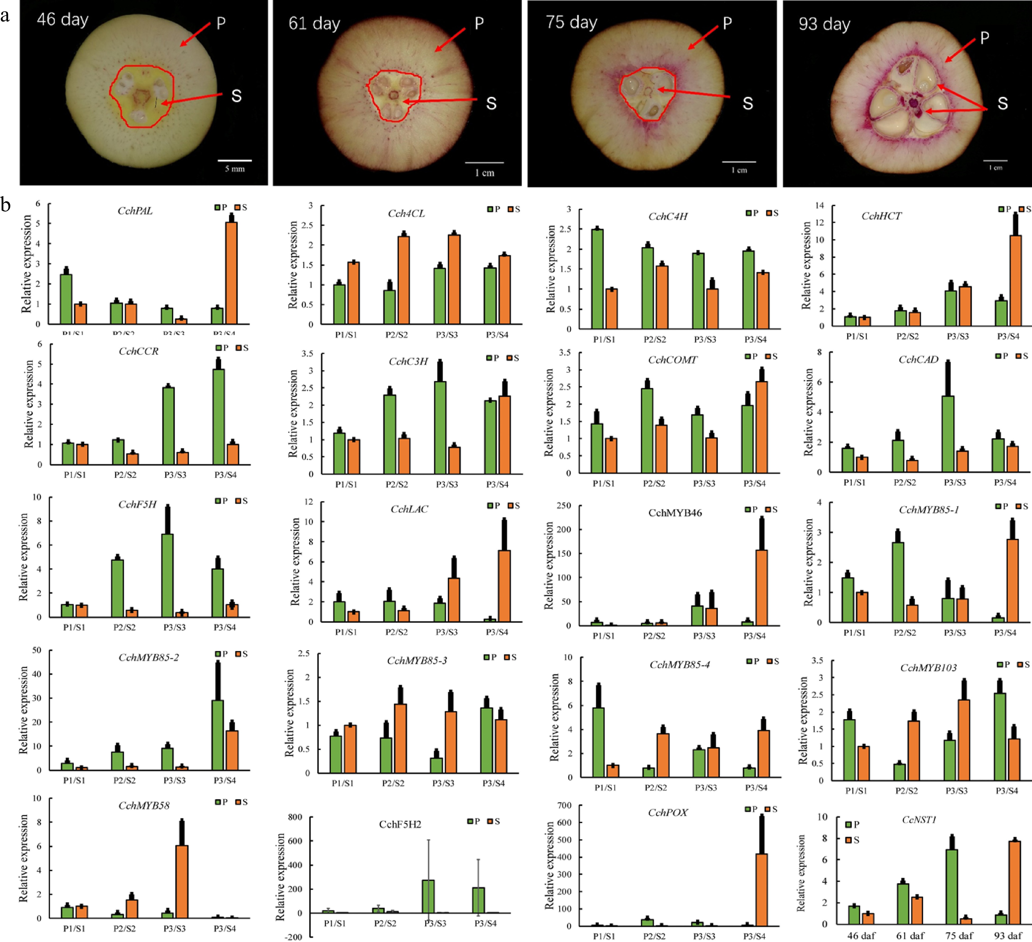
      Figure 4. 

      Expression analysis of genes involved in the regulation of lignin biosynthesis in pericarp and seed tissues during the fruit development of Camellia chekiangoleosa. (a) Staining of the vertical section of C. chekiangoleosa fruit in different periods. The purple stain signals from phloroglucinol-HCl indicate the lignified cells; the red arrowheads indicate the respective pericarp and seed tissue portions sampled at different stages of fruit development. The selection of the sampling is based on the developmental curves of C. chekiangoleosa fruits. P denotes mixed pericarp tissues; S denotes the mixed seed tissues. The arrows point to the areas of mixed tissues sampled at different developmental stages. (b) The qRT-PCR analysis of expression patterns of lignin biosynthesis genes at the four critical stages of P and S tissues. The expression of CchNST1 was significantly up-regulated at 75 to 93 d post-fertilization in the P and S samples, corresponding to the lignification of the endocarp and seed coat. Values are means ± s.d. of three biological replicates.

    • To investigate the roles of CcNST1, overexpression lines in Arabidopsis were generated. The T2 lines were identified via PCR using construct-specific primers (Supplemental Table S1; Supplemental Fig. S4a). Three lines displaying strong phenotypic alterations and high expression levels were used for further analyses (Supplemental Fig. S4b). We found that the overexpression lines displayed pleiotropic growth defects in different tissues, including a smaller size, upward curling of leaves, and distorted stems (Fig. 5a). Histological analysis of the wild type versus transgenic lines was carried out to understand the cellular changes of the CcNST1 overexpression lines. Evidently, the overexpression lines displayed markedly enhanced lignified vascular bundles (Fig. 5e & f), and the vascular tissue possessed more lignified cell layers and enlarged areas than did the wild type (Fig. 5cf & i). Furthermore, the mature zone of root tissues contained many more lignified cells in the transgenic lines than the wild type (Fig. 5gh & j). Subcellular analysis of the CcNST1-GFP fusion protein revealed that CcNST1 was localized in the nucleus (Fig. 5b). To investigate the downstream events, we analyzed the expression levels of AtBLH6, AtMYB46, and AtMYB83 (downstream targets of AtNST1). All three tested genes were significantly up-regulated in the transgenic lines, while the expression of AtNST1 went unchanged (Fig. 5k). These results indicated CcNST1 was a potential key regulator for initiating the lignin biosynthesis pathway and that it therefore might play important roles in fruit lignification.
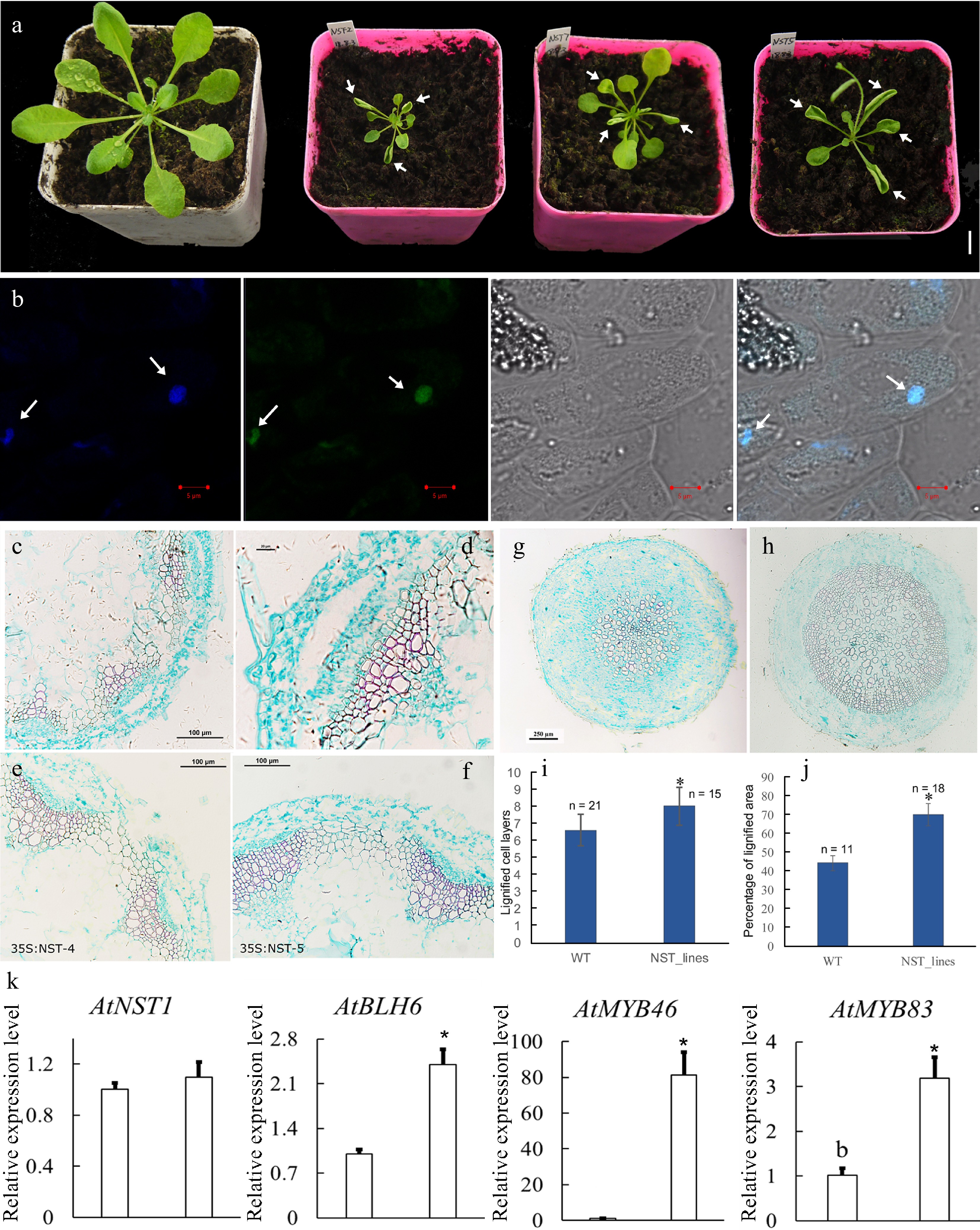
      Figure 5. 

      Overexpression of CcNST1 in Arabidopsis. (a) Overexpression lines displayed various growth defects. Scale bar = 1 cm. The arrows indicate the striking curling leaves. (b) Subcellular localization analysis of the CcNST1:GFP fusion protein by confocal microscopy. Arrows indicate the signals in the nucleus. From left to right, the panels depict the DAPI signal, GFP signal, bright field, and superimposed images; scale bars = 5 µm. (c) & (d) Histological analysis of stem morphology in wild-type stems. (e) & (f) Histological analysis of stem morphology in the transgenic lines; scale bars = 100 µm. Histological analysis of root morphology in the transgenic lines (g) and (h) wild type; scale bars = 100 µm. (i) Statistical analysis of lignified cells in stem and root tissues. n indicated the independent measurements; values are means ± s.d.. (j) Statistical analysis of lignified areas in root tissues. The number of samples used for each statistical analysis is indicated by n. (k) Relative expression levels of Arabidopsis NST1, BLH6, MYB46 and MYB83 genes between the wild type and transgenic lines. Three independent transgenic lines were used for gene expression analysis. The expression of endogenous AtNST1 was not significantly changed. Asterisks indicate significant p-values (< 0.05) for the Student’s t-test.

    • The NST-type transcription factor has been shown to possess conserved functions in the model tree species P. trichocarpa[16]. Accordingly, it is interesting to know whether the function of CcNST1 is conserved across woody species. We performed a transgenic analysis using hybrid poplar ('Nanlin895') and generated overexpression lines of CcNST1. Expression of CcNST1 in independent poplar lines was confirmed and the enhanced expression of CcNST1 was detectable at the early stage of leaf development (Fig. 6b). During vegetative growth, the transgenic poplar featured consistent phenotypes, including drooping leaves and disordered leaf veins (Fig. 6a). By contrast, no obvious stem phenotypes were distinguishable. Then a histological analysis was performed to characterize the leaf midrib and stem tissue of the wild type versus overexpression lines in poplar. This showed that, transgenic line, their midribs at the distorted position had abnormal vascular tissues: some lignified cells formed irregular vascular-like tissues (Fig. 6c & d). The anatomy of stem structures was investigated further by using the fourth internode where the vascular system is established[38]. We found that the transgenic lines displayed enhanced lignified cells in their xylems (Fig. 6di), similar to the results for A. thaliana (Fig. 5cf). Further, the transgenic lines evidently contained condensed parenchyma cells (Fig. 6gi), suggesting a role for secondary cell wall formation. To evaluate the potential functioning of CcNST1, the downstream TFs of the NST homolog in P. trichocarpa were tested: the expression levels of SND1, MYB21 and MYB74 were all significantly up-regulated in the overexpression lines (Fig. 6j). Taken together, from these results we concluded that CcNST1 harbors conserved functions of lignin biosynthesis and secondary cell wall formation in woody plant species.
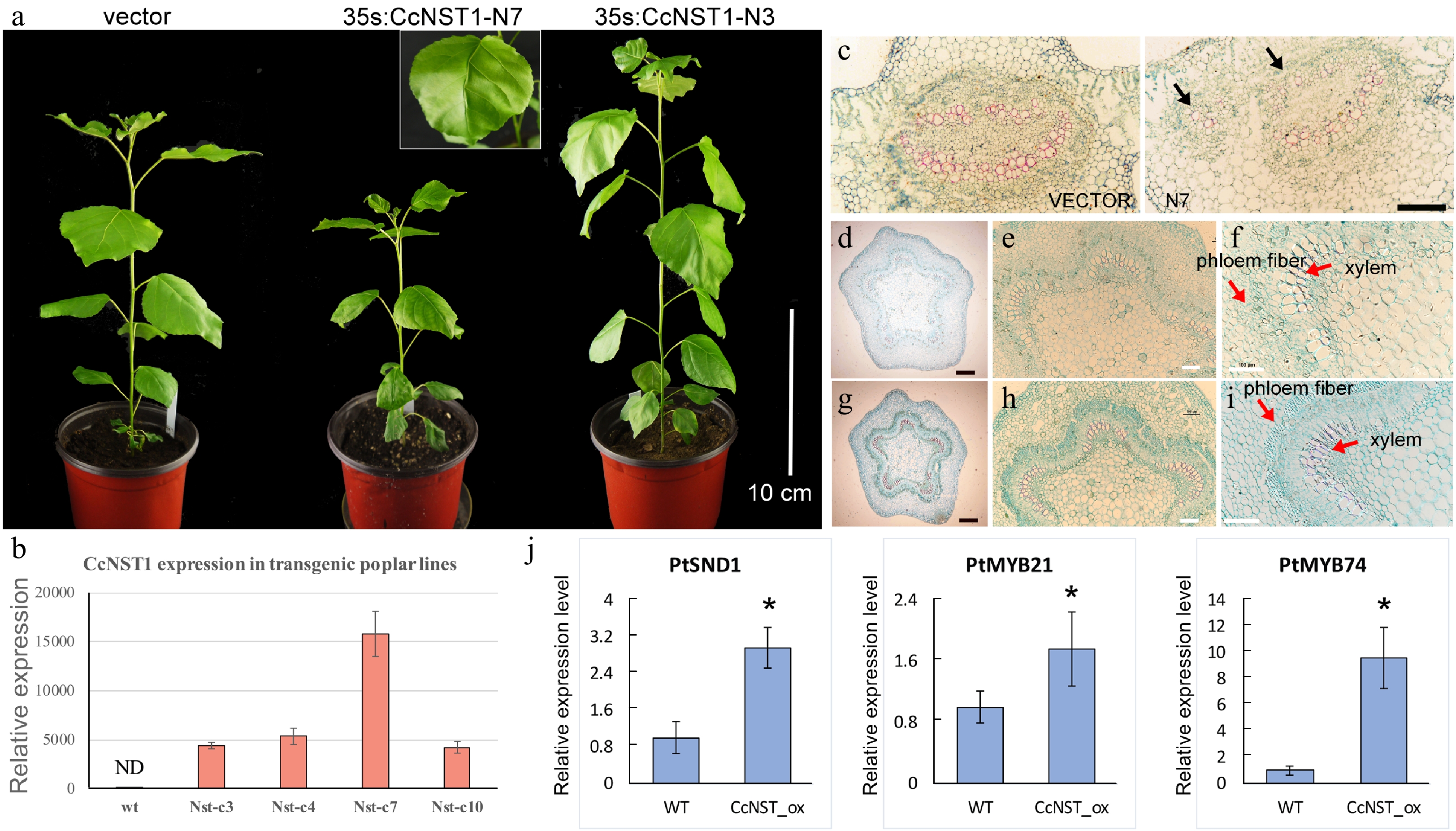
      Figure 6. 

      Ectopic expression of CchNST1 in hybrid poplar (‘Nanlin 895’). (a) Comparison of overall morphology between the control and transgenic poplar plants. On the left is a transgenic plant of the empty vector as a control; middle and right are plants of independent lines of 35s:CcNST1. The inset shows a close-up view of the distorted leaf midvein in the transgenic lines. Scale bar = 10 cm. (b) Expression level of CcNST1 in different independent transgenic lines. ND, not detected. (c) Vertical sections of the midrib in control (left) and 35s:CcNST1 (right) lines. (d)-(f) Cross sections of the fourth internode of control lines. Red arrows indicate the phloem fiber cells and xylem cells. Black scale bars = 250 µm, white scale bars = 100 µm. (g)-(i) Cross sections of the fourth internode of the 35s:CcNST1 lines. The enhanced secondary cell wall in phloem fiber cells and xylem cells are shown. Black scale bars = 250 µm, white bars 100 = µm. (j) Expression of downstream genes PtSND1, PtMYB21, and PtMYB74 in the control and transgenic CcNST1 lines. Three independent transgenic lines were used for gene expression analysis. Asterisks indicate a significant Student’s t-test (p < 0.05). In (b) and (j) values are means ± s.d. of three biological replicates.

    • Tissue-specific transcriptomics analysis is widely used to identify regulators involved in plant development, growth, and responses to environmental stress[39]. Lignification of specific fruit tissues is an evolutionary significant process that affects seed dispersal, whose regulation is also of great economic importance in fruit crops. Although a vast number of transcriptomic studies of various fruit types have been reported in recent years, the comprehensive analysis of fruit tissue-specific transcriptomics remains relatively scarce. In the present work, we performed a detailed tissue-specific transcriptome analysis of C. chekiangoleosa based on its fruit lignification pattern (Fig. 1a). The DEGs’ analysis focused on those genes associated with the highly lignified EN and SC tissue, which revealed thousands of them that were potentially involved in the fruit lignification process (Fig. 1). Functional analysis suggested the enriched DEGs were related to various biological pathways including the phenylpropanoid biosynthesis pathway (Fig. 2c & d), which implicates a central role for lignin biosynthesis during fruit tissue patterning. In peach fruit, for example, a genome-wide characterization of its transcriptome during the phase of stone cell formation in endocarp found evidence for the induction of prominent phenylpropanoid, lignin, and flavonoid pathway genes[40,41]. Likewise, a transcriptomics study of three developmental stages of pear fruit demonstrated that up-regulation of Cinnamoyl-CoA Reductase (CCR) was involved in stone cell formation[42]. Our results from the gene expression analyses are largely consistent with previous work (Fig. 3), which suggests that common regulatory pathways are involved in establishing fruit lignification patterns.

      The formation of a specific lignification pattern in fruits is regulated by the coordination of several types of TFs active during the developmental stages of fruit. Camellia plants form typical capsular fruits that undergo two independent lignification events, that of fruit peels and that of the seed coat[31]. A genetic model for how lignification of C. chekiangoleosa fruits is directed has been proposed: a cascade of TFs, starting with the SHP-FUL MADS-box TFs through to bHLH-type TFs, NAC, MYB, and BLH TFs work together to regulate the biosynthesis of the cell wall and secondary metabolites during fruit development[40]. We showed that the expression patterns of different types of TFs, including NAC, MYB, and BLH-like families, are correlated with the lignin accumulation in C. chekiangoleosa fruits (Fig. 3b). Hence, our results provided empirical evidence of the transcriptional network underlying that fruit’s lignification pattern. We also found that Camellia fruits are diverse in their size, secondary metabolites, and seed oil contents[31]. But little is known about genetic regulation of fruit development in Camellia species, probably because of insufficient molecular biology tools. In the future, the functional characterizations of those TFs in C. chekiangoleosa will be essential for elucidating the regulatory mechanism responsible for that plant’s specific lignification pattern.

      Lignification is a unique process contributing critically toward the maintenance and regulation of plants growth and development and their responses to biotic/abiotic stresses. Although lignin biosynthesis and its transcriptional regulation have been extensively studied for wood formation, lignification's regulation during fruit development is not yet well characterized, especially in fruit crops. The genus Camellia contains many species whose seed oil production is economically valuable. The fruit lignification is also a critical breeding trait associated with fruit size, seed dispersal, and oil yield[31]. Recent work on C. japonica characterized the homolog gene of SHP1/2 (CjPLE) and revealed its potential role in regulating the pattern of fruit lignification; however, based on the callus-transformation assay, direct activation of lignin biosynthesis genes by CjPLE was not proved[24]. Here we evaluated the key lignin biosynthesis genes and TFs in C. chekiangoleosa, finding that major lignin-related genes were highly expressed in both EN and SC tissues (Fig. 3). Therefore, we proposed that the activation of lignin biosynthesis in specified tissues requires a hierarchical interaction of TFs during fruit development.

      The NST-like TFs are recognized as master regulators in the regulation of lignin biosynthesis for secondary cell wall formation in two well studied plants, A. thaliana and P. trichocarpa[15]. Research on fruit crops has uncovered conserved functions of homologs of NST-like NAC genes for regulating the fruit lignification process[43]. In loquat fruits, four NAC TFs (EjNAC1-4) are correlated with lignin accumulation in response to low temperature storage and heat stress[27]. Functional analyses showed that EjNAC1 and EjNAC3 are capable of directly activating the expression of lignin biosynthesis genes[27]. We found that CcNST1 was highly expressed in EN and SC tissues, whose levels correlated with the lignification pattern (Figs 1 & 3). Further, we showed that ectopic expression of CcNST1 in A. thaliana and hybrid poplar augmented tissue lignification (Figs 5 & 6). These results provided evidence that CcNST1 acts as a positive regulator of lignin biosynthesis in C. chekiangoleosa. Also, in the transgenic lines of poplar, the expression of SND1, MYB21, and MYB74 — downstream TFs of the poplar NST gene — was significantly up-regulated (Fig. 6). This result suggests CcNST1 is a high hierarchical activator of lignin biosynthesis during fruit development. Future work using the Camellia-based genetic transformation systems is now required to uncover the downstream genes regulated by CcNST1.

      • This work was supported by Nonprofit Research Projects (CAFYBB2021QD001) of Chinese Academy of Forestry and National Key R&D Program of China (2019YFD1001602). We would like to thank Dr. Zhifeng Wang from Northeast Forestry University, China, for help with the poplar experiments.

      • The authors declare that they have no conflict of interest.

      • # These authors contributed equally: Chao Yan, Ziyan Nie

      • Supplemental Table S1 Primers used in this study.
      • Supplemental Table S2 Identification of NAC domain transcription factors of Camellia chekiangoleosa  transcriptome based on BLAST search. In total, 15 transcripts were found to contain complete open reading frame (ORF).
      • Supplemental Table S3 An overview of the RNA-seq statistics of Camellia chekiangoleosa tissues. Each tissue type includes three biological replicates. Q20 and Q30 represent base call accuracy of 99% and 99.9% respectively. FB, Floral Bud; EX, Exocarp; ME, Mesocarp; EN, Endocarp; SC, Seed Coat; SK, Seed Kernel.
      • Supplemental Table S4 The statistics of Camellia chekiangoleosa transcriptome. The transcripts and unigenes are obtained by using the Trinity v2.11.0 software. Sequences less than 200 bp are removed.
      • Supplemental Table S5 The identification of genes related to lignin biosynthesis and transcriptional regulation based on the information from Arabidopsis thaliana (TAIR v.11).
      • Supplemental Fig. S1 The evaluation of the annotation of Camellia chekiangoleosa transcriptome. A, The distribution of unigenes that are annotated in the Gene Ontology (GO) database. B, The distribution of unigenes that are annotated in the eggNOG database. C, The distribution of unigenes that are annotated in the KEGG database.
      • Supplemental Fig. S2
      • Supplemental Fig. S2 Morphological characterizations of the growth of Camellia chekiangoleosa fruits. A, The morphology of fruit at different timepoints of fruit growth. The lignin patterns at each corresponding stage are displayed below. Bar 2 cm. B, A growth curve of the C. chekiangoleosa fruits is presented by measuring of fruit weight, transverse diameter and vertical diameter. Transverse diameter1 and transverse diameter 2 are two independent measurements with angle around 90 degrees at each sampling point of fruits.
      • Supplemental Fig. S3 The identification of NST homolog in Camellia chekiangoleosa. A, The alignment of protein sequences of CcNST1 and its homologs from diverse plant species. The red rectangle indicates the conserved NAM domain. The accession numbers of sequences:   Vitis vinifera: VV15G07370;  Manihot esculenta: ME03581G00220;  Ricinus communis: RC27964G00230;  Theobroma cacao:TC0001G33230; Gossypium raimondii: GR01G15080; Populus trichocarpa: PT14G10480; Citrus sinensis: CS00001G04980; Arabidopsis thaliana: AT2G46770. B, The expression profiles of NST-like transcripts that are identified from the C. chekiangoleosa transcriptome as described in Supple. Table 2. The candidate NST ortholog was underlined by red. C, The phylogenic tree of NST-like genes from Arabidopsis thaliana and C. chekiangoleaosa.
      • Supplemental Fig. S4 The verification of transgenic Arabidopsis lines. A, The amplification of construct specific fragments of Arabidopsis genomic DNA. Wt, wild type; N4, N7, N10 are representative 35s:CcNST1 lines. B, The expression of CcNST1 in Arabidopsis lines. The wild type is not detectable (nd). Different letters (a, b) indicate significant difference by the Student’s test p < 0.05.
      • Copyright: © 2022 by the author(s). Published by Maximum Academic Press, Fayetteville, GA. This article is an open access article distributed under Creative Commons Attribution License (CC BY 4.0), visit https://creativecommons.org/licenses/by/4.0/.
    Figure (6)  References (43)
  • About this article
    Cite this article
    Yan C, Nie Z, Hu Z, Huang H, Ma X, et al. 2022. Tissue-specific transcriptomics reveals a central role of CcNST1 in regulating the fruit lignification pattern in Camellia chekiangoleosa, a woody oil-crop. Forestry Research 2:10 doi: 10.48130/FR-2022-0010
    Yan C, Nie Z, Hu Z, Huang H, Ma X, et al. 2022. Tissue-specific transcriptomics reveals a central role of CcNST1 in regulating the fruit lignification pattern in Camellia chekiangoleosa, a woody oil-crop. Forestry Research 2:10 doi: 10.48130/FR-2022-0010

Catalog

  • About this article

/

DownLoad:  Full-Size Img  PowerPoint
Return
Return