ARTICLE   Open Access    

Efficient organogenesis and taxifolin production system from mature zygotic embryos and needles in larch

More Information
  • The deciduous conifer larch has been widely distributed around the world, is a high-quality wood species and is also used to extract industrial raw materials and medicines. In this study, we developed an organogenesis protocol for Larix olgensis from both mature zygotic embryos and needles, and analyzed the content of taxifolin in different tissues. The highest callus induction (96.8%) from mature zygotic embryo was found in the Douglas-fir Cotyledon Revised (DCR) medium augmented with 2.0 mg·L−1 6-Benzylaminopurine (6-BA) and 0.2 mg·L−1 α-Naphthaleneacetic acid (NAA), while from needles the highest callus induction (92.03%) was found in the Murashige and Skoog (MS) medium augmented with 3 mg·L−1 6-BA and 0.3 mg·L−1 NAA. The best shoot regeneration capacity from zygotic embryo-derived calli (83.3%) was obtained in DCR medium augmented with 1.0 mg·L−1 6-BA and 0.01 mg·L−1 NAA, and needle-derived calli were 77.3%. The shoots achieved the highest elongation (75.6%) in the DCR medium supplemented with 0.5 mg·L−1 6-BA, 0.05 mg·L−1 NAA and 2 g·L−1 activated charcoal (AC). The rooting rate was 62.8% in DCR medium augmented with 3 mg·L−1 Indole-3-butyric acid (IBA) and 100 mg·L−1 phloroglucinol (PG). The accumulation of the taxifolin in elongation shoots and lignified elongation shoots have greatly improved along with the development process, were 28.6 µg·g−1, and 53 µg·g−1 respectively. The content of the taxifolin in callus was 1.99−5.26 µg·g−1, adventitious shoots were 4.8 µg·g−1, and adventitious roots were 2.86 µg·g−1. We report an efficient organogenesis and taxifolin production protocol in larch for the first time.
  • With the development of the world economy, people's lifestyles have changed dramatically, and long-term high-intensity work has put many people's bodies in a sub-healthy state. The increasing incidence of various chronic diseases has not only put enormous pressure on society's healthcare systems but also caused endless suffering to people[1]. Therefore, people's demands on the functionality and safety of food are increasing, and it has become the consensus of people that 'not just eating enough, but more importantly eating well'.

    Rice is the staple food for more than half of the world's population and the main economic source for a large number of rural people[2]. However, due to the rising cost of rice cultivation, farmers are gaining less and less economic benefits from growing rice, which seriously undermines their incentive to grow rice and poses a serious threat to world food security. Increasing the added value of rice not only helps to increase farmers' income but also helps to ensure world food security. The presence of a large number of functional ingredients in rice makes it possible to increase the added value of rice, and functional rice has therefore been widely noticed.

    Functional rice refers to rice containing certain specific components that play a regulatory and balancing role in human physiological functions in addition to the nutrients necessary for human growth and development in the endosperm, embryo, and rice bran. They can increase human physiological defense mechanisms, prevent certain diseases, help recovery, delay aging, and boost physical strength and energy levels[3]. Rice is a staple food for more than half of the world's population[4], and its functional components have a great potential to be exploited for human welfare. Using functional rice as a carrier to address health problems and realize 'medicine-food homology' is an excellent motivation for promoting functional rice. The current typical functional rice is introduced in this paper. It also summarizes the breeding and cultivation technologies of functional rice.

    Rice has a high glycemic index. Its long-term consumption leads to obesity, diabetes, and colon disease in many people[5]. However, the consumption of rice rich in resistant starch (RS) can greatly reduce the risk of these diseases[6]. Therefore, breeding rice varieties with high RS content has attracted considerable attention from breeders in various countries. However, the variability of RS content between different rice varieties is low, and there are few germplasm resources available for selection, thus making it challenging to breed rice varieties with high RS content using traditional breeding methods. Combining traditional and modern molecular breeding techniques can greatly improve the successful production of high RS rice breeds. Nishi et al.[7] selected a high RS rice variety EM10 by treating fertilized egg cells of Kinmaze with N-methyl-N-nitrosourea. However, its yield was very low, and it was not suitable for commercial production. Wada et al.[8] crossed 'Fukei 2032' and 'EM129' as parents and selected Chikushi-kona 85, a high RS rice variety with a higher yield than EM10. Miura et al.[9] bred ultra-high RS BeI-BEIIB double mutant rice by crossing the Abe I and Abe IIB mutant strains, and the content of RS in the endosperm reached 35.1%. Wei et al.[10] found that the simultaneous inhibition of starch branching enzyme (SBE) genes SBEIIb and SBEI in Teqing by antisense RNA could increase the RS content in rice to 14.9%. Zhu et al.[11] used RNAi technology to inhibit the expression of SBEI and SBEII genes in rice, which increased the content of RS in rice endosperm from 0 to 14.6 %. Zhou et al.[6] found that rice RS formation is mainly controlled by soluble starch synthase (SSIIA). However, its regulation is dependent on the granule-bound starch synthase Waxy (Wx), and SSIIA deficiency combined with high expression of Wxa facilitates the substantial accumulation of RS in the rice. The results of Tsuiki et al.[12] showed that BEIB deficiency was the main reason for the increased accumulation of RS in rice. Itoh et al.[13] developed new mutant rice lines with significantly higher levels of RS in rice by introducing genes encoding starch synthase and granule-bound starch synthase in the rice into the BEIB-deficient mutant line be2b.

    The accumulation of anthocyanins/proanthocyanidins in the seed coat of the rice grain gives brown rice a distinct color[14]. Most common rice varieties lack anthocyanins in the seed coat, and so far, no rice variety with colored endosperm in its natural state has been identified. However, Zhu et al.[15] bred rice with purple endosperm using transgenic technology. Red rice contains only proanthocyanidins, while black and purple rice contain anthocyanidins and proanthocyanidins[16]. Red seed coat of rice was found to be controlled by the complementary effects of two central effect genes Rc and Rd. The loss of function of the Rc gene prevented the synthesis of proanthocyanidins, while the Rd gene could enhance the effect of the Rc gene in promoting proanthocyanidins synthesis[17]. Purple seed coat color is controlled by two dominant complementary genes Pb and Pp. Pb determines the presence or absence of seed coat color, and Pp determines the depth of seed coat color[18]. In addition, phycocyanin synthesis is also regulated by transcription factors such as MYB, bHLH, HY5, and WD40[14], but the exact regulatory mechanism is not clear. Colored rice is rich in bioactive components, such as flavonoids, phenolic acids, vitamin E (VE), glutelin, phytosterols, and phytic acid (PA). It also contains large amounts of micronutrients such as Ca, Fe, Zn, and Se[19], and has a much higher nutritional and health value than ordinary white rice. In addition, Zhu et al.[20] successfully developed rice with enriched astaxanthin in the endosperm by introducing the genes sZmPSY1, sPaCrtI, sCrBKT, and sHpBHY. This achievement has laid a solid foundation for the further development of functional rice industry.

    Giant embryo rice refers to rice varieties whose embryo volume is more than twice that of ordinary rice[21]. Rice embryo contains more nutrients than the endosperm; therefore, the nutritional value of giant embryo rice greatly exceeds that of ordinary rice. Studies have found that the levels of γ-aminobutyric acid (GABA), essential amino acids, VE, γ-oryzanol, phenols, and trace elements in giant embryo rice are considerably higher than that in ordinary rice[21]. Satoh & Omura[22] used the chemical mutagen N-methyl-N-nitrosourea to treat the fertilized egg cells of the rice variety Kinmaze to obtain a 'giant embryo' mutant. The mutants’ embryo occupied 1/4–1/3 of the rice grain volume and was 3–4 times larger than normal rice embryo[23]. Its GABA content increased dramatically after the rice was soaked in water. Maeda et al.[24] crossed the giant embryo mutant EM40 of Kinmaze with the high-yielding variety Akenohoshi to produce the giant embryo rice variety 'Haiminori'. The embryo size of 'Haiminori' is 3–4 times that of ordinary rice, and the GABA content of its brown rice is 3–4 times higher than that of 'Nipponbare' and 'Koshihikari' after soaking for four hours in water. A few genes that can regulate the size of rice embryos have been identified, and GE is the first identified rice giant embryo gene[25]. Nagasawa et al.[26] found that the loss of GE gene function resulted in enlarged embryos and smaller endosperm in rice. Lee et al.[27] found that the inhibition of LE gene expression by RNAi technology could lead to embryo enlargement in rice, but the regulatory mechanism remains to be investigated.

    Protein is the second most crucial nutrient in rice, accounting for 7–10% of the grain weight, and glutenin accounts for 60%–80% of the total protein content in rice grains[28]. Compared to other proteins, glutenin is more easily digested and absorbed by the body[29]. Therefore, higher glutenin content in rice can improve its nutritional value. However, people with renal disease (a common complication of diabetes) have impaired protein metabolism, and consumption of rice with lower glutelin content can help reduce their protein intake and metabolic burden[30]. Japanese breeders treated Nihonmasari with the chemical mutagen ethyleneimine and selected the low-glutelin rice mutant NM67[31]. Iida et al.[31] developed a new rice variety LGC-1 (Low glutelin content-1) with a glutelin content of less than 4% by backcrossing the NM67 mutant with the original variety 'Nihonmasari'. According to Miyahara[32], the low glutelin trait in LGC-1 is controlled by a single dominant gene Lgc-1 located on chromosome 2. Subsequently, Nishimura et al.[33] produced two rice varieties, 'LGC Katsu' and 'LGC Jun' with lower glutelin content by crossing LGC1 with a mutant line Koshikari (γ-ray induction) lacking 26 kDa globulin (another easily digestible protein).

    Vitamin A (VA) is one of the essential nutrients for the human body[34]. However, rice, a staple food, lacks VA, leading to a VA deficiency in many people. β-carotene is a precursor for VA synthesis and can be effectively converted into VA in the human body[35]. Therefore, breeding rice varieties rich in β-carotene has attracted the attention of breeders in various countries. Ye et al.[36] simultaneously transferred phytoene synthase (psy), phytoene desaturase (crt I), and lycopene β-cyclase (lcy) genes into rice using the Agrobacterium-mediated method and produced the first generation of golden rice with a β-carotene content of 1.6 µg·g−1 in the endosperm. However, due to the low content of β-carotene in rice, it is difficult to meet the human body's demand for VA. To increase β-carotene content in rice, Paine et al.[37] introduced the phytoene synthase (psy) gene from maize and the phytoene desaturase (crt I) gene from Erwinia into rice. They obtained the second generation of golden rice with 37 µg g−1 of β-carotene in the endosperm, with nearly 23-fold increase in β-carotene content compared to the first generation of golden rice.

    Fe and Zn are essential trace elements for human beings. The contents of Fe and Zn in common rice are about 2 μg·g−1 and 16 μg·g−1, respectively[38], which are far from meeting human needs. In 2004, to alleviate micronutrient deficiencies among underprivileged people in developing countries, the Consultative Group on International Agricultural Research launched the HarvestPlus international collaborative program for improving Fe, Zn, and β-carotene levels in staple crops, with breeding targets of 13 μg·g−1 and 28 μg·g−1 for Fe and Zn in rice, respectively. Masuda et al.[39] found that expression of the nicotianamine synthase (NAS) gene HvNAS in rice resulted in a 3-fold increase in Fe and a 2-fold increase in Zn content in polished rice. Trijatmiko et al.[38] overexpressed rice OsNAS2 gene and soybean ferritin gene SferH-1 in rice, and the Fe and Zn content in polished rice of rice variety NASFer-274 reached 15 μg·g−1 and 45.7 μg·g−1, respectively. In addition, it has been found that increasing Fe intake alone does not eliminate Fe deficiency but also decreases the amount of Fe absorption inhibitors in the diet or increases the amount of Fe absorption enhancers[40]. The negatively charged phosphate in PA strongly binds metal cations, thus reducing the bioavailability of Fe and Zn in rice[41], while the sulfhydryl group in cysteine binds Fe, thereby increasing the absorption of non-heme Fe by the body[42]. To improve the bioavailability of Fe and Zn, Lucca et al.[40] introduced a heat-tolerant phytase (phyA) gene from Aspergillus fumigatus into rice and overexpressed the cysteine-rich protein gene (rgMT), which increased the content of phytase and cysteine residues in rice by 130-fold and 7-fold, respectively[40].

    The functional quality of rice is highly dependent on germplasm resources. Current functional rice breeding mainly adopts transgenic and mutagenic technologies, and the cultivated rice varieties are mainly enriched with only one functional substance and cannot meet the urgent demand by consumers for rice enriched with multiple active components. The diversity of rice active components determines the complexity of multifunctional rice breeding. In order to cultivate multifunctional rice, it is necessary to strengthen the application of different breeding technologies. Gene polymerization breeding is a crop breeding technology that can polymerize multiple superior traits that have emerged in recent years, mainly including traditional polymerization breeding, transgenic polymerization breeding, and molecular marker-assisted selection polymerization breeding.

    The transfer of beneficial genes in different species during traditional polymeric breeding is largely limited by interspecific reproductive isolation, and it is challenging to utilize beneficial genes between different species effectively. Gene transfer through sexual crosses does not allow accurate manipulation and selection of a gene and is susceptible to undesirable gene linkage, and in the process of breed selection, multiple backcrosses are required[43]. Thus, the period of selecting target plants is long, the breeding cost is high, and the human resources and material resources are costly[44]. Besides, it is often difficult to continue the breakthrough after a few generations of backcrossing due to linkage drag. Thus, there are significant limitations in aggregating genes by traditional breeding methods[45].

    Transgenic technology is an effective means of gene polymerization breeding. Multi-gene transformation makes it possible to assemble multiple beneficial genes in transgenic rice breeding rapidly and can greatly reduce the time and workload of breeding[46]. The traditional multi-gene transformation uses a single gene transformation and hybridization polymerization method[47], in which the vector construction and transformation process is relatively simple. However, it is time-consuming, laborious, and requires extensive hybridization and screening efforts. Multi-gene-based vector transformation methods can be divided into two major categories: multi-vector co-transformation and multi-gene single vector transformation[47]. Multi-vector co-transformation is the simultaneous transfer of multiple target genes into the same recipient plant through different vectors. The efficiency of multi-vector co-transformation is uncertain, and the increase in the number of transforming vectors will increase the difficulty of genetic screening, resulting in a reduced probability of obtaining multi-gene co-transformed plants. Multi-gene single vector transformation constructs multiple genes into the T-DNA region of a vector and then transfers them into the same recipient plant as a single event. This method eliminates the tedious hybridization and backcrossing process and solves the challenges of low co-transformation frequency and complex integration patterns. It can also avoid gene loss caused by multi-gene separation and recombination in future generations[47]. The transgenic method can break through the limitations of conventional breeding, disrupt reproductive isolation, transfer beneficial genes from entirely unrelated crops to rice, and shorten the cycle of polymerizing target genes significantly. However, there are concerns that when genes are manipulated, unforeseen side effects may occur, and, therefore, there are ongoing concerns about the safety of transgenic crops[48]. Marker-free transgenic technology through which selective marker genes in transgenic plants can be removed has been developed. This improves the safety of transgenic crops, is beneficial to multiple operations of the same transgenic crop, and improves the acceptance by people[49].

    Molecular marker-assisted selection is one of the most widely used rice breeding techniques at present. It uses the close linkage between molecular markers and target genes to select multiple genes directly and aggregates genes from different sources into one variety. This has multiple advantages, including a focused purpose, high accuracy, short breeding cycle, no interference from environmental conditions, and applicability to complex traits[50]. However, few genes have been targeted for the main effect of important agronomic traits in rice, and they are mainly focused on the regulation of rice plant type and the prevention and control of pests and diseases, and very few genes related to the synthesis of active components, which can be used for molecular marker-assisted selection are very limited. Furthermore, the current technical requirements and costs for analyzing and identifying DNA molecular markers are high, and the identification efficiency is low. This greatly limits the popularization and application of functional rice polymerization breeding. Therefore, to better apply molecular marker-assisted selection technology to breed rice varieties rich in multiple active components, it is necessary to construct a richer molecular marker linkage map to enhance the localization of genes related to functional substance synthesis in rice[51]. Additionally, it is important to explore new molecular marker technologies to improve efficiency while reducing cost.

    It is worth noting that the effects of gene aggregation are not simply additive. There are cumulative additive effects, greater than cumulative epistatic effects, and less than cumulative epistatic effects among the polymerization genes, and the effects are often smaller than the individual effect. Only with a clearer understanding of the interaction between different QTLs or genes can functional rice pyramiding breeding be carried out reasonably and efficiently. Except for RS and Se, other active components of rice mainly exist in the rice bran layer, and the content of active components in the endosperm, the main edible part, is extremely low. Therefore, cultivating rice varieties with endosperm-enriched active components have broad development prospects. In addition, because crops with high quality are more susceptible to pests and diseases[52], the improvement of rice resistance to pests and diseases should be considered during the polymerization breeding of functional rice.

    The biosynthesis of active components in rice is influenced by rice varieties but also depends on cultivation management practices and their growth environment.

    Environmental conditions have a greater effect on protein content than genetic forces[53]. Both light intensity and light duration affect the synthesis and accumulation of active components in rice. Low light intensity in the early stage of rice growth is not conducive to the accumulation of glutelin in rice grains but favors the accumulation of amylose, while the opposite is true in the late stage of rice growth[54]. Low light intensity during the grain-filling period reduces the accumulation of total flavonoids in rice[55] and decreases Fe ions' movement in the transpiration stream and thereby the transport of Fe ions to rice grains[56]. An appropriate increase in light intensity is beneficial to the accumulation of flavonoids, anthocyanins, and Fe in rice, but the photostability of anthocyanins is poor, and too much light will cause oxidative degradation of anthocyanins[57]. Therefore, functional rice is best cultivated as mid-late rice, which would be conducive to accumulating active components in rice.

    The temperature has a great influence on the synthesis of active components in rice. An appropriate increase in the temperature is beneficial to the accumulation of γ-oryzanol[58] and flavonoids[59] in rice. A high temperature during the grain-filling period leads to an increase in glutelin content in rice[60], but an increase in temperature decreases the total phenolic content[61]. The results regarding the effect of temperature on the content of PA in rice were inconsistent. Su et al.[62] showed that high temperatures during the filling period would increase the PA content, while Goufo & Trindade[61] reported that the increase in temperature would reduce the PA content. This may be due to the different growth periods and durations of temperature stress on rice in the two studies. The synthesis of anthocyanins/proanthocyanidins in colored rice requires a suitable temperature. Within a certain range, lower temperatures favor the accumulation of anthocyanins/proanthocyanidins in rice[63]. Higher temperatures will lead to degradation, and the thermal stability of proanthocyanidins being higher than that of anthocyanins[64]. In addition, cold or heat stress facilitates GABA accumulation in rice grains[65]. Therefore, in actual production, colored rice and low-glutelin rice are best planted as late rice, and the planting time of other functional rice should be determined according to the response of its enriched active components to temperature changes.

    Moderate water stress can significantly increase the content of glutelin[66] and GABA[67] in rice grains and promote the rapid transfer of assimilation into the grains, shorten the grain filling period, and reduce the RS content[68]. Drought stress can also induce the expression of the phytoene synthase (psy) gene and increase the carotenoid content in rice[69]. Soil moisture is an important medium in Zn diffusion to plant roots. In soil with low moisture content, rice roots have low available Zn, which is not conducive to enriching rice grains with Zn[70]. Results from studies on the effect of soil water content on Se accumulation in rice grains have been inconsistent. Li et al.[71] concluded that flooded cultivation could significantly increase the Se content in rice grains compared to dry cultivation. However, the results of Zhou et al.[72] showed that the selenium content in rice grains under aerobic and dry-wet alternative irrigation was 2.44 and 1.84 times higher than that under flood irrigation, respectively. This may be due to the forms of selenium contained in the soil and the degree of drought stress to the rice that differed between experiments[73]. In addition, it has been found that too much or too little water impacts the expression of genes related to anthocyanin synthesis in rice, which affects the accumulation of anthocyanins in rice[74]. Therefore, it is recommended to establish different irrigation systems for different functional rice during cultivation.

    Both the amount and method of nitrogen application affect the accumulation of glutelin. Numerous studies have shown that both increased and delayed application of nitrogen fertilizer can increase the accumulation of lysine-rich glutelin to improve the nutritional quality of rice (Table 1). However, this improvement is not beneficial for kidney disease patients who cannot consume high glutelin rice. Nitrogen stress can down-regulate the expression of ANDs genes related to the anthocyanins biosynthesis pathway in grains, resulting in a decrease in anthocyanins synthesis[55]. Increased nitrogen fertilizer application can also increase the Fe, Zn, and Se content in rice[75,76]. However, some studies have found that increased nitrogen fertilizer application has no significant effect on the Fe content of rice[77], while other studies have shown that increased nitrogen fertilizer application will reduce the Fe content of rice[78]. This may be influenced by soil pH and the form of the applied nitrogen fertilizer. The lower the soil pH, the more favorable the reduction of Fe3+ to Fe2+, thus promoting the uptake of Fe by rice. Otherwise, the application of ammonium fertilizer can improve the availability of soil Fe and promote the absorption and utilization of Fe by rice. In contrast, nitrate fertilizer can inhibit the reduction of Fe3+ and reduce the absorption of Fe by rice[79].

    Table 1.  Effect of nitrogen fertilizer application on glutelin content of rice.
    SampleN level
    (kg ha−1)
    Application timeGlutelin content
    (g 100 g−1)
    References
    Rough rice05.67[66]
    270Pre-transplanting : mid tillering : panicle initiation : spikelet differentiation = 2:1:1:16.92
    300Pre-transplanting : mid tillering : panicle initiation : spikelet differentiation = 5:2:2:16.88
    Brown rice05.35[83]
    90Pre-transplanting : after transplanting = 4:16.01
    Pre-transplanting : after transplanting = 1:16.60
    180Pre-transplanting : after transplanting = 4:16.53
    Pre-transplanting : after transplanting = 1:17.29
    270Pre-transplanting : after transplanting = 4:17.00
    Pre-transplanting : after transplanting = 1:17.66
    Rough rice05.59[84]
    187.5Pre-transplanting : after transplanting = 4:16.47
    Pre-transplanting : after transplanting = 1:16.64
    300Pre-transplanting : after transplanting = 4:17.02
    Pre-transplanting : after transplanting = 1:17.14
    Polished rice03.88[85]
    90Pre-transplanting : tillering : booting = 2:2:14.21
    180Pre-transplanting : tillering : booting = 2:2:14.43
    270Pre-transplanting : tillering : booting = 2:2:16.42
    360Pre-transplanting : tillering : booting = 2:2:14.87
    Brown rice09.05[86]
    120Flowering22.14
     | Show Table
    DownLoad: CSV

    Appropriate application of phosphorus fertilizer is beneficial in promoting the translocation of Fe and Zn from leaves to rice grains, thus increasing the content in rice grains[80]. However, the excessive application of phosphate fertilizer will reduce the availability of Fe and Zn in soil, resulting in less uptake by the roots and a lower content in the rice grains[81]. The content of PA in rice increased with a higher phosphorus fertilizer application rate[80]. Increasing the phosphorus fertilizer application rate would increase the values of [PA]/[Fe] and [PA]/[Zn] and reduce the effectiveness of Fe and Zn in rice[80]. Currently, there are few studies on the effect of potassium fertilization on the synthesis of active components in rice. Available studies report that increased application of nitrogen fertilizer can increase the Zn content in rice[82]. Therefore, the research in this area needs to be strengthened.

    Because the iron in soil mainly exists in the insoluble form Fe3+, the application of iron fertilizer has little effect on rice biofortification[87]. There are different opinions about the effect of Zn fertilizer application methods. Phattarakul et al.[88] believed that foliar spraying of Zn fertilizer could significantly improve the Zn content in rice grains. Jiang et al.[89] concluded that most of the Zn accumulated in rice grains were absorbed by the roots rather than from the reactivation of Zn in leaves. In contrast, Yuan et al.[90] suggested that soil application of Zn fertilizer had no significant effect on Zn content in rice grains. The different results may be affected by the form of zinc fertilizer applied and the soil conditions in the experimental sites. Studies have found that compared with the application of ZnEDTA and ZnO, zinc fertilizer in the form of ZnSO4 is most effective for increasing rice's Zn[70]. In addition, the application of zinc fertilizer reduces the concentration of PA in rice grains[70].

    The form of selenium fertilizer and the method and time of application will affect the accumulation of Se in rice grains. Regarding selenium, rice is a non-hyperaccumulative plant. A moderate application of selenium fertilizer can improve rice yield. However, the excessive application can be toxic to rice, and the difference between beneficial and harmful supply levels is slight[91]. Selenite is readily adsorbed by iron oxide or hydroxide in soil, and its effectiveness in the soil is much lower than selenite[92]. In addition, selenate can migrate to the roots and transfer to rice shoots through high-affinity sulfate transporters. In contrast, selenite is mainly assimilated into organic selenium in the roots and transferred to the shoots in smaller amounts[93]. Therefore, the biological effectiveness of Se is higher in selenate-applied soil than in selenite application[94] (Table 2). Zhang et al.[95] found that the concentration of Se in rice with soil application of 100 g Se ha-1 was only 76.8 μg·kg-1, while the concentration of Se in rice with foliar spray of 75 g Se ha-1 was as high as 410 μg·kg-1[73]. However, the level of organic selenium was lower in rough rice with foliar application of selenium fertilizer compared to soil application[96], while the bioavailability of organic selenium in humans was higher than inorganic selenium[97]. Deng et al.[73] found that the concentrations of total selenium and organic selenium in brown rice with selenium fertilizer applied at the full heading stage were 2-fold higher than those in brown rice with selenium fertilizer applied at the late tillering stage (Table 2). Although the application of exogenous selenium fertilizer can rapidly and effectively increase the Se content of rice (Table 2), it can easily lead to excessive Se content in rice and soil, which can have adverse effects on humans and the environment. Therefore, breeding Se-rich rice varieties is a safer and more reliable way to produce Se-rich rice. In summary, functional rice production should include the moderate application of nitrogen and phosphorus fertilizer and higher levels of potassium fertilizer, with consideration to the use of trace element fertilizers.

    Table 2.  Effect of selenium fertilizer application on the selenium content of rice.
    SampleSe level (g Se ha−1)Selenium fertilizer formsApplication methodSe content (μg·g−1)References
    Rough rice00.002[98]
    18SeleniteFoliar spray at full heading0.411
    Polished rice00.071[99]
    20SeleniteFoliar spray at full heading0.471
    20SelenateFoliar spray at full heading0.640
    Rough rice75SeleniteFoliar spray at late tillering0.440[73]
    75SeleniteFoliar spray at full heading1.290
    75SelenateFoliar spray at late tillering0.780
    75SelenateFoliar spray at full heading2.710
    Polished rice00.027[100]
    15SeleniteFoliar spray at full heading0.435
    45SeleniteFoliar spray at full heading0.890
    60SeleniteFoliar spray at full heading1.275
     | Show Table
    DownLoad: CSV

    The content of many active components in rough rice is constantly changing during the development of rice. It was found that the content of total flavonoids in brown rice increased continuously from flowering stage to dough stage and then decreased gradually[101]. The γ-oryzanol content in rice decreased by 13% from milk stage to dough stage, and then gradually increased to 60% higher than milk stage at full maturity[101]. The results of Shao et al.[102] showed that the anthocyanin content in rice reached its highest level at two weeks after flowering and then gradually decreased. At full ripeness, and the anthocyanins content in brown rice was only about 50% of the maximum level. The content of total phenolics in rice decreased with maturity from one week after flowering to the fully ripe stage, and the loss of total phenolics reached more than 47% by the fully ripe stage. In contrast, the content of total phenolics in black rice increased with maturity[102]. Moreover, RS content in rough rice decreases during rice maturation[68]. Therefore, the production process of functional rice should be timely and early harvested to obtain higher economic value.

    Pests and diseases seriously impact the yield and quality of rice[103]. At present, the two most effective methods to control pests and diseases are the use of chemical pesticides and the planting of pest and disease-resistant rice varieties. The use of chemical pesticides has greatly reduced the yield loss of rice. However, excessive use of chemical pesticides decreases soil quality, pollutes the environment, reduces soil biodiversity[104], increases pest resistance, and aggravates the adverse effects of pests and diseases on rice production[105]. It also increases residual pesticide levels in rice, reduces rice quality, and poses a severe threat to human health[106].

    Breeding pest and disease-resistant rice varieties are among the safest and effective ways to control rice pests and diseases[107]. In recent years, many pest and disease resistance genes from rice and microorganisms have been cloned[47]. Researchers have used these genes to breed rice varieties resistant to multiple pests and diseases through gene polymerization breeding techniques. Application in production practices delivered good ecological and economic benefits[108].

    Green pest and disease control technologies must consider the synergies between rice and water, fertilizer, and pest and disease management. In this regard, the rice-frog, rice-duck, and other comprehensive rice production models that have been widely used in recent years are the most representative. These rice production models significantly reduced chemical pesticide usage and effectively controlled rice pests and diseases[109]. The nutritional imbalance will reduce the resistance of rice to pests and diseases[110]. Excessive application of nitrogen fertilizer stimulates rice overgrowth, protein synthesis, and the release of hormones, increasing its attractiveness to pests[111]. Increased soluble protein content in rice leaves is more conducive to virus replication and increases the risk of viral infection[112]. Increasing the available phosphorus content in the soil will increase crop damage by pests[113], while insufficient potassium supply will reduce crop resistance to pests and diseases[114]. The application of silica fertilizer can boost the defense against pests and diseases by increasing silicon deposition in rice tissue, inducing the expression of genes associated with rice defense mechanisms[115] and the accumulation of antifungal compounds in rice tissue[116]. The application of silica fertilizer increases the release of rice volatiles, thereby attracting natural enemies of pests and reducing pest damage[117]. Organic farming increases the resistance of rice to pests and diseases[118]. In addition, rice intercropping with different genotypes can reduce pests and diseases through dilution and allelopathy and changing field microclimate[119].

    In conclusion, the prevention and control of rice pests and diseases should be based on chemical and biological control and supplemented by fertilizer management methods such as low nitrogen, less phosphorus, high potassium and more silicon, as well as agronomic measures such as rice-aquaculture integrated cultivation, organic cultivation and intercropping of different rice varieties, etc. The combined use of multiple prevention and control measures can improve the yield and quality of functional rice.

    Functional rice contains many active components which are beneficial to maintaining human health and have high economic and social value with broad market prospects. However, the current development level of the functional rice industry is low. The development of the functional rice requires extensive use of traditional and modern polymerization breeding techniques to cultivate new functional rice varieties with endosperm that can be enriched with multiple active components and have broad-spectrum resistance to pests and diseases. It is also important to select suitable planting locations and times according to the response characteristics of different functional rice active components to environmental conditions.

    This work is supported by the National Natural Science Foundation of China (Project No. 32060430 and 31971840), and Research Initiation Fund of Hainan University (Project No. KYQD(ZR)19104).

  • The authors declare that they have no conflict of interest.

  • Supplemental Fig. S1 Rooting of in vitro regenerated shoots of elongation larch plantlets.
    Supplemental Fig. S2 The content of taxifolin in different tissues was determined by HPLC.
  • [1]

    Fang W, Zhao Q, Cai Q, Eziz A, Chen G, et al. 2020. The relationships among structure variables of larch forests in China. Forest Ecosystems 7:61

    doi: 10.1186/s40663-020-00273-w

    CrossRef   Google Scholar

    [2]

    Fleischer P, Pichler V, Merganič J, Gömöryová E, Homolák M, et al. 2022. Declining Growth Response of Siberian Spruce to Climate Variability on the Taiga-Tundra Border in the Putorana Mountains (Northwest Siberia). Forests 13:131

    doi: 10.3390/f13010131

    CrossRef   Google Scholar

    [3]

    Smith RH. 2013. Plant Tissue Culture (Third Edition). San Diego: Academic Press. pp. 23-29. https://doi.org/10.1016/C2011-0-04367-3

    [4]

    Krutovsky KV, Tretyakova IN, Oreshkova NV, Pak ME, Kvitko OV, et al. 2014. Somaclonal variation of haploid in vitro tissue culture obtained from Siberian larch (Larix sibirica Ledeb.) megagametophytes for whole genome de novo sequencing. In Vitro Cellular & Developmental Biology - Plant 50:655−64

    doi: 10.1007/s11627-014-9619-z

    CrossRef   Google Scholar

    [5]

    Tret’yakova IN, Barsukova AV. 2012. Somatic embryogenesis in in vitro culture of three larch species. Russian Journal of Developmental Biology 43:353−61

    doi: 10.1134/S1062360412060082

    CrossRef   Google Scholar

    [6]

    Jiang S, Chen X, Gao Y, Cui Y, Kong L, et al. 2021. Plant regeneration via somatic embryogenesis in Larix principis-rupprechtii Mayr. Forests 12:1335

    doi: 10.3390/f12101335

    CrossRef   Google Scholar

    [7]

    Ogita S, Sasamoto H. 2001. Efficient plant regeneration of Larix Kaempferi. Progress in Biotechnology 18:289−96

    doi: 10.1016/S0921-0423%2801%2980084-8

    CrossRef   Google Scholar

    [8]

    Song Y, Li S, Bai X, Zhang H. 2018. Screening and verification of the factors influencing somatic embryo maturation of Larix olgensis. Journal of Forestry Research 29:1581−89

    doi: 10.1007/s11676-018-0694-6

    CrossRef   Google Scholar

    [9]

    Zhang S, Yan S, An P, Cao Q, Wang C, et al. 2021. Embryogenic callus induction from immature zygotic embryos and genetic transformation of Larix kaempferiLarix gmelinii 9. PloS one 16:e0258654

    doi: 10.1371/journal.pone.0258654

    CrossRef   Google Scholar

    [10]

    Rout GR, Mohapatra A, Jain SM. 2006. Tissue culture of ornamental pot plant: a critical review on present scenario and future prospects. Biotechnology advances 24:531−60

    doi: 10.1016/j.biotechadv.2006.05.001

    CrossRef   Google Scholar

    [11]

    Lu Z, Li L, Liu Y, Qi L. 1991. Micropropagation in tissue culture of Dahurian Larch. Journal of Northeast Forestry University 2:1−8

    doi: 10.1007/BF02874784

    CrossRef   Google Scholar

    [12]

    Lin X, Zhang W, Takano H, Takio S, Ono K. 2004. Efficient plant regeneration and micropropagation from callus derived from mature zygotic embryo of Larix gmelinii. Plant Biotechnology 21:159−63

    doi: 10.5511/plantbiotechnology.21.159

    CrossRef   Google Scholar

    [13]

    Harry IS, Thompson MR, Thorpe TA. 1991. Regeneration of plantlets from mature embryos of western larch. In Vitro Cellular & Developmental Biology - Plant 27:89−98

    doi: 10.1007/BF02632134

    CrossRef   Google Scholar

    [14]

    Wyman J, Brassard N, Flipo D, Laliberte'e S. 1992. Ploidy level stability of callus tissue, axillary and adventitious shoots of Larix × eurolepis Henry regenerated in vitro. Plant Science 85:189−96

    doi: 10.1016/0168-9452(92)90115-3

    CrossRef   Google Scholar

    [15]

    SHIN DI, SUL, Ill-Whan P, Young G 1997. Shoot Regeneration from Cambial Tissue Culture of European Larch (Larix decidua). Korean Journal of Plant Tissue Culture 24: 351-5

    [16]

    Zayova E, Nedev T, Petrova D, Zhiponova M, Kapchina V, et al. 2020. Tissue culture applications of Artemisia annua L. callus for indirect organogenesis and production phytochemical. Plant Tissue Culture and Biotechnology 30:97−106

    doi: 10.3329/ptcb.v30i1.47795

    CrossRef   Google Scholar

    [17]

    Rathore MS, Patel PR, Siddiqui SA. 2020. Callus culture and plantlet regeneration in date palm (Phoneix dactylifera L.): An important horticultural cash crop for arid and semi-arid horticulture. Physiology and Molecular Biology of Plants 26:391−98

    doi: 10.1007/s12298-019-00733-w

    CrossRef   Google Scholar

    [18]

    Espinosa-Leal CA, Puente-Garza CA, García-Lara S. 2018. In vitro plant tissue culture: means for production of biological active compounds. Planta 248:1−18

    doi: 10.1007/s00425-018-2910-1

    CrossRef   Google Scholar

    [19]

    Chandran H, Meena M, Barupal T, Sharma K. 2020. Plant tissue culture as a perpetual source for production of industrially important bioactive compounds. Biotechnology Reports 26:e00450

    doi: 10.1016/j.btre.2020.e00450

    CrossRef   Google Scholar

    [20]

    Efferth T. 2019. Biotechnology applications of plant callus cultures. Engineering 5:50−59

    doi: 10.1016/j.eng.2018.11.006

    CrossRef   Google Scholar

    [21]

    Karwasara VS, Jain R, Tomar P, Dixit VK. 2010. Elicitation as yield enhancement strategy for glycyrrhizin production by cell cultures of Abrus precatorius Linn. In Vitro Cellular & Developmental Biology - Plant 46:354−62

    doi: 10.1007/s11627-010-9278-7

    CrossRef   Google Scholar

    [22]

    Liu X, Zhao Y, Chen X, Dong L, Zheng Y, et al. 2021. Establishment of callus induction system, histological evaluation and taxifolin production of Larch. Plant Cell, Tissue and Organ Culture 147:467−75

    doi: 10.1007/s11240-021-02139-7

    CrossRef   Google Scholar

    [23]

    Lelu-Walter MA, Pâques LE. 2009. Simplified and improved somatic embryogenesis of hybrid larches (Larix × eurolepis and Larix × marschlinsii). Perspectives for breeding. Annals of Forest Science 66:1

    doi: 10.1051/forest/2008079

    CrossRef   Google Scholar

    [24]

    Phillips GC, Garda M. 2019. Plant tissue culture media and practices: an overview. In Vitro Cellular & Developmental Biology - Plant 55:242−57

    doi: 10.1007/s11627-019-09983-5

    CrossRef   Google Scholar

    [25]

    Gantait S, Mahanta M. 2021. Picloram-induced enhanced callus-mediated regeneration, acclimatization, and genetic clonality assessment of gerbera. Journal of Genetic Engineering and Biotechnology 19:175

    doi: 10.1186/s43141-021-00269-1

    CrossRef   Google Scholar

    [26]

    Youssef NM, Shaaban SA, Ghareeb ZF, Taha LS. 2019. In vitro bulb formation of direct and indirect regeneration of Lilium orientalis cv. "Starfighter" plants. Bulletin of the National Research Centre 43:211

    doi: 10.1186/s42269-019-0246-z

    CrossRef   Google Scholar

    [27]

    Kadapatti SS, Murthy HN. 2021. Rapid plant regeneration, analysis of genetic fidelity, and neoandrographolide content of micropropagated plants of Andrographis alata (Vahl) Nees. Journal of Genetic Engineering and Biotechnology 19:20

    doi: 10.1186/s43141-021-00122-5

    CrossRef   Google Scholar

    [28]

    Adsul AA, Chavan JJ, Gaikwad NB, Gurav RV, Dixit GB, Yadav SR. 2019. In vitro regeneration approaches for restoration of Ceropegia mohanramii—an endemic and critically endangered asclepiad. Journal of Genetic Engineering and Biotechnology 17:2

    doi: 10.1186/s43141-019-0003-6

    CrossRef   Google Scholar

    [29]

    Nowakowska K, Pińkowska A, Siedlecka E, Pacholczak A. 2022. The effect of cytokinins on shoot proliferation, biochemical changes and genetic stability of Rhododendron 'Kazimierz Odnowiciel' in the in vitro cultures. Plant Cell, Tissue and Organ Culture (PCTOC) 149:675−84

    doi: 10.1007/s11240-021-02206-z

    CrossRef   Google Scholar

    [30]

    Hazubska-Przybył T. 2019. Propagation of Juniper Species by Plant Tissue Culture: A Mini-Review. Forests 10:1028

    doi: 10.3390/f10111028

    CrossRef   Google Scholar

    [31]

    Samiei L, Davoudi Pahnehkolayi M, Tehranifar A, Karimian Z. 2021. Organic and inorganic elicitors enhance in vitro regeneration of Rosa canina. Journal of Genetic Engineering and Biotechnology 19:60

    doi: 10.1186/s43141-021-00166-7

    CrossRef   Google Scholar

    [32]

    Fernandes P, Tedesco S, Vieira da Silva I, Santos C, Machado H, et al. 2020. A new clonal propagation protocol develops quality root systems in chestnut. Forests 11:826

    doi: 10.3390/f11080826

    CrossRef   Google Scholar

    [33]

    Yu L, Li X, Tian H, Liu H, Xiao Y, et al. 2020. Effects of hormones and epigenetic regulation on the callus and adventitious bud induction of Fraxinus mandshurica Rupr. Forests 11:590

    doi: 10.3390/f11050590

    CrossRef   Google Scholar

    [34]

    Välimäki S, Rusanen M, Pečínková D, Tikkinen M, Aronen T. 2021. Cryopreservation and micropropagation methods for conservation of genetic resources of Ulmus laevis and Ulmus glabra. Forests 12:1121

    doi: 10.3390/f12081121

    CrossRef   Google Scholar

    [35]

    Liu Z, Jia J, Chen F, Yang F, Zu Y, et al. 2014. Development of an Ionic Liquid-Based Microwave-Assisted Method for the Extraction and Determination of Taxifolin in Different Parts of Larix gmelinii. Molecules 19:19471−90

    doi: 10.3390/molecules191219471

    CrossRef   Google Scholar

    [36]

    Li K, Han H, Xie Y, Sun X. 2021. Optimization of Factors Influencing Adventitious Rooting in Hybrid Larch. Phyton 90:583−93

    doi: 10.32604/phyton.2021.013912

    CrossRef   Google Scholar

  • Cite this article

    Yan X, Wang K, Zheng K, Zhang L, Ye Y, et al. 2023. Efficient organogenesis and taxifolin production system from mature zygotic embryos and needles in larch. Forestry Research 3:4 doi: 10.48130/FR-2023-0004
    Yan X, Wang K, Zheng K, Zhang L, Ye Y, et al. 2023. Efficient organogenesis and taxifolin production system from mature zygotic embryos and needles in larch. Forestry Research 3:4 doi: 10.48130/FR-2023-0004

Figures(3)  /  Tables(4)

Article Metrics

Article views(5847) PDF downloads(802)

ARTICLE   Open Access    

Efficient organogenesis and taxifolin production system from mature zygotic embryos and needles in larch

Forestry Research  3 Article number: 4  (2023)  |  Cite this article

Abstract: The deciduous conifer larch has been widely distributed around the world, is a high-quality wood species and is also used to extract industrial raw materials and medicines. In this study, we developed an organogenesis protocol for Larix olgensis from both mature zygotic embryos and needles, and analyzed the content of taxifolin in different tissues. The highest callus induction (96.8%) from mature zygotic embryo was found in the Douglas-fir Cotyledon Revised (DCR) medium augmented with 2.0 mg·L−1 6-Benzylaminopurine (6-BA) and 0.2 mg·L−1 α-Naphthaleneacetic acid (NAA), while from needles the highest callus induction (92.03%) was found in the Murashige and Skoog (MS) medium augmented with 3 mg·L−1 6-BA and 0.3 mg·L−1 NAA. The best shoot regeneration capacity from zygotic embryo-derived calli (83.3%) was obtained in DCR medium augmented with 1.0 mg·L−1 6-BA and 0.01 mg·L−1 NAA, and needle-derived calli were 77.3%. The shoots achieved the highest elongation (75.6%) in the DCR medium supplemented with 0.5 mg·L−1 6-BA, 0.05 mg·L−1 NAA and 2 g·L−1 activated charcoal (AC). The rooting rate was 62.8% in DCR medium augmented with 3 mg·L−1 Indole-3-butyric acid (IBA) and 100 mg·L−1 phloroglucinol (PG). The accumulation of the taxifolin in elongation shoots and lignified elongation shoots have greatly improved along with the development process, were 28.6 µg·g−1, and 53 µg·g−1 respectively. The content of the taxifolin in callus was 1.99−5.26 µg·g−1, adventitious shoots were 4.8 µg·g−1, and adventitious roots were 2.86 µg·g−1. We report an efficient organogenesis and taxifolin production protocol in larch for the first time.

    • The deciduous conifer Larix olgensis (Henry), also known as Changbai larch, is mainly distributed in temperate mountainous areas of the northern hemisphere. This species is highly valued in forestry production due to its good adaptability to the environment and short rotation periods in plantations, which also plays an important role in the maintenance of the mountain environment and the construction of the mountain landscape[1]. The monomolecular fibers of larch wood are long and have the advantage of corrosion resistance and pressure resistance, making it a high-quality building material[2], and the basic raw material for high-grade printing paper. Taxifolin and arabinogalactan are two important metabolites that exist in the xylem of larch, which have a wide range of applications in medicine, food, health care products, and other industries. Recently, these two ingredients have been permitted to be used as food additives.

      With the increasing shortage of forest resources in the world, there is a large market for high-quality larch breeding. The genetic improvement of larch has received much attention. However, larch species have high heterozygosity, large progeny variability, long breeding cycle, and slow effect of trait improvement. Traditional breeding methods are difficult to achieve directional trait improvement. At present, the main propagation method for larch plantation is seedling cuttings. Therefore, it is very urgent to establish an efficient in vitro regeneration system in larch. The in vitro regeneration technology has many excellent characteristics, such as production efficiency (a large number of seedlings obtained in a short time) and drastically shortened breeding time. The whole process is stable and reliable with strong controllability[3], since it is not affected by external conditions. Therefore, the in vitro regeneration technology has been widely used in large-scale breeding of high-quality seedlings, genetic transformation, and gene editing.

      There have been some reports on the in vitro regeneration of larch, such as Larix sibirica[4], Larix gmelinii[5], Larix gmelinii var. principis-rupprechtii[6], Larix kaempferi[7], Larix olgensis[8] and hybrid larch (Larix kaempferi × Larix gmelinii )[9] etc. Most of them focus on somatic embryogenesis studies, but it is usually along with deformed embryos and seedling problems, which is far from the 80%−85% germination rate of somatic embryos in commercial application requirements[10]. Besides, there are fewer studies on the in vitro regeneration system of larch organogenesis from Larch gmelinii mature embryos, old tree shoots[11,12], western American larch mature embryos[13], hybrid larch (Larix × Eurolepis Henry) shoots[14], European larch shoots[15]. In these mentioned reports, a certain number of adventitious buds and a small amount of intact regenerated plants were obtained via the callus route, but problems such as low callus differentiation efficiency, slow elongation, difficult rooting, and low proliferation efficiency are still concomitant, which inhibited the subsequently commercial application. Although in vitro regeneration with leaf explants have been reported in other woody species, such as Artemisia annua[16], Robinia pseudoacacia[17] etc, organogenesis via callus from needles is rarely reported in conifers. Obviously, a complete and efficient regeneration system for larch is lacking. In addition to the rapid propagation of plants based on the established organogenesis system, the plant tissues have artificially regulated potential in biological efficacy. In recent years, the advantages of tissue culture in the production of pharmaceutical ingredients are gradually realized[18,19]. For example, the content of secondary metabolites could be induced to a higher level through tissue culture[20], thereby promoting its large-scale medicinal use and maximizing the medicinal value of the plant[21]. Taxifolin is believed to exist in the xylem of the rhizomes of larch. Some researchers established a callus induction system for taxifolin extraction by using the branches of larch as explants[22], but the experiment stopped in the callus induction without subsequent differentiation process. Thus, the establishment of the production of the active ingredients by in vitro regeneration system is very challenging.

      In this study, we developed a protocol for the organogenesis of larch using mature zygotic embryos and needles (of in vitro regenerated plantlets). The establishment of this efficient in vitro regeneration system can not only be used for the supply of plantation seedlings but also provide sustainable alternative medical raw materials without exploiting natural plants, which is of great significance to promoting the social economy and maintaining the ecological environment. To our knowledge, this is the first study of the complete and efficient regeneration system in larch. Moreover, we first report indirect organogenesis using needles as explants in conifer.

    • Seeds of Larix olgensis (L. olgensis) that had been randomly collected from mature and healthy plants from county Shalan, Ning’an (E 128º27' − 128º55' and N 44º02' − 44º20') of Heilongjiang province, China, were provided by the Xiaobeihu Mushulin Forest Farm, China.

      After de-husking, the healthy-looking seeds were washed thoroughly with flowing tap water and distilled water and then surface-sterilized in potassium permanganate (1‰ [v/v] for 3 min) ethanol (70% [v/v] for 1 min) and sodium hypochlorite (20% [v/v] for 12 min), followed by five rinses with double-distilled autoclaved water. The endosperm should be removed before embryo explant inoculation.

      For needle explants, the seeds were first planted in plastic pots (22 cm in diameter), containing a mixture of autoclaved horticulture soil and perlite in a 2:1 ratio, and maintained within growth chambers (Shanghai Chenshan Botanical Garden, Songjiang district, Shanghai, China) under a 16-h photoperiod (33.73 µmol∙m−2∙s−1 light intensity provided by cool white fluorescent tubes) at a temperature of approximately 25 °C and relative humidity of 80%.

      After a 2-week growth period of the L. olgensis plants (Fig. 1a, b), shoot tips (10 – 25 mm) were collected and thoroughly washed under running tap water with cleanser essence for approximately 10 min and then transferred to a laminar flow clean bench. The shoot tips of L. olgensis were washed again with double-distilled autoclaved water and then surface-sterilized in 70% (v/v) ethanol for 30 s twice, followed by 1% (v/v) benzalkonium bromide for 6 min, and rinsed three times with sterile water. The sterilized L. olgensis shoot apices were further cut into smaller pieces (7–15 mm) with sterile scalpels to remove cut end surfaces that were in direct contact with the sterilizing agents. These shoot tips were inoculated in Murashige & Skoog (MS) Medium with B5 vitamins supplemented with 0.1% (v/v) Plant Preservative Mixture (PPM) for pre-culture. After an additional 6 weeks, L. olgensis needles were collected from the pre-culture shoot tips, pre-culture needles were used for the in vitro regeneration experiments. The zygotic embryos and pre-culture needles were then excised and used for the induction of callus.
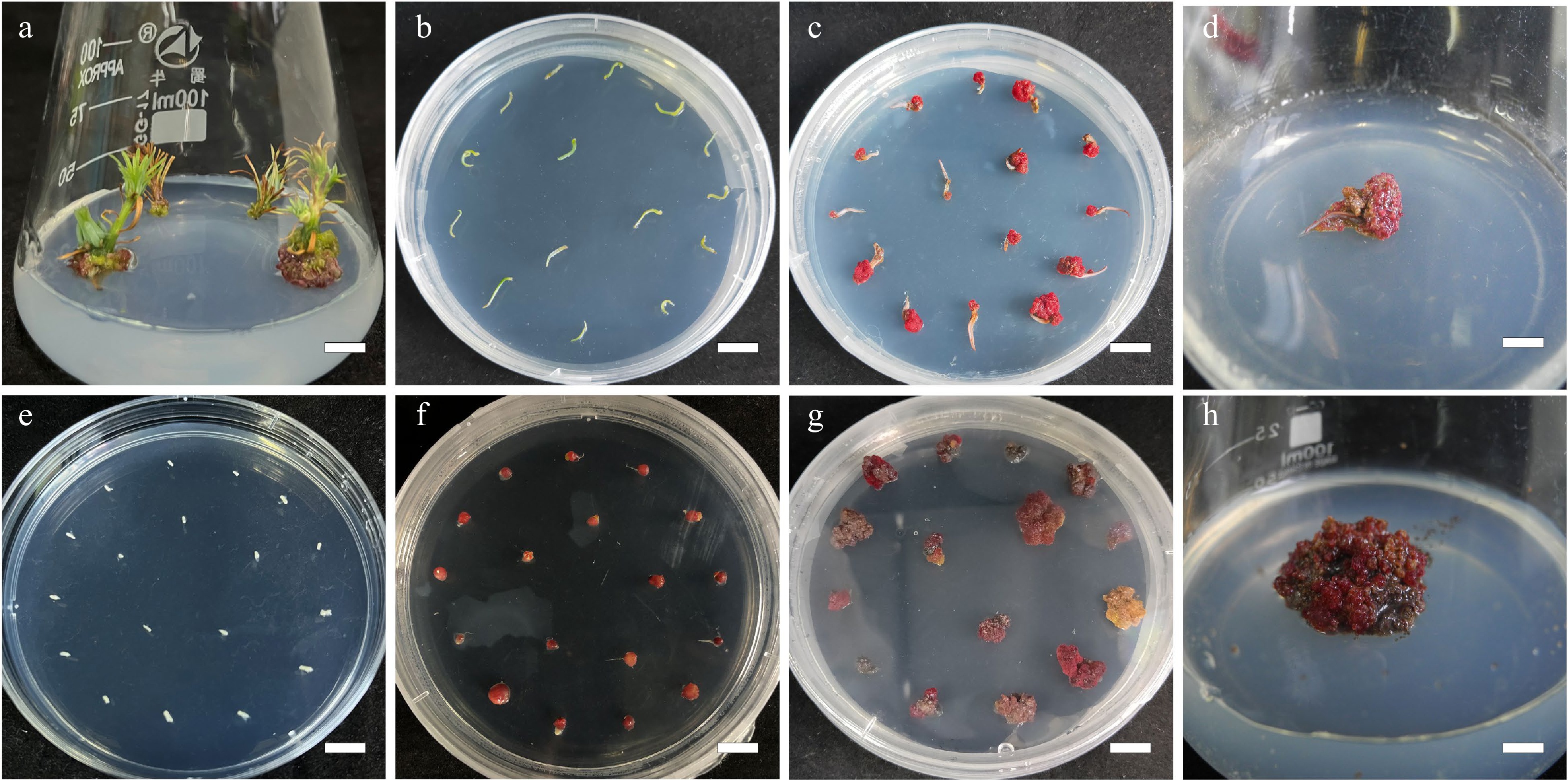
      Figure 1. 

      Callus induction from needle explants and mature zygotic embyro explants in L. olgensis. (a) Sterilized stem sections for obtaining needle explants, bar = 0.7 cm. (b) Needle explants operated in DCR medium augmented with 3 mg·L−1 6-BA and 0.3 mg·L−1 NAA, bar = 1 cm. (c) Calli from needle explants generated in 4 w, bar = 1 cm. (d) Calli from needle explants generated in 8 w, bar = 0.6 cm. (e) Sterilized mature zygotic embryo as explants, bar = 1 cm. (f) Calli from mature zygotic embryo explants generated in 3.0 mg·L−1 6-BA combined with 0.3 mg·L−1 NAA, bar = 1 cm. (g) Calli from mature zygotic embryo explants generated in 4 w, bar = 1 cm. (h) Calli from mature zygotic embryo explants generated in 8 w, bar = 0.6 cm.

    • The explants (zygotic embryos and needles) were obtained in sterile environments. The endosperm was removed from the seeds and the leftover mature zygotic embryos were used directly for callus induction. The needles were collected as described above. Fifteen explants (of zygotic embryo or needles) were placed in 40 mL of callus induction medium (CIM) in a 90 mm × 20 mm crystal-grade polystyrene Petri dish (DA TANG MEDICAL INSTRUMENT) with six replicates. The zygotic embryo and needles were placed separately. The MS medium and DCR medium supplemented with cytokinins 6-BA (0.1, 0.2, 0.5, 1, 2, and 3 mg∙L−1) in combination with auxin NAA at various concentrations(0.1, 0.2, 0.5, 1, 2, and 3 mg·L−1) was used as the CIM. The explants in CIM were kept at 25 ± 2 °C and 70% relative humidity under white fluorescent tubes (60 μmol∙m−2∙s−1 light intensity) in a 16-h photoperiod system until the callus developed (Fig. 1). The nature of the callus and the callus percentage induction were determined after 8 weeks of incubation ( Fig. 2 ).
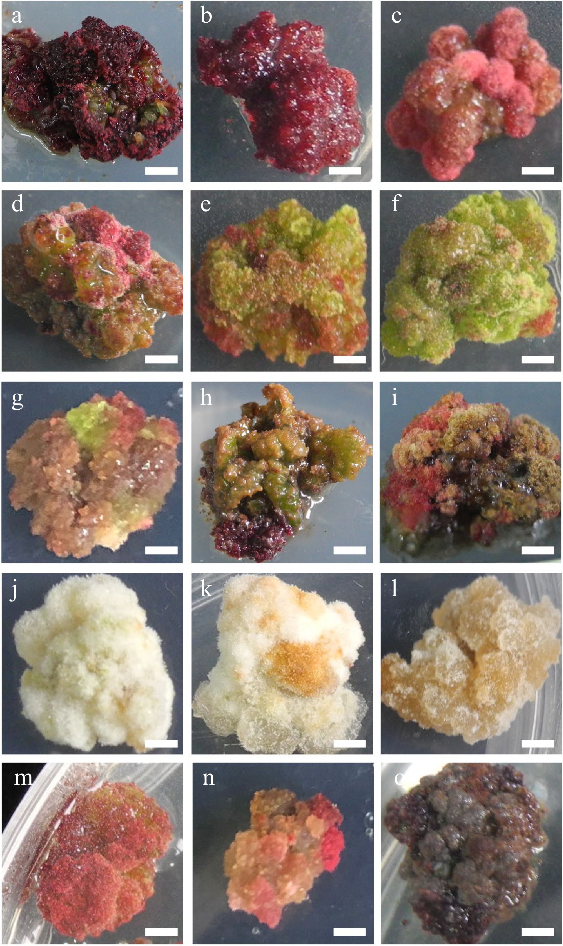
      Figure 2. 

      Characteristic nature of Larix olgensis callus from different treatments. (a) DCR + 3 mg∙L−1 6-BA + 0.3 mg∙L−1 NAA (90%+). (b) DCR+2 mg∙L−1 6-BA+0.2 mg∙L−1 NAA. (c) DCR + 1 mg∙L−1 6-BA + 0.1 mg∙L−1 NAA. (d) DCR + 3 mg∙L−1 6-BA + 0.3 mg∙L−1 NAA, a genotype different from that in (a). (e) DCR + 1 mg∙L−1 6-BA + 1 mg∙L−1 NAA. (f) DCR + 0.3 mg∙L−1 6-BA + 3 mg∙L−1 NAA. (g) MS + 0.5 mg∙L−1 6-BA + 0.05 mg∙L−1 NAA. (h) DCR + 0.5 mg∙L−1 6-BA + 0.05 mg∙L−1 NAA. (i) DCR + 0.5 mg∙L−1 6-BA + 0.05 mg∙L−1 NAA transfer to DCR + 2 mg∙L−1 6-BA + 0.2 mg∙L−1 NAA. (j) DCR + 0.3 mg∙L−1 6-BA + 3 mg∙L−1 NAA (in dark). (k) MS+0.05 mg∙L−1 6-BA + 0.5 mg∙L−1 NAA (in dark). (l) MS + 3 mg∙L−1 6-BA + 0.3 mg∙L−1 NAA (in dark). (m), (n) Modified high auxin culture, the yellow callus turn red. (o) After subculture over five times, the callus turned brown. The bar in the pictures is 0.32 cm except in (n) which is 0.43 cm.

    • Callus was moved to DCR medium augmented with cytokinin 6-BA (0.5, 1.0, 2.0 mg∙L−1) and auxin NAA at various concentrations (0.05, 0.1, 0.2, and 0.5 mg∙L−1) for shoot regeneration. Six replicates were made for each treatment, comprising 10 calli in 50 mL of the shoot regeneration medium in an Erlenmeyer flask (GG-17, 100 mL, SHUNIU). The callus cultures were kept at 25 ± 1 °C and relative humidity of 70% under white fluorescent tubes (60 μmol∙m−2∙s−1 light intensity) in a 16-h photoperiod system. At 2-week intervals, until shoots regenerated, the L. olgensis calli (Fig. 3a) were subcultured onto fresh medium of the same composition with or without AC (Fig. 3b). Shoots produced per callus were counted, and shoot regeneration rate was determined after 6 weeks. Regenerated L. olgensis shoots thereafter were moved to the medium for elongation (Fig. 3c).
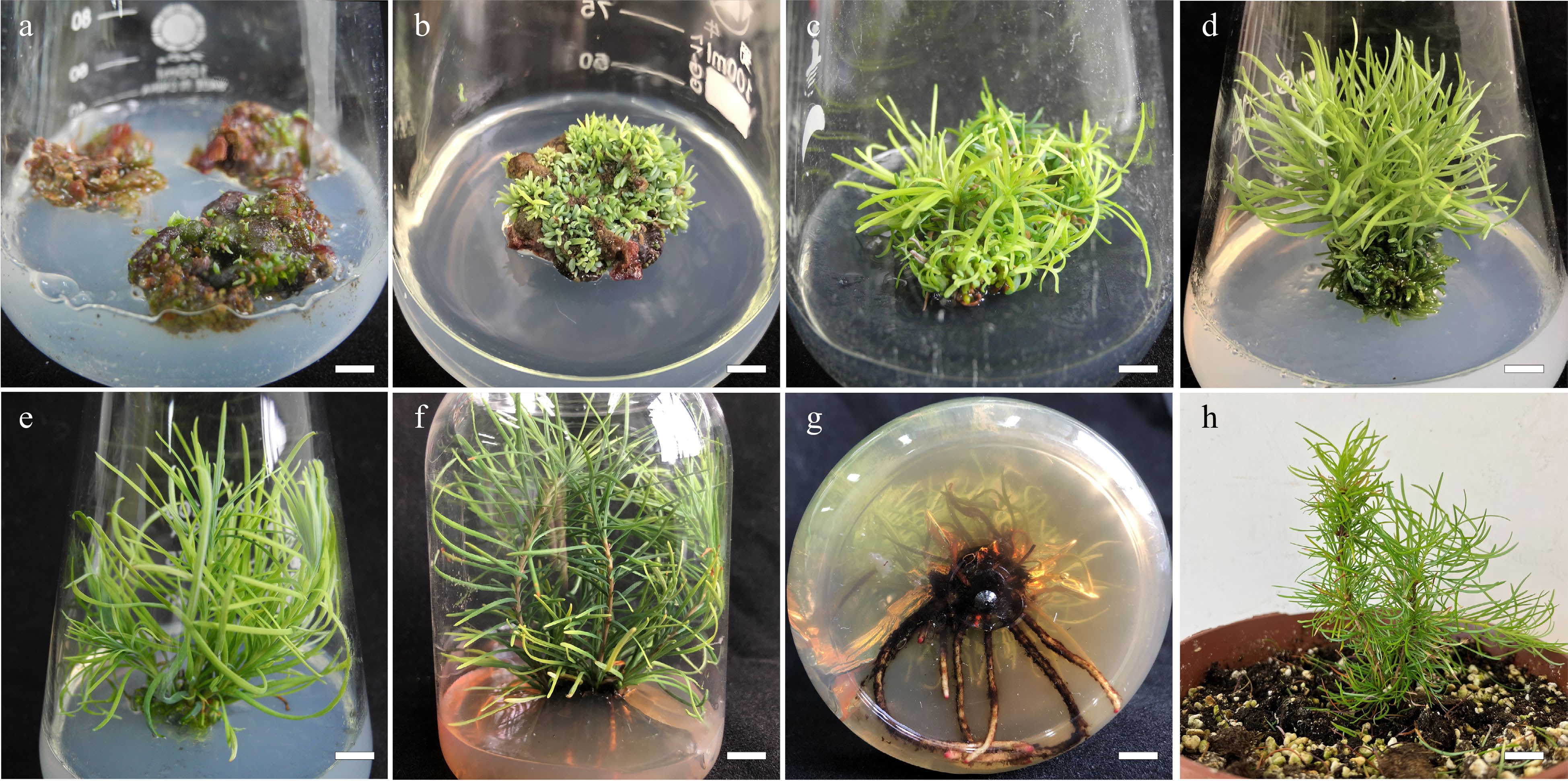
      Figure 3. 

      Summary of in vitro propagation from callus of Larix olgensis. (a) New shoots developed from callus placed in the DCR medium supplemented with 1.0 mg∙L−1 BA and 0.1 mg∙L−1 NAA, bar = 0.6 cm. (b) Shoot organogenesis occurring on callus after 8 weeks in regeneration medium, bar = 0.6 cm. (c) Developed shoots from callus, bar = 0.6 cm. (d) Adventitious shoots elongation, bar = 0.6 cm. (e) Further shoot elongation, bar = 0.68 cm. (f) Rooting of in vitro regenerated shoots in DCR medium supplemented with 3 mg∙L−1 IBA and 100 mg∙L−1 PG, bar = 0.83 cm. (g) Roots of fully developed plantlets, bar = 0.61 cm. (h) Acclimatized potted plants, bar = 2 cm.

    • The regenerated L. olgensis shoots were cultured in the DCR medium (50 mL) supplemented with AC (0, 2 g∙L−1) in addition to cytokinins 6-BA (0.05, 0.1, 0.15, 0.2, and 3 mg∙L−1) and auxin NAA at various concentrations (0.005, 0.01, 0.015, 0.02 and 0.03 mg∙L−1) in combination for shoot elongation in polystyrene culture vessels (ZP5-330, SHJIAFENG). Three regenerated L. olgensis shoots were set up in each vessel, with 20 replications for this experiment. The elongation cultures were kept at 25 ± 1 °C and relative humidity of 70% under white fluorescent tubes (60 μmol∙m−2∙s−1 light intensity) in a 16-h photoperiod system. At 2-week intervals, the L. olgensis shoots were subcultured on fresh media of the same composition (Fig. 3d). Shoot elongation percentage (%) were counted, and elongation lengths were determined after 6 weeks. Elongated L. olgensis shoots thereafter were moved to the medium for rooting (Fig. 3e).

    • The elongated L. olgensis shoots were cultured in DCR medium (100 mL) of various strengths (i.e. DCR, 1/2 DCR) supplemented with auxin [1-naphthaleneacetic acid (NAA) or indole-3-butyric acid (IBA)] (0.5, 1.0, 1.5, 2.0, 2.5 mg∙L−1) either singly or in combination with Phloroglucinol (PG) (0, 50 , 100, 150 mg∙L−1) and AC 2 g∙L−1 in polystyrene culture vessels (125 mm × 110 mm). Four regenerated L. olgensis shoots with 20 replications for this experiment. The elongation cultures were kept at 25 ± 1 °C and relative humidity of 70% under white fluorescent tubes (60 μmol∙m−2∙s−1 light intensity) in a 16-h photoperiod system. Rooting rates and root numbers were determined for each treatment after culture for 10 weeks, with no subculture during rooting (Fig. 3f, g).

    • After removing the medium traces from the roots of each regenerated L. olgensis plantlet by rinsing in running water from a tap, the plantlets were moved to a mixture of peat : organic cultivation soil : perlite (3:6:1) in 22 cm diameter plastic pots (Fig. 3h). The plantlets were covered with transparent plastic bags ensuring adequate humidity and kept in growth chambers operating under a 16-h photoperiod (33.73 μmol∙m−2∙s−1 light intensity) at ~25 °C and 70% relative humidity. The polyethylene coverings were opened gradually after 3 weeks as the plantlets acclimatized. Plant survival rates were determined at 6 weeks following acclimatization.

    • In this study, different fresh calli and tissue of various stages were used for taxifolin content determination. Callus-1 (Fig. 2n), callus-2 (Fig. 2h), callus-3 (Fig. 2a), adventitious shoots, elongation shoots, lignified elongation shoots, adventitious roots, were freeze-dried at −70 °C for 24 h. The dried tissue was ground into a powder with a mortar and sifted through 40 mesh for standby. Each sample was weighed accurately with 100 mg, and taxifolin was extracted by adding 1 mL methanol for tissue bomogenate(8,500 rpm, 4 × 15 s)and ultrasound 100 khz for 20 min. After that, the solution was centrifuged for 6 min (15,000 rpm), the supernatant was taken and diluted 10 times with methanol, and then mixed with H2O 1:1 for LC-MS detection.

      LC-MS analysis was carried out on Waters ACQUITY I-Class (Waters Technology Shanghai, China), and Sciex Triple Quad 5500 (Sciex Shanghai, China). The injection volume of the sample was 2 µL and the column temperature was kept at 30 °C. The binary elution solvent consisted of A [0.1% Formic Acid in Methanol/Acetonitrile (1/9, v/v)] and B (0.1% Formic Acid in H2O): 85% : 15%, and a gradient elution procedure was used. A cosmosil column Waters HSS T3 (100 mm × 2.1 mm, 1.6 μM) was used. The flow rate was maintained at 0.5 mL·min−1. The UV spectrum of taxifolin was obtained with 290 nm detection wavelength.

    • All experimental data were analyzed by one-way ANOVA with Tukey's post-hoc multiple comparison tests, using SPSS (IBM SPSS Statistics 27.0). In CIM, three leaf/root segment explants with 20 replications were used. For shoot regeneration, six calli pieces were used with 10 replications, and four regenerated L. olgensis shoots with 20 replications were used for rooting. Means were regarded as statistically significant at p ≤ 0.05.

    • Among all the treatments, the highest percentage of callus induction was recorded in the explants cultivated on the DCR medium augmented with 3.0 mg∙L−1 6-BA together with 0.3 mg∙L−1 NAA for both L. olgensis zygotic embryo (96.8%) and needle (86.7%) explants (Table 1).

      Table 1.  Induction percentage and characteristics of callus from mature zygotic explants and needle explants of Larix olgensis.

      OrderBasic
      medium
      Plant growth regulators (mg∙L−1)Mature zygotic embryo explantsNeedle explants
      Callus induction rate (%)ColorTextureCallus induction
      rate (%)
      ColorTexture
      1MS6-BA 3:NAA 0.390.2 ± 0.11aRose redCompact43.3 ± 1.91bcBrownCompact
      26-BA 2:NAA 0.290.1 ± 1.73aPinkCompact38.9 ± 1.13bcdBrownCompact
      36-BA 1:NAA 0.178.2 ± 4.45bRed and whitefriable35.6 ± 2.94cdcbrownCompact
      46-BA 0.5:NAA0.0552.1 ± 1.41cdRed and whiteFriable27.8 ± 1.11fgBrownFriable
      56-BA 1: NAA 151.6 ± 1.86cdCreamCompact43.3 ± 0bcCreamFriable
      66-BA 0.05:NAA 0.532.2 ± 1.68fCreamFriable38.9 ± 111bcdYellow and greenFriable
      76-BA 0.1:NAA 143.4 ± 3.41deYellow and greenFriable27.8 ± 1.11fgYellow and greenFriable
      86-BA 0.3:NAA 344.5 ± 3.26deYellow and greenFriable13.3 ± 1.93hYellow and greenCompact
      9DCR6-BA 3:NAA 0.396.8 ± 1.86aRose redCompact86.7 ± 1.93aRose redCompact
      106-BA 2:NAA 0.292.2 ± 0.97aRose redCompact46.7 ± 1.93bGreenFriable
      116-BA 1:NAA 0.176.1 ± 1.94bPinkFriable22.2 ± 1.11ghYellow and greenFriable
      126-BA 0.5:NAA0.0549.0 ± 5.35cdPinkFriable14.55 ± 2.22hDark brownFriable
      136-BA 1: NAA 151.2 ± 2.46cdRed and whiteCompact32.2 ± 1.11efgGreenCompact
      146-BA 0.05:NAA 0.536.6 ± 3.51efRed and whiteFriable34.5 ± 7.78cdeCreamFriable
      156-BA 0.1:NAA 150.1 ± 2.50cdRed and whiteCompact32.2 ± 8.89efgYellow and greenCompact
      166-BA 0.3:NAA 355.7 ± 4.93cCreamCompact23.3 ± 0ghYellow and greenCompact
      Means ( ± standard error) within a column followed by the same superscript letter are not significantly different using Tukey’s multiple comparison test and p ≤ 0.05.

      Although in the about-mention medium both the zygotic embryo explants and needle explants could achieve the highest percentage of callus induction rate, the two type of explants responded significantly differently in other treatments. For zygotic embryo explants, there are no significant differences (in the callus induction rate) from the highest percentage callus in MS media containing 3.0 mg∙L−1 6-BA and 0.3 mg∙L−1 NAA (90.2%), 2.0 mg∙L−1 BA and 0.2 mg∙L−1 NAA (90.1%), and DCR media augmenting with 3.0 mg∙L−1 6-BA and 0.3 mg∙L−1 (96.8%), 2.0 mg∙L−1 BA and 0.2 mg∙L−1 NAA (90.1%) among others (Fig. 1eh, Table 1).

      The highest percentage of callus induction from the needle explants was 86.7% in the DCR medium augmented with 3 mg∙L−1 6-BA and 0.3 mg∙L−1 NAA, which was significantly higher (p ≤ 0.05) than that in the other treatments (Fig. 1ad, Table 1). The lowest percentage of induced callus from L. olgensis zygotic embryo explants was 32.3% in the MS medium augmented with 0.05 mg∙L−1 6-BA together with 0.5 mg∙L−1 NAA, and that from needle explants were in the DCR medium augmented with 0.3 mg∙L−1 6-BA together with 0.3 mg∙L−1 NAA (13.3%).

      The effect of plant growth regulators (PGRs) combination was tested. The ratio of auxin and cytokinin of 1/10 showed a better callus induction response than that of 1/1 and 10/1 (Table 1). Once the ratio was determined, it was found that the callus induction rate was increased along with the promotion in the concentration of the PGRs combination. Meanwhile, the suitable basic mediums of callus induction from mature zygotic embryos were MS and DCR, while needle explants preferred DCR basic medium.

      The calli produced from both zygotic embryos and needle explants had different textures and colors. These colors were pink (Fig. 2ad), green (Fig. 2e, f), cream (Fig. 2gi), or dark brown (Fig. 2o), etc (Fig. 2gi, m, n), and their textures were either compact or friable depending on the medium composition and explant type (Table 1, Fig. 2). Furthermore, the compact rose red callus is the best for shoot regeneration (Fig. 2a, b).

    • For the zygotic explants, we found that the MS medium augmented with 1.0 mg∙L−1 6-BA and 0.1 mg∙L−1 NAA had the highest shoot regeneration rate (83.3 ± 1.93% and 528 ± 11.5 number of shoots per callus), followed by the medium supplemented with 1.0 mg/L 6-BA in addition to 0.2 mg∙L−1 NAA (76.7 ± 1.93%) shoot regeneration rate with the highest shoot number per callus (636 ± 21.7) (Table 2, Fig. 3a).

      Table 2.  Percentage shoot regeneration from calli of Larix olgensis.

      Mature zygotic embryo explantsNeedle explants
      Plant growth regulators (mg∙L−1)Percentage shoot regeneration (%)Average number of adventitious shootsPercentage shoot regeneration (%)Average number of adventitious shoots
      6-BANAA
      10.50.0541.1 ± 2.94fg304 ± 7.5d32.22 ± 1.11de120 ± 6.1h
      20.50.142.2 ± 1.11fg315 ± 4.5d32.22 ± 2.22de111 ± 3.8h
      30.50.232.2 ± 1.11h150 ± 14.8f28.89 ± 1.11e66 ± 2.3i
      40.50.540.0 ± 1.93g211 ± 5.5e34.44 ± 1.11d51 ± 1.8g
      510.0557.8 ± 1.11d426 ± 13.9c68.89 ± 1.11a161 ± 6.2f
      610.183.3 ± 1.93a528 ± 11.5b73.33 ± 1.93a307 ± 1.8b
      710.276.7 ± 1.93b636 ± 21.7a72.22 ± 1.11a323 ± 3.8a
      810.565.6 ± 2.22c460 ± 22.8c73.33 ± 1.93a221 ± 6.4c
      920.0553.3 ± 3.85de322 ± 11.7d50.00 ± 1.93c176 ± 5.5e
      1020.147.8 ± 2.94ef237 ± 11.5d51.11 ± 1.11c141 ± 7.0g
      1120.246.7 ± 1.93efg423 ± 23.5c57.78 ± 2.22b203 ± 3.9d
      1220.547.8 ± 1.11ef200 ± 6.1e33.33 ± 1.93de57 ± 1.2ig
      Means (± standard error) within a column followed by the same superscript letter are not significantly different using Tukey’s multiple comparison test and p ≤ 0.05.

      Meanwhile, for the needle explants, the highest percentage of regeneration (73.3 ± 1.93%) and the number of shoots per callus (307 ± 1.8) were recorded in the explants cultivated on the DCR medium augmented with 1.0 mg∙L−1 6-BA, 0.1 mg∙L−1 NAA, and 0.1 mg∙L−1 TDZ, followed by the results in the medium supplemented with 1.0 mg∙L−1 6-BA in addition to 0.2 mg∙L−1 NAA (with shoot regeneration percentage and the number of shoots per callus, 72.22 ± 1.11% and 323 ± 3.8, respectively) (Table 2, Fig. 3b). The highest percentage of regeneration (73.3 ± 1.93%) was also recorded in the medium supplemented with 1.0 mg∙L−1 6-BA in addition to 0.5 mg∙L−1 NAA, but the number of shoots per callus (221 ± 6.4) was significantly lower than that in medium 6.

      Both shoot regeneration percentage and shoot number per callus were generally higher in media supplemented with cytokinin (BA) in combination with auxin (NAA), which ratio ranges from 10/1 to 5/1. In addition, medium 2-6 and medium 2-7, 2-1, 2-2, and 2-11 also showed relatively high induction rates. Although the differences in shoot regeneration percentage were not statistically significant, medium supplemented with 6-BA in combination with higher NAA were generally associated with a low number of shoots induced from each callus on average (Table 2, Fig. 3). If the ratio of 6-BA to NAA is fixed, with the increase of the PGRs concentration, the shoot regeneration percentage and the number of shoots per callus showed an upward trend initially and then declined.

      Due to the limited callus size and the number of the subculture of needles, the number of shoots per callus induced from needles was lower than that from zygotic embryos, but there is no significant difference in the shoot regeneration percentage between zygotic embryos and needles.

    • The zygotic embryo explants and the needle explants were inoculated in the same shoot regeneration culture medium for 4 weeks before subculture to the elongation treatment. The DCR medium augmented with 0.5 mg∙L−1 BA, 0.05 mg∙L−1 NAA, and 2 g∙L−1 AC achieved the highest elongation percentage of shoots (75.6 ± 2.94%) and the longest average shoot length (3.5 ± 0.11 cm). The percentage shoot elongation and average shoot length significantly differed (p < 0.05) from that in DCR without any AC (control) (Table 3, Fig. 3ce). In the same medium without AC, the percentage of shoot elongation and average shoot length were 65.2 ± 1.11% and 1.6 ± 0.11 cm, respectively. Compared medium 3-2 (65.6 ± 1.11%, 1.6 ± 0.06 cm) to medium 3-3 (75.6 ± 2.92%, 3.5 ± 0.11 cm), medium 3-4 (46.7 ± 1.93%, 1.3 ± 0.03 cm) to medium 3-5 (61.1 ± 1.11%, 2.8 ± 0.12 cm), medium 3-6 (21.1 ± 1.11%, 1.4 ± 0.01 cm) to medium 3-7 (35.6 ± 2.22%, 2.2 ± 0.1 cm), it was clear that AC significantly increased the percentage of shoots elongation and average shoot length.

      Table 3.  Effect of different concentrations of 6-BA and NAA on adventitious bud elongation of Larix olgensis.

      Plant growth regulators and AC (mg∙L−1)Adventitious
      shoot elongation
      percentage (%)
      Average shoot length (cm)
      16-BA 1:NAA 0.121.1 ± 2.22e0.9 ± 0.03e
      26-BA 0.5:NAA 0.0565.6 ± 1.11b1.6 ± 0.06d
      36-BA 0.5:NAA 0.05:AC 200075.6 ± 2.94a3.5 ± 0.11a
      46-BA 0.3:NAA 0.0346.7 ± 1.93c1.3 ± 0.03d
      56-BA 0.3:NAA 0.03:AC 200061.1 ± 1.11b2.8 ± 0.12b
      66-BA 0.1:NAA 0.0121.1 ± 1.11e1.4 ± 0.01d
      76-BA 0.1:NAA 0.01:AC 200035.6 ± 2.22d2.2 ± 0.10c
      Means (± standard error) within a column followed by the same superscript letter are not significantly different using Tukey’s multiple comparison test and p ≤ 0.05.

      The elongation culture of L. olgensis was DCR medium supplemented with a certain ratio but different concentrations of BA and NAA. According to Table 3, a comparison of the elongation rate and average shoot length among medium 3-1 (21.2 ± 2.22%, 0.9 ± 0.03 cm), medium 3-2 (65.6 ± 1.11%, 1.6 ± 0.06 cm), medium 3-4 (46.7 ± 1.93%, 1.3 ± 0.03 cm), which depicted lower concentrations of the PGRs promoted the shoot elongation, but when it reduced to a certain level, poor elongation also resulted.

    • In our study, the first regeneration plantlet with roots (1−2 mm) was seen on the 38th day in DCR medium supplemented with 3 m∙L−1 IBA and 100 mg∙L−1 PG, and the highest adventitious root induction rate was 62.2 ± 5.88% (Table 4, Fig. 3f, g). Even if the concentration of auxin is continuously increased, the single application of auxin has little effect on rooting. The rooting rates of DCR medium supplemented with 3 mg∙L−1 of IBA or NAA were 13.3%, and 8.9 ± 2.22%, respectively. But in 1/2 DCR medium with the same PGRs were 11.11 ± 2.22%, and 15.56 ± 2.22%, respectively. Rooting in these mediums took at least 60 d.

      Table 4.  Rooting of regenerated shoots in DCR media supplemented with auxin, AC and PG strength.

      Basic mediumExogenous
      additives (mg∙L−1)
      Adventitious
      root induction
      percentage (%)
      Rooting start time (d)
      DCRNAA 38.9 ± 2.22e70
      IBA 2:NAA 215.6 ± 2.22de64
      IBA 3:AC 200046.7 ± 3.85b52
      IBA 313.3 ± 0.00e63
      IBA 3:PG 5033.3 ± 3.85c45
      IBA 3:PG 10062.2 ± 5.88a38
      IBA 3:PG 15057.8 ± 4.44ab36
      IBA 3:PG 100:AC 200053.3 ± 3.85b32
      1/2DCRNAA 315.56 ± 2.22de70
      IBA 2:NAA 222.22 ± 2.22d63
      IBA 3:AC 200035.56 ± 2.22c60
      IBA 311.11 ± 2.22e60
      IBA 3:PG 5031.11 ± 4.44c50
      IBA 3:PG 10048.89 ± 2.22b41
      IBA 3:PG 15046.67 ± 0b40
      IBA 3:PG 100:AC 200037.78 ± 2.22c36
      Means (± standard error) within a column followed by the same superscript letter are not significantly different using Tukey’s multiple comparison test and p ≤ 0.05.

      The addition of AC and PG promoted root formation to a large extent and significantly increased the rooting percentage. In the medium supplemented with PG, once the root primordium is produced, the adventitious roots were produced along the main stem, and the roots were quickly formed to produce a strong root system (Fig. 3g). The concentration of PG significantly affected the rooting rate in both 1/2 DCR and DCR medium. Specifically, among the three concentrations of PG tested, 50 mg∙L−1, 100 mg∙L−1, and 150 mg∙L−1, the 100mg∙L−1 PG had a better effect on rooting (Supplemental Fig. 1ad). The root system from the medium supplemented with AC was slender, and lateral roots were produced in prolonged culture (Supplemental Fig. 1e, f).

    • After 6 weeks of acclimatization, the in vitro regenerated L. olgensis plantlets showed a high survival rate of 90%. The acclimatized L. olgensis plantlets grew well and displayed normal growth characteristics and morphology typical of the plant species (Fig. 3h).

    • To determine the taxifolin content in different tissues and stages of L. olgensis plants, callus of different stages (callus-1, callus-2, callus-3), adventitious shoots, elongation shoots, lignified elongation shoots, adventitious roots, were selected and analyzed by HPLC (Supplemental Fig. 2). The result indicated that the taxifolin content in different tissues and different stages of the regenerated L. olgensis plants varied significantly. The content of the taxifolin in callus-1 was 1.99 µg∙g−1, callus-2 was 3.9 µg∙g−1, callus-3 was 5.26 µg∙g−1, and in adventitious shoots, the content of the taxifolin was 4.8µg/g, while in adventitious roots was 2.86 µg∙g−1. The accumulation of the taxifolin in elongation shoots and lignified elongation shoots was 28.6 µg∙g−1 and 53 µg∙g−1 respectively, much higher than that in other tissues.

      Meanwhile, the results showed that calli in different colors and textures might affect the accumulation of taxifolin. For example, the rose-red calli accumulated more taxifolin than the calli of two other colors. The result also illustrated that the development of vascular tissue was beneficial to the accumulation of taxifolin since in the elongation shoots, the content of taxifolin was much higher. Compared with the un-lignified elongation shoots, the content of taxifolin in the lignified shoots nearly doubled. Therefore, in vitro regeneration is an efficient and quick method to produce secondary metabolites.

    • Due to the current large demand for larch timber, the supply of seedlings and the establishment of plantations have become very urgent. In vitro regeneration technology provides an efficient way for large numbers of seedling production in a short time and has been widely used in large-scale propagation of high-quality seedlings, genetic transformation, and gene editing. The L. olgensis regeneration system is set up in this study, the process is stable and reliable, and not easily affected by external conditions, and therefore, is highly controllable and annual production can be assured.

      The research on the in vitro regeneration system of larch mainly contains somatic embryogenesis and organogenesis. Current research on in vitro regeneration of larch mainly focuses on inducing embryogenic callus that leads to somatic embryogenesis. The induced somatic embryos have characteristics similar to mature zygotic embryos and can directly generate stems and roots through suitable culture. However, some research showed that the germination rate of the induced somatic embryos was uncontrollable[6], and the malformed embryos accounted for a large proportion. Some believe that somatic embryos are more suitable for cryopreservation and production of artificial seeds[23], but the subsequent growing time is the same or even longer than that of larch seedlings from natural seeds. In this study, mature embryos are regenerated via the callus, and under suitable culture conditions, more calli can be subcultured, and more adventitious shoots can subsequently be differentiated. Since a large amount of biomass can be produced under certain culture conditions, the growth rate and development direction can be adjusted by using different culture conditions. Tissue products in various culture stages can also be used for active ingredient extraction. The in vitro regeneration system of larch was optimized in the following aspects.

      Different species respond to in vitro regeneration quite differently, which might be the reflection of differences in nutrient absorption. It is crucial for species to confirm a rationally basic medium. Classical MS medium with a high nutrient concentration of inorganic salts is favored in plant tissue culture[24], especially in the cultivation of herbaceous plants, such as cornflower[25] (Gerbera jamesonii), lily (Lilium orientalis)[26], andrographis (Andrographis alata)[27], chandelier flowers (Ceropegia mohanramii)[28], etc. Generally, researchers believe that at present, the regeneration of woody plants is more difficult than that of herbaceous plants[29]. It is shown that the absorption capacity of the basic medium is different for different life-form plants, and sometimes the MS medium does not yield good results in some woody plants. For example, for the conifer juniper[30] (Juniperus L.), researchers gradually replaced MS with WPM medium during the culture. The researchers used a modified MS medium with half-strength salt and reduced the concentration of KNO3 in the medium at a later stage for a good culture effect. The study by Samiei et al.[31] suggested that Van der Salm (VS) modified by MS medium with reduced inorganic salts has a better effect than MS in culturing Rosa canina. In the cultivation of Fagaceae chestnut (Castanea sativa × Castanea mollissima)[32], researchers used MS medium with reduced salt concentration, combined with WPM, to obtain stable chestnut regeneration seedlings. The macroelement salt ion molar concentration of MS is about three times that of WPM and nine times that of DCR medium. Researchers who used MS medium to cultivate the plantlets have so far not achieved efficient results in larch. In our research, the lower inorganic salt ion concentration DCR basic medium is suitable for L. olgensis subsequent development. The results are consistent with the above mentioned reports (Table 1, Fig. 1).

      Many studies have shown that the use of single plant growth regulator has a limited effect on callus tissue during larch regeneration. In this study, we investigated the effects of auxin and cytokinin in different ratios and the strength of the combined concentration on various processes in the regeneration of mature zygotic embryos and needles. The dominant plant growth regulator and the ratios of different PGRs both played crucial roles in callus induction. The combination and concentration of hormones for in vitro regeneration of mature zygotic embryos and needles were determined. In the in vitro regeneration of plants, the combined use of cytokinins and auxins can influence the growth direction of the materials. The combination of cytokinin 6-BA and auxin NAA in different concentration groups were used to study the concentration ratio and intensity of growth regulators required in each stage of larch development. It was found that in the stage of callus induction of mature zygotic embryos and needles, larch needs a higher concentration of cytokinin, thus forming a large number of calli. When the 6-BA/NAA ratio is 10, it is beneficial to promote callus induction. At this ratio, increasing the concentration to three times (taking 6-BA as 1 mg∙L−1 as an example) can accelerate the formation of calli. However, under the condition of high auxin, callus quality is poor and consequently difficult to differentiate (Table 1, Fig. 2l, o). A shorter subculture cycle may be beneficial for callus induction under high auxin culture conditions.

      Our study suggests that the adventitious shoots subsequently differentiate from callus and should require a lower concentration of cytokinin in larch (Fig. 3). The method was considered to be effective to obtain regenerated plantlets. In addition, adding a certain amount of activated carbon is conducive to the elongation of larch (Table 3). In contrast, in the regeneration of Ash (Fraxinus mandshurica) in 2020[33], using long-term and high concentration phytohormone cultivation, the number of adventitious bud differentiation is extremely low, and complete plants cannot be obtained. The results of elm trees (Ulmus glabra and Ulmus laevis) in vitro regeneration also showed[34] that 0.5 mg∙L−1 6-BA was appropriate for plant regeneration and stem differentiation. In some ranges, both the broad-leaved tree and conifer maybe have similar responses to adventitious shoots differentiation.

      Among all the tissues differentiated, the content was high in the lignified elongation shoot and the green elongation shoot. Moreover, the states of the tissues influenced the content of taxifolin. For example, the rose-red calli accumulated more taxifolin than the calli of the two other colors. It can be referred that the development of vascular tissue was beneficial to the accumulation of taxifolin since in the lignified shoots, the content of taxifolin was much higher (Supplemental Fig. S2). The result is consistent with natural larch, which also proves the potential of active ingredient production with artificial regulation[35]. Overall, it is obvious that in vitro regeneration is an efficient and quick method to produce secondary metabolites.

      The in vitro rooting of larch is very difficult. In this study, we established a rooting system for larch. Previously, there is no effective rooting method for larch in vitro regeneration, or the rooting process is complicated[36]. Induction of adventitious roots is the most difficult step in the in vitro regeneration of larch, with unstable rooting and a low induction rate of adventitious roots. In the process of adventitious shoot rooting, single application of auxin was not good (Table 4), increasing the concentration of auxin to 3 mg∙L−1, or two auxins NAA and IBA in combination, the rooting effect did still not work well. However, the addition of exogenous substances such as PG and AC in combination with auxin has a good effect on rooting. In this study, the combination of 100 mg∙L−1 PG and IBA obtained a good rooting effect (Fig. 3, Supplemental Fig. S1), indicating that PG is a good exogenous additive for inducing rooting.

      Above all, an efficient and complete organogenesis regeneration system was established for the first time, which would greatly benefit larch plantation. This protocol can be used for large-scale propagation of high-quality seedlings, genetic transformation, gene editing, and in vitro production of raw materials in various industries. Furthermore, it is a reliable reference for in vitro regeneration in recalcitrant species.

    • In this study, we established an efficient and complete regeneration system for larch organogenesis regeneration for the first time, especially from the needle explants. Effects of combination of auxin and cytokinin in different ratios and different intensities on regeneration were investigated. Furthermore, we firstly reported the taxifolin accumulation and content in the different larch tissues. To the best of our knowledge, this is the first study to develop an efficient indirect regeneration protocol for L. olgensis, which can be used for large-scale breeding of high-quality seedlings, genetic transformation, and gene editing and offers a basis for the production of raw materials in various industries. It is also a reliable reference for in vitro regeneration in recalcitrant species.

      • This work was supported by the National Science and Technology Major Projec of China (2018ZX08020003-005-001)

      • The authors declare that they have no conflict of interest.

      • Copyright: © 2023 by the author(s). Published by Maximum Academic Press, Fayetteville, GA. This article is an open access article distributed under Creative Commons Attribution License (CC BY 4.0), visit https://creativecommons.org/licenses/by/4.0/.
    Figure (3)  Table (4) References (36)
  • About this article
    Cite this article
    Yan X, Wang K, Zheng K, Zhang L, Ye Y, et al. 2023. Efficient organogenesis and taxifolin production system from mature zygotic embryos and needles in larch. Forestry Research 3:4 doi: 10.48130/FR-2023-0004
    Yan X, Wang K, Zheng K, Zhang L, Ye Y, et al. 2023. Efficient organogenesis and taxifolin production system from mature zygotic embryos and needles in larch. Forestry Research 3:4 doi: 10.48130/FR-2023-0004

Catalog

  • About this article

/

DownLoad:  Full-Size Img  PowerPoint
Return
Return