ARTICLE   Open Access    

Green lacewing Chrysoperla rufilabris (Neuroptera: Chrysopidae) is a potential biological agent for crapemyrtle bark scale (Hemiptera: Eriococcidae) pest management

More Information
  • Crapemyrtle bark scale (CMBS; Acanthococcus lagerstroemiae), an invasive sap-sucking hemipteran, has spread across 16 US states. Infestation of CMBS negatively impacts the flowering and reduces the aesthetic quality of crapemyrtles. The widespread use of soil-applied neonicotinoid insecticides to suppress the CMBS infestation may be hazardous to pollinators and other beneficial insects. Natural enemies of CMBS are important agents for developing integrated environmentally friendly management strategies. This study evaluated the performance of larval green lacewing (Chrysoperla rufilabris) as a biocontrol agent of CMBS. Predatory behavior of the larval C. rufilabris upon CMBS was documented under a stereomicroscope using infested crapemyrtle samples collected from different locations in College Station (Texas, USA). Predation potential of C. rufilabris upon CMBS eggs and foraging performance using Y-maze assay were investigated under laboratory conditions. Results confirmed that larval C. rufilabris preyed on CMBS nymphs, eggs, and adult females. The evaluation of predation potential results showed that 3rd instar C. rufilabris consumed significantly more CMBS eggs (176.4 ± 6.9) than 2nd (151.5 ± 6.6) or 1st instar (11.8 ± 1.3) in 24 hours. Results from the Y-maze assay indicated that larval C. rufilabris could target CMBS in the dark, indicating that some cues associated with olfactory response were likely involved when preying on CMBS. This study is the first report that validated C. rufilabris as a natural predator of CMBS and its potential as a biological agent to control CMBS. Future investigation about the olfactory response of larval C. rufilabris to CMBS would benefit the development of environmentally friendly strategies to manage CMBS.
  • The Trichoderma genus encompasses a wide-ranging collection of filamentous fungi, prevalent in various natural ecosystems[1]. Trichoderma species within this genus have earned acclaim for their exceptional capacity to inhabit plant roots, stimulate plant growth, and showcasing biocontrol attributes against a spectrum of fungal adversaries[2]. Employing tactics like mycoparasitism, antibiosis, competitive resource acquisition, and plant resistance induction, these species effectively manage fungal diseases[3]. Notably, they are increasingly utilized in agriculture as biofertilizers and biopesticides[1]. Trichoderma-based bio-fungicides, available in different formulations like wettable powders, granules, and flowable concentrates, offer a convenient application to seeds, seedlings, soil, and foliage[4,5]. Besides their disease-fighting properties, these bio-fungicides promote plant growth through various mechanisms such as phytohormone production, nutrient solubilization, and stress tolerance enhancement[3]. Recent progress in Trichoderma-based formulations has led to innovative materials, advanced nanotechnology strategies, and genetic engineering techniques aimed at boosting stability, shelf life, and efficacy[4]. Among these advancements, biochar has shown promise as an ideal carrier for Trichoderma formulations due to its high porosity, surface area, and soil stability maintenance abilities[6]. New research indicates that biochar can strengthen Trichoderma's biocontrol properties[7,8]. Experiments show that using Trichoderma bio-fungicides on soil blended with biochar is more effective in fungal disease control than on unamended earth[9]. Likewise, applying these bio-fungicides on biochar-coated seeds provides better resistance against fungal diseases in seedlings[7,10]. Such biotechnological advancements in Trichoderma-based formulations can promote sustainable agricultural practices by reducing reliance on chemical pesticides[11]. This, in turn, helps mitigate the ecological impact of agricultural activities and enhances food and feed safety[12]. By protecting plants from fungal diseases and improving soil fertility, Trichoderma-based bio-fungicides hold promise for enhancing crop yield[1]. Trichoderma formulations play a crucial role in minimizing harm to non-target organisms while maximizing the effectiveness of the active ingredient[13]. While Trichoderma is significant in ensuring agronomic safety, challenges in their formulation persist due to potential degradation of the biomass or bioactive metabolite caused by factors like exposure to air, light, and temperature[14]. Additionally, these products need to be easy to handle, apply, and produce[15,16]. To address this objective, the present study aims to offer a comprehensive examination of various technological advancements that enhance the efficiency of natural preparations. Distinguishing itself from typical literature reviews that predominantly delve into the biological attributes of metabolites, this review incorporates a bibliometric analysis of biopesticides and their formulations[17]. This analysis employs quantitative and statistical indicators to identify patterns related to the most critical pest issues, agriculture's susceptibility, sources of biological control, innovative methodologies, and the current status of Trichoderma formulations. The insights presented in this analysis significantly contribute to the bibliometric methodology, potentially promoting positive strides in the advancement of technology for Trichoderma formulation. Additionally, it offers valuable suggestions for researchers engaged in this field.

    Bibliometric analysis is a technique employed to scrutinize the characteristics and evolving patterns within academic literature using various mathematical and statistical methods[10]. Through this approach, we can quantitatively assess the overall state of the literature, collaborative relationships, research areas of interest, and the development trends in a specific research field[18]. A descriptive analysis of the corpus of published research pertaining to Trichoderma formulations was conducted. This analysis entailed the examination of co-occurring terms within the body of published articles, allowing for the elucidation of evolutionary trends in scientific themes[19]. The fundamental aim of this research is to conduct an exhaustive review of the existing body of literature on Trichoderma formulations and to project the areas of highest interest and potential for future investigation[20]. One of the primary objectives of bibliometric analysis is to assess the trends in research related to Trichoderma formulations, and to identify the most influential authors and institutions in the field of Trichoderma research. Determining the impact of research in terms of citations, patents, or applications in real-world scenarios.

    As a result, this study seeks to investigate the following research objectives: In the field of Trichoderma formulations, what are the key research themes and trends observed from 2016 to 2023 include:

    (1) How is research on Trichoderma formulations distributed geographically, and what regions exhibit the most active contributions to the field? (2) Can bibliometric analysis predict future trends and potential innovations in Trichoderma formulations research based on historical patterns? (3) The article follows a well organised structure[21]. Initially the research methodology adopted for the study is outlined. Subsequently, a well-organized article is crucial for effectively communicating research findings to the intended audience[22].

    Literature retrieval was performed online through the Science Citation Index Expanded (SCI-E) of the Web of Science Core Collection (WoSCC, Clarivate Analytics) from 2016 to 2023[21,22]. Scopus is a preferred data source for bibliometric analysis, and it provides comprehensive information and data from a multi-disciplinary field of literature[23]. To retrieve literature comprehensively and accurately on Trichoderma formulations, different search terms and retrieval strategies were assembled in this study. Finally, the optimal search items were set as follows: TS = ('Trichoderma formulation*') OR ('Bio formulation of Trichoderma*') OR ('Bio control') OR ('Antagonist') OR ('Rhizosphere fungus')[24]. The data range was set from 2016 to 2023, to collect all relevant publications. It is worth noting that as the Scopus database data network is constantly updated, the results may vary depending on the exact retrieval date.

    A detailed literature retrieval process was performed online through Science Citation Index Expanded (SCI-E) of the Web of Science Core Collection (WoSCC, Clarivate Analytics) from 2016 to 2023, and considered Scopus as a preferred data source for bibliometric analysis. Additionally, we outlined the search terms and retrieval strategies that are used in our study and set the range of data from 2016 to 2023 to collect all relevant publications. If we sum up our descriptions narrate on the following key points, like data sources, search terms, data range, constantly updated data base, and optimal search items[25]. The search strategy was designed to capture relevant literature on Trichoderma formulations. The search terms included Trichoderma formulation, bio-formulation of Trichoderma, biocontrol, antagonist and Rhizosphere fungus. The asterisks, in the search terms are used as wildcard characters to capture different word endings. The data range was set from 2016 to 2023 to collect all relevant publications within that timeframe. This was the period during which the literature retrieval was performed[25]. It is mentioned that as the Scopus database data network is constantly updated, and the results may vary depending on the exact retrieval date. This indicates that the study acknowledged the dynamic nature of the database and its potential impact on the results[26]. After assembling different search terms and retrieval strategies, the study determined the optimal search items, which were the selected search terms that would yield the most comprehensive and accurate results for the study's objectives. Overall, the present study took a systematic approach to literature retrieval, considering multiple data sources and employing a combination of search terms to ensure the retrieval of relevant publications on Trichoderma formulations. It is worth noting that as the Scopus database data network is constantly updated, to add upon, the results may vary depending on the exact retrieval date.

    To ensure the credibility of the research conclusions, this study gathered peer-reviewed English journal articles to summarize global research perspectives. It's important to mention that articles not aligned with this paper's purpose were manually omitted in the final phase of data collection[27]. For instance, some articles explored the relationship between plants and microorganisms on leaves. Eventually, a total of 287 articles that met all criteria were sourced from Scopus. These pieces represented almost all top-tier experimental studies on Trichoderma formulations from 2016 to 2023 worldwide. The variability among these articles could effectively indicate the trend of related research development. Therefore, these publications were prioritized for further analysis and assessment. Figure 1 illustrates the flowchart of the literature retrieved in this study[28].

    Figure 1.  Flow chart of literature review methodology.

    Table 1 presents the top 10 countries/regions, institutions, authors, and journals that published the most studies on Trichoderma formulations. As indicated in Table 1, China emerged as the country making the most significant contribution, with 1,231 publications, accounting for 20.33% of the total. India and Pakistan followed closely, ranking second and third, with 1,096 (18.10%) and 618 (10.20%) publications, respectively. Among the institutions, the Chinese Academy of Sciences held the top spot, boasting 160 (2.64%) publications. Following closely was the University of Agriculture, Faisalabad (144, 2.37%), and Nanjing Agricultural University (137, 2.26%). In the realm of scholarly contributions, prolific authors often set the tone for research trends. Identifying these influential scholars can shed light on the direction of the research field[29]. The leading author in the study of rhizosphere microorganisms was Wang Y, with 102 publications. Li Y, Zhang Y, and Hkan M were also highly prolific, each publishing nearly 90 studies. These works were predominantly featured in prominent journals in Ecology and Botany, such as Frontiers in Microbiology (3.73%), Frontiers in Plant Science (2.36%), and Plant and Soil (2.03%).

    Table 1.  Leading journals contributing to the existing body of knowledge in the field of formulation of Trichoderma.
    Sl. no. Name of journal No. of publication Citations
    1 Journal of Applied Microbiology 2 40
    2 Applied Microbiology and Biotechnology 2 16
    3 Indian Phytopathology 3 2
    4 Biological Control 2 59
    5 Crop Protection 2 30
    6 Frontiers in Microbiology 2 23
    7 Journal of Biological Control 2 1
    8 Medicinal Plants 2 5
     | Show Table
    DownLoad: CSV

    The analysis of scientific production on Trichoderma formulations demonstrated the trend of publications per year on Trichoderma formulation studies. It was observed that published research showed a significant increase of 71.24% over the last decade (2016–2023) (Fig. 1). The increasing trend is possibly related to economic support from government programs, since funding for innovative, sustainable, and ecological research is being considered to meet the demand for food and mitigate environmental pollution[30]. Figure 2 illustrates a co-citation map of authors collaborating in the field of Trichoderma formulation. The purpose of conducting this co-citation analysis is to visually portray the knowledge base of the specific area of review. The analysis identifies three distinct clusters, each represented by different colored nodes: blue, red, and green. The green cluster stands out as it is associated with Harman, who has the highest collaboration, working with nine researchers on Trichoderma formulation research. The red cluster, on the other hand, signifies the second-highest collaboration, led by Mukherjee et al.[31] with six researchers. Lastly, the pink cluster represents the lowest level of collaboration among researchers, with only two researchers working together in this area.

    Figure 2.  Co-citation analysis of cited authors as the unit of analysis in the field of Trichoderma formulation.

    Table 2 showcases the country–wise citation analysis, and the series presented here seems to have large variability in distribution. In terms of total citations of studies dedicated to Trichoderma formulations. Citations count of articles by country as a unit of analysis represents the popularity of a field of research in a particular region. India with 20 publications having 115 citations topped the list and, therefore, is the most impactful country contributing to the existing body of knowledge in the said domain followed by Italy with four publications having 69 citations and Brazil with three publications having 51 citations. From the viewpoint of the total number of publications, India holds first position, having 20 publications, followed by Italy having four publications.

    Table 2.  Leading countries contributing to the existing body of knowledge in the field of formulation of Trichoderma.
    Sl. no. Country No. of publication Citations
    1 Argentina 1 20
    2 Belgium 2 30
    3 Brazil 3 51
    4 China 2 82
    5 Croatia 1 30
    6 Finland 1 37
    7 India 20 115
    8 Italy 4 69
    9 New Zealand 2 21
    10 Portugal 1 67
    11 South korea 1 54
     | Show Table
    DownLoad: CSV

    The top researchers working in the field of Trichoderma formulation are presented in Table 2. The authors' citation count represents the recognition of their research work in a particular field of research. It is quite clear from the list of 13 author's citations that all the authors have at least 10 citations to their name based on the total citation count. Among the 13 authors Park et al.[32] has the highest number of citations of 57 followed by Herrera-Téllez et al.[33] having 47 and Hewedy et al.[34] having 44 citations. The analysis of bibliographic coupling in the Trichoderma formulation domain is represented in Fig. 3. This technique utilizes references from existing publications to elucidate the relevant literature[35]. For this study, five thematic clusters have been identified, labeled green, red, blue, yellow, and purple. Among these interconnected groups, India stands out as the country with the most extensive collaboration network, linked with 15 other countries. Given that India also holds the highest number of published documents (115), it was anticipated to be the central node in this cooperation network. The findings demonstrate the strong relationships between researchers and their respective institutional affiliations, emphasizing the scientific cooperation aimed at developing sustainable and ecologically sound strategies for crop protection in a competitive manner[36].

    Figure 3.  Bibliographic coupling of articles in the field of Trichoderma formulation.

    In Fig. 4, a co-occurrence analysis of keywords with a minimum threshold of five occurrences is displayed. The network illustrates the most frequently utilized terms within the 'Trichoderma formulation' research domain, capturing the essence of the article's core content. The prevalence of these keywords can be indicative of the research direction and content within this specific field[37]. This analysis allows for the identification of developmental trends within a field and a comprehensive understanding of its current research status[38]. The co-occurrence graph of keywords reveals a total of four co-occurrence clusters (Fig. 4), encompassing themes like biocontrol, formulation, Trichoderma, and shelf life. Each cluster is further examined below, providing an in-depth portrayal of the prominent topics within the Trichoderma formulations landscape during the research period. Annual publication number leading years contributing to the existing body of knowledge in the field of formulation of Trichoderma is presented in Table 3.

    Figure 4.  Co-occurrence analysis based upon keywords from articles in the field of Trichoderma formulation.
    Table 3.  Annual publication number leading years contributing to the existing body of knowledge in the field of formulation of Trichoderma.
    Sl. no. Year No. of publication
    1 2016 8
    2 2017 13
    3 2018 21
    4 2019 16
    5 2020 20
    6 2021 17
    7 2022 15
    8 2023 8
     | Show Table
    DownLoad: CSV

    The shift in annual publication counts serves as a vital benchmark for gauging the progress of a research field, lending insights into potential development trends[28]. Figure 4 provides a clear portrayal of the publication distribution in Trichoderma formulation from 2016 to 2023, illustrating a noticeable increase in annual article output. This surge suggests a heightened interest in the field over the past few years. Scientific research fields typically undergo a four-stage evolution[39] ; (1) the inception phase, characterized by the introduction of novel research areas or directions by notable scientists; (2) the expansion phase, where scientists gravitate toward the emerging research direction, leading to a proliferation of discussion topics; (3) the stabilization phase, marked by the amalgamation of new knowledge to form a distinct research context; and (4) the contraction phase, in which the number of new publications diminishes. Notably, the research on Trichoderma formulation seems to be currently in the expansion phase[40].

    The publication pattern reveals a growing research interest in Trichoderma formulations, a relatively new field that is attracting increasing enthusiasm among scholars. Notably, the top contributors to this area, as identified through VOS viewer analysis, include key individuals, organizations, sources, and countries. Park emerges as the leading author based on citation count, followed by Herrera-Téllez and Hewedy. China, Portugal, Italy, and South Korea are recognized as major contributors to research in this field, as reflected in their citation counts. Conversely, India leads in terms of document count, demonstrating a significant contribution to the literature.

    Co-citation and bibliographic coupling analyses have identified three distinct thematic clusters. In the co-citation analysis, these clusters relate to application methods, types of Trichoderma formulations, and their biocontrol efficacy. Additionally, insights from the bibliometric analysis of biopesticide formulations have facilitated the integration of methods and strategies aimed at enhancing the effectiveness of Trichoderma formulations.

    This paper conducts a bibliometric analysis to critically examine articles related to biological control, focusing specifically on those published in various indexed journals, and offers a comprehensive overview of the evolution of Trichoderma formulations over time. The primary objective of this research is to investigate and characterize the key literature on this topic, covering historical, current, and emerging developments in this dynamic field. Bibliometric techniques are employed to visualize the Trichoderma formulation landscape.

    To achieve this goal, the study analyses a dataset of articles obtained from the core collection databases of Scopus and Web of Science. Within the scope of this study, significant publications on Trichoderma formulations are meticulously reviewed to highlight the potential trajectory of Trichoderma's role in biological disease control in plants. The study identifies and examines the various developmental phases of Trichoderma formulations, offering a comprehensive analysis that can shape the future of this critical research area. Given the scarcity of comprehensive bibliometric studies on Trichoderma formulation research, this study seeks to fill this gap, making a significant contribution to the extensive readership interested in Trichoderma.

    This study has several limitations and challenges that must be considered by future researchers. First, the study relies on a single database, which could restrict the amount of available data. Additionally, the search criteria were limited to research articles, and only those with specific phrases in the title were included, which may not represent the complete dataset. However, Trichoderma's biological control mechanisms against plant fungal and nematode diseases involve various strategies, including competition, antibiosis, antagonism, and mycoparasitism. In addition to these, Trichoderma enhances plant growth and induces systemic resistance in plants, making it effective in controlling a wide range of plant fungal and nematode diseases[41]. Although biological control is effective, it generally requires time to become established in the environment, making it a slower process. Therefore, optimizing the formulation of Trichoderma-based products is essential to ensure their stability and efficacy. It is important to ensure that these formulations are compatible with other agricultural treatments, such as chemical fertilizers and pesticides, to maximize their overall effectiveness. Proper formulation can improve the shelf life, ease of application, and survival of Trichoderma under varying environmental conditions. Ongoing research is necessary to refine these formulations for broader application in integrated pest management programs[42]. This is a significant limitation as the analysis was restricted to articles published in journals, excluding other valuable sources like reviews, conferences, books, and book chapters. To overcome this limitation, future researchers should consider utilizing additional databases such as Scopus and Science Direct, which can provide more comprehensive data. This limitation may have impacted the study's ability to provide a comprehensive overview of the field.

    The authors confirm contribution to the paper as follows: conceptualization: Kumar V, Mishra KK, Panda SR; writing − original draft preparation: Kumar V, Wagh AK, Mishra KK; writing − review and editing: Panda SR, Kumar V, Wagh AK; supervision: Kumar V, Panda SR, All authors have read and agreed to the published version of the manuscript.

    The data that support the findings of this study are available on request from the corresponding authors.

  • The authors declare that they have no conflict of interest.

  • Supplemental Fig. S1 Rearing Chrysoperla rufilabris. Each individual of larval C. rufilabris was placed in each Petri dish and fed with eggs and nymphs of CMBS and 20μL-30μL droplets of artificial diet (Prosser and Douglas 1992) (A) . After three larval stages, the third instars secreted silken cocoons to cover themselves to developed as pupae (B) . Winged  C .rufilabris adults emerged from the cocoons (C) and were reared using 20 μL-30 μL droplets of artificial diet and water (D) . Several C. rufilabris adults were transferred into the same Petri dish supplemented with the artificial food for reproduction (E) . Green eggs were laid on the lid of the Petri dish nearby the food (F) . The 1st instar C. rufilabris hatched from the eggs (G) and provided with the artificial foods (H).
    Supplemental Fig. S2 Life cycle of Chrysoperla rufilabris. After hatching from eggs (A) in 5-8 d, the C. rufilabris instars developed through 1st (B-1), 2nd (B-2), and 3rd (B-3) larval stages in 14-21 d. Then, the third instars secreted silken cocoons to cover themselves and developed as pupae. Winged C. rufilabris adults emerged from the cocoons in 10-15 d.
  • [1]

    Merchant ME, Gu M, Robbins J, Vafaie E, Barr N, et al. 2014. Discovery and spread of Eriococcus lagerstroemiae Kuwana (Hemiptera: Eriococcidae), a new invasive pest of crape myrtle, Lagerstroemia spp. https://bugwoodcloud.org/resource/pdf/ESAPosterDiscovAndSpread2014.pdf

    [2]

    Gu M, Merchant M, Robbins J, Hopkins J. 2014. Crape myrtle bark scale: A new exotic pest. https://cdn-ext.agnet.tamu.edu/wp-content/uploads/2018/10/EHT-049-crape-myrtle-bark-scale-a-new-exotic-pest.pdf

    [3]

    EDDMapS. 2021. Early Detection & Distribution Mapping System. https://www.eddmaps.org/distribution/viewmap.cfm?sub=80722

    [4]

    Gill S. 2021. Crapemyrtle bark scale: Now in Maryland. https://extension.umd.edu/resource/crapemyrtle-bark-scale

    [5]

    Kalaman H. 2021. Crape myrtle bark scale (Acanthococcus lagerstroemia) recently discovered in Florida. http://blogs.ifas.ufl.edu/pestalert/2021/05/23/crape-myrtle-bark-scale-acanthococcus-lagerstroemia-recently-discovered-in-florida/

    [6]

    Merchant M, Garcia J, Gu M. 2018. Longevity of imidacloprid as a soil treatment for crapemyrtle bark scale on crapemyrtle, 2016–2017. Arthropod Management Tests 43:tsy012

    doi: 10.1093/amt/tsy012

    CrossRef   Google Scholar

    [7]

    Wang Z, Chen Y, Gu M, Vafaie E, Merchant M, et al. 2016. Crapemyrtle bark scale: A new threat for crapemyrtles, a popular landscape plant in the U. S. Insects 7:78

    doi: 10.3390/insects7040078

    CrossRef   Google Scholar

    [8]

    Xie R, Wu B, Dou H, Liu C, Knox GW, et al. 2020. Feeding preference of crapemyrtle bark scale (Acanthococcus lagerstroemiae) on different species. Insects 11:399

    doi: 10.3390/insects11070399

    CrossRef   Google Scholar

    [9]

    Wu B, Xie R, Knox GW, Qin H, Gu M. 2021. Host suitability for crapemyrtle bark scale (Acanthococcus lagerstroemiae) differed significantly among crapemyrtle species. Insects 12:6

    doi: 10.3390/insects12010006

    CrossRef   Google Scholar

    [10]

    Wu B, Xie R, Knox GW, Qin H, Gu M. 2022. Evaluating beautyberry and fig species as potential hosts of invasive crapemyrtle bark scale in the United States. HortTechnology 32:10−15

    doi: 10.21273/HORTTECH04897-21

    CrossRef   Google Scholar

    [11]

    Xie R, Wu B, Gu M, Jones SR, Robbins J, et al. 2021. Confirmation of New Crapemyrtle Bark Scale (Acanthococcus lagerstroemiae) Hosts (Spiraea and Callicarpa) through DNA Barcoding. HortScience 56:1549−51

    doi: 10.21273/HORTSCI16151-21

    CrossRef   Google Scholar

    [12]

    Schultz PB, Szalanski AL. 2019. Hypericum kalmianum (St. Johnswort) confirmed as a new host of the crapemyrtle bark scale in Virginia, USA. Journal of Agricultural and Urban Entomology 35:12−14

    doi: 10.3954/1523-5475-35.1.12

    CrossRef   Google Scholar

    [13]

    Hall CR, Hodges AW, Haydu JJ. 2006. The economic impact of the green industry in the United States. HortTechnology 16:345−53

    doi: 10.21273/HORTTECH.16.2.0345

    CrossRef   Google Scholar

    [14]

    Harrington RA, Kujawski R, Ryan HDP. 2003. Invasive plants and the green industry. Journal of Arboriculture 29:42−48

    doi: 10.48044/jauf.2003.006

    CrossRef   Google Scholar

    [15]

    Bergmann EJ, Venugopal PD, Martinson HM, Raupp MJ, Shrewsbury PM. 2016. Host plant use by the invasive Halyomorpha halys (Stål) on woody ornamental trees and shrubs. PLoS One 11:e0149975

    doi: 10.1371/journal.pone.0149975

    CrossRef   Google Scholar

    [16]

    Bradshaw CJA, Leroy B, Bellard C, Roiz D, Albert C, et al. 2016. Massive yet grossly underestimated global costs of invasive insects. Nature Communications 7:12986

    doi: 10.1038/ncomms12986

    CrossRef   Google Scholar

    [17]

    Kenis M, Auger-Rozenberg MA, Roques A, Timms L, Péré C, et al. 2009. Ecological effects of invasive alien insects. Biological Invasions 11:21−45

    doi: 10.1007/s10530-008-9318-y

    CrossRef   Google Scholar

    [18]

    Jiang N, Xu H. 1998. Observation on Eriococcus lagerostroemiae Kuwana. Journal of Anhui Agricultural University 2:142−44

    Google Scholar

    [19]

    Thurmond AA. 2019. Defining and mitigating the impacts of Acanthococcus lagerstroemiae (Hemiptera: Eriococcidae) management on pollinators. Master's Thesis. Auburn University, USA. http://hdl.handle.net/10415/7059

    [20]

    Riddle TC, Mizell III RF. 2016. Use of crape myrtle, Lagerstroemia (Myrtales: Lythraceae), cultivars as a pollen source by native and non-native bees (Hymenoptera: Apidae) in Quincy, Florida. Florida Entomologist 99:38−46

    doi: 10.1653/024.099.0108

    CrossRef   Google Scholar

    [21]

    Braman SK, Quick JC. 2018. Differential bee attraction among crape myrtle cultivars (Lagerstroemia spp. : Myrtales: Lythraceae). Environmental Entomology 47:1203−8

    doi: 10.1093/ee/nvy117

    CrossRef   Google Scholar

    [22]

    Lau P, Bryant V, Ellis JD, Huang ZY, Sullivan J, et al. 2019. Seasonal variation of pollen collected by honey bees (Apis mellifera) in developed areas across four regions in the United States. PLoS One 14:e0217294

    doi: 10.1371/journal.pone.0217294

    CrossRef   Google Scholar

    [23]

    Bolques A, Knox G. 1997. Growth and flowering phenology of six crape myrtle cultivars in north Florida. Proc. Southern Nursery Association Conference Proceedings, 1997, 42: 365−68. https://sna1.wildapricot.org/Resources/Documents/97resprocsec10.pdf

    [24]

    Pounders CT, Blythe EK, Fare DC, Knox GW, Sibley JL. 2010. Crapemyrtle genotype × environment interactions, and trait stability for plant height, leaf-out, and flowering. HortScience 45:198−207

    doi: 10.21273/HORTSCI.45.2.198

    CrossRef   Google Scholar

    [25]

    Van der Sluijs JP, Simon-Delso N, Goulson D, Maxim L, Bonmatin JM, et al. 2013. Neonicotinoids, bee disorders and the sustainability of pollinator services. Current Opinion in Environmental Sustainability 5:293−305

    doi: 10.1016/j.cosust.2013.05.007

    CrossRef   Google Scholar

    [26]

    Gill RJ, Ramos-Rodriguez O, Raine NE. 2012. Combined pesticide exposure severely affects individual-and colony-level traits in bees. Nature 491:105−8

    doi: 10.1038/nature11585

    CrossRef   Google Scholar

    [27]

    Wang Z, Chen Y, Diaz R. 2016. The cactus lady beetle: a voracious predator of scale insects. Bug Biz 3480:11−15

    Google Scholar

    [28]

    Tauber MJ, Tauber CA. 1983. Life history traits of Chrysopa cornea and Chrysopa rufilabris (Neuroptera: Chrysopidae): influence of humidity. Annals of the Entomological Society of America 76:282−85

    doi: 10.1093/aesa/76.2.282

    CrossRef   Google Scholar

    [29]

    Agnew C, Sterling W, Dean D. 1981. Notes on the Chrysopidae and Hemerobiidae of eastern Texas with keys for their identification [Phytoparasitic pests, taxonomy]. Southwest Entomology (USA) 4:1−20

    Google Scholar

    [30]

    Hydorn S. 1971. Food preferences of Chrysopa rufilabris Burmeister in central Florida. Master's Thesis. University of Florida, Gainesville, FL

    [31]

    Smith RC. 1922. The biology of the Chrysopidae. Cornell University

    [32]

    Tauber MJ, Tauber CA, Daane KM, Hagen KS. 2000. Commercialization of predators: recent lessons from green lacewings (Neuroptera: Chrysopidae: Chrosoperla). American Entomologist 46:26−38

    doi: 10.1093/ae/46.1.26

    CrossRef   Google Scholar

    [33]

    Jessie CN, Giles KL, Royer TA, Payton ME, Elliott NC, et al. 2019. Suitability of Schizaphis graminum parasitized by Lysiphlebus testaceipes as intraguild prey for Chrysoperla rufilabris. Southwestern Entomologist 44:21−33

    doi: 10.3958/059.044.0103

    CrossRef   Google Scholar

    [34]

    Hydorn SB, Whitcomb WH. 1979. Effects of larval diet on Chrysopa rufilabris. The Florida Entomologist 62:293−98

    doi: 10.2307/3493982

    CrossRef   Google Scholar

    [35]

    Chen T, Liu T. 2001. Relative consumption of three aphid species by the lacewing, Chrysoperla rufilabris, and effects on its development and survival. BioControl 46:481−91

    doi: 10.1023/A:1014115906698

    CrossRef   Google Scholar

    [36]

    Nordlund DA, Morrison RK. 1990. Handling time, prey preference, and functional response for Chrysoperla rufilabris in the laboratory. Entomologia Experimentalis et Applicata 57:237−42

    doi: 10.1111/j.1570-7458.1990.tb01435.x

    CrossRef   Google Scholar

    [37]

    Daane K. 2001. Ecological studies of released lacewings in crops. In Lacewings in the Crop Environment, eds. McEwen PK, New TR, Whittington AE. New York, USA: Cambridge University Press. pp. 338−50 https://doi.org/10.1017/CBO9780511666117.017

    [38]

    Kunkel BA, Cottrell TE. 2014. Oviposition response of green lacewings (Neuroptera: Chrysopidae) to aphids (Hemiptera: Aphididae) and potential attractants on pecan. Environmental Entomology 36:577−83

    doi: 10.1603/0046-225X(2007)36[577:OROGLN]2.0.CO;2

    CrossRef   Google Scholar

    [39]

    Gautam S, Singh AK, Gautam RD. 2010. Olfactory responses of green lacewings, Chrysoperla sp (carnea group) and Mallada desjardinsi on mealybug, Phenacoccus solenopsis (Homoptera: Pseudococcidae) fed on cotton. Acta Entomologica Sinica 53:497−507

    doi: 10.16380/j.kcxb.2010.05.008

    CrossRef   Google Scholar

    [40]

    Zhu J, Cossé AA, Obrycki JJ, Boo KS, Baker TC. 1999. Olfactory reactions of the twelve-spotted lady beetle, Coleomegilla maculata and the green lacewing, Chrysoperla carnea to semiochemicals released from their prey and host plant: electroantennogram and behavioral responses. Journal of Chemical Ecology 25:1163−77

    doi: 10.1023/A:1020846212465

    CrossRef   Google Scholar

    [41]

    Bond AB. 1980. Optimal foraging in a uniform habitat: the search mechanism of the green lacewing. Animal Behaviour 28:10−19

    doi: 10.1016/S0003-3472(80)80003-0

    CrossRef   Google Scholar

    [42]

    Bond AB. 1983. The foraging behaviour of lacewing larvae on vertical rods. Animal Behaviour 31:990−1004

    doi: 10.1016/S0003-3472(83)80004-9

    CrossRef   Google Scholar

    [43]

    Clark TL, Messina FJ. 1998. Foraging behavior of lacewing larvae (Neuroptera: Chrysopidae) on plants with divergent architectures. Journal of Insect Behavior 11:303−17

    doi: 10.1023/A:1020979112407

    CrossRef   Google Scholar

    [44]

    Magro A, Téné JN, Bastin N, Dixon AFG, Hemptinne JL. 2007. Assessment of patch quality by ladybirds: relative response to conspecific and heterospecific larval tracks a consequence of habitat similarity. Chemoecology 17:37−45

    doi: 10.1007/s00049-006-0357-5

    CrossRef   Google Scholar

    [45]

    Mishra G, Singh N, Shahid M, Omkar. 2012. Effect of presence and semiochemicals of conspecific stages on oviposition by ladybirds (Coleoptera: Coccinellidae). European Journal of Entomology 109:363−71

    doi: 10.14411/eje.2012.047

    CrossRef   Google Scholar

    [46]

    Cui L, Dong J, Francis F, Liu Y, Heuskin S, et al. 2012. E-β-farnesene synergizes the influence of an insecticide to improve control of cabbage aphids in China. Crop Protection 35:91−96

    doi: 10.1016/j.cropro.2012.01.003

    CrossRef   Google Scholar

    [47]

    Liu J, Zhao X, Zhan Y, Wang K, Francis F, et al. 2021. New slow release mixture of (E)-β-farnesene with methyl salicylate to enhance aphid biocontrol efficacy in wheat ecosystem. Pest Management Science 77:3341−48

    doi: 10.1002/ps.6378

    CrossRef   Google Scholar

    [48]

    Prosser WA, Douglas AE. 1992. A test of the hypotheses that nitrogen is upgraded and recycled in an aphid (Acyrthosiphon pisum) symbiosis. Journal of Insect Physiology 38:93−99

    doi: 10.1016/0022-1910(92)90037-E

    CrossRef   Google Scholar

    [49]

    Simonnet MM, Berthelot-Grosjean M, Grosjean Y. 2014. Testing Drosophila olfaction with a Y-maze assay. Journal of Visualized Experiments 2014:51241

    doi: 10.3791/51241

    CrossRef   Google Scholar

    [50]

    Vafaie E, Merchant M, Cai X, Hopkins JD, Robbins JA, et al. 2020. Seasonal population patterns of a new scale pest, Acanthococcus lagerstroemiae Kuwana (Hemiptera: Sternorrhynca: Eriococcidae), of crapemyrtles in Texas, Louisiana, and Arkansas. Journal of Environmental Horticulture 38:8−14

    doi: 10.24266/0738-2898-38.1.8

    CrossRef   Google Scholar

    [51]

    Cardinale BJ, Harvey CT, Gross K, Ives AR. 2003. Biodiversity and biocontrol: emergent impacts of a multi-enemy assemblage on pest suppression and crop yield in an agroecosystem. Ecology Letters 6:857−65

    doi: 10.1046/j.1461-0248.2003.00508.x

    CrossRef   Google Scholar

    [52]

    Prasad RP, Snyder WE. 2006. Polyphagy complicates conservation biological control that targets generalist predators. Journal of Applied Ecology 43:343−52

    doi: 10.1111/j.1365-2664.2006.01129.x

    CrossRef   Google Scholar

    [53]

    Snyder WE, Wise DH. 1999. Predator interference and the establishment of generalist predator populations for biocontrol. Biological Control 15:283−92

    doi: 10.1006/bcon.1999.0723

    CrossRef   Google Scholar

  • Cite this article

    Wu B, Xie R, Gu M, Qin H. 2022. Green lacewing Chrysoperla rufilabris (Neuroptera: Chrysopidae) is a potential biological agent for crapemyrtle bark scale (Hemiptera: Eriococcidae) pest management. Technology in Horticulture 2:3 doi: 10.48130/TIH-2022-0003
    Wu B, Xie R, Gu M, Qin H. 2022. Green lacewing Chrysoperla rufilabris (Neuroptera: Chrysopidae) is a potential biological agent for crapemyrtle bark scale (Hemiptera: Eriococcidae) pest management. Technology in Horticulture 2:3 doi: 10.48130/TIH-2022-0003

Figures(4)  /  Tables(1)

Article Metrics

Article views(4831) PDF downloads(808)

Other Articles By Authors

ARTICLE   Open Access    

Green lacewing Chrysoperla rufilabris (Neuroptera: Chrysopidae) is a potential biological agent for crapemyrtle bark scale (Hemiptera: Eriococcidae) pest management

Technology in Horticulture  2 Article number: 3  (2022)  |  Cite this article

Abstract: Crapemyrtle bark scale (CMBS; Acanthococcus lagerstroemiae), an invasive sap-sucking hemipteran, has spread across 16 US states. Infestation of CMBS negatively impacts the flowering and reduces the aesthetic quality of crapemyrtles. The widespread use of soil-applied neonicotinoid insecticides to suppress the CMBS infestation may be hazardous to pollinators and other beneficial insects. Natural enemies of CMBS are important agents for developing integrated environmentally friendly management strategies. This study evaluated the performance of larval green lacewing (Chrysoperla rufilabris) as a biocontrol agent of CMBS. Predatory behavior of the larval C. rufilabris upon CMBS was documented under a stereomicroscope using infested crapemyrtle samples collected from different locations in College Station (Texas, USA). Predation potential of C. rufilabris upon CMBS eggs and foraging performance using Y-maze assay were investigated under laboratory conditions. Results confirmed that larval C. rufilabris preyed on CMBS nymphs, eggs, and adult females. The evaluation of predation potential results showed that 3rd instar C. rufilabris consumed significantly more CMBS eggs (176.4 ± 6.9) than 2nd (151.5 ± 6.6) or 1st instar (11.8 ± 1.3) in 24 hours. Results from the Y-maze assay indicated that larval C. rufilabris could target CMBS in the dark, indicating that some cues associated with olfactory response were likely involved when preying on CMBS. This study is the first report that validated C. rufilabris as a natural predator of CMBS and its potential as a biological agent to control CMBS. Future investigation about the olfactory response of larval C. rufilabris to CMBS would benefit the development of environmentally friendly strategies to manage CMBS.

    • As an invasive sap-sucking hemipteran initially found on crapemyrtle (Lagerstroemia sp.) in Richardson (Texas, USA), crapemyrtle bark scale (CMBS; Acanthococcus lagerstroemiae) has spread across 16 US states[15]. Sooty mold accumulation resulting from feeding and honeydew secretion of CMBS leads to reductions in growth and blooming of host plants[6] and even branch die-back[7], which negatively impacts the landscape use of crapemyrtles in the US. Besides on its primary host, increasing observations of CMBS infestation were reported on other economically important plants[811] and native species[7,9,12], indicating that the CMBS is a polyphagous invasive insect and poses a great risk to the ornamental plant and landscape industry[1315] and ecosystems[16,17] in the US.

      The effectiveness of bark-sprayed insecticides to control CMBS is limited due to: (1) the ability of CMBS to shelter under plant crevices and suck phloem-sap of hosts; (2) the protective wax coverings secreted by adult females and late-instar males of CMBS; and (3) its high fecundity[2,18,19]. Neonicotinoids systemically applied through soil drench are effective in suppressing CMBS[6]. However, crapemyrtle is an important pollen source for native and non-native bees in the US[2022] from late spring to early fall[23,24], especially when other resources are scarce. The negative impact of neonicotinoid residuals in crapemyrtle pollens on pollinators raises great concern[19,25,26]. Hence, environmentally friendly and effective non-chemical alternatives for CMBS management, including plant resistance breeding and biocontrol agents, are needed[9]. To date, cactus lady beetle (Chilocorus cacti) is the only biocontrol agent confirmed in laboratory conditions as a predator of CMBS[7,27]. Given the relatively broad host range of CMBS and limited pest management strategies, it is imperative to evaluate other potential biocontrol agents against this invasive pest in the US.

      Chrysoperla rufilabris is a common green lacewing in many horticultural and agricultural cropping systems throughout much of the US[2830]. The larvae of C. rufilabris are generalist predators of various soft-bodied arthropods with a relatively high prey searching and consumption capacity[3033]. In practice, the C. rufilabris larvae have been applied to control Aphididae[34,35] and Heliothis spp.[36] in important crops[32,37,38]. Even though the foraging efficiency of Chrysoperla carnea or lady beetles upon prey was determined by various cues[3945], little information is available regarding the predation behavior of C. rufilabris on CMBS. This study hypothesized that C. rufilbaris located CMBS through certain odors emitted by CMBS. Furthermore, these odors could be exploited to attract and retain C. rufilabris on the plants as a preventive measure to suppress CMBS infestation[46,47]. To validate whether C. rufilabris can be integrated into sustainable CMBS management programs, this study: (1) investigated predation activities of the green lacewings upon CMBS in landscape and laboratory conditions; (2) evaluated in-vitro predation potential of the green lacewing by different developmental stages; and (3) tested its foraging performance under dark conditions.

    • Two batches of Chrysoperla rufilabris larvae and eggs were purchased from ARBICO OrganicsTM (Oro Valley, AZ, USA) in June and October 2019. Upon arrival, the individual lacewing larvae and eggs were each placed in a VWR® disposable Petri dish (60 mm in diameter) and maintained inside a CONVIRON®-BDR 16 growth chamber (Controlled Environments Ltd., Winnipeg, Manitoba, Canada) at 25 ± 1 °C, 60 ± 5% RH, and a 16 h L:8 h D photoperiod. The lacewings were provided with eggs and nymphs of CMBS, 20 μL-30 μL droplets of artificial diet[48], and water separately placed on fresh crapemyrtle leaves in the Petri dish (Supplemental Fig. S1).

      Nymphs, adults, and eggs of CMBS were collected from naturally CMBS-infested crapemyrtle plants on the Texas A&M University campus in College Station, Texas (USA).

    • The CMBS-infected branches were collected at different Texas A&M University campus locations from April to November 2019 for preliminary landscape research to investigate if C. rufilabris are present in plants under CMBS infestation. Nymphs, adults, and eggs of CMBS were randomly distributed in a Petri dish (60 mm in diameter), then one larval C. rufilabris was introduced into the same Petri dish to test the predation response of larval C. rufilabris to CMBS. The predation behavior was documented under a Stemi 2000 stereomicroscope (Carl Zeiss AG, Oberkochen, Germany).

    • Two independent experiments were conducted in June and October 2019, respectively, to evaluate the predation potential of C. rufilabris on CMBS. Feeding duration, which refers to the time taken by a larval C. rufilabris to consume the first CMBS egg completely, was recorded under the stereomicroscope from the time when the first egg was captured to the time when the egg was utterly consumed. Number of consumed CMBS, which refers to the number of CMBS eggs in a Petri dish (60 mm in diameter) consumed by an individual larval C. rufilabris during a 24-h observation period, was counted with the help of ImageJ (National Institutes of Health, Bethesda, MD, USA). A Petri dish containing approximately 300 fresh CMBS eggs without C. rufilabris feeding served as the reference. The Petri dish images before and after feeding were used to accurately determine the number of eggs consumed by C. rufilabris in 24 h.

      In June, regardless of its developmental stages, 13 larval C. rufilabris (starved for 24 h beforehand) were individually placed into separate Petri dishes to test the feeding duration and predation potential. Each Petri dish contained approximately 300 fresh CMBS eggs. The June experiment was repeated six times using the same 13 C. rufilabris.

      In October, 20 larval C. rufilabris within the same stage (starved for 4 h beforehand) were utilized to investigate the effect of C. rufilabris developmental stages (1st instar, 2nd instar, and 3rd instar; Supplemental Fig. S2) on the feeding duration and predation potential. The October experiment was repeated three times using 20 new larval C. rufilabris within the same stage.

    • To better understand the cues primarily impacting the foraging efficiency of C. rufilabris, which provides basic information about the CMBS biocontrol strategies, a primary foraging performance test was conducted using a Y-maze in the dark (Fig. 1). The Y-maze consisted of three glass vials, namely loading vial, baited vial, and control vial being joined by a Bel-Art Y-tubing connector (SP Scienceware, Wayne, NJ, USA). Before being fixed with the connector using 1 mL pipette tips and Parafilm®, 10 living gravid females without ovisacs, 10 crawlers, and 20 eggs of CMBS were placed in the baited vial, one larval C. rufilabris was introduced into the loading vial, and the control vial was vacant.
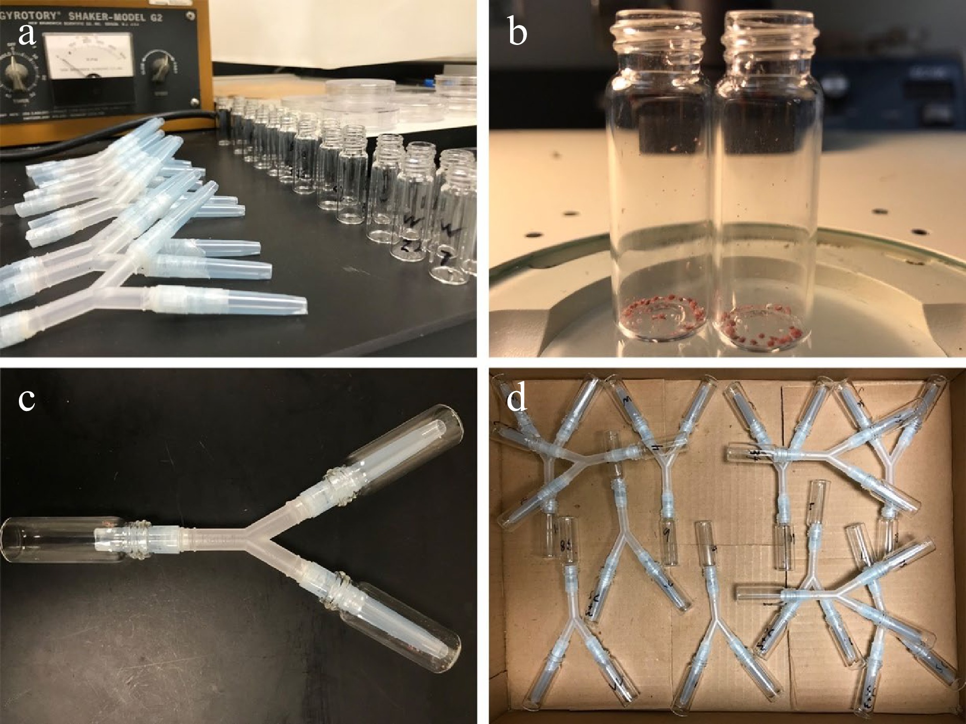
      Figure 1. 

      Y-maze assay. (a) Each Y-tube setup was assembled by a Y-tubing connector and contained a loading vial, a baited vial, and a control vial. (b) Before being fixed to the Y-tubing connector, CMBS females and crawlers were placed into the baited vial. (c) Three 1-mL pipette tips wrapped with Parafilm were used to tightly fix the connector to the three vials. Narrow ends of the two pipette tips were cut to connect the baited vial and the control vial, which could deter the lacewing crawling back once it had made its decision[49]. The wide end of the other pipette tip was cut to connect the loading vial where a larval lacewing was introduced. (d) Twelve Y-mazes were placed horizontally in a box and tested per time and replicated 10 times at 25 ± 1 °C, 60 ± 5% RH in the dark.

      Twelve larval C. rufilabris were individually placed into each Y-maze setup for the foraging performance test at 25 ± 1 °C, 60 ± 5% RH and a 24-h dark photoperiod. After 24 h, the number of C. rufilabris that entered the baited vials (B) and the control vials (C) was counted, respectively. This experiment was repeated 10 times using new larval C. rufilabris. Foraging performance index (FPI) of C. rufilabris targeting CMBS in the dark was calculated as:

      FPI = (the number of lacewings choosing B – the number of lacewings choosing C) / Total number of lacewings that made a choice

      Positive response ratio (PRR) of C. rufilabris foraging performance was calculated as:

      PRR = the number of lacewings choosing B / Total number of lacewings that made a choice

    • For the predation potential experiment in June, datasets of the 13 C. rufilabris biological replicates of the feeding duration and the number of consumed CMBS were averaged (mean ± SE) and compared among the six technical replicates. For the October experiment, datasets of 20 C. rufilabris biological replicates with three technical replicates of the feeding duration and the number of consumed CMBS for each larval stage were analyzed by using one-way analysis of variance (ANOVA) with the JMP® 16 (SAS Institute, Cary, NC, USA). Then, the analysis results regarding the feeding duration and the number of consumed CMBS were separated by C. rufilabris developmental stage by using Tukey's honestly significant difference (HSD; α = 0.05) to test if different stages of development impact the in-vitro predation potential of larval C. rufilabris upon CMBS eggs.

    • By examining the samples collected at different campus locations, we observed larval green lacewings feeding on CMBS gravid females [Fig. 2a & b, (30°36'39" N, 96°20'58" W); (30°36'55" N, 96°20'24" W)]. Lacewings’ eggs were deposited on twigs of CMBS-infested crapemyrtles [Fig. 2c & d, (30°37'03'' N, 96°20'08'' W); (30°36'30'' N, 96°21'02'' W)]. These observations allowed us to evaluate the potential of C. rufilabris as a biocontrol agent for sustainable management practices against CMBS. Indeed, in laboratory conditions (Fig. 3), the larval green lacewings not only voraciously consumed CMBS gravid females and eggs but were also able to grab and devour tiny crawling nymphal CMBS. The observations in both landscape and lab conditions confirmed C. rufilabris as the natural predator on CMBS.
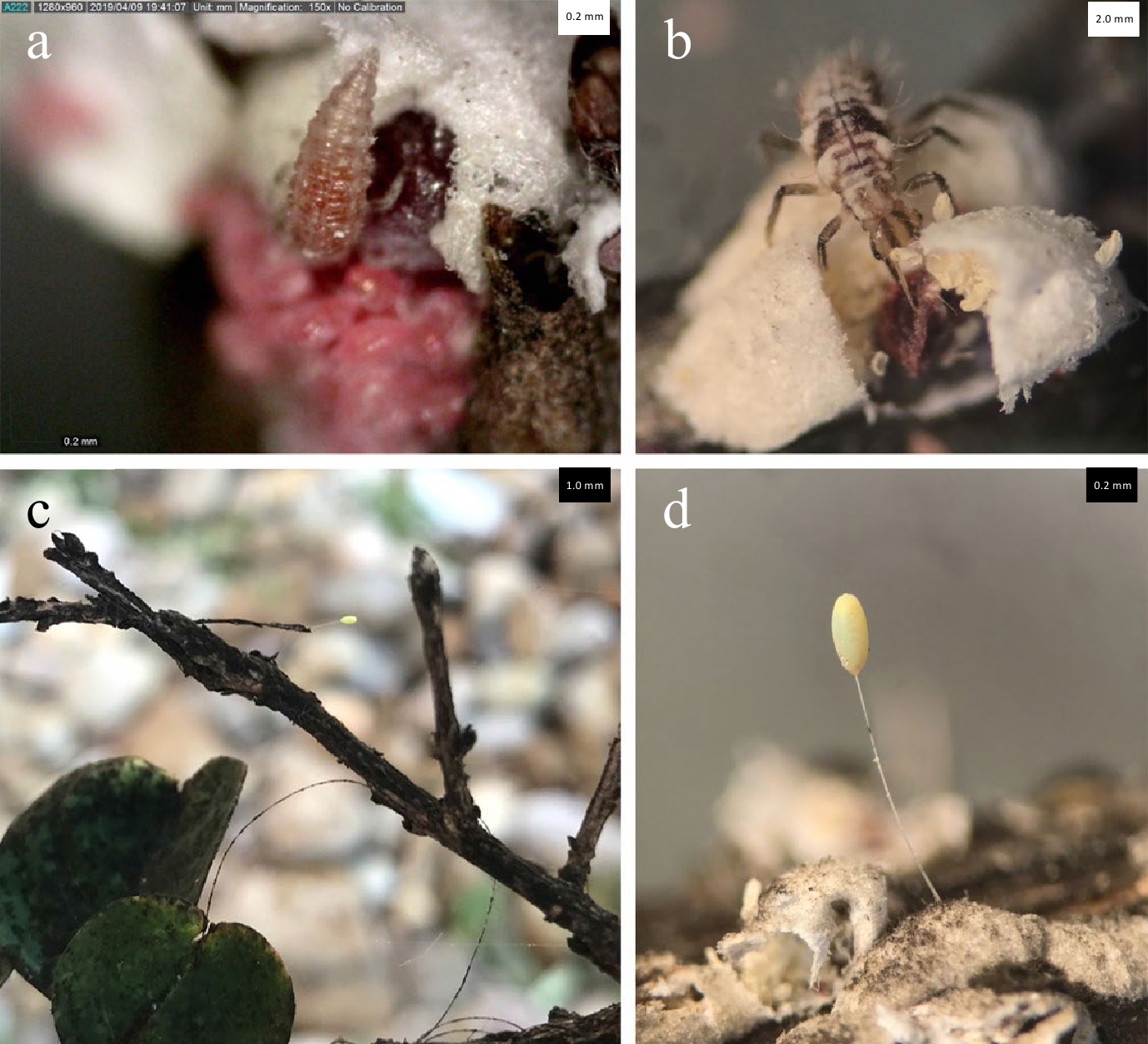
      Figure 2. 

      Observations of Chrysoperla rufilabris were reported at different locations on Texas A&M campus (USA). Larval C. rufilabris were observed preying on CMBS gravid females during the landscape investigations on April 9th (a) (30°36'39" N, 96°20'58" W) and June 28th (b) (30°36'55" N, 96°20'24" W). Lacewing eggs were found in CMBS-infested crapemyrtles on Oct 18th (c) (30°37'03" N, 96°20'08" W) and Nov 15th, 2019 (d) (30°36'30" N, 96°21'02" W).
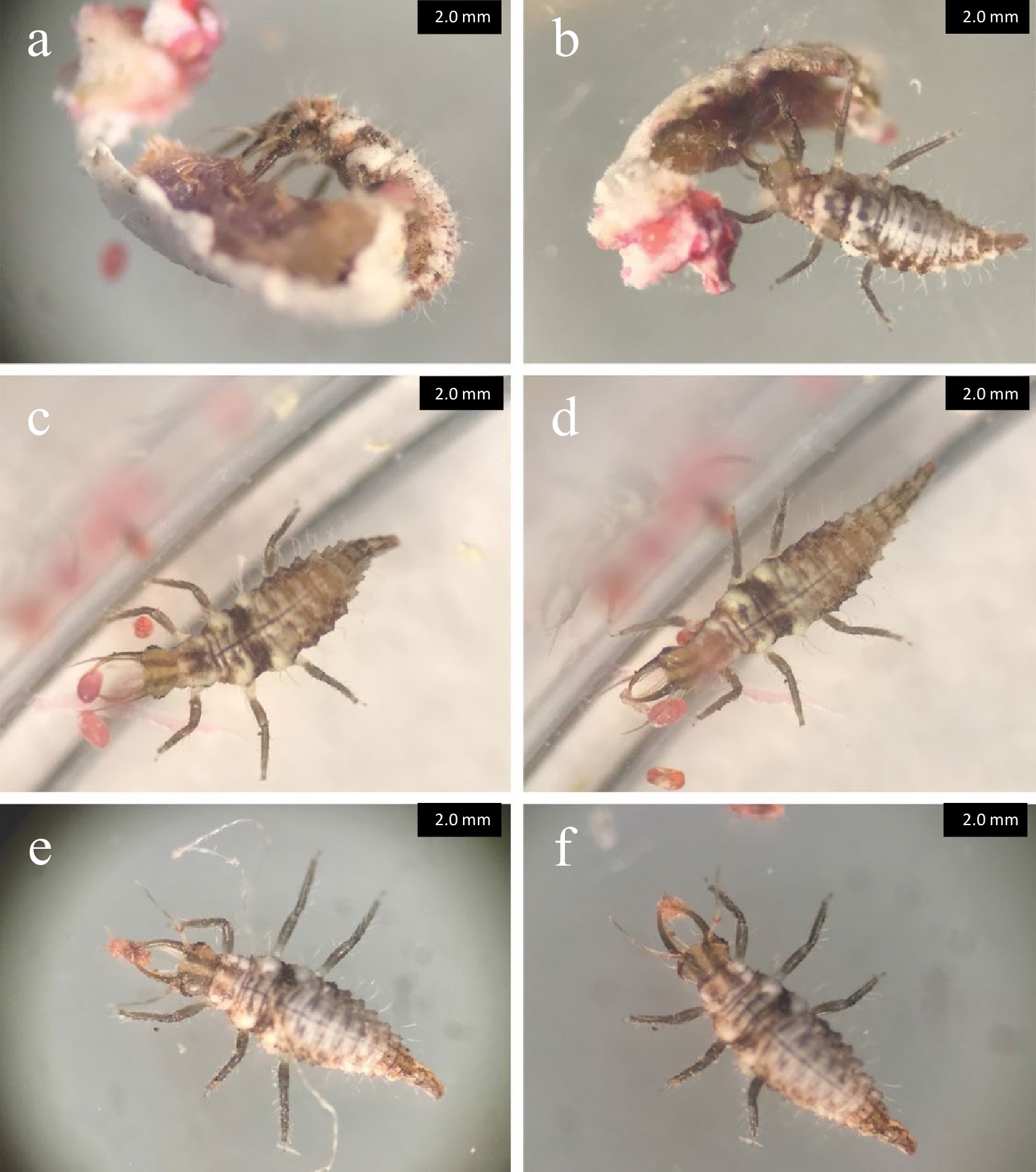
      Figure 3. 

      Larval Chrysoperla rufilabris individuals preying on CMBS under laboratory conditions. Larva of C. rufilabris targeted a female adult of CMBS (a) and voraciously seized and consumed body fluids of the CMBS using its large, sucking jaws (b) after placing them in the same Petri dish. A green lacewing larva easily grabbed a CMBS egg (c) and consumed the egg in about 1 min (d) after placing them in the same Petri dish. Larva seizing a crawling nymphal CMBS (e) and consumed it quickly (f) under the same experimental conditions.

    • In the June test, the results showed that the feeding duration ranged from 53.2 ± 2.5 s to 73.2 ± 2.7 s (mean ± SE) and the number of CMBS eggs consumed ranged from 154.1 ± 2.7 to 195.5 ± 2.5 (mean ± SE). The predation potential (or predation capacity) of C. rufilabris upon CMBS eggs was similar to that reported for 4th instar aphids of Aphis gossypii and Myzus persicae[35].

      In the October test, the developmental stages significantly affected the feeding duration (F 2, 177 = 101.1332, p < 0.0001) and the number of CMBS eggs consumed in 24 h (F 2, 177 = 252.6378, p < 0.0001) (Table 1). As C. rufilabris aging, the feeding duration dropped from 141.4 ± 4.8 s in the 1st instar to 60.3 ± 3.0 s in the 3rd instar; meanwhile, the consumed egg number increased (Table 1). The number of CMBS eggs consumed by larval lacewing at the 3rd stage was significantly higher than the 2nd and 1st instars. Chrysoperla rufilabris, a commercially available biocontrol agent[32], has been validated as the CMBS’s natural predator in this study. The first major peak in CMBS crawler activity occurred in April[50], therefore, to effectively suppress the CMBS population in practice, augmentative releases of the 2nd and 3rd instar C. rufilabris during this period should be evaluated further.

      Table 1.  Feeding duration and numbers of CMBS eggs consumed by Chrysoperla rufilabris at different developmental stages.

      Predator developmental stageFeeding duration
      (sec)z
      Number of
      consumed CMBSz
      1st instar141.4 ± 4.8a11.8 ± 1.3c
      2nd instar77.5 ± 4.7b151.5 ± 6.6b
      3rd instar60.3 ± 3.0c176.4 ± 6.9a
      Statistical analysisF 2,177 = 101.1332,
      p < 0.0001
      F 2,177 = 252.6378,
      p < 0.0001
      z Means ± SE (N = 3, representing a total of 60 tested for each developmental stage), in the same column, followed by different letters are significantly different as determined by Tukey's HSD test (α = 0.05).

      As the lady beetles and green lacewings share the same food resource- CMBS, their predator-predator-CMBS interactions may enhance the pest suppression of CMBS due to predator facilitation[51] or reduce the pest suppression due to predator interference and intraguild predation[52,53]. Therefore, to better implement C. rufilabris as the biocontrol agent of CMBS, further identification of the relative contribution of the lady beetles and green lacewings to CMBS suppression is needed.

    • In the 24-h Y-maze assay (Fig. 4a), the FPI of C. rufilabris upon CMBS was 0.56 ± 0.09 (mean ± SE). Among the green lacewings that made a choice, 78.14 ± 4.74% (PRR) larval C. rufilabris successfully targeted CMBS in the baited vial in the dark (Fig. 4b). The results indicated that some cues primarily associated with olfactory response were likely involved in the foraging performance. Testing olfactory response to volatiles secreted by prey would help ascertain the attractants or repellents related to lacewing-CMBS interaction, which better guides the integrated pest management of CMBS by using C. rufilabris.
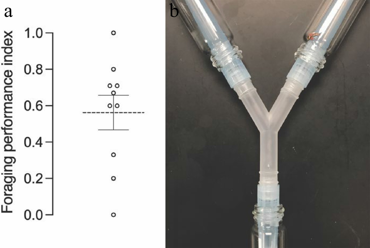
      Figure 4. 

      Foraging performance test in 24-h Y-mazes. (a) The foraging performance index (FPI) of C. rufilabris in the 24-h Y-maze assay was 0.56 ± 0.09 (mean ± SE). (b) Among the lacewings that made a choice, 78.14 ± 4.74% larval C. rufilabris successfully targeted CMBS in the dark.

    • Our study validated C. rufilabris as a natural predator of CMBS and investigated C. rufilabris predation potential as a biocontrol agent under laboratory conditions. The results regarding its predation potential upon CMBS eggs suggest using 2nd and 3rd instar C. rufilabris could be more efficient in suppressing the CMBS population. The foraging performance of larval C. rufilabris upon CMBS in the dark indicated that olfactory response was likely involved in CMBS predation. Future investigation focusing on the olfactory response of C. rufilabris to CMBS would benefit the development of the integrated pest management of CMBS.

      • This work is supported by TAMU T3 246495-2019, Crop Protection and Pest Management project ‘Integrated pest management strategies for crape myrtle bark scale, a new exotic pest’ (No. 2014-70006-22632) and Specialty Crop Research Initiative project 'Systematic Strategies to Manage Crapemyrtle Bark Scale, An Emerging Exotic Pest' (grant no. 2017-51181-26831) from the USDA National Institute of Food and Agriculture. Any opinions, findings, conclusions, or recommendations expressed in this publication are those of the authors and do not necessarily reflect the view of the U.S. Department of Agriculture.

      • The authors declare that they have no conflict of interest.

      • Supplemental Fig. S1 Rearing Chrysoperla rufilabris. Each individual of larval C. rufilabris was placed in each Petri dish and fed with eggs and nymphs of CMBS and 20μL-30μL droplets of artificial diet (Prosser and Douglas 1992) (A) . After three larval stages, the third instars secreted silken cocoons to cover themselves to developed as pupae (B) . Winged  C .rufilabris adults emerged from the cocoons (C) and were reared using 20 μL-30 μL droplets of artificial diet and water (D) . Several C. rufilabris adults were transferred into the same Petri dish supplemented with the artificial food for reproduction (E) . Green eggs were laid on the lid of the Petri dish nearby the food (F) . The 1st instar C. rufilabris hatched from the eggs (G) and provided with the artificial foods (H).
      • Supplemental Fig. S2 Life cycle of Chrysoperla rufilabris. After hatching from eggs (A) in 5-8 d, the C. rufilabris instars developed through 1st (B-1), 2nd (B-2), and 3rd (B-3) larval stages in 14-21 d. Then, the third instars secreted silken cocoons to cover themselves and developed as pupae. Winged C. rufilabris adults emerged from the cocoons in 10-15 d.
      • Copyright: © 2022 by the author(s). Published by Maximum Academic Press, Fayetteville, GA. This article is an open access article distributed under Creative Commons Attribution License (CC BY 4.0), visit https://creativecommons.org/licenses/by/4.0/.
    Figure (4)  Table (1) References (53)
  • About this article
    Cite this article
    Wu B, Xie R, Gu M, Qin H. 2022. Green lacewing Chrysoperla rufilabris (Neuroptera: Chrysopidae) is a potential biological agent for crapemyrtle bark scale (Hemiptera: Eriococcidae) pest management. Technology in Horticulture 2:3 doi: 10.48130/TIH-2022-0003
    Wu B, Xie R, Gu M, Qin H. 2022. Green lacewing Chrysoperla rufilabris (Neuroptera: Chrysopidae) is a potential biological agent for crapemyrtle bark scale (Hemiptera: Eriococcidae) pest management. Technology in Horticulture 2:3 doi: 10.48130/TIH-2022-0003

Catalog

  • About this article

/

DownLoad:  Full-Size Img  PowerPoint
Return
Return