ARTICLE   Open Access    

Structural characteristics of the melon mitochondria genome

More Information
  • The mitochondrial genome can provide important genetic information of melon. We reported the mitochondrial genome sequence of melon in a previous study, the structural characteristics of the melon mitochondrial genome were further analyzed in the present study. The mitochondrial genome of melon is comprised of three circular DNA molecules, with a total length of about 2.9 Mb, contains 4,861 pairs of homologous repeats, 439 pairs of inverted repeats, 653 tandem repeats and 218 SSR sequences. The coding genes accounted for 1.54% and non-coding gene sequences accounted for 98.46% of the melon mitochondrial genome. The total repetitive sequence of mitochondrial genome of melon was the highest among Cucumis melo, Cucumis sativus, Cucurbita pepo and Citrullus lanatus. The large number of repeated sequences and nuclear genome sequences were the main reason for the increasing size and variation of melon mitochondrial genome. Melon mitochondrial genome has the highest GC content and tRNA quantity. These regions were the main source of mitochondrial genome differences among all species here analyzed.
  • Passion fruit (Passiflora edulis), belonging to the Passifloraceae family and the Passiflora genus, is widely cultivated in tropical and subtropical regions[1,2]. It is renowned for its aromatic fruits, which exhibit fragrances reminiscent of various fruits, including bananas and pineapples. The fruit is not only nutritionally rich but can also be harvested year-round, contributing significantly to its economic value[36]. Moreover, passion fruit displays vibrant and large flowers with a distinctive secondary corona structure – a white or colorful fringe-like structure formed between the petals and pistils[2]. It also holds great significance as a horticultural plant, finding widespread use in applications like fence landscaping and small potted plants[7,8]. Furthermore, the special floral structure (sepal-petal-corona-stamen-carpel) in passion fruit also makes it an excellent and unique plant model for investigating floral organ morphology.

    MADS-box gene family members are ubiquitously distributed among plants, animals, and fungi, playing crucial roles throughout plant growth and development[911]. MADS-box genes are instrumental in governing a spectrum of biological processes[1214], encompassing floral transition, control of flowering time, endosperm and embryo development, seed germination, fruit development, abiotic stress responses, and notably, they assume central roles in the morphogenesis of plant floral organs[15,16]. Based on protein domain characteristics and phylogenetic tree relationships, the MADS-box gene family can be classified into two primary types: Type I and Type II[17]. Type I genes can be further divided into three subclasses: Mα, Mβ, and [18], while Type II genes, also known as MIKC-type genes in plants, consist of the MIKCC and MIKC* groups. Moreover, the MIKCc group can be further classified into 12 major subclades[19]. Notably, Type I genes have undergone more rapid evolutionary changes from birth to death compared to Type II genes[20]. Both Type I and Type II protein sequences share a conserved domain named as MADS-domain, comprising 58-60 amino acids located in the N-terminal region, which serves as an identifying feature of MADS-box proteins. Whereas, Type II MADS-box proteins in plants possess three additional domains in addition to the MADS-box domain: an intervening region with relatively low conservation, a moderately conserved Keratin-like domain, and a variable C-terminal region[21].

    Among the MIKCC-type genes, specific members are known to form the well-established ABC(D)E model, providing a genetic basis for floral organ specification and development[22,23]. According to this model, the identification of floral organs in each whorl is governed by unique combinations of genes from classes A, B, C, D, and E. Sepal identification is jointly determined by A- and E-class genes, whereas petals are controlled by A-, B-, and E-class genes. The formation of stamens relies on B-, C- and E-class genes, while carpels are regulated by C- and E-class genes. Ovule specification is particularly controlled by C-, D-, and E-class genes[2325]. In MADS-box gene family of Arabidopsis thaliana, numerous functional genes have been confirmed to be associated with the ABC(D)E model. For instance, A-class genes include APETALA1(AP1)[26]; APETALA3(AP3) and PISTILLATA(PI) belongs to class B[27,28]; AGAMOUS (AG) is a C-class gene and also involved in floral meristem development[29]; class D gene AGL11 is expressed in ovules[30]; the E class comprises SEPALLATA (SEP1, SEP2, SEP3, and SEP4), which exhibit functional redundancy and are required for specifying the identity of all floral organs[31,32]. Proteins encoded by ABC(D)E model genes form quaternary complexes that bind to CArG-box (CC[A/T]6GG) of DNA sequences, and this complex subsequently acts on downstream target genes to regulate flower development[33]. Additionally, MADS-box genes in A. thaliana perform various functions in other development processes, AGAMOUS-LIKE 20 (AGL20) serves as a key activator in integrating floral inductive pathways[34] , while FLC and SVP are involved in regulating flowering time[35]. AGL21 and AGL12 are associated with root development[36,37], and the AGL6 is involved in floral meristem and seed development[38]. FRUITFULL (FUL), SHATTERPROOF 1 and 2 (SHP1 and SHP2), collectively function in the complex regulation of fruit ripening and development[39].

    The distinctive floral morphology of passion fruit makes it an excellent candidate for the study of floral organogenesis and enhances our understanding of the ABC(D)E model. While extensive research has explored MADS-box gene family members in various plants[4043], a comprehensive investigation of these members in passion fruit has not yet been carried out. Here, we identified a total of 52 MADS-box genes in passion fruit and conducted a systematic analysis, including analysis of gene structure, motif composition, phylogenetic relationships, chromosomal localization, collinearity, and expression patterns. Notably, we also proposed an ABC(D)E model to elucidate the regulation of the unique floral structure (sepal-petal-corona-stamen-carpel) in passion fruit. Our findings could provide valuable insights for further functional investigations of passion fruit MADS-box genes and their potential applications in floral modification breeding.

    Passion fruit genome and protein sequences were obtained from the National Genomics Data Center (NGDC) database (https://ngdc.cncb.ac.cn/) under the accession number GWHAZTM00000000. We employed two distinct strategies, namely the Hidden Markov Model (HMM) search and BLAST search, to identify MADS-box genes in passion fruit. Regarding the HMM method, the MADS-box SFR family domain (PF00319) was sourced from the Pfam database (www.pfam.org) and subsequently applied for a comprehensive search against the passion fruit protein sequences using HMMER3 (v3.3.2) software (http://hmmer.janelia.org/)[44]. Simultaneously, plant MADS-box protein sequences were obtained from the NCBI database (www.ncbi.nlm.nih.gov) and used as initial queries for BLASTP searches. Only homologous sequences with an E-value of less than 1e-5 were retained for subsequent analyses. The sequences identified from both the HMM search and BLAST search were consolidated and the redundant members were removed to produce a set of preliminary MADS-box candidates. These candidates then underwent rigorous verification of the MADS-box domain through the SMART (http://smart.embl-heidelberg.de)[45] and CDD (Conserved Domain Database) search (www.ncbi.nlm.nih.gov/cdd)[46] . The confirmed MADS-box genes were renamed according to their positions on the passion fruit chromosomes. Subcellular localization analysis was conducted using the WoLF PSORT tool (https://wolfpsort.hgc.jp), while the ExPASy website tool (www.expasy.org)[47] was harnessed to predict molecular weight (MW), isoelectric point (pI), and the grand average of hydropathicity (GRAVY).

    Multiple sequence alignment of all 52 putative MADS-box protein sequences was performed by MUSCLE[48], and the alignment was visualized and edited by software Jalview v 2.11.2.0[49]. For phylogenetic analysis, the 105 MADS-box protein sequences of Arabidopsis were downloaded from the TAIR database (www.arabidopsis.org)[18], those sequences were combined with putative members from passion fruit to construct the phylogenetic tree. The multiple protein sequence alignment of all 157 protein sequences was produced using software MAFFT v7.407[50]. The resulting alignments were used for exploring the phylogenetic relationship of MADS-box protein in Arabidopsis and passion fruit. Phylogenetic analysis was performed by software IQ-TREE v1.6.12[51] with maximum likelihood estimation based on the most suitable model and 1000 bootstraps. The phylogenetic tree was visualized using Evolview (https://evolgenius.info/evolview-v2/)[52].

    To identify the conservative motifs among the putative MADS-box members of passion fruit, the full-length protein sequences were analyzed website tool MEME (https://meme-suite.org/meme/tools/meme)[53] with parameters that the maximum number of finding motifs is 10. Exon-intron structure of MADS-box genes were distinguished using GFF files, which was acquired from the annotation information of the passion fruit genome. The exact region of conservative MADS-box domain was identified from the CDD (Conserved Domain Database) database (www.ncbi.nlm.nih.gov/cdd)[46].

    To explore the possible-regulator-factors of MADS-box genes from passion fruit, TBtools v1.1047 software[54] was used to extract the 2000-bp upstream region as putative promoter regions, which end with the translation initiation codon of each MADS-box genes. The cis-acting elements contained in promoter regions were marked using the PlantCARE database (http://bioinformatics.psb.ugent.be/webtools/plantcare/html)[55], and the statistics analysis and plotting were finished using R package pheatmap (https://CRAN.R-project.org/package=pheatmap).

    The exact location of MADS-box genes was extracted from the annotation file of the passion fruit genome, and anchored to corresponding chromosomes. Duplication events of genes occurring in the evolution of the passion fruit genome were analyzed by Multicollinearity Scanning Toolkit (MCScanX)[56] with default parameters. The genomic data of Oryza sativa (v7.0), Solanum lycopersicum (ITAG3.2), A. thaliana (TAIR10), Vitis vinifera (Genoscope.12X), Zea mays (RefGen_V4) were retrieved from the JGI Phytozome database (https://phytozome-next.jgi.doe.gov). A similar procedure was also applied to demonstrate the collinearity relationships and gene duplications of orthologous MADS-box genes obtained from passion fruit and these five species. The result was visualized by functions of TBtools[54]. Based on the above results of duplicated events within passion fruit, the ratios Ka/Ks of tandem duplicated and segmental duplicated gene pairs were also calculated using TBtools[54].

    All MADS-box protein sequences were submitted to SWISS-MODEL (https://swissmodel.expasy.org)[57] for predicting tertiary structure with homology modeling method, and secondary structures could be marked by the corresponding original model from PDB databases (www.rcsb.org). The predicted models were visualized using Pymol[58].

    The fresh samples of passion fruit (P. edulis) were collected from the orchard located in the Institute of Horticulture, Guangxi Academy of Agricultural Sciences (China). The whole floral tissue was divided into bract, sepal, corona filament or corona, stamen, stigma, ovule (dividing under dissecting microscope). The identification of development stages of floral tissues was based on the horizontal width of buds with bracts (more details about distinguishing different development stages are listed in Supplemental Table S1). Samples of leaves, stems and tendril tissues were collected at 110 d after anthesis. The fruit were collected at 53 d post anthesis (DPA), DPA60, DPA100 and DPA128, respectively. The tissues samples were immediately frozen in liquid nitrogen after picking and stored at −70 °C.

    The grown and healthy plants of passion fruit were subjected to abiotic stress treatments (cold and heat) with three biological replicates. The whole plants were transferred into a growth chamber with temperature of 20 °C as the cold-stress condition while the heat-stress condition had a temperature of 30 °C. Samples of floral buds were collected under different treatment times (1, 4, 12, 24 h for heat stress; 4, 24 h for cold stress). Plants cultivated under 25 °C were used as control. All samples were collected for RNA extraction.

    Reference to previous common methods of RNA extraction[59], total RNA from different tissues was extracted by RNA extraction Kit (Omega Bio-Tek, Shanghai, China) with manufacturer's protocol. Following a standardized process, RNA samples were quantified and PCR library construction was performed. The analysis result was sequenced using NEB next Ultra RNA Library Prep Kit (NEB, Beverly City, MA, USA) for Illumina Biolabs. Each biological replicate used 1 µg RNA as an experiment sample. After the related processing flow of initial RNA-seq data file, TPM value of RNA-seq for each PeMADS gene were calculated, and the heatmap of the TPM value was constructed using R package (pheatmap) and chiplot (www.chiplot.online) with log2 (TPM + 0.0001) as the unit of measure.

    Based on the above standards of identifying different development stages for flowers, the tissues used for qRT-PCR analysis was br1 (bract at stage1), se1, pe1, ca1, st8, sg1 and ov5. Two-month-old healthy passion fruit plants were also subjected to ABA (100 μM) and GA (100 μM) treatments, while untreated plants served as controls. Leaf samples from phytohormone-treated plants were collected at 0, 12, 24, and 48 h post-treatment from three independent seedlings. All collected samples were immediately stored in liquid nitrogen before total RNA extraction. The Trizol reagent (Invitrogen, Carlsbad, CA, USA) was used to extract total RNA. ThermoScript RT-PCR kit (Thermo Fisher Scientific, Carlsbad, CA, USA) was used for reverse transcription. The quantitative real-time PCR (qRT-PCR) was performed by the following procedures: 95 °C for 30 s, followed by 40 cycles of 95 °C for 10 s and 60 °C for 30 s. The reaction volume was 20 µl, including 1 µl cDNA per sample, 10 µl 2× Taq Pro Universal SYBR qPCR Master Mix (Vazyme, Nanjing, China) and 0.4 µl per primer, the whole system was complemented by ddH2O to 20 µl. The reaction was carried out in a Bio-Rad Real-time PCR system (Foster City, CA, USA) with three biological replicates per sample. EF1α was used as a reference gene[60]. The relative expression levels were calculated using the 2−ΔΔCᴛ method. Primers were designed using the IDT website (https://sg.idtdna.com/pages). Primer sequences are listed in Supplemental Table S2.

    To ensure a comprehensive screening of gene family members, the putative PeMADS genes were identified by combining the results from hidden Markov model (HMM) and the blast search. Ultimately, a total of 52 MADS-box gene family members were successfully identified from the whole genome of passion fruit (Table 1). The members were designated as PeMADS1-PeMADS51 based on their sequential positions on the chromosomes, while a single member (P_eduliaContig70023089.g) mapped to a contig was named PeMADS52. Predicted physicochemical properties of PeMADS genes coding proteins are list in Table 1. These proteins exhibit variable lengths, with the longest one being 650 aa (PeMADS7) while the shortest one is 66 aa (PeMADS29). Correspondingly, PeMADS7 possesses the heaviest molecular weight (MW) of 71,572.97 Da, while PeMADS29 has the lightest protein, only 7,697.02 Da. The protein isoelectric points (PI) range from 4.89 (PeMADS41) to 10.69 (PeMADS29). According to the Instability Index[61], only PeMADS4, PeMADS13, and PeMADS23 are considered stable proteins (value is smaller than 40). The aliphatic amino acid index (A.I.) varies from 66.05 (PeMADS4) to 100.41 (PeMADS27), indicating a significant difference in thermal stability among the PeMADS proteins. With the exception of PeMADS22, the grand average of hydropathicity score (GRAVY) of other proteins are negative, which means only PeMADS22 is a hydrophobic protein, and most proteins are hydropathicity. The prediction of subcellular localization reveals that the major action site of proteins is the nucleus, followed by chloroplasts and mitochondria. Two members, PeMADS23 and PeMADS28, are located in the cytoplasm. For detailed sequence information of all members, refer to Supplemental Table S3.

    Table 1.  Characteristics about 52 PeMADS proteins of passion fruit
    Gene nameGene IDChromosomeSize (aa)MW (Da)PIInstability
    Index
    A.I.GRAVYPredicted Location
    PeMADS1P_edulia010000232.gLG0135540,132.206.3642.6675.27−0.63Nucleus
    PeMADS2P_edulia010000334.gLG0119021,875.009.7648.9376.47−0.826Nucleus
    PeMADS3P_edulia010000557.gLG0119422,380.579.7348.1376.91−0.818Nucleus
    PeMADS4P_edulia010000858.gLG0138742,577.71935.1666.05−0.687Nucleus
    PeMADS5P_edulia010002143.gLG0122525,469.3210.3148.6472.8−0.629Nucleus
    PeMADS6P_edulia010002220.gLG0125129,014.905.8463.8980−0.551Nucleus
    PeMADS7P_edulia010002256.gLG0165071,572.978.8842.5789.43−0.259Nucleus
    PeMADS8P_edulia010002353.gLG0122325,645.299.5153.2283.54−0.658Nucleus
    PeMADS9P_edulia010002348.gLG0122325,567.898.1157.0892.29−0.209Mitochondria
    PeMADS10P_edulia010002733.gLG0122325,520.758.1258.3390.54−0.242Mitochondria
    PeMADS11P_edulia010002726.gLG0122325,663.329.5150.7482.24−0.671Nucleus
    PeMADS12P_edulia010003864.gLG0122725,943.719.085382.91−0.609Nucleus
    PeMADS13P_edulia010004192.gLG0112814,242.5410.3337.3691.33−0.087Nucleus
    PeMADS14P_edulia010004199.gLG0116619,203.349.345.2793.31−0.031Nucleus
    PeMADS15P_edulia010004299.gLG0134238,721.376.0855.6974.18−0.57Nucleus
    PeMADS16P_edulia010004354.gLG0128332,306.418.2256.2575.51−0.63Nucleus
    PeMADS17P_edulia010004671.gLG0127431,672.199.2843.2380.47−0.444Nucleus
    PeMADS18P_edulia010004713.gLG0121224,765.599.7444.184.2−0.322Nucleus
    PeMADS19P_edulia010005413.gLG0115417,785.609.6143.3287.34−0.638Nucleus
    PeMADS20P_edulia020006530.gLG02789,025.349.6944.2483.72−0.615Nucleus
    PeMADS21P_edulia020007073.gLG0224428,034.946.4856.5785.53−0.696Nucleus
    PeMADS22P_edulia030008390.gLG0314115,691.086.840.5989.360.119Nucleus
    PeMADS23P_edulia030008412.gLG0323327,189.878.3937.5386.57−0.842Cytoplasm
    PeMADS24P_edulia030008784.gLG0325529,597.757.6962.0485.25−0.698Nucleus
    PeMADS25P_edulia030008868.gLG0323327,283.188.3264.5785.75−0.738Mitochondria
    PeMADS26P_edulia030009361.gLG0322925,959.029.244.399.21−0.374Nucleus
    PeMADS27P_edulia040010097.gLG0429634,731.288.3674.87100.41−0.349Nucleus
    PeMADS28P_edulia040010305.gLG0422626,236.009.1845.2975.49−0.773Cytoplasm
    PeMADS29P_edulia040010846.gLG04667,697.0210.6939.1779.7−0.239Nucleus
    PeMADS30P_edulia050011612.gLG05677,764.039.8343.493.13−0.452Nucleus
    PeMADS31P_edulia050012002.gLG0511712,998.9710.2146.3866.67−0.506Nucleus
    PeMADS32P_edulia060013073.gLG0624227,917.838.7244.4482.64−0.695Nucleus
    PeMADS33P_edulia060013103.gLG0616819,304.068.4350.7891.19−0.642Nucleus
    PeMADS34P_edulia060013465.gLG068810,152.749.5976.0982.95−0.448Nucleus
    PeMADS35P_edulia060013580.gLG0624027,317.309.1749.1284.17−0.561Nucleus
    PeMADS36P_edulia060013765.gLG0629131,621.315.3352.1372.51−0.417Chloroplast
    PeMADS37P_edulia060015308.gLG0633137,420.775.2862.0671−0.684Nucleus
    PeMADS38P_edulia060015605.gLG0622125,409.119.1740.883.8−0.576Nucleus
    PeMADS39P_edulia060015604.gLG0618821,618.598.8143.1884.04−0.624Nucleus
    PeMADS40P_edulia060016494.gLG0624328,153.007.1357.7991.11−0.658Nucleus
    PeMADS41P_edulia060016711.gLG0617520,210.434.8954.1869.66−1.058Chloroplast
    PeMADS42P_edulia060016709.gLG0617920,635.045.0558.7273.02−1.001Chloroplast
    PeMADS43P_edulia060016710.gLG0619822,057.865.6958.4181.82−0.657Chloroplast
    PeMADS44P_edulia070017407.gLG0718120,894.808.8571.8170.61−0.809Nucleus
    PeMADS45P_edulia070017456.gLG0724828,225.449.5847.590.48−0.497Nucleus
    PeMADS46P_edulia070017776.gLG0717219,278.411050.288.37−0.621Nucleus
    PeMADS47P_edulia080019101.gLG0821424,404.509.6653.6570.23−0.866Nucleus
    PeMADS48P_edulia080020106.gLG0813014,779.159.3842.3584.69−0.338Nucleus
    PeMADS49P_edulia090020757.gLG0920623,473.426.9844.3267.57−0.681Nucleus
    PeMADS50P_edulia090021063.gLG0920423,738.659.2344.2686.96−0.739Nucleus
    PeMADS51P_edulia090021542.gLG0921424,401.799.0659.6967.48−0.697Nucleus
    PeMADS52P_eduliaContig70023089.gContig725328,637.179.4649.6494.47−0.316Nucleus
     | Show Table
    DownLoad: CSV

    The protein sequences of 52 candidate PeMADS were subjected to alignment analysis to further elucidate the constitution and structure of MADS-domain (Supplemental Table S4). As is shown in Fig. 1a, there is a highly conserved domain in all 52 PeMADS proteins. The sequence logo was utilized to accurately depict the composition of the MADS-box region. By employing the SWISS-MODEL web server and utilizing protein homology modeling, a three-dimensional (3-D) model of MADS-box domain was constructed. This model could be divided approximately into three regions, comprising one α-helix and two β-sheets (Fig. 2b). Through analysis of amino acid distribution at each residue position within the MADS domain, 24 sites out of the 57 residues were identified as highly conserved if the percentage of a specific amino acid exceeded 80% at that particular site (Fig. 2c). Integrating the aforementioned information with the proteins' tertiary structure, the first helix contained 14 conserved residues (Arg-17, Gln-18, Val-19, Thr-20, Lys-23, Arg-24, Arg-25, Gln-27, Leu-28, Lys-30, Lys-31, Glu-34, Leu-35 and Cys-39), while the first sheet region contained one conserved residue (Phe-48), and the second region lacked any conserved residues. Among 57 residues, Arg-3, Arg-17, Arg-24, Lys-31, Glu-34 exist in all protein sequences.

    Figure 1.  Characterization of MADS-box domain. (a) Multiple sequence alignment of MADS-domain region from 52 PeMADS proteins. (b) Predicted three-dimensional structure of MADS-domain. (c) Distribution of amino acids in conserved residues of MADS-domain: number represents the positions of residues; bottom label represents species of amino acids.
    Figure 2.  Phylogenetic tree of PeMADS proteins from Passiflora edulis (Pe) and Arabidopsis thaliana (At). Red starts and green triangles indicate passion fruit and Arabidopsis, respectively. The circle placed on the tree indicated the bootstrap value (≥ 90, red circle; 60~90, yellow circle; < 60, not shown).

    To examine the phylogenetic relationships among MADS-box proteins from different species and ascertain the subfamily to which each PeMADS belong, a phylogenetic tree was constructed. The tree was based on the alignment result of MADS protein which is 105 sequences from Arabidopsis and 52 sequences from passion fruit (Fig. 2, Supplemental Table S5). According to previous studies and topology of the phylogenetic tree[18,6264], the PeMADS members were classified into two groups: type I, consisting of nine members (four as Mα, three as Mβ, and two as Mγ) while 43 members were classified as type II. Within the type II group, only six PeMADS proteins belonged to MIKC* category, while the remaining members were classified as MIKCC. The MIKCC proteins were further divided into 12 clades. Sequences of both passion fruit and Arabidopsis could be identified in all clades. Most PeMADS were determined in AG-like clade (six members), while the PI-like, FLC-like and AP1-like clades each had only one member. Inside the PI-like clade, the Arabidopsis MADS-box proteins showed homology with only one other member (PISTILATA and PeMADS28).

    The phylogenetic relationship of PeMADS proteins was depicted using a phylogenetic tree with bootstrap values. The classification obtained from the phylogenetic tree analysis aligned with the aforementioned analysis (Fig. 3a). To identify and compare the common motifs of proteins within different groups, the MEME search tool was employed to 52 PeMADS proteins, and the first ten motifs with the highest conservation were detected (motif 1−10; Fig. 3b). As illustrated in the figure, members within the same group typically exhibited similar construction of motifs, particularly noticeable in B sister, AP3-like group and AGL6-like group. PeMADS proteins displayed varying numbers and distribution of conserved motifs, with a maximum of seven motifs and a minimum of one. Except for Mβ members, motif 1 could be observed in all proteins, which could be an obvious character of MADS-box proteins. Generally, the construction of Type I members was simpler than Type II members, especially Mγ group which contained only one motif. The location of MADS-box domain sequences was mapped to the full-length proteins (Fig. 3c). All members could detect MADS domain, whereas the length of the MADS domain is relatively short in the Mβ group. Except for MIKC* group, most Type II members contained K-box domain. Type II protein lacking the K-box domain were shorter than same group members, possibly due to the loss of terminal protein sequences.

    Figure 3.  The phylogenetic relationship, conserved motifs and gene structures of PeMADSs. (a) The phylogenetic tree of 52 PeMADS proteins. (b) Distribution of conserved motifs in PeMADS proteins. (c) Distribution of MADS-domain and K-domain of PeMADS proteins.(d) The gene structures of the PeMADSs, include coding sequences (CDS) and untranslated regions (UTR).

    To investigate the structural diversity of PeMADS gene, the intron-exon organization of coding sequences was visualized reference to the GFF annotation file of passion fruit genome (Fig. 3c). The structure of type II group (MIKCC and MIKC*) was more complex. Nearly 33 (76.74%) type II members contained at least five CDS regions, among which the CDS regions of PeMADS7 was up to 15. Otherwise, type I groups (Mα, Mβ and Mγ) possessed less CDS regions, most members only had zero or single CDS regions, only PeMADS36 had two introns. The above discovery suggests that the transcription and splicing process of type II genes might be more complex than type I.

    Cis-element species and distribution could partly imply the transcription regulation and the expression pattern of associate genes. The putative promoters of PeMADS genes,spanning 2000 base pairs, were predicted using the PlantCARE server (Supplemental Table S6). A total of 39 types of cis-elements were identified and categorized into five groups: light response (16), phytohormone response (10), stress response (7) and plant growth regulation (6). Figure 4b illustrates that the majority of genes exhibited a higher proportion of light-responsive elements, followed by phytohormone-responsive elements, whereas elements associated with plant growth regulation were the least abundant. The proportions indicate that the expression of the PeMADS gene was substantially affected by light. G-box element (127), Box 4 (117), GT1-motif (70) were the most common light-responsive elements. Phytohormone-responsive elements accounted for a considerable portion of the cis-elements, with ABRE (134; abscisic acid response), AAGAA motif (106; abscisic acid response), CGTCA motif (79; MeJA response) and ERE (53; ethylene response) being the most frequently identified elements. Some types of elements centralized exist on one gene, for instance, the number of ABRE element was obviously higher in PeMADS2 and PeMADS50, implying those genes might be more possible to play a vital role in corresponding phytohormone regulation. ARE elements (135; anaerobic induction), 58 drought stress response elements (MBS and DRE core), 25 WUN-motif (mechanical injury response) and 38 LTR (low temperature response) indicated that PeMADSs could respond to abiotic stresses. ARE elements could be detected in promoter sequences of all genes, speculating anaerobic conditions was one of critical factors which induce the transcription of PeMADS gene. However, cis-elements related to plant growth such as CAT-box, CCGTCC-box, and RY-element were comparatively limited. Within the promoter region of each gene, the count of such elements did not exceed four.

    Figure 4.  (a) Analysis of cis-elements in the promoter region of PeMADS genes. (b) Heatmap of the number of cis-elements, the different color represents the number of cis-elements. (c) The sum of cis-elements in categories shown as a histogram.

    Based on the passion fruit genome's annotation files, 51 out of 52 PeMADS genes were assigned to nine linkage groups (LG01-LG09), and only one member (PeMADS52) was found on unassembled contig 7. The majority of MADS genes were concentrated on LG01 (19, 36.54%), particularly on the terminal region of LG01, encompassing approximately 33 Mb and containing 15 PeMADS genes. LG06 had the second highest number of MADS genes (12, 23.07%), followed by LG03 (5, 9.61%). The remaining linkage groups (LG02, LG04, LG05, LG07, LG08, LG09) contained only 1−3 PeMADS genes each. Numerous MADS genes were located near the proximate or the distal ends of the linkage groups. There was no apparent evidence suggesting a direct connection between the number of MADS genes and the length of linkage groups.

    Based on the collinearity analysis conducted using MCScanX, only one tandem duplication gene pair, PeMADS42 and PeMADS43, was identified from LG06. Additionally, a total of 20 segmental duplication gene pairs were discovered, comprising 28 PeMADS genes located on duplicated segments across nine linkage groups (Fig. 5). Most duplicated gene pairs belong to the MIKCc group (24 pairs, accounting for 85.71% of the total). The MIKC* group and Mα have two gene pairs respectively, and those genes could be produced by whole genome duplication or the segmental genome duplication events.

    Figure 5.  Distribution and collinearity of PeMADS genes in the passion fruit genome. PeMADSs marked by red has synteny with other genes. Gray lines indicate synteny blocks in passion fruit genome, brown lines indicate segmental duplicated MADS gene pairs of passion fruit.

    To further investigate the evolutionary process of the passion fruit MADS-box gene family, a comparative systemic analysis was conducted between passion fruit and six representative species (Fig. 6), which cover three dicots (A. thaliana, S. lycopersicum and V. vinifera) and two monocots (O. sativa and Z. mays). The number of synteny blocks observed in A. thaliana, S. lycopersicum, V. vinifera, O. sativa and Z. mays were 996, 982, 922, 399 and 328 respectively. Among the 52 PeMADS genes, different numbers of genes exhibited syntenic relationships with the six species. A total of 33 members show colinearity with grape (33), followed by tomato (26), Arabidopsis (19) and rice (5), maize contains the least number (2). These findings suggest that the collinearity relationship between passion fruit and dicotyledons was closer than that between monocotyledons. Notably, most identified PeMADS exhibited synteny relationships with more than one species. PeMADS15 showed collinear counterparts in passion fruit and five other species, indicating its potential existence prior to ancestral divergence and its association with vital characteristics. Whereas some of the PeMADS syntenic gene pairs were exclusively found in dicotyledons or monocotyledons, suggesting their emergence after the divergence between these two groups. Syntenic PeMADS genes in five species are highlighted in Supplemental Table S7.

    Figure 6.  Synteny analysis of PeMADS genes and five representative plants species. Gray lines in the background indicate the collinear blocks between passion fruit and other plant genomes, while red lines highlight the systemic gene pairs of PeMADSs between passion fruit and other plant genomes.

    To gain insights into the evolutionary constraints affecting the passion fruit MADS-box gene family, we calculated the Ka/Ks (non-synonymous substitution/synonymous substitution) ratios for both segmental duplicated gene pairs and tandem duplicated gene pairs using the NG method. As shown in Table 2, it can be observed that all segmental duplicated gene pairs have Ka/Ks values below 1, indicating a strong purifying selective pressure acting on these duplicated gene pairs throughout evolution. In contrast, the only tandem duplicated gene pairs (PeMADS42 and PeMADS43) exhibited Ka/Ks > 1, suggesting that positive selection has influenced these gene pairs[65].

    Table 2.  The Ka/Ks ratios of the duplicated PeMADS gene pairs.
    Duplicated gene pairsKaKsKa/KsGroupDuplicated type
    PeMADS2 & PeMADS300.010MIKCC/MIKCCSegmental
    PeMADS3 & PeMADS70.130.570.24MIKCC/MIKCCSegmental
    PeMADS3 & PeMADS190.261.380.19MIKCC/MIKCCSegmental
    PeMADS8 & PeMADS110.010.020.28MIKCC/MIKCCSegmental
    PeMADS8 & PeMADS450.040.540.08MIKCC/MIKCCSegmental
    PeMADS9 & PeMADS100.010.010.43MIKCC/MIKCCSegmental
    PeMADS15 & PeMADS1600.020.29MIKC*/MIKC*Segmental
    PeMADS17 & PeMADS180.010.010.42MIKCC/MIKCCSegmental
    PeMADS17 & PeMADS280.291.290.22MIKCC/MIKCCSegmental
    PeMADS17 & PeMADS380.050.670.08MIKCC/MIKCCSegmental
    PeMADS18 & PeMADS280.221.150.19MIKCC/MIKCCSegmental
    PeMADS18 & PeMADS380.070.550.12MIKCC/MIKCCSegmental
    PeMADS20 & PeMADS470.030.510.07MIKCC/MIKCCSegmental
    PeMADS24 & PeMADS2500.010.3MIKCC/MIKCCSegmental
    PeMADS26 & PeMADS350.220.710.31MIKCC/MIKCCSegmental
    PeMADS28 & PeMADS380.321.520.21MIKCC/MIKCCSegmental
    PeMADS29 & PeMADS310.010.290.05MIKCC/MIKCCSegmental
    PeMADS32 & PeMADS330.010.020.28MIKCC/MIKCCSegmental
    PeMADS46 & PeMADS510.553.980.14Mα/MαSegmental
    PeMADS52 & PeMADS450.160.220.71MIKCC/MIKCCSegmental
    PeMADS42 & PeMADS430.140.062.42Mβ/Mβtandem
     | Show Table
    DownLoad: CSV

    The protein structure homology modeling was performed using the SWISS-MODEL database, utilizing homologous templates retrieved from the PDB database. The three-dimensional models for all PeMADS protein sequences were generated and are presented in Supplemental Table S8. The predicted structures with highest GMQE and QMEAN scores for each subfamily were visualized using PyMOL and displayed in Fig. 7. Based on previous research on MADS transcription factors, the highly conserved MADS domain structure could be broadly categorized into one α-helix and two β-sheets. The flexible intervening domain was predominantly α-helical, while the K domain consisted of three α-helices. C-terminal domain generally exhibits variable structures[66,67]. Analogously, except for PeMADS7, which has the longest protein chain and a more complex structure, the remaining members displayed comparable structures, consisting of one to three α-helices, two β-sheets. Notably, nearly all members exhibited αββ structures in the N-terminal region, resembling the MDAS domain structure identified in human and yeast. However, the structure of other regions did not correspond to the expected characteristics of the I-domain and K-domain. This discrepancy may be attributed to the availability of suitable homologous models for the majority of PeMADS protein sequences. The coverage of homology modeling was approximately 50%, primarily focused on the N-terminal region, suggesting that successful modeling of certain structures towards the C-terminal region may have been limited.

    Figure 7.  Predicted three-dimensional structures of the passion fruit MADS-box protein sequences.

    Based on the conclusions drawn from previous studies, the 'ABC(D)E model' which proposes a hypothesis for the formation and identity of floral organs, was closely relevant to the MADS-box gene family[68]. In order to gain insights into the expression pattern of MADS-box genes, the flower (dividing into six species of floral structures) and the fruit were selected as sampling parts. The expression levels of 52 PeMADS genes were detected from these tissues during different development stages (Supplemental Table S9). Only the genes with the highest TPM value exceeding 6 are shown in Fig. 8.

    Figure 8.  The expression profile of MADS-box genes in floral tissues. (a) The ABC(D)E model in Eudicots, the bottom illustration indicates the gene expression values (bar heights) of ABC(D)E members in passion fruit. (b) Gene expression patterns of MADS-box gene family from floral tissues of passion fruit.

    As shown in Fig. 8b, the expression level of Type I genes (PeMADS36/46/43) were generally lower compared to most Type II genes. As to Type II genes, PeMADS27 and PeMADS37 (MIKC* group) were detected in the early development stage and mid-and-late stage of stigmas, respectively, which might indicate genes from the MIKC* group influence stigmas development. Moreover, the expression profile within MIKCc group exhibited greater variability, with many genes showing high expression in multiple tissues. For example, PeMADS8/PeMADS11 were highly expressed in the last stage of stigmas and the last three stage of ovules. PeMADS32/33 were primarily expressed in any sampled tissues except sepals and the early stage of stamens. PeMADS17/18 were abundant in the early stages of petals, coronas, stamens, stigma and nearly all stages of ovules. Similarly, PeMADS28, also from the same subfamily, exhibited preferential expression in stamens, with slightly higher expression levels in petals and stigmas compared to other tissues. PeMADS40 was highly expressed in sepals and petals. PeMADS20 was mainly expressed in the late stage of coronas and stamens, nearly all stage of stigmas and ovules, and it was similar to the expression profile of PeMADS47, which exhibited relatively low expression in the corona, part stages of stamens and stigmas. PeMADS2/3/7 were abundant in sepals, petals and coronas. On the contrary, the expression patterns of many PeMADS genes were highly tissue-specific. For instance, PeMADS6/24/25/45/52 were abundant in all stages of ovules. PeMADS38/39 were only highly expressed in the first stage of stamen. Additionally, the expression levels of certain genes changed during the development process of specific tissue. For example, during the development of sepals and stigmas, the expression level of PeMADS35 gradually increased, and PeMADS28 displayed a similar ascending trend in stamens.

    Based on the expression profiles described above, PeMADS genes that match the expression patterns of homologues genes within the same subfamily were identified as potential members of the ABC(D)E model of passion fruit. Ultimately, PeMADS40 was categorized as an A group member, while PeMADS17, PeMADS18 and PeMADS28 belong to the B group, PeMADS20 was identified as a member of the C group, PeMADS32/PeMADS33 were classified as members of the E group. Additionally, three genes, namely PeMADS6, PeMADS24, and PeMADS25, belong to the B sister group, while PeMADS2, PeMADS3, and PeMADS7 belong to the AGL6-like group. The details of the expression trends are shown in Fig. 8a.

    To validate the reliability of predicted ABC(D)E model for passion fruit. Six PeMADS genes from different groups were selected as representatives for qRT-PCR analysis (Fig. 9b). PeMADS40, as an A group member, exhibited high expression levels in sepals, petals, and corona. B group member, PeMADS28 highly expressed in petals, stamens and sigmas. PeMADS20 (C/D group member) mainly expressed in the inner four whorls (ovule, stigma stamen and corona). PeMADS33 (E group gene) expressed in nearly all tissues excepting sepal. PeMADS6 (B sister) specifically expressed in ovules. PeMADS2 from AGL6-like group presented preferential expression in the first three whorls (sepal, petal and corona). Overall, the qRT-PCR analysis results were consistent with the RNA-seq data.

    Figure 9.  qRT-PCR analysis of part members from the ABC(D)E model. (a) Illustration of passion fruit floral structure and summarization of the ABC(D)E model in passion fruit. (b) qRT-PCR results of six representative members from the ABC(D)E model, all experiments were performed independently at least three times. Error bars represent the standard deviation. Asterisks indicate significant differences in transcript levels compared with the early development stage of bract (br1). (* p < 0.05).

    Except for the core functions in floral meristem, MADS-box genes have been reported to be involved in various growth regulatory processes, like the maturation of fruit[39]. To further investigate the tissue-specific expression of PeMADS genes, the RNA-seq data of vegetative organs (tendril, stem, leaf) and fruits with different stages of maturation were extracted for analysis. Excluding the MADS-box members with rather lower expression level (highest TPM value is less than 5), 31 PeMADSs were retained (Fig. 10a). The expression levels of partial genes were higher in fruit tissue, like PeMADS2/11/32/33/34/47/45. Notably, PeMADS34 showed significant induction during the early development stage of fruits, with its expression decreasing as the fruit matured. Most genes expressed during the fruit maturation belong to the AG-like and SEP-like subfamilies. Conversely, certain PeMADS genes were primarily detected in vegetative organs. PeMADS9/12/40 highly expressed in tendril and stem. PeMADS22/35 mainly expressed in tendril, stem and leaf. The expression level of PeMADS1/29/31 were higher in stem and leaf. It should be noticed that the members from ABCD(E) model were also associated with the development of these non-floral organs, which might indicate the diversified functions of MADS-box members.

    Figure 10.  Expression profiles of PeMADS genes in (a) non-floral organs and (b) under temperature stresses, DPA indicates days post anthesis; '1, 4, 12, 24 h' represents the time of stress treatment. Temperature of 20 °C (T20) is regarded as cold stress, while a temperature of 30 °C is regarded as heat stress (T30). The pink stars highlight the members with specific expression characteristics during fruit development or under temperature treatment.

    The differentiation of flower buds and the development of floral organs in passion fruit are highly temperature-sensitive. As observed by Chang & Cheng, high temperatures (30/25 °C) could make flower buds more susceptible to abortion, while low temperatures (20/15 °C) result in the inhibition of flower bud formation[69,70]. To explore the response of PeMADS genes under low-temperature (20 °C) and high-temperature stress (30 °C), the RNA-seq data was acquired from examined buds with a series of processing-time gradient. After excluding members with low expression levels, significant differences in expression profiles were observed among the remaining PeMADS genes under temperature stresses (Fig. 10b). Some members were induced under cold conditions, including PeMADS2/3/30/32/33/34/40. Among them, the expression of PeMADS30/32/33/40 were significantly increased with longer processing time. The change in expression were more complex under heat conditions, the expression level of most genes increased in different degrees. PeMADS38/39, were up-regulated at 1 and 4 h of processing time, while down-regulated at 12 and 24 h. As for PeMADS7/35/20/28/29/30/43, their expression levels noticeably increased at specific processing times, while the expression of PeMADS17/18/47 were repressed under heat stress. Presumably, the E-class genes (PeMADS32/33) and the A-class genes (PeMADS40) might respond to cold stress, while the C/D-class gene (PeMADS20) and the B-class gene (PeMADS28) were mainly induced under heat stress. The expression levels of the aforementioned genes are listed in Supplemental Table S10.

    To elucidate the response patterns of PeMADS genes to phytohormone treatments, qRT-PCR was employed to analyze the relative expression profiles of six selected PeMADS genes in plant leaves subjected to different phytohormones (ABA and GA) at various time points (0, 12, 24, and 48 h) (Fig. 11, Supplemental Table S11). Under ABA treatment, the expression of PeMADS2, PeMADS28, and PeMADS38 was significantly downregulated at different time points. The expression of PeMADS17 and PeMADS34 were initially induced and gradually upregulated at 12 and 24 h post treatment, followed by downregulation at 48 h. PeMADS19 exhibited a significant induction of high expression within a short period (12 h), but as the treatment time extended, the expression gradually decreased, with notable suppression observed at 48 h. In the case of GA treatment, PeMADS2 showed a gradual downregulation with the prolongation of treatment time, while the relative expression of PeMADS17 was initially suppressed at 12 h post-treatment, followed by induced upregulation, reaching its highest expression level at 24 h post-treatment. The relative expression of PeMADS19 was induced and upregulated after GA treatment, with the highest expression level observed at 48 h. Additionally, the expression of PeMADS28, PeMADS34, and PeMADS38 was all inhibited, with significant differences observed at 12 h. These findings suggested that PeMADS genes generally play roles in responding to phytohormones (Supplemental Table S11).

    Figure 11.  Expression patterns of PeMADS genes in response to phytohormones. Leaf samples were collected at 0, 12, 24 and 48 h after GA and ABA treatments. Significant differences were analyzed by the Student's t-test (* p-value < 0.05, ** p-value < 0.01, *** p-value < 0.001, and **** p-value < 0.0001).

    The MADS-box gene family plays crucial roles in the morphology development of diverse plant organs, particularly in the floral development of angiosperms[71]. Since the completion of the genome sequences of numerous significant plant species, MADS-box genes have been systematically identified and examined at the genome level in various plants. In the present work, we have successfully identified 52 MADS-box genes in the genome of the passion fruit, and conducted analyses on their phylogenic relationships, gene structure, gene synteny and prediction of cis-elements in all members. However, it should be noted that the gene number is relatively low compared to other flowering plants (angiosperms). Several common crops, such as Arabidopsis, wheat, and tomato, possess over 100 members in the MADS-box gene family. It is well-known that whole genome duplication events play a significant role in the expansion of gene numbers and species diversification. Whole-genome duplication events exert a significant influence on gene proliferation and species diversification, prominently shaping the evolutionary trajectory of flowering plants through subsequent gene losses[72]. In the case of passion fruit, the reduction of type I genes resulted in a reduction in the size of the MADS-box gene family. Moreover, on a genome scale we reveal that specific subfamilies of PeMADS primarily evolved via segmental duplication rather than tandem amplification. Furthermore, the higher number of exons in type II (MIKC) genes (ranging from 5 to 15) compared to type I (1−2) is consistent with observations in other species, including sesame, rice, and soybeans. This findings align with the more intricate and versatile functions attributed to type II (MIKC) genes in contrast to type I (M-type) genes[73,74].

    In this study, we provide a summary of the ABC(D)E model in passion fruit based on tissue-specific expression profiles. The six parts of floral organs in passion fruit were regulated by different combinations of ABC(D)E-class genes. Specifically, the carpel (including ovules and stigmas), stamens and coronas were controlled by the same pattern, B-, C(D)- and E-class genes. Petals are regulated by A-, B- and E-class genes. A-class genes is the only type functions in sepals (Fig. 9a).

    Referring to the well-studied A. thaliana MADS gene family, the classic ABC(D)E model of floral organ identities can be described as follows: AP1 belongs to A group (performing functions for sepals and petals), AP3 and PI are classified as B group (performing functions for petals and stamens), AG is identified as C group (performing functions for stamen and carpel), while SEP genes belongs to E group (performing functions in all floral whorls)[75,76]. In our study, the expression profiles of passion fruit ABC(D)E homologues partly agree with the putative model derived from A. thaliana. The regulation of petals and stamens in passion fruit is similar to that of A. thaliana, but differs in terms of carpels and sepals. With the exception of previous detected C- and E-genes, we found that B-class genes are found expressed in carpels and corona, indicating an expanded regulatory role of B-class in passion fruit. Conversely, the types of genes that regulate sepals are reduced, with only A-class genes associated with sepal development. Notably, the mutation of the B-class gene DoLL1, in Physalis floridana leads to abnormal development of ovules and stigmas, suggesting the regulatory functions of B-class genes in carpels[77]. Previous studies have speculated that the wider expression patterns could be associated with subfunctionalization and neofunctionalization of ABC(D)E genes[78]. Hence, we suggest that the increased number of whorls regulated by B-class gene in passion fruit might be a result of adaption to its floral development.

    In addition to the classic ABC(D)E model, two other types of homologues, B sister and AGL6-like genes, also exhibit tissue-specific characteristics in passion fruit flowers. The phylogenetic relationship indicates that AGL6-like genes are sister to SEP-like gene(E-class)[79,80]. Correlational research suggests that AGL6-like in rice could regulate the development of four whorls of floral organs, which is similar to the function of E-class genes[81]. The expression of AGL6 homologous gene has been detected in the first and second whorl of flowers in Hyacinthus orientalis[82]and Petunia hybrida[83]. AGL-6 like genes may be associated with inner perianth formation in angiosperms[84]. In passion fruit, E-class gene are detected in all flower whorls except for sepals. While the expression level of AGL6-like genes is extremely high in sepal. We propose that AGL6 homologues partly assume the function of E-class genes in the evolution of passion fruit and assist A-class genes in sepal identification. Similarly, B sister genes are primarily transcribed in the female reproductive organs of plants[85], and their aberrant expression leads to ovule development failure in wheat[86]. The same expression pattern is observed in passion fruit, where B sister genes are specifically expressed in ovule tissues, suggesting their role in ovule development. In addition to the above results, three members of MIKC* gene family exhibit prominent expression in stamens, aligning with previous speculations regarding the conserved function of MIKC* gene in male gametophytes of angiosperms[87]. Therefore, we propose the MIKC* genes may serve a similar function in male gametophytes of passion fruit.

    Corona filaments are a distinctive structure in passion fruit and a significant attraction of its floral organs. However, the exact regulatory pathway underlying corona filament formation remains unknown. In this study, we found that corona filaments are mainly controlled by B-, C/D- and E- class genes, with C/D-class exhibiting the highest expression levels among all genes. Previous researches has also detected the expression of B- and C/D-class genes in coronas and the speculated that the tissue's origin is from stamens based on the species of regulated genes and its developmental characteristics[88]. Nevertheless, stronger evidence is still needed to elucidate the origin of the corona tissue.

    Arabidopsis has reported a total of 108 MADS-box genes[18], whereas passion fruit has only identified 52 MADS-box members. However, there is no significant difference in the number of Type II genes between two species (46 in Arabidopsis, 43 in passion fruit). Thus, the disparity mainly lies in the number of Type I genes (59 in Arabidopsis, nine in passion fruit), and duplication events of Type I genes are frequent in Arabidopsis[20]. These duplication events of Type I genes may explain the imbalanced distribution of MADS-box gene members between Arabidopsis and passion fruit.

    Based on the phylogenetic relationship, the 37 MIKCc genes were unevenly classified into 12 gene clades (Fig. 2). Interestingly, compared to their homologous members in Arabidopsis within the same clade, there is an expansion of corresponding members in passion fruit, particularly in the AG-like, AGL6-like, AP3/PI-like and SVP-like clades. This expansion is especially prominent in the AG-like (C-class homologous of ABCE model) and AP3/PI-like (B-class homologous of ABCE model homologous) clades. In our study, most genes in the AP3/PI-like clade and all genes in the AG-like clade were generated through segmental duplications, and nearly all members showed high expression levels in various tissues. This broader regulation scope of the B- and C-class genes, compared to the classic ABC(D)E model, may explain the increase in these two clades. Moreover, there is a decrease in the number of gene members in certain clades, including SOC1-like, AP1-like and FLC-like. SOC1-like genes mediate the control of flowering through vernalization, photoperiod and gibberellin-dependent pathways[89]. The FLC gene serves as a central factor in Arabidopsis vernalization[64,90]. The loss of genes from these two clades in passion fruit could potentially affect the flowering machinery of the plant.

    Beside the function in flower development, MADS-box genes also play a crucial role in multiple physiological processes of plants, including abiotic stress response and development of non-floral organs[91]. AGL4 encodes a MADS and are involved in weakening cell walls during dehiscence, abscission, and cellular expansion[92]. In passion fruit, its homologue (PeMADS32/33) exhibits expression in various tissues, with a significant up-regulation observed under cold stress conditions. Based on these findings, we propose that these two genes could play a role in cell wall remodeling during cold stress. Besides, hormone-responsive elements were detected in the putative promoter regions of many PeMADSs including PeMADS2/19/34, and expression analysis using qRT-PCR also revealed that these genes were widely involved in phytohormone responses. The diverse cis-regulatory elements in the promoter regions may contribute to the functioning of PeMADS genes in different processes. Our results shown that the expression profile of AP1 homologue (PeMADS40) exhibits exceptionally high expression in tendrils, which is consistent with previous studies[93]. It provides some evidence supporting the origin of tendrils. In banana and tomato, members of the AG-like and SEP-like subfamily are considered key regulators of fruit development and ripening processes[40,94,95]. Similarly, among the PeMADS genes that highly expressed in fruit of passion fruit, a significant portion belong to the AG-like and SEP-like subfamilies.

    Passion fruit is an economic valuable tropical fruit crop characterized with nutritious juice, aromatic smell and bright flowers with distinct coronal filaments. Here, we present a comprehensive analysis of MADS-box genes in passion fruit, covering gene identification, phylogenetic relationships, gene structure, motif composition, chromosomal mapping, gene duplication and synteny, cis-element predictions, and tissue-specific expression profiles. A total of 52 PeMADS genes were identified and classified into two types with five subgroups (type I: Mα, Mβ, Mγ; type II: MIKCC, MIKC*) based on phylogenetic analysis. The relatively limited abundance of this gene family in passion fruit can be attributed to the reduction in type I genes, while certain subfamilies undergo amplification of PeMADSs predominantly through segmental duplications. Structural analysis including exon-intron organization, motif composition and homologous protein modeling reveals the relatively conserved features of PeMADSs within the same subfamily. Furthermore, we systematically discussed MADS-box genes involved in the ABC(D)E model of flower organ identity. Our findings indicate that the regulation of petals and stamens in passion fruit is similar to that of Arabidopsis but diverges concerning carpels and sepals. Notably, the distinctive floral organ in passion fruit, the corona, is primarily controlled by B-, C(D)-, and E-class genes, supporting the speculation of its stamen origin. In addition to the classic ABC(D)E model, B sister and AGL6-like genes exhibit tissue-specific characteristics in passion fruit flowers. AGL6 homologues might be involved in sepal identification, while B sister genes are specifically expressed in ovule tissues, suggesting their role in ovule development. Besides, many of PeMADSs tend to express in both reproductive and vegetative organs, and some members are also induced under temperature stresses. These results give an insight into the relationship between the structure and function of MADS-box genes in passion fruit. Ultimately, this study lays a foundation for further investigations into the functions of MADS-box genes in passion fruit tissue development, particularly in flower organ identity.

    The authors confirm contribution to the paper as follows: study conception and design: Qin Y, Zheng P; data collection: Lu L, Chen S, Shen M; analysis and interpretation of results: Cai X, Liu R, Wang X; draft manuscript preparation: An C, Liao J. All authors reviewed the results and approved the final version of the manuscript.

    The data presented in this study are available in the article, Supplementary materials and online repositories. The passion fruit genome data and transcriptome data used in this work were deposited in the National Genome Data Center (NGDC) (https://ngdc.cncb.ac.cn) database under accession number GWHAZTM00000000 and CNP0002747, respectively.

    This work was supported by Science and Technology Innovation Project of Pingtan Science and Technology Research Institute (PT2021007, PT2021003), General Project of Guangxi Natural Science Foundation (2022GXNSFAA035535), Guangxi Academy of Agricultural Sciences basic Research Project (Gui Nong Ke 2021YT046). We thank Zhenjiang Zheng from Fujian Lianmi Ecological Agriculture Development Co., LTD for his assistance during sample collection.

  • The authors declare that they have no conflict of interest.

  • [1]

    Oda K, Yamato K, Ohta E, Nakamura Y, Takemura M, et al. 1992. Gene organization deduced from the complete sequence of liverwort Marchantia polymorpha mitochondrial DNA: A primitive form of plant mitochondrial genome. Journal of Molecular Biology 223:1−7

    doi: 10.1016/0022-2836(92)90708-R

    CrossRef   Google Scholar

    [2]

    Alverson AJ, Wei X, Rice DW, Stern DB, Barry K, et al. 2010. Insights into the evolution of mitochondrial genome size from complete sequences of Citrullus lanatus and Cucurbita pepo (Cucurbitaceae). Molecular Biology and Evolution 27:1436−48

    doi: 10.1093/molbev/msq029

    CrossRef   Google Scholar

    [3]

    Alverson AJ, Rice DW, Dickinson S, Barry K, Palmer JD. 2011. Origins and recombination of the bacterial-sized multichromosomal mitochondrial genome of cucumber. The Plant Cell 23:2499−513

    doi: 10.1105/tpc.111.087189

    CrossRef   Google Scholar

    [4]

    Ward BL, Anderson RS, Bendich AJ. 1981. The mitochondrial genome is large and variable in a family of plants (Cucurbitaceae). Cell 25:793−803

    doi: 10.1016/0092-8674(81)90187-2

    CrossRef   Google Scholar

    [5]

    Rodríguez-Moreno L, González VM, Benjak A, Martí MC, Puigdomènech P, et al. 2011. Determination of the melon chloroplast and mitochondrial genome sequences reveals that the largest reported mitochondrial genome in plants contains a significant amount of DNA having a nuclear origin. BMC Genomics 12:424

    doi: 10.1186/1471-2164-12-424

    CrossRef   Google Scholar

    [6]

    Ding Z, Cui H, Zhu Q, Wu Y, Zhang T, et al. 2020. Complete sequence of mitochondrial genome of Cucumis melo L. Mitochondrial DNA Part B 5:3176−77

    doi: 10.1080/23802359.2020.1808543

    CrossRef   Google Scholar

    [7]

    Sloan DB, Alverson AJ, Chuckalovcak JP, Wu M, McCauley DE, et al. 2012. Rapid evolution of enormous, multichromosomal genomes in flowering plant mitochondria with exceptionally high mutation rates. PLoS Biology 10:e1001241

    doi: 10.1371/journal.pbio.1001241

    CrossRef   Google Scholar

    [8]

    Covello PS, Gray MW. 1989. RNA editing in plant mitochondria. Nature 341:662−66

    doi: 10.1038/341662a0

    CrossRef   Google Scholar

    [9]

    Liu YJ, Xiu ZH, Meeley R, Tan BC. 2013. Empty pericarp5 encodes a pentatricopeptide repeat protein that is required for mitochondrial RNA editing and seed development in maize. The Plant Cell 25:868−83

    doi: 10.1105/tpc.112.106781

    CrossRef   Google Scholar

    [10]

    Rüdinger M, Funk HT, Rensing SA, Maier UG, Knoop V. 2009. RNA editing: only eleven sites are present in the Physcomitrella patens mitochondrial transcriptome and a universal nomenclature proposal. Molecular Genetics and Genomics 281:473−81

    doi: 10.1007/s00438-009-0424-z

    CrossRef   Google Scholar

    [11]

    Giegé P, Brennicke A. 1999. RNA editing in Arabidopsis mitochondria effects 441 C to U changes in ORFs. PNAS 96:15324−29

    doi: 10.1073/pnas.96.26.15324

    CrossRef   Google Scholar

    [12]

    He P, Xiao G, Liu H, Zhang L, Zhao L, Tang M, et al. 2018. Two pivotal RNA editing sites in the mitochondrial atp1 mRNA are required for ATP synthase to produce sufficient ATP for cotton fiber cell elongation. New Phytologist 218:167−82

    doi: 10.1111/nph.14999

    CrossRef   Google Scholar

    [13]

    Lu MZ, Szmidt AE, Wang XR. 1998. RNA editing in gymnosperms and its impact on the evolution of the mitochondrial coxI gene. Plant Molecular Biology 37:225−34

    Google Scholar

    [14]

    Yang Y, Zhu G, Li R, Yan S, Fu D, et al. 2017. The RNA editing factor SlORRM4 is required for normal fruit ripening in tomato. Plant Physiology 175:1690−1702

    doi: 10.1104/pp.17.01265

    CrossRef   Google Scholar

    [15]

    Xie H, Chen G, Li S, Tan Y. 2005. Advances in mitochondrial RNA editing. Crop Research 5:404−8

    Google Scholar

    [16]

    Kurtz S, Choudhuri JV, Ohlebusch E, Schleiermacher C, Stoye J, et al. 2001. REPuter: the manifold applications of repeat analysis on a genomic scale. Nucleic Acids Research 29:4633−42

    doi: 10.1093/nar/29.22.4633

    CrossRef   Google Scholar

    [17]

    Warburton PE, Giordano J, Cheung F, Gelfand Y, Benson G. 2004. Inverted repeat structure of the human genome: the X-chromosome contains a preponderance of large, highly homologous inverted repeats that contain testes genes. Genome Research 14:1861−69

    doi: 10.1101/gr.2542904

    CrossRef   Google Scholar

    [18]

    Thiel T, Michalek W, Varshney RK, Graner A. 2003. Exploiting EST databases for the development and characterization of gene-derived SSR-markers in barley (Hordeum vulgare L.). Theoretical and Applied Genetics 106(3):411−22

    doi: 10.1007/s00122-002-1031-0

    CrossRef   Google Scholar

    [19]

    Sullivan MJ, Petty NK, Beatson SA. 2011. Easyfig: a genome comparison visualizer. Bioinformatics 27:1009−10

    doi: 10.1093/bioinformatics/btr039

    CrossRef   Google Scholar

    [20]

    Voorrips RE. 2002. MapChart: Software for the graphical presentation of linkage maps and QTLs. The Journal of Heredity 93:77−78

    doi: 10.1093/jhered/93.1.77

    CrossRef   Google Scholar

    [21]

    Mower JP. 2005. PREP-Mt: predictive RNA editor for plant mitochondrial genes. BMC Bioinformatics 6:96

    doi: 10.1186/1471-2105-6-96

    CrossRef   Google Scholar

    [22]

    Ogihara Y, Yamazaki Y, Murai K, Kanno A, Terachi T, et al. 2005. Structural dynamics of cereal mitochondrial genomes as revealed by complete nucleotide sequencing of the wheat mitochondrial genome. Nucleic Acids Research 33:6235−50

    doi: 10.1093/nar/gki925

    CrossRef   Google Scholar

    [23]

    Handa H. 2003. The complete nucleotide sequence and RNA editing content of the mitochondrial genome of rapeseed (Brassica napus L.): comparative analysis of the mitochondrial genomes of rapeseed and Arabidopsis thaliana. Nucleic Acids Research 31:5907−5916

    doi: 10.1093/nar/gkg795

    CrossRef   Google Scholar

    [24]

    Christensen AC. 2013. Plant mitochondrial genome evolution can be explained by DNA repair mechanisms. Genome Biology and Evolution 5:1079−86

    doi: 10.1093/gbe/evt069

    CrossRef   Google Scholar

    [25]

    Araya A, Bégu D, Litvak S. 1994. RNA editing in plants. Physiologia Plantarum 91:543−50

    doi: 10.1111/j.1399-3054.1994.tb02986.x

    CrossRef   Google Scholar

    [26]

    Araya A, Zabaleta E, Blanc V, Bégu D, Hernould M, et al. 1998. RNA editing in plant mitochondria; cytoplasmic male sterility and plant breeding. Electronic Journal of Biotechnology 15:31−39

    Google Scholar

    [27]

    Kubo T, Newton KJ. 2008. Angiosperm mitochondrial genomes and mutations. Mitochondrion 8:5−14

    doi: 10.1016/j.mito.2007.10.006

    CrossRef   Google Scholar

    [28]

    Bonavita S, Regina TMR. 2016. The evolutionary conservation of rps3 introns and rps19-rps3-rpl16 gene cluster in Adiantum capillus-veneris mitochondria. Current Genetics 62:173−84

    doi: 10.1007/s00294-015-0512-z

    CrossRef   Google Scholar

  • Cite this article

    Cui H, Ding Z, Zhu Q, Gao P. 2022. Structural characteristics of the melon mitochondria genome. Vegetable Research 2:20 doi: 10.48130/VR-2022-0020
    Cui H, Ding Z, Zhu Q, Gao P. 2022. Structural characteristics of the melon mitochondria genome. Vegetable Research 2:20 doi: 10.48130/VR-2022-0020

Figures(2)  /  Tables(6)

Article Metrics

Article views(3712) PDF downloads(734)

Other Articles By Authors

ARTICLE   Open Access    

Structural characteristics of the melon mitochondria genome

Vegetable Research  2 Article number: 20  (2022)  |  Cite this article

Abstract: The mitochondrial genome can provide important genetic information of melon. We reported the mitochondrial genome sequence of melon in a previous study, the structural characteristics of the melon mitochondrial genome were further analyzed in the present study. The mitochondrial genome of melon is comprised of three circular DNA molecules, with a total length of about 2.9 Mb, contains 4,861 pairs of homologous repeats, 439 pairs of inverted repeats, 653 tandem repeats and 218 SSR sequences. The coding genes accounted for 1.54% and non-coding gene sequences accounted for 98.46% of the melon mitochondrial genome. The total repetitive sequence of mitochondrial genome of melon was the highest among Cucumis melo, Cucumis sativus, Cucurbita pepo and Citrullus lanatus. The large number of repeated sequences and nuclear genome sequences were the main reason for the increasing size and variation of melon mitochondrial genome. Melon mitochondrial genome has the highest GC content and tRNA quantity. These regions were the main source of mitochondrial genome differences among all species here analyzed.

    • Liverwort (Marchantia polymorpha) was the first complete mitochondrial genome sequenced[1]. In Cucurbitaceae, sequencing of the mitochondrial genome of watermelon, zucchini[2] and cucumber[3] have been completed. Ward et al.[4] suggested that the mitochondrial genome of melon was about 2.9 Mb. In 2011, the mitochondrial genome sequence of melon was reported, mainly composed of five scaffolds and four contigs[5]. In 2020, we reported the completed mitochondrial genome of melon including a main loop and two small loops[6]. Melon mitochondrial genome can provide additional genetic information, such as cytoplasmic male sterility and mitochondrial RNA editing. Therefore, further research focusing on the mitochondrial genome of melon is expected.

      Plant mitochondrial genomes have more complex structural characteristics. The size of plant mitochondrial genomes varies greatly between plants, ranging from 187 kb in liverwort[1] to 11.3 Mb in flycatcher[7], but there is no linear relationship between the size of the mitochondrial genome and the number of genes it encodes, flycatcher has 32 coding genes, but ground money has 74 coding genes. The GC content of the mitochondrial genome is about 43%−45%. The number of protein-coding genes in plant mitochondrial genomes is typically 30−50, mainly including complex I-V genes, cytochrome C biosynthetic genes, ribosomal protein genes, matR genes, mttB genes and ORFs of unknown function. The non-protein-coding genome includes rRNA and tRNA genes. rRNA includes 5s rRNA, 18s rRNA and 28s rRNA; tRNA includes three different types of tRNAs, including its own intrinsic tRNA and tRNAs transferred from the chloroplast and nuclear genomes, but with the exception of liverwort, the mitochondrial genomes of all other plants do not cover tRNA genes encoding all 21 amino acids.

      RNA editing refers to a predetermined codon modification caused by nucleotide changes at the RNA level[8]. This molecular process mainly exists in chloroplasts and mitochondria of plants, where it maintains the normal biological functions of these organelles. Mitochondrial RNA editing mostly occurs along protein coding regions. In this case, RNA editing can increase the conservation of the encoded protein product, with regards to its primary structure, between different species[9]. The number of RNA editing sites can vary greatly among different species. There are only 11 RNA editing sites in the mitochondrial genome of the moss Physcomitrella patens[10], in contrast to 456 and 692 RNA editing sites identified, respectively, in the mitochondria of Arabidopsis thaliana[11] and Gossypium spp[12]. Lu et al.[13] studied the process of RNA editing in eight species from four families of gymnosperms and found a substantial difference in the number of RNA editing sites as well as their positions along the DNA of distinct families and genera of gymnosperms. Mitochondrial RNA editing can affect many important traits in plants. RNA editing of the cotton mitochondrial Ghatp1 gene, at the C1292 and C1415 loci, affects ATPase production and promotes epidermal hair and fiber elongation[12]. In tomato, a decreased RNA editing of nad3 and sdh4 genes can disrupt the biological function of mitochondria, therefore, reduce the respiratory efficiency of the fruit, which can ultimately inhibit its ripening[14]. Inadequate and deviated mitochondrial RNA editing may be associated with certain biological conditions, such as cytoplasmic male sterility[15]. The structural analysis of the mitochondrial genome will provide a theoretical basis for an in-depth study of the genetic characteristics of melon mitochondria.

    • Mitochondrial genomes were sequenced and assembled using dark-treated MR-1 yellowing seedlings as plant material[6].

    • The annotation of protein-coding genes in the mitochondrial genome was carried out using BLASTN and BLASTX to search the nucleotide and protein libraries of the NCBI/GenBank database. rRNA and tRNA annotations were carried out using RNAmmer (rnammer -S euk-m lsu,ssu,tsu -xml melon.xml -gff melon.gff -hmelon.hmmreport < melon.fsa) and tRNAScan-SE (tRNAscan-SE -o tRNA.out -f rRNA.ss -m tRNA.stats /home/gjs/fasta/h.fa).

      Forward repeats in the mitochondrial genome were analyzed using Reputer software[16] with minimum repeat size set to 20 bp, hamming distance of 0, and similarity greater than 90%. Inverted repeats were analyzed using IRF (Inverted Repeats Finder) software[17] with parameters set to: match 2, mismatch 3, delta 5, match probability 80, indel probability 10, min-score 40, max-length to report 500,000, max-loop 500,000. Tandem repeat was analyzed using TRF (Tandem Repeat Finder) software with parameters set to: min. align. score 50, max. period size 500. Simple sequence repeat was analyzed using MISA software[18] with parameters set to 1 base. The parameters were set to 10 and more repetitions of 1 base, five and more repetitions of 2 bases, four and more repetitions of 3 bases, four and more repetitions of 4 bases, four and more repetitions of 5 bases, four and more repetitions of 6 bases, and only those base repetitions meeting the criteria were considered as microsatellite sequences.

    • Analyses were delineated using Easyfig[19], and then illustrated with MapChart 2.2[20]. Comparative analysis of sequences between organelle and nuclear genomes was performed by BLASTN and Tbtools.

    • The RNA editing sites of all protein-coding gene sequences, located in the mitochondrial genomes of melon (Cucumis melo), cucumber (Cucumis sativus), watermelon (Citrullus lanatus) and zucchini (Cucurbita pepo), were predicted by PREP-Mt[21].

    • Mitochondrial genomes were aligned using ClustalX (www.clustal.org/clustal2). A phylogenetic tree was constructed via Neighbor Joining (NJ) using Mega7 software.

    • The melon mitochondrial genome contains 4,861 pairs of forward repeats, 439 pairs of inverted repeats, 653 tandem repeats and 218 SSR sequences. The total length of these repeats is about 44.2% of those detected in the whole genome. The coding genes accounted for 1.54% of C. melo mitochondrial genome (Table 1). In fact, non-coding gene sequences accounted for 98.46% of the mitochondrial genome, but their function still require more detailed characterization.

      Table 1.  The basic features of the C. melo mitochondrial genome.

      FeatureValue
      Total length (bp)2,906,673
      Chromosome number3
      GC content44.77%
      Gene number88
      Protein genes40
      rRNA genes8
      tRNA genes40
      Genes with introns10
      Trans-spliced genes3
      Coding sequence1.54%
      Protein coding1.23%
      tRNAs and rRNAs0.31%
      Non-coding sequence98.46%
      Repetitive content44.2%
      SSRs0.1% (218)
      Tandem repeats (TRs)2.1% (653)
      Inverted repeats (IRs)2.4% (439)
      Forward repeats (FRs)39.4 (4,861)
      Chloroplast-like2.73%
      Nuclear-like48.62%
    • By comparing the mitochondrial genomes of four cucurbit plants, we discovered that sequences, with a consistency of no less than 80% in C. sativus, C. pepo and C. lanatus, accounted for 33%, 40% and 65% of the whole mitochondrial genome length in melon, respectively. The sequence shared by the four species accounts for about 6% of the full length of the melon mitochondrial genome. In addition, both melon and the other three crops have similar gene coding regions, but the non-coding regions are quite distinct (Fig. 1).
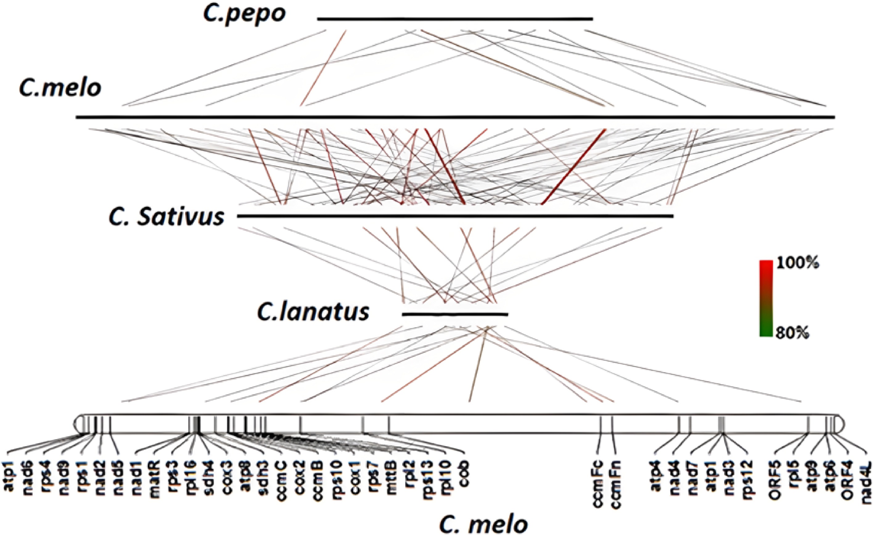
      Figure 1. 

      Collinearity analysis of C. melo mitochondrial genome with other three Cucurbitaceae plants.

      The linear relationships of mitochondrial coding genes among C. melo, C. sativus, C. pepo and C. lanatus were compared. Two or more collinear gene groups are called gene clusters, and we get 7 to 13 gene clusters with different gene numbers (Table 2). A higher number of collinear gene clusters was identified in C. melo and C. sativus, contrarily, a fewer number of collinear gene clusters was found in C. pepo and C. lanatus.

      Table 2.  The gene clusters of melon collinearity with three other mitochondrial genomes.

      SpeciesAmountC. melo
      C. lanatus7nad6-rps4; trnY-nad2; trnF-trnS; rps3-rpl16; sdh4-cox3-atp8; rrn5s-rrn18s; nad3-rps12
      C. sativus13nad6-rps4; nad9-rps1; nad2-sdh3; trnF-trnS; matR-trnH; rps3-rpl16; sdh4-cox3-atp8; rrn5S-rrn18S; rpl10-trnD; ccmFc-trnW-atp4; nad3-rps12; atp9-atp6
      C. pepo8nad6-rps4; nad9-rps1; rps3-rpl16; sdh4-cox3-atp8; rrn5S-rrn18S; nad3-rps12; atp9-atp6; trnM-trnG
      Gene clusters in bold are the gene clusters common to the four mitochondrial genomes.
    • There were no significant differences in mitochondrial genomic GC content among C. melo, C. sativus, C. pepo and C. lanatus (between 42.8 and 45.1%, Table 3). The total number of repeat sequences in the melon mitochondrial genome was the highest among all four species, accounting for 44.2% of the total genome sequence. The number of repeat sequences in the mitochondrial genome of C. sativus, C. pepo and C. lanatus was 44.1%, 24.4% and 9.6% of the total genome, respectively.

      Table 3.  Mitochondrial genome summary of C. melo, C. sativus, C. pepo and C. lanatus

      FeatureC. meloC. lanatusC. sativusC. pepo
      Genome
      AccessionMG947207
      MG947208
      MG947209
      NC_014043NC_016004
      NC_016005
      NC_016006
      NC_014050
      Size in bp2,906,673379,2361,644,236982,833
      Chromosome number3131
      Topology StructureCircleCircleCircleCircle
      GC content (%)44.8%45.1%44.3%42.8%
      Gene
      Protein-coding genes40393738
      Protein-coding genes in bp (%)35,613 (1.23%)32,370 (8.5%)32,550 (3.31%)32,032 (3.26%)
      Single-copy protein genes37373737
      Single-copy protein genes in bp (%)34,080 (1.17%)31,986 (8.4%)32,550 (3.31%)31,806 (3.23%)
      Intron
      Trans-spliced5455
      Cis-spliced17201819
      Cis-spliced introns in bp (%)46,000 (1.6%)32,476 (8.6%)47,996 (2.9%)30,557 (3.1%)
      tRNA genes40182013
      Native17373
      Chloroplast-derived23151310
      Total tRNAs in bp (%)2,999 (0.1%)1,358 (0.4%)1,486 (0.09%)966 (0.1%)
      rRNA genes8363
      Total rRNAs in bp (%)5,815 (0.2%)5,148 (1.4%)11044 (0.67%)5,109 (0.5%)
      Noncoding regions in bp (%)2,862,246 (98.5%)340,360 (89.7%)1,599,156 (97.3%)944,726 (96.1%)
      Repetitive content
      SSR (num.)0.1% (218)0.2% (54)0.1% (144)0.2% (144)
      TR (num.)2.1% (653)0.3% (14)0.4% (120)1.9 (287)
      IR (pairs)2.4% (439)0.4% (14)6.3% (539)0.2% (17)
      FR (pairs)39.6% (4861)8.7% (209)37.3% (4974)22.1% (1608)
      Maximum large repeat length (bp)5,5327,28617,159621
      Number of repeats (>1 kb)871100
      Total repeats (%)44.2%9.6%44.1%24.4%
      Chloroplast-like in bp (%)79,463 (2.73%)28,703 (7.6%)70,702 (4.3%)113,347(11.5%)
      Mitochondrial-like in bp (%)967,450 (33.3%)159,032 (41.9%)907,251 (55.2%)180,008 (18.3%)
      Nuclear-like in bp (%)1,413,224 (48.62%)24,352 (6.4%)501,491 (30.5%)20,638 (2.1%)

      A total of 40 protein-encoding genes were annotated in the mitochondrial genome of melon. The number of protein-encoding genes in the mitochondrial genome of C. sativus, C. pepo and C. lanatus was 37, 38 and 39, respectively. Melon mitochondrial genome lost rps19 gene, two copies of atp1 gene, and two more ORF genes (orf1 and orf2). The mitochondrial genome of these four plants contained three different types of ribosomal genes (rrn5S, rrn18S and rrn26S). The highest number of rRNAs were detected in melon, including six copies of rrn5S, and two single copies of rrn18S and rrn26. Six rRNA genes were present in the cucumber mitochondrial genome, and each ribosomal gene presented two repeats. Only three rRNAs were observed in watermelon and zucchini, all of which were single-copy genes (Table 4).

      Table 4.  Comparison of the gene content among C. melo, C. sativus, C. pepo and C. lanatus mitochondrial genome.

      GeneC. meloC. lanatusC. sativusC. pepo
      Complex I
      nad1,2,3,4,4L,5,6,7,9++++
      Complex II
      sdh3+2++
      sdh4++++
      Complex III
      cob++++
      Complex IV
      cox1,2,3++++
      Complex V
      atp12+++
      atp4,6,8,9++++
      Cytochrome c biogenesis
      ccmB, C, Fc, Fn++++
      Ribosomal RNAs
      rrn5S6+2+
      rrn18S++2+
      rrn26S++2+
      Ribosomal proteins
      rpl2,5,16++++
      rpl10++
      rps1,3,4,7,10,12,13++++
      rps1922
      Other ORFs
      matR, mttB++++
      orf1,2+
      Total number48424341
      +: indicates the presence and uniqueness of this gene; −: represents the absence of this gene, and the number represents the copy number of this gene.

      We compared the tRNA use of C. melo, C. sativus, C. pepo and C. lanatus (Table 5). The results showed that the mitochondrial genome sequences of all four species could not encode a complete set of tRNA that could recognize all codons or transport a complete set of 20 amino acids.

      Table 5.  Comparison of the tRNA genes.

      tRNAC. meloC. lanatusC. sativusC. pepo
      trnC-GCAMMMMM
      trnD-GUCCCMMC
      trnE-UUCMMMMM
      trnF-GAACMMCCCM
      trnfM-CAUMMM
      trnG-GCCCMMMMM
      trnH-GUGCCMCCCC
      trnI-CAUMMMM
      trnK-UUUMM
      trnL-CAACM
      trnM-CAUCCCMMMCMC
      trnN-GUUCCMMCC
      trnP-UGGMMMM
      trnQ-UUGMMMMMM
      trnR-ACGMM
      trnS-GCUMMM
      trnS-UGACM
      trnV-GACC
      trnW-CCACCCMMMMC
      trnY-GUAMMMM
      Choloroplast-derived17373
      Mitochondrial-derived23151310
      Total (type)40 (18)18 (15)20 (15)13 (13)
    • Our results show that C. lanatus had the largest number of RNA editing sites along its mitochondrial genome (509 sites). C. melo and C. sativus contained 507 and 498 RNA editing sites, respectively. C. pepo presented the lowest number of RNA editing sites (total of 486). Nad4, ccmB and ccmFn were the three genes with the highest RNA editing sites (Table 6).

      Table 6.  Number of RNA editing sites in the four mitochondrial genomes.

      OrderGenesC. meloC. lanatusC. sativusC. pepo
      1Atp14555
      2Atp411131213
      3Atp623222222
      4Atp82422
      5Atp95656
      6ccmB34343330
      7ccmC28272726
      8ccmFc17171817
      9ccmFn36363635
      10Cob16141614
      11Cox117171819
      12Cox213131313
      13Cox38987
      14matR12121212
      15mttB27242324
      16Nad121212120
      17Nad225252524
      18Nad312121210
      19Nad437383633
      20Nad4L13131313
      21Nad528272823
      22Nad615101010
      23Nad725272726
      24Nad97777
      25Rpl25332
      26Rpl5101098
      27Rpl165555
      28Rps13434
      29Rps37978
      30Rps417171718
      31Rps72222
      32Rps106565
      33Rps125777
      34Rps134334
      35Rps19pseudo3pseudo4
      36Sdh33435
      37Sdh44443
      Total507509498486
      Note: pseudo indicates that the gene is a pseudogene.
    • In this study, the phylogenetic relationship of C. melo, C. sativus, C. pepo and C. lanatus were analyzed based on 10 conserved coding genes (atp6, nad6, cox3, rps12, atp1, nad4, nad9, nad7, nad4L) that are present in all 14 species. Evolutionary relationships were analyzed (Fig. 2) and it was found that grapes and Cucurbitaceae are closely related, and Cucurbitaceae can cluster well in a clade, but the genetic distance of C. pepo in the phylogenetic tree is closer to C. melo and C. sativus compared to C. lanatus.
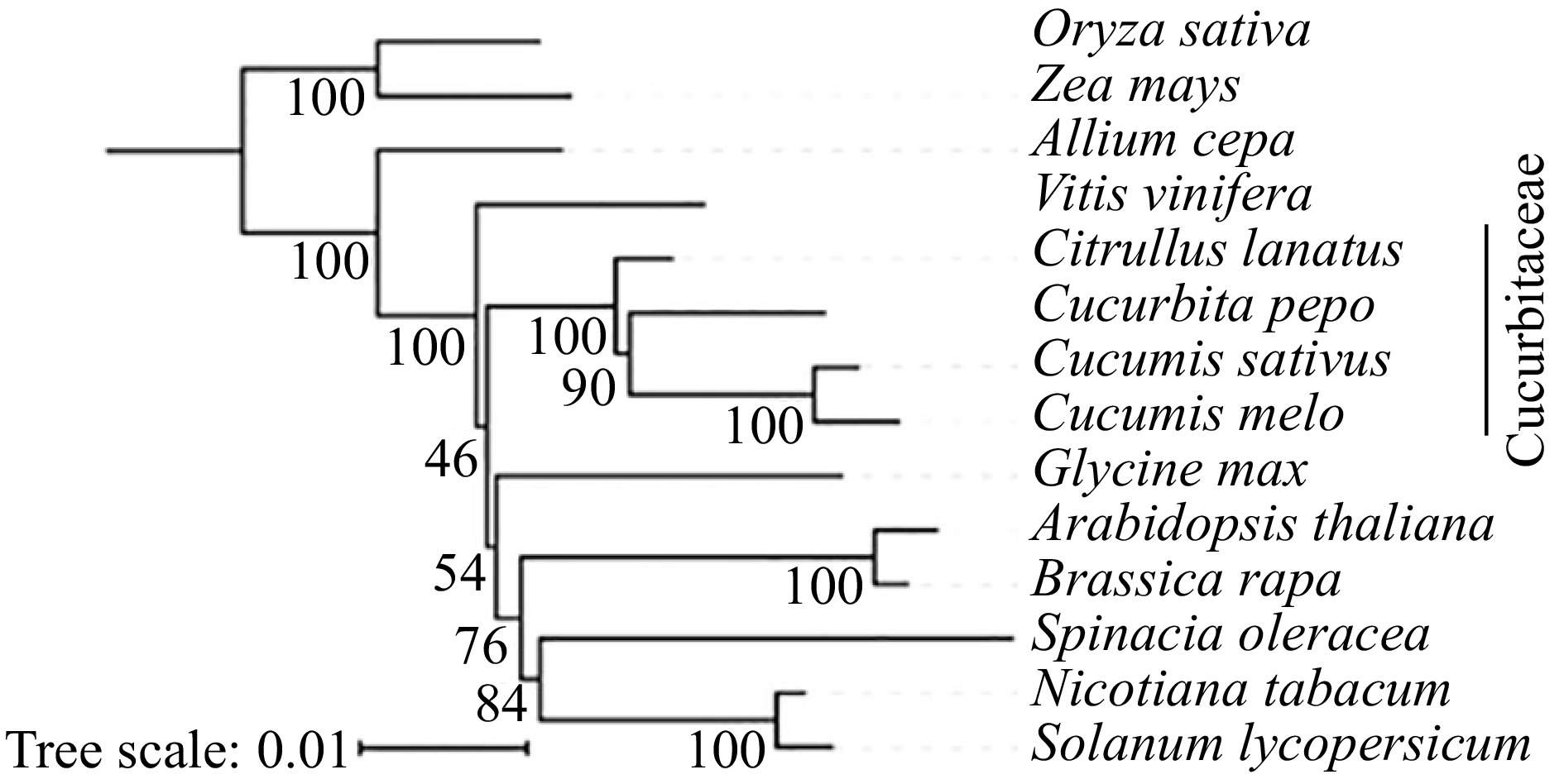
      Figure 2. 

      The construction of phylogenetic tree among 14 species based on mitochondrial conserved genes.

    • Our study corroborates with previous reports that utilized renaturation kinetics and restriction endonuclease technology to analyze the mitochondrial genome analysis of Cucurbitaceae[4]. The structure of both C. melo and C. sativus mitochondrial genomes is polycyclic. Likewise, this genome structure has been observed in other plants, including wheat[22] and rape[23].

      The protein-coding genes in melon mitochondrial genome are like those in the three mitochondrial genomes. Similar conclusions have been reached in studies of other higher plants, where the coding regions of plant mitochondrial genomes are more conserved than the non-coding regions[24]. Melon mitochondria contain two more ORF genes than the other three reference genomes, and ORFs may encode proteins with important functions. In fact, some mitochondrial ORFs have been associated with cytoplasmic male sterility in many plants.

      With the expectation of T-urf13, atp6 of radish and orf256 of wheat, it has been verified that most of the transcripts of protein-coding genes are edited in the mitochondria of higher plants, but the editing degree of transcripts from different genes is variable[25, 26]. The male sterility of plants may be caused by some genes in the mitochondrial genome recombining with ORF to form chimeric genes or inadequate RNA editing[27]. In the present study, a slightly distinct number of RNA editing sites was detected in the mitochondrial genomes of four Cucurbitaceae crops. The elucidation of these RNA editing sites may provide data support for the research of RNA editing in cucurbits.

      The mitochondrial genomes of both melon and three other kinds of Cucurbitaceae plants were linearly analyzed. C. melo and C. sativus showed more collinearity between mitochondrial gene clusters, which further shows that C. melo has a closer relationship with C. sativus. In the four kinds of Cucurbitaceae plants some gene cluster, rps3-rpl16 is widespread in the plant mitochondrial genome typical gene cluster[28].

    • This study upon in-depth comparative analysis of the mitochondrial gene structure of C. melo, C. sativus, C. pepo and C. lanatus, revealed that the large number of repetitive and nuclear genome sequences were the potential reasons for the increasing scale and variation of the melon mitochondrial genome. These results provide the basis for the genetic variation of the mitochondrial genome in Cucurbitaceae plants.

      • This study was funded by Scientific Research Foundation of Hebei Normal University of Science and Technology (2022YB004) and the National Nature Science Foundation of China (U21A20229 and 31960607).

      • The authors declare that they have no conflict of interest.

      • Copyright: © 2023 by the author(s). Published by Maximum Academic Press, Fayetteville, GA. This article is an open access article distributed under Creative Commons Attribution License (CC BY 4.0), visit https://creativecommons.org/licenses/by/4.0/.
    Figure (2)  Table (6) References (28)
  • About this article
    Cite this article
    Cui H, Ding Z, Zhu Q, Gao P. 2022. Structural characteristics of the melon mitochondria genome. Vegetable Research 2:20 doi: 10.48130/VR-2022-0020
    Cui H, Ding Z, Zhu Q, Gao P. 2022. Structural characteristics of the melon mitochondria genome. Vegetable Research 2:20 doi: 10.48130/VR-2022-0020

Catalog

  • About this article

/

DownLoad:  Full-Size Img  PowerPoint
Return
Return