ARTICLE   Open Access    

Genome-wide identification and expression analysis of sucrose phosphate synthase and sucrose-6-phosphate phosphatase family genes in Camellia sinensis

More Information
  • Sucrose phosphate synthetase (SPS, EC 2.4.1.14) and sucrose phosphate phosphatase (SPP, EC 3.1.3.24) are two key enzymes for sucrose biosynthesis, which play essential roles in growth, development and stress responses of plants. However, the roles of SPS and SPP have not been illustrated well in tea plants until now. In this study, a unique CsSPP and five CsSPSs (CsSPS1-5) were identified from the tea plant genome. Bioinformatic analysis results found that CsSPP and CsSPSs were clustered together with the known SPPs and SPSs of other species, respectively, and their amino acid sequences contain the conserved domains required for sucrose biosynthesis. Tissue-specific analysis results showed that CsSPP and CsSPSs were widely involved in vegetative and reproductive growth of tea plant, among which the transcription levels of CsSPP was highest in immature stem, while CsSPSs were highest in flower. Spatio-temporal expression analysis results showed that all of these genes are involved in abiotic stress responses of tea plants. Meanwhile, SPS activity and the contents of sucrose, glucose, fructose, and total soluble sugar in 'Shuchazao' cultivar increased more than that of 'Baiye1' under low temperature conditions. Correlation analysis results showed that the expression profiles of CsSPS2/4/5 were significantly and positively correlated with sucrose content in 'Shuchazao' cultivars under low temperature conditions, suggesting the significant roles of these genes in sucrose accumulation. In conclusion, this study will provide a theoretical basis for further functional research of SPS and SPP in plants.
  • Atractylodes macrocephala Koidz. (common names 'Baizhu' in Chinese and 'Byakujutsu' in Japanese) is a diploid (2n = 2x = 24) and out-crossing perennial herb in the Compositae family, and has a long history of cultivation in temperate and subtropical areas of East Asia as it is widely used in traditional herbal remedies with multiple pharmacological activities[13]. The 'Pharmacopoeia of the People's Republic of China' states that 'Baizhu' is the dry rhizome of A. macrocephala Koidz. (Atractylodis Macrocephalae Rhizoma, AMR). However, in Japanese traditional medicine 'Baizhu' can be referred to both: A. japonica or A. macrocephala[4].

    A. macrocephala is naturally endemic to China and cultivated in more than 200 towns in China, belonging to Zhejiang, Hunan, Jiangxi, Anhui, Fujian, Sichuan, Hubei, Hebei, Henan, Jiangsu, Guizhou, Shanxi, and Shaanxi Provinces[3]. A. macrocephala grows to a height of 20–60 cm (Fig. 1). The leaves are green, papery, hairless, and generally foliole with 3–5 laminae with cylindric glabrous stems and branches. The flowers grow and aggregate into a capitulum at the apex of the stem. The corollas are purplish-red, and the florets are 1.7 cm long. The achenes, densely covered with white, straight hairs, are obconic and measure 7.5 mm long. The rhizomes used for medicinal purposes are irregular masses or irregularly curving cylinders about 3–13 cm long and 1.5–7 cm in diameter with an outwardly pale greyish yellow to pale yellowish color or a sparse greyish brown color. The periderm-covered rhizomes are externally greyish brown, often with nodose protuberances and coarse wrinkles. The cross-sections are white with fine dots of light yellowish-brown to brown secretion. Rhizomes are collected from plants that are > 2 years old during the spring. The fibrils are removed, dried, and used for medicinal purposes[5, 6].

    Figure 1.  Plant morphology of A. macrocephala.

    The medicinal properties of AMRs are used for spleen deficiency, phlegm drinking, dizziness, palpitation, edema, spontaneous sweating, benefit Qi, and fetal restlessness[7]. The AMR contains various functional components, among which high polysaccharide content, with a yield close to 30%[8]. Therefore, the polysaccharides of A. macrocephala Koidz. rhizome (AMRP) are essential in assessing the quality control and bioactivity of A. macrocephala. Volatile oil accounts for about 1.4% of AMR, with atractylon and atractylodin as the main components[9]. Atractylon can be converted to atractylenolide I (AT-I), atractylenolide II (AT-II), and atractylenolide III (AT-III) under ambient conditions. AT-III can be dehydrated to AT-II under heating conditions[10, 11]. AMRs, including esters, sesqui-, and triterpenes, have a wide range of biological activities, such as improving immune activity, intestinal digestion, neuroprotective activity, immune anti-inflammatory, and anti-tumor.

    In recent years, research on the pharmacological aspects of AMR has continued to increase. Still, the discovery of the main active components in AMR is in its infancy. The PAO-ZHI processing of AMR is a critical step for AMR to exert its functional effects, but also, in this case, further work is required. Studies on the biosynthesis of bioactive compounds and different types of transcriptomes advanced current knowledge of A. macrocephala, but, as mentioned, required more systematic work. Ulteriorly, an outlook on the future research directions of A. macrocephala was provided based on the advanced technologies currently applied in A. macrocephala (Fig. 2).

    Figure 2.  Current progress of A. macrocephala.

    A. macrocephala is distributed among mountainous regions more than 800 m above sea level along the middle and lower reaches of the Yangtze River (China)[5]. Due to over-exploitation and habitat destruction, natural populations are rare, threatened, and extinct in many locations[1,12]. In contrast to its native range, A. macrocephala is widely cultivated throughout China, in a total area of 2,000–2,500 ha, with a yield of 7,000 t of rhizomes annually[13]. A. macrocephala is mainly produced in Zhejiang, Anhui, and Hebei (China)[14]. Since ancient times, Zhejiang has been the famous producing area and was later introduced to Jiangxi, Hunan, Hebei, and other places[15]. Wild A. macrocephala is currently present in at least 14 provinces in China. It is mainly distributed over three mountain ranges, including the Tianmu and Dapan mountains in Zhejiang Province and the Mufu mountains along the border of Hunan and Jiangxi Provinces. A. macrocephala grows in a forest, or grassy areas on mountain or hill slopes and valleys at an altitude of 600–2,800 m. A. macrocephala grows rapidly at a temperature of 22–28 °C, and favors conditions with total precipitation of 300–400 mm evenly distributed among the growing season[16]. Chen et al. first used alternating trilinear decomposition (ATLD) to characterize the three-dimensional fluorescence spectrum of A. macrocephala[17]. Then they combined the three-dimensional fluorescence spectrum with partial least squares discriminant analysis (PLS-DA) and k-nearest neighbor method (kNN) to trace the origin of Atractylodes samples. The results showed that the classification models established by PLS-DA and kNN could effectively distinguish the samples from three major Atractylodes producing areas (Anhui, Hunan, and Zhejiang), and the classification accuracy rate (CCR) of Zhejiang atractylodes was up to 80%, and 90%, respectively[17]. Zhang et al. compared the characteristics, volatile oil content, and chemical components of attested materials from six producing areas of Zhejiang, Anhui, Hubei, Hunan, Hebei, and Henan. Differences in the shape, size, and surface characteristics were reported, with the content of volatile oil ranging from 0.58% to 1.22%, from high to low, Hunan (1.22%) > Zhejiang (1.20%) > Anhui (1.02%) > Hubei (0.94%) > Henan (0.86%) > Hebei (0.58%)[18]. This study showed that the volatile oil content of A. macrocephala in Hunan, Anhui, and Hubei is not much different from that of Zhejiang, which is around 1%. A. macrocephala is a local herb in Zhejiang, with standardized cultivation techniques, with production used to reach 80%–90% of the country. However, in recent years, the rapid development of Zhejiang's real estate economy has reduced the area planted with Zhejiang A. macrocephala, resulting in a sudden decrease in production. Therefore, neighboring regions, such as Anhui and Hunan, vigorously cultivate A. macrocephala, and the yield and quality of A. macrocephala can be comparable to those of Zhejiang. The results were consistent with the data reports[18]. Guo et al. analyzed the differentially expressed genes of Atractylodes transcripts from different regions by the Illumina HiSeq sequencing platform. It was found that 2,333, 1,846, and 1,239 DEGs were screened from Hubei and Hebei, Anhui and Hubei, and Anhui and Hebei Atrexia, respectively, among which 1,424, 1,091, and 731 DEGs were annotated in the GO database. There were 432, 321, and 208 DEGs annotated in the KEGG database. These DEGs were mainly related to metabolic processes and metabolic pathways of secondary metabolites. The highest expression levels of these genes were found in Hubei, indicating higher terpenoid production in Hubei[19]. Other compounds were differentially accumulated in Atractylodes. Chlorogenic acid from Hebei was 0.22%, significantly higher than that from Zhejiang and Anhui[20]. Moreover, the content of neochlorogenic acid and chlorogenic acid decreased after processing, with the highest effect reported in Zhejiang, with the average transfer rate of neochlorogenic acid and chlorogenic acid reaching 55.68% and 55.05%[20]. All these changes would bring great help in distinguishing the origins of A. macrocephala.

    Medicinal AMR can be divided into raw AMR and cooked AMR. The processing method is PAO-ZHI; the most traditional method is wheat bran frying. The literature compared two different treatment methods, crude A. macrocephala (CA) and bran-processed A. macrocephala, and found that the pharmacological effects of AMR changed after frying with wheat bran, mainly in the anti-tumor, antiviral and anti-inflammatory effects[21]. The anti-inflammatory effect was enhanced, while the anti-tumor and antiviral effects were somewhat weakened, which may be related to the composition changes of the compounds after frying. The study of the content of AT-I, II, and III, and atractyloside A, in rat serum provided helpful information on the mechanism of wheat bran processing[22]. In addition to frying wheat bran, Sun et al. used sulfur fumigation to treat AMR[23]. They found that the concentration of different compounds changed, producing up to 15 kinds of terpenoids. Changes in pharmacological effects were related to treatment and the type of illumination[24,25]. Also, artificial light can improve the various biological functions. A. macrocephala grew better under microwave electrodeless light, with a chlorophyll content of 57.07 ± 0.65 soil and plant analyzer develotrnent (SPAD)[24]. The antioxidant activity of AMR extract treated with light-emitting diode (LED)-red light was the highest (95.3 ± 1.1%) compared with other treatments[24]. The total phenol and flavonoid contents of AMR extract treated with LED-green light were the highest at 24.93 ± 0.3 mg gallic acid equivalents (GAE)/g and 11.2 ± 0.3 mg quercetin equivalents (QE)/g compared with other treatments[24, 25]. Polysaccharides from Chrysanthemun indicum L.[26] and Sclerotium rolfsiisacc[27] can improve AMR's biomass and bioactive substances by stimulating plant defense and thus affect their efficacy. In summary, there are compositional differences between A. macrocephala from different origins. Besides, different treatments, including processing mode, light irradiation, and immune induction factors, which can affect AMR's biological activity, provide some reference for the cultivation and processing of A. macrocephala (Fig. 3).

    Figure 3.  Origin, distribution and processing of A. macrocephala.

    The AMR has been reported to be rich in polysaccharides, sesquiterpenoids (atractylenolides), volatile compounds, and polyacetylenes[3]. These compounds have contributed to various biological activities in AMR, including immunomodulatory effects, improving gastrointestinal function, anti-tumor activity, neuroprotective activity, and anti-inflammatory.

    AMRP has received increasing attention as the main active component in AMR because of its rich and diverse biological activities. In the last five years, nine AMRP have been isolated from AMR. RAMP2 had been isolated from AMR, with a molecular weight of 4.354 × 10³ Da. It was composed of mannose, galacturonic acid, glucose, galactose, and arabinose, with the main linkages of →3-β-glcp-(1→, →3,6-β-glcp-(1→, →6-β-glcp-(1→, T-β-glcp-(1→, →4-α-galpA-(1→, →4-α-galpA-6-OMe-(1→, →5-α-araf-(1→, →4,6-β-manp-(1→ and →4-β-galp-(1→[28]. Three water-soluble polysaccharides AMAP-1, AMAP-2, and AMAP-3 were isolated with a molecular weight of 13.8 × 10⁴ Da, 16.2 × 10⁴ Da, and 8.5 × 10⁴ Da, respectively. Three polysaccharides were deduced to be natural pectin-type polysaccharides, where the homogalacturonan (HG) region consists of α-(1→4)-linked GalpA residues and the ramified region consists of alternating α-(1→4)-linked GalpA residues and α-(1→2)-linked Rhap residues. Besides, three polysaccharides were composed of different ratios of HG and rhamnogalacturonan type I (RG-I) regions[29]. Furthermore, RAMPtp has been extracted from AMR with a molecular weight of 1.867 × 10³ Da. It consists of glucose, mannose, rhamnose, arabinose, and galactose with 60.67%, 14.99%, 10.61%, 8.83%, and 4.90%, connected by 1,3-linked β-D Galp and 1,6-linked β-D Galp residues[30]. Additionally, PAMK was characterized by a molecular weight of 4.1 kDa, consisting of galactose, arabinose, and glucose in a molar ratio of 1:1.5:5, with an alpha structure and containing 96.47% polysaccharide and small amounts of protein, nucleic acid, and uric acid[31]. Another PAMK extracted from AMR had a molecular weight of 2.816 × 10³ Da and consisted of glucose and mannose in molar ratios of 0.582 to 0.418[32]. Guo et al. isolated PAMK with a molecular weight of 4.748 × 10³ g/mol from AMR, consisting of glucose, galactose, arabinose, fructose, and mannose in proportions of 67.01%, 12.32%, 9.89%, 1.18%, and 0.91%, respectively[33]. In addition, AMP1-1 is a neutral polysaccharide fragment with a molecular weight of 1.433 kDa isolated from AMR. It consists of glucose and fructose, and the structure was identified as inulin-type fructose α-D-Glcp-1→(2-β-D-Fruf-1)7[34]. These reports indicated that, in general, polysaccharides are extracted by water decoction, ultrasonic-assisted extraction, enzyme hydrolysis method, and microwave-assisted extraction. The separation and purification are column chromatography, stepwise ethanol precipitation, and ultrafiltration. Their physicochemical properties and structural characterization are generally achieved by determining the molecular weight, determining the monosaccharide composition, analyzing the secondary structure, and glycosidic bond configuration of polysaccharides with Fourier transform infrared (FT-IR) and nuclear magnetic resonance (NMR). The advanced structures of polysaccharides can be identified by high-performance size exclusion chromatography-multiangle laser light scattering (HPSEC-MALLS), transmission electron microscopy (TEM), and scanning electron microscopy (SEM) techniques (Table 1). AMRP has various physiological functions, including immunomodulatory effects, improving gastrointestinal function, and anti-tumor activity. The related biological activities, animal models, monitoring indicators, and results are summarized in Table 1.

    Table 1.  Components and bioactivity of polysaccharides from Atractylodes macrocephala Koidz. Rhizome.
    Pharmacological activitiesDetailed functionPolysaccharides informationModelDoseTest indexResultsRef.
    Immunomodulatory effectsRestore immune
    function
    /Chicken models
    (HS-induced)
    200 mg/kgOxidative index;
    Activities of mitochondrial complexes and ATPases;
    Ultrastructure in chicken spleens;
    Expression levels of cytokines, Mitochondrial dynamics- and apoptosis-related genes
    Alleviated
    the expression of
    IL-1 ↑,TNF-α ↑, IL-2 ↓, IFN- γ ↓; mitochondrial dynamics- and anti-apoptosis-related genes ↓; pro-apoptosis-related genes ↑;
    the activities of mitochondrial complexes and ATPases ↓ caused by HS
    [35]
    Regulate the immune function/Chicken models
    (HS-induced)
    200 mg/kgiNOS–NO activities;
    ER stress-related genes;
    Apoptosis-related genes;
    Apoptosis levels
    Alleviated NO content ↑; activity of iNOS ↑ in the chicken spleen; GRP78, GRP94, ATF4, ATF6, IRE ↑; caspase3 ↑; Bcl-2 ↓ caused by HS[36]
    Relieve immunosuppressionCommercial AMR powder (purity 70%)Geese models
    (CTX-induced)
    400 mg/kgSpleen development;
    Percentages of leukocytes in peripheral blood
    Alleviated the spleen damage;
    T and B cell proliferation ↓; imbalance of leukocytes; disturbances of humoral; cellular immunity caused by CTX
    [37]
    Active the lymphocytesCommercial AMR powder (purity 95%)Geese models
    (CTX-induced)
    400 mg/kgThymus morphology;
    The level of serum GMC-SF, IL-1b, IL-3, IL-5;
    mRNA expression of CD25, novel_mir2, CTLA4 and CD28 signal pathway
    Maintain normal cell morphology of thymus;
    Alleviated GMC-SF ↓, IL-1b ↓, IL-5↓, IL-6↓, TGF-b↓; IL-4 ↑, IL-10 ↑; novel_mir2 ↓, CD25↓, CD28↓ in thymus and lymphocytes caused by CTX
    [38]
    Alleviate immunosuppressionCommercial AMR powder (purity 70%)Geese models
    (CTX-induced)
    400 mg/kgThymus development;
    T cell proliferation rate;
    The level of CD28, CD96, MHC-II;
    IL-2 levels in serum;
    differentially expressed miRNAs
    Alleviated thymus damage;
    T lymphocyte proliferation rate ↓; T cell activation ↓; IL-2 levels ↓ caused by CTX;
    Promoted novel_mir2 ↑; CTLA4 ↓; TCR-NFAT signaling pathway
    [39]
    Alleviates T cell activation declineCommercial AMR powder (purity 95%)BALB/c female mice (CTX-induced)200 mg/kgSpleen index;
    Morphology, death, cytokine concentration of splenocytes;
    Th1/Th2 ratio, activating factors of lymphocytes;
    T cell activating factors;
    mRNA expression level in CD28 signal pathway
    Improved the spleen index;
    Alleviated abnormal splenocytes morphology and death; Balance Th1/Th2 ratio; IL-2 ↑, IL-6 ↑, TNF-α ↑, IFN-γ ↑; mRNA levels of CD28, PLCγ-1, IP3R, NFAT, AP-1 ↑
    [40]
    Immunoregulation and ImmunopotentiationCommercial AMR powder (purity 80%)BMDCs (LPS-induced);
    Female BALB/c mice (ovalbumin as a model antigen)
    /Surface molecule expression of BMDCs;
    Cytokines secreted by dendritic cell supernatants;
    OVA-specific antibodies in serum;
    Cytokines in serum;
    Lymphocyte immunophenotype
    Expression of CD80 and CD86 ↑; IL-1β ↑, IL-12 ↑, TNF-α↑ and IFN-γ ↑; OVA-specific antibodies in serum ↑; Secretion of cytokines ↑; Proliferation rate of spleen lymphocytes ↑; Activation of CD3+CD4+ and CD3+CD8+ lymphocytes[46]
    Increase immune-response capacity of the spleen in miceCommercial AMR powder (purity 70%)BALB/c female mice100, 200, 400 mg/kgSpleen index;
    Concentrations of cytokines;
    mRNA and protein expression levels in TLR4 signaling
    In the medium-PAMK group:
    IL-2, IL-4, IFN-c, TNF-a ↑; mRNA and protein expression of TLR4, MyD88, TRAF6, TRAF3, NF-κB in the spleen ↑
    [41]
    Immunological activityCommercial AMR powder (purity 80%)Murine splenic lymphocytes (LPS or PHA-induced)13, 26, 52, 104, 208 μg/mLT lymphocyte surface markersLymphocyte proliferation ↑;
    Ratio of CD4+/CD8+ T cells ↑
    [47]
    Immunomodulatory activityTotal carbohydrates content 95.66 %Mouse splenocytes
    (Con A or LPS-induced)
    25, 50, 100 μg/mLSplenocyte proliferation;
    NK cytotoxicity;
    Productions of NO and cytokines;
    Transcription factor activity;
    Signal pathways and receptor
    Promoted splenocyte proliferation; Cells enter S and G2/M phases; Ratios of T/B cells ↑; NK cytotoxicity ↑; Transcriptional activities of NFAT ↑; NF-κB, AP-1 ↑; NO, IgG, IL-1α, IL-1β, IL-2, IL-3, IL-4, IL-6, IL-10, IL-12p40, IL-12p70, IL-13, IFN-γ, TNF-α, G-CSF, GM-CSF, KC, MIP-1α, MIP-1β, RANTES, Eotaxin ↑[42]
    Promote the proliferation of thymic epithelial cellsContents of fucrhaara, galactose, glucose, fructose,
    and xylitol: 0.98%, 0.40%, 88.67%, 4.47%, and 5.47%
    MTEC1 cells50 μg/mLCell viability and proliferation;
    lncRNAs, miRNAs, and mRNAs expression profiles in MTEC1 cells
    The differential genes were 225 lncRNAs, 29 miRNAs, and 800 mRNAs; Genes enriched in cell cycle, cell division, NF-κB signaling, apoptotic process, and MAPK signaling pathway[44]
    Immunomodulatory activityMW: 4.354 × 10³ Da;
    Composed of mannose, galacturonic acid, glucose, galactose and arabinose;
    The main linkages are →3-β-glcp-(1→, →3,6-β-glcp-(1→, →6-β-glcp-(1→, T-β-glcp-(1→,
    →4-α-galpA-(1→, →4-α-galpA-6-OMe-(1→, →5-α-araf-(1→, →4,6-β-manp-(1→ and →4-β-galp-(1→
    CD4+ T cell50, 100, 200 μg/mLMolecular weight;
    Monosaccharide composition;
    Secondary structure;
    Surface topography;
    Effect on Treg cells
    Treg cells percentage ↑; mRNA expressions of Foxp3, IL-10 and IL-2 ↑; STAT5 phosphorylation levels ↑; IL-2/STAT5 pathway[28]
    Immunostimulatory activityMW of AMAP-1, AMAP-2, and AMAP-3 were 13.8×10⁴ Da, 16.2×10⁴ Da and 8.5×10⁴ Da;
    HG region consists of α-(1→4)-linked GalpA residues
    RAW264.7 cells (LPS-induced)80, 40, 200 μg/mLMolecular weight;
    Total carbohydrate;
    Uronic acid contents;
    Secondary structure;
    Monosaccharide composition;
    Immunostimulatory activity
    RG-I-rich AMAP-1 and AMAP-2 improved the release of NO[29]
    Immunomodulatory effectMW: 1.867×10³ Da;
    Contents of glucose, mannose, rhamnose,
    arabinose and galactose: 60.67%, 14.99%, 10.61%, 8.83% and 4.90%
    SMLN lymphocytes25
    μg/ml
    Molecular weight;
    Monosaccharide composition;
    Ultrastructure;
    Intracellular Ca2+concentration;
    Target genes;
    Cell cycle distribution
    [Ca2+]i ↑; More cells in S and G2/M phases; IFN-γ ↑, IL-17A ↑; mRNA expressions of IL-4 ↓[30]
    Macrophage activationTotal carbohydrates content 95.66 %RAW264.7 macrophages (LPS-induced)25, 50, 100 μg/mLPinocytic activity;
    Phagocytic uptake;
    Phenotypic characterization;
    Cytokine production;
    Bioinformatics analysis;
    Transcription factor inhibition
    IL-6, IL-10 and TNF-α ↑; CCL2 and CCL5 ↑; Pinocytic and phagocytic activity ↑; CD40, CD80, CD86, MHC-I, MHC-II ↑; NF-κB and Jak-STAT pathway[43]
    Immunomodulatory effectTotal carbohydrates content 95.66 %SMLN lymphocytes25, 50, 100 μg/mLCytokine production;
    CD4+ and CD8+ lymphocytes;
    Target genes;
    Bioinformatics analysis;
    T and B lymphocyte proliferation;
    Receptor binding and blocking
    IFN-γ, IL-1α, IL-21, IFN-α, CCL4, CXCL9, CXCL10 ↑; CD4+ and CD8+subpopulations proportions ↑;
    c-JUN, NFAT4, STAT1, STAT3 ↑;
    67 differentially expressed miRNAs (55 ↑ and
    12 ↓), associated with immune system pathways; Affect T and B lymphocytes
    [45]
    Improving gastrointestinal functionRelieve enteritis and improve intestinal
    flora disorder
    Commercial AMR powder (purity 70%);
    Contents of fucrhaara, galactose, glucose, xylitol, and fructose: 0.98%, 0.40%, 88.67%, 4.47%, and 5.47%
    Goslings (LPS-induced)400 mg/kgSerum CRP, IL-1β, IL-6, and TNF-α levels;
    Positive rate of IgA;
    TLR4, occludin, ZO-1, cytokines, and immunoglobulin mRNA expression;
    Intestinal flora of gosling excrement
    Relieved IL-1β, IL-6, TNF-α levels in serum ↑; the number of IgA-secreting cells ↑; TLR4 ↑; tight junction occludin and ZO-1 ↓; IL-1β mRNA expression in the small intestine ↑; Romboutsia ↓ caused by LPS[48]
    Ameliorate ulcerative colitisMW: 2.391 × 10⁴ Da;
    Composed of mannose, glucuronic acid, glucose and arabinose in a molar ratio of 12.05:6.02:72.29:9.64
    Male C57BL/6J mice (DDS-induced)10, 20, 40 mg/kg bwHistopathological evaluation;
    Inflammatory mediator;
    Composition of gut microbiota;
    Feces and plasma for global metabolites profiling
    Butyricicoccus, Lactobacillus ↑;
    Actinobacteria, Akkermansia, Anaeroplasma, Bifidobacterium, Erysipelatoclostridium, Faecalibaculum, Parasutterella,
    Parvibacter, Tenericutes, Verrucomicrobia ↓;
    Changed 23 metabolites in fecal content; 21 metabolites in plasma content
    [49]
    Attenuate ulcerative colitis/Male SD rats (TNBS-induced);
    Co-culture BMSCs and IEC-6 cells
    540 mg/kg
    (for rats);
    400 μg/mL (for cell)
    Histopathological analysis;
    Cell migration;
    Levels of cytokines
    Potentiated BMSCs’ effect on preventing colitis and homing the injured tissue, regulated cytokines;
    BMSCs and AMP promoted the migration of IEC
    [52]
    Against intestinal mucosal injuryMW: 3.714 × 10³ Da;
    Composed of glucose, arabinose, galactose, galacturonic acid, rhamnose
    and mannose with molar ratios of 59.09:23.22:9.32:4.70:2.07:1.59
    Male C57BL/6 mice (DDS-induced)100 mg/kgIntestinal morphology;
    IL-6, TNF-α and IL-1β in serum;
    mRNA expression;
    Intestinal microbiota
    Alleviated body weight ↓; colon length ↓; colonic damage caused by DSS;
    Over-expression of TNF-α, IL-1β, IL-6 ↓; Infiltration of neutrophils in colon ↓; Mucin 2 ↑;
    Tight junction protein Claudin-1 ↑;
    Harmful bacteria content ↓;
    Beneficial bacteria content ↑
    [50]
    Against intestinal injuryTotal carbohydrates 95.66 %IECs (DDS-induced)5, 25, 50 μg/mLCell proliferation and apoptosis;
    Expression levels of intercellular TJ proteins;
    lncRNA screening
    Proliferation and survival of IECs ↑;
    Novel lncRNA ITSN1-OT1 ↑;
    Blocked the nuclear import of phosphorylated STAT2
    [51]
    Anti-tumor activityInduce apoptosis in transplanted H22 cells in miceMW: 4.1× 10³ Da;
    Neutral heteropolysaccharide composed of galactose, arabinose, and glucose with α-configuration (molar ratio, 1:1.5:5)
    Female Kunming mice100 and 200 mg/kg (for rats)Secondary structure;
    Molecular weight;
    Molecular weight;
    Thymus index and Spleen index;
    Lymphocyte Subpopulation in peripheral blood;
    Cell cycle distribution
    In tumor-bearing mice CD3+, CD4+, CD8+ ↓;
    B cells ↑
    [31]
    Regulate the innate immunity of colorectal cancer cellsCommercial AMR powder (purity 70%)C57BL/6J mice (MC38 cells xenograft model)500 mg/kgExpression of pro-inflammatory cytokines and secretionIL-6, IFN-λ, TNF-α, NO ↑ through MyD88/TLR4-dependent signaling pathway;
    Survival duration of mice with tumors ↑;
    Prevent tumorigenesis in mice
    [54]
    Induce apoptosis of Eca-109 cellsMW: 2.1× 10³ Da;
    Neutral hetero polysaccharide composed
    of arabinose and glucose (molar ratio, 1:4.57) with pyranose rings and α-type and β-type glycosidic linkages
    Eca-109 cells0.25, 0.5, 1, 1.5, 2.00 mg/mLCell morphology;
    Cell cycle arrest;
    Induction of apoptosis
    Accelerate the apoptosis of Eca109 cells[53]
    '/' denotes no useful information found in the study.
     | Show Table
    DownLoad: CSV

    To study the immunomodulatory activity of AMRP, the biological models generally adopted are chicken, goose, mouse, and human cell lines. Experiments based on the chicken model have generally applied 200 mg/kg doses. It was reported that AMRP protected the chicken spleen against heat stress (HS) by alleviating the chicken spleen immune dysfunction caused by HS, reducing oxidative stress, enhancing mitochondrial function, and inhibiting cell apoptosis[35]. Selenium and AMRP could improve the abnormal oxidation and apoptosis levels and endoplasmic reticulum damage caused by HS, and could act synergistically in the chicken spleen to regulate biomarker levels[36]. It indicated that AMRP and the combination of selenium and AMRP could be applied as chicken feed supplementation to alleviate the damage of HS and improve chicken immunity.

    The general application dose in the goose model is also 200 mg/kg, and the main injury inducer is cyclophosphamide (CTX). AMRP alleviated CTX-induced immune damage in geese and provided stable humoral immune protection[37]. Little is known about the role of AMRP in enhancing immunity in geese through the miRNA pathway. It was reported that AMRP alleviated CTX-induced decrease in T lymphocyte activation levels through the novel _mir2/CTLA4/CD28/AP-1 signaling pathway[38]. It was also reported that AMRP might be achieved by upregulating the TCR-NFAT pathway through novel_mir2 targeting of CTLA4, thereby attenuating the immune damage induced by CTX[39]. This indicated that AMRP could also be used as goose feed supplementation to improve the goose's autoimmunity.

    The typical injury inducer for mouse models is CTX, and the effects on mouse spleen tissue are mainly observed. BALB/c female mice were CTX-induced damage. However, AMRP increased cytokine levels and attenuated the CTX-induced decrease in lymphocyte activation levels through the CD28/IP3R/PLCγ-1/AP-1/NFAT signaling pathway[40]. It has also been shown that AMRP may enhance the immune response in the mouse spleen through the TLR4-MyD88-NF-κB signaling pathway[41].

    Various cellular models have been used to study the immune activity of AMRP, and most of these studies have explored the immune activity with mouse splenocytes and lymphocytes. Besides, the commonly used damage-inducing agents are LPS, phytohemagglutinin (PHA), and concanavalin A (Con A).

    In one study, the immunoreactivity of AMRP was studied in cultured mouse splenocytes. LPS and Con A served as controls. Specific inhibitors against mitogen-activated protein kinases (MAPKs) and NF-κB significantly inhibited AMRP-induced IL-6 production. The results suggested that AMRP-induced splenocyte activation may be achieved through TLR4-independent MAPKs and NF-κB signaling pathways[42]. Besides, AMRP isolated from AMR acting on LPS-induced RAW264.7 macrophages revealed that NF-κB and Jak-STAT signaling pathways play a crucial role in regulating immune response and immune function[43]. RAMP2 increased the phosphorylation level of STAT5 in Treg cells, indicating that RAMP2 could increase the number of Treg cells through the IL-2/STAT5 signaling pathway[28]. Furthermore, the relationship between structure and immune activity was investigated. Polysaccharides rich in RG-I structure and high molecular weight improved NO release from RAW264.7 cells. Conversly, polysaccharides rich in HG structure and low molecular weight did not have this ability, indicating that the immunoreactivity of the polysaccharide may be related to the side chain of RG-I region[29]. Moreover, the effect of AMRP on the expression profile of lncRNAs, miRNAs, and mRNAs in MTEC1 cells has also been investigated. The differentially expressed genes include lncRNAs, Neat1, and Limd1. The involved signaling pathways include cell cycle, mitosis, apoptotic process, and MAPK[44].

    Xu et al. found that AMRP affects supramammary lymph node (SMLN) lymphocytes prepared from healthy Holstein cows. Sixty-seven differentially expressed miRNAs were identified based on microRNA sequencing and were associated with immune system pathways such as PI3K-Akt, MAPKs, Jak-STAT, and calcium signaling pathways. AMRP exerted immunostimulatory effects on T and B lymphocytes by binding to T cell receptor (TCR) and membrane Ig alone, thereby mobilizing immune regulatory mechanisms within the bovine mammary gland[45].

    AMRP can also be made into nanostructured lipid carriers (NLC). Nanoparticles as drug carriers can improve the action of drugs in vivo. NLC, as a nanoparticle, has the advantages of low toxicity and good targeting[46]. The optimization of the AMRP-NLC preparation process has been reported. The optimum technologic parameters were: the mass ratio of stearic acid to caprylic/capric triglyceride was 2:1. The mass ratio of poloxamer 188 to soy lecithin was 2:1. The sonication time was 12 min. The final encapsulation rate could reach 76.85%[47]. Furthermore, AMRP-NLC interfered with the maturation and differentiation of bone marrow-derived dendritic cells (BMDCs). Besides, AMRP-NLC, as an adjuvant of ovalbumin (OVA), could affect ova-immunized mice with enhanced immune effects[46].

    AMRP also has the effect of alleviating intestinal damage. They are summarized in Table 1. The common damage-inducing agents are lipopolysaccharide (LPS), dextran sulfate sodium (DDS), and trinitrobenzene sulfonic acid (TNBS). A model of LPS-induced enteritis in goslings was constructed to observe the effect of AMRP on alleviating small intestinal damage. Gosling excrement was analyzed by 16S rDNA sequencing to illuminate the impact of AMRP on the intestinal flora. Results indicated that AMRP could maintain the relative stability of cytokine levels and immunoglobulin content and improve intestinal flora disorder[48]. Feng et al. used DDS-induced ulcerative colitis (UC) in mice and explored the alleviating effects of AMRP on UC with 16S rDNA sequencing technology and plasma metabolomics. The results showed that AMRP restored the DDS-induced disruption of intestinal flora composition, regulated the production of metabolites such as short-chain fatty acids and cadaveric amines, and regulated the metabolism of amino acids and bile acids by the host and intestinal flora[49]. A similar study has reported that AMRP has a protective effect on the damage of the intestinal mucosal barrier in mice caused by DSS. It was found that AMRP increased the expression of Mucin 2 and the tight junction protein Claudin-1. In addition, AMRP decreased the proportion of harmful bacteria and increased the potentially beneficial bacteria content in the intestine[50]. The protective effect of AMRP on DSS-induced damage to intestinal epithelial cells (IECs) has also been investigated. The results showed that AMRP promoted the proliferation and survival of IECs.

    In addition, AMRP induced a novel lncRNA ITSN1-OT1, which blocked the nuclear import of phosphorylated STAT2 and inhibited the DSS-induced reduced expression and structural disruption of tight junction proteins[51]. AMRP can also act in combination with cells to protect the intestinal tract. The ulcerative colitis model in Male Sprague-Dawley (SD) rats was established using TNBS, and BMSCs were isolated. IEC-6 and BMSCs were co-cultured and treated by AMRP. The results showed that AMRP enhanced the prevention of TNBS-induced colitis in BMSCs, promoted the migration of IEC, and affected the expression of various cytokines[52]. These reports indicated that the 16S rDNA sequencing technique could become a standard method to examine the improvement of gastrointestinal function by AMRP.

    AMRP has anti-tumor activity and other biological activities. AMRP can induce apoptosis in Hepatoma-22 (H22) and Eca-109 cells and modulate the innate immunity of MC38 cells. For instance, the anti-tumor effects of AMRP were investigated by constructing a tumor-bearing mouse model of H22 tumor cells. AMRP blocked the S-phase of H22 tumor cells and induced an immune response, inhibiting cell proliferation[31]. In addition, AMRP can inhibit cell proliferation through the mitochondrial pathway and by blocking the S-phase of Eca-109 tumor cells[53]. AMRP affects MC38 tumor cells, and the anti-tumor effect of AMRP was investigated with Toll-like receptor 4 (TLR4) KO C57BL/6 mice and the construction of the MC38 tumor cell xenograft model. AMRP significantly inhibited the development of MC38 cells in mice and prolonged the survival of tumor-bearing mice. AMRP activity was diminished in TLR4 KO mice. Combined with the immunoblotting assay results, it was shown that TLR4 regulated the MyD88-dependent signaling pathway, which has a critical effect on the anti-tumor effect of AMRP[54].

    AMR contains a large number of bioactive compounds. Among them, small molecule compounds include esters, sesquiterpenes, and other compounds. These small molecule compounds have significant pharmacological activities, including anti-tumor, neuroprotective, immunomodulatory, and anti-inflammatory. In the last five years, small molecule compounds have been increasingly identified (Fig. 4), with atractylenolides as the main component of AMR extracts[11]. Atractylenolides are a small group of sesquiterpenoids. Atractylenolides include AT-I, AT-II, and AT-III, lactones isolated from AMR.

    Figure 4.  Structure of small molecule compounds with bioactivities from AMR. Atractylenolide I (1); Atractylenolide II (2); Atractylenolide III (3); 3β-acetoxyl atractylenolide I (4); 4R,5R,8S,9S-diepoxylatractylenolide II (5); 8S,9S-epoxyla-tractylenolide II (6); Atractylmacrols A (7); Atractylmacrols B (8); Atractylmacrols C (9); Atractylmacrols D (10); Atractylmacrols E (11); 2-[(2E)-3,7-dimethyl-2,6-octadienyl]-6-methyl-2,5-cyclohexadiene-1,4-dione (12); 8-epiasterolid (13); (3S,4E,6E,12E)-1-acetoxy-tetradeca-4,6,12-triene-8,10-diyne-3,14-diol (14); (4E,6E,12E)-tetradeca-4,6,12-triene-8,10-diyne-13,14-triol (15); 1-acetoxy-tetradeca-6E,12E-diene-8, 10-diyne-3-ol (16); 1,3-diacetoxy-tetradeca-6E, 12E-diene-8,10-diyne (17); Biatractylenolide II (18); Biepiasterolid (19); Biatractylolide (20).

    The anti-tumor activity was mainly manifested by AT-I and AT-II, especially AT-I (Table 2). Anti-tumor activity has been studied primarily in vivo and in vitro. However, there is a lack of research on the anti-tumor activity of atractylenolide in human clinical trials. The concentration of atractylenolide applied on cell lines was < 400 μM, or < 200 mg/kg on tumor-bearing mice.

    Table 2.  Anti-tumor activity of atractylenolides.
    TypesSubstancesModelIndexDoseSignal pathwayResultsRef.
    Human colorectal cancerAT-IIIHCT-116 cell;
    HCT-116 tumor xenografts bearing in nude mice
    Cell viability;
    Cell apoptotic;
    mRNAs and protein
    expressions of Bax, Bcl-2, caspase-9 and caspase-3
    25, 50, 100, 200 μM (for cell);
    50, 100,
    200 mg/kg (for rats)
    Bax/Bcl-2 signaling pathwayPromoting the expression of proapoptotic related gene/proteins; Inhibiting the expression of antiapoptotic related gene/protein; Bax↑; Caspase-3↓; p53↓; Bcl-2↓[55]
    Human gastric carcinomaAT-IIHGC-27 and AGS cell
    Cell viability;
    Morphological changes;
    Flow cytometry;
    Wound healing;
    Cell proliferation, apoptosis, and motility
    50, 100, 200, 400 μMAkt/ERK signaling pathwayCell proliferation, motility↓; Cell apoptosis↑; Bax↑;
    Bcl-2↓; p-Akt↓; p-ERK↓
    [56]
    Mammary
    tumorigenesis
    AT-IIMCF 10A cell;
    Female SD rats (NMU-induced)
    Nrf2 expression and nuclear accumulation;
    Cytoprotective effects;
    Tumor progression;
    mRNA and protein levels of Nrf2;
    Downstream detoxifying enzymes
    20, 50, 100 μM (for cell);
    100 and 200 mg/kg (for rats)
    JNK/ERK-Nrf2-ARE signaling pathway;
    Nrf2-ARE signaling pathway
    Nrf2 expressing↑; Nuclear translocation↑; Downstream detoxifying enzymes↓; 17β-Estradiol↓; Induced malignant transformation[57]
    Human colon adenocarcinomaAT-IHT-29 cellCell viability;
    TUNEL and Annexin V-FITC/PI double stain;
    Detection of initiator and
    executioner caspases level
    10, 20, 40, 80, 100 μMMitochondria-dependent pathwayPro-survival Bcl-2↓; Bax↑; Bak↑; Bad↑; Bim↑; Bid↑; Puma↑[58]
    Sensitize triple-negative
    TNBC cells to paclitaxel
    AT-IMDA-MB-231 cell;
    HS578T cell;
    Balb/c mice (MDA-MB-231 cells-implanted)
    Cell viability
    Transwell migration
    CTGF expression
    25, 50, 100 μM (for cell);
    50 mg/kg (for rats)
    /Expression and secretion of CTGF↓; CAF markers↓; Blocking CTGF expression and fibroblast activation[59]
    Human ovarian cancerAT-IA2780 cellCell cycle;
    Cell apoptosis;
    Cyclin B1 and CDK1 level
    12.5, 25, 50, 100 and 200 μMPI3K/Akt/mTOR
    signaling pathway
    Cyclin B1, CDK1↓; Bax↑;
    Caspase-9↓; Cleaved caspase-3↓; Cytochrome c↑; AIF↑; Bcl-2↓; Phosphorylation level of PI3K, Akt, mTOR↓
    [60]
    Impaired metastatic properties transfer of CSCsAT-ILoVo-CSCs; HT29-CSCsCell migration
    and invasion;
    miR-200c expression;
    Cell apoptosis
    200 μMPI3K/Akt/mTOR signaling pathwaySuppressing miR-200c activity; Disrupting EV uptake by non-CSCs[61]
    Colorectal cancerAT-IHCT116 cell;
    SW480 cell;
    male BALB/c nude mice (HCT116-implanted)
    Cell viability;
    Cell apoptosis;
    Glucose uptake;
    Lactate Production;
    STAT3 expression;
    Immunohistological analysis
    25, 50, 100, 150, 200 μM (for cell);
    50 mg/kg (for rats)
    JAK2/STAT3 signalingCaspase-3↑; PARP-1↓;
    Bax↑; Bcl-2↓; Rate-limiting glycolytic
    enzyme HK2↓; STAT3 phosphorylation↓
    [62]
    Human lung cancerAT-INSCLC cells (A549 and H1299);
    female nude mice (A549-Luc cells- implanted)
    Cell viability;
    Cell cycle;
    Phosphorylation and protein expression of
    ERK1/2, Stat3,
    PDK1, transcription factor SP1;
    mRNA levels of PDK1 gene
    12.5, 25, 50, 100, 150 μM (for cell);
    25 and 75 mg/kg (for rats)
    /ERK1/2↑; Stat3↓; SP1↓;
    PDK1↓
    [63]
    '/' denotes no useful information found in the study.
     | Show Table
    DownLoad: CSV

    AT-III affects human colorectal cancer. AT-II affects human gastric carcinoma and mammary tumorigenesis. AT-I affects human colon adenocarcinoma, human ovarian cancer, metastatic properties transfer of Cancer stem cells (CSCs), colorectal cancer, and human lung cancer, and enhances the sensitivity of triple-negative breast cancer cells to paclitaxel. Current techniques have made it possible to study the effects of atractylenolide on tumors at the signaling pathway level (Table 2). For instance, AT-III significantly inhibited the growth of HCT-116 cells and induced apoptosis by regulating the Bax/Bcl-2 apoptotic signaling pathway. In the HCT116 xenograft mice model, AT-III could inhibit tumor growth and regulate the expression of related proteins or genes. It indicated that AT-III could potentially treat human colorectal cancer[55]. AT-II significantly inhibited the proliferation and motility of HGC-27 and AGS cells and induced apoptosis by regulating the Akt/ERK signaling pathway. It suggested that AT-II can potentially treat gastric cancer[56]. However, in this study, the anti-tumor effects of AT-II in vivo were not examined. AT-II regulated intracellular-related enzyme expression in MCF 10A cells through the JNK/ERK-Nrf2-ARE signaling pathway. AT-II reduced inflammation and oxidative stress in rat mammary tissue through the Nrf2-ARE signaling pathway. AT-II inhibited tumor growth in the N-Nitroso-N-methyl urea (NMU)-induced mammary tumor mice model, indicating that AT-II can potentially prevent breast cancer[57]. AT-I induced apoptosis in HT-29 cells by activating anti-survival Bcl-2 family proteins and participating in a mitochondria-dependent pathway[58]. It indicated that AT-I is a potential drug effective against HT-29 cells. However, the study was only conducted in vitro; additional in vivo experimental data are needed. AT-I can enhance the sensitivity of triple-negative breast cancer (TNBC) cells to paclitaxel. MDA-MB-231 and HS578T cell co-culture systems were constructed, respectively. AT-I was found to impede TNBC cell migration. It also enhanced the sensitivity of TNBC cells to paclitaxel by inhibiting the conversion of fibroblasts into cancer-associated fibroblasts (CAFs) by breast cancer cells. In the MDA-MB-231 xenograft mice model, AT-I was found to enhance the effect of paclitaxel on tumors and inhibit the metastasis of tumors to the lung and liver[59]. AT-I inhibited the growth of A2780 cells through PI3K/Akt/mTOR signaling pathway, promoting apoptosis and blocking the cell cycle at G2/M phase change, suggesting a potential therapeutic agent for ovarian cancer[60]. However, related studies require in vivo validation trials. CSCs are an important factor in tumorigenesis. CSCs isolated from colorectal cancer (CRC) cells can metastasize to non-CSCs via miR-200c encapsulated in extracellular vesicles (EVs).

    In contrast, AT-I could inhibit the activity and transfer of miR-200c. Meanwhile, interfere with the uptake of EVs by non-CSCs. This finding contributes to developing new microRNA-based natural compounds against cancer[61]. AT-I has the function of treating colorectal cancer. HCT116 and SW480 cells were selected for in vitro experiments, and AT-I was found to regulate STAT3 phosphorylation negatively. The HCT116 xenograft mice model was constructed, and AT-I was found to inhibit the growth of HCT116. AT-I induced apoptosis in CRC cells, inhibited glycolysis, and blocked the JAK2/STAT3 signaling pathway, thus exerting anti-tumor activity[62]. The in vitro experiments were performed with A549 and H1299 cell lines. The in vivo experiments were performed to construct the A549-Luc xenograft mice model. The results showed that AT-I inhibited lung cancer cell growth by activating ERK1/2. AT-I inhibited SP1 protein expression and phosphorylation of Stat3, decreasing PDK1 gene expression. The study showed that AT-I could inhibit lung cancer cell growth and targeting PDK1 is a new direction for lung cancer treatment[63]. The research on the anti-tumor of atractylenolide is relatively complete, and there are various signaling pathways related to its anti-tumor activity. Based on the above information, the anti-tumor mechanism of atractylenolide in the past five years was schemed (Fig. 5).

    Figure 5.  Schematic diagram for the anti-tumor mechanism of atractylenolides.

    In recent years, few studies have been conducted on the neuroprotective activity of esters or sesquiterpenoids from AMR. The neuroprotective effects of AT-III have been studied systematically. Biatractylolide has also been considered to have a better neuroprotective effect. New compounds continue to be identified, and their potential neuroprotective effects should be further explored. The related biological activities, animal models, monitoring indicators, and results are summarized in Table 3. Neuroprotective effects include the prevention and treatment of various diseases, such as Parkinson's, Alzheimer's, anti-depressant anxiety, cerebral ischemic injury, neuroinflammation, and hippocampal neuronal damage. In vivo and in vitro will shed light on the potential effect of sesquiterpenoids from AMR and other medicinal plants.

    Table 3.  Neuroprotective effects of esters and sesquiterpenoids.
    ActivitiesSubstancesModelIndexDoseSignal pathwayResultsRef.
    Establish a PD modelAT-II; AT-I;
    Biepiasterolid;
    Isoatractylenolide I;
    AT-III; 3β-acetoxyl atractylenolide I;
    (4E,6E,12E)- tetradeca-4,6,12-triene-8,10-diyne-13,14-triol;
    (3S,4E,6E,12E)-1-acetoxy-tetradeca-4,6,12-triene-8,10-diyne-3,14-diol
    SH-SY5Y cell (MPP+-induced)Cell viability10, 1, 0.1 μM/All compounds have inhibitory activity on MPP+-
    induced SH-SY5Y cell
    [64]
    /4R,5R,8S,9S-diepoxylatractylenolide II;
    8S,9S-epoxyla-tractylenolide II
    BV-2 microglia cells (LPS-induced)Cell viability;
    NO synthase
    inhibitor;
    IL-6 levels
    6.25, 12.5, 25, 50, 100 μMNF-κB signaling
    pathway
    NO inhibition with IC50 values
    of 15.8, and 17.8 μМ, respectively;
    IL-6 ↓
    [65]
    Protecting Alzheimer’s diseaseBiatractylolidePC12 cell (Aβ25-35-induced);
    Healthy male Wistar rats (Aβ25-35-induced)
    Cell viability;
    Morris water maze model;
    TNF-α, IL-6, and IL-1β
    20, 40, 80 μM (for cells);
    0.1, 0.3, 0.9 mg/kg (for rats)
    NF-κB signaling
    pathway
    Reduce apoptosis; Prevent cognitive decline; Reduce the activation of NF-κB signal pathway[66]
    /BiatractylolidePC12 and SH-SY5Y cell (glutamate-induced)Cell viability;
    Cell apoptosis;
    LDA;
    Protein expression
    10, 15, 20 μMPI3K-Akt-GSK3β-Dependent
    Pathways
    GSK3β protein expression ↓;
    p-Akt protein expression ↑
    [67]
    Parkinson's DiseaseAT-IBV-2 cells (LPS-induced);
    Male C57BL6/J mice (MPTP-intoxicated)
    mRNA and protein levels;
    Immunocytochemistry; Immunohistochemistry;
    25, 50, 100 μM (for cells);
    3, 10, 30 mg/kg/mL (for rats)
    /NF-κB ↓; HO-1 ↑; MnSOD ↑; TH-immunoreactive neurons ↑; Microglial activation ↓[68]
    Anti depressant like effectAT-IMale ICR mice (CUMS induced depressive like behaviors)Hippocampal neurotransmitter levels;
    Hippocampal pro inflammatory cytokine levels;
    NLRP3 inflammasome in the hippocampi
    5, 10, 20 mg/kg/Serotonin ↓;
    Norepinephrine ↓; NLRP3 inflammasome ↓; (IL)-1β ↓
    [69]
    Alzheimer's diseaseBiatractylenolide II/AChE inhibitory activities;
    Molecular docking
    //Biatractylenolide II can interact with PAS and CAS of AChE[70]
    Cerebral ischemic injury and
    neuroinflammation
    AT-IIIMale C57BL/6J mice (MCAO- induced);
    Primary microglia (OGDR
    stimulation)
    Brain infarct size;
    Cerebral blood flow;
    Brain edema;
    Neurological deficits;
    Protein expressions of proinflammatory;
    Anti-inflammatory
    cytokines
    0.01, 0.1, 1, 10, 100 μM (for cells);
    0.1–10 mg/kg
    (for rats)
    JAK2/STAT3/Drp1-dependent mitochondrial fissionBrain infarct size ↓;
    Restored CBF;
    ameliorated brain edema; Improved neurological deficits;
    IL-1β ↓; TNF-α ↓; IL-6 ↓;
    Drp1 phosphorylation ↓
    [71]
    Reduces depressive- and anxiogenic-like behaviorsAT-IIIMale SD rats (LPS-induced and CUMS rat model)Forced swimming test;
    Open field test;
    Sucrose preference test;
    Novelty-suppressed feeding test;
    Proinflammatory cytokines levels
    3, 10, 30 mg/kg/30 mg/kg AT-III produced an anxiolytic-like effect; Prevented depressive- and anxiety-like behaviors; Proinflammatory cytokines levels ↓[72]
    Alleviates
    injury in rat
    hippocampal neurons
    AT-IIIMale SD rats (isoflurane-induced)Apoptosis and autophagy in the hippocampal neurons;
    Inflammatory factors;
    Levels of p-PI3K,
    p-Akt, p-mTOR
    1.2, 2.4, 4.8 mg/kgPI3K/Akt/mTOR signaling pathwayTNF-α ↓; IL-1β ↓; IL-6 ↓; p-PI3K ↑; p-Akt ↑; p-mTOR ↑[73]
    ''/' denotes no useful information found in the study.
     | Show Table
    DownLoad: CSV

    Zhang et al. identified eight compounds from AMR, two newly identified, including 3β-acetoxyl atractylenolide I and (3S,4E,6E,12E)-1-acetoxy-tetradecane-4,6,12-triene-8,10-diyne-3,14-diol. 1-Methyl-4-phenylpyridinium (MPP+) could be used to construct a model of Parkinson's disease. A model of MPP+-induced damage in SH-SY5Y cells was constructed. All eight compounds showed inhibitory effects on MPP+-induced damage[64]. Si et al. newly identified eight additional sesquiterpenoids from AMR. A model of LPS-induced BV-2 cell injury was constructed. 4R, 5R, 8S, 9S-diepoxylatractylenolide II and 8S, 9S-epoxylatractylenolide II had significant anti-neuroinflammatory effects. Besides, the anti-inflammatory effect of 4R, 5R, 8S, 9S-diepoxylatractylenolide II might be related to the NF-κB signaling pathway[65]. Biatractylolide has a preventive effect against Alzheimer's disease. In vitro experiments were conducted by constructing an Aβ25-35-induced PC12 cell injury model. In vivo experiments were conducted by constructing an Aβ25-35-induced mice injury model to examine rats' spatial learning and memory abilities. Biatractylolide reduced hippocampal apoptosis, alleviated Aβ25-35-induced neurological injury, and reduced the activation of the NF-κB signaling pathway. Thus, it can potentially treat Aβ-related lesions in the central nervous system[66]. It has also been shown that biatractylolide has neuroprotective effects via the PI3K-Akt-GSK3β-dependent pathway to alleviate glutamate-induced damage in PC12 and SH-SY5Y cells[67]. The attenuating inflammatory effects of AT-I were examined by constructing in vivo and in vitro Parkinson's disease models. Furthermore, AT-I alleviated LPS-induced BV-2 cell injury by reducing the nuclear translocation of NF-κB. AT-I restored 1-methyl-4-phenyl-1,2,3,6-tetrahydropyridine (MPTP)-induced behavioral impairment in C57BL6/J mice, protecting dopaminergic neurons[68]. AT-I also has anti-depressant effects. Chronic unpredictable mild stress (CUMS) induced depressive behavior in institute of cancer research (ICR) mice, and AT-I achieved anti-depressant function by inhibiting the activation of NLRP3 inflammatory vesicles, thereby reducing IL-1β content levels[69]. Biatractylenolide II is a newly identified sesquiterpene compound with the potential for treating Alzheimer's disease. The AChE inhibitory activity of biatractylenolide II was measured, and molecular simulations were also performed. It was found to interact with the peripheral anion site and active catalytic site of AChE[70]. AT-III has a broader neuroprotective function. The middle cerebral artery (MCAO) mouse model and oxygen-glucose deprivation-reoxygenation (OGDR) microglia model were constructed. AT-III was found to ameliorate brain edema and neurological deficits in MCAO mice. In addition, AT-III suppressed neuroinflammation and reduced ischemia-related complications through JAK2/ STAT3-dependent mitochondrial fission in microglia[71]. In order to investigate the anti-depressant and anti-anxiolytic effects of AT-III, the LPS-induced depression model and CUMS model were constructed. Combined with the sucrose preference test (SPT), novelty-suppressed feeding test (NSFT), and forced swimming test (FST) to demonstrate that AT-III has anti-depressant and anti-anxiolytic functions by inhibiting hippocampal neuronal inflammation[72]. In addition, AT-III also has the effect of attenuating hippocampal neuronal injury in rats. An isoflurane-induced SD rats injury model was constructed. AT-III alleviated apoptosis, autophagy, and inflammation in hippocampal neurons suggesting that AT-III can play a role in anesthesia-induced neurological injury[73]. However, AT-III attenuates anesthetic-induced neurotoxicity is not known.

    Immunomodulatory and anti-inflammatory activities are studied in vivo and in vitro. The construction of an inflammatory cell model in vitro generally uses RAW 264.7 macrophages. Different cells, such as BV2 microglia, MG6 cells, and IEC-6 cells, can also be used. Active compounds' immune and anti-inflammatory activity is generally examined using LPS-induced cell and mouse models. For enteritis, injury induction is performed using TNBS and DSS. Several studies have shown that AT-III has immunomodulatory and anti-inflammatory activities. Other sesquiterpene compounds also exhibit certain activities. The related biological activities, animal models, monitoring indicators, and results are summarized in Table 4. For example, five new sesquiterpene compounds, atractylmacrols A-E, were isolated from AMR. The anti-inflammatory effect of the compounds was examined with LPS-induced RAW264.7 macrophage damage, and atractylmacrols A-E were found to inhibit NO production[74]. Three compounds, 2-[(2E)-3,7-dimethyl-2,6-octadienyl]-6-methyl-2, 5-cyclohexadiene-1, 4-dione (1); 1-acetoxy-tetradeca-6E,12E-diene-8, 10-diyne-3-ol (2); 1,3-diacetoxy-tetradeca-6E, 12E-diene-8,10-diyne (3) were isolated from AMR. All three compounds could inhibit the transcriptional activity and nuclear translocation of NF-κB. The most active compound was compound 1, which reduced pro-inflammatory cytokines and inhibited MAPK phosphorylation[75]. Twenty-two compounds were identified from AMR. LPS-induced RAW 264.7 macrophages and BV2 cell injury models were constructed, respectively. Among them, three compounds, AT-I, AT-II, and 8-epiasterolid showed significant damage protection in both cell models and inhibited LPS-induced cell injury by inactivating the NF-κB signaling pathway[76]. To construct a TNBS-induced mouse colitis model, AT-III regulated oxidative stress through FPR1 and Nrf2 signaling pathways, alleviated the upregulation of FPR1 and Nrf2 proteins, and reduced the abundance of Lactobacilli in injured mice[77]. AT-III also has anti-inflammatory effects in peripheral organs. A model of LPS-injured MG6 cells was constructed. AT-III alleviated LPS injury by significantly reducing the mRNA expression of TLR4 and inhibiting the p38 MAPK and JNK pathways[78]. It indicated that AT-III has the potential as a therapeutic agent for encephalitis. The neuroprotective and anti-inflammatory effects of AT-III were investigated in a model of LPS-induced BV2 cell injury and a spinal cord injury (SCI) mouse model. AT-III alleviated the injury in SCI rats, promoted the conversion of M1 to M2, and attenuated the activation of microglia/macrophages, probably through NF-κB, JNK MAPK, p38 MAPK, and Akt signaling pathways[79]. AT-III has a protective effect against UC. DSS-induced mouse model and LPS-induced IEC-6 cell injury model were constructed. AT-III alleviated DSS and LPS-induced mitochondrial dysfunction by activating the AMPK/SIRT1/PGC-1α signaling pathway[80].

    Table 4.  Immunomodulatory and anti-inflammatory activities of esters and sesquiterpenoids.
    ActivitiesSubstanceModelIndexDoseSignal pathwayResultRef.
    Against LPS-induced NO productionAtractylmacrols A-ERAW264.7 macrophages (LPS-induced)Isolation;
    Structural identification;
    Inhibition activity of
    NO production
    25 μM/Have effects on LPS-induced NO production[74]
    Anti-inflammatory2-[(2E)-3,7-dimethyl-2,6-octadienyl]-6-methyl-2,5-cyclohexadiene-1,
    4-dione;
    1-acetoxy-tetradeca-6E,12E-diene-8, 10-diyne-3-ol;
    1,3-diacetoxy-tetradeca-6E, 12E-diene-8,
    10-diyne
    RAW 264.7
    macrophages (LPS-induced)
    Level of NO and PGE2;
    Level of iNOS, COX-2;
    Levels of pro-inflammatory cytokines;
    Phosphorylation of MAPK(p38, JNK, and ERK1/2)
    2 and 10 μMNF-κB signaling pathwayIL-1β ↓; IL-6 ↓; TNF-α ↓;
    p38 ↓; JNK ↓; ERK1/2 ↓
    [75]
    Anti-inflammatoryAT-I; AT-II;
    8-epiasterolid
    RAW264.7 macrophages;
    BV2 microglial cells (LPS-
    induced)
    Structure identification;
    NO, PGE2 production;
    Protein expression of iNOS, COX-2, and cytokines
    40 and 80 μMNF-κB signaling pathway.NO ↓; PGE2 ↓; iNOS ↓;
    COX-2 ↓; IL-1β ↓; IL-6 ↓; TNF-α ↓
    [76]
    Intestinal inflammationAT-IIIMale C57BL/6 mice (TNBS-induced)Levels of myeloperoxidase;
    Inflammatory factors;
    Levels of the prooxidant markers, reactive oxygen species, and malondialdehyde;
    Antioxidant-related enzymes;
    Intestinal flora
    5, 10, 20 mg/kgFPR1 and Nrf2 pathwaysDisease activity index score ↓; Myeloperoxidase ↓; Inflammatory factors interleukin-1β ↓; Tumor necrosis factor-α ↓; Antioxidant enzymes catalase ↓; Superoxide dismutase ↓; Glutathione peroxidase ↓; FPR1 and Nrf2 ↑; Lactobacilli ↓[77]
    Anti-inflammatoryAT-IIIMG6 cells (LPS-
    induced)
    mRNA and protein levels of TLR4,
    TNF-α, IL-1β, IL-6, iNOS, COX-2;
    Phosphorylation of p38 MAPK and JNK
    100 μMp38 MAPK and JNK signaling pathwaysTNF-α ↓; IL-1β ↓; IL-6 ↓;
    iNOS ↓; COX-2 ↓
    [78]
    Ameliorates spinal cord injuryAT-IIIBV2 microglial (LPS-
    induced);
    Female SD rats (Infinite Horizon impactor)
    Spinal cord lesion area;
    Myelin integrity;
    Surviving neurons;
    Locomotor function;
    Microglia/macrophages;
    Inflammatory factors
    1, 10, 100 μM (for cell);
    5 mg/kg (for rats)
    NF-κB,
    JNK MAPK, p38 MAPK, and Akt pathways
    Active microglia/macrophages;
    Inflammatory mediators ↓
    [79]
    Ulcerative colitisAT-IIIIEC-6 (LPS-induced);
    C57BL/6J male mice (DSS-induced)
    MDA,GSH content;
    SOD activity;
    Intestinal permeability;
    Mitochondrial membrane potential;
    Complex I and complex IV activity
    40 and 80 μM (for cell);
    5 and 10 mg/kg (for rats)
    AMPK/
    SIRT1/PGC-1α signaling pathway
    Disease activity index ↓;
    p-AMPK ↑; SIRT1 ↑;
    PGC-1α ↑;
    Acetylated PGC-1α ↑
    [80]
    '/' denotes no useful information found in the study.
     | Show Table
    DownLoad: CSV

    The biosynthetic pathways for bioactive compounds of A. macrocephala are shown in Fig. 6. The biosynthetic pathways of all terpenes include the mevalonate (MVA) pathway in the cytosol and the 2-C-methyl-D-erythritol 4-phosphate (MEP) pathway in the plastid[81]. The cytosolic MVA pathway is started with the primary metabolite acetyl-CoA and supplies isopentenyl (IPP), and dimethylallyl diphosphate (DMAPP) catalyzed by six enzymatic steps, including acetoacetyl-CoA thiolase (AACT), hydroxymethylglutaryl-CoA synthase (HMGS), hydroxymethylglutaryl-CoA reductase (HMGR), mevalonate kinase (MVK), phosphomevalonate kinase (PMK) and mevalonate 5-phosphate decarboxylase (MVD)[82]. IPP and DMAPP can be reversibly isomerized by isopentenyl diphosphate isomerase (IDI)[83]. In the MEP pathway, D-glyceraldehyde-3-phosphate (GAP) and pyruvate are transformed into IPP and DMAPP over seven enzymatic steps, including 1-deoxy-d-xylulose 5-phosphate synthase (DXS), 1-deoxy-d-xylulose 5-phosphate reductoisomerase (DXR), 2C-methyl-d-erythritol 4-phosphate cytidyltransferase (MECT), 4-(cytidine 5′-diphospho)-2C-methyl-d-erythritol kinase (CMK), 2C-methyl-d-erythritol-2,4-cyclodiphosphate synthase (MECP), 4-hydroxy-3-methylbut-2-enyl diphosphate synthase (HDS) and 4-hydroxy-3-methylbut-2-enyl diphosphate reductase (HDR) were involved in the whole process[84]. The common precursor of sesquiterpenes is farnesyl diphosphate (FPP) synthesized from IPP and DMAPP under the catalysis of farnesyl diphosphate synthase (FPPS)[85]. Various sesquiterpene synthases, such as β-farnesene synthase (β-FS), germacrene A synthase (GAS), β-caryophyllene synthase (QHS), convert the universal precursor FPP into more than 300 different sesquiterpene skeletons in different species[8689]. Unfortunately, in A. macrocephala, only the functions of AmFPPS in the sesquiterpenoid biosynthetic pathway have been validated in vitro[90]. Identifying sesquiterpene biosynthesis in A. macrocephala is difficult due to the lack of: isotope-labeled biosynthetic pathways, constructed genetic transformation system, and high-quality genome.

    Figure 6.  Biosynthetic pathways for bioactive compounds of A. macrocephala.

    With the gradual application of transcriptome sequencing technology in the study of some non-model plants, the study of A. macrocephala has entered the stage of advanced genetics and genomics. Yang et al. determined the sesquiterpene content in the volatile oil of AMR by gas chromatography and mass spectrometry (GC-MS) in A. macrocephala. Mixed samples of leaves, stems, rhizomes, and flowers of A. macrocephala were sequenced by Illumina high throughput sequencing technology[91]. Similarly, compounds' relative content in five A. macrocephala tissue was quantitatively detected by ultra-performance liquid chromatography-tandem mass spectrometry. Sesquiterpenoids accumulations in rhizomes and roots were reported[90]. Seventy-three terpenoid skeleton synthetases and 14 transcription factors highly expressed in rhizomes were identified by transcriptome analysis. At the same time, the function of AmFPPS related to the terpenoid synthesis pathway in A. macrocephala was verified in vitro[90]. In addition to the study of the different tissue parts of A. macrocephala, the different origin of A. macrocephala is also worthy of attention. The AMR from different producing areas was sequenced by transcriptome. Seasonal effects in A. macrocephala were also studied. Interestingly, compared with one-year growth AMR, the decrease of terpenes and polyketone metabolites in three-year growth AMR was correlated with the decreased expression of terpene synthesis genes[92]. Infestation of Sclerotium rolfsii sacc (S. rolfsii) is one of the main threats encountered in producing A. macrocephala[93]. To explore the expression changes of A. macrocephala-related genes after chrysanthemum indicum polysaccharide (CIP) induction, especially those related to defense, the samples before and after treatment were sequenced. The expression levels of defense-related genes, such as polyphenol oxidase (PPO) and phenylalanine ammonia-lyase (PAL) genes, were upregulated in A. macrocephala after CIP treatment[94].

    Traditional Chinese Medicine (TCM), specifically herbal medicine, possesses intricate chemical compositions due to both primary and secondary metabolites that exhibit a broad spectrum of properties, such as acidity-base, polarity, molecular mass, and content. The diverse nature of these components poses significant challenges when conducting quality investigations of TCM[95]. Recent advancements in analytical technologies have contributed significantly to the profiling and characterizing of various natural compounds present in TCM and its compound formulae. Novel separation and identification techniques have gained prominence in this regard. The aerial part of A. macrocephala (APA) has been studied for its anti-inflammatory and antioxidant properties. The active constituents have been analyzed using high-performance liquid chromatography-electrospray ionization-tandem mass spectrometry (HPLC-ESI-MS/MS). The results indicated that APA extracts and all sub-fractions contain a rich source of phenolics and flavonoids. The APA extracts and sub-fractions (particularly ACE 10-containing constituents) exhibited significant anti-inflammatory and antioxidant activity[96]. In another study, a four-dimensional separation approach was employed using offline two-dimensional liquid chromatography ion mobility time-of-flight mass spectrometry (2D-LC/IM-TOF-MS) in combination with database-driven computational peak annotation. A total of 251 components were identified or tentatively characterized from A. macrocephala, including 115 sesquiterpenoids, 90 polyacetylenes, 11 flavonoids, nine benzoquinones, 12 coumarins, and 14 other compounds. This methodology significantly improved in identifying minor plant components compared to conventional LC/MS approaches[97]. Activity-guided separation was employed to identify antioxidant response element (ARE)-inducing constituents from the rhizomes of dried A. macrocephala. The combination of centrifugal partition chromatography (CPC) and an ARE luciferase reporter assay performed the separation. The study's results indicate that CPC is a potent tool for bioactivity-guided purification from natural products[98]. In addition, 1H NMR-based metabolic profiling and genetic assessment help identify members of the Atractylodes genus[99]. Moreover, there were many volatile chemical compositions in A. macrocephala. The fatty acyl composition of seeds from A. macrocephala was determined by GC-MS of fatty acid methyl esters and 3-pyridylcarbinol esters[100]. Fifteen compounds were identified in the essential oil extracted from the wild rhizome of Qimen A. macrocephala. The major components identified through gas chromatography-mass spectrometry (GC-MS) analysis were atractylone (39.22%) and β-eudesmol (27.70%). Moreover, gas purge microsolvent extraction (GP-MSE) combined with GC-MS can effectively characterize three species belonging to the Atractylodes family (A. macrocephala, A. japonica, and A. lancea)[101].

    So far, the research on A. macrocephala has focused on pharmacological aspects, with less scientific attention to biogeography, PAO-ZHI processing, biosynthesis pathways for bioactive compounds, and technology application. The different origins lead to specific differences in appearance, volatile oil content, volatile oil composition, and relative percentage content of A. macrocephala. However, A. macrocephala resources lack a systematic monitoring system regarding origin traceability and quality control, and there is no standardized process for origin differentiation. Besides, the PAO-ZHI processing of A. macrocephala is designed to reduce toxicity and increase effectiveness. The active components will have different changes before and after processing. But current research has not been able to decipher the mechanism by which the processing produces its effects. Adaptation of in vivo and in vitro can facilitate understanding the biological activity. The choice of the models and doses is particularly important. The recent studies that identified AMR bioactivities provided new evidence but are somewhat scattered. For example, in different studies, the same biological activity corresponds to different signaling pathways, but the relationship between the signaling pathways has not been determined. Therefore, a more systematic study of the various activities of AMR is one of the directions for future pharmacological activity research of A. macrocephala. In addition, whether there are synergistic effects among the active components in AMR also deserves further study, but they are also more exhaustive. As for the biosynthesis of bioactive compounds in A. macrocephala, the lack of isotopic markers, mature genetic transformation systems, and high-quality genomic prediction of biosynthetic pathways challenge the progress in sesquiterpene characterization. In recent years, the transcriptomes of different types of A. macrocephala have provided a theoretical basis and research foundation for further exploration of functional genes and molecular regulatory mechanisms but still lack systematicity. Ulteriorly, applying new technologies will gradually unlock the mystery of A. macrocephala.

    This work was supported by the Key Scientific and Technological Grant of Zhejiang for Breeding New Agricultural Varieties (2021C02074), National Young Qihuang Scholars Training Program, National 'Ten-thousand Talents Program' for Leading Talents of Science and Technology Innovation in China, National Natural Science Foundation of China (81522049), Zhejiang Provincial Program for the Cultivation of High level Innovative Health Talents, Zhejiang Provincial Ten Thousands Program for Leading Talents of Science and Technology Innovation (2018R52050), Research Projects of Zhejiang Chinese Medical University (2021JKZDZC06, 2022JKZKTS18). We appreciate the great help/technical support/experimental support from the Public Platform of Pharmaceutical/Medical Research Center, Academy of Chinese Medical Science, Zhejiang Chinese Medical University.

  • The authors declare that they have no conflict of interest.

  • Supplemental Table S1 qRT-PCR primers.
    Supplemental Fig. S1 Amino acid sequences alignment analysis of CsSPP and CsSPSs.
    Supplemental Fig. S2 Chromosomal location of CsSPP and CsSPSs in 'Tieguanyin' and 'Huandan' genomes, respectively.
    Supplemental Fig. S3 Correlation analysis of CsSPP, CsSPSs, CsINV5 and soluble sugar contents in different tea plant tissues.
  • [1]

    Vargas W, Cumino A, Salerno GL. 2003. Cyanobacterial alkaline/neutral invertases. Origin of sucrose hydrolysis in the plant cytosol? Planta 216(6):951−60

    doi: 10.1007/s00425-002-0943-x

    CrossRef   Google Scholar

    [2]

    Ruan YL, Jin Y, Yang YJ, Li GJ, Boyer JS. 2010. Sugar input, metabolism, and signaling mediated by invertase: roles in development, yield potential, and response to drought and heat. Molecular Plant 3(6):942−55

    doi: 10.1093/mp/ssq044

    CrossRef   Google Scholar

    [3]

    Hubbard NL, Pharr DM, Huber SC. 1991. Sucrose phosphate synthase and other sucrose metabolizing enzymes in fruits of various species. Physiologia Plantarum 82:191−96

    doi: 10.1111/j.1399-3054.1991.tb00080.x

    CrossRef   Google Scholar

    [4]

    Jayashree B, Pradeep R, Kumar A, Gopal B. 2008. Correlation between the sucrose synthase protein subfamilies, variations in structure and expression in stress-derived expressed sequence tag datasets. Journal of Proteomics & Bioinformatics 1(8):408−23

    doi: 10.4172/jpb.1000050

    CrossRef   Google Scholar

    [5]

    Syamaladevi DP, Syamaladevi DP, Jayaraman N, Subramonian N. 2013. Evolutionary relationship between available homologous sequences of sucrose phosphate phosphatase (SPP) enzyme. Sugar Tech 15(2):136−44

    doi: 10.1007/s12355-012-0198-1

    CrossRef   Google Scholar

    [6]

    Castleden CK, Aoki N, Gillespie VJ, MacRae EA, Quick WP, et al. 2004. Evolution and function of the sucrose phosphate synthase gene families in wheat and other grasses. Plant Physiology 135(3):1753−64

    doi: 10.1104/pp.104.042457

    CrossRef   Google Scholar

    [7]

    Botha FC, Black KG. 2000. Sucrose phosphate synthase and sucrose synthase activity during maturation of internodal tissue in sugarcane. Functional Plant Biology 27:81−85

    doi: 10.1071/PP99098

    CrossRef   Google Scholar

    [8]

    Wang D, Zhao J, Hu B, Li J, Qin Y, et al. 2018. Identification and expression profile analysis of the sucrose phosphate synthase gene family in Litchi chinensis Sonn. PeerJ 15(6):e4379

    doi: 10.7717/peerj.4379

    CrossRef   Google Scholar

    [9]

    Vimolmangkang S, Zheng H, Peng Q, Jiang Q, Wang H, et al. 2016. Assessment of sugar components and genes involved in the regulation of sucrose accumulation in peach fruit. Journal of Agricultural and Food Chemistry 64(35):6723−29

    doi: 10.1021/acs.jafc.6b02159

    CrossRef   Google Scholar

    [10]

    Huang T, Luo X, Wei M, Shan Z, Zhu Y, et al. 2020. Molecular cloning and expression analysis of sucrose phosphate synthase genes in cassava (Manihot esculenta Crantz). Scientific Reports 10(1):20707

    doi: 10.1038/s41598-020-77669-9

    CrossRef   Google Scholar

    [11]

    Okamura M, Aoki N, Hirose T, Yonekura M, Ohto C, et al. 2011. Tissue specificity and diurnal change in gene expression of the sucrose phosphate synthase gene family in rice. Plant Science 181(2):159−66

    doi: 10.1016/j.plantsci.2011.04.019

    CrossRef   Google Scholar

    [12]

    Jiang SY, Chi YH, Wang JZ, Zhou JX, Cheng YS, et al. 2015. Sucrose metabolism gene families and their biological functions. Scientific Reports 5:17583

    doi: 10.1038/srep17583

    CrossRef   Google Scholar

    [13]

    Ma P, Zhang X, Chen L, Zhao Q, Zhang Q, et al. 2020. Comparative analysis of sucrose phosphate synthase (SPS) gene family between Saccharum officinarum and Saccharum spontaneum. BMC Plant Biology 20(1):422

    doi: 10.1186/s12870-020-02599-7

    CrossRef   Google Scholar

    [14]

    Volkert K, Debast S, Voll LM, Voll H, Schießl I, et al. 2014. Loss of the two major leaf isoforms of sucrose-phosphate synthase in Arabidopsis thaliana limits sucrose synthesis and nocturnal starch degradation but does not alter carbon partitioning during photosynthesis. Journal of Experimental Botany 65(18):5217−29

    doi: 10.1093/jxb/eru282

    CrossRef   Google Scholar

    [15]

    Bahaji A, Baroja-Fernández E, Ricarte-Bermejo A, Sánchez-López ÁM, Muñoz FJ, et al. 2015. Characterization of multiple SPS knockout mutants reveals redundant functions of the four Arabidopsis sucrose phosphate synthase isoforms in plant viability, and strongly indicates that enhanced respiration and accelerated starch turnover can alleviate the blockage of sucrose biosynthesis. Plant Science 238:135−47

    doi: 10.1016/j.plantsci.2015.06.009

    CrossRef   Google Scholar

    [16]

    Zhang LH, Zhu LC, Xu Y, Lü L, Li XG, et al. 2023. Genome-wide identification and function analysis of the sucrose phosphate synthase MdSPS gene family in apple. Journal of Integrative Agriculture 22(7):2080−93

    doi: 10.1016/j.jia.2023.05.024

    CrossRef   Google Scholar

    [17]

    Bilska-Kos A, Mytych J, Suski S, Magoń J, Ochodzki P, et al. 2020. Sucrose phosphate synthase (SPS), sucrose synthase (SUS) and their products in the leaves of Miscanthus × giganteus and Zea mays at low temperature. Planta 252(2):23

    doi: 10.1007/s00425-020-03421-2

    CrossRef   Google Scholar

    [18]

    Lunn JE, Ashton AR, Hatch MD, Heldt HW. 2000. Purification, molecular cloning, and sequence analysis of sucrose-6F-phosphate phosphohydrolase from plants. Proceedings of the National Academy of Sciences of United States of America 97(23):12914−19

    doi: 10.1073/pnas.230430197

    CrossRef   Google Scholar

    [19]

    Lunn JE. 2003. Sucrose-phosphatase gene families in plants. Gene 303:187−96

    doi: 10.1016/S0378-1119(02)01177-0

    CrossRef   Google Scholar

    [20]

    Jing F, Miao Y, Zhang P, Chen T, Liu Y, et al. 2022. Characterization of TaSPP-5A gene associated with sucrose content in wheat (Triticum aestivum L.). BMC Plant Biology 22(1):58

    doi: 10.1186/s12870-022-03442-x

    CrossRef   Google Scholar

    [21]

    Chen S, Hajirezaei M, Peisker M, Tschiersch H, Sonnewald U, Börnke F. 2005. Decreased sucrose-6-phosphate phosphatase level in transgenic tobacco inhibits photosynthesis, alters carbohydrate partitioning, and reduces growth. Planta 221(4):479−92

    doi: 10.1007/s00425-004-1458-4

    CrossRef   Google Scholar

    [22]

    Cao S, Yang Z, Zheng Y. Sugar metabolism in relation to chilling tolerance of loquat fruit. Food Chemistry 136(1): 139-43

    [23]

    Van den Ende W, Valluru R. 2008. Sucrose, sucrosyl oligosaccharides, and oxidative stress: scavenging and salvaging? Journal of Experimental Botany 60(1):9−18

    doi: 10.1093/jxb/ern297

    CrossRef   Google Scholar

    [24]

    Qian W, Xiao B, Wang L, Hao X, Yue C, et al. 2018. CsINV5, a tea vacuolar invertase gene enhances cold tolerance in transgenic Arabidopsis. BMC Plant Biology 18(1):228

    doi: 10.1186/s12870-018-1456-5

    CrossRef   Google Scholar

    [25]

    Qian W, Yue C, Wang Y, Cao H, Li N, et al. 2016. Identification of the invertase gene family (INVs) in tea plant and their expression analysis under abiotic stress. Plant Cell Reports 35(11):1−15

    doi: 10.1007/s00299-016-2033-8

    CrossRef   Google Scholar

    [26]

    Li NN, Qian WJ, Wang L, Cao HL, Hao XY, et al. 2017. Isolation and expression features of hexose kinase genes under various abiotic stresses in the tea plant (Camellia sinensis). Journal of Plant Physiology 209:95−104

    doi: 10.1016/j.jplph.2016.11.007

    CrossRef   Google Scholar

    [27]

    Wang L, Yao L, Hao X, Li N, Qian W, et al. 2018. Tea plant SWEET transporters: expression profiling, sugar transport, and the involvement of CsSWEET16 in modifying cold tolerance in Arabidopsis. Plant Molecular Biology 96(6):577−92

    doi: 10.1007/s11103-018-0716-y

    CrossRef   Google Scholar

    [28]

    Jiang L, Song C, Zhu X, Yang J. 2021. SWEET transporters and the potential functions of these sequences in tea (Camellia sinensis). Frontiers in Genetics 12:655843

    doi: 10.3389/fgene.2021.655843

    CrossRef   Google Scholar

    [29]

    Zhu Z. 2011. Screening of physiological indexes of cold resistance and Establishment of the method for identifying cold resistance in Camellia sinensis. Thesis. Anhui Agricultural University, Hefei, China. 58 pp.

    [30]

    Ding F. 2012. Cloning and expression of genes related to glycometabolism in tea plant(Camellia sinensis (L.) O. Kuntze) under low temperature. Thesis. Anhui Agricultural University, Hefei, China. 55 pp.

    [31]

    Yang X, Huang X, Han X, Liu T, Yue X, et al. 2020. Effect of exogenous substances on cold tolerance and key sucrose metabolic gene expression in Camellia sinensis. Chinese Bulletin of Botany 55:21−30

    doi: 10.11983/CBB19024

    CrossRef   Google Scholar

    [32]

    Cheng Y, Ban Q, Mao J, Lin M, Zhu X, et al. 2023. Integrated metabolomic and transcriptomic analysis reveals that amino acid biosynthesis may determine differences in cold-tolerant and cold-sensitive tea cultivars. International Journal of Molecular Sciences 24(3):1907

    doi: 10.3390/ijms24031907

    CrossRef   Google Scholar

    [33]

    Xia E, Tong W, Hou Y, An Y, Chen L, et al. 2020. The reference genome of tea plant and resequencing of 81 diverse accessions provide insights into genome evolution and adaptation. Molecular Plant 13(7):1013−26

    doi: 10.1016/j.molp.2020.04.010

    CrossRef   Google Scholar

    [34]

    Zhang Y, Zhu X, Chen X, Song C, Zou Z, et al. 2014. Identification and characterization of cold-responsive microRNAs in tea plant (Camellia sinensis) and their targets using high-throughput sequencing and degradome analysis. BMC Plant Biology 14:271

    doi: 10.1186/s12870-014-0271-x

    CrossRef   Google Scholar

    [35]

    Wang H, Ding Z, Gou M, Hu J, Wang Y, et al. 2021. Genome-wide identification, characterization, and expression analysis of tea plant autophagy-related genes (CsARGs) demonstrates that they play diverse roles during development and under abiotic stress. BMC Genomics 22(1):121

    doi: 10.1186/s12864-021-07419-2

    CrossRef   Google Scholar

    [36]

    Li B, He S, Zheng Y, Wang Y, Lang X, et al. 2022. Genome-wide identification and expression analysis of the calmodulin-binding transcription activator (CAMTA) family genes in tea plant. BMC Genomics 23:667

    doi: 10.1186/s12864-022-08894-x

    CrossRef   Google Scholar

    [37]

    Finn RD, Tate J, Mistry J, Coggill PC, Sammut SJ, et al. 2007. The Pfam protein families database. Nucleic Acids Research 36:D281−D288

    doi: 10.1093/nar/gkm960

    CrossRef   Google Scholar

    [38]

    Zhang X, Chen S, Shi L, Gong D, Zhang S, et al. 2021. Haplotype-resolved genome assembly provides insights into evolutionary history of the tea plant Camellia sinensis. Nature Genetics 53(8):1250−59

    doi: 10.1038/s41588-021-00895-y

    CrossRef   Google Scholar

    [39]

    Wang P, Yu J, Jin S, Chen S, Yue C, et al. 2021. Genetic basis of high aroma and stress tolerance in the oolong tea cultivar genome. Horticulture Research 8(1):107

    doi: 10.1038/s41438-021-00542-x

    CrossRef   Google Scholar

    [40]

    Letunic I, Khedkar S, Bork P. 2021. SMART: recent updates, new developments and status in 2020. Nucleic Acids Research 49:D458−D460

    doi: 10.1093/nar/gkaa937

    CrossRef   Google Scholar

    [41]

    Lu S, Wang J, Chitsaz F, Derbyshire MK, Geer RC, et al. 2020. CDD/SPARCLE: the conserved domain database in 2020. Nucleic Acids Research 48:D265−D268

    doi: 10.1093/nar/gkz991

    CrossRef   Google Scholar

    [42]

    Wilkins MR, Gasteiger E, Bairoch A, Sanchez JC, Williams KL, et al. 1999. Protein identification and analysis tools in the exPASy server. Methods in Molecular Biology 112:531−52

    doi: 10.1385/1-59259-584-7:531

    CrossRef   Google Scholar

    [43]

    Almagro Armenteros JJ, Tsirigos KD, Sønderby CK, Petersen TN, Winther O, et al. 2019. SignalP 5.0 improves signal peptide predictions using deep neural networks. Nature Biotechnology 37(4):420−23

    doi: 10.1038/s41587-019-0036-z

    CrossRef   Google Scholar

    [44]

    Krogh A, Larsson B, von HeijneG, Sonnhammer ELL. 2001. Predicting transmembrane protein topology with a hidden markov model: application to complete genomes. Journal of Molecular Biology 305(3):567−80

    doi: 10.1006/jmbi.2000.4315

    CrossRef   Google Scholar

    [45]

    Almagro Armenteros JJ, Salvatore M, Emanuelsson O, Winther O, von Heijne G, et al. 2019. Detecting novel sequence signals in targeting peptides using deep learning. Life Science Alliance 2(5):e201900429

    doi: 10.26508/lsa.201900429

    CrossRef   Google Scholar

    [46]

    Kumar S, Stecher G, Tamura K. 2016. MEGA7: molecular evolutionary genetics analysis version 7.0 for bigger datasets. Molecular Biology and Evolution 33(7):1870−74

    doi: 10.1093/molbev/msw054

    CrossRef   Google Scholar

    [47]

    Subramanian B, Gao S, Lercher MJ, Hu S, Chen WH. 2019. Evolview v3: a webserver for visualization, annotation, and management of phylogenetic trees. Nucleic Acids Research 47(W1):W270−W275

    doi: 10.1093/nar/gkz357

    CrossRef   Google Scholar

    [48]

    Chen C, Chen H, Zhang Y, Thomas HR, Frank MH, et al. 2020. TBtools: an integrative toolkit developed for interactive analyses of big biological data. Molecular Plant 13(8):1194−202

    doi: 10.1016/j.molp.2020.06.009

    CrossRef   Google Scholar

    [49]

    CNCB-NGDC Members and Partners. 2022. Database resources of the national genomics data center, china national center for bioinformation in 2022. Nucleic Acids Research 50(D1):D27−D38

    doi: 10.1093/nar/gkab951

    CrossRef   Google Scholar

    [50]

    Lescot M, Déhais P, Thijs G, Marchal K, Moreau Y, et al. 2002. PlantCARE, a database of plant cis-acting regulatory elements and a portal to tools for in silico analysis of promoter sequences. Nucleic Acids Research 30(1):325−27

    doi: 10.1093/nar/30.1.325

    CrossRef   Google Scholar

    [51]

    Xia EH, Li FD, Tong W, Li PH, Wu Q, et al. 2019. Tea Plant Information Archive: a comprehensive genomics and bioinformatics platform for tea plant. Plant Biotechnology Journal 17(10):1938−53

    doi: 10.1111/pbi.13111

    CrossRef   Google Scholar

    [52]

    Zhang R, Ma Y, Hu X, Chen Y, He X, et al. 2020. TeaCoN: a database of gene co-expression network for tea plant (Camellia sinensis). BMC Genomics 21:461

    doi: 10.1186/s12864-020-06839-w

    CrossRef   Google Scholar

    [53]

    Hao X, Horvath DP, Chao WS, Yang Y, Wang X, Xiao B. 2014. Identification and evaluation of reliable reference genes for quantitative real-time PCR analysis in tea plant (Camellia sinensis (L.) O. Kuntze). International Journal of Molecular Sciences 15(12):22155−72

    doi: 10.3390/ijms151222155

    CrossRef   Google Scholar

    [54]

    Livak KJ, Schmittgen TD. 2001. Analysis of relative gene expression data using real-time quantitative PCR and the 2−ΔΔCᴛ Method. Methods 25(4):402−8

    doi: 10.1006/meth.2001.1262

    CrossRef   Google Scholar

    [55]

    Wang L, Yao L, Hao X, Li N, Wang Y, et al. 2019. Transcriptional and physiological analyses reveal the association of ROS metabolism with cold tolerance in tea plant. Environmental and Experimental Botany 160:45−58

    doi: 10.1016/j.envexpbot.2018.11.011

    CrossRef   Google Scholar

    [56]

    Cumino A, Curatti L, Giarrocco L, Salerno GL. 2002. Sucrose metabolism: Anabaena sucrose-phosphate synthase and sucrose-phosphate phosphatase define minimal functional domains shuffled during evolution. FEBS Letters 517(1-3):19−23

    doi: 10.1016/S0014-5793(02)02516-4

    CrossRef   Google Scholar

    [57]

    Chua TK, Bujnicki JM, Tan TC, Huynh F, Patel BK, et al. 2008. The structure of sucrose phosphate synthase from Halothermothrix orenii reveals its mechanism of action and binding mode. Plant Cell 20(4):1059−72

    doi: 10.1105/tpc.107.051193

    CrossRef   Google Scholar

    [58]

    Quy LV, Champigny ML. 1992. NO3 enhances the kinase activity for phosphorylation of phosphoenolpyruvate carboxylase and sucrose phosphate synthase proteins in wheat leaves. Plant Physiology 99(1):344−47

    doi: 10.1104/pp.99.1.344

    CrossRef   Google Scholar

    [59]

    Huber SC, Toroser D, Winter H, Athwal GS, Huber JL. 1998. Metabolic enzymes as targets for 14-3-3 proteins. Plant Molecular Biology 50(6):1053−63

    doi: 10.1023/a:1021284002779

    CrossRef   Google Scholar

    [60]

    Huber SC, Huber JL. 1996. Role and regulation of sucrose-phosphate synthase in higher plants. Annual Review of Plant Physiology and Plant Molecular Biology 47:431−44

    doi: 10.1146/annurev.arplant.47.1.431

    CrossRef   Google Scholar

    [61]

    Partida VGS, Dias HM, Corcino DSM, Van Sluys MA. 2021. Sucrose-phosphate phosphatase from sugarcane reveals an ancestral tandem duplication. BMC Plant Biology 21(1):23

    doi: 10.1186/s12870-020-02795-5

    CrossRef   Google Scholar

    [62]

    Echeverría E, Salerno G. 1994. Properties of sucrose-phosphate phosphatase from rice (Oryza sativa) leaves. Plant Science 96:15−19

    doi: 10.1016/0168-9452(94)90217-8

    CrossRef   Google Scholar

    [63]

    Whitaker DP. 1984. Purification and properties of sucrose-6-phosphatase from Pisum sativum shoots. Phytochemistry 23(11):2429−30

    doi: 10.1016/S0031-9422(00)84069-8

    CrossRef   Google Scholar

    [64]

    Echeverria E, Salvucci ME, Gonzalez P, Paris G, Salerno G. 1997. Physical and kinetic evidence for an association between sucrose-phosphate synthase and sucrose-phosphate phosphatase. Plant Physiology 115(1):223−27

    doi: 10.1104/pp.115.1.223

    CrossRef   Google Scholar

    [65]

    Victoria JM, Ji-Young P, Faride U, Shawn DM. 2015. Sucrose phosphate synthase and sucrose phosphate phosphatase interact in planta and promote plant growth and biomass accumulation. Journal of Experimental Botany 66(14):4383−94

    doi: 10.1093/jxb/erv101

    CrossRef   Google Scholar

    [66]

    Anur RM, Mufithah N, Sawitri WD, Sakakibara H, Sugiharto B. 2020. Overexpression of sucrose phosphate synthase enhanced sucrose content and biomass production in transgenic sugarcane. Plants 9(2):200

    doi: 10.3390/plants9020200

    CrossRef   Google Scholar

    [67]

    Micallef BJ, Haskins KA, Vanderveer PJ, Roh KS, Shewmaker CK, et al. 1995. Altered photosynthesis, flowering, and fruiting in transgenic tomato plants that have an increased capacity for sucrose synthesis. Planta 196:327−34

    doi: 10.1007/BF00201392

    CrossRef   Google Scholar

    [68]

    Babb VM, Haigler CH. 2001. Sucrose phosphate synthase activity rises in correlation with high-rate cellulose synthesis in three heterotrophic systems. Plant Physiology 127(3):1234−42

    doi: 10.1104/pp.010424

    CrossRef   Google Scholar

    [69]

    He S, Li B, Wang H, Liang S, Ding Z, et al. 2023. Characterization of invertase inhibitors (InvInhs) in tea plant, and their potential roles in participating in growth, development and cold response. Scientia Horticulturae 308:111580

    doi: 10.1016/j.scienta.2022.111580

    CrossRef   Google Scholar

    [70]

    Van Doorn WG. 2008. Is the onset of senescence in leaf cells of intact plants due to low or high sugar levels? 2008. Journal of Experimental Botany 59(8):1963−72

    doi: 10.1093/jxb/ern076

    CrossRef   Google Scholar

    [71]

    Asim M, Hussain Q, Wang X, Sun Y, Liu H, et al. 2022. Mathematical modeling reveals that sucrose regulates leaf senescence via dynamic sugar signaling pathways. International Journal of Molecular Sciences 23(12):6498

    doi: 10.3390/ijms23126498

    CrossRef   Google Scholar

    [72]

    Wan H, Wu L, Yang Y, Zhou G, Ruan YL. 2018. Evolution of sucrose metabolism: the dichotomy of invertases and beyond. Trends in Plant Science 23:163−77

    doi: 10.1016/j.tplants.2017.11.001

    CrossRef   Google Scholar

    [73]

    Rosa M, Prado C, Podazza G, Interdonato R, González JA, et al. 2009. Soluble sugars - metabolism, sensing and abiotic stress: a complex network in the life of plants. Plant Signaling & Behavior 4(5):388−93

    doi: 10.4161/psb.4.5.8294

    CrossRef   Google Scholar

    [74]

    Geigenberger P, Reimholz R, Deiting U, Sonnewald U, Stitt M. 1999. Decreased expression of sucrose phosphate synthase strongly inhibits the water stress-induced synthesis of sucrose in growing potato tubers. Plant Journal 19(2):119−29

    doi: 10.1046/j.1365-313x.1999.00506.x

    CrossRef   Google Scholar

    [75]

    Guy CL, Huber JLA, Huber SC. 1992. Sucrose phosphate synthase and sucrose accumulation at low temperature. Plant Physiology 100(1):502−8

    doi: 10.1104/pp.100.1.502

    CrossRef   Google Scholar

    [76]

    Fresneau C, Ghashghaie J, Cornic G. 2007. Drought effect on nitrate reductase and sucrose-phosphate synthase activities in wheat (Triticum durum L.): role of leaf internal CO2. Journal of Experimental Botany 58(11):2983−92

    doi: 10.1093/jxb/erm150

    CrossRef   Google Scholar

    [77]

    Bagnato L, Tosato E, Gurrieri L, Trost P, Forlani G, et al. 2023. Arabidopsis thaliana sucrose phosphate synthase A2 affects carbon partitioning and drought response. Biology 12(5):685

    doi: 10.3390/biology12050685

    CrossRef   Google Scholar

    [78]

    Li NN, Yue C, Cao HL, Qian WJ, Hao XY, et al. 2018. Transcriptome sequencing dissection of the mechanisms underlying differential cold sensitivity in young and mature leaves of the tea plant (Camellia sinensis). Journal of Plant Physiology 224-225:144−55

    doi: 10.1016/j.jplph.2018.03.017

    CrossRef   Google Scholar

    [79]

    Wang XC, Zhao QY, Ma CL, Zhang ZH, Cao HL, et al. 2013. Global transcriptome profiles of Camellia sinensis during cold acclimation. BMC Genomics 14(1):415

    doi: 10.1186/1471-2164-14-415

    CrossRef   Google Scholar

    [80]

    Liu SC, Jin JQ, Ma JQ, Yao MZ, Ma CL, et al. 2016. Transcriptomic analysis of tea plant responding to drought stress and recovery. PLoS One 11(1):e0147306

    doi: 10.1371/journal.pone.0147306

    CrossRef   Google Scholar

    [81]

    Zhang Q, Cai M, Yu X, Wang L, Guo C, et al. 2017. Transcriptome dynamics of Camellia sinensis in response to continuous salinity and drought stress. Tree Genetics & Genomes 13(4):78

    doi: 10.1007/s11295-017-1161-9

    CrossRef   Google Scholar

    [82]

    Yue C, Cao HL, Wang L, Zhou YH, Huang YT, et al. 2015. Effects of cold acclimation on sugar metabolism and sugar-related gene expression in tea plant during the winter season. Plant Molecular Biology 88(6):591−608

    doi: 10.1007/s11103-015-0345-7

    CrossRef   Google Scholar

    [83]

    Yao L, Ding C, Hao X, Zeng J, Yang Y, et al. 2020. CsSWEET1a and CsSWEET17 mediate growth and freezing tolerance by promoting sugar transport across the plasma membrane. Plant & Cell Physi ology 61(9):1669−82

    doi: 10.1093/pcp/pcaa091

    CrossRef   Google Scholar

    [84]

    Garab G. 1999. Photosynthesis: mechanisms and effects. Proceedings of the XIth International Congress on Photosynthesis, Budapest, Hungary, August 17–22, 1998. I-V: 1−53. Budapust: Springer, Dordrecht. https://doi.org/10.1007/978-94-011-3953-3

    [85]

    Almadanim MC, Alexandre BM, Rosa MTG, Sapeta H, Leitão AE, et al. 2017. Rice calcium-dependent protein kinase OsCPK17 targets plasma membrane intrinsic protein and sucrose-phosphate synthase and is required for a proper cold stress response. Plant Cell & Environment 40(7):1197−213

    doi: 10.1111/pce.12916

    CrossRef   Google Scholar

    [86]

    Ding C, Lei L, Yao N, Wang L, Hao X, et al. 2019. The involvements of calcium-dependent protein kinases and catechins in tea plant [Camellia sinensis (L.) O. Kuntze] cold responses. Plant Physiology and Biochemistry 143:190−202

    doi: 10.1016/j.plaphy.2019.09.005

    CrossRef   Google Scholar

    [87]

    Shen J, Xu Y, Yuan S, Jin F, Huang Y, et al. 2023. Genome-wide identification of GmSPS gene family in soybean and expression analysis in response to cold stress. International Journal of Molecular Sciences 24(16):12878

    doi: 10.3390/ijms241612878

    CrossRef   Google Scholar

  • Cite this article

    Liang S, Wang H, Yamashita H, Zhang S, Lang X, et al. 2024. Genome-wide identification and expression analysis of sucrose phosphate synthase and sucrose-6-phosphate phosphatase family genes in Camellia sinensis. Beverage Plant Research 4: e015 doi: 10.48130/bpr-0024-0007
    Liang S, Wang H, Yamashita H, Zhang S, Lang X, et al. 2024. Genome-wide identification and expression analysis of sucrose phosphate synthase and sucrose-6-phosphate phosphatase family genes in Camellia sinensis. Beverage Plant Research 4: e015 doi: 10.48130/bpr-0024-0007

Figures(7)  /  Tables(1)

Article Metrics

Article views(3624) PDF downloads(319)

ARTICLE   Open Access    

Genome-wide identification and expression analysis of sucrose phosphate synthase and sucrose-6-phosphate phosphatase family genes in Camellia sinensis

Beverage Plant Research  4 Article number: e015  (2024)  |  Cite this article

Abstract: Sucrose phosphate synthetase (SPS, EC 2.4.1.14) and sucrose phosphate phosphatase (SPP, EC 3.1.3.24) are two key enzymes for sucrose biosynthesis, which play essential roles in growth, development and stress responses of plants. However, the roles of SPS and SPP have not been illustrated well in tea plants until now. In this study, a unique CsSPP and five CsSPSs (CsSPS1-5) were identified from the tea plant genome. Bioinformatic analysis results found that CsSPP and CsSPSs were clustered together with the known SPPs and SPSs of other species, respectively, and their amino acid sequences contain the conserved domains required for sucrose biosynthesis. Tissue-specific analysis results showed that CsSPP and CsSPSs were widely involved in vegetative and reproductive growth of tea plant, among which the transcription levels of CsSPP was highest in immature stem, while CsSPSs were highest in flower. Spatio-temporal expression analysis results showed that all of these genes are involved in abiotic stress responses of tea plants. Meanwhile, SPS activity and the contents of sucrose, glucose, fructose, and total soluble sugar in 'Shuchazao' cultivar increased more than that of 'Baiye1' under low temperature conditions. Correlation analysis results showed that the expression profiles of CsSPS2/4/5 were significantly and positively correlated with sucrose content in 'Shuchazao' cultivars under low temperature conditions, suggesting the significant roles of these genes in sucrose accumulation. In conclusion, this study will provide a theoretical basis for further functional research of SPS and SPP in plants.

    • As a type of non-reducing soluble sugar, sucrose (Suc) is mainly biosynthesized by photosynthesis and can be transported over long distances through sieve tubes in the phloem to provide carbon and energy for various reservoir tissue cells in plants[1]. In the Suc metabolism pathway, the dynamic equilibrium between Suc synthesis and hydrolysis plays an important role in regulating cell turgor pressure, sink-source relationship, growth and development and stress responses[2]. At present, two Suc biosynthesis-related enzymes, SPS and SPP, have been reported as key regulators of Suc metabolism in plants[3]. In the Suc synthesis pathway, SPS catalyzes fructose-6-phosphate (F6P) and UDP-glucose (UDPG) to produce sucrose-6-phosphate, and then SPP further hydrolyzes sucrose 6-phosphate to yield Suc. Among them, SPPs contain the conserved and catalytic phosphonic acid hydrolase domain (S6PP, PF05116) in higher plants, which can further form S6PP-SPP_C structures with the carbon-terminal SPP_C (PF08472) domain[4,5]. SPSs possess three conserved domains, including sucrose synthetase domain (Sucrose-synth, PF00862), glycosyltransferase domain (Glycos-transf-1, PF00534) and phosphohydrolase domain (S6PP). Meanwhile, three phosphorylation sites of SPSs, including Ser-158, Ser-229 and Ser-424, were relatively conserved in higher plants[68].

      Due to the important roles of SPP and SPS, more and more SPS and SPP genes have been identified and functionally verified in different plant species[913]. Functional analysis studies found that SPSs were widely involved in plant growth and development and stress responses. In Arabidopsis, mutation of SPS gene inhibited Suc synthesis, the development of rosettes, flowers, and horn fruit, and also the seed germination both in spsa1 and spsc mutants[14]. A similar result was also found by Bahaji et al.[15] where they found the growth of rosettes, flowers and siliques in spsa1/spsc and spsa1/spsa2/spsc mutants were hindered, respectively. Besides, spsa1/spsb/spsc and spsa1/spsa2/spsb/spsc mutants also exhibited poor seed germination and produced abnormal and sterile plants[15]. In apple, the expression of MdSPSA2.3 was positively correlated with Suc accumulation, and silencing of MdSPSA2.3 significantly decreased the Suc content in fruit, suggesting that MdSPSA2.3 plays a dominant role in Suc synthesis of apple[16]. Western-blot analysis found that the abundance of SPS increased in the leaves of Miscanthus × giganteus and chilling-sensitive Zea mays line under low temperature conditions, but not in chilling-tolerant Zea mays. Meanwhile, SPS labelling was significantly increased in the leaves of chilling-sensitive Zea mays line, especially in mesophyll cells, under low temperature conditions[17].

      SPP is a rate-limiting enzyme that catalyzes the SPS reaction product, Suc-6-phosphate, to dephosphorylation and release Suc[4,5]. In plants, the number of SPPs is less than that of SPSs. Currently, partial SPPs have also been identified from different plant species[18,19]. In recent years, the functional research of SPP has been gradually developed. In wheat, 1-bp insertion-deletion (InDel) and three single nucleotide polymorphisms (SNPs) mutation events occurred in the coding region of TaSPP-5A, resulting in two haplotypes of TaSPP-5Aa and P-5Ab. Expression analysis found that the expression level of TaSPP-5Aa in the leaves of seedling wheat was higher than that of TaSPP-5Ab, which was also positively correlated with the increase of Suc content and thousand-grain weight. Besides, the expression of TaSPP-5A and Suc content in TaSPP-5Aa haplotype were higher than those in TaSPP-5Ab haplotype under 20% PEG-6000 and 100 μM ABA conditions, respectively[20]. In tobacco, inhibition of SPP expression directly decreased SPP activity by 10%, retarded the chlorophyll content, photosynthesis and growth rate of transgenic tobacco, and also reduced the contents of Suc, Glc and Fru in RNAi lines[21]. In sorghum, the expression of Sobic.009G040900 was down-regulated 30% by PEG treatment, but up-regulated by Glc and Suc treatments; Sobic.009g041000 was upregulated 30% by PEG, NaCl, cold, Glc and Suc treatments. Overexpression of Sobic.009G040900 significantly reduced the seed germination rate of the transgenic Arabidopsis under 150 mM NaCl conditions, suggesting a negative role of Sobic.009G040900 in dealing with salt stress[12].

      Tea plant (Camellia sinensis) is a typical perennial evergreen plant, the growth and development of tea plant usually affected by various biotic and abiotic stresses. Previous studies have demonstrated that Suc metabolism is widely involved in vegetative and reproductive growth, and also in responding to low temperature, drought, and salt stresses[22,23]. With the completion of tea plant genome sequencing, a series of genes involved in Suc metabolism, such as CsINVs[24,25], hexokinase (CsHXKs)[26] and sugar transporters (CsSWEETs)[27,28], have been identified and functionally verified. In terms of the study on the molecular mechanism of Suc biosynthesis in tea plant, a SPS gene (CsSPS) was cloned previously, which showed differential transcriptions in different tea plant tissues and under cold treatment conditions[29,30]. Besides, Yang et al. found that a SPS gene was significantly induced by low temperature treatment, meanwhile, exogenous γ-aminobutyric acid (GABA), green algae powder and bamboo vinegar could further significantly induce the expression of CsSPS under low temperature conditions[31]. Despite the above studies, how many SPS and SPP genes are contained in tea plants, and what roles do they play? These questions have not yet been resolved. Therefore, based on the sequenced tea plant genomes, the present study carried out the genome-identification of CsSPSs and CsSPPs, and analyzed their biological information, tissue-specific and spatio-temporal expression patterns. In addition, the roles of CsSPSs and CsSPP in two tea plant cultivars, 'Shuchazao' ('SCZ') and 'Baiye 1' ('BY1'), were investigated under cold stress conditions. For these tea plant cultivars, 'SCZ' was reported as a cold-resistant tea plant cultivar[32], which has completed genome sequencing[33]; 'BY1', as a type of temperature-sensitive albino mutant, was reported to be a cold-sensitive tea plant cultivar[34]. The results will further enrich the theory of the molecular mechanisms of Suc metabolism regulation, and lay a foundation for further investigation of the function of Suc in tea plants.

    • One bud and two leaves, mature leaves, senescent leaves, flower buds, flowers, young fruits, immature stems, mature stems and roots of the ten-year-old tea plant cultivar 'SCZ' were sampled for tissue-specific analysis. The detailed sampling method was carried out as reported by Wang et al.[35].

      The two-year-old clonal cuttings of 'SCZ' and 'BY1' were used to perform cold treatment. Before cold treatment, all tea cuttings were firstly cultured in the plant growth chamber for one week with the following growth parameters: 25 °C, 85% relative humidity, 100 μmol·m−2·s−1 light and 16 h light/8 h darkness. Then, the temperature of the climate chamber was dropped to 0 °C for cold treatment without changing the other parameters, and a total of 5 d of cold treatment was carried out. Finally, the temperature was turned up to 25 °C for 3 d of recovery. The third to fifth mature leaves of each tea plant cultivar were collected after 0 d and 5 d of 0 °C treatment, and also 3 d of 25 °C recovery. For abiotic stress treatments, 10% (w/v) polyethylene glycol (PEG), 150 mmol·L−1 NaCl and 4 °C were performed respectively to simulate drought, salt and cold stresses as described by Wang et al.[35]. Briefly, one-year-old tea cuttings of 'SCZ' cultivar with a similar growth state were cultured in the climate chamber for one week, some of these were then fed with 10% PEG and 150 mmol·L−1 NaCl to simulate drought and salt stresses, respectively. In addition, some of the tea cuttings were moved into a 4 °C climate chamber for cold treatment. After 0, 12, 24, and 48 h of each stress treatment, the third to fifth mature leaves of tea cuttings were collected for expression analysis. Each treatment processed three biological replicates, and each replicate contained ten tea cuttings with similar growth states. All collected samples were quickly frozen in liquid nitrogen and stored at −80 °C until use.

    • The genome-identification procedure of CsSPSs and CsSPP was carried out following the method as described by Li et al.[36]. Firstly, the conserved Hidden Markov Models (HMM) of SPS and SPP, including PF05116, PF08472, PF00862, and PF00534 were obtained from protein families (Pfam) database[37]. Following, the above domains were respectively matched to the protein databases of the 'SCZ'[33], 'Tieguanyin' ('TGY')[38] and 'Huangdan' ('HD')[39] by using HMMER 3.0 software. Subsequently, the obtained sequences were respectively submitted to the simple modular architecture research tool (SMART) server[40] and the conserved domain database of national center for biotechnology information (NCBI)[41] for confirming whether they belong to the SPP and SPS families. Finally, the above qualified sequences containing the SPS and SPP functional domains were used for bioinformatics and expression analysis.

    • The NCBI ORF finder website (www.ncbi.nlm.nih.gov/orffinder) was used to predict the opening reading frame (ORF) lengths of CsSPS and CsSPPs. The protein parameter (ProtParam) tool[42] was used to calculate the molecular weights, theoretical pI and aliphatic index of CsSPP and CsSPSs. The signal peptide (SignalP) server[43] and the transmembrane protein topology with a Hidden Markov Model (TMHMM) Server v.2.0[44] were respectively used to predict the signal peptides and transmembrane regions (TMHs), and TargetP 2.0[45] was used to predict the sub-cellular location of CsSPSs and CsSPP.

    • There are 46 SPSs and 18 SPPs originating from tea plant, Arabidopsis thaliana, Oryza sativa, Spinacia oleracea, Nicotiana tabacum, Cucumis melo, Citrus unshiu, Vitis vinifera, Triticum aestivum, Solanum tuberosum, Litchi chinensis, Sorghum bicolor Solanum lycopersicum, and Solanum lycopersicum, were used to construct a phylogenetic tree by using the neighbor-joining method of MEGA 7.0 software[46]. The detailed parameters were as follows: 1000 repeated bootstrap tests, p-distance method and pairwise deletion treatment. Then, Evolview[47] was used to annotate and manage the phylogenetic tree.

    • The chromosomal of CsSPSs and CsSPP, and the inter-species collinearity analysis between 'SCZ' cultivar and Arabidopsis, 'HD' and 'TGY' genomes were performed and visualized by using TBtools software respectively[48]. The genome data of Arabidopsis was downloaded from the NCBI website (www.ncbi.nlm.nih.gov). The genomes of 'TGY'[38] and 'HD'[39] were respectively obtained from national genomics data center (NGDC)[49].

    • TBtools software was used to predict and display the exon-intron structures, protein domain distribution[48]. In order to explore the types and quantities of cis-acting elements in CsSPSs and CsSPP promoter regions, TBtools was used to extract 2000-bp upstream non-coding region sequence of the translation initiation site (ATG) in each CsSPSs and CsSPP genome sequence, then each sequence was submitted to plant cis-acting regulatory element (PlantCARE) web server[50] for predicting putative cis-acting elements involved in responding to stresses and hormones. Finally, TBtools was used to visualize the prediction results in the form of a heatmap[48].

    • Firstly, the TPIA online website (http://tpia.teaplants.cn/geneIdConvert.html) was used to convert the version 2 'SCZ' genome IDs of CsSPP and CsSPSs into the version 1 'SCZ' genome IDs[51]. Then, the target IDs were uploaded to the TeaCoN website using 261 high quality RNA-Seq data, for constructing the gene co-expression network with Pearson correlation coefficients (PCC-values) > 0.7 and statistical p-values < 0.05 following the method as described by Zhang et al.[52]. Finally, the resulting CSV annotation file was downloaded and submitted to the Graphbiol website (www.graphbio1.com/) for further embellishment and beautification.

    • Total RNA was isolated from different samples as mentioned above by using the RNA extraction kit (Bioflux, Hangzhou, China). Immediately, the first-strand cDNA was synthesized using the reverse transcription kit (Takara, Dalian, China). The programs and systems used for qRT-PCR were conducted as described by Wang et al.[35]. Polypyrimidine tract-binding protein (CsPTB) of tea plant[53] was used as the reference gene to quantify the relative expression of each CsSPSs and CsSPP. The results were calculated by the 2−ΔCᴛ or the 2−ΔΔCᴛ method[54], and visualized as the mean values ± standard error (± SE). The qRT-PCR primers are shown in Supplemental Table S1.

    • To compare the cold tolerance of 'SCZ' and 'BY1' cultivars after 2 d of 0 °C treatment, the relative electrolytic leakage (EL), malondialdehyde (MDA) content, the maximum quantum yield of PSII (Fv/Fm) and net photosynthetic rate (NP) were measured in this study. The EL was determined following the method described by Wang et al.[55]. The FluorPen FP 110 (Photon Systems Instruments, spol.sr.o., Drásov, Czech Republic) and LI-6400XT (LI-COR, USA) were used to measure Fv/Fm and NP following the instructions of the instrument, respectively. Three biological replicates were performed, and each replicate contained six tea cuttings with similar growth state. The SPS activity, MDA content, and the contents of total soluble sugar (TSS), Suc, Glc and Fru in 'SCZ' cultivar and 'BY1' cultivar were respectively measured using the corresponding measurement kits following the introduction of the reagent kits (Suzhou Comin Biotechnology, Suzhou, China).

    • The statistical differences were analyzed by One-way Analysis of Variance (ANOVA) followed by Duncan's test. Correlation heatmaps were drawn using online websites (www.chiplot.online/correlation_heatmap.html). Bar charts were drawn by using GraphPad Prism 6.0 (www.uone-tech.cn/graphpad-prism.html).

    • In this study, five CsSPSs (CsSPS1-5) and one CsSPP were identified from three tea plant genomes by using the conserved HMM models of SPS (PF00862, PF00534 and PF05116) and SPP (PF05116 and PF08472), respectively. As shown in Table 1, CsSPP is highly conserved among three tea plant cultivars, except for two and one non-synonymous mutations in 'SCZ' and 'HD' genomes, respectively (Supplemental Fig. S1). In terms of CsSPSs, although the amino acid sequence length of the same SPS may be varied in different tea plant cultivars, each CsSPS was also highly conserved among these three tea plant cultivars. In particular, the amino acid sequence of CsSPS5 was identical in these three cultivars except for a non-synonymous mutation in the 'SCZ' genome. Besides, CsSPS1 was only identified in the 'SCZ' genome, which may be the product of the tandem repeat of CsSPS2 in the 'SCZ' genome, as CsSPS1 and CsSPS2 shared 99.4% amino acid sequence identity, located on the same chromosome, and only separated by six genes. Subcellular localization further predicted that CsSPP and CsSPSs were located in cytoplasm. In brief, these results indicate that CsSPP and CsSPSs in different tea plant cultivars possess the same function as the 'switch' of Suc biosynthesis.

      Table 1.  Basic information of CsSPP and CsSPSs.

      GeneAccession numberORF (bp)AAMW (KDa)pIAliphatic indexLocSignalPTMHs
      CsSPPCSS0017072.1('SCZ')
      GWHPASIV039206 ('TGY')
      GWHPAZTZ037371 ('HD')
      GWHPBAUV077964 ('HD'-HB)
      GWHPASIX044577 ('TGY'-HA)
      GWHPASIX046144 ('TGY'-HB)
      1,27542448.13
      48.11
      48.10
      48.11
      48.11
      48.11
      5.55
      5.62
      5.55
      5.62
      5.62
      5.62
      81.37
      82.29
      82.29
      82.29
      82.29
      82.29
      CytoplasmNONO
      CsSPS1CSS0047114.1('SCZ')2,988995111.155.6586.04CytoplasmNONO
      CsSPS2CSS0020276.1 ('SCZ')
      GWHPASIV037217 ('TGY')
      GWHPAZTZ035335 ('HD')
      GWHPBAUV071117 ('HD'-HA)
      GWHPBAUV073449 ('HD'-HB)
      3,057
      2,796
      2,916
      2,916
      2,916
      1018
      931
      971
      971
      971
      113.16
      103.18
      107.81
      107.81
      107.83
      5.71
      5.76
      5.76
      5.76
      5.76
      86.58
      87.65
      86.86
      86.86
      86.86
      CsSPS3CSS0009603.1 ('SCZ')
      GWHPASIV029409 ('TGY')
      GWHPAZTZ027893 ('HD')
      GWHPBAUV055399 ('HD'-HA)
      GWHPBAUV058548 ('HD'-HB)
      GWHPASIX032838 ('TGY'-HA)
      GWHPASIX034616 ('TGY'-HB)
      3,111
      3,120
      2,916
      3,192
      3,120
      3,120
      3,120
      1036
      1039
      1039
      1039
      1039
      1039
      1039
      116.78
      117.77
      117.66
      215.79
      117.66
      117.66
      117.77
      5.92
      6.32
      6.26
      6.53
      6.26
      6.21
      6.32
      90.52
      88.59
      88.21
      94.36
      88.21
      88.59
      88.59
      CytoplasmNONO
      CsSPS4CSS0024623.1('SCZ')
      GWHPAZTZ027407('HD')
      GWHPASIV029106('TGY')
      GWHPBAUV054918('HD'-HA)
      GWHPBAUV058088 ('HD'-HB)
      3,192
      2,916
      3,864
      3,237
      3,192
      1063
      1063
      1287
      1078
      1063
      119.71
      119.64
      144.79
      119.19
      119.64
      6.05
      6.00
      6.01
      6.10
      6.00
      83.57
      83.47
      86.83
      83.40
      83.47
      CytoplasmNONO
      ORF, Opening reading fame; AA, The numbers of amino acid residues; MW, Molecule weight; pI, Theoretical isoelectric point; Loc, Subcellular location; SignalP, Signal peptide; TMHs, Transmembrane helices. 'SCZ', 'TGY' and 'HD' mean 'Shuchazao', 'Tieguanyin', and 'Huangdan', respectively. 'HA' and 'HB' represent haplotype A and haplotype B genomes of 'Huangdan' and 'Tieguanyin' cultivars, respectively.
    • To explore the phylogenetic relationship among different SPSs and SPPs in different plant species, a phylogenetic tree was constructed. As shown in Fig. 1, all of these SPSs could be divided into four subfamilies (I−IV). As a typical dicotyledonous plant, CsSPS1 and CsSPS2 of tea plant were clustered into subfamily I and showed the closest relationship with MD02G1022300 and MD15G1164900. CsSPS5 was also clustered into subfamily I and showed the closest relationship with StSPS. CsSPS3 belonged to subfamily III, and showed the closest relationship with NtSPS3 and AtSPS4, while CsSPS4 belonged to subfamily II and showed the closet relationship with NtSPS2 and SlSPS2. In addition, the phylogenetic analysis of SPPs showed that the unique CsSPP presented the closest relationship with MD12G1045400 and MD14G1044300.
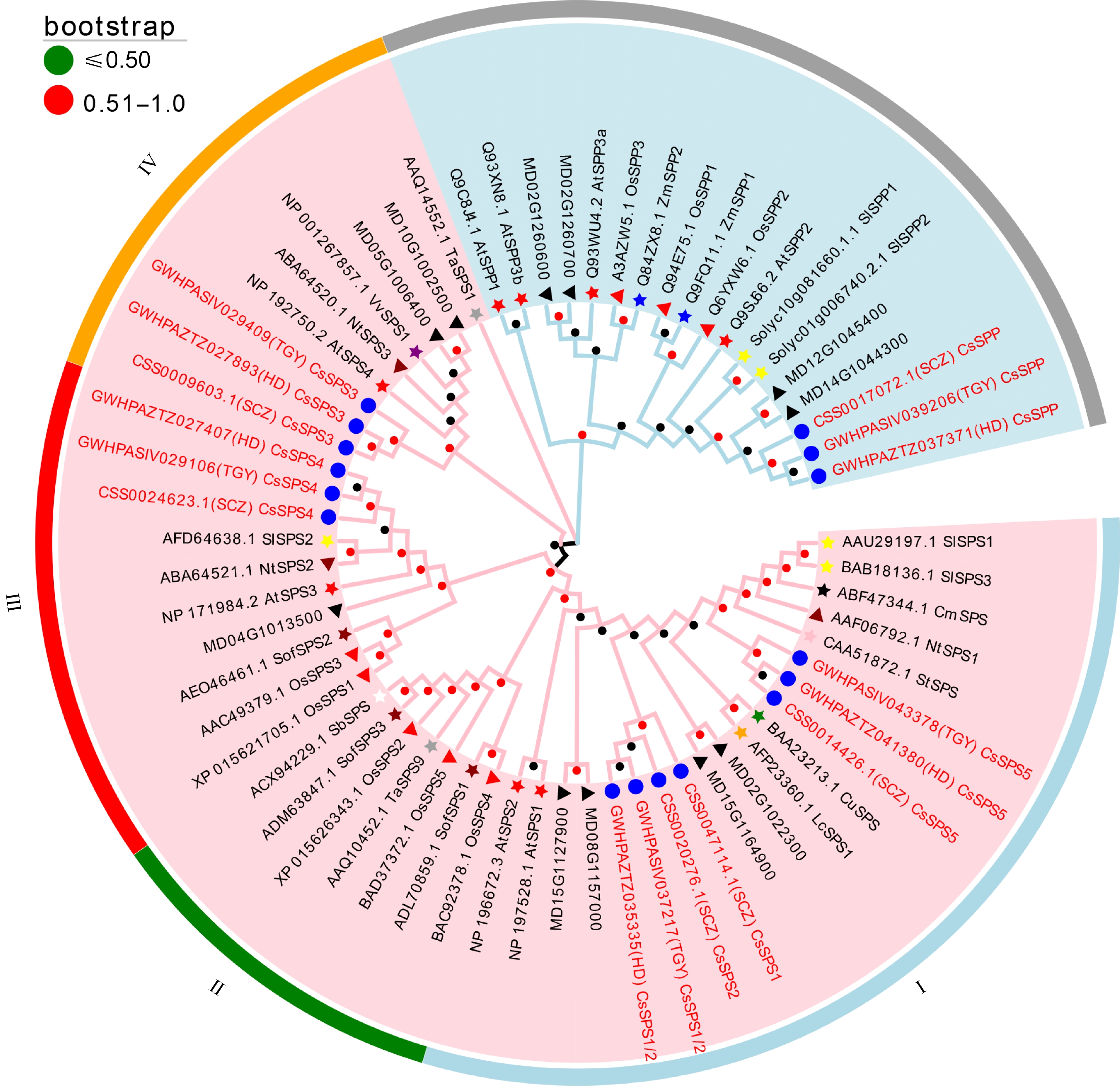
      Figure 1. 

      Phylogenetic analysis of SPPs and SPSs originating from 15 different plant species. Pink area: SPS family; Light blue area: SPP family. Blue circle: tea plant; red star: Arabidopsis; red triangle: rice; blue star: maize; yellow star: tomato; dark red star: spinach; dark red triangle: tobacco; black star: melon; green star: citrus; purple star: grape; gray star: wheat; pink star: potato; orange star: litchi; white star: sorghum; black triangle; apple. Bootstrap values of all branches are above 50%.

    • The chromosomal distribution of CsSPP and CsSPSs in three tea plant genomes was predicted and visualized by TBtools software. As shown in Fig. 2a and Supplemental Fig. S2, CsSPSs and CsSPP shared same chromosomal distribution in these three tea plant genomes, respectively. In detail, CsSPP located on Chr13, CsSPS1 and CsSPS2 co-located on Chr12, CsSPS3 and CsSPS4 co-located on Chr9, and CsSPS5 located on Chr14.
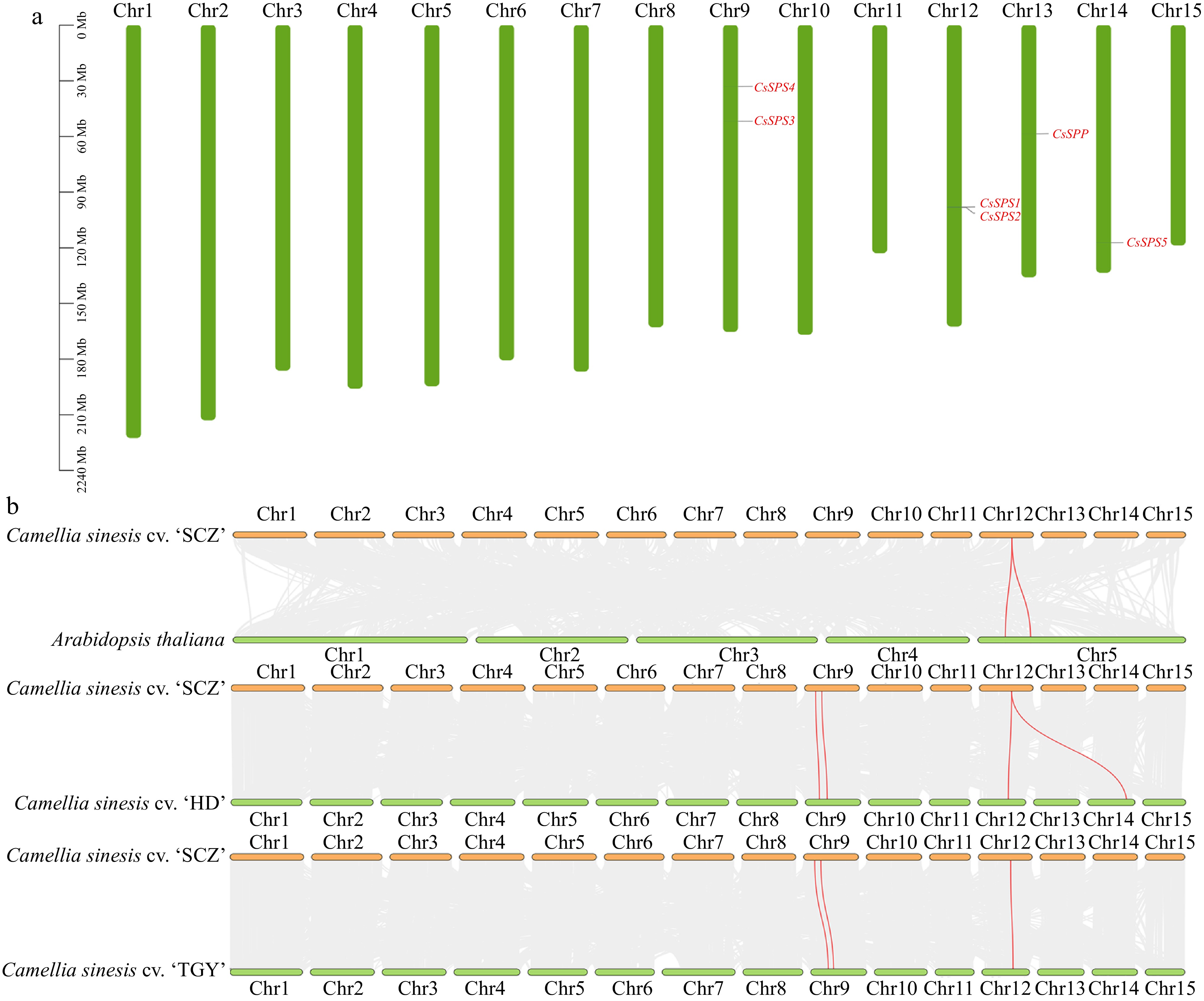
      Figure 2. 

      Chromosomal location and collinearity analysis of CsSPP and CsSPSs. (a) Chromosomal distribution of CsSPP and CsSPSs in 'Shuchazao' genome. (b) Interspecies synteny analysis of CsSPP and CsSPSs in 'Shuchazao' associated with Arabidopsis, 'Huangdan' and 'Tieguanyin' genomes.

      To further understand the evolutionary relationships of CsSPP and CsSPSs among different plant species, the inter-species collinearity relationships between 'SCZ' and 'HD', 'TGY' and Arabidopsis were constructed, respectively. As shown in Fig. 2b, both CsSPS1 and CsSPS2 belong to orthologous genes with AtSPS1 (NP197528.1) and AtSPS2 (NP196672.3) in Arabidopsis, HD.09G0012280.t1 and HD.12GOO24590.t1 in 'HD' cultivar, and TGY103558.t1 in 'TGY' cultivar. Besides, CsSPS3 and CsSPS4 are orthologous genes of HD.10G0021440.t1 and HD.10G0017080.t1 in 'HD' cultivar, and TGY080122.t1 and TGY081105.t1 in 'TGY' cultivar, respectively. These results also corresponded to the results of chromosome localization and phylogenetic analysis. In addition, the distribution and numbers of CsSPP and CsSPSs homologous genes in 'SCZ' genome were further explored through intra-special collinearity analysis, while there has no genome replication or fragment replication events occurred between CsSPSs and CsSPP (data not shown), indicating that CsSPSs and CsSPP are highly conserved in different tea plant cultivars.

    • To understand whether CsSPP and CsSPSs are involved in stress and hormone responses, the cis-acting elements contained in 2000-bp 5'-terminal untranslated region (UTR) sequences of CsSPP and CsSPSs were predicted. As shown in Fig. 3a, the type, number and distribution of cis-acting elements in UTR sequences of CsSPP and CsSPSs were varied among each other. Overall, all of them contain numerous light responsiveness related elements (data not shown). Besides, myeloblastosis (MYB) and myelocytomatosis (MYC) elements were also enriched in these promoter regions. Meanwhile, different numbers of anaerobic induction element (ARE) were also found in these promoters, indicating that CsSPSs and CsSPP play important roles in photosynthesis and respiration of tea plants. Besides, different numbers of hormone response elements, such as auxin-responsive element (TGA), MeJA-responsiveness (MeJA) element, abscisic acid responsiveness element (ABRE), and gibberellin (GA) element were predicted in these promoters, especially 3 MeJA elements were respectively enriched in the promoter regions of CsSPS2 and CsSPS4, suggesting their central roles in responding to hormones. Moreover, low-temperature response element (LTR) elements were enriched in the promoter regions of the CsSPP and CsSPS1/3/5, indicating these genes participate in cold stress response of tea plants. Furthermore, we found the numbers and types of cis-acting elements were most abundant in promoter of CsSPS4, which suggested that CsSPS4 may be widely involved in various stress responses of tea plants. In short, the above results showed that CsSPP and CsSPSs play important roles in mediating hormones and abiotic stress responses.

      Figure 3. 

      The cis-acting elements in the promoters of CsSPP and CsSPSs, and co-expression networks of CsSPP and CsSPSs. (a) cis-acting elements in promoters of CsSPP and CsSPSs. The heat map displays the type and number of cis-acting elements and the bar chart displays the number of cis-acting elements. MYB: myeloblastosis; MYC: myelocytomatosis; DSR: defense and stress responsiveness; LTR: low-temperature responsiveness; ABRE: abscisic acid responsiveness; GA: gibberellin-responsiveness; ARE: anaerobic induction; TGA: auxin-responsive element; MeJA: MeJA-responsiveness. (b) Co-expression networks of CsSPP and CsSPSs 3/4.

      Here, the co-expression networks of CsSPP and CsSPSs were also predicted with the help of the TeaCoN web server. As a result, only CsSPP, CsSPS3 and CsSPS4 predicted to contain 31, 70, and 110 co-expressed genes with strong associations (PCC-value > 0.7), respectively (Fig. 3b). Among them, most of the co-expressed genes of CsSPP are related to photosynthesis and respiration in plants. For example, a co-expressed gene of CsSPP, CSS0031288.1, encodes zeaxanthin epoxidase, which is involved in zeaxanthin synthesis and could adapt to different light intensity by controlling the amount of zeaxanthin accumulation in plant photosynthesis. Similar to CsSPP, the highly correlated genes of CsSPS3 were also related to photosynthesis and respiration. Besides, the expression profiles of two transcription factors, including CsMYB35 (CSS0014516) and CsGLOBOSA-like (CSS0022940), were highly correlated with CsSPS3, suggesting there may be a potential transcriptional regulatory relationship between them. Moreover, CSS0030453 (ATP synthase) and CSS0006328 (Peroxisomal membrane protein) are two highly correlated co-expressed genes in the co-expression network of CsSPS4. Among them, CSS0030453 plays an important role in cellular energy metabolism, plant photosynthesis and respiration, and CSS0006328 participates in scavenging free radicals.

    • The DNA structure analysis results showed that each of these genes contains more than 10 exons. Among them, CsSPP contains eight exons, CsSPS1 contains 14 exons, CsSPS3 contains 13 exons, CsSPS5 contains 11 exons, while CsSPS2 and CsSPS4 contain 12 exons, respectively (Fig. 4a). Based on the complex structures of these genes, we speculated that the functions of these genes may be irreplaceable in tea plants. Conserved motif analysis result showed that CsSPSs are highly conserved, and all of them contain 15 motifs except motif 14 which is missing in CsSPS3 (Fig. 4b). Besides, CsSPP is distinct from CsSPSs, indicating the different functions they played. This conclusion is further proved in Fig. 4c, where we found each CsSPS contains three conserved domains, including Sucrose_synth, Glycos_transf_1 and S6PP, while CsSPP contains the conserved S6PP and S6PP_C domains. In addition, the S6PP domain and the S6PP_C domain of CsSPP is closely connected. However, there are some amino acid sequences between the CsSPSs domains and a variable Linker between Glycos_transf_1 and S6PP. Moreover, some potential conserved serine phosphorylation sites were also identified in all five CsSPSs. Among them, two of the same phosphorylation sites, Ser191 and Ser385, were identified both in CsSPS1 and CsSPS2. Besides, Ser148 and Ser217 in CsSPS4, and Ser221 and Ser416 in CsSPS5 are also potential conserved phosphorylation sites, respectively. Moreover, three phosphorylation sites, Ser146, Ser220 and Ser409, were identified in CsSPS3. These results further confirmed that CsSPP and CsSPSs own Suc biosynthesis ability, and their activities are regulated by phosphorylation.
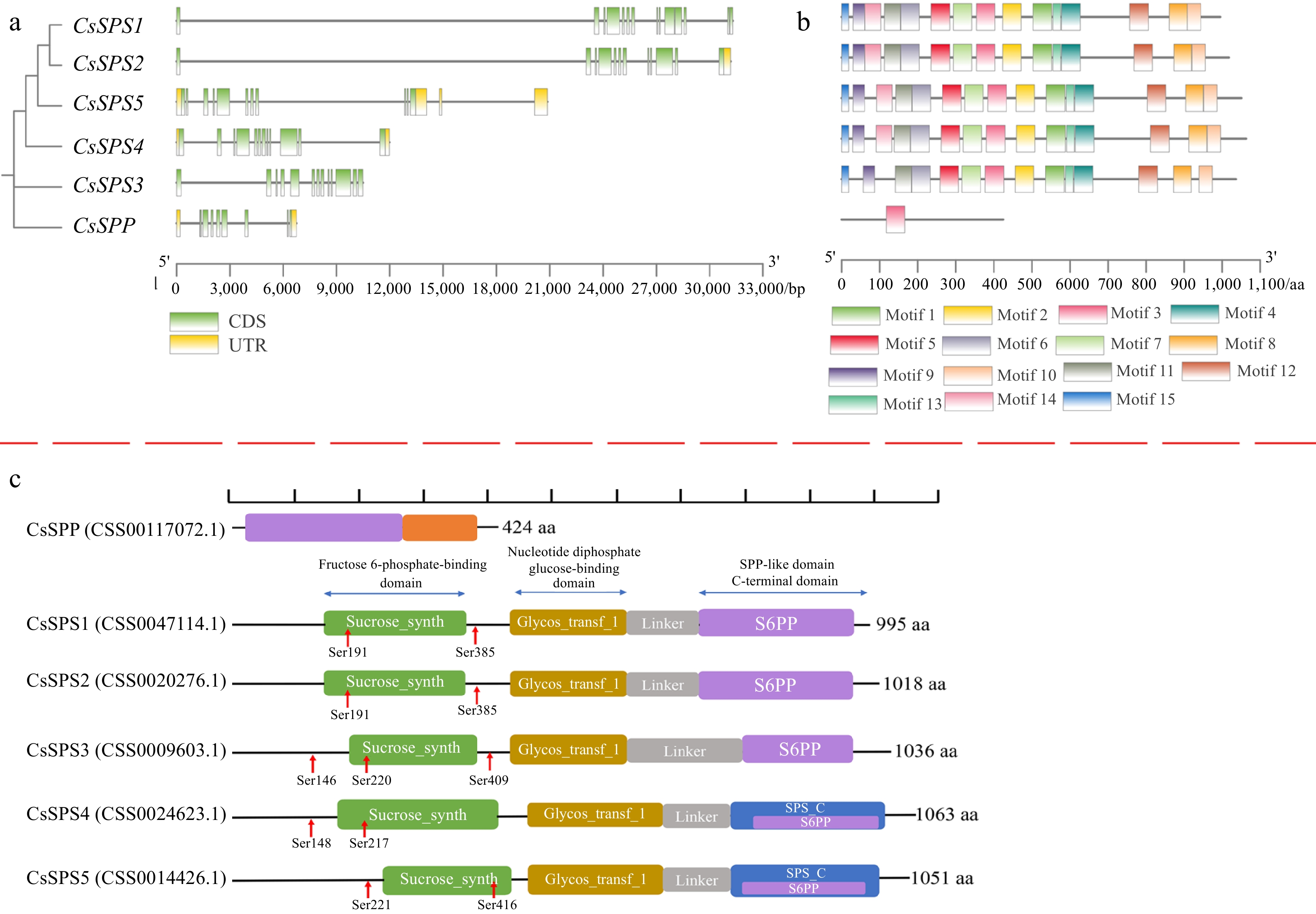
      Figure 4. 

      The exon-intron structures CsSPP and CsSPSs, conserved motifs and domains of CsSPP and CsSPSs. (a) The exon-intron structures CsSPP and CsSPSs. Green boxes represent exons, yellow boxes represent untranslated upstream/downstream regions, and lines indicate introns. (b) Conserved motifs of CsSPP and CsSPSs. Different motifs are presented by different colored squares. (c) Conserved domains of CsSPP and CsSPSs. Different domains are shown in different colors.

    • Tissue-specifics of CsSPSs and CsSPP were detected in nine different tissues of the 'SCZ' cultivar. As shown in Fig. 5, CsSPP and CsSPSs transcripts were detected in all tissues, but the transcription abundance of each gene varied among the detected tissues. Among them, the transcription abundance of CsSPP was highest in immature stem, while significantly lower in other tissues. Besides, all of CsSPSs showed highest transcription abundances in flower than that in other tissues, except for CsSPS2 that showed a similar expression level in senescent leaf. Meanwhile, all CsSPSs showed extremely low transcription abundances in root. In addition, the transcription abundance of CsSPS5 in each detected tissue was significantly higher than that in other CsSPSs, which speculated that CsSPS5 may play a leading role in Suc synthesis during the growth and development of tea plants. In brief, it followed that CsSPP and CsSPSs mediated entire vegetative and reproductive progress of tea plants. In particular, CsSPSs may play important roles in floral nectar production of flower, and CsSPP is necessary for the immature stem growth of tea plants.
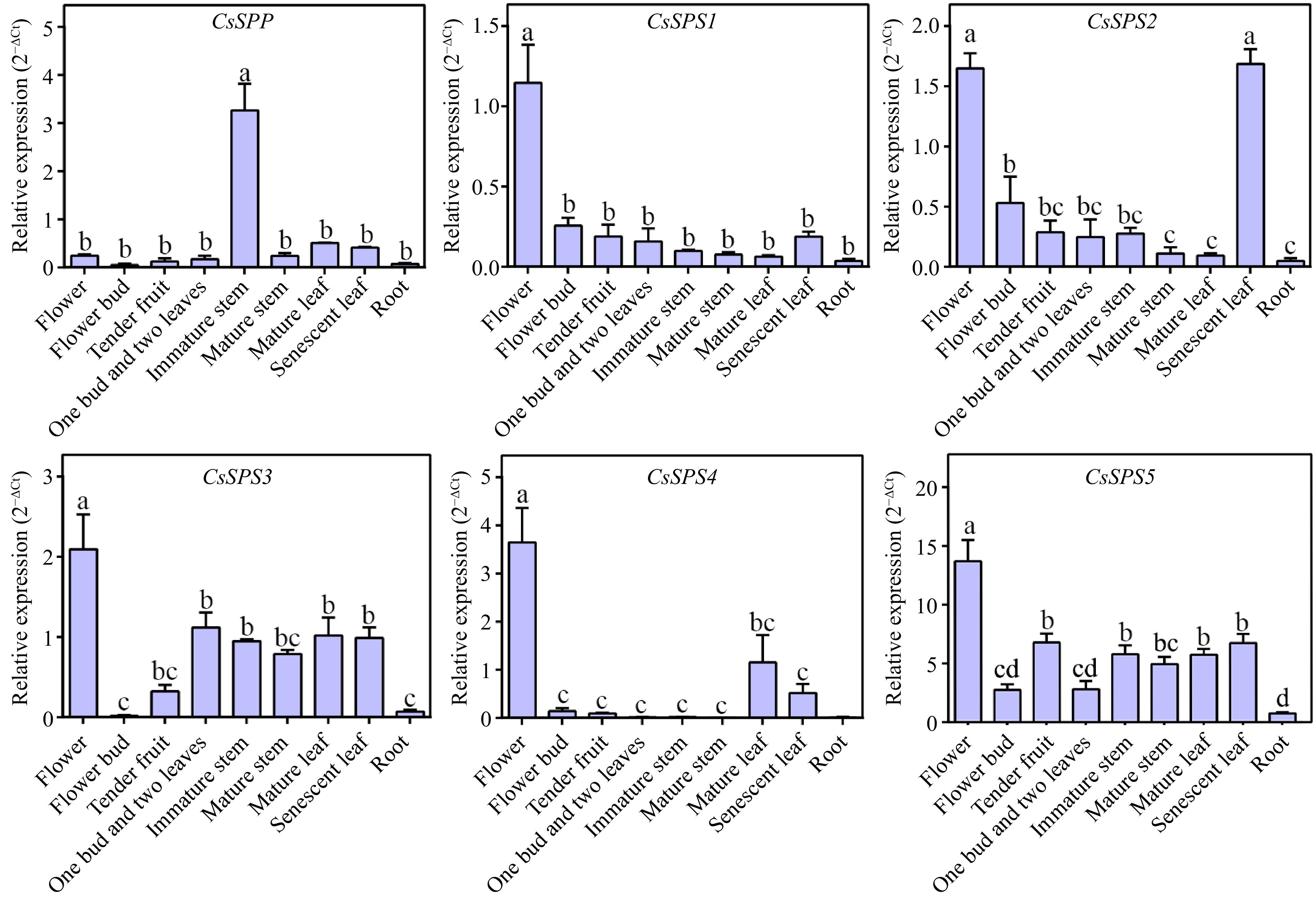
      Figure 5. 

      Tissue-specific analysis of CsSPP and CsSPSs in tea plant.

    • The spatial-temporal expression patterns of CsSPP and CsSPSs were analyzed under various abiotic stress conditions. As shown in Fig. 6, CsSPP and CsSPSs are differentially expressed under different stress conditions. Under salt treatment (ST) conditions, the expression of CsSPP was up-regulated nearly 3-fold after 1 d of ST, and then decreased when the treatment time continued. CsSPS4 was down-regulated within 2 d of ST. CsSPS2 was slightly up-regulated after 12 h and 48 h of ST, respectively, while down-regulated after 24 h of ST. Besides, CsSPS1/3/5 showed similar expression patterns under ST condition, which were highly induced within 12 h of ST, and then decreased when the treatment time continued. Under cold treatment (CT) condition, CsSPP and CsSPS1-3 showed similar expression patterns, all of them were down-regulated firstly within 12 h of CT, and then up-regulated by CT, of which CsSPS2/3 transcripts were respectively induced more than 3- and 2-fold after 48 h of CT as compared to 0 h. Besides, CsSPS4 was inhibited by CT within 2 d of CT, while CsSPS5 was continuously induced with the increased treatment time. Under drought treatment (DT) conditions, CsSPP and CsSPS2/4 showed similar expression patterns, all of them were constantly induced, and reached maximum expression levels after 24 h of DT, and then to some extent reduced. Similarly, CsSPS3/5 were constantly induced and reached maximum expression levels at 12 h. In addition, CsSPS1 was not significantly affected by DT. Briefly, CsSPP and CsSPSs participated in different stress responses of tea plants, but the time and degree of their effects were different.
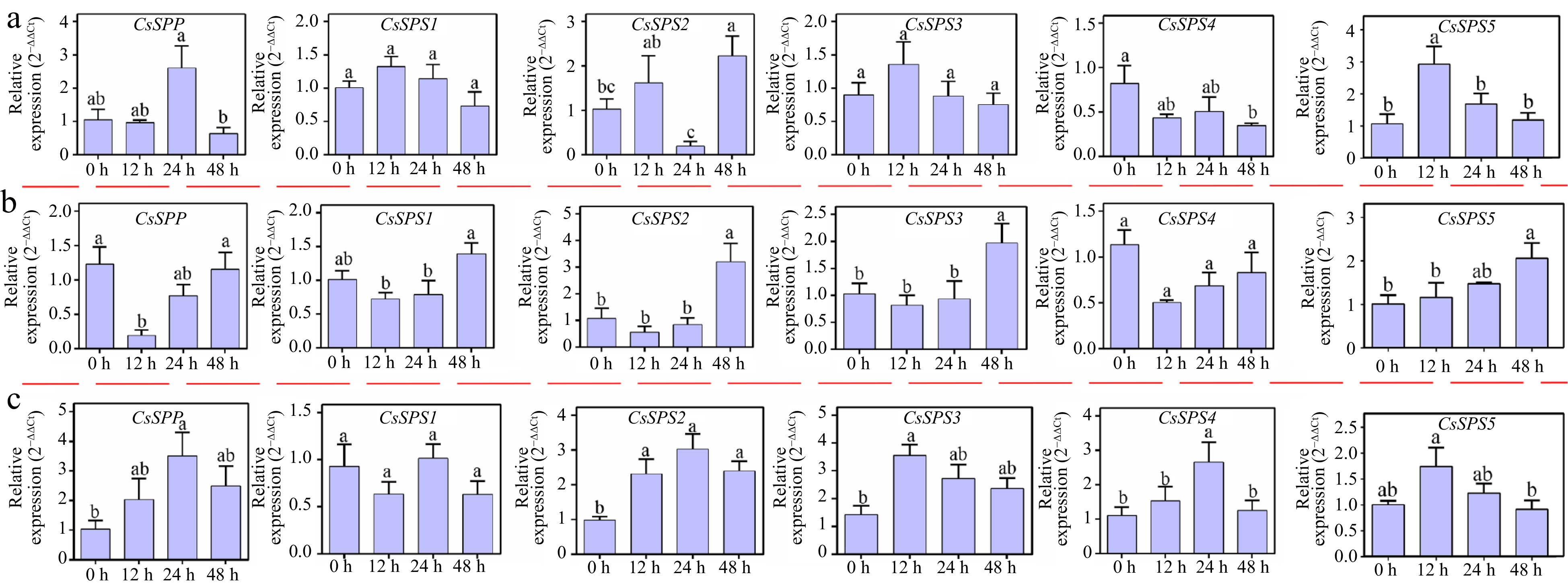
      Figure 6. 

      Expression analysis of CsSPP and CsSPSs under different abiotic stress conditions. (a) Expression profiles of CsSPP and CsSPSs under salt stress conditions. (b) Expression profiles of CsSPP and CsSPSs under cold stress conditions. (c) Expression profiles of CsSPP and CsSPSs under drought stress conditions.

    • In this study, the cold tolerance of two tea plant cultivars, 'SCZ' and 'BY1', were compared with different physiological indexes. As shown in Fig. 7a, the Fv/Fm and NP values of 'SCZ' cultivar were higher than that of the 'BY1' cultivar, but the EL value and MDA content in 'SCZ' cultivar were lower than that of the 'BY1' cultivar (Fig. 7a), indicating that the cold resistance of the 'SCZ' cultivar was higher than that of the 'BY1' cultivar. Therefore, these two cultivars were further used to investigate the relationships among the expression of CsSPSs and CsSPP, SPS activity, soluble sugar content and cold stress. As shown in Fig. 7b, except for CsSPS1, all of them were up-regulated under 0 oC treatment for 5 d both in these two cultivars, and returned to normal levels after 3 d of recovery. Specifically, CsSPP and CsSPS2/4/5 transcripts were increased more than 2-fold after 5 d of CT, respectively. Besides, we found the expression level of CsSPS2 was significantly higher in the 'BY1' cultivar than that in the 'SCZ' cultivar, while the expression level of CsSPS5 was significantly higher in the 'SCZ' cultivar than that in the 'BY1' cultivar, indicating the different roles of CsSPSs in coping with cold stress in different tea plant cultivars. Moreover, we found the SPS activity was obviously increased in the 'SCZ' cultivar, but not significantly changed in the 'BY1' cultivar after 5 d of CT. After 3 d of recovery growth, the SPS activity was decreased in the 'SCZ' cultivar, while significantly increased in the 'BY1' cultivar. Furthermore, the contents of TSS, Suc, Glc and Fru were increased after 5 d of CT except for TSS in the 'BY1' cultivar, and then decreased to normal levels after 3 d of NT. Furthermore, the 'BY1' cultivar contained a relatively higher content of TSS, Suc and Fru under CT and recovery conditions. As shown in Fig. 7c, CsSPP and CsSPS2-5 were positively correlated with SPS activity and the contents of soluble sugar (TSS, Suc, Glc and Fru) in the 'SCZ' cultivar, especially CsSPS2/4/5, significantly correlated with Suc content, respectively. Besides, CsSPS3 was also significantly and positively correlated with Glc and Fru contents in the 'SCZ' cultivar, respectively. These results indicated that the high expressions levels of CsSPP and CsSPSs contributed to the accumulation of different types of soluble sugars in the 'SCZ' cultivar, thus improving the adaptability to low temperatures. Different from the 'SCZ' cultivar, CsSPS1 was positively correlated with SPS activity in the 'BY1' cultivar. Meanwhile, although CsSPS2/4/5 were positively correlated with Suc, Glc, and Fru, all of them were negatively correlated with SPS activity, which suggested that SPS activity in the 'BY1' cultivar may be regulated by post-transcriptional and post-translational regulation, as some potential phosphorylation sites were identified in the amino acid sequences of CsSPSs (Fig. 4c). Different from the 'BY1' cultivar, the higher cold resistance of the 'SCZ' cultivar may be due to the up-regulated expression of CsSPSs leading to the increase of SPS activity, which promotes the synthesis of soluble sugar content to improve cold tolerance.
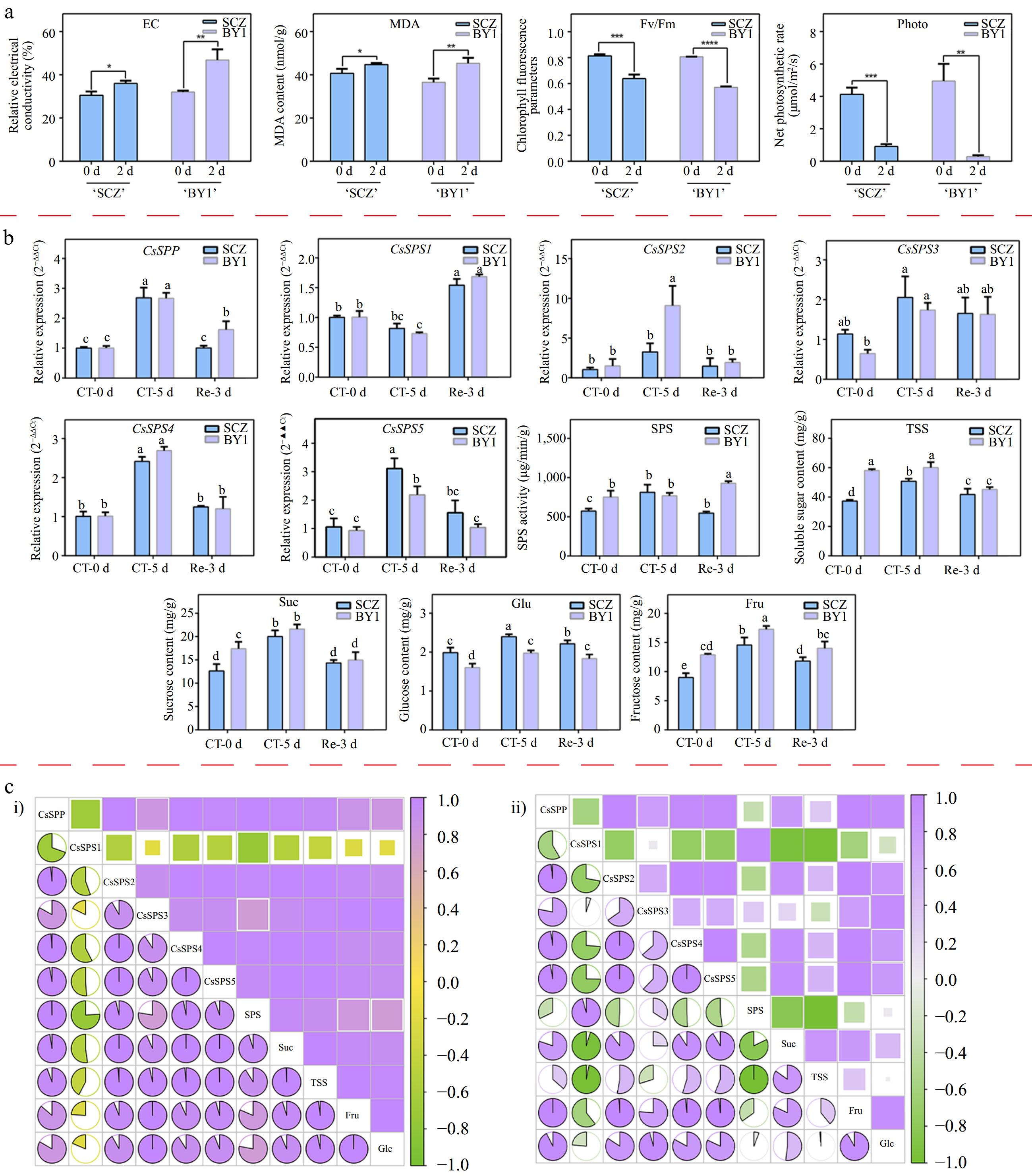
      Figure 7. 

      Expression analysis of CsSPP and CsSPSs in different tea plant cultivars under cold stress conditions. (a) Relative electrolytic leakage, malondialdehyde and photosynthetic parameters of 'Shuchazao' and 'Baiye1' under cold stress conditions. (b) Expression levels of CsSPP and CsSPSs, SPS activity and different types of soluble sugar content. (c) Correlation analysis of CsSPP and CsSPSs, SPS activity and different types of soluble sugar components in 'Shuchazao' and 'Baiye1' cultivars, respectively. (c-i) Correlation analysis of CsSPP, CsSPSs, SPS activity and soluble sugars in the 'Shuchazao' cultivar. (c-ii) Correlation analysis of CsSPP, CsSPSs, SPS activity and soluble sugars in the 'Baiye1' cultivar. Green color means negative correlation, purple color means positive correlation.

    • As the main product of photosynthesis, Suc plays an important role in plant growth and development, yield and quality formation, and stress responses. Previous studies found that Suc biosynthesis is mainly regulated by SPS and SPP[56]. In recent years, the protein structures and the roles of SPSs and SPPs have been explored in many plant species. Among them, SPSs mainly contain a D-fructose 6-phosphate (F6P)-binding domain, nucleotide diphosphate glucose (NDPGlc)-binding domain, and a SPP-related C-terminal domain[57]. Besides, SPS activity was regulated by multisite protein phosphorylation[6,58]. For example, Ser-158 may be mainly responsible for light/dark modulation, Ser-229 may be a binding site for 14-3-3 inhibitory proteins, and Ser-424 is thought to be responsible for osmotic stress activation of SPS[59,60]. In this study, five CsSPSs were identified from three tea plant genomes, among which CsSPS1 was a tandem duplication product of CsSPS2 in the 'SCZ' genome. All of these CsSPSs contain the conserved F6P-binding domain, NDPGlc-binding domain, and the SPP-related C-terminal domain (Fig. 4c), indicating that these CsSPSs possess the Suc biosynthesis ability. Meanwhile, some potential phosphorylation sites, such as Ser191 and Ser385 in CsSPS1/2, Ser146, Ser220 and Ser409 in CsSPS3, Ser148 and Ser217 in CsSPS4, and Ser221 and Ser416 in CsSPS5, were also identified (Fig. 4c), suggesting that SPS activity was also influenced by light, osmotic stress, and also 14-3-3 inhibitory proteins in tea plants.

      In addition to SPS, there are fewer SPPs than SPSs in most plant species, and some SPP tandem duplication events occurred in plants[61]. Previous study found that SPP protein structure mainly consists of S6PP (PF05116) and S6PP_C (PF08472) domain, and SPP depends on Mg2+ to specifically dephosphorylate S6P to produce Suc[62]. Besides, SPP formed homodimer in some vascular plants, and the molecular weight of the SPP monomer is usually 50 KDa, while the molecular weight of the homodimer is about 120 KDa[18,62,63]. Moreover, since SPS contains the SPP-related C-terminal domain, SPP and SPS could interact to improve the efficiency of Suc synthesis[64]. Maloney et al.[65] found that SPS could directly interact with SPP to affect the soluble carbohydrate pool and the allocation of carbon to starch. Meanwhile, co-overexpression of AtSPS-AtSPP and AtSPP-AtSPS chimera increased the content of soluble carbohydrates and also promoted the growth rates both in Arabidopsis and hybrid poplar, respectively[19]. In the present study, a unique and conserved CsSPP with 48.11 KDa MW was identified from three tea plant genomes. Meanwhile, both the conserved S6PP domain and S6PP_C domain were contained in CsSPP, suggesting that CsSPP participates in Suc biosynthesis of tea plants, and this process is highly conserved in different tea plant cultivars. In addition, CsSPP may also interact directly with CsSPSs to participate in Suc accumulation of tea plants due to the conserved SPP-related C-terminal domain observed in CsSPSs. However, this hypothesis will be further certificated in the future.

    • Previous studies stated that SPS and SPP widely participated in the flowering, plant growth, seed germination and pollen activity in plants[6668]. In tobacco, inhibition of NtSPP expression of tobacco significantly reduced the SPP activity, Suc and Hex contents, but dramatically increased starch content, and thus reducing the photosynthesis, chlorosis and growth rate of transgenic plants[21]. In this study, we found that the expressions of CsSPSs and CsSPP could be detected in all tissues, indicating that Suc is inseparable from all stages of vegetative and reproductive growth of tea plants. However, the expression levels of CsSPP and CsSPSs were lowest in roots, which indicated that Suc may be mainly synthesized from the source tissue of the above-ground part and then transported to the underground part. Besides, all CsSPSs showed the highest transcriptions in flower, which suggested that Suc plays an important role in floral nectar production of flower. However, our previous study found that Suc content of flowers was not the highest compared with other tissues, instead, the contents of TSS, Glc and Fru were highest in flowers[69]. Meanwhile, the vacuole INV activity and the transcription abundance of a vacuole INV gene (CsINV5), were highest in flower[24]. Here, we further performed the correlation analysis among the expressions of Suc-related genes (CsSPP, CsSPSs, and CsINV5), VIN activity and the contents of soluble sugars (TSS, Suc, Glc, and Fru) (Supplemental Fig. S3), where we found each component was positively correlated with each other except CsSPP, which speculated that the SPP/SPS-VIN module mediated the Suc metabolism in the flower of tea plants. In detail, after the biosynthesis of Suc by SPS and SPP in the flower of tea plants, part of the Suc needs to be further hydrolyzed by INV to form two monosaccharides, Glc and Fru, and then participates in floral nectar production, pollination, fertilization and fruit formation. In addition to mediating reproductive growth, previous studies reported that Suc accumulation might be regarded as one of the key indicators of leaf senescence[70,71]. A similar phenomenon was also found in our previous study, where we found Suc content was highest in senescent leaf compared with the other tissues[69], suggesting a great role of Suc in the aging process of tea plant leaves. Here, we further found the transcription abundance of CsSPS2 was higher in senescent leaf than the other tissues except for flower, indicating that CsSPS2 may be an important regulator of leaf senescence through mediating Suc accumulation in tea plants.

    • Many studies found that multiple carbohydrate metabolism-related genes involved in carbohydrate biosynthesis, hydrolysis, and transport were differentially expressed under stress conditions[67]. Besides, there is increasing evidence that Suc metabolism is one of the key regulatory systems that confer stress tolerance in plants[72,73]. Changes in the activity of SPS and SPP significantly influenced Suc accumulation, thus affecting plant growth and development and stress tolerance[7476]. In Arabidopsis, mutation of SPSA2 did not affect the seeds and plants of the mutant, but reduced the drought tolerance of the spsa2 mutant through the regulation of proline content, sugar accumulation and antioxidant response[77]. Under normal water condition, the reduction of SPS activity by 70%−80% resulted in a corresponding reduction of Suc synthesis by 30%−50%, while under water deficit condition, the reduction of SPS activity prevented dry-matter allocation to tubers, indicating that SPS is essential for adaptive changes in tuber metabolism and whole plant allocation process[74]. In terms of tea plant, previous studies revealed that carbohydrate metabolism plays an important role in cold[78,79], drought[80], and salt[81] stress responses. In particular, many sugar-related genes involved in cold acclimation of tea plant have been identified by Yue et al.[82]. Among them, CsINVs (e.g., CsINV2/5/10)[24,25], CsSWEETs (e.g., CsSWEET1a/16/17)[27,83], and CsHXKs (e.g., CsHXK3/4)[26] have been further demonstrated to be involved in the cold response of tea plants through Suc hydrolysis, sugar transport and sugar signaling, respectively. In this study, we further found the expressions of CsSPP and CsSPSs were induced by cold, drought and salt stresses at different treatment time points, respectively, indicating that SPP and SPS positively modulate abiotic stress responses of tea plants. Besides, CsSPP and CsSPSs differentially expressed in two tea plant cultivars with different cold tolerance under cold stress condition. Interestingly, we found SPS activity was higher in the 'BY1' cultivar under normal temperature conditions, while slightly lower under low temperature conditions compared with the 'SCZ' cultivar. Meanwhile, higher soluble sugar content except Glc found in 'BY1' cultivar both under normal and low temperature conditions. However, from these results, we found that the SPS activity and soluble sugar content of the 'SCZ' cultivar increased significantly more than that of the 'BY1' cultivar, indicating that the Suc synthesis ability of the 'SCZ' cultivar through photosynthesis was higher than that of the 'BY1' cultivar, and the low temperature adaptability of the 'SCZ' cultivar was higher than that of the 'BY1' cultivar. Based on the above studies, it can be seen that the increase of SPP and SPS activities in tea plant under stress conditions can promote Suc synthesis. Subsequently, partial Suc is transported to vacuoles, cell walls and other sub-organelles by sugar transporters, and is hydrolyzed by INV to form monosaccharides. Monosaccharides can be further phosphorylated by HXK to participate in the synthesis of other substances (e.g., inositol, trehalose and mannitol); finally, these synthetic Suc and monosaccharides participate in the stress responses of tea plants with carbon sources, osmoprotectants, reactive oxygen scavengers and sugar signaling molecules.

      Although SPSs and SPPs are known to be involved in various stress responses, their transcriptional and post-translational regulation mechanisms have been poorly studied. As is mentioned above, the phosphorylation levels of SPSs were affected by different environmental factors, among which Ser158, Ser229 and Ser424 were three important phosphorylation sites[59,60,84]. Besides, previous study found that SPS was also phosphorylated by calcium-dependent protein kinase (CDPK) via calcium signaling pathway. In detail, the encoding protein of a cold-reduced gene, OsCPK17, could directly phosphorylate OsSPS4 in rice. Under low temperature conditions, the phosphorylation level of Ser170 residue in OsSPS4 was higher in wild type plant than that in oscpk17 mutant, indicating that the reduction of OsSPS4 activity possibly regulated by OsCPK17 through directly phosphorylating OsSPS4 during the early stages of cold stress[85]. In tea plant, a previous study found that CsCDPKs play important roles in coping with various stresses, among which CsCPK4/5/9/30 may be the main cold regulators of tea plants[86]. Combined with the expression profiles and the conserved Ser residues of CsSPSs, we speculated that SPS activity may also be regulated by CDPK phosphorylation, and participate in stress response through the calcium signaling pathway. On the other hand, there are few studies on the transcriptional regulation of SPSs and SPPs. Recently, seven GmSPSs genes were identified from Glycine max, and all of them were up-regulated by cold stress in soybean leaves, especially GmSPS8 and GmSPS18. Promoter analysis found that many potential inducers of CBF expression 1 (ICE1) binding sites were predicted in the promoter regions of GmSPSs. Electrophoretic mobility shift assay (EMSA) further proved that GmICE1 could regulate the transcription abundances of GmSPS8 and GmSPS18 in N. benthamiana[87]. In the present study, in addition to stress-related cis-acting elements, many hormone-related cis-acting elements, such as ABRE, GA, MeJA, and TGA, were contained in the promoter regions of CsSPSs and CsSPP, suggesting that CsSPPs and CsSPP participate in development and stress response of tea plant via hormone-signaling pathway. In particular, the promoter region of CsSPS4 contains three MeJA, two ABRE, one GA, and one TGA, which indicated that CsSPS4 may be a central component in the cascade of sugar signaling and hormone signaling in tea plants. Moreover, co-expression analysis results found that the expression profiles of CsMYB35 (CSS0014516) and CsGLOBOSA-like (CSS0022940) were positively correlated with CsSPS3, suggesting that CsMYB35 and CsGLOBOSA-like may be two candidate regulators of CsSPS3. However, the function and the regulation mechanism of these two transcription factors need to be further explored in the future.

    • In this study, a unique CsSPP and five CsSPSs genes were identified from three tea plant genomes. Bioinformatic analysis results showed that CsSPP and CsSPSs were highly conserved in different tea plant cultivars respectively, and all of them can participate in Suc biosynthesis in the cytoplasm of tea plants. Tissue-specific analysis found that CsSPP and CsSPSs are necessary for vegetative and reproductive growth of tea plants, especially for the floral nectar production of flower. In addition, CsSPP and CsSPSs were differentially expressed under various abiotic stress conditions, among which CsSPS2/3/5 were induced by cold, drought and salt stress treatments at different treatment time points. Under cold stress conditions, the SPS activity and the soluble sugar contents of the 'SCZ' cultivar increased more than that of the 'BY1' cultivar, indicating that the 'SCZ' cultivar owns higher photosynthetic capacity and Suc synthesis ability under cold stress conditions. This study will provide theoretical foundation for further exploring the function of SPP and SPS involved in abiotic stress responses of tea plants.

    • The authors confirm contributions to the paper as follows: study conception and design: Qian W, Ikka T; material preparation and data collection: Liang S, Lang X, Yue J, He S; data analysis: Liang S, Zhang S, Wang H, Fan K; draft manuscript preparation: Liang S, Qian W, Wang H; review and editing: Wang Y, Ding Z, Yamashita H, Ikka T; Partial funds and consultation: Qian W, Wang Y. All authors read and approved the final manuscript.

    • The datasets generated during and/or analyzed during the current study are available from the corresponding author on reasonable request.

      • This research was supported by the National Natural Science Foundation of China (32272767, 31800588), the Shandong Agricultural Elite Variety Project (2020LZGC010), and 'Provincial and School Joint Training Program' for Government-sent Overseas Visiting Scholars of Shandong Province in 2020.

      • The authors declare that they have no conflict of interest.

      • Copyright: © 2024 by the author(s). Published by Maximum Academic Press, Fayetteville, GA. This article is an open access article distributed under Creative Commons Attribution License (CC BY 4.0), visit https://creativecommons.org/licenses/by/4.0/.
    Figure (7)  Table (1) References (87)
  • About this article
    Cite this article
    Liang S, Wang H, Yamashita H, Zhang S, Lang X, et al. 2024. Genome-wide identification and expression analysis of sucrose phosphate synthase and sucrose-6-phosphate phosphatase family genes in Camellia sinensis. Beverage Plant Research 4: e015 doi: 10.48130/bpr-0024-0007
    Liang S, Wang H, Yamashita H, Zhang S, Lang X, et al. 2024. Genome-wide identification and expression analysis of sucrose phosphate synthase and sucrose-6-phosphate phosphatase family genes in Camellia sinensis. Beverage Plant Research 4: e015 doi: 10.48130/bpr-0024-0007

Catalog

  • About this article

/

DownLoad:  Full-Size Img  PowerPoint
Return
Return