REVIEW   Open Access    

Genetic transformation in conifers: current status and future prospects

More Information
  • Received: 11 December 2023
    Revised: 30 January 2024
    Accepted: 28 February 2024
    Published online: 21 March 2024
    Forestry Research  4 Article number: e010 (2024)  |  Cite this article
  • Genetic transformation has been a cornerstone in plant molecular biology research and molecular design breeding, facilitating innovative approaches for the genetic improvement of trees with long breeding cycles. Despite the profound ecological and economic significance of conifers in global forestry, the application of genetic transformation in this group has been fraught with challenges. Nevertheless, genetic transformation has achieved notable advances in certain conifer species, while these advances are confined to specific genotypes, they offer valuable insights for technological breakthroughs in other species. This review offers an in-depth examination of the progress achieved in the genetic transformation of conifers. This discussion encompasses various factors, including expression vector construction, gene-delivery methods, and regeneration systems. Additionally, the hurdles encountered in the pursuit of a universal model for conifer transformation are discussed, along with the proposal of potential strategies for future developments. This comprehensive overview seeks to stimulate further research and innovation in this crucial field of forest biotechnology.
  • The competition for consumer preference for fresh apples (Malus domestica) from exotic and tropical fruits is intense. Red-fleshed (RF) apple may not only provide a novel point of differentiation and enhanced visual quality, but also a source of increased concentration of potentially health-benefiting compounds within both the fresh fruit and snack/juice markets[1]. Two different types of RF apples have been characterised: Type 1 RF apple has red colouration not only in the fruit core and cortex, but also in vegetative tissues, including stems and leaves; Type 2 RF apples display red pigment only in the fruit cortex[1, 2]. To facilitate trade and lengthen the supply-window, harvested fruit are usually cold stored, which can induce a series of disorders, including physiological breakdown manifesting as a flesh browning disorder (FBD) in RF apples[3, 4]. FBD in RF apples can be caused by senescence, and there is also some evidence to suggest that a large proportion of RF apples are chilling-sensitive (Jason Johnston, Plant & Food Research Hawke's Bay, personal communication).

    Earlier studies suggested that Type 1 RF colour was determined by a promoter mutation of MdMYB10 that has a tandem replication of a myeloblastosis (MYB) binding cis-element (R6) within the promoter, resulting in autoregulation of MdMYB10[5]. However, van Nocker et al.[6] observed a large variation in the degree and pattern of red pigmentation within the cortex among the accessions carrying MdMYB10, and concluded that the presence of this gene alone was not sufficient to ensure the RF colour. A genome-wide association study (GWAS)[3], reported that, in addition to the MdMYB10 gene, other genetic factors (e.g. MdLAR1, a key enzyme in the flavonoid biosynthetic pathway) were associated with RF colour, too. Wang et al.[7] reported that many of the up-regulated genes in RF apples were associated with flavonoid biosynthesis (e.g., chalcone synthase (CHS), chalcone isomerase (CHI), dihydroflavonol 4-reductase (DFR), anthocyanin synthase (ANS), UDP-glucosyltransferase (UGT) and MYB transcription factors). Recently, MdNAC42 was shown to share similar expression patterns in RF fruit with MdMYB10 and MdTTG1, and it interacts with MdMYB10 to participate in the regulation of anthocyanin synthesis in the RF apple Redlove®[8].

    Several transcription factor genes (e.g., MYB, WRKY, bHLH, NAC, ERF, bZIP and HSF) were reported to be differentially expressed during cold-induced morphological and physiological changes in 'Golden Delicious' apples[9]. A study by Zhang et al.[10] showed that ERF1B was capable of interacting with the promotors of anthocyanin and proanthocyanidin (PA) MYB transcription factors, and suggested that ethylene regulation and anthocyanin regulation might be linked in either direction. It was reported that ethylene signal transduction pathway genes or response genes, such as ERS (ethylene response sensor), EIN3 (ethylene-insensitive3) and ERFs (ethylene response factors), may play an important role in the regulatory network of PA biosynthesis[11].

    Espley et al.[12] observed no incidence of FBD in cold-stored fruit of 'Royal Gala', but over-expression of MdMYB10 in 'Royal Gala' resulted in a high rate of FBD in RF fruit, which was hypothesised to be caused by elevated fruit ethylene concentrations before harvest and more anthocyanin, chlorogenic acid (CGA) and pro-cyanidins in RF fruit. In addition, the MYB10 transcription factor was shown to elevate the expression levels of MdACS, MdACO, and MdERF106 ethylene-regulating genes[12]. To elucidate the mechanism regulating the FBD of RF apples, Zuo et al.[13] analysed the transcriptome of tree-ripe apples at 0, 0.5 and 4 h after cutting, and reported that the differentially expressed genes at different sampling points were mainly related to plant–pathogen interactions.

    GWAS is a powerful technique for mining novel functional variants. One of the limitations of GWAS, using SNP arrays, is that they require genotyping of large numbers of individuals, which may be expensive for large populations. DNA pooling-based designs (i.e., bulk segregant analysis) test differential allele distributions in pools of individuals that exhibit extreme phenotypes (XP) in bi-parental populations, large germplasm collections or random mating populations[1416]. In addition to reducing the number of samples to be genotyped, the use of whole genome sequencing (WGS)-based XP-GWAS has the potential to identify small-effect loci and rare alleles via extreme phenotypic selection.

    In this WGS-based XP-GWAS, we investigated the genetic basis of RF and FBD by sequencing the pools of individuals that exhibited extreme phenotypes for these two traits, and analysed the differences in allele frequencies between phenotypic classes. This method combines the simplicity of genotyping pools with superior mapping resolution. We also examined the transcriptome from transgenic apple fruit harbouring the R6:MYB10 promoter as a model for red flesh in apple. Differences in gene expression of a highly pigmented line were compared with expression in control fruit and these genes were then used for comparison with the seedling population. Understanding the genetic basis of the link between RF and FBD will help in design of strategies for selection against FBD in high-quality Type 1 RF apple cultivars.

    A snapshot of visual variation in FBD and WCI is presented in Fig. 1. The average WCI and FBD across all ~900 seedlings ranged from 0 to 7, and from 0% to 58%, respectively. Based on the MLM analysis, the estimated narrow-sense heritability (h2) of WCI and FBD was 0.57 (standard error = 0.18) and 0.09 (standard error = 0.05), respectively. The estimated genetic correlation between WCI and FBD was 0.58, and several fruit quality traits displayed unfavourable correlations with WCI (Supplemental Fig. S1). Seedlings with higher WCI scores were generally characterised by poor firmness and crispness, plus higher astringency and sourness. Estimated phenotypic and genetic correlations between all pairs of traits are listed in Supplemental Table S1.

    Figure 1.  Transverse cross-sections of apple slices showing range in (a) flesh colouration, and (b) flesh browning disorder for Type 1 red-fleshed apple. The weighted cortical intensity (WCI) scores (0−9 scale) and the proportion of the cortex area showing symptoms of flesh browning disorder are also displayed.

    A few seedlings had no red pigment in the cortex, but the average WCI score across all seedlings was 2.25. About two-thirds of the seedlings did not display any FBD symptoms, but among the remaining seedlings, FBD ranged between 1% and 58% (Fig. 2). The average WCI score for the 'low' and 'high' WCI pool was 0.45 and 5.2, respectively, while the average FBD was 0% and 20.6% for the 'low' and 'high' FBD pool, respectively (Supplemental Table S2). The average WCI score of the seedlings in the FBD pools was similar (Low: 5.5; High: 4.6), while the average FBD of the high- and low-WCI pools was 4.5% and 0.1%, respectively.

    Figure 2.  The distribution of weighted cortex intensity (WCI) scores (a) and the internal flesh browning disorder IFBD%; (b) in the population of ~900 apple seedlings. The green and red circles highlight the individuals used to form the 'low' and 'high' pools of samples.

    After filtering, about 204,000 SNPs were used and the average sequencing depth of SNP loci was similar for the two pools (42 vs 44). There was a near-perfect correlation between the Z-test statistics and G-statistics, so only the latter are discussed hereafter. A plot of the G' values, smoothed over 2 Mb windows, is shown for all 17 chromosomes (Chrs) in Supplemental Fig. S2. XP-GWAS identified genomic regions significantly associated with FBD on 12 out of the 17 Chrs (Fig. 3), and putative candidate genes within ±1.0 Mb distanceof the significant G' peaks were identified (Table 1). Additional genomic regions, which did not meet the significance threshold but displayed distinguished G' peaks, were also identified across all chromosomes (Supplemental Table S3).

    Figure 3.  G'-statistics across the linkage groups (LG) showing significant association with the flesh browning disorder (FBD) in apple The horizontal red lines indicate the significance threshold. The putative candidate genes (refer to Table 1) underpinning various G' peaks are also shown.
    Table 1.  A list of the genomic regions associated with internal flesh browning disorder (FBD) in apples. Putative candidate genes residing within these regions are also listed using GDDH13v1.1 reference genome assembly.
    ChrGenomic region (Mb)Putative genes functions
    221.9–23.5Ethylene-responsive element binding factor 13 (MdERF13: MD02G1213600);
    33.1–4.9cinnamate 4-hydroxylase (C4H) enzyme (MD03G1051100, MD03G1050900 and MD03G1051000); MdWRKY2: MD03G1044400; MdWRKY33 (MD03G1057400)
    38.2–9.6ascorbate peroxidase 1 (MdAPX1: MD03G1108200, MD03G1108300)
    314.7–16.6senescence-related MdNAC90 (MD03G1148500)
    336.5–37.5Ethylene response sensor 1 (MdERS1: MD03G1292200); flavonoid biosynthesis protein MdMYB12 (MD03G1297100); Heat shock protein DnaJ (MD03G1296600, MD03G1297000); pectin methylesterase (MdPME) inhibitor protein (MD03G1290800, MD03G1290900, MD03G1291000).
    411.0–13.0phenylalanine and lignin biosynthesis protein MdMYB85 (MD04G1080600)
    422.1–24.4MYB domain protein 1 (MD04G1142200); HSP20-like protein (MD04G1140600); UDP-glucosyltransferase (UGT) proteins UGT85A7 (MD04G1140700, MD04G1140900); UGT85A3 (MD04G1140800); UGT (MD04G1141000, MD04G1141300); UGT85A2 (MD04G1141400); UGT85A4 (MD04G1141500); DNAJ heat shock protein (MD04G1153800, MD04G1153900, MD04G1154100)
    427.6–29.6HCT/HQT regulatory genes MD04G1188000 and MD04G1188400
    629.8–31.7Volz et al. (2013) QTL for IFBD; anthocyanin regulatory proteins MdMYB86 (MD06G1167200); triterpene biosynthesis transcription factor MdMYB66 (MD06G1174200); Cytochrome P450 (MD06G1162600; MD06G1162700, MD06G1162800, MD06G1163100, MD06G1163300, MD06G1163400, MD06G1163500, MD06G1163600, MD06G1163800, MD06G1164000, MD06G1164100, MD06G1164300, MD06G1164400, MD06G1164500, MD06G1164700)
    713.9–15.9heat shock protein 70B (MD07G1116300)
    717.4–19.0Drought-stress WRKY DNA-binding proteins (MdWRKY56: MD07G1131000, MD07G1131400)
    723.6–25.2MdPAL2 (MD07G1172700); drought-stress gene NGA1 (MD07G1162400); DNAJ heat shock family protein (MD07G1162300, MD07G1162200), stress-response protein (MdNAC69: MD07G1163700, MD07G1164000)
    97.9–11.8MD09G1110500 involved in ascorbate oxidase (AO); MdUGT proteins (MD09G1141200, MD09G1141300, MD09G1141500, MD09G1141600, MD09G1141700, MD09G1141800, MD09G1142000, MD09G1142500, MD09G1142600, MD09G1142800, MD09G1142900, MD09G1143000, MD09G1143200, MD09G1143400) involved in flavonoids biosynthesis; heat shock proteins 89.1 (MD09G1122200) and HSP70 (MD09G1137300); Ethylene-forming enzyme MD09G1114800; Anthocyanin regulatory protein MdNAC42 (MD09G1147500, MD09G1147600)
    914.9–16.5triterpene biosynthesis transcription factor protein MdMYB66 (MD09G1183800);
    111.7–3.0ethylene response factor proteins (MdEIN-like 3: MD11G1022400)
    1138.6–40.6Senescence-related gene 1 (MD11G1271400, MD11G1272300, MD11G1272000, MD11G1272100, MD11G1272300, MD11G1272400, and MD11G1272500); Chalcone-flavanone isomerase (CHI) protein (MD11G1273600) and MdbHLH3 (MD11G1286900, MDP0000225680); cytochrome P450 enzyme (MD11G1274000, MD11G1274100, MD11G1274200, MD11G1274300, MD11G1274500, and MD11G1274600); heat shock transcription factor A6B (MdHSFA6B: MD11G1278900) – involved in ABA-mediated heat response and flavonoid biosynthesis.
    122.2–3.5heat shock protein 70-1 ((MD12G1025600, MD12G1025700 and MD12G1026300) and heat shock protein 70 (MD12G1025800 and MD12G1025900 and MD12G1026000); ethylene (MD12G1032000) and auxin-responsive (MD12G1027600) proteins.
    127.2–8.4DNAJ heat shock domain-containing protein (MD12G1065200 and MD12G1067400)
    1327.5–30.5MD13G1257800 involved in polyphenol 4-coumarate:CoA ligase (4CL)
    1337.5–39.5pectin methyl esterase inhibitor superfamily protein MdPMEI (MD13G1278600)
    1425.4–27.5Drought-stress response gene MdWRKY45 (MD14G1154500); chalcone synthase (CHS) family proteins (MD14G1160800 and MD14G1160900); triterpene biosynthesis transcription factor MdMYB66 (MD14G1180700, MD14G1181000, MD14G1180900); anthocyanin biosynthesis protein (MdMYB86: MD14G1172900); cytochrome P450 proteins (MD14G1169000, MD14G1169200, MD14G1169600, MD14G1169700)
    150–1.5Ethylene synthesis proteins (MD15G1020100, MD15G1020300 and MD15G1020500); dihydroflavonol reductase (DFR) gene (MD15G1024100)
    154.6–6.8MdMYB73 (MD15G1076600, MD15G1088000) modulates malate transportation/accumulation via interaction with MdMYB1/10; MdNAC52: MD15G1079400) regulates anthocyanin/PA; heat shock transcription factor B4 (MD15G1080700); stress-response protein MdWRKY7 (MD15G1078200)
    1553.5–54.9MdC3H (MD15G1436500) involved in chlorogenic acid biosynthesis; MdEIN3 (MD15G1441000) involved in regulating ethylene synthesis and anthocyanin accumulation
    168.9–10.9SAUR-like auxin-responsive protein (MD16G1124300) and MdNAC83: (MD16G1125800) associated with fruit ripening; MD16G1140800 regulates proanthocyanidin; MdPAE genes (MD16G1132100, MD16G1140500) regulates ethylene production.
     | Show Table
    DownLoad: CSV

    The ethylene-responsive factor 13 (MdERF13: MD02G1213600) resided within the significant region (21.9–23.5 Mb) on Chr2, while the ascorbate peroxidase 3 (MdAPX3: MD02G1127800, MD02G1132200) resided in the prominent region between 9.45 and 11.28 Mb) (Fig. 3, Table 1). There were several genomic regions showing association with FBD on Chr3. The first region (8.2–9.5 Mb) flanked MdAPX1 (MD03G1108200, MD03G1108300), while MdERF3 (MD03G1194300) and heat shock protein 70 (HSP70: MD03G1201800, D03G1201700) resided within the prominent peak region (25.5–27.5 Mb). Another significant region (36.5–37.5 Mb) at the bottom on Chr3 harboured ethylene response sensor 1 (MdERS1: MD03G1292200), a flavonoid-biosynthesis related protein MdMYB12 (MD03G1297100), HSP DnaJ and pectin methyl esterase (PME) inhibitor proteins (Fig. 3, Table 1).

    The significant G' region (27.6–29.6 Mb) on Chr4 flanked the genes MD04G1188000 and MD04G1188400 involved in the biosynthesis of hydroxycinnamoyl CoA shikimate/quinate hydroxycinnamoyl transferase (HCT/HQT), while the adjacent region (22.1–24.3 Mb) harboured a cluster of UDP-glucosyltransferase (UGT) proteins and HSP (Table 1). Another region between 11.0 and 13.0 Mb encompassed phenylalanine and lignin biosynthesis gene MYB85 (MD04G1080600)[17]. The only significant region associated with FBD on Chr6 spanned between 29.8 and 31.7 Mb, which included a SNP earlier reported associated with FBD[4]. This region also harboured several MYB proteins (MdMYB86: MD06G1167200; MdMYB98: MD06G1172900; MdMYB66: MD06G1174200) and a large cluster of cytochrome P450 proteins (Table 1).

    A significant FBD-associated region (13.9–15.9 Mb) on Chr7 encompassed the HSP 70B (MD07G1116300), while another significant region between 23.6 Mb and 25.2 Mb flanked the gene coding for phenylalanine ammonia lyase 2 (MdPAL2: MD07G1172700), a drought stress gene NGA1 (MD07G1162400), DNAJ HSP, and stress-response protein (MdNAC69: MD07G1163700, MD07G1164000) (Fig. 3, Table 1). A large significant region spanning between 7.9 and 11.8 Mb on Chr9 encompassed the gene MD09G1110500 putatively involved in ascorbate oxidase (AO), HSP (HSP70: MD09G1137300; HSP89.1: MD09G1122200), an ethylene-forming enzyme (MD09G1114800), and MdNAC42 (MD09G1147500, MD09G1147600). Another significant genomic region on Chr9 was between 14.9 and 16.5 Mb, which harbours the MYB domain protein MdMYB66 (MD09G1183800) (Table 1).

    A sharp G' peak region (1.7–3.0 Mb) on Chr11 associated with FBD encompassed ethylene insensitive 3 (MdEIN3: MD11G1022400) along with a cluster of UGT proteins, WD-40, and bHLHL proteins (Table 1). A significant region between 38.6 and 40.6 Mb at the bottom of Chr11 was dominated by clusters of senescence-related genes and cytochrome P450 enzymes. This genomic region also flanked a chalcone-flavanone isomerase (CHI) family protein (MD11G1273600) and a bHLH protein (MdbHLH3: MD11G1286900, MDP0000225680), along with the heat shock transcription factor A6B (HSFA6B: MD11G1278900) (Table 1, Supplemental Table S3).

    The region (2.2–3.3 Mb) associated with FBD on Chr12 flanked genes for the HSP 70 and 70-1, NAC proteins, ethylene and auxin-responsive proteins (Table 1). An adjacent significant region (7.2–8.4 Mb) flanked DNAJ HSP, along with bZIP, bHLH and WD-40 repeat-like proteins (Table 1; Supplemental Table S3). There was a large genomic region on Chr13 showing a significant association with FBD. In this region, the first G' peak (27.5–30.5 Mb) harboured MD13G1257800, which regulates polyphenol 4-coumarate: CoA ligase (4CL) synthesis. The second G' peak region (32.6–34.7 Mb) corresponded to an earlier mapped QTL for flesh browning in white-fleshed apples[18].

    The significant region (25.4–27.5 Mb) on Chr14 harboured various genes for proteins with different putative functions, such as AP2 proteins, WD-40 repeat family proteins, bHLH proteins, MdNAC83 (MD14G1150900), MdWRKY45 (MD14G1154500), chalcone synthase (CHS) family proteins (MD14G1160800 and MD14G1160900), cytochrome P450 proteins, and several MYB domain proteins (MdMYB86: MD14G1172900; MdMYB98: MD14G1179000; MdMYB66: MD14G1180700, MD14G1181000, MD14G1180900) (Fig. 3, Table 1, Supplemental Table S3). A sharp G' peak (15.2–17.2 Mb) on Chr14 did not meet the significance threshold corresponding to the FBD QTL in white-fleshed apples[18].

    The upper 1.5 Mb region on Chr15 associated with FBD encompassed several transcription factor families, including WD-40 repeats, bHLH, bZIP, ethylene synthesis proteins, and dihydroflavonol reductase (DFR) protein (MD15G1024100) (Fig. 3, Table 1; Supplemental Table S3). Another significant region on Chr15 (4.6−6.8 Mb) harboured HSF B4, MdMYB73 (which interacts with MdMYB1/10 to modulate malate transportation) and MdNAC52 (MD15G1079400), which regulates anthocyanin and PA synthesis by directly regulating MdLAR[19]. A significantly associated region at the bottom of Chr15 (53.5–54.9 Mb) harboured MdNAC35 (MD15G1444700), MdC3H (MD15G1436500) involved in the production of p-coumarate 3-hydroxylase (C3H) enzyme, which plays a role in chlorogenic acid biosynthesis, and MdEIN3 (MD15G1441000).

    The significant region between 8.9 and 10.9 Mb on Chr16 harboured a gene for SAUR-like auxin-responsive protein (MD16G1124300) and MdNAC83 (MD16G1125800), both of which have been reportedly associated with apple fruit ripening[20]. This region also encompassed MD16G1140800, which regulates PA[11], and a gene for the pectin acetyl esterase protein MdPAE10 (MD16G1132100) involved in ethylene production and shelf-life[21]. A sharp G' peak region (20.4–22.4 Mb) in the middle of Chr16 flanked MdERF1B (MD16G1216900) and MdMYB15 (MD16G1218000 and MD16G1218900), involved in altering anthocyanin and PA concentrations[10] (Fig. 3, Table 1, Supplemental Table S3).

    The average sequencing depth of the SNP loci (~160,000) retained for marker-trait association was similar for the two WCI pools (41 vs 44). The significant regions were located on Chrs 2, 4, 6, 7, 10, 15 and 16 (Fig. 4). The genomic intervals within ±1.0 Mb of the significant G' peaks, and the putative candidate genes within those intervals, are listed in Table 2. Additional genomic regions, which did not meet the significance threshold but displayed distinct G' peaks, were also identified across most chromosomes ( Supplemental Fig. S2, Supplemental Table S3). A significant region on Chr4 encompassed chalcone synthase (CHS) genes, along with an ERF (MD04G1009000) involved in regulating PA biosynthesis[11].

    Figure 4.  G’-statistics across the linkage groups (LG) showing significant association with the weighted cortex intensity (WCI) in apple. The horizontal red lines indicate the significance threshold. The putative candidate genes (refer to Table 2) underpinning various G' peaks are also shown.
    Table 2.  A list of the genomic regions significantly associated with the weighted cortex intensity (WCI) in apples. Putative candidate genes residing within these regions are also listed using GDDH13v1.1 reference genome assembly.
    ChrGenomic region (Mb)Putative genes functions
    211.2–13.2UDP-glucosyltransferase (UGT) proteins (MD02G1153000, MD02G1153100, MD02G1153200, MD02G1153300; MD02G1153400; MD02G1153500; MD02G1153700; MD02G1153800; MD02G1153900);
    40–1.2Chalcone synthase (CHS) genes (MD04G1003000; MD04G1003300 and MD04G1003400); DNAJ heat shock protein (MD04G1003500); MdERF (MD04G1009000) involved in regulating PA biosynthesis.
    69.5–11.0Ubiquitin protein (MD06G1061100); stress-response WRKY protein MdWRKY21 (MD06G1062800);
    612.1–13.6pectin methylesterase inhibitor superfamily protein (MdPME: MD06G1064700); phenylalanine and lignin biosynthesis gene (MdMYB85)
    616.0–17.6MD06G1071600 (MDP0000360447) involved in leucoanthocyanidin dioxygenase (LDOX) synthesis
    624.0–26.0UDP-glycosyltransferase proteins (MD06G1103300, MD06G1103400, MD06G1103500 and MD06G1103600); auxin response factor 9 (MdARF9: MD06G1111100) and heat shock protein 70 (Hsp 70; MD06G1113000).
    74.1–5.6Ethylene insensitive 3 protein (MdEIN3: MD07G1053500 and MD07G1053800) involved in proanthocyanidins (PA) biosynthesis; ubiquitin-specific protease (MD07G1051000, MD07G1051100, MD07G1051200, MD07G1051500, MD07G1051300, MD07G1051700 and MD07G1051800).
    1015.9–18.3bHLH proteins (MD10G1098900, MD10G1104300, and MD10G1104600); UGT protein (MD10G1101200); UGT 74D1 (MD10G1110800), UGT 74F1 (MD10G1111100), UGT 74F2 (MD10G1111000).
    1035.6–38.6Stress-response WRKY proteins (MdWRKY28: MD10G1266400; MdWRKY65: MD10G1275800). ethylene responsive factors (MdERF2: MD10G1286300; MdERF4: MD10G1290400, and MdERF12: MD10G1290900) and a NAC domain protein (MdNAC73: MD10G1288300); polyphenol oxidase (PPO) genes ((MD10G1298200; MD10G1298300; MD10G1298400; MD10G1298500; MD10G1298700; MD10G1299100; MD10G1299300; MD10G1299400).
    1524.8–26.8heat shock factor 4 (MD15G1283700), drought-stress response WRKY protein 7 (MdWRKY7: MD15G1287300), MdMYB73 (MD15G1288600) involved in ubiquitination and malate synthesis
    1531.7–34.2MYB domain protein 93 (MdMYB93: MD15G1323500) regulates flavonoids and suberin accumulation (Legay et al. 2016); ubiquitin -specific protease 3 (MD15G1318500),
    161.5–3.4MYB domain proteins (MD16G1029400) regulates anthocyanin; senescence-associated gene 12 (MD16G1031600); ethylene response factor proteins (MdERF118: MD16G1043500, and MD16G1047700 (MdRAV1); MdMYB62 ( MD16G1040800) flavonol regulation; malate transporter MdMa2 (MD16G1045000: MDP0000244249), Ubiquitin-like superfamily protein (MD16G1036000),
    165.4–7.4MdMYB88 (MD16G1076100) regulates phenylpropanoid synthesis and ABA-mediated anthocyanin biosynthesis; MdMYB66 (MD16G1093200) regulates triterpene biosynthesis, MdWRKY72 (MD16G1077700) mediates ultraviolet B-induced anthocyanin synthesis.
     | Show Table
    DownLoad: CSV

    There were distinct G' peaks within a large genomic region (spanning between 9.0 and 18.0 Mb) significantly associated with WCI on Chr6 (Fig. 4). The G' peak region 12.1–13.6 Mb encompassed MdMYB85 (MD06G1064300), and a gene for a pectin methyl esterase

    (PME) inhibitor protein (MD06G1064700); while the region between 16.0 and 17.6 Mb flanked the genes involved in leucoanthocyanidin dioxygenase (LDOX) synthesis (Table 2). The auxin response factor 9 (MdARF9: MD06G1111100) and the HSP70 (MD06G1113000) resided in the significant region between 24.0 and 26.0 Mb on Chr6 (Table 2).

    The genomic region (4.1–5.6 Mb) with significant association with WCI on Chr7 flanked MdEIN3 (MD07G1053500, MD07G1053800), which plays an important role in the regulatory network of PA biosynthesis[11]. A significant region (35.6–38.7 Mb) on Chr10 encompassed gene clusters for bHLH and WRKY proteins along with an ethylene repressor factor (MdERF2: MD10G1286300) (Fig. 4, Table 2; Supplemental Table S3). This region also harboured MdERF4 (MD10G1290400), MdERF12 (MD10G1290900) and NAC domain genes (MdNAC73: MD10G1288300), which have been reported to be associated with fruit ripening[20]. In addition, there was a cluster of polyphenol oxidase (PPO) genes residing in this region (Table 2).

    On Chr15, the significant WCI-associated region spanning between 31.7 Mb and 34.2 Mb encompassed several TF families, including MdMYB93 (MD15G1323500) (Fig. 4, Table 2, Supplemental Table S3). A distinguished G' peak region (24.9–26.9 Mb) on Chr15 harboured genes for WD-40 repeat-like proteins, bHLH proteins, redox responsive transcription factor 1 (MD15G1283200), cytochrome P450 proteins, HSF4 (MD15G1283700), and MdWRKY7 (MD15G1287300). The MdMYB73 (MD15G1288600) residing in this region has been shown to interact with MdMYB1/10 and regulates several functions, including cold-stress response, ubiquitination and malate synthesis[22, 23].

    The significant 1.5–3.4 Mb region on Chr16 flanked an anthocyanin repressor MYB protein (MD16G1029400), senescence-associated gene 12 (MD16G1031600), malate transporter MdMa2 (MD16G1045000: MDP0000244249), and two ethylene response factor genes (MdERF118: MD16G1043500; MdRAV1: MD16G1047700), which interact in retaining flesh firmness[24]. The MdMYB62 (MD16G1040800) gene residing in this region is phylogenetically linked to MdMYB8 (MD06G1217200), which plays a major role in flavonoid biosynthesis[25]. Another significant region (5.4–7.4 Mb) on Chr16 harboured several bHLH genes, including MdMYB88 (MD16G1076100) involved in phenylpropanoid synthesis resulting in drought resistance[26] and ABA-mediated anthocyanin production[27]. The MdMYB66 (MD16G1093200) gene, which is involved in triterpene biosynthesis, and the MdWRKY72 (MD16G1077700) gene involved in anthocyanin synthesis, were also present in this region (Table 2, Fig. 4).

    The genomic regions tagged by XP-GWAS were enriched for regulatory functions, and several of these have a paralog (Table 3). For example, paralogues for a trio of genes (MdMYB86, MdMYB98 and Cytochrome 450) resided in the FBD-associated genomic regions on Chr6 and Chr14. Several pairs of genes resided together in the FBD-associated paralog regions; for example, MdNAC90 and germin-like protein 10 on Chr3 and Chr11; WRKY55 and WRKY70 on Chr1 and Chr7; and ERF1 and ERF5 on Chr4 and Chr6. The ethylene response factor MdERF1B had a paralog in the FBD-associated region on Chr13 and Chr16. Interestingly, three copies of MdNAC83 (Chrs 14, 16 17) and four copies of MdMYB66 (Chrs 4, 6, 9 and 14) resided in the FBD-associated regions. A pair of genes (RAV1 and MYB62) resided together in the WCI-associated paralog regions on Chrs13 and 16 (Table 3).

    Table 3.  A list of homologues genes/genomic regions associated with the internal flesh browning disorder (IFBD) and red flesh (WCI) in apples. Putative candidate genes residing within these regions are also listed using GDDH13v1.1 reference genome assembly.
    TraitChr (genomic region: Mb)Gene namePredicted gene IDPutative function
    IFBDChr6 (29.8–31.7 Mb)MdMYB66MD06G1174200Suberin/triterpene deposition
    IFBDChr14 (25.4–27.5 Mb)MdMYB66MD14G1180700, MD14G1181000, MD14G1180900Suberin/triterpene deposition
    IFBDChr4 (7.0–8.1 Mb)MdMYB66MD04G1060200Suberin/triterpene deposition
    IFBDChr9 (14.9–16.5 Mb)MdMYB66MD09G1183800Suberin/triterpene deposition
    IFBDChr6 (29.8–31.7 Mb)MdMYB86MD06G1167200Anthocyanin regulation
    IFBDChr14 (25.4–27.5 Mb)MdMYB86MD14G1172900Anthocyanin regulation
    IFBDChr6 (29.8–31.7 Mb)MdMYB98MD06G1172900Drought stress response
    IFBDChr14 (25.4–27.5 Mb)MdMYB98MD14G1179000Drought stress response
    IFBDChr6 (29.8–31.7 Mb)Cytochrome P450MD06G1162600, MD06G1162700, MD06G1162800, MD06G1163100, MD06G1163300, MD06G1163400, MD06G1163500, MD06G1163600, MD06G1163800, MD06G1164000, MD06G1164100, MD06G1164300, MD06G1164400, MD06G1164500, MD06G1164700Flavonoid and triterpenic metabolism
    IFBDChr14 (25.4–27.5 Mb)Cytochrome P450MD14G1169000, MD14G1169200, MD14G1169600, MD14G1169700Flavonoid and triterpenic metabolism
    IFBDChr3 (14.7–16.6 Mb)MdNAC90MD03G1148500Senescence-related
    IFBDChr11 (16.5–18.5 Mb)MdNAC90MD11G11679000Senescence-related
    IFBDChr14 (25.4–27.5 Mb)MdNAC83MD14G1150900Senescence/ripening-related
    IFBDChr16 (8.9–10.9 Mb)MdNAC83MD16G1125800Senescence/ripening-related
    IFBDChr17 (0–0. 7 Mb)MdNAC83MD17G1010300Senescence/ripening-related
    IFBDChr3 (14.7–16.6 Mb)Germin-like protein 10MD03G1148000Polyphenol oxidase
    IFBDChr11 (16.5–18.5 Mb)Germin-like protein 10MD11G1167000, MD11G1167100, MD11G1167400, MD11G1169200Polyphenol oxidase
    IFBDChr1 (26.9–28.5 Mb)MdWRKY55MD01G1168500Drought stress response
    IFBDChr7 (30.3–31.9 Mb)MdWRKY55MD07G1234600Drought stress response
    IFBDChr1 (26.9–28.5 Mb)MdWRKY70MD01G1168600Drought stress response
    IFBDChr7 (30.3–31.9 Mb)MdWRKY70MD07G1234700Drought stress response
    IFBDChr4 (7.0–8.1 Mb)MdERF1MD04G1058000Ethylene responsive factor
    IFBDChr6 (5.5–7.3 Mb)MdERF1MD06G1051800Ethylene responsive factor
    IFBDChr4 (7.0–8.1 Mb)MdERF5MD04G1058200Ethylene responsive factor
    IFBDChr6 (5.5–7.3 Mb)MdERF5MD06G1051900Ethylene responsive factor
    IFBDChr13 (18.0–20.0 Mb)MdERF1BMD13G1213100Ethylene response factor 1
    IFBDChr16 (20.4–22.4 Mb)MdERF1BMD16G1216900Ethylene response factor 1
    WCIChr13 (2.6–4.4 Mb)MdRAV1MD13G1046100Ethylene responsive factor
    WCIChr16 (1.5–3.4 Mb)MdRAV1MD16G1047700Ethylene responsive factor
    WCIChr13 (2.6–4.4 Mb)MdMYB62MD13G1039900Flavonol biosynthesis
    WCIChr16 (1.5–3.4 Mb)MdMYB62MD16G1040800Flavonol biosynthesis
    IFBDChr9 (7.9–11.8 Mb)MdNAC42MD09G1147500, MD09G1147600Anthocyanin accumulation
    WCIChr17 (11.4–12.4 Mb)MdNAC42MD17G1134400Anthocyanin accumulation
    IFBDChr9 (7.9–11.8 Mb)HSP 70MD09G1137300Heat stress response
    WCIChr17 (11.4–12.4 Mb)HSP 70MD17G1127600Heat stress response
    IFBDChr4 (11.0–13.0 Mb)MdMYB85MD04G1080600Phenylalanine and lignin biosynthesis
    WCIChr6 (12.1–13.6 Mb)MdMYB85MD06G1064300Phenylalanine and lignin biosynthesis
    IFBDChr15 (13.2–14.2 Mb)MdEBF1MD15G1171800Ethylene inhibition
    WCIChr8 (15.2–17.2 Mb)MdEBF1MD08G1150200Ethylene inhibition
    IFBDChr15 (53.5–54.9 Mb)MdEIN3MD15G1441000Ethylene insensitive 3 protein
    WCIChr8 (30.3–31.6 Mb)MdEIN3MD08G1245800Ethylene insensitive 3 protein
    IFBDChr11 (1.7–3.0 Mb)MdEIN3MD11G1022400Ethylene insensitive 3 protein
    WCIChr7 (4.1–5.6 Mb)MdEIN3MD07G1053500, MD07G1053800Ethylene insensitive 3 protein
    IFBDChr15 (4.6–6.8 Mb)MdMYB73MD15G1076600, MD15G1088000Cold-stress response & malate accumulation
    WCIChr15 (24.8–26.8 Mb)MdMYB73MD15G1288600Cold-stress response & malate accumulation
    IFBDChr15 (4.6–6.8M b)MdWRKY7MD15G1078200Anthocyanin accumulation
    WCIChr15 (24.8–26.8 Mb)MdWRKY7MD15G1287300Anthocyanin accumulation
    IFBDChr15 (43.5–45.5 Mb)MdMYB93MD15G1369700Flavonoid & suberin accumulation
    WCIChr15 (31.7–34.2 Mb)MdMYB93MD15G1323500Flavonoid & suberin accumulation
     | Show Table
    DownLoad: CSV

    Paralogs of several regulatory functions were also found in the regions associated with either FBD or WCI; for example, a significant region (7.9–11.8 Mb) harbouring a gene trio (MdNAC42, HSP70 and HSP89.1) on Chr9 was associated with FBD, but the paralogs of this trio also resided in a distinct G' region associated with WCI on Chr17 (Table 3, Supplemental Table S3). Some other examples included MdMYB85 (Chr4 for FBD, and Chr6 for WCI), MdEBF1 (Chr8 for WCI, and Chr15 for FBD), MdEIN3 (Chr8 for WCI, and Chr15 for FBD), and EIN3 (Chr7 for WCI, and Chr11 for FBD). A pair of genes (MdMYB73 and MdWRKY7) resided together in the separate regions associated with FBD (4.6–6.8 Mb) and WCI (24.8–26.8 Mb) within Chr15 (Table 3).

    The red pigmentation in fruit flesh differed amongst transgenic lines, with fruit from line A10 presenting the most deeply pigmented tissues (Supplemental Fig. S3), while those from lines A2 and A4 were similar in having a lower intensity of pigmentation. No pigmentation was observed in the flesh of control 'Royal Gala' (RG) fruit. RNAseq analysis of a representative transgenic line (A2) compared with RG revealed that a total of 1,379 genes were differentially expressed (log 2-fold), with 658 genes upregulated and 721 genes downregulated. This list was then assessed for commonality with the genomic regions and candidate genes from the XP-GWAS.

    Genes that contained mis-sense SNPs (which would result in a change in predicted protein) and that were also differentially expressed between A2 red-fleshed transgenic line and white-fleshed 'Royal Gala' (control) apples included anthocyanin-related flavanone 3-hydroxylase, chalcone synthase and dihydroflavonol 4-reductase (Table 2 & 4). Many more upstream DNA variants (e.g. in potential promoter-controlling elements) were seen in this group of differentially expressed genes (DEGs) that were also in regions underlying WCI or FBD. Genes encoding enzymes that may be involved in FBD, such as a Rho GTPase activating protein, peroxidase, lipoxygenase 1, and ethylene-forming enzyme (ACO4), were DEGs and showed mis-sense SNPs, including a potential stop codon in the Rho GTPase-activating protein (Table 4). This intersection between DNA change and differential expression warrants further research to evaluate the functions of these genes.

    Table 4.  List of candidate genes associated with WCI or FBD in the XP-GWA and R6:MdMYB10 apple datasets.
    Mutations in GWAS apple populationPredicted gene functionExpression in R6 and ‘Royal Gala’ apples
    GeneMissense
    SNPs
    SNP
    Stop
    Upstream
    variants
    DatasetLocusAnnotation and TAIR IDAverage
    RPKM
    R6 flesh
    Average RPKM
    WT flesh
    log2Fold
    Change
    MD02G11322001FBD Supplemental Table S3Chr02:9450615-11289014flavanone 3-hydroxylase
    (F3H, TT6, F3'H) AT3G51240
    387752.47
    MD02G11336003FBD Supplemental Table S3Chr02:9450615-11289014fatty acid desaturase 5
    (FAD5) AT3G15850
    18109.98
    MD02G11537001WCI Table 2Chr02:11278254-13234556UDP-Glycosyltransferase, lignin related AT2G1856010385341.03
    MD03G10592002FBD Supplemental Table S3Chr03:3177994-4910866Peroxidase AT5G053402514.21
    MD03G1143300422FBD Table 1Chr03:14797661-16611554bZIP transcription factor (DPBF2, AtbZIP67) AT3G44460114852-2.80
    MD03G1147700121FBD Table 1Chr03:14797661-16611554Rho GTPase activating protein AT5G22400205891.29
    MD04G1003400331WCI Table 2Chr04:1-1284894Chalcone synthase (CHS, TT4) AT5G1393014433122.34
    MD04G120410063FBD Table 1Chr04:27629536-29612282lipoxygenase 1 (LOX1, ATLOX1) AT1G550205462681.09
    MD06G1160700129FBD Table 1Chr06:29862341-31737341peptide met sulfoxide reductase AT4G251302121254562.04
    MD06G116140014FBD Table 1Chr06:29862341-31737341Pectin lyase-like protein AT5G63180614129530-2.22
    MD07G1240700126FBD Supplemental Table S3Chr07:30357332-31979025Fe superoxide dismutase 2 AT5G511001051826-3.99
    MD07G13069006FBD Supplemental Table S3Chr07:34607570-36531467UDP-glucosyl transferase 78D2 AT5G170506771112.78
    MD08G12491002WCI Supplemental Table S3Chr08:30486569-31607516HSP20-like chaperone (ATHSP22.0) AT4G102503186456.12
    MD09G111400032FBD Table 1Chr09:7999212-11883202fatty acid desaturase 5 (FAD5) AT3G158507608.68
    MD09G11468001FBD Table 1Chr09:7999212-11883202PHYTOENE SYNTHASE (PSY) AT5G1723038456160901.32
    MD10G132810045FBD Supplemental Table S3Chr10:40235258-41736791ethylene-forming enzyme (ACO4) AT1G050108769033910331.21
    MD15G10236001FBD Table 1Chr15:1-1487288jasmonic acid carboxyl methyltransferase (JMT) AT1G19640472111552.07
    MD15G10241008FBD Table 1Chr15:1-1487288dihydroflavonol 4-reductase (DFR, TT3, M318) AT5G428008723091.63
    MD17G11334004WCI Supplemental Table S3Chr17:11422073-12433463PHYTOENE SYNTHASE (PSY) AT5G172306001721.87
    MD17G12606002FBD Supplemental Table S3Chr17:31098955-32776972dehydroascorbate reductase 1 (DHAR3) AT5G167101867−1.78
     | Show Table
    DownLoad: CSV

    Several gene families (e.g. chalcone synthase, UGT, anthocyanin synthase, HSP, PAL, ERF, WRKY proteins, ABA, and bZIP transcription factors) residing in WCI-associated genomic regions have been reported to be associated with RF in apple[7, 12, 28]. Several of these genes are implicated in stress responses, suggesting that flavonoids and anthocyanin biosynthesis could also be associated with stress (e.g., drought, water loss) tolerance of RF apples[7].

    MdMYB73, which may play a role in the cold-stress response[23, 29], resided in the WCI-associated regions. Ethylene is among the various modulators of environmental stresses induced by factors such as drought and cold temperatures[9, 28]. Several ERF proteins (e.g. MdERF1B, MdERF3) interact with the promoters of MYB domain proteins to regulate anthocyanin and proanthocyanidin (PA) accumulation in apple[10, 11, 30]. The WCI-associated genomic regions in our study flanked several ERFs previously reported to have roles in anthocyanin and PA regulation. MdERF4 (MD10G1290400), one of the three ERFs residing in the WCI-associated region on Chr10, has high phylogenetic similarity with MdERF38, which interacts with MdMYB1 to regulate drought-related anthocyanin biosynthesis[31]. Some ERFs (e.g. MdERF2: MD10G1286300; MdERF4: MD10G1290400, and MdERF12: MD10G1290900) and MdNAC73 residing in the WCI-associated regions have been reported to be associated with fruit maturation[20] – suggesting an interaction between ethylene production and RF colour[12].

    The WCI-associated region on Chr15 included a gene MdMYB73 (MD15G1288600) involved in malate acid synthesis. Previous studies have reported that MdMYB1 regulates both anthocyanin and malate accumulation, and perhaps MdBT2 regulates MdMYB73-mediated anthocyanin accumulation[22, 23]. The malate transporter gene MdMa2 and the MdMYB7 gene, involved in the regulation of anthocyanin and flavanols in RF apple[32], resided together in the WCI-associated upper region of LG16. Taken together, these results support the hypothesis that there is interplay between anthocyanin and malate accumulation in the RF apple. There was a cluster of ubiquitin-specific proteases underpinning the WCI-associated regions on Chrs 7 and 15, which is supported by earlier reports suggesting that ubiquitin-specific proteases respond to auxin and might suppress anthocyanin biosynthesis proteins[33]. The auxin response factor 9 (MdARF9: MD06G1111100), which has also been shown to suppress anthocyanin biosynthesis in RF callus samples[33], also resided in the WCI-associated genomic region on LG7.

    Seedlings in both the low- and high-FBD pools carried the MdMYB10 gene, which suggests that MdMYB10 itself is not the causal factor of FBD in RF apples. Long-term cold storage generally results in senescence-related flesh breakdown, and several transcription factor genes (e.g. MYB, WRKY, NAC, ERF, cytochrome P450, and HSP) have been shown to express differentially during long-term cold storage[9]. The FBD-associated genomic regions in our study harboured several ERFs, suggesting that ethylene synthesis proteins may be contributing to cell wall disassembly, allowing PPO enzymes to come into contact with phenolic compounds and potentially leading to FBD symptoms[18].

    Pectin methyl esterase (PME) genes resided in the FBD-associated significant regions on Chrs 3, 13 and 17. Volatile generation and senescence degradation have been suggested to be bio-markers of FBD, and the expression levels of methyl esters were found to be associated with FBD and senescence in 'Fuji' apples after cold storage[34]. The MdPME2 gene has also been reported to be associated with apple flesh firmness and mealiness[21, 35]. The co-location of genes encoding cell wall-degrading enzymes (MdPME) and QTLs for FBD has been reported in apple[18, 36].

    The clusters of cytochrome P450 enzymes and senescence-related genes resided in some of the FBD-associated regions in this study. There are no earlier reports of the involvement of P450 enzymes in apple FBD expression, but some genes related to cytochrome P450 were found to be upregulated during litchi fruit senescence[37]. Pericarp browning in litchi is mainly attributable to the degradation of anthocyanin, and the ABA-initiated oxidation of phenolic compounds by PPO[37]. Flavonoids are among the major polyphenols in RF apples[5], and cytochrome P450 is part of the regulatory mechanism for flavonoid metabolism[38]. Association of FBD with the genomic region harbouring P450 would suggest its role in enhanced polyphenol synthesis causing FBD. The co-occurrence of a senescence-related gene MdNAC90 (MD03G1148500; MD11G11679000) and the PPO regulator germin-like proteins (in the FBD-associated paralog regions on Chrs 3 and 11) lends support to an interplay between senescence and oxidation of phenolics and anthocyanins.

    A significant region on Chr9 encompassed a cluster of UGT proteins, which play a role in the regulation of flavonoids and phenolic compounds, as well as converting phloretin to phloridzin[39]. Cytochrome P450, which resided within several FBD-associated regions, is reportedly involved in flavonoid metabolism, such as chlorogenic acid (CGA) acid and phloridzin, which have also been positively associated with suberin production and cell wall disassembly[40, 41].

    Reactive oxygen species (ROS) play an important role in regulating physiological processes in plants, such as senescence[37]. Legay et al.[42] suggested that MdMYB93 (MD15G1369700), found residing in the FBD-associated regions in this study, plays a critical role in remobilisation of flavonoid/phenolic compounds, which can be utilised for detoxification of ROS in the case of oxidative stress. However, flavonoid biosynthesis has also been linked with suberin production causing cuticle cracks in apples[42, 43], and the development of cuticular cracks could accelerate flesh browning as a result of an enhanced oxidative process[44].

    Several HSP (e.g. HSP70, HSP70-1, HSP60, HSP89.1, and HSP DNAJ) and HSF (e.g. HSF4, HSFB4, HSFA6B) resided in the FBD-associated genomic regions. Ferguson et al.[45] showed that, during summer, apple flesh temperature could reach as high as 43 °C, and that an increase in the expression of HSP in apples was associated with high daily flesh temperatures, suggesting a role of HSP to counter heat stress. HSPs have been reported to interact with AP2/ERFs and to play a role in flavonoid biosynthesis and drought tolerance in apple[46]. Heat stress affects lignin accumulation and its substrate, O-phenols, and has been reported to play role in enzymatic browning[47]. Additionally, HSF that regulate HSP expression have also been reported to be regulated by cold stress to generate heat-induced cold tolerance in banana[48]. HSF1 was shown to transcriptionally regulate the promoters of HSP to enhance chilling tolerance in loquat fruit[49]. Activity of the enzymes (PAL, C4H, 4CL) of the phenylpropanoid pathway was positively correlated with loquat fruit lignification, whilst suppression of their expression by heat shock treatment and low-temperature conditioning significantly reduced fruit lignification[50].

    Wang et al.[7] showed that ascorbate peroxidase (APX) was among the genes that were upregulated in RF apple compared with in white-fleshed apples. Several genes (e.g. MdAPX1, MdAPX3, MdAPX4, and MdDHAR1) involved in ascorbate synthesis resided in the genomic regions associated with FBD. Co-localisation of MdDHAR and ascorbic acid (AsA) synthesis genes in the FBD-linked genomic regions have been reported[51], suggesting that the low AsA content increases fruit susceptibility to FBD[52]. It has been shown that the expression of MdMYB1 and MdDHAR genes was strongly correlated in RF apples, and that AO and APX were upregulated by anthocyanin regulatory genes[31].

    There were several genes associated with CGA biosynthesis residing in the FBD-associated regions on various linkage groups, including the genes MYB19 (MD07G1268000) and MdC3H (MD15G1436500). Higher concentrations of CGA were reported in transgenic apple lines carrying MdMYB10[12], suggesting a role of CGA metabolism in the expression of FBD[53]. Interestingly, some of the FBD-associated regions (e.g. Chrs 9, 11, 13, 14 and 17) reported here in RF apples coincide with those reported earlier for FBD in white-fleshed apples[18, 36, 53], suggesting some common underlying genetic mechanisms.

    As discussed above, ERFs have been reported to be involved in the accumulation of anthocyanin and PA biosynthesis, while ethylene synthesis proteins also contribute to cell membrane breakdown, allowing the PPO enzyme to come into contact with phenolic compounds, potentially leading to FBD symptoms[12, 18]. We observed clusters of anthocyanin biosynthesis proteins (bHLH), ERFs (MdERF2, MdERF4, MdERF12) and PPO genes together in the genomic region associated with WCI on Chr10. The co-occurrence of these gene families perhaps facilitates potential interactions that contribute to the genetic correlation between WCI and FBD. We noted that genes involved in flavonoid regulation and ethylene synthesis occurred together in the FBD-associated regions on several chromosomes (e.g. MD16G1140800 and MdPAE10: MD16G1132100; MdERF1B: MD16G1216900 and MdMYB15: MD16G1218000, MD16G1218900) – suggesting these genes could be in linkage disequilibrium and this would contribute to the expression of WCI and FBD.

    MYB7 (MD16G1029400) resided in the WCI-associated upper region of Chr16, and the expression level of MYB7 was shown to be correlated with that of LAR1 in peach fruit[54]. The MdLAR1 protein (MDP0000376284), which is located about 1.2 Mb upstream of MYB7 (MD16G1029400), was reported to be associated with WCI and FBD in apple[3]. Mellidou et al.[36] reported that 4CL (MD13G1257800 in the FBD-linked region on Chr13), which catalyses the last step of the phenylpropanoid pathway, leading either to lignin or to flavonoids, was upregulated in browning-affected flesh tissues. The gene MdMYB85, involved in the regulation of flavonoid and lignin biosynthesis, resided in the WCI-associated region on Chr6 and FBD-associated paralogous region on Chr4. Metabolic interactions between anthocyanin and lignin biosynthesis have been reported for apple[55] and strawberry[56], while flesh lignification and internal browning during low-temperature storage in a red-fleshed loquat cultivar was shown to be modulated by the interplay between ERF39 and MYB8[57].

    The co-localisation of MdMYB66 and cytochrome P450 proteins, along with the anthocyanin regulatory protein MdMYB86, in paralogous FBD-associated genomic regions on Chr6 and Chr14 suggests that these genes interact as a 'hub' contributing to the WCI-FBD genetic link. The paralogs of some other genes were found to be residing in the regions associated with either WCI or FBD. For example, MdNAC42 and HSP70 co-localised in the FBD-associated region on Chr9, but this same pair of genes also resided in the most prominent region associated with WCI on Chr17. Similarly, paralogs of MdEIN3 resided in the WCI-associated region on Chr7 (MD07G1053500, MD07G1053800) and FBD-associated region on Chr11 (MD11G1022400). Interestingly, the paralogs of MdMYB73 and MdWRKY7 co-localised in the WCI- (24.6–26.8 Mb) and FBD-associated (4.6–6.8 Mb) regions on Chr15. The WCI-associated region at the bottom of Chr11 hosted a cluster of senescence-related genes, along with the anthocyanin biosynthesis gene MdbHLH3 (MD11G1286900), suggesting they might interact in the genetic nexus between FBD and WCI.

    FBD in RF apples can be caused by senescence, injury via extreme temperature exposure (chilling or heat), or enzymatic (cut fruit) reaction. Genes reported to be connected to all three factors were located in various FBD-associated genomic regions in this study. Postharvest strategies that both delay senescence and limit exposure to low temperatures may be needed to manage FBD. We also hypothesise that high ascorbic acid content could help to minimise expression of FBD in Type-1 RF cultivars. The adverse genetic correlation between WCI and FBD appears to arise from dual and/or interactive roles of several transcription factors, which would pose challenges for designing a conventional marker-assisted selection strategy. The use of bivariate genomic BLUP to estimate breeding values to simultaneously improve adversely correlated polygenic traits (e.g. WCI and FBD), could be an alternative approach[58, 59].

    A population of 900 apple seedlings composed of 24 full-sib families was generated in 2011 by selected crossings between six red-leaved pollen parents and six white-fleshed female parents. All six pollen parents inherited their red-leaf phenotype from the same great-grandparent 'Redfield'[1]. Each pollen parent was involved in four crosses, and the female parents were involved in three to six crosses each. Foliage colour of young seedlings is a phenotypic marker for Type 1 RF apple. The main purpose of this trial was to understand the flesh colour variation and FBD in the Type 1 RF seedlings, so only the seedlings with red foliage (i.e. carrying MdMYB10) were kept for this trial. The number of seedlings per family varied from 10 to 95. The seedlings were grafted onto 'M9' rootstock and were planted in duplicate at the Plant & Food Research orchard in Hawke's Bay, New Zealand (39°39′ S, 176°53′ E) in 2015.

    Phenotyping for RF and FBD was conducted over two consecutive fruiting seasons (2017 and 2018). Fruit were harvested once, when judged mature, based on a change in skin background colour from green to yellow, and when the starch pattern index (SPI) was between 1.0 and 2.0 (on a scale of 0 to 7). In each season, six fruit were harvested from each plant and stored for 70 d at 0.5 °C, followed by 7 d at 20 °C before fruit evaluation. Fruit were cut in half across the equator and the proportion of the cortex area (PRA) that was red in colour, and the intensity of the red colour (RI) (= 1 (low) to 9 (high)) was scored. A weighted cortical intensity (WCI) was then calculated (PRA × RI) as an estimation of the amount of red pigment in the fruit. The proportion of the cortex area showing symptoms of FBD was also recorded. WCI and FBD were averaged over all fruit for a particular seedling. Fruit were also assessed for the following eating quality traits on a 1 (lowest) to 9 (highest) scale: firmness, crispness, juiciness, sweetness, sourness and astringency, to understand the genetic correlations of eating-quality traits with WCI and FBD.

    The binary vector pSA277-R6:MYB10 was transferred into Agrobacterium tumefaciens strain LAB4404 by electroporation. Transgenic 'Royal Gala' plants were generated by Agrobacterium-mediated transformation of leaf pieces, using a method previously reported[5]. Wild-type 'Royal Gala' and three independent transgenic lines (A2, A4 and A10) of R6:MdMYB10 were grown under glasshouse conditions in full potting mix with natural light. The resulting fruit were assessed for flesh colour phenotypes at harvest (around 135 d after full bloom). Fruit peel and cortex from three biological replicates were collected and frozen in liquid nitrogen, with each replicate compiled from five pooled mature fruit for each transgenic line or wild-type control.

    Total RNA of 36 samples (3 R6:MYB10 lines and 1 wild type control, 3 time points, 3 biological replicates) was extracted, using Spectrum Plant Total RNA Kit (SIGMA). Removal of genomic DNA contamination and first-strand cDNA synthesis were carried out using the mixture of oligo (dT) and random primers according to the manufacturer's instructions (QuantiTect Reverse Transcription Kit, Qiagen). Real-time qPCR DNA amplification and analysis was carried out using the LightCycler 480 Real-Time PCR System (Roche), with LightCycler 480 software version 1.5. The LightCycler 480 SYBR Green I Master Mix (Roche) was used following the manufacturer's method. The qPCR conditions were 5 min at 95 °C, followed by 45 cycles of 5 s at 95 °C, 5 s at 60°C, and 10 s at 72 °C, followed by 65 °C to 95 °C melting curve detection. The qPCR efficiency of each gene was obtained by analyzing the standard curve of a cDNA serial dilution of that gene. The expression was normalized to Malus × domestica elongation factor 1-alpha MdEF1α (XM_008367439) due to its consistent transcript levels throughout samples, with crossing threshold values changing by less than 2.

    Individual fruit measurements were first averaged for each seedling. As the phenotyping was repeated over two years, we used a mixed linear model (MLM) accounting for this 'permanent environmental effect', as previously described[58]. Pedigree-based additive genetic relationships among seedlings were taken into account for estimation of genetic parameters using ASReml software[60]. Product-moment correlations between best linear unbiased predictions (BLUP) of breeding values of all seedlings for different traits were used as estimates of genetic correlation among traits.

    A selective DNA pooling procedure was adopted to construct DNA pools. A high-pool and a low-pool were constructed separately for the two traits (WCI and FBD). Genomic DNA was extracted from the leaves of selected seedlings, and quantified by fluorimetry using the picogreen reagent (Cat#P11496, Thermo). The low and high pools consisted of 35 seedlings each, and normalised amounts (~300 ng) of DNA from individuals were pooled. The pools were dried down with DNA Stable reagent (Cat#93021001, Biomatrica) in a centrifugal evaporator and shipped for sequencing. Each DNA pool was sequenced using paired-end 125 bp reads on the Illumina HiSeq 2500 platform. The quality of raw sequence reads was checked with FastQC/0.11.2 and MultiQC/1.2. Based on the quality control reports, the reads were aligned to the published apple reference genome GDDH13 v1.1[61] using the program bowtie2/2.3.4.3[62] with trimming from both ends before alignments and aligning in full read length ("-5 6 -3 5 –end-to-end"). The mapping results were marked for duplicate alignments, sorted, compressed and indexed with samtools/1.12[63]. Based on the alignment of binary alignment map (BAM) files of the high and low pools, single nucleotide polymorphism (SNP) identification was performed using samtools/1.12 ('samtools mpileup') and bcftools/1.12 ('bcftools call –mv')[63, 64]. Variant sites with missing genotype in any of the pools, or having the same genotype between the pools, were discarded. To minimise the influence of sequencing quality on association analysis, the identified SNPs were further filtered according to the following criteria: 1) a Phred-scaled quality score > 20; and 2) the read depth in each pool was neither < 35, nor > 500.

    The allele frequencies between each pair of bulk DNAs (low versus high WCI; low versus high FBD) were compared at each SNP locus. Differences in the allele frequencies between the low and high pool were expected to be negligible for unlinked SNP markers, but allele frequency differences would be larger for SNPs closely linked to the underlying quantitative trait loci (QTLs) contributing to the extreme phenotypes. A nonparametric test (G-statistic = 2 × Σni ln(ni/nexp), where ni (i = 1 to 4) represented counts of reference and alternate alleles at a particular SNP generated from sequencing of the low and high pool, and nexp was the expected allele count assuming no allele frequency divergence between the two DNA pools[65].

    We then calculated a modified statistic (G'), which took into account read count variation caused by sampling of segregants as well as variability inherent in short-read sequencing of pooled samples[65]. Using R package QTLseqr[66], firstly a G-statistic was calculated for each SNP marker, and then a weighted average using Nadaraya-Watson kernel was obtained to yield a G' statistic for a sliding genomic window of 2 Mb size. The Nadaraya-Watson method weights neighbouring markers' G-values by their distance from the focal SNP so that closer SNPs receive higher weights. The 95th percentile value of G' was used as a threshold to identify significant hotspots and to identify the putative candidate genes residing within the ±1 Mb region around the G' peak. For comparison purposes, a standard two-sided Z-test[14] was also performed to determine the significance of allele frequency differences at SNP loci between the pools for each trait.

    The GDDH gene models intersecting with the XP-GWA hotspots were pulled out with bedtools/2.30.0 ("bedtools intersect -wo -nonamecheck"). The selected genes were further blasted to TAIR10 ("-evalue 1e-5") and the annotated functions from Arabidopsis genes with the best blast score, the highest % identity, and the longest aligned length, were used. Then the expressions of genes located in the GWA hotspots were extracted from the RNAseq analysis of the R6:MdMYB10 representative transgenic line, and the log 2-fold change between the R6:MdMYB10 and 'Royal Gala' apples were calculated.

    This research was funded in 2017/18 by the Strategic Science Investment Fund of the New Zealand Ministry of Business, Innovation and Employment (MBIE) and from 2019 by the Plant & Food Research Technology Development – Pipfruit programme. We thank our colleague Jason Johnston for providing some pictures of the flesh browning disorder in red-fleshed apples. Richard Volz and Jason Johnston provided constructive comments and suggestions on the manuscript.

  • The authors declare that they have no conflict of interest.

  • [1]

    Niu S, Li J, Bo W, Yang W, Zuccolo A, et al. 2022. The Chinese pine genome and methylome unveil key features of conifer evolution. Cell 185:204−217.e14

    doi: 10.1016/j.cell.2021.12.006

    CrossRef   Google Scholar

    [2]

    Niu S, Li W, Li Y. 2022. Chinese pine (Pinus tabuliformis Carr.). Trends in Genetics 38:409−11

    doi: 10.1016/j.tig.2022.01.006

    CrossRef   Google Scholar

    [3]

    Warren RL, Keeling CI, Yuen MMS, Raymond A, Taylor GA, et al. 2015. Improved white spruce (Picea glauca) genome assemblies and annotation of large gene families of conifer terpenoid and phenolic defense metabolism. The Plant Journal 83:189−212

    doi: 10.1111/tpj.12886

    CrossRef   Google Scholar

    [4]

    Stevens KA, Wegrzyn JL, Zimin A, Puiu D, Crepeau M, et al. 2016. Sequence of the sugar pine megagenome. Genetics 204:1613−26

    doi: 10.1534/genetics.116.193227

    CrossRef   Google Scholar

    [5]

    Zimin AV, Stevens KA, Crepeau MW, Puiu D, Wegrzyn JL, et al. 2017. Erratum to: An improved assembly of the loblolly pine mega-genome using long-read single-molecule sequencing. GigaScience 6:gix072

    doi: 10.1093/gigascience/gix072

    CrossRef   Google Scholar

    [6]

    Fillatti JJ, Sellmer J, McCown B, Haissig B, Comai L. 1987. Agrobacterium mediated transformation and regeneration of Populus. Molecular & General Genetics 206:192−99

    doi: 10.1007/BF00333574

    CrossRef   Google Scholar

    [7]

    Huang Y, Diner AM, Karnosky DF. 1991. Agrobacterium rhizogenes-mediated genetic transformation and regeneration of a conifer: Larix decidua. In Vitro Cellular & Developmental Biology - Plant 27:201−07

    doi: 10.1007/BF02632217

    CrossRef   Google Scholar

    [8]

    Han K, Fleming P, Walker K, Loper M, Scott Chilton W, et al. 1994. Genetic transformation of mature Taxus: an approach to genetically control the in vitro production of the anticancer drug, taxol. Plant Science 95:187−96

    doi: 10.1016/0168-9452(94)90092-2

    CrossRef   Google Scholar

    [9]

    Levée V, Garin E, Klimaszewska K, Séguin A. 1999. Stable genetic transformation of white pine (Pinus strobus L.) after cocultivation of embryogenic tissues with Agrobacterium tumefaciens. Molecular breeding 5:429−40

    doi: 10.1023/A:1009683605841

    CrossRef   Google Scholar

    [10]

    Tang W, Lin J, Newton RJ. 2007. Okadaic acid and trifluoperazine enhance Agrobacterium-mediated transformation in eastern white pine. Plant Cell Reports 26:673−82

    doi: 10.1007/s00299-006-0270-y

    CrossRef   Google Scholar

    [11]

    Grant JE, Cooper PA, Dale TM. 2004. Transgenic Pinus radiata from Agrobacterium tumefaciens-mediated transformation of cotyledons. Plant Cell Reports 22:894−902

    doi: 10.1007/s00299-004-0769-z

    CrossRef   Google Scholar

    [12]

    Nigro SA, Makunga NP, Jones NB, Staden JV. 2008. An Agrobacterium-mediated system for gene transfer in Pinus patula. South African Journal of Botany 74:144−48

    doi: 10.1016/j.sajb.2007.08.009

    CrossRef   Google Scholar

    [13]

    Maleki SS, Mohammadi K, Ji KS. 2018. Study on factors influencing transformation efficiency in Pinus massoniana using Agrobacterium tumefaciens. Plant Cell, Tissue and Organ Culture (PCTOC) 133:437−45

    doi: 10.1007/s11240-018-1388-7

    CrossRef   Google Scholar

    [14]

    Bishop-Hurley SL, Zabkiewicz RJ, Grace L, Gardner RC, Wagner A, et al. 2001. Conifer genetic engineering: transgenic Pinus radiata (D. Don) and Picea abies (Karst) plants are resistant to the herbicide Buster. Plant Cell Reports 20:235−43

    doi: 10.1007/s002990100317

    CrossRef   Google Scholar

    [15]

    Grace LJ, Charity JA, Gresham B, Kay N, Walter C. 2005. Insect-resistant transgenic Pinus radiata. Plant Cell Reports 24:103−11

    doi: 10.1007/s00299-004-0912-x

    CrossRef   Google Scholar

    [16]

    Parasharami VA, Naik VB, von Arnold S, Nadgauda RS, Clapham DH. 2006. Stable transformation of mature zygotic embryos and regeneration of transgenic plants of chir pine (Pinus roxbughii Sarg.). Plant Cell Reports 24:708−14

    doi: 10.1007/s00299-005-0019-z

    CrossRef   Google Scholar

    [17]

    Tian LN, Charest PJ, Séguin A, Rutledge RG. 2000. Hygromycin resistance is an effective selectable marker for biolistic transformation of black spruce (Picea mariana). Plant Cell Reports 19:358−62

    doi: 10.1007/s002990050740

    CrossRef   Google Scholar

    [18]

    Wei T. 2001. Conifer genetic engineering: Particle bombardment and Agrobacterium-mediated gene transfer and its application in future forests. Journal of Forestry Research 12:219−28

    doi: 10.1007/BF02856711

    CrossRef   Google Scholar

    [19]

    Sarmast MK. 2016. Genetic transformation and somaclonal variation in conifers. Plant Biotechnology Reports 10:309−25

    doi: 10.1007/s11816-016-0416-5

    CrossRef   Google Scholar

    [20]

    Tang W, Newton RJ. 2003. Genetic transformation of conifers and its application in forest biotechnology. Plant Cell Reports 22:1−15

    doi: 10.1007/s00299-003-0670-1

    CrossRef   Google Scholar

    [21]

    Shin D, Podila GK, Huang Y, Karnosky DF. 1994. Transgenic larch expressing genes for herbicide and insect resistance. Canadian Journal of Forest Research 10:2059−67

    doi: 10.1139/x94-264

    CrossRef   Google Scholar

    [22]

    Tang W, Peng X, Newton RJ. 2005. Enhanced tolerance to salt stress in transgenic loblolly pine simultaneously expressing two genes encoding mannitol-1-phosphate dehydrogenase and glucitol-6-phosphate dehydrogenase. Plant Physiology and Biochemistry 43:139−46

    doi: 10.1016/j.plaphy.2005.01.009

    CrossRef   Google Scholar

    [23]

    Bříza J, Pavingerová D, Vlasák J, Niedermeierová H. 2013. Norway spruce (Picea abies) genetic transformation with modified Cry3A gene of Bacillus thuringiensis. Acta Biochimica Polonica 60:395−400

    Google Scholar

    [24]

    Kang Y, Li W, Zhang L, Qi L. 2021. Over-expression of the cell-cycle gene LaCDKB1;2 promotes cell proliferation and the formation of normal cotyledonary embryos during Larix kaempferi somatic embryogenesis. Genes 12:1435

    doi: 10.3390/genes12091435

    CrossRef   Google Scholar

    [25]

    An P, Qin R, Zhao Q, Li X, Wang C, et al. 2022. Genetic transformation of LoHDZ2 and analysis of its function to enhance stress resistance in Larix olgensis. Scientific Reports 12:12831

    doi: 10.1038/s41598-022-17191-2

    CrossRef   Google Scholar

    [26]

    Lachance D, Hamel LP, Pelletier F, Valéro J, Bernier-Cardou M, et al. 2007. Expression of a Bacillus thuringiensis cry1Ab gene in transgenic white spruce and its efficacy against the spruce budworm (Choristoneura fumiferana). Tree Genetics & Genomes 3:153−67

    doi: 10.1007/s11295-006-0072-y

    CrossRef   Google Scholar

    [27]

    Wadenbäck J, von Arnold S, Egertsdotter U, Walter MH, Grima-Pettenati J, et al. 2008. Lignin biosynthesis in transgenic Norway spruce plants harboring an antisense construct for cinnamoyl CoA reductase (CCR). Transgenic Research 17:379−92

    doi: 10.1007/s11248-007-9113-z

    CrossRef   Google Scholar

    [28]

    Humara JM, Lopez M, Ordas RJ. 1999. Agrobacterium tumefaciens-mediated transformation of Pinus pinea L. cotyledons: an assessment of factors influencing the efficiency of uidA gene transfer. Plant Cell Reports 19:51−58

    doi: 10.1007/s002990050709

    CrossRef   Google Scholar

    [29]

    Le VQ, Belles-Isles J, Dusabenyagasani M, Tremblay FM. 2001. An improved procedure for production of white spruce (Picea glauca) transgenic plants using Agrobacterium tumefaciens. Journal of Experimental Botany 52:2089−95

    doi: 10.1093/jexbot/52.364.2089

    CrossRef   Google Scholar

    [30]

    Tang W, Xiao B, Fei Y. 2014. Slash pine genetic transformation through embryo cocultivation with A. tumefaciens and transgenic plant regeneration. In Vitro Cellular & Developmental Biology - Plant 50:199−209

    doi: 10.1007/s11627-013-9551-7

    CrossRef   Google Scholar

    [31]

    Liu S, Ma J, Liu H, Guo Y, Li W, et al. 2020. An efficient system for Agrobacterium-mediated transient transformation in Pinus tabuliformis. Plant Methods 16:52

    doi: 10.1186/s13007-020-00594-5

    CrossRef   Google Scholar

    [32]

    Grant JE, Cooper PA, Dale TM. 2015. Genetic transformation of micropropagated shoots of Pinus radiata D. Don. bioRxiv

    doi: 10.1101/030080

    CrossRef   Google Scholar

    [33]

    Odell JT, Nagy F, Chua NH. 1985. Identification of DNA sequences required for activity of the cauliflower mosaic virus 35S promoter. Nature 313:810−12

    doi: 10.1038/313810a0

    CrossRef   Google Scholar

    [34]

    Lin X, Zhang W, Takechi K, Takio S, Ono K, et al. 2005. Stable genetic transformation of Larix gmelinii L. by particle bombardment of zygotic embryos. Plant Cell Reports 24:418−25

    doi: 10.1007/s00299-005-0955-7

    CrossRef   Google Scholar

    [35]

    Song Y, Bai X, Dong S, Yang Y, Dong H, et al. 2020. Stable and Efficient Agrobacterium-mediated genetic transformation of larch using embryogenic callus. Frontiers in Plant Science 11:584492

    doi: 10.3389/fpls.2020.584492

    CrossRef   Google Scholar

    [36]

    Ye S, Ding W, Bai W, Lu J, Zhou L, et al. 2023. Application of a novel strong promoter from Chinese fir (Cunninghamia lanceolate) in the CRISPR/Cas mediated genome editing of its protoplasts and transgenesis of rice and poplar. Frontiers in Plant Science 14:1179394

    doi: 10.3389/fpls.2023.1179394

    CrossRef   Google Scholar

    [37]

    Stavolone L, Kononova M, Pauli S, Ragozzino A, de Haan P, et al. 2003. Cestrum yellow leaf curling virus (CmYLCV) promoter: a new strong constitutive promoter for heterologous gene expression in a wide variety of crops. Plant Molecular Biology 53:663−713

    doi: 10.1023/B:PLAN.0000019110.95420.bb

    CrossRef   Google Scholar

    [38]

    Christensen AH, Sharrock RA, Quail PH. 1992. Maize polyubiquitin genes: structure, thermal perturbation of expression and transcript splicing, and promoter activity following transfer to protoplasts by electroporation. Plant Molecular Biology 18:675−89

    doi: 10.1007/BF00020010

    CrossRef   Google Scholar

    [39]

    Gao S, Xu H, Cheng X, Chen M, Xu Z, et al. 2005. Improvement of wheat drought and salt tolerance by expression of a stress-inducible transcription factor GmDREB of soybean (Glycine max). Chinese Science Bulletin 50:2714−23

    doi: 10.1007/BF02899641

    CrossRef   Google Scholar

    [40]

    Wei H, Wang M, Moore PH, Albert HH. 2003. Comparative expression analysis of two sugarcane polyubiquitin promoters and flanking sequences in transgenic plants. Journal of Plant Physiology 160:1241−51

    doi: 10.1078/0176-1617-01086

    CrossRef   Google Scholar

    [41]

    Cornejo MJ, Luth D, Blankenship KM, Anderson OD, Blechl AE. 1993. Activity of a maize ubiquitin promoter in transgenic rice. Plant Molecular Biology 23:567−81

    doi: 10.1007/BF00019304

    CrossRef   Google Scholar

    [42]

    Takimoto I, Christensen AH, Quail PH, Uchimiya H, Toki S. 1994. Non-systemic expression of a stress-responsive maize polyubiquitin gene (Ubi-1) in transgenic rice plants. Plant Molecular Biology 26:1007−12

    doi: 10.1007/BF00028868

    CrossRef   Google Scholar

    [43]

    Ahmad N, Sant R, Bokan M, Steadman KJ, Godwin ID. 2012. Expression pattern of the alpha-kafirin promoter coupled with a signal peptide from Sorghum bicolor L. Moench. Journal of Biomedicine and Biotechnology 2012:752391

    doi: 10.1155/2012/752391

    CrossRef   Google Scholar

    [44]

    Christensen AH, Quail PH. 1996. Ubiquitin promoter-based vectors for high-level expression of selectable and/or screenable marker genes in monocotyledonous plants. Transgenic Research 5:213−18

    doi: 10.1007/BF01969712

    CrossRef   Google Scholar

    [45]

    Fang RX, Nagy F, Sivasubramaniam S, Chua NH. 1989. Multiple cis regulatory elements for maximal expression of the cauliflower mosaic virus 35S promoter in transgenic plants. The Plant Cell 1:141−50

    doi: 10.1105/tpc.1.1.141

    CrossRef   Google Scholar

    [46]

    Benfey PN, Ren L, Chua NH. 1990. Combinatorial and synergistic properties of CaMV 35S enhancer subdomains. The EMBO Journal 9:1685−96

    doi: 10.1002/j.1460-2075.1990.tb08292.x

    CrossRef   Google Scholar

    [47]

    Nanasato Y, Mikami M, Futamura N, Endo M, Nishiguchi M, et al. 2021. CRISPR/Cas9-mediated targeted mutagenesis in Japanese cedar (Cryptomeria japonica D. Don). Scientific Reports 11:16186

    doi: 10.1038/s41598-021-95547-w

    CrossRef   Google Scholar

    [48]

    Nigro SA, Makunga NP, Jones NB, van Staden J. 2004. A biolistic approach towards producing transgenic Pinus patula embryonal suspensor masses. Plant Growth Regulation 44:187−97

    doi: 10.1007/s10725-004-4630-8

    CrossRef   Google Scholar

    [49]

    Ellis DD, McCabe DE, Mcinnis S, Ramachandran R, Russel DR, et al. 1993. Stable transformation of Picea glauca by particle acceleration. Bio/Technology 11:84−89

    doi: 10.1038/nbt0193-84

    CrossRef   Google Scholar

    [50]

    Hassani SB, Trontin J, Raschke J, Zoglauer K, Rupps A. 2022. Constitutive overexpression of a conifer WOX2 homolog Affects somatic embryo development in Pinus pinaster and promotes somatic embryogenesis and organogenesis in Arabidopsis seedlings. Frontiers in Plant Science 13:838421

    doi: 10.3389/fpls.2022.838421

    CrossRef   Google Scholar

    [51]

    Wenck AR, Quinn M, Whetten RW, Pullman G, Sederoff R. 1999. High-efficiency Agrobacterium-mediated transformation of Norway spruce (Picea abies) and loblolly pine (Pinus taeda). Plant Molecular Biology 39:407−16

    doi: 10.1023/A:1006126609534

    CrossRef   Google Scholar

    [52]

    Tang W, Sederoff R, Whetten R. 2001. Regeneration of transgenic loblolly pine (Pinus taeda L.) from zygotic embryos transformed with Agrobacterium tumefaciens. Planta 213:981−89

    doi: 10.1007/s004250100566

    CrossRef   Google Scholar

    [53]

    Gould JH, Zhou Y, Padmanabhan V, Magallanes-Cedeno ME, Newton RJ. 2002. Transformation and regeneration of loblolly pine: shoot apex inoculation with Agrobacterium. Molecular Breeding 10:131−41

    doi: 10.1023/A:1020324604279

    CrossRef   Google Scholar

    [54]

    Tang W. 2003. Additional virulence genes and sonication enhance Agrobacterium tumefaciens-mediated loblolly pine transformation. Plant Cell Reports 21:555−62

    doi: 10.1007/s00299-002-0550-0

    CrossRef   Google Scholar

    [55]

    Tang W, Luo H, Newton RJ. 2004. Effects of antibiotics on the elimination of Agrobacterium tumefaciens from loblolly pine (Pinus taeda) zygotic embryo explants and on transgenic plant regeneration. Plant Cell, Tissue and Organ Culture 79:71−81

    doi: 10.1007/s11240-004-4657-6

    CrossRef   Google Scholar

    [56]

    Cerda F, Aquea F, Gebauer M, Medina C, Arce-Johnson P. 2002. Stable transformation of Pinus radiata embryogenic tissue by Agrobacterium tumefaciens. Plant Cell, Tissue and Organ Culture 70:251−57

    doi: 10.1023/A:1016508031151

    CrossRef   Google Scholar

    [57]

    Charity JA, Holland L, Grace LJ, Walter C. 2005. Consistent and stable expression of the nptII, uidA and bar genes in transgenic Pinus radiata after Agrobacterium tumefaciens-mediated transformation using nurse cultures. Plant Cell Reports 23:606−16

    doi: 10.1007/s00299-004-0851-6

    CrossRef   Google Scholar

    [58]

    Tereso S, Miguel C, Zoglauer K, Valle-Piquera C, Oliveira MM. 2006. Stable Agrobacterium-mediated transformation of embryogenic tissues from Pinus pinaster Portuguese genotypes. Plant Growth Regulation 50:57−68

    doi: 10.1007/s10725-006-9126-2

    CrossRef   Google Scholar

    [59]

    Alvarez JM, Ordás RJ. 2013. Stable Agrobacterium -mediated transformation of maritime pine based on kanamycin selection. The Scientific World Journal 2013:681792

    doi: 10.1155/2013/681792

    CrossRef   Google Scholar

    [60]

    Levee V, Lelu MA, Jouanin L, Cornu D, Pilate G. 1997. Agrobacterium tumefaciens-mediated transformation of hybrid larch (Larix kaempferi T L. decidua) and transgenic plant regenerationn. Plant Cell Reports 16:680−85

    doi: 10.1007/s002990050301

    CrossRef   Google Scholar

    [61]

    Zhang S, Yan S, An P, Cao Q, Wang C, et al. 2021. Embryogenic callus induction from immature zygotic embryos and genetic transformation of Larix kaempferi 3x Larix gmelinii 9. PLoS ONE 16:e258654

    doi: 10.1371/journal.pone.0258654

    CrossRef   Google Scholar

    [62]

    Drake PMW, John A, Power JB, Davey MR. 1997. Expression of the gus A gene in embryogenic cell lines of Sitka spruce following Agrobacterium-mediated transformation. Journal of Experimental Botany 48:151−55

    doi: 10.1093/jxb/48.1.151

    CrossRef   Google Scholar

    [63]

    Klimaszewska K, Lachance D, Pelletier G, Lelu MA, Séguin A. 2001. Regeneration of transgenic Picea glauca, P. mariana, and P. abies after cocultivation of embryogenic tissue with Agrobacterium tumefaciens. In Vitro Cellular & Developmental Biology - Plant 37:748−55

    doi: 10.1007/s11627-001-0124-9

    CrossRef   Google Scholar

    [64]

    Klimaszewska K, Pelletier G, Overton C, Stewart D, Rutledge RG. 2010. Hormonally regulated overexpression of Arabidopsis WUS and conifer LEC1 (CHAP3A) in transgenic white spruce: implications for somatic embryo development and somatic seedling growth. Plant Cell Reports 29:723−34

    doi: 10.1007/s00299-010-0859-z

    CrossRef   Google Scholar

    [65]

    Salaj T, Moravčíková J, Vooková B, Salaj J. 2009. Agrobacterium-mediated transformation of embryogenic tissues of hybrid firs (Abies spp.) and regeneration of transgenic emblings. Biotechnology Letters 31:647−52

    doi: 10.1007/s10529-009-9923-6

    CrossRef   Google Scholar

    [66]

    Lee H, Moon HK, Park SH. 2014. Agrobacterium-mediated transformation via somatic embryogenesis system in Korean fir (Abies koreana Wil.), a Korean native conifer. Korean Journal of Plant Resources 27:242−48

    doi: 10.7732/kjpr.2014.27.3.242

    CrossRef   Google Scholar

    [67]

    Taniguchi T, Kurita M, Ohmiya Y, Kondo T. 2005. Agrobacterium tumefaciens-mediated transformation of embryogenic tissue and transgenic plant regeneration in Chamaecyparis obtusa Sieb. et Zucc. Plant Cell Reports 23:796−802

    doi: 10.1007/s00299-004-0895-7

    CrossRef   Google Scholar

    [68]

    Konagaya K, Kurita M, Taniguchi T. 2013. High-efficiency Agrobacterium-mediated transformation of Cryptomeria japonica D. Don by co-cultivation on filter paper wicks followed by meropenem treatment to eliminate Agrobacterium. Plant Biotechnology 30:523−28

    doi: 10.5511/plantbiotechnology.13.0909a

    CrossRef   Google Scholar

    [69]

    Konagaya K, Nanasato Y, Taniguchi T. 2020. A protocol for Agrobacterium-mediated transformation of Japanese cedar, Sugi (Cryptomeria japonica D. Don) using embryogenic tissue explants. Plant Biotechnology 37:147−56

    doi: 10.5511/plantbiotechnology.20.0131a

    CrossRef   Google Scholar

    [70]

    Le-Feuvre R, Triviño C, Sabja AM, Bernier-Cardou M, Moynihan MR, et al. 2013. Organic nitrogen composition of the tissue culture medium influences Agrobacterium tumefaciens growth and the recovery of transformed Pinus radiata embryonal masses after cocultivation. In Vitro Cellular & Developmental Biology - Plant 49:30−40

    doi: 10.1007/s11627-013-9492-1

    CrossRef   Google Scholar

    [71]

    Ozyigit II, Yucebilgili Kurtoglu K. 2020. Particle bombardment technology and its applications in plants. Molecular Biology Reports 47:9831−47

    doi: 10.1007/s11033-020-06001-5

    CrossRef   Google Scholar

    [72]

    Tian L, Séguin A, Charest PJ. 1997. Expression of the green fluorescent protein gene in conifer tissues. Plant Cell Reports 16:267−71

    doi: 10.1007/BF01088278

    CrossRef   Google Scholar

    [73]

    Stomp AM, Weissinger A, Sederoff RR. 1991. Transient expression from microprojectile-mediated DNA transfer in pinus taeda. Plant Cell Reports 10:187−90

    doi: 10.1007/BF00234292

    CrossRef   Google Scholar

    [74]

    Goldfarb B, Strauss SH, Howe GT, Zaerr JB. 1991. Transient gene expression of microprojectile-introduced DNA in Douglas-fir cotyledons. Plant Cell Reports 10:517−521

    doi: 10.1007/BF00234585

    CrossRef   Google Scholar

    [75]

    Klimaszewska K, Devantier Y, Lachance D, Lelu MA, Charest PJ. 1997. Larix laricina (tamarack): somatic embryogenesis and genetic transformation. Canadian Journal of Forest Research 27:538−50

    doi: 10.1139/x96-208

    CrossRef   Google Scholar

    [76]

    Duchesn LC, Charet PJ. 1992. Effect of promoter sequence on transient expression of the β-glucuronidase gene in embryogenic calli of Larix × eurolepis and Picea mariana following microprojection. Canadian Journal of Botany 70:175−80

    doi: 10.1139/b92-02

    CrossRef   Google Scholar

    [77]

    Robertson D, Weissinger AK, Ackley R, Glover S, Sederoff RR. 1992. Genetic transformation of Norway spruce (Picea abies (L.) Karst) using somatic embryo explants by microprojectile bombardment. Plant Molecular Biology 19:925−35

    doi: 10.1007/BF00040525

    CrossRef   Google Scholar

    [78]

    Brukhin V, Clapham D, Elfstrand M, von Arnold S. 2000. Basta tolerance as a selectable and screening marker for transgenic plants of Norway spruce. Plant Cell Reports 19:899−903

    doi: 10.1007/s002990000217

    CrossRef   Google Scholar

    [79]

    Haggman HM, Aronen TS, Nikkanen TO. 1997. Gene transfer by particle bombardment to Norway spruce and Scots pine pollen. Canadian Journal of Forest Research 27:928−35

    doi: 10.1139/x97-024

    CrossRef   Google Scholar

    [80]

    Yibrah HS, Manders G, Clapham DH, Von Arnold S. 1994. Biological factors affecting transient transformation in embryogenic suspension cultures of Picea abies. Journal of Plant Physiology 144:472−78

    doi: 10.1016/S0176-1617(11)82125-5

    CrossRef   Google Scholar

    [81]

    Hay I, Lachance D, Von Aderkas P, Charest PJ. 1994. Transient chimeric gene expression in pollen of five conifer species following microparticle bombardment. Canadian Journal of Forest Research 24:2417−23

    doi: 10.1139/x94-312

    CrossRef   Google Scholar

    [82]

    Ellis DD, McCabe D, Russell D, Martinell B, McCown BH. 1991. Expression of inducible angiosperm promoters in a gymnosperm, Picea glauca (white spruce). Plant Molecular Biology 17:19−27

    doi: 10.1007/BF00036802

    CrossRef   Google Scholar

    [83]

    Walter C, Grace LJ, Wagner A, White DWR, Walden AR, et al. 1998. Stable transformation and regeneration of transgenic plants of Pinus radiata D. Don. Plant Cell Reports 17:460−68

    doi: 10.1007/s002990050426

    CrossRef   Google Scholar

    [84]

    Charest PJ, Devantier Y, Lachance D. 1996. Stable genetic transformation of Picea mariana (Black spruce) via microprojectile bombardment. In Vitro - Plant 32:91−99

    doi: 10.1007/BF02823137

    CrossRef   Google Scholar

    [85]

    Find JI, Charity JA, Grace LJ, Kristensen MMMH, Krogstrup P, et al. 2005. Stable genetic transformation of embryogenic cultures of Abies nordmanniana (nordmann fir) and regeneration of transgenic plants. In Vitro Cellular & Developmental Biology - Plant 41:725−30

    doi: 10.1079/IVP2005704

    CrossRef   Google Scholar

    [86]

    Salaj T, Moravčíková J, Grec-Niquet L, Salaj J. 2005. Stable transformation of embryogenic tissues of Pinus nigra Arn. using a biolistic method. Biotechnology Letters 27:899−903

    doi: 10.1007/s10529-005-7178-4

    CrossRef   Google Scholar

    [87]

    Campbell MA, Kinlaw CS, Neale DB. 1992. Expression of luciferase and β-glucuronidase in Pinus radiata suspension cells using electroporation and particle bombardment. Canadian Journal of Forest Research 22:2014−18

    doi: 10.1139/x92-265

    CrossRef   Google Scholar

    [88]

    Walter C, Smith DR, Connett MB, Grace L, White DW. 1994. A biolistic approach for the transfer and expression of a gusA. reporter gene in embryogenic cultures of Pinus radiata. Plant Cell Reports 14:69−74

    doi: 10.1007/BF00233764

    CrossRef   Google Scholar

    [89]

    Rey M, González MV, Ordás RJ, Tavazza R, Ancora G. 1996. Factors affecting transient gene expression in cultured radiata pine cotyledons following particle bombardment. Physiologia Plantarum 96:630−36

    doi: 10.1111/j.1399-3054.1996.tb00236.x

    CrossRef   Google Scholar

    [90]

    Möller R, McDonald AG, Walter C, Harris PJ. 2003. Cell differentiation, secondary cell-wall formation and transformation of callus tissue of Pinus radiata D. Don. Planta 217:736−47

    doi: 10.1007/s00425-003-1053-0

    CrossRef   Google Scholar

    [91]

    Aronen T, Häggman H, Hohtola A. 1994. Transient beta-glucuronidase expression in Scots pine tissues derived from mature trees. Canadian Journal of Forest Research 24:2006−11

    doi: 10.1139/x94-257

    CrossRef   Google Scholar

    [92]

    Fernando DD, Owens JN, Misra S. 2000. Transient gene expression in pine pollen tubes following particle bombardment. Plant Cell Reports 19:224−28

    doi: 10.1007/s002990050003

    CrossRef   Google Scholar

    [93]

    Bommineni VR, Chibbar RN, Datla RSS, Tsang EWT. 1993. Transformation of white spruce (Picea glauca) somatic embryos by microprojectile bombardment. Plant Cell Reports 13:17−23

    doi: 10.1007/BF00232308

    CrossRef   Google Scholar

    [94]

    Duchesne LC, Charest PJ. 1991. Transient expression of the β-glucuronidase gene in embryogenic callus of Picea mariana following microprojection. Plant Cell Reports 10:191−94

    doi: 10.1007/BF00234293

    CrossRef   Google Scholar

    [95]

    Walter C, Grace LJ, Donaldson SS, Moody J, Gemmell JE, et al. 1999. An efficient Biolistic® transformation protocol for Picea abies embryogenic tissue and regeneration of transgenic plants. Canadian Journal of Forest Research 29:1539−46

    doi: 10.1139/x99-116

    CrossRef   Google Scholar

    [96]

    Davey MR, Anthony P, Power JB, Lowe KC. 2005. Plant protoplasts: status and biotechnological perspectives. Biotechnology Advances 23:131−71

    doi: 10.1016/j.biotechadv.2004.09.008

    CrossRef   Google Scholar

    [97]

    Xu Y, Li R, Luo H, Wang Z, Li M, et al. 2022. Protoplasts: small cells with big roles in plant biology. Trends in Plant Science 27:828−29

    doi: 10.1016/j.tplants.2022.03.010

    CrossRef   Google Scholar

    [98]

    Poddar S, Tanaka J, Cate JHD, Staskawicz B, Cho MJ. 2020. Efficient isolation of protoplasts from rice calli with pause points and its application in transient gene expression and genome editing assays. Plant Methods 16:151

    doi: 10.1186/s13007-020-00692-4

    CrossRef   Google Scholar

    [99]

    Wang Q, Yu G, Chen Z, Han J, Hu Y, et al. 2021. Optimization of protoplast isolation, transformation and its application in sugarcane (Saccharum spontaneum L). The Crop Journal 9:133−42

    doi: 10.1016/j.cj.2020.05.006

    CrossRef   Google Scholar

    [100]

    Wang J, Wang Y, Lü T, Yang X, Liu J, et al. 2022. An efficient and universal protoplast isolation protocol suitable for transient gene expression analysis and single-cell RNA sequencing. International Journal of Molecular Sciences 23:3419

    doi: 10.3390/ijms23073419

    CrossRef   Google Scholar

    [101]

    Bekkaoui F, Pilon M, Laine E, Raju DSS, Crosby WL, et al. 1988. Transient gene expression in electroporated Picea glauca protoplasts. Plant Cell Reports 7:481−84

    doi: 10.1007/BF00272736

    CrossRef   Google Scholar

    [102]

    Gupta PK, Dandekar AM, Durzan DJ. 1988. Somatic proembryo formation and transient expression of a luciferase gene in Douglas fir and loblolly pine protoplasts. Plant Science 58:85−92

    doi: 10.1016/0168-9452(88)90157-4

    CrossRef   Google Scholar

    [103]

    Tautorus TE, Bekkaoui F, Pilon M, Datla RSS, Crosby WL, et al. 1989. Factors affecting transient gene expression in electroporated black spruce (Picea mariana) and jack pine (Pinus banksiana) protoplasts. Theoretical and Applied Genetics 78:531−36

    doi: 10.1007/BF00290838

    CrossRef   Google Scholar

    [104]

    Bekkaoui F, Datla RSS, Pilon M, Tautorus TE, Crosby WL, et al. 1990. The effects of promoter on transient expression in conifer cell lines. Theoretical and Applied Genetics 79:353−59

    doi: 10.1007/BF01186079

    CrossRef   Google Scholar

    [105]

    Wei W, Zhang Q, Wu J, Ma X, Gu L. 2021. Establishment of high-efficiency callus induction and transient transformation system of Chinese fir. Molecular Plant Breeding 2021:1−15

    Google Scholar

    [106]

    Berlyn GP, Beck RC, Renfroe MH. 1986. Tissue culture and the propagation and genetic improvement of conifers: problems and possibilities. Tree Physiology 1:227−40

    doi: 10.1093/treephys/1.2.227

    CrossRef   Google Scholar

    [107]

    Sarmast MK. 2018. In vitro propagation of conifers using mature shoots. Journal of Forestry Research 29:565−74

    doi: 10.1007/s11676-018-0608-7

    CrossRef   Google Scholar

    [108]

    Burrows GE, Doley DD, Haines RJ, Nikles DG. 1988. In vitro propagation of Araucaria cunninghamii and other species of the araucariaceae via axillary meristems. Australian Journal of Botany 36:665−76

    doi: 10.1071/BT9880665

    CrossRef   Google Scholar

    [109]

    Hasnain S, Cheliak W. 1986. Tissue culture in forestry: economic and genetic potential. The Forestry Chronicle 62:219−25

    doi: 10.5558/tfc62219-4

    CrossRef   Google Scholar

    [110]

    Igasaki T, Sato T, Akashi N, Mohri T, Maruyama E, et al. 2003. Somatic embryogenesis and plant regeneration from immature zygotic embryos of Cryptomeria japonica D. Don. Plant Cell Reports 22:239−43

    doi: 10.1007/s00299-003-0687-5

    CrossRef   Google Scholar

    [111]

    Hu R, Sun Y, Wu B, Duan H, Zheng H, et al. 2017. Somatic embryogenesis of immature Cunninghamia lanceolata (Lamb.) hook zygotic embryos. Scientific Reports 7:56

    doi: 10.1038/s41598-017-00156-1

    CrossRef   Google Scholar

    [112]

    Delvas N, Bauce É, Labbé C, Ollevier T, Bélanger R. 2011. Phenolic compounds that confer resistance to spruce budworm. Entomologia Experimentalis et Applicata 141:35−44

    doi: 10.1111/j.1570-7458.2011.01161.x

    CrossRef   Google Scholar

    [113]

    Legault J, Girard-Lalancette K, Dufour D, Pichette A. 2013. Antioxidant potential of bark extracts from boreal forest conifers. Antioxidants 2:77−89

    doi: 10.3390/antiox2030077

    CrossRef   Google Scholar

    [114]

    Sabri N, Pelissier B, Teissie J. 1996. Transient and stable electrotransformations of intact black Mexican sweet maize cells are obtained after preplasmolysis. Plant Cell Reports 15:924−28

    doi: 10.1007/BF00231589

    CrossRef   Google Scholar

    [115]

    Ortiz-Matamoros MF, Villanueva MA, Islas-Flores T. 2018. Genetic transformation of cell-walled plant and algae cells: delivering DNA through the cell wall. Briefings in Functional Genomics 17:26−33

    doi: 10.1093/bfgp/elx014

    CrossRef   Google Scholar

    [116]

    Nagle M, Déjardin A, Pilate G, Strauss SH. 2018. Opportunities for innovation in genetic transformation of forest trees. Frontiers in Plant Science 9:1443

    doi: 10.3389/fpls.2018.01443

    CrossRef   Google Scholar

    [117]

    Lowe K, Wu E, Wang N, Hoerster G, Hastings C, et al. 2016. Morphogenic regulators Baby boom and Wuschel improve monocot transformation. The Plant Cell 28:1998−2015

    doi: 10.1105/tpc.16.00124

    CrossRef   Google Scholar

    [118]

    Mookkan M, Nelson-Vasilchik K, Hague J, Zhang ZJ, Kausch AP. 2017. Selectable marker independent transformation of recalcitrant maize inbred B73 and sorghum P898012 mediated by morphogenic regulators BABY BOOM and WUSCHEL2. Plant Cell Reports 36:1477−91

    doi: 10.1007/s00299-017-2169-1

    CrossRef   Google Scholar

    [119]

    Cody JP, Maher MF, Nasti RA, Starker CG, Chamness JC, et al. 2023. Direct delivery and fast-treated Agrobacterium co-culture (Fast-TrACC) plant transformation methods for Nicotiana benthamiana. Nature Protocols 18:81−107

    doi: 10.1038/s41596-022-00749-9

    CrossRef   Google Scholar

    [120]

    Cao X, Xie H, Song M, Lu J, Ma P, et al. 2023. Cut–dip–budding delivery system enables genetic modifications in plants without tissue culture. The Innovation 4:100345

    doi: 10.1016/j.xinn.2022.100345

    CrossRef   Google Scholar

    [121]

    Hakman IC, von Arnold S. 1983. Isolation and growth of protoplasts from cell suspensions of Pinus contorta Dougl. ex Loud. Plant Cell Reports 2:92−94

    doi: 10.1007/BF00270174

    CrossRef   Google Scholar

    [122]

    Menon M, Bagley JC, Page GFM, Whipple AV, Schoettle AW, et al. 2021. Adaptive evolution in a conifer hybrid zone is driven by a mosaic of recently introgressed and background genetic variants. Communications Biology 4:160

    doi: 10.1038/s42003-020-01632-7

    CrossRef   Google Scholar

    [123]

    Cui Y, Zhao J, Gao Y, Zhao R, Zhang J, et al. 2021. Efficient multi-sites genome editing and plant regeneration via somatic embryogenesis in Picea glauca. Frontiers in Plant Science 12:751891

    doi: 10.3389/fpls.2021.751891

    CrossRef   Google Scholar

    [124]

    Poovaiah C, Phillips L, Geddes B, Reeves C, Sorieul M, et al. 2021. Genome editing with CRISPR/Cas9 in Pinus radiata (D. Don). BMC Plant Biology 21:363

    doi: 10.1186/s12870-021-03143-x

    CrossRef   Google Scholar

    [125]

    Davis ME, Zuckerman JE, Choi CHJ, Seligson D, Tolcher A, et al. 2010. Evidence of RNAi in humans from systemically administered siRNA via targeted nanoparticles. Nature 464:1067−70

    doi: 10.1038/nature08956

    CrossRef   Google Scholar

    [126]

    Yan M, Du J, Gu Z, Liang M, Hu Y, et al. 2010. A novel intracellular protein delivery platform based on single-protein nanocapsules. Nature Nanotechnology 5:48−53

    doi: 10.1038/nnano.2009.341

    CrossRef   Google Scholar

  • Cite this article

    Zhao H, Zhang J, Zhao J, Niu S. 2024. Genetic transformation in conifers: current status and future prospects. Forestry Research 4: e010 doi: 10.48130/forres-0024-0007
    Zhao H, Zhang J, Zhao J, Niu S. 2024. Genetic transformation in conifers: current status and future prospects. Forestry Research 4: e010 doi: 10.48130/forres-0024-0007

Figures(1)  /  Tables(3)

Article Metrics

Article views(5033) PDF downloads(770)

Other Articles By Authors

REVIEW   Open Access    

Genetic transformation in conifers: current status and future prospects

Forestry Research  4 Article number: e010  (2024)  |  Cite this article

Abstract: Genetic transformation has been a cornerstone in plant molecular biology research and molecular design breeding, facilitating innovative approaches for the genetic improvement of trees with long breeding cycles. Despite the profound ecological and economic significance of conifers in global forestry, the application of genetic transformation in this group has been fraught with challenges. Nevertheless, genetic transformation has achieved notable advances in certain conifer species, while these advances are confined to specific genotypes, they offer valuable insights for technological breakthroughs in other species. This review offers an in-depth examination of the progress achieved in the genetic transformation of conifers. This discussion encompasses various factors, including expression vector construction, gene-delivery methods, and regeneration systems. Additionally, the hurdles encountered in the pursuit of a universal model for conifer transformation are discussed, along with the proposal of potential strategies for future developments. This comprehensive overview seeks to stimulate further research and innovation in this crucial field of forest biotechnology.

    • Globally, conifers are pivotal sources of timber and pulpwood, thus holding immense economic and environmental value. The huge genome, high heterozygosity, prolonged vegetative growth period, and restricted genetic transformation system of conifers[15] limit the availability of genetic tools for investigating their developmental regulation, resulting in sluggish research progress. Studies identifying gene function in conifers have relied on heterologous expression in angiosperm model species. Since the initial report of transgenic Populus in 1987[6], significant strides have been made in achieving stable genetic transformation in various forest tree species. Subsequent to this, various genetic transformation systems for conifers have been reported. In 1991, Agrobacterium rhizogenes was employed to infect aseptic seedlings of European larch (Larix decidua Mill.), yielding transgenic plants with stable foreign gene expression[7]. Numerous Agrobacterium strains, leading to tumor development in a variety of coniferous species, have been identified[7, 8]. However, reports of successful regeneration in conifers stably transformed using Agrobacterium[913], as well as stable transformation via particle bombardment[1417], are scarce, primarily due to inadequate regeneration procedures[18]. Recent developments and explorations in transgenic methods have made the mere transfer of DNA into plant cells no longer a limiting factor. Yet, the ability to regenerate complex tissues or organs after DNA transfer remains a major challenge[19]. Additionally, the establishment of genetic transformation systems is ongoing for most coniferous species, with successful transformation limited to a few species, often hindered by issues like low efficiency[20]. Currently, the focus of conifer genetic transformation is on enhancing growth rates, wood properties, pest resistance, stress tolerance, and herbicide resistance[2127].

      This review offers a comprehensive overview of recent advancements in genetic transformation technologies and their applications in conifers. Influencing factors in genetic transformation encompass vector construction (Agrobacterium strain type, promoter types, and target genes), DNA delivery methods (Agrobacterium-mediated, biobombardment, and protoplast transformation), and plant regeneration pathways. We also propose various strategies to advance genetic transformation in conifers, including optimizing transformation protocols, elucidating molecular mechanisms, enhancing tissue culture techniques, overcoming cell wall barriers, exploring genetic variation, employing nanoparticle and non-tissue culture-mediated transformation, utilizing genome editing tools, and encouraging international collaboration.

    • The strains of Agrobacterium utilized in plant genetic transformation are categorized into three types: octopine, nopaline, and agropine (succinamopine), represented by strains LBA4404, GV3101, and EHA101/EHA105, respectively. Agrobacterium strains exhibit differential abilities to transform recipient material (Table 1). Humara et al. documented the transfer and expression of foreign chimeric genes in the cotyledons of Pinus pinea[28]. It was observed that EHA105, containing the plasmid p35SGUSint, demonstrated greater infectivity compared to LBA4404 or C58GV3850, with 49.7% of cotyledons exhibiting diffuse blue staining 7 d post-infection. Similarly, Le et al. employed three strains, EHA105, LBA4404, and GV3101, to facilitate the transformation of white spruce, yet only EHA105 proved effective[29]. In another study testing various A. tumefaciens strains (EHA105, GV3101, and LBA4404), the highest frequency (60%) of transient β-glucuronidase expression in Slash pine embryos was observed with Agrobacterium strain GV3101, yielding over 330 blue spots per embryo[30]. Liu successfully developed a high-efficiency Agrobacterium-mediated transient gene expression system for P. tabuliformis callus using strain GV3101, achieving a peak transient transformation efficiency of 70.1%[31]. Even within the same Agrobacterium strain, the effects vary significantly owing to differences in the structures of the constructed vectors. Grant et al. introduced six distinct plasmids – pMP2482, pTGUS, p4CL, pSLJ1111, pLN27, and pLUG – into A. tumefaciens strain KYRT1 and demonstrated that the pSLJ1111 and p4CL plasmids were markedly more effective than the others[32]. Consequently, trials targeting specific conifer species are essential to ascertain suitable strains for transformation.

      Table 1.  Plant expression vector construction.

      Tree speciesPlasmidsStrainsGenesPromotersRef.
      Pinus
      Pinus pineap35SGUSintEHA105/LBA4404/
      C58GV3850
      uidA35S[28]
      Pinus strobuspGIN/pBIV/pBIVSAR/pBINm-gfp5-ERC58pMP90GUS35S/2 × 35S[9]
      pCAMBIA1301GV3101GUS35S[10]
      Pinus taedapAD1289/pToK47/pBISN1/pWWS006LBA4404/GV3101/EHA105GUS35S[51]
      pPCV6NFHygGUSINTGV3101GUS35S[52]
      pGUS3/pSSLa.3EHA101/EHA105GUS35S/RbcS[53]
      pCAMBIA1301EHA105GUS35S[54]
      pCAMBIA1301GV3101/EHA105/LBA4404GUS35S[55]
      pBIGMLBA4404Mt1D/GutD35S[22]
      Pinus radiatapBI121LBA4404GUS35S[56]
      pGA643AGL1GUS35S[11]
      pGUL/pKEAEHA105NPTII/uidA/Bar35S[57]
      pMP2482/pTGUS/ p4CL/pSLJ1111/pLN27/pLUGKYRT1GFP35S/CoA ligase 1[32]
      Pinus pinasterpPCV6NFGUSC58pMP90GUS35S[58]
      pBINUbiGUSintEHA105/AGL1/LBA4404GUSubi1[59]
      Pinus patulapAHC25LBA4404GUSubiquitin[12]
      Pinus elliottiipCAMBIA1301EHA105/GV3101/LBA4404GUS35S[30]
      Pinus massonianapBI121EHA105CslA235S[13]
      Pinus tabuliformispBI121GV3101GUS35S[31]
      Larix
      Larix deciduapRi11325Rhizogenes strains 11325Ri plasmid/[7]
      pCGN1133/pWB139strains 11325Bt/aroA35S[21]
      hybrid larchpMRKE70KmC58pMP90NPTII35S[60]
      pCAMBIA1301GV3101GUS35S[61]
      Larix olgensispCAMBIA1300/pBI121GV3101GUS35S/PtHCA2-1[35]
      VB191103GV3101LoHDZ235S[25]
      Larix kaempferiSuper1300-GFPGV3101LaCDKB1;2Super[24]
      Picea
      Picea sitchensisMOG23LBA4404/strain 1065GUS35S[62]
      Picea abiespAD1289/pToK47/pBISN1/pWWS006LBA4404/GV3101/EHA105GUS35S[51]
      pBIV10C58/pMP90GUS2 × 35S[63]
      pET-22bLBA4404Cry3A35S[23]
      Picea marianapBIV10C58/pMP90GUS2 × 35S[63]
      Picea glaucapBIV10C58/pMP90GUS2 × 35S[63]
      pBI121EHA105/GV3101/
      LBA4404
      GUS35S[29]
      pUC19C58pMP90WUS/CHAP3AG10[64]
      Abies
      Abies spp.pTS2AGLOGUS2 × 35S[65]
      Abies koreanapBIV10/MP90C58/pMP90/LBA4404GUS2 × 35S[66]
      Taxus
      Taxus brevifolia/Taxus baccata/Bo542/C58//[8]
      Chamaecyparis
      Chamaecyparis obtusapBin19-sgfpC58/pMP90GFP35S[67]
      Cryptomeria
      Cryptomeria japonicapIG121-Hm/pUbiP-GFP-HygGV3101/pMP90GFP/GUS35S/ubiquitin[68]
      pIG121-HmGV3101/pMP90GFP35S[69]
    • Although a variety of promoters are utilized in angiosperms for the genetic engineering of both monocots and dicots, their use in gymnosperms remains limited (Table 1). The cauliflower mosaic virus (CaMV) 35S promoter, a prominent constitutive driver of transgene expression, is predominantly utilized in dicots[33]. However, despite their frequent use for gene overexpression, the activity of constitutive CaM35S promoters is notably lower in conifers[34, 35]. Constructs containing the uidA gene, which encodes β-glucuronidase (GUS), or the green fluorescent protein (GFP) gene, were introduced into embryogenic tissues to monitor the activities of these protein products over time. Expression levels of the uidA gene were minimal with a 35S-gus intron construct, yet increased twentyfold when using a 35S-35S-AMVgus::nptII construct[9].

      Furthermore, although the CaM35S promoter is functional in certain conifers, there remains a lack of efficient promoters capable of high-level, constitutive gene expression that can accommodate multiple transgenes within a single vector. Consequently, there is a need for diverse and robust promoters specifically tailored for gymnosperms, potentially in synergy with CRISPR/Cas-mediated gene editing technology[36]. CmYLCV[37], isolated from Cestrum yellow leaf curling virus—a double-stranded DNA plant pararetrovirus of the Caulimoviridae family—demonstrates heritable, strong, and constitutive activity in both monocot and dicot species. ZmUbi[38], a ubiquitin promoter derived from maize, exhibits high efficiency exclusively in monocot species, including maize[38], wheat[39], sugarcane[40], rice[41, 42], sorghum[43], and others[44]. Utilizing transient expression technology in Chinese fir protoplasts, an in vivo molecular biological investigation compared the activities of Cula11 and Cula08—constitutive expression promoters from Chinese fir—with CaM35S[45, 46], CmYLCV, and ZmUbi, commonly used in plant genetic engineering, revealing that Cula11 and Cula08 exhibited higher activity[36]. Seven constitutive promoters underwent screening via a dual luciferase (LUC) transient expression assay, revealing that PcUbi exhibited the highest activity in Cryptomeria japonica embryogenic tissue and was thus deemed the most suitable promoter for driving SpCas9 expression[47]. The pCAMBIA1300-PtHCA2-1 promoter-GUS binary expression vector, harboring the open reading frame (ORF) of the GUS gene under the control of the poplar high cambial PtHCA2–1 promoter, was subjected to testing, resulting in the observation of tissue-specific expression of the GUS gene in somatic embryos of transgenic larch[35].

    • Despite significant progress in transgenic methodologies for conifers, the preponderance of exogenous genes employed thus far are screening marker genes (e.g., uidA, npt II, hpt, GFP, and GUS). Reports of transformations involving target genes that hold genuine potential for practical applications in production are scarce (Table 1). The initial report on the regeneration of transgenic conifer plants, specifically larch, expressing value-added genes involved herbicide and insect resistance genes via Agrobacterium-mediated gene transfer[21]. Some research groups have successfully transferred insect and herbicide resistance genes into various conifer species[1416, 23, 26, 48, 49]. Overexpression of the LoHDZ2 gene in the embryonic tissues of L. olgensis has been suggested to confer enhanced stress resistance[25]. Simultaneously express two genes: mannitol-1-phosphate dehydrogenase (Mt1D) and glucitol-6-phosphate dehydrogenase (GutD) enhanced tolerance to salt stress in transgenic loblolly pine[22]. The overexpression of the LaCDKB1;2 gene in the embryonic tissues of L. kaempferi has been shown to promote cell proliferation and high-quality cotyledon embryo formation during somatic embryogenesis. This provides a foundation for examining the regulatory mechanisms of somatic embryogenesis in larch and for developing new breeding materials[24]. Overexpression of WUSCHEL-related HOMEOBOX 2 (WOX2) during proliferation and maturation of somatic embryos of P. pinaster led to alterations in the quantity and quality of cotyledonary embryos[50]. However, reports of transformation involving target genes that possess genuine potential for practical applications remain limited.

    • Agrobacterium-mediated transformation represents the most prevalent method for achieving stable genetic transformation. Cell lines generated through this method demonstrate enhanced stability in transgene expression among progeny and reduced instances of transcriptional and posttranscriptional gene silencing[19]. However, this method encompasses several drawbacks, such as bacterial overgrowth and tissue necrosis, arising from adverse co-cultivation conditions, potentially affecting the transformation frequency[19]. Nevertheless, from the standpoint of conversion efficiency, it remains a valuable technology[68]. Since the inaugural report of conifer transformation[7], there have been significant advancements in Agrobacterium-mediated genetic transformation. In recent years, there has made encouraging progress in the field of genetic transformation of conifers (Fig. 1a & Table 2), resulting in transgenic plants derived from European larch[21], hybrid larch[60, 61], white spruce[29, 63, 64], Norway spruce[23, 51], loblolly pine[20, 52, 53, 55], and radiata pine[11, 32, 56, 57].
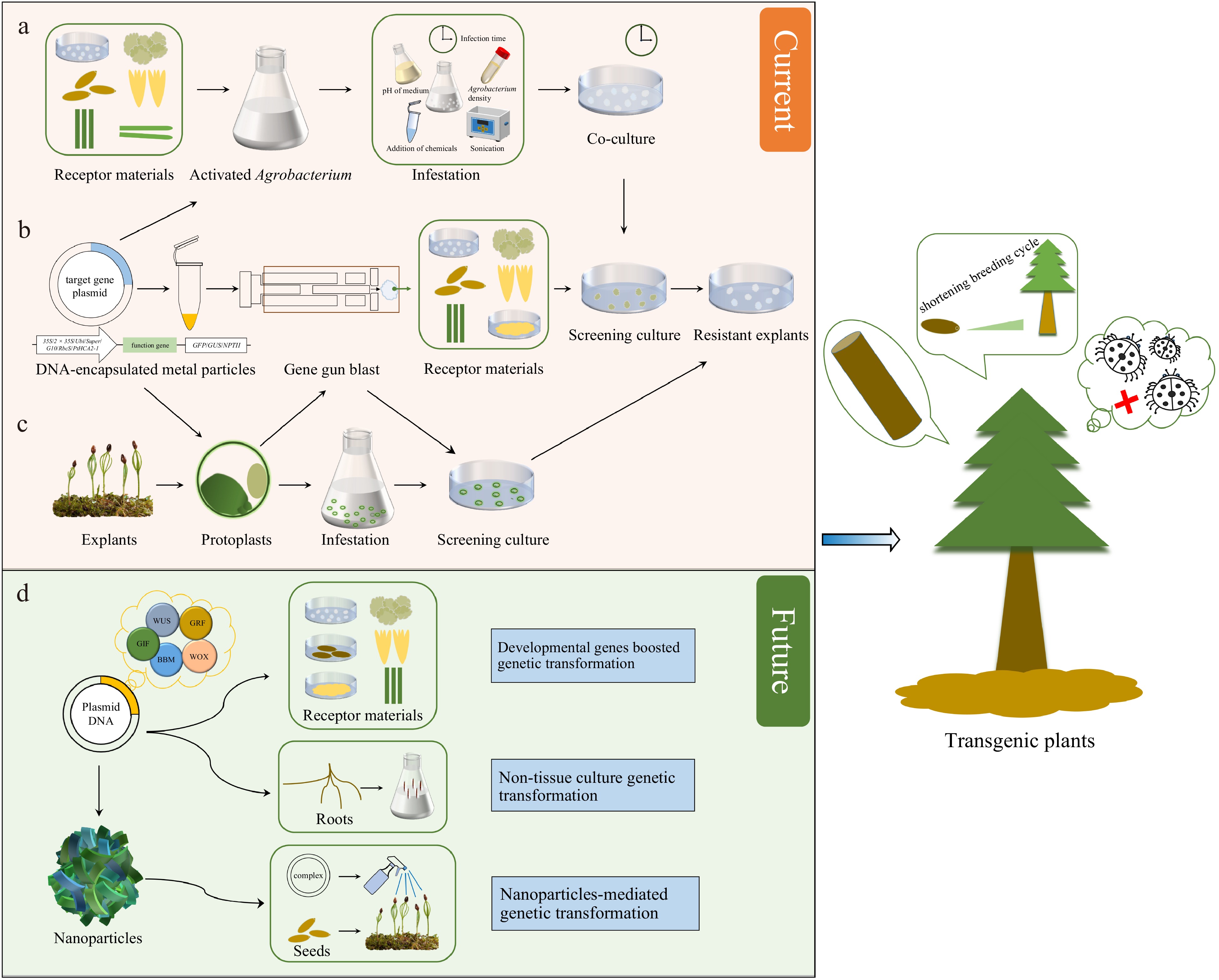
      Figure 1. 

      Techniques and prospects for genetic transformation of conifers. (a) Agrobacterium-mediated genetic transformation. (b) Genetic transformation via biolistic bombardment. (c) Protoplast transformation. (d) Potential strategies for transformation improvement in conifers.

      Table 2.  Agrobacterium-mediated transformation in conifers.

      Tree speciesAcceptor materialsCo-culture timeOD600nm ResultsRef.
      Pinus
      Pinus pineaCotyledons3 d1Cotyledons forming buds[28]
      Pinus strobusEmbryogenic tissues2 d0.6Regenerated plant[9]
      Mature zygotic embryos12 h0.8−1.0Regenerated plant[10]
      Pinus taedaEmbryogenic tissues2 d1Transient expression[51]
      Mature zygotic embryos3−5 d/Regenerated plant[52]
      Shoot apex7 d/Transgenic plants[53]
      Mature zygotic embryos3−5 d0.8−1.0Transgenic plants[54]
      Mature zygotic embryos3−5 d0.8−1.0Transgenic plants[55]
      Mature zygotic embryos3−5 d0.5−1.0Improve salt tolerance[22]
      Pinus radiataEmbryogenic tissues1 d0.6Stable transformation[56]
      Cotyledons5−60 minOD550nm = 0.4Transgenic plants[11]
      Embryogenic tissues5 dOD550nm = 0.5−0.8Transgenic plants[57]
      Micropropagated shoot3 dOD550nm = 0.35−0.4Transgenic plants[32]
      Pinus pinasterEmbryogenic tissues36 h0.6Transgenic plants[58]
      Embryogenic tissues3 d0.3Transgenic plants[59]
      Pinus patulaEmbryogenic tissues2 d0.5−0.75Transgenic tissues[12]
      Pinus elliottiiMature zygotic embryos3 d0.9Transgenic plants[30]
      Pinus massonianaMature zygotic embryos3 d0.5Transgenic plants[13]
      Pinus tabuliformisCallus/hypocotyls/Needles3 d0.8Transient expression[31]
      Larix
      Larix deciduaHypocotyls2−3 d/Regenerated plant[7]
      Hypocotyls4 d/Regenerated plant[21]
      hybrid larchEmbryogenic tissues2 d0.3Regenerated plant[60]
      Embryogenic tissues2 d0.5Regenerated plant[61]
      Larix olgensisEmbryogenic tissues3 d0.6Transgenic plants[35]
      Embryogenic tissues2 d0.5Enhance stress resistance[25]
      Larix kaempferiEmbryogenic tissues2 d0.1Promotes cell proliferation[24]
      Picea
      Picea sitchensisEmbryogenic cell lines3 d0.8−1.1Stable transformation[62]
      Picea abiesEmbryogenic tissues2 d1Transient expression[51]
      Embryogenic tissues2 d0.6Transgenic plants[63]
      Embryogenic tissues2 d/Transgenic plants[23]
      Picea marianaEmbryogenic tissues2 d0.6Transgenic plants[63]
      Picea glaucaEmbryogenic tissues2 d0.6Transgenic plants[63]
      Embryogenic tissues2 d1Transgenic plants[29]
      Embryogenic tissues//Transgenic plants[64]
      Abies
      Abies spp.Embryogenic tissues2 d0.6Transgenic plants[65]
      Abies koreanaEmbryogenic tissues3 d0.6Transgenic plants[66]
      Taxus
      Taxus brevifolia/Taxus baccataShoot segments3 d/Gall formation[8]
      Chamaecyparis
      Chamaecyparis obtusaEmbryogenic tissues2 d0.3Transgenic plants[67]
      Cryptomeria
      Cryptomeria japonicaEmbryogenic tissues2 d0.15Enhance transformation[68]
      Embryogenic tissues2 d0.2−0.6Transgenic plants[69]

      Although Agrobacterium-mediated gene transfer is extensively employed in numerous biotechnology laboratories, its large-scale application in conifer transformation is hindered by challenges in propagating explant material, selection inefficiencies, and low transformation rates[51]. Wenck et al. explored co-cultivation conditions and various disarmed Agrobacterium strains to enhance transformation efficiency. They discovered that incorporating additional virulence genes, such as a constitutively active virG or extra copies of virG and virB from pTiBo542, amplified the transformation efficiency of Norway spruce by 1000-fold relative to initial experiments, which exhibited minimal or nonexistent transient expression[51]. Tang examined the influence of additional virulence (vir) genes in A. tumefaciens and the impact of sonication on the transformation efficiency of loblolly pine[54]. Utilizing plasmids with supplementary vir genes and sonication significantly enhanced the transfer efficiency, affecting not only transient expression but also the recovery of hygromycin-resistant lines. In their studies on Agrobacterium-mediated hybrid larch transformation, Levee et al. observed one to two transformation events per 100 cocultured masses[60]. Introducing 100 µM of coniferyl alcohol led to an increase in yield. Other studies demonstrated that sonication[10, 30] and the addition of chemicals, including okadaic acid, trifluoperazine, acetosyringone, thidiazuron, and others[10, 30, 35, 66, 70], significantly enhanced the transformation efficiency of conifers and further advanced the transformation system. Additionally, several groups have illustrated that cold treatment of Agrobacterium can augment transformation efficiency[13].

      Transformation frequencies depend on species, genotype, and post-cultivation protocol. In a study involving three species, Picea mariana was transformed at the highest frequency, followed by P. glauca and P. abies[63]. Furthermore, for all the species, transgenic plants were regenerated using modified protocols for somatic embryo maturation and germination. Le et al. devised an efficient method for the reproducible transformation of embryogenic white spruce tissue using A. tumefaciens-mediated gene transfer[29]. A shoot-based, genotype-independent transformation method employing A. tumefaciens facilitated plant recovery and enabled the transformation of elite germplasm[53]. Shoots from 4- to 6-week-old seedlings and adventitious shoots from cultures were inoculated with A. tumefaciens, underwent selection, and were subsequently regenerated. Micropropagated shoot explants from P. radiate have successfully been employed to produce stable transgenic plants via A. tumefaciens-mediated transformation[32]. It is crucial during the transformation process to inhibit and prevent contamination caused by excessive Agrobacterium growth. In the A. tumefaciens-mediated transformation of P. pinea cotyledons, a high cotyledon mortality rate occurs, possibly related to the plant's hypersensitive response to bacterial infection[28]. For conifers, non-toxic antibiotics to plant cells, like cefotaxime sodium (Cef) or timentin, are frequently incorporated into the medium. Also, in the post-transformation selection medium, selecting transformants is crucial for obtaining transgenic plants. If tissues are initially cultivated for 10 d on a medium with timentin (400 mg·L–1) to avert bacterial overgrowth, the recovery of kanamycin-resistant tissues is enhanced before applying selection pressure[29]. An evaluation of three antibiotics was conducted to assess their effectiveness in eliminating A. tumefaciens during the genetic transformation of loblolly pine using mature zygotic embryos[55]. Exposing the cultures to 350 mg·L–1 of carbenicillin, Cef, and timentin for a duration of up to 6 weeks failed to eliminate Agrobacterium; however, increasing the concentration to 500 mg·L–1 successfully eradicated the bacterium from co-cultured zygotic embryos[55].

      Identifying the optimal combination of infection time and concentration is crucial for successful conifer transgenesis during genetic transformation experiments. Generally, the bacterial solution concentration for infecting conifers is maintained at an OD600 of 0.3–0.8. Elevating the Agrobacterium concentration and extending the infection duration can result in excessive bacterial proliferation and hypersensitive necrosis of explants, thereby diminishing transformation efficiency[28]. Conversely, employing a low-density Agrobacterium suspension and a brief infection period often results in weak infectivity, which similarly reduces transformation efficiency[13]. Moreover, the infection duration influences T-DNA transfer and, consequently, the efficiency of genetic transformation. The infection duration, typically less than 30 min, varies depending on the explant type and the physiological status of the conifer species. However, both the concentration and infection duration of the bacterial solution must be tailored to the condition, type, and environmental factors of the explants, necessitating further research.

    • Particle bombardment, also known as biolistics, serves as an alternative method for plant genetic transformation, circumventing the limitations associated with Agrobacterium-mediated genetic transformation[71]. This method is not limited by biological constraints and is applicable to a broad spectrum of plant species. However, in the context of conifer transformation frequency, biolistic techniques are generally regarded as less effective than Agrobacterium-mediated genetic transformation[68]. Foreign genes have successfully been expressed in all tested conifer tissues via particle bombardment, encompassing embryos, seedlings, xylem, pollen, needles, buds, cell suspension cultures, embryogenic callus, cell aggregate cultures, and roots (Fig. 1b & Table 3). While most of these attempts yielded only transient expression, they have offered insightful information about the factors influencing gene expression in various tissues capable of regeneration[20]. GFP introduction into conifer tissues has been achieved through microprojectile bombardment, with transient expression subsequently observed[72]. The CaMV35S promoter facilitated GUS gene expression in loblolly pine tissues[73]. Microprojectile bombardment proves to be an effective technique for assaying transient gene expression in pine, and it harbors potential for generating transgenic pine plants. Using high-velocity microprojectiles, plasmid DNA with the GUS gene, under the control of the CaMV35S promoter, has been introduced into cultured Douglas fir cotyledons[74]. Additionally, the particle gun technique has been employed to transform a variety of receptor materials in different tree species, including callus and pollen of larch[75, 76], Chir pine[16], and Norway spruce[14, 7780]. Particle bombardment has been applied to Lodgepole pine, yellow cypress, western hemlock, jack pine, and black spruce pollen to achieve transient GUS gene expression, demonstrating the method's viability for pollen transformation[81]. Furthermore, particle bombardment has facilitated the testing of transient expression of heterologous promoters in organized tissues and angiosperm promoters in gymnosperms[82]. Comparative analyses have been conducted on the initiation strengths of transient expression for eight distinct promoter sequences, based on the relative levels of GUS expression[76].

      Table 3.  Biolistic bombardment genetic transformation in conifers.

      Tree speciesAcceptor materialsPlasmidsPromotersGenesResultsRef.
      Pinus
      Pinus taedaCotyledonspBI22135SGUSTransient expression[73]
      Pinus radiataSuspension cellspBI22135SGUSTransient expression[87]
      Embryogenic tissuespCW103/pCWI222 × 35SgusATransient expression[88]
      CotyledonspBI121/pCGUΔl/
      pAIGusN/pActl-D
      35S/UbBI/Adhl/ActlgusATransient expression[89]
      Embryogenic tissuespRC101/pCW12235S/EmuuidATransgenic plants[83]
      Embryogenic tissuespAHC25/pCW122maize ubiquitin/35SGUS/BarTransgenic plants[14]
      CallipCW122/pCADsense35Snpt II/CadTransgenic calli[90]
      Embryogenic tissuespMYC3425/pAW16/
      pCW132/pRN2
      Emu/ubiCry1AcTransgenic plants[15]
      Pinus concorta/Pinus banksianaMature pollenpBM113Kp/pRT99GUS/
      pAct1-D/pGA984
      35S/rice actinGUSTransient expression[81]
      Pinus sylvestrisCalli/Vegetative buds/
      Suspension cells
      pBI22135SGUSTransient expression[91]
      PollenpBI221/pRT99/pBI410/
      pBI426/pBM113
      35S/EmP/UbB1GUSTransient expression[79 ]
      Pinus strobusEmbryonal massesp35S-GFP/mGFP435SGFPTransient expression[72]
      Pinus aristata/Pinus griffithii/Pinus monticolaPollen tubespBI22135SGUSTransient expression[92]
      Pinus patulaEmbryogenic tissuespAHC2535SBar/GUSSomatic embryos[48]
      Pinus nigraEmbryogenic tissuespCW1222 × 35SGUSSomatic embryos[86]
      Pinus roxbughiiMature zygotic embryospAHC25maize ubiquitinBar/GUSTransgenic plants[16]
      Picea
      Picea glaucaZygotic embryos/Seedlings/
      embryogenic callus
      pUC1935SGUSTransient expression[82]
      Somatic embryospBI42635SGUSStable transformation[93]
      Somatic embryospTVBT4110035SGUS/BtTransgenic plants[49]
      Embryonal massesp35S-GFP/mGFP435SGFPTransient expression[72]
      Embryogenic tissuespKUB/pBI426maize ubiquitin/35Scry1AbTransgenic plants[26]
      Picea marianaEmbryogenic tissuespRT99GUS/pBM113Kp35SGUSTransient expression[94]
      Embryogenic tissuespRT99GUS/pGUSInt/
      pMON9909
      35S/Em protein of wheat/Rbcs/NOS/
      Actin/Arabin
      GUSTransient expression[76]
      Mature pollenpBM113Kp/pRT99GUS/
      pAct1-D/pGA984
      35S/rice actinGUSTransient expression[81]
      Embryonal massespRT99GUS/pBI42635SGUSTransgenic plants[84]
      Pollen/Embryonal masses/ Somatic embryosp35S-GFP/mGFP435SGFPTransient expression[72]
      Mature somatic embryospBI221.2335SGUSTransgenic plants[17]
      Picea abiesSomatic embryopRT99gus35SGUSStable transformation[77]
      Embryogenic tissuespRT99gus/pJIT65/
      Dc8gus/pBMI13Kp
      35S/2 × 35S/
      Act1-D/Dc8
      GUSTransient expression[80]
      PollenpBI221/pRT99/pBI410/
      pBI426/pBM113
      35S/EmP/UbB1GUSTransient expression[79]
      Embryogenic tissuespCW12235SGUSTransgenic plants[95]
      Embryogenic tissuespAHC25maize ubiquitinBarTransgenic plants[78]
      Embryogenic tissuespAHC25/pCW122maize ubiquitin/35SGUS/BarTransgenic plants[14]
      Embryogenic tissuespAHC25maize ubiquitinCCRTransgenic plants[27]
      Larix
      Larix × eurolepisEmbryogenic tissuespRT99GUS/pGUSInt/
      pMON9909
      35S/Em protein of wheat/Rbcs/NOS/
      Actin/Arabin
      GUSTransient expression[76]
      Larix laricinaEmbryonal massespBI426/pRT99gus/
      pRT66gus/pRT55gus
      35S/2 × 35SGUSTransient expression[75]
      Larix gmeliniiZygotic embryospUC-GHG/pBI221-HPT35SGUS/GFPTransgenic plants[34]
      Pseudotsuga
      Pseudotsuga menziesiiCotyledonspTVBTGUS35SGUSTransient expression[74]
      Chamaecyparis
      Chamaecyparis nootkatensisMature pollenpBM113Kp/pRT99GUS/
      pAct1-D/pGA984
      35S/rice actinGUSTransient expression[81]
      Tsuga
      Tsuga heterophyllaMature pollenpBM113Kp/pRT99GUS/
      pAct1-D/pGA984
      35S/rice actinGUSTransient expression[81]
      Abies
      Abies nordmannianaEmbryogenic tissuespCW12235SGUSTransgenic plants[85]

      Particle bombardment-mediated transformation is capable of regenerating whole plants. In P. glauca plants, the stable expression of an exogenous gene marked the first successful creation of transgenic plants using the particle gun method[49]. Walter et al. used a particle gun to bombard four embryonic cell lines of P. radiate, resulting in over 150 transgenic plants from 20 transformation experiments[83]. Analyses using Southern and Northern blotting confirmed the integration of the target gene into the genome. Particle bombardment facilitated the stable genetic transformation of P. mariana in two target tissues: mature cotyledonary somatic embryos and suspensions from embryonal masses, employing the Biolistic PDS-1000/He device[84]. The expression of the GUS gene in needles of regenerated seedlings demonstrates the potential for sustained transgene expression in spruce[17]. Using biolistic transformation, stable genetic transformation has been accomplished in embryogenic cultures of Abies nordmanniana, leading to the regeneration of transgenic plants[85]. A biolistic approach has successfully achieved stable transformation in embryogenic tissues of P. nigra Arn., specifically cell line E104[86]. Given its versatility and broad applicability, particle bombardment is anticipated to continue as a primary method in genetic transformation.

      Particle bombardment possesses significant potential for producing transgenic conifer plants. A key objective in tree breeding involves reducing lignin content or modifying its composition, which would aid in delignification during pulping processes. When the antisense construct of the cinnamoyl CoA reductase (CCR) gene was introduced into Norway spruce, a significant reduction in the total lignin content of dry wood was observed compared to controls[27]. Lachance et al. conducted a study on the accumulation of crylAb protein in embryogenic tissues, somatic seedling needles, and 5-year-old field-grown needles of white spruce[26]. Insect feeding trials, both in the laboratory and the field, indicated that multiple transgenic spruce lines proved lethal to spruce budworm larvae. Through biolistic transformation of embryogenic tissue, transgenic radiata pine plants harboring the Bacillus thuringiensis (Bt) toxin gene, cry1Ac, were successfully produced[15]. Ongoing research is being conducted on functional genes utilizing this technology[14, 16, 78].

    • Protoplast technology enables various unique approaches to the genetic improvement of plants[96]. Protoplast transient expression assays serve as versatile tools in genomics, transcriptomics, metabolic, and epigenetic studies[97]. Coupling protoplast transient expression experiments with high-resolution imaging enables simple, rapid, and efficient analysis and characterisation of gene functions and regulatory networks. This includes protein subcellular localisation, protein-protein interactions, transcriptional regulatory networks, and gene responses to external cues[98100]. Reporter genes commonly used, like LUC and GUS, are employed to assess gene activity in conifer protoplasts[87]. P. glauca protoplasts were transformed with the chloramphenicol acetyltransferase (CAT) reporter gene through electroporation[101]. Fir and pine protoplasts were successfully transformed with the LUC gene through electroporation, with gene expression enhanced by the addition of polyethylene glycol (PEG) to the mixture[102]. Developments in methods for transient gene expression have been made for protoplasts of black spruce and jack pine[103]. In electroporated protoplasts of P. glauca, P. mariana, and P. banksiana, the activity levels of exogenous genes depend on the promoter, electroporation conditions, and the targeted cell line[104]. A new transient transformation system for Chinese fir protoplasts has been established, achieving cell wall regeneration and protoplast division. This method serves as a reference for conducting functional studies on Chinese fir-related genes[105]. However, the challenges in establishing protoplast regeneration systems in conifers mean that protoplast-based genetic transformation studies primarily focus on transient gene expression and the investigation of gene function and expression regulation (Fig. 1c).

    • Establishing an effective and stable regeneration system is crucial for rapidly expanding conifer populations for seedling production and successful heritage transformation. A range of plant materials, each with unique advantages, serves as transformation receptors for conifers. These include zygotic embryos, hypocotyls, embryonic tissues, somatic embryos, protoplasts, stem tips, and pollen[7, 10, 13, 31, 51, 53, 81, 101]. Embryonic tissues have been the focus of extensive research as receptors in numerous studies[9, 27, 35, 51, 57, 58, 85]. Additionally, Agrobacterium-mediated genetic transformation using mature zygotic embryos as explants has been successfully implemented in P. taeda[22, 52, 54, 55], P. elliottii[30], and P. massoniana[13]. Cotyledons and hypocotyls are identified as suitable explants for genetic transformation[7, 11, 21, 28]. Currently, embryonic tissue of conifers is predominantly used as recipient material through the somatic embryogenesis pathway to obtain stably-transformed regenerated plants (Tables 2 & 3).

      A primary challenge in the genetic transformation of coniferous trees involves plant regeneration[106]. This challenge arises primarily from the unique biological properties and regeneration mechanisms of conifers. Tissue culture in conifers proves more challenging than in other plants. This is attributed to the cells of conifers, especially those from mature trees, which have a lower capacity for differentiation and regeneration[107]. The tissue culture process entails inducing cells or tissues from the parent plant to develop into new plants under controlled conditions, a process notably less efficient in conifers. Furthermore, during tissue culture, particularly over extended periods, the genetic stability of conifers may be affected. Cell division and differentiation, occurring during tissue culture, may introduce genetic mutations; additionally, genome doubling, leading to the formation of polyploids, can also occur. Consequently, even if plant regeneration is successful, the resultant plants may exhibit genetic variations, potentially posing challenges in subsequent applications and research[19, 106]. The regeneration of conifer tissue is notably sensitive to the balance of plant hormones and other culture conditions. Different species of conifers often require specific combinations of hormones and culture environments, thereby complicating the identification of a universal method applicable to all types[108]. Conifers generally exhibit a long regeneration process, which implies that the entire process from tissue culture to mature plant consumes a considerable amount of time, acting as a limiting factor in research and application. Variations in regeneration capabilities among different species of conifers are notable.

      In summary, although the genetic transformation and regeneration of coniferous trees are theoretically feasible, their practical implementation is fraught with several challenges, most notably in tissue culture efficiency, genetic stability maintenance, and adaptation to different species' characteristics[109]. Addressing these challenges necessitates in-depth research and substantial technological innovation.

    • Despite the numerous promising success cases mentioned, it must be acknowledged that genetic transformation continues to pose a significant challenge for most conifer researchers. To date, none of these methods have proven universally applicable across multiple species or varied genotypes. Consequently, while a method may appear promising, it often remains confined to successful implementation under specific laboratory conditions, lacking widespread applicability. Significant progress is still required to develop a universal model for conifers that is as straightforward, efficient, and reproducible as those established for angiosperm model species.

    • Conifers possess distinct and complex biological characteristics, setting them apart from commonly utilized genetic engineering plants like Arabidopsis or tobacco. Their prolonged generation times, expansive genomes, and elaborate reproductive processes contribute to the challenges in working with them[1, 2, 4].

    • Despite the establishment of transformation protocols, the efficiency of integrating foreign genes into the conifer genome frequently remains low[54, 63]. Consequently, only a minor fraction of transformed cells effectively express the introduced gene, posing significant challenges in producing stable and predictable genetically modified organisms.

    • Various conifer species exhibit unique biological traits and varying responses to transformation techniques. A technique effective in one conifer species might not yield similar results in another, necessitating tailored optimization for each species.

    • The size and complexity of conifer genomes pose challenges in the introduction and expression of foreign genes. A thorough understanding of the regulatory elements and mechanisms within conifer genomes is crucial for genetic engineering success[35]. However, such knowledge is typically less comprehensive than that available for model plant species.

    • Conifers often require specialized tissue culture techniques for regeneration and propagation. Developing suitable tissue culture methods for conifers, particularly those compatible with genetic transformation, is a significant hurdle. Studies have indicated that the induction rate of embryogenic tissues from immature seeds in conifers is influenced by both the genotype and the embryonic developmental stage[110, 111].

    • Conifers, like many plants, contain high levels of phenolic compounds, such as lignins and polyphenols[112, 113]. These compounds may exert inhibitory effects on the enzymes used in the genetic transformation process. Phenolic compounds are known to contribute to oxidative stress, DNA degradation, and may interfere with the integration of foreign genes into the plant genome.

    • Conifers produce a diverse array of secondary metabolites, including terpenoids and flavonoids, which can potentially affect the success of genetic transformation. These compounds can exhibit toxic effects on the transformed cells or may interfere with the activity of introduced genes.

    • The cell walls of conifers are notably complex and rigid, serving to provide structural support to the plant. However, this complexity may impede the delivery of foreign DNA into plant cells. Efficient transformation frequently necessitates overcoming these barriers to ensure that the introduced genetic material successfully reaches the nucleus of the target cells[114, 115].

    • The presence of genetic variation within conifer populations may influence the success of genetic transformation. Individuals within a species often exhibit varying responses to transformation protocols, and optimizing these protocols for broader applicability presents a significant challenge.

      Addressing these biochemical factors typically necessitates the development of specialized techniques and treatments within the genetic transformation process. For instance, researchers might utilize tissue culture conditions designed to mitigate the effects of phenolic compounds, or employ specialized methods to enhance the delivery of foreign DNA through the cell wall.

      Comprehending the biochemical makeup of conifers and customizing transformation methods to suit their unique characteristics is an active area of research. Advances in biotechnology, encompassing the development of more robust transformation protocols and the elucidation of genes involved in stress responses, may play a pivotal role in surmounting these biochemical barriers in the future.

    • Addressing the challenges associated with the genetic transformation of conifers necessitates a comprehensive approach that integrates advancements across multiple key domains (Fig. 1d). The following delineates potential strategies and focal areas.

    • It is imperative for researchers to persist in refining and optimizing transformation protocols tailored to various conifer species. This encompasses enhancing the efficiency of introducing foreign genes into conifer cells and developing uniform methods applicable across diverse species. The utilization of developmental genes may prove beneficial in promoting transformation. These genes, capable of acting through diverse developmental mechanisms to enhance the regeneration of transgenic cells, have seen extensive use in model plants to stimulate embryogenesis and, in some instances, organogenesis[116118]. In summary, the overexpression of regeneration-regulating transcription factors, including BBM, WUS2, WOX5, GRF4, and GIF1, could enhance genetic transformation in conifers characterized by low regeneration efficiency, substantial transformation difficulty, and genotype limitation.

    • Gaining a deeper understanding of the molecular and biochemical processes in conifers is essential. This necessitates research into the regulation of gene expression, understanding the role of secondary metabolites, and comprehending the response of conifers to stress conditions. This knowledge is crucial in informing the development of transformation methods that are synergistic with the unique biology of conifers.

    • The improvement of tissue culture techniques, crucial for supporting the regeneration and propagation of conifer plants, is vital. The development of protocols for efficient plant regeneration from transformed cells can significantly bolster the success of genetic transformation. Conversely, most prevailing methods for plant genome modification entail regenerating plants from genetically modified cells in tissue culture, a process that is technically challenging, costly, time-consuming, and limited to a narrow range of plant species or genotypes[119]. Cao et al. outlined a notably straightforward cut–dip–budding (CDB) delivery system, which includes inoculating explants with A. rhizogenes, subsequently generating transformed roots that yield transformed buds through root suckering[120]. The advancement of methods that circumvent laborious procedures, such as tissue culture, and facilitate obtaining transgenic and gene-edited plants, marks a significant breakthrough in conifer research.

    • Exploring strategies to overcome the challenges presented by the complex cell walls of conifers is imperative. This could involve employing enzymes or other agents to facilitate the penetration of foreign DNA into plant cells. In the realm of conifer biotechnology, the initial protoplast extraction in P. contorta laid the foundation for the establishment of a transient transformation system in conifers[121].

    • Recognizing and addressing genetic variation within conifer species is critical. Customizing transformation protocols to accommodate the diverse genetic backgrounds of individuals within a species can lead to broader success in genetic transformation[122].

    • The utilization of cutting-edge biotechnological tools, notably CRISPR/Cas9 gene editing, can offer more precise control over the modification of conifer genomes. These advanced technologies have the potential to overcome several challenges associated with traditional genetic transformation methods. Genome editing represents a powerful technology for functional genomic research and trait improvement. Cui et al. successfully achieved knockout of the DXS1 gene in white spruce (P. glauca) employing the conifer-specific CRISPR/Cas9 toolbox[123]. Recently, CRISPR/Cas9-mediated targeted mutagenesis has been demonstrated in radiata pine[124], Japanese cedar[47], and Chinese fir[36], underscoring its feasibility in conifers. This represents a potent genome editing system of significant importance for gene function studies and the genetic improvement of plant traits, likely to make substantial contributions to the development of molecular breeding in conifers.

    • In future research endeavors, the use of nanomaterials for genetic modification promises to expand the scope of plant molecular research, particularly for conifers, which currently lack efficient systems for regeneration and stable genetic transformation. Nanocarriers are characterized by their large surface area, facilitating efficient gene loading, alongside high biocompatibility to safeguard the loaded genes, coupled with low toxicity and enhanced safety[125, 126]. Consequently, nanoparticles hold the potential to be utilized in developing transgenic technologies for conifer regeneration without dependency on tissue culture, potentially overcoming the technical challenges in genetic transformation of recalcitrant plant genotypes. Conversely, the exploration of stable and targeted nanocarrier-mediated gene editing technologies offers the prospect of achieving genetic improvements in conifers.

    • Considering the ecological significance of conifers, comprehensive risk assessments and detailed ecological studies should accompany all attempts at genetic modification. Comprehending the potential environmental impact and addressing public concerns are imperative for the responsible and sustainable deployment of genetically modified conifers.

    • Given the global distribution of conifers, international collaboration among researchers, institutions, and regulatory bodies is essential to foster the sharing of knowledge, resources, and expertise. Such collaborative efforts can significantly accelerate progress and enhance the effectiveness in addressing challenges.

      Sustained research and ongoing technological advancements, in conjunction with a holistic and interdisciplinary approach, are crucial to unlocking the full potential of genetic transformation in conifers, while simultaneously ensuring the responsible and ethical application of these technologies.

    • Many reports have documented the successful expression of exogenous genes in conifers using Agrobacterium-mediated, particle bombardment-mediated, and protoplast-based genetic transformation methods. However, the genetic transformation of conifers faces several challenges, including low transformation efficiency, high dependence on recipient genotypes, difficulties in plant regeneration. Overall, the genetic transformation of conifers remains heavily reliant on extensive experience and sophisticated technical skills, rendering its widespread application challenging for most conifer researchers. Overcoming these challenges will usher in a new era of productivity and quality in forestry. Several potential strategies have been proposed to improve conifer transformation, including the optimization of transformation protocols, understanding molecular mechanisms, improving tissue culture techniques, overcoming cell wall barriers, understanding genetic variation, employing nanoparticle- and non-tissue culture-mediated genetic transformation, utilizing genome editing tools, fostering international collaboration, and more. In conclusion, with the ongoing development of molecular biotechnology and enhancement of various regeneration and transformation systems, research on the genetic transformation of conifer species is poised for continued progress and broader applicability.

    • The authors confirm contribution to the paper as follows: study conception and design: Zhao J, Niu S, Zhang J; draft manuscript preparation: Zhao H; Figure creation: Zhao H. All authors reviewed the results and approved the final version of the manuscript.

    • Data sharing is not applicable to this article as no datasets were generated or analyzed during the current study.

      • This work was supported by the National Key R&D Program of China (Grant No. 2023YFD2200102), the National Natural Science Foundation of China (Grant No. 32371834), the National Natural Science Foundation of China (Grant No. 32271836), the National Key R&D Program of China (Grant No. 2023YFD2200104).

      • The authors declare that they have no conflict of interest. Shihui Niu is the Editorial Board member of Forestry Research who was blinded from reviewing or making decisions on the manuscript. The article was subject to the journal's standard procedures, with peer-review handled independently of this Editorial Board member and the research groups.

      • Copyright: © 2024 by the author(s). Published by Maximum Academic Press, Fayetteville, GA. This article is an open access article distributed under Creative Commons Attribution License (CC BY 4.0), visit https://creativecommons.org/licenses/by/4.0/.
    Figure (1)  Table (3) References (126)
  • About this article
    Cite this article
    Zhao H, Zhang J, Zhao J, Niu S. 2024. Genetic transformation in conifers: current status and future prospects. Forestry Research 4: e010 doi: 10.48130/forres-0024-0007
    Zhao H, Zhang J, Zhao J, Niu S. 2024. Genetic transformation in conifers: current status and future prospects. Forestry Research 4: e010 doi: 10.48130/forres-0024-0007

Catalog

  • About this article

/

DownLoad:  Full-Size Img  PowerPoint
Return
Return