REVIEW   Open Access    

Accumulation and regulation of malate in fruit cells

More Information
  • Received: 01 May 2024
    Revised: 02 July 2024
    Accepted: 13 July 2024
    Published online: 02 September 2024
    Fruit Research  4 Article number: e031 (2024)  |  Cite this article
  • Fruit acidity is an important component of flavor quality in fleshy fruit. The accumulation of malate, the dominant organic acid in the acidity formation of most mature fruit, is highly regulated by metabolism and transportation during fruit development. The knowledge on the mechanism of fruit acidification, as well as the major genes and substances is however still limited. In the present paper, the research advances on the relevance between malate accumulation and the genes associated with malate metabolism and transportation, as well as the transcriptional regulation of malate in fruit was reviewed. Furthermore, positive future research could provide a theoretical reference for optimizing fruit quality and genetic improvement.
  • Plants are continuously subjected to unpredictable environmental conditions and encounter a multitude of stressors throughout their growth and development, posing a significant challenge to global crop production and food security[1]. Heat and drought are undoubtedly the two most important stresses that have a huge impact on crops. Both elicit a wide array of biochemical, molecular, and physiological alterations and responses, impacting diverse cellular processes and ultimately influencing crop yield and quality[2].

    A primary physiological consequence of both stresses is the diminished photosynthetic capacity, partially resulting from the degradation of chlorophyll due to leaf senescence under stress conditions. Chlorophyll accumulation was diminished in numerous plants subjected to drought or heat stress conditions[3,4]. Various environmental stresses prompt excessive generation of reactive oxygen species (ROS), initiating oxidative damage that compromises lipids, and proteins, and poses a serious threat to cellular functions[2]. To mitigate oxidative stress and minimize damage, plants have developed various protective mechanisms to neutralize ROS. Several antioxidant enzymes, such as SOD, POD, and CAT, are integral to cellular antioxidative defense mechanisms. Additionally, antioxidants such as anthocyanins and proline serve as crucial ROS scavengers[5,6]. The elevation in temperature typically induces the transient synthesis of heat shock proteins (Hsps), which function as molecular chaperones in protecting proteins from denaturation and aggregation, with their activity primarily regulated at the transcriptional level by heat shock factors (Hsfs)[7]. The significance of Hsps and Hsfs in all organisms, including plants, has been assessed in various stress conditions that could disrupt cellular homeostasis and result in protein dysfunction[7]. Drought stress can also trigger the transcription of a suite of marker genes, including RD29A, RD29B, NCED3, AREB1, Rab18, etc., which assist plants in mitigating cellular damage during dehydration and bolstering their resilience to stress[810].

    Previous research efforts focusing on the regulatory control of stress-related genes have largely centered around protein-coding genes. In recent years, non-protein-coding transcripts have emerged as important regulatory factors in gene expression. Among them, long non-coding RNAs (lncRNAs) lncRNAs have been identified as implicated in various abiotic stresses[11,12]. LncRNAs are a class of non-coding RNAs (ncRNAs) exceeding 200 nucleotides in length. They possess minimal or no protein-coding potential[13]. In plants, lncRNAs are specifically transcribed by RNA polymerases Pol IV, Pol V, Pol II, and Pol III[14,15]. LncRNAs exhibit low abundance and display strong tissue and cellular expression specificity relative to mRNAs. Moreover, sequence conservation of lncRNAs is was very poor across different plant species[13,16,17]. The widespread adoption of high-throughput RNA sequencing technology has revealed lncRNAs as potential regulators of plant development and environmental responses. In cucumber, RNA-seq analysis has predicted 2,085 lncRNAs to be heat-responsive, with some potentially acting as competitive endogenous RNAs (ceRNAs) to execute their functions[18]. In radish, a strand-specific RNA-seq (ssRNA-seq) technique identified 169 lncRNAs that were differentially expressed following heat treatment[19]. In Arabidopsis, asHSFB2a, the natural antisense transcript of HSFB2a was massively induced upon heat stress and exhibited a counteracted expression trend relative to HSFB2a. Overexpression of asHSFB2a entirely suppressed the expression of HSFB2a and impacted the plant's response to heat stress[20]. For drought stress resistance, 244 lncRNAs were predicted in tomatoes to be drought responsive probably by interacting with miRNAs and mRNAs[21]. Under drought stress and rehydration, 477 and 706 lncRNAs were differentially expressed in drought-tolerant Brassica napus Q2 compared to drought-sensitive B. napus, respectively[22]. In foxtail millet and maize, 19 and 644 lncRNAs, respectively, were identified as drought-responsive[23,24]. Despite the identification of numerous lncRNAs by high-throughput sequencing, which suggests their potential involvement in various abiotic stress processes, only a minority have been experimentally validated for function.

    In our previous study, we characterized 1,229 differentially expressed (DE) lncRNAs in Chinese cabbage as heat-responsive, and subsequent bioinformatics analysis reduced this number to 81, which are more likely associated with heat resistance[25]. lnc000283 and lnc012465 were selected from among them for further functional investigation. The findings indicated that both lnc000283 and lnc012465 could be promptly induced by heat shock (HS). Overexpression of either lnc000283 or lnc012465 in Arabidopsis plants enhanced their capacity to tolerate heat stress. Additionally, both lnc000283 and lnc012465 conferred drought tolerance to transgenic Arabidopsis.

    The lncRNA sequences examined in this study were from Chiifu-401-42 Chinese cabbage and all Arabidopsis plants were of the Col-0 background. Transgenic plants expressing lnc000283 and lnc012465 were generated using the Agrobacterium tumefaciens-mediated floral dip method[26]. Single-copy and homozygous T3 plants were identified through genetic segregation on an agar medium supplemented with kanamycin. The T3 generation plants, or their homozygous progeny, were utilized in the experiments.

    For phenotypic assessment, Arabidopsis seeds were initially sown on filter paper moistened with ddH2O and placed in a 4 °C freezer for 2 d. Subsequently, they were evenly planted in nutrient-rich soil and transferred to a growth chamber operating a 16-h day/8-h night cycle, with day/night temperatures of 22 °C/18 °C and a light intensity of 250 μmol·m−2·s−1. After 10 d of growth, Arabidopsis plants with uniform growth were transferred to 50-hole plates. Arabidopsis plants grown in Petri dishes were firstly seed-sterilized and then sown on 1/2 MS medium supplemented with 10 g·L−1 sucrose. The seeds were then placed in a 4 °C refrigerator for 2 d in the dark before transferring them to a light incubator. The day/night duration was set to 16 h/8 h, the day/night temperature to 21 °C/18 °C, and the light intensity to 100 μmol·m−2·s−1.

    For heat treatment, 3-week-old seedlings were subjected to 38 °C for 4 d within a light incubator, subsequently transferred to their original growth conditions under the same light/dark cycles. For drought treatment, 3-week-old Arabidopsis seedlings were deprived of water for 10 d, followed by rehydration to facilitate a 2-d recovery period. Plants were photographed and surveyed both before and after treatment.

    The lncRNA sequences (lnc000283 and lnc012465) were chemically synthesized based on RNA-seq data, with restriction sites for BamH1 and Kpn1 engineered upstream and downstream. The resultant lncRNA constructs were subcloned into the pCambia2301 binary vector, incorporating a cauliflower mosaic virus (CaMV) 35S promoter. The recombinant vectors were transformed into Escherichia coli TOP10 competent cells (Clontech), incubated at 37 °C overnight, after which single clones were selected for PCR verification, and the confirmed positive colonies were submitted for sequencing. Following verification, the correct plasmids were introduced into A. tumefaciens strain GV3101 using the freeze-thaw method and subsequently transformed into Arabidopsis wild-type (Col) plants.

    To quantify the chlorophyll content, the aerial portions of wild-type and transgenic Arabidopsis plants, grown in Petri dishes were weighed, minced, and then subjected to boiling in 95% ethanol until fully decolorized. Aliquots of 200 μL from the extract were transferred to a 96-well plate and the absorbance at 663 nm and 645 nm was measured via spectrophotometry by a microplate reader (Multiskan GO, Thermo Scientific, Waltham, MA, USA). Three biological replicates were analyzed for WT and each transgenic line. Chlorophyll content was determined according to the formula of the Arnon method[27]: Chlorophyll a = (12.72A663 − 2.59A645) v/w, Chlorophyll b = (22.88A645 − 4.67A663) v/w, Total chlorophyll = (20.29A645 + 8.05A663) v/w.

    The quantification of anthocyanin was performed as follows: aerial parts of wild-type and transgenic Arabidopsis plants, cultivated in Petri dishes, were weighed and ground to powder in liquid nitrogen. Subsequently, the samples were incubated in 600 μL of acidified methanol (containing 1% HCl) at 70 °C for 1 h. Following this, 1 mL of chloroform was added, and the mixture was vigorously shaken to remove chlorophyll. The mixture was then centrifuged at 12,000 rpm for 5 min, after which the absorbance of the aqueous phase was determined at 535 nm using a spectrophotometer (Shimadzu, Kyoto, Japan). Three biological replicates were analyzed for WT and each transgenic line. The relative anthocyanin content was calculated according to anthocyanin concentration and extraction solution volume. One anthocyanin unit is defined as an absorption unit at a wavelength of 535 nm in 1 mL of extract solution. In the end, the quantity was normalized to the fresh weight of each sample.

    Three-week-old transgenic and WT A. thaliana plants, subjected to normal conditions or varying durations of heat or drought stress, were utilized for subsequent physiological assessments. All assays were performed in accordance with the method described by Chen & Zhang[28]. In brief, 0.1 g of fresh leaf tissue was homogenized in 500 μL of 100 mM PBS (pH 7.8) while chilled on ice. The homogenate was then centrifuged at 4 °C, and the resultant supernatant was employed for further analysis. For the determination of MDA content, 100 μL of the supernatant was combined with 500 μL of a 0.25% thiobarbituric acid (TBA) solution (which was prepared by dissolving 0.125 g of TBA in 5 mL of 1 mol·L−1 NaOH before being added to 45 mL of 10% TCA) and boiled for 15 min. Following a 5 min cooling period on ice, the absorbance was measured at 532 nm and 600 nm. The activity of POD was determined as follows: initially, 28 μL of 0.2% guaiacol and 19 μL of 30% H2O2 were sequentially added to 50 mL of 10mM PBS (pH 7.0), after thorough heating and mixing, 1 mL was transferred into a cuvette, then 50 μL of the supernatant was added to the cuvette and the absorbance at 470 nm was monitored every 15 s for 1 min. To determine the proline content, a reaction solution was prepared by mixing 3% sulfosalicylic acid, acetic acid, and 2.5% acidic ninhydrin in a ratio of 1:1:2, then 50 μL of the supernatant was added to 1 mL of the reaction solution, which was then subjected to a boiling water bath for 15 min (the solution turned red after the boiling water bath). Following cooling on ice, the absorbance at 520 nm was recorded. For the quantification of proline, an L-proline standard curve was prepared by dissolving 0, 5, 10, 15, 20, 25, and 30 μg of L-proline in 0.5 mL of ddH2O, followed by the addition of 1 mL of the reaction solution and measuring the absorbance at 520 nm. The proline content in the samples was then determined based on the L-proline standard curve.

    Total RNA was isolated from the aerial parts of Arabidopsis using the TaKaRa MiniBEST Plant RNA Extraction Kit, followed by purification and reverse transcription using the PrimeScript RT reagent Kit with gDNA Eraser (Takara). The cDNA product was diluted 10 times and real-time PCR was conducted in triplicate for each biological replicate using SYBR PCR Master Mix (Applied Biosystems) on the ABI 7500 system under the following conditions: 98 °C for 3 min, followed by 40 cycles of 98 °C for 2 s and 60 °C for 30 s. The relative expression levels of each gene were normalized against the transcript abundance of the endogenous control UBC30 (At5g56150) and calculated using the 2−ΔCᴛ method. The specific primers employed for qRT-PCR are detailed in Supplemental Table S1.

    In our prior investigation, dozens of lncRNAs associated with the heat stress response in Chinese cabbage were identified through informatics analysis. Two lncRNAs (lnc000283 and lnc012465) were chosen for genetic transformation in Arabidopsis to elucidate their functions comprehensively. Transcriptome data analysis indicated that the expression of lnc000283 and lnc012465 in Chinese cabbage were both induced by HS. To verify the accuracy, the expression patterns of lnc000283 and lnc012465 were confirmed through quantitative real-time PCR (qRT-PCR), and the results from qRT-PCR were consistent with those obtained from RNA-seq (Fig. 1a). The corresponding homologous genes in Arabidopsis were identified as CNT2088434 and CNT2088742, exhibiting sequence similarities of 88% and 87%, respectively (Supplemental Fig. S1). Subcellular localization predictions using the lnclocator database (www.csbio.sjtu.edu.cn/bioinf/lncLocator) suggested that both lncRNAs are localized within the nucleus (Supplemental Table S2). Bioinformatics analysis was conducted using the CPC tool (http://cpc.cbi.pku.edu.cn/) indicated that lnc000283 and lnc012465 are noncoding sequences, with coding probabilities of 0.0466805 and 0.0432148, respectively comparable to the well-characterized lncRNAs COLDAIR and Xist, but significantly lower than those of the protein-coding genes UBC10 and ACT2 (Fig. 1b).

    Figure 1.  Characteristics of lnc000283 and lnc012465. (a) Expression level of lnc000283 and lnc012465 in Chinese cabbage leaves treated at 38 °C at different time points, as determined by qRT-PCR and RNA-seq. CK is a representative plant before heating, and T1, T4, T8, and T12 denote plants that were subjected to 38 °C for 1, 4, 8, and 12 h, respectively. The expression levels were normalized to the expression level of Actin. (b) Analysis of coding potential for lnc000283 and lnc012465. The coding potential scores were calculated using the CPC program. UBC10 (At5g53300) and ACT2 (At3g18780) are positive controls that encode proteins. COLDAIR (HG975388) and Xist (L04961) serve as negative controls, exhibiting minimal protein-coding potential.

    To elucidate the role of lnc000283 and lnc012465 in response to abiotic stress, overexpression vectors were constructed for these lncRNAs, driven by the CaMV 35S promoter, and they were introduced into Arabidopsis thaliana (Col-0 ecotype). Through PCR identification and generational antibiotic screening, two homozygous positive lines for lnc012465 and lnc000283 were obtained. The relative expression levels of these lncRNAs were assessed using qRT-PCR (Fig. 2a). When plants were grown in 1/2 MS medium, with the consumption of nutrients, and reduction of water, the leaves of WT began to turn yellow, but the lnc000283 and lnc012465 overexpression lines developed a deep purple color of leaf veins (Fig. 2b). Examination of chlorophyll and anthocyanin contents in the plants revealed that both overexpression lines had higher levels of chlorophyll and anthocyanin compared to the WT, suggesting that the transgenic plants might possess enhanced resistance to nutritional or water stress (Fig. 2c, d).

    Figure 2.  Arabidopsis plants overexpressing lnc000283 and lnc012465 had higher anthocyanins and chlorophyll content. (a) The relative expression level of lnc000283 and lnc012465 in WT and different transgenic lines. UBC10 (At5g53300) was used as an internal control. Each value is mean ± sd (n = 3). (b) The phenotype of WT and Arabidopsis overexpressing lnc000283 or lnc012465 grown on 1/2 MS medium 50 d after sowing. The (c) anthocyanin and (d) chlorophyll content of WT and transgenic Arabidopsis overexpressing lnc000283 or lnc012465. The asterisks above the bars indicate statistical significance using Student's t-test (*, p < 0.05; **, p < 0.01).

    Given that lnc000283 and lnc012465 were highly induced by heat, the thermotolerance of the overexpressing (OE) plants were compared to that of the wild type. Arabidopsis plants were initially exposed to a an HS treatment at 38 °C for 4 d, followed by recovery at room temperature. The death caused by HS was processive. Post-severe HS challenge for 4 d, OE plants initially appeared similar to WT, but upon recovery, their leaves started to fold or curl, followed by a transition to yellow, white, and eventually drying out (Fig. 3a). OE lnc000283 and OE lnc012465 plants exhibited enhanced thermotolerance compared to WT, with lnc012465 showing particularly strong tolerance (Fig. 3a; Supplemental Fig. S2a). After 5 d of recovery, leaf coloration indicated that transgenic plants maintained a significantly higher percentage of green leaves and a lower percentage of bleached leaves compared to WT (Fig. 3b; Supplemental Fig. S2b). Under non-heat-stress conditions, WT and OE plants possessed comparable water content. However, following heat stress, the fresh-to-dry weight ratio of OE lnc000283 and lnc012465 lines was significantly greater than that of WT (Fig. 3c; Supplemental Fig. S2c). Abiotic stresses frequently trigger the production of excessive reactive oxygen species (ROS), which are believed to cause lipid peroxidation of membrane lipids, leading to damage to macromolecules. Leaf MDA content is commonly used as an indicator of lipid peroxidation under stress conditions; therefore, the MDA content in both transgenic and WT plants was assessed. Figure 3d shows that the MDA content in WT plants progressively increased after heat treatment, whereas in the two lines overexpressing lnc012465, the MDA content increased only slightly and remained significantly lower than that in WT at all time points. In plants overexpressing lnc000283, the MDA content did not significantly differ from that of WT before heat stress. However, after 4 d of heat treatment, the MDA content was significantly lower compared to WT (Supplemental Fig. S2d). The results suggested that the expression of both lnc012465 and lnc000283 can mitigate injury caused by membrane lipid peroxidation under heat-stress conditions. Peroxidase (POD) is a crucial antioxidant enzyme involved in ROS scavenging. Figure 3e and Supplemental Fig. S2e demonstrate that POD activity increased in both transgenic and WT plants after heat treatment. However, the increase in WT plants was modest, whereas OE lnc000283 and OE lnc012465 plants exhibited consistently higher POD activity. As anticipated, proline levels were induced in response to stress in all studied plants (Fig. 3f; Supplemental Fig. S2f). However, under normal conditions and 2 d post-heat stress treatment, the proline content in OE lnc000283 and OE lnc012465 plants did not exhibit significant changes compared to WT (Fig. 3f; Supplemental Fig. S2f). Moreover, after 4 d of heat stress, the proline content in OE lnc012465 lines was significantly lower than in WT, and the OE lnc000283 transgenic line 12-6 also showed a marked decrease in proline content compared to WT (Fig. 3f; Supplemental Fig. S2f). The results indicated that the thermotolerance of plants overexpressing either lnc000283 or lnc012465 was independent of proline accumulation.

    Figure 3.  Overexpressing lnc012465 lines are more tolerant to heat stress. (a) Phenotypes of WT and OE lnc012465 plants were assessed before and after exposure to heat stress. The heat treatment was applied to 25-day-old Arabidopsis plants. (b) The percentage of leaves with different colors in Arabidopsis after heat treatment and recovery for 5 d. (c) The fresh-to-dry weight ratio of Arabidopsis leaves was measured before and after 38 °C heat treatment. (d)−(f) depict the MDA content, POD activity, and proline content in Arabidopsis leaves at varying durations of heat stress. The asterisks above the bars indicate statistical significance using Student's t-test (*, p < 0.05; **, p < 0.01; ***, p < 0.001).

    To elucidate the molecular mechanisms by which lncRNAs enhance thermotolerance in Arabidopsis, the expression of the Hsf gene HsfA7a and three Hsps (Hsp25.3, Hsa32, and Hsp18.1-CI) in OE lnc000283, OE lnc012465, and WT Arabidopsis plants were investigated at various time points following heat treatment. As shown in Fig. 4 and Supplemental Fig. S3, both Hsf and Hsps exhibited a rapid response to heat stress with strong induction. Notably, the transcripts of HsfA7a and Hsp25.3 were significantly upregulated at 1 h after heat exposure, then experienced a sharp decrease. Hsa32 and Hsp18.1-CI were highly induced at 1 h and, unlike the other proteins, sustained high expression levels at 3 h (Fig. 4; Supplemental Fig. S3). At 1 h post-heat treatment, the transcript levels of Hsa32 and HsfA7a in OE lnc000283 did not significantly differ from those in WT. However, by 3 h, Hsa32 expression was roughly 50% of the WT level, while HsfA7a expression was approximately double that of WT (Supplemental Fig. S3). The overexpression of lnc000283 did not significantly affect the transcript level of Hsp25.3 at any of the tested time points. Notably, Hsp18.1-CI expression in both lines overexpressing lnc000283 was significantly induced at all three detection points post-heat treatment, reaching approximately 4-9-fold higher levels than in the WT (Supplemental Fig. S3). In Arabidopsis plants with elevated expression of lnc012465, the expression patterns of all Hsp and Hsf genes were similar to those in plants overexpressing lnc000283, with the notable exception of Hsa32. Unlike the WT, Hsa32 did not show a trend of down-regulation at 3 h post-heat treatment (Fig. 4). The findings suggest that the substantial induction of Hsp18.1-CI may play a role in enhancing the thermotolerance of Arabidopsis plants overexpressing lnc000283 and lnc012465.

    Figure 4.  The expression of HSF and HSP genes in lnc012465 overexpressing lines before and after different heat treatment times. Gene expression levels were quantified using RT-qPCR and normalized to UBC10 (At5g53300). Each value represents the mean ± standard deviation (n = 3). The asterisks above the bars indicate statistical significance using Student's t-test (*, p < 0.05; **, p < 0.01; ***, p < 0.001).

    Prior research has implicated a significant proportion of genes in conferring resistance to various abiotic stresses. To elucidate the functions of lnc000283 and lnc012465 more thoroughly, WT and transgenic plants were subjected to drought stress by depriving them of water for 9 d. It was noted that the majority of leaves in WT plants withered and dried, whereas the OE lnc000283 and OE lnc012465 plants exhibited reduced withering, with only a minority displaying dryness (Fig. 5a; Supplemental Fig. S4a). Eight days post-rewatering, a negligible fraction of WT seedlings exhibited recovery, whereas the overwhelming majority of transgenic plants regained vigorous growth (Fig. 5a; Supplemental Fig. S4a). The transgenic plants demonstrated a significantly higher survival rate compared to the WT plants. Following 9 d of water deficit treatment, less than 40% of the WT plants survived, whereas the OE 012465 lines 8-7 and 9-1 exhibited survival rates of 100% and 95%, respectively, and the OE 000283 lines 11-10 and 12-6 had survival rates of 87% each. (Fig. 5b; Supplemental Fig. S4b). Water loss serves as a critical metric for assessing plant drought tolerance, hence the fresh-to-dry weight ratio of excised leaves was assessed via desiccation analysis. Following 4 d of drought treatment, the fresh-to-dry weight ratio for WT plants was reduced to 43%, whereas for OE lnc000283 lines 11-10 and 12-6, it was reduced to 73% and 75%, respectively. For OE 012465 lines 8-7 and 9-1, the ratios were reduced to 67% and 62%, respectively (Fig. 5c; Supplemental Fig. S4c). The findings indicated that lnc000283 and lnc012465 endow the transgenic plants with drought tolerance.

    Figure 5.  Overexpressing lnc012465 lines are more tolerant to drought stress. (a) Phenotype of WT and OE lnc012465 plants before and after subjecting to drought stress. Drought treatment was carried out on 20-day-old Arabidopsis plants. (b) The percentage of leaves with different colors in Arabidopsis after heat treatment and recovery for 5 d. (c) The fresh weight to dry weight ratio of Arabidopsis leaves before and after undergoing 38 °C heat treatment. (d)−(f) MDA content, POD activity, and proline content in Arabidopsis leaves under different times of heat stress. The asterisks above the bars indicate statistical significance using Student's t-test (*, p < 0.05; **, p < 0.01; ***, p < 0.001)

    MDA content in leaves is a standard biomarker for assessing the extent of drought stress-induced damage. Prior to drought stress exposure, MDA levels in WT and transgenic plants were comparable. However, following 7 and 9 d of water deficit, the MDA content in the transgenic plants was markedly reduced compared to the WT, suggesting a less severe degree of membrane lipid peroxidation in the transgenic plants (Fig. 5d; Supplemental Fig. S4d). Oxidative stress frequently coincides with drought stress, hence the activity of POD was assessed to evaluate the ROS scavenging ability. The findings indicated that as the duration of drought treatment increased, POD activity progressively rose. Before drought exposure, the POD activity in lines 11-10 and 12-6 of OE 000283 was 2.4-fold and 2.2-fold higher than that of the WT, respectively (refer to Supplemental Fig. S4e). Following drought treatment, the POD activity in the transgenic lines remained significantly elevated compared to the wild type, although the enhancement was less pronounced than before the treatment (Supplemental Fig. S4e). In the OE 012465 plants, the POD activity in lines 8-7 and 9-1 significantly surpassed that of the wild type, with the discrepancy being more pronounced during drought stress (Fig. 5e). The proline content in WT and OE 000283 plants exhibited no significant differences before and after 7 d of treatment. However, after 9 d of drought, the proline content in OE 000283 plants was significantly lower compared to that in the WT (Supplemental Fig. S4f). OE 000465 plants showed no significant difference from the wild type before and after drought treatment (Fig. 5f). The findings were consistent with those under heat stress, indicating that the enhanced stress resistance due to the overexpression of lnc000283 and lnc012465 in Arabidopsis is not reliant on proline accumulation.

    Following drought stress treatment, the expression levels of drought-related genes such as RD29A, RD29B, NCED3, AREB1, and Rab18 were significantly elevated in plants overexpressing lnc000283 and lnc012465 compared to WT plants. These findings suggest that lnc000283 and lnc012465 modulate Arabidopsis drought tolerance by regulating the expression of genes associated with the drought stress response (Fig. 6; Supplemental Fig. S5).

    Figure 6.  The expression of drought-responsive genes in lnc012465 overexpressing lines before and after different drought treatment time. Gene expression levels were determined by qRT-PCR normalized against UBC10 (At5g53300). Each value is mean ± sd (n = 3). The asterisks above the bars indicate statistical significance using Student's t-test (*, p < 0.05; **, p < 0.01; ***, p < 0.001).

    The integrity of global food security is under threat due to the confluence of rapid population expansion and profound climatic shifts[29]. Amidst the shifting climatic landscape, heat and drought stress have emerged as primary limitations to crop yield and global food security. Understanding how plants detect stress cues and acclimate to challenging conditions is a pivotal biological inquiry. Moreover, enhancing plant resilience to stress is essential for maintaining agricultural productivity and fostering environmental sustainability[2]. Concurrently, the advancement of next-generation sequencing (NGS) technology has led to the identification of a substantial number of lncRNAs that participate in diverse stress responses, with functional analyses having been conducted on several of these molecules.[30] For instance, in the case of potatoes, the lncRNA StFLORE has been identified to modulate water loss through its interaction with the homologous gene StCDF1[31]. LncRNA TCONS_00021861 can activate the IAA biosynthetic pathway, thereby endowing rice with resistance to drought stress[32]. In wheat, the expression of TalnRNA27 and TalnRNA5 was upregulated in response to heat stress[33]. Our prior investigation identified a total of 81 lncRNAs in Chinese cabbage that engage in intricate interactions with their respective mRNA targets across various phases of heat treatment[25]. Two lncRNAs, lnc000283 and lnc012465, were chosen for subsequent functional analysis. Findings confirmed that these lncRNAs endow transgenic Arabidopsis plants with enhanced tolerance to both heat and drought, thereby offering novel resources for enhancing stress resistance through genetic engineering.

    Abiotic stresses frequently trigger the synthesis of anthocyanins, serving as natural antioxidants that mitigate oxidative damage by neutralizing surplus reactive oxygen species (ROS), thereby protecting plants from growth inhibition and cell death, allowing plants to adapt to abiotic stresses[34,35]. For instance, during chilling stress, the accumulation of anthocyanins within leaves can mitigate oxidative damage, thereby enhancing the photosynthetic rate[36]. Consequently, the level of abiotic stress tolerance can be inferred from the concentration of anthocyanins. The reduction of photosynthetic ability is one of the key physiological phenomena of stresses, which is partly due to the degradation of chlorophyll caused by leaf senescence during stress. The reduced accumulation of chlorophyll in the plants was seen in many plants when exposed to drought or heat stress conditions. The current investigation revealed that lncRNA-overexpressing plants cultivated in Petri dishes exhibited increased accumulation of both chlorophyll and anthocyanins in advanced growth phases, indicating that these transgenic plants, overexpressing lnc000283 and lnc012465, demonstrated enhanced stress tolerance and superior growth performance relative to WT (Fig. 2c, d).

    Upon exposure to heat stress, there is a marked induction of transcription for numerous genes that encode molecular chaperones in plants, with the vast majority of these genes contributing to the prevention of protein denaturation-related damage and the augmentation of thermotolerance[3739]. The present investigation identified multiple heat-inducible genes in plants overexpressing lnc000283 and lnc012465, as well as in WT (Fig. 4; Supplemental Fig. S3). The findings indicated that of the four HSP or HSF genes examined, Hsp18.1-CI exhibited a significantly greater abundance in both OE lnc000283 and OE lnc012465 plants compared to the WT following heat treatment for several days. Hsp18.1-CI, formerly referred to as Hsp18.2 has been the subject of investigation since 1989.[40] Following the fusion of the 5' region of Hsp18.2 in frame with the uidA gene of Escherichia coli, the activity of GUS, serving as the driver gene was observed to increase upon exposure to HS[40]. The Arabidopsis hsfA2 mutant exhibited diminished thermotolerance after heat acclimation, with the transcript levels of Hsp18.1-CI being substantially reduced compared to those in wild-type plants following a 4-h recovery period[41]. The findings revealed that the upregulation of Hsp18.1-CI protein is a critical mechanism by which plants achieve enhanced protection against heat stress in adverse environmental conditions, thereby bolstering their thermotolerance.

    Plants cultivated in natural settings are often subjected to concurrent multiple abiotic stresses, which can exacerbate threats to their routine physiological functions, growth, and developmental processes[42,43]. Elucidating the molecular mechanisms underlying plant responses to abiotic stress is crucial for the development of new crop varieties with enhanced tolerance to multiple abiotic stresses. Previous research has indicated that the overexpression of certain protein-coding genes can endow plants with resistance to a variety of abiotic stresses. For instance, tomatoes with robust expression of ShCML44 demonstrated significantly enhanced tolerance to drought, cold, and salinity stresses[44]. Overexpression of PeCBF4a in poplar plants confers enhanced tolerance to a range of abiotic stresses[45]. With respect to lncRNAs, transgenic Arabidopsis plants that overexpress lncRNA-DRIR displayed marked increased tolerance to salt and drought stresses compared to the wild-type[46]. In the present study, both overexpression lines of lnc000283 and lnc012465 exhibited resistance to heat and drought stresses, thereby contributing to the enhancement of plant resilience against multiple stresses (Figs 3, 5; Supplemental Figs S2, S4).

    The number of genes implicated in plant drought resistance is regulated by both ABA-dependent and ABA-independent pathways[47,48]. It is well established that the expression of RD29A exhibits a high level of responsiveness to drought stress, operating through both ABA-dependent and ABA-independent mechanisms[49]. RD29B, AREB1, and RAB18 are governed by an ABA-dependent regulatory pathway[10,49,50]. NCED3 is involved in ABA biosynthesis[51]. In the present study, the transcript levels of RD29A, RD29B, NCED3, AREB1, and RAB18 were significantly elevated in OE lnc000283 and OE lnc012465 plants compared to those in the WT plants (Fig. 6; Supplemental Fig. S5). The findings indicated that the drought tolerance imparted by OE lnc000283 and OE lnc012465 plants is contingent upon an ABA-dependent mechanism.

    Prior research has indicated that certain long non-coding RNAs (lncRNAs) can assume analogous roles across diverse biological contexts. For example, the lncRNA bra-miR156HG has been shown to modulate leaf morphology and flowering time in both B. campestris and Arabidopsis[52]. Heterogeneous expression of MSL-lncRNAs in Arabidopsis has been associated with the promotion of maleness, and similarly, it is implicated in the sexual lability observed in female poplars[53]. In the present study, lnc000283 and lnc012465 were induced by heat in Chinese cabbage, and their heterologous expression was found to confer heat tolerance in Arabidopsis. Additionally, sequences homologous to lnc000283 and lnc012465 were identified in Arabidopsis (Supplemental Fig. S1). The data suggest that these sequences may share a comparable function to that of heat-inducible sequences, potentially accounting for the conservation of lnc000283 and lnc012465'os functionality across various species.

    In conclusion, the functions of two heat-inducible lncRNAs, lnc000283 and lnc012465 have been elucidated. Transgenic Arabidopsis lines overexpressing these lncRNAs accumulated higher levels of anthocyanins and chlorophyll at a later stage of growth compared to the WT when grown on Petri dishes. Furthermore, under heat and drought stress conditions, these OE plants exhibited enhanced stress tolerance, with several genes related to the stress resistance pathway being significantly upregulated. Collectively, these findings offer novel insights for the development of new varieties with tolerance to multiple stresses.

    The authors confirm contribution to the paper as follows: study conception and supervision: Li N, Song X; experiment performing: Wang Y, Sun S; manuscript preparation and revision: Wang Y, Feng X, Li N. All authors reviewed the results and approved the final version of the manuscript.

    All data generated or analyzed during this study are included in this published article and its supplementary information files.

    This work was supported by the National Natural Science Foundation of China (32172583), the Natural Science Foundation of Hebei (C2021209019), the Natural Science Foundation for Distinguished Young Scholars of Hebei (C2022209010), and the Basic Research Program of Tangshan (22130231H).

  • The authors declare that they have no conflict of interest.

  • [1]

    Etienne A, Génard M, Lobit P, Mbeguié-A-Mbéguié D, Bugaud C. 2013. What controls fleshy fruit acidity? A review of malate and citrate accumulation in fruit cells. Journal of Experimental Botany 64:1451−69

    doi: 10.1093/jxb/ert035

    CrossRef   Google Scholar

    [2]

    Wu W, Chen F. 2016. Malate transportation and accumulation in fruit cell. Endocytobiosis and Cell Research 27:107−12

    Google Scholar

    [3]

    Jaeger SR, Andani Z, Wakeling IN, MacFie HJH. 1998. Consumer preferences for fresh and aged apples: a cross-cultural comparison. Food Quality and Preference 9:355−66

    doi: 10.1016/s0950-3293(98)00031-7

    CrossRef   Google Scholar

    [4]

    Huang XY, Wang CK, Zhao YW, Sun CH, Hu DG. 2021. Mechanisms and regulation of organic acid accumulation in plant vacuoles. Horticulture Research 8:227

    doi: 10.1038/s41438-021-00702-z

    CrossRef   Google Scholar

    [5]

    Yang M, Hou G, Peng Y, Wang L, Liu X, et al. 2023. FaGAPC2/FaPKc2.2 and FaPEPCK reveal differential citric acid metabolism regulation in late development of strawberry fruit. Frontiers in Plant Science 14:1138865

    doi: 10.3389/fpls.2023.1138865

    CrossRef   Google Scholar

    [6]

    Yao YX, Li M, Liu Z, You CX, Wang DM, et al. 2009. Molecular cloning of three malic acid related genes MdPEPC, MdVHA-A, MdcyME and their expression analysis in apple fruits. Scientia Horticulturae 122:404−8

    doi: 10.1016/j.scienta.2009.05.033

    CrossRef   Google Scholar

    [7]

    Gao M, Zhao H, Zheng L, Zhang L, Peng Y, et al. 2022. Overexpression of apple Ma12, a mitochondrial pyrophosphatase pump gene, leads to malic acid accumulation and the upregulationof malate dehydrogenase in tomato and apple calli. Horticulture Research 9:uhab053

    doi: 10.1093/hr/uhab053

    CrossRef   Google Scholar

    [8]

    Zhang L, Wang C, Jia R, Yang N, Jin L, et al. 2022. Malate metabolism mediated by the cytoplasmic malate dehydrogenase gene MdcyMDH affects sucrose synthesis in apple fruit. Horticulture Research 9:uhac194

    doi: 10.1093/hr/uhac194

    CrossRef   Google Scholar

    [9]

    Gao M, Yang N, Shao Y, Shen T, Li W, et al. 2024. An insertion in the promoter of a malate dehydrogenase gene regulates malic acid content in apple fruit. Plant Physiology 00:kiae303

    doi: 10.1093/plphys/kiae303

    CrossRef   Google Scholar

    [10]

    Sweetman C, Deluc LG, Cramer GR, Ford CM, Soole KL. 2009. Regulation of malate metabolism in grape berry and other developing fruits. Phytochemistry 70:1329−44

    doi: 10.1016/j.phytochem.2009.08.006

    CrossRef   Google Scholar

    [11]

    Hu DG, Li YY, Zhang QY, Li M, Sun CH, et al. 2017. The R2R3-MYB transcription factor MdMYB73 is involved in malate accumulation and vacuolar acidification in apple. The Plant Journal 91:443−54

    doi: 10.1111/tpj.13579

    CrossRef   Google Scholar

    [12]

    Ma B, Liao L, Fang T, Peng Q, Ogutu C, et al. 2019. A Ma10 gene encoding P-type ATPase is involved in fruit organic acid accumulation in apple. Plant Biotechnology Journal 17:674−86

    doi: 10.1111/pbi.13007

    CrossRef   Google Scholar

    [13]

    Martinoia E. 2018. Vacuolar transporters – companions on a longtime journey. Plant Physiology 176:1384−407

    doi: 10.1104/pp.17.01481

    CrossRef   Google Scholar

    [14]

    Alabd A, Cheng H, Ahmad M, Wu X, Peng L, et al. 2023. ABRE-BINDING FACTOR3-WRKY DNA-BINDING PROTEIN44 module promotes salinity-induced malate accumulation in pear. Plant Physiology 192:1982−96

    doi: 10.1093/plphys/kiad168

    CrossRef   Google Scholar

    [15]

    Zhang C, Geng Y, Liu H, Wu M, Bi J, et al. 2023. Low-acidity ALUMINUM-DEPENDENT MALATE TRANSPORTER4 genotype determines malate content in cultivated jujube. Plant Physiology 191:414−27

    doi: 10.1093/plphys/kiac491

    CrossRef   Google Scholar

    [16]

    Li C, Krishnan S, Zhang M, Hu D, Meng D, et al. 2024. Alternative splicing underpins the ALMT9 transporter function for vacuolar malic acid accumulation in apple. Advanced Science 11:2310159

    doi: 10.1002/advs.202310159

    CrossRef   Google Scholar

    [17]

    Li W, Lu X, Li J. 2022. The effect of organic nutrient solution on flavor in ripe cherry tomato fruit—Transcriptome and metabolomic analyses. Environmental and Experimental Botany 194:104721

    doi: 10.1016/j.envexpbot.2021.104721

    CrossRef   Google Scholar

    [18]

    Zheng L, Ma W, Liu P, Song S, Wang L, et al. 2024. Transcriptional factor MdESE3 controls fruit acidity by activating genes regulating malic acid content in apple. Plant Physiology 00:kiae282

    doi: 10.1093/plphys/kiae282

    CrossRef   Google Scholar

    [19]

    Cohen S, Itkin M, Yeselson Y, Tzuri G, Portnoy V, et al. 2014. The PH gene determines fruit acidity and contributes to the evolution of sweet melons. Nature Communications 5:4026

    doi: 10.1038/ncomms5026

    CrossRef   Google Scholar

    [20]

    Jia D, Shen F, Wang Y, Wu T, Xu X, et al. 2018. Apple fruit acidity is genetically diversified by natural variations in three hierarchical epistatic genes: MdSAUR37, MdPP2CH and MdALMTII. The Plant Journal 95:427−43

    doi: 10.1111/tpj.13957

    CrossRef   Google Scholar

    [21]

    Ban S, Xu K. 2020. Identification of two QTLs associated with high fruit acidity in apple using pooled genome sequencing analysis. Horticulture Research 7:171

    doi: 10.1038/s41438-020-00393-y

    CrossRef   Google Scholar

    [22]

    Shi CY, Hussain SB, Yang H, Bai YX, Khan MA, et al. 2019. CsPH8, a P-type proton pump gene, plays a key role in the diversity of citric acid accumulation in citrus fruits. Plant Science 289:110288

    doi: 10.1016/j.plantsci.2019.110288

    CrossRef   Google Scholar

    [23]

    Lu Z, Huang Y, Mao S, Wu F, Liu Y, et al. 2022. The high-quality genome of pummelo provides insights into the tissue-specific regulation of citric acid and anthocyanin during domestication. Horticulture Research 9:uhac175

    doi: 10.1093/hr/uhac175

    CrossRef   Google Scholar

    [24]

    Zheng B, Zhao L, Jiang X, Cherono S, Liu J, et al. 2021. Assessment of organic acid accumulation and its related genes in peach. Food Chemistry 334:127567

    doi: 10.1016/j.foodchem.2020.127567

    CrossRef   Google Scholar

    [25]

    Umer MJ, Bin Safdar L, Gebremeskel H, Zhao S, Yuan P, et al. 2020. Identification of key gene networks controlling organic acid and sugar metabolism during watermelon fruit development by integrating metabolic phenotypes and gene expression profiles. Horticulture Research 7:193

    doi: 10.1038/s41438-020-00416-8

    CrossRef   Google Scholar

    [26]

    Yang J, Zhang J, Niu XQ, Zheng XL, Chen X, et al. 2021. Comparative transcriptome analysis reveals key genes potentially related to organic acid and sugar accumulation in loquat. PLoS One 16:e0238873

    doi: 10.1371/journal.pone.0238873

    CrossRef   Google Scholar

    [27]

    Li M, Li P, Ma F, Dandekar AM, Cheng L. 2018. Sugar metabolism and accumulation in the fruit of transgenic apple trees with decreased sorbitol synthesis. Horticulture Research 5:60

    doi: 10.1038/s41438-018-0064-8

    CrossRef   Google Scholar

    [28]

    Yao YX, Li M, Zhai H, You CX, Hao YJ. 2011. Isolation and characterization of an apple cytosolic malate dehydrogenase gene reveal its function in malate synthesis. Journal of Plant Physiology 168:474−80

    doi: 10.1016/j.jplph.2010.08.008

    CrossRef   Google Scholar

    [29]

    Diakou P, Svanella L, Raymond P, Gaudillère JP, Moing A. 2000. Phosphoenolpyruvate carboxylase during grape berry development: protein level, enzyme activity and regulation. Australian Journal of Plant Physiology 27:221−29

    doi: 10.1071/pp99141

    CrossRef   Google Scholar

    [30]

    Terrier N, Glissant D, Grimplet J, Barrieu F, Abbal P, et al. 2005. Isogene specific oligo arrays reveal multifaceted changes in gene expression during grape berry (Vitis vinifera L.) development. Planta 222:832−47

    doi: 10.1007/s00425-005-0017-y

    CrossRef   Google Scholar

    [31]

    Shi J, Li FF, Ma H, Li ZY, Xu JZ. 2015. Effects of different dwarfing interstocks on key enzyme activities and the expression of genes related to malic acid metabolism in Red Fuji apples. Genetics and Molecular Research 14:17673−83

    doi: 10.4238/2015.December.21.40

    CrossRef   Google Scholar

    [32]

    Martínez-Esteso MJ, Sellés-Marchart S, Lijavetzky D, Pedreño MA, Bru-Martínez R. 2011. A DIGE-based quantitative proteomic analysis of grape berry flesh development and ripening reveals key events in sugar and organic acid metabolism. Journal of Experimental Botany 62:2521−69

    doi: 10.1093/jxb/erq434

    CrossRef   Google Scholar

    [33]

    Wang QJ, Sun H, Dong QL, Sun TY, Jin ZX, et al. 2016. The enhancement of tolerance to salt and cold stresses by modifying the redox state and salicylic acid content via the cytosolic malate dehydrogenase gene in transgenic apple plants. Plant Biotechnology Journal 14:1986−97

    doi: 10.1111/pbi.12556

    CrossRef   Google Scholar

    [34]

    Yu JQ, Gu KD, Sun CH, Zhang QY, Wang JH, et al. 2021. The apple bHLH transcription factor MdbHLH3 functions in determining the fruit carbohydrates and malate. Plant Biotechnology Journal 19:285−99

    doi: 10.1111/pbi.13461

    CrossRef   Google Scholar

    [35]

    Zhang L, Ma B, Wang C, Chen X, Ruan YL, et al. 2022. MdWRKY126 modulates malate accumulation in apple fruit by regulating cytosolic malate dehydrogenase (MdMDH5). Plant Physiology 188:2059−72

    doi: 10.1093/plphys/kiac023

    CrossRef   Google Scholar

    [36]

    Mignard P, Beguería S, Reig G, Font i Forcada C, Moreno MA. 2021. Genetic origin and climate determine fruit quality and antioxidant traits on apple (Malus × domestica Borkh). Scientia Horticulturae 285:110142

    doi: 10.1016/j.scienta.2021.110142

    CrossRef   Google Scholar

    [37]

    Chen F, Liu X, Chen L. 2009. Developmental changes in pulp organic acid concentration and activities of acid-metabolising enzymes during the fruit development of two loquat (Eriobotrya japonica Lindl.) cultivars differing in fruit acidity. Food Chemistry 114:657−64

    doi: 10.1016/j.foodchem.2008.10.003

    CrossRef   Google Scholar

    [38]

    Plotto A, Bai J, Baldwin E. 2020. Effect of CA/MA on sensory quality. In Controlled and Modified Atmospheres for Fresh and Fresh-Cut Produce, eds Gil MI, Beaudry R. Amsterdam: Elsevier. pp. 109−30. https://doi.org/10.1016/B978-0-12-804599-2.00007-7

    [39]

    Walker RP, Chen ZH. 2002. Phosphoenolpyruvate carboxykinase: structure, function and regulation. Advances in Botanical Research 38:93−189

    doi: 10.1016/s0065-2296(02)38029-7

    CrossRef   Google Scholar

    [40]

    Famiani F, Battistelli A, Moscatello S, Cruz-Castillo JG, Walker RP. 2015. The organic acids that are accumulated in the flesh of fruits: occurrence, metabolism and factors affecting their contents–a review. Revista Chapingo Serie Horticultura 21:97−128

    doi: 10.5154/r.rchsh.2015.01.004

    CrossRef   Google Scholar

    [41]

    Famiani F, Cultrera NGM, Battistelli A, Casulli V, Proietti P, et al. 2005. Phosphoenolpyruvate carboxykinase and its potential role in the catabolism of organic acids in the flesh of soft fruit during ripening. Journal of Experimental Botany 56:2959−69

    doi: 10.1093/jxb/eri293

    CrossRef   Google Scholar

    [42]

    Famiani F, Farinelli D, Frioni T, Palliotti A, Battistelli A, et al. 2016. Malate as substrate for catabolism and gluconeogenesis during ripening in the pericarp of different grape cultivars. Biologia Plantarum 60:155−62

    doi: 10.1007/s10535-015-0574-2

    CrossRef   Google Scholar

    [43]

    Famiani F, Farinelli D, Moscatello S, Battistelli A, Leegood RC, et al. 2016. The contribution of stored malate and citrate to the substrate requirements of metabolism of ripening peach (Prunus persica L. Batsch) flesh is negligible. Implications for the occurrence of phosphoenolpyruvate carboxykinase and gluconeogenesis. Plant Physiology and Biochemistry 101:33−42

    doi: 10.1016/j.plaphy.2016.01.007

    CrossRef   Google Scholar

    [44]

    MacRae AR, Moorhouse R. 1970. The oxidation of malate by mitochondria isolated from cauliflower buds. European Journal of Biochemistry 16:96−102

    doi: 10.1111/j.1432-1033.1970.tb01058.x

    CrossRef   Google Scholar

    [45]

    Ollat N, Gaudillère JP. 2000. Carbon balance in developing grapevine berries. Acta Horticulturae 526:345−50

    doi: 10.17660/actahortic.2000.526.37

    CrossRef   Google Scholar

    [46]

    Yang LT, Xie CY, Jiang HX, Chen LS. 2011. Expression of six malate-related genes in pulp during the fruit development of two loquat (Eriobotrya japonica) cultivars differing in fruit acidity. African Journal of Biotechnology 10:2414−22

    Google Scholar

    [47]

    Iannetta PPM, Escobar NM, Ross HA, Souleyre EJF, Hancock RD, et al. 2004. Identification, cloning and expression analysis of strawberry (Fragaria × ananassa) mitochondrial citrate synthase and mitochondrial malate dehydrogenase. Physiologia Plantarum 121:15−26

    doi: 10.1111/j.0031-9317.2004.00302.x

    CrossRef   Google Scholar

    [48]

    Pracharoenwattana I, Smith SM. 2008. When is a peroxisome not a peroxisome? Trends in Plant Science 13:522−25

    doi: 10.1016/j.tplants.2008.07.003

    CrossRef   Google Scholar

    [49]

    Liu S, Yang Y, Murayama H, Taira S, Fukushima T. 2004. Effects of CO2 on respiratory metabolism in ripening banana fruit. Postharvest Biology and Technology 33:27−34

    doi: 10.1016/j.postharvbio.2004.01.006

    CrossRef   Google Scholar

    [50]

    Pua EC, Chandramouli S, Han P, Liu P. 2003. Malate synthase gene expression during fruit ripening of Cavendish banana (Musa acuminata cv. Williams). Journal of Experimental Botany 54:309−16

    doi: 10.1093/jxb/erg030

    CrossRef   Google Scholar

    [51]

    Fontes N, Gerós H, Delrot S. 2011. Grape berry vacuole: a complex and heterogeneous membrane system specialized in the accumulation of solutes. American Journal of Enology and Viticulture 62:270−78

    doi: 10.5344/ajev.2011.10125

    CrossRef   Google Scholar

    [52]

    Shiratake K, Martinoia E. 2007. Transporters in fruit vacuoles. Plant Biotechnology 24:127−33

    doi: 10.5511/plantbiotechnology.24.127

    CrossRef   Google Scholar

    [53]

    Martinoia E, Maeshima M, Neuhaus HE. 2007. Vacuolar transporters and their essential role in plant metabolism. Journal of Experimental Botany 58:83−102

    doi: 10.1093/jxb/erl183

    CrossRef   Google Scholar

    [54]

    Ligaba A, Dreyer I, Margaryan A, Schneider DJ, Kochian L, et al. 2013. Functional, structural and phylogenetic analysis of domains underlying the Al sensitivity of the aluminum-activated malate/anion transporter, TaALMT1. The Plant Journal 76:766−80

    doi: 10.1111/tpj.12332

    CrossRef   Google Scholar

    [55]

    Ma B, Liao L, Zheng H, Chen J, Wu B, et al. 2015. Genes encoding aluminum-activated malate transporter II and their association with fruit acidity in apple. The Plant Genome 8:eplantgenome2015.03.0016

    doi: 10.3835/plantgenome2015.03.0016

    CrossRef   Google Scholar

    [56]

    Sasaki T, Yamamoto Y, Ezaki B, Katsuhara M, Ahn SJ, et al. 2004. A wheat gene encoding an aluminum-activated malate transporter. The Plant Journal 37:645−53

    doi: 10.1111/j.1365-313x.2003.01991.x

    CrossRef   Google Scholar

    [57]

    Kovermann P, Meyer S, Hörtensteiner S, Picco C, Scholz-Starke J, et al. 2007. The Arabidopsis vacuolar malate channel is a member of the ALMT family. The Plant Journal 52:1169−80

    doi: 10.1111/j.1365-313X.2007.03367.x

    CrossRef   Google Scholar

    [58]

    Liu J, Magalhaes JV, Shaff J, Kochian LV. 2009. Aluminum-activated citrate and malate transporters from the MATE and ALMT families function independently to confer Arabidopsis aluminum tolerance. The Plant Journal 57:389−99

    doi: 10.1111/j.1365-313X.2008.03696.x

    CrossRef   Google Scholar

    [59]

    Ligaba A, Maron L, Shaff J, Kochian L, Piñeros M. 2012. Maize ZmALMT2 is a root anion transporter that mediates constitutive root malate efflux. Plant, Cell & Environment 35:1185−200

    doi: 10.1111/j.1365-3040.2011.02479.x

    CrossRef   Google Scholar

    [60]

    Peng W, Wu W, Peng J, Li J, Lin Y, et al. 2018. Characterization of the soybean GmALMT family genes and the function of GmALMT5 in response to phosphate starvation. Journal of Integrative Plant Biology 60:216−31

    doi: 10.1111/jipb.12604

    CrossRef   Google Scholar

    [61]

    Ligaba A, Katsuhara M, Ryan PR, Shibasaka M, Matsumoto H. 2006. The BnALMT1 and BnALMT2 genes from rape encode aluminum-activated malate transporters that enhance the aluminum resistance of plant cells. Plant Physiology 142:1294−303

    doi: 10.1104/pp.106.085233

    CrossRef   Google Scholar

    [62]

    Ye J, Wang X, Hu T, Zhang F, Wang B, et al. 2017. An InDel in the promoter of Al-ACTIVATED MALATE TRANSPORTER9 selected during tomato domestication determines fruit malate contents and aluminum tolerance. The Plant Cell 29:2249−68

    doi: 10.1105/tpc.17.00211

    CrossRef   Google Scholar

    [63]

    Bai Y, Dougherty L, Li M, Fazio G, Cheng L, et al. 2012. A natural mutation-led truncation in one of the two aluminum-activated malate transporter-like genes at the Ma locus is associated with low fruit acidity in apple. Molecular Genetics and Genomics 287:663−78

    doi: 10.1007/s00438-012-0707-7

    CrossRef   Google Scholar

    [64]

    Xu L, Qiao X, Zhang M, Zhang S. 2018. Genome-wide analysis of aluminum-activated malate transporter family genes in six rosaceae species, and expression analysis and functional characterization on malate accumulation in Chinese white pear. Plant Science 274:451−65

    doi: 10.1016/j.plantsci.2018.06.022

    CrossRef   Google Scholar

    [65]

    Barbier-Brygoo H, De Angeli A, Filleur S, Frachisse JM, Gambale F, et al. 2011. Anion channels/transporters in plants: from molecular bases to regulatory networks. Annual Review of Plant Biology 62:25−51

    doi: 10.1146/annurev-arplant-042110-103741

    CrossRef   Google Scholar

    [66]

    Eisenach C, Baetz U, Huck NV, Zhang J, De Angeli A, et al. 2017. ABA-induced stomatal closure involves ALMT4, a phosphorylation-dependent vacuolar anion channel of Arabidopsis. The Plant Cell 29:2552−69

    doi: 10.1105/tpc.17.00452

    CrossRef   Google Scholar

    [67]

    Luu K, Rajagopalan N, Ching JCH, Loewen MC, Loewen ME. 2019. The malate-activated ALMT12 anion channel in the grass Brachypodium distachyon is co-activated by Ca2+/calmodulin. Journal of Biological Chemistry 294:6142−56

    doi: 10.1074/jbc.RA118.005301

    CrossRef   Google Scholar

    [68]

    Hoekenga OA, Maron LG, Piñeros MA, Cançado GMA, Shaff J, et al. 2006. AtALMT1, which encodes a malate transporter, is identified as one of several genes critical for aluminum tolerance in Arabidopsis. Proceedings of the National Academy of Sciences of the United States of America 103:9738−43

    doi: 10.1073/pnas.0602868103

    CrossRef   Google Scholar

    [69]

    Gruber BD, Delhaize E, Richardson AE, Roessner U, James RA, et al. 2011. Characterisation of HvALMT1 function in transgenic barley plants. Functional Plant Biology 38:163−75

    doi: 10.1071/FP10140

    CrossRef   Google Scholar

    [70]

    Ding ZJ, Yan JY, Xu XY, Li GX, Zheng SJ. 2013. WRKY46 functions as a transcriptional repressor of ALMT1, regulating aluminum-induced malate secretion in Arabidopsis. The Plant Journal 76:825−35

    doi: 10.1111/tpj.12337

    CrossRef   Google Scholar

    [71]

    Piñeros MA, Cançado GMA, Maron LG, Lyi SM, Menossi M, et al. 2008. Not all ALMT1-type transporters mediate aluminum-activated organic acid responses: the case of ZmALMT1 – an anion-selective transporter. The Plant Journal 53:352−67

    doi: 10.1111/j.1365-313X.2007.03344.x

    CrossRef   Google Scholar

    [72]

    Zhou Y, Neuhäuser B, Neumann G, Ludewig U. 2020. LaALMT1 mediates malate release from phosphorus-deficient white lupin root tips and metal root to shoot translocation. Plant, Cell & Environment 43:1691−706

    doi: 10.1111/pce.13762

    CrossRef   Google Scholar

    [73]

    Sasaki T, Tsuchiya Y, Ariyoshi M, Nakano R, Ushijima K, et al. 2016. Two members of the aluminum-activated malate transporter family, SlALMT4 and SlALMT5, are expressed during fruit development, and the overexpression of SlALMT5 alters organic acid contents in seeds in tomato (Solanum lycopersicum). Plant and Cell Physiology 57:2367−79

    doi: 10.1093/pcp/pcw157

    CrossRef   Google Scholar

    [74]

    Sasaki T, Ariyoshi M, Yamamoto Y, Mori IC. 2022. Functional roles of ALMT-type anion channels in malate-induced stomatal closure in tomato and Arabidopsis. Plant, Cell & Environment 45:2337−50

    doi: 10.1111/pce.14373

    CrossRef   Google Scholar

    [75]

    Meyer S, Scholz-Starke J, De Angeli A, Kovermann P, Burla B, et al. 2011. Malate transport by the vacuolar AtALMT6 channel in guard cells is subject to multiple regulation. The Plant Journal 67:247−57

    doi: 10.1111/j.1365-313X.2011.04587.x

    CrossRef   Google Scholar

    [76]

    Hafke JB, Hafke Y, Smith JAC, Lüttge U, Thiel G. 2003. Vacuolar malate uptake is mediated by an anion-selective inward rectifier. The Plant Journal 35:116−28

    doi: 10.1046/j.1365-313x.2003.01781.x

    CrossRef   Google Scholar

    [77]

    Li C, Dougherty L, Coluccio AE, Meng D, El-Sharkawy I, et al. 2020. Apple ALMT9 requires a conserved C-terminal domain for malate transport underlying fruit acidity. Plant Physiology 182:992−1006

    doi: 10.1104/pp.19.01300

    CrossRef   Google Scholar

    [78]

    Xu K, Wang A, Brown S. 2012. Genetic characterization of the Ma locus with pH and titratable acidity in apple. Molecular Breeding 30:899−912

    doi: 10.1007/s11032-011-9674-7

    CrossRef   Google Scholar

    [79]

    De Angeli A, Baetz U, Francisco R, Zhang J, Chaves MM, et al. 2013. The vacuolar channel VvALMT9 mediates malate and tartrate accumulation in berries of Vitis vinifera. Planta 238:283−91

    doi: 10.1007/s00425-013-1888-y

    CrossRef   Google Scholar

    [80]

    Fu BL, Wang WQ, Li X, Qi TH, Shen QF, et al. 2023. A dramatic decline in fruit citrate induced by mutagenesis of a NAC transcription factor, AcNAC1. Plant Biotechnology Journal 21:1695−706

    doi: 10.1111/pbi.14070

    CrossRef   Google Scholar

    [81]

    Liu S, Liu X, Gou B, Wang D, Liu C, et al. 2022. The interaction between CitMYB52 and CitbHLH2 negatively regulates citrate accumulation by activating CitALMT in Citrus fruit. Frontiers in Plant Science 13:848869

    doi: 10.3389/fpls.2022.848869

    CrossRef   Google Scholar

    [82]

    Yamaki S. 1984. Isolation of vacuoles from immature apple fruit flesh and compartmentation of sugars, organic acids, phenolic compounds and amino acids. Plant and Cell Physiology 25:151−66

    doi: 10.1093/oxfordjournals.pcp.a076688

    CrossRef   Google Scholar

    [83]

    Hurth MA, Suh SJ, Kretzschmar T, Geis T, Bregante M, et al. 2005. Impaired pH homeostasis in Arabidopsis lacking the vacuolar dicarboxylate transporter and analysis of carboxylic acid transport across the tonoplast. Plant Physiology 137:901−10

    doi: 10.1104/pp.104.058453

    CrossRef   Google Scholar

    [84]

    Emmerlich V, Linka N, Reinhold T, Hurth MA, Traub M, et al. 2003. The plant homolog to the human sodium/dicarboxylic cotransporter is the vacuolar malate carrier. Proceedings of the National Academy of Sciences of the United States of America 100:11122−26

    doi: 10.1073/pnas.1832002100

    CrossRef   Google Scholar

    [85]

    Frei B, Eisenach C, Martinoia E, Hussein S, Chen XZ, et al. 2018. Purification and functional characterization of the vacuolar malate transporter tDT from Arabidopsis. Journal of Biological Chemistry 293:4180−90

    doi: 10.1074/jbc.RA117.000851

    CrossRef   Google Scholar

    [86]

    Medeiros DB, Barros KA, Barros JAS, Omena-Garcia RP, Arrivault S, et al. 2017. Impaired malate and fumarate accumulation due to the mutation of the tonoplast dicarboxylate transporter has little effects on stomatal behavior. Plant Physiology 175:1068−81

    doi: 10.1104/pp.17.00971

    CrossRef   Google Scholar

    [87]

    Shimada T, Nakano R, Shulaev V, Sadka A, Blumwald E. 2006. Vacuolar citrate/H+ symporter of citrus juice cells. Planta 224:472−80

    doi: 10.1007/s00425-006-0223-2

    CrossRef   Google Scholar

    [88]

    Lin Q, Li S, Dong W, Feng C, Yin X, et al. 2015. Involvement of CitCHX and CitDIC in developmental-related and postharvest-hot-air driven citrate degradation in citrus fruits. PLoS One 10:e0119410

    doi: 10.1371/journal.pone.0119410

    CrossRef   Google Scholar

    [89]

    Liu R, Li B, Qin G, Zhang Z, Tian S. 2017. Identification and functional characterization of a tonoplast dicarboxylate transporter in tomato (Solanum lycopersicum). Frontiers in Plant Science 8:186

    doi: 10.3389/fpls.2017.00186

    CrossRef   Google Scholar

    [90]

    Liao L, Zhang W, Zhang B, Fang T, Wang XF, et al. 2021. Unraveling a genetic roadmap for improved taste in the domesticated apple. Molecular Plant 14:1454−71

    doi: 10.1016/j.molp.2021.05.018

    CrossRef   Google Scholar

    [91]

    Yao YX, Dong QL, You CX, Zhai H, Hao YJ. 2011. Expression analysis and functional characterization of apple MdVHP1 gene reveals its involvement in Na+, malate and soluble sugar accumulation. Plant Physiology and Biochemistry 49:1201−8

    doi: 10.1016/j.plaphy.2011.05.012

    CrossRef   Google Scholar

    [92]

    Hu DG, Sun CH, Ma QJ, You CX, Cheng L, et al. 2016. MdMYB1 regulates anthocyanin and malate accumulation by directly facilitating their transport into vacuoles in apples. Plant Physiology 170:1315−30

    doi: 10.1104/pp.15.01333

    CrossRef   Google Scholar

    [93]

    Terrier N, Sauvage FX, Ageorges A, Romieu C. 2001. Changes in acidity and in proton transport at the tonoplast of grape berries during development. Planta 213:20−28

    doi: 10.1007/s004250000472

    CrossRef   Google Scholar

    [94]

    Regalado A, Pierri CL, Bitetto M, Laera VL, Pimentel C, et al. 2013. Characterization of mitochondrial dicarboxylate/tricarboxylate transporters from grape berries. Planta 237:693−703

    doi: 10.1007/s00425-012-1786-8

    CrossRef   Google Scholar

    [95]

    Mohammed SA, Nishio S, Takahashi H, Shiratake K, Ikeda H, et al. 2012. Role of vacuolar H+-inorganic pyrophosphatase in tomato fruit development. Journal of Experimental Botany 63:5613−21

    doi: 10.1093/jxb/ers213

    CrossRef   Google Scholar

    [96]

    Hussain SB, Shi CY, Guo LX, Du W, Bai YX, et al. 2020. Type I H+-pyrophosphatase regulates the vacuolar storage of sucrose in citrus fruit. Journal of Experimental Botany 71:5935−47

    doi: 10.1093/jxb/eraa298

    CrossRef   Google Scholar

    [97]

    Jiang YT, Tang RJ, Zhang YJ, Xue HW, Ferjani A, et al. 2020. Two tonoplast proton pumps function in Arabidopsis embryo development. New Phytologist 225:1606−17

    doi: 10.1111/nph.16231

    CrossRef   Google Scholar

    [98]

    Gao MY, Liang J, li H, Zhong R, Di-an N. 2021. Loss-of-function of vacuolar-type H+ pyrophosphatase gene lead to reduce in stomatal aperture and density. IOP Conference Series: Earth and Environmental Science 657:012024

    doi: 10.1088/1755-1315/657/1/012024

    CrossRef   Google Scholar

    [99]

    Maeshima M. 2000. Vacuolar H+-pyrophosphatase. Biochimica et Biophysica Acta (BBA) - Biomembranes 1465:37−51

    doi: 10.1016/s0005-2736(00)00130-9

    CrossRef   Google Scholar

    [100]

    Lu XP, Liu YZ, Zhou GF, Wei QJ, Hu HJ, et al. 2011. Identification of organic acid-related genes and their expression profiles in two pear (Pyrus pyrifolia) cultivars with difference in predominant acid type at fruit ripening stage. Scientia Horticulturae 129:680−87

    doi: 10.1016/j.scienta.2011.05.014

    CrossRef   Google Scholar

    [101]

    Etienne C, Moing A, Dirlewanger E, Raymond P, Monet R, et al. 2002. Isolation and characterization of six peach cDNAs encoding key proteins in organic acid metabolism and solute accumulation: involvement in regulating peach fruit acidity. Physiologia Plantarum 114:259−70

    doi: 10.1034/j.1399-3054.2002.1140212.x

    CrossRef   Google Scholar

    [102]

    Aprile A, Federici C, Close TJ, De Bellis L, Cattivelli L, et al. 2011. Expression of the H+-ATPase AHA10 proton pump is associated with citric acid accumulation in lemon juice sac cells. Functional & Integrative Genomics 11:551−63

    doi: 10.1007/s10142-011-0226-3

    CrossRef   Google Scholar

    [103]

    Li SJ, Yin XR, Xie XL, Allan AC, Ge H, et al. 2016. The Citrus transcription factor, CitERF13, regulates citric acid accumulation via a protein-protein interaction with the vacuolar proton pump, CitVHA-c4. Scientific Reports 6:20151

    doi: 10.1038/srep20151

    CrossRef   Google Scholar

    [104]

    Jia D, Wu P, Shen F, Li W, Zheng X, et al. 2021. Genetic variation in the promoter of an R2R3−MYB transcription factor determines fruit malate content in apple (Malus domestica Borkh.). Plant Physiology 186:549−68

    doi: 10.1093/plphys/kiab098

    CrossRef   Google Scholar

    [105]

    Faraco M, Spelt C, Bliek M, Verweij W, Hoshino A, et al. 2014. Hyperacidification of vacuoles by the combined action of two different P-ATPases in the tonoplast determines flower color. Cell Reports 6:32−43

    doi: 10.1016/j.celrep.2013.12.009

    CrossRef   Google Scholar

    [106]

    Li Y, Provenzano S, Bliek M, Spelt C, Appelhagen I, et al. 2016. Evolution of tonoplast P-ATPase transporters involved in vacuolar acidification. New Phytologist 211:1092−107

    doi: 10.1111/nph.14008

    CrossRef   Google Scholar

    [107]

    Eisenach C, Baetz U, Martinoia E. 2014. Vacuolar proton pumping: more than the sum of its parts? Trends in Plant Science 19:344−46

    doi: 10.1016/j.tplants.2014.03.008

    CrossRef   Google Scholar

    [108]

    Faraco M, Di Sansebastiano GP, Spelt K, Koes RE, Quattrocchio FM. 2011. One protoplast is not the other! Plant Physiology 156:474−78

    doi: 10.1104/pp.111.173708

    CrossRef   Google Scholar

    [109]

    Strazzer P, Spelt CE, Li S, Bliek M, Federici CT, et al. 2019. Hyperacidification of Citrus fruits by a vacuolar proton-pumping P-ATPase complex. Nature Communications 10:744

    doi: 10.1038/s41467-019-08516-3

    CrossRef   Google Scholar

    [110]

    Zheng L, Ma W, Deng J, Peng Y, Tian R, et al. 2022. A MdMa13 gene encoding tonoplast P3B-type ATPase regulates organic acid accumulation in apple. Scientia Horticulturae 296:110916

    doi: 10.1016/j.scienta.2022.110916

    CrossRef   Google Scholar

    [111]

    Amato A, Cavallini E, Walker AR, Pezzotti M, Bliek M, et al. 2019. The MYB5-driven MBW complex recruits a WRKY factor to enhance the expression of targets involved in vacuolar hyper-acidification and trafficking in grapevine. The Plant Journal 99:1220−41

    doi: 10.1111/tpj.14419

    CrossRef   Google Scholar

    [112]

    Song JX, Chen YC, Lu ZH, Zhao GP, Wang XL, et al. 2022. PbPH5, an H+ P-ATPase on the tonoplast, is related to malic acid accumulation in pear fruit. Journal of Integrative Agriculture 21:1645−57

    doi: 10.1016/s2095-3119(21)63790-5

    CrossRef   Google Scholar

    [113]

    Pant BD, Oh S, Lee HK, Nandety RS, Mysore KS. 2020. Antagonistic regulation by CPN60A and CLPC1 of TRXL1 that regulates MDH activity leading to plant disease resistance and thermotolerance. Cell Reports 33:108512

    doi: 10.1016/j.celrep.2020.108512

    CrossRef   Google Scholar

    [114]

    Zhang QY, Gu KD, Wang JH, Yu JQ, Wang XF, et al. 2020. BTB-BACK-TAZ domain protein MdBT2-mediated MdMYB73 ubiquitination negatively regulates malate accumulation and vacuolar acidification in apple. Horticulture Research 7:151

    doi: 10.1038/s41438-020-00384-z

    CrossRef   Google Scholar

    [115]

    Wang JH, Gu KD, Zhang QY, Yu JQ, Wang CK, et al. 2023. Ethylene inhibits malate accumulation in apple by transcriptional repression of aluminum-activated malate transporter 9 via the WRKY31-ERF72 network. New Phytologist 239:1014−34

    doi: 10.1111/nph.18795

    CrossRef   Google Scholar

    [116]

    Peng Y, Yuan Y, Chang W, Zheng L, Ma W, et al. 2023. Transcriptional repression of MdMa1 by MdMYB21 in Ma locus decreases malic acid content in apple fruit. The Plant Journal 115:1231−42

    doi: 10.1111/tpj.16314

    CrossRef   Google Scholar

    [117]

    Zheng L, Liao L, Duan C, Ma W, Peng Y, et al. 2023. Allelic variation of MdMYB123 controls malic acid content by regulating MdMa1 and MdMa11 expression in apple. Plant Physiology 192:1877−91

    doi: 10.1093/plphys/kiad111

    CrossRef   Google Scholar

    [118]

    Nakatsukasa K, Okumura F, Kamura T. 2015. Proteolytic regulation of metabolic enzymes by E3 ubiquitin ligase complexes: lessons from yeast. Critical Reviews in Biochemistry and Molecular Biology 50:489−502

    doi: 10.3109/10409238.2015.1081869

    CrossRef   Google Scholar

    [119]

    Li YY, Mao K, Zhao C, Zhao XY, Zhang HL, et al. 2012. MdCOP1 ubiquitin E3 ligases interact with MdMYB1 to regulate light-induced anthocyanin biosynthesis and red fruit coloration in apple. Plant Physiology 160:1011−22

    doi: 10.1104/pp.112.199703

    CrossRef   Google Scholar

    [120]

    Hu DG, Sun CH, Zhang QY, An JP, You CX, et al. 2016. Glucose sensor MdHXK1 phosphorylates and stabilizes MdbHLH3 to promote anthocyanin biosynthesis in apple. PLoS Genetics 12:e1006273

    doi: 10.1371/journal.pgen.1006273

    CrossRef   Google Scholar

    [121]

    Hu DG, Yu JQ, Han PL, Xie XB, Sun CH, et al. 2019. The regulatory module MdPUB29-MdbHLH3 connects ethylene biosynthesis with fruit quality in apple. New Phytologist 221:1966−82

    doi: 10.1111/nph.15511

    CrossRef   Google Scholar

    [122]

    Zhang QY, Gu KD, Cheng L, Wang JH, Yu JQ, et al. 2020. BTB-TAZ domain protein MdBT2 modulates malate accumulation and vacuolar acidification in response to nitrate. Plant Physiology 183:750−64

    doi: 10.1104/pp.20.00208

    CrossRef   Google Scholar

    [123]

    Chen Q, Xu X, Xu D, Zhang H, Zhang C, et al. 2019. WRKY18 and WRKY53 coordinate with HISTONE ACETYLTRANSFERASE1 to regulate rapid responses to sugar. Plant Physiology 180:2212−26

    doi: 10.1104/pp.19.00511

    CrossRef   Google Scholar

    [124]

    Li X, Guo W, Li J, Yue P, Bu H, et al. 2020. Histone acetylation at the promoter for the transcription factor PuWRKY31 affects sucrose accumulation in pear fruit. Plant Physiology 182:2035−46

    doi: 10.1104/pp.20.00002

    CrossRef   Google Scholar

    [125]

    Sicilia A, Scialò E, Puglisi I, Lo Piero AR. 2020. Anthocyanin biosynthesis and DNA methylation dynamics in sweet orange fruit [Citrus sinensis L. (osbeck)] under cold stress. Journal of Agricultural and Food Chemistry 68:7024−31

    doi: 10.1021/acs.jafc.0c02360

    CrossRef   Google Scholar

    [126]

    Yu H, Zhang C, Lu C, Wang Y, Ge C, et al. 2024. The lemon genome and DNA methylome unveil epigenetic regulation of citric acid biosynthesis during fruit development. Horticulture Research 11:uhae005

    doi: 10.1093/hr/uhae005

    CrossRef   Google Scholar

    [127]

    Giné Bordonaba J, Terry LA. 2010. Manipulating the taste-related composition of strawberry fruits (Fragaria × ananassa) from different cultivars using deficit irrigation. Food Chemistry 122:1020−26

    doi: 10.1016/j.foodchem.2010.03.060

    CrossRef   Google Scholar

    [128]

    Wu BH, Quilot B, Génard M, Kervella J, Li SH. 2005. Changes in sugar and organic acid concentrations during fruit maturation in peaches, P. davidiana and hybrids as analyzed by principal component analysis. Scientia Horticulturae 103:429−39

    doi: 10.1016/j.scienta.2004.08.003

    CrossRef   Google Scholar

    [129]

    Centeno DC, Osorio S, Nunes-Nesi A, Bertolo ALF, Carneiro RT, et al. 2011. Malate plays a crucial role in starch metabolism, ripening, and soluble solid content of tomato fruit and affects postharvest softening. The Plant Cell 23:162−84

    doi: 10.1105/tpc.109.072231

    CrossRef   Google Scholar

    [130]

    Osorio S, Vallarino JG, Szecowka M, Ufaz S, Tzin V, et al. 2013. Alteration of the interconversion of pyruvate and malate in the plastid or cytosol of ripening tomato fruit invokes diverse consequences on sugar but similar effects on cellular organic acid, metabolism, and transitory starch accumulation. Plant Physiology 161:628−43

    doi: 10.1104/pp.112.211094

    CrossRef   Google Scholar

    [131]

    Wei L, Mao W, Jia M, Xing S, Ali U, et al. 2018. FaMYB44.2, a transcriptional repressor, negatively regulates sucrose accumulation in strawberry receptacles through interplay with FaMYB10. Journal of Experimental Botany 69:4805−20

    doi: 10.1093/jxb/ery249

    CrossRef   Google Scholar

    [132]

    Bastías A, López-Climent M, Valcárcel M, Rosello S, Gómez-Cadenas A, et al. 2011. Modulation of organic acids and sugar content in tomato fruits by an abscisic acid-regulated transcription factor. Physiologia Plantarum 141:215−26

    doi: 10.1111/j.1399-3054.2010.01435.x

    CrossRef   Google Scholar

  • Cite this article

    Zhang LH, Zhang AN, Xu Y, Zhu LC, Ma BQ, et al. 2024. Accumulation and regulation of malate in fruit cells. Fruit Research 4: e031 doi: 10.48130/frures-0024-0025
    Zhang LH, Zhang AN, Xu Y, Zhu LC, Ma BQ, et al. 2024. Accumulation and regulation of malate in fruit cells. Fruit Research 4: e031 doi: 10.48130/frures-0024-0025

Figures(2)  /  Tables(1)

Article Metrics

Article views(2089) PDF downloads(415)

REVIEW   Open Access    

Accumulation and regulation of malate in fruit cells

Fruit Research  4 Article number: e031  (2024)  |  Cite this article

Abstract: Fruit acidity is an important component of flavor quality in fleshy fruit. The accumulation of malate, the dominant organic acid in the acidity formation of most mature fruit, is highly regulated by metabolism and transportation during fruit development. The knowledge on the mechanism of fruit acidification, as well as the major genes and substances is however still limited. In the present paper, the research advances on the relevance between malate accumulation and the genes associated with malate metabolism and transportation, as well as the transcriptional regulation of malate in fruit was reviewed. Furthermore, positive future research could provide a theoretical reference for optimizing fruit quality and genetic improvement.

    • Organic acid, serving as the primary contributor of fruit acidity measured by titrable acidity and pH, influences synergistically taste and flavor quality with soluble sugars in many fleshy fruits[1,2]. Flavor quality is believed to be a vital driver of consumer preference and its formation and regulation mechanism is one of the strategic requirements for the quality optimization and variety genetic improvement of fruit[3]. Knowing the accumulation mechanism of organic acid in fruit cells is necessary to improve fruit quality.

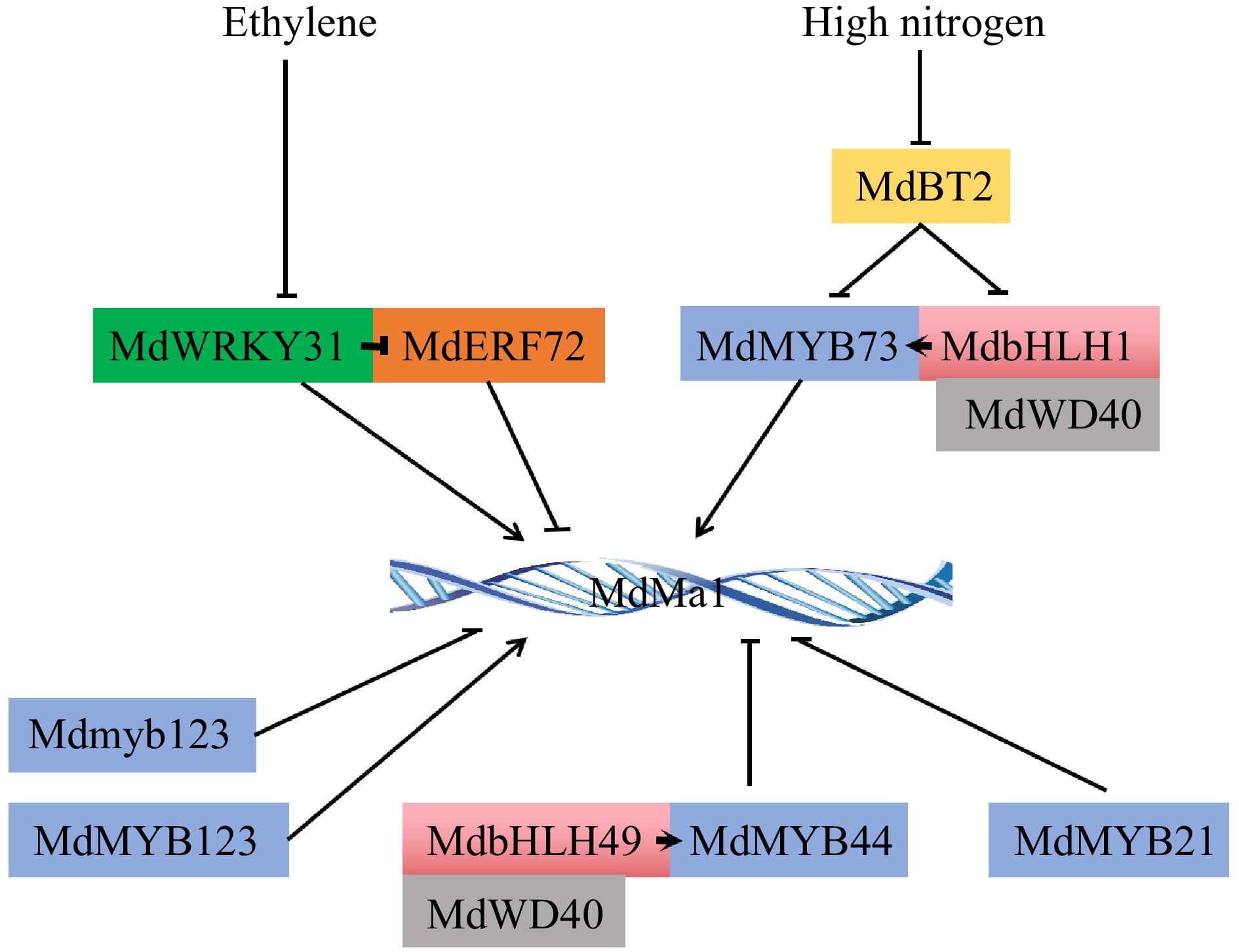
      Fruit acidity is due to the accumulation of organic acids such as malic acid, citric acid, and tartaric acid in vacuoles. The composition and content of organic acids in different fruits are significantly different, while malate is the main organic acid presented in most mature fruits[1,4]. Malate content in fleshy fruits is affected gravely by acid metabolism and transport[2,5]. In the cytoplasm, the intermediate product phosphoenolpyruvate (PEP) from glycolysis is catalyzed sequentially to malic acid by PEPC (PEP carboxylase) and NAD-cyt-MDH (NAD–dependent malate dehydrogenase)[1,68]. The function of NAD-cyt-MDH on increasing fruit acidity has been defined[8,9], which is believed to be the major pathway for malic acid synthesis[6,10]. Part of the organic acids accumulated in the cytoplasm are consumed as substances and energy to maintain the normal physiological activities of the cells, while the rest are transported and stored in the vacuole[2,11,12]. However, whether it can accumulate in large quantities in vacuoles is highly dependent on specific transmembrane transporters[4,12,13]. Currently, major fruit malate-regulated transporters include aluminum-activated malate transporter ALMT4 and ALMT9/Ma1[1417] and tonoplast dicarboxylate transporter tDT[4,17,18]. Additionally, H+ pumped into the cell via proton pump protein on the vacuole membrane combine with malate2− to form protonated malate for storage in the vacuole[1,12,13,19], providing continuous driving force for malate2− to enter the cell.

      The genetic mechanism of fruit acidity, a quantitative trait regulated by multiple genes, is relatively complex[2,20]. The function and regulatory mechanism analysis of malate metabolism and transport genes during fruit acidity accumulation is of great significance for scientific regulation of fruit quality. With the development of transcriptome, proteomics, and the application of gene fine-mapping technology in forward genetics, researchers have identified a series of metabolic and transport genes related to malate accumulation in apple[20,21], citrus[22,23], peach[24], watermelon[25], jujube[15], and loquat[26], and verified their regulatory functions of fruit acidity. This review focuses on the recent advances in metabolism, transport genes and upstream regulators related to malate accumulation in horticultural fruits and the future research direction of improving fruit quality by molecular biological methods are prospected.

    • The accumulation of malate in fruit cells is a complex phenomenon involving many metabolic pathways. The first step of malate synthesis in the cytoplasm is to fix CO2 to the carbon skeleton from hexose catabolism. The photocontracted products are delivered to the sink fruit over long distance transport of phloem[27], and the glucose produced by its decomposition is converted into the key substrate phosphoenolpyruvate (PEP) of malate metabolism in glycolysis. Then, PEP is converted to malate catalyzed by phosphoenolpyruvate carboxylase (PEPC) and subsequent cytoplasmic NAD-dependent malate dehydrogenase (NAD-cyMDH), and the reverse reaction is mediated by phosphoenolpyruvate carboxykinase (PEPCK)[1]. In addition, malate accumulated in the cytoplasm can be degraded to pyruvate via the NADP-dependent malic enzyme (NADP-cyME), and the remaining malate is transported and stored in the vacuole[28]. Therefore, the major enzymes involved in malate metabolism include PEPC, NAD-cyMDH, PEPCK, and NADP-cyME (Fig. 1).
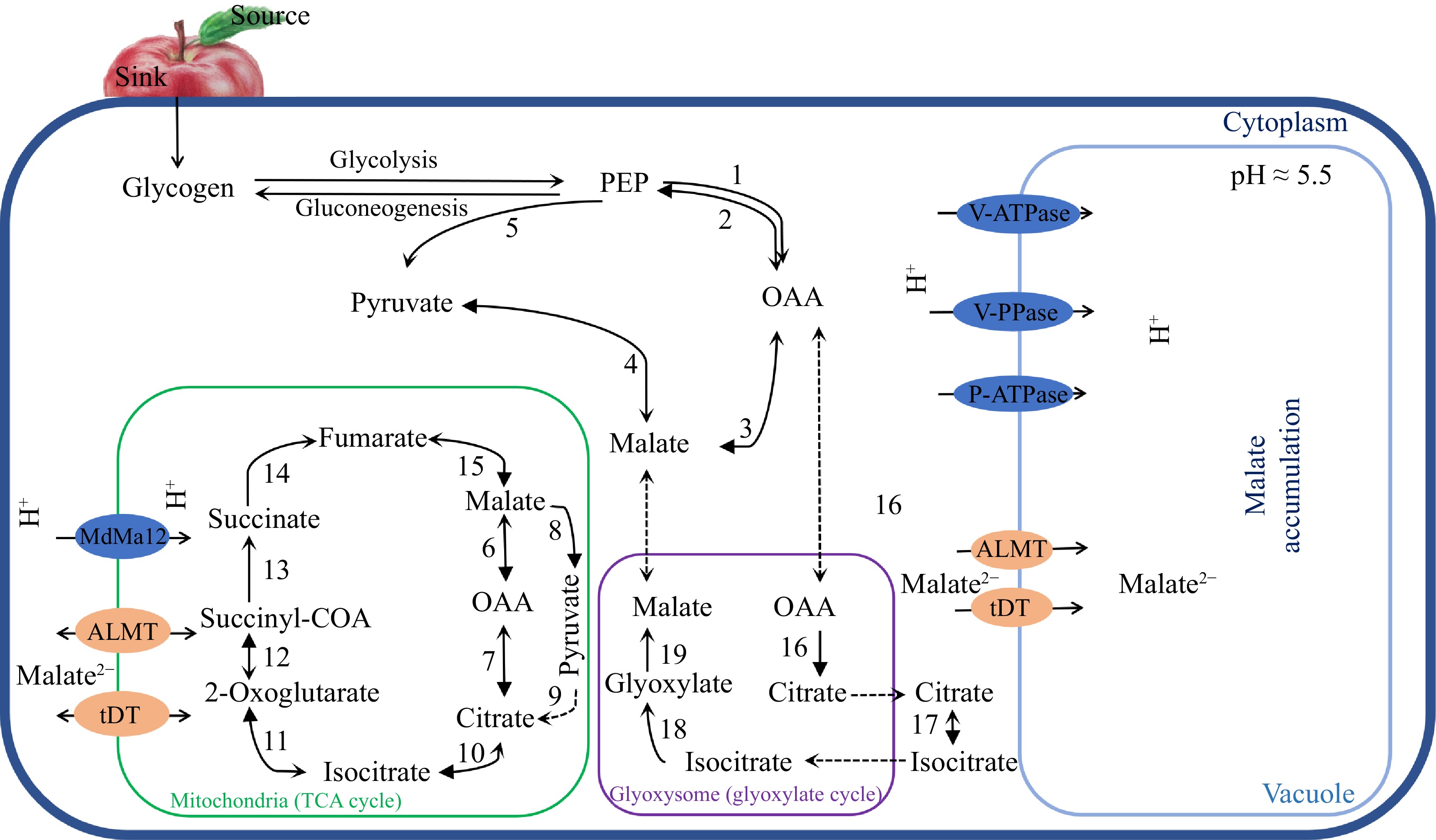
      Figure 1. 

      A simplified model for malate metabolism, transport and accumulation in fruit. Major enzymes involved in malate metabolism in fruit: 1, phosphoenolpyruvate carboxylase (PEPC); 2, phosphoenolpyruvate carboxykinase (PEPCK); 3, NAD-cyMDH (NAD-cytoplasmic malate dehydrogenase); 4, NADP-cytoplasmic malate enzyme (NADP-cyME); 5, pyruvate kinase (PK); 6, NAD-mitochondrial malate dehydrogenase (NAD-mtMDH); 7, mitochondrial citrate synthase (mtCS); 8, NAD-mitochondrial malate enzyme (NAD-mtME); 9, pyruvate dehydrogenase (PDH); 10, mitochondrial aconitase (mtACO); 11, isocitrate dehydrogenase (ICDH); 12, α-oxoglutarate dehydrogenase (α-OGDH); 13, succinyl-coa synthase (SCS); 14, succinate dehydrogenase (SuDH); 15, fumarase; 16, cytoplasmic citrate synthase (cyCS); 17, glyoxylate aconitase (glyACO); 18, isocitrate lyase (ICL); 19, malate synthase (MS).

      PEPC and NAD-cyMDH are involved in the synthesis and accumulation of malate in fruit. There was a positive correlation between the malate content and the expression levels of PEPC in apple and grape fruit during the early developmental stage[2931]. Similarly, NAD-cyMDH has also been shown to promote malate synthesis in young grape fruits[10,32]. In apple, NAD-cyMDH1 could improve malate accumulation in apple calli and fruit, as well as increase the salt tolerance of apple plants[9,28,33,34]. Recent studies have also demonstrated the important function of another apple gene NAD-cyMDH5 in enhancing malate accumulation in fruit[35].

      Physiological studies have revealed that the malate content of fleshy fruits such as apple, peach, and grape decreases significantly in the later stages of development[10,36], which is closely related to the malate decomposition genes NADP-cyME and PEPCK. Sweetman et al. pointed out that NADP-ME had low activity in the early stage of grape fruit development and was involved in the malate generating by fixing CO2[10]. As fruits gradually matured, the activity of NADP-ME increased significantly and began to catalyze the degradation of malate. Additionally, NADP-cyME is thought to be involved in the reduction of malate content during the ripening of loquat, apple, and grape fruit[6,37,38].

      In the later stage of fruit development, soluble sugars accumulated in large quantities, sugar catabolic metabolism slowed down, and vacuolar malate was released into the cytoplasm as a carbon source to participate in energy metabolism and compound biosynthesis through gluconeogenesis[10]. The first step of gluconeogenesis is the production of PEP catalyzed by PEPCK or phosphopyruvate dikinase (PPDK)[39,40]. Due to the bare expression of PPDK in fruits, PEPCK is believed to be more essential in the gluconeogenesis of fruits, which has been validated in grape skins and ripe peach, blueberry, redcurrant, and raspberry fruits[4143]. Compared with high-acid varieties, PEPCK activity in low-acid apples increased threefold, and14C-labeled malate consumption was higher, suggesting that gluconeogenesis might increase malate consumption[36]. Recent studies have suggested that the large accumulation of malate in the cytoplasm altered soluble sugar content in apple fruit, which might be caused by the up-regulating expression of MdPEPCK in gluconeogenesis[8]. In conclusion, PEPCK plays a key role in the regulation of malate accumulation in fruit.

    • Malate in the TCA cycle can be oxidized via two competing metabolic pathways. It is converted reversibly to OAA by NAD-mtMDH[10], or converted to pyruvate by NAD-mtME[44], which ultimately results in citrate synthesis and an altered ratio of malate and citrate. Omics analysis showed that NAD-mtMDH in mitochondria mainly catalyzed malate degradation during fruit ripening[10,32]. While NAD-mtMDH can also catalyze the malate synthesis when the conditions exist for the reversible reaction of the TCA[7,45]. In addition, the regulation of NAD-mtMDH varies among species. NAD-mtMDH appears to be regulated by the gene expression in loquat[46], while regulated at the post-transcriptional level in strawberry[47].

      Malate synthase glyMS located in the glyoxysome is involved in malate synthesis in the glyoxylic acid cycle[48] (Fig. 1). Studies have shown that the glyoxylic acid cycle is activated during the ripening of banana fruit after harvest, providing substrates for gluconeogenesis[49]. Pua et al.[50] detected the expression of glyMS only in banana fruit tissues, its expression was up-regulated during the whole ripening stage, whose change was similar to the trend of malate accumulation. However, ICL proteins required for glyoxylate (MS catalytic substrates) synthesis in the glyoxylate cycle was not detected at any stage of development in raspberry, blueberry, strawberry or redcurrant fruits[41], suggesting that the malate accumulation involved in the glyoxylate cycle during fruit ripening may be species-specific.

    • Malate synthesized in the cytoplasm and mitochondria will be transported and stored in vacuoles[7,35,51]. The passage of malate into or out of the vacuole requires specific anion channels or transporters located on the vacuole membrane as carriers, which can specifically recognize and transport malate[52,53]. In the cytoplasm with near-neutral pH, most malic acid exists in anion forms, while they will combine with cations (such as K+, Na+, Ca2+), forming salt in the acidic vacuole, which maintains an electrochemical gradient between the vacuole, allowing subsequent organic acids to enter the vacuole[1,53]. The main transporters associated with malate transmembrane delivery identified in horticultural crops include malate channel protein ALMT (aluminum-activated malate transporter), malate transporter tDT (tonoplast dicarboxylate transporter), and proton pump.

    • ALMT encodes aluminum-activated channel protein with typical transmembrane domains that transport malate2− in different organelles[54,55]. ALMT1 was initially identified in wheat root tip tissue and its malate transport properties were confirmed by the heterologous expression of xenopus cells[56]. Subsequently, researchers identified successively a series of ALMT proteins in the model plant Arabidopsis[57,58], major crops[59,60], vegetables[61,62] and fruit trees[63,64].

      The ALMT family is mainly divided into four subfamilies (ALMT I−IV) and the protein functions of each subfamily are various, mainly including aluminum tolerance, symbiotic nitrogen fixation, fertilization, ion transport, stomatal regulation, and fruit flavor[6567]. Therein, most of the ALMTs of the subfamily I and II are mainly located on the vacuole membrane and participate in malate transportation in plant cells. For example, the ALMT subfamily I genes AtALMT1, BnALMT1/2, HvALMT1 and ZmALMT2 from respectively Arabidopsis (Arabidopsis thaliana), rape (Brassica napus), barley (Hordeum vulgare L.), and maize (Zea mays L.) are primarily expressed in root and function on the regulation of malate secretion[6871]. The interference of LaALMT1 (Lupinus albus) led to a decrease in malate concentration in xylem sap[72], and the ALMT subfamily II genes SlALMT5 significantly increased malate and citrate contents in tomato seeds[73,74]. SlALMT11 located in leave guard cells transports malate to mediate stomatal closure in tomato[75]. AtALMT6 and AtALMT9 mediated the transcellular transport of malate or fumaric acid[57,75,71]. Moreover, it was found that AtALMT6 and AtALMT9 have strong inward rectification via membrane electrophysiology techniques, and malate transport only occurred under the condition of positive potential inside the vacuolar membrane[75,76]. As the vacuole pH decreases, the pathway by which AtALMT6 and AtALMT9 transport malate might be closed, which may be an active protection against excessive acidification of the vacuole.

      ALMT family genes are closely related to the regulation of fruit acid quality. Therefore, the function of ALMT9 on fruit vacuole acidification in various horticultural crops is the most widely studied. The Ma1 located on chromosome 16 in apple encodes an ALMT9 homologous gene, which is considered to be the main gene controlling the acidity of apple fruit[16,63,77]. The single nucleotide polymorphism (SNP) of Ma1 at 1,455 bases causes the premature termination of its protein translation, thereby losing the capability to transport malate into the vacuole, which is closely related to the decrease of fruit acidity[77]. In general, the acidity of mature apple fruit of genotype ma1/ma1 was significantly lower than that of genotype Ma1/Ma1 or Ma1/ma1[55,78]. While vacuolar membrane-localized VvALMT9 and SlALMT9 also are the homologs of AtALMT9 in grapes and tomatoes, respectively. VvALMT9 mediates the transport of malic acid and tartaric acid in grape fruit[79]. SlALMT9, located on chromosome 6 of tomato, is a major gene leading to the variation of malate content in tomato fruit, and the deletion of 3-bp in the promoter region of SlALMT9 destroys the W-box binding site and prevents the binding of its upstream transcriptional suppressor SlWRKY42, resulting in a high accumulation of malate in fruit[62]. Recent studies have also indicated that PpALMT9 mediated the accumulation of malate in pear fruit under salt stress[14]. Additionally, overexpression of ZjALMT4 and AcALMT1 significantly promoted the increase of organic acid content in sour jujube (Ziziphus jujuba Mill.) and in kiwifruit (Actinidia spp.) fruit[15,80]. Allogeneic expression of the ALMT family gene Pbr020270.1 of pear (Pyrus bretschneideri) could increase malate accumulation in tomato fruit[64]. While, a CitALMT gene of citrus (Citrus reticulata B.) negatively affects citrate accumulation in citrus fruit[81]. These results indicate that the aluminum-activated channel protein ALMT regulates malate accumulation in fruit vacuoles.

    • Tonoplast dicarboxylate transporters (tDT) are the first class of transmembrane transporters with malate transport properties discovered in plants[82]. In contrast to malate ion channel proteins, tDT has little rectification and plays an essential role in maintaining intracellular pH homeostasis[83]. Researchers first identified and demonstrated the acid transport function of AttDT in Arabidopsis thaliana. Overexpression of AttDT significantly increased malate content and decreased citrate accumulation in Arabidopsis leaves[84,85]. Further studies confirm that AttDT can also transport fumaric acid and succinic acid, and participate in the regulation of cytoplasmic pH homeostasis[83,85,86].

      So far, AttDT homologous genes have been isolated from various fruit such as apple (Malus domestica), tomato (Solanum lycopersicum), grape (Vitis vinifera), and citrus (Citrus sinensis). During citrus maturation, the AttDT homologous gene, CsCit1, encodes a vacuolar citrate3−/H+ symporter that mediates the effluence of H+ and CitH2− in vacuole to maintain vacuolar acidic pH and citrate balance[87]. Lin et al.[88] pointed out that CitDIC, a dicarboxylate transporter, and CitCHX, a cation/H+ exchange protein, were involved in the degradation of citrate during fruit development and the reduction of citrate in fruit after harvest triggered by hot air. The content of malate in SltDT overexpressed tomato fruit was significantly increased, while the citrate accumulation was inhibited[89]. Similarly, MdtDT negatively regulates the citrate content[90] and positively participates in the accumulation of malate in cultivated apple fruit[11,91,92]. The AttDT homologous gene in grape fruit is actively transporting tartaric acid into the vacuole[93]. In addition, mitochondrial dicarboxylate transporters VvDTC2 and VvDTC3 identified in grape are likely responsible for malate transport to mitochondria in grape fruit[94]. These studies indicate that tDT positively regulates malate and negatively regulates citrate accumulation in most fruit.

    • The transmembrane transport of malate is also affected by the vacuole pH and the electrochemical gradient (∆ψ) inside and outside the vacuole[1], while the activity and function of the proton pump greatly affects the vacuole pH and ∆ψ. Proton pump is a kind of membrane-integrated glycoprotein that can transport H+ across membranes against the concentration gradient, which mostly exists in the vacuolar membrane and plasma membrane, mainly including V-ATPase/H+-ATPase, V-PPase/H+-PPiase and P-ATPase[1,12,13,19], they pump H+ into the vacuole by hydrolyzing ATP or pyrophosphate, reducing the vacuole pH while increasing the ∆ψ on both sides of the membrane, thereby providing power for the transport of organic acids.

      V-ATPase and V-PPase are widely present in a variety of horticultural crops and are involved in secondary metabolite transport, vacuole acidification, ion homeostasis, and stress tolerance[9598]. Although V-ATPase and V-PPase are both effective in acidifying vacuoles, their activity varies in different plants and at different developmental stages of the same plant. V-ATPase is the main proton pump in the vacuoles of most horticultural plants, but V-PPase is more active than V-ATPase in some C4 plants. A large amount of highly active V-PPase is enriched in the early development stage of young tissue, hydrolyzing and removing pyrophosphate to inhibit the polymerization reactions such as RNA and starch synthesis. While, the synthesis of pyrophosphate in mature tissues is reduced and cell respiration continues to provide ATP, so V-ATPase activity dominates[4,99]. The activity analysis of the proton pump during the development of pear (Pyrus pyrifolia) fruit supported the above conclusion. V-PPase activity was highest in young fruit and decreased with the maturation of pear fruit, whereas V-ATPase activity was highest in mature fruit[100]. However, V-PPase is also the main vacuolar proton pump in grape berries whose vacuoles are strongly acidic (pH < 3)[93].

      The expression patterns of V-ATPase and V-PPase were similar in high-acid and low-acid loquat varieties, but their expression levels were higher in low-acid varieties[26]. Etienne et al.[101] found that the expression of V-ATPase and V-PPase in the fruit of different peach varieties were positively correlated with organic acid accumulation, indicating that V-ATPase and V-PPase were involved in the regulation of organic acid accumulation in fruit. Overexpression of V-ATPase proton pump MdVHP1 in apple calli increased the accumulation of malate and soluble sugar in vacuoles[91]. Further studies confirmed the active function of MdVHA-A3, MdVHA-D2, MdVHA-B1, MdVHA-B2, MdVHA-E2, CitAHA10, and CitVHA-c4 in fruit vacuole acidification[92,102104]. Moreover, a mitochondria-targeted PPase gene, Ma12, was identified in apple and its overexpression increased malate accumulation in apple calli and tomato fruit by up-regulating the expression of mitochondrial malate dehydrogenase mMDH12[7]. These results fully demonstrated the indispensable functions of V-ATPase and V-PPase in fruit vacuole acidification.

      P-ATPase is a new class of proton pump genes with proton transport and vacuolar acidification properties, which are divided into five subfamilies (P1−P5), among which the P3 subfamily ATPase is involved in the transport of organic acids. P3A-ATPase proton pump PhPH5, which is localized in the vacuolar membrane in petunias interacts with the P3B-ATPase proton pump PhPH1 to form a complex, which affects petal color by acidifying the vacuole[105,106]. Interestingly, the vast majority of P3A subfamily members are located in the plasma membrane, while only PhPH5 belongs to the vacuole membrane localization gene, exhibiting a strong ability to transport protons across membranes. It is unclear how PhPH5 acquired this unique cellular localization during evolution. The pH1−pH5 complex can reduce the stoichiometric value of H+/ATP from 1.0 to 0.5 for super acidifying vacuoles[107].

      The function of PH1−PH5 complex highly acidifying vacuoles exists only in a few angiosperms, and PH1 homologs are lacking in most plants[106]. How the independent loss of PH1 homologs occurs in multiple plants are unclear. Studies have shown that PH1 and PH5 can be expressed ectopically in plants where certain tissues do not express them, resulting in a decrease of vacuole pH in the corresponding tissues[108]. At present, the researchers have begun to explore whether the pH1−pH5 complex has the function of acidifying fruit vacuoles. The hyper-acidification of citrus fruit is regulated by the P-ATPase complex CitPH1-CitPH5[109]. Similarly, the PhPH5 homolog Ma10 and the PhPH1 homolog Ma13 in apple were reported to regulate the malate accumulation in apple calli and tomato fruit[12,110], however, it is not clear whether there is an interactive relationship between the Ma10 and Ma13. The interference of VvWRKY26 and VvMYB5 in grape leaves significantly decreased the transcriptional expression of VvPH5 and VvPH1, causing increased vacuole pH[111]. In the same year, researchers identified and proved that the expression level of CsPH8, a homologous gene of PhpH5, was highly consistent with the changing trend of citrate content in various citrus fruits at different developmental stages, and overexpression of CsPH8 significantly increased the citrate accumulation in strawberry fruit[22]. Similarly, overexpression of the P3A-ATPase proton pump gene PbPH5 also significantly increased the malate accumulation in pear fruit[112]. It can be seen that P-ATPase alone or in the complex form, both play an important roles in vacuole acidifying.

    • Many transcription factors play essential roles in the regulation of malate content by influencing the expression of genes related to malate metabolism and transport (Table 1). MdcyMDH1 is identified as a major gene associated with malate accumulation via MapQTL in 'Honeycrisp' × 'Qinguan' F1 hybrids, and after MdcyMDH1 is overexpressed, the malate concentration of fruit is enhanced[8,9,28]. MdbHLH3 and MdWRKY126 directly activated the transcriptional expression of MdcyMDH1, and also increased the expression of malate transport-related genes such as MdtDT, increasing malate content in fruit[34,35]. The indel of a repeat sequence in MdcyMDH1 (MA7) promoter region in 'Gala' (MA7/MA7) and 'Fuji' (ma7/ma7) apple varieties were named respectively 'MA7' and 'ma7', and the upstream regulator, MdbHLH74, could enhance the expression of MdcyMDH1 in apple with MA7/MA7 genotype, but not with ma7/ma7 genotype, affecting the malate content in different varieties[9]. Additionally, TRXL1 up-regulates NADP-cyMDH activity, increases malate accumulation, and inhibits superoxide radical formation in response to high-temperature stress, and the expression of TRXL1 is positively regulated by CPN60A and negatively regulated by CLPC1[113]. Based on current research, the main object of transcriptional regulation of malate metabolism is cytoplasmic malate dehydrogenase, and the regulatory network of other malate metabolism-related genes need to be further studied.

      Table 1.  The crucial genes and their upstream regulatory factors of fruit acidity regulation.

      Gene family Gene name Protein name Activation (+)/
      inhibition (−)
      Module control Species
      MYB transcription factor MdMa1 MdMYB73 + MdBT2-MdCIbHLH1
      -MdMYB73
      Malus domestica
      MdMa1/MdMa11 MdMYB123 + Malus domestica
      MdMa1 MdMYB44 MdbHLH49-MdMYB44 Malus domestica
      MdMa1 MdMYB21 Malus domestica
      MdVHA-A3/D2 Ma10 MdMYB44 WD40-MdbHLH49
      -MdMYB44
      Malus domestica
      MdVHA-B1/E MdVHP1
      MdtDT
      MYB1/10 + MdTTG1-MdbHLH3
      -MdMYB1/10
      Malus domestica
      MdVHA-A MdVHP1 MdMYB73 + MdBT2-
      WD40-MdbHLH1
      -MdMYB73
      Malus domestica
      CitPH5 CitPH4 + CitTRL-CitPH4 Citrus reticulata
      WRKY transcription factor SlALMT9 SlWRKY42 Solanum lycopersicum
      PpALMT9 PpWRKY44 + PpABF3-PpWRKY44 Pyrus spp.
      ZjALMT4 ZjWRKY7 + Ziziphus jujuba
      MdMa1 MdWRKY31 + Malus domestica
      MdMDH1 MdWRKY126 + Malus domestica
      bHLH transcription factor MdMDH1 MdbHLH3 + Malus domestica
      NAC transcription factor AcALMT1 AcNAC1 + Actinidia spp.
      ERF transcription factor MdMa1 MdERF72 MdWRKY31-MdERF72 Malus domestica
      CitVHA-C4 CiERF13 + Citrus reticulata
      PP2C family MdVHA-A3/B2/D2 Ma10 MdPP2CH SAUR37-MdPP2CH Malus domestica
    • Jia et al.[20] identified three major genes associated with malate transport (MdPP2CH, MdMYB44, and MdSAUR37) via MapQTL and BSA-seq and verified their functions. MdPP2CH reduced the malate accumulation by phosphorylating the proton pump gene in apple calli, while MdSAUR37 could inhibit the phosphorylation activity of MdPP2CH and positively regulate malate content. Another acid accumulation major gene, MdMYB44, negatively regulated proton pump gene Ma10, MdVHA-A3, and MdVHA-D2 and malate transporter Ma1 to inhibit fruit vacuole acidification. A further study indicated that the presence of SNP (A/T) in the MdMYB44 promoter affected the ability of its upstream transcription factor MdbHLH49 to regulate the activity of the MdMYB44 promoter and malate accumulation of fruit[104]. Additionally, other MYB transcription factors also play an essential role in proton pump regulation. Apple MdMYB1/10 directly binds and activates the expression of proton pump genes MdVHA-B1, MdVHA-B2, MdVHA-E2, and MdVHP1, accelerating the malate accumulation of vacuoles[92]. The MdCIbHLH1-MdMYB73 module regulates downstream proton pump genes MdVHA-A, and MdVHP1 for the acidification of fruit vacuole[11], while the MdBT2 response to nitrate treatment could ubiquititatively degrade MdCIbHLH1, and malate content in MDBT2-silenced apple calli is significantly upregulated[114].

      Studies on the transcriptional regulation of malate transporters focus on tDT and ALMT proteins. tDT positively regulates the malate content and negatively participates in the accumulation of citrate in tomato and apple fruit[89,90], which was regulated by transcription factor MdMYB1[92], MdMYB73[11] and MdbHLH3[34]. The latest study revealed that AP2 domain-containing transcription factor MdESE3 activate their expression of MdtDT, MdMa11, and MdMDH12 to increase malate accumulation in apple[18]. While, ALMT9, the major contributor of fruit malate accumulation, has constant attention from reseachers, and an increasing number of transcriptional regulatory mechanisms regarding the influence of ALMT9 on fruit acidity have emerged. The ALMT9 homologous genes have been identified as a crucial gene functioning in enhancing malate transport, and vacuole acidification in various horticultural crops such as apple[77], tomato[62], grape[79], and pear[14], where expression is regulated respectively by the transcription factors MdWRKY31-MdERF72[115], MdMYB73[11,114], MdMYB21[116], MdMYB123[117], MdMYB44[104], SlWRKY42[62], and PpABF3-PpWRKY44[14] (Fig. 2). Interestingly, the malate content in stable Ma1-overexpressed apple fruits was significantly reduced. Further studies showed that alternative splicing generates two Ma1 isoforms (higher expression of MA1α and lower expression of Ma1β). Ma1β is only able to form polymers with MA1α protein for strong malate transport function and the absence or reduced transport activity of MA1α/Ma1β polymers in Ma1 transgenic fruits decreased malate accumulation, which was regulated by MdMYB73[16].

      Figure 2. 

      Upstream regulators of major gene Ma1 regulating apple fruit acidity. The arrows represent positive regulation and the rest represent negative regulation.

    • Protein post-translational modification is a kind of chemical modification existing in the late stage of protein biosynthesis that affects the protein stability and activity by changing different biochemical functional groups on amino acid residues of proteins, including protein phosphorylation, ubiquitination, acetylation and methylation. MdPP2CH decreased the malate content via phosphorylating the proton pump MdVHA-A3, MdVHA-B2, MdVHA-D2 and ALMT transporter MdALMTII, and its dephosphatase activity was inhibited by MdSAUR37 in apple[20]. Some enzymes involved in malate metabolism, such as malate dehydrogenase (MDH2) and phosphoenolpyruvate carboxykinase (PEPCK1), are also regulated by either the glucose-induced degradation-deficient pathway or the vacuole import and degradation pathway[118], but the specific protein modification mechanisms remain to be investigated.

      Posttranslational modification except for the above directly modified functional proteins associated with malate accumulation, can also modify upstream regulatory proteins of functional genes to affect fruit acidity. The ubiquitin E3 ligase MdCOP1 degrades MdMYB1 in the dark through a ubiquitin-dependent pathway to regulate anthocyanins and malate accumulation[92,119]. Similar, high glucose-inhibited U-box-type E3 ubiquitin ligase MdPUB29 and glucose sensor MdHXK1 ubiquitinates and phosphorylates MdbHLH3, respectively, affecting malate concentration[120,121]. It was found that the apple transcription factor MdCIbHLH1 acidified fruit vacuole by enhancing the activity of MdMYB73, which promoted the up-regulated expression of MdVHA-A, MdVHP1 and MdALMT9. While BTB-BACK-TAZ domain protein MdBT2 degrades ubiquititatively MdCIbHLH1 and MdMYB73 via ubiquitin/26S proteasome pathway to regulate the malate content of vacuole in apple plants under nitrate stress[114,122]. These studies provide groundbreaking insights into the direct posttranslational modification of organic acid-related functional proteins and their upstream regulatory proteins, which is helpful to cultivate high-quality horticultural crop varieties from the perspective of post-translational modification.

    • To date, researchers have performed some basic research on the accumulation of malate in horticultural crops. Members of the acid metabolism and transport families have been screened and identified at the genome-wide level in most horticultural crops, and their function on vacuole acidification has been demonstrated in apple, tomato, pear, and Arabidopsis. However, the systematic regulatory network of malate accumulation and the cross-regulation between sugar and acid metabolism and transport remain to be further explored. Therefore, the following suggestions are put forward for the future research direction of malate accumulation regulation in horticultural crops.

    • Epigenetics is a kind of 'post-genetics' that can achieve genetic heritability under the premise that the nuclear DNA sequence remains unchanged through the methylation modification or histones acetylation, phosphorylation, and ubiquitination of gene promoter DNA. This regulatory mechanism of epigenetic modification has been elucidated to some extent in fruit soluble sugar and anthocyanin accumulation[123125], indicating epigenetic modification plays a crucial role in fruit quality formation. In pummelo (Citrus maxima LCA) and lemon (Citrus limon (L.) Burm f.), DNA methylation changes in promoters of key genes involved in citrate synthesis and accumulation directly affect the citrate content in the flesh[23,126], revealing a previously unexplored link between epigenetic regulation and organic acid accumulation of horticultural fruits. A recent study identified a CgAN1, BHLH-type regulator coupling citrate and anthocyanin, from citrus varieties with high citrate, anthocyanin, and low citrate, anthocyanin, and confirmed that the reduction of the methylation level of the gene promoter can enhance the citrate accumulation of fruit[23]. However, the epigenetic mechanism related to fruit acidity, especially malate, is relatively limited, the urgent task is to uncover the complex DNA methylation mechanisms controlling key genes in malate synthesis, and accumulation pathways in horticultural crops.

    • The differences in the composition and content of soluble sugars and organic acids play a decisive role in fruit quality, and flavor[127]. Physiological studies have revealed that during the ripening process of fleshy fruit such as apple, peach, and grape, organic acid content decreases, and soluble sugar accumulation increases[10,28,36,128]. The variousness in malate content caused by the overexpression of MDH or ME genes could lead to a change in the redox state of the plastid thus affecting the accumulation of starch and sugar in tomato fruit cells[129,130]. Yao et al.[28,91] pointed out that the overexpression of MdVHP1 and MdcyMDH1 in apple calli both increased malate and soluble sugar contents, and the effect of MdcyMDH1 on malate and soluble sugar accumulation was regulated by transcription factor MdbHLH3[34]. While high glucose-inhibited U-box-type E3 ubiquitin ligase MdPUB29 and glucose sensor MdHXK1 ubiquitinates and phosphorylates MdbHLH3, respectively, affecting the expression of its downstream genes[120,121]. FaMYB44.2 could inhibit the expression of FaSPS, reducing both the sucrose and malate content in banana fruit[131]. Similarly, the increased accumulation of malate, citrate, glucose, and fructose was observed in SlAREB1 overexpressed red ripe peel compared to antisense-inhibited lines[132]. A recent study has also indicated that the accumulation of malate in the cytoplasm mediated by MdcyMDH1 increased the sucrose content in apple fruit by up-regulating the expression of MdSPS, which is likely to be achieved via starch cleavage or gluconeogenesis[8]. These above studies indicate that there is an interactive relationship between carbohydrate and organic acid accumulation in fruit. However, the spatiotemporal crosstalk between sugars and acids during fruit development remains unclear. Therefore, elucidation of the potential cross-regulatory mechanisms of sugars and acids in fruit is important to optimize the ratio of sugars to acids in fruit and improve fruit quality.

      The exploration of a clear and thorough regulatory network of malate accumulation and regulation in horticultural crops relies on the functional identification of major genes in malate metabolism and transport in fruit and the in-depth study of the above directions, which lays an important foundation for the improvement of fruit quality via molecular-assisted breeding.

    • The authors confirm contribution to the paper as follows: study conception and design: Zhang LH; data collection: Li MJ, Zhang LH, Zhang AN, Xu Y; analysis and interpretation of results: Zhang LH, Zhang AN, Xu Y, Zhu LC, Ma BQ; draft manuscript preparation: Zhang LH, Zhang AN, Xv Y, Zhu LC, Ma BQ. All authors reviewed the results and approved the final version of the manuscript.

    • All data generated or analyzed during this study are included in this published article.

      • This work was supported by the Outstanding Youth Science Foundation of Heilongjiang Province (YQ2023C006), the China Postdoctoral Science Foundation (2023MD744175), the Talent Introduction Program of Northeast Agricultural University of China, and Modern Agricultural Industrial Technology Collaborative Innovation and Promotion System of Heilongjiang Province. The authors would like to thank Mr. Li Dalong from Northeast Agricultural University for providing the instruments.

      • The authors declare that they have no conflict of interest.

      • Copyright: © 2024 by the author(s). Published by Maximum Academic Press, Fayetteville, GA. This article is an open access article distributed under Creative Commons Attribution License (CC BY 4.0), visit https://creativecommons.org/licenses/by/4.0/.
    Figure (2)  Table (1) References (132)
  • About this article
    Cite this article
    Zhang LH, Zhang AN, Xu Y, Zhu LC, Ma BQ, et al. 2024. Accumulation and regulation of malate in fruit cells. Fruit Research 4: e031 doi: 10.48130/frures-0024-0025
    Zhang LH, Zhang AN, Xu Y, Zhu LC, Ma BQ, et al. 2024. Accumulation and regulation of malate in fruit cells. Fruit Research 4: e031 doi: 10.48130/frures-0024-0025

Catalog

  • About this article

/

DownLoad:  Full-Size Img  PowerPoint
Return
Return