LETTER   Open Access    

Neofunctionalization of B-class genes in regulating rice flower development

More Information
  • Received: 12 April 2024
    Revised: 21 June 2024
    Accepted: 26 June 2024
    Published online: 02 August 2024
    Seed Biology  3 Article number: e013 (2024)  |  Cite this article
  • Rice (Oryza sativa L.) is a world staple crop that feeds over half of the world's population[1,2]. In recent years, high-temperature events are becoming more frequent and intensive as a result of global warming, which can severely affect rice grain yield and quality[3,4]. During flowering and grain filling stages, high-temperature stress can result in a significant reduction in seed setting rate and influence amylose content, starch fine structure, functional properties and chalkiness degree of rice[57]. Transcriptome and proteome analysis in rice endosperm have also been used to demonstrate the differences in high-temperature environments at gene and protein expression levels[810]. In addition, as an important post-translational modification, protein phosphorylation has proven to be involved in the regulation of starch metabolism in response to high-temperature stress[11]. However, little is known about whether protein ubiquitination regulates seed development under high-temperature stress.

    Ubiquitination is another form of post-translational modification that plays key roles in diverse cellular processes[12]. Several reports have described the functions of ubiquitination in rice defense responses based on ubiquitome analysis. Liu et al.[13] investigated relationships between ubiquitination and salt-stress responses in rice seedlings using a gel-based shotgun proteomic analysis and revealed the potential important role of protein ubiquitination in salt tolerance in rice. Xie et al.[14] identified 861 peptides with ubiquitinated lysines in 464 proteins in rice leaf cells by combining highly sensitive immune affinity purification and high resolution liquid chromatography-tandem mass spectrometry (LC-MS/MS). These ubiquitinated proteins regulated a wide range of processes, including response to stress stimuli. A later study revealed the relationships between ubiquitination status and activation of rice defense responses, and generated an in-depth quantitative proteomic catalog of ubiquitination in rice seedlings after chitin and flg22 treatments, providing useful information for understanding how the ubiquitination system regulates the defense responses upon pathogen attack[15]. Although many studies have shown that ubiquitination plays improtant roles in the heat response of plant[16,17], there has been little systematic discussion on the ubiquitome of rice endosperm in the context of global climate change.

    In this study, we examine the high-temperature induced ubiquitination change in two rice varieties with different starch qualities, through a label-free quantitative ubiquitome analysis. This study provides a comprehensive view of the function of ubiquitination in high-temperature response of rice developing seed, which will shed new light on the improvement of rice grain quality under heat stress.

    Two indica rice varieties with different starch quality, 9311 and Guangluai4 (GLA4), were used as materials. 9311 is a heat-sensitive variety, which displays low amylose content with good starch quality; while GLA4 is known to be the parental variety of HT54, an indica breeding line with heat tolerance, and thus GLA4 is possibly heat tolerant, which shows high amylose content with poor starch quality[18,19]. Rice growth conditions, sample treatment and collection were conducted as previously described[11].

    Husk, pericarp and embryo were detached from immature rice grains on ice[20]. Rice endosperm was then ground with liquid nitrogen, and the cell powder was sonicated 10 rounds of 10 s sonication and 15 s off-sonication on ice in lysis buffer (6 M Guanidine hydrochloride, pH 7.8−8.0, 0.1% Protease Inhibitor Cocktail) using a high intensity ultrasonic processor. Following lysis, the suspension was centrifuged at 14,000 g for 40 min at 4 °C to remove the debris. The supernatant was collected, and the protein concentration was estimated using BCA assay (Pierce BCA Protein assay kit, Thermo Fisher Scientific, Waltham, MA, USA) before further analysis.

    The protein mixture was reduced by DTT with the final concentration of 10 mM at 37 °C for 1 h, alkylated by iodoacetamide with a final concentration of 50 mM at room temperature in the dark for 0.5 h, and digested by trypsin (1:50) at 37 °C for 16 h. Then the sample was diluted by adding trifluoroacetic acid (TFA) to the final concentration of 0.1%. The enzymatic peptides were desalted on a Sep-Pak C18 cartridge (Waters, Milford, MA, USA), concentrated by lyophilization and reconstituted in precooled IAP buffer (50 mM MOPS-NaOH PH 7.2, 10 mM Na2HPO4, 50 mM NaCl) for further analysis.

    The peptides solution was incubated with prewashed K-ε-GG antibody beads (PTMScan Ubiquitin Remnant Motif (K-ε-GG) Kit), and gently shaken at 4 °C for 1.5 h. The suspension was centrifuged at 2,000 g for 30 s, and the supernatant was removed. The Anti-K-ε-GG antibody beads were washed with IAP Buffer three times and with ddH2O three times. The peptides were eluted from the beads with 0.15% trifluoroacetic acid (TFA). Finally, the eluted fractions were combined and desalted with C18 Stage Tips.

    LC-MS/MS analysis were performed using the methods of Pang et al.[11]. Raw mass spectrometric data were analyzed with MaxQuant software (version 1.3.0.5) and were compared with the indica rice protein sequence database (Oryza sativa subsp. indica-ASM465v1). Parameters were set according to Pang et al.[11]. All measurements were obtained from three separate biological replicates.

    Quantification of the modified peptides was performed using the label-free quantification (LFQ) algorithm[11]. Differentially ubiquitinated sites (proteins) in response to high-temperature were identified by Student's t-test (p < 0.05, log2(fold-change) > 1) with at least two valid values in any condition or the ubiquitination sites that exhibited valid values in one condition (at least two of three replicates) and none in the other.

    Subcellular localization was performed using CELLO database (http://cello.life.nctu.edu.tw). Gene Ontology (GO) annotation proteome was derived from the AgriGO (http://bioinfo.cau.edu.cn/agriGO/). The differential metabolic profiles were visualized with MapMan software (version 3.6.0RC1). Kyoto Encyclopedia of Genes and Genomes (KEGG) pathway annotation was performed by using KEGG Automatic Annotation Server (KAAS) software. A p-value of < 0.05 was used as the threshold of significant enrichment. SWISS-MODEL was used to generate the tertiary structure of GBSSI (SWISS-MODEL, http://swissmodel.expasy.org/). The figures were annotated with Adobe Illustrator (Adobe Systems, San Jose, CA, USA).

    To elucidate how high-temperature stress influences rice developing endosperm at the ubiquitination level, a label-free analysis was performed to quantify ubiquitome from two indica rice varieties under normal (9311-C and GLA4-C) and high-temperature conditions (9311-H and GLA4-H). The distribution of mass error of all the identified peptides was near zero and most of them (71.5%) were between -1 and 1 ppm, suggesting that the mass accuracy of the MS data fits the requirement (Fig. 1a). Meanwhile, the length of most peptides distributed between 8 and 42, ensuring that sample preparation reached standard conditions (Fig. 1b).

    Figure 1.  Characteristics of the ubiquitinated proteome of rice endosperm and QC validation of MS data. (a) Mass error distribution of all identified ubiquitinated peptides. (b) Peptide length distribution. (c) Frequency distribution of ubiquitinated proteins according to the number of ubiquitination sites identified.

    In all endosperm samples, a total of 437 ubiquitinated peptides were identified from 246 ubiquitinated proteins, covering 488 quantifiable ubiquitinated sites (Supplemental Table S1). Among the ubiquitinated proteins, 60.6% had only one ubiquitinated lysine site, and 18.7%, 8.1%, 5.3%, or 7.3% had two, three, four, or five and more ubiquitinated sites, respectively. In addition, four proteins (1.6%, BGIOSGA004052, BGIOSGA006533, BGIOSGA006780, BGIOSGA022241) were ubiquitinated at 10 or more lysine sites (Fig. 1c, Supplemental Table S1). The proteins BGIOSGA008317 had the most ubiquitination sites with the number of 16. It was noted that besides ubiquitin, two related ubiquitin-like proteins NEDD8 and ISG15 also contain C-terminal di-Gly motifs generated by trypsin cleavage, and the modifications of these three proteins cannot be distinguished by MS[21]. Here, the di-Gly-modified proteome therefore represents a composite of proteins modified by these three proteins. However, the sites from NEDD8 or ISG15 modifications were limited because they mediate only a few reactions in cells[21].

    To better understand the lysine ubiquitome changes in rice endosperm induced by high-temperature, we performed a Gene Ontology (GO) functional annotation analysis on all identified ubiquitinated proteins (Fig. 2a). In the biological process GO category, 'metabolic process' and 'cellular process' were mainly enriched, accounting for 75.1% and 74.1% of ubiquitinated proteins, respectively. In addition, 34.6% proteins were associated with 'response to stimulu', emphasizing the regulatory role of ubiquitination modification in response to high-temperature stress. From the cellular component perspective, ubiquitinated proteins were mainly associated with 'cellular anatomical entity' (99.4%), 'intracellular' (84.4%) and 'protein-containing complex' (29.9%). The molecular function category result suggested that these proteins were largely involved in 'binding' (62.7%), 'catalytic activity' (43.4%) and 'structural molecule activity' (16.5%). Furthermore, subcellular location annotation information indicated that 34.7%−39.4% proteins were located in the cytoplasm, and other were mostly located in the nucleus (23.5%−27.7%), plasma membrane (9.4%−11.4%), and chloroplast (9.6%−12.8%) (Fig. 2b). It is noteworthy that the ubiquitinated proteins located in the cytoplasm were decreased in high-temperature environments in both varieties.

    Figure 2.  Analysis of ubiquitinated proteins and motifs. (a) Gene ontology (GO) functional characterization of ubiquitinated proteins. (b) Subcellular localization of ubiquitinated proteins. From the inside out, the ring represents 9311-C, 9311-H, GLA4-C and GLA4-H, respectively. (c) Motif enrichment analysis of ubiquitinated proteins.

    The following two significantly enriched motifs from all of the identified ubiquitinated sites were identified using MoDL analysis: [A/S]xKub and Kubxx[E/Q/R/V]x[E/G/L/P/Q/R/Y], which covered 84 and 100 sequences, respectively (Fig. 2c). Further analysis showed that the conserved alanine (A) and glutamic acid (E) were included in upstream and downstream of the ubiquitinated lysine sites in rice endosperm. A similar phenomenon also occurred in rice leaf[14], wheat leaf[22], and petunia[23], indicating that alanine (A) and glutamic acid (E) were likely to be the specific amino acids in conserved ubiquitination motifs in plants. Additionally, serine (S) was enriched at the position -2 (upstream) of the ubiquitinated lysine, while various amino acids such as arginine (R), glutamic acid (E), glutamine (Q), valine (V) were found at positions +3 and +5 (downstream).

    To detect possible changes in rice endosperm ubiquitome attributable to high-temperature stress, we then performed LFQ analysis on all quantifiable ubiquitination sites within our dataset. As shown in Fig. 3a, more ubiquitinated proteins, peptides and sites were detected in the treatment groups (9311-H and GLA4-H), suggesting that exposure to high-temperature stress may increase the ubiquitination events in rice endosperm. Only 282 common ubiquitinated sites in 158 proteins were quantifiable for all sample groups due to reversible ubiquitination induced by high-temperature (Fig. 3b). Principal component analysis (PCA) showed that three repeats of each sample clustered together, and four groups were clearly separated (Fig. 3c). Furthermore, the differentially expression profiles of ubiquitination sites (proteins) in 9311 and GLA4 under high-temperature stress were depicted to further understand the possible changes (Fig. 3d). Where LFQ values were missing, the data were filtered to identify those ubiquitination sites with a consistent presence/absence expression pattern. These analyses yielded 89 ubiquitination sites that were only present in 9311-H and six that were only present in 9311-C (Fig. 3d, Supplemental Table S2). Similarly, 51 differentially expressed ubiquitination sites were present in GLA4-H and 13 ubiquitination sites only occurred in GLA4-C (Fig. 3d & Supplemental Table S3). Beyond that, a total of 113 and 50 significantly changed ubiquitination sites (p < 0.05, log2(fold-change) >1) were screened out in 9311 and GLA4, respectively (Fig. 3d, Supplemental Tables S4 & S5). For subsequent comparative analysis, the ubiquitination expression profiles with consistent presence/absence and ubiquitination sites with significant differences in statistical testing were combined and named as 9311-Up, 9311-Down, GLA4-Up, and GLA4-Down, respectively (Fig. 3d). The number of significantly up-regulated ubiquitination sites was far greater than down-regulated ubiquitination sites in both 9311 and GLA4 varieties. These findings indicated that high temperature not only induced the occurrence of ubiquitination sites, but also significantly upregulated the intensity of ubiquitination. Beyond that, the magnitude of the up-regulation in 9311 was higher than that in GLA4 (Fig. 3b & d), indicating that the ubiquitination modification of heat-sensitive varieties was more active than heat-resistant varieties in response to high-temperature stress.

    Figure 3.  A temperature regulated rice endosperm ubiquitome. (a) The number of ubiquitinated proteins, peptides and sites detected in four group samples. (b) Venn diagram of ubiquitination sites (proteins) detected in four group samples. (c) PCA based on ubiquitination intensity across all four sample groups with three biological repetitions. (d) Differentially expression profiles of ubiquitination sites (proteins) in 9311 and GLA4 under high-temperature stress. The expression profiles of selected ubiquitination sites (p < 0.05, log2(fold-change) >1) were normalized using the Z-score and presented in a heatmap.

    To further investigate the ubiquitination regulatory pattern under high temperature stress in two varieties, four groups with significantly regulated sites were analyzed. There were 37 ubiquitination sites showed the same regulatory trend in 9311 and GLA4, accounting for 17.8% and 32.5% of the total differentially expressed sites in 9311 and GLA4, respectively. Among them, 36 ubiquitination sites were upregulated and one site was downregulated (Fig. 4a). In addition, 159 upregulated ubiquitination sites and three downregulated sites were only present in 9311, while 53 upregulated sites and 15 downregulated sites were only present in GLA4. Moreover, nine ubiquitination sites showed opposite regulatory trends in 9311 and GLA4. A similar regulatory trend of ubiquitination proteins is shown in Fig. 4b. It is noted that some proteins had both upregulated and downregulated ubiquitination sites (Supplemental Tables S6 & S7), indicating that significant differences in ubiquitination were, to some extent, independent of protein abundance.

    Figure 4.  Comparison of differentially ubiquitinated sites and proteins in 9311 and GLA4 under high-temperature stress.

    To understand the function of ubiquitination in response to the high-temperature stress of rice endosperm, we conducted GO enrichment-based clustering analysis of the differentially ubiquitinated proteins in 9311 and GLA4 at high temperature, respectively (Fig. 5). In the biological process category of 9311, proteins were relatively enriched in the carbohydrate metabolic process, polysaccharide metabolic process, starch biosynthetic process, cellular macromolecule localization, protein localization, intracellular transport, and phosphorylation (Fig. 5). For the molecular function analysis, we found that the proteins related to kinase activity, nucleotidyltransferase activity, phosphotransferase activity, and nutrient reservoir activity were enriched (Fig. 5). The two principal cellular components were intrinsic component of membrane and integral component of membrane (Fig. 5). There was no significantly enriched GO term in the GLA4 group due to the dataset containing relatively few proteins, and thus, further enrichment analysis was conducted on the proteins that were common to both varieties. The results showed that proteins were over-represented in carbon metabolism, including starch biosynthesis and metabolism, glucan biosynthesis and metabolism, and polysaccharide biosynthesis and metabolism (Fig. 5), indicating the importance of carbohydrate synthesis and metabolism in the ubiquitination regulatory network.

    Figure 5.  Enrichment analysis of differentially expressed ubiquitinated proteins based on Gene Ontology (GO) terms.

    To identify pathways which were differentially ubiquitinated under high-temperature stress, the KEGG pathway-based clustering analysis was conducted. The results showed that the differentially ubiquitinated proteins in both 9311 and GLA4 were mostly abundant in the pathways of carbohydrate metabolism, starch and sucrose metabolism, folding, sorting and degradation, translation, ribosome, and protein processing in endoplasmic reticulum (Fig. 6a). In the 9311 group, the pathways of carbohydrate metabolism, starch and sucrose metabolism, glycosyltransferases, glycolysis, and energy metabolism were enriched in the differentially ubiquitinated proteins (Fig. 6b); while there was no significantly enriched KEGG pathway in the GLA4 group. We further found the proteins that were common to both varieties were only significantly enriched in the starch and sucrose metabolism pathways (p = 0.04). The ubiquitination proteins involved in the starch and sucrose metabolism mainly include: sucrose hydrolysis (SUS, FK, UGPase), and starch synthesis (AGPase, GBSSI, BEI, BEIIb, PUL, PHO1), which are discussed below.

    Figure 6.  KEGG classification and enrichment analysis of differentially ubiquitinated proteins. (a) Number of differentially ubiquitinated proteins based on KEGG classification in 9311 and GLA4. (b) KEGG enrichment analysis of differentially ubiquitinated proteins in 9311.

    Although many reports have described specific examples of ubiquitination in rice defense responses[13,15,16], our knowledge on global changes in the developing endosperm ubiquitome under high-temperature stress is still lacking. In this study, a label-free quantitative proteomic analysis of ubiquitination was applied to examine the high-temperature induced ubiquitination change of two indica rice varieties (9311 and GLA4) with distinct starch quality. We identified many new lysine modification sites on proteins involved in various pathways, highlighting the complexity of the ubiquitination-mediated regulatory system in high-temperature stress responses in rice.

    Heat shock proteins accumulate under various stresses and play important roles in plant defenses against abiotic stresses[24,25]. Research has shown that a number of heat shock proteins were prominent in the rice ubiquitome network, of which OsHSP71.1, and OsHSP82A showed increased ubiquitination levels under chitin and flg22 treatment[15]. Here, seven lysine residues on five heat shock proteins possessed ubiquitination modification in rice endosperm. Three sites (BGIOSGA011420-K78, BGIOSGA026764-K99, BGIOSGA029594-K106) showed significant up-regulation in 9311 under high-temperature stress, while in GLA4, the ubiquitination level of BGIOSGA011420-K78 was down-regulated. This differential ubiquitination of heat-tolerant and heat-sensitive varieties provided a basis for studying the regulation of post-translational modification of heat shock proteins under high-temperature stress, despite the regulatory role of those heat shock proteins being still unclear.

    Transcription factors (TFs) play an essential role in the regulation of gene expression. A total of three transcription factors were identified in the ubiquitination dataset, of which two were NAC family members. As one of the largest plant-specific TF families, NAC is involved in the responses to abiotic and biotic stresses[26]. The ubiquitination modification of K173 in BGIOSGA018048 was specifically expressed in the 9311-H group, which may affect the stress resistance level in high-temperature environments. In addition, two sites K148 and K149 of ERF TF family member BGIOSGA024035 was downregulated in GLA4 under high-temperature stress. This differential ubiquitination was likely to affect the expression of related genes regulated by this transcription factor.

    The results of GO and KEGG enrichment analysis of differential ubiquitination proteins indicated that the sucrose and starch metabolic pathway was largely affected by ubiquitination regulation under high-temperature stress (Figs 5 & 6). The ubiquitination sites involved in sucrose and starch metabolism are listed in Table 1. To assess how high-temperature stress affects the crucial pathway, the significantly differential ubiquitination sites in 9311 and GLA4 were displayed in the heatmap of specific proteins (Fig. 7).

    Table 1.  Ubiquitination sites related to sucrose and starch metabolism in rice endosperm.
    Gene nameAnnotationProtein entryModification site(s)
    SUS1Sucrose synthase 1BGIOSGA010570K172, K177
    SUS2Sucrose synthase 2BGIOSGA021739K160, K165, K176, K804
    SUS3Sucrose synthase 3BGIOSGA026140K172, K177, K541, K544, K588
    FKFructokinaseBGIOSGA027875K143
    UGPaseUDP-glucose pyrophosphorylaseBGIOSGA031231K27, K150, K303, K306
    AGPS1ADP-glucose pyrophosphorylase small subunit 1BGIOSGA030039K94, K464, K484
    AGPS2ADP-glucose pyrophosphorylase small subunit 2BGIOSGA027135K106, K132, K385, K403, K406, K476, K496
    AGPL2ADP-glucose pyrophosphorylase large subunit 2BGIOSGA004052K41, K78, K134, K191, K227, K254, K316, K338, K394, K396, K463, K508, K513
    AGPL3ADP-glucose pyrophosphorylase large subunit 3BGIOSGA017490K509
    GBSSIGranule bound starch synthase IBGIOSGA022241K130, K173, K177, K181, K192, K258, K371, K381, K385, K399, K462, K517, K530, K549, K571, K575
    BEIStarch branching enzyme IBGIOSGA020506K103, K108, K122
    BEIIbStarch branching enzyme IIbBGIOSGA006344K134
    PULStarch debranching enzyme:PullulanaseBGIOSGA015875K230, K330, K431, K736, K884
    PHO1Plastidial phosphorylaseBGIOSGA009780K277, K445, K941
     | Show Table
    DownLoad: CSV
    Figure 7.  Sucrose and starch pathway at the ubiquitination levels in rice endosperm under high-temperature stress.

    In cereal endosperm, sucrose is the substrate for the biosynthesis of starch. The formation of glucose 1-phosphate (G1P, used in starch synthesis, see below) from sucrose requires a series of enzymes[27]. Here, we found that sucrose synthase 1 (SUS1), SUS2, SUS3, fructokinase (FK), and UDP-glucose pyrophosphorylase (UGPase) were ubiquitinated among all sample groups (Table 1, Fig. 7). In the ubiquitome of seedling and leaf in japonica rice, ubiquitination sites have been found in SUS1, SUS2, UGPase, and FK, which were related to sucrose hydrolysis[14,15]. SUS catalyzed the process of cleaving sucrose into UDP-glucose (UDPG) and fructose. Two ubiquitination sites, K172 and K177, were identified in SUS1 in rice endosperm, which were also found in rice leaves[14]. A total of four ubiquitination sites were identified in SUS2, two of which were also reported in rice seedling and leaf, indicating the conservation of the lysine residues in different rice tissues. It was noted that all four ubiquitination sites in SUS2 were upregulated in high-temperature environments, although the regulated sites of 9311 and GLA4 were different. In 9311, the ubiquitination levels of K160, K174, and K804 were increased, while GLA4 was only upregulated in K176. The ubiquitination sites K541, K544, and K588 in SUS3 were screened from developing rice seeds for the first time. In addition, SUS3 had two completely overlapping sites K172 and K177 with SUS1, and it was difficult to determine which enzymes the two sites belonged to. The ubiquitination levels of SUS3-K541 and SUS3-K544 in 9311 significantly increased in high-temperature environments, while there was no significant difference in the ubiquitination level of SUS3 in GLA4. Overall, the ubiquitination sites of SUS in rice endosperm were located in the functional domain except for SUS2-K804, reflecting the importance of ubiquitination regulation in SUS.

    UGPase catalyses the conversion of glucose 1-phosphate and UTP into UDPG[28]. Research has shown that the mutation of UGPase gene lead to chalky endosperm[29]. As shown in Table 1, four ubiquitination sites K27, K150, K303, and K306 were identified in rice endosperm, which were completely inconsistent with the seven ubiquitination sites in rice seedlings and two in leaves[14,15], reflecting the tissue specificity. We speculated that the UGPase with different modification sites may play different regulatory roles in metabolic pathways in different tissues. Under high-temperature stress, the ubiquitination level of UGPase-K27 was 8.1-fold up-regulated. Liao et al.[10] demonstrated that the expression of UDPase was down-regulated in both heat-tolerant and heat-sensitive rice lines under high temperature conditions, which could reasonably explain the significant up-regulation of UGPase-K27 ubiquitination level. The ubiquitination site K143 of FK was also reported in seedling tissues[15].

    The AGPase reaction represents the first committed step of starch biosynthesis[27]. A total of 22 lysine ubiquitination sites were identified in four AGPase subunits (AGPL2, AGPL3, AGPS1, AGPS2). AGPL2 had 13 ubiquitination sites, of which six were located in NTP_transferase domain, including K254, K338, K191, K134, K227, and K316. High-temperature stress resulted in an increase in the ubiquitination level of K254 in both 9311 and GLA4, and significant upregulation of K508 and K513 in 9311, as well as K191, K227, and K316 in GLA4. In contrast, AGPL2-K394 were significantly downregulated in GLA4. AGPL3 contained one ubiquitination site K509, and the modification level of AGPL3-K509 was up-regulated in high-temperature environments in 9311. AGPS1 had one specific ubiquitination site K464 and another two sites K94 and K484 that completely overlapped with AGPS2-K106 and AGPS2-K496, respectively. The modification levels of K464 and K484 significantly increased in high-temperature environments in 9311, and K94 was significantly up-regulated in both varieties. There were seven ubiquitination sites in AGPS2 in rice endosperm, which were different with the sites found in rice leaves[14]. In addition to the two sites that overlapped with AGPS1, AGPS2 had another two ubiquitination sites (K406 and K132) that upregulated in high-temperature environments.

    Amylose content is one of the key determinants that strongly influence rice grain quality[30]. The biosynthesis of amylose requires the catalytic effect of granule-bound starch synthase I (GBSSI)[30,31]. Here, a total of 16 ubiquitination sites were identified in GBSSI (Table 1, Fig. 8a). Among these ubiquitination sites, six lysine residues (K130, K173, K177, K181, K192, K258) were located in glycosyltransferase 5 (GT5) domain, and three sites (K399, K462, K517) were located in GT1 domain (Fig. 6), indicating the important role of ubiquitination regulation of GBSSI. Under high-temperature stress, the ubiquitination levels of six sites (K130, K177, K399, K381, K385, K549) increased in two indica rice varieties, while one sites (K258) showed downregulation in 9311 (Fig. 8a). Numerous studies had described that the amylose content was reduced under high-temperature stress in rice[5,7], which might be due to the degradation of GBSSI proteins caused by the increased significantly up-regulated ubiquitination sites. These ubiquitination sites identified in rice GBSSI with significant differences under high-temperature stress were expected to become a new breakthrough point for the improvement of starch quality.

    Figure 8.  Structure of GBSSI. (a) Domain structure of GBSSI and ubiquitination sites with significant differences in response to high-temperature stress. (b) 3D model of GBSSI and the relationship between ubiquitination sites K462 and ADP, SO4 (salt bridge or hydrogen bond).

    To further determine the regulatory role of the ubiquitination sites in GBSSI, SWISS-MODEL was used to predict 3D structural model. As shown in Fig. 8b, GBSSI had three SO4 (sulfate ions) and one ADP ligand. These ligands interact with GBSSI through hydrogen bonds and salt bridges. Three sites, K447, R458, and K462, were associated with SO4 through salt bridges, while G100, N265, Q412, K462, and Q493 interact with the hydrogen bonds of ADP in GBSSI[32,33]. Based on this finding, it can be reasonably inferred that the K462 site with ubiquitination modification located in the GT1 domain played an important role in the interaction between GBSSI, SO4, and ADP. An in-depth investigation was necessary to gain a more comprehensive understanding of the regulatory function of ubiquitination modification at GBSSI-K462, although there was no significant difference in the ubiquitination level under high-temperature stress.

    Amylopectin, the major component of starch, is synthesized by the coordinated action of multiple enzymes including soluble starch synthase (SSs), starch branching enzyme (BEs), starch debranching enzyme (DBEs), and phosphorylases (PHOs or Phos) with ADPG as a substrate. In this study, ubiquitination sites were detected in BEs, DBEs, and Phos.

    BEs, covering two isoforms, BEI and BEII, are responsible for catalyzing the formation of α-1,6-glucosidic linkages of amylopectin[34]. There were three ubiquitination sites (K103, K108, and K122) identified in BEI (Fig. 9a). K122 was the first amino acid in the carbohydrate-binding module 48 (CBM48) domain. Sequence alignment analyses of BEs from eight plants revealed K122 was conserved among all plants' BEI (Fig. 9a), suggesting a high probability of the functional effects of ubiquitination modification at this site. In high-temperature environments, ubiquitination levels of K108 and K122 were significantly up-regulated in 9311, while no significantly regulated ubiquitination sites of BEI were observed in GLA4. Only one ubiquitination site, K134, was found in BEIIb (Fig. 9a). The ubiquitination levels showed a slightly upward trend with no significant differences in high-temperature environments in both varieties. These changes could be one of the reasons for increased gelatinization temperature and relative crystallinity of rice starch in response to high-temperature[5].

    Figure 9.  Domain structure of (a) BEs, (b) PUL and (c) Pho1 as well as their ubiquitination sites with significant differences in response to heat stress. Residues in red indicate the ubiquitination site. Non-ubiquitinated residues are shown in dark grey.

    DBEs consists of isoamylase (ISA) and pullulanase (PUL) with catalytic function for hydrolyzing α-1,6-glucosic linkages[35]. In the present study, we found that only PUL was ubiquitinated in rice endosperm (Fig. 9b). Among five ubiquitination sites (K230, K330, K432, K736, and K884) identified in PUL, K230 was located in the PULN2 domain, while K330 was in the CBM48 domain. Under high-temperature stress, K330 showed completely opposite regulatory trends in two cultivars. In addition, the ubiquitination level of K884, located in the DUF3372 domain, was significantly up-regulated in 9311. Previous study has reported that the expression of PUL was significantly up-regulated in 9311 under high-temperature stress, while GLA4 showed down-regulation in PUL abundance[11]. Consequently, there might be two possible functions of these ubiquitination sites. One possibility is that ubiquitination sites were unrelated to protein degradation; instead, they regulated the biosynthesis of amylopectin by affecting other functions of the protein. Secondly, ubiquitination sites were associated with protein degradation, and the levels of ubiquitination modification were based on protein abundance, resulting in a completely consistent regulation of ubiquitination modification and protein abundance under high-temperature stress.

    PHOs, including two types, Pho1/PHO1 and Pho2/PHO2, are responsible for the transfer of glucosyl units from Glc-1-P to the non-reducing end of a-1,4-linked glucan chains[36]. Pho1 is a temperature-dependent enzyme and considered crucial not only during the maturation of amylopectin but also in the initiation process of starch synthesis[37,38]. The three ubiquitination sites (K277, K445, K941) identified in Pho1 were located in two phosphorylase domains. We found that two sites, Pho1-K277 and Pho1-K445, were only ubiquitinated in high-temperature environments in 9311 and GLA4, respectively. Pang et al.[11] has demonstrated that the protein abundance of Pho1 decreased under high-temperature stress, especially in GLA4. Satoh et al.[38] reported that the functional activity of Pho1 was weakened under conditions of high temperature and its function might be complement by one or more other factors. Hence, these ubiquitination modifications that specifically occurred in high-temperature environments might be related to the degradation of Pho1 proteins.

    As a factory for protein synthesis in cells, the ribosome is an extremely crucial structure in the cell[39]. It has been proven that multiple ribosomal subunits were abundantly ubiquitinated in Arabidopsis and wheat[22]. In the present study, 57 ubiquitination sites involving 33 ribosome subunits were identified in 40S and 60S ribosome complexes in rice. Under high-temperature stress, the ubiquitination levels of some sites were significantly upregulated or downregulated, implying that ubiquitination of ribosomal proteins is likely to be an important regulatory mechanism in high-temperature response in rice endosperm. The results of GO and KEGG enrichment analysis indicated that the ribosome system was one of the most active systems for ubiquitination regulation under high-temperature stress. We speculated that the ubiquitin-proteasome system might be involved in the removal of subunits or entire ribosomes that were improperly folded in high-temperature environments. As shown in Fig. 10, the S10e, L18Ae, S27Ae, L9e, S3e, S28e, S20e, and S2e subunits were significantly up-regulated in 9311, while L13e subunits showed a completely opposite regulatory trend at the ubiquitination sites K81 and K88. In GLA4, the ubiquitination levels of S10e, S27Ae, L10Ae, L9e, S3e, S2e, and L4e showed a significant increase, while the ubiquitination level of L17e was significantly down-regulated under high-temperature stress. A total of seven ubiquitination sites involving S10e, S27Ae, L9e, S3e, and S2e subunits were jointly up-regulated in both two varieties. These sites might be related to the degradation of improperly folded ribosome subunits under high-temperature stress, while other ubiquitination sites with variety specificity might be associated with ribosomal function.

    Figure 10.  Ribosome system at the ubiquitination levels in rice endosperm under high-temperature stress. Grey shadings represent ubiquitinated proteins with no significant differences under heat stress. Red and blue shadings indicate up-regulated and down-regulated ubiquitinated proteins, respectively. Orange shading displays a combination of up- and down-regulated ubiquitinated sites in the same ubiquitinated protein.

    In conclusion, this study provides the first comprehensive view of the ubiquitome in rice developing endosperm, and demonstrated that ubiquitination has diverse functions in the high-temperature response of rice endosperm by modulating various cellular processes, especially the sucrose and starch metabolism. Comparative analysis of the temperature-induced ubiquitination status revealed some similarities and more interesting differences between 9311 and GLA4. These differences might be the reason for the different qualities formation of the two indica rice varieties, which could provide potential genetic resources for the improvement of the heat resistance in rice. Considering the diversity of ubiquitination modification, it is worthwhile to further validate and explore the function and regulatory mechanism of the key targets and key pathways. The findings provide valuable insights into the role of ubiquitination in response to high-temperature stress and lay a foundation for further functional analysis of lysine ubiquitination in rice.

    The authors confirm contribution to the paper as follows: study conception and design: Bao J, Pang Y; data collection: Pang Y; analysis and interpretation of results: Pang Y; draft manuscript preparation: Ying Y; Revised manuscript preparation: Ying Y, Pang Y, Bao J. All authors reviewed the results and approved the final version of the manuscript.

    The datasets generated during and/or analyzed during the current study are available from the corresponding author on reasonable request.

    This work was financially supported by the AgroST Project (NK2022050102) and Zhejiang Provincial Natural Science Foundation (LZ21C130003).

  • The authors declare that they have no conflict of interest.

  • Supplemental Fig. S1 OsMADS2 mutant alleles (a) Schematic diagram of OsMADS2 gene structure. Grey boxes represent 5’UTR and 3’UTR, black boxes represent exons, and thick black lines represent introns. The black arrowhead indicates the sgRNA target site, Osmads2-1 and Osmads2-2 had “A” and “TT” insertion in the OsMADS2 coding sequence, respectively. The underlined letters indicate protospacer adjacent motif (PAM), Red letters indicate mutation types. (b) The sequencing results showing mutation information of Osmads2-1 and Osmads2-2, compared to that in wild type. Red arrows indicate mutation site.
    Supplemental Fig. S2 Phenotypic analysis of spikelet morphology in Osmads2-1 and Osmads2-2 (a) and (b) Spikelet of Osmads2-1 (a1-a4) and Osmads2-2 (b1-b4). Pink arrowheads in a2, a4, b1 and b4 indicate extended lodicules; elo, extended lodicule; le, lemma; lo, lodicule; pa, palea; pi, pistil; st, stamen. Scale bars, 2 mm.
    Supplemental Fig. S3 OsMADS4 mutant alleles (a) Schematic diagram of the OsMADS4 gene. Grey boxes represent 5’UTR and 3’UTR, black boxes represent exons, and thick black lines represent introns. The black arrowhead indicates the mutation target site, Osmads4-1 and Osmads4-2 contain a T deletion and a T insertion, respectively, in the OsMADS4 coding sequence. (b) Sequencing results of the target site of wild type, Osmads4-1 and Osmads4-2.
    Supplemental Fig. S4 The in-situ hybridization sense control of DL DL is the abbreviation of DROOPING LEAF; Scale bars, 50 μm.
    Supplemental Fig. S5 Phenotypic analysis of spikelet development in Osmads4-1 and Osmads4-2 (a) and (b) Spikelet of osmads4-1 (a1-a4) and osmads4-2 (b1-b4) single mutant. ca, carpel; le, lemma; lo, lodicule; pa, palea; pi, pistil; st, stamen. Scale bars, 2 mm.
    Supplemental Fig. S6 Phenotypic analysis of spikelet morphology in Osmads2-1Osmads4-1 double mutant. (a) Spikelet of Osmads2-1 Osmads4-1.(b) Spikelet of Osmads2-1 Osmads4-1 after the removal of lemma. (c) Spikelet of Osmads2-1 Osmads4-1 after both lemma and palea were removed. Blue arrowheads indicate glume-like structure organs; red arrowhead indicate abnormal-pistils; yellow arrowheads indicate enlarged ovaries. a-pi, abnormal-pistil. eno, enlarged ovary; gll, glume-like structure; le, lemma; lo, lodicule; pa, palea. Scale bars, 2 mm.
    Supplemental Fig. S7 Expression analysis of B-class genes in WT and mutants. (a) OsMADS2 expression pattern in the inflorescence of WT and Osmads4-1. (b)  RT-qPCR analysis of OsMADS4 in WT and Osmads2-1. (c) RT-qPCR analysis of OsMADS4 in WT, Osmads2-1 and Osmads4-1. Total RNA was isolated from 3-mm and 5-7-mm young inflorescence of WT, Osmads2-1 and Osmads4-1. Data are shown as mean± sd, Error bars indicate SD for three biological replicates. ** indicates P-values < 0.01 and * indicates P-values between 0.05 and 0.01, analyzed by the Student-t test.
    Supplemental Fig. S8 Phenotypic characterization of the spikelet in the Osmads2-1 Osmads6-1, Osmads4-1 Osmads6-1 and Osmads2-1 Osmads4-1 Osmads6-1 mutants (a) to (c) Spikelet morphology (1-4) and schematic diagram (5) of Osmads2-1 Osmads6-1 (a), Osmads4-1 Osmads6-1 (b) and Osmads2-1 Osmads4-1 Osmads6-1 (c). Aqua blue arrowheads indicate glume-like structures; Orange arrowheads indicate lodicule-stamen mosaic organs; Light yellow arrowheads indicate abnormal pistils. a-pi, abnormal pistil; bop, body of palea; fl, floret; gll, glume like organ; le, lemma; lo, lodicule; l-s, lodicule-stamen mosaic organ; mrp, margin region of palea; pa, palea; rg, rudimentary glume; sl, sterile lemma; sp, spikelet; v, vascular bundles. All scale bars are 2 mm.
    Supplemental Fig. S9 Phenotypic analysis of the spikelet in the Osmads2-1 Osmads3-4 and Osmads4-1 Osmads3-4 double mutants. (a)to (c) Spikelet morphology of Osmads3-4. (d) to (f) Spikelet morphology of Osmads2-1 Osmads3-4. (g) to (i) Spikelet morphology of Osmads4-1 Osmads3-4. Orange arrowheads indicate lodicule-stamen mosaic organ; Yellow arrowheads indicate abnormal pistil; Rose arrowheads indicate lodicule-stigma mosaic organ. le, lemma; lo, lodicule; l-p, lodicule-pistil mosaic organ; l-s, lodicule-stamen mosaic organ; pa, palea; pi, pistil; st, stamen. Scale bars, 2 mm.
    Supplemental Fig. 10 Expression analysis of DL and some MADS-box genes in WT and mutants. (a) Expression level of DL in WT, Osmads2-1 and Osmads4-1. (b) to (f) Expression levels of class-E genes (OsMADS1, OsMADS5, OsMADS7, OsMADS8 and OsMADS34) (b, c, d, e and f, respectively) in WT and the Osmads2-1 and Osmads4-1 mutants. (g) and (h) Expression level of OsMADS32 and OsMADS6 in WT and the Osmads2-1 and Osmads4-1 mutants.
    Supplemental Table S1 Primers used in this study.
  • [1]

    Airoldi CA, Davies B. 2012. Gene duplication and the evolution of plant MADS-box transcription factors. Journal of Genetics and Genomics 39:157−65

    doi: 10.1016/j.jgg.2012.02.008

    CrossRef   Google Scholar

    [2]

    Mondragón-Palomino M, Theissen G. 2011. Conserved differential expression of paralogous DEFICIENS- and GLOBOSA-like MADS-box genes in the flowers of Orchidaceae: refining the 'orchid code'. The Plant Journal 66:1008−19

    doi: 10.1111/j.1365-313X.2011.04560.x

    CrossRef   Google Scholar

    [3]

    Yuan Z, Persson S, Zhang D. 2020. Molecular and genetic pathways for optimizing spikelet development and grain yield. aBIOTECH 1:276−92

    doi: 10.1007/s42994-020-00026-x

    CrossRef   Google Scholar

    [4]

    Yoshida H. 2012. Is the lodicule a petal: molecular evidence? Plant Science 184:121−28

    doi: 10.1016/j.plantsci.2011.12.016

    CrossRef   Google Scholar

    [5]

    Hu Y, Liang W, Yin C, Yang X, Ping B, et al. 2015. Interactions of OsMADS1 with Floral Homeotic Genes in Rice Flower Development. Molecular Plant 8:1366−84

    doi: 10.1016/j.molp.2015.04.009

    CrossRef   Google Scholar

    [6]

    Li H, Liang W, Jia R, Yin C, Zong J, et al. 2010. The AGL6-like gene OsMADS6 regulates floral organ and meristem identities in rice. Cell Research 20:299−313

    doi: 10.1038/cr.2009.143

    CrossRef   Google Scholar

    [7]

    Hu Y, Wang L, Jia R, Liang W, Zhang X, et al. 2021. Rice transcription factor MADS32 regulates floral patterning through interactions with multiple floral homeotic genes. Journal of Experimental Botany 72:2434−49

    doi: 10.1093/jxb/eraa588

    CrossRef   Google Scholar

    [8]

    Yun D, Liang W, Dreni L, Yin C, Zhou Z, et al. 2013. OsMADS16 genetically interacts with OsMADS3 and OsMADS58 in specifying floral patterning in rice. Molecular Plant 6:743−56

    doi: 10.1093/mp/sst003

    CrossRef   Google Scholar

    [9]

    Yao SG, Ohmori S, Kimizu M, Yoshida H. 2008. Unequal genetic redundancy of rice PISTILLATA orthologs, OsMADS2 and OsMADS4, in lodicule and stamen development. Plant and Cell Physiology 49:853−57

    doi: 10.1093/pcp/pcn050

    CrossRef   Google Scholar

    [10]

    Li H, Liang W, Hu Y, Zhu L, Yin C, et al. 2011. Rice MADS6 interacts with the floral homeotic genes SUPERWOMAN1, MADS3, MADS58, MADS13, and DROOPING LEAF in specifying floral organ identities and meristem fate. The Plant Cell 23:2536−52

    doi: 10.1105/tpc.111.087262

    CrossRef   Google Scholar

    [11]

    Li H, Liang W, Yin C, Zhu L, Zhang D. 2011. Genetic interaction of OsMADS3, DROOPING LEAF, and OsMADS13 in specifying rice floral organ identities and meristem determinacy. Plant Physiology 156:263−74

    doi: 10.1104/pp.111.172080

    CrossRef   Google Scholar

    [12]

    Yadav SR, Prasad K, Vijayraghavan U. 2007. Divergent regulatory OsMADS2 functions control size, shape and differentiation of the highly derived rice floret second-whorl organ. Genetics 176:283−94

    doi: 10.1534/genetics.107.071746

    CrossRef   Google Scholar

    [13]

    Smaczniak C, Immink RGH, Muiño JM, Blanvillain R, Busscher M, et al. 2012. Characterization of MADS-domain transcription factor complexes in Arabidopsis flower development. Proceedings of the National Academy of Sciences of the United States of America 109:1560−65

    doi: 10.1073/pnas.1112871109

    CrossRef   Google Scholar

  • Cite this article

    Wang L, Li QL, Hu JP, Yuan Z. 2024. Neofunctionalization of B-class genes in regulating rice flower development. Seed Biology 3:e013 doi: 10.48130/seedbio-0024-0012
    Wang L, Li QL, Hu JP, Yuan Z. 2024. Neofunctionalization of B-class genes in regulating rice flower development. Seed Biology 3:e013 doi: 10.48130/seedbio-0024-0012

Figures(1)

Article Metrics

Article views(2516) PDF downloads(400)

Other Articles By Authors

LETTER   Open Access    

Neofunctionalization of B-class genes in regulating rice flower development

Seed Biology  3 Article number: e013  (2024)  |  Cite this article
  • A typical flower comprises four whorls of organs: sepal, petal, stamen, and pistil. Based on the floral quartet model (FQM), the identities of these floral organs are determined by various combinations of the ABCDE-class MADS-box proteins that function in overlapping domains within the flower[1]. The proliferation of the ABCDE genes in modern flowering plants is strongly correlated with the evolution and diversification of floral patterns, as exemplified by the independent duplication of B-class genes that led to petal derivation in the Orchid and Liliaceae families[2].

    Spikelet, the unique flower structure of grass inflorescence, contains highly specialized non-reproductive organs[3]. In rice for example, each spikelet comprises two pairs of bract-like organs and a floret that consists of lemma and palea in the first whorl, two lodicules that are generally regarded as petal equivalent in the second whorl[4], six stamens in the third whorl, and one pistil in the center whorl (Fig. 1a). OsMADS14, OsMADS15, OsMADS18, and OsMADS20 are A-class genes that regulate palea development. OsMADS2, OsMADS4, and OsMADS16 are B-class genes that act together with the A-class genes to determine lodicule development. OsMADS3 and OsMADS58 are C-class genes that act in combination with the B-class genes to control stamen formation. OsMADS13 and OsMADS21 are D-class genes that function together with the C-class genes to regulate ovule formation and development. OsMADS1, OsMADS5, OsMADS7, OsMADS8, and OsMADS34 are rice SEP-like (E-class) genes, serving as the 'glume' genes that participate in all floral organ development processes by interacting with proteins from the other classes to specify floral organ identity and determinacy[5]. Additionally, OsMADS6 and OsMADS17 are AGAMOUS-LIKE6 (AGL6) homologs with similar function to the E-class proteins, whereby OsMADS6 is known to control floral meristem (FM) and floral determinacies[6]. OsMADS32, an orphan protein in monocotyledonous plants, regulates floral context by interacting with other floral homeotic proteins[7]. However, to what extent the FQM can be applied to the development of the non-reproductive organs in the spikelet is still largely unclear.
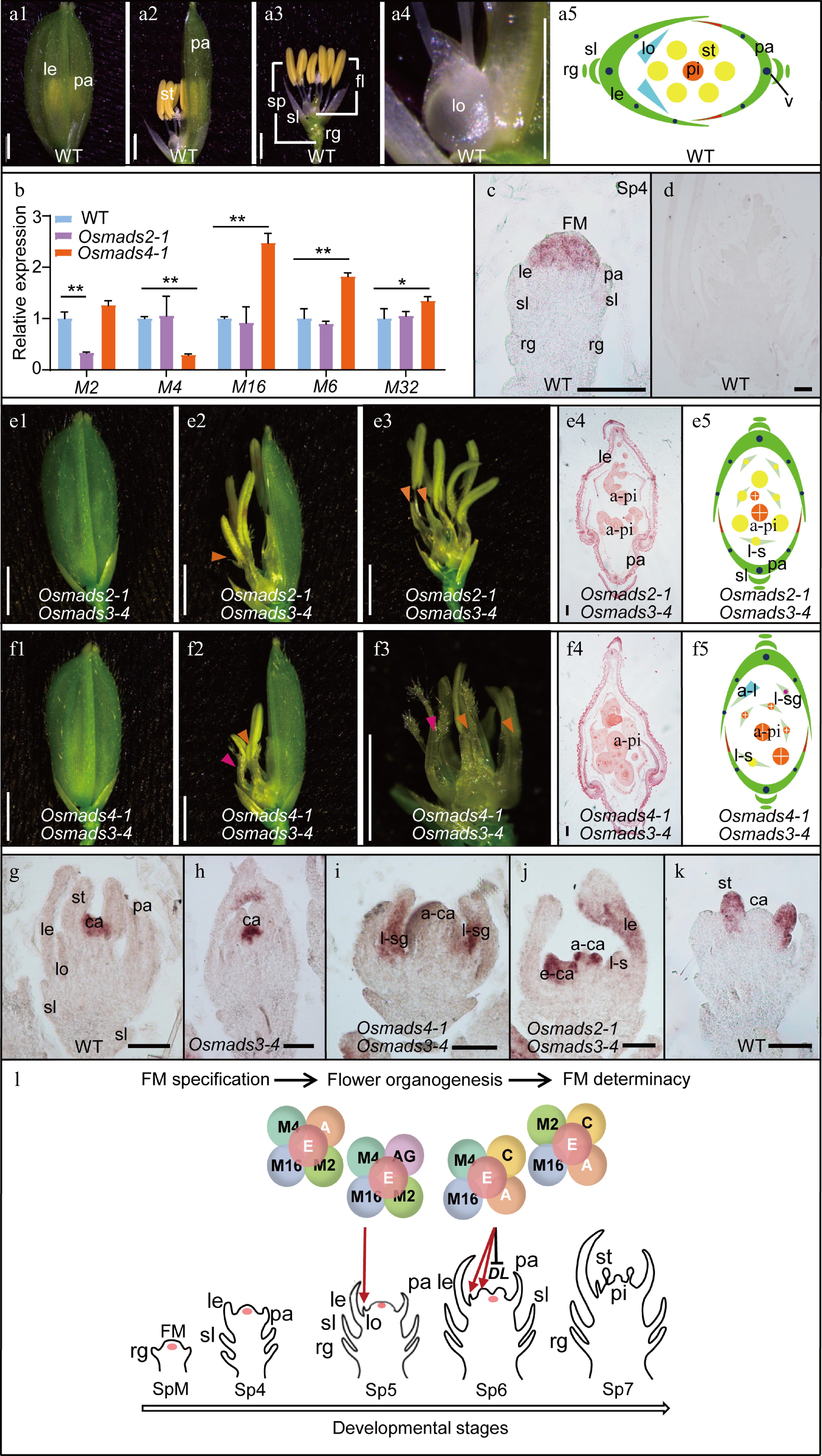
    Figure 1. 

    OsMADS2 and OsMADS4 play partially distinct roles in lodicule and stamen specification. (a) Spikelet morphology (a1−a4) and cartoon diagram (a5) of Wild Type (WT). (b) Expression level of the B-class genes (OsMADS2, OsMADS4 and OsMADS16), OsMADS6 and OsMADS32 in the 2-mm inflorescence of WT, Osmads2-1 and Osmads4-1. Results are shown as mean ± SD. Error bars indicate SD for three biological replicates. ** indicates p-values < 0.01, * indicates P-values between 0.05 to 0.01, analyzed by Student-t test. (c), (d) In-situ hybridization analysis of OsMADS4. In the WT, signals for the anti-sense probe were detected in FM (c), whereas no signals for the sense probe were detected (d). Spikelet morphology (1−3), transverse section (4) and cartoon diagram (5) of the (e) Osmads2-1 Osmads3-4 and (f) Osmads4-1 Osmads3-4 double mutants. (g)−(j) In-situ hybridization analysis of DL in WT, Osmads3-4, Osmads4-1 Osmads3-4 and Osmads2-1 Osmads3-4, respectively. (k) In-situ hybridization analysis of OsMADS4 in WT. (l) A working model of the function of OsMADS2 and OsMADS4 in regulating rice flower development. OsMADS2 and OsMADS4 may engage with different protein complexes in specifying lodicule and stamen identity and morphogenesis. To specify lodicule identity, OsMADS2 and OsMADS4 play partially redundant roles in forming a complex with OsMADS16, the A-class proteins, AGL6-like proteins, and/or E-class proteins. OsMADS2 plays an additional role in regulating lodicule morphogenesis. On the other hand, OsMADS4 may form a protein complex with OsMADS16, and C-, A- and E-class proteins to determine stamen identity. OsMADS4 has a specific role in inhibiting the expression of DL in the lodicule and stamen to specify their identity and morphogenesis. Orange arrowheads in (e) and (f) indicate lodicule-stamen mosaic organs; Rose arrowheads in (e) indicate lodicule-stigma mosaic organ. a-ca, abnormal carpel; a-pi, abnormal pistil; e-ca, ectopic carpel; fl, floret; FM, floral meristem; le, lemma; lo, lodicule; l-s, lodicule-stamen mosaic organ; l-sg, lodicule-stigma mosaic organ; pa, palea; pi, pistil; rg, rudimentary glume; sl, sterile lemma; sp, spikelet; st, stamen. Scale bars = 2 mm in a1 to a4, e1 to e3 and f1 to f3; scale bars = 100 μm in e4 and f4; and scale bars = 50 μm in g to k. Red arrows in l indicate positive regulation in flower organ identity specification. The black bar in l indicates negative regulation. A, A-class proteins; AG, AGL6-like proteins; C, C-class protein; E, E-class proteins; SpM, spikelet meristem. M is the abbreviation of OsMADS; DL, DROOPING LEAF. Sp refers to a developmental stage of rice spikelet.

    Plant genomes contain two main lineages of B-class genes, PI/GLO and paleoAP3/DEF, which arose before the emergence of angiosperms. In rice, OsMADS2 and OsMADS4 are paralogs in the PI/GLO family that play the same role as the paleoAP3/DEF ortholog, OsMADS16 (also named as SUPERWOMAN1), in specifying lodicule and stamen[8]. However, previous studies of OsMADS2 and OsMADS4 RNAi plants indicated that these two genes play unequal roles in lodicule morphogenesis[9]. Whether they function differentially in floral meristem (FM) activity and floral organ development remains elusive.

    To further distinguish the function between OsMADS2 and OsMADS4, the CRISPR-Cas9 system was used to generate targeted mutations within the two genes to obtain strong mutant alleles. Two mutational events were identified for OsMADS2 : Osmads2-1 has an 'A' insertion and Osmads2-2 has a 'TT' insertion (Supplemental Fig. S1a), both causing a frameshift and premature translational termination of the protein (Supplemental Fig. S1b). Compared with the wild-type (WT) plant, there was no visible abnormal phenotype from the vegetative to the reproductive stage, except that the lodicules were extended during floret development in both alleles (Supplemental Fig. S2), which is consistent with a previous report[9]. For OsMADS4, two types of mutants were obtained: Osmads4-1 has a deletion of 'T' and Osmads4-2 has an insertion of 'T' (Supplemental Fig. S3a), both leading to a frameshift and premature translational termination of the protein as well (Supplemental Fig. S3b). Compared with the WT plant, both Osmads4-1 and Osmads4-2 displayed normal vegetative and reproductive growth and the floret contained normal lodicules and stamens (Supplemental Fig. S5), which is also consistent with the previously reported phenotypes of the OsMADS4 RNAi plant[9].

    Double mutants were then generated by crossing Osmads2-1 with Osmads4-1. Abnormal spikelet phenotypes were observed in the double mutant that mimicked Osmads16/spw1-1[5], in which the lodicules were transformed into margin region of palea (mrp)-like organs and stamens into carpel-like organs (Supplemental Fig. S6). Interestingly, enlarged ovaries appeared in the inner whorl of Osmads2-1 Osmads4-1 (Supplemental Fig. S6c). Therefore, the genetic analysis supports results from the previous study that OsMADS2 and OsMADS4 are functionally redundant with an essential role in determining the identities of lodicules and stamens, while OsMADS2 also has a distinct role in lodicule morphogenesis[9]. RT-qPCR analysis of the expression of OsMADS2, OsMADS4 and OsMADS16 in the mutant lines revealed that, although OsMADS2 and OsMADS4 did not seem to impact each other's expression, the expression of OsMADS16 increased significantly in the Osmads4-1 mutant (Fig. 1b; Supplemental Fig. S7), suggesting that OsMADS16 might compensate for the loss of OsMADS4 through transcriptional upregulation.

    A previous study showed that OsMADS6 (AGL6-like gene) and OsMADS3 (C-class gene) are also involved in determining floral organ identities and meristem fate[10]. To determine whether these genes interact genetically with OsMADS2 and OsMADS4, double and triple mutants between Osmads6-1, Osmads3-4 and Osmads2-1 and Osmads4-1 were generated. The spikelet of the Osmads2-1 Osmads6-1 double mutant displayed defects in the outer three whorls, and ectopic glume-like organs and lodicule-stamen mosaic structures were present in whorls 2 and 3 (Supplemental Fig. S8a), which mimicked phenotypes of Osmads6-1[10]. Similarly, the Osmads4-1 Osmads6-1 double mutant also contained abnormal spikelet structure, with glume-like structures enclosing the stamen filament, as well as a reduced number of stamens (Supplemental Fig. S8b). Furthermore, Osmads2-1 Osmads4-1 Osmads6-1 triple mutants were made by crossing Osmads2-1 with the Osmads4-1 Omads6-1(+/−) heterozygous double mutant. Homeotic transformation of lodicules and stamens into glume-like organs and abnormal pistils was observed in the triple mutant (Supplemental Fig. S8c), mimicking phenotypes of the spw1-1 Osmads6-1 double mutant except that it lacked the secondary inflorescence inside the spikelet[10]. Taken together, the genetic evidence indicated that OsMADS2 and OsMADS4 are partially redundant with OsMADS6 in specifying lodicule and stamen identities. Unlike OsMADS16/SPW1, OsMADS2 and OsMADS4 may not participate in regulating secondary inflorescence growth and FM determination.

    The C-class gene mutant Osmads3-4 displayed mild homeotic transformation of lodicules and stamens into lodicules-like or lodicule-anther mosaic organs in the spikelet (Supplemental Fig. S9ac)[11]. In Osmads2-1 Osmads3-4, some lodicules were transformed into lodicule-stamen organs (Fig. 1e; Supplemental Fig. S9df), whereas the ectopic expression of the stigma identity gene, DROOPING LEAF (DL), was observed in the ectopic carpels (Fig. 1j). On the other hand, the Osmads4-1 Osmads3-4 double mutant displayed new phenotypes, including the transformation of lodicules into lodicule-stigma or lodicule-stamen mosaic organs (Fig. 1f; Supplemental Fig. S9g, h), and ectopic generation of abnormal carpels in whorl 3 (Fig. 1f; Supplemental Fig. S9g, i). These data suggest that in the absence of OsMADS3 and OsMADS4, OsMADS58 alone cannot fully determine the stamen identity. It is possible that in the Osmads3 Osmads4 double mutant, OsMADS58 together with DL are ectopically expressed in the second whorl to help specify stamen identity.

    RT-qPCR analysis showed that the expression of DL increased significantly in the Osmads4-1 single mutant at the floral maturation stage (Supplemental Fig. S10a), which prompted in-situ hybridization to investigate the expression pattern of DL in the mutants. Compared to WT and Osmads3-4 (Fig. 1g, h; Supplemental Fig. S4), ectopic expression of DL was detected in the lodicule-stigma mosaic organs of Osmads4-1 Osmads3-4 (Fig. 1i). The combined data suggests that, while the two rice PI-like proteins, OsMADS2 and OsMADS4, play redundant roles in lodicule and stamen specification, they might form different protein complexes in specifying lodicule and stamen identities. Besides its role in lodicule morphogenesis, OsMADS2 also genetically interacts with OsMADS3 in specifying stamen identity. OsMADS58 may need to form a complex with OsMADS4 to better specify stamen identity, and when only OsMADS2 is available, the formation of OsMADS58-SEP tetramers are favored to determine the carpel identity.

    Since the expression of OsMADS16 and DL increased in the Osmads4-1 mutant, we performed RT-qPCR to detect transcript levels for all the MADS-box genes known to be involved in the development of lodicule and other reproductive organs to further understand the role of OsMADS4. Interestingly, expression of the E-class genes OsMADS1, OsMADS5, OsMADS7, OsMADS8 and OsMADS34 (Supplemental Fig. S10bf), as well as OsMADS32 and OsMADS6, was obviously increased during early spikelet development in Osmads4-1, but not in Osamds2-1 (Fig. 1b; Supplemental Fig. S10g, h). A similar expression pattern was observed for AP1-like genes OsMADS14 and OsMADS15 and OsMADS58 (Supplemental Fig. S10ik), suggesting that OsMADS4 may have evolved a function in regulating rice floral meristem development. Previous evidence indicated that changes in gene expression and/or protein function might cause functional divergence for duplicated genes[1]. The in-situ hybridization analysis detected OsMADS4 transcripts in the FM at Sp4 and stamen at Sp7, similar to those of OsMADS2[12], but hardly detected signals in the FM at Sp7 (Fig. 1c, k), which is different from a previous report in which the expression of OsMADS4 was detected in carpel primordium[12]. This discrepancy might have been caused by the stage of the analyzed materials, as the floret used in the present study was at early Sp7, whereas the previous report used material at the late Sp7 stage. Together, these results indicate that the spatial-temporal expression of the rice PI-like genes as well as the formation of their protein complexes are key mechanisms that drive their specific functions.

    In Arabidopsis, the B-class genes regulate FM maintenance and termination in an AG-dependent manner, as the AG/SEP-AG/SEP complex can switch to the AG/SEP-AP3/PI quartets under the ectopic expression of AP3/PI proteins[13]. Extra mrp-like glumes and lodicule-stigma mosaic organs grew in the Osmads4-1 Osmads3-4 (Fig. 1f), spw1-1 Osmads3-4 and spw1-1 Osmads58 double mutants[5], suggesting that rice B genes also play a conserved role in maintaining the size of FM. Therefore, the B-class proteins may be key regulators that determine stage-specific protein quartet complex formation during flower development, both at the transcription and protein levels. Exploring the specificities of different protein quartets at various stages of flower development would help us discern the spatial-temporal regulatory networks in FM maintenance and termination.

    Similar to the present observation with Osmads3 spw1-1, a previous study also observed lodicule-stigma mosaic organs in the spw1-1 Osmads58 double mutants[8]. It is therefore speculated that OsMADS4, OsMADS16, OsMADS3, and OsMADS58, together with E-class and AGL6-like proteins, might form different complexes in specifying lodicule, stamen, and pistil identities (Fig. 1l). Additional in vivo protein interaction data are needed in the future to support the concept of a 'complex transition' that occurs in different whorls. Our observation, along with the ectopic expression of the leaf- and stigma-specific gene DL, in the lodicule of the Osmads4-1 Osmads3-4 mutants, suggests that rice lodicule is a bracteopetal organ derived from a modified leaf, rather than an andropetal stamen-derived organ. In this case, duplication of rice B-class genes may have contributed to the diversification of petal-like organs in grasses, just like in eudicot. To test this hypothesis, it will be important to elucidate in the future whether and how the OsMADS4-OsMADS3 complex represses the expression of DL.

  • The authors confirm contribution to the paper as follows: study conception and design: Yuan Z, Wang L; project supervision: Yuan Z; Osmads2 and Osmads4 CRISPR lines generation: Li QL; analysis and interpretation of results (experiments): Yuan Z, Wang L; draft manuscript preparation and revision: Wang L, Yuan Z, Hu JP. All authors reviewed the results and approved the final version of the manuscript.

  • All data used in this work are publicly available.

    • This paper is dedicated to the late Prof. Dabing Zhang, who provided unwavering support to this project until his tragic passing in June 2023. We would like to acknowledge funds from the Natural Science Foundation of China (32170322, 31671260), China-Germany Mobility Program (M-0141), Special Funds for Construction of Innovative Provinces in Hunan Province (2021NK1002), China Innovative Research Team, Ministry of Education, the Program of Introducing Talents of Discipline to Universities (111 Project, B14016), and the SMC Morningstar Young Scholarship of Shanghai Jiao Tong University to Z.Y.

    • The authors declare that they have no conflict of interest.

    • Supplemental Fig. S1 OsMADS2 mutant alleles (a) Schematic diagram of OsMADS2 gene structure. Grey boxes represent 5’UTR and 3’UTR, black boxes represent exons, and thick black lines represent introns. The black arrowhead indicates the sgRNA target site, Osmads2-1 and Osmads2-2 had “A” and “TT” insertion in the OsMADS2 coding sequence, respectively. The underlined letters indicate protospacer adjacent motif (PAM), Red letters indicate mutation types. (b) The sequencing results showing mutation information of Osmads2-1 and Osmads2-2, compared to that in wild type. Red arrows indicate mutation site.
    • Supplemental Fig. S2 Phenotypic analysis of spikelet morphology in Osmads2-1 and Osmads2-2 (a) and (b) Spikelet of Osmads2-1 (a1-a4) and Osmads2-2 (b1-b4). Pink arrowheads in a2, a4, b1 and b4 indicate extended lodicules; elo, extended lodicule; le, lemma; lo, lodicule; pa, palea; pi, pistil; st, stamen. Scale bars, 2 mm.
    • Supplemental Fig. S3 OsMADS4 mutant alleles (a) Schematic diagram of the OsMADS4 gene. Grey boxes represent 5’UTR and 3’UTR, black boxes represent exons, and thick black lines represent introns. The black arrowhead indicates the mutation target site, Osmads4-1 and Osmads4-2 contain a T deletion and a T insertion, respectively, in the OsMADS4 coding sequence. (b) Sequencing results of the target site of wild type, Osmads4-1 and Osmads4-2.
    • Supplemental Fig. S4 The in-situ hybridization sense control of DL DL is the abbreviation of DROOPING LEAF; Scale bars, 50 μm.
    • Supplemental Fig. S5 Phenotypic analysis of spikelet development in Osmads4-1 and Osmads4-2 (a) and (b) Spikelet of osmads4-1 (a1-a4) and osmads4-2 (b1-b4) single mutant. ca, carpel; le, lemma; lo, lodicule; pa, palea; pi, pistil; st, stamen. Scale bars, 2 mm.
    • Supplemental Fig. S6 Phenotypic analysis of spikelet morphology in Osmads2-1Osmads4-1 double mutant. (a) Spikelet of Osmads2-1 Osmads4-1.(b) Spikelet of Osmads2-1 Osmads4-1 after the removal of lemma. (c) Spikelet of Osmads2-1 Osmads4-1 after both lemma and palea were removed. Blue arrowheads indicate glume-like structure organs; red arrowhead indicate abnormal-pistils; yellow arrowheads indicate enlarged ovaries. a-pi, abnormal-pistil. eno, enlarged ovary; gll, glume-like structure; le, lemma; lo, lodicule; pa, palea. Scale bars, 2 mm.
    • Supplemental Fig. S7 Expression analysis of B-class genes in WT and mutants. (a) OsMADS2 expression pattern in the inflorescence of WT and Osmads4-1. (b)  RT-qPCR analysis of OsMADS4 in WT and Osmads2-1. (c) RT-qPCR analysis of OsMADS4 in WT, Osmads2-1 and Osmads4-1. Total RNA was isolated from 3-mm and 5-7-mm young inflorescence of WT, Osmads2-1 and Osmads4-1. Data are shown as mean± sd, Error bars indicate SD for three biological replicates. ** indicates P-values < 0.01 and * indicates P-values between 0.05 and 0.01, analyzed by the Student-t test.
    • Supplemental Fig. S8 Phenotypic characterization of the spikelet in the Osmads2-1 Osmads6-1, Osmads4-1 Osmads6-1 and Osmads2-1 Osmads4-1 Osmads6-1 mutants (a) to (c) Spikelet morphology (1-4) and schematic diagram (5) of Osmads2-1 Osmads6-1 (a), Osmads4-1 Osmads6-1 (b) and Osmads2-1 Osmads4-1 Osmads6-1 (c). Aqua blue arrowheads indicate glume-like structures; Orange arrowheads indicate lodicule-stamen mosaic organs; Light yellow arrowheads indicate abnormal pistils. a-pi, abnormal pistil; bop, body of palea; fl, floret; gll, glume like organ; le, lemma; lo, lodicule; l-s, lodicule-stamen mosaic organ; mrp, margin region of palea; pa, palea; rg, rudimentary glume; sl, sterile lemma; sp, spikelet; v, vascular bundles. All scale bars are 2 mm.
    • Supplemental Fig. S9 Phenotypic analysis of the spikelet in the Osmads2-1 Osmads3-4 and Osmads4-1 Osmads3-4 double mutants. (a)to (c) Spikelet morphology of Osmads3-4. (d) to (f) Spikelet morphology of Osmads2-1 Osmads3-4. (g) to (i) Spikelet morphology of Osmads4-1 Osmads3-4. Orange arrowheads indicate lodicule-stamen mosaic organ; Yellow arrowheads indicate abnormal pistil; Rose arrowheads indicate lodicule-stigma mosaic organ. le, lemma; lo, lodicule; l-p, lodicule-pistil mosaic organ; l-s, lodicule-stamen mosaic organ; pa, palea; pi, pistil; st, stamen. Scale bars, 2 mm.
    • Supplemental Fig. 10 Expression analysis of DL and some MADS-box genes in WT and mutants. (a) Expression level of DL in WT, Osmads2-1 and Osmads4-1. (b) to (f) Expression levels of class-E genes (OsMADS1, OsMADS5, OsMADS7, OsMADS8 and OsMADS34) (b, c, d, e and f, respectively) in WT and the Osmads2-1 and Osmads4-1 mutants. (g) and (h) Expression level of OsMADS32 and OsMADS6 in WT and the Osmads2-1 and Osmads4-1 mutants.
    • Supplemental Table S1 Primers used in this study.
    • Copyright: © 2024 by the author(s). Published by Maximum Academic Press on behalf of Hainan Yazhou Bay Seed Laboratory. This article is an open access article distributed under Creative Commons Attribution License (CC BY 4.0), visit https://creativecommons.org/licenses/by/4.0/.
Figure (1)  References (13)
  • About this article
    Cite this article
    Wang L, Li QL, Hu JP, Yuan Z. 2024. Neofunctionalization of B-class genes in regulating rice flower development. Seed Biology 3:e013 doi: 10.48130/seedbio-0024-0012
    Wang L, Li QL, Hu JP, Yuan Z. 2024. Neofunctionalization of B-class genes in regulating rice flower development. Seed Biology 3:e013 doi: 10.48130/seedbio-0024-0012
  • Catalog

    • About this article

    /

    DownLoad:  Full-Size Img  PowerPoint
    • Table 1.  Ubiquitination sites related to sucrose and starch metabolism in rice endosperm.
      Gene nameAnnotationProtein entryModification site(s)
      SUS1Sucrose synthase 1BGIOSGA010570K172, K177
      SUS2Sucrose synthase 2BGIOSGA021739K160, K165, K176, K804
      SUS3Sucrose synthase 3BGIOSGA026140K172, K177, K541, K544, K588
      FKFructokinaseBGIOSGA027875K143
      UGPaseUDP-glucose pyrophosphorylaseBGIOSGA031231K27, K150, K303, K306
      AGPS1ADP-glucose pyrophosphorylase small subunit 1BGIOSGA030039K94, K464, K484
      AGPS2ADP-glucose pyrophosphorylase small subunit 2BGIOSGA027135K106, K132, K385, K403, K406, K476, K496
      AGPL2ADP-glucose pyrophosphorylase large subunit 2BGIOSGA004052K41, K78, K134, K191, K227, K254, K316, K338, K394, K396, K463, K508, K513
      AGPL3ADP-glucose pyrophosphorylase large subunit 3BGIOSGA017490K509
      GBSSIGranule bound starch synthase IBGIOSGA022241K130, K173, K177, K181, K192, K258, K371, K381, K385, K399, K462, K517, K530, K549, K571, K575
      BEIStarch branching enzyme IBGIOSGA020506K103, K108, K122
      BEIIbStarch branching enzyme IIbBGIOSGA006344K134
      PULStarch debranching enzyme:PullulanaseBGIOSGA015875K230, K330, K431, K736, K884
      PHO1Plastidial phosphorylaseBGIOSGA009780K277, K445, K941
       | Show Table
      DownLoad: CSV