ARTICLE   Open Access    

Talaromyces peaticola (Aspergillaceae, Eurotiales), a new species from the Zoige wetlands, China

More Information
  • Species in Talaromyces section Trachyspermi are isolated from a wide range of substrates, including soil, house dust, leaf, wood and fruit from temperate region to tropical areas. During a survey of fungal diversity in Zoige wetlands, three isolates with biverticillate adpressed penicilli and spheroidal conidia belonging to Talaromyces are isolated from peat soil. Phylogenetic analyses based on a combined ITS, BenA, CaM and RPB2 sequence data suggests they represent a novel taxon in Talaromyces section Trachyspermi, namely Talaromyces peaticola. In spite of T. peaticola has a close affinity to T. diversus in phylogeny, it is readily distinguished from the later, resulted from growing slowly on CREA at 25oC, exudating small clear droplets on MEA, and producing smaller conidia than that of T. diversus.
  • Although aquatic hyphomycetes have been adapted mainly to the lotic habitats, they are also known from other ecological niches (e.g. terrestrial litter, lentic habitats, tree canopies and caves) (Sridhar 2009, Chauvet et al. 2016, Kozlova & Mazina 2020, Sharathchandra & Sridhar 2020, Sridhar et al. 2020). The microclimatic conditions exist in caves will be favourable for growth and functions of several fungi (Kozlova & Mazina 2020). Fungal diversity in cave streams is of special interest owing to their diversity, adaptation, mineralization of organic matter and energy flow. Usually, fungi will be transferred to the caves by various routes (e.g. infiltration of spores through soil strata, insects, birds, bat guano and surface transfer of plant detritus) (Novákova 2009, Souza-Silva et al. 2012, Vanderwolf et al. 2013, Ghate & Sridhar 2015, Prous et al. 2015, Kozlova & Mazina 2020). Novákova (2009) depicted that the fungal diversity in karst cave ecosystems rely on the abundance of substrates. Due to cool and dark conditions in lateritic cave streams, various insects, birds and bats will be attracted and their activity will enrich the fungal population in water. In addition, penetrated roots from the cave roof, growth of algae, lichens, mosses, bryophytes and ferns on the walls of caves also support diverse mycota. However, the extent and diversity of biota vary in the photic zones (entrance part with light penetration) and dysphotic zones (feeble or without light penetration). Thus, there may be a gradient of biota in different parts of the cave streams.

    Although aquatic hyphomycetes in the coastal region of the southwest coast of India have been evaluated in streams, intermittent streams, estuary, canopy and terrestrial litters (Sridhar & Kaveriappa 1988, Sridhar et al. 1992, Sridhar 2009, Sridhar et al. 2013, Ghate & Sridhar 2015, Sridhar et al. 2020, Sridhar & Sharthchandra 2020), no studies are available in the lateritic cave streams. In the coastal region of southern Karnataka and northern Kerala, indigenous water management will be carried out by construction of horizontal tunnels up to 30-40 m (~1 × 2-2.5 m) besides the lateritic hills, which results in extraction of groundwater by the gravitational pull. This traditional knowledge has history over a century and the water source although feeble, it will be a permanent source to use for drinking, domestic and irrigation purposes. Occurrence of specific tree species (e.g. Ficus virens, Macranga indica and Vateria indica) and termite mounds confirms the water source as well as direction of tunnel construction. Such ventures will be performed during the summer season and percolating water will be collected in a tank. Approximately there are about 5000 lateritic caves exists in Kerala and Karnataka. Due to paucity of information on cave streams, the present study focuses the assemblage and diversity of aquatic hyphomycetes in four lateritic caves of southwestern region. This study involves evaluation of water parameters, species richness and diversity of aquatic hyphomycetes in water, foam, leaf litter and woody litter.

    Four cave streams selected were Kodandooru, Pilinguli, Nekkare and Nayarmoole located in the southwestern Karnataka (12°41′ N, 75°3′ E) (Fig. 1). Tree species like Ficus spp., Macranga indica and Vateria indica were common in the vicinity of cave streams. The study has been carried out during the post-monsoon season (November 2020). Humidity and air temperature (Mextech Digital Thermo Hygrometer M288CTHW, Mumbai, India) and water temperature (mercury thermometer) (~2 m inside the cave) were assessed. The pH, conductivity and total dissolved solids (TDS) of cave waters were assessed using a water analysis kit (Water Analyzer 371, Systronics India Ltd., Ahmedabad, Gujarat, India). The water samples of caves were fixed on the sampling sites to assess dissolved oxygen by Winkler's method in the laboratory (APHA 1998). Other parameters such as total alkalinity, total hardness, chloride, inorganic phosphate, sulfate, silicate and magnesium of water samples were assessed as per the standard protocols of APHA (1998). The total alkalinity was determined by titration using strong acid followed by methyl orange and phenolphthalein indicators; total hardness was evaluated by ethylene diamine tetra-acetic acid (EDTA) titration method using eriochrome black-T and murexide indicators; chloride content was assessed by argentometric method (APHA 1998). The inorganic phosphate was found out by stannous chloride method; nitrate content was evaluated by brucine sulfanilic acid method; silicate and sulphate contents were determined by molybdosilicate and by turbidimetric methods, respectively (APHA 1998).

    Figure 1.  Sampling in Pilinguli cave stream (a), close up view (b) and overall view (c) of Nayarmoole cave stream.

    Three replicate samples of foam, water, leaf litter and woody litter from each cave stream were collected about 1-2 m outflow region. Overall scheme of assessment of samples is given in Fig. 2. The foam and water samples were assessed by direct microscopic examination, while the leaf litter, bark and cambium were assessed by indirect examination using bubble chamber incubation. Foam samples were fixed on the sampling site in formalin-acetic-alcohol (FAA: Mixture of 50 ml 95% ethanol + 10 ml glacial acetic acid + 10 ml 37% formaldehyde + 30 ml distilled water). Water samples (25 ml each) were filtered on the sampling site through the Millipore filters (5 µm diameter) and stained the filters by aniline blue in lactophenol and stored in sterile Petri plates in dark. Submerged decomposing leaf and woody litters were collected in sterile polythene bags and transferred to laboratory. The leaf samples were rinsed in distilled water, 4-5 different leaves were packed and punched into 1.5 cm disks using a cork borer. Leaf discs (4-5) per discs were transferred into conical flasks (250 ml) with sterile distilled water (150 ml) and aerated up to 48 hr with fine jet of air with Pasteur pipette using aquarium aerator (bubble chamber incubation). From the woody litter, bark and cambium were separated and cut into segments (~0.2×0.5×2 cm). Bark and cambium pieces (4-5) were separately transferred in to the conical flasks and incubated in bubble chambers similar to the leaf discs. The aerated water of leaf discs, bark and cambium were filtered through Millipore filters (5 µm diameter) and stained with aniline blue in lactophenol and stored in sterile petri plates in dark. Facesoffungi numbers of the cultures were registered as mentioned in Jayasiri et al. (2015).

    Figure 2.  Scheme of direct and indirect assessment of cave stream samples for aquatic hyphomycetes.

    Fixed foam samples were transferred drop by drop into the sterile slides and applied cover glass with a small quantity of aniline blue in lactophenol. Up to 300 conidia of aquatic hyphomycetes were scored per sample, different conidia were identified based on their morphology (Ingold 1975, Nawawi 1985, Marvanová 1997, Gulis et al. 2020) and the numbers of conidia of each species were enumerated. Stained filters of water samples (25 ml each) were cut into half, mounted on microscope slides with a few drops of lactic acid to score and identify the conidia, contribution of conidia of each species has been expressed in per cent. Stained filters of leaf discs, bark and cambium pieces were also assessed microscopically similar to water samples to identify and score the conidia. Aerated leaf discs, bark and cambium were dried at 80 C up to 24 hr to determine the dry mass. Conidial output per gram dry mass of substrate has been expressed in per cent contribution.

    Based on the mean conidia of each aquatic hyphomycete (among 300 conidia per foam sample), water (in 25 ml sample), leaf litter, bark and cambium (per g dry mass) were compiled. This data was used to calculate the diversities (Simpson's and Shannon's diversity) (Magurran 1988) and Pielou's equitability (Pielou 1975).

    Humidity in cave habitats ranged from 77.1-82.2%, temperature of air was higher than water (30.1-32.2 vs. 21.3-25.2 C) (Table 1). The pH of water samples was towards alkaline range (7.3-8.1) except for the cave Nekkare (6.9). The mean values of pH, conductivity, total alkalinity, total hardness, chloride and magnesium contents of water samples fall within the range stipulated by the WHO (2004) standards for potability. However, the phosphate content exceeded the WHO (2004) standard (4.6 vs. 0.1 mg/l). On comparison of cave stream water qualities with the ground water (bore well water) in a nearby region (~22 km), water temperature, conductivity, total hardness, TDS, chloride and magnesium contents were lower, while the pH and phosphate content were higher (Bhagya & Sridhar, unpublished observation).

    Table 1.  Physicochemical features of four lateritic cave streams (mean, n = 3)
    Cave Overall mean±SD
    Kodandooru Pilinguli Nekkare Nayarmoole
    Humidity (%) 78.1 80.8 77.1 82.2 79.6 ± 2.4
    Air temperature (℃) 31.1 32.2 30.1 31.7 31.3 ± 0.9
    Water temperature (℃) 23.2 21.3 24.1 25.2 23.5 ± 1.7
    pH 7.3 7.7 6.9 8.1 7.5 ± 0.5
    Conductivity (µS/cm) 51.0 44.0 69.6 55.6 55.1 ± 10.8
    Dissolved oxygen (mg/l) 8.6 8.2 8.0 8.9 8.4 ± 0.4
    Total alkalinity (mg/l) 30.5 39.6 26.9 42.9 35.0 ± 7.5
    Total hardness (as CaCO3) 16.6 22.4 27.2 24.2 22.6 ± 4.5
    Total dissolved solids (mg/l) 25.2 24.8 39.2 42.9 33.0 ± 9.4
    Chloride (mg/l) 4.4 4.3 6.2 7.3 5.6 ± 1.5
    Sulphate (mg/l) 1.8 2.4 2.7 1.9 2.2 ± 0.4
    Phosphate (mg/l) 6.2 4.9 2.6 4.5 4.6 ± 1.5
    Silicate (mg/l) 23.5 18.1 26.7 22.5 22.7 ± 3.6
    Magnesium (mg/l) 3.2 4.5 6.3 4.3 4.6 ± 1.3
     | Show Table
    DownLoad: CSV

    A total of 21 species of aquatic hyphomycetes was found in cave streams (Fig. 3). The number of species were highest in leaf litter (15 spp.) followed by water (14 spp.), bark (11 spp.), cambium (9 spp.) and foam (7 spp.) samples (Table 2). Water samples showed the highest Simpson's and Shannon's diversities, while they were least in leaf litter and foam samples, respectively (Table 3). The species richness was highest (9 spp.) in water samples of the cave Nayarmoole followed by water in Pilinguli and leaf litter in Kodandooru (8 spp.) (Fig. 4). Water and leaf litter of Nekkare cave and leaf litter of Nayarmoole cave possess 7 spp. The number of species of aquatic hyphomycetes in cave streams is comparable with Konaje stream located about 20 km from the cave streams (Sridhar & Kaveriappa 1984, 1989a, b, Sridhar et al. 2013). Similarly, the top three species (Anguillospora longissima, Flagellospora curvula and Lunulospora curvula) matches with the earlier studies in nearby streams (Sridhar et al. 2013, Ghate & Sridhar 2015). Besides these species, Anguillospora crassa also constitutes a core-group species in the cave streams. In addition, Actinospora megalospora, Anguillospora crassa, Anguillospora angulata, Condylospora spumigena, Ingoldiella fibulata and Trinacrium subtile were the additional species not found in earlier studies (Sridhar & Kaveriappa 1984, Sridhar et al. 2013, Ghate & Sridhar 2015).

    Figure 3.  Conidia of selected aquatic hyphomycetes obtained from the lateritic cave stream samples.
    a Anguillospora crassa. b Condylospora spumigena. c Helicosporium sp. d Ingoldiella hamata. e Lunulospora curvula. f Speiropsis pedatospora. g Tricladium sp. h Tripospermum sp. i Triscelophorus acuminatus. j Triscelophorus konajensis. Scale bars = 20 µm.
    Table 2.  Per cent contribution of conidia of aquatic hyphomycetes in lateritic cave streams (n = 4, mean) (*, among 300 conidia in foam; **, in 25 ml water; ***, in g dry mass of leaf, bark and cambium)
    Direct observation Bubble chamber incubation
    Leaf litter Woody litter
    Foam Water Bark Cambium
    Species richness 7 14 15 11 9
    Simpson diversity 0.809 0.835 0.796 0.812 0.828
    Shannon diversity 2.541 3.005 2.967 2.893 2.724
    Pielou's equitability 0.905 0.789 0.759 0.836 0.859
     | Show Table
    DownLoad: CSV
    Table 3.  Species richness, diversity and equitability of aquatic hyphomycetes in lateritic cave streams
    FOF number Direct observation Bubble chamber incubation
    Leaf litter*** Woody litter***
    Foam* Water** Bark Cambium
    Lunulospora curvula Ingold FoF10483 14.5 14.6 39.7 22.0 15.0
    Anguillospora longissima (Sacc. & P. Syd.) Ingold FoF10479 28.0 30.3 9.3 34.0 25.4
    Flagellospora curvula Ingold FoF10482 21.0 12.4 14.7 8.0 13.7
    Anguillospora crassa Ingold FoF10478 18.0 7.9 5.8 4.0 13.7
    Cylindrocarpon sp. - 14.6 7.5 - 20.4
    Triscelophorus acuminatus Nawawi FoF10487 - 1.1 6.4 4.0 -
    Speiropsis pedatospora Tubaki FoF02652 10.5 3.4 1.0 - 2.2
    Wiesneriomyces laurinus (Tassi) P.M. Kirk FoF09127 5.5 7.9 3.1 - -
    Ingoldiella hamata D.E. Shaw FoF10485 - - 2.1 4.0 1.4
    Condylospora spumigena Nawawi FoF10481 - - 2.1 4.0 -
    Anguillospora angulata (R.H. Petersen) Redhead & G.P. White FoF10477 - - - 8.0 -
    Actinospora megalospora Ingold FoF10476 - - 1.0 4.0 -
    Clavariopsis aquatica de Wild. FoF10480 - - 1.0 4.0 -
    Tricladium sp. - 2.3 2.1 - -
    Helicosporium sp. - 1.1 - 4.0 -
    Triscelophorus monosporus Ingold FoF10489 - 1.1 2.1 - -
    Tripospermum sp. - - 2.1 - -
    Ingoldiella fibulata Nawawi FoF10484 2.5 1.1 - - 1.4
    Triscelophorus konajensis K.R. Sridhar & Kaver. FoF10488 - - - - 6.8
    Anguillospora sp. - 1.1 - - -
    Trinacrium subtile Riess FoF10486 - 1.1 - - -
     | Show Table
    DownLoad: CSV
    Figure 4.  Species richness of aquatic hyphomycetes in different samples in lateritic cave streams.

    From the four cave streams (Kodandooru, Pilinguli, Nekkare and Nayarmoole located in the southwestern India) foam, water, leaf litter and woody litter samples yielded up to 21 species of aquatic hyphomycetes, which is comparable to the population in nearby streams. The species richness was highest (9 spp.) in water samples of the cave stream Nayarmoole followed by water samples of Pilinguli and leaf litter samples of Kodandooru (8 spp.) cave streams. The highest richness of aquatic hyphomycetes was found in leaf litter (bubble chamber incubation) (15 spp.) followed water (direct examination) (14 spp.) and bark (bubble chamber incubation) (11 spp.). The frequent core-group fungi matches with other studies carried out in nearby streams. Simpson's and Shannon's diversities were higher in water samples than other samples. Six aquatic hyphomycetes (Actinospora megalospora, Anguillospora crassa, Anguillospora angulata, Condylospora spumigena, Ingoldiella fibulata and Trinacrium subtile) were the new records to the southwestern India. Cave stream water being used for drinking and domestic purposes, the water quality almost fulfils the WHO (2004) stipulated standards.

    Authors are indebted to the Department of Biosciences, Mangalore University for the facilities to carry out this study. Authors are thankful to Dr. Thilini Chethana for providing the Faces of Fungi (FOF) numbers for the species.
  • Asemaninejad A, Thorn RG, Lindo Z. 2017 – Vertical distribution of fungi in hollows and hummocks of boreal peatlands. Fungal Ecology 27, 59-68. doi: 10.1016/j.funeco.2017.02.002

    CrossRef    Google Scholar

    Benjamin CR. 1955 – Ascocarps of Aspergillus and Penicillium. Mycologia 47, 669–687. doi: 10.2307/3755578

    CrossRef    Google Scholar

    Castresana J. 2000 – Selection of conserved blocks from multiple alignments for their use in phylogenetic analysis. Molecular Biology and Evolution 17, 540–552. doi: 10.1093/oxfordjournals.molbev.a026334

    CrossRef    Google Scholar

    Chen AJ, Sun BD, Houbraken J, Frisvad JC et al. 2016 – New Talaromyces species from indoor environments in China. Studies in Mycology 84, 119–144. doi: 10.1016/j.simyco.2016.11.003

    CrossRef    Google Scholar

    Frisvad JC, Yilmaz N, Thrane U, Rasmussen KB et al. 2013 – Talaromyces atroroseus, a new species efficiently producing industrially relevant red pigments. Plos One 8. Doi10.1371/journal. pone. 0084102

    Google Scholar

    Gilbert D, Mitchell EA. 2006 – Microbial diversity in Sphagnum peatlands. Developments in Earth Surface Processes 9, 287–318.

    Google Scholar

    Grum-Grzhimaylo OA, Debets AJM, Bilanenko EN. 2016 – The diversity of microfungi in peatlands originated from the White Sea. Mycologia 108, 233–254. doi: 10.3852/14-346

    CrossRef    Google Scholar

    Houbraken J, Kocsub–S, Visagie CM, Yilmaz N et al. 2020 – Classification of Aspergillus, Penicillium, Talaromyces and related genera (Eurotiales): An overview of families, genera, subgenera, sections, series and species, Studies in Mycology 95, 5–169. doi: 10.1016/j.simyco.2020.05.002

    CrossRef    Google Scholar

    Katoh K, Standley DM. 2013 – MAFFT Multiple sequence alignment software version 7: improvements in performance and usability. Molecular Biology and Evolution 30, 772–780. doi: 10.1093/molbev/mst010

    CrossRef    Google Scholar

    improvements in performance and usability. Molecular Biology and Evolution 30, 772–780.

    Google Scholar

    Luo Y, Lu XH, Bi W, Liu F, Gao WW. 2016 – Talaromyces rubrifaciens, a new species discovered from heating, ventilation and air conditioning systems in China. Mycologia 108, 773–779. doi: 10.3852/15-233

    CrossRef    Google Scholar

    Myers B, Webster KL, Mclaughlin JW, Basiliko N. 2012 – Microbial activity across a boreal peatland nutrient gradient: the role of fungi and bacteria. Wetlands Ecology and Management 20, 77–88. doi: 10.1007/s11273-011-9242-2

    CrossRef    Google Scholar

    Nylander JAA. 2004 – MrModeltest v2. Program distributed by the author. Available from: http://www.abc.se/~nylander/mrmodeltest2/mrmodeltest2.html

    Google Scholar

    Rambaut A. 2014 – FigTree v1. 4.2 from http://tree.bio.ed.ac.uk/software/figtree/ (Retrieved on July 1, 2016)

    Google Scholar

    Romero SM, Romero AI, Barrera V, Comerio R. 2016 – Talaromyces systylus, a new synnematous species from Argentinean semi-arid soil. Nova Hedwigia 12, 241–256.

    Google Scholar

    Ronquist F, Teslenko M, van der Mark P, Ayres DL et al. 2012 – MrBayes 3.2: Efficient Bayesian phylogenetic inference and model choice across a large model space. Systematic Biology 61, 539–542. doi: 10.1093/sysbio/sys029

    CrossRef    Google Scholar

    Samson RA, Yilmaz N, Houbraken J, Spierenburg H et al. 2011 – Phylogeny and nomenclature of the genus Talaromyces and taxa accommodated in Penicillium subgenus Biverticillium. Studies in Mycology 70, 159–183. doi: 10.3114/sim.2011.70.04

    CrossRef    Google Scholar

    Seifert KA, Samson RA, Houbraken J, Lévesque CA et al. 2007 – Prospects for fungus identification using CO1 DNA barcodes, with Penicillium as a test case. Proceedings of the National Academy of Sciences 104, 3901–3906. doi: 10.1073/pnas.0611691104

    CrossRef    Google Scholar

    Silvestro D, Michalak I. 2012 – raxmlGUI: a graphical front-end for RAxML. Organisms Diversity & Evolution 12: 335–337.

    Google Scholar

    Skouboe P, Frisvad JC, Taylor JW, Lauritsen D et al. 1999 – Phylogenetic analysis of nucleotide sequences from the ITS region of terverticillate Penicillium species. Mycological Research 103, 873–881. doi: 10.1017/S0953756298007904

    CrossRef    Google Scholar

    Stamatakis A. 2006 – RAxML-Ⅵ-HPC: Maximum likelihood-based phylogenetic analyses with thousands of taxa and mixed models. Bioinformatics 22, 2688–2690. doi: 10.1093/bioinformatics/btl446

    CrossRef    Google Scholar

    Thormann MN. 2006 – Diversity and function of fungi in peatlands: A carbon cycling perspective. Canadian Journal of Soil Science 86, 281–293. doi: 10.4141/S05-082

    CrossRef    Google Scholar

    Thormann MN, Bayley SE. Currah RS. 2001 – The microbial wildcard in peatland carbon storage: implications for global warming. Proceedings of the International Workshop on Carbon Dynamics of Forested Peatlands: Knowledge Gaps, Uncertainty and Modeling Approaches.

    Google Scholar

    Turetsky MR, Benscoter B, Page S, Rein G et al. 2015 – Global vulnerability of peatlands to fire and carbon loss. Nature Geoscience 8, 11–14. doi: 10.1038/ngeo2325

    CrossRef    Google Scholar

    Vaidya G, Lohman DJ, Meier R. 2011 – SequenceMatrix: concatenation software for the fast assembly of multi-gene datasets with character set and codon information. Cladistics 27, 171–180. doi: 10.1111/j.1096-0031.2010.00329.x

    CrossRef    Google Scholar

    Visagie CM, Hirooka Y, Tanney JB, Whitfield E et al. 2014 – Aspergillus, Penicillium and Talaromyces isolated from house dust samples collected around the world. Studies in Mycology 78, 63–139. doi: 10.1016/j.simyco.2014.07.002

    CrossRef    Google Scholar

    Wang XC, Chen K, Qin WT, Zhuang WY. 2017 – Talaromyces heiheensis and T. mangshanicus, two new species from China. Mycological Progress 16, 73–81. doi: 10.1007/s11557-016-1251-3

    CrossRef    Google Scholar

    Wang XC, Chen K, Xia YW, Wang L et al. 2016 – A new species of Talaromyces (Trichocomaceae) from the Xisha Islands, Hainan, China. Phytotaxa 267, 187–200. doi: 10.11646/phytotaxa.267.3.2

    CrossRef    Google Scholar

    Wu B, Tian JQ, Bai CM, Xiang MC et al. 2013 – The biogeography of fungal communities in wetland sediments along the Changjiang River and other sites in China. ISME Journal 7(7), 1299–1309. doi: 10.1038/ismej.2013.29

    CrossRef    Google Scholar

    Yilmaz N, Houbraken J, Hoekstra ES, Frisvad JC, Visagie1 CM, Samson RA. 2012 – Delimitation and characterisation of Talaromyces purpurogenus and related species. Persoonia 29, 39–54. doi: 10.3767/003158512X659500

    CrossRef    Google Scholar

    Yilmaz N, Visagie J, Houbraken, CM, Frisvad JC, Samson RA. 2014 -olyphasic taxonomy of the genus Talaromyces. Studies in Mycology 78, 175–341. doi: 10.1016/j.simyco.2014.08.001

    CrossRef    Google Scholar

    Zhang YJ, Zhang S, Liu XZ, Wen HA, Wang M. 2010 – A simple method of genomic DNA extraction suitable for analysis of bulk fungal strains. Letters in Applied Microbiology 51, 114–118.

    Google Scholar

  • Cite this article

    JQ Tian, YF Wang, JZ Sun. 2021. Talaromyces peaticola (Aspergillaceae, Eurotiales), a new species from the Zoige wetlands, China. Studies in Fungi 6(1):391−400 doi: 10.5943/sif/6/1/29
    JQ Tian, YF Wang, JZ Sun. 2021. Talaromyces peaticola (Aspergillaceae, Eurotiales), a new species from the Zoige wetlands, China. Studies in Fungi 6(1):391−400 doi: 10.5943/sif/6/1/29

Figures(3)  /  Tables(1)

Article Metrics

Article views(3217) PDF downloads(509)

Other Articles By Authors

ARTICLE   Open Access    

Talaromyces peaticola (Aspergillaceae, Eurotiales), a new species from the Zoige wetlands, China

Studies in Fungi  6 Article number: 29  (2021)  |  Cite this article

Abstract: Species in Talaromyces section Trachyspermi are isolated from a wide range of substrates, including soil, house dust, leaf, wood and fruit from temperate region to tropical areas. During a survey of fungal diversity in Zoige wetlands, three isolates with biverticillate adpressed penicilli and spheroidal conidia belonging to Talaromyces are isolated from peat soil. Phylogenetic analyses based on a combined ITS, BenA, CaM and RPB2 sequence data suggests they represent a novel taxon in Talaromyces section Trachyspermi, namely Talaromyces peaticola. In spite of T. peaticola has a close affinity to T. diversus in phylogeny, it is readily distinguished from the later, resulted from growing slowly on CREA at 25oC, exudating small clear droplets on MEA, and producing smaller conidia than that of T. diversus.

  • The genus Talaromyces was initially described by Benjamin (1955) to accommodate sexual morph Penicillium species with soft and yellowish ascomata surrounded by multiple layers of interwoven hyphae. Samson et al. (2011) redefined Talaromyces by transferring Penicillium subgenus Biverticillium into Talaromyces regarding the phylogenetic analysis of sequence data from the nuclear ribosomal internal transcribed spacer (ITS) and DNA-dependent RNA polymerase Ⅱ largest subunit (RBP2) genes. Based on a multi-gene phylogeny of a combination of ITS, β-tubulin gene (BenA) and DNA-dependent RNA polymerase Ⅱ second largest subunit (RPB2), Talaromyces has been divided into seven sections, namely as sections Bacillispori, Helici, Islandici, Purpurei, Subinflati, Talaromyces and Trachyspermi (Yilmaz et al. 2014). Currently, phylogenetic analyses of the ITS, BenA, CaM, and RPB2 genes are imperative for new species identification of Talaromyces (Yilmaz et al. 2014, Chen et al. 2016, Houbraken et al. 2020). The number of species in Talaromyces grew rapidly and have now reached more than 170 species (Houbraken et al. 2020).

    Species in section Trachyspermi differ from other Talaromyces by conidiophores producing biverticillate phialides and when ascomata produced, have a creamy white or yellow color (Yilmaz et al. 2014, Chen et al. 2016, Wang et al. 2017). Additionally, they grow restrictedly on Czapek yeast autolysate agar (CYA), yeast extract sucrose agar (YES), and dichloran 18% glycerol agar (DG18), and slightly faster on malt extract agar (MEA) (Yilmaz et al. 2014, Chen et al. 2016, Luo et al. 2016, Romero et al. 2016, Wang et al. 2017). Consideration of the phylogeny and morphological features, 27species in Trachyspermi have been accepted by Houbraken et al. (2020) These species are always isolated from a wide range of substrates, including soil, house dust, leaf, wood and fruit from temperate region to tropical area (Chen et al. 2016, Romero et al. 2016, Wang et al. 2016). Their surviving strategy in the low osmotic environment, such as heat and dry-resistance as well, and bioactive compounds were studied comprehensively (Frisvad et al. 2013, Chen et al. 2016, Romero et al. 2016).

    Peatlands only cover about 3% of the land surface but currently maintain one-third of global carbon stores (Turetsky et al. 2015). Fungi, with their heterogeneous physiology, metabolic activities, and ecological functions, are recognized as key decomposers of organic matter in these ecosystems (Thormann et al. 2001, Gilbert & Mitchell 2006). Studies of fungi in peatlands mostly focused on the relationship between fungal diversity and environmental factors using sequencing methods (Myers et al. 2012, Asemaninejad et al. 2017). A few culture-dependent studies indicated that Aspergillaceae (accounted for 25-30%) are the most frequently isolated fungi from peatlands (Thormann 2006, Wu et al. 2013), while Talaromyces accounted for 4.2% of the isolates (Wu et al. 2013).

    During a survey of fungal diversity in Zoige wetlands, three isolates with biverticillate adpressed penicilli and spheroidal rough walled conidia are isolated from peat soil. They are described here as a new taxon of Talaromyces based on single and combined ITS, BenA, CaM and the RPB2 gene sequences and cultural features on the recommended media.

  • The cultures are isolated by dilution methods from soil samples collected in Zoige wetlands (33°3'54N, 102°34'23.9E) in Qinghai-Tibetan Plateau, China. Dried cultures are deposited in the Herbarium Mycologicum Academiae Sinicae (HMAS), and the ex-type strains are preserved in the China General Microbiological Culture Collection Center (CGMCC, http://www.cgmcc.net/english/OrderingOfCultures.html).

  • Macroscopic characters are studied on CYA, CYA supplemented with 5 % NaCl (CYAS), YES, creatine sucrose agar (CREA), DG18, oatmeal agar (OA) and MEA (Samson et al. 2011). Isolates are inoculated at three points on 90 mm Petri dishes and incubated for 7 d at 25℃ in darkness. Additionally, CYA plates are incubated at 30 and 37℃, and MEA plates were incubated at 30℃. After 7 d of incubation, colony diameters are recorded. The colony texture, degree of sporulation, front and reverse colony colors, the production of soluble pigments and exudates are observed. Acid production on CREA is indicated by a change in the pH sensitive bromocresol purple dye, from a purple to yellow color in media surrounding colonies. Microscope preparations are made from 1-week old colonies grown on MEA (Yilmaz et al. 2014). A Nikon Ellipse 80i light microscope equipped with differential interference contrast (DIC) optics is used to capture digital images.

  • Isolates are grown on potato dextrose agar (PDA, Oxoid malt) 1 week, and fungal mycelium was scraped off for genomic DNA extraction. Genomic DNA is extracted by using a simple and rapid "hermolysis" method (Zhang et al. 2010) and stored at -20℃. The ITS, BenA, CaM, and RPB2 genes are amplified and sequenced using methods and primers previously described in Yilmaz et al. (2014).

  • The ITS sequences of isolates are firstly blasted on NCBI, resulting in 99% similar to Trachyspermi diversus ex-type strain CBS 320.48, which belongs to Talaromyces section Trachyspermi. Then, sequences of ITS, BenA, CaM, and RPB2 of species belong to section Trachyspermi determined from recent studies (Yilmaz et al. 2014, Chen et al. 2016, Luo et al. 2016, Romero et al. 2016, Wang et al. 2016, 2017, Houbraken et al. 2020), are downloaded from GenBank (Table 1). A single gene data-set is aligned using MAFFT version 7.03 with the Q-INS-I strategy (Katoh & Standley 2013). The ambiguous areas of alignment are located and removed using the Gblocks version 0.91b software program (Castresana 2000). The appropriate nucleotide substitution model for each gene is tested using the Akaike information criterion (AIC) with MrModeltest v2.3 (Nylander 2004). The 'GTRGAMMAI' model is the best model for ITS, BenA and RPB2 sequences, and the'GTRGAMMA' model is the best model for CaM sequences. The model for multi-gene analysis is the combination of all models of the single gene. Talaromyces purpurogenus in Talaromyces section Talaromyces is set as outgroup in each analysis.

    Table 1.  Species used in phylogenetic analyses

    Species name Collection numbera Accession number
    BenA CaM ITS RPB2
    T. aerius CBS 140611T KU866835 KU866731 KU866647 KU866991
    T. albobiverticillius CBS 133440T KF114778 KJ885258 HQ605705 KM023310
    T. albobiverticillius CBS 133441 KF114777 - - -
    T. assiutensis CBS 147.78T KJ865720 KJ885260 JN899323 KM023305
    T. assiutensis CBS 645.80 KF114802 - JN899334 -
    T. assiutensis CBS 116554 KM066124 - KM066167
    T. atroroseus CBS 133442T KF114789 KJ775418 KF114747 KM023288
    T. atroroseus DTO 270-D5 KJ775227 - KJ775734 -
    T. atroroseus DTO 270-D6 KJ775228 - KJ775735 -
    T. austrocalifornicus CBS 644.95T KJ865732 KJ885261 JN899357 -
    T. convolutus CBS 100537T KF114773 - JN899330 JN121414
    T. diversus CBS 320.48T KJ865723 KJ885268 KJ865740 KM023285
    T. diversus DTO 131-I6 KJ775193 - KJ775700 -
    T. diversus DTO 133-A7 KJ775194 - KJ775701 -
    T. erythromellis CBS 644.80T HQ156945 KJ885270 JN899383 KM023290
    T. heiheensis HMAS 248789T KX447525 KX447532 KX447526 KX447529
    T. minioluteus CBS 642.68T KF114799 KJ885273 JN899346 JF417443
    T. minioluteus CBS 137.84 KF114798 - KM066171 -
    T. minioluteus CBS 270.35 KM066129 - KM066172 -
    T. peaticola CGMCC 3.18620 T MF284705 MF284703 MF135613 MF284704
    T. peaticola CGMCC3.18767 MF960859 MF960861 MF960857 MF960863
    T. peaticola CGMCC3.18768 MF960860 MF960862 MF960858 MF960864
    T. purpurogenus CBS 286.36T JX315639 KF741947 JN899372 JX315709
    T. rubrifaciens CGMCC 3.17658T KR855649 KR855653 KR855658 KR855663
    T. solicola CBS 133445T GU385731 KJ885279 FJ160264 KM023295
    T. solicola CBS 133446 KF114775 - KF114730 -
    T. systylus BAFCcult 3419T KR233838 KR233837 KP026917 -
    T. trachyspermus CBS 118438 KM066128 - KM066166 -
    T. trachyspermus CBS 116556 KM066126 - KM066170 -
    T. trachyspermus CBS 373.48T KF114803 KJ885281 JN899354 JF417432
    T. ucrainicus CBS 162.67T KF114771 KJ885282 JN899394 KM023289
    T. ucrainicus CBS 127.64 - - KM066173 -
    T. ucrainicus CBS 583.72A KM066130 - KM066174 -
    T. udagawae CBS 579.72T KF114796 KX961260 JN899350 -

    Combined sequences of the ITS, BenA, CaM, and RPB2 are concatenated using the Sequence Matrix for Windows version 1.7.8 (Vaidya et al. 2011), and missing data are treated as gaps. Single and combined genes are analyzed using maximum likelihood (ML) performed in RAxML (Stamatakis 2006) implemented in raxmlGUI v.1.3 (Silvestro & Michalak 2012) with rapid bootstrap analysis with 1000 replicates. For Bayesian analyses, the posterior probabilities were determined by Markov chain Monte Carlo sampling (MCMC) in MrBayes v3.2 (Ronquist et al. 2012) based on the best models from MrModeltest. An average standard deviation of < 0.01 for split frequencies is used to suggest a convergence between parallel runs. The first 25% of sampled trees were discarded as 'urn-in' Trees are figured in FigTree v1.4.2 (Rambaut 2014). Bootstrap values higher than 70% from Paup (BSMP), RAxML (BSML), and Bayesian posterior probabilities (BYPP) greater than 0.95 are indicated in the phylogenetic trees.

  • A first phylogeny concerning all currently accepted species in section Trachyspermi, including the type stain of our new isolates, was performed by using a sequence data-set of combined ITS (448 bp), BenA (351 bp), CaM (476 bp), and RPB2 (841 bp) genes (Fig. 1). The phylogenetic tree presented our putative new species Talaromyces peaticola (CGMCC 3.18620) positioned robustly in section Trachysperm. Within section, T. peaticola (CGMCC 3.18620), Talaromyces clemensii (PPRI 26753), and T. diversus (CBS 320.48) formed a clade with strong support (100, BSMP / 100, BSML / 1.00, BYPP). Within this clade, CGMCC 3.18620 and T. diversus (CBS 320.48) formed a distinct subclade with strong support (99, BSMP / 100, BSML / 1.00, BYPP) as well, suggesting CGMCC 3.18620 is closely related to T. diversus. Consideration of the limited resolution of the ITS in the Trichocomaceae, BenA was proposed as the secondary DNA barcode for Talaromyces (Yilmaz et al. 2014). A phylogenetic tree was reconstructed by employing all available sequences BenA of species in section Trachysperm (Fig. 2). It presented that all isolates of T. peaticola and T. diversus formed a clade with strong support (100, BSMP / 100, BSML / 1.00, BYPP). Within this clade, T. peaticola formed a distinct subclade with strong support (95, BSMP / 75, BSML / 1.00, BYPP) separated from T. diversus, suggesting T. diversus is new taxon.

    Figure 1.  Phylogenetic tree generated from ML analysis combined ITS, BenA, CAM and RPB2 sequence data for Talaromyces section Trachyspermi. Talaromyces purpurogenus was chosen as outgroup. Bootstrap values higher than 70% for MP analysis (BSMP) (left) and ML analysis (BSML) (middle) are given above the nodes respectively. Bayesian posterior probabilities greater than 0.95 are indicated (BYPP) (right). * indicates bootstrap values of less than 70% or Bayesian posterior probabilities lower than 0.95 for a lineage. T indicates the ex-living type.

    Figure 2.  Phylogenetic tree generated from ML analysis BenA sequence data for Talaromyces section Trachyspermi. Talaromyces purpurogenus was chosen as an outgroup. Bootstrap values higher than 70% for MP analysis (BSMP) (left) and ML analysis (BSML) (middle) are given above the nodes respectively. Bayesian posterior probabilities greater than 0.95 are indicated (BYPP) (right). * indicates bootstrap values of less than 70% or Bayesian posterior probabilities lower than 0.95 for a lineage. T indicates the ex-living type.

    Talaromyces peaticola Jian Q. Tian & Jing Z. Sun sp. nov.                  Fig. 3

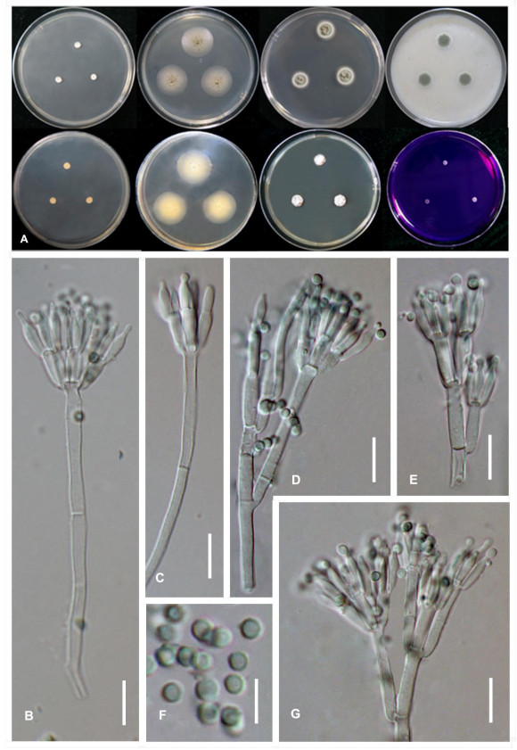
    Figure 3.  Morphological characters of T. peaticola (ex-type CGMCC3.18620). A Colonies from left to right (top row) CYA, MEA, YES, and OA; (bottom row) CYA reverse, MEA reverse, DG 18 and CREA. B–G Conidiophores. H Conidia. Scale bars: B–C = 20 μm, D–H = 10 μm.

    Index Fungorum number: IF553909; Facesoffungi number: FoF 09706

    Etymology-peaticola, the stem peati- refers to the substrate that the type strain is isolated, the ending -cola means"weller, inhabit".

    Diagnosis-Colonies slow-growing and concave in centers on CYA at 25℃, extremely slow-growing on CYA at 30℃ and 37℃, acid production absents on CREA at 25℃; conidiophores biverticillate; conidia globose, smooth-walled.

    Colony diam, 7 d, 25℃ (unless stated otherwise): CYA 5.0-6.9 mm; MEA 24.7-24.9 mm; DG18 16.0-16.5 mm; YES 9 -11 mm; CREA 3.3-3.9 mm; OA 11.7-12.5 mm; CYAS 2.9-3.5 mm; CYA 30℃ 4.7-6.6 mm; CYA 37℃ 4.5-4.7 mm; MEA 30 ℃ 35.5-37.8 mm.

    Colony characters -CYA 25℃, 7d: colonies slightly raised at center, slightly sulcate; mycelia white; texture velvety; sporulation absent; soluble pigments absent; exudates absent; reverse medium beige (Fig. 3A). MEA 25℃, 7d: colonies low plane; margins low, plane, entire; mycelia white; sporulation loose; conidia grayish green; texture floccose; soluble pigments absent; exudates small clear droplets; reverse bone white (Fig. 3A). YES 25℃, 7d: colonies slightly raised at center, sunken at center, sulcate; margin slow, plane, entire (< 1 mm); mycelia white; texture velvety; sporulation absents to sparse; soluble pigment absent; exudates absent; reverser white (Fig. 3A). DG18 25℃, 7d: colonies raised at center, sunken in the centre, sulcate; margins low, plane, entire (1-2 mm); mycelia white; texture velvety; sporulation moderately dense to dense; conidia dark green; soluble pigments absent; exudate absent (Fig. 3A). OA 25℃, 7d: colonies low, plane; margins low, plane, entire; mycelia white; texture floccose; sporulation dense, conidia dark green; soluble pigments absent; exudates absent (Fig. 3A); CREA 25℃, 7d: restricted growth; acid production absent (Fig. 3A). On MEA 25℃, 7d conidiophores biverticillate (Fig. 3B-G); stipes smooth-walled, 160-200 × 3-4 μm (Fig. 3D, G); metulae, 3-8, divergent, 7.5-11.5 × 2-3 μm; phialides acerose, per metulae, 7.0-13.5×1.5-2.0 μm (Fig. 3E); conidia smooth, in chain, subglobose, globose, smooth-walled, 1.5-2.5×1.5-2.0 μm (Fig. 3H).

    Teleomorph-Undetermined

    Known distribution-CHINA. Sichuan Province.

    Material examined – CHINA. Sichuan Province, Aba Autonomous Prefecture, Hongyuan County, Zoigê wetland, 33o3′54N, 102o34′23.9E, peaty soil, September 15, 2016, Jianqing Tian, ZRT-4 (holotype: HMAS 247296). ex-type living culture: CGMCC3.18620, CGMCC3.18767, CGMCC3.18768. ibid. LB1.17020001.

    Notes - Talaromyces peaticola belongs to Talaromyces section Trachyspermi are well supported by the phylogenetic analyses of a combination of ITS, BenA, CaM and RPB2 sequence data-set. BenA marker and multi-gene of ITS, BenA, CaM and RPB2 performed well in differing T. peaticola from T. diversus. It also can be distinguished from other Talaromyces species by colony slow-growing on CYA at 25℃, extremely slow-growing on CYA at 30℃ and 37℃, acid production absents on CREA at 25℃; conidiophores biverticillate; conidia globose, smooth-walled. In spite of T. peaticola phylogenetically and morphologically closed to T. diversus, it could be easily distinguished from T. diversus by the later could not grow on CREA at 25℃ and T. peaticola producing smaller conidia (Yilmaz et al. 2014).

  • The taxonomy of Talaromyces was redefined recently on the basis of DNA sequence data, extrolite profiles and other phenotypic features (Yilmaz et al. 2012, 2014, Chen et al. 2016). The phylogenetic analysis resulted from the combined sequence of ITS, BenA, CaM and RPB2 well distinguish Talaromyces peaticola from other Talaromyces species in section Trachyspermi (Fig. 1), which supported combined ITS, BenA, CaM and RPB2 sequence is imperative for new species identification (Yilmaz et al. 2014, Chen et al. 2016). Additionally, phylogenetic analysis conducted by the single gene of BenA well differs T. peaticola from other species within Talaromyces section Trachyspermi, as well as its sister taxon T. diversus, which resulted from a 9 bp difference in BenA locus (448/457). This confirms that BenA, is imperative for new species identification of Talaromyces (Yilmaz et al. 2014, Chen et al. 2016). However, both phylogenetic analyses based on the single gene of ITS and RPB2 not well differ T. peaticola from T. diversus owning the little differences ITS sequence (585/590 bp) and RPB2 sequences (850/852 bp). These results agree with that ITS and RPB2 sequences are insufficiently variable to reliably discriminate species in Talaromyces (Skouboe et al. 1999, Yilmaz et al. 2012, Frisvad et al. 2013, Yilmaz et al. 2014). BenA and CaM sequences perform well in species delimitation of Aspergillaceae, especially in Penicillium, Aspergillus and Talaromyces, and even some intraspecies (Seifert et al. 2007, Visagie et al. 2014, Yilmaz et al. 2014, Chen et al. 2016).

    Colony features on seven standardized media and morphological characters on MEA and are recommended as important phenotypic features in the identification of Talaromyces (Visagie et al. 2014, Yilmaz et al. 2014). Talaromyces peaticola characteristically displays restricted growth on CYA and CREA and exudates small clear droplets on MEA (Fig. 3). In spite that T. peaticola has an affinity to T. diversus in all phylogenetic trees (Fig. 1). These two species are readily distinguished from each otherin regard to T. diversus could not grow on CREA at 25℃ and exudates small clear droplets on MEA (Yilmaz et al. 2014). Additionally, the conidia of T. peaticola are smaller than of T. diversus. Therefore, Talaromyces peaticola is introduced as a new taxon.

    Zoige peatland is the largest alpine peatland, characterized by high moisture and low temperature. Previous studies have reported that species in genus Talaromyces are one of the major fungal survivals and frequently isolated from peatland (Gilbert & Mitchell 2006, Wu et al. 2013, Grum-Grzhimaylo et al. 2016, Asemaninejad et al. 2017), the ecological function and metabolic activities need to be further explored.

    • This research is jointly supported by the National Key Research and Development Program of China (No. 2016YFC0501802) and the Natural Science Foundation of China (no. 31600024).
Figure (3)  Table (1) References (32)
  • About this article
    Cite this article
    JQ Tian, YF Wang, JZ Sun. 2021. Talaromyces peaticola (Aspergillaceae, Eurotiales), a new species from the Zoige wetlands, China. Studies in Fungi 6(1):391−400 doi: 10.5943/sif/6/1/29
    JQ Tian, YF Wang, JZ Sun. 2021. Talaromyces peaticola (Aspergillaceae, Eurotiales), a new species from the Zoige wetlands, China. Studies in Fungi 6(1):391−400 doi: 10.5943/sif/6/1/29
  • Catalog

    • About this article

    /

    DownLoad:  Full-Size Img  PowerPoint
    Return
    Return