ARTICLE   Open Access    

Generation of myxomycete data from three discrete experiments using moist chamber cultures in a Neotropical forest

More Information
  • The moist chamber technique is widely used in ecological research on myxomycetes. However, limited assessments on the usefulness of the technique have been carried out using empirical data. In the present study, three discrete experiments were carried out in a tropical forest in Costa Rica with the main objective of providing meaningful parameters for the design of future studies in similar environments. All three experiments showed that results could be maximized for representativeness by designing studies that purposedly target ecological components of the studied system. In a comparison of recorded data at three heights above the forest ground, a significantly higher number of records and species were observed in the higher vertical partitions, suggesting that collecting research material from the ground, in this ecological setting, reduces the probability of recording the highest species diversity. However, the ground level was associated with a high number of records and species within the genus Didymium, offering relevant information for studies targeting this genus. Similarly, based on effort, results from the present study suggest that a collecting effort designed to record system variability represents a superior cost-benefit situation for synecological studies than a more intense effort designed only for a limited spatial or temporal space, which in turn would reduce the ecological significance of the resultant data.
  • The Lonicera Linn. genus is a constituent member of the Caprifoliaceae family[1]. It is the largest genus in this family and comprises at least 200 species with a notable presence in North Africa, North America, Asia, and Europe[1]. Members of the Lonicera genus possess a wide range of economic benefits from their use as ornamental plants to food and as plants credited with numerous health benefits. Conspicuous among the numerous members of this genus with known medicinal uses are L. japonica, L. macranthoides, L. hypoglauca, L. confusa, and L. fulvotomentosa[2]. Though these species feature prominently in the Chinese Pharmacopoeia, other species such as L. acuminata, L. buchananii, and L. similis are recognized as medicinal resources in certain parts of China[1]. Among the aforementioned species, L. japonica takes precedence over the rest due to its high medicinal and nutritional value[3,4]. For instance, the microRNA MIR2911, an isolate from L. japonica, has been reported to inhibit the replication of viruses[57]. Also, the water extract of L. japonica has been used to produce various beverages and health products[8]. The Lonicera genus therefore possesses huge prospects in the pharmaceutical, food, and cosmetic industries as an invaluable raw material[9].

    The main active constituents of the Lonicera genus include organic acids, flavonoids, iridoids, and triterpene saponins. Chlorogenic acids, iridoids, and flavones are mainly credited with the anti-inflammatory, antiviral, anticancer, and antioxidant effects of the various Lonicera species[1013]. Their hepatoprotective, immune modulatory, anti-tumor and anti-Alzheimer’s effects are for the most part ascribed to the triterpene saponins[1416]. As stated in the Chinese Pharmacopoeia and backed by the findings of diverse research groups, the plants of the Lonicera genus are known to possess high amounts of organic acids (specifically chlorogenic acid) and pentacyclic triterpenoid saponins[2,1719]. The flower and flower bud have traditionally served as the main medicinal parts of the Lonicera genus even though there is ample evidence that the leaves possess the same chemical composition[20]. A perusal of the current scientific literature reveals the fact that little attention has been devoted to exploring the biosynthesis of the chemical constituents of the Lonicera genus with the view to finding alternative means of obtaining higher yields. It is therefore imperative that priority is given to the exploration of the biosynthesis of these bioactive compounds as a possible means of resource protection. There is also the need for further research on ways to fully tap the medicinal benefits of other plant parts in the Lonicera genus.

    Here, we provide a comprehensive review of relevant scientific literature covering the structure, pharmacology, multi-omics analyses, phylogenetic analysis, biosynthesis, and metabolic engineering of the main bioactive constituents of the Lonicera genus. Finally, we proffer suggestions on the prospects of fully exploiting and utilizing plants of the Lonicera genus as useful medicinal plant resources.

    A total of at least 400 secondary metabolites have been reported for the Lonicera genus. These metabolites are categorized into four main groups (Fig. 1a), including not less than 50 organic acids, 80 flavonoids, 80 iridoids, and 80 triterpene saponins[2123]. Organic acids are mainly derivatives of p-hydroxycinnamic acid and quinic acid. Among the organic acids, chlorogenic acids are reported to be the main bioactive compounds in L. japonica[2426]. The organic acids are most abundant in the leaves, while the least amounts are found in the stem of L. japonica. The flowers of the plant are known to contain moderately high amounts of organic acids[27]. The basic core structure of the flavonoids is 2-phenylchromogen. Luteolin and its glycoside which are characteristic flavonoids of the Lonicera genus are most abundant in L. japonica[28]. On the whole, the flavonoid contents in L. japonica are also highest in the leaves, available in moderate amounts in the flowers, and in lowest amounts in the stem[21]. The core structures of the iridoids are iridoid alcohols, the chemical properties of which are similar to hemiacetal. The iridoids often exist in the form of iridoid glycosides in plants. Secoiridoids glycosides are predominant in the Lonicera genus[25]. In L. japonica, the contents of the iridoids are most abundant in the flowers, moderate in leaves, and lowest in the stem[21]. The characteristic saponins of the Lonicera genus are mainly pentacyclic triterpenoids, including the hederin-, oleanane-, lupane-, ursulane- and fernane-types, etc[22]. The hederin-type saponins are reported in the highest amounts in L. macranthoides[17] (Fig. 1b).

    Figure 1.  Core structures of main secondary metabolites and their distribution in five species of Lonicera. (a) 1 and 2, the main core structures of organic acids; 3, the main core structures of flavonoids; 4, the main core structures of iridoids; 5, the main core structures of triterpene saponins. (b) Comparison of dry weight of four kinds of substances in five species of Lonicera[17,28].

    The similarities between chlorogenic acid (CGA) and flavonoids can be traced back to their biosynthesis since p-coumaroyl CoA serves as the common precursor for these compounds[29]. p-coumaroyl CoA is obtained through sequential catalysis of phenylalanine and its biosynthetic intermediates by phenylalanine-ammonia-lyase (PAL), cinnamate 4-hydroxylase (C4H) and 4-coumarate CoA ligase (4CL)[3033].

    CGA is a phenolic acid composed of caffeic acid and quinic acid and is the most important bioactive compound among the organic acids. Its biosynthesis has been relatively well-established; three main biosynthetic routes have been propounded (Fig. 2a). One route relates to the catalysis of caffeoyl-CoA and quinic acid by hydroxycinnamoyl-CoA quinate transferase (HQT)/hydroxycinnamoyl CoA shikimate/quinate hydroxycinnamoyl transferase (HCT) to produce CGA[3437]. The HQT-mediated pathway has been deemed the major route for CGA synthesis in in different plant species[38,39]. The second biosynthetic route stems from the biosynthesis of p-coumaroyl quinate through the catalytic effect of HCT/HQT and subsequent hydroxylation of p-coumaroyl quinate under the catalysis of p-coumarate 3'-hydroxylase (C3’H)[34,36,37]. For the third route, caffeoyl glucoside serves as the intermediate to form CGA, a process that is catalyzed by hydroxycinnamyl D-glucose: quinic acid hydroxycinnamyl transferase (HCGQT)[40,41].

    Figure 2.  Biosynthetic pathways of main bioactive constituents of Lonicera. (a) Biosynthetic pathways of chlorogenic acid. (b) Biosynthetic pathways of luteoloside. (c) Biosynthetic pathways of secologanin. (d) Biosynthetic pathways of hederin-type triterpene saponins. PAL, phenylalanine ammonia-lyase; C4H, cinnamate 4-hydroxylase; 4CL, 4-hydroxycinnamoyl CoA ligase; HCT, hydroxycinnamoyl CoA shikimate/quinate hydroxycinnamoyl transferase; C3’H, p-coumaroyl 3-hydroxylase; HQT, hydroxycinnamoyl-CoA quinate transferase; UGCT, UDP glucose: cinnamate glucosyl transferase; CGH, p-coumaroyl-D-glucose hydroxylase; HCGQT, hydroxycinnamoyl D-glucose: quinate hydroxycinnamoyl transferase; CHS, Chalcone synthase; CHI, Chalcone isomerase; FNS, Flavone synthase; F3H, flavonoid 30-monooxygenase/flavonoid 30-hydroxylase; UF7GT, flavone 7-O-β-glucosyltransferase; GPS, Geranyl pyrophosphatase; GES, geraniol synthase; G8O, geraniol 10-hydroxylase/8-oxidase; 8HO, 8-hydroxygeraniol oxidoreductase; IS, iridoid synthase; IO, iridoid oxidase; 7DLGT, 7-deoxyloganetic acid glucosyltransferase; 7DLH, 7-deoxyloganic acid hydroxylase; LAMT, loganic acid O-methyltransferase; SLS, secologanin synthase; FPS, farnesyl pyrophosphate synthase; SS, squalene synthase; SE, squalene epoxidase; β-AS, β-amyrin synthase; OAS, oleanolic acid synthetase.

    The key enzymes in the biosynthesis of p-coumaroyl CoA, and invariably CGA, thus, PAL, C4H, and 4CL have been established in diverse studies such as enzyme gene overexpression/knockdown[42], enzyme activity analysis[33] and transcriptomics[18]. However, the centrality of HQT in the biosynthesis of CGA remains disputable. While some studies have reported a strong correlation between HQT expression level with CGA content and distribution[18,35,39,43,44], others found no such link[45], bringing into question the role of HQT as a key enzyme in CGA biosynthesis.

    Few studies have been conducted on the regulation of CGA biosynthesis in the Lonicera genus. It was found that overexpression of the transcription factor, LmMYB15 in Nicotiana benthamiana can promote CGA accumulation by directly activating 4CL or indirectly binding to MYB3 and MYB4 promoters[46]. LjbZIP8 can specifically bind to PAL2 and act as a transcriptional repressor to reduce PAL2 expression levels and CGA content[47]. Under NaCl stress, increased PAL expression promoted the accumulation of phenolic substances in leaves without oxidative damage, a condition that was conducive to the accumulation of the bioactive compounds in leaves[48].

    Luteolin and its derivative luteolin 7-O- glucoside (luteoloside) are representative flavonoids of the Lonicera genus. Similar to CGA, luteolin is biosynthesized from p-coumaroyl CoA but via a different route. The transition from p-coumaroyl CoA to luteolin is underpinned by sequential catalysis by chalcone synthetase (CHS), chalcone isomerase (CHI), flavonoid synthetase (FNS), and flavonoid 3'-monooxygenase/flavonoid 3'-hydroxylase (F3'H)[45,49,50] (Fig. 2b). Luteoloside is synthesized from luteolin by UDP glucose-flavonoid 7-O-β-glucosyltransferase (UF7GT)[51]. Similar to CGA biosynthesis, the key enzymes of luteolin synthesis include PAL, C4H, and 4CL in addition to FNS[33,45,52]. The content of luteoloside was found to be highly abundant in senescing leaves relative to other tissues such as stem, flowers, and even young leaves[52]. Through transcriptomic analysis, luteoloside biosynthesis-related differentially expressed unigenes (DEGs) such as PAL2, C4H2, flavone 7-O-β-glucosyltransferase (UFGT), 4CL, C4H, chalcone synthase 2 and flavonoid 3'-monooxygenase (F3'H) genes were found to be upregulated in the senescing leaves. Biosynthesis-related transcription factors such as MYB, bHLH, and WD40 were also differentially expressed during leaf senescence[52], while bHLH, ERF, MYB, bZIP, and NAC were differentially expressed during flower growth[53]. Further analysis of the transcription factors revealed that MYB12, MYB44, MYB75, MYB114, MYC12, bHLH113, and TTG1 are crucial in luteoloside biosynthesis[52,53].

    The biosynthesis of terpenoids mainly involves three stages; formation of intermediates, formation of basic structural skeleton, and modification of basic skeleton[54]. The intermediates of terpenoids are mainly formed through the mevalonate (MVA) and methylerythritol phosphate (MEP) pathways, and eventually converted to the universal isoprenoid precursors, isopentenyl pyrophosphate (IPP) and its isomer dimethylallyl pyrophosphate (DMAPP) through a series of enzyme-catalyzed reactions. Under the catalysis of geranyl pyrophosphatase (GPS), IPP is then converted to geranyl pyrophosphate (GPP). Different terpenoids are subsequently derived from GPP as the intermediate product. For instance, in the formation of secoiridoid, GPP first removes the phosphoric acid group to obtain geraniol, second through a series of reactions such as oxidation and cyclization, the skeleton of iridoid, namely iridodial, can be obtained. Finally, through a series of reactions, the basic carbon skeleton of the secoiridoid, namely secologanin, is obtained[5561] (Fig. 2c). In the formation of triterpene saponins, the key step lies in the formation of the precursor, 2,3-oxidosqualene, a reaction that is catalyzed by squalene epoxidase (SE). There are many pentacyclic triterpenes in the Lonicera genus, the most important type being the hederin-type saponins with hederagenin as aglycones. Hederin-type saponins are produced after the synthesis of oleanolic acid from β-amyrin and catalyzed by β-starch synthetase (β-AS) and Oleanolic acid synthase (OAS)[62,63]. The skeletal modification of the triterpenoid saponins is mainly achieved via the activities of the CYP450 enzymes and UDP-glycosyltransferase (UGT). Hence, the corresponding aglycones are first obtained via oxidation by the CYP450 enzymes (e.g., CYP72A), and further subjected to glycosylation by the appropriate UGT enzyme[6365] (Fig. 2d). Skeletal formations of the iridoids and triterpene saponins in general have been well researched, but the same cannot be said about the enzymes involved in biosynthesis of these groups of compounds in the Lonicera genus. To fully utilize the iridoids and triterpene saponins in the Lonicera genus, it is necessary to further explore their biosyntheses with the view to enhancing and optimizing the process.

    Given the importance of the bioactive compounds in the Lonicera genus, continual isolation of these compounds using the traditional methods are not only tedious and time-consuming, but also unsustainable. With the development and application of microbial metabolic engineering, different strategies have been introduced to produce these bioactive compounds by heterologous synthesis (Table 1).

    Table 1.  Biosynthesis of Lonicera-specialized metabolites using metabolic engineering.
    Engineering bacteriaOperational methodsProductsYieldRefs
    S. cerevisiaeEliminate the tyrosine-induced feedback inhibition, delete genes involved in competing pathways and overexpress rate-limiting enzymesCaffeic acid569.0 mg/L[69]
    S. cerevisiaeEmploye a heterologous tyrosine ammonia lyase and a 4HPA3H complex composed of HpaB and HpaC derived from different speciesCaffeic acid289.4 mg/L[73]
    S. cerevisiaeSupply and recycle of three cofactors: FADH2, S-adenosyl-L-methion, NADPHCaffeic acid
    Ferulic acid
    Caffeic acid: 5.5 g/L;
    Ferulic acid: 3.8 g/L
    [117]
    E. coliKnocking out competing pathwaysCaffeic acid7,922 mg/L[118]
    E. coliArtificial microbial community, a polyculture of three recombinant Escherichia coli strainsChlorogenic acid250 μM[68]
    Cell-free biosynthesisExtract and purify spy-cyclized enzymes (CFBS-mixture)Chlorogenic acid711.26 mg/L[70]
    S. cerevisiaeThree metabolic engineering modules were systematically optimized: shikimate pathway and carbon distribution, branch pathways, CGA pathway genesChlorogenic acidFlask fermentation: 234.8 mg/L;
    Fed-batch fermentation:
    806.8 mg/L
    [119]
    E. coliUsing modular coculture engineering: construction of the defective strain improves the production and utilization of precursor substancesChlorogenic acid131.31 mg/L[122]
    E. coliIntroduce heterologous UDP-glucose biosynthetic genesLuteolin34 mg/L[120]
    Y. lipolyticaOverexpression of the key genes involved in the mevalonate pathway, the gene encoding cytochrome P450 (CYP716A12) to that encoding NADPH-P450 reductaseOleanolic acid129.9 mg/L[85]
    S. cerevisiaeImprove the pairing efficiency between Cytochrome P450 monooxygenase and reductase and the expression level of key genesOleanolic acid606.9 mg/L[121]
    S. cerevisiaeHeterologous expression and optimization of CrAS, CrAO, and AtCPR1, and regulation of ERG1 and NADPH regeneration systemOleanolic acid433.9 mg/L[123]
     | Show Table
    DownLoad: CSV

    Due to the demand for CGA in the food, pharmaceutical, chemical, and cosmetic industries, the traditional means of obtaining the same requires a relatively longer period for plant maturation to obtain low yields of the desired product. This therefore brings into question the sustainability and efficiency of this approach. The alternative and sustainable approach has been to produce CGA using synthetic biology and metabolic engineering.

    Current research has sought to utilize Escherichia coli (and its mutant strain) and Saccharomyces cerevisiae to synthetically generate CGA and other flavonoids[6673]. For instance, Cha et al. employed two strains of E. coli to produce a relatively good yield of CGA (78 mg/L). Their approach was based on the ability of one strain to generate caffeic acid from glucose and the other strain to use the caffeic acid produced and quinic acid as starting materials to synthesize CGA[66]. Using a bioengineered mutant of E. coli (aroD mutant), Kim et al. increased the yield of CGA to as high as 450 mg/L[67]. Others have sought to increase the yield of CGA by employing a polyculture of three E. coli strains that act as specific modules for the de novo biosynthesis of caffeic acid, quinic acid and CGA. This strategy eliminates the competition posed by the precursor of CGA (i.e., caffeic acid and quinic acid) and generally results in improved production of CGA[68]. Saccharomyces cerevisiae is a chassis widely used for the production of natural substances from plants with an intimal structure that can be used for the expression of cytochrome P450 enzymes that cannot be expressed in E. coli. Researchers have used yeast to increase the production of organic acids[69]. A de novo biosynthetic pathway for the construction of CGA in yeast has been reported new cell-free biosynthetic system based on a mixture of chassis cell extracts and purified Spy cyclized enzymes were adopted by Niu et al. to a produce the highest yield of CGA reported so far up to 711.26 ± 15.63 mg/L[70].

    There are many studies on the metabolic engineering for the synthesis of flavonoids, but few on luteolin and its glycosides. Strains of E. coli have been engineered with specific uridine diphosphate (UDP)-dependent glycosyltransferase (UGT) to synthesize three novel flavonoid glycosides. These glycosides were quercetin 3-O-(N-acetyl) quinovosamine (158.3 mg/L), luteolin 7-O-(N-acetyl) glucosaminuronic acid (172.5 mg/L) and quercetin 3-O-(N-acetyl)-xylosamine (160.8 mg/L)[71]. Since most of the flavonoid glycosides synthesized in E. coli are glucosylated, Kim et al. in their bid to synthesize luteolin-7-O-glucuronide, deleted the araA gene that encodes UDP-4-deoxy-4-formamido-L-arabinose formyltransferase/UDP-glucuronic acid C-4'' decarboxylase in E.coli and were able to obtain a yield of 300 mg/L of the desired product[72].

    Terpenoidal saponins are mostly derived from slow-growing plants and usually possess multiple chiral centers[74]. Traditional isolation and even chemical synthesis of the terpenoidal saponins are both tedious and uneconomical for large-scale production. Therefore, it is necessary to find other ways to synthesize these compounds known to have diverse pharmacological functions.

    Heterologous synthesis has become an important way to improve the target products. With the development of synthetic biology, heterologous synthesis of triterpene saponins involves chassis of both plant and microbial origin. In this regard, Nicotiana benthamiana is a model plant species for the reconstruction of the biosynthetic pathways of different bioactive compounds including monoterpenes, hemiterpenes, and diterpenes[59,7577]. Aside from Nicotiana benthamiana, other plants have also been used as heterologous hosts[78]. Heterologous synthesis using microbial hosts mainly involves Saccharomyces cerevisiae and Escherichia coli[7981], and other microorganisms[82,83]. Comparatively, plants as biosynthetic hosts have the advantages of an established photosynthetic system, abundant supply of relevant enzymes, and presence of cell compartments, etc. They are however not as fast growing as the microorganisms, and it is also difficult to extract and separate the desired synthesized compounds from them as hosts.

    Although heterologous synthesis has many advantages, the premise of successful construction of synthetic pathway in host is to elucidate the unique structure of the compound and the key enzyme reaction mechanism in the biosynthetic pathway. There is little research on metabolic engineering of the hederin-type pentacyclic triterpene saponins in Lonicera, but there are studies on the heterologous synthesis of its aglycone precursor, oleanolic acid[84,85]. There is a dearth of scientific literature on key enzymes in the biosynthesis of pentacyclic triterpenoid saponins in the Lonicera genus.

    Scientific evidence by diverse research groups has linked members of the Lonicera genus to a wide range of pharmacological effects (Fig. 3). These pharmacological effects are elicited by different chemical constituents, much of the underlying mechanisms of which have been elucidated by the omics techniques. Here, we summarize the pharmacological effects and pharmacodynamics of the Lonicera genus in the last 6 years.

    Figure 3.  Schematic summary of four main pharmacological effects (anti-inflammatory, antimicrobial, anti-oxidative and hepatoprotective effects) of the Lonicera genus and the underlying mechanisms of actions.

    Bioactive compounds of plants in the Lonicera genus have demonstrated varying degrees of anti-inflammatory actions. In a recent study, Lv et al. showed that lonicerin inhibits the activation of NOD-like receptor thermal protein domain associated protein 3 (NLRP3) through regulating EZH2/AtG5-mediated autophagy in bone marrow-derived macrophages of C57BL/6 mice[86]. The polysaccharide extract of L. japonica reduces atopic dermatitis in mice by promoting Nrf2 activation and NLRP3 degradation through p62[87]. Several products of Lonicera have been reported to have ameliorative effects on DSS-induced colitis. Among them, flavonoids of L. rupicola can improve the ulcerative colitis of C57BL/6 mice by inhibiting PI3K/AKT, and pomace of L. japonica can improve the ulcerative colitis of C57BL/6 mice by improving the intestinal barrier and intestinal flora[88,89]. The flavonoids can also ameliorate ulcerative colitis induced by local enema of 2,4,6-trinitrobenzene sulfonic acid (TNBS) in Wistar rats by inhibiting NF-κB pathway[90]. Ethanol extract from L. Japonica has demonstrated the potential to inhibit the expressions of inflammatory cytokines in serum and macrophages of LPS-induced ICR mice[91]. The water extract of L. japonica and luteolin were found to exhibit their anti-inflammatory effects via the inhibition of the JAK/STAT1/3-dependent NF-κB pathway and induction of HO-1 expression in RAW263.7 cells induced by pseudorabies virus (PRV)[92].

    Existing scientific evidence indicates that the extracts of plants in the Lonicera genus exhibit strong inhibition against different pathogenic microorganisms. Phenolic compounds from L. japonica demonstrated a particularly significant inhibitory effect against Staphylococcus aureus and Escherichia coli, in vitro, making these compounds potential food preservatives[93]. Influenza A virus is a serious threat to human health. Recent research has found the ethanol extract of L. japonica to possess a strong inhibitory effect against H1N1 influenza virus-infected MDCK cells and ICR mice[94]. The incidence of the COVID-19 pandemic called to action various scientists in a bid to find safe and efficacious treatment[95]. Traditional Chinese medicines became an attractive alternative in this search. The water extract of the flower bud of L. japonica which has traditionally served as a good antipyretic and antitussive agent attracted the attention of researchers. Scientific evidences have confirmed that the water extract of L. japonica can induce let-7a expression in human rhabdomyosarcoma cells or neuronal cells and blood of lactating mice, inhibiting the entry and replication of the virus in vitro and in vivo[96]. In addition, the water extract of L. japonica also inhibits the fusion of human lung cancer cells Calu-3 expressing ACE2 receptor and BGK-21 cells transfected with SARS-CoV-2 spike protein, and up-regulates the expression of miR-148b and miR-146a[97].

    Oxidative stress has been implicated in the pathophysiology of many diseases, hence, amelioration of the same could be a good therapeutic approach[98,99]. In keeping with this therapeutic strategy, various compounds from the Lonicera genus have demonstrated the ability to relieve oxidative stress due to their pronounced antioxidant effects. For instance, the polyphenolic extract of L. caerulea berry was found to activate the expression of AMPK-PGC1α-NRF1-TFAM proteins in the skeletal muscle mitochondria, improve the activity of SOD, CAT and GSH-Px enzymes in blood and skeletal muscle, relieve exercise fatigue in mice by reducing oxidative stress in skeletal muscle, and enhance mitochondrial biosynthesis and cell proliferation[100]. The diverse health benefits of the anthocyanins from L. japonica have been mainly credited to their antioxidant and anti-inflammatory effects. The anthocyanin and cyanidin-3-o-glucoside have been reported to possess the potential to prolong life and delay senescence of Drosophila through the activation of the KEAP1/NRF2 signaling pathway[101].

    The liver is an essential organ that contributes to food digestion and detoxification of the body. These functions expose the liver to diverse toxins and metabolites. The Lonicera genus is rich in phytochemicals that confer protection on the liver against various toxins. The phenolic compound, 4, 5-di-O-Caffeoylquinic acid methyl ester was shown to be able to improve H2O2-induced liver oxidative damage in HepG2 cells by targeting the Keap1/Nrf2 pathway[102]. Hepatic fibrosis is a complex dynamic process, with the propensity to progress to liver cancer in severe cases. The L. japonicae flos water extract solution increased the cell viability of FL83B cells treated with thioacetamide (TAA), decreased the levels of serum alanine aminotransferase (ALT) and alkaline phosphatase (ALP), inhibited the transformation growth factor β1 (TGF-β1) and liver collagen deposition[103]. Sweroside, a secoiridoid glucoside isolate of L. japonica is known to protect the C57BL/6 mice liver from hepatic fibrosis by up-regulating miR-29a and inhibiting COL1 and TIMP1[104].

    Aside from the aforementioned, other pharmacological effects have been ascribed to the Lonicera genus. The ethanolic extract of L. caerulea has been reported to inhibit the proliferation of SMMC-7721 and H22 hepatoma cells, while its anthocyanins induced the apoptosis of tumor cells via the release of cytochrome C and activation of caspase[105]. AMPK/PPARα axes play an important role in lipid metabolism. A chlorogenic acid-rich extract of L. Japonica was found to significantly decrease the early onset of high-fat diet-induced diabetes in Sprague-Dawley rats via the CTRPs-AdipoRs-AMPK/PPARα axes[106]. In a high-fat diet-induced non-alcoholic fatty liver disease in C57BL/6 mice, treatment with L. caerulea polyphenol extract decreased serum inflammatory factors and endotoxin levels and the Firmicutes/Bacteroidetes ratio, an indication of its modulatory effect on the gut microbiota[107]. The iridoid-anthocyanin extract from L. caerulea berry contributed to alleviating the symptoms of intestinal infection with spirochaeta in mice[108].

    The traditional classification of the Lonicera genus based on the morphology of member plants is further categorized into two subgenera, Chamaecerasus and Periclymenum. The Chamaecerasus includes four categories, Coeloxylosteum, Isika, Isoxylosteum and Nintooa. The Periclymenum includes two categories, Subsect. Lonicera and Subsect. Phenianthi (Supplemental Table S1).

    High-throughput chloroplast genome sequencing of L. japonica found its length to be 155078 bp, which is similar to the structure of the typical angiosperm chloroplast genome. It contains a pair of inverted repeat regions (IRa and IRb, 23774 bp), a large single copy region (LSC, 88858 bp) and a small single copy area (SSC, 18672 bp)[109,110]. However, compared with chloroplast genomes of other plants, the chloroplast genome of L. japonica has a unique rearrangement between trnI-CAU and trnN-GUU[110]. Based on the phylogenetic analysis of the plastid genomes of seven plants in the Lonicera genus, 16 diverging hot spots were identified as potential molecular markers for the development of the Lonicera plants[111]. The phylogeny of Lonicera is rarely researched at the molecular level and the pattern of repetitive variation and adaptive evolution of the genome sequence is still unknown. Chloroplast genome sequences are highly conserved, but insertions and deletions, inversions, substitutions, genome rearrangements, and translocations also occur and have become powerful tools for studying plant phylogeny[112,113].

    We present here the phylogenetic tree of the Lonicera genus based on the published complete chloroplast genome sequences downloaded from the National Center for Biotechnology Information (NCBI) database using the Maximum likelihood method (Fig. 4). Based on our chloroplast phylogenies, we propose to merge L. harae into Sect. Isika and L. insularis into Chamaecerasus, but whether L. insularis belongs to Sect. Isika or Sect. Coeloxylosteum is uncertain. Based on protein-coding regions (CDS) of the chloroplast genome or complete chloroplast genomes, Liu et al. and Chen et al. supported the classification of the two subgenera in Lonicera[111,114]. Sun et al. and Srivastav et al. demonstrated a classification between the two subgenera with more species by using sequences of nuclear loci generated, chloroplast genome, and restriction site-associated DNA sequencing (RADSeq)[115,116]. However, our phylogenetic analysis and that of Sun et al. show relations within the subgenus Chamaecerasus are tanglesome in some respects[116]. Plant traits are affected by the environment to varying degrees. Since evidence of plant speciation is implicit in its genome sequence, comparative analysis at the molecular level provides a relatively accurate depiction of inherent changes that might have occurred over time. These findings suggest the need for more species of the Lonicera genus to be sequenced to provide a more accurate theoretical basis for the evolution of the Lonicera plants and a more effective revision in the classification of the Lonicera genus.

    Figure 4.  Phylogenetic tree of 42 species of the Lonicera genus based on complete chloroplast genome sequence data. The phylogenetic tree was constructed by the maximum likelihood method. Coeloxylosteum, Isika, Isoxylosteum, and Nintooa belong to Chamaecerasus and Subsect. Lonicera belongs to the Periclymenum. Chamaecerasus and Periclymenum are the two subgenera of Lonicera. 'Not retrieved' indicates that the species failed to retrieve a subordinate taxon in the Lonicera.

    The Lonicera genus is rich in diverse bioactive compounds with immeasurable prospects in many fields. Members of this genus have been used for thousands of years in traditional Chinese medicine for heat-clearing and detoxification. These plants generally have a good taste and form part of the ingredients of various fruit juices. In cosmetics, they are known to possess anti-aging and moisturizing functions. Plants of the Lonicera genus are also known for their good ecological adaptability and can be used to improve soil and ecological environment. Based on the value of the Lonicera genus, besides researching their use through molecular biological means, their efficient utilization can also be promoted in the following ways: (1) The stems and leaves of the plants could be developed for consumption and use since the chemical profiles of these parts do not differ significantly from the flowers. This way, the wastage of this scarce resource could be minimized or avoided. (2) Most of the Lonicera plants are vines or shrubs and their natural regeneration speed is slow, so the introduction and domestication of species could be strengthened to avoid overexploitation of wild resources.

    At present, only the research on the biosynthesis and efficacy of chlorogenic acid is quite comprehensive and has been used widely in various fields. There is limited research on various aspects of other bioactive compounds and should therefore be given priority in future research goals. Currently, the multi-omics analytical approach has gradually evolved as a reliable and helpful analytical platform. Hence, multi-omics research on the Lonicera genus could lead to discoveries in drug discovery and human health.

    The authors confirm contribution to the paper as follows: study conception and design, draft manuscript preparation: Yin X, Chen X, Li W, Tran LSP, Lu X; manuscript revision: Yin X, Chen X, Li W, Tran LSP, Lu X, Chen X, Yin X, Alolga RN; data/literature collection: Chen X, Yin X; figure preparation: Chen X, Yin X; figure revision: Alolga RN, Yin X, Chen X, Li W, Tran LSP, Lu X. All authors reviewed the results and approved the final version of the manuscript.

    All data generated or analyzed during this study are included in this published article and its supplementary information file.

    This work was partially supported by the National Natural Science Foundation of China (NSFC, Nos 82173918 and 82373983).

  • The authors declare that they have no conflict of interest. Xiaojian Yin is the Editorial Board member of Medicinal Plant Biology who was blinded from reviewing or making decisions on the manuscript. The article was subject to the journal's standard procedures, with peer-review handled independently of this Editorial Board member and the research groups.

  • Alexopoulos CJ. 1953 – Myxomycetes developed in moist chamber culture on bark from living Florida trees, with notes on an undescribed species of Comatricha. Quarterly Journal of the Florida Academy of Science 16(4), 254–262.

    Google Scholar

    Alexopoulos CJ. 1964 – The rapid sporulation of some myxomycetes in moist chamber culture. The Southwestern Naturalist 9(3), 155–159. doi: 10.2307/3669571

    CrossRef    Google Scholar

    Campbell A, Dooley J. 2008 – Composition and ecology of Myxomycetes in a temperate deciduous forest of southeastern Ohio. Ohio Journal of Science 108(1), A21.

    Google Scholar

    Cannon P, Sutton B. 2004 – Microfungi on wood and plant debris. In: Mueller G, Bills G, Foster M (eds), Biodiversity of Fungi: Inventory and Monitoring Methods. Burlington, Massachusetts: Elsevier Academic Press. pp 217–239.

    Google Scholar

    Cavalcanti LH, Ferreira IN, Bezerra ACC, Costa AAA. 2015 – Microhabitats occupied by Myxomycetes in the Brazilian Atlantic forest: Heliconiaceae inflorescences. Brazilian Journal of Biology 75(4), 859–867. doi: 10.1590/1519-6984.01714

    CrossRef    Google Scholar

    Dagamac NHA, Stephenson SL, Dela Cruz TEE. 2014 – The occurrence of litter Myxomycetes at different elevations in Mt. Arayat National Park, Pampanga, Philippines. Nova Hedwigia 98, 187–196. doi: 10.1127/0029-5035/2013/0142

    CrossRef    Google Scholar

    Gilbert HC, Martin GW. 1933 – Myxomycetes found on the bark of living trees. University of Iowa Studies in Natural History 15(3), 3–8.

    Google Scholar

    Hammer Ø, Harper DAT, Ryan PD. 2001 – PAST: Paleontological statistics software package for education and data analysis. Palaeontologica Electrononica 4(1), 9.

    Google Scholar

    Hosokawa A, Reid CR, Latty T. 2019 – Slimes in the city: The diversity of myxomycetes from inner-city and semi-urban parks in Sydney, Australia. Fungal Ecology 39, 37–44. doi: 10.1016/j.funeco.2018.11.004

    CrossRef    Google Scholar

    Kryvomaz TI, Maximenko DV. 2016 – Modeling of environmental pollution by metals depending on their concentration in myxomycetes Fuligo septica. Environmental Safety and Sustainable Resources Management 2(14), 87–92.

    Google Scholar

    Novozhilov YK, Rollins AW, Schnittler M. 2017 – Ecology and distribution of myxomycetes. In: Stephenson SL, Rojas C (eds), Myxomycetes: Biology, Systematics, Biogeography and Ecology. Academic Press, London. pp. 253–297.

    Google Scholar

    Li M, Tao X, Li B, Du Q et al. 2021 – Spatiotemporal distribution and dynamic changes of myxomycetes in subtropical forests of China. Fungal Ecology 53, 101078. doi: 10.1016/j.funeco.2021.101078

    CrossRef    Google Scholar

    Rollins AW, Stephenson SL. 2012 – Myxogastrid distribution within the leaf litter microhabitat. Mycosphere 3(5), 543–549. doi: 10.5943/mycosphere/3/5/2

    CrossRef    Google Scholar

    Rojas C, Matarrita-Gutiérrez K, Rojas PA, Rollins AW. 2021 – Can the location of the isolation laboratory affect the generation of myxomycete data using moist chambers? An experiment in the Neotropics. Current Research in Environmental & Applied Mycology (Journal of Fungal Biology) 11(1), 67–75.

    Google Scholar

    Rojas C. Stephenson SL. 2020 – Comparative analysis on datasets of myxomycetes associated with boreal, temperate and tropical regions of North America. Karstenia 58(2): 190–200.

    Google Scholar

    Rojas C, Stephenson SL. 2021 – Phenology of myxomycetes in Turrialba, Costa Rica. Karstenia 59(1-2), 1–12.

    Google Scholar

    Rojas C, Valverde R, Rollins AW, Murillo Roos M. 2016 – What can myxomycetes tell us about floricolous microbial systems. Nova Hedwigia 104 (1–3), 211–220.

    Google Scholar

    Schnittler M, Stephenson SL. 2002 – Inflorescences of Neotropical herbs as a newly discovered microhabitat for myxomycetes. Mycologia 94(1), 6–20. doi: 10.2307/3761841

    CrossRef    Google Scholar

    Stephenson SL, Stempen H. 1994 – Myxomycetes: A Handbook of Slime Molds. Timber Press, Oregon.

    Google Scholar

    Stephenson SL, Schnittler M, Lado C. 2004 – Ecological characterization of a tropical myxomycete assemblage – Maquipucuna Cloud Forest Reserve, Ecuador. Mycologia 96(3), 488–497.

    Google Scholar

    Stephenson SL, Marbaniang TM, Gupta P, Rojas C. 2020 – Assemblages of corticolous myxomycetes associated with species of Pinus (Pinaceae) in four different regions of the world. Nova Hedwigia 111(1–2), 199–217. doi: 10.1127/nova_hedwigia/2020/0585

    CrossRef    Google Scholar

    Tucker G, Hill D, Fasham M. 2005 – Introduction to planning. In: Hill D, Fasham M, Tucker G, Shewry M, Shaw P (eds. ), Handbook of Biodiversity Methods: Survey, Evaluation and Monitoring. Cambridge University Press, Cambridge, UK. pp. 3–5.

    Google Scholar

    Wellman P. 2020 – Australian corticolous myxomycetes: models of distribution and development. Australian Journal of Botany 67(8), 617–629.

    Google Scholar

    Walker LM, Cedeño-Sanchez M, Carbonero F, Herre EA et al. 2019 – The response of litter-associated myxomycetes to long-term nutrient addition in a lowland tropical forest. Journal of Eukaryotic Microbiology, 66: 757–770. doi: 10.1111/jeu.12724

    CrossRef    Google Scholar

    Wrigley de Basanta D, Estrada-Torres A. 2017 – Techniques for recording and isolating myxomycetes. In: Stephenson SL, Rojas C (eds), Myxomycetes: Biology, Systematics, Biogeography and Ecology. Academic Press, London. pp. 333–363.

    Google Scholar

  • Cite this article

    C Rojas, AW Rollins, R Valverde. 2021. Generation of myxomycete data from three discrete experiments using moist chamber cultures in a Neotropical forest. Studies in Fungi 6(1):450−459 doi: 10.5943/sif/6/1/34
    C Rojas, AW Rollins, R Valverde. 2021. Generation of myxomycete data from three discrete experiments using moist chamber cultures in a Neotropical forest. Studies in Fungi 6(1):450−459 doi: 10.5943/sif/6/1/34

Figures(2)  /  Tables(3)

Article Metrics

Article views(3777) PDF downloads(429)

Other Articles By Authors

ARTICLE   Open Access    

Generation of myxomycete data from three discrete experiments using moist chamber cultures in a Neotropical forest

Studies in Fungi  6 Article number: 34  (2021)  |  Cite this article

Abstract: The moist chamber technique is widely used in ecological research on myxomycetes. However, limited assessments on the usefulness of the technique have been carried out using empirical data. In the present study, three discrete experiments were carried out in a tropical forest in Costa Rica with the main objective of providing meaningful parameters for the design of future studies in similar environments. All three experiments showed that results could be maximized for representativeness by designing studies that purposedly target ecological components of the studied system. In a comparison of recorded data at three heights above the forest ground, a significantly higher number of records and species were observed in the higher vertical partitions, suggesting that collecting research material from the ground, in this ecological setting, reduces the probability of recording the highest species diversity. However, the ground level was associated with a high number of records and species within the genus Didymium, offering relevant information for studies targeting this genus. Similarly, based on effort, results from the present study suggest that a collecting effort designed to record system variability represents a superior cost-benefit situation for synecological studies than a more intense effort designed only for a limited spatial or temporal space, which in turn would reduce the ecological significance of the resultant data.

  • The moist chamber culture technique (Gilbert & Martin 1933) enables researchers to document myxomycete species at a faster rate than collecting sporocarps that have developed under natural conditions in the field (Alexopoulos 1953). As such, the technique is strongly recommended and widely employed in conjunction with field-based methods to record sporocarps for diversity surveys (Wrigley de Basanta & Estrada-Torres 2017). Several researchers have noted that data generated using the moist chamber technique are taxonomically biased and exclude species with longer developmental times and larger fruiting structures (i.e., Alexopoulos 1964, Stephenson et al. 2004). The assumption has been that this bias exerts minimal impact on diversity studies if the associated field component is comprehensive with multiple sampling periods strategically planned to span the environmental and seasonal variation across the study areas. To date, very little effort has been made to quantify the extent of the bias imposed by using the moist chamber technique.

    Most biological work with myxomycetes has centered on documenting the diversity and distribution of species (Novozhilov et al. 2017). The resultant data has often been used to infer ecological attributes even though the studies were not explicitly designed to quantify such characteristics. Interest in designing observational and experimental studies to explicitly test ecological hypotheses and produce reliable and robust information that can be used to understand general ecological principles and processes as well as inform the management of natural resources has increased. Toward these means, projects aimed at elucidating ecological attributes cannot continue to rely on traditional diversity approaches. Thorough evaluations of existing diversity-based approaches to quantify the sources and extent of bias, error, and variability are required to inform study design, statistical approaches, and interpretation for ecological studies. The use of the moist chamber culture technique, based on Gilbert and Martin's approach, does not provide a true replicable standard appropriate for ecological studies. There are several factors, not considered in biodiversity assessments, that can influence the results such as substrate mass, moisture levels, laboratory climate, and temporal extent. Rojas et al. (2021) provided quantified data validating that some of these factors can explain differences among datasets despite the large influence of a system's natural variability.

    The sampling schemes used to collect substrate materials (e.g., plot, plotless, random, systematic, etc.), the precise parameters used for moist chamber preparation (e.g., mass, heterogeneity, quality of substrate material(s) etc.), the laboratory incubation conditions (temperature, humidity, quantity of electromagnetic radiation, air flow, etc), and the data collection parameters (e.g., number of sporocarps, number of spores produced, days until fully developed, etc.) have been highly variable among modern investigations and a standardized methodological approach has not been proposed. An opportunistic approach as described by Cannon & Sutton (2004) has been highly recommended for diversity studies as it maximizes the probability of surveying the widest range of potential productive niches; however, biodiversity assessments regularly utilize other sampling schemes (e.g., Campbell & Dooley 2008). Plot-based sampling approaches, which are designed to document the density and frequency of organisms, have been used as a framework to collect substrates for myxomycete studies even though the studies being conducted were not intended to study such parameters (i.e., Dagamac et al. 2014). These discrepancies occur due to the absence of a widely accepted standardized methodological approach developed from the results of quantitative studies of the moist chamber technique and associated sampling schemes. Despite the issues mentioned above, the technique is remarkably useful for macroecological purposes (Wellman 2020) or ecological studies that require researchers to execute a higher level of control on ecological forces (i.e., Walker et al. 2019).

    The study presented herein discusses the results of three distinct investigations conducted in a Costa Rican tropical forest where a wider, long-term, effort to understand the ecology of myxomycetes is ongoing (Rojas & Stephenson 2021). The main objective of the present discussion is to elucidate relevant parameters that can be used to assess the utility and limitations of the moist chamber culture technique for ecological and applied studies using myxomycetes as focus organisms. The three experiments explained herein are integrated within the idea that the moist chamber technique is a relevant method to address the effect of global changes on myxomycetes. By using the sporocarp phase as a proxy to quantify their ecological associations with natural and non-natural environments, myxomycete data is remarkably relevant for easy-to-implement monitoring of microbiota.

  • The study presented herein was carried out in the Finca Experimental Interdisciplinaria de Modelos Agroecológicos (FEIMA) in Turrialba, Costa Rica during 2018 and 2019. This research station, administrated by the University of Costa Rica, encompasses a 28-ha successional forest patch in the premontane tropical moist forest life zone. In this area, a thorough 30-month assessment of myxomycete occurrence, based on field collections, was carried out to evaluate phenological patterns in tropical species (Rojas & Stephenson 2021). Based on the general recommendations for myxomycete surveys (Wrigley de Basanta & Estrada-Torres 2017), the moist chamber technique was utilized to increase the number of species recorded from the study area.

    With the latter technique, three discrete experiments were carried out with the goal of evaluating the effect of methodological modifications on the generation of data (Fig. 1). As such, the first experiment tested the idea that different results can be obtained when the isolation process takes place in different laboratories in a similar manner to Rojas et al. (2021). For this experiment a total of 432 substrate samples were collected in the form of 12 samples of both litter and bark at ground level, at 1 m and at 2 m of distance from the ground in six independent locations. Once collected, the material was divided in two sets of 216 samples corresponding to exactly half of the collecting effort and taken to different laboratories (named laboratory one and two in this study) for isolation with the moist chamber technique. In this case, litter samples collected at the ground were considered ground litter, whereas litter samples collected above the ground were considered aerial litter.
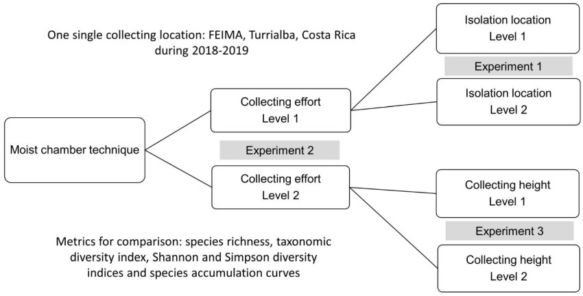
    Figure 1.  Organization of the present study in three discrete experiments based on comparisons of moist chamber data obtained at two levels for each variable under evaluation.

    The second experiment was designed with the goal of testing the effect of sampling effort. For this, a similar design to the first experiment was used but with an 67% reduction in collection effort. As such, only 72 total samples were collected in the form of two samples (instead of 12) of both litter and bark at ground level, at 1m and at 2m above the ground, in the same six independent locations. The material was taken to one of the laboratories used in the previous experiment for the isolation process with the moist chamber technique. In this case, the resulting dataset was only compared with the extended sub dataset of the first experiment isolated in the same laboratory.

    The third experiment was implemented with the goal of evaluating whether the original height at which the material was collected in the field could influence the resulting data. As such, this experiment utilized the reduced effort strategy consisting of 72 samples, but with this approach, six samples of twigs and six of litter were collected only at ground level in each of the six independent locations. For analysis, this dataset was compared with the dataset of 72 samples used in the second experiment that reflected the reduced effort.

    In all cases, substrate samples were used to set up moist chamber cultures in the manner described by Stephenson & Stempen (1994). The results from all experiments were compared in terms of species richness, the Shannon and Simpson (1-D) diversity indices, and the taxonomic diversity index as response variables. Compositional similarity was analyzed using the Bray Curtis distance ranging from 0 for two datasets that are completely different to 1 for two datasets that were exactly the same. A cluster analysis was constructed in the second experiment using these data. With the first and second experiments, the analysis of the same parameters by height was also performed. Analyses of Variance was used to evaluate differences in the number of records and species by height categories in the first experiment. In all cases, the software PAST (Hammer et al. 2001), v 4.06b was used.

  • Thirty-two species of myxomycetes were recorded across all three experiments. From these, 25 were recorded in the first experiment (432 moist chambers), 22 in the second experiment (288 moist chambers) and 25 in the third experiment (144 moist chambers). Similarly, 224 records of myxomycetes were made in the first experiment, 175 records in the second one and 136 records in the third one. As such, the observed productivity was 0.51, 0.60 and 0.94 records/moist chamber for the first, second and third experiments, respectively. In both terms of species and records, the results of the three experiments did not show a particular pattern based on the number of studied moist chambers. The relationship between effort and productivity was strong and negative, but not significant (r = -0.94, p = 0.2).

    The first experiment produced 108 records representing 18 species at laboratory one and 116 records representing 20 species at laboratory two (Table 1). The Shannon index of diversity was 2.63, the Simpson index of diversity was 0.91, and the taxonomic diversity index was 1.8 in both cases. As such, no differences in diversity were observed. The Bray Curtis distance between datasets was 0.47 indicating that the species assemblages recovered between the labs were compositionally different.

    Table 1.  Records of myxomycetes by species observed in the first experiment of the present study arranged by height above the ground

    Species Laboratory one Laboratory two
    0 m 1 m 2 m Subtotal 0 m 1 m 2 m Subtotal
    Arcyria afroalpina 2 1 1 4
    Arcyria cinerea 2 5 4 11 7 6 5 18
    Arcyria insignis 1 1
    Comatricha elegans 1 1
    Comatricha nigra 2 2 1 1
    Comatricha tenerrima 1 1 2
    Cribraria violacea 1 1 2 3 5
    Didymium anellus 1 1 2
    Didymium bahiense 5 7 7 19 1 1
    Didymium difforme 1 2 3 2 2 4
    Didymium minus 2 7 7 16
    Didymium squamulosum 3 2 5 3 1 1 5
    Diderma hemisphaericum 1 2 4 7 4 4 2 10
    Hemitrichia minor 3 1 4
    Hemitrichia pardina 1 1
    Lamproderma scintillans 1 1 3 5 2 2 4
    Perichaena chrysosperma 4 3 6 13 3 3 4 10
    Perichaena pedata 2 4 4 10 1 1
    Physarum compressum 3 6 5 14 5 5 6 16
    Physarum didermoides 2 1 3 6 3 6 4 13
    Physarum pusillum 1 1 2 2 2
    Physarum superbum 1 1
    Stemonitis axifera 1 1
    Stemonitis fusca 1 1 2
    Stemonitopsis aequalis 1 1

    With respect to the three heights from which the samples were collected, 56 records representing 17 species were recorded at ground level, 81 records representing 19 species were collected at 1 m above the ground and 87 records representing 21 species were collected at 2 m above the ground. Both the number of records and the number of species were significantly higher at 2 m when compared to the other two heights (F(2, 33) = 2.6, p = 0.03, F(2, 33) = 3.9, p = 0.03, Tukey p = 0.03). The Shannon index of diversity was 2.7 for the ground level and 2.8 for the other two heights, and the Simpson index of diversity was 0.92 for the ground level and 0.93 for the other two heights. The taxonomic diversity index had a value of 2.1 in all cases. The Bray Curtis distance calculated for the comparison between datasets collected at the ground level and 1 m above the ground was 0.80, whereas the same value for the comparison between datasets at 1 and 2 m from the ground was 0.89. The same value for the comparison between datasets from ground level and 2 m above the ground was 0.77.

    In the second experiment, 108 records representing 18 species were observed with the full effort of 216 samples and 67 records representing 19 species were observed with the reduced effort of 72 samples (Table 2). The productivity of the full dataset was 0.5 records and 0.08 species/moist chamber and for the reduced effort were 0.93 and 0.26, respectively. The Shannon diversity index was 2.7 in both cases, whereas the Simpson diversity index was 0.91 for the full dataset and 0.90 for the reduced dataset. The taxonomic diversity index was 1.8 for the full dataset and 2.1 for the reduced effort. The Bray Curtis distance between datasets was 0.64.

    Table 2.  Records of myxomycetes by species observed in the second experiment of the present study arranged by height above the ground

    Species Full effort Reduced effort
    0 m 1 m 2 m Subtotal 0 m 1 m 2 m Subtotal
    Arcyria afroalpina 1 2 3
    Arcyria cinerea 2 5 4 11 4 2 1 7
    Arcyria insignis 1 1 2 2
    Comatricha nigra 2 2
    Comatricha tenerrima 1 1 2 1 1 2
    Cribraria violacea 1 1 2 2
    Didymium bahiense 5 7 7 19 1 2 2 5
    Didymium clavus 1 1 2
    Didymium difforme 1 2 3 1 2 3
    Didymium iridis 1 1
    Didymium squamulosum 3 2 5 1 4 3 8
    Diderma hemisphaericum 1 2 4 7 2 2
    Hemitrichia minor 3 1 4 1 1 2
    Lamproderma scintillans 1 1 3 5 1 2 3
    Perichaena chrysosperma 4 3 6 13 1 1 1 3
    Perichaena depressa 1 1
    Perichaena pedata 2 4 4 10 1 1
    Physarum compressum 3 6 5 14 5 7 5 17
    Physarum didermoides 2 1 3 6 1 1
    Physarum pusillum 1 1 2 1 1 2
    Physarum superbum 1 1
    Stemonitis fusca 1 1 2

    In terms of height, at ground level, 11 species were observed in both datasets, but the full and reduced dataset were associated with 21 and 31 records, respectively. At the height of 1 m above ground, the full dataset documented 40 records representing 14 species, whereas the reduced dataset was associated with 41 records representing 13 species. At the height of 2 m above ground, 45 records representing 15 species were observed in the full dataset, whereas 42 records representing 16 species were associated with the reduced dataset. Both the Shannon and Simpson indices of diversity were similar between datasets for all heights, with values of 2.4 and 0.91 for ground level, 2.5 and 0.91 for 1 m and 2.7 and 0.93 for 2 m above the ground. The Bray Curtis distances were used to create a cluster dendrogram illustrating that the datasets were compositionally distinct, with the assemblages associated with the ground layer being the most distinct in both cases (Fig. 2).
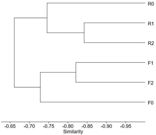
    Figure 2.  Cluster analysis carried out with the data from the second experiment showing the Bray Curtis similarities among sub datasets collected at different heights for both studied datasets. F = full dataset, R = reduced dataset. 0, 1 and 2 refer to ground level, 1 m and 2 m above the ground.

    In the third experiment, both datasets showed similar results. A total of 19 species representing 67 records were observed in the dataset with the full height sampling scheme, whereas 19 species representing 69 records were observed in the dataset that only evaluated ground-level substrates. With these results, an average of 0.94 records/moist chamber and 0.26 species/moist chamber were observed in both cases. The Shannon index was 2.7 and the Simpson index was 0.90 in both cases. The taxonomic diversity index was 1.9 for the dataset with full height and 2.1 for the ground-level dataset. The Bray Curtis distance between datasets was 0.52 suggesting differences in the structural composition that are visible on Table 3.

    Table 3.  Records of myxomycetes by species observed in the third experiment of the present study arranged by sampling type

    Species Full height sampling Ground level sampling
    Arcyria afroalpina 3 11
    Arcyria cinerea 7 16
    Arcyria insignis 2 6
    Comatricha nigra 3
    Comatricha tenerrima 2 8
    Cribraria violacea 2 2
    Didymium anellus 1
    Didymium bahiense 5 1
    Didymium clavus 2
    Didymium difforme 3 1
    Didymium iridis 1
    Didymium minus 2
    Didymium squamulosum 8
    Diderma hemisphaericum 2 3
    Hemitricha minor 2 1
    Lamproderma scintillans 3 2
    Perichaena chrysosperma 3 2
    Perichaena corticalis 3
    Perichaena depressa 1
    Perichaena pedata 1 2
    Perichaena vermicularis 1
    Physarum compressum 17 3
    Physarum didermoides 1
    Physarum oblatum 1
    Physarum pusillum 2
  • The experiments discussed above illustrated clear differences between using the moist chamber technique for diversity or ecological studies using myxomycetes. The main goal of the first approach is to record as many species as possible with a given effort, whereas the goal of the latter was to elucidate patterns and discover the underlying mechanisms producing them through species-environment interactions (Tucker et al. 2005). The moist chamber culture technique was not developed to generate diversity or ecological data, but its original intent was more aligned with the former. The original objective of the experiment explained by Gilbert & Martin (1933) was to promote algae grow on pieces of bark, and the observation of myxomycetes was a surprising secondary result. These researchers eventually experimented with different bark samples and started recording myxomycetes that were rarely collected at the time (see comments from Alexopoulos 1964), which ultimately led them to recommend the technique for diversity surveys. In this manner, the moist chamber technique was conceptually conceived as a successful strategy to record myxomycetes, but from a methodological point of view, the technique only generates presence-based data on the group of myxomycetes that can form sporocarps in the microcosm of the culture.

    The limitations associated with the use of the technique, however, work as a stabilizing force, that albeit artificial, facilitates the formation of myxomycete sporocarps by increasing the homogeneity of the microcosms. As such, the results shown herein were not particularly unexpected. The data from all three experiments carried out in the present study were consistent in showing the lack of differences in response metrics between the two levels associated with the studied factors in each case. As such, diversity-based variables such as species richness, species diversity (calculated with the two indices) and the taxonomic diversity index were equivalent in all cases. This result has two implications since it demonstrates the consistency of the moist chamber technique to generate data, but it also shows, as observed in the experiment two, that sampling effort is a factor of consideration in the design of biodiversity surveys (see the cost-benefit balance of Rollins & Stephenson 2012). The only meaningful differences for data interpretation observed herein were based on compositional aspects of the myxomycete assemblages associated with the different evaluated levels (see Tables 1, 2 and 3 and low values of the Bray Curtis distances).

    Interestingly, differences in the taxonomic structure of datasets, both in terms of species richness and number of observations per species, provide ecological meaning to results. However, as discussed earlier, if the moist chamber technique is implemented using the original simplistic protocol, empirically based ecological conclusions cannot be obtained and substantiated. For instance, Cavalcanti et al. (2015) recorded 10 species of floricolous myxomycetes from 368 moist chambers, but Rojas et al. (2016) recorded an average of 12.8 species in six groups of 90 moist chambers each. This means that with an effort of 24%, the second authors recorded as many species growing on tropical inflorescences as the first ones, demonstrating again that smaller efforts can yield greater productivity in a given situation. In fact, Schnittler & Stephenson (2002) carried out a study with a similar effort to Cavalcanti et al. (2015), but recorded three times as many species. In all these studies, however, it seems likely that experimental considerations such as the time of the year when substrates were collected, the chemical quality of the substrates (see Stephenson et al. 2020), or even the distance from the ground at which samples were collected (observed herein), could be associated with the differences in results. Moreover, other methodological aspects of the laboratory procedures may have affected results as well. In this manner, it is impossible to make a meaningful ecological comparison of results among these three studies since the only methodological elements in common among all of them were the use of Petri dishes, filter paper, distilled water, and floral parts of Heliconia plants. A large number of other "comparable" studies would be subject to the same limitations with respect to drawing valid comparisons.

    Significant compositional evaluations of myxomycete assemblages associated with specific compartments of ecosystems require a refined protocol of the moist chamber technique. In recent years, some applications where myxomycete data is useful have been evaluated, including urban assessments (Hosokawa et al. 2019), bioremediation (Kryvomaz & Maximenko 2016), and climate interactions (see Li et al. 2021). For these ecological studies, and many others, a standardized technique would have generated data with increased broader impacts as they could truly be compared among studies. In this manner results from each experiment in the present study are comparable at the different set levels because researchers used standardized conditions throughout the entire process, but they are not truly comparable with similar assessments made by other researchers in other parts of the world. In other words, the lack of a standardized moist chamber technique imposes an inherent constraint on the use of ecological data generated with the technique.

    In general, the other three lessons that can be drawn with data from the present study are that moist chamber productivity decreases at higher levels of sample coverage, that aerial litter is a highly productive substrate in tropical forests, and that different substrates generate data with different compositional structure. None of these observations are new and they all support the results from previous assessments using moist chambers. For instance, lower effort levels generate data at a faster pace than higher ones, simply because accumulated data tends to stabilize after a particular effort threshold (see results from Rojas & Stephenson 2020). Also, aerial litter has been recognized as a very good substrate for myxomycetes (Novozhilov et al. 2017) and on some occasions, such as this study, response variable values can be greater on it. However, ground litter is often associated with a high number of records and species within the genus Didymium (also observed herein) and studies targeting such genus could use this information for design purposes.

    Results from the present study suggest that a collecting effort designed to record system variability (i.e., spatial and temporal heterogeneity) represents a superior cost-benefit situation for synecological studies than a more intense effort designed only for a limited spatial or temporal purpose. However, common practices such as substrate differentiation and the establishment of different sampling areas can have varied implications for the results obtained from diversity or ecological surveys. In the former, they simply increase the probability of recording more myxomycetes, but for the latter they increase the probability of recording more variability and thus, generate data more appropriate for pattern detection. As such, substrate collection for moist chamber evaluations should be opportunistic (not based on plots or transects) when a floristic or diversity study is intended. In those cases, less samples per collecting site, but more (and different) collecting sites would likely generate a better picture of the myxobiota. Density or frequency evaluations that require plot sampling, should be restricted for ecological studies. However, for these, more analyses on the use of the moist chamber technique are needed, particularly at the level of laboratory procedure, in order to generate a more complete picture of the utility and limitations associated with the use of this technique. Only then will meaningful and truly comparable data be generated for ecological studies of myxomycetes.

    • This work was financed by the University of Costa Rica (Vicerrectoría de Investigación 570-B8-006). Appreciation is extended to Shiori Nakajima and Werner Rodríguez at FEIMA. This study was carried out as part of a research agreement between the University of Costa Rica and Lincon Memorial University.
Figure (2)  Table (3) References (25)
  • About this article
    Cite this article
    C Rojas, AW Rollins, R Valverde. 2021. Generation of myxomycete data from three discrete experiments using moist chamber cultures in a Neotropical forest. Studies in Fungi 6(1):450−459 doi: 10.5943/sif/6/1/34
    C Rojas, AW Rollins, R Valverde. 2021. Generation of myxomycete data from three discrete experiments using moist chamber cultures in a Neotropical forest. Studies in Fungi 6(1):450−459 doi: 10.5943/sif/6/1/34
  • Catalog

    • About this article

    /

    DownLoad:  Full-Size Img  PowerPoint
    Return
    Return