ARTICLE   Open Access    

Inhibition of ice crystal growth by protein hydrolysates from different plant- and animal-based proteins

More Information
  • Due to the quality deterioration and the corresponding waste of frozen foods, there is a constant need for an effective, consumer and environmentally friendly, and sustainable anti-freezing ingredient. In this study, enzymatic hydrolysis and food grade chemical modification (succinylation) were evaluated to produce ice re-crystallization inhibition (IRI) active peptide-based ingredients. Six different types of plant- and animal-based proteins were evaluated. Soy protein isolate (SPI), after 15- and 60-min alcalase hydrolysis, demonstrated a significant IRI activity compared to polyethylene glycol (PEG; as negative control) resulting in a two-third reduction of ice crystal size annealed at −8 °C. Annealing temperature and peptide concentration also affected the IRI activity of SPI hydrolysate where decreased temperature and increased concentration resulted in a higher IRI activity. Increased hydrolysis time decreased the IRI activity of SPI hydrolysates but increased the IRI activity of sodium caseinate (SC) and pea protein isolate (PPI) hydrolysates. SPI-60 was less temperature resistant compared to SPI-15 which inhibited the growth of ice crystals (smaller than 23 µm). Moreover, chemical modification via succinylation reaction with octenyl succinic anhydride (OSA) enhanced the IRI activity of the PPI and SPI hydrolysates leading to a 55.3% and 60.6% reduction in the size of ice crystals, respectively. Overall, this study demonstrates a great potential in the utilization of common food proteins for the production of value-added natural anti-freezing ingredients which could benefit the food industry by enhancing frozen foods' shelf-life and reducing waste.
  • Rice (Oryza sativa L.) is a world staple crop that feeds over half of the world's population[1,2]. In recent years, high-temperature events are becoming more frequent and intensive as a result of global warming, which can severely affect rice grain yield and quality[3,4]. During flowering and grain filling stages, high-temperature stress can result in a significant reduction in seed setting rate and influence amylose content, starch fine structure, functional properties and chalkiness degree of rice[57]. Transcriptome and proteome analysis in rice endosperm have also been used to demonstrate the differences in high-temperature environments at gene and protein expression levels[810]. In addition, as an important post-translational modification, protein phosphorylation has proven to be involved in the regulation of starch metabolism in response to high-temperature stress[11]. However, little is known about whether protein ubiquitination regulates seed development under high-temperature stress.

    Ubiquitination is another form of post-translational modification that plays key roles in diverse cellular processes[12]. Several reports have described the functions of ubiquitination in rice defense responses based on ubiquitome analysis. Liu et al.[13] investigated relationships between ubiquitination and salt-stress responses in rice seedlings using a gel-based shotgun proteomic analysis and revealed the potential important role of protein ubiquitination in salt tolerance in rice. Xie et al.[14] identified 861 peptides with ubiquitinated lysines in 464 proteins in rice leaf cells by combining highly sensitive immune affinity purification and high resolution liquid chromatography-tandem mass spectrometry (LC-MS/MS). These ubiquitinated proteins regulated a wide range of processes, including response to stress stimuli. A later study revealed the relationships between ubiquitination status and activation of rice defense responses, and generated an in-depth quantitative proteomic catalog of ubiquitination in rice seedlings after chitin and flg22 treatments, providing useful information for understanding how the ubiquitination system regulates the defense responses upon pathogen attack[15]. Although many studies have shown that ubiquitination plays improtant roles in the heat response of plant[16,17], there has been little systematic discussion on the ubiquitome of rice endosperm in the context of global climate change.

    In this study, we examine the high-temperature induced ubiquitination change in two rice varieties with different starch qualities, through a label-free quantitative ubiquitome analysis. This study provides a comprehensive view of the function of ubiquitination in high-temperature response of rice developing seed, which will shed new light on the improvement of rice grain quality under heat stress.

    Two indica rice varieties with different starch quality, 9311 and Guangluai4 (GLA4), were used as materials. 9311 is a heat-sensitive variety, which displays low amylose content with good starch quality; while GLA4 is known to be the parental variety of HT54, an indica breeding line with heat tolerance, and thus GLA4 is possibly heat tolerant, which shows high amylose content with poor starch quality[18,19]. Rice growth conditions, sample treatment and collection were conducted as previously described[11].

    Husk, pericarp and embryo were detached from immature rice grains on ice[20]. Rice endosperm was then ground with liquid nitrogen, and the cell powder was sonicated 10 rounds of 10 s sonication and 15 s off-sonication on ice in lysis buffer (6 M Guanidine hydrochloride, pH 7.8−8.0, 0.1% Protease Inhibitor Cocktail) using a high intensity ultrasonic processor. Following lysis, the suspension was centrifuged at 14,000 g for 40 min at 4 °C to remove the debris. The supernatant was collected, and the protein concentration was estimated using BCA assay (Pierce BCA Protein assay kit, Thermo Fisher Scientific, Waltham, MA, USA) before further analysis.

    The protein mixture was reduced by DTT with the final concentration of 10 mM at 37 °C for 1 h, alkylated by iodoacetamide with a final concentration of 50 mM at room temperature in the dark for 0.5 h, and digested by trypsin (1:50) at 37 °C for 16 h. Then the sample was diluted by adding trifluoroacetic acid (TFA) to the final concentration of 0.1%. The enzymatic peptides were desalted on a Sep-Pak C18 cartridge (Waters, Milford, MA, USA), concentrated by lyophilization and reconstituted in precooled IAP buffer (50 mM MOPS-NaOH PH 7.2, 10 mM Na2HPO4, 50 mM NaCl) for further analysis.

    The peptides solution was incubated with prewashed K-ε-GG antibody beads (PTMScan Ubiquitin Remnant Motif (K-ε-GG) Kit), and gently shaken at 4 °C for 1.5 h. The suspension was centrifuged at 2,000 g for 30 s, and the supernatant was removed. The Anti-K-ε-GG antibody beads were washed with IAP Buffer three times and with ddH2O three times. The peptides were eluted from the beads with 0.15% trifluoroacetic acid (TFA). Finally, the eluted fractions were combined and desalted with C18 Stage Tips.

    LC-MS/MS analysis were performed using the methods of Pang et al.[11]. Raw mass spectrometric data were analyzed with MaxQuant software (version 1.3.0.5) and were compared with the indica rice protein sequence database (Oryza sativa subsp. indica-ASM465v1). Parameters were set according to Pang et al.[11]. All measurements were obtained from three separate biological replicates.

    Quantification of the modified peptides was performed using the label-free quantification (LFQ) algorithm[11]. Differentially ubiquitinated sites (proteins) in response to high-temperature were identified by Student's t-test (p < 0.05, log2(fold-change) > 1) with at least two valid values in any condition or the ubiquitination sites that exhibited valid values in one condition (at least two of three replicates) and none in the other.

    Subcellular localization was performed using CELLO database (http://cello.life.nctu.edu.tw). Gene Ontology (GO) annotation proteome was derived from the AgriGO (http://bioinfo.cau.edu.cn/agriGO/). The differential metabolic profiles were visualized with MapMan software (version 3.6.0RC1). Kyoto Encyclopedia of Genes and Genomes (KEGG) pathway annotation was performed by using KEGG Automatic Annotation Server (KAAS) software. A p-value of < 0.05 was used as the threshold of significant enrichment. SWISS-MODEL was used to generate the tertiary structure of GBSSI (SWISS-MODEL, http://swissmodel.expasy.org/). The figures were annotated with Adobe Illustrator (Adobe Systems, San Jose, CA, USA).

    To elucidate how high-temperature stress influences rice developing endosperm at the ubiquitination level, a label-free analysis was performed to quantify ubiquitome from two indica rice varieties under normal (9311-C and GLA4-C) and high-temperature conditions (9311-H and GLA4-H). The distribution of mass error of all the identified peptides was near zero and most of them (71.5%) were between -1 and 1 ppm, suggesting that the mass accuracy of the MS data fits the requirement (Fig. 1a). Meanwhile, the length of most peptides distributed between 8 and 42, ensuring that sample preparation reached standard conditions (Fig. 1b).

    Figure 1.  Characteristics of the ubiquitinated proteome of rice endosperm and QC validation of MS data. (a) Mass error distribution of all identified ubiquitinated peptides. (b) Peptide length distribution. (c) Frequency distribution of ubiquitinated proteins according to the number of ubiquitination sites identified.

    In all endosperm samples, a total of 437 ubiquitinated peptides were identified from 246 ubiquitinated proteins, covering 488 quantifiable ubiquitinated sites (Supplemental Table S1). Among the ubiquitinated proteins, 60.6% had only one ubiquitinated lysine site, and 18.7%, 8.1%, 5.3%, or 7.3% had two, three, four, or five and more ubiquitinated sites, respectively. In addition, four proteins (1.6%, BGIOSGA004052, BGIOSGA006533, BGIOSGA006780, BGIOSGA022241) were ubiquitinated at 10 or more lysine sites (Fig. 1c, Supplemental Table S1). The proteins BGIOSGA008317 had the most ubiquitination sites with the number of 16. It was noted that besides ubiquitin, two related ubiquitin-like proteins NEDD8 and ISG15 also contain C-terminal di-Gly motifs generated by trypsin cleavage, and the modifications of these three proteins cannot be distinguished by MS[21]. Here, the di-Gly-modified proteome therefore represents a composite of proteins modified by these three proteins. However, the sites from NEDD8 or ISG15 modifications were limited because they mediate only a few reactions in cells[21].

    To better understand the lysine ubiquitome changes in rice endosperm induced by high-temperature, we performed a Gene Ontology (GO) functional annotation analysis on all identified ubiquitinated proteins (Fig. 2a). In the biological process GO category, 'metabolic process' and 'cellular process' were mainly enriched, accounting for 75.1% and 74.1% of ubiquitinated proteins, respectively. In addition, 34.6% proteins were associated with 'response to stimulu', emphasizing the regulatory role of ubiquitination modification in response to high-temperature stress. From the cellular component perspective, ubiquitinated proteins were mainly associated with 'cellular anatomical entity' (99.4%), 'intracellular' (84.4%) and 'protein-containing complex' (29.9%). The molecular function category result suggested that these proteins were largely involved in 'binding' (62.7%), 'catalytic activity' (43.4%) and 'structural molecule activity' (16.5%). Furthermore, subcellular location annotation information indicated that 34.7%−39.4% proteins were located in the cytoplasm, and other were mostly located in the nucleus (23.5%−27.7%), plasma membrane (9.4%−11.4%), and chloroplast (9.6%−12.8%) (Fig. 2b). It is noteworthy that the ubiquitinated proteins located in the cytoplasm were decreased in high-temperature environments in both varieties.

    Figure 2.  Analysis of ubiquitinated proteins and motifs. (a) Gene ontology (GO) functional characterization of ubiquitinated proteins. (b) Subcellular localization of ubiquitinated proteins. From the inside out, the ring represents 9311-C, 9311-H, GLA4-C and GLA4-H, respectively. (c) Motif enrichment analysis of ubiquitinated proteins.

    The following two significantly enriched motifs from all of the identified ubiquitinated sites were identified using MoDL analysis: [A/S]xKub and Kubxx[E/Q/R/V]x[E/G/L/P/Q/R/Y], which covered 84 and 100 sequences, respectively (Fig. 2c). Further analysis showed that the conserved alanine (A) and glutamic acid (E) were included in upstream and downstream of the ubiquitinated lysine sites in rice endosperm. A similar phenomenon also occurred in rice leaf[14], wheat leaf[22], and petunia[23], indicating that alanine (A) and glutamic acid (E) were likely to be the specific amino acids in conserved ubiquitination motifs in plants. Additionally, serine (S) was enriched at the position -2 (upstream) of the ubiquitinated lysine, while various amino acids such as arginine (R), glutamic acid (E), glutamine (Q), valine (V) were found at positions +3 and +5 (downstream).

    To detect possible changes in rice endosperm ubiquitome attributable to high-temperature stress, we then performed LFQ analysis on all quantifiable ubiquitination sites within our dataset. As shown in Fig. 3a, more ubiquitinated proteins, peptides and sites were detected in the treatment groups (9311-H and GLA4-H), suggesting that exposure to high-temperature stress may increase the ubiquitination events in rice endosperm. Only 282 common ubiquitinated sites in 158 proteins were quantifiable for all sample groups due to reversible ubiquitination induced by high-temperature (Fig. 3b). Principal component analysis (PCA) showed that three repeats of each sample clustered together, and four groups were clearly separated (Fig. 3c). Furthermore, the differentially expression profiles of ubiquitination sites (proteins) in 9311 and GLA4 under high-temperature stress were depicted to further understand the possible changes (Fig. 3d). Where LFQ values were missing, the data were filtered to identify those ubiquitination sites with a consistent presence/absence expression pattern. These analyses yielded 89 ubiquitination sites that were only present in 9311-H and six that were only present in 9311-C (Fig. 3d, Supplemental Table S2). Similarly, 51 differentially expressed ubiquitination sites were present in GLA4-H and 13 ubiquitination sites only occurred in GLA4-C (Fig. 3d & Supplemental Table S3). Beyond that, a total of 113 and 50 significantly changed ubiquitination sites (p < 0.05, log2(fold-change) >1) were screened out in 9311 and GLA4, respectively (Fig. 3d, Supplemental Tables S4 & S5). For subsequent comparative analysis, the ubiquitination expression profiles with consistent presence/absence and ubiquitination sites with significant differences in statistical testing were combined and named as 9311-Up, 9311-Down, GLA4-Up, and GLA4-Down, respectively (Fig. 3d). The number of significantly up-regulated ubiquitination sites was far greater than down-regulated ubiquitination sites in both 9311 and GLA4 varieties. These findings indicated that high temperature not only induced the occurrence of ubiquitination sites, but also significantly upregulated the intensity of ubiquitination. Beyond that, the magnitude of the up-regulation in 9311 was higher than that in GLA4 (Fig. 3b & d), indicating that the ubiquitination modification of heat-sensitive varieties was more active than heat-resistant varieties in response to high-temperature stress.

    Figure 3.  A temperature regulated rice endosperm ubiquitome. (a) The number of ubiquitinated proteins, peptides and sites detected in four group samples. (b) Venn diagram of ubiquitination sites (proteins) detected in four group samples. (c) PCA based on ubiquitination intensity across all four sample groups with three biological repetitions. (d) Differentially expression profiles of ubiquitination sites (proteins) in 9311 and GLA4 under high-temperature stress. The expression profiles of selected ubiquitination sites (p < 0.05, log2(fold-change) >1) were normalized using the Z-score and presented in a heatmap.

    To further investigate the ubiquitination regulatory pattern under high temperature stress in two varieties, four groups with significantly regulated sites were analyzed. There were 37 ubiquitination sites showed the same regulatory trend in 9311 and GLA4, accounting for 17.8% and 32.5% of the total differentially expressed sites in 9311 and GLA4, respectively. Among them, 36 ubiquitination sites were upregulated and one site was downregulated (Fig. 4a). In addition, 159 upregulated ubiquitination sites and three downregulated sites were only present in 9311, while 53 upregulated sites and 15 downregulated sites were only present in GLA4. Moreover, nine ubiquitination sites showed opposite regulatory trends in 9311 and GLA4. A similar regulatory trend of ubiquitination proteins is shown in Fig. 4b. It is noted that some proteins had both upregulated and downregulated ubiquitination sites (Supplemental Tables S6 & S7), indicating that significant differences in ubiquitination were, to some extent, independent of protein abundance.

    Figure 4.  Comparison of differentially ubiquitinated sites and proteins in 9311 and GLA4 under high-temperature stress.

    To understand the function of ubiquitination in response to the high-temperature stress of rice endosperm, we conducted GO enrichment-based clustering analysis of the differentially ubiquitinated proteins in 9311 and GLA4 at high temperature, respectively (Fig. 5). In the biological process category of 9311, proteins were relatively enriched in the carbohydrate metabolic process, polysaccharide metabolic process, starch biosynthetic process, cellular macromolecule localization, protein localization, intracellular transport, and phosphorylation (Fig. 5). For the molecular function analysis, we found that the proteins related to kinase activity, nucleotidyltransferase activity, phosphotransferase activity, and nutrient reservoir activity were enriched (Fig. 5). The two principal cellular components were intrinsic component of membrane and integral component of membrane (Fig. 5). There was no significantly enriched GO term in the GLA4 group due to the dataset containing relatively few proteins, and thus, further enrichment analysis was conducted on the proteins that were common to both varieties. The results showed that proteins were over-represented in carbon metabolism, including starch biosynthesis and metabolism, glucan biosynthesis and metabolism, and polysaccharide biosynthesis and metabolism (Fig. 5), indicating the importance of carbohydrate synthesis and metabolism in the ubiquitination regulatory network.

    Figure 5.  Enrichment analysis of differentially expressed ubiquitinated proteins based on Gene Ontology (GO) terms.

    To identify pathways which were differentially ubiquitinated under high-temperature stress, the KEGG pathway-based clustering analysis was conducted. The results showed that the differentially ubiquitinated proteins in both 9311 and GLA4 were mostly abundant in the pathways of carbohydrate metabolism, starch and sucrose metabolism, folding, sorting and degradation, translation, ribosome, and protein processing in endoplasmic reticulum (Fig. 6a). In the 9311 group, the pathways of carbohydrate metabolism, starch and sucrose metabolism, glycosyltransferases, glycolysis, and energy metabolism were enriched in the differentially ubiquitinated proteins (Fig. 6b); while there was no significantly enriched KEGG pathway in the GLA4 group. We further found the proteins that were common to both varieties were only significantly enriched in the starch and sucrose metabolism pathways (p = 0.04). The ubiquitination proteins involved in the starch and sucrose metabolism mainly include: sucrose hydrolysis (SUS, FK, UGPase), and starch synthesis (AGPase, GBSSI, BEI, BEIIb, PUL, PHO1), which are discussed below.

    Figure 6.  KEGG classification and enrichment analysis of differentially ubiquitinated proteins. (a) Number of differentially ubiquitinated proteins based on KEGG classification in 9311 and GLA4. (b) KEGG enrichment analysis of differentially ubiquitinated proteins in 9311.

    Although many reports have described specific examples of ubiquitination in rice defense responses[13,15,16], our knowledge on global changes in the developing endosperm ubiquitome under high-temperature stress is still lacking. In this study, a label-free quantitative proteomic analysis of ubiquitination was applied to examine the high-temperature induced ubiquitination change of two indica rice varieties (9311 and GLA4) with distinct starch quality. We identified many new lysine modification sites on proteins involved in various pathways, highlighting the complexity of the ubiquitination-mediated regulatory system in high-temperature stress responses in rice.

    Heat shock proteins accumulate under various stresses and play important roles in plant defenses against abiotic stresses[24,25]. Research has shown that a number of heat shock proteins were prominent in the rice ubiquitome network, of which OsHSP71.1, and OsHSP82A showed increased ubiquitination levels under chitin and flg22 treatment[15]. Here, seven lysine residues on five heat shock proteins possessed ubiquitination modification in rice endosperm. Three sites (BGIOSGA011420-K78, BGIOSGA026764-K99, BGIOSGA029594-K106) showed significant up-regulation in 9311 under high-temperature stress, while in GLA4, the ubiquitination level of BGIOSGA011420-K78 was down-regulated. This differential ubiquitination of heat-tolerant and heat-sensitive varieties provided a basis for studying the regulation of post-translational modification of heat shock proteins under high-temperature stress, despite the regulatory role of those heat shock proteins being still unclear.

    Transcription factors (TFs) play an essential role in the regulation of gene expression. A total of three transcription factors were identified in the ubiquitination dataset, of which two were NAC family members. As one of the largest plant-specific TF families, NAC is involved in the responses to abiotic and biotic stresses[26]. The ubiquitination modification of K173 in BGIOSGA018048 was specifically expressed in the 9311-H group, which may affect the stress resistance level in high-temperature environments. In addition, two sites K148 and K149 of ERF TF family member BGIOSGA024035 was downregulated in GLA4 under high-temperature stress. This differential ubiquitination was likely to affect the expression of related genes regulated by this transcription factor.

    The results of GO and KEGG enrichment analysis of differential ubiquitination proteins indicated that the sucrose and starch metabolic pathway was largely affected by ubiquitination regulation under high-temperature stress (Figs 5 & 6). The ubiquitination sites involved in sucrose and starch metabolism are listed in Table 1. To assess how high-temperature stress affects the crucial pathway, the significantly differential ubiquitination sites in 9311 and GLA4 were displayed in the heatmap of specific proteins (Fig. 7).

    Table 1.  Ubiquitination sites related to sucrose and starch metabolism in rice endosperm.
    Gene nameAnnotationProtein entryModification site(s)
    SUS1Sucrose synthase 1BGIOSGA010570K172, K177
    SUS2Sucrose synthase 2BGIOSGA021739K160, K165, K176, K804
    SUS3Sucrose synthase 3BGIOSGA026140K172, K177, K541, K544, K588
    FKFructokinaseBGIOSGA027875K143
    UGPaseUDP-glucose pyrophosphorylaseBGIOSGA031231K27, K150, K303, K306
    AGPS1ADP-glucose pyrophosphorylase small subunit 1BGIOSGA030039K94, K464, K484
    AGPS2ADP-glucose pyrophosphorylase small subunit 2BGIOSGA027135K106, K132, K385, K403, K406, K476, K496
    AGPL2ADP-glucose pyrophosphorylase large subunit 2BGIOSGA004052K41, K78, K134, K191, K227, K254, K316, K338, K394, K396, K463, K508, K513
    AGPL3ADP-glucose pyrophosphorylase large subunit 3BGIOSGA017490K509
    GBSSIGranule bound starch synthase IBGIOSGA022241K130, K173, K177, K181, K192, K258, K371, K381, K385, K399, K462, K517, K530, K549, K571, K575
    BEIStarch branching enzyme IBGIOSGA020506K103, K108, K122
    BEIIbStarch branching enzyme IIbBGIOSGA006344K134
    PULStarch debranching enzyme:PullulanaseBGIOSGA015875K230, K330, K431, K736, K884
    PHO1Plastidial phosphorylaseBGIOSGA009780K277, K445, K941
     | Show Table
    DownLoad: CSV
    Figure 7.  Sucrose and starch pathway at the ubiquitination levels in rice endosperm under high-temperature stress.

    In cereal endosperm, sucrose is the substrate for the biosynthesis of starch. The formation of glucose 1-phosphate (G1P, used in starch synthesis, see below) from sucrose requires a series of enzymes[27]. Here, we found that sucrose synthase 1 (SUS1), SUS2, SUS3, fructokinase (FK), and UDP-glucose pyrophosphorylase (UGPase) were ubiquitinated among all sample groups (Table 1, Fig. 7). In the ubiquitome of seedling and leaf in japonica rice, ubiquitination sites have been found in SUS1, SUS2, UGPase, and FK, which were related to sucrose hydrolysis[14,15]. SUS catalyzed the process of cleaving sucrose into UDP-glucose (UDPG) and fructose. Two ubiquitination sites, K172 and K177, were identified in SUS1 in rice endosperm, which were also found in rice leaves[14]. A total of four ubiquitination sites were identified in SUS2, two of which were also reported in rice seedling and leaf, indicating the conservation of the lysine residues in different rice tissues. It was noted that all four ubiquitination sites in SUS2 were upregulated in high-temperature environments, although the regulated sites of 9311 and GLA4 were different. In 9311, the ubiquitination levels of K160, K174, and K804 were increased, while GLA4 was only upregulated in K176. The ubiquitination sites K541, K544, and K588 in SUS3 were screened from developing rice seeds for the first time. In addition, SUS3 had two completely overlapping sites K172 and K177 with SUS1, and it was difficult to determine which enzymes the two sites belonged to. The ubiquitination levels of SUS3-K541 and SUS3-K544 in 9311 significantly increased in high-temperature environments, while there was no significant difference in the ubiquitination level of SUS3 in GLA4. Overall, the ubiquitination sites of SUS in rice endosperm were located in the functional domain except for SUS2-K804, reflecting the importance of ubiquitination regulation in SUS.

    UGPase catalyses the conversion of glucose 1-phosphate and UTP into UDPG[28]. Research has shown that the mutation of UGPase gene lead to chalky endosperm[29]. As shown in Table 1, four ubiquitination sites K27, K150, K303, and K306 were identified in rice endosperm, which were completely inconsistent with the seven ubiquitination sites in rice seedlings and two in leaves[14,15], reflecting the tissue specificity. We speculated that the UGPase with different modification sites may play different regulatory roles in metabolic pathways in different tissues. Under high-temperature stress, the ubiquitination level of UGPase-K27 was 8.1-fold up-regulated. Liao et al.[10] demonstrated that the expression of UDPase was down-regulated in both heat-tolerant and heat-sensitive rice lines under high temperature conditions, which could reasonably explain the significant up-regulation of UGPase-K27 ubiquitination level. The ubiquitination site K143 of FK was also reported in seedling tissues[15].

    The AGPase reaction represents the first committed step of starch biosynthesis[27]. A total of 22 lysine ubiquitination sites were identified in four AGPase subunits (AGPL2, AGPL3, AGPS1, AGPS2). AGPL2 had 13 ubiquitination sites, of which six were located in NTP_transferase domain, including K254, K338, K191, K134, K227, and K316. High-temperature stress resulted in an increase in the ubiquitination level of K254 in both 9311 and GLA4, and significant upregulation of K508 and K513 in 9311, as well as K191, K227, and K316 in GLA4. In contrast, AGPL2-K394 were significantly downregulated in GLA4. AGPL3 contained one ubiquitination site K509, and the modification level of AGPL3-K509 was up-regulated in high-temperature environments in 9311. AGPS1 had one specific ubiquitination site K464 and another two sites K94 and K484 that completely overlapped with AGPS2-K106 and AGPS2-K496, respectively. The modification levels of K464 and K484 significantly increased in high-temperature environments in 9311, and K94 was significantly up-regulated in both varieties. There were seven ubiquitination sites in AGPS2 in rice endosperm, which were different with the sites found in rice leaves[14]. In addition to the two sites that overlapped with AGPS1, AGPS2 had another two ubiquitination sites (K406 and K132) that upregulated in high-temperature environments.

    Amylose content is one of the key determinants that strongly influence rice grain quality[30]. The biosynthesis of amylose requires the catalytic effect of granule-bound starch synthase I (GBSSI)[30,31]. Here, a total of 16 ubiquitination sites were identified in GBSSI (Table 1, Fig. 8a). Among these ubiquitination sites, six lysine residues (K130, K173, K177, K181, K192, K258) were located in glycosyltransferase 5 (GT5) domain, and three sites (K399, K462, K517) were located in GT1 domain (Fig. 6), indicating the important role of ubiquitination regulation of GBSSI. Under high-temperature stress, the ubiquitination levels of six sites (K130, K177, K399, K381, K385, K549) increased in two indica rice varieties, while one sites (K258) showed downregulation in 9311 (Fig. 8a). Numerous studies had described that the amylose content was reduced under high-temperature stress in rice[5,7], which might be due to the degradation of GBSSI proteins caused by the increased significantly up-regulated ubiquitination sites. These ubiquitination sites identified in rice GBSSI with significant differences under high-temperature stress were expected to become a new breakthrough point for the improvement of starch quality.

    Figure 8.  Structure of GBSSI. (a) Domain structure of GBSSI and ubiquitination sites with significant differences in response to high-temperature stress. (b) 3D model of GBSSI and the relationship between ubiquitination sites K462 and ADP, SO4 (salt bridge or hydrogen bond).

    To further determine the regulatory role of the ubiquitination sites in GBSSI, SWISS-MODEL was used to predict 3D structural model. As shown in Fig. 8b, GBSSI had three SO4 (sulfate ions) and one ADP ligand. These ligands interact with GBSSI through hydrogen bonds and salt bridges. Three sites, K447, R458, and K462, were associated with SO4 through salt bridges, while G100, N265, Q412, K462, and Q493 interact with the hydrogen bonds of ADP in GBSSI[32,33]. Based on this finding, it can be reasonably inferred that the K462 site with ubiquitination modification located in the GT1 domain played an important role in the interaction between GBSSI, SO4, and ADP. An in-depth investigation was necessary to gain a more comprehensive understanding of the regulatory function of ubiquitination modification at GBSSI-K462, although there was no significant difference in the ubiquitination level under high-temperature stress.

    Amylopectin, the major component of starch, is synthesized by the coordinated action of multiple enzymes including soluble starch synthase (SSs), starch branching enzyme (BEs), starch debranching enzyme (DBEs), and phosphorylases (PHOs or Phos) with ADPG as a substrate. In this study, ubiquitination sites were detected in BEs, DBEs, and Phos.

    BEs, covering two isoforms, BEI and BEII, are responsible for catalyzing the formation of α-1,6-glucosidic linkages of amylopectin[34]. There were three ubiquitination sites (K103, K108, and K122) identified in BEI (Fig. 9a). K122 was the first amino acid in the carbohydrate-binding module 48 (CBM48) domain. Sequence alignment analyses of BEs from eight plants revealed K122 was conserved among all plants' BEI (Fig. 9a), suggesting a high probability of the functional effects of ubiquitination modification at this site. In high-temperature environments, ubiquitination levels of K108 and K122 were significantly up-regulated in 9311, while no significantly regulated ubiquitination sites of BEI were observed in GLA4. Only one ubiquitination site, K134, was found in BEIIb (Fig. 9a). The ubiquitination levels showed a slightly upward trend with no significant differences in high-temperature environments in both varieties. These changes could be one of the reasons for increased gelatinization temperature and relative crystallinity of rice starch in response to high-temperature[5].

    Figure 9.  Domain structure of (a) BEs, (b) PUL and (c) Pho1 as well as their ubiquitination sites with significant differences in response to heat stress. Residues in red indicate the ubiquitination site. Non-ubiquitinated residues are shown in dark grey.

    DBEs consists of isoamylase (ISA) and pullulanase (PUL) with catalytic function for hydrolyzing α-1,6-glucosic linkages[35]. In the present study, we found that only PUL was ubiquitinated in rice endosperm (Fig. 9b). Among five ubiquitination sites (K230, K330, K432, K736, and K884) identified in PUL, K230 was located in the PULN2 domain, while K330 was in the CBM48 domain. Under high-temperature stress, K330 showed completely opposite regulatory trends in two cultivars. In addition, the ubiquitination level of K884, located in the DUF3372 domain, was significantly up-regulated in 9311. Previous study has reported that the expression of PUL was significantly up-regulated in 9311 under high-temperature stress, while GLA4 showed down-regulation in PUL abundance[11]. Consequently, there might be two possible functions of these ubiquitination sites. One possibility is that ubiquitination sites were unrelated to protein degradation; instead, they regulated the biosynthesis of amylopectin by affecting other functions of the protein. Secondly, ubiquitination sites were associated with protein degradation, and the levels of ubiquitination modification were based on protein abundance, resulting in a completely consistent regulation of ubiquitination modification and protein abundance under high-temperature stress.

    PHOs, including two types, Pho1/PHO1 and Pho2/PHO2, are responsible for the transfer of glucosyl units from Glc-1-P to the non-reducing end of a-1,4-linked glucan chains[36]. Pho1 is a temperature-dependent enzyme and considered crucial not only during the maturation of amylopectin but also in the initiation process of starch synthesis[37,38]. The three ubiquitination sites (K277, K445, K941) identified in Pho1 were located in two phosphorylase domains. We found that two sites, Pho1-K277 and Pho1-K445, were only ubiquitinated in high-temperature environments in 9311 and GLA4, respectively. Pang et al.[11] has demonstrated that the protein abundance of Pho1 decreased under high-temperature stress, especially in GLA4. Satoh et al.[38] reported that the functional activity of Pho1 was weakened under conditions of high temperature and its function might be complement by one or more other factors. Hence, these ubiquitination modifications that specifically occurred in high-temperature environments might be related to the degradation of Pho1 proteins.

    As a factory for protein synthesis in cells, the ribosome is an extremely crucial structure in the cell[39]. It has been proven that multiple ribosomal subunits were abundantly ubiquitinated in Arabidopsis and wheat[22]. In the present study, 57 ubiquitination sites involving 33 ribosome subunits were identified in 40S and 60S ribosome complexes in rice. Under high-temperature stress, the ubiquitination levels of some sites were significantly upregulated or downregulated, implying that ubiquitination of ribosomal proteins is likely to be an important regulatory mechanism in high-temperature response in rice endosperm. The results of GO and KEGG enrichment analysis indicated that the ribosome system was one of the most active systems for ubiquitination regulation under high-temperature stress. We speculated that the ubiquitin-proteasome system might be involved in the removal of subunits or entire ribosomes that were improperly folded in high-temperature environments. As shown in Fig. 10, the S10e, L18Ae, S27Ae, L9e, S3e, S28e, S20e, and S2e subunits were significantly up-regulated in 9311, while L13e subunits showed a completely opposite regulatory trend at the ubiquitination sites K81 and K88. In GLA4, the ubiquitination levels of S10e, S27Ae, L10Ae, L9e, S3e, S2e, and L4e showed a significant increase, while the ubiquitination level of L17e was significantly down-regulated under high-temperature stress. A total of seven ubiquitination sites involving S10e, S27Ae, L9e, S3e, and S2e subunits were jointly up-regulated in both two varieties. These sites might be related to the degradation of improperly folded ribosome subunits under high-temperature stress, while other ubiquitination sites with variety specificity might be associated with ribosomal function.

    Figure 10.  Ribosome system at the ubiquitination levels in rice endosperm under high-temperature stress. Grey shadings represent ubiquitinated proteins with no significant differences under heat stress. Red and blue shadings indicate up-regulated and down-regulated ubiquitinated proteins, respectively. Orange shading displays a combination of up- and down-regulated ubiquitinated sites in the same ubiquitinated protein.

    In conclusion, this study provides the first comprehensive view of the ubiquitome in rice developing endosperm, and demonstrated that ubiquitination has diverse functions in the high-temperature response of rice endosperm by modulating various cellular processes, especially the sucrose and starch metabolism. Comparative analysis of the temperature-induced ubiquitination status revealed some similarities and more interesting differences between 9311 and GLA4. These differences might be the reason for the different qualities formation of the two indica rice varieties, which could provide potential genetic resources for the improvement of the heat resistance in rice. Considering the diversity of ubiquitination modification, it is worthwhile to further validate and explore the function and regulatory mechanism of the key targets and key pathways. The findings provide valuable insights into the role of ubiquitination in response to high-temperature stress and lay a foundation for further functional analysis of lysine ubiquitination in rice.

    The authors confirm contribution to the paper as follows: study conception and design: Bao J, Pang Y; data collection: Pang Y; analysis and interpretation of results: Pang Y; draft manuscript preparation: Ying Y; Revised manuscript preparation: Ying Y, Pang Y, Bao J. All authors reviewed the results and approved the final version of the manuscript.

    The datasets generated during and/or analyzed during the current study are available from the corresponding author on reasonable request.

    This work was financially supported by the AgroST Project (NK2022050102) and Zhejiang Provincial Natural Science Foundation (LZ21C130003).

  • The authors declare that they have no conflict of interest.

  • [1]

    Delgado A, Sun DW. 2001. Heat and mass transfer models for predicting freezing processes – a review. Journal of Food Engineering 47:157−74

    doi: 10.1016/S0260-8774(00)00112-6

    CrossRef   Google Scholar

    [2]

    Damodaran S. 2007. Inhibition of ice crystal growth in ice cream mix by gelatin hydrolysate. Journal of Agricultural and Food Chemistry 55:10918−23

    doi: 10.1021/jf0724670

    CrossRef   Google Scholar

    [3]

    Zhu S, Ramaswamy HS, Le Bail A. 2005. Ice-crystal formation in gelatin gel during pressure shift versus conventional freezing. Journal of Food Engineering 66:69−76

    doi: 10.1016/j.jfoodeng.2004.02.035

    CrossRef   Google Scholar

    [4]

    Atıcı Ö, Nalbantoǧlu B. 2003. Antifreeze proteins in higher plants. Phytochemistry 64:1187−96

    doi: 10.1016/S0031-9422(03)00420-5

    CrossRef   Google Scholar

    [5]

    Venketesh S, Dayananda C. 2008. Properties, potentials, and prospects of antifreeze proteins. Critical Reviews in Biotechnology 28:57−82

    doi: 10.1080/07388550801891152

    CrossRef   Google Scholar

    [6]

    Xiang H, Yang X, Ke L, Hu Y. 2020. The properties, biotechnologies, and applications of antifreeze proteins. International Journal of Biological Macromolecules 153:661−75

    doi: 10.1016/j.ijbiomac.2020.03.040

    CrossRef   Google Scholar

    [7]

    Ustun NS, Turhan S. 2015. Antifreeze proteins: characteristics, function, mechanism of action, sources and application to foods. Journal of Food Processing and Preservation 39:3189−97

    doi: 10.1111/jfpp.12476

    CrossRef   Google Scholar

    [8]

    Gilbert JA, Hill PJ, Dodd CER, Laybourn-Parry J. 2004. Demonstration of antifreeze protein activity in Antarctic lake bacteria. Microbiology 150:171−80

    doi: 10.1099/mic.0.26610-0

    CrossRef   Google Scholar

    [9]

    Wen D, Laursen RA. 1993. A d-antifreeze polypeptide displays the same activity as its natural l-enantiomer. FEBS letters 317:31−34

    doi: 10.1016/0014-5793(93)81485-I

    CrossRef   Google Scholar

    [10]

    Griffith M, Ewart KV. 1995. Antifreeze proteins and their potential use in frozen foods. Biotechnology Advances 13:375−402

    doi: 10.1016/0734-9750(95)02001-J

    CrossRef   Google Scholar

    [11]

    Regand A, Goff HD. 2006. Ice recrystallization inhibition in ice cream as affected by ice structuring proteins from winter wheat grass. Journal of Dairy Science 89:49−57

    doi: 10.3168/jds.S0022-0302(06)72068-9

    CrossRef   Google Scholar

    [12]

    Gaukel V, Leiter A, Spieß WEL. 2014. Synergism of different fish antifreeze proteins and hydrocolloids on recrystallization inhibition of ice in sucrose solutions. Journal of Food Engineering 141:44−50

    doi: 10.1016/j.jfoodeng.2014.05.016

    CrossRef   Google Scholar

    [13]

    Mueller JP, Liceaga AM. 2016. Characterization and cryoprotection of invasive silver carp (Hypophthalmicthys molitrix) protein hydrolysates. Journal of Aquatic Food Product Technology 25:131−43

    doi: 10.1080/10498850.2013.832452

    CrossRef   Google Scholar

    [14]

    Wang S, Damodaran S. 2009. Ice-structuring peptides derived from bovine collagen. Journal of Agricultural and Food Chemistry 57:5501−9

    doi: 10.1021/jf900524y

    CrossRef   Google Scholar

    [15]

    Damodaran S, Wang S. 2017. Ice crystal growth inhibition by peptides from fish gelatin hydrolysate. Food Hydrocolloids 70:46−56

    doi: 10.1016/j.foodhyd.2017.03.029

    CrossRef   Google Scholar

    [16]

    Baardsnes J, Davies PL. 2002. Contribution of hydrophobic residues to ice binding by fish type III antifreeze protein. Biochimica et Biophysica Acta (BBA) - Proteins and Proteomics 1601:49−54

    doi: 10.1016/S1570-9639(02)00431-4

    CrossRef   Google Scholar

    [17]

    Chao H, Houston ME, Hodges RS, Kay CM, Sykes BD, et al. 1997. A diminished role for hydrogen bonds in antifreeze protein binding to ice. Biochemistry 36:14652−60

    doi: 10.1021/bi970817d

    CrossRef   Google Scholar

    [18]

    Davies PL, Roach AH, Hew CL. 1982. DNA sequence coding for an antifreeze protein precursor from winter flounder. Proceedings of the National Academy of Sciences 79:335−9

    doi: 10.1073/pnas.79.2.335

    CrossRef   Google Scholar

    [19]

    Gronwald W, Chao H, Reddy DV, Davies PL, Sykes BD, et al. 1996. NMR characterization of side chain flexibility and backbone structure in the type I antifreeze protein at near freezing temperatures. Biochemistry 35:16698−704

    doi: 10.1021/bi961934w

    CrossRef   Google Scholar

    [20]

    Devries AL, Lin Y. 1977. Structure of a peptide antifreeze and mechanism of adsorption to ice. Biochimica et Biophysica Acta (BBA) - Protein Structure 495:388−92

    doi: 10.1016/0005-2795(77)90395-6

    CrossRef   Google Scholar

    [21]

    Graham B, Fayter AER, Houston JE, Evans RC, Gibson MI. 2018. Facially amphipathic glycopolymers inhibit ice recrystallization. Journal of the American Chemical Society 140:5682−5

    doi: 10.1021/jacs.8b02066

    CrossRef   Google Scholar

    [22]

    Tachibana Y, Fletcher GL, Fujitani N, Tsuda S, Monde K, et al. 2004. Antifreeze glycoproteins: elucidation of the structural motifs that are essential for antifreeze activity. Angewandte Chemie International Edition 43:856−62

    doi: 10.1002/anie.200353110

    CrossRef   Google Scholar

    [23]

    Attia YA, Al-Harthi MA, Korish MA, Shiboob MH. 2020. Protein and Amino Acid Content in Four Brands of Commercial Table Eggs in Retail Markets in Relation to Human Requirements. Animals 10:406

    doi: 10.3390/ani10030406

    CrossRef   Google Scholar

    [24]

    Iqbal A, Ateeq N, Khalil IA, Perveen S, Saleemullah S. 2006. Physicochemical characteristics and amino acid profile of chickpea cultivars grown in Pakistan. Journal of Foodservice 17:94−101

    doi: 10.1111/j.1745-4506.2006.00024.x

    CrossRef   Google Scholar

    [25]

    Loveday SM. 2019. Food proteins: technological, nutritional, and sustainability attributes of traditional and emerging proteins. Annual Review of Food Science and Technology 10:311−39

    doi: 10.1146/annurev-food-032818-121128

    CrossRef   Google Scholar

    [26]

    Van Vlierberghe S, Graulus GJ, Keshari Samal S, Van Nieuwenhove I, Dubruel P. 2014. Porous hydrogel biomedical foam scaffolds for tissue repair. In Biomedical Foams for Tissue Engineering Applications, ed. PA Netti. UK: Woodhead Publishing. pp. 335−90 https://doi.org/10.1533/9780857097033.2.335

    [27]

    Quan TH, Benjakul S. 2018. Compositions, protease inhibitor and gelling property of duck egg albumen as affected by salting. Korean Journal for Food Science of Animal Resources 38:14−25

    doi: 10.5851/kosfa.2018.38.1.014

    CrossRef   Google Scholar

    [28]

    Saxena I, Tayyab S. 1997. Protein proteinase inhibitors from avian egg whites. Cellular and Molecular Life Sciences CMLS 53:13−23

    doi: 10.1007/PL00000575

    CrossRef   Google Scholar

    [29]

    Mitchell DE, Gibson MI. 2015. Latent ice recrystallization inhibition activity in nonantifreeze proteins: Ca2+-activated plant lectins and cation-activated antimicrobial peptides. Biomacromolecules 16:3411−16

    doi: 10.1021/acs.biomac.5b01118

    CrossRef   Google Scholar

    [30]

    Primacella M, Fei T, Acevedo N, Wang T. 2018. Effect of food additives on egg yolk gelation induced by freezing. Food Chemistry 263:142−50

    doi: 10.1016/j.foodchem.2018.04.071

    CrossRef   Google Scholar

    [31]

    Rudolph AS, Crowe JH. 1985. Membrane stabilization during freezing: The role of two natural cryoprotectants, trehalose and proline. Cryobiology 22:367−77

    doi: 10.1016/0011-2240(85)90184-1

    CrossRef   Google Scholar

    [32]

    Dou M, Lu C, Sun Z, Rao W. 2019. Natural cryoprotectants combinations of L-proline and trehalose for red blood cells cryopreservation. Cryobiology 91:23−9

    doi: 10.1016/j.cryobiol.2019.11.002

    CrossRef   Google Scholar

    [33]

    Bang JK, Lee JH, Murugan RN, Lee SG, Do H, et al. 2013. Antifreeze peptides and glycopeptides, and their derivatives: potential uses in biotechnology. Marine Drugs 11:2013−41

    doi: 10.3390/md11062013

    CrossRef   Google Scholar

    [34]

    Nguyen DH, Colvin ME, Yeh Y, Feeney RE, Fink WH. 2002. The Dynamics, Structure, and Conformational Free Energy of Proline-Containing Antifreeze Glycoprotein. Biophysical Journal 82:2892−905

    doi: 10.1016/S0006-3495(02)75630-0

    CrossRef   Google Scholar

    [35]

    FDA. 2018. Are You Storing Food Safely? www.fda.gov/consumers/consumer-updates/are-you-storing-food-safely

    [36]

    Biggs CI, Bailey TL, Graham B, Stubbs C, Fayter A, et al. 2017. Polymer mimics of biomacromolecular antifreezes. Nature Communications 8:1546

    doi: 10.1038/s41467-017-01421-7

    CrossRef   Google Scholar

    [37]

    Li T, Zhao Y, Zhong Q, Wu T. 2019. Inhibiting Ice Recrystallization by Nanocelluloses. Biomacromolecules 20:1667−74

    doi: 10.1021/acs.biomac.9b00027

    CrossRef   Google Scholar

    [38]

    Alizadeh-Pasdar N, Li-Chan ECY. 2000. Comparison of protein surface hydrophobicity measured at various pH values using three different fluorescent probes. Journal of Agricultural and Food Chemistry 48:328−34

    doi: 10.1021/jf990393p

    CrossRef   Google Scholar

    [39]

    Zhang T, Ding M, Tao L, Liu L, Tao N, et al. 2020. Octenyl succinic anhydride modification of bovine bone and fish skin gelatins and their application for fish oil-loaded emulsions. Food Hydrocolloids 108:106041

    doi: 10.1016/j.foodhyd.2020.106041

    CrossRef   Google Scholar

    [40]

    Bredow M, Vanderbeld B, Walker VK. 2017. Ice-binding proteins confer freezing tolerance in transgenic Arabidopsis thaliana. Plant Biotechnology Journal 15:68−81

    doi: 10.1111/pbi.12592

    CrossRef   Google Scholar

    [41]

    Li T, Li M, Dia VP, Lenaghan S, Zhong Q, et al. 2020. Electrosterically stabilized cellulose nanocrystals demonstrate ice recrystallization inhibition and cryoprotection activities. International Journal of Biological Macromolecules 165:2378−86

    doi: 10.1016/j.ijbiomac.2020.10.143

    CrossRef   Google Scholar

    [42]

    Price SJ, Pangloli P, Krishnan HB, Dia VP. 2016. Kunitz trypsin inhibitor in addition to Bowman-Birk inhibitor influence stability of lunasin against pepsin-pancreatin hydrolysis. Food Research International 90:205−15

    doi: 10.1016/j.foodres.2016.10.051

    CrossRef   Google Scholar

    [43]

    Achouri A, Zhang W, Xu S. 1998. Enzymatic hydrolysis of soy protein isolate and effect of succinylation on the functional properties of resulting protein hydrolysates. Food Research International 31:617−23

    doi: 10.1016/S0963-9969(98)00104-5

    CrossRef   Google Scholar

    [44]

    Kulchaiyawat C, Wang T, Han Z. 2016. Improving albumen thermal stability using succinylation reaction with octenyl succinic anhydride. LWT 73:630−9

    doi: 10.1016/j.lwt.2016.07.003

    CrossRef   Google Scholar

    [45]

    Means GE, Feeney RE. 1998. Chemical modifications of proteins: A review. Journal of food biochemistry 22:399−426

    doi: 10.1111/j.1745-4514.1998.tb00253.x

    CrossRef   Google Scholar

    [46]

    Zhao Y, Ma C-Y, Yuen S-N, Phillips DL. 2004. Study of succinylated food proteins by Raman spectroscopy. Journal of Agricultural and Food Chemistry 52:1815−23

    doi: 10.1021/jf030577a

    CrossRef   Google Scholar

    [47]

    Nisov A, Ercili-Cura D, Nordlund E. 2020. Limited hydrolysis of rice endosperm protein for improved techno-functional properties. Food Chemistry 302:125274

    doi: 10.1016/j.foodchem.2019.125274

    CrossRef   Google Scholar

    [48]

    Kalman DS. 2014. Amino acid composition of an organic brown rice protein concentrate and isolate compared to soy and whey concentrates and isolates. Foods 3:394−402

    doi: 10.3390/foods3030394

    CrossRef   Google Scholar

    [49]

    Barać M, Cabrilo S, Pešić M, Stanojević S, Pavlićević M, et al. 2011. Functional properties of pea (Pisum sativum, L.) protein isolates modified with chymosin. International Journal of Molecular Sciences 12:8372−87

    doi: 10.3390/ijms12128372

    CrossRef   Google Scholar

    [50]

    Bassan JC, Goulart AJ, Nasser ALM, Bezerra TMS, Garrido SS, et al. 2015. Buffalo Cheese Whey Proteins, Identification of a 24 kDa Protein and Characterization of Their Hydrolysates: In Vitro Gastrointestinal Digestion. PLoS One 10:e0139550

    doi: 10.1371/journal.pone.0139550

    CrossRef   Google Scholar

    [51]

    Benjakul S, Kittiphattanabawon P. 2019. Gelatin. In Encyclopedia of Food Chemistry, ed. Melton L, Shahidi F, Varelis P. USA: Elsevier. pp. 121−27 https://doi.org/10.1016/B978-0-08-100596-5.21588-6

    [52]

    Iwashita K, Inoue N, Handa A, Shiraki K. 2015. Thermal aggregation of hen egg white proteins in the presence of salts. The Protein Journal 34:212−19

    doi: 10.1007/s10930-015-9612-3

    CrossRef   Google Scholar

    [53]

    Luo Y, Hu Q. 2017. Food-derived biopolymers for nutrient delivery. In Nutrient Delivery, ed. Grumezescu AM. UK: Academic Press, Elsevier. pp. 251−91 https://doi.org/10.1016/B978-0-12-804304-2.00007-X

    [54]

    Vincent D, Elkins A, Condina MR, Ezernieks V, Rochfort S. 2016. Quantitation and identification of intact major milk proteins for high-throughput LC-ESI-Q-TOF MS analyses. PLoS One 11:e0163471

    doi: 10.1371/journal.pone.0163471

    CrossRef   Google Scholar

  • Cite this article

    Wan Z, Fei T, Wang T. 2022. Inhibition of ice crystal growth by protein hydrolysates from different plant- and animal-based proteins. Food Materials Research 2:17 doi: 10.48130/FMR-2022-0017
    Wan Z, Fei T, Wang T. 2022. Inhibition of ice crystal growth by protein hydrolysates from different plant- and animal-based proteins. Food Materials Research 2:17 doi: 10.48130/FMR-2022-0017

Figures(7)  /  Tables(3)

Article Metrics

Article views(5286) PDF downloads(831)

Other Articles By Authors

ARTICLE   Open Access    

Inhibition of ice crystal growth by protein hydrolysates from different plant- and animal-based proteins

Food Materials Research  2 Article number: 17  (2022)  |  Cite this article

Abstract: Due to the quality deterioration and the corresponding waste of frozen foods, there is a constant need for an effective, consumer and environmentally friendly, and sustainable anti-freezing ingredient. In this study, enzymatic hydrolysis and food grade chemical modification (succinylation) were evaluated to produce ice re-crystallization inhibition (IRI) active peptide-based ingredients. Six different types of plant- and animal-based proteins were evaluated. Soy protein isolate (SPI), after 15- and 60-min alcalase hydrolysis, demonstrated a significant IRI activity compared to polyethylene glycol (PEG; as negative control) resulting in a two-third reduction of ice crystal size annealed at −8 °C. Annealing temperature and peptide concentration also affected the IRI activity of SPI hydrolysate where decreased temperature and increased concentration resulted in a higher IRI activity. Increased hydrolysis time decreased the IRI activity of SPI hydrolysates but increased the IRI activity of sodium caseinate (SC) and pea protein isolate (PPI) hydrolysates. SPI-60 was less temperature resistant compared to SPI-15 which inhibited the growth of ice crystals (smaller than 23 µm). Moreover, chemical modification via succinylation reaction with octenyl succinic anhydride (OSA) enhanced the IRI activity of the PPI and SPI hydrolysates leading to a 55.3% and 60.6% reduction in the size of ice crystals, respectively. Overall, this study demonstrates a great potential in the utilization of common food proteins for the production of value-added natural anti-freezing ingredients which could benefit the food industry by enhancing frozen foods' shelf-life and reducing waste.

    • Freezing is one of the oldest and most widely used food preservation methods, which enables better retention of taste and nutritional value of most foods compared to other methods such as thermal processing[1]. This low temperature inhibits the growth of microorganisms and reduces the rate of chemical reactions, thus maintaining the quality of foods. With the advances in freezing operations, there are still several drawbacks of freezing preservations which deteriorate the quality of food products. Ice recrystallization is one of the major causes of quality deterioration of frozen products, since the size and quantity of ice crystals closely relate to the quality of frozen foods[2,3].

      To minimize the formation of large ice crystals, it is critical for frozen food manufacturers to select appropriate processing parameters (e.g. quick freezing) and utilize ice crystal growth inhibiting ingredients[3]. For instance, polysaccharide gums are used in ice cream production to prevent the coarse and icy texture after heat shock during storage and transportation by enhancing viscosity. However, this could lead to undesirable melting characteristics such as higher mix viscosity and a chewy body. Hence, there is still a need to identify new ingredients which can inhibit ice recrystallization in frozen foods with or without minimal quality changes.

      With an increased consumer demand for healthy and natural foods, food industries are striving to meet these trends by offering 'clean-label' products which consist of ingredients perceived as natural and known by consumers. This brings the interest in finding natural anti-freezing alternatives such as anti-freezing proteins (AFPs). AFPs are a group of proteins that protect organisms from deep freezing temperatures and are naturally present in many foods, such as fish, peach, winter wheat, and kale[4,5]. The presence of AFPs enable the survival of many organisms under extreme cold conditions[6,7]. APFs bind to the surface of nascent ice crystals thus depressing the freezing point of water in a non-colligative manner[8]. They are believed to inhibit the growth of ice crystals by an adsorption-inhibition mechanism[5]. The adsorption of AFPs at the ice-solution interface results in local surface curvature effects, leading to the inhibition of ice crystal growth[9,10]. This ability of AFPs to retard ice recrystallization enables their potential use as a natural ice growth inhibitor in frozen products such as ice cream[11] and sucrose solutions[12]. However, the high cost and low yield of natural AFPs limit their applications in food manufacturing[5].

      Hence, an economical and practical source with ice recrystallization inhibition (IRI) activity needs to be explored for application in food processing. Some studies have demonstrated that protein hydrolysates could be potentially utilized to inhibit ice recrystallization in food products[2,1315]. Mueller & Liceaga[13] have reported the cryoprotective activity of silver carp protein hydrolysates. Hydrolyzed fish gelatin and bovine collagen, both rich in proline, have demonstrated ice crystallization inhibition activity in ice creams[2,14,15]. However, besides the studies on gelatin, collagen, and fish hydrolysates, there have not been many other studies on the IRI activity of protein hydrolysates. Hence, this study focused on investigating the IRI activity of protein hydrolysates obtained from different types of animal- and plant-based proteins via enzymatic hydrolysis.

      The objective of this study is to evaluate the IRI capabilities of different animal- or plant-based protein hydrolysates for their applications in food processing. Studies have demonstrated the importance of amino acid composition on the IRI activity of AFPs[2,14,16,17]. Specifically, threonine[16,17], asparagine[18,19], alanine[18,20], and proline[2,14] are found to play an important role in ice binding. In addition, the amphiphilic structure of AFPs is considered to also contribute greatly to their IRI activity[21,22]. Hence, six proteins of different amino acid compositions were examined in this study, including sodium caseinate (SC), whey protein isolate (WPI), egg white protein (EWP), pea protein isolate (PPI), soy protein isolate (SPI), and commercial gelatin (GE). These proteins are common food proteins with adequate amounts of the four IRI contributing amino acids[2326], thus making them economical and practical sources to produce IRI protein hydrolysates which can be utilized to ensure the quality and extend the shelf-life of frozen food products. Moreover, succinylation reaction was performed on selected peptides to examine the effect of chemical modification and amphiphilicity change on the IRI activity of hydrolysates.

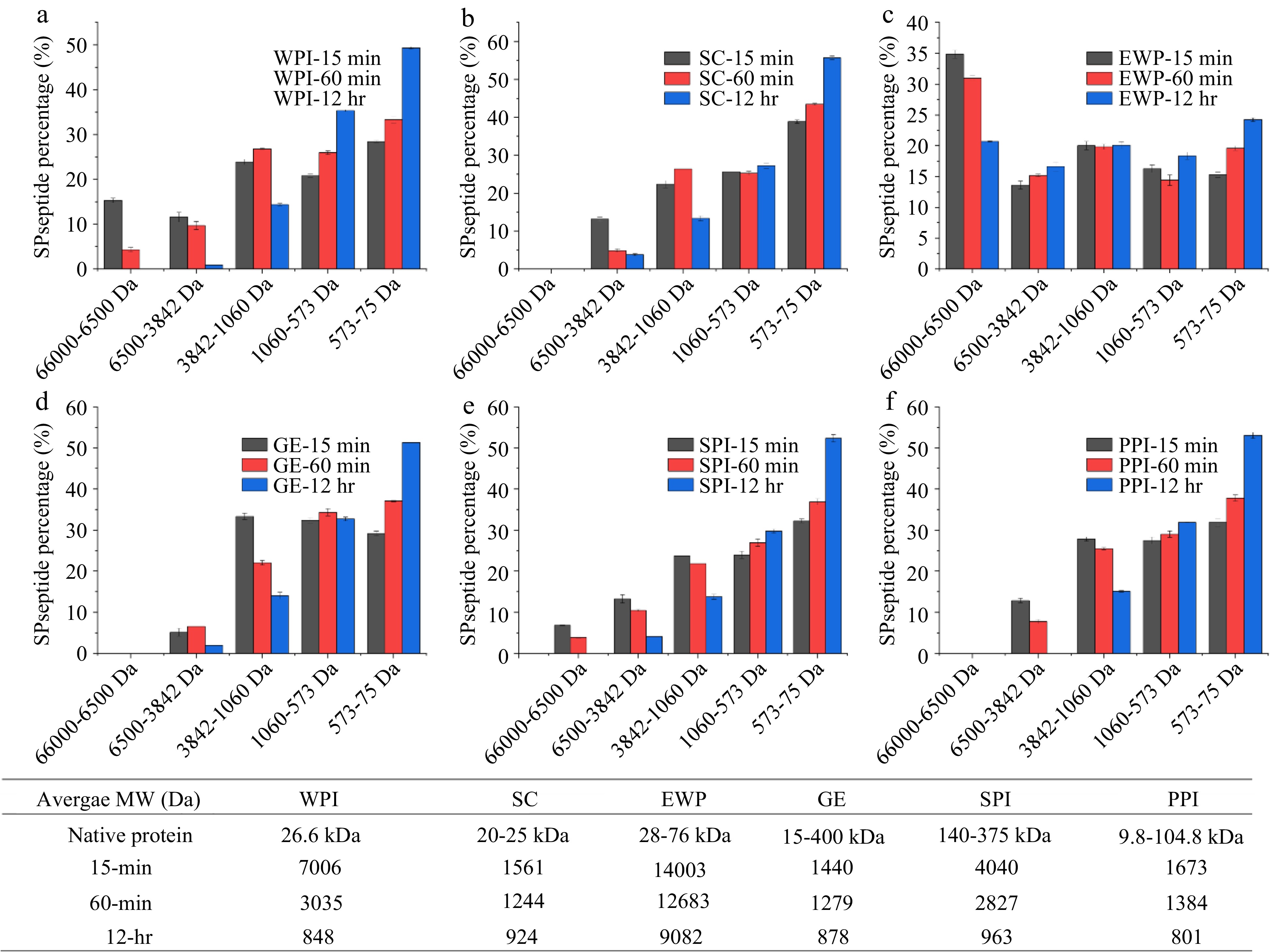
    • To evaluate the potential IRI activity of protein hydrolysates, alcalase, a non-specific protease, was used to hydrolyze six different types of protein including SC, WPI, EWP, GE, PPI and SPI. The hydrolyzed peptide underwent molecular size distribution measurement using HPLC and IRI activity assessment by splat assay. Figure 1 demonstrates the peptide molecular weight distribution of the hydrolyzed protein samples and their weighed average molecular weight (Da) at different hydrolyzing times. Overall, increased hydrolysis time led to a decrease in the average molecular weight of sample as shown in Fig. 1. Except for EWP, the extensive 12-hr hydrolysis has resulted in an average molecular weight of the hydrolysates less than 1,000 Da. This indicates the lower efficiency of alcalase in hydrolyzing EWP compared to the hydrolysis of the other five proteins examined in this study. EWP has been reported to contain proteinase inhibitors which could lead to the reduced hydrolysis activity of alcalase[27,28].

      Figure 1. 

      Peptide molecular weight distribution of various protein samples upon different hydrolysis time. (a) WPI; (b) SC; (c) EWP; (d) GE; (e) SPI; (f) PPI. * Molecular weights of native proteins were reported from literature[4954].

      Figures 2 & 3 demonstrate the ice crystal microscopic images and mean ice crystal sizes of various hydrolysate samples in PBS buffer at 4% (w/w) after 30 min annealing at −8 °C. Polyethylene glycol (PEG) in PBS buffer at 4% (w/w) was the negative control[22]. Without enzymatic hydrolysis, except WPI, the five types of native proteins were not soluble in PBS buffer at 4% (w/w) concentration, and thus could not be tested for IRI activity. WPI was soluble but did not show IRI activity at 4% (w/w) concentration. As shown in Fig. 2, compared to the 4% PEG, much smaller ice crystals were observed for the treatment of 4% SPI-15min hydrolysate, which indicates this sample is IRI active, whereas similar or larger ice crystals were observed in SC, WPI, EWP, GE and PPI after 15 min enzymatic hydrolysis. Quantitative ice crystal results are presented in Fig. 3 which indicates that after 15 min hydrolysis, except SPI hydrolysates, the other five samples tested did not have an IRI activity compared to PEG.
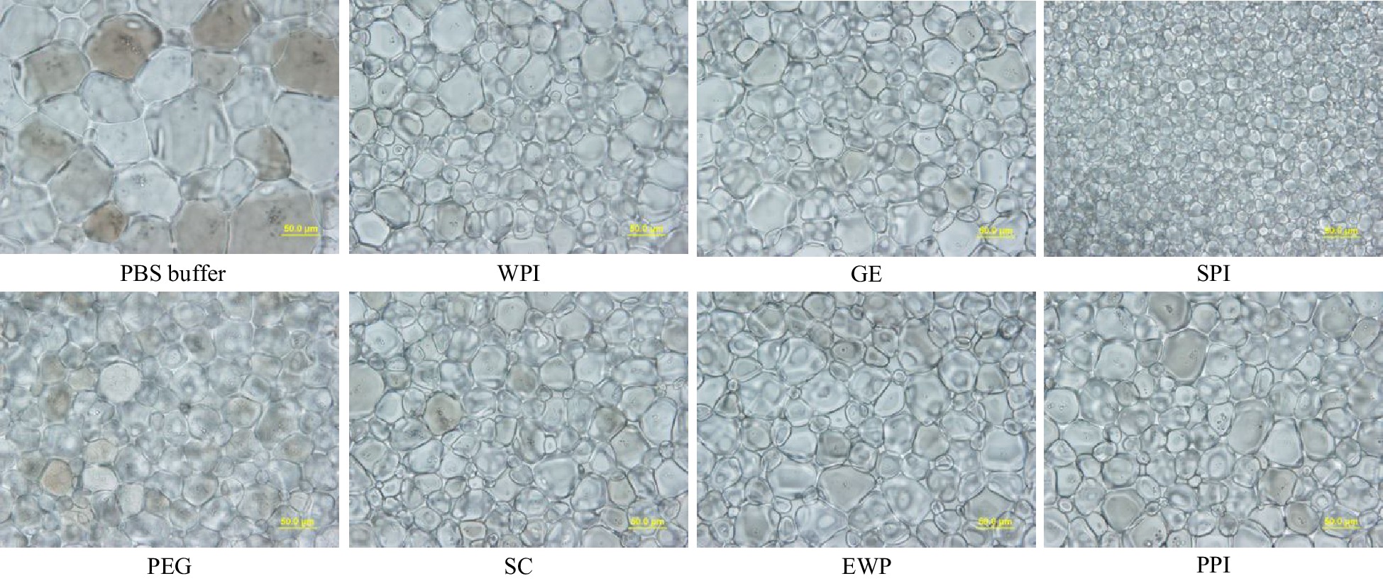
      Figure 2. 

      Standard splat assay microscopic image at 40× for PEG (negative control), and GE, SPI, SC, EWP and PPI hydrolysates after 15-min alcalase hydrolysis at 4% (w/w) concentration, and phosphate buffer saline (PBS – the buffer solvent) annealed at −8 °C for 30 min (scale bar = 50 μm).
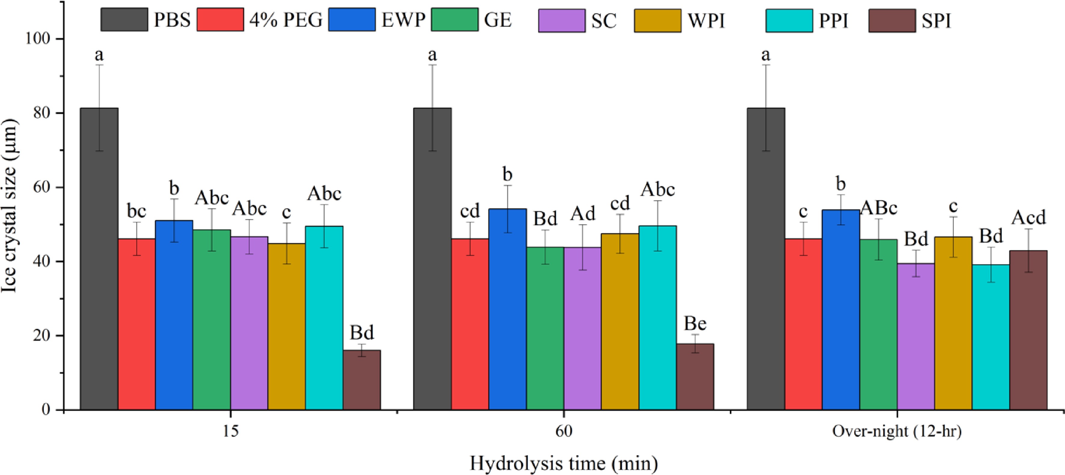
      Figure 3. 

      Mean average ice crystal size (μm) from different protein hydrolysates (4% w/w) solutions under −8 °C. Same lower-case letter indicate no significant difference (p > 0.05) within the same hydrolysis time, while same upper-case letters indicate no significant difference within the same protein (p > 0.05).

      As shown in Fig. 3, after 15- and 60-min hydrolysis, only SPI hydrolysates resulted in the formation of significantly (p < 0.05) smaller ice crystals compared to the negative control PEG and the solvent, PBS. However, after 12-hr hydrolysis, SPI-15 and SPI-60 hydrolysates at 4% (w/w) concentration lost its IRI activity. This is likely due to extensive hydrolysis of SPI resulting in a higher amount of small peptides and single amino acids which are not IRI active. As shown in Fig. 1e, the IRI active SPI hydrolysates (15- and 60-min hydrolysis), contained a large molecular fraction (66,000 – 6,500 Da), whereas the 12-hr hydrolyzed sample did not, which indicates that this large molecular fraction might have contributed to the IRI activity of the SPI-15 and SPI-60. In order to have an IRI activity, a molecule needs to bind to the ice surface through electrostatic interaction followed by structural realignment to optimally hydrogen bond with the oxygen-oxygen lattice on the ice surface, and then a partial nonpolar environment is ideally created by neighboring hydrophobic residues of the peptide[5]. Thus, it is critical for the hydrolyzed peptide to dynamically adapt its conformation when it approaches an ice surface and align its oxygen-containing groups with that of the ice surface to bind and inhibit the ice crystal growth[14]. Based on the current literature, peptides with IRI activity should have at least a molecular size ranging from 2 to 3.5 kDa[15,29]. The average molecular weights of SPI-15 and SPI-60 are within and close to this range as shown in Fig. 1. In this case, a single amino acid or small peptide (di- or tri-peptide) might not be efficient enough in inhibiting ice crystal growth.

      After extensive hydrolysis (12-hr), SC and PPI hydrolysates resulted in statistically significant (p < 0.05) smaller ice crystal sizes compared to PEG (Fig. 3), although not as effective as SPI-15 and SPI-60. As shown in Fig. 1b & f, after 12-hr hydrolysis, large and medium sized peptide fractions were broken down to produce smaller peptide fractions with a significant increase in the small peptide and amino acids fraction (573–75 Da) indicating the IRI active peptide/amino acid, such as proline, might present in this fraction for SC and PPI. Primacella et al.[30] observed the gelation inhibition activity of proline in egg yolk upon freezing storage due to the formation of proline-water hydrogen bonding and shielding of the hydrophobic sites of proteins. Proline is also known as a natural cryoprotectant which is found in plants and marine invertebrates[31], and can be used in cryopreservation of red blood cells[32].

      In addition, WPI-60 and SPI-60 shared very similar peptide molecular weight distribution with a comparable average molecular weight as shown in Fig. 1a & e. However, WPI-60 was found insignificant (p > 0.05) in ice crystal inhibition compared to 4% PEG (the negative control). This might be due to the differences in amino acid composition and peptide sequence of the hydrolyzed products. Studies have shown that threonine, asparagine, alanine, and proline were critical in ice binding for anti-freezing proteins (AFPs) and IRI active hydrolysates[2,14,1620]. Table 1 indicates the four IRI contributing amino acid compositions of the six examined proteins. Overall, all six types of protein contain an adequate amount of these four IRI contributing amino acids with a total content of about 25% (w/w). Compared to WPI, SPI only contains a higher amount of asparagine but has a lower content of the other three amino acids. SPI and PPI have a similar content of these four amino acids, but PPI did not produce IRI active hydrolysates after 15- and 60-min enzymatic hydrolysis. Bovine bone gelatin is the most proline abundant among these six proteins, but did not exhibit an IRI activity in this study. This indicates the importance of the peptide sequence on its IRI activity. As discussed earlier, it is important for IRI active proteins/peptides to bind the ice surface via electrostatic interaction and structural realignments. For example, studies have found that the repeated tripeptide sequence of (Ala-Ala-Thr)n with a disaccharide (3-O-(β-D-galactosyl)-D-N-acetylgalactosamine) bonded to the β-oxygen of the threonine residue is essential for the IRI activity of AFPs[33,34]. Hence, an IRI active hydrolysate needs to not only contain the IRI active amino acids but also have them in the correct sequence in order to efficiently bind to the ice surface thus inhibiting ice crystal growth.

      Table 1.  Composition (%) of IRI-contributing amino acids in various proteins[2326,48].

      Amino acidsEgg white proteinBovine bones gelatinWhey proteinCasein proteinSoy proteinPea protein
      Threonine4.751.776.874.053.603.70
      Alanine8.938.695.552.764.104.40
      Asparagine10.084.209.187.5711.6011.50
      Proline4.2512.546.669.335.605.00
      Total28.0127.228.2623.7124.924.6

      Overall, among the six types of protein, SPI hydrolysates, after 15- and 60-min enzymatic hydrolysis, demonstrate a great potential for further anti-freezing applications in food processing. While no significant difference (p > 0.05) in IRI activity was observed between SPI-15 and SPI-60 (Fig. 3), both samples were further evaluated under different annealing temperatures, time, and concentration for their IRI activity evaluation.

    • To investigate the effect of temperature on ice crystal growth, the SPI-15 and SPI-60 samples (at 4% w/w) were examined for their IRI activity under different annealing temperatures ranging from −10 to −4 °C. Overall, SPI-60 was found to be less temperature resistant compared to SPI-15 as shown in Fig. 4a & b. SPI-15, at 4% (w/w) concentration, was able to inhibit the growth of ice crystals, with sizes smaller than 23 μm (vs. 37 to 48 μm for PEG), between −10 to −6 °C, whereas SPI-60 was only able to retard ice crystal growth at −8 °C or lower. At −6 ºC, SPI-15 was able to maintain the average ice crystal size around 22 µm at and beyond 20 min annealing, whereas ice crystal in SPI-60 system continued to grow until the end of the 30 min annealing, resulting in an average ice crystal size of 35.9 µm. When the temperature continued to increase to −4 °C, both SPI-15 and SPI-60 lost their IRI activity, thus demonstrating no significant difference (p > 0.05) in ice crystal size compared to PEG under the same annealing condition. At −4 °C, ice crystals in SPI-15 and SPI-60 (4% w/w) continued growing within 30 min annealing duration. The amount of IRI active peptides applied (4% w/w) was probably not enough to ensure the IRI activity of SPI-15 and SPI-60 at increased unfrozen water fractions and high annealing temperature. As discussed in the previous section, the increased hydrolysis time resulted in a reduction of high molecular fraction (65000-6600 Da) which could be responsible for the IRI activity of SPI hydrolysates (SPI-15 and SPI-60). The reduced amount of the IRI active fraction in SPI-60 led to its lower temperature-resistant capability compared to SPI-15.
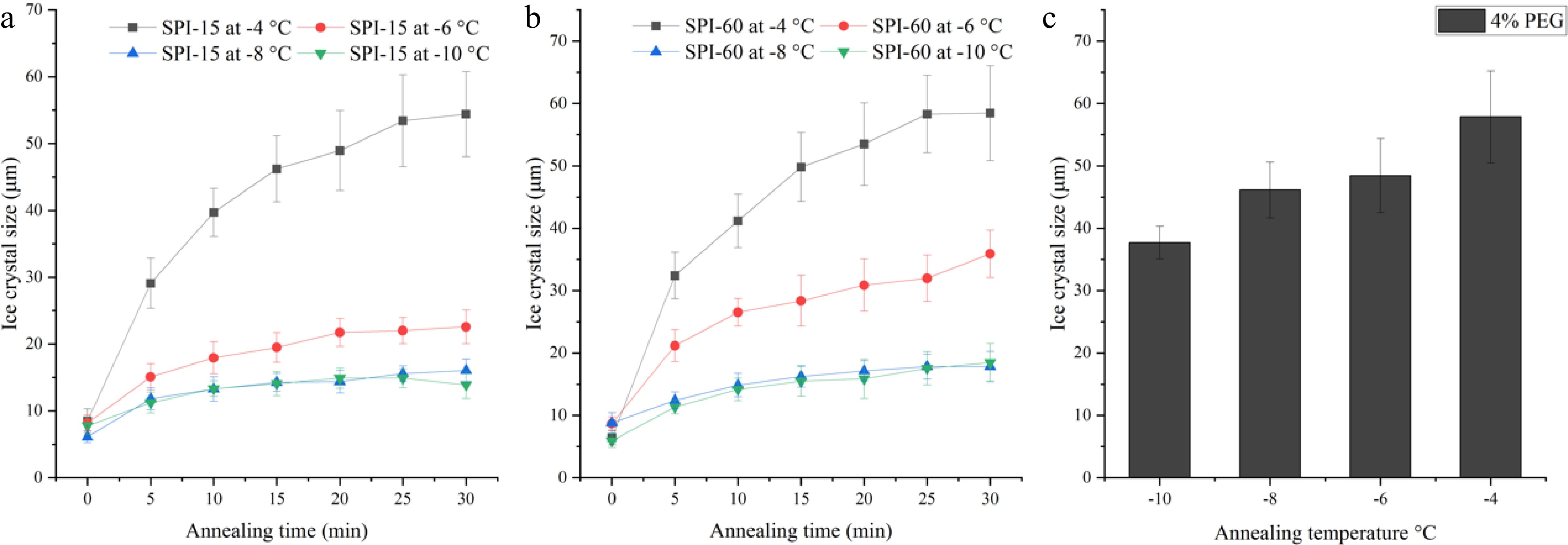
      Figure 4. 

      Ice crystal size of 4% (a) SPI-15 and (b) SPI-60 hydrolysates in PBS buffer system annealed under −10 to −4 °C for various time. (c) Ice crystal size of 4% PEG (negative control) after 30 min annealing under various temperatures.

      To evaluate the effect of peptide concentration on the IRI activity of SPI hydrolysates, different concentrations of SPI-15 (2%−4% w/w) were applied. As shown in Fig. 5, decreased concentration of SPI-15 resulted in a decreased temperature resistance, where SPI-15 at 2% (w/w) was not able to retard ice crystal growth even at −10 °C. In addition, SPI-15 at 3% (w/w) could not inhibit ice crystal growth at and beyond −6 °C compared to PEG and demonstrated lower effectiveness compared to 4% (w/w) SPI-15 under −10 and −8 °C, indicating the importance of the IRI active peptide content. Generally, in a commercial household freezer (−18 °C), the temperature is unlikely to fluctuate above −10 °C[35]. Thus, SPI-15, used at 3% or 4% (w/w), could be effective in withstanding temperature fluctuation and maintaining a small ice crystal for frozen products during storage and transportation.
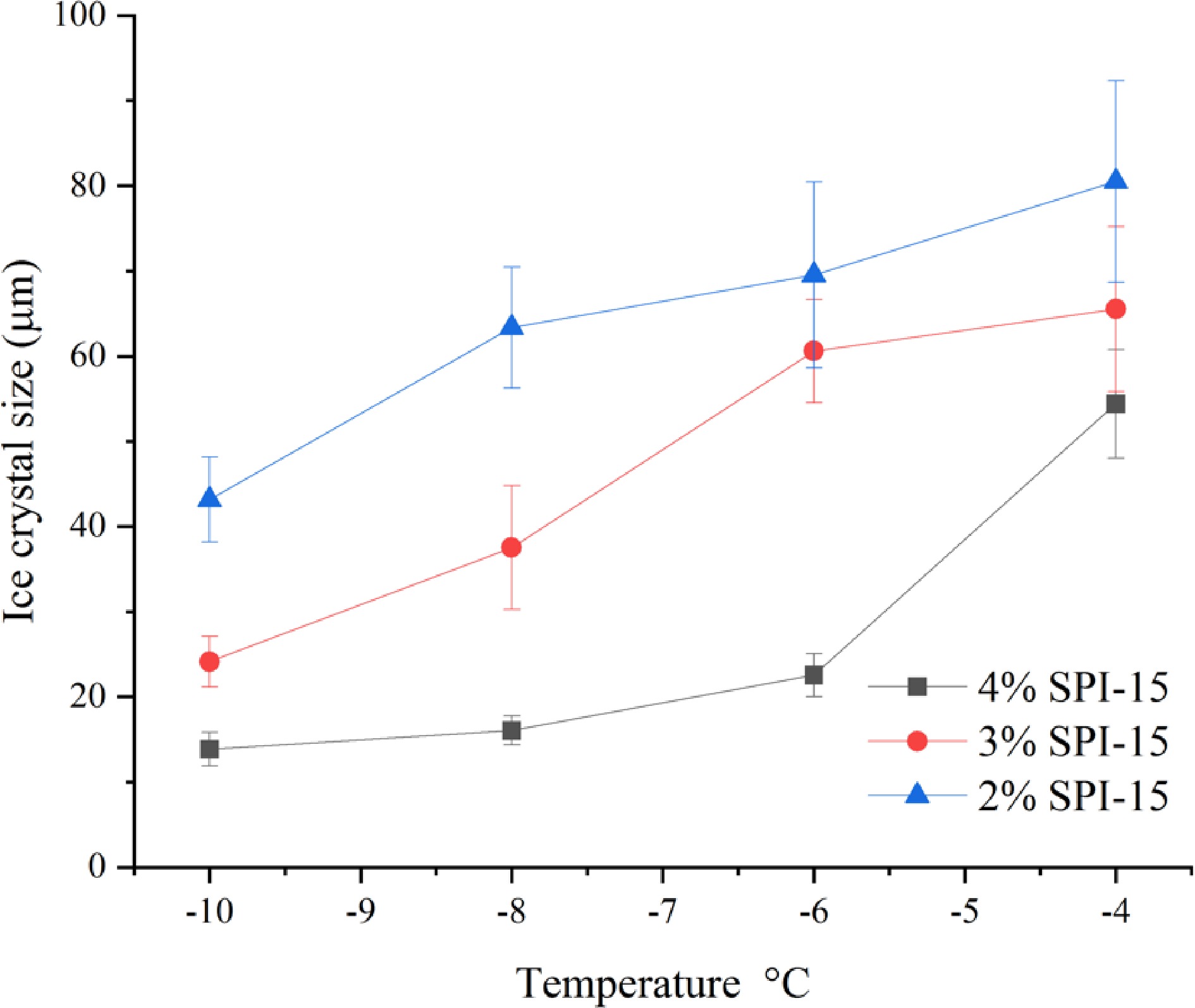
      Figure 5. 

      Ice crystal size of SPI-15 hydrolysates in PBS buffer at varied concentrations (2%−4% w/w) annealed under different temperatures (−10 to −4 °C) for 30 min.

    • As discussed earlier, an enhanced IRI activity was observed in PPI hydrolysates after 12-hr enzymatic hydrolysis resulting in significantly smaller (p < 0.05) ice crystal size compared to PEG, whereas SPI hydrolysates lost their IRI activity after 12-hr hydrolysis. Hence, succinylation reaction was performed using OSA to modify SPI-12hr and PPI-12hr hydrolysates to evaluate the change in structure and amphiphilicity on the IRI activity of the modified samples. As shown in Table 2 and Fig. 6, both OSA-modified SPI-12hr and PPI-12hr, at 4% (w/w) concentration, demonstrated a significant increased IRI activity (p < 0.05) compared to their non-modified counterparts and the negative control (PEG) annealed at −8 °C. At −8 °C, the ice crystal size in OSA-modified hydrolysates was almost half of the size compared to the non-modified treatments. As the temperature increased to −6 °C, only OSA-SPI demonstrated a significant IRI activity compared to SPI-12hr and PEG (p < 0.05). This result supports the hypothesis on the significance of structural amphiphilicity in the IRI activity of AFPs which has one side hydrophilic for ice binding and on the other side hydrophobic feature preventing ice crystal growth[21,22]. Thus, the hydrophilic SPI and PPI hydrolysates modified with the hydrophobic octenyl succinic anhydride (OSA) through succinylation reaction had an enhanced amphiphilicity. Besides AFPs, synthetic polymers have been examined for their IRI activities with amphiphilicity[36]. Li et al.[37] have also observed the amphiphilicity of nanocelluloses with IRI activity.

      Table 2.  Ice crystal size of 4% legume protein (SPI and PPI) hydrolysates and their derivatives (OSA-modified) under annealing temperatures of −8 and −6 °C with 4% PEG as negative control.

      SampleIce crystal diameter
      (μm) −8 °C
      Ice crystal diameter
      (μm) −6 °C
      PEG46.13a48.43a
      SPI-12hr42.95b48.45a
      OSA-SPI-12hr16.93d26.29b
      PPI-12hr39.12c49.37a
      OSA-PPI-12hr17.47d49.37a
      Same superscript indicates no significant difference (p > 0.05) within the same column.
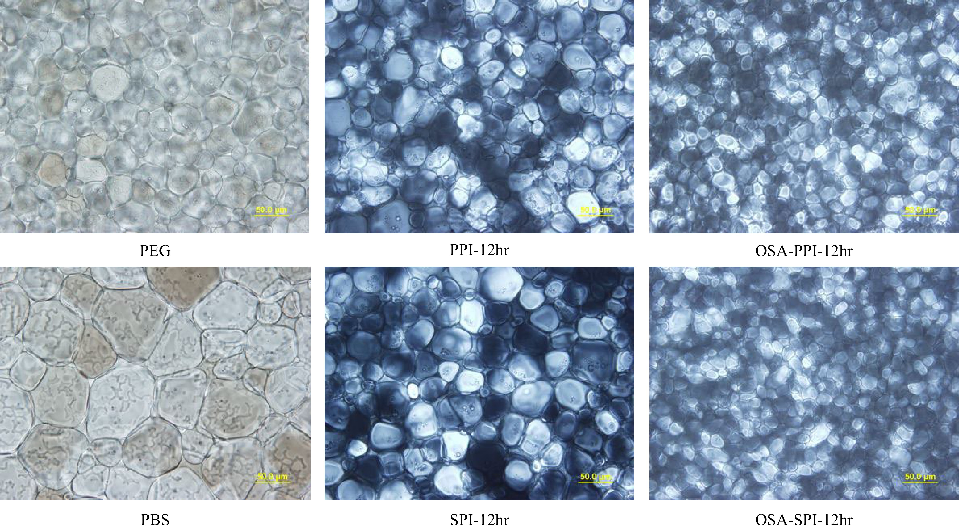
      Figure 6. 

      Ice crystal image under polarized microscope at 40x for 4% SPI hydrolysates, PPI hydrolysates and their OSA-modified derivatives annealed at −8 °C compared to the negative control 4% PEG and PBS (the buffer solvent). (scale bar = 50 μm).

      Figure 7 demonstrates the ice crystal growth of SPI hydrolysates and PPI hydrolysate with their OSA-modified derivatives suspended in PBS (4% w/w) within 30 min annealed at temperatures of −8 and −6 °C. For OSA-SPI-12hr, ice crystal size was stabilized after 15 min under both −8 and −6 °C, while ice crystal size in the non-modified SPI-12hr continued to grow beyond 15 min. While, OSA-PPI-12hr was only able to stabilize ice crystal growth at −8 °C at and beyond 15 min, it could not inhibit ice crystal growth when the temperature increased to −6 °C, indicating that the enhancement by the addition of OSA might not be enough to ensure the activity of OSA-PPI-12hr at increased fractions of unfrozen water at high annealing temperature. The ability of OSA-SPI-12hr in inhibiting ice crystal growth under increased temperature makes it a better agent in preventing ice recrystallization in frozen foods compared to OSA-PPI-12hr.
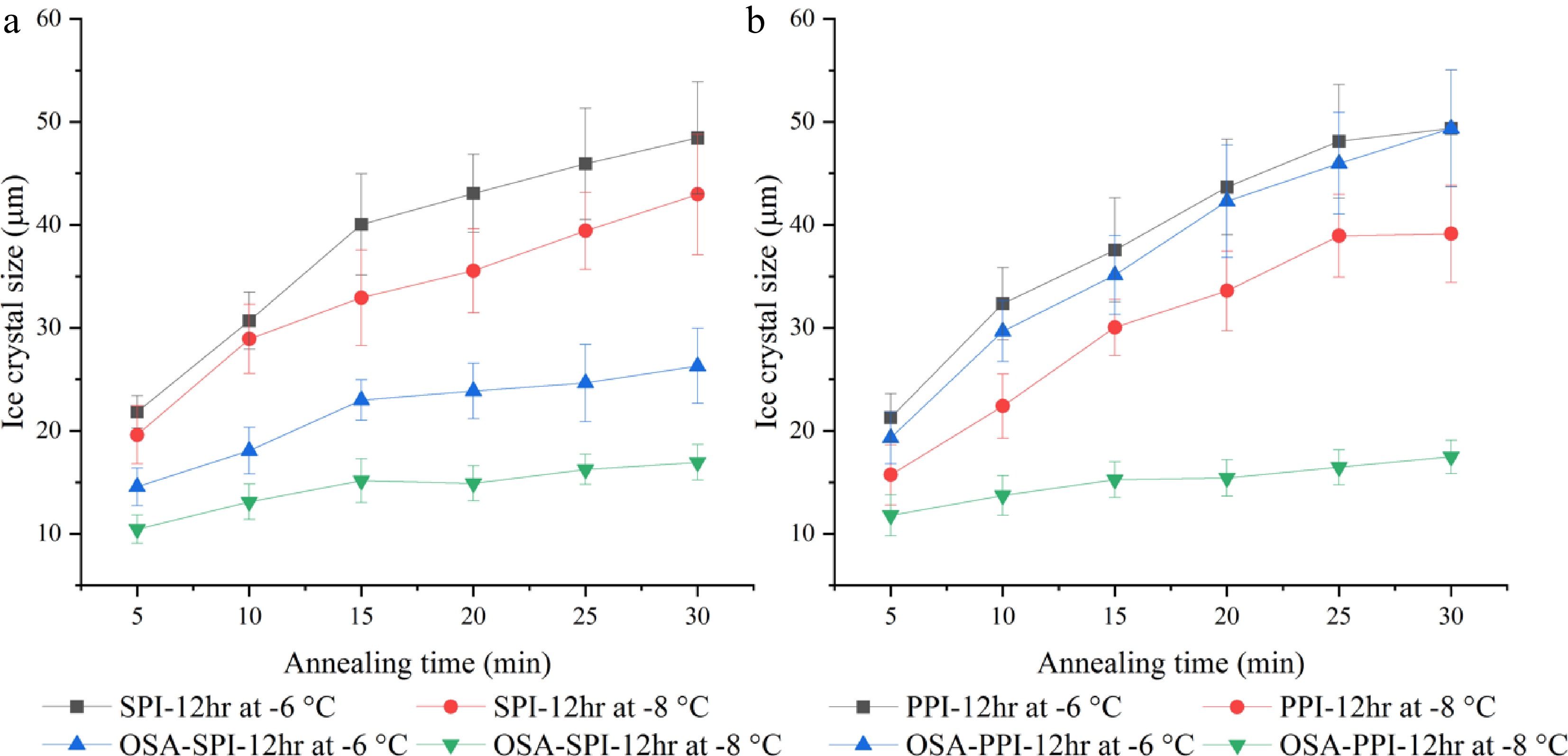
      Figure 7. 

      Ice crystal growth within 30 min annealed at −8 °C and −6 °C in 4% (w/w) (a) SPI-12hr hydrolysate and OSA modified SPI-12hr, and (b) PPI-12hr hydrolysate and OSA modified PPI-12hr.

      Surface hydrophobicity was also measured to assess the effect of OSA modification on the hydrophobicity of the SPI and PPI hydrolysates using the ANS fluorescent method. Table 3 shows the surface hydrophobicity of the modified and non-modified PPI and SPI hydrolysates. OSA-PPI-12hr was found to be significantly more hydrophobic (p < 0.05) compared to the other three samples in which no significant difference was observed (p > 0.05). As a fluorescent probe, ANS binds to the hydrophobic regions of proteins, thus leads to an increase in fluorescence, which is used as a measure of protein surface hydrophobicity[38]. In this case, the ANS method might not be sensitive enough to detect the changes by the modification, since protein hydrolysates were highly soluble in water and more hydrophilic than their native form, leading to the low surface hydrophobicity as measured by the ANS method even after the addition of OSA. In addition, the presence of carboxyl and carbonyl groups in OSA-modified samples might interfere with the ANS binding resulting in a low hydrophobicity[39]. Hence, in future studies, other methods should be identified to characterize the structural changes of the modified hydrolysates in order to understand the effect of OSA modifications on its IRI activity. Different degrees of modification will also be examined to optimize the modification process.

      Table 3.  Surface hydrophobicity of SPI and PPI hydrolysates and their OSA modified products.

      SampleSurface hydrophobicity
      SPI-12hr6685b
      OSA-SPI-12hr6196b
      PPI-12hr7123b
      OSA-PPI-12hr9134a
      Same superscript indicates no significant difference (p > 0.05) among samples.
    • Overall, this study has demonstrated the high potential of utilizing enzymatic hydrolysis to produce an IRI active hydrolysate from common food proteins. This will enable the utilization of cost-effective proteins for the production of IRI agents in a sustainable manner rather than using the high-cost and low-yield natural AFPs. Among the six proteins examined, SPI is found to be an effective protein for the production of IRI active hydrolysates. Its IRI activity is influenced by the annealing temperature and peptide concentration, where lower temperature and increased concentration result in enhanced IRI activity. The effectiveness of hydrolysates' IRI activity could be attributed to their amino acid composition and sequences. Moreover, chemical modification of SPI and PPI hydrolysates by the addition of OSA was found to enhance its IRI activity. OSA-SPI-12hr demonstrated better temperature resistance compared to OSA-PPI-12hr. Future studies will further evaluate the effect of degree of OSA modification on the IRI activity and characterize the structural changes of the modified hydrolysates.

    • All protein samples, sodium caseinate (SC; Sigma-Aldrich, MO, USA), whey protein isolate (WPI), egg white protein (EWP; prepared from freeze-dried egg white), pea protein isolate (PPI; BulkSuppliments.com, NV, USA), soy protein isolate (SPI; BulkSuppliments.com, NV), and commercial gelatin (GE; Kraft Foods, IL, USA), were adjusted to a standard moisture content before the hydrolysis treatment. Enzymatic hydrolysis of protein samples by alcalase was performed based on methods established in previous studies with some modifications[2,14]. Protein dispersion of 10% (w/w) was hydrolyzed at 45 °C for up to 60 min at pH 9.0 with 0.176 Anson unit enzyme/g protein added. Extensive, 12-hr, hydrolysis was also performed to evaluate the effect of enzymatic hydrolysis on the anti-freezing properties of the hydrolysates. After hydrolysis, enzyme was inactivated by incubating the solution for 10 min in boiling water followed by centrifugation at 10,000 x g for 10 min to obtain the supernatant of peptide products. The supernatants were collected and freeze-dried for ice recrystallization inhibition (IRI) activity measurement.

    • A standard splat assay was used to assess the IRI activity of the hydrolyzed peptides. For the assay[40], a 10 µl droplet of sample solution, with various concentrations (2%– 4% w/w), was dropped from a height of 1.5 m onto a glass slide surface that is precooled to −80 °C. The sample was then annealed under different temperatures between −10 °C and −4 °C for up to 30 min using a cryo-stage HCS 302 (Instec Instruments, Boulder, CO, USA) before collecting images using polarized light microscopy (Olympus BX51, Tokyo, Japan) with a built-in digital camera (DP 70, Olympus, Tokyo, Japan) to determine the size of the ice crystals and examine the effect of annealing temperature on IRI activity of each peptide. The size of the 10 largest ice crystals in the image was measured using Image J (National Institutes of Health, Bethesda, MD, USA). The average mean size of the ice crystals of each sample was calculated from three independent splat assay measurements. Polyethylene glycol (PEG) was used as a negative control[37,41].

    • The molecular weight profiles of the hydrolyzed samples were analyzed by size exclusion chromatography on an Agilent HPLC system (Agilent Technologies, Santa Clara, CA, USA). The separation was performed on a BioSep-SEC-S2000 column (300 × 7.80 mm, Torrance, CA, USA) as described by Price et al.[42]. In brief, the mobile phase was 45% acetonitrile in water with 0.1% trifluoroacetic acid at a flow rate of 1.0 mL/min. The sample was injected at 20 µL with 20 min run time detected at 214 nm at ambient temperature. The elution profile was compared to the standards with known molecular weights (albumin, glycine, and a mixed peptide standard) to determine molecular size distribution of the hydrolysates. The average peptide size of the hydrolyzed sample was calculated as the sum of the multiplication of the mean molecular size and percentage of each molecular range obtained via HPLC analysis.

    • Modification of hydrolysates legume proteins was performed based on methods established in the previous studies with minor modifications[43,44]. Octenyl succinic anhydride (OSA) has been used to react with all nucleophilic groups, including amine groups and aliphatic hydroxyl groups[45,46]. Hydrolysate samples, 2 g, were dispersed in 250 mL 0.075 M phosphate buffer (pH 8). OSA was added to hydrolysates, with constant stirring over a 3-hr period at levels of 40% of the weight of the hydrolysate sample. During the reaction, pH was maintained at 8–8.1 with 2 N NaOH. After 3-hr reaction, pH of the reaction solution was adjusted to 6.5 followed by centrifugation to obtain derived protein and remove any insoluble compounds prior to dialysis. Modified hydrolysates then underwent dialysis with 100-500 MWCO membrane in deionized water for 3 d under refrigerated conditions (4 °C) to remove any unreacted OSA, the deionized water was changed daily. After dialysis, the modified hydrolysates were freeze-dried for IRI activity and surface hydrophobicity analysis.

    • Surface hydrophobicity was determined using 1-phenylamino-2-napthalenesulfonic acid (ANS) as fluorescent probe as previously reported[47]. Briefly, 200 µL of different concentrations of legume proteins and derivatives (0.005 to 0.5 mg protein/mL in phosphate buffer, pH 7.4) was plated in 96-well black-walled microplate and 10 µL ANS probe (0.8 mM in phosphate buffer, pH 7.4) was added. The mixture was incubated in the dark for 10 min and fluorescence intensity was measured at excitation and emission wavelengths of 390 and 480 nm, respectively. The fluorescence intensity was plotted against protein concentration, and the slope of the linear regression curve was reported as surface hydrophobicity.

    • All experiments and measurements were performed in duplicates. Statistical analysis was carried out using the JMP statistical package. Analysis of variance was used, and the data were represented as mean value ± standard deviation. Tukey's multiple sample comparison tests were carried out to assess the significance of the difference between treatments. Statistical significance was indicated at a confidence level of 95% (p ≤ 0.05).

      • This study was supported by the Hatch Multi-State Project (Accession # 1023982).

      • The authors declare that they have no conflict of interest.

      • Copyright: © 2022 by the author(s). Published by Maximum Academic Press on behalf of Nanjing Agricultural University. This article is an open access article distributed under Creative Commons Attribution License (CC BY 4.0), visit https://creativecommons.org/licenses/by/4.0/.
    Figure (7)  Table (3) References (54)
  • About this article
    Cite this article
    Wan Z, Fei T, Wang T. 2022. Inhibition of ice crystal growth by protein hydrolysates from different plant- and animal-based proteins. Food Materials Research 2:17 doi: 10.48130/FMR-2022-0017
    Wan Z, Fei T, Wang T. 2022. Inhibition of ice crystal growth by protein hydrolysates from different plant- and animal-based proteins. Food Materials Research 2:17 doi: 10.48130/FMR-2022-0017

Catalog

  • About this article

/

DownLoad:  Full-Size Img  PowerPoint
Return
Return