ARTICLE   Open Access    

Efficient organogenesis and taxifolin production system from mature zygotic embryos and needles in larch

More Information
  • The deciduous conifer larch has been widely distributed around the world, is a high-quality wood species and is also used to extract industrial raw materials and medicines. In this study, we developed an organogenesis protocol for Larix olgensis from both mature zygotic embryos and needles, and analyzed the content of taxifolin in different tissues. The highest callus induction (96.8%) from mature zygotic embryo was found in the Douglas-fir Cotyledon Revised (DCR) medium augmented with 2.0 mg·L−1 6-Benzylaminopurine (6-BA) and 0.2 mg·L−1 α-Naphthaleneacetic acid (NAA), while from needles the highest callus induction (92.03%) was found in the Murashige and Skoog (MS) medium augmented with 3 mg·L−1 6-BA and 0.3 mg·L−1 NAA. The best shoot regeneration capacity from zygotic embryo-derived calli (83.3%) was obtained in DCR medium augmented with 1.0 mg·L−1 6-BA and 0.01 mg·L−1 NAA, and needle-derived calli were 77.3%. The shoots achieved the highest elongation (75.6%) in the DCR medium supplemented with 0.5 mg·L−1 6-BA, 0.05 mg·L−1 NAA and 2 g·L−1 activated charcoal (AC). The rooting rate was 62.8% in DCR medium augmented with 3 mg·L−1 Indole-3-butyric acid (IBA) and 100 mg·L−1 phloroglucinol (PG). The accumulation of the taxifolin in elongation shoots and lignified elongation shoots have greatly improved along with the development process, were 28.6 µg·g−1, and 53 µg·g−1 respectively. The content of the taxifolin in callus was 1.99−5.26 µg·g−1, adventitious shoots were 4.8 µg·g−1, and adventitious roots were 2.86 µg·g−1. We report an efficient organogenesis and taxifolin production protocol in larch for the first time.
  • Aquaporin’s (AQPs) are small (21–34 kD) channel-forming, water-transporting trans-membrane proteins which are known as membrane intrinsic proteins (MIPs) conspicuously present across all kingdoms of life. In addition to transporting water, plant AQPs act to transport other small molecules including ammonia, carbon dioxide, glycerol, formamide, hydrogen peroxide, nitric acid, and some metalloids such as boron and silicon from the soil to different parts of the plant[1]. AQPs are typically composed of six or fewer transmembrane helices (TMHs) coupled by five loops (A to E) and cytosolic N- and C-termini, which are highly conserved across taxa[2]. Asparagine-Proline-Alanine (NPA) boxes and makeup helices found in loops B (cytosolic) and E (non-cytosolic) fold back into the protein's core to form one of the pore's two primary constrictions, the NPA region[1]. A second filter zone exists at the pore's non-cytosolic end, where it is called the aromatic/arginine (ar/R) constriction. The substrate selectivity of AQPs is controlled by the amino acid residues of the NPA and ar/R filters as well as other elements of the channel[1].

    To date, the AQP gene families have been extensively explored in the model as well as crop plants[39]. In seed plants, AQP distributed into five subfamilies based on subcellular localization and sequence similarities: the plasma membrane intrinsic proteins (PIPs; subgroups PIP1 and PIP2), the tonoplast intrinsic proteins (TIPs; TIP1-TIP5), the nodulin26-like intrinsic proteins (NIPs; NIP1-NIP5), the small basic intrinsic proteins (SIPs; SIP1-SIP2) and the uncategorized intrinsic proteins (XIPs; XIP1-XIP3)[2,10]. Among them, TIPs and PIPs are the most abundant and play a central role in facilitating water transport. SIPs are mostly found in the endoplasmic reticulum (ER)[11], whereas NIPs homologous to GmNod26 are localized in the peribacteroid membrane[12].

    Several studies reported that the activity of AQPs is regulated by various developmental and environmental factors, through which water fluxes are controlled[13]. AQPs are found in all organs such as leaves, roots, stems, flowers, fruits, and seeds[14,15]. According to earlier studies, increased AQP expression in transgenic plants can improve the plants' tolerance to stresses[16,17]. Increased root water flow caused by upregulation of root aquaporin expression may prevent transpiration[18,19]. Overexpression of Tamarix hispida ThPIP2:5 improved osmotic stress tolerance in Arabidopsis and Tamarix plants[20]. Transgenic tomatoes having apple MdPIP1;3 ectopically expressed produced larger fruit and improved drought tolerance[21]. Plants over-expressing heterologous AQPs, on the other hand, showed negative effects on stress tolerance in many cases. Overexpression of GsTIP2;1 from G. soja in Arabidopsis plants exhibited lower resistance against salt and drought stress[22].

    A few recent studies have started to establish a link between AQPs and nanobiology, a research field that has been accelerating in the past decade due to the recognition that many nano-substances including carbon-based materials are valuable in a wide range of agricultural, industrial, and biomedical activities[23]. Carbon nanotubes (CNTs) were found to improve water absorption and retention and thus enhance seed germination in tomatoes[24,25]. Ali et al.[26] reported that Carbon nanoparticles (CTNs) and osmotic stress utilize separate processes for AQP gating. Despite lacking solid evidence, it is assumed that CNTs regulate the aquaporin (AQPs) in the seed coats[26]. Another highly noticed carbon-nano-molecule, the fullerenes, is a group of allotropic forms of carbon consisting of pure carbon atoms[27]. Fullerenes and their derivatives, in particular the water-soluble fullerols [C60(OH)20], are known to be powerful antioxidants, whose biological activity has been reduced to the accumulation of superoxide and hydroxyl[28,29]. Fullerene/fullerols at low concentrations were reported to enhance seed germination, photosynthesis, root growth, fruit yield, and salt tolerance in various plants such as bitter melon and barley[3032]. In contrast, some studies also reported the phytotoxic effect of fullerene/fullerols[33,34]. It remains unknown if exogenous fullerene/fullerol has any impact on the expression or activity of AQPs in the cell.

    Garden pea (P. sativum) is a cool-season crop grown worldwide; depending on the location, planting may occur from winter until early summer. Drought stress in garden pea mainly affects the flowering and pod filling which harm their yield. In the current study, we performed a genome-wide identification and characterization of the AQP genes in garden pea (P. sativum), the fourth largest legume crop worldwide with a large complex genome (~4.5 Gb) that was recently decoded[35]. In particular, we disclose, for the first time to our best knowledge, that the transcriptional regulations of AQPs by osmotic stress in imbibing pea seeds were altered by fullerol supplement, which provides novel insight into the interaction between plant AQPs, osmotic stress, and the carbon nano-substances.

    The whole-genome sequence of garden pea ('Caméor') was retrieved from the URGI Database (https://urgi.versailles.inra.fr/Species/Pisum). Protein sequences of AQPs from two model crops (Rice and Arabidopsis) and five other legumes (Soybean, Chickpea, Common bean, Medicago, and Peanut) were used to identify homologous AQPs from the garden pea genome (Supplemental Table S1). These protein sequences, built as a local database, were then BLASTp searched against the pea genome with an E-value cutoff of 10−5 and hit a score cutoff of 100 to identify AQP orthologs. The putative AQP sequences of pea were additionally validated to confirm the nature of MIP (Supplemental Table S2) and transmembrane helical domains through TMHMM (www.cbs.dtu.dk/services/TMHMM/).

    Further phylogenetic analysis was performed to categorize the AQPs into subfamilies. The pea AQP amino acid sequences, along with those from Medicago, a cool-season model legume phylogenetically close to pea, were aligned through ClustalW2 software (www.ebi.ac.uk/Tools/msa/clustalw2) to assign protein names. The unaligned AQP sequences to Medicago counterparts were once again aligned with the AQP sequences of Arabidopsis, rice, and soybean. Based on the LG model, unrooted phylogenetic trees were generated via MEGA7 and the neighbor-joining method[36], and the specific name of each AQP gene was assigned based on its position in the phylogenetic tree.

    By using the conserved domain database (CDD, www.ncbi.nlm.nih.gov/Structure/cdd/cdd.shtml), the NPA motifs were identified from the pea AQP protein sequences[37]. The software TMHMM (www.cbs. dtu.dk/services/TMHMM/)[38] was used to identify the protein transmembrane domains. To determine whether there were any alterations or total deletion, the transmembrane domains were carefully examined.

    Basic molecular properties including amino acid composition, relative molecular weight (MW), and instability index were investigated through the online tool ProtParam (https://web.expasy.org/protparam/). The isoelectric points (pI) were estimated by sequence Manipulation Suite version 2 (www.bioinformatics.org/sms2)[39]. The subcellular localization of AQP proteins was predicted using Plant-mPLoc[40] and WoLF PSORT (www.genscript.com/wolf-psort.html)[ 41] algorithms.

    The gene structure (intron-exon organization) of AQPs was examined through GSDS ver 2.0[42]. The chromosomal distribution of the AQP genes was illustrated by the software MapInspect (http://mapinspect.software.informer.com) in the form of a physical map.

    To explore the tissue expression patterns of pea AQP genes, existing NGS data from 18 different libraries covering a wide range of tissue, developmental stage, and growth condition of the variety ‘Caméor’ were downloaded from GenBank (www.ncbi.nlm.nih.gov/bioproject/267198). The expression levels of the AQP genes in each tissue and growth stage/condition were represented by the FPKM (Fragments Per Kilobase of transcript per Million fragments mapped) values. Heatmaps of AQPs gene were generated through Morpheus software (https://software.broadinstitute.org/morpheus/#).

    Different solutions, which were water (W), 0.3 M mannitol (M), and fullerol of different concentrations dissolved in 0.3 M mannitol (MF), were used in this study. MF solutions with the fullerol concentration of 10, 50, 100, and 500 mg/L were denoted as MF1, MF2, MF3, and MF4, respectively. Seeds of 'SQ-1', a Chinese landrace accession of a pea, were germinated in two layers of filter paper with 30 mL of each solution in Petri dishes (12 cm in diameter) each solution, and the visual phenotype and radicle lengths of 150 seeds for each treatment were analyzed 72 h after soaking. The radicle lengths were measured using a ruler. Multiple comparisons for each treatment were performed using the SSR-Test method with the software SPSS 20.0 (IBM SPSS Statistics, Armonk, NY, USA).

    Total RNA was extracted from imbibing embryos after 12 h of seed soaking in the W, M, and MF3 solution, respectively, by using Trizol reagent (Invitrogen, Carlsbad, CA, USA). The quality and quantity of the total RNA were measured through electrophoresis on 1% agarose gel and an Agilent 2100 Bioanalyzer respectively (Agilent Technologies, Santa Rosa, USA). The TruSeq RNA Sample Preparation Kit was utilized to construct an RNA-Seq library from 5 µg of total RNA from each sample according to the manufacturer's instruction (Illumina, San Diego, CA, USA). Next-generation sequencing of nine libraries were performed through Novaseq 6000 platform (Illumina, San Diego, CA, USA).

    First of all, by using SeqPrep (https://github.com/jstjohn/SeqPrep) and Sickle (https://github.com/najoshi/sickle) the raw RNA-Seq reads were filtered and trimmed with default parameters. After filtering, high-quality reads were mapped onto the pea reference genome (https://urgi.versailles.inra.fr/Species/Pisum) by using TopHat (V2.1.0)[43]. Using Cufflinks, the number of mapped reads from each sample was determined and normalised to FPKM for each predicted transcript (v2.2.1). Pairwise comparisons were made between W vs M and W vs M+F treatments. The DEGs with a fold change ≥ 1.5 and false discovery rate (FDR) adjusted p-values ≤ 0.05 were identified by using Cuffdiff[44].

    qPCR was performed by using TOROGGreen® qPCR Master Mix (Toroivd, Shanghai, China) on a qTOWER®3 Real-Time PCR detection system (Analytik Jena, Germany). The reactions were performed at 95 °C for 60 s, followed by 42 cycles of 95 °C for 10 s and 60 °C for 30 s. Quantification of relative expression level was achieved by normalization against the transcripts of the housekeeping genes β-tubulin according to Kreplak et al.[35]. The primer sequences for reference and target genes used are listed in Supplemental Table S3.

    The homology-based analysis identifies 41 putative AQPs in the garden pea genome. Among them, all but two genes (Psat0s3550g0040.1, Psat0s2987g0040.1) encode full-length aquaporin-like sequences (Table 1). The conserved protein domain analysis later validated all of the expected AQPs (Supplemental Table S2). To systematically classify these genes and elucidate their relationship with the AQPs from other plants' a phylogenetic tree was created. It clearly showed that the AQPs from pea and its close relative M. truncatula formed four distinct clusters, which represented the different subfamilies of AQPs i.e. TIPs, PIPs, NIPs, and SIPs (Fig. 1a). However, out of the 41 identified pea AQPs, 4 AQPs couldn't be tightly aligned with the Medicago AQPs and thus were put to a new phylogenetic tree constructed with AQPs from rice, Arabidopsis, and soybean. This additional analysis assigned one of the 4 AQPs to the XIP subfamily and the rest three to the TIP or NIP subfamilies (Fig. 1b). Therefore, it is concluded that the 41 PsAQPs comprise 11 PsTIPs, 15 PsNIPs, 9 PsPIPs, 5 PsSIPs, and 1 PsXIP (Table 2). The PsPIPs formed two major subgroups namely PIP1s and PIP2s, which comprise three and six members, respectively (Table 1). The PsTIPs formed two major subgroups TIPs 1 (PsTIP1-1, PsTIP1-3, PsTIP1-4, PsTIP1-7) and TIPs 2 (PsTIP2-1, PsTIP2-2, PsTIP2-3, PsTIP2-6) each having four members (Table 2). Detailed information such as gene/protein names, accession numbers, the length of deduced polypeptides, and protein structural features are presented in Tables 1 & 2

    Table 1.  Description and distribution of aquaporin genes identified in the garden pea genome.
    Chromosome
    S. NoGene NameGene IDGene length
    (bp)
    LocationStartEndTranscription length (bp)CDS length
    (bp)
    Protein length
    (aa)
    1PsPIP1-1Psat5g128840.32507chr5LG3231,127,859231,130,365675675225
    2PsPIP1-2Psat2g034560.11963chr2LG149,355,95849,357,920870870290
    3PsPIP1-4Psat2g182480.11211chr2LG1421,647,518421,648,728864864288
    4PsPIP2-1Psat6g183960.13314chr6LG2369,699,084369,702,397864864288
    5PsPIP2-2-1Psat4g051960.11223chr4LG486,037,44686,038,668585585195
    6PsPIP2-2-2Psat5g279360.22556chr5LG3543,477,849543,480,4042555789263
    7PsPIP2-3Psat7g228600.22331chr7LG7458,647,213458,649,5432330672224
    8PsPIP2-4Psat3g045080.11786chr3LG5100,017,377100,019,162864864288
    9PsPIP2-5Psat0s3550g0040.11709scaffold0355020,92922,63711911191397
    10PsTIP1-1Psat3g040640.12021chr3LG589,426,47389,428,493753753251
    11PsTIP1-3Psat3g184440.12003chr3LG5393,920,756393,922,758759759253
    12PsTIP1-4Psat7g219600.12083chr7LG7441,691,937441,694,019759759253
    13PsTIP1-7Psat6g236600.11880chr6LG2471,659,417471,661,296762762254
    14PsTIP2-1Psat1g005320.11598chr1LG67,864,8107,866,407750750250
    15PsTIP2-2Psat4g198360.11868chr4LG4407,970,525407,972,392750750250
    16PsTIP2-3Psat1g118120.12665chr1LG6230,725,833230,728,497768768256
    17PsTIP2-6Psat2g177040.11658chr2LG1416,640,482416,642,139750750250
    18PsTIP3-2Psat6g054400.11332chr6LG254,878,00354,879,334780780260
    19PsTIP4-1Psat6g037720.21689chr6LG230,753,62430,755,3121688624208
    20PsTIP5-1Psat7g157600.11695chr7LG7299,716,873299,718,567762762254
    21PsNIP1-1Psat1g195040.21864chr1LG6346,593,853346,595,7161863645215
    22PsNIP1-3Psat1g195800.11200chr1LG6347,120,121347,121,335819819273
    23PsNIP1-5Psat7g067480.12365chr7LG7109,420,633109,422,997828828276
    24PsNIP1-6Psat7g067360.12250chr7LG7109,270,462109,272,711813813271
    25PsNIP1-7Psat1g193240.11452chr1LG6344,622,606344,624,057831831277
    26PsNIP2-1-2Psat3g197520.1669chr3LG5420,092,382420,093,050345345115
    27PsNIP2-2-2Psat3g197560.1716chr3LG5420,103,168420,103,883486486162
    28PsNIP3-1Psat2g072000.11414chr2LG1133,902,470133,903,883798798266
    29PsNIP4-1Psat7g126440.11849chr7LG7209,087,362209,089,210828828276
    30PsNIP4-2Psat5g230920.11436chr5LG3463,340,575463,342,010825825275
    31PsNIP5-1Psat6g190560.11563chr6LG2383,057,323383,058,885867867289
    32PsNIP6-1Psat5g304760.45093chr5LG3573,714,868573,719,9605092486162
    33PsNIP6-2Psat7g036680.12186chr7LG761,445,34161,447,134762762254
    34PsNIP6-3Psat7g259640.12339chr7LG7488,047,315488,049,653918918306
    35PsNIP7-1Psat6g134160.24050chr6LG2260,615,019260,619,06840491509503
    36PsSIP1-1Psat3g091120.13513chr3LG5187,012,329187,015,841738738246
    37PsSIP1-2Psat1g096840.13609chr1LG6167,126,599167,130,207744744248
    38PsSIP1-3Psat7g203280.12069chr7LG7401,302,247401,304,315720720240
    39PsSIP2-1-1Psat0s2987g0040.1706scaffold02987177,538178,243621621207
    40PsSIP2-1-2Psat3g082760.13135chr3LG5173,720,100173,723,234720720240
    41PsXIP2-1Psat7g178080.12077chr7LG7335,167,251335,169,327942942314
    bp: base pair, aa: amino acid.
     | Show Table
    DownLoad: CSV
    Figure 1.  Phylogenetic analysis of the identified AQPs from pea genome. (a) The pea AQPs proteins aligned with those from the cool-season legume Medicago truncatual. (b) The four un-assigned pea AQPs in (a) (denoted as NA) were further aligned with the AQPs of rice, soybean, and Arabidopsis by using the Clustal W program implemented in MEGA 7 software. The nomenclature of PsAQPs was based on homology with the identified aquaporins that were clustered together.
    Table 2.  Protein information, conserved amino acid residues, trans-membrane domains, selectivity filter, and predicted subcellular localization of the 39 full-length pea aquaporins.
    S. NoAQPsGeneLengthTMHNPANPAar/R selectivity filterpIWoLF PSORTPlant-mPLoc
    LBLEH2H5LE1LE2
    Plasma membrane intrinsic proteins (PIPs)
    1PsPIP1-1Psat5g128840.32254NPA0F0008.11PlasPlas
    2PsPIP1-2Psat2g034560.12902NPANPAFHTR9.31PlasPlas
    3PsPIP1-4Psat2g182480.12886NPANPAFHTR9.29PlasPlas
    4PsPIP2-1Psat6g183960.12886NPANPAFHT08.74PlasPlas
    5PsPIP2-2-1Psat4g051960.1195300FHTR8.88PlasPlas
    6PsPIP2-2-2Psat5g279360.22635NPANPAFHTR5.71PlasPlas
    7PsPIP2-3Psat7g228600.22244NPA0FF006.92PlasPlas
    8PsPIP2-4Psat3g045080.12886NPANPAFHTR8.29PlasPlas
    Tonoplast intrinsic proteins (TIPs)
    1PsTIP1-1Psat3g040640.12517NPANPAHIAV6.34PlasVacu
    2PsTIP1-3Psat3g184440.12536NPANPAHIAV5.02Plas/VacuVacu
    3PsTIP1-4Psat7g219600.12537NPANPAHIAV4.72VacuVacu
    4PsTIP1-7Psat6g236600.12546NPANPAHIAV5.48Plas/VacuVacu
    5PsTIP2-1Psat1g005320.12506NPANPAHIGR8.08VacuVacu
    6PsTIP2-2Psat4g198360.12506NPANPAHIGR5.94Plas/VacuVacu
    7PsTIP2-3Psat1g118120.12566NPANPAHIAL6.86Plas/VacuVacu
    8PsTIP2-6Psat2g177040.12506NPANPAHIGR4.93VacuVacu
    9PsTIP3-2Psat6g054400.12606NPANPAHIAR7.27Plas/VacuVacu
    10PsTIP4-1Psat6g037720.22086NPANPAHIAR6.29Vac/ plasVacu
    11PsTIP5-1Psat7g157600.12547NPANPANVGC8.2Vacu /plasVacu/Plas
    Nodulin-26 like intrisic proteins (NIPs)
    1PsNIP1-1Psat1g195040.22155NPA0WVF06.71PlasPlas
    2PsNIP1-3Psat1g195800.12735NPANPVWVAR6.77PlasPlas
    3PsNIP1-5Psat7g067480.12766NPANPVWVAN8.98PlasPlas
    4PsNIP1-6Psat7g067360.12716NPANPAWVAR8.65Plas/VacuPlas
    5PsNIP1-7Psat1g193240.12776NPANPAWIAR6.5Plas/VacuPlas
    6PsNIP2-1-2Psat3g197520.11152NPAOG0009.64PlasPlas
    7PsNIP2-2-2Psat3g197560.116230NPA0SGR6.51PlasPlas
    8PsNIP3-1Psat2g072000.12665NPANPASIAR8.59Plas/VacuPlas
    9PsNIP4-1Psat7g126440.12766NPANPAWVAR6.67PlasPlas
    10PsNIP4-2Psat5g230920.12756NPANPAWLAR7.01PlasPlas
    11PsNIP5-1Psat6g190560.12895NPSNPVAIGR7.1PlasPlas
    12PsNIP6-1Psat5g304760.41622NPA0I0009.03PlasPlas
    13PsNIP6-2Psat7g036680.1254000G0005.27ChloPlas/Nucl
    14PsNIP6-3Psat7g259640.13066NPANPVTIGR8.32PlasPlas
    15PsNIP7-1Psat6g134160.25030NLK0WGQR8.5VacuChlo/Nucl
    Small basic intrinsic proteins (SIPs)
    1PsSIP1-1Psat3g091120.12466NPTNPAVLPN9.54PlasPlas/Vacu
    2PsSIP1-2Psat1g096840.12485NTPNPAIVPL9.24VacuPlas/Vacu
    3PsSIP1-3Psat7g203280.12406NPSNPANLPN10.32ChloPlas
    4PsSIP2-1-2Psat3g082760.12404NPLNPAYLGS10.28PlasPlas
    Uncharacterized X intrinsic proteins (XIPs)
    1PsXIP2-1Psat7g178080.13146SPVNPAVVRM7.89PlasPlas
    Length: protein length (aa); pI: Isoelectric point; Trans-membrane helicase (TMH) represents for the numbers of Trans-membrane helices predicted by TMHMM Server v.2.0 tool; WoLF PSORT and Plant-mPLoc: best possible cellualr localization predicted by the WoLF PSORT and Plant-mPLoc tool, respectively (Chlo Chloroplast, Plas Plasma membrane, Vacu Vacuolar membrane, Nucl Nucleus); LB: Loop B, L: Loop E; NPA: Asparagine-Proline-Alanine; H2 represents for Helix 2, H5 represents for Helix 5, LE1 represents for Loop E1, LE2 represents for Loop E2, Ar/R represents for Aromatic/Arginine.
     | Show Table
    DownLoad: CSV

    To understand the genome distribution of the 41 PsAQPs, we mapped these genes onto the seven chromosomes of a pea to retrieve their physical locations (Fig. 2). The greatest number (10) of AQPs were found on chromosome 7, whereas the least (2) on chromosome 4 (Fig. 2 and Table 1). Chromosomes 1 and 6 each contain six aquaporin genes, whereas chromosomes 2, 3, and 5 carry four, seven, and four aquaporin genes, respectively (Fig. 2). The trend of clustered distribution of AQPs was seen on specific chromosomes, particularly near the end of chromosome 7.

    Figure 2.  Chromosomal localization of the 41 PsAQPs on the seven chromosomes of pea. Chr1-7 represents the chromosomes 1 to 7. The numbers on the right of each chromosome show the physical map positions of the AQP genes (Mbp). Blue, green, orange, brown, and black colors represent TIPs, NIPs, PIPs, SIPs, and XIP, respectively.

    The 39 full-length PsAQP proteins have a length of amino acid ranging from 115 to 503 (Table 1) and Isoelectric point (pI) values ranging from 4.72 to 10.35 (Table 2). As a structural signature, transmembrane domains were predicted to exist in all PsAQPs, with the number in individual AQPs varying from 2 to 6. By subfamilies, TIPs harbor the greatest number of TM domains in total, followed by PIPs, NIPs, SIPs, and XIP (Table 2). Exon-intron structure analysis showed that most PsAQPs (16/39) having two introns, while ten members had three, seven members had four, and five members had only one intron (Fig. 3). Overall, PsAQPs exhibited a complex structure with varying intron numbers, positions, and lengths.

    Figure 3.  The exon-intron structures of the AQP genes in pea. Upstream/downstream region, exon, and intron are represented by a blue box, yellow box, and grey line, respectively.

    As aforementioned, generally highly conserved two NPA motifs generate an electrostatic repulsion of protons in AQPs to form the water channel, which is essential for the transport of substrate molecules[15]. In order to comprehend the potential physiological function and substrate specificity of pea aquaporins, NPA motifs (LB, LE) and residues at the ar/R selectivity filter (H2, H5, LE1, and LE2) were examined. (Table 2). We found that all PsTIPs and most PsPIPs had two conserved NPA motifs except for PsPIP1-1, PsPIP2-2-1, and PsPIP2-3, each having a single NPA motif. Among PsNIPs, PsNIP1-6, PsNIP1-6, PsNIP1-7, PsNIP3-1, PsNIP4-1 and PSNIP4-2 had two NPA domains, while PsNIP1-1, PsNIP2-1-2, PsNIP2-2-2 and PsNIP6-1 each had a single NPA motif. In the PsNIP sub-family, the first NPA motif showed an Alanine (A) to Valine (V) substitution in three PsNIPs (PsNIP1-3, PsNIP1-5, and PsNIP6-3) (Table 2). Furthermore, the NPA domains of all members of the XIP and SIP subfamilies were different. The second NPA motif was conserved in PsSIP aquaporins, however, all of the first NPA motifs had Alanine (A) replaced by Leucine (L) (PsSIP2-1-1, PsSIP2-1-2) or Threonine (T) (PsSIP1-1). In comparison to other subfamilies, this motif variation distinguishes water and solute-transporting aquaporins[45].

    Compared to NPA motifs, the ar/R positions were more variable and the amino acid composition appeared to be subfamily-dependent. The majority of PsPIPs had phenylalanine at H2, histidine at H5, threonine at LE1, and arginine at LE2 selective filter (Table 2). All of the PsTIP1 members had a Histidine-Isoleucine-Alanine-Valine structure at this position, while all PsTIP2 members but PsTIP2-3 harbored Histidine-Isoleucine-Glycine-Arginine. Similarly, PsNIPs, PsSIPs and PsXIP also showed subgroup-specific variation in ar/R selectivity filter (Table 2). Each of these substitutions partly determines the function of transporting water[46].

    Sequence-based subcellular localization analysis using WoLF PSORT predicted that all PsPIPs localized in the plasma membrane, which is consistent with their subfamily classification (Table 2). Around half (5/11) of the PsTIPs (PsTIP1-4, PsTIP2-1, PsTIP2-6, PsTIP4-1, and PsTIP5-1) were predicted to localize within vacuoles. However, several TIP members (PsTIP1-1, PsTIP1-3, PsTIP1-7, PsTIP2-2, PsTIP2-3 and PsTIP3-2) were predicted to localize in plasma membranes. We then further investigated their localizations by using another software (Plant-mPLoc, Table 2), which predicted that all the PsTIPs localize within vacuoles, thus supporting that they are tonoplast related. An overwhelming majority of PsNIPs (14/15) and PsXIP were predicted to be found only in plasma membranes., which was also expected (Table 2). Collectively, the versatility in subcellular localization of the pea AQPs is implicative of their distinct roles in controlling water and/or solute transport in the context of plant cell compartmentation.

    Tissue expression patterns of genes are indicative of their functions. Since there were rich resources of RNA-Seq data from various types of pea tissues in the public database, they were used for the extraction of expression information of PsAQP genes as represented by FPKM values. A heat map was generated to show the expression patterns of PsAQP genes in 18 different tissues/stages and their responses to nitrate levels (Fig. 4). According to the heat map, PsPIP1-2, PsPIP2-3 were highly expressed in root and nodule G (Low-nitrate), whereas PsTIP1-4, PsTIP2-6, and PsNIP1-7 were only expressed in roots in comparison to other tissues. The result also demonstrated that PsPIP1-1 and PsNIP3-1 expressed more abundantly in leaf, tendril, and peduncle, whereas PsPIP2-2-2 and PsTIP1-1 showed high to moderate expressions in all the samples except for a few. Interestingly, PsTIP1-1 expression in many green tissues seemed to be oppressed by low-nitrate. In contrast, some AQPs such as PsTIP1-3, PsTIP1-7, PsTIP5-1, PsNIP1-5, PsNIP4-1, PsNIP5-1, and PsSIP2-1-1 showed higher expression only in the flower tissue. There were interesting developmental stage-dependent regulations of some AQPs in seeds (Fig. 4). For example, PsPIP2-1, PsPIP2-2-1, PsNIP1-6, PsSIP1-1, and PsSIP1-2 were more abundantly expressed in the Seed_12 dap (days after pollination;) tissue than in the Seed_5 dai (days after imbibition) tissue; reversely, PsPIP2-2-2, PsPIP2-4, PsTIP2-3, and PsTIP3-2 showed higher expression in seed_5 dai in compare to seed_12 dap tissues (Fig. 4). The AQP genes may have particular functional roles in the growth and development of the pea based on their tissue-specific expression.

    Figure 4.  Heatmap analysis of the expression of pea AQP gene expressions in different tissues using RNA-seq data (PRJNA267198). Normalized expression of aquaporins in terms of reads per kilobase of transcript per million mapped reads (RPKM) showing higher levels of PIPs, NIPs, TIPs SIPs, and XIP expression across the different tissues analyzed. (Stage A represents 7-8 nodes; stage B represents the start of flowering; stage D represents germination, 5 d after imbibition; stage E represents 12 d after pollination; stage F represents 8 d after sowing; stage G represents 18 d after sowing, LN: Low-nitrate; HN: High-nitrate.

    Expressions of plant AQPs in vegetative tissues under normal and stressed conditions have been extensively studied[15]; however, little is known about the transcriptional regulation of AQP genes in seeds/embryos. To provide insights into this specific area, wet-bench RNA-Seq was performed on the germinating embryo samples isolated from water (W)-imbibed seeds and those treated with mannitol (M, an osmotic reagent), mannitol, and mannitol plus fullerol (F, a nano-antioxidant). The phenotypic evaluation showed that M treatment had a substantial inhibitory effect on radicle growth, whereas the supplement of F significantly mitigated this inhibition at all concentrations, in particular, 100 mg/mL in MF3, which increased the radicle length by ~33% as compared to that under solely M treatment (Fig. 5). The expression values of PsAQP genes were removed from the RNA-Seq data, and pairwise comparisons were made within the Group 1: W vs M, and Group 2: W vs MF3, where a total of ten and nince AQPs were identified as differentially expressed genes (DEGs), respectively (Fig. 6). In Group 1, six DEGs were up-regulated and four DEGs down-regulated, whereas in Group 2, six DEGs were up-regulated and three DEGs down-regulated. Four genes viz. PsPIPs2-5, PsNIP6-3, PsTIP2-3, and PsTIP3-2 were found to be similarly regulated by M or MF3 treatment (Fig. 6), indicating that their regulation by osmotic stress couldn't be mitigated by fullerol. Three genes, all being PsNIPs (1-1, 2-1-2, and 4-2), were up-regulated only under mannitol treatment without fullerol, suggesting that their perturbations by osmotic stress were migrated by the antioxidant activities. In contrast, four other genes namely PsTIP2-2, PsTIP4-1, PsNIP1-5, and PsSIP1-3 were only regulated under mannitol treatment when fullerol was present.

    Figure 5.  The visual phenotype and radicle length of pea seeds treated with water (W), 0.3 M mannitol (M), and fullerol of different concentrations dissolved in 0.3 M mannitol (MF). MF1, MF2, MF3, and MF4 indicated fullerol dissolved in 0.3 M mannitol at the concentration of 10, 50, 100, and 500 mg/L, respectively. (a) One hundred and fifty grains of pea seeds each were used for phenotype analysis at 72 h after treatment. Radicle lengths were measured using a ruler in three replicates R1, R2, and R3 in all the treatments. (b) Multiple comparison results determined using the SSR-Test method were shown with lowercase letters to indicate statistical significance (P < 0.05).
    Figure 6.  Venn diagram showing the shared and unique differentially expressed PsAQP genes in imbibing seeds under control (W), Mannitol (M) and Mannitol + Fullerol (MF3) treatments. Up-regulation (UG): PsPIP2-5, PsNIP1-1, PsNIP2-1-2, PsNIP4-2, PsNIP6-3, PsNIP1-5, PsTIP2-2, PsTIP4-1, PsSIP1-3, PsXIP2-1; Down-regulation (DG): PsTIP2-3, PsTIP3-2, PsNIP1-7, PsNIP5-1, PsXIP2-1.

    As a validation of the RNA-Seq data, eight genes showing differential expressions in imbibing seeds under M or M + F treatments were selected for qRT-PCR analysis, which was PsTIP4-1, PsTIP2-2, PsTIP2-3, PsTIP3-2, PsPIP2-5, PsXIP2-1, PsNIP6-3 and PsNIP1-5 shown in Fig 6, the expression modes of all the selected genes but PsXIP2-1 were well consistent between the RNA-Seq and the qRT-PCR data. PsXIP2-1, exhibiting slightly decreased expression under M treatment according to RNA-Seq, was found to be up-regulated under the same treatment by qRT-PCR (Fig. 7). This gene was therefore removed from further discussions.

    Figure 7.  The expression patterns of seven PsAQPs in imbibing seeds as revealed by RNA-Seq and qRT-PCR. The seeds were sampled after 12 h soaking in three different solutions, namely water (W), 0.3 M mannitol (M), and 100 mg/L fullerol dissolved in 0.3 M mannitol (MF3) solution. Error bars are standard errors calculated from three replicates.

    This study used the recently available garden pea genome to perform genome-wide identification of AQPs[35] to help understand their functions in plant growth and development. A total of 39 putative full-length AQPs were found in the garden pea genome, which is very similar to the number of AQPs identified in many other diploid legume crops such as 40 AQPs genes in pigeon pea, chickpea, common bean[7,47,48], and 44 AQPs in Medicago[49]. On the other hand, the number of AQP genes in pea is greater compared to diploid species like rice (34)[4], Arabidopsis thaliana (35)[3], and 32 and 36 in peanut A and B genomes, respectively[8]. Phylogenetic analysis assigned the pea AQPs into all five subfamilies known in plants, whereas the presence of only one XIP in this species seems less than the number in other diploid legumes which have two each in common bean and Medicago[5,48,49]. The functions of the XIP-type AQP will be of particular interest to explore in the future.

    The observed exon-intron structures in pea AQPs were found to be conserved and their phylogenetic distribution often correlated with these structures. Similar exon-intron patterns were seen in PIPs and TIPs subfamily of Arabidopsis, soybean, and tomato[3,6,50]. The two conserved NPA motifs and the four amino acids forming the ar/R SF mostly regulate solute specificity and transport of the substrate across AQPs[47,51]. According to our analysis, all the members of each AQP subfamilies in garden pea showed mostly conserved NPA motifs and a similar ar/R selective filter. Interestingly, most PsPIPs carry double NPA in LB and LE and a hydrophilic ar/R SF (F/H/T/R) as observed in three legumes i.e., common bean[48], soybean[5] chickpea[7], showing their affinity for water transport. All the TIPs of garden pea have double NPA in LB and LE and wide variation at selectivity filters. Most PsTIP1s (1-1, 1-3, 1-4, and 1-7) were found with H-I-A-V ar/R selectivity filter similar to other species such as Medicago, Arachis, and common bean, that are reported to transport water and other small molecules like boron, hydrogen peroxide, urea, and ammonia[52]. Compared with related species, the TIPs residues in the ar/R selectivity filter were very similar to those in common bean[48], Medicago[49], and Arachis[8]. In the present study, the NIPs, NIP1s (1-3, 1-5, 1-6, and1-7), and NIP2-2-2 genes have G-S-G-R selectivity. Interestingly, NIP2s with a G-S-G-R selectivity filter plays an important role in silicon influx (Si) in many plant species such as Soybean and Arachis[6,8]. It was reported that Si accumulation protects plants against various types of biotic and abiotic stresses[53].

    The subcellular localization investigation suggested that most of the PsAQPs were localized to the plasma membrane or vacuolar membrane. The members of the PsPIPs, PsNIPs, and PsXIP subfamilies were mostly located in the plasma membrane, whereas members of the PsTIPs subfamily were often predicted to localize in the vacuolar membrane. Similar situations were reported in many other legumes such as common bean, soybean, and chickpea[5,7,48]. Apart from that, PsSIPs subfamily were predicted to localize to the plasma membrane or vacuolar membrane, and some AQPs were likely to localize in broader subcellular positions such as the nucleus, cytosol, and chloroplast, which indicates that AQPs may be involved in various molecular transport functions.

    AQPs have versatile physiological functions in various plant organs. Analysis of RNA-Seq data showed a moderate to high expression of the PsPIPs in either root or green tissues except for PsPIP2-4, indicating their affinity to water transport. In several other species such as Arachis[8], common bean[48], and Medicago[49], PIPs also were reported to show high expressions and were considered to play an important role to maintain root and leaf hydraulics. Also interestingly, PsTIP2-3 and PsTIP3-2 showed high expressions exclusively in seeds at 5 d after imbibition, indicating their specific roles in seed germination. Earlier, a similar expression pattern for TIP3s was reported in Arabidopsis during the initial phase of seed germination and seed maturation[54], soybean[6], canola[55], and Medicago[49], suggesting that the main role of TIP3s in regulating seed development is conserved across species.

    Carbon nanoparticles such as fullerol have a wide range of potential applications as well as safety concerns in agriculture. Fullerol has been linked to plant protection from oxidative stress by influencing ROS accumulation and activating the antioxidant system in response to drought[56]. The current study revealed that fullerol at an adequate concentration (100 mg/L), had favorable effects on osmotic stress alleviation. In this study, the radical growth of germinating seeds was repressed by the mannitol treatment, and many similar observations have been found in previous studies[57]. Furthermore, mannitol induces ROS accumulation in plants, causing oxidative stress[58]. Our work further validated that the radical growth of germinating seeds were increased during fullerol treatment. Fullerol increased the length of roots and barley seeds, according to Panova et al.[32]. Fullerol resulted in ROS detoxification in seedlings subjected to water stress[32].

    Through transcriptomic profiling and qRT-PCR, several PsAQPs that responded to osmotic stress by mannitol and a combination of mannitol and fullerol were identified. Most of these differentially expressed AQPs belonged to the TIP and NIP subfamilies. (PsTIP2-2, PsTIP2-3, and PsTIP 3-2) showed higher expression by mannitol treatment, which is consistent with the fact that many TIPs in other species such as GmTIP2;3 and Eucalyptus grandis TIP2 (EgTIP2) also showed elevated expressions under osmotic stress[54,59]. The maturation of the vacuolar apparatus is known to be aided by the TIPs, which also enable the best possible water absorption throughout the growth of embryos and the germination of seeds[60]. Here, the higher expression of PsTIP (2-2, 2-3, and 3-2) might help combat water deficiency in imbibing seeds due to osmotic stress. The cellular signals triggering such transcriptional regulation seem to be independent of the antioxidant system because the addition of fullerol didn’t remove such regulation. On the other hand, the mannitol-induced regulation of most PsNIPs were eliminated when fullerol was added, suggesting either a response of these NIPs to the antioxidant signals or being due to the mitigated cellular stress. Based on our experimental data and previous knowledge, we propose that the fullerol-induced up- or down-regulation of specific AQPs belonging to different subfamilies and locating in different subcellular compartments, work coordinatedly with each other, to maintain the water balance and strengthen the tolerance to osmotic stress in germinating pea seeds through reduction of ROS accumulation and enhancement of antioxidant enzyme levels. Uncategorized X intrinsic proteins (XIPs) Aquaporins are multifunctional channels that are accessible to water, metalloids, and ROS.[32,56]. Due likely to PCR bias, the expression data of PsXIP2-1 from qRT-PCR and RNA-Seq analyses didn’t match well, hampering the drawing of a solid conclusion about this gene. Further studies are required to verify and more deeply dissect the functions of each of these PsAQPs in osmotic stress tolerance.

    A total of 39 full-length AQP genes belonging to five sub-families were identified from the pea genome and characterized for their sequences, phylogenetic relationships, gene structures, subcellular localization, and expression profiles. The number of AQP genes in pea is similar to that in related diploid legume species. The RNA-seq data revealed that PsTIP (2-3, 3-2) showed high expression in seeds for 5 d after imbibition, indicating their possible role during the initial phase of seed germination. Furthermore, gene expression profiles displayed that higher expression of PsTIP (2-3, 3-2) in germinating seeds might help maintain water balance under osmotic stress to confer tolerance. Our results suggests that the biological functions of fullerol in plant cells are exerted partly through the interaction with AQPs.

    Under Bio project ID PRJNA793376 at the National Center for Biotechnology Information, raw data of sequencing read has been submitted. The accession numbers for the RNA-seq raw data are stored in GenBank and are mentioned in Supplemental Table S4.

    This study is supported by the National Key Research & Development Program of China (2022YFE0198000) and the Key Research Program of Zhejiang Province (2021C02041).

  • Pei Xu is the Editorial Board member of journal Vegetable Research. He was blinded from reviewing or making decisions on the manuscript. The article was subject to the journal's standard procedures, with peer-review handled independently of this Editorial Board member and his research group.

  • Supplemental Fig. S1 Rooting of in vitro regenerated shoots of elongation larch plantlets.
    Supplemental Fig. S2 The content of taxifolin in different tissues was determined by HPLC.
  • [1]

    Fang W, Zhao Q, Cai Q, Eziz A, Chen G, et al. 2020. The relationships among structure variables of larch forests in China. Forest Ecosystems 7:61

    doi: 10.1186/s40663-020-00273-w

    CrossRef   Google Scholar

    [2]

    Fleischer P, Pichler V, Merganič J, Gömöryová E, Homolák M, et al. 2022. Declining Growth Response of Siberian Spruce to Climate Variability on the Taiga-Tundra Border in the Putorana Mountains (Northwest Siberia). Forests 13:131

    doi: 10.3390/f13010131

    CrossRef   Google Scholar

    [3]

    Smith RH. 2013. Plant Tissue Culture (Third Edition). San Diego: Academic Press. pp. 23-29. https://doi.org/10.1016/C2011-0-04367-3

    [4]

    Krutovsky KV, Tretyakova IN, Oreshkova NV, Pak ME, Kvitko OV, et al. 2014. Somaclonal variation of haploid in vitro tissue culture obtained from Siberian larch (Larix sibirica Ledeb.) megagametophytes for whole genome de novo sequencing. In Vitro Cellular & Developmental Biology - Plant 50:655−64

    doi: 10.1007/s11627-014-9619-z

    CrossRef   Google Scholar

    [5]

    Tret’yakova IN, Barsukova AV. 2012. Somatic embryogenesis in in vitro culture of three larch species. Russian Journal of Developmental Biology 43:353−61

    doi: 10.1134/S1062360412060082

    CrossRef   Google Scholar

    [6]

    Jiang S, Chen X, Gao Y, Cui Y, Kong L, et al. 2021. Plant regeneration via somatic embryogenesis in Larix principis-rupprechtii Mayr. Forests 12:1335

    doi: 10.3390/f12101335

    CrossRef   Google Scholar

    [7]

    Ogita S, Sasamoto H. 2001. Efficient plant regeneration of Larix Kaempferi. Progress in Biotechnology 18:289−96

    doi: 10.1016/S0921-0423%2801%2980084-8

    CrossRef   Google Scholar

    [8]

    Song Y, Li S, Bai X, Zhang H. 2018. Screening and verification of the factors influencing somatic embryo maturation of Larix olgensis. Journal of Forestry Research 29:1581−89

    doi: 10.1007/s11676-018-0694-6

    CrossRef   Google Scholar

    [9]

    Zhang S, Yan S, An P, Cao Q, Wang C, et al. 2021. Embryogenic callus induction from immature zygotic embryos and genetic transformation of Larix kaempferiLarix gmelinii 9. PloS one 16:e0258654

    doi: 10.1371/journal.pone.0258654

    CrossRef   Google Scholar

    [10]

    Rout GR, Mohapatra A, Jain SM. 2006. Tissue culture of ornamental pot plant: a critical review on present scenario and future prospects. Biotechnology advances 24:531−60

    doi: 10.1016/j.biotechadv.2006.05.001

    CrossRef   Google Scholar

    [11]

    Lu Z, Li L, Liu Y, Qi L. 1991. Micropropagation in tissue culture of Dahurian Larch. Journal of Northeast Forestry University 2:1−8

    doi: 10.1007/BF02874784

    CrossRef   Google Scholar

    [12]

    Lin X, Zhang W, Takano H, Takio S, Ono K. 2004. Efficient plant regeneration and micropropagation from callus derived from mature zygotic embryo of Larix gmelinii. Plant Biotechnology 21:159−63

    doi: 10.5511/plantbiotechnology.21.159

    CrossRef   Google Scholar

    [13]

    Harry IS, Thompson MR, Thorpe TA. 1991. Regeneration of plantlets from mature embryos of western larch. In Vitro Cellular & Developmental Biology - Plant 27:89−98

    doi: 10.1007/BF02632134

    CrossRef   Google Scholar

    [14]

    Wyman J, Brassard N, Flipo D, Laliberte'e S. 1992. Ploidy level stability of callus tissue, axillary and adventitious shoots of Larix × eurolepis Henry regenerated in vitro. Plant Science 85:189−96

    doi: 10.1016/0168-9452(92)90115-3

    CrossRef   Google Scholar

    [15]

    SHIN DI, SUL, Ill-Whan P, Young G 1997. Shoot Regeneration from Cambial Tissue Culture of European Larch (Larix decidua). Korean Journal of Plant Tissue Culture 24: 351-5

    [16]

    Zayova E, Nedev T, Petrova D, Zhiponova M, Kapchina V, et al. 2020. Tissue culture applications of Artemisia annua L. callus for indirect organogenesis and production phytochemical. Plant Tissue Culture and Biotechnology 30:97−106

    doi: 10.3329/ptcb.v30i1.47795

    CrossRef   Google Scholar

    [17]

    Rathore MS, Patel PR, Siddiqui SA. 2020. Callus culture and plantlet regeneration in date palm (Phoneix dactylifera L.): An important horticultural cash crop for arid and semi-arid horticulture. Physiology and Molecular Biology of Plants 26:391−98

    doi: 10.1007/s12298-019-00733-w

    CrossRef   Google Scholar

    [18]

    Espinosa-Leal CA, Puente-Garza CA, García-Lara S. 2018. In vitro plant tissue culture: means for production of biological active compounds. Planta 248:1−18

    doi: 10.1007/s00425-018-2910-1

    CrossRef   Google Scholar

    [19]

    Chandran H, Meena M, Barupal T, Sharma K. 2020. Plant tissue culture as a perpetual source for production of industrially important bioactive compounds. Biotechnology Reports 26:e00450

    doi: 10.1016/j.btre.2020.e00450

    CrossRef   Google Scholar

    [20]

    Efferth T. 2019. Biotechnology applications of plant callus cultures. Engineering 5:50−59

    doi: 10.1016/j.eng.2018.11.006

    CrossRef   Google Scholar

    [21]

    Karwasara VS, Jain R, Tomar P, Dixit VK. 2010. Elicitation as yield enhancement strategy for glycyrrhizin production by cell cultures of Abrus precatorius Linn. In Vitro Cellular & Developmental Biology - Plant 46:354−62

    doi: 10.1007/s11627-010-9278-7

    CrossRef   Google Scholar

    [22]

    Liu X, Zhao Y, Chen X, Dong L, Zheng Y, et al. 2021. Establishment of callus induction system, histological evaluation and taxifolin production of Larch. Plant Cell, Tissue and Organ Culture 147:467−75

    doi: 10.1007/s11240-021-02139-7

    CrossRef   Google Scholar

    [23]

    Lelu-Walter MA, Pâques LE. 2009. Simplified and improved somatic embryogenesis of hybrid larches (Larix × eurolepis and Larix × marschlinsii). Perspectives for breeding. Annals of Forest Science 66:1

    doi: 10.1051/forest/2008079

    CrossRef   Google Scholar

    [24]

    Phillips GC, Garda M. 2019. Plant tissue culture media and practices: an overview. In Vitro Cellular & Developmental Biology - Plant 55:242−57

    doi: 10.1007/s11627-019-09983-5

    CrossRef   Google Scholar

    [25]

    Gantait S, Mahanta M. 2021. Picloram-induced enhanced callus-mediated regeneration, acclimatization, and genetic clonality assessment of gerbera. Journal of Genetic Engineering and Biotechnology 19:175

    doi: 10.1186/s43141-021-00269-1

    CrossRef   Google Scholar

    [26]

    Youssef NM, Shaaban SA, Ghareeb ZF, Taha LS. 2019. In vitro bulb formation of direct and indirect regeneration of Lilium orientalis cv. "Starfighter" plants. Bulletin of the National Research Centre 43:211

    doi: 10.1186/s42269-019-0246-z

    CrossRef   Google Scholar

    [27]

    Kadapatti SS, Murthy HN. 2021. Rapid plant regeneration, analysis of genetic fidelity, and neoandrographolide content of micropropagated plants of Andrographis alata (Vahl) Nees. Journal of Genetic Engineering and Biotechnology 19:20

    doi: 10.1186/s43141-021-00122-5

    CrossRef   Google Scholar

    [28]

    Adsul AA, Chavan JJ, Gaikwad NB, Gurav RV, Dixit GB, Yadav SR. 2019. In vitro regeneration approaches for restoration of Ceropegia mohanramii—an endemic and critically endangered asclepiad. Journal of Genetic Engineering and Biotechnology 17:2

    doi: 10.1186/s43141-019-0003-6

    CrossRef   Google Scholar

    [29]

    Nowakowska K, Pińkowska A, Siedlecka E, Pacholczak A. 2022. The effect of cytokinins on shoot proliferation, biochemical changes and genetic stability of Rhododendron 'Kazimierz Odnowiciel' in the in vitro cultures. Plant Cell, Tissue and Organ Culture (PCTOC) 149:675−84

    doi: 10.1007/s11240-021-02206-z

    CrossRef   Google Scholar

    [30]

    Hazubska-Przybył T. 2019. Propagation of Juniper Species by Plant Tissue Culture: A Mini-Review. Forests 10:1028

    doi: 10.3390/f10111028

    CrossRef   Google Scholar

    [31]

    Samiei L, Davoudi Pahnehkolayi M, Tehranifar A, Karimian Z. 2021. Organic and inorganic elicitors enhance in vitro regeneration of Rosa canina. Journal of Genetic Engineering and Biotechnology 19:60

    doi: 10.1186/s43141-021-00166-7

    CrossRef   Google Scholar

    [32]

    Fernandes P, Tedesco S, Vieira da Silva I, Santos C, Machado H, et al. 2020. A new clonal propagation protocol develops quality root systems in chestnut. Forests 11:826

    doi: 10.3390/f11080826

    CrossRef   Google Scholar

    [33]

    Yu L, Li X, Tian H, Liu H, Xiao Y, et al. 2020. Effects of hormones and epigenetic regulation on the callus and adventitious bud induction of Fraxinus mandshurica Rupr. Forests 11:590

    doi: 10.3390/f11050590

    CrossRef   Google Scholar

    [34]

    Välimäki S, Rusanen M, Pečínková D, Tikkinen M, Aronen T. 2021. Cryopreservation and micropropagation methods for conservation of genetic resources of Ulmus laevis and Ulmus glabra. Forests 12:1121

    doi: 10.3390/f12081121

    CrossRef   Google Scholar

    [35]

    Liu Z, Jia J, Chen F, Yang F, Zu Y, et al. 2014. Development of an Ionic Liquid-Based Microwave-Assisted Method for the Extraction and Determination of Taxifolin in Different Parts of Larix gmelinii. Molecules 19:19471−90

    doi: 10.3390/molecules191219471

    CrossRef   Google Scholar

    [36]

    Li K, Han H, Xie Y, Sun X. 2021. Optimization of Factors Influencing Adventitious Rooting in Hybrid Larch. Phyton 90:583−93

    doi: 10.32604/phyton.2021.013912

    CrossRef   Google Scholar

  • Cite this article

    Yan X, Wang K, Zheng K, Zhang L, Ye Y, et al. 2023. Efficient organogenesis and taxifolin production system from mature zygotic embryos and needles in larch. Forestry Research 3:4 doi: 10.48130/FR-2023-0004
    Yan X, Wang K, Zheng K, Zhang L, Ye Y, et al. 2023. Efficient organogenesis and taxifolin production system from mature zygotic embryos and needles in larch. Forestry Research 3:4 doi: 10.48130/FR-2023-0004

Figures(3)  /  Tables(4)

Article Metrics

Article views(5849) PDF downloads(803)

ARTICLE   Open Access    

Efficient organogenesis and taxifolin production system from mature zygotic embryos and needles in larch

Forestry Research  3 Article number: 4  (2023)  |  Cite this article

Abstract: The deciduous conifer larch has been widely distributed around the world, is a high-quality wood species and is also used to extract industrial raw materials and medicines. In this study, we developed an organogenesis protocol for Larix olgensis from both mature zygotic embryos and needles, and analyzed the content of taxifolin in different tissues. The highest callus induction (96.8%) from mature zygotic embryo was found in the Douglas-fir Cotyledon Revised (DCR) medium augmented with 2.0 mg·L−1 6-Benzylaminopurine (6-BA) and 0.2 mg·L−1 α-Naphthaleneacetic acid (NAA), while from needles the highest callus induction (92.03%) was found in the Murashige and Skoog (MS) medium augmented with 3 mg·L−1 6-BA and 0.3 mg·L−1 NAA. The best shoot regeneration capacity from zygotic embryo-derived calli (83.3%) was obtained in DCR medium augmented with 1.0 mg·L−1 6-BA and 0.01 mg·L−1 NAA, and needle-derived calli were 77.3%. The shoots achieved the highest elongation (75.6%) in the DCR medium supplemented with 0.5 mg·L−1 6-BA, 0.05 mg·L−1 NAA and 2 g·L−1 activated charcoal (AC). The rooting rate was 62.8% in DCR medium augmented with 3 mg·L−1 Indole-3-butyric acid (IBA) and 100 mg·L−1 phloroglucinol (PG). The accumulation of the taxifolin in elongation shoots and lignified elongation shoots have greatly improved along with the development process, were 28.6 µg·g−1, and 53 µg·g−1 respectively. The content of the taxifolin in callus was 1.99−5.26 µg·g−1, adventitious shoots were 4.8 µg·g−1, and adventitious roots were 2.86 µg·g−1. We report an efficient organogenesis and taxifolin production protocol in larch for the first time.

    • The deciduous conifer Larix olgensis (Henry), also known as Changbai larch, is mainly distributed in temperate mountainous areas of the northern hemisphere. This species is highly valued in forestry production due to its good adaptability to the environment and short rotation periods in plantations, which also plays an important role in the maintenance of the mountain environment and the construction of the mountain landscape[1]. The monomolecular fibers of larch wood are long and have the advantage of corrosion resistance and pressure resistance, making it a high-quality building material[2], and the basic raw material for high-grade printing paper. Taxifolin and arabinogalactan are two important metabolites that exist in the xylem of larch, which have a wide range of applications in medicine, food, health care products, and other industries. Recently, these two ingredients have been permitted to be used as food additives.

      With the increasing shortage of forest resources in the world, there is a large market for high-quality larch breeding. The genetic improvement of larch has received much attention. However, larch species have high heterozygosity, large progeny variability, long breeding cycle, and slow effect of trait improvement. Traditional breeding methods are difficult to achieve directional trait improvement. At present, the main propagation method for larch plantation is seedling cuttings. Therefore, it is very urgent to establish an efficient in vitro regeneration system in larch. The in vitro regeneration technology has many excellent characteristics, such as production efficiency (a large number of seedlings obtained in a short time) and drastically shortened breeding time. The whole process is stable and reliable with strong controllability[3], since it is not affected by external conditions. Therefore, the in vitro regeneration technology has been widely used in large-scale breeding of high-quality seedlings, genetic transformation, and gene editing.

      There have been some reports on the in vitro regeneration of larch, such as Larix sibirica[4], Larix gmelinii[5], Larix gmelinii var. principis-rupprechtii[6], Larix kaempferi[7], Larix olgensis[8] and hybrid larch (Larix kaempferi × Larix gmelinii )[9] etc. Most of them focus on somatic embryogenesis studies, but it is usually along with deformed embryos and seedling problems, which is far from the 80%−85% germination rate of somatic embryos in commercial application requirements[10]. Besides, there are fewer studies on the in vitro regeneration system of larch organogenesis from Larch gmelinii mature embryos, old tree shoots[11,12], western American larch mature embryos[13], hybrid larch (Larix × Eurolepis Henry) shoots[14], European larch shoots[15]. In these mentioned reports, a certain number of adventitious buds and a small amount of intact regenerated plants were obtained via the callus route, but problems such as low callus differentiation efficiency, slow elongation, difficult rooting, and low proliferation efficiency are still concomitant, which inhibited the subsequently commercial application. Although in vitro regeneration with leaf explants have been reported in other woody species, such as Artemisia annua[16], Robinia pseudoacacia[17] etc, organogenesis via callus from needles is rarely reported in conifers. Obviously, a complete and efficient regeneration system for larch is lacking. In addition to the rapid propagation of plants based on the established organogenesis system, the plant tissues have artificially regulated potential in biological efficacy. In recent years, the advantages of tissue culture in the production of pharmaceutical ingredients are gradually realized[18,19]. For example, the content of secondary metabolites could be induced to a higher level through tissue culture[20], thereby promoting its large-scale medicinal use and maximizing the medicinal value of the plant[21]. Taxifolin is believed to exist in the xylem of the rhizomes of larch. Some researchers established a callus induction system for taxifolin extraction by using the branches of larch as explants[22], but the experiment stopped in the callus induction without subsequent differentiation process. Thus, the establishment of the production of the active ingredients by in vitro regeneration system is very challenging.

      In this study, we developed a protocol for the organogenesis of larch using mature zygotic embryos and needles (of in vitro regenerated plantlets). The establishment of this efficient in vitro regeneration system can not only be used for the supply of plantation seedlings but also provide sustainable alternative medical raw materials without exploiting natural plants, which is of great significance to promoting the social economy and maintaining the ecological environment. To our knowledge, this is the first study of the complete and efficient regeneration system in larch. Moreover, we first report indirect organogenesis using needles as explants in conifer.

    • Seeds of Larix olgensis (L. olgensis) that had been randomly collected from mature and healthy plants from county Shalan, Ning’an (E 128º27' − 128º55' and N 44º02' − 44º20') of Heilongjiang province, China, were provided by the Xiaobeihu Mushulin Forest Farm, China.

      After de-husking, the healthy-looking seeds were washed thoroughly with flowing tap water and distilled water and then surface-sterilized in potassium permanganate (1‰ [v/v] for 3 min) ethanol (70% [v/v] for 1 min) and sodium hypochlorite (20% [v/v] for 12 min), followed by five rinses with double-distilled autoclaved water. The endosperm should be removed before embryo explant inoculation.

      For needle explants, the seeds were first planted in plastic pots (22 cm in diameter), containing a mixture of autoclaved horticulture soil and perlite in a 2:1 ratio, and maintained within growth chambers (Shanghai Chenshan Botanical Garden, Songjiang district, Shanghai, China) under a 16-h photoperiod (33.73 µmol∙m−2∙s−1 light intensity provided by cool white fluorescent tubes) at a temperature of approximately 25 °C and relative humidity of 80%.

      After a 2-week growth period of the L. olgensis plants (Fig. 1a, b), shoot tips (10 – 25 mm) were collected and thoroughly washed under running tap water with cleanser essence for approximately 10 min and then transferred to a laminar flow clean bench. The shoot tips of L. olgensis were washed again with double-distilled autoclaved water and then surface-sterilized in 70% (v/v) ethanol for 30 s twice, followed by 1% (v/v) benzalkonium bromide for 6 min, and rinsed three times with sterile water. The sterilized L. olgensis shoot apices were further cut into smaller pieces (7–15 mm) with sterile scalpels to remove cut end surfaces that were in direct contact with the sterilizing agents. These shoot tips were inoculated in Murashige & Skoog (MS) Medium with B5 vitamins supplemented with 0.1% (v/v) Plant Preservative Mixture (PPM) for pre-culture. After an additional 6 weeks, L. olgensis needles were collected from the pre-culture shoot tips, pre-culture needles were used for the in vitro regeneration experiments. The zygotic embryos and pre-culture needles were then excised and used for the induction of callus.
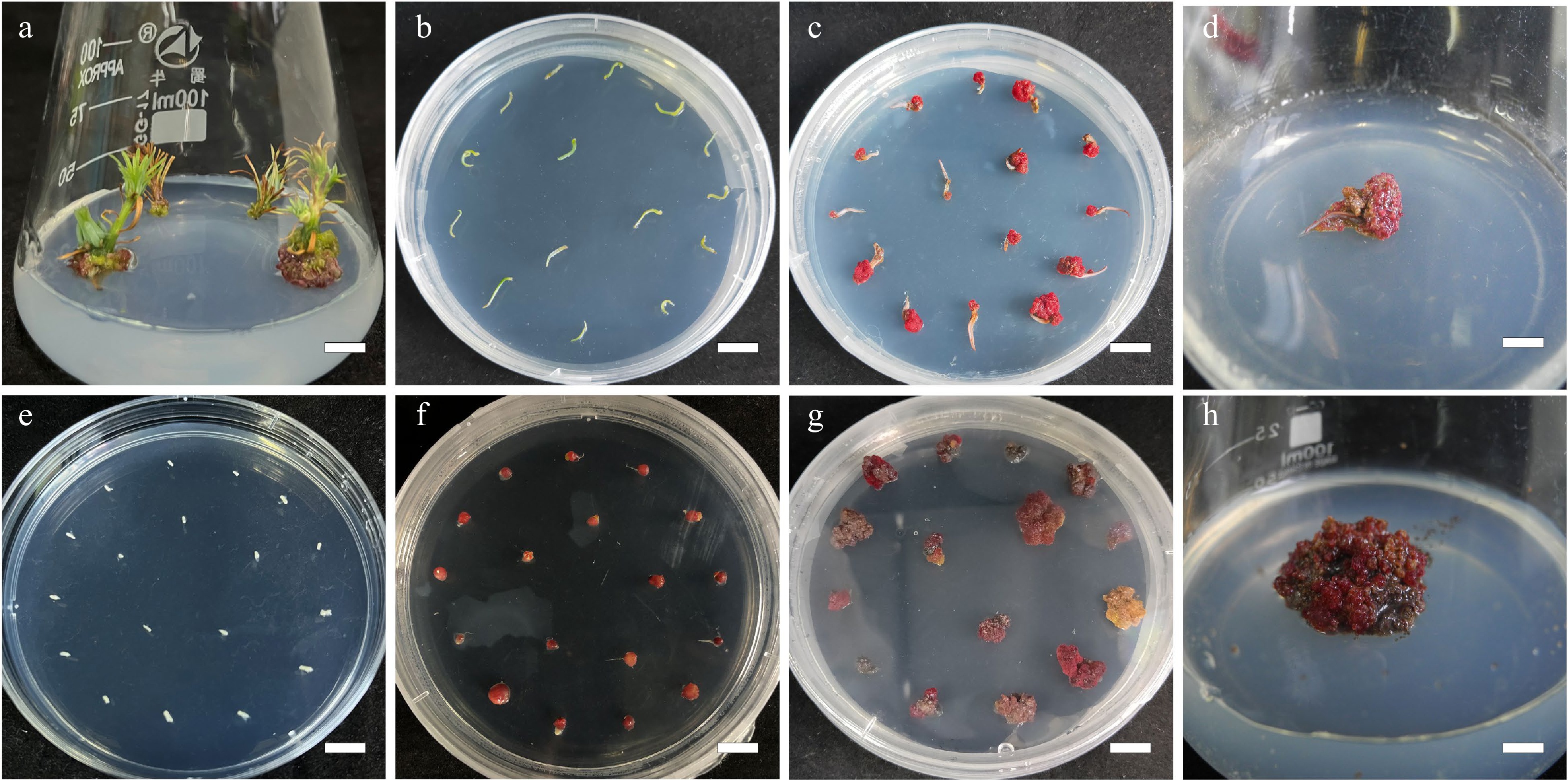
      Figure 1. 

      Callus induction from needle explants and mature zygotic embyro explants in L. olgensis. (a) Sterilized stem sections for obtaining needle explants, bar = 0.7 cm. (b) Needle explants operated in DCR medium augmented with 3 mg·L−1 6-BA and 0.3 mg·L−1 NAA, bar = 1 cm. (c) Calli from needle explants generated in 4 w, bar = 1 cm. (d) Calli from needle explants generated in 8 w, bar = 0.6 cm. (e) Sterilized mature zygotic embryo as explants, bar = 1 cm. (f) Calli from mature zygotic embryo explants generated in 3.0 mg·L−1 6-BA combined with 0.3 mg·L−1 NAA, bar = 1 cm. (g) Calli from mature zygotic embryo explants generated in 4 w, bar = 1 cm. (h) Calli from mature zygotic embryo explants generated in 8 w, bar = 0.6 cm.

    • The explants (zygotic embryos and needles) were obtained in sterile environments. The endosperm was removed from the seeds and the leftover mature zygotic embryos were used directly for callus induction. The needles were collected as described above. Fifteen explants (of zygotic embryo or needles) were placed in 40 mL of callus induction medium (CIM) in a 90 mm × 20 mm crystal-grade polystyrene Petri dish (DA TANG MEDICAL INSTRUMENT) with six replicates. The zygotic embryo and needles were placed separately. The MS medium and DCR medium supplemented with cytokinins 6-BA (0.1, 0.2, 0.5, 1, 2, and 3 mg∙L−1) in combination with auxin NAA at various concentrations(0.1, 0.2, 0.5, 1, 2, and 3 mg·L−1) was used as the CIM. The explants in CIM were kept at 25 ± 2 °C and 70% relative humidity under white fluorescent tubes (60 μmol∙m−2∙s−1 light intensity) in a 16-h photoperiod system until the callus developed (Fig. 1). The nature of the callus and the callus percentage induction were determined after 8 weeks of incubation ( Fig. 2 ).
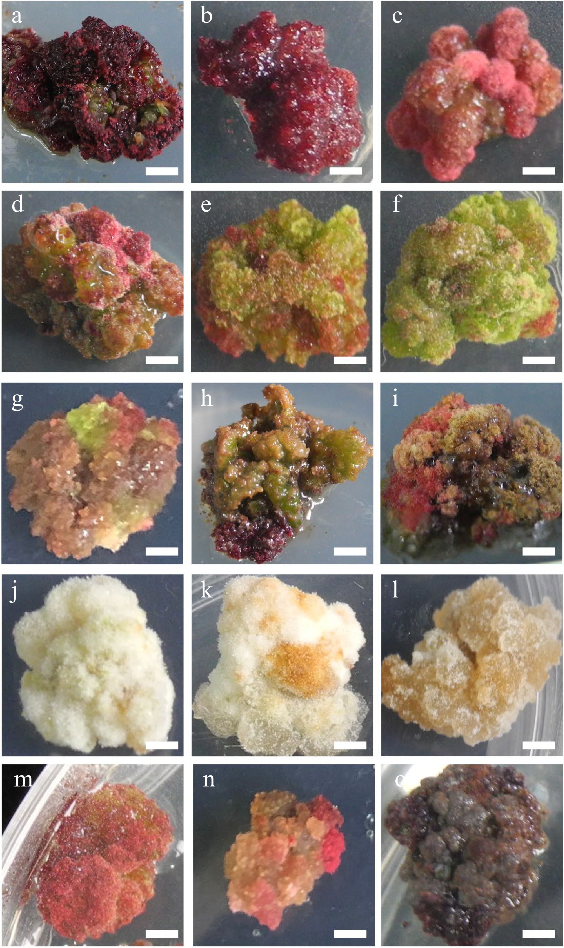
      Figure 2. 

      Characteristic nature of Larix olgensis callus from different treatments. (a) DCR + 3 mg∙L−1 6-BA + 0.3 mg∙L−1 NAA (90%+). (b) DCR+2 mg∙L−1 6-BA+0.2 mg∙L−1 NAA. (c) DCR + 1 mg∙L−1 6-BA + 0.1 mg∙L−1 NAA. (d) DCR + 3 mg∙L−1 6-BA + 0.3 mg∙L−1 NAA, a genotype different from that in (a). (e) DCR + 1 mg∙L−1 6-BA + 1 mg∙L−1 NAA. (f) DCR + 0.3 mg∙L−1 6-BA + 3 mg∙L−1 NAA. (g) MS + 0.5 mg∙L−1 6-BA + 0.05 mg∙L−1 NAA. (h) DCR + 0.5 mg∙L−1 6-BA + 0.05 mg∙L−1 NAA. (i) DCR + 0.5 mg∙L−1 6-BA + 0.05 mg∙L−1 NAA transfer to DCR + 2 mg∙L−1 6-BA + 0.2 mg∙L−1 NAA. (j) DCR + 0.3 mg∙L−1 6-BA + 3 mg∙L−1 NAA (in dark). (k) MS+0.05 mg∙L−1 6-BA + 0.5 mg∙L−1 NAA (in dark). (l) MS + 3 mg∙L−1 6-BA + 0.3 mg∙L−1 NAA (in dark). (m), (n) Modified high auxin culture, the yellow callus turn red. (o) After subculture over five times, the callus turned brown. The bar in the pictures is 0.32 cm except in (n) which is 0.43 cm.

    • Callus was moved to DCR medium augmented with cytokinin 6-BA (0.5, 1.0, 2.0 mg∙L−1) and auxin NAA at various concentrations (0.05, 0.1, 0.2, and 0.5 mg∙L−1) for shoot regeneration. Six replicates were made for each treatment, comprising 10 calli in 50 mL of the shoot regeneration medium in an Erlenmeyer flask (GG-17, 100 mL, SHUNIU). The callus cultures were kept at 25 ± 1 °C and relative humidity of 70% under white fluorescent tubes (60 μmol∙m−2∙s−1 light intensity) in a 16-h photoperiod system. At 2-week intervals, until shoots regenerated, the L. olgensis calli (Fig. 3a) were subcultured onto fresh medium of the same composition with or without AC (Fig. 3b). Shoots produced per callus were counted, and shoot regeneration rate was determined after 6 weeks. Regenerated L. olgensis shoots thereafter were moved to the medium for elongation (Fig. 3c).
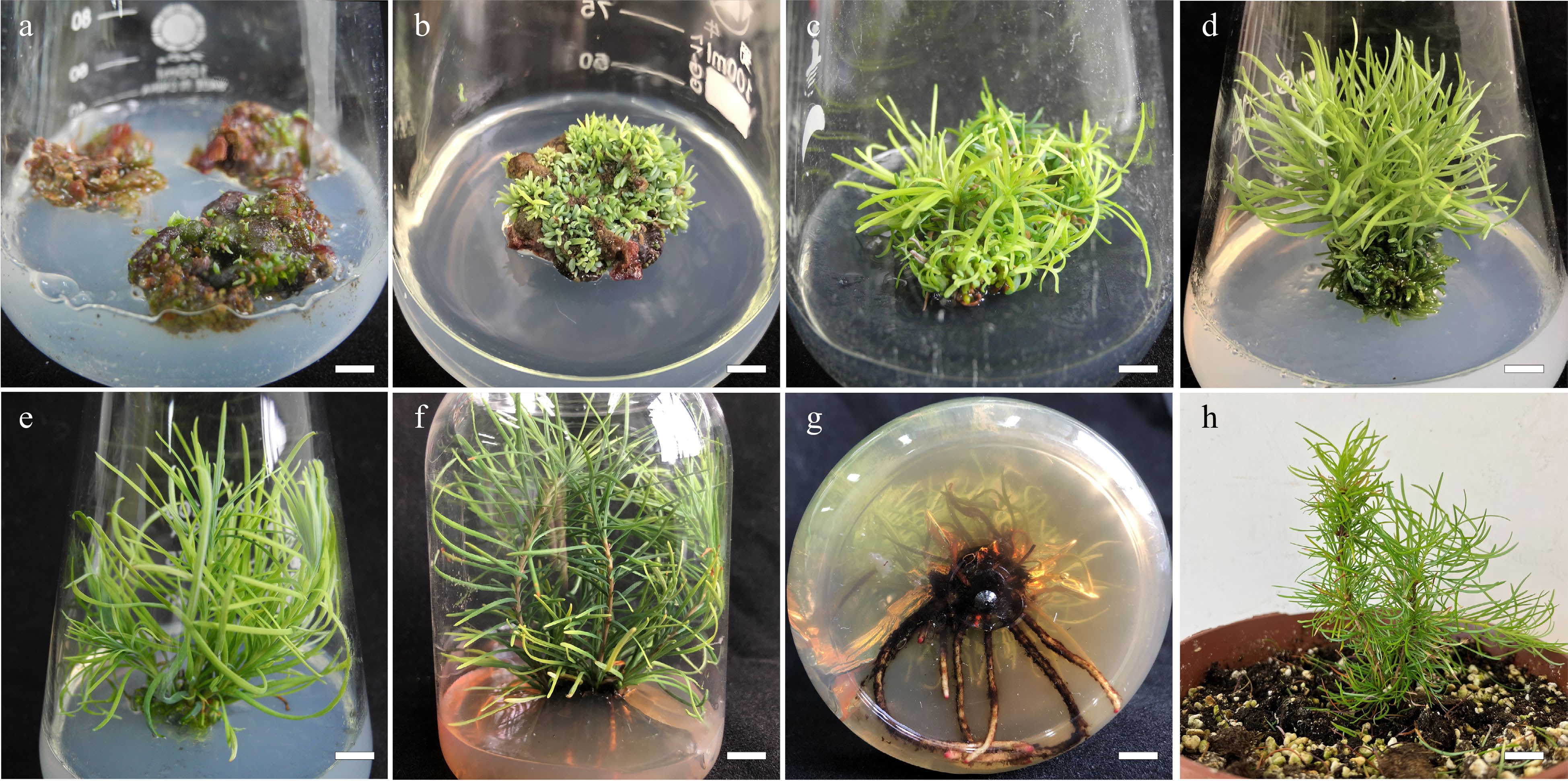
      Figure 3. 

      Summary of in vitro propagation from callus of Larix olgensis. (a) New shoots developed from callus placed in the DCR medium supplemented with 1.0 mg∙L−1 BA and 0.1 mg∙L−1 NAA, bar = 0.6 cm. (b) Shoot organogenesis occurring on callus after 8 weeks in regeneration medium, bar = 0.6 cm. (c) Developed shoots from callus, bar = 0.6 cm. (d) Adventitious shoots elongation, bar = 0.6 cm. (e) Further shoot elongation, bar = 0.68 cm. (f) Rooting of in vitro regenerated shoots in DCR medium supplemented with 3 mg∙L−1 IBA and 100 mg∙L−1 PG, bar = 0.83 cm. (g) Roots of fully developed plantlets, bar = 0.61 cm. (h) Acclimatized potted plants, bar = 2 cm.

    • The regenerated L. olgensis shoots were cultured in the DCR medium (50 mL) supplemented with AC (0, 2 g∙L−1) in addition to cytokinins 6-BA (0.05, 0.1, 0.15, 0.2, and 3 mg∙L−1) and auxin NAA at various concentrations (0.005, 0.01, 0.015, 0.02 and 0.03 mg∙L−1) in combination for shoot elongation in polystyrene culture vessels (ZP5-330, SHJIAFENG). Three regenerated L. olgensis shoots were set up in each vessel, with 20 replications for this experiment. The elongation cultures were kept at 25 ± 1 °C and relative humidity of 70% under white fluorescent tubes (60 μmol∙m−2∙s−1 light intensity) in a 16-h photoperiod system. At 2-week intervals, the L. olgensis shoots were subcultured on fresh media of the same composition (Fig. 3d). Shoot elongation percentage (%) were counted, and elongation lengths were determined after 6 weeks. Elongated L. olgensis shoots thereafter were moved to the medium for rooting (Fig. 3e).

    • The elongated L. olgensis shoots were cultured in DCR medium (100 mL) of various strengths (i.e. DCR, 1/2 DCR) supplemented with auxin [1-naphthaleneacetic acid (NAA) or indole-3-butyric acid (IBA)] (0.5, 1.0, 1.5, 2.0, 2.5 mg∙L−1) either singly or in combination with Phloroglucinol (PG) (0, 50 , 100, 150 mg∙L−1) and AC 2 g∙L−1 in polystyrene culture vessels (125 mm × 110 mm). Four regenerated L. olgensis shoots with 20 replications for this experiment. The elongation cultures were kept at 25 ± 1 °C and relative humidity of 70% under white fluorescent tubes (60 μmol∙m−2∙s−1 light intensity) in a 16-h photoperiod system. Rooting rates and root numbers were determined for each treatment after culture for 10 weeks, with no subculture during rooting (Fig. 3f, g).

    • After removing the medium traces from the roots of each regenerated L. olgensis plantlet by rinsing in running water from a tap, the plantlets were moved to a mixture of peat : organic cultivation soil : perlite (3:6:1) in 22 cm diameter plastic pots (Fig. 3h). The plantlets were covered with transparent plastic bags ensuring adequate humidity and kept in growth chambers operating under a 16-h photoperiod (33.73 μmol∙m−2∙s−1 light intensity) at ~25 °C and 70% relative humidity. The polyethylene coverings were opened gradually after 3 weeks as the plantlets acclimatized. Plant survival rates were determined at 6 weeks following acclimatization.

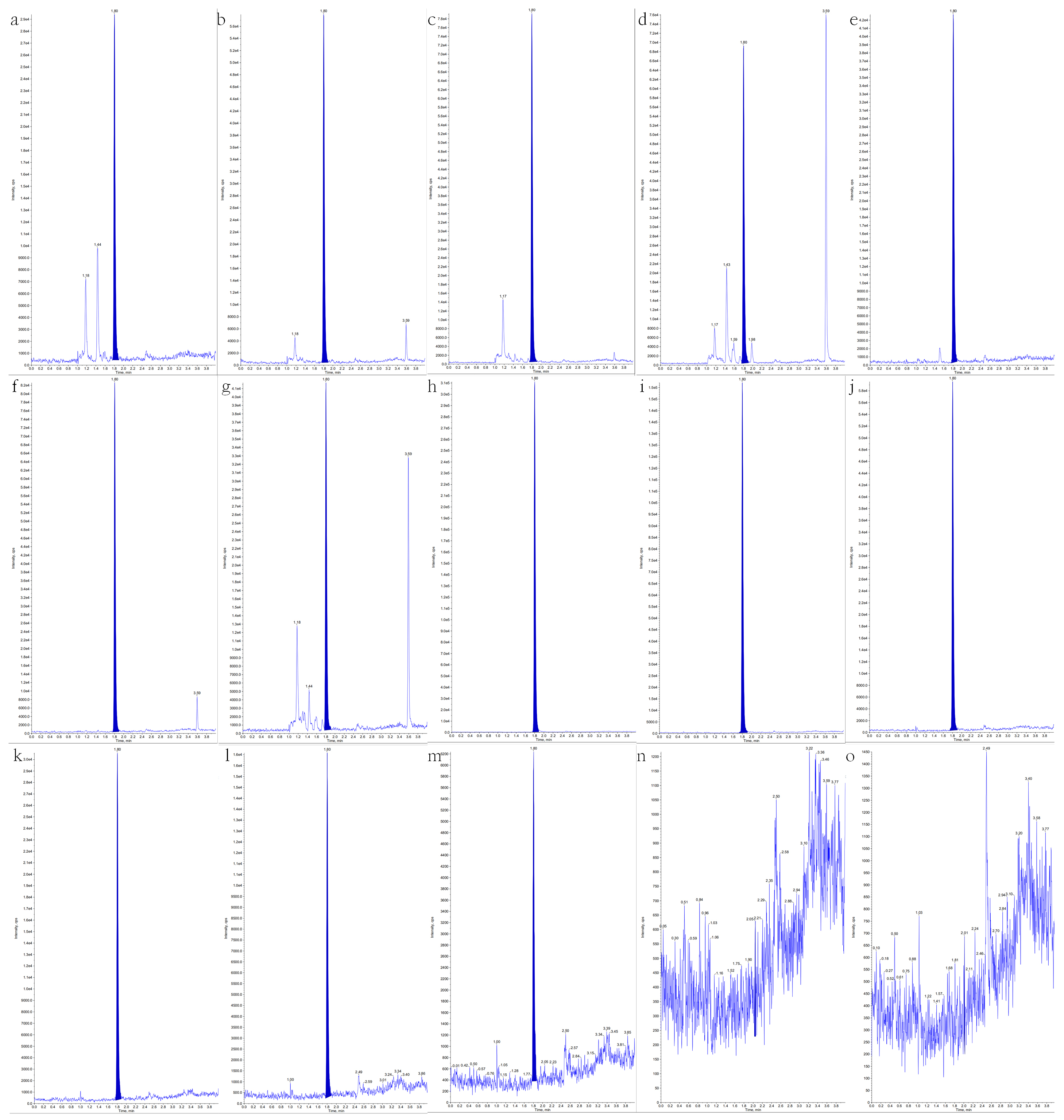
    • In this study, different fresh calli and tissue of various stages were used for taxifolin content determination. Callus-1 (Fig. 2n), callus-2 (Fig. 2h), callus-3 (Fig. 2a), adventitious shoots, elongation shoots, lignified elongation shoots, adventitious roots, were freeze-dried at −70 °C for 24 h. The dried tissue was ground into a powder with a mortar and sifted through 40 mesh for standby. Each sample was weighed accurately with 100 mg, and taxifolin was extracted by adding 1 mL methanol for tissue bomogenate(8,500 rpm, 4 × 15 s)and ultrasound 100 khz for 20 min. After that, the solution was centrifuged for 6 min (15,000 rpm), the supernatant was taken and diluted 10 times with methanol, and then mixed with H2O 1:1 for LC-MS detection.

      LC-MS analysis was carried out on Waters ACQUITY I-Class (Waters Technology Shanghai, China), and Sciex Triple Quad 5500 (Sciex Shanghai, China). The injection volume of the sample was 2 µL and the column temperature was kept at 30 °C. The binary elution solvent consisted of A [0.1% Formic Acid in Methanol/Acetonitrile (1/9, v/v)] and B (0.1% Formic Acid in H2O): 85% : 15%, and a gradient elution procedure was used. A cosmosil column Waters HSS T3 (100 mm × 2.1 mm, 1.6 μM) was used. The flow rate was maintained at 0.5 mL·min−1. The UV spectrum of taxifolin was obtained with 290 nm detection wavelength.

    • All experimental data were analyzed by one-way ANOVA with Tukey's post-hoc multiple comparison tests, using SPSS (IBM SPSS Statistics 27.0). In CIM, three leaf/root segment explants with 20 replications were used. For shoot regeneration, six calli pieces were used with 10 replications, and four regenerated L. olgensis shoots with 20 replications were used for rooting. Means were regarded as statistically significant at p ≤ 0.05.

    • Among all the treatments, the highest percentage of callus induction was recorded in the explants cultivated on the DCR medium augmented with 3.0 mg∙L−1 6-BA together with 0.3 mg∙L−1 NAA for both L. olgensis zygotic embryo (96.8%) and needle (86.7%) explants (Table 1).

      Table 1.  Induction percentage and characteristics of callus from mature zygotic explants and needle explants of Larix olgensis.

      OrderBasic
      medium
      Plant growth regulators (mg∙L−1)Mature zygotic embryo explantsNeedle explants
      Callus induction rate (%)ColorTextureCallus induction
      rate (%)
      ColorTexture
      1MS6-BA 3:NAA 0.390.2 ± 0.11aRose redCompact43.3 ± 1.91bcBrownCompact
      26-BA 2:NAA 0.290.1 ± 1.73aPinkCompact38.9 ± 1.13bcdBrownCompact
      36-BA 1:NAA 0.178.2 ± 4.45bRed and whitefriable35.6 ± 2.94cdcbrownCompact
      46-BA 0.5:NAA0.0552.1 ± 1.41cdRed and whiteFriable27.8 ± 1.11fgBrownFriable
      56-BA 1: NAA 151.6 ± 1.86cdCreamCompact43.3 ± 0bcCreamFriable
      66-BA 0.05:NAA 0.532.2 ± 1.68fCreamFriable38.9 ± 111bcdYellow and greenFriable
      76-BA 0.1:NAA 143.4 ± 3.41deYellow and greenFriable27.8 ± 1.11fgYellow and greenFriable
      86-BA 0.3:NAA 344.5 ± 3.26deYellow and greenFriable13.3 ± 1.93hYellow and greenCompact
      9DCR6-BA 3:NAA 0.396.8 ± 1.86aRose redCompact86.7 ± 1.93aRose redCompact
      106-BA 2:NAA 0.292.2 ± 0.97aRose redCompact46.7 ± 1.93bGreenFriable
      116-BA 1:NAA 0.176.1 ± 1.94bPinkFriable22.2 ± 1.11ghYellow and greenFriable
      126-BA 0.5:NAA0.0549.0 ± 5.35cdPinkFriable14.55 ± 2.22hDark brownFriable
      136-BA 1: NAA 151.2 ± 2.46cdRed and whiteCompact32.2 ± 1.11efgGreenCompact
      146-BA 0.05:NAA 0.536.6 ± 3.51efRed and whiteFriable34.5 ± 7.78cdeCreamFriable
      156-BA 0.1:NAA 150.1 ± 2.50cdRed and whiteCompact32.2 ± 8.89efgYellow and greenCompact
      166-BA 0.3:NAA 355.7 ± 4.93cCreamCompact23.3 ± 0ghYellow and greenCompact
      Means ( ± standard error) within a column followed by the same superscript letter are not significantly different using Tukey’s multiple comparison test and p ≤ 0.05.

      Although in the about-mention medium both the zygotic embryo explants and needle explants could achieve the highest percentage of callus induction rate, the two type of explants responded significantly differently in other treatments. For zygotic embryo explants, there are no significant differences (in the callus induction rate) from the highest percentage callus in MS media containing 3.0 mg∙L−1 6-BA and 0.3 mg∙L−1 NAA (90.2%), 2.0 mg∙L−1 BA and 0.2 mg∙L−1 NAA (90.1%), and DCR media augmenting with 3.0 mg∙L−1 6-BA and 0.3 mg∙L−1 (96.8%), 2.0 mg∙L−1 BA and 0.2 mg∙L−1 NAA (90.1%) among others (Fig. 1eh, Table 1).

      The highest percentage of callus induction from the needle explants was 86.7% in the DCR medium augmented with 3 mg∙L−1 6-BA and 0.3 mg∙L−1 NAA, which was significantly higher (p ≤ 0.05) than that in the other treatments (Fig. 1ad, Table 1). The lowest percentage of induced callus from L. olgensis zygotic embryo explants was 32.3% in the MS medium augmented with 0.05 mg∙L−1 6-BA together with 0.5 mg∙L−1 NAA, and that from needle explants were in the DCR medium augmented with 0.3 mg∙L−1 6-BA together with 0.3 mg∙L−1 NAA (13.3%).

      The effect of plant growth regulators (PGRs) combination was tested. The ratio of auxin and cytokinin of 1/10 showed a better callus induction response than that of 1/1 and 10/1 (Table 1). Once the ratio was determined, it was found that the callus induction rate was increased along with the promotion in the concentration of the PGRs combination. Meanwhile, the suitable basic mediums of callus induction from mature zygotic embryos were MS and DCR, while needle explants preferred DCR basic medium.

      The calli produced from both zygotic embryos and needle explants had different textures and colors. These colors were pink (Fig. 2ad), green (Fig. 2e, f), cream (Fig. 2gi), or dark brown (Fig. 2o), etc (Fig. 2gi, m, n), and their textures were either compact or friable depending on the medium composition and explant type (Table 1, Fig. 2). Furthermore, the compact rose red callus is the best for shoot regeneration (Fig. 2a, b).

    • For the zygotic explants, we found that the MS medium augmented with 1.0 mg∙L−1 6-BA and 0.1 mg∙L−1 NAA had the highest shoot regeneration rate (83.3 ± 1.93% and 528 ± 11.5 number of shoots per callus), followed by the medium supplemented with 1.0 mg/L 6-BA in addition to 0.2 mg∙L−1 NAA (76.7 ± 1.93%) shoot regeneration rate with the highest shoot number per callus (636 ± 21.7) (Table 2, Fig. 3a).

      Table 2.  Percentage shoot regeneration from calli of Larix olgensis.

      Mature zygotic embryo explantsNeedle explants
      Plant growth regulators (mg∙L−1)Percentage shoot regeneration (%)Average number of adventitious shootsPercentage shoot regeneration (%)Average number of adventitious shoots
      6-BANAA
      10.50.0541.1 ± 2.94fg304 ± 7.5d32.22 ± 1.11de120 ± 6.1h
      20.50.142.2 ± 1.11fg315 ± 4.5d32.22 ± 2.22de111 ± 3.8h
      30.50.232.2 ± 1.11h150 ± 14.8f28.89 ± 1.11e66 ± 2.3i
      40.50.540.0 ± 1.93g211 ± 5.5e34.44 ± 1.11d51 ± 1.8g
      510.0557.8 ± 1.11d426 ± 13.9c68.89 ± 1.11a161 ± 6.2f
      610.183.3 ± 1.93a528 ± 11.5b73.33 ± 1.93a307 ± 1.8b
      710.276.7 ± 1.93b636 ± 21.7a72.22 ± 1.11a323 ± 3.8a
      810.565.6 ± 2.22c460 ± 22.8c73.33 ± 1.93a221 ± 6.4c
      920.0553.3 ± 3.85de322 ± 11.7d50.00 ± 1.93c176 ± 5.5e
      1020.147.8 ± 2.94ef237 ± 11.5d51.11 ± 1.11c141 ± 7.0g
      1120.246.7 ± 1.93efg423 ± 23.5c57.78 ± 2.22b203 ± 3.9d
      1220.547.8 ± 1.11ef200 ± 6.1e33.33 ± 1.93de57 ± 1.2ig
      Means (± standard error) within a column followed by the same superscript letter are not significantly different using Tukey’s multiple comparison test and p ≤ 0.05.

      Meanwhile, for the needle explants, the highest percentage of regeneration (73.3 ± 1.93%) and the number of shoots per callus (307 ± 1.8) were recorded in the explants cultivated on the DCR medium augmented with 1.0 mg∙L−1 6-BA, 0.1 mg∙L−1 NAA, and 0.1 mg∙L−1 TDZ, followed by the results in the medium supplemented with 1.0 mg∙L−1 6-BA in addition to 0.2 mg∙L−1 NAA (with shoot regeneration percentage and the number of shoots per callus, 72.22 ± 1.11% and 323 ± 3.8, respectively) (Table 2, Fig. 3b). The highest percentage of regeneration (73.3 ± 1.93%) was also recorded in the medium supplemented with 1.0 mg∙L−1 6-BA in addition to 0.5 mg∙L−1 NAA, but the number of shoots per callus (221 ± 6.4) was significantly lower than that in medium 6.

      Both shoot regeneration percentage and shoot number per callus were generally higher in media supplemented with cytokinin (BA) in combination with auxin (NAA), which ratio ranges from 10/1 to 5/1. In addition, medium 2-6 and medium 2-7, 2-1, 2-2, and 2-11 also showed relatively high induction rates. Although the differences in shoot regeneration percentage were not statistically significant, medium supplemented with 6-BA in combination with higher NAA were generally associated with a low number of shoots induced from each callus on average (Table 2, Fig. 3). If the ratio of 6-BA to NAA is fixed, with the increase of the PGRs concentration, the shoot regeneration percentage and the number of shoots per callus showed an upward trend initially and then declined.

      Due to the limited callus size and the number of the subculture of needles, the number of shoots per callus induced from needles was lower than that from zygotic embryos, but there is no significant difference in the shoot regeneration percentage between zygotic embryos and needles.

    • The zygotic embryo explants and the needle explants were inoculated in the same shoot regeneration culture medium for 4 weeks before subculture to the elongation treatment. The DCR medium augmented with 0.5 mg∙L−1 BA, 0.05 mg∙L−1 NAA, and 2 g∙L−1 AC achieved the highest elongation percentage of shoots (75.6 ± 2.94%) and the longest average shoot length (3.5 ± 0.11 cm). The percentage shoot elongation and average shoot length significantly differed (p < 0.05) from that in DCR without any AC (control) (Table 3, Fig. 3ce). In the same medium without AC, the percentage of shoot elongation and average shoot length were 65.2 ± 1.11% and 1.6 ± 0.11 cm, respectively. Compared medium 3-2 (65.6 ± 1.11%, 1.6 ± 0.06 cm) to medium 3-3 (75.6 ± 2.92%, 3.5 ± 0.11 cm), medium 3-4 (46.7 ± 1.93%, 1.3 ± 0.03 cm) to medium 3-5 (61.1 ± 1.11%, 2.8 ± 0.12 cm), medium 3-6 (21.1 ± 1.11%, 1.4 ± 0.01 cm) to medium 3-7 (35.6 ± 2.22%, 2.2 ± 0.1 cm), it was clear that AC significantly increased the percentage of shoots elongation and average shoot length.

      Table 3.  Effect of different concentrations of 6-BA and NAA on adventitious bud elongation of Larix olgensis.

      Plant growth regulators and AC (mg∙L−1)Adventitious
      shoot elongation
      percentage (%)
      Average shoot length (cm)
      16-BA 1:NAA 0.121.1 ± 2.22e0.9 ± 0.03e
      26-BA 0.5:NAA 0.0565.6 ± 1.11b1.6 ± 0.06d
      36-BA 0.5:NAA 0.05:AC 200075.6 ± 2.94a3.5 ± 0.11a
      46-BA 0.3:NAA 0.0346.7 ± 1.93c1.3 ± 0.03d
      56-BA 0.3:NAA 0.03:AC 200061.1 ± 1.11b2.8 ± 0.12b
      66-BA 0.1:NAA 0.0121.1 ± 1.11e1.4 ± 0.01d
      76-BA 0.1:NAA 0.01:AC 200035.6 ± 2.22d2.2 ± 0.10c
      Means (± standard error) within a column followed by the same superscript letter are not significantly different using Tukey’s multiple comparison test and p ≤ 0.05.

      The elongation culture of L. olgensis was DCR medium supplemented with a certain ratio but different concentrations of BA and NAA. According to Table 3, a comparison of the elongation rate and average shoot length among medium 3-1 (21.2 ± 2.22%, 0.9 ± 0.03 cm), medium 3-2 (65.6 ± 1.11%, 1.6 ± 0.06 cm), medium 3-4 (46.7 ± 1.93%, 1.3 ± 0.03 cm), which depicted lower concentrations of the PGRs promoted the shoot elongation, but when it reduced to a certain level, poor elongation also resulted.

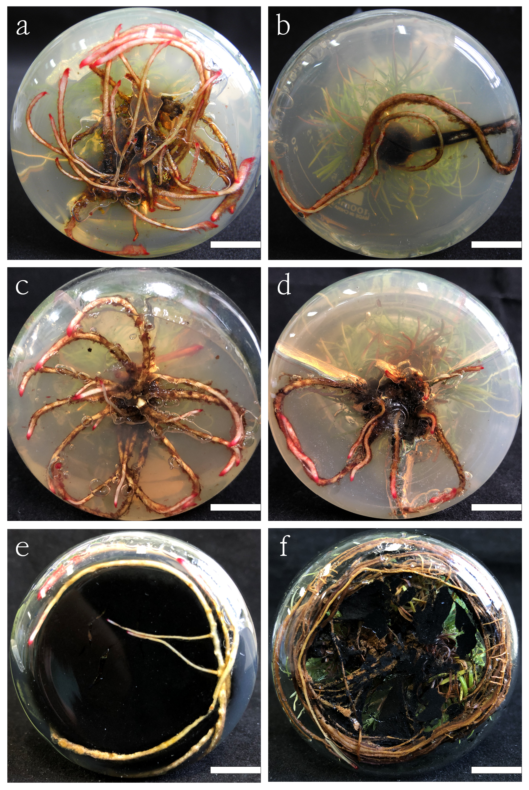
    • In our study, the first regeneration plantlet with roots (1−2 mm) was seen on the 38th day in DCR medium supplemented with 3 m∙L−1 IBA and 100 mg∙L−1 PG, and the highest adventitious root induction rate was 62.2 ± 5.88% (Table 4, Fig. 3f, g). Even if the concentration of auxin is continuously increased, the single application of auxin has little effect on rooting. The rooting rates of DCR medium supplemented with 3 mg∙L−1 of IBA or NAA were 13.3%, and 8.9 ± 2.22%, respectively. But in 1/2 DCR medium with the same PGRs were 11.11 ± 2.22%, and 15.56 ± 2.22%, respectively. Rooting in these mediums took at least 60 d.

      Table 4.  Rooting of regenerated shoots in DCR media supplemented with auxin, AC and PG strength.

      Basic mediumExogenous
      additives (mg∙L−1)
      Adventitious
      root induction
      percentage (%)
      Rooting start time (d)
      DCRNAA 38.9 ± 2.22e70
      IBA 2:NAA 215.6 ± 2.22de64
      IBA 3:AC 200046.7 ± 3.85b52
      IBA 313.3 ± 0.00e63
      IBA 3:PG 5033.3 ± 3.85c45
      IBA 3:PG 10062.2 ± 5.88a38
      IBA 3:PG 15057.8 ± 4.44ab36
      IBA 3:PG 100:AC 200053.3 ± 3.85b32
      1/2DCRNAA 315.56 ± 2.22de70
      IBA 2:NAA 222.22 ± 2.22d63
      IBA 3:AC 200035.56 ± 2.22c60
      IBA 311.11 ± 2.22e60
      IBA 3:PG 5031.11 ± 4.44c50
      IBA 3:PG 10048.89 ± 2.22b41
      IBA 3:PG 15046.67 ± 0b40
      IBA 3:PG 100:AC 200037.78 ± 2.22c36
      Means (± standard error) within a column followed by the same superscript letter are not significantly different using Tukey’s multiple comparison test and p ≤ 0.05.

      The addition of AC and PG promoted root formation to a large extent and significantly increased the rooting percentage. In the medium supplemented with PG, once the root primordium is produced, the adventitious roots were produced along the main stem, and the roots were quickly formed to produce a strong root system (Fig. 3g). The concentration of PG significantly affected the rooting rate in both 1/2 DCR and DCR medium. Specifically, among the three concentrations of PG tested, 50 mg∙L−1, 100 mg∙L−1, and 150 mg∙L−1, the 100mg∙L−1 PG had a better effect on rooting (Supplemental Fig. 1ad). The root system from the medium supplemented with AC was slender, and lateral roots were produced in prolonged culture (Supplemental Fig. 1e, f).

    • After 6 weeks of acclimatization, the in vitro regenerated L. olgensis plantlets showed a high survival rate of 90%. The acclimatized L. olgensis plantlets grew well and displayed normal growth characteristics and morphology typical of the plant species (Fig. 3h).

    • To determine the taxifolin content in different tissues and stages of L. olgensis plants, callus of different stages (callus-1, callus-2, callus-3), adventitious shoots, elongation shoots, lignified elongation shoots, adventitious roots, were selected and analyzed by HPLC (Supplemental Fig. 2). The result indicated that the taxifolin content in different tissues and different stages of the regenerated L. olgensis plants varied significantly. The content of the taxifolin in callus-1 was 1.99 µg∙g−1, callus-2 was 3.9 µg∙g−1, callus-3 was 5.26 µg∙g−1, and in adventitious shoots, the content of the taxifolin was 4.8µg/g, while in adventitious roots was 2.86 µg∙g−1. The accumulation of the taxifolin in elongation shoots and lignified elongation shoots was 28.6 µg∙g−1 and 53 µg∙g−1 respectively, much higher than that in other tissues.

      Meanwhile, the results showed that calli in different colors and textures might affect the accumulation of taxifolin. For example, the rose-red calli accumulated more taxifolin than the calli of two other colors. The result also illustrated that the development of vascular tissue was beneficial to the accumulation of taxifolin since in the elongation shoots, the content of taxifolin was much higher. Compared with the un-lignified elongation shoots, the content of taxifolin in the lignified shoots nearly doubled. Therefore, in vitro regeneration is an efficient and quick method to produce secondary metabolites.

    • Due to the current large demand for larch timber, the supply of seedlings and the establishment of plantations have become very urgent. In vitro regeneration technology provides an efficient way for large numbers of seedling production in a short time and has been widely used in large-scale propagation of high-quality seedlings, genetic transformation, and gene editing. The L. olgensis regeneration system is set up in this study, the process is stable and reliable, and not easily affected by external conditions, and therefore, is highly controllable and annual production can be assured.

      The research on the in vitro regeneration system of larch mainly contains somatic embryogenesis and organogenesis. Current research on in vitro regeneration of larch mainly focuses on inducing embryogenic callus that leads to somatic embryogenesis. The induced somatic embryos have characteristics similar to mature zygotic embryos and can directly generate stems and roots through suitable culture. However, some research showed that the germination rate of the induced somatic embryos was uncontrollable[6], and the malformed embryos accounted for a large proportion. Some believe that somatic embryos are more suitable for cryopreservation and production of artificial seeds[23], but the subsequent growing time is the same or even longer than that of larch seedlings from natural seeds. In this study, mature embryos are regenerated via the callus, and under suitable culture conditions, more calli can be subcultured, and more adventitious shoots can subsequently be differentiated. Since a large amount of biomass can be produced under certain culture conditions, the growth rate and development direction can be adjusted by using different culture conditions. Tissue products in various culture stages can also be used for active ingredient extraction. The in vitro regeneration system of larch was optimized in the following aspects.

      Different species respond to in vitro regeneration quite differently, which might be the reflection of differences in nutrient absorption. It is crucial for species to confirm a rationally basic medium. Classical MS medium with a high nutrient concentration of inorganic salts is favored in plant tissue culture[24], especially in the cultivation of herbaceous plants, such as cornflower[25] (Gerbera jamesonii), lily (Lilium orientalis)[26], andrographis (Andrographis alata)[27], chandelier flowers (Ceropegia mohanramii)[28], etc. Generally, researchers believe that at present, the regeneration of woody plants is more difficult than that of herbaceous plants[29]. It is shown that the absorption capacity of the basic medium is different for different life-form plants, and sometimes the MS medium does not yield good results in some woody plants. For example, for the conifer juniper[30] (Juniperus L.), researchers gradually replaced MS with WPM medium during the culture. The researchers used a modified MS medium with half-strength salt and reduced the concentration of KNO3 in the medium at a later stage for a good culture effect. The study by Samiei et al.[31] suggested that Van der Salm (VS) modified by MS medium with reduced inorganic salts has a better effect than MS in culturing Rosa canina. In the cultivation of Fagaceae chestnut (Castanea sativa × Castanea mollissima)[32], researchers used MS medium with reduced salt concentration, combined with WPM, to obtain stable chestnut regeneration seedlings. The macroelement salt ion molar concentration of MS is about three times that of WPM and nine times that of DCR medium. Researchers who used MS medium to cultivate the plantlets have so far not achieved efficient results in larch. In our research, the lower inorganic salt ion concentration DCR basic medium is suitable for L. olgensis subsequent development. The results are consistent with the above mentioned reports (Table 1, Fig. 1).

      Many studies have shown that the use of single plant growth regulator has a limited effect on callus tissue during larch regeneration. In this study, we investigated the effects of auxin and cytokinin in different ratios and the strength of the combined concentration on various processes in the regeneration of mature zygotic embryos and needles. The dominant plant growth regulator and the ratios of different PGRs both played crucial roles in callus induction. The combination and concentration of hormones for in vitro regeneration of mature zygotic embryos and needles were determined. In the in vitro regeneration of plants, the combined use of cytokinins and auxins can influence the growth direction of the materials. The combination of cytokinin 6-BA and auxin NAA in different concentration groups were used to study the concentration ratio and intensity of growth regulators required in each stage of larch development. It was found that in the stage of callus induction of mature zygotic embryos and needles, larch needs a higher concentration of cytokinin, thus forming a large number of calli. When the 6-BA/NAA ratio is 10, it is beneficial to promote callus induction. At this ratio, increasing the concentration to three times (taking 6-BA as 1 mg∙L−1 as an example) can accelerate the formation of calli. However, under the condition of high auxin, callus quality is poor and consequently difficult to differentiate (Table 1, Fig. 2l, o). A shorter subculture cycle may be beneficial for callus induction under high auxin culture conditions.

      Our study suggests that the adventitious shoots subsequently differentiate from callus and should require a lower concentration of cytokinin in larch (Fig. 3). The method was considered to be effective to obtain regenerated plantlets. In addition, adding a certain amount of activated carbon is conducive to the elongation of larch (Table 3). In contrast, in the regeneration of Ash (Fraxinus mandshurica) in 2020[33], using long-term and high concentration phytohormone cultivation, the number of adventitious bud differentiation is extremely low, and complete plants cannot be obtained. The results of elm trees (Ulmus glabra and Ulmus laevis) in vitro regeneration also showed[34] that 0.5 mg∙L−1 6-BA was appropriate for plant regeneration and stem differentiation. In some ranges, both the broad-leaved tree and conifer maybe have similar responses to adventitious shoots differentiation.

      Among all the tissues differentiated, the content was high in the lignified elongation shoot and the green elongation shoot. Moreover, the states of the tissues influenced the content of taxifolin. For example, the rose-red calli accumulated more taxifolin than the calli of the two other colors. It can be referred that the development of vascular tissue was beneficial to the accumulation of taxifolin since in the lignified shoots, the content of taxifolin was much higher (Supplemental Fig. S2). The result is consistent with natural larch, which also proves the potential of active ingredient production with artificial regulation[35]. Overall, it is obvious that in vitro regeneration is an efficient and quick method to produce secondary metabolites.

      The in vitro rooting of larch is very difficult. In this study, we established a rooting system for larch. Previously, there is no effective rooting method for larch in vitro regeneration, or the rooting process is complicated[36]. Induction of adventitious roots is the most difficult step in the in vitro regeneration of larch, with unstable rooting and a low induction rate of adventitious roots. In the process of adventitious shoot rooting, single application of auxin was not good (Table 4), increasing the concentration of auxin to 3 mg∙L−1, or two auxins NAA and IBA in combination, the rooting effect did still not work well. However, the addition of exogenous substances such as PG and AC in combination with auxin has a good effect on rooting. In this study, the combination of 100 mg∙L−1 PG and IBA obtained a good rooting effect (Fig. 3, Supplemental Fig. S1), indicating that PG is a good exogenous additive for inducing rooting.

      Above all, an efficient and complete organogenesis regeneration system was established for the first time, which would greatly benefit larch plantation. This protocol can be used for large-scale propagation of high-quality seedlings, genetic transformation, gene editing, and in vitro production of raw materials in various industries. Furthermore, it is a reliable reference for in vitro regeneration in recalcitrant species.

    • In this study, we established an efficient and complete regeneration system for larch organogenesis regeneration for the first time, especially from the needle explants. Effects of combination of auxin and cytokinin in different ratios and different intensities on regeneration were investigated. Furthermore, we firstly reported the taxifolin accumulation and content in the different larch tissues. To the best of our knowledge, this is the first study to develop an efficient indirect regeneration protocol for L. olgensis, which can be used for large-scale breeding of high-quality seedlings, genetic transformation, and gene editing and offers a basis for the production of raw materials in various industries. It is also a reliable reference for in vitro regeneration in recalcitrant species.

      • This work was supported by the National Science and Technology Major Projec of China (2018ZX08020003-005-001)

      • The authors declare that they have no conflict of interest.

      • Copyright: © 2023 by the author(s). Published by Maximum Academic Press, Fayetteville, GA. This article is an open access article distributed under Creative Commons Attribution License (CC BY 4.0), visit https://creativecommons.org/licenses/by/4.0/.
    Figure (3)  Table (4) References (36)
  • About this article
    Cite this article
    Yan X, Wang K, Zheng K, Zhang L, Ye Y, et al. 2023. Efficient organogenesis and taxifolin production system from mature zygotic embryos and needles in larch. Forestry Research 3:4 doi: 10.48130/FR-2023-0004
    Yan X, Wang K, Zheng K, Zhang L, Ye Y, et al. 2023. Efficient organogenesis and taxifolin production system from mature zygotic embryos and needles in larch. Forestry Research 3:4 doi: 10.48130/FR-2023-0004

Catalog

  • About this article

/

DownLoad:  Full-Size Img  PowerPoint
Return
Return