


-
Alzheimer's disease (AD) is a common neurodegenerative disease that seriously affects memory and thinking. More than 60% of dementia cases are attributed to AD. Statistics from the World Alzheimer Report 2022 showed that more than 55 million patients worldwide suffered from AD, and this number is expected to exceed 139 million by 2050[1]. Based on incomplete statistics, the cost of treatment and care for AD exceeded USD
${\$} $ ${\$} $ Table 1. Structures of dietary phytochemicals and their potential anti-AD mechanisms.
Dietary phytochemicals Model Dosage Molecular mechanism Ref. 
Wistar rats Cur-PLGA-NPs
(5−20 mg/kg body weight, 3 weeks)Cur-PLGA-NPs causes enhances the nuclear translocation of β-catenin, decreases GSK-3β levels, and increases promoter activity of the TCF/LEF and cyclin-D1. [18] Transgenic APP/PS1
miceCurcumin
(160 ppm, 6 months)Curcumin reduces the level of neuropro-inflammatory miR-146a, up-regulates the expression of CFH protein, and inhibits the phenotype of M1 microglia. [20] 
ICR mice TGN-Res@SeNPs
(50 mg/kg body weight, 16 weeks)kappa B↓ / protein kinase↓ / Akt↓
NF-κB/ mitogen-activated protein kinase/Akt signal pathway.[26] Wistar rats RSV-SeNPs (200 mg/kg body weight,
8 weeks)RSV-SeNPs up-regulates the expression of GSK3β and SIRT1, and down-regulates the expression of microRNA-134, consequently increasing neurite outgrowth. [25] 
Transgenic APP/PS1 mice Lycopene (4 mg/kg body weight, 5 days) LXR↑ / PI3K↑ / AKT↑
Lycopene alleviates neurovascular changes in APP/PS1 mice by activating the LXR–PI3K–AKT signaling pathway.[6] Wistar rats Lycopene
(1−4 mg/kg body weight, 2 weeks)Lycopene decreases NF-κB expression and downregulates IL-1β and TNF-α production. [28] 
Transgenic APP/PS1 mice Gallic acid (20 mg/kg body weight,
6 months)Gallic acid increases the ADAM10 proprotein convertase furin, activates ADAM10 and directly inhibits BACE1 activity, does not alter ADAM10 or BACE1 transcription. [7] 
Transgenic APP/tau/PS1 mice Berberine
(100 mg/kg body weight, 4 months)Berberine ameliorates cognitive deficits, reduces the Aβ accumulation, inhibits the apoptosis of neurons, and promotes the formation of microvessels in the mouse brain by enhancing brain CD31, VEGF, N-cadherin, and Ang-1. [8] 
Sprague dawley rat hippocampal neurons Ginsenoside Rg1
(60 μM, 24 h)CDΚ5↓ / IDE↑ / BACE1↑
Ginsenoside Rg1 significantly decreases CDK5 expression, inhibits PPARγ phosphorylation at serine 273, elevates IDE expression, downregulates BACE1 and APP expression.[44] Tree shrews Ginsenoside Rg1
(30 mg/kg body weight, 8 weeks)Bcl-2/Bax↑ / Wnt↑ / GSK-3β↓ / β-cateni↑
Rg1 increases the ratio of Bcl-2 to Bax and the expression of neuronal markers MAP2 and NeuN Rg1 regulates oxidative stress, cell apoptosis, and neuroinflammation by the Wnt/GSK-3β/β-catenin signaling pathway.[45] 
Wistar rats Pseudoginsenoside-F11 (2−8 mg/kg body weight, 4 weeks) Calpain I↓ / CDK5↓ / GSK-3β↓
Pseudoginsenoside-F11’s decreased GSK-3β (Ser9) phosphorylation and CDK5 activity.[46] 
ICR mice Ginsenoside Rh2
(12.5 and 25 mg/kg, 14 days)ERK↑ / CREB↑ / BDNF↑
Rh2 upregulates the phosphorylation of the ERK-CREB-BDNF pathway in the hippocampus.[47] 
PC12 cells Artemisinin
(0−50 μM, 0−80 min)ERK1/2↑
Artemisinin suppresses LDH release;
Artemisinin restraines the production of intracellular ROS;
Artemisinin modulating Δψm and caspase 3/7 dependent pathway;
Artemisinin activates ERK1/2 signaling.[48] 
ICR mice Torularhodin
(0.5 and 1.5 mg/kg body weight,
4 weeks)Nrf2↑ / NF-κB↓
Torularhodin ameliorates neuronal oxidative damage via the activation of Nrf2 translocation, upregulation of HO-1, and inactivation of NF-κB.[49] A daily dose of berberine also significantly improved learning and memory (Table 1)[8]. Previous studies have shown that berberine can play a neuroprotective role in AD caused by heavy metals[9]. In recent years, studies have demonstrated that ginsenosides have protective effects on AD, including ginsenoside Rb1 and ginsenoside Rg1[10]. Moreover, the number of publications on 'Alzheimer's disease and phytochemicals' (Indexed by Web of Science) has improved significantly since 2012 and has seen the most rapid growth over the past four years (2018−2021) (Fig. 1). Together, these data indicate that dietary phytochemicals have potential in treating AD. In this review, we have collected representative literature from the last 10 years from the Web of Science. 'AD and dietary phytochemicals' was used as keywords to search highly cited literature, we found that curcumin, resveratrol, lycopene, gallic acid, berberine and ginsenoside are the most frequently studied. Then, we searched the literature using 'xx and AD' as keywords like curcumin and AD. For each phytochemical, we selected 3−5 reports with high citation rate or the latest research (in the last two years). In the 'others' section, we found dietary phytochemicals related to those we identified in the first step, such as torularhodin, and recent substances of interest, such as artemisinin, and more cited articles, not highly cited articles, such as sesamin. Based on this, we discussed the molecular mechanisms of several representative dietary phytochemicals in the treatment of AD. This review will contribute to the development of potentially effective AD treatment strategies.
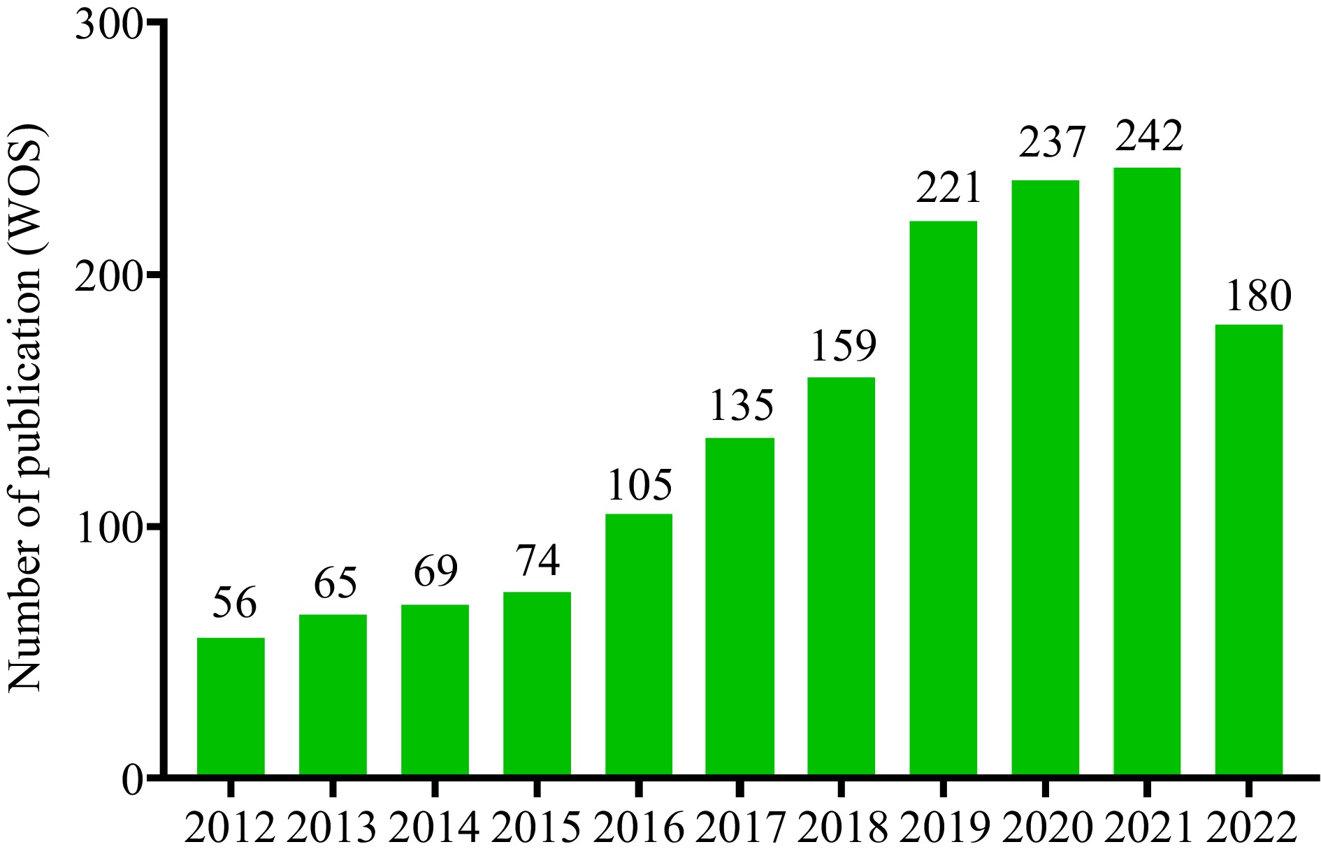
Figure 1.
The number of publications on 'Alzheimer's disease and dietary phytochemicals' (indexed by Web of Science) significantly increased since 2012 and the breakout increase occurred in the last four years (2018−2021).
-
The pathological features, pathogenesis, and drug treatment of AD have been studied for more than half a century since 1963 by Robert Terry and Michael Kidley, who observed neurofibrillary tangles (NFTs) using electron microscopy[11,12]. At present, more than a dozen hypotheses about the pathogenesis of AD, including the tau propagation hypothesis. Among these hypotheses of AD, the tau propagation hypothesis is the most influential (Fig. 2)[13]. However, this is just a hypothesis, and causes of AD are still being explored. Fortunately, the three major pathological features of AD are known, including amyloid plaques (Aβ), tau protein tangle, and neurofibrillary tangle in AD's brains[14]. Unfortunately, the accumulation of neurofibrillary tangle leads to neuron loss and degeneration, a form of cell death. Furthermore, the continued neuron loss and degeneration activated microglia and reactive astrocytes further contribute to Blood Brain Barrier (BBB) leakage and neuroinflammation (Fig. 3)[15].
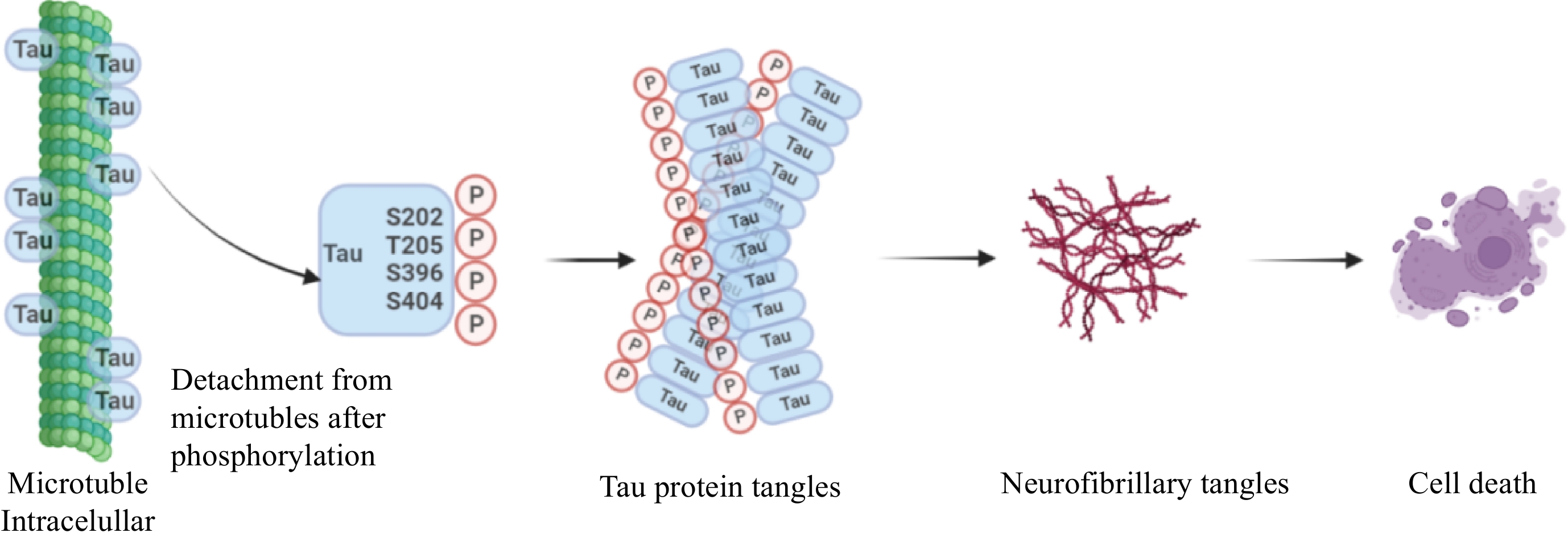
Figure 2.
Hypothesis of tau propagation. The tau proteins are usually hyperphosphorylated by binding to amino residues, typically Ser202, Thr205, Ser396, and Ser404. Then these monomers aggregate to form tau protein tangles, a complex oligomer, that eventually form neurofibrillary tangles, resulting in cell death. Drawing on
https://app.biorender.com/ .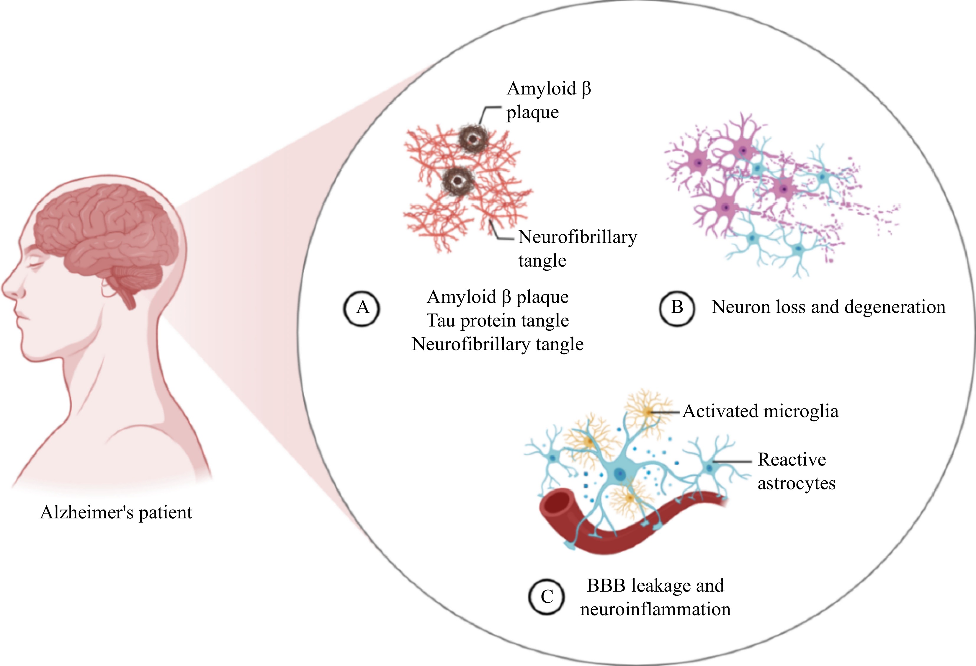
Figure 3.
(A) Three main pathological features of Alzheimer's disease (AD), amyloid beta plaques (Aβ), tau protein tangle and neurofibrillary tangle in the brains of AD patients. (B) The accumulation of neurofibrillary tangle leads to neuronal loss and degeneration, a form of cell death. (C) The continued neuronal loss and degeneration activates microglia and reactive astrocytes, which further contributes to blood brain barrier (BBB) leakage and neuroinflammation. Drawing on
https://app.biorender.com/ . -
Curcumin (Cur) (Table 1), a natural dietary polyphenol isolated from turmeric, has various biological activities and has been shown to be beneficial for many brain diseases[16]. Various studies show that Cur is not only well documented for its anti-carcinogenic, antioxidant and anti-inflammatory properties, but also possessing neuroprotective and cognitive-enhancing properties that may help delay or prevent neurodegenerative diseases, including AD[17]. New material carriers and Cur derivatives activate transcription factor EB, promote lysosomal and autophagy activity, attenuate Aβ and tau pathology, are also effective in preventing memory impairment in AD[4]. Cur nanoparticles show neuroprotective effects by increasing neuronal differentiation through activation of the Wnt/β-catenin pathway (Fig. 4), which enhances the brain's self-repairing mechanism and has great potential in alleviating AD (Table 1)[18]. Interestingly, Curcumin-primed exosomes potently ameliorate cognitive function in AD mice by inhibiting hyperphosphorylation of the tau protein through the AKT/GSK-3 beta pathway[19]. In addition, Gong & Sun confirmed that Cur can significantly reduce the level of neuropro-inflammatory miR-146A, and play a role in treating AD (Table 1)[20]. These studies laid a foundation for the development of Cur as a novel drug for AD.
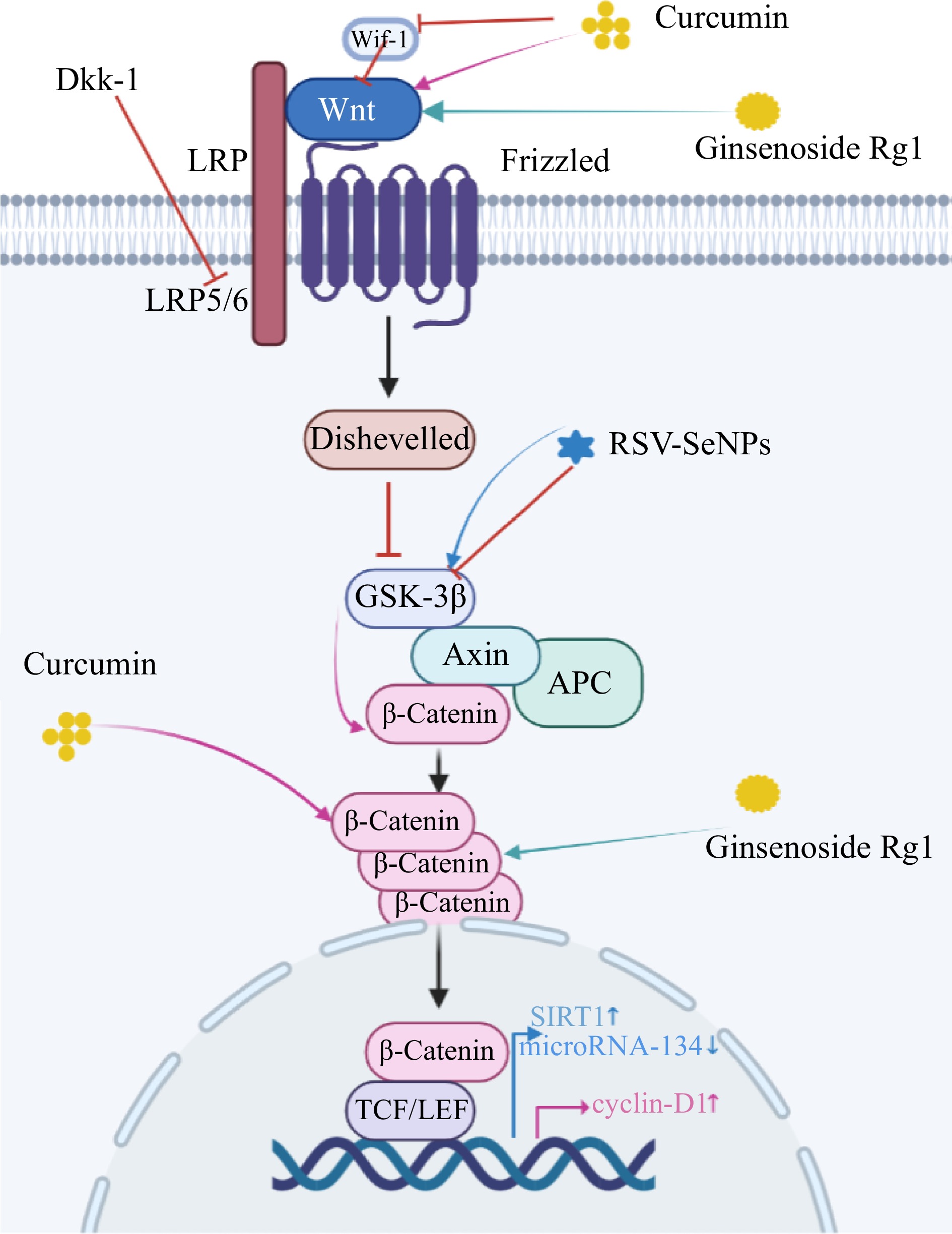
Figure 4.
Schematic model of the role of dietary phytochemicals in neurogenesis through activation of the Wnt/ β-catenin signaling pathway. Pink arrows: Curcumin interacts with Wif-1 and Dkk-1 to increase Wnt levels and activate the Wnt pathway. Wnt interacts with frizzled receptors to down-regulate low-density lipoprotein (LRP-5/6) expression and trigger cytoplasmic disheveled (Dvl). Then it breaks the Axin/APC/GS K-3β homeostasis and down-regulates the expression of GSK-3β. This sequence of reactions ultimately up-regulates cytoplasmic β-catenin expression and transfers it from the cytoplasm to the nucleus. After cellular internalization curcumin directly upregulates cytoplasmic β-catenin levels. In the nucleus, TCF/LEF and cyclin d1 promoter activity were enhanced. Green arrows: Rg1 activates Wnt/GSK-3β/β-Catenin signaling pathway by inhibiting the activation of GSK-3β and phosphorylation of β-Catenin. Wnt signaling pathways are critical in the pathogenesis of the AD. Blue arrows: RSV-SeNPs upregulate the expression of GSK3β, Sirtuin-1 (SIRT1) and decrease that of microRNA-134, consequently increasing neurite outgrowth. Drawing on
https://app.biorender.com/ .Resveratrol
-
Resveratrol (Res) (Table 1), a natural dietary polyphenol, has been shown to have pleiotropic activity in numerous clinical trials[21]. Moussa et al. found that Res may slow cognitive decline by improving the coordination of the peripheral and central immune systems[22]. In previous studies, it has shown that Res offer neuroprotection via modulation of proteolytic mechanisms[23]. Although the application of Res is supported by a wealth of clinical data, the development of Res is limited by its poor stability and bioavailability[24]. With this new development, new composite material is at a breakthrough point. Res-selenium nanoparticles, a new material, not only reduce neuroinflammation and neurotoxicity, but also maximize the therapeutic potential of Res for AD (Fig. 4 and Table 1)[25]. Moreover, Res-selenium-peptide nanocomposites, a novel composite material decorated with a TGN peptide (blood-brain barrier transport peptide), significantly alleviated neuroinflammation by improving delivery efficiency (Table 1)[26].
Lycopene
-
Lycopene (Lyc) (Table 1) is a fat-soluble carotenoid. As a potent antioxidant, its antioxidant far exceeds vitamin E and carotene[27]. Regular Lyc intake can reduce memory damage in the brain (Table 1)[28]. Fang et al. have confirmed that Lyc prevents Aβ-induced damage by reducing the expression of β-secretase. Moreover, in vitro experiments have shown that Lyc can alleviate oxidative stress by inhibiting the expression of BACE[29]. Lyc is effective in reducing neuroinflammation (Fig. 5 and Table 1)[6, 30], and also significantly reduces oxidative stress[31]. In addition, Lyc can improve cognitive and motor impairments by increasing dopamine levels[32].
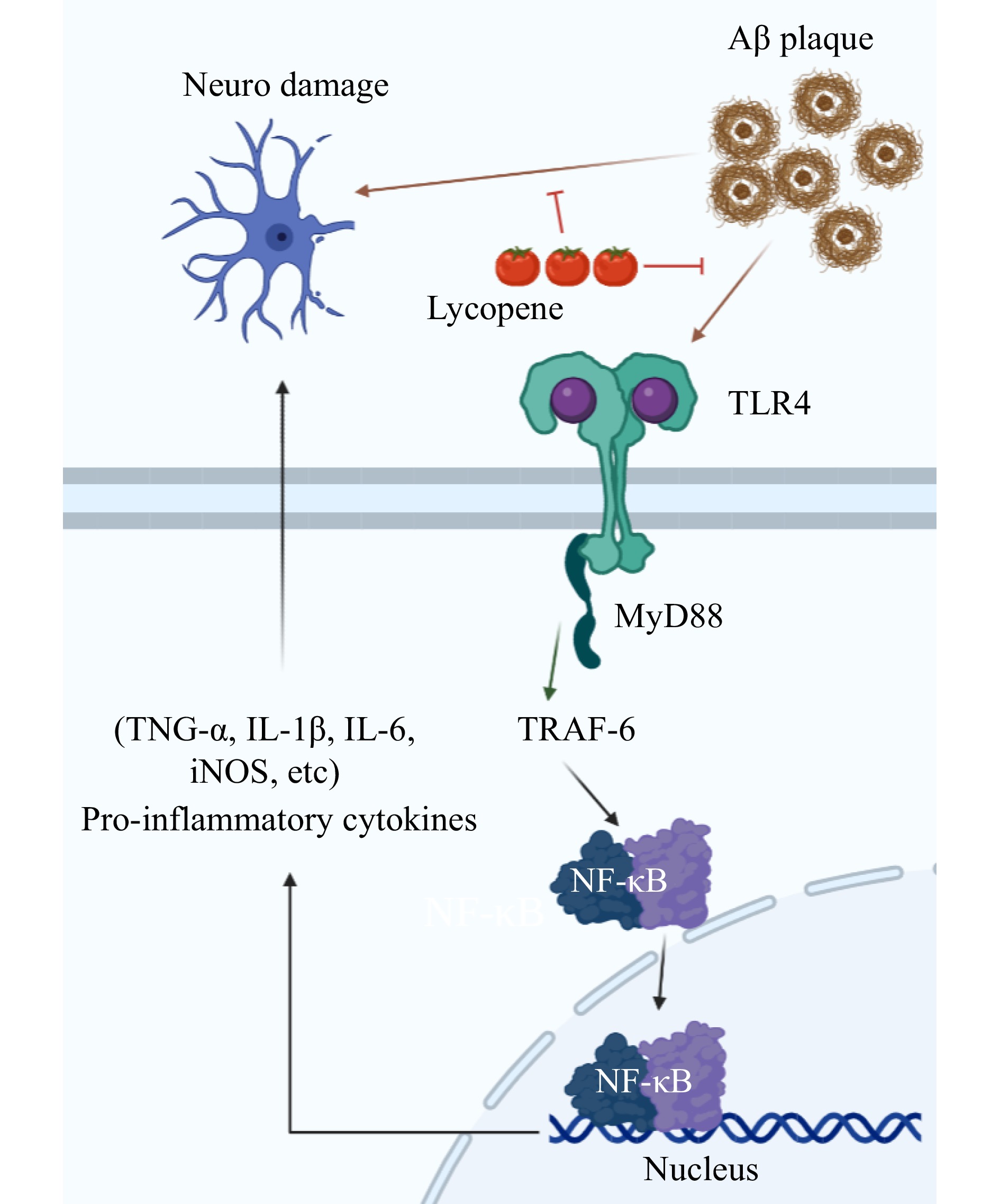
Figure 5.
Lycopene inhibits the down-regulation of TLR4 (Toll-like receptors 4) by Aβ, which further affects MyD88 (Myeloid differentiation primary response gene 88) and TRAF6, thereby activating the NF-κB pathway. On the other hand, lycopene directly inhibits Aβ induced neuronal damage, as shown by decreased levels of serum inflammatory cytokines and increased expression of the p65 subunit and TLR4. Drawing on
https://app.biorender.com/ .Gallic acid
-
Gallic acid (GA) (Table 1), also known as benzoic acid, is a dual α/β-secretase modulator[33]. Mori et al. have demonstrated that GA alleviates neuro-inflammation and stabilizes oxidative stress[6]. We can see that dietary GA supplementation can effectively alleviate oxidative stress induced AD, preserve the healthy state of the hippocampus to against environmental neurotoxins[34]. Furthermore, GA reacts with gallic catechins to get Epigallocatechin-3-Gallate (EGCG). Payne et al. have shown that EGCG treats AD by inhibiting neuroinflammation, aging, protein aggregation, and autophagy[35]. In addition, Araújo et al. have designed a new drug-carrying molecules, a dendritic macromolecule based on GA-terminated. The special structure further enhancing the ability of GA to destroy Aβ fibers to protect the nervous system in AD[36].
Berberine
-
Berberine (BBR) (Table 1), an alkaloid, has neuroprotective effects[37]. Zhang et al. have shown that BBR has a neuroprotective effect in AD[38]. Živančević et al. have confirmed that BBR antagonizes genes affected by mutual for AD and metal toxicity[39]. In addition, it has been shown that BBR improved cognitive deficits, inhibited neuronal apoptosis, and further promoted micro-vessel formation (Table 1)[7]. In vitro studies confirmed the efficacy of BBR against AD by showing reduced proinflammatory cytokine production[40]. Inevitably, the bioavailability of BBR is also not high. El-Enin et al. have designed a new material, BER-CTS-NLCs (BBR-laden nanostructured lipid carriers overlaid with chitosan). It effectively transmits to the brain via intranasal pathways[41]. In the same vein, lactoferrin-modified berberine nanoliposomes is also a breakthrough, it inhibits hippocampus apoptosis and enhances the neuro-protective effects of berberine nanoliposomes in AD[42].
Ginsenoside
-
Ginsenoside, a tetracyclic triterpenoid compound, have many different monomers, have been certified to relieve AD through antioxidant and anti-inflammatory effects[43]. Ginsenoside Rg1 (Rg1), one of the monomers, has shown neuroprotective effects in in vitro studies (Table 1)[44]. In addition, in vivo studies have confirmed the antioxidant and anti-inflammatory effects of Rg1 (Fig. 3 & Table 1)[45]. Rg1 has neuroprotective effects against AD. Pseudoginsenoside-F11 (PF11) and Ginsenoside Rh2 (Rh2) have the same efficacy (Table 1). In vivo studies have confirmed that PF-11 improves learning and memory deficits in AD (Table 1)[46]. Thus, the therapeutic potential of PF11 in managing AD is excellent. Rh2 has also shown excellent antioxidant action in vivo (Table 1)[47].
Others
-
Other dietary phytochemicals also have excellent therapeutic potential in managing AD, including artemisinin and torularhodin. In vitro studies have shown that artemisinin alleviated AD by its antioxidant action (Table 1)[48]. Moreover, in vivo studies have shown that torularhodin can effectively improve neuroinflammation and cognitive dysfunction by inhibiting oxidative stress, thereby preventing AD (Table 1)[49]. Sesamin protects the nervous system through antioxidant action, is a potential dietary phytochemical in treating AD[50]. Many dietary phytochemicals have been reported to have significant effects on the prevention and treatment of AD. However, additional research is needed to turn these natural compounds into novel drugs.
-
Research into dietary phytochemicals will eventually have to revert to clinical applications. Cur has been subject to numerous patents and clinical trials, but none of them have yielded conclusive results[51]. The instability and low bioavailability of Cur limit its clinical application. Nanocarriers have the potential to solve both challenges[52]. Res has two major disadvantages: low bioavailability and low solubility in vivo, which prevents patent treatment[23,53,54]. For Lyc and GA, research on AD is still in its infancy, thus, currently most clinical studies focus on other diseases, such as chronic periodontitis[55], prostate cancer[56], osteoporosis[57], Type 2 diabetes[58], and acne vulgaris[59]. Although clinical research on BBR for AD is also in its infancy, schizophrenia[60] and cardiovascular disease[61] are still closely related to AD. For ginsenoside, ginsenoside H dripping pill (GH), a novel Rh2 product, is in a phase 2 clinical study[62]. However, the trial is focused on its anticancer effect, not its anti-AD effect. For artemisinin, it is a miracle cure for malaria, and triple artemisinin-based combination therapies are enrolled in a new randomized clinical trial[63]. Similarly, artemisinin is known for its antimalarial effect, not its anti-AD effect. Furthermore, research for torularhodin is still experimental. Thus, the commercial potential of torularhodin is still unexplored[64].
-
AD is a common disease, and scientists all over the world are trying to find ways to prevent and treat AD. Dietary phytochemicals are safe and have low toxicity, which have been reported to exhibit preventive and therapeutic effects on AD, such as curcumin, resveratrol, lycopene, gallic acid, berberine, ginsenoside Rg1, pseudoginsenoside-F11, ginsenoside Rh2, artemisinin, and torularhodin. Many underlying mechanisms have been identified, including reducing Aβ deposition and inhibiting tau hyperphosphorylation to rescue synaptic dysfunction, thereby improving mitochondrial activity, anti-apoptosis, anti-oxidation and anti-inflammatory. In future studies, we should focus on evaluating the alleviating effects of natural compounds in human AD, and come up with better ways develop these natural compounds into new drugs faster, to treat the increasing number of AD patients. Therefore, to develop new drugs from dietary phytochemicals as quickly as possible, clinical trials are essential. Unignorably, safety is still of key importance, although natural compounds are usually safe, research must be performed to find the safest pharmaceutical and intake concentration. Finally, dietary phytochemicals can be developed into novel drugs for the prevention and treatment of AD, and into food health products.
This work was financially supported by the National Natural Science Foundation of China (21978236 and 21978229, Natural Science Basic Research Program of Shaanxi (2023-JC-JQ-17), and Qin Chuangyuan cited the High-level Innovation and Entrepreneurship Talent Program (QCYRCXM-2022-129).
-
The authors declare that they have no conflict of interest.
- Copyright: © 2023 by the author(s). Published by Maximum Academic Press on behalf of China Agricultural University, Zhejiang University and Shenyang Agricultural University. This article is an open access article distributed under Creative Commons Attribution License (CC BY 4.0), visit https://creativecommons.org/licenses/by/4.0/.
-
About this article
Cite this article
Ren Z, Yang H, Zhu C, Fan D, Deng J. 2023. Dietary phytochemicals: As a potential natural source for treatment of Alzheimer's Disease. Food Innovation and Advances 2(1):36−43 doi: 10.48130/FIA-2023-0007 

Dietary phytochemicals: As a potential natural source for treatment of Alzheimer's Disease
- Received: 10 January 2023
- Accepted: 08 February 2023
- Published online: 06 March 2023
Abstract: Alzheimer's disease (AD) is a common neurodegenerative disease, which seriously impairs human health and life. At present, scientists have proposed more than a dozen hypotheses about the pathogenesis of AD, including the tau propagation hypothesis. However, the exact ultimate pathogenic factor of AD remains unknown. Based on the current hypotheses, some anti-AD drugs (e.g., donepezil and Ketamine) have been developed and used in clinical treatment, which fall into two main categories, acetylcholinesterase inhibitors (AChEIs) and N-methyl-D-aspartate (NMDA) receptor antagonists, the former representative drug is donepezil, and the latter representative drug is memantine. Since these drugs have undesirable side effects, it is necessary to find safer alternatives for AD treatment. Interestingly, dietary phytochemicals have the advantages of wide source, safety, and high biological activity, which is the natural route for screening anti-AD drugs. In this study, several representatives’ dietary phytochemicals with anti-AD effect, including resveratrol, lycopene, gallic acid, berberine, ginsenoside Rg1, pseudoginsenoside-F11, ginsenoside Rh2, artemisinin, and torularhodin were selected from the published data over the last 10 years and their potential molecular mechanisms and clinical applications reviewed in the treatment of AD.
-
Key words:
- Alzheimer's disease /
- Dietary phytochemicals /
- Mechanism /
- Clinical application