ARTICLE   Open Access    

Comparative transcriptome-based identification and expression analysis of ATP-binding cassette (ABC) transporters reveals a likely role in the transport of β-caryophyllene and response to abiotic stress in Brassica campestris

More Information
  • β-caryophyllene has physiological activities such as antibacterial, antiedemic, anti-inflammatory, antitumor, and fungicidal. Previous research has focused on the synthesis of β-caryophyllene, however little information is available about its involvement in β-caryophyllene transport. ATP-binding cassette (ABC) transporters are involved in the transport of multiple substrates such as amino acids, terpenes, and heavy metals. Herein, we present a genome-wide comparative transcriptome analysis between non-β-caryophyllene cultivars ('SZQ') and high β-caryophyllene cultivars ('XQC') to identify potential ABC functional gene activities that occur during plant development. This article analyzes potential ABC transporters that may transport β-caryophyllene by comparative transcriptome and GC-MS, and explores the genetic structure, evolutionary relationship, cis-acting element analysis, and transcription patterns of these transporters under different treatment conditions. Phylogenetic and cis-acting element analysis indicated that eight genes in 'SZQ' and 'XQC' belonged to the ABCC, ABCG, and ABCE subfamilies and had the highest number of light-responsive elements and MYB binding sites. The RNA-seq and qRT-PCR results showed that eight ABC genes were expressed in a tissue- and development-specific manner, while their abundance apparently varied when plants were exposed to different treatment conditions. Pearson correlation analysis showed that ABCG18 is a potential carrier of β-caryophyllene, which might participate in substance transport in leaves and petioles. In summary, these observations open up new future research directions for β-caryophyllene transport and provide insight into ABC family genes in Brassica campestris.
  • Amaranth, a green leafy vegetable with high edible value, is rich in flavonoids, carotenoids, betalains, and other secondary metabolites[14]. Aromatic amino acids are precursors of secondary metabolites such as flavonoids, carotenoids, and alkaloids[5].

    Flavonoid compounds are important secondary metabolites produced by plants[6]. They are widely found in seeds, flowers, leaves, and fruits of plants, and most of them accumulate in the vacuoles of plant cells[7,8]. Flavonoids have a variety of biological functions. They can not only affect the transport of plant hormones[9,10], but also regulate the level of reactive oxygen species (ROS) in plants[1113]. Phenylalanine ammonia-lyase (PAL) is the first enzyme in the phenylpropane metabolic pathway and a key enzyme in the flavonoid metabolic pathway[14,15]. Chalcone synthase (CHS) is the first key enzyme in the flavonoid synthesis pathway, which leads the phenylpropane metabolic pathway to the flavonoid synthesis pathway[1618]. Chalcone isomerase (CHI) is an important rate-limiting enzyme in the synthesis pathway of flavonoid compounds, which catalyzes the chalcone cyclization[19,20]. Flavanone 3-carboxylase (F3H) catalyzes the synthesis of flavonols and substrates for anthocyanin synthesis[2123].

    Carotenoids are a group of yellow, red, or orange-red polyene substances. There are a wide variety of carotenoids, about 850 kinds[24], and they are widely found in the chloroplast and chromoplast membranes in plants. Carotenoids could participate in photosynthesis in plant cells and reduce photooxidation in precursor cells[24,25]. Carotenoids participated in the synthesis of plant hormones[26,27]. In the metabolic pathway of carotenoids, the transcription level of related enzymes affects the synthesis of carotenoids, geranylgeranyl diphosphate (GGPP) is a direct precursor in carotenoid biosynthesis pathway[28,29]. Two GGPPs are condensed into colorless phytoene under the catalysis of phytoene synthase (PSY)[30], which is the rate-limiting enzyme in the carotenoid synthesis pathway[3133]. Phytoene is converted to red phytoene by the co-catalysis of phytoene dehydrogenase (PDS) and carotene dehydrogenase (ZDS)[3436]. The content of phytoene was changed in maize by the PDS gene mutation[37], and the content of carotenoids was changed in Arabidopsis because of the ZDS gene mutation[38].

    Plant tissue culture is an important part of biotechnology and has become one of the most effective methods for the production of secondary metabolites[39,40]. During plant tissue culture, callus growth, and accumulation of secondary metabolites are affected by many factors, such as plant growth regulators, exogenous additives, light conditions, and so on[4143]. Amino acids, a sort of exogenous additive, are important factors affecting callus biomass, and different amino acids have different effects on the accumulation of secondary metabolites[44,45]. The addition of amino acids could increase the accumulation of alkaloids in the suspension callus of motherwort (Leonurus heterophyllus Sweet)[46]. Low concentrations of L-proline increased biomass and alkaloid accumulation in callus of Hydrocotyle bonariensis, while high concentrations inhibited it[47].

    Our previous study showed that flavonoids could be produced using amaranth callus[48]. However, the effects of aromatic amino acids on the callus growth, the accumulation of secondary metabolites, and the expression of related genes in amaranth are still unclear. Therefore, based on the previous research, an amaranth callus was used as the material to analyze the effects of aromatic amino acids on the growth of amaranth callus and the synthesis of secondary metabolites, suitable conditions were screened out for the production of flavonoids and carotenoids from amaranth callus, and the effect of exogenous amino acids on the gene expression of flavonoids and carotenoids were determined. The results provide a scientific basis and method for callus culture and flavonoid production by the callus of amaranth.

    The callus was induced by 'Suxian No.1' as material, which were provided by Suzhou Academy of Agricultural Sciences (Suzhou, China).

    Amaranth callus was inoculated into a basic medium (MS + 0.5 mg/L 2,4-D + 6.0 mg/L 6-BA + 30 g/L sucrose + 7 g/L agar)[48], supplemented with different concentrations of tyrosine (0, 2.0, 4.0, 6.0, and 8.0 mg/L), phenylalanine (0, 1.0, 2.0, 3.0, and 4.0 mg/L), and tryptophan (0, 0.5, 1.0, 1.5, and 2.0 mg/L). Each concentration was inoculated into 30 bottles, and three small pieces of amaranth callus (each piece was about 0.2 g fresh weight) were in each bottle. Three biological repeats were performed for each treatment. After 35 d of treatment, the growth of amaranth callus was observed, and the fresh weight and dry weight of callus were counted and sampled.

    Proliferation coefficient = Callus biomass after proliferation (g/bottle) / Initial callus biomass (g/bottle)

    The flavonoid content in Amaranthus tricolor was determined according to the flavonoid extraction and determination protocol (Comin Biotechnology Co., Ltd., Suzhou, China). 10 mL 60% (v/v) ethanol solution was added into a conical flask with 20 mg dried powder of amaranth callus. Flavonoid extraction was performed with shaking at 60 °C for 2 h, followed by centrifugation at 10,000 rpm at 25 °C for 10 min. The supernatant, containing flavonoid, was detected at a wavelength of 510 nm in a ultraviolet-visible spectrum spectrophotometer (UV-900, Shanghai Yuan Analysis Instrument Co., Ltd, Shanghai, China). For quantitation, rutin was used as an internal standard for calibration.

    Standard curve: y = 5.02x + 0.0007,R2 = 0.9996

    Total flavonoid content (mg·g−1 DW) = (△A−0.0007) / 5.02 / (w/v)

    Carotenoid content was determined by ultraviolet-visible spectrum spectrophotometer. 2 ml of extraction solution of acetone : petroleum ether (1:1, v/v) was added into a tube with 20 mg dried powder of amaranth callus. And then the mixture was placed on a 200 rpm shaker to extract carotenoid for 8 h under dark conditions. Subsequently, the supernatant was collected by centrifuging at 10,000 rpm for 5 min at room temperature. Finally, the absorption peak of carotenoid at 445 nm was determined by a ultraviolet-visible spectrum spectrophotometer (UV-900, Shanghai Yuan Analysis Instrument Co., Ltd., Shanghai, China).

    Total RNA was extracted was from all samples using a MolPure Plant Plus RNA Kit (Yeasen, China) according to the manufacturer's instructions. First-strand cDNA was then synthesized from 1 mg of total RNA using Recombinant M-MLV reverse transcriptase (TransGen Biotech, Beijing, China). Quantitative real time-PCR (qRT-PCR) was performed in optical 96-well plates using the Roche LightCycler 480II instrument (Roche, Sweden). The reactions were carried out in a 20 μL volume containing 10 μL of Hieff qPCR SYBR Green PCR Master Mix (Yeasen Biotechnology, China), 0.5 μL of gene-specific primers, 2 μL of diluted cDNA, and 7.0 μL of ddH2O. The PCR conditions were as follows: 30 s at 95 °C, 40 cycles of 10 s at 95 °C and 12 s at 59 °C, followed by 12 s at 72 °C. Three biological repeats were performed for each material. EF1α[49] was used as the reference gene. The 2ΔΔCᴛ method was used for quantitative analyses of gene expression. The primers used for qRT-PCR are listed in Supplemental Table S1.

    Data are presented as mean ± standard error and were submitted to analysis of variance (ANOVA). Values of p < 0.05 were significant in comparisons between the treatments and controls. All statistical analyses were performed using SPSS 26 (IBM Corp., Armonk, NY, USA). GraphPad Prism 8.1 (GraphPad Software Inc., La Jolla, CA, USA) was used for the bar chart drawing.

    After treatment with different concentrations of aromatic amino acids for 35 d, the amaranth callus growth was normal without browning (Fig. 1). On the medium with tyrosine, the callus color was green and yellow, but on the medium with phenylalanine, the callus color was white. The best medium was with tryptophan, where the callus color was yellow.

    Figure 1.  Effect of amaranth callus growth treated with aromatic amino acids. A1-A5 represents the amaranth callus with 0, 2, 4, 6, 8 mg/L tyrosine for 35 d; B1-B5 represents the amaranth callus with 0, 1, 2, 3, 4 mg/L phenylalanine for 35 d; C1-C5 represents the amaranth callus with 0, 0.5, 1.0, 1.5, 2.0 mg/L tryptophan for 35 d.

    Phenylalanine, tyrosine, and tryptophan all had significant promoting effects on the proliferation and dry matter accumulation of amaranth callus, besides 2.0 mg/L tryptophan (Fig. 2). With the increase in tyrosine concentration, the proliferation coefficient of amaranth callus increased first and then decreased (Fig. 2a). When the tyrosine concentration was 4.0 mg/L, the proliferation coefficient and dry weight of callus reached the maximum, reaching 15.6 and 0.43, respectively, which were significantly higher than those of other concentrations and control groups.

    Figure 2.  The effect of different concentrations of aromatic amino acids on amaranth callus proliferation and dry weight. Treatment of (a) tyrosine, (b) phenylalanine, and (c) tryptophan.

    With the increase in phenylalanine concentration, the callus proliferation coefficient and dry weight decreased first and then increased (Fig. 2b). When the concentration of phenylalanine was 2.0 mg/L, the callus proliferation coefficient and dry weight decreased to 9.7 and 0.29, respectively, and the callus proliferation coefficient was significantly lower than that of the control group, but the dry weight was not significant. When the concentration of phenylalanine was 3.0 mg/L, the callus proliferation coefficient and dry weight reached 14.82 and 0.4, respectively, which were significantly higher than those of the control group.

    With the increase of tryptophan concentration, the proliferation coefficient of amaranth callus showed a trend of first increasing and then decreasing (Fig. 2c). The proliferation coefficient increased to the highest at the concentration of 1 mg/L of tryptophan, reaching 17.39, which was significantly different from the control group. The dry weight showed an upward trend, and the highest at the concentration of 2 mg/L of tryptophan, reaching 0.4, which was significantly different from the control group.

    The effect of different concentrations of aromatic amino acids on the flavonoid content in amaranth callus is shown in Fig. 3. With the increase in tyrosine concentration, the content of flavonoids in the callus reached the highest level (4.77 mg/g) when the tyrosine concentration was 2.0 mg/L, which was significantly different from that of the control (Fig. 3a).

    Figure 3.  Effects of different concentrations of aromatic amino acids on flavonoid content in amaranth callus. (a) Tyrosine, (b) phenylalanine and, (c) tryptophan.

    With the increase in phenylalanine concentration, the content of flavonoids in the callus showed an upward trend (Fig. 3b). When the concentration of phenylalanine was 2.0 mg/L, the content of flavonoids decreased to 2.63 mg/g, which was lower than that of the control group (2.94 mg/g). When the concentration of phenylalanine was 1.0 mg/L, the content of flavonoids in the callus were the highest. It reached 3.42 mg/g, which was significantly different from the control group.

    With the increase in tryptophan concentration, the content of flavonoids in the callus decreased gradually, and the lowest was 1.18 mg/g when the tyrosine concentration was 1.5 mg/L, which was significantly different from the control group (Fig. 3c).

    The effect of different concentrations of aromatic amino acids on carotenoid content in amaranth callus is shown in Fig. 4. With the increase in tyrosine concentration, the content of carotenoids in the callus decreased, and the minimum was 99.2 μg/g when the tyrosine concentration was 4.0 mg/L (Fig. 4a). With the increase of phenylalanine concentration, the content of carotenoids in callus decreased. When the concentration of phenylalanine was 2.0 mg/L, the content of carotenoids decreased to the lowest, reaching 64.6 μg/g (Fig. 4b).

    Figure 4.  Effects of different concentrations of aromatic amino acids on carotenoid content in amaranth callus.

    With the increase in tryptophan concentration, the content of flavonoids in the callus decreased first and then increased. When the concentration of tryptophan was 1 mg/L, the content of flavonoids in callus was the lowest (63.6 μg/g), and when the concentration of tryptophan was 2.0 mg/L, the content of carotenoids was the highest (140.6 μg/g), which was lower than that of the control group (166 μg/g) (Fig. 4c).

    The relative expression levels of flavonoid synthesis-related genes in amaranth callus treated with different concentrations of phenylalanine are shown in Fig. 5. The results showed that the gene expression of PAL, F3H, and CHS increased to the highest at the level of 1.0 mg/L phenylalanine, which was significantly different from the control.

    Figure 5.  The effect of aromatic amino acids on the expression of flavonoids metabolism related genes in amaranth callus. (a) Tyrosine, (b) phenylalanine, and (c) tryptophan.

    The relative expression levels of genes involved in flavonoid synthesis in amaranth callus treated with different concentrations of tyrosine are shown in Fig. 5. The addition of tyrosine promoted PAL gene expression, reaching a significant difference compared with the control. However, there was no significant difference between different concentrations of tyrosine. The relative expression of the F3H and CHS genes were the highest when the concentration of tyrosine was 6.0 and 2.0 mg/L, respectively.

    The relative expression levels of genes involved in flavonoid synthesis in amaranth callus treated with different concentrations of tryptophan are shown in Fig. 5. The relative expression of PAL, F3H, and CHS genes were the highest when the concentration of tyrosine was 1.5, 0.5, and 2.0 mg/L, respectively. And they all reached a significant difference compared with the control.

    SPSS 26 software was used to analyze the correlation between flavonoid content and flavonoid biosynthesis-related genes in amaranth callus treated with three aromatic amino acids (Table 1). The results show that the content of flavonoids was positively correlated with PAL under tyrosine treatment, but not under phenylalanine and tryptophan treatments. There was a significant positive correlation between F3H and flavonoid content under phenylalanine and tryptophan treatments, besides tryptophan treatment. There was a significant positive correlation between flavonoid content and CHS only under tyrosine treatment.

    Table 1.  Correlation analysis of flavonoid content and flavonoid-related genes.
    Aromatic
    amino acids
    PAL F3H CHS
    Tyrosine Pearson correlation 0.615* −0.659** 0.694**
    Significance (two-tailed) 0.015 0.007 0.004
    Phenylalanine Pearson correlation −0.183 0.669** 0.258
    Significance (two-tailed) 0.513 0.006 0.353
    Tryptophan Pearson correlation −0.858 0.753 0.076
    Significance (two-tailed) 0.063 0.142 0.90
    * indicates significant correlation at the p < 0.05 level, ** indicates extremely significant correlation at the p < 0.01 level.
     | Show Table
    DownLoad: CSV

    Tyrosine had different effects on the expression of carotenoid synthesis genes in amaranth callus (Fig. 6a). Low concentrations of tyrosine (0−4 mg/L) could promote gene expression, whereas high concentrations inhibit PSY gene expression. When the tyrosine concentration was 4.0 mg/L, the relative expression reached the highest level. Tyrosine had no effect on the PDS gene expression However, it could inhibit the expression of the ZDS gene.

    Figure 6.  The effect of aromatic amino acids on the expression of carotenoid metabolism related genes in amaranth callus. (a) Tyrosine, (b) phenylalanine, and (c) tryptophan.

    The relative expression levels of carotenoid synthesis-related genes in amaranth callus treated with different concentrations of phenylalanine are shown in Fig. 6b. Phenylalanine could inhibit the expression of PDS and PSY, and there was no significant difference in the expression of the ZDS gene between the phenylalanine treatment and control. Under tryptophan treatment, the carotenoid synthesis gene (PDS, PSY, and ZDS) expression was inhibited in amaranth callus. There were significant differences from the control (Fig. 6c).

    SPSS 26 software was used to analyze the correlation between carotenoid content and carotenoid biosynthesis related genes in amaranth callus treated with three aromatic amino acids (Table 2). The results show that the content of carotenoid was positively correlated with PDS under phenylalanine treatment, but not under tyrosine and tryptophan treatments. There was a significant positive correlation between PSY and carotenoid content under tryptophan treatments. There was a significant positive correlation between flavonoid content and ZDS under tyrosine and tryptophan treatment.

    Table 2.  Correlation analysis of carotenoids content and carotenoids-related genes.
    Aromatic
    amino acids
    PDS ZDS PSY
    Tyrosine Pearson correlation −0.348 −0.356 0.638*
    Significance (two-tailed) 0.204 0.192 0.011
    Phenylalanine Pearson correlation 0.694** −0.025 0.442
    Significance (two-tailed) 0.004 0.93 0.099
    Tryptophan Pearson correlation 0.342 0.591* 0.57*
    Significance (two-tailed) 0.212 0.02 0.026
    * indicates p < 0.05, ** indicates p < 0.01.
     | Show Table
    DownLoad: CSV

    Amino acids as a source of organic nitrogen can promote plant growth and development, and play key roles in plant cells and tissues[45,50], and are important regulators in vitro[51]. Exogenous supplementation of amino acids and casein hydrolysate could enhance callogenesis from plumular explants of coconut (Cocos nucifera L.)[52]. The present results showed that phenylalanine, tyrosine, and tryptophan all had significant promoting effects on the proliferation and dry matter accumulation of amaranth callus. When the concentration of tryptophan was 1 and 2.0 mg/L, the effect of callus proliferation and dry matter accumulation was the best, and the effect was better than that of phenylalanine and tyrosine. It was hypothesized that tryptophan is an important precursor substance for auxin biosynthesis in plants and its structure is similar to IAA, and auxin is beneficial for callus induction and proliferation. So the addition of tryptophan 1.0–2.0 mg/L in the culture medium was most conducive to callus proliferation and dry matter accumulation.

    Aromatic amino acids are precursors of secondary metabolites such as flavonoids, carotenoids, and alkaloids[5]. Flavonoid compounds are important secondary metabolites produced by plants[6]. Flavonoids have a variety of biological functions. They can not only affect the transport of plant hormones[9,10], but also regulate the level of reactive oxygen species (ROS) in plants[1113]. Phenylalanine ammonia-lyase (PAL) is the first enzyme in the phenylpropane metabolic pathway and a key enzyme in the flavonoid metabolic pathway[14,15]. PAL and C4H can convert phenylalanine to p-coumaric acid. Meanwhile, tyrosine can be directly converted to p-coumaric acid[53]. Subsequently, p-coumaric acid is converted to flavonoids under the action of a series of enzymes, including 4CL, CHS[19,20], CHI, and F3H[2123]. The present results show that tyrosine was beneficial to increase the content of flavonoids in amaranth callus, and it was superior to phenylalanine. The two precursor amino acids are converted to flavonoids by different pathways. Through correlation analysis, there was a significant positive correlation between the PAL gene, CHS gene, F3H gene, and flavonoid content under tyrosine treatment, indicating that tyrosine affected the synthesis of flavonoids by regulating the expression of the three key genes. Under phenylalanine treatment, there was a significant positive correlation between F3H and flavonoid content, indicating that phenylalanine regulates the expression of F3H.

    Carotenoids are a sort of yellow, red, or orange-red polyene substance[24], which could participate in photosynthesis[24,25] and the synthesis of plant hormones[26,27]. Two geranylgeranyl diphosphates (GGPPs) are condensed into colorless phytoene under the catalysis of phytoene synthase (PSY)[30], which is the rate-limiting enzyme in the carotenoid synthesis pathway[3133]. Phytoene is converted to red phytoene by the co-catalysis of phytoene dehydrogenase (PDS) and carotene dehydrogenase (ZDS)[3436]. The present results showed that aromatic amino acids inhibited the carotenoids biosynthesis. Perhaps the degradation and transport of carotenoids affected their concentrations. However, the mechanism is not still clear.

    Aromatic amino acids, especially tyrosine, could promote the growth of amaranth callus and flavonoid synthesis, and regulate related gene expression. In contrast, aromatic amino acids inhibited carotenoids synthesis in amaranth callus.

    The authors confirm contribution to the paper as follows: writing – original draft, writing – review & editing: Liu S, Xuan Y; validation: Xuan Y; conceptualization: Liu S, Lai Z; methodology: Xuan Y, Feng W. All authors reviewed the results and approved the final version of the manuscript.

    All data generated or analyzed during this study are included in this published article and its supplementary information files.

    This work was supported by the Natural Science Foundation of Fujian Province (2023J01449), Innovation Foundation of Fujian Agriculture and Forestry University (KFb22024XA), Rural Revitalization Social Service Team of Fujian Agriculture and Forestry University (11899170125).

  • The authors declare that the research was conducted in the absence of any commercial or financial relationships that could be construed as a potential conflict of interest.

  • Supplemental Fig. S1 Volcano plot of DEGs (differentially expressed genes) between ‘XQC’ and ‘SZQ’ at 30, 60, 90, and 120 days.
    Supplemental Fig. S2 The number of DEGs in different comparison groups. Oranges indicate upregulation, and blue indicates downregulation.
    Supplemental Fig. S3 Distribution of ABC genes on Brassica campestris chromosomes.
    Supplemental Table S1 RNA-seq data used in this study.
    Supplemental Table S2 425 transport proteins ID.
    Supplemental Table S3 169 ABC protein sequence in Brassica campestris.
    Supplemental Table S4 ABC proteins of Arabidopsis.
    Supplemental Table S5 Basic information on ABC genes identified in Brassica campestris.
  • [1]

    Frede K, Schreiner M, Baldermann S. 2019. Light quality-induced changes of carotenoid composition in pak choi Brassica rapa ssp. chinensis. Journal of Photochemistry and Photobiology B: Biology 193:18−30

    doi: 10.1016/j.jphotobiol.2019.02.001

    CrossRef   Google Scholar

    [2]

    Khan WA, Hu H, Ann Cuin T, Hao Y, Ji X, et al. 2022. Untargeted metabolomics and comparative flavonoid analysis reveal the nutritional aspects of pak choi. Food Chemistry 383:132375

    doi: 10.1016/j.foodchem.2022.132375

    CrossRef   Google Scholar

    [3]

    Verrier PJ, Bird D, Burla B, Dassa E, Forestier C, et al. 2008. Plant ABC proteins – a unified nomenclature and updated inventory. Trends in Plant Science 13:151−59

    doi: 10.1016/j.tplants.2008.02.001

    CrossRef   Google Scholar

    [4]

    Anfang M, Shani E. 2021. Transport mechanisms of plant hormones. Current Opinion in Plant Biology 63:102055

    doi: 10.1016/j.pbi.2021.102055

    CrossRef   Google Scholar

    [5]

    Yang Z, Yang F, Liu J, Wu H, Yang H, et al. 2022. Heavy metal transporters: functional mechanisms, regulation, and application in phytoremediation. Science of the Total Environment 809:151099

    doi: 10.1016/j.scitotenv.2021.151099

    CrossRef   Google Scholar

    [6]

    Yazaki K. 2006. ABC transporters involved in the transport of plant secondary metabolites. FEBS Letters 580:1183−91

    doi: 10.1016/j.febslet.2005.12.009

    CrossRef   Google Scholar

    [7]

    Kang J, Hwang JU, Lee M, Kim YY, Assmann SM, et al. 2010. PDR-type ABC transporter mediates cellular uptake of the phytohormone abscisic acid. Proceedings of the National Academy of Sciences of the United States of America 107:2355−60

    doi: 10.1073/pnas.0909222107

    CrossRef   Google Scholar

    [8]

    Yazaki K, Shitan N, Takamatsu H, Ueda K, Sato F. 2001. A novel Coptisjaponica multidrug-resistant protein preferentially expressed in the alkaloid-accumulating rhizome. Journal of Experimental Botany 52:877−79

    doi: 10.1093/jexbot/52.357.877

    CrossRef   Google Scholar

    [9]

    Goodman CD, Casati P, Walbot V. 2004. A multidrug resistance-associated protein involved in anthocyanin transport in Zea mays. The Plant Cell 16:1812−26

    doi: 10.1105/tpc.022574

    CrossRef   Google Scholar

    [10]

    Fu X, Shi P, He Q, Shen Q, Tang Y, et al. 2017. AaPDR3, a PDR transporter 3, is involved in sesquiterpene β-caryophyllene transport in Artemisia annua. Frontiers in Plant Science 8:723

    doi: 10.3389/fpls.2017.00723

    CrossRef   Google Scholar

    [11]

    Jasinski M, Ducos E, Martinoia E, Boutry M. 2003. The ATP-binding cassette transporters: structure, function, and gene family comparison between rice and Arabidopsis. Plant Physiology 131:1169−77

    doi: 10.1104/pp.102.014720

    CrossRef   Google Scholar

    [12]

    Garcia O, Bouige P, Forestier C, Dassa E. 2004. Inventory and comparative analysis of rice and Arabidopsis ATP-binding cassette (ABC) systems. Journal of Molecular Biology 343:249−65

    doi: 10.1016/j.jmb.2004.07.093

    CrossRef   Google Scholar

    [13]

    Pang K, Li Y, Liu M, Meng Z, Yu Y. 2013. Inventory and general analysis of the ATP-binding cassette (ABC) gene superfamily in maize (Zea mays L). Gene 526:411−28

    doi: 10.1016/j.gene.2013.05.051

    CrossRef   Google Scholar

    [14]

    Rea PA. 2007. Plant ATP-binding cassette transporters. Annual Review of Plant Biology 58:347−75

    doi: 10.1146/annurev.arplant.57.032905.105406

    CrossRef   Google Scholar

    [15]

    Wang H, Zong C, Bai A, Yuan S, Li Y, et al. 2022. Transcriptome sequencing and gas chromatography–mass spectrometry analyses provide insights into β-caryophyllene biosynthesis in Brassica campestris. Food Chemistry: Molecular Sciences 5:100129

    doi: 10.1016/j.fochms.2022.100129

    CrossRef   Google Scholar

    [16]

    Sánchez-Fernández R, Davies TG, Coleman JO, Rea PA. 2001. The Arabidopsis thaliana ABC protein superfamily, a complete inventory. Journal of Biological Chemistry 276:30231−44

    doi: 10.1074/jbc.M103104200

    CrossRef   Google Scholar

    [17]

    Li Y, Liu G, Ma L, Liu T, Zhang C, et al. 2020. A chromosome-level reference genome of non-heading Chinese cabbage [Brassica campestris (syn Brassica rapa) ssp. chinensis]. Horticulture Research 7:212

    doi: 10.1038/s41438-020-00449-z

    CrossRef   Google Scholar

    [18]

    Ofori PA, Mizuno A, Suzuki M, Martinoia E, Reuscher S, et al. 2018. Genome-wide analysis of ATP binding cassette (ABC) transporters in tomato. PLoS One 13:e0200854

    doi: 10.1371/journal.pone.0200854

    CrossRef   Google Scholar

    [19]

    Yan C, Duan W, Lyu S, Li Y, Hou X. 2017. Genome-wide identification, evolution, and expression analysis of the ATP-binding cassette transporter gene family in Brassica rapa. Frontiers in Plant Science 8:349

    doi: 10.3389/fpls.2017.00349

    CrossRef   Google Scholar

    [20]

    Yamagishi M. 2022. High temperature enhances anthocyanin coloration in Asiatic hybrid lily flowers via upregulation of the MYB12 positive regulator. Horticultural Plant Journal 8:769−76

    doi: 10.1016/j.hpj.2022.05.003

    CrossRef   Google Scholar

    [21]

    Bhatia C, Gaddam SR, Pandey A, Trivedi PK. 2021. COP1 mediates light-dependent regulation of flavonol biosynthesis through HY5 in Arabidopsis. Plant Science 303:110760

    doi: 10.1016/j.plantsci.2020.110760

    CrossRef   Google Scholar

    [22]

    Zhang Y, Kilambi HV, Liu J, Bar H, Lazary S, et al. 2021. ABA homeostasis and long-distance translocation are redundantly regulated by ABCG ABA importers. Science Advances 7:eabf6069

    doi: 10.1126/sciadv.abf6069

    CrossRef   Google Scholar

    [23]

    Hwang JU, Song WY, Hong D, Ko D, Yamaoka Y, et al. 2016. Plant ABC transporters enable many unique aspects of a terrestrial plant's lifestyle. Molecular Plant 9:338−55

    doi: 10.1016/j.molp.2016.02.003

    CrossRef   Google Scholar

    [24]

    Kuromori T, Miyaji T, Yabuuchi H, Shimizu H, Sugimoto E, et al. 2010. ABC transporter AtABCG25 is involved in abscisic acid transport and responses. Proceedings of the National Academy of Sciences of the United States of America 107:2361−66

    doi: 10.1073/pnas.0912516107

    CrossRef   Google Scholar

    [25]

    Li Q, Zheng J, Li S, Huang G, Skilling SJ, et al. 2017. Transporter-mediated nuclear entry of jasmonoyl-isoleucine is essential for jasmonate signaling. Molecular Plant 10:695−708

    doi: 10.1016/j.molp.2017.01.010

    CrossRef   Google Scholar

    [26]

    Zhang Z, Tong T, Fang Y, Zheng J, Zhang X, et al. 2020. Genome-wide identification of barley ABC genes and their expression in response to abiotic stress treatment. Plants 9:1281

    doi: 10.3390/plants9101281

    CrossRef   Google Scholar

    [27]

    Thomine S, Wang R, Ward JM, Crawford NM, Schroeder JI. 2000. Cadmium and iron transport by members of a plant metal transporter family in Arabidopsis with homology to Nramp genes. Proceedings of the National Academy of Sciences of the United States of America 97:4991−96

    doi: 10.1073/pnas.97.9.4991

    CrossRef   Google Scholar

  • Cite this article

    Wang H, Zong C, Bai Y, Yuan S, Li Y, et al. 2023. Comparative transcriptome-based identification and expression analysis of ATP-binding cassette (ABC) transporters reveals a likely role in the transport of β-caryophyllene and response to abiotic stress in Brassica campestris. Vegetable Research 3:13 doi: 10.48130/VR-2023-0013
    Wang H, Zong C, Bai Y, Yuan S, Li Y, et al. 2023. Comparative transcriptome-based identification and expression analysis of ATP-binding cassette (ABC) transporters reveals a likely role in the transport of β-caryophyllene and response to abiotic stress in Brassica campestris. Vegetable Research 3:13 doi: 10.48130/VR-2023-0013

Figures(5)

Article Metrics

Article views(4721) PDF downloads(673)

ARTICLE   Open Access    

Comparative transcriptome-based identification and expression analysis of ATP-binding cassette (ABC) transporters reveals a likely role in the transport of β-caryophyllene and response to abiotic stress in Brassica campestris

Vegetable Research  3 Article number: 13  (2023)  |  Cite this article

Abstract: β-caryophyllene has physiological activities such as antibacterial, antiedemic, anti-inflammatory, antitumor, and fungicidal. Previous research has focused on the synthesis of β-caryophyllene, however little information is available about its involvement in β-caryophyllene transport. ATP-binding cassette (ABC) transporters are involved in the transport of multiple substrates such as amino acids, terpenes, and heavy metals. Herein, we present a genome-wide comparative transcriptome analysis between non-β-caryophyllene cultivars ('SZQ') and high β-caryophyllene cultivars ('XQC') to identify potential ABC functional gene activities that occur during plant development. This article analyzes potential ABC transporters that may transport β-caryophyllene by comparative transcriptome and GC-MS, and explores the genetic structure, evolutionary relationship, cis-acting element analysis, and transcription patterns of these transporters under different treatment conditions. Phylogenetic and cis-acting element analysis indicated that eight genes in 'SZQ' and 'XQC' belonged to the ABCC, ABCG, and ABCE subfamilies and had the highest number of light-responsive elements and MYB binding sites. The RNA-seq and qRT-PCR results showed that eight ABC genes were expressed in a tissue- and development-specific manner, while their abundance apparently varied when plants were exposed to different treatment conditions. Pearson correlation analysis showed that ABCG18 is a potential carrier of β-caryophyllene, which might participate in substance transport in leaves and petioles. In summary, these observations open up new future research directions for β-caryophyllene transport and provide insight into ABC family genes in Brassica campestris.

    • The Brassica campestris (syn. Brassica rapa) ssp. chinensis is an economically important vegetable crop cultivated and researched worldwide. It is considered a rich source of multiple nutrients, such as vitamins, thioglycosides, carotenoids, and flavonoids[1,2]. However, little information is available about terpenes in Brassica campestris, especially terpene transport.

      The genes encoding ABC proteins constitute a large gene family in plants that are categorized phylogenetically into eight subfamilies[3], and ABC transporters manage the active transport of a wide range of molecules across biological membranes, including heavy metals, secondary metabolites, and plant hormones[46]. The ABCG subfamily has a reverse domain including nucleotide-binding domains and trans-membrane domain (NBD-TMD) organization and plays a role in cellular efflux. In Arabidopsis, AtABCG25 and AtABCG40 function as plasma membrane abscisic acid (ABA) uptake transporters and play a role in the response to ABA[7]. CjABCB1/CjMDR1 is localized in the plasma membrane and catalyzes the import of berberine in Coptis japonica[8]. The ABCC transporters ZmMRP3 and ZmMRP4 are required for anthocyanin accumulation in maize kernels[9]. NtPDR1 and AaPDR3 participate in the transport of diterpenoids of Nicotiana tabacum and β-caryophyllene of Artemisia annua, respectively[10]. To date, there have been few studies on the transport of terpenoids in Brassica campestris.

      Complete genome sequences of several organisms have revealed that genes encoding ABC transporters comprise a large gene family in plants; there are more than 131 members in Arabidopsis thaliana[11], 121 members in Oryza sativa[12] and 130 members in Zea mays[13]. The basic ABC transporter consists of four core structural domains, two transmembrane domains (TMDs) and two nucleotide-binding domains (NBDs), which provide a translocation pathway and hydrolyze ATP to provide the energy needed for ABC proteins, respectively[14].

      In our previous study, it was found that β-caryophyllene content varies greatly among different varieties of Brassica campestris[15], but little is known about β-caryophyllene transport. In this study, we first performed genome-wide identification of ABC family members in Brassica campestris. Then, eight differentially expressed genes were screened using RNA-seq data, and their phylogenetic relationships, conserved motifs, and gene structure characteristics were characterized. Finally, the spatiotemporal expression patterns of eight ABC genes were explored in pak-choi. Overall, this work provides new insights into the transport of β-caryophyllene and its response to abiotic stress in Brassica campestris.

    • Brassica campestris, Suzhouqing and Xiangqingcai ('XQC' and 'SZQ'), used for transcriptome sequencing and Gas Chromatography-Mass Spectrometry (GC-MS) were harvested at 30, 60, 90, and 120 d after germination in a greenhouse located in Wu Jiang City, Jiang Su, China. At 60 d, 'XQC' samples were treated with 100 mmol·L−1 NaCl, 100 µmol·L−1 ABA, 100 µmol·L−1 MeJA, 4 °C/25 °C (low temperature, LT), and 35 °C/25 °C (high temperature, HT) in a growth chamber with light for 16 h per day and the relative humidity was 70%. Samples were collected from three seedlings and pooled together as one biological repeat at 0, 4, 8, 12, 24, and 48 h after three abiotic stresses and two hormone treatments. The collected leaves were immediately frozen in liquid nitrogen and stored at −80 °C for subsequent total RNA extraction and expression profiling. Three biological repeats were performed for each treatment at each time point.

    • Information about ABC proteins in Arabidopsis thaliana was obtained as previously described[16] and used as a probe to execute an HMMER (http://hmmer.org/) search of putative ABC proteins in the non-heading Chinese cabbage 001 (NHCC001) (Brassica campestris) reference genome[17]. After obtaining the full-length protein sequences, the putative Brassica campestris ABCs were verified with an additional online domain analysis tool, the Conserved Domain Database (CDD) (www.ncbi.nlm.nih.gov/Structure/bwrpsb/bwrpsb.cgi).

    • Using the integrated tool MEGA7, protein sequence alignment for ABC members and AtABCs was carried out with the MUSCLE program, and a phylogenetic tree was then built with the maximum likelihood algorithm, wherein the bootstrap replicate value was set as 1000. Thereafter, the physicochemical properties of the ABC genes were analyzed with the online tool ExPASy (http://www.expasy.org).

    • The genome of 'NHCC001' was used as a reference genome, and the exon‒intron distribution of ABC genes was visualized with Tbtools. Cis-regulatory elements (CREs) in ABC promoters, which were defined as 2.0-kb genomic regions upstream of translational start codons, were detected with an online tool, PlantCARE (http://bioinformatics.psb.ugent.be/webtools/plantcare/html/). The CREs were identified by PlantCARE and visualized with Tbtools. Motif analysis for ABC proteins was carried out using the Multiple EM Motif Elicitation (MEME) program (http://meme-Suite.org/), of which the maximum motif number was set as 10 under default mode.

    • To further explore the content of β-caryophyllene in Brassica campestris, 2 g of fresh leaves was ground to a powder in liquid nitrogen. All samples were detected with a gas chromatograph (TRACE 1310, Thermo Scientific, America) coupled to a triple quadrupole mass spectrometer at 250 °C for 3 min in split mode (flow rate 1 mL/min). Three replicates of each treatment were performed. Subsequently, 2 g of the powder was transferred to a 20 mL headspace vial (Agilent, CA, USA) containing 2 mL of NaCl-saturated solution to inhibit the enzyme reaction. An 85-µm CAR/PDMS fiber was exposed to the headspace of the sample for 40 min at 35 °C for solid-phase microextraction (SPME) analysis[15].

    • Total RNA was extracted from Brassica campestris using a plant RNA isolation kit (Mage, Beijing, China) according to the manufacturer's instructions for RNA library preparation and sequencing. After verification of the total RNA samples, the library was constructed with the MGIEasy RNA library. The library was constructed with three biological replicate samples at each time point. Raw reads were obtained from each sample after sequencing. After removing primer sequences, adaptor sequences, and low-quality reads, clean reads consisted of more than 80% base pairs with a Q-value ≥ 30. High-quality reads were compared with the spliced transcriptome. The transcripts per million (TPM) value was calculated for each transcription region to quantify expression abundance and variations, and differentially expressed genes (DEGs) were identified using DESeq2. DEG screening criteria were |log2FC| ≥ 1 and p < 0.05.

    • Total RNA was extracted using a HiPure Plant RNA Mini Kit (R4151-02, MEGA) for qRT‒PCR. Then, the RNA was reverse transcribed to first-strand cDNA using the Reverse Transcription Kit (PrimeScriptTM RT Master Mix, Takara). The relative expression level at different treatments was calculated according to the 2−ΔΔCᴛ method. The tubulin gene was used as the reference gene. The complete CDs sequence of ABCG18 without a stop codon was subcloned into PJX003 to generate 35S::ABCG18 effector constructs. Agrobacterium solution containing PJX003-BcABCG18-GFP was injected into tobacco leaves. Green fluorescent protein (GFP) fluorescence (LSM780, Zeiss) was observed at 3 d after injection.

    • All data were analyzed using SPSS statistical software, and performed with three biological replicates as the average of the RNA-seq results or the average of the three biological repeats ± standard deviation (SD) for the qRT‒PCR. The differences between Bssraica campestris samples were evaluated by one-way ANOVA at a significance level of 0.05.

    • The morphology differed between Brassica campestris cultivars (XQC and SZQ) in shape, texture, and size of leaves at 30 d (Fig. 1a). The 'XQC' and 'SZQ' materials at 30, 60, 90, and 120 d were used to measure β-caryophyllene. The results showed that β-caryophyllene could be detected only in 'XQC', and the content of β-caryophyllene was highest at 60 d (Fig. 1b & c). The GC‒MS peak indicated a difference in the β-caryophyllene content in four stages (Fig. 1b).
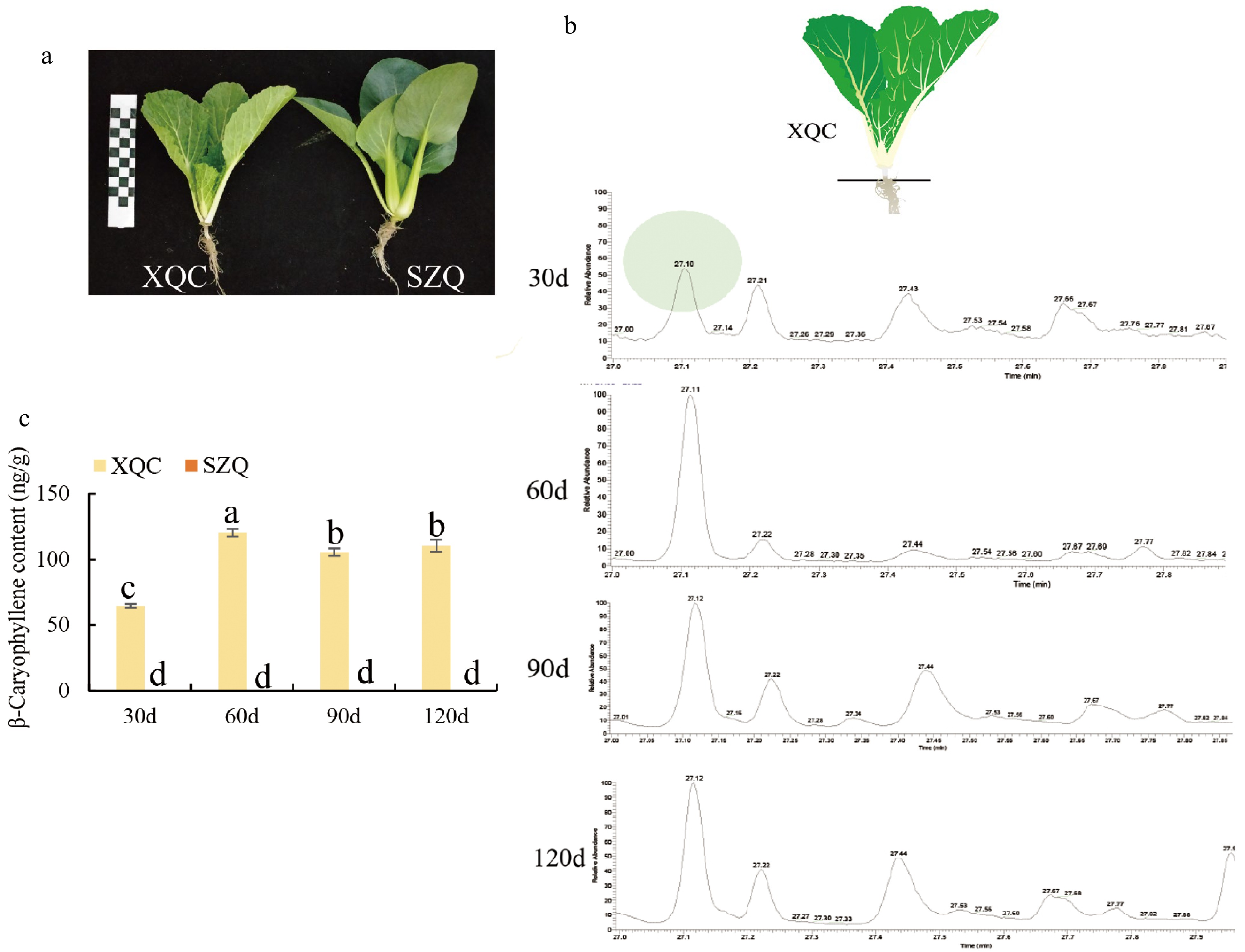
      Figure 1. 

      β-Caryophyllene content in pak-choi cultivars 'XQC' and 'SZQ'. (a) Phenotypes of two pak-choi cultivars, 'XQC' and 'SZQ', at the 30 d stage. (b) Retention time of β-caryophyllene volatiles in GC-MS at 30, 60, 90, and 120 d. (c) β-caryophyllene content at 30, 60, 90, and 120 d in 'XQC' and 'SZQ'.

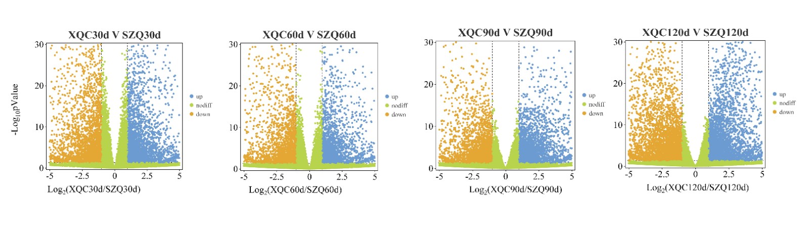
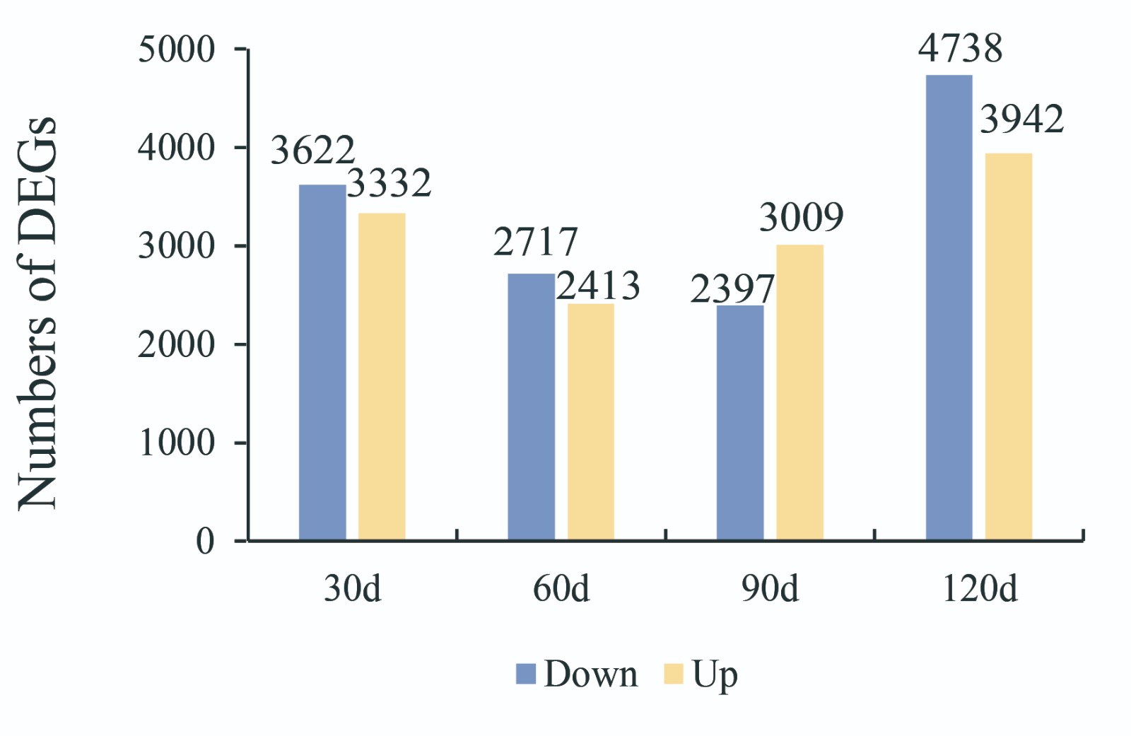
    • To investigate the transport of β-caryophyllene, transcriptome sequencing was carried out at 30, 60, 90, and 120 d after germination, with three replicates at each time point. From these samples, approximately 1.13 billion raw reads were produced. Following filtering, 1.12 billion high-quality-filtered (clean) reads proceeded to the next step, and the average read count for each sample ranged from approximately 40 to 61 million (Supplemental Table S1). The resulting clean reads were aligned against the Brassica campestris genome[17]. Differential expression results were generated using DESeq2, and gene expression was quantified using transcripts per million (TPM). Genes with expression |fold change| > 2 (p < 0.05) were considered to be differentially expressed, and they were visualized through volcano plots (Supplemental Fig. S1). In total, 6,954 (3,332 were upregulated and 3,622 were downregulated), 5130 (2717 were upregulated and 2413 were downregulated), 5406 (3009 were upregulated and 3,797 were downregulated), and 8680 (3942 were upregulated and 4738 were downregulated) differentially expressed genes (DEGs) were identified at 30, 60, 90, and 120 days, respectively (Fig. 2a & Supplemental Fig. S2). In this study, nine unigenes that encode enzymes associated with terpene biosynthesis were identified based on the enriched KEGG pathways and GO functional analysis. The terpene synthase gene (TPS), the last key enzyme in the synthesis of β-caryophyllene, is related to the content of β-caryophyllene. The correlation heatmap of DEGs involved in the terpene biosynthetic pathway shows the importance of the terpene synthase gene (Fig. 2b). Moreover, 426 transport protein genes were identified as DEGs, and among them, sixty transport proteins were identified with positive correlation coefficients with TPS21, including one equilibrative nucleotide transporter 3 (ENT3), two ABCs, one sulfate transporter 3.4 (SULTR3, 4), and one putative auxin efflux carrier component 5 (PIN5) (p > 0.8) (Fig. 2c & Supplemental Table S2).
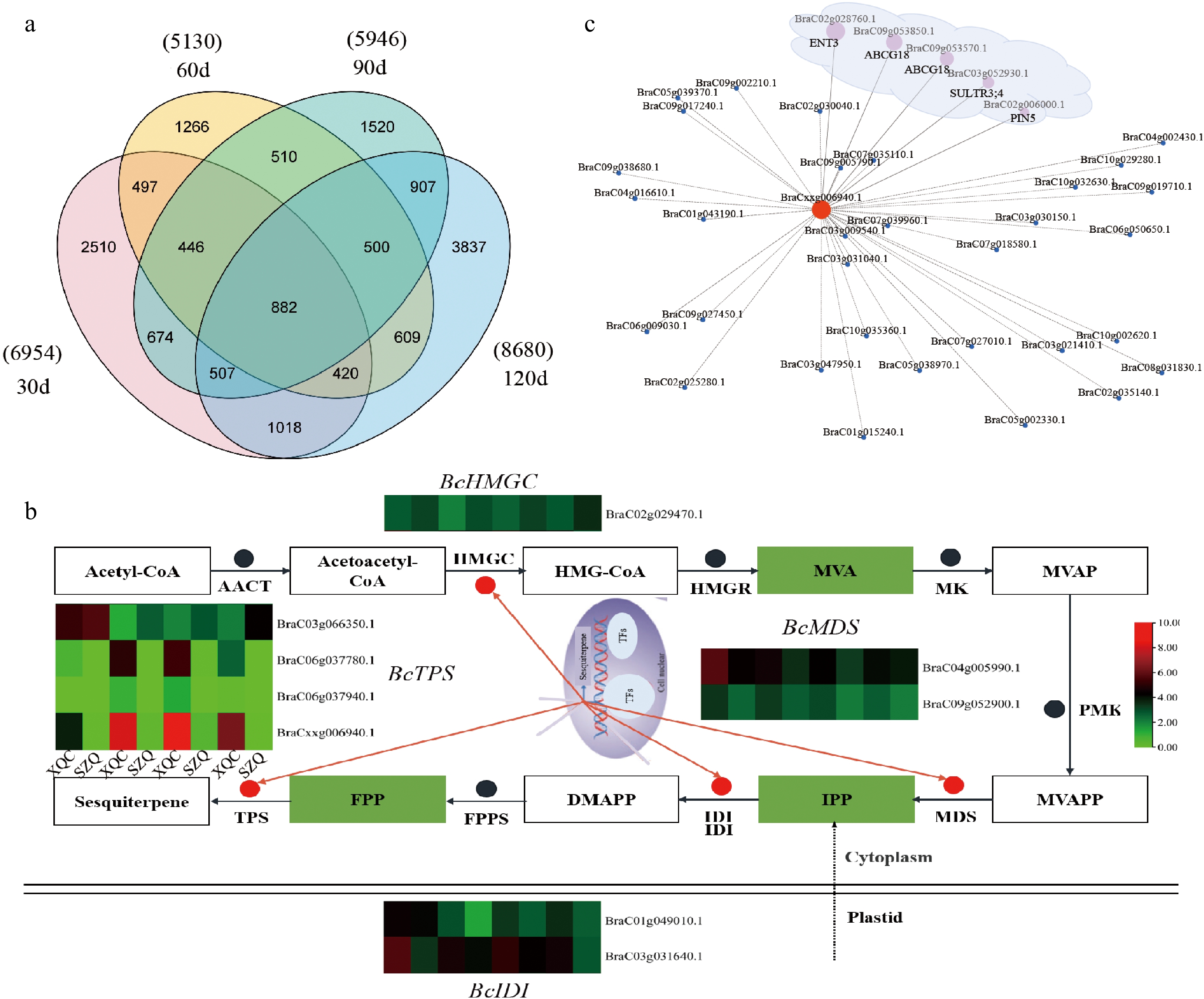
      Figure 2. 

      Transcriptomic analysis of 'XQC' and 'SZQ' at 30, 60, 90, and 120 d. (a) Venn diagram showing the number of DEGs identified between 'XQC' and 'SZQ' at 30, 60, 90, and 120 d. (b) DEG heatmap of the RNA-Seq expression involved in the sesquiterpene pathway. (c) Network between TPS21 and the differential transporters. Circle size indicates Person correlation.

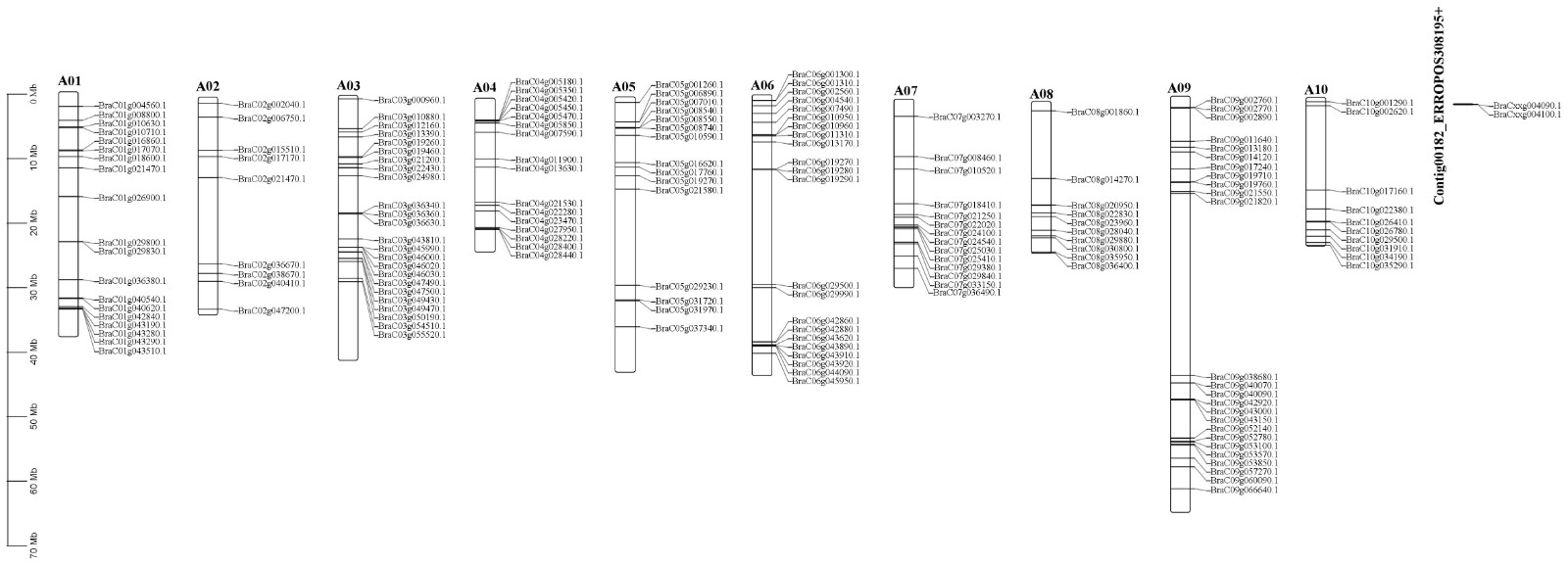
    • The ABC transporter, which is highly correlated with TPS21, is thought to be an important carrier for the transport of metabolites in plants. According to the genome, a total of 169 ABC proteins, listed in Supplemental Table S3, were recognized as members of the Brassica campestris ABC family, and their physical locations are shown in Supplemental Fig. S3 in the Brassica campestris genome. The phylogenetic tree was constructed with ABC proteins from Brassica campestris and Arabidopsis, and proteins of Arabidopsis are listed in Supplemental Table S4. Phylogenetic analysis of ABC proteins from Brassica campestris and Arabidopsis indicated that among the eight subfamilies, subfamily G had the most genes, with 55, followed by subfamily B, with 37 (Fig. 3a). In order to identify the role of β-caryophyllene-related transporters and analyze ABC transporters in abiotic stress, seven differential ABC transporters were identified through the transcription data of 30, 60, 90, and 120 d of material grown in greenhouses. Meanwhile, according to a previous study, the ABCG33 gene may respond to stress. Eight ABC genes, five members belonged to ABCG, two members belonged to ABCC, and only one member belonged to ABCE. Eight ABC proteins were named according to homology (BraC05g008540.1, ABCG33; BraC05g010590.1, ABCC2; BraC05g037340.1, ABCG29; BraC06g010960.1, ABCG35; BraC08g014270.1, ABCE2; BraC08g023960.1, ABCC1; BraC09g053570.1, ABCG18; BraC10g031910.1, ABCG22). The results showed that eight ABC proteins were divided into four subgroups. The first was composed of two BcABCs (BraC08g23960.1; BraC05g10590), the second consisted of BraC08g14270, the third consisted of three BcABCs (BraC05g008540.1; BraC05g037340.1; BraC06g010960.1), and the fourth consisted of two BcABC (BraC10g031910.1; BraC09g053570.1) (Fig. 3b-X). Using the MEME program, we further performed motif prediction for ABC proteins, and 10 different motifs (motifs 1–10) were identified (Fig. 3b). Motif 1 was identified in all ABC proteins (Fig. 3b). Structural features were then characterized for eight ABC genes, and a large divergence in exon number was observed (Fig. 3b-Z). The length, molecular weight, and theoretical isoelectric point of eight ABC proteins ranged from 605 to 1626 aa with an average of 540 aa, from 68 to 182 kDa with an average of 135 kDa, and from 5.9 to 9.37 with an average of 7.39, respectively (Supplemental Table S5). To understand their transcriptional regulation patterns, CREs were identified in ABC gene promoter regions. A total of 16 CREs, namely, anaerobic induction-, meristem expression-, abscisic acid-responsive element-, anoxic inducibility-, circadian control-, endosperm expression-, gibberellin responsive element, light responsive element, low temperature responsive element, MeJA responsive element, MYB binding site, MYBHv1 binding site, MYC binding site, salicylic acid responsive element, sugar metabolism and plant defense signaling, and wound responsive element types, were identified (Fig. 3c). Light-responsive elements were detected in eight ABC promoters, and MeJA-responsive elements were detected in six ABC promoters except for ABCG33 and ABCG18. In addition to ABCE2, the ABA motif was detected in seven ABC promoters.
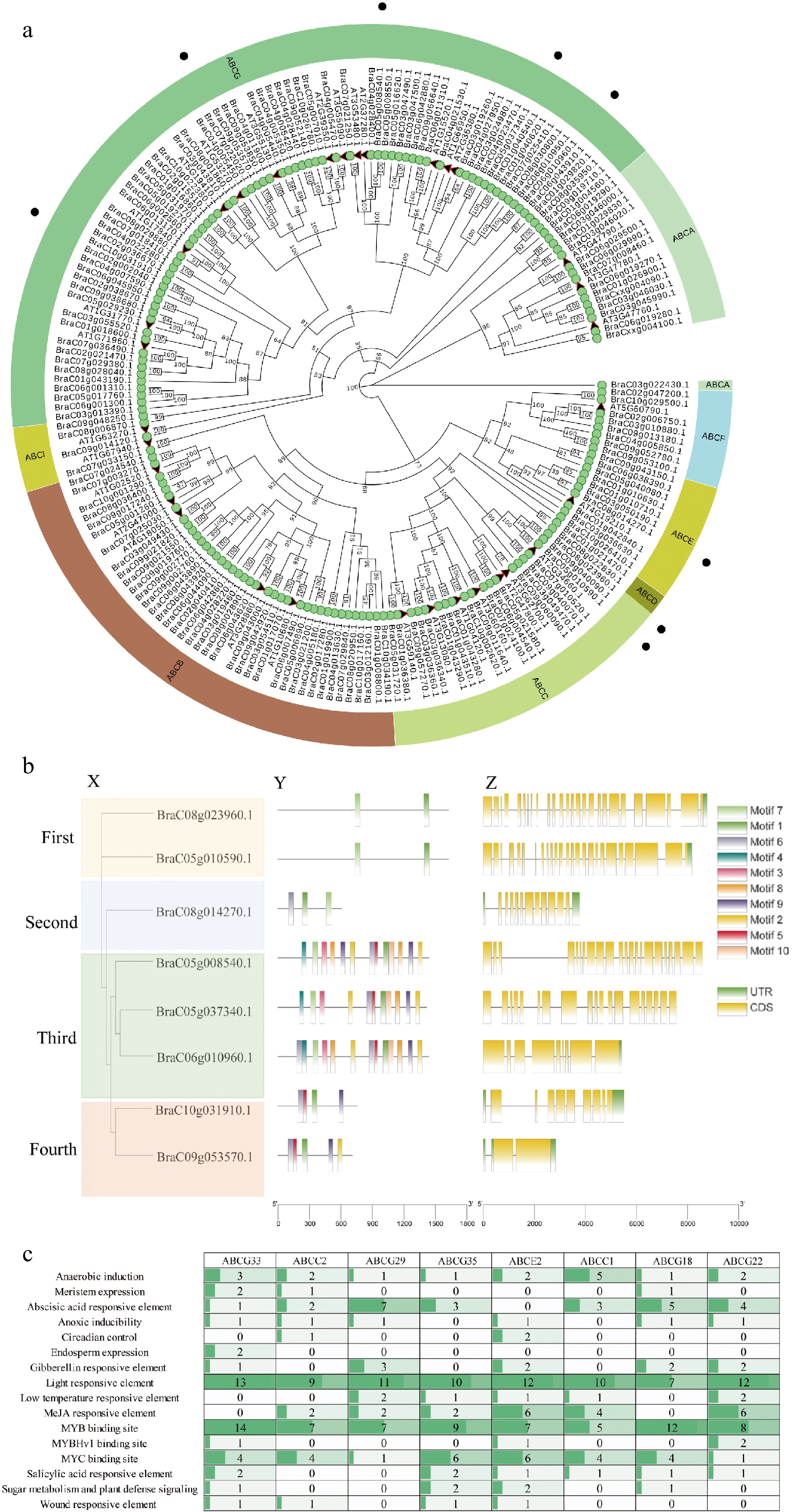
      Figure 3. 

      Phylogenetic analysis, gene structure, conserved domains, and cis-acting elements of ABC proteins from Brassica campestris and Arabidopsis. (a) Phylogenic relationship of the identified ABC genes in the Brassica campestris genome. (b) Eight ABC transporter motif distributions and exon-intron distribution in Brassica campestris. The motifs identified in the BcABC proteins are indicated by different colored boxes and named motifs 1–10. The closed yellow boxes and black lines represent exons and introns, respectively. (c) The number and type of existing putative cis-acting elements in two kb upstream regions of the eight ABC genes.

    • The expression levels of the seven transporters (except ABCG33) in different stages of development were observed, and the results indicated that they could be classified into two groups, and ABCC1 and ABCG18 were clustered with TPS21 (Fig. 4a). We further analyzed the correlation between seven ABC transporters and TPS21 and found that ABCG18 was significantly correlated with TPS21 (p > 0.85) (Fig. 4b). A potential regulatory network of ABCG18 is shown in Fig. 4c, and MYB12 is the most closely related to ABCG18 and may be involved in the transcription of ABCG18 (Fig. 4c).
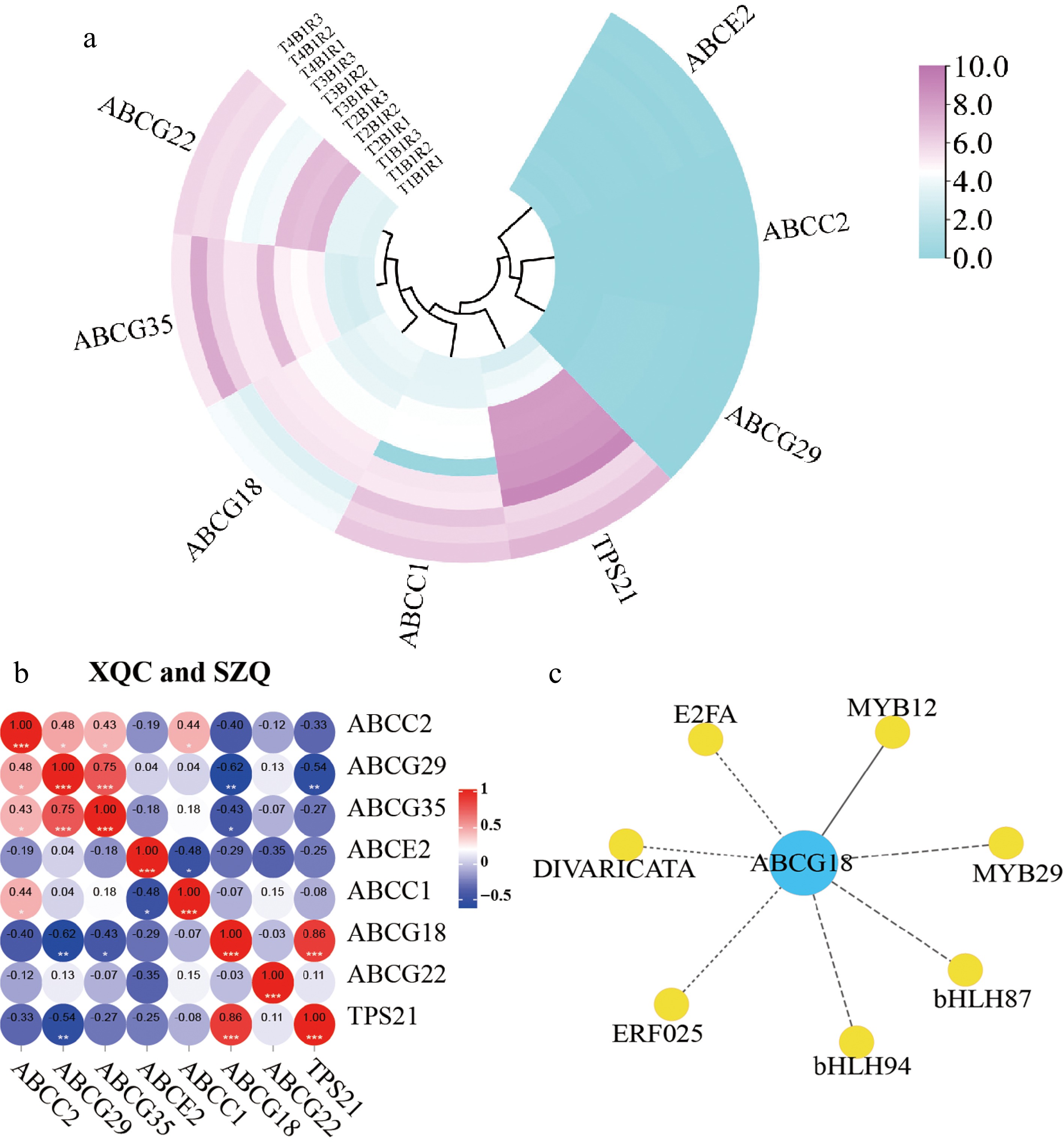
      Figure 4. 

      Pearson analysis of 'XQC' and 'SZQ' at 30, 60, 90, and 120 d. (a) Heatmap of RNA-Seq expression of seven ABC differential transporters. (b) Correlation among the ABC proteins in 'XQC' and 'SZQ'. (c) The correlation between transcription factors and ABCG18 of 'XQC' and 'SZQ' transcriptome data, solid line -> 0.8.

    • To cope with environmental changes, the transport of materials in plants is often dependent on carriers. We further examined the expression profiles of eight ABC genes and TPS21 using qRT‒PCR under three kinds of abiotic stress and two hormonal treatments (Fig. 5a). After 12 h of MeJA or ABA treatment, two ABCs (ABCG33 and ABCC1) were significantly upregulated. Interestingly, the ABCC1 transcription level had a similar expression pattern to TPS21 under all five treatments. Under low temperature, ABCG22 and ABCG35 increased markedly, while ABCG22 and ABCG35 decreased under the other treatments. Our results show that TPS21 is expressed in two ways: one is first increased and then decreased and was lower than CK under salt stress, low temperature treatment and ABA treatment, and the other is always higher than CK under MeJA and high temperature treatment. Furthermore, TPSa21 and ABCG18 transcription patterns were investigated in different tissues (Fig. 5b). TPS21 was mainly expressed in the leaf, while the expression of TPS21 in the root and petiole was almost zero. ABCG18 was mainly expressed in leaves and petioles but not in roots. These results suggest that ABCG18 may play an important role in the transportation of β-caryophyllene under normal conditions, while ABCG35 may be an important transport protein under low temperature. BraC09g053570.1 had the best homology with ABCG18 (AT3G55110.1) (Fig. 5c). The membrane localization ABCG transporter plays a key role in the export of β-caryophyllene and other secondary metabolites. A green fluorescent protein-fused ABCG18 reporter gene was constructed and used for transient transformation experiments in tobacco leaves, and it was found to be a membrane localization protein (Fig. 5d).
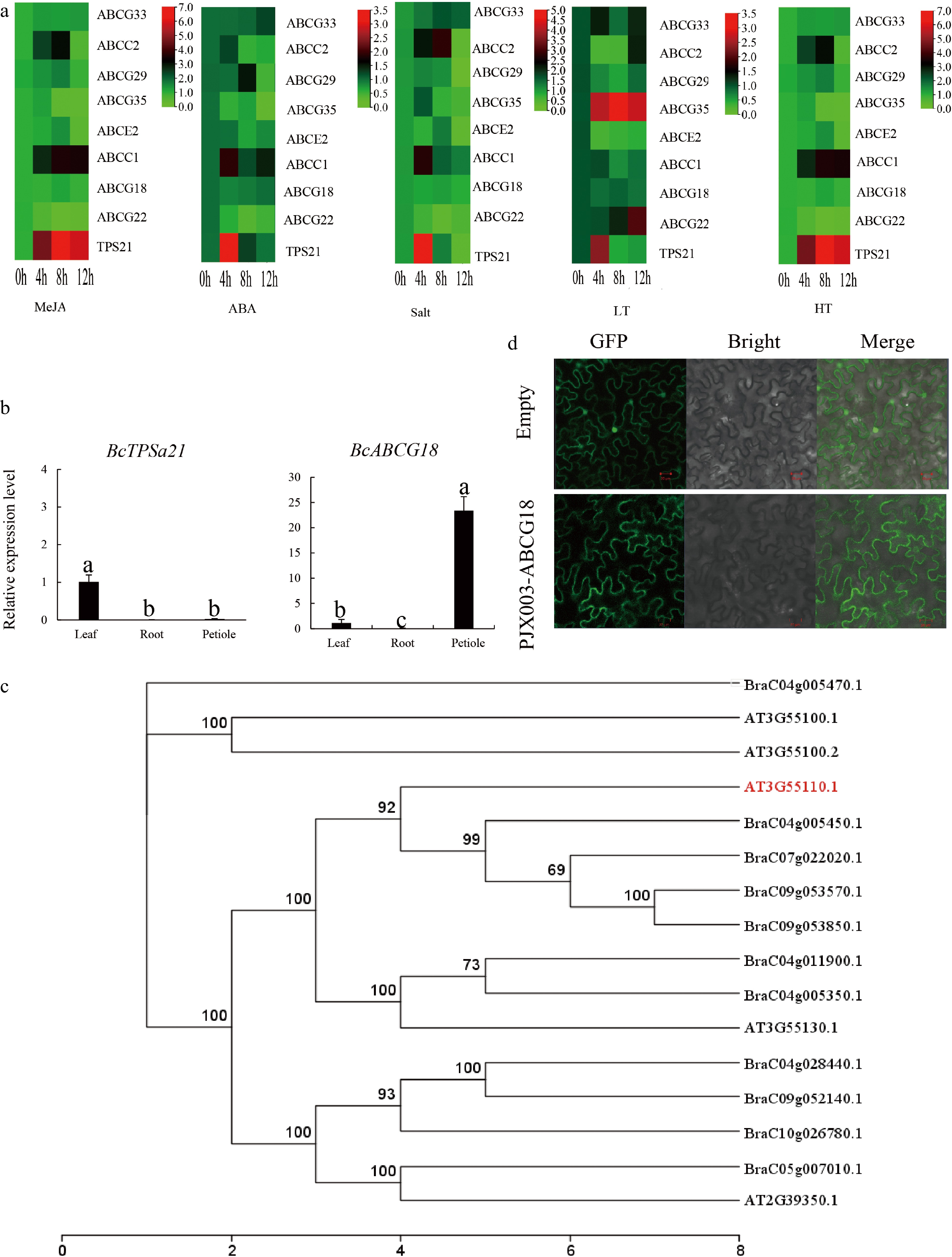
      Figure 5. 

      Expression profiles of BcABC genes and subcellular localization. (a) Expression profiles of BcABC genes under different conditions (MeJA, ABA, salt, high temperature, low temperature). (b) Transcript levels of ABCG18 and TPS21 in leaf, stem, and root tissue. (c), (d) Finding the best homology of the ABCG18 gene and subcellular localization.

    • Plants are frequently exposed to various stresses that affect their growth and development. To survive, plants have evolved sophisticated response systems to cope with a variety of stressful environments. Among these responsive networks, ABC-mediated transport is a representative system that has attracted increasing attention from researchers. ABC transporters are a family that can take part in the transport of a broad range of terpenoids. Genome-wide analyses of ABC transporters in Solanum lycopersicum[18], Arabidopsis[16], Oryza sativa[12], Zea mays[13], and Brassica rapa[19] have already been performed. However, little is known about ABC transporters related to β-caryophyllene in Brassica campestris. To address this, we performed comparative transcriptome analysis in Brassica campestris to investigate the potential function of the ABC transporter in β-caryophyllene transport and response to abiotic stress in Brassica campestris.

      Terpenoids show a wide array of pharmacological activities and are one of the most extensively studied and structurally diverse classes of natural compounds. Terpenoids can be classified based on the number of isoprene units in the parent structure, such as monoterpenoids (C10), sesquiterpenoids (C15), diterpenoids (C20), sesterterpenoids (C25), triterpenoids (C30), tetraterpenes (C40) and polyterpenes. AaPDR3 (ABCG40) is involved in sesquiterpene beta-caryophyllene transport in Artemisia annua[10]. This study indicated that TPS21 was one of the most important enzymes in the synthesis of caryophyllene (Fig 2d). Five highly correlated transporters with TPS21 were identified (p > 0.8), including nucleotide transport and ABC transporters. Thus, ABCG18 may become a potential caryophyllene transporter according to correlation. Our results indicate that MYB12 is a potential transporter regulating ABCG18. MYB12 is involved in the regulation of metabolism of various substances. High temperature enhances anthocyanin coloration in Asiatic hybrid lily flowers via upregulation of the MYB12 positive regulator[20]. COP1 is involved in the dark-dependent repression of MYB12 expression and flavonol accumulation[21]. Under abiotic stress conditions, ABCG17 and ABCG18 are transcriptionally repressed, promoting active ABA movement and response[22]. ABA homeostasis under normal and abiotic stress conditions can be regulated by ABCG18 protein. This is similar to our results with ABA treatment and provides a new perspective for the study of caryophyllene transport. WGDs (whole genome duplications) are strongly supported by evidence commonly found in many species-rich lineages of eukaryotes and thus are considered a major driving force in species diversification. In Arabidopsis, a total of 129 ABC transporter genes were identified. This study showed that a total of 169 annotated genes were identified as Brassica campestris ABC family members, which was more than that in Arabidopsis but similar to that in Brassica rapa (Chiifu-401)[19].

      Evolutionary analysis showed that seven subfamilies of Brassica campestris and Arabidopsis ABC transporters (ABCA, ABCB, ABCC, ABCD, ABCE, ABCF, ABCG, and ABCI subfamilies) were generated. ABCG had the most genes in subfamilies. This phenomenon was consistent with observations in Brassica rapa and Arabidopsis[19]. Eight differentially expressed ABC protein genes were screened based on transcriptome data, and eight ABC proteins were predicted to be located in the plasma membrane, which may be related to their transport functions. Variations in structure or motifs can be relatively reliable parameters to evaluate the evolution of a gene family. In this study, eight ABC proteins were classified into four groups, and similar gene structures and motifs were commonly shared by most members in the same group.

      Functional conservation and diversification can also be uncovered to a great extent by the expression profiles. It is clear that the expression of many ABCs in different plant species can be stimulated by various hormones or stresses, such as ABA, methyl jasmonate (MeJA), and low temperature[2325]. Fifteen differentially expressed ABC genes exhibited diverse expression responses to stress treatments, including drought, cadmium, and salt stress[26]. We investigated the temporal and spatial expression profiles of these ABC transporters in different developmental stages and under different treatments. Expression analysis results showed that the transcription of ABCs was differentially regulated by various abiotic stresses and hormone treatments. ABCC1 is a vacuolar membrane-localized transporter; ABCC is responsible for the transport of the phytochelatins (PCs) and cadmium (PCs–Cd) complex into the vacuoles[27]. Notably, ABCC1, which is a membrane-bound, energy-dependent efflux transporter, belonging to the superfamily of ABC transporters, has a similar expression pattern under MeJA treatment and low temperature. Moreover, ABCG22 and ABCG35 were significantly induced under low temperature stress. The effect of ABCG35 at low temperature is still unclear, our results indicated that it might be a key gene for participating in low temperature.

      In conclusion, this study provides a genome-wide analysis of ABC transporters from the Brassica campestris genome. The expression profiles of eight ABC transporters were validated using both RNA-seq analyses and qRT‒PCR. Furthermore, this study provides insights into ABC transporter gene expression after exposure to stress. These combined results may offer a basis for future studies on the transport of β-caryophyllene by ABC transporters in Brassica campestris.

    • Taken together, this study represents the characterization of eight different ABC gene spatiotemporal expression patterns according to the genome database and transcriptome of Brassica campestris for the first time. Our results indicate that ABCG18 is a potential carrier of β-caryophyllene, which might participate in substance transport in leaves and petioles, and MYB12 may be involved in the regulation of ABCG18 in Brassica campestris. ABCG35 and ABCG22 may be important for low temperature. These results are helpful for understanding the mechanism of β-caryophyllene transport metabolism in Brassica campestris and provide a theoretical basis for further research on the physiological function of ABC transporter genes in Brassica campestris.

      • This work was supported by the Jiangsu Seed Industry Revitalization Project [JBGS (2021)015], the National vegetable industry technology system (CARS-23-A-16), the National Key Research and Development Program (2018YFD1000805), and a project funded by the priority academic program development of Jiangsu higher education institutions (China).

      • Dr. Ying Li is the Editorial Board member of journal Vegetable Research. She was blinded from reviewing or making decisions on the manuscript. The article was subject to the journal's standard procedures, with peer-review handled independently of the Editorial Board member and her research group.

      • Copyright: © 2023 by the author(s). Published by Maximum Academic Press, Fayetteville, GA. This article is an open access article distributed under Creative Commons Attribution License (CC BY 4.0), visit https://creativecommons.org/licenses/by/4.0/.
    Figure (5)  References (27)
  • About this article
    Cite this article
    Wang H, Zong C, Bai Y, Yuan S, Li Y, et al. 2023. Comparative transcriptome-based identification and expression analysis of ATP-binding cassette (ABC) transporters reveals a likely role in the transport of β-caryophyllene and response to abiotic stress in Brassica campestris. Vegetable Research 3:13 doi: 10.48130/VR-2023-0013
    Wang H, Zong C, Bai Y, Yuan S, Li Y, et al. 2023. Comparative transcriptome-based identification and expression analysis of ATP-binding cassette (ABC) transporters reveals a likely role in the transport of β-caryophyllene and response to abiotic stress in Brassica campestris. Vegetable Research 3:13 doi: 10.48130/VR-2023-0013

Catalog

  • About this article

/

DownLoad:  Full-Size Img  PowerPoint
  • Table 1.  Correlation analysis of flavonoid content and flavonoid-related genes.
    Aromatic
    amino acids
    PAL F3H CHS
    Tyrosine Pearson correlation 0.615* −0.659** 0.694**
    Significance (two-tailed) 0.015 0.007 0.004
    Phenylalanine Pearson correlation −0.183 0.669** 0.258
    Significance (two-tailed) 0.513 0.006 0.353
    Tryptophan Pearson correlation −0.858 0.753 0.076
    Significance (two-tailed) 0.063 0.142 0.90
    * indicates significant correlation at the p < 0.05 level, ** indicates extremely significant correlation at the p < 0.01 level.
     | Show Table
    DownLoad: CSV
  • Table 2.  Correlation analysis of carotenoids content and carotenoids-related genes.
    Aromatic
    amino acids
    PDS ZDS PSY
    Tyrosine Pearson correlation −0.348 −0.356 0.638*
    Significance (two-tailed) 0.204 0.192 0.011
    Phenylalanine Pearson correlation 0.694** −0.025 0.442
    Significance (two-tailed) 0.004 0.93 0.099
    Tryptophan Pearson correlation 0.342 0.591* 0.57*
    Significance (two-tailed) 0.212 0.02 0.026
    * indicates p < 0.05, ** indicates p < 0.01.
     | Show Table
    DownLoad: CSV