ARTICLE   Open Access    

Identification of long-lived and stable mRNAs in the aged seeds of wheat

More Information
  • Received: 28 October 2022
    Accepted: 28 July 2023
    Published online: 07 October 2023
    Seed Biology  2 Article number: 14 (2023)  |  Cite this article
  • Seed germination relies on preserving mRNA integrity in dry seeds. However, the quality of mRNA in aged wheat seeds is not well understood. Here, we investigated 20 wheat varieties for seed longevity using controlled deterioration treatment (CDT) and identified that Chinese Spring seeds exhibit moderate longevity. We observed correlations between seed viability and RNA integrity in the aleurone and embryo cells after aging-treatment. Nanopore sequencing of whole seeds from natural aging treatment (NAT) and CDT for 25 d identified 3,083 full-length transcripts. We performed RNA-seq transcriptome profiling to classify the tissue origin of these transcripts under eight aging treatments, revealing the presence of 2,064 overlapping long-lived mRNAs (LLRs) in the seed embryo and 2,130 in the aleurone layers. These LLRs corresponded to genes with detectable transcription levels and at least one full-length transcript in their coding sequence. Notably, degradation percentages of these mRNAs varied among seeds of different wheat varieties with similar ages. We predicted 21 most stable LLRs with high GC% content and short coding sequence length, among which only one LLR was seed-specifically expressed and belonged to the late-embryogenesis-abundant (LEA) protein family. RT-PCR confirmed the expression of the seven LLR fragments in the aleurone layer and embryo of Chinese Spring seeds. We found three of the most stable LLRs (LLR13, LLR15, and LLR20) identified in Chinese Spring were more stable in high longevity varieties than in short longevity varieties after aging, indicating their potential roles in seed longevity and germination.
  • Aquaporin’s (AQPs) are small (21–34 kD) channel-forming, water-transporting trans-membrane proteins which are known as membrane intrinsic proteins (MIPs) conspicuously present across all kingdoms of life. In addition to transporting water, plant AQPs act to transport other small molecules including ammonia, carbon dioxide, glycerol, formamide, hydrogen peroxide, nitric acid, and some metalloids such as boron and silicon from the soil to different parts of the plant[1]. AQPs are typically composed of six or fewer transmembrane helices (TMHs) coupled by five loops (A to E) and cytosolic N- and C-termini, which are highly conserved across taxa[2]. Asparagine-Proline-Alanine (NPA) boxes and makeup helices found in loops B (cytosolic) and E (non-cytosolic) fold back into the protein's core to form one of the pore's two primary constrictions, the NPA region[1]. A second filter zone exists at the pore's non-cytosolic end, where it is called the aromatic/arginine (ar/R) constriction. The substrate selectivity of AQPs is controlled by the amino acid residues of the NPA and ar/R filters as well as other elements of the channel[1].

    To date, the AQP gene families have been extensively explored in the model as well as crop plants[39]. In seed plants, AQP distributed into five subfamilies based on subcellular localization and sequence similarities: the plasma membrane intrinsic proteins (PIPs; subgroups PIP1 and PIP2), the tonoplast intrinsic proteins (TIPs; TIP1-TIP5), the nodulin26-like intrinsic proteins (NIPs; NIP1-NIP5), the small basic intrinsic proteins (SIPs; SIP1-SIP2) and the uncategorized intrinsic proteins (XIPs; XIP1-XIP3)[2,10]. Among them, TIPs and PIPs are the most abundant and play a central role in facilitating water transport. SIPs are mostly found in the endoplasmic reticulum (ER)[11], whereas NIPs homologous to GmNod26 are localized in the peribacteroid membrane[12].

    Several studies reported that the activity of AQPs is regulated by various developmental and environmental factors, through which water fluxes are controlled[13]. AQPs are found in all organs such as leaves, roots, stems, flowers, fruits, and seeds[14,15]. According to earlier studies, increased AQP expression in transgenic plants can improve the plants' tolerance to stresses[16,17]. Increased root water flow caused by upregulation of root aquaporin expression may prevent transpiration[18,19]. Overexpression of Tamarix hispida ThPIP2:5 improved osmotic stress tolerance in Arabidopsis and Tamarix plants[20]. Transgenic tomatoes having apple MdPIP1;3 ectopically expressed produced larger fruit and improved drought tolerance[21]. Plants over-expressing heterologous AQPs, on the other hand, showed negative effects on stress tolerance in many cases. Overexpression of GsTIP2;1 from G. soja in Arabidopsis plants exhibited lower resistance against salt and drought stress[22].

    A few recent studies have started to establish a link between AQPs and nanobiology, a research field that has been accelerating in the past decade due to the recognition that many nano-substances including carbon-based materials are valuable in a wide range of agricultural, industrial, and biomedical activities[23]. Carbon nanotubes (CNTs) were found to improve water absorption and retention and thus enhance seed germination in tomatoes[24,25]. Ali et al.[26] reported that Carbon nanoparticles (CTNs) and osmotic stress utilize separate processes for AQP gating. Despite lacking solid evidence, it is assumed that CNTs regulate the aquaporin (AQPs) in the seed coats[26]. Another highly noticed carbon-nano-molecule, the fullerenes, is a group of allotropic forms of carbon consisting of pure carbon atoms[27]. Fullerenes and their derivatives, in particular the water-soluble fullerols [C60(OH)20], are known to be powerful antioxidants, whose biological activity has been reduced to the accumulation of superoxide and hydroxyl[28,29]. Fullerene/fullerols at low concentrations were reported to enhance seed germination, photosynthesis, root growth, fruit yield, and salt tolerance in various plants such as bitter melon and barley[3032]. In contrast, some studies also reported the phytotoxic effect of fullerene/fullerols[33,34]. It remains unknown if exogenous fullerene/fullerol has any impact on the expression or activity of AQPs in the cell.

    Garden pea (P. sativum) is a cool-season crop grown worldwide; depending on the location, planting may occur from winter until early summer. Drought stress in garden pea mainly affects the flowering and pod filling which harm their yield. In the current study, we performed a genome-wide identification and characterization of the AQP genes in garden pea (P. sativum), the fourth largest legume crop worldwide with a large complex genome (~4.5 Gb) that was recently decoded[35]. In particular, we disclose, for the first time to our best knowledge, that the transcriptional regulations of AQPs by osmotic stress in imbibing pea seeds were altered by fullerol supplement, which provides novel insight into the interaction between plant AQPs, osmotic stress, and the carbon nano-substances.

    The whole-genome sequence of garden pea ('Caméor') was retrieved from the URGI Database (https://urgi.versailles.inra.fr/Species/Pisum). Protein sequences of AQPs from two model crops (Rice and Arabidopsis) and five other legumes (Soybean, Chickpea, Common bean, Medicago, and Peanut) were used to identify homologous AQPs from the garden pea genome (Supplemental Table S1). These protein sequences, built as a local database, were then BLASTp searched against the pea genome with an E-value cutoff of 10−5 and hit a score cutoff of 100 to identify AQP orthologs. The putative AQP sequences of pea were additionally validated to confirm the nature of MIP (Supplemental Table S2) and transmembrane helical domains through TMHMM (www.cbs.dtu.dk/services/TMHMM/).

    Further phylogenetic analysis was performed to categorize the AQPs into subfamilies. The pea AQP amino acid sequences, along with those from Medicago, a cool-season model legume phylogenetically close to pea, were aligned through ClustalW2 software (www.ebi.ac.uk/Tools/msa/clustalw2) to assign protein names. The unaligned AQP sequences to Medicago counterparts were once again aligned with the AQP sequences of Arabidopsis, rice, and soybean. Based on the LG model, unrooted phylogenetic trees were generated via MEGA7 and the neighbor-joining method[36], and the specific name of each AQP gene was assigned based on its position in the phylogenetic tree.

    By using the conserved domain database (CDD, www.ncbi.nlm.nih.gov/Structure/cdd/cdd.shtml), the NPA motifs were identified from the pea AQP protein sequences[37]. The software TMHMM (www.cbs. dtu.dk/services/TMHMM/)[38] was used to identify the protein transmembrane domains. To determine whether there were any alterations or total deletion, the transmembrane domains were carefully examined.

    Basic molecular properties including amino acid composition, relative molecular weight (MW), and instability index were investigated through the online tool ProtParam (https://web.expasy.org/protparam/). The isoelectric points (pI) were estimated by sequence Manipulation Suite version 2 (www.bioinformatics.org/sms2)[39]. The subcellular localization of AQP proteins was predicted using Plant-mPLoc[40] and WoLF PSORT (www.genscript.com/wolf-psort.html)[ 41] algorithms.

    The gene structure (intron-exon organization) of AQPs was examined through GSDS ver 2.0[42]. The chromosomal distribution of the AQP genes was illustrated by the software MapInspect (http://mapinspect.software.informer.com) in the form of a physical map.

    To explore the tissue expression patterns of pea AQP genes, existing NGS data from 18 different libraries covering a wide range of tissue, developmental stage, and growth condition of the variety ‘Caméor’ were downloaded from GenBank (www.ncbi.nlm.nih.gov/bioproject/267198). The expression levels of the AQP genes in each tissue and growth stage/condition were represented by the FPKM (Fragments Per Kilobase of transcript per Million fragments mapped) values. Heatmaps of AQPs gene were generated through Morpheus software (https://software.broadinstitute.org/morpheus/#).

    Different solutions, which were water (W), 0.3 M mannitol (M), and fullerol of different concentrations dissolved in 0.3 M mannitol (MF), were used in this study. MF solutions with the fullerol concentration of 10, 50, 100, and 500 mg/L were denoted as MF1, MF2, MF3, and MF4, respectively. Seeds of 'SQ-1', a Chinese landrace accession of a pea, were germinated in two layers of filter paper with 30 mL of each solution in Petri dishes (12 cm in diameter) each solution, and the visual phenotype and radicle lengths of 150 seeds for each treatment were analyzed 72 h after soaking. The radicle lengths were measured using a ruler. Multiple comparisons for each treatment were performed using the SSR-Test method with the software SPSS 20.0 (IBM SPSS Statistics, Armonk, NY, USA).

    Total RNA was extracted from imbibing embryos after 12 h of seed soaking in the W, M, and MF3 solution, respectively, by using Trizol reagent (Invitrogen, Carlsbad, CA, USA). The quality and quantity of the total RNA were measured through electrophoresis on 1% agarose gel and an Agilent 2100 Bioanalyzer respectively (Agilent Technologies, Santa Rosa, USA). The TruSeq RNA Sample Preparation Kit was utilized to construct an RNA-Seq library from 5 µg of total RNA from each sample according to the manufacturer's instruction (Illumina, San Diego, CA, USA). Next-generation sequencing of nine libraries were performed through Novaseq 6000 platform (Illumina, San Diego, CA, USA).

    First of all, by using SeqPrep (https://github.com/jstjohn/SeqPrep) and Sickle (https://github.com/najoshi/sickle) the raw RNA-Seq reads were filtered and trimmed with default parameters. After filtering, high-quality reads were mapped onto the pea reference genome (https://urgi.versailles.inra.fr/Species/Pisum) by using TopHat (V2.1.0)[43]. Using Cufflinks, the number of mapped reads from each sample was determined and normalised to FPKM for each predicted transcript (v2.2.1). Pairwise comparisons were made between W vs M and W vs M+F treatments. The DEGs with a fold change ≥ 1.5 and false discovery rate (FDR) adjusted p-values ≤ 0.05 were identified by using Cuffdiff[44].

    qPCR was performed by using TOROGGreen® qPCR Master Mix (Toroivd, Shanghai, China) on a qTOWER®3 Real-Time PCR detection system (Analytik Jena, Germany). The reactions were performed at 95 °C for 60 s, followed by 42 cycles of 95 °C for 10 s and 60 °C for 30 s. Quantification of relative expression level was achieved by normalization against the transcripts of the housekeeping genes β-tubulin according to Kreplak et al.[35]. The primer sequences for reference and target genes used are listed in Supplemental Table S3.

    The homology-based analysis identifies 41 putative AQPs in the garden pea genome. Among them, all but two genes (Psat0s3550g0040.1, Psat0s2987g0040.1) encode full-length aquaporin-like sequences (Table 1). The conserved protein domain analysis later validated all of the expected AQPs (Supplemental Table S2). To systematically classify these genes and elucidate their relationship with the AQPs from other plants' a phylogenetic tree was created. It clearly showed that the AQPs from pea and its close relative M. truncatula formed four distinct clusters, which represented the different subfamilies of AQPs i.e. TIPs, PIPs, NIPs, and SIPs (Fig. 1a). However, out of the 41 identified pea AQPs, 4 AQPs couldn't be tightly aligned with the Medicago AQPs and thus were put to a new phylogenetic tree constructed with AQPs from rice, Arabidopsis, and soybean. This additional analysis assigned one of the 4 AQPs to the XIP subfamily and the rest three to the TIP or NIP subfamilies (Fig. 1b). Therefore, it is concluded that the 41 PsAQPs comprise 11 PsTIPs, 15 PsNIPs, 9 PsPIPs, 5 PsSIPs, and 1 PsXIP (Table 2). The PsPIPs formed two major subgroups namely PIP1s and PIP2s, which comprise three and six members, respectively (Table 1). The PsTIPs formed two major subgroups TIPs 1 (PsTIP1-1, PsTIP1-3, PsTIP1-4, PsTIP1-7) and TIPs 2 (PsTIP2-1, PsTIP2-2, PsTIP2-3, PsTIP2-6) each having four members (Table 2). Detailed information such as gene/protein names, accession numbers, the length of deduced polypeptides, and protein structural features are presented in Tables 1 & 2

    Table 1.  Description and distribution of aquaporin genes identified in the garden pea genome.
    Chromosome
    S. NoGene NameGene IDGene length
    (bp)
    LocationStartEndTranscription length (bp)CDS length
    (bp)
    Protein length
    (aa)
    1PsPIP1-1Psat5g128840.32507chr5LG3231,127,859231,130,365675675225
    2PsPIP1-2Psat2g034560.11963chr2LG149,355,95849,357,920870870290
    3PsPIP1-4Psat2g182480.11211chr2LG1421,647,518421,648,728864864288
    4PsPIP2-1Psat6g183960.13314chr6LG2369,699,084369,702,397864864288
    5PsPIP2-2-1Psat4g051960.11223chr4LG486,037,44686,038,668585585195
    6PsPIP2-2-2Psat5g279360.22556chr5LG3543,477,849543,480,4042555789263
    7PsPIP2-3Psat7g228600.22331chr7LG7458,647,213458,649,5432330672224
    8PsPIP2-4Psat3g045080.11786chr3LG5100,017,377100,019,162864864288
    9PsPIP2-5Psat0s3550g0040.11709scaffold0355020,92922,63711911191397
    10PsTIP1-1Psat3g040640.12021chr3LG589,426,47389,428,493753753251
    11PsTIP1-3Psat3g184440.12003chr3LG5393,920,756393,922,758759759253
    12PsTIP1-4Psat7g219600.12083chr7LG7441,691,937441,694,019759759253
    13PsTIP1-7Psat6g236600.11880chr6LG2471,659,417471,661,296762762254
    14PsTIP2-1Psat1g005320.11598chr1LG67,864,8107,866,407750750250
    15PsTIP2-2Psat4g198360.11868chr4LG4407,970,525407,972,392750750250
    16PsTIP2-3Psat1g118120.12665chr1LG6230,725,833230,728,497768768256
    17PsTIP2-6Psat2g177040.11658chr2LG1416,640,482416,642,139750750250
    18PsTIP3-2Psat6g054400.11332chr6LG254,878,00354,879,334780780260
    19PsTIP4-1Psat6g037720.21689chr6LG230,753,62430,755,3121688624208
    20PsTIP5-1Psat7g157600.11695chr7LG7299,716,873299,718,567762762254
    21PsNIP1-1Psat1g195040.21864chr1LG6346,593,853346,595,7161863645215
    22PsNIP1-3Psat1g195800.11200chr1LG6347,120,121347,121,335819819273
    23PsNIP1-5Psat7g067480.12365chr7LG7109,420,633109,422,997828828276
    24PsNIP1-6Psat7g067360.12250chr7LG7109,270,462109,272,711813813271
    25PsNIP1-7Psat1g193240.11452chr1LG6344,622,606344,624,057831831277
    26PsNIP2-1-2Psat3g197520.1669chr3LG5420,092,382420,093,050345345115
    27PsNIP2-2-2Psat3g197560.1716chr3LG5420,103,168420,103,883486486162
    28PsNIP3-1Psat2g072000.11414chr2LG1133,902,470133,903,883798798266
    29PsNIP4-1Psat7g126440.11849chr7LG7209,087,362209,089,210828828276
    30PsNIP4-2Psat5g230920.11436chr5LG3463,340,575463,342,010825825275
    31PsNIP5-1Psat6g190560.11563chr6LG2383,057,323383,058,885867867289
    32PsNIP6-1Psat5g304760.45093chr5LG3573,714,868573,719,9605092486162
    33PsNIP6-2Psat7g036680.12186chr7LG761,445,34161,447,134762762254
    34PsNIP6-3Psat7g259640.12339chr7LG7488,047,315488,049,653918918306
    35PsNIP7-1Psat6g134160.24050chr6LG2260,615,019260,619,06840491509503
    36PsSIP1-1Psat3g091120.13513chr3LG5187,012,329187,015,841738738246
    37PsSIP1-2Psat1g096840.13609chr1LG6167,126,599167,130,207744744248
    38PsSIP1-3Psat7g203280.12069chr7LG7401,302,247401,304,315720720240
    39PsSIP2-1-1Psat0s2987g0040.1706scaffold02987177,538178,243621621207
    40PsSIP2-1-2Psat3g082760.13135chr3LG5173,720,100173,723,234720720240
    41PsXIP2-1Psat7g178080.12077chr7LG7335,167,251335,169,327942942314
    bp: base pair, aa: amino acid.
     | Show Table
    DownLoad: CSV
    Figure 1.  Phylogenetic analysis of the identified AQPs from pea genome. (a) The pea AQPs proteins aligned with those from the cool-season legume Medicago truncatual. (b) The four un-assigned pea AQPs in (a) (denoted as NA) were further aligned with the AQPs of rice, soybean, and Arabidopsis by using the Clustal W program implemented in MEGA 7 software. The nomenclature of PsAQPs was based on homology with the identified aquaporins that were clustered together.
    Table 2.  Protein information, conserved amino acid residues, trans-membrane domains, selectivity filter, and predicted subcellular localization of the 39 full-length pea aquaporins.
    S. NoAQPsGeneLengthTMHNPANPAar/R selectivity filterpIWoLF PSORTPlant-mPLoc
    LBLEH2H5LE1LE2
    Plasma membrane intrinsic proteins (PIPs)
    1PsPIP1-1Psat5g128840.32254NPA0F0008.11PlasPlas
    2PsPIP1-2Psat2g034560.12902NPANPAFHTR9.31PlasPlas
    3PsPIP1-4Psat2g182480.12886NPANPAFHTR9.29PlasPlas
    4PsPIP2-1Psat6g183960.12886NPANPAFHT08.74PlasPlas
    5PsPIP2-2-1Psat4g051960.1195300FHTR8.88PlasPlas
    6PsPIP2-2-2Psat5g279360.22635NPANPAFHTR5.71PlasPlas
    7PsPIP2-3Psat7g228600.22244NPA0FF006.92PlasPlas
    8PsPIP2-4Psat3g045080.12886NPANPAFHTR8.29PlasPlas
    Tonoplast intrinsic proteins (TIPs)
    1PsTIP1-1Psat3g040640.12517NPANPAHIAV6.34PlasVacu
    2PsTIP1-3Psat3g184440.12536NPANPAHIAV5.02Plas/VacuVacu
    3PsTIP1-4Psat7g219600.12537NPANPAHIAV4.72VacuVacu
    4PsTIP1-7Psat6g236600.12546NPANPAHIAV5.48Plas/VacuVacu
    5PsTIP2-1Psat1g005320.12506NPANPAHIGR8.08VacuVacu
    6PsTIP2-2Psat4g198360.12506NPANPAHIGR5.94Plas/VacuVacu
    7PsTIP2-3Psat1g118120.12566NPANPAHIAL6.86Plas/VacuVacu
    8PsTIP2-6Psat2g177040.12506NPANPAHIGR4.93VacuVacu
    9PsTIP3-2Psat6g054400.12606NPANPAHIAR7.27Plas/VacuVacu
    10PsTIP4-1Psat6g037720.22086NPANPAHIAR6.29Vac/ plasVacu
    11PsTIP5-1Psat7g157600.12547NPANPANVGC8.2Vacu /plasVacu/Plas
    Nodulin-26 like intrisic proteins (NIPs)
    1PsNIP1-1Psat1g195040.22155NPA0WVF06.71PlasPlas
    2PsNIP1-3Psat1g195800.12735NPANPVWVAR6.77PlasPlas
    3PsNIP1-5Psat7g067480.12766NPANPVWVAN8.98PlasPlas
    4PsNIP1-6Psat7g067360.12716NPANPAWVAR8.65Plas/VacuPlas
    5PsNIP1-7Psat1g193240.12776NPANPAWIAR6.5Plas/VacuPlas
    6PsNIP2-1-2Psat3g197520.11152NPAOG0009.64PlasPlas
    7PsNIP2-2-2Psat3g197560.116230NPA0SGR6.51PlasPlas
    8PsNIP3-1Psat2g072000.12665NPANPASIAR8.59Plas/VacuPlas
    9PsNIP4-1Psat7g126440.12766NPANPAWVAR6.67PlasPlas
    10PsNIP4-2Psat5g230920.12756NPANPAWLAR7.01PlasPlas
    11PsNIP5-1Psat6g190560.12895NPSNPVAIGR7.1PlasPlas
    12PsNIP6-1Psat5g304760.41622NPA0I0009.03PlasPlas
    13PsNIP6-2Psat7g036680.1254000G0005.27ChloPlas/Nucl
    14PsNIP6-3Psat7g259640.13066NPANPVTIGR8.32PlasPlas
    15PsNIP7-1Psat6g134160.25030NLK0WGQR8.5VacuChlo/Nucl
    Small basic intrinsic proteins (SIPs)
    1PsSIP1-1Psat3g091120.12466NPTNPAVLPN9.54PlasPlas/Vacu
    2PsSIP1-2Psat1g096840.12485NTPNPAIVPL9.24VacuPlas/Vacu
    3PsSIP1-3Psat7g203280.12406NPSNPANLPN10.32ChloPlas
    4PsSIP2-1-2Psat3g082760.12404NPLNPAYLGS10.28PlasPlas
    Uncharacterized X intrinsic proteins (XIPs)
    1PsXIP2-1Psat7g178080.13146SPVNPAVVRM7.89PlasPlas
    Length: protein length (aa); pI: Isoelectric point; Trans-membrane helicase (TMH) represents for the numbers of Trans-membrane helices predicted by TMHMM Server v.2.0 tool; WoLF PSORT and Plant-mPLoc: best possible cellualr localization predicted by the WoLF PSORT and Plant-mPLoc tool, respectively (Chlo Chloroplast, Plas Plasma membrane, Vacu Vacuolar membrane, Nucl Nucleus); LB: Loop B, L: Loop E; NPA: Asparagine-Proline-Alanine; H2 represents for Helix 2, H5 represents for Helix 5, LE1 represents for Loop E1, LE2 represents for Loop E2, Ar/R represents for Aromatic/Arginine.
     | Show Table
    DownLoad: CSV

    To understand the genome distribution of the 41 PsAQPs, we mapped these genes onto the seven chromosomes of a pea to retrieve their physical locations (Fig. 2). The greatest number (10) of AQPs were found on chromosome 7, whereas the least (2) on chromosome 4 (Fig. 2 and Table 1). Chromosomes 1 and 6 each contain six aquaporin genes, whereas chromosomes 2, 3, and 5 carry four, seven, and four aquaporin genes, respectively (Fig. 2). The trend of clustered distribution of AQPs was seen on specific chromosomes, particularly near the end of chromosome 7.

    Figure 2.  Chromosomal localization of the 41 PsAQPs on the seven chromosomes of pea. Chr1-7 represents the chromosomes 1 to 7. The numbers on the right of each chromosome show the physical map positions of the AQP genes (Mbp). Blue, green, orange, brown, and black colors represent TIPs, NIPs, PIPs, SIPs, and XIP, respectively.

    The 39 full-length PsAQP proteins have a length of amino acid ranging from 115 to 503 (Table 1) and Isoelectric point (pI) values ranging from 4.72 to 10.35 (Table 2). As a structural signature, transmembrane domains were predicted to exist in all PsAQPs, with the number in individual AQPs varying from 2 to 6. By subfamilies, TIPs harbor the greatest number of TM domains in total, followed by PIPs, NIPs, SIPs, and XIP (Table 2). Exon-intron structure analysis showed that most PsAQPs (16/39) having two introns, while ten members had three, seven members had four, and five members had only one intron (Fig. 3). Overall, PsAQPs exhibited a complex structure with varying intron numbers, positions, and lengths.

    Figure 3.  The exon-intron structures of the AQP genes in pea. Upstream/downstream region, exon, and intron are represented by a blue box, yellow box, and grey line, respectively.

    As aforementioned, generally highly conserved two NPA motifs generate an electrostatic repulsion of protons in AQPs to form the water channel, which is essential for the transport of substrate molecules[15]. In order to comprehend the potential physiological function and substrate specificity of pea aquaporins, NPA motifs (LB, LE) and residues at the ar/R selectivity filter (H2, H5, LE1, and LE2) were examined. (Table 2). We found that all PsTIPs and most PsPIPs had two conserved NPA motifs except for PsPIP1-1, PsPIP2-2-1, and PsPIP2-3, each having a single NPA motif. Among PsNIPs, PsNIP1-6, PsNIP1-6, PsNIP1-7, PsNIP3-1, PsNIP4-1 and PSNIP4-2 had two NPA domains, while PsNIP1-1, PsNIP2-1-2, PsNIP2-2-2 and PsNIP6-1 each had a single NPA motif. In the PsNIP sub-family, the first NPA motif showed an Alanine (A) to Valine (V) substitution in three PsNIPs (PsNIP1-3, PsNIP1-5, and PsNIP6-3) (Table 2). Furthermore, the NPA domains of all members of the XIP and SIP subfamilies were different. The second NPA motif was conserved in PsSIP aquaporins, however, all of the first NPA motifs had Alanine (A) replaced by Leucine (L) (PsSIP2-1-1, PsSIP2-1-2) or Threonine (T) (PsSIP1-1). In comparison to other subfamilies, this motif variation distinguishes water and solute-transporting aquaporins[45].

    Compared to NPA motifs, the ar/R positions were more variable and the amino acid composition appeared to be subfamily-dependent. The majority of PsPIPs had phenylalanine at H2, histidine at H5, threonine at LE1, and arginine at LE2 selective filter (Table 2). All of the PsTIP1 members had a Histidine-Isoleucine-Alanine-Valine structure at this position, while all PsTIP2 members but PsTIP2-3 harbored Histidine-Isoleucine-Glycine-Arginine. Similarly, PsNIPs, PsSIPs and PsXIP also showed subgroup-specific variation in ar/R selectivity filter (Table 2). Each of these substitutions partly determines the function of transporting water[46].

    Sequence-based subcellular localization analysis using WoLF PSORT predicted that all PsPIPs localized in the plasma membrane, which is consistent with their subfamily classification (Table 2). Around half (5/11) of the PsTIPs (PsTIP1-4, PsTIP2-1, PsTIP2-6, PsTIP4-1, and PsTIP5-1) were predicted to localize within vacuoles. However, several TIP members (PsTIP1-1, PsTIP1-3, PsTIP1-7, PsTIP2-2, PsTIP2-3 and PsTIP3-2) were predicted to localize in plasma membranes. We then further investigated their localizations by using another software (Plant-mPLoc, Table 2), which predicted that all the PsTIPs localize within vacuoles, thus supporting that they are tonoplast related. An overwhelming majority of PsNIPs (14/15) and PsXIP were predicted to be found only in plasma membranes., which was also expected (Table 2). Collectively, the versatility in subcellular localization of the pea AQPs is implicative of their distinct roles in controlling water and/or solute transport in the context of plant cell compartmentation.

    Tissue expression patterns of genes are indicative of their functions. Since there were rich resources of RNA-Seq data from various types of pea tissues in the public database, they were used for the extraction of expression information of PsAQP genes as represented by FPKM values. A heat map was generated to show the expression patterns of PsAQP genes in 18 different tissues/stages and their responses to nitrate levels (Fig. 4). According to the heat map, PsPIP1-2, PsPIP2-3 were highly expressed in root and nodule G (Low-nitrate), whereas PsTIP1-4, PsTIP2-6, and PsNIP1-7 were only expressed in roots in comparison to other tissues. The result also demonstrated that PsPIP1-1 and PsNIP3-1 expressed more abundantly in leaf, tendril, and peduncle, whereas PsPIP2-2-2 and PsTIP1-1 showed high to moderate expressions in all the samples except for a few. Interestingly, PsTIP1-1 expression in many green tissues seemed to be oppressed by low-nitrate. In contrast, some AQPs such as PsTIP1-3, PsTIP1-7, PsTIP5-1, PsNIP1-5, PsNIP4-1, PsNIP5-1, and PsSIP2-1-1 showed higher expression only in the flower tissue. There were interesting developmental stage-dependent regulations of some AQPs in seeds (Fig. 4). For example, PsPIP2-1, PsPIP2-2-1, PsNIP1-6, PsSIP1-1, and PsSIP1-2 were more abundantly expressed in the Seed_12 dap (days after pollination;) tissue than in the Seed_5 dai (days after imbibition) tissue; reversely, PsPIP2-2-2, PsPIP2-4, PsTIP2-3, and PsTIP3-2 showed higher expression in seed_5 dai in compare to seed_12 dap tissues (Fig. 4). The AQP genes may have particular functional roles in the growth and development of the pea based on their tissue-specific expression.

    Figure 4.  Heatmap analysis of the expression of pea AQP gene expressions in different tissues using RNA-seq data (PRJNA267198). Normalized expression of aquaporins in terms of reads per kilobase of transcript per million mapped reads (RPKM) showing higher levels of PIPs, NIPs, TIPs SIPs, and XIP expression across the different tissues analyzed. (Stage A represents 7-8 nodes; stage B represents the start of flowering; stage D represents germination, 5 d after imbibition; stage E represents 12 d after pollination; stage F represents 8 d after sowing; stage G represents 18 d after sowing, LN: Low-nitrate; HN: High-nitrate.

    Expressions of plant AQPs in vegetative tissues under normal and stressed conditions have been extensively studied[15]; however, little is known about the transcriptional regulation of AQP genes in seeds/embryos. To provide insights into this specific area, wet-bench RNA-Seq was performed on the germinating embryo samples isolated from water (W)-imbibed seeds and those treated with mannitol (M, an osmotic reagent), mannitol, and mannitol plus fullerol (F, a nano-antioxidant). The phenotypic evaluation showed that M treatment had a substantial inhibitory effect on radicle growth, whereas the supplement of F significantly mitigated this inhibition at all concentrations, in particular, 100 mg/mL in MF3, which increased the radicle length by ~33% as compared to that under solely M treatment (Fig. 5). The expression values of PsAQP genes were removed from the RNA-Seq data, and pairwise comparisons were made within the Group 1: W vs M, and Group 2: W vs MF3, where a total of ten and nince AQPs were identified as differentially expressed genes (DEGs), respectively (Fig. 6). In Group 1, six DEGs were up-regulated and four DEGs down-regulated, whereas in Group 2, six DEGs were up-regulated and three DEGs down-regulated. Four genes viz. PsPIPs2-5, PsNIP6-3, PsTIP2-3, and PsTIP3-2 were found to be similarly regulated by M or MF3 treatment (Fig. 6), indicating that their regulation by osmotic stress couldn't be mitigated by fullerol. Three genes, all being PsNIPs (1-1, 2-1-2, and 4-2), were up-regulated only under mannitol treatment without fullerol, suggesting that their perturbations by osmotic stress were migrated by the antioxidant activities. In contrast, four other genes namely PsTIP2-2, PsTIP4-1, PsNIP1-5, and PsSIP1-3 were only regulated under mannitol treatment when fullerol was present.

    Figure 5.  The visual phenotype and radicle length of pea seeds treated with water (W), 0.3 M mannitol (M), and fullerol of different concentrations dissolved in 0.3 M mannitol (MF). MF1, MF2, MF3, and MF4 indicated fullerol dissolved in 0.3 M mannitol at the concentration of 10, 50, 100, and 500 mg/L, respectively. (a) One hundred and fifty grains of pea seeds each were used for phenotype analysis at 72 h after treatment. Radicle lengths were measured using a ruler in three replicates R1, R2, and R3 in all the treatments. (b) Multiple comparison results determined using the SSR-Test method were shown with lowercase letters to indicate statistical significance (P < 0.05).
    Figure 6.  Venn diagram showing the shared and unique differentially expressed PsAQP genes in imbibing seeds under control (W), Mannitol (M) and Mannitol + Fullerol (MF3) treatments. Up-regulation (UG): PsPIP2-5, PsNIP1-1, PsNIP2-1-2, PsNIP4-2, PsNIP6-3, PsNIP1-5, PsTIP2-2, PsTIP4-1, PsSIP1-3, PsXIP2-1; Down-regulation (DG): PsTIP2-3, PsTIP3-2, PsNIP1-7, PsNIP5-1, PsXIP2-1.

    As a validation of the RNA-Seq data, eight genes showing differential expressions in imbibing seeds under M or M + F treatments were selected for qRT-PCR analysis, which was PsTIP4-1, PsTIP2-2, PsTIP2-3, PsTIP3-2, PsPIP2-5, PsXIP2-1, PsNIP6-3 and PsNIP1-5 shown in Fig 6, the expression modes of all the selected genes but PsXIP2-1 were well consistent between the RNA-Seq and the qRT-PCR data. PsXIP2-1, exhibiting slightly decreased expression under M treatment according to RNA-Seq, was found to be up-regulated under the same treatment by qRT-PCR (Fig. 7). This gene was therefore removed from further discussions.

    Figure 7.  The expression patterns of seven PsAQPs in imbibing seeds as revealed by RNA-Seq and qRT-PCR. The seeds were sampled after 12 h soaking in three different solutions, namely water (W), 0.3 M mannitol (M), and 100 mg/L fullerol dissolved in 0.3 M mannitol (MF3) solution. Error bars are standard errors calculated from three replicates.

    This study used the recently available garden pea genome to perform genome-wide identification of AQPs[35] to help understand their functions in plant growth and development. A total of 39 putative full-length AQPs were found in the garden pea genome, which is very similar to the number of AQPs identified in many other diploid legume crops such as 40 AQPs genes in pigeon pea, chickpea, common bean[7,47,48], and 44 AQPs in Medicago[49]. On the other hand, the number of AQP genes in pea is greater compared to diploid species like rice (34)[4], Arabidopsis thaliana (35)[3], and 32 and 36 in peanut A and B genomes, respectively[8]. Phylogenetic analysis assigned the pea AQPs into all five subfamilies known in plants, whereas the presence of only one XIP in this species seems less than the number in other diploid legumes which have two each in common bean and Medicago[5,48,49]. The functions of the XIP-type AQP will be of particular interest to explore in the future.

    The observed exon-intron structures in pea AQPs were found to be conserved and their phylogenetic distribution often correlated with these structures. Similar exon-intron patterns were seen in PIPs and TIPs subfamily of Arabidopsis, soybean, and tomato[3,6,50]. The two conserved NPA motifs and the four amino acids forming the ar/R SF mostly regulate solute specificity and transport of the substrate across AQPs[47,51]. According to our analysis, all the members of each AQP subfamilies in garden pea showed mostly conserved NPA motifs and a similar ar/R selective filter. Interestingly, most PsPIPs carry double NPA in LB and LE and a hydrophilic ar/R SF (F/H/T/R) as observed in three legumes i.e., common bean[48], soybean[5] chickpea[7], showing their affinity for water transport. All the TIPs of garden pea have double NPA in LB and LE and wide variation at selectivity filters. Most PsTIP1s (1-1, 1-3, 1-4, and 1-7) were found with H-I-A-V ar/R selectivity filter similar to other species such as Medicago, Arachis, and common bean, that are reported to transport water and other small molecules like boron, hydrogen peroxide, urea, and ammonia[52]. Compared with related species, the TIPs residues in the ar/R selectivity filter were very similar to those in common bean[48], Medicago[49], and Arachis[8]. In the present study, the NIPs, NIP1s (1-3, 1-5, 1-6, and1-7), and NIP2-2-2 genes have G-S-G-R selectivity. Interestingly, NIP2s with a G-S-G-R selectivity filter plays an important role in silicon influx (Si) in many plant species such as Soybean and Arachis[6,8]. It was reported that Si accumulation protects plants against various types of biotic and abiotic stresses[53].

    The subcellular localization investigation suggested that most of the PsAQPs were localized to the plasma membrane or vacuolar membrane. The members of the PsPIPs, PsNIPs, and PsXIP subfamilies were mostly located in the plasma membrane, whereas members of the PsTIPs subfamily were often predicted to localize in the vacuolar membrane. Similar situations were reported in many other legumes such as common bean, soybean, and chickpea[5,7,48]. Apart from that, PsSIPs subfamily were predicted to localize to the plasma membrane or vacuolar membrane, and some AQPs were likely to localize in broader subcellular positions such as the nucleus, cytosol, and chloroplast, which indicates that AQPs may be involved in various molecular transport functions.

    AQPs have versatile physiological functions in various plant organs. Analysis of RNA-Seq data showed a moderate to high expression of the PsPIPs in either root or green tissues except for PsPIP2-4, indicating their affinity to water transport. In several other species such as Arachis[8], common bean[48], and Medicago[49], PIPs also were reported to show high expressions and were considered to play an important role to maintain root and leaf hydraulics. Also interestingly, PsTIP2-3 and PsTIP3-2 showed high expressions exclusively in seeds at 5 d after imbibition, indicating their specific roles in seed germination. Earlier, a similar expression pattern for TIP3s was reported in Arabidopsis during the initial phase of seed germination and seed maturation[54], soybean[6], canola[55], and Medicago[49], suggesting that the main role of TIP3s in regulating seed development is conserved across species.

    Carbon nanoparticles such as fullerol have a wide range of potential applications as well as safety concerns in agriculture. Fullerol has been linked to plant protection from oxidative stress by influencing ROS accumulation and activating the antioxidant system in response to drought[56]. The current study revealed that fullerol at an adequate concentration (100 mg/L), had favorable effects on osmotic stress alleviation. In this study, the radical growth of germinating seeds was repressed by the mannitol treatment, and many similar observations have been found in previous studies[57]. Furthermore, mannitol induces ROS accumulation in plants, causing oxidative stress[58]. Our work further validated that the radical growth of germinating seeds were increased during fullerol treatment. Fullerol increased the length of roots and barley seeds, according to Panova et al.[32]. Fullerol resulted in ROS detoxification in seedlings subjected to water stress[32].

    Through transcriptomic profiling and qRT-PCR, several PsAQPs that responded to osmotic stress by mannitol and a combination of mannitol and fullerol were identified. Most of these differentially expressed AQPs belonged to the TIP and NIP subfamilies. (PsTIP2-2, PsTIP2-3, and PsTIP 3-2) showed higher expression by mannitol treatment, which is consistent with the fact that many TIPs in other species such as GmTIP2;3 and Eucalyptus grandis TIP2 (EgTIP2) also showed elevated expressions under osmotic stress[54,59]. The maturation of the vacuolar apparatus is known to be aided by the TIPs, which also enable the best possible water absorption throughout the growth of embryos and the germination of seeds[60]. Here, the higher expression of PsTIP (2-2, 2-3, and 3-2) might help combat water deficiency in imbibing seeds due to osmotic stress. The cellular signals triggering such transcriptional regulation seem to be independent of the antioxidant system because the addition of fullerol didn’t remove such regulation. On the other hand, the mannitol-induced regulation of most PsNIPs were eliminated when fullerol was added, suggesting either a response of these NIPs to the antioxidant signals or being due to the mitigated cellular stress. Based on our experimental data and previous knowledge, we propose that the fullerol-induced up- or down-regulation of specific AQPs belonging to different subfamilies and locating in different subcellular compartments, work coordinatedly with each other, to maintain the water balance and strengthen the tolerance to osmotic stress in germinating pea seeds through reduction of ROS accumulation and enhancement of antioxidant enzyme levels. Uncategorized X intrinsic proteins (XIPs) Aquaporins are multifunctional channels that are accessible to water, metalloids, and ROS.[32,56]. Due likely to PCR bias, the expression data of PsXIP2-1 from qRT-PCR and RNA-Seq analyses didn’t match well, hampering the drawing of a solid conclusion about this gene. Further studies are required to verify and more deeply dissect the functions of each of these PsAQPs in osmotic stress tolerance.

    A total of 39 full-length AQP genes belonging to five sub-families were identified from the pea genome and characterized for their sequences, phylogenetic relationships, gene structures, subcellular localization, and expression profiles. The number of AQP genes in pea is similar to that in related diploid legume species. The RNA-seq data revealed that PsTIP (2-3, 3-2) showed high expression in seeds for 5 d after imbibition, indicating their possible role during the initial phase of seed germination. Furthermore, gene expression profiles displayed that higher expression of PsTIP (2-3, 3-2) in germinating seeds might help maintain water balance under osmotic stress to confer tolerance. Our results suggests that the biological functions of fullerol in plant cells are exerted partly through the interaction with AQPs.

    Under Bio project ID PRJNA793376 at the National Center for Biotechnology Information, raw data of sequencing read has been submitted. The accession numbers for the RNA-seq raw data are stored in GenBank and are mentioned in Supplemental Table S4.

    This study is supported by the National Key Research & Development Program of China (2022YFE0198000) and the Key Research Program of Zhejiang Province (2021C02041).

  • Pei Xu is the Editorial Board member of journal Vegetable Research. He was blinded from reviewing or making decisions on the manuscript. The article was subject to the journal's standard procedures, with peer-review handled independently of this Editorial Board member and his research group.

  • Supplemental Fig. S1 Tissue purity and correlation between biological replicates in Illumina RNA-seq.
    Supplemental Fig. S2 The water content and RNA integrity number (RIN) of different varieties.
    Supplemental Fig. S3 Identification of stored mRNA in aged wheat seeds by Illumina RNA-seq.
    Supplemental Fig. S4 Gene Ontology (GO) enrichment of overlapping long-lived mRNAs (LLRs).
    Supplemental Fig. S5 The sequencing depth from 5' to 3' of the most stable long-lived mRNAs (LLRs) and easily degraded mRNAs.
    Supplemental Fig. S6 Sequence analysis of the most stable long-lived mRNAs (LLRs).
    Supplemental Fig. S7 The correlation between longevity and the most stable long-lived mRNAs (LLRs).
    Supplemental Table S1 Wheat accessions used in this study.
    Supplemental Table S2 Quality of Nanopore-Seq data obtained in this study for NAT_0Y (fresh seeds) and CDT_25D seeds.
    Supplemental Table S3 Full-length long-lived mRNAs (LLRs) identified by Nanopore-seq data.
    Supplemental Table S4 Quality of Illumina RNA-seq data.
    Supplemental Table S5 Identification of stored mRNAs in embryo and aleurone layer using Illumina RNA-seq.
    Supplemental Table S6 Transcripts Per Million (TPM) analysis of full-length long-lived mRNAs (LLRs) in embryo and aleurone layers using Illumina RNA-seq.
    Supplemental Table S7 long-lived mRNAs (LLRs) with different fold changes (FC) in the embryos and aleurone layers compared with NAT_0Y.
    Supplemental Table S8 Gene Ontology (GO) enrichment analysis of overlapping long-lived mRNAs (LLRs) with relatively decreased and significantly increased transcript levels in embryos and aleurone layers.
    Supplemental Table S9 Motifs in the promoter of the most stable long-lived mRNAs.
    Supplemental Table S10 Expression profiles of the most stable long-lived mRNAs in different tissues and treatments.
    Supplemental Table S11 List of genes and primers used for Reverse Transcription-Polymerase Chain Reaction (RT-PCR) and Real-Time Quantitative Polymerase Chain Reaction (qPCR) experiments.
  • [1]

    Marcos-Filho J. 2015. Seed vigor testing: an overview of the past, present and future perspective. Scientia Agricola 72:363−74

    doi: 10.1590/0103-9016-2015-0007

    CrossRef   Google Scholar

    [2]

    Oge L, Bourdais G, Bove J, Collet B, Godin B, et al. 2008. Protein repair L-isoaspartyl methyltransferase1 is involved in both seed longevity and germination vigor in Arabidopsis. The Plant Cell 20:3022−37

    doi: 10.1105/tpc.108.058479

    CrossRef   Google Scholar

    [3]

    Rajjou L, Lovigny Y, Groot SPC, Belghazi M, Job C, et al. 2008. Proteome-wide characterization of seed aging in Arabidopsis: a comparison between artificial and natural aging protocols. Plant Physiology 148:620−41

    doi: 10.1104/pp.108.123141

    CrossRef   Google Scholar

    [4]

    Groot SPC, Surki AA, de Vos RCH, Kodde J. 2012. Seed storage at elevated partial pressure of oxygen, a fast method for analysing seed ageing under dry conditions. Annals of Botany 110:1149−59

    doi: 10.1093/aob/mcs198

    CrossRef   Google Scholar

    [5]

    Walters C, Wheeler LM, Grotenhuis JM. 2005. Longevity of seeds stored in a genebank: species characteristics. Seed Science Research 15:1−20

    doi: 10.1079/SSR2004195

    CrossRef   Google Scholar

    [6]

    Spanò C, Buselli R, Ruffini Castiglione M, Bottega S, Grilli I. 2007. RNases and nucleases in embryos and endosperms from naturally aged wheat seeds stored in different conditions. Journal of Plant Physiology 164:487−95

    doi: 10.1016/j.jplph.2006.03.015

    CrossRef   Google Scholar

    [7]

    Lehner A, Bailly C, Flechel B, Poels P, Côme D, et al. 2006. Changes in wheat seed germination ability, soluble carbohydrate and antioxidant enzyme activities in the embryo during the desiccation phase of maturation. Journal of Cereal Science 43:175−82

    doi: 10.1016/j.jcs.2005.07.005

    CrossRef   Google Scholar

    [8]

    Ballesteros D, Walters C. 2019. Solid-state biology and seed longevity: a mechanical analysis of glasses in pea and soybean embryonic axes. Frontiers in Plant Science 10:920

    doi: 10.3389/fpls.2019.00920

    CrossRef   Google Scholar

    [9]

    Rajjou L, Duval M, Gallardo K, Catusse J, Bally J, et al. 2012. Seed germination and vigor. Annual Review of Plant Biology 63:507−33

    doi: 10.1146/annurev-arplant-042811-105550

    CrossRef   Google Scholar

    [10]

    Landjeva S, Lohwasser U, Börner A. 2010. Genetic mapping within the wheat D genome reveals QTL for germination, seed vigour and longevity, and early seedling growth. Euphytica 171:129−43

    doi: 10.1007/s10681-009-0016-3

    CrossRef   Google Scholar

    [11]

    Patil KG, Karjule A, Patel D, Sasidharan N. 2019. Effect of accelerated ageing on viability and longevity of wheat (Triticum aestivum) seed. The Indian Journal of Agricultural Sciences 89:920−28

    doi: 10.56093/ijas.v89i6.90760

    CrossRef   Google Scholar

    [12]

    Willi J, Küpfer P, Evéquoz D, Fernandez G, Katz A, et al. 2018. Oxidative stress damages rRNA inside the ribosome and differentially affects the catalytic center. Nucleic Acids Research 46:1945−57

    doi: 10.1093/nar/gkx1308

    CrossRef   Google Scholar

    [13]

    Saighani K, Kondo D, Sano N, Murata K, Yamada T, et al. 2021. Correlation between seed longevity and RNA integrity in the embryos of rice seeds. Plant Biotechnology 38:277−83

    doi: 10.5511/plantbiotechnology.21.0422a

    CrossRef   Google Scholar

    [14]

    Rushton PJ, Bray CM. 1987. Stored and de novo synthesised polyadenylated RNA and loss of vigour and viability in wheat seed. Plant Science 51:51−59

    doi: 10.1016/0168-9452(87)90220-2

    CrossRef   Google Scholar

    [15]

    Fleming MB, Hill LM, Walters C. 2019. The kinetics of ageing in dry-stored seeds: a comparison of viability loss and RNA degradation in unique legacy seed collections. Annals of Botany 123:1133−46

    doi: 10.1093/aob/mcy217

    CrossRef   Google Scholar

    [16]

    Rajjou L, Gallardo K, Debeaujon I, Vandekerckhove J, Job C, et al. 2004. The effect of α-amanitin on the Arabidopsis seed proteome highlights the distinct roles of stored and neosynthesized mRNAs during germination. Plant Physiology 134:1598−613

    doi: 10.1104/pp.103.036293

    CrossRef   Google Scholar

    [17]

    Nakabayashi K, Okamoto M, Koshiba T, Kamiya Y, Nambara E. 2005. Genome-wide profiling of stored mRNA in Arabidopsis thaliana seed germination: epigenetic and genetic regulation of transcription in seed. The Plant Journal 41:697−709

    doi: 10.1111/j.1365-313X.2005.02337.x

    CrossRef   Google Scholar

    [18]

    Kimura M, Nambara E. 2010. Stored and neosynthesized mRNA in Arabidopsis seeds: effects of cycloheximide and controlled deterioration treatment on the resumption of transcription during imbibition. Plant Molecular Biology 73:119−29

    doi: 10.1007/s11103-010-9603-x

    CrossRef   Google Scholar

    [19]

    Fleming MB, Richards CM, Walters C. 2017. Decline in RNA integrity of dry-stored soybean seeds correlates with loss of germination potential. Journal of Experimental Botany 68:2219−30

    doi: 10.1093/jxb/erx100

    CrossRef   Google Scholar

    [20]

    Zhao L, Wang H, Fu YB. 2020. Analysis of stored mRNA degradation in acceleratedly aged seeds of wheat and canola in comparison to Arabidopsis. Plants 9:1707

    doi: 10.3390/plants9121707

    CrossRef   Google Scholar

    [21]

    Sano N, Ono H, Murata K, Yamada T, Hirasawa T, et al. 2015. Accumulation of long-lived mRNAs associated with germination in embryos during seed development of rice. Journal of Experimental Botany 66:4035−46

    doi: 10.1093/jxb/erv209

    CrossRef   Google Scholar

    [22]

    Sano N, Takebayashi Y, To A, Mhiri C, Rajjou L, et al. 2019. Shotgun proteomic analysis highlights the roles of long-lived mRNAs and de novo transcribed mRNAs in rice seeds upon imbibition. Plant and Cell Physiology 60:2584−96

    doi: 10.1093/pcp/pcz152

    CrossRef   Google Scholar

    [23]

    Bai B, van der Horst S, Cordewener JHG, America TAHP, Hanson J, et al. 2020. Seed-Stored mRNAs that Are Specifically Associated to Monosomes Are Translationally Regulated during Germination. Plant Physiology 182:378−92

    doi: 10.1104/pp.19.00644

    CrossRef   Google Scholar

    [24]

    Agacka-Mołdoch M, Arif MAR, Lohwasser U, Doroszewska T, Qualset CO, et al. 2016. The inheritance of wheat grain longevity: a comparison between induced and natural ageing. Journal of Applied Genetics 57:477−81

    doi: 10.1007/s13353-016-0348-3

    CrossRef   Google Scholar

    [25]

    Gianella M, Balestrazzi A, Ravasio A, Mondoni A, Börner A, et al. 2022. Comparative seed longevity under genebank storage and artificial ageing: a case study in heteromorphic wheat wild relatives. Plant Biology 24(5):836−45

    doi: 10.1111/plb.13421

    CrossRef   Google Scholar

    [26]

    Petruzzelli L, Carella G. 1983. The effect of ageing conditions on loss of viability in wheat (T. durum). Journal of Experimental Botany 34:221−25

    doi: 10.1093/jxb/34.2.221

    CrossRef   Google Scholar

    [27]

    Wang W, Xu D, Sui Y, Ding X, Song X. 2022. A multiomic study uncovers a bZIP23-PER1A-mediated detoxification pathway to enhance seed vigor in rice. PNAS 119(9):e2026355119

    doi: 10.1073/pnas.2026355119

    CrossRef   Google Scholar

    [28]

    Zhao L, Wang S, Fu YB, Wang H. 2019. Arabidopsis seed stored mRNAs are degraded constantly over aging time, as revealed by new quantification methods. Frontiers in Plant Science 10:1764

    doi: 10.3389/fpls.2019.01764

    CrossRef   Google Scholar

    [29]

    Hassan MJ, Geng W, Zeng W, Raza MA, Khan I, et al. 2021. Diethyl aminoethyl hexanoate priming ameliorates seed germination via involvement in hormonal changes, osmotic adjustment, and dehydrins accumulation in white clover under drought stress. Frontiers in plant science 12:709187

    doi: 10.3389/fpls.2021.709187

    CrossRef   Google Scholar

    [30]

    Jerkovic A, Kriegel AM., Bradner JR, Atwell BJ, Roberts TH, et a. 2010. Strategic distribution of protective proteins within bran layers of wheat protects the nutrient-rich endosperm. Plant Physiology 152(3):1459−70

    doi: 10.1104/pp.109.149864

    CrossRef   Google Scholar

    [31]

    Jones RL, Jacobsen JV. 1991. Regulation of synthesis and transport of secreted proteins in cereal aleurone. International Review of Cytology 126:49−88

    doi: 10.1016/s0074-7696(08)60682-8

    CrossRef   Google Scholar

    [32]

    Bewley JD, Bradford KJ, Hilhorst HWM, Nonogaki H. 2013. Seeds: Physiology of Development, Germination and Dormancy. 3rd Edition. New York: Springer. pp. 203. https://doi.org/10.1007/978-1-4614-4693-4

    [33]

    Fath A, Bethke PC, Jones RL. 2001. Enzymes that scavenge reactive oxygen species are down-regulated prior to gibberellic acid-induced programmed cell death in barley aleurone. Plant Physiology 126:156−66

    doi: 10.1104/pp.126.1.156

    CrossRef   Google Scholar

    [34]

    Johnson RR, Dyer WE. 2000. Degradation of endosperm mRNAs during dry afterripening of cereal grains. Seed Science Research 10:233−41

    doi: 10.1017/S096025850000026X

    CrossRef   Google Scholar

    [35]

    Puchta M, Boczkowska M, Groszyk J. 2020. Low RIN value for RNA-Seq library construction from long-term stored seeds: A case study of barley seeds. Genes 11(10):1190

    doi: 10.3390/genes11101190

    CrossRef   Google Scholar

    [36]

    Fleming MB, Patterson EL, Reeves PA, Richards CM, Gaines TA, et al. 2018. Exploring the fate of mRNA in aging seeds: protection, destruction, or slow decay? Journal of Experimental Botany 69:4309−21

    doi: 10.1093/jxb/ery215

    CrossRef   Google Scholar

    [37]

    Righetti K, Vu JL, Pelletier S, Vu BL, Glaab E, et al. 2015. Inference of longevity-related genes from a robust coexpression network of seed maturation identifies regulators linking seed storability to biotic defense-related pathways. The Plant Cell 27:2692−708

    doi: 10.1105/tpc.15.00632

    CrossRef   Google Scholar

    [38]

    International Seed Testing Association. 1976. International rules for seed testing. Seed Science and Technology 4:3−49

    Google Scholar

    [39]

    de Souza RR, Moraes MP, Paraginski JA, Moreira TF, Bittencourt KC, et al. 2022. Effects of Trichoderma asperellum on germination indexes and seedling parameters of lettuce cultivars. Current Microbiology 79:5

    doi: 10.1007/s00284-021-02713-4

    CrossRef   Google Scholar

    [40]

    Wang B, Wang S, Tang Y, Jiang L, He W, et al. 2022. Transcriptome-wide characterization of seed aging in rice: Identification of specific long-lived mRNAs for seed longevity. Frontiers in plant science 13:857390

    doi: 10.3389/fpls.2022.857390

    CrossRef   Google Scholar

    [41]

    Zhu YY, Machleder EM, Chenchik A, Li R, Siebert PD. 2001. Reverse transcriptase template switching: A SMART™ approach for full-length cDNA library construction. Biotechniques 30:892−97

    doi: 10.2144/01304pf02

    CrossRef   Google Scholar

    [42]

    Zhao S, Ye Z, Stanton R. 2020. Misuse of RPKM or TPM normalization when comparing across samples and sequencing protocols. RNA 26:903−9

    doi: 10.1261/rna.074922.120

    CrossRef   Google Scholar

    [43]

    Sun H, Liu Y, Ma J, Wang Y, Song H, et al. 2021. Transcriptome analysis provides strategies for postharvest lotus seeds preservation. Postharvest Biology and Technology 179:111583

    doi: 10.1016/j.postharvbio.2021.111583

    CrossRef   Google Scholar

    [44]

    Reiman M, Laan M, Rull K, Sõber S. 2017. Effects of RNA integrity on transcript quantification by total RNA sequencing of clinically collected human placental samples. The FASEB Journal 31:3298−308

    doi: 10.1096/fj.201601031RR

    CrossRef   Google Scholar

    [45]

    Ma S, Wang M, Wu J, Guo W, Chen Y, et al. 2021. WheatOmics: A platform combining multiple omics data to accelerate functional genomics studies in wheat. Molecular Plant 14:1965−68

    doi: 10.1016/j.molp.2021.10.006

    CrossRef   Google Scholar

    [46]

    Chen C, Chen H, Zhang Y, Thomas HR, Frank MH, et al. 2020. TBtools: an integrative toolkit developed for interactive analyses of big biological data. Molecular Plant 13:1194−202

    doi: 10.1016/j.molp.2020.06.009

    CrossRef   Google Scholar

    [47]

    Okamoto K, Kitano H, Akazawa T. 1980. Biosynthesis and excretion of hydrolases in germinating cereal seeds. Plant and Cell Physiology 21:201−4

    doi: 10.1093/oxfordjournals.pcp.a075983

    CrossRef   Google Scholar

    [48]

    Ahmed Z, Yang H, Fu YB. 2016. The Associative Changes in Scutellum Nuclear Content and Morphology with Viability Loss of Naturally Aged and Accelerated Aging Wheat (Triticum aestivum) Seeds. Frontiers in Plant Science 7:1474

    doi: 10.3389/fpls.2016.01474

    CrossRef   Google Scholar

    [49]

    Floris C, Anguillesi MC. 1974. Ageing of isolated embryos and endosperms of durum wheat: an analysis of chromosome damage. Mutation Research/Fundamental and Molecular Mechanisms of Mutagenesis 22:133−38

    doi: 10.1016/0027-5107(74)90093-1

    CrossRef   Google Scholar

    [50]

    Chauhan D, Deswal D, Dahiya O, Punia R. 2011. Change in storage enzymes activities in natural and accelerated aged seed of wheat (Triticum aestivum). Indian Journal of Agricultural Sciences 81:1037−40

    Google Scholar

    [51]

    Floris C. 1970. Ageing in Triticum durum seeds: behaviour of embryos and endosperms from aged seeds as revealed by the embryo-transplantation technique. Journal of Experimental Botany 21:462−68

    doi: 10.1093/jxb/21.2.462

    CrossRef   Google Scholar

    [52]

    Livesley MA, Bray CM. 1993. Heat shock and recovery in aged wheat aleurone layers. Seed Science Research 3:179−86

    doi: 10.1017/S0960258500001768

    CrossRef   Google Scholar

    [53]

    Deruyffelaere C, Bouchez I, Morin H, Guillot A, Miquel M, et al. 2015. Ubiquitin-Mediated Proteasomal Degradation of Oleosins is Involved in Oil Body Mobilization During Post-Germinative Seedling Growth in Arabidopsis. Plant and Cell Physiology 56:1374−87

    doi: 10.1093/pcp/pcv056

    CrossRef   Google Scholar

    [54]

    Lee SE, Yoon IS, Hwang YS. 2022. Abscisic acid activation of oleosin gene HvOle3 expression prevents the coalescence of protein storage vacuoles in barley aleurone cells. Journal of Experimental Botany 73:817−34

    doi: 10.1093/jxb/erab471

    CrossRef   Google Scholar

    [55]

    Ritchie S, Swanson SJ, Gilroy S. 2000. Physiology of the aleurone layer and starchy endosperm during grain development and early seedling growth: new insights from cell and molecular biology. Seed Science Research 10:193−212

    doi: 10.1017/S0960258500000234

    CrossRef   Google Scholar

    [56]

    Nagel M, Börner A. 2010. The longevity of crop seeds stored under ambient conditions. Seed Science Research 20:1−12

    doi: 10.1017/S0960258509990213

    CrossRef   Google Scholar

    [57]

    Boca S, Koestler F, Ksas B, Chevalier A, Leymarie J, et al. 2014. Arabidopsis lipocalins AtCHL and AtTIL have distinct but overlapping functions essential for lipid protection and seed longevity. Plant, Cell & Environment 37:368−81

    doi: 10.1111/pce.12159

    CrossRef   Google Scholar

    [58]

    Wiebach J, Nagel M, Börner A, Altmann T, Riewe D. 2020. Age-dependent loss of seed viability is associated with increased lipid oxidation and hydrolysis. Plant, Cell & Environment 43:303−14

    doi: 10.1111/pce.13651

    CrossRef   Google Scholar

    [59]

    González-Thuillier I, Salt L, Chope G, Penson S, Skeggs P, et al. 2015. Distribution of lipids in the grain of wheat (cv. Hereward) determined by lipidomic analysis of milling and pearling fractions. Journal of Agricultural and Food Chemistry 63:10705−16

    doi: 10.1021/acs.jafc.5b05289

    CrossRef   Google Scholar

    [60]

    Dell'Aquila A, De Leo P, Caldiroli E, Zocchi G. 1978. Damages at translational level in aged wheat embryos. Plant Science Letters 12:217−26

    doi: 10.1016/0304-4211(78)90071-8

    CrossRef   Google Scholar

    [61]

    Grilli I, Bacci E, Lombardi T, Spano C, Floris C. 1995. Natural ageing: Poly (A) polymerase in germinating embryos of Triticum durum wheat. Annals of Botany 76:15−21

    doi: 10.1006/anbo.1995.1073

    CrossRef   Google Scholar

    [62]

    Rehman Arif MA, Börner A. 2020. An SNP based GWAS analysis of seed longevity in wheat. Cereal Research Communications 48:149−56

    doi: 10.1007/s42976-020-00025-0

    CrossRef   Google Scholar

    [63]

    Mathieu O, Yukawa Y, Prieto JL, Vaillant I, Sugiura M, et al. 2003. Identification and characterization of transcription factor IIIA and ribosomal protein L5 from Arabidopsis thaliana. Nucleic Acids Research 31:2424−33

    doi: 10.1093/nar/gkg335

    CrossRef   Google Scholar

    [64]

    Arif MAR, Nagel M, Lohwasser U, Börner A. 2017. Genetic architecture of seed longevity in bread wheat (Triticum aestivum L.). Journal of Biosciences 42:81−89

    doi: 10.1007/s12038-016-9661-6

    CrossRef   Google Scholar

    [65]

    Tsugama D, Liu S, Takano T. 2012. A bZIP protein, VIP1, is a regulator of osmosensory signaling in Arabidopsis. Plant Physiology 159:144−55

    doi: 10.1104/pp.112.197020

    CrossRef   Google Scholar

    [66]

    Campos F, Cuevas-Velazquez C, Fares MA, Reyes JL, Covarrubias AA. 2013. Group 1 LEA proteins, an ancestral plant protein group, are also present in other eukaryotes, and in the archeae and bacteria domains. Molecular Genetics and Genomics 288:503−17

    doi: 10.1007/s00438-013-0768-2

    CrossRef   Google Scholar

    [67]

    Chatelain E, Hundertmark M, Leprince O, Gall SL, Satour P, et al. 2012. Temporal profiling of the heat-stable proteome during late maturation of Medicago truncatula seeds identifies a restricted subset of late embryogenesis abundant proteins associated with longevity. Plant, Cell & Environment 35:1440−55

    doi: 10.1111/j.1365-3040.2012.02501.x

    CrossRef   Google Scholar

    [68]

    Wu X, Liu H, Wang W, Chen S, Hu X, et al. 2011. Proteomic analysis of seed viability in maize. Acta Physiologiae Plantarum 33:181−91

    doi: 10.1007/s11738-010-0536-4

    CrossRef   Google Scholar

    [69]

    Hundertmark M, Buitink J, Leprince O, Hincha DK. 2011. The reduction of seed-specific dehydrins reduces seed longevity in Arabidopsis thaliana. Seed Science Research 21:165−73

    doi: 10.1017/S0960258511000079

    CrossRef   Google Scholar

    [70]

    Apweiler R, Bairoch A, Wu CH, Barker WC, Boeckmann B, et al. 2004. UniProt: the universal protein knowledgebase. Nucleic Acids Research 32:D115−D119

    doi: 10.1093/nar/gkh131

    CrossRef   Google Scholar

    [71]

    Guillaumot D, Guillon S, Déplanque T, Vanhee C, Gumy C, et al. 2009. The Arabidopsis TSPO-related protein is a stress and abscisic acid-regulated, endoplasmic reticulum-Golgi-localized membrane protein. The Plant Journal 60:242−56

    doi: 10.1111/j.1365-313X.2009.03950.x

    CrossRef   Google Scholar

    [72]

    Vanhee C, Zapotoczny G, Masquelier D, Ghislain M, Batoko H. 2011. The Arabidopsis multistress regulator TSPO is a heme binding membrane protein and a potential scavenger of porphyrins via an autophagy-dependent degradation mechanism. The Plant Cell 23:785−805

    doi: 10.1105/tpc.110.081570

    CrossRef   Google Scholar

    [73]

    Kaur H, Petla BP, Majee M. 2016. Small heat shock proteins: roles in development, desiccation tolerance and seed longevity. In Heat shock proteins and plants, eds. Asea A, Kaur P, Calderwood S. vol 10. Switzerland: Springer, Cham. pp. 3−18. https://doi.org/10.1007/978-3-319-46340-7_1

    [74]

    Chen H, Chu P, Zhou Y, Ding Y, Li Y, et al. 2016. Ectopic expression of NnPER1, a Nelumbo nucifera 1-cysteine peroxiredoxin antioxidant, enhances seed longevity and stress tolerance in Arabidopsis. The Plant Journal 88:608−19

    doi: 10.1111/tpj.13286

    CrossRef   Google Scholar

    [75]

    Chen X, Börner A, Xin X, Nagel M, He J, et al. 2021. Comparative proteomics at the critical node of vigor loss in wheat seeds differing in storability. Frontiers in Plant Science 12:707184

    doi: 10.3389/fpls.2021.707184

    CrossRef   Google Scholar

    [76]

    Mühlemann O, Lykke-Andersen J. 2010. How and where are nonsense mRNAs degraded in mammalian cells? RNA Biology 7:28−32

    doi: 10.4161/rna.7.1.10578

    CrossRef   Google Scholar

  • Cite this article

    Liang W, Dong H, Guo X, Rodríguez V, Cheng M, et al. 2023. Identification of long-lived and stable mRNAs in the aged seeds of wheat. Seed Biology 2:14 doi: 10.48130/SeedBio-2023-0014
    Liang W, Dong H, Guo X, Rodríguez V, Cheng M, et al. 2023. Identification of long-lived and stable mRNAs in the aged seeds of wheat. Seed Biology 2:14 doi: 10.48130/SeedBio-2023-0014

Figures(6)

Article Metrics

Article views(5039) PDF downloads(533)

ARTICLE   Open Access    

Identification of long-lived and stable mRNAs in the aged seeds of wheat

Seed Biology  2 Article number: 14  (2023)  |  Cite this article

Abstract: Seed germination relies on preserving mRNA integrity in dry seeds. However, the quality of mRNA in aged wheat seeds is not well understood. Here, we investigated 20 wheat varieties for seed longevity using controlled deterioration treatment (CDT) and identified that Chinese Spring seeds exhibit moderate longevity. We observed correlations between seed viability and RNA integrity in the aleurone and embryo cells after aging-treatment. Nanopore sequencing of whole seeds from natural aging treatment (NAT) and CDT for 25 d identified 3,083 full-length transcripts. We performed RNA-seq transcriptome profiling to classify the tissue origin of these transcripts under eight aging treatments, revealing the presence of 2,064 overlapping long-lived mRNAs (LLRs) in the seed embryo and 2,130 in the aleurone layers. These LLRs corresponded to genes with detectable transcription levels and at least one full-length transcript in their coding sequence. Notably, degradation percentages of these mRNAs varied among seeds of different wheat varieties with similar ages. We predicted 21 most stable LLRs with high GC% content and short coding sequence length, among which only one LLR was seed-specifically expressed and belonged to the late-embryogenesis-abundant (LEA) protein family. RT-PCR confirmed the expression of the seven LLR fragments in the aleurone layer and embryo of Chinese Spring seeds. We found three of the most stable LLRs (LLR13, LLR15, and LLR20) identified in Chinese Spring were more stable in high longevity varieties than in short longevity varieties after aging, indicating their potential roles in seed longevity and germination.

    • The reduction in seed vigor with aging significantly impacts crop production[13]. Seed longevity is determined by a complex interaction between genetic, environmental, and stochastic factors[4,5]. Environmental factors, such as moisture content, relative humidity, oxygen pressure, and storage temperature, can affect seed longevity. Mature orthodox seeds are desiccation tolerant and can endure extreme conditions such as high[6] and low temperatures[7]. Even in a low hydration state, seeds age at a readily manipulated rate, making them an appropriate model for studying longevity and aging[8,9]. Moreover, seed longevity varies widely even among isogenic individuals[5,10,11].

      RNA integrity number (RIN) is a reliable marker for studying seed aging, as RNA is highly susceptible to oxidative damage[1215]. In dry seeds, various stored mRNAs are essential for protein synthesis during the early stages of germination[13,1618]. About 12,000-18,000 mRNAs with different functions are present in dry seed, and they have been connected to seed viability at different aging stages and seed resurrection after rehydration[19,20]. Rice seeds have been shown to contain 529 stored LLRs associated with germination capacity after inhibiting new mRNA synthesis during rehydration[21,22]. Notably, seed-stored mRNAs associated with monosomes undergo translational regulation during germination[23]. These LLRs and de novo transcribed mRNAs during rehydration are crucial to the germination process, with the former providing energy during the initial stages of germination and the latter accelerating energy production thereafter[21]. A shared locus with a positive allele was found, but the parental origin of the allele differed. Correlation analysis did not reveal any relationship between induced aging treatments and long-term storage[2426]. However, most mRNAs showed a similar pattern of deterioration during both NAT and CDT[27]. The change in stored mRNA levels during seed aging showed that CDT seeds aged similarly to NAT seeds, but the degradation of stored mRNAs in CDT seeds occurred in a shorter time frame (years)[28].

      Bran from bread wheat (Triticum aestivum 'Babbler') contains multiple outer layers of dead maternal tissues that cover living aleurone cells[29,30]. These outer layers with dead cells act as a protective barrier against degradation, while the aleurone layer mobilizes organic substrates from the endosperm during post-germinative growth. Thus, damage to stored RNA in the aleurone cells is expected to impact post-germination and early seedling development[3133].

      Previous studies analyzing transcriptome changes during seed storage have focused on the maturity stage after the drying phase[34]. Illumina sequencing has been used to detect stored mRNAs in dry, mature seeds, but this technology requires interruption of mRNAs before sequencing to ensure accuracy, while the length of mRNA in aging seeds (low RIN samples) can still exceed 8,000 bp[35]. As a result, short-read sequencing makes it impossible to determine whether the mRNAs were full-length in the aging seeds. However, the Oxford Nanopore platform, a third-generation technology, can directly sequence DNA/RNA with over 10 Kbps, enabling the detection of mRNA in aged seeds[36].

      Our study focuses on the presence and integrity of mRNAs in aged wheat seeds. We evaluated the longevity of a diverse collection of wheat germplasms. We evaluated the germination percentage (GP) of the wheat cultivar Chinese Spring. Using MinION Nanopore sequencing, we identified full-length LLRs in wheat seeds, and next-generation sequencing (NGS) technology was further applied to investigate their expression patterns across different tissues. We identified the most stable LLRs in the embryo and the aleurone layer, including a seed-specific LLR which belongs to the LEA protein family. We further surveyed the stability of seven LLRs in Chinese Spring seeds and 18 other wheat varieties using RT-PCR and PCR amplification. We found three of the most stable LLRs identified in Chinese Spring were more stable in high longevity varieties than in short longevity varieties after aging, indicating their potential roles in seed longevity and germination. Overall, our study provides valuable insights into the mechanisms of seed longevity and may contribute to developing more effective seed storage and preservation strategies.

    • Twenty wheat (Triticum aestivum L.) varieties were subjected to CDT for 12 d for the purpose of germination assays and 25 d for PCR amplification (Supplemental Table S1). Chinese Spring seeds under NAT were harvested in 2013 (Guanghan, Sichuan, China; Natural Aging Treatment, NAT for 8 years), 2016 (Chongzhou, Sichuan, China; NAT for 5 years), 2018 (Chongzhou, Sichuan, China; NAT for 3 years), and 2020 (Chongzhou, Sichuan, China; NAT for 1 year). Chinese Spring seeds for CDT initiation were harvested in 2018 (Chongzhou, Sichuan, China) and then subjected to CDT for 5, 15, and 25 d in 2021.

      Fresh Chinese Spring seeds were harvested in 2021 (45 d after flowering; NAT for 0 years) from plants grown in 15 cm pots in a thermostatic growth chamber with a controlled temperature of 20/12 °C (day/night) and a 16/8 h photoperiod. Then, the seeds were naturally air-dried as a control. All seeds were stored in a dry glass jar at −80 °C.

    • The initial moisture content can influence the longevity of seeds[5]. Thus, seed moisture content was determined using near-infrared transmittance (NIT; Foss-Tecator 1241, Foss, Högänas, Sweden). For CDT, 100 g of wheat seeds were wrapped in nylon bags (three replicates per accession) and subjected to a 43 °C temperature and 76% relative humidity (RH) in a climate chamber[11,37]. For NAT, the seeds were dried to a consistent moisture content and stored at room temperature for 1, 3, 5, and 8 years.

      Seeds were germinated on germination paper and incubated in the dark at 20 °C. Each germination assay starts with 50 seeds with three replicates for each accession. After 7 d of imbibition, seeds were scored as germinated when the radicle emerged from the seed coat[38]. Ni is the number of germinated seeds on Day i, and the estimated germination indices were as follows[39]: (Germination percentage) GP: N7/50. After subjecting seed from each of the 20 different varieties to identical aging treatments, the ranking of seed longevity was determined by comparing the changes in germination percentage (ΔGP%) between non-aged seeds (NAT_0Y) and aged seeds (CDT_12D). ΔGP% was calculated as:

      [(NAT_0Y_GP − CDT_12D_GP)/NAT_0Y_GP] × 100%.

      The half inhibitory time is defined as the number of days required for the aging of seeds to reach a ΔGP% of 50%[40].

    • Twenty whole wheat seeds were mixed directly without tissue segmentation for RNA isolation, which was then used for Nanopore sequencing. As for Illumina RNA sequencing, each embryo and aleurone layer sample was separated from 10 and 20 dry seeds by hand-cutting, respectively. After the tissue was cut, it was placed directly under the microscope and slices frozen for observation (Supplemental Fig. S1a, S1b). For samples that were used for RNA extraction, we rapidly separated the tissues on dry ice. We immediately transferred them into liquid nitrogen for storage at −80 °C to prevent RNA degradation and ensure accurate downstream analysis. Real-Time Quantitative Polymerase Chain Reaction (qPCR) was employed to validate the expression of tissue-specific genes and provide the isolated tissue's purity (Supplemental Fig. S1c). RNA extraction followed the Nanopore and Next-generation sequencing (NGS) protocol. Total RNA was extracted with the Befitt kit (Invitrogen, California, USA). The quantity and quality of the extracted RNA were determined using a Nanodrop 2000 spectrophotometer (Thermo Scientific, USA) and verified using an Agilent 2100 bioanalyzer (Agilent Technologies, USA). Then RNA was stored at −80 °C for later use. The RNA Integrity Number (RIN) was calculated for RNA extracted from Chinese Spring, Aikang 58, and Zhengmai 366 seeds before and after aging.

    • The poly(A) mRNAs Magnetic Isolation Module of VAHTS mRNA Capture Beads (Vazyme, Nanjing, China) was used to enrich mRNAs according to the manufacturer's protocol. Approximately 37.5 µg of total RNA was used for each sample. The final poly-A+ RNA concentration was measured using a Quantus Fluorometer (Promega Corporation, Madison, WI, USA) and checked by an Agilent 2100 bioanalyzer (Agilent Technologies, USA).

      Synthesis of cDNA for sequencing was performed by following the strand-switching protocol from Oxford Nanopore Technologies. With the protocol, an incomplete cDNA sequence should arise from an incomplete or fragmented template[41]. According to Oxford Nanopore protocols, libraries were barcoded, pooled, and prepared for sequencing. Briefly, each library pool consists of two samples, CK and CDT_25D, and was sequenced on a MinION SpotON Flow Cell MK I (R9.4) (Oxford Nanopore Technologies, Oxford, UK). Sequencing data were obtained using Albacore 0.8.4 (Oxford Nanopore Technologies). Reads were de-multiplexed based on the barcode using porkchop 0.2.0 (https://github.com/rrwick/Porechop, released 3/27/2017) with default settings. Blast (https://ftp.ncbi.nlm.nih.gov/blast/) was used to compare aligned reads and reference transcript lengths (iwgsc_refseqv1.1_ annotation_200916_HCLC_cds.fa) to identify transcripts with at least one sequence alignment in all samples. The integrity of the read was normalized between 0 and 100%, and an identity ≥ 90% was considered[36]. The sequence with the highest completeness was selected as the representative of the transcript, and this value represents the best transcript performance after decay.

    • The NGS was performed on equal molar amounts of the RNA libraries using the Illumina HiSeq-2000 and Hi-Seq Ten platforms by Berry Genomics Co., Ltd. Three sets of RNA-seq data were replicated and combined for analysis. The fluorescence image files were converted into short reads through base calling and stored in FASTQ format. The data processing followed the instructions provided by Berry Genomics in Beijing (China).

      Chinese Spring seeds were divided into aleurone layers and embryos after undergoing NAT (0, 1, 3, 5, and 8 years) and CDT (5, 15, and 25 d) and then sent for sequencing, respectively. Quality control was based on the Q30 (> 80%), GC content (50%~60%), and sequence duplication levels of the clean data. Principal component analysis (PCA) and a correlation heatmap were performed on all samples to demonstrate the reproducibility and usability of the data. Transcripts per million (TPM) calculations were performed by Kallisto[42] and compared to the reference transcriptome. The overlapping genes from CDT and NAT samples with a detectable transcripts level (TPM ≥ 1) were identified as candidates for LLRs. The average TPM values of the three replicates were calculated as the transcript level of genes in each sample (except A25_Embryo_1).

      After TPM normalization, genes with a detectable transcripts level (TPM ≥ 1) or not detectable (TPM < 1) were identified. The genes that were significant differences in transcript levels (log2 fold change (FC) ≥ 1 or log2FC ≤ −1, FDR < 0.05) were analyzed by the EdgR package (version 3.18.1) of the Trimmed Mean of M-values (TMM) algorithm[43]. After the data are normalized to equalize expression level distributions between samples, the stable transcripts attain elevated read counts in degraded samples[44]. For each most stable LLR, read depth was plotted against the base pair position to establish the distribution of sequence lengths, which was used to analyze the degradation of the transcripts. The data for other tissues (anther, leaf, pistils, root, shell, and stem) and for the developing embryo (2−38 d) and developing endosperm (8−32 d) were obtained from WheatGmap (www.wheatgmap.org) and Wheatomics[45].

    • The wheat gene promoter, 5' untranslated regions, 3' untranslated regions, coding sequence, and protein sequences were extracted from the IWGSC Genome (GFF, GTF) in NCBI (www.ncbi.nlm.nih.gov) and TBtools (GXF sequences extract function)[46]. The sequence 2000 bp upstream of the start codon was used for cis-acting element analysis by Plantcare (https://bioinformatics.psb.ugent.be/webtools/plantcare/html/). MEME was used to identify the enriched motifs in the coding sequence and protein sequences (https://meme-suite.org/meme/). Briefly, the classical mode was selected for motif discovery. Sequences were uploaded into the primary sequence box. The motif length was set to 6–9 bp. The average GC% content and coding sequence length of the most stable LLRs were analyzed by Notepad++, and the frequencies of the background genes (high-confidence genes) were also calculated.

    • For Gene Ontology (GO) enrichment analysis, TGT (https://wheat.cau.edu.cn/TGT/) was used to assess the LLRs with log2FC < 0 and LLRs with log2FC ≥ 1 and FDR < 0.05 set, respectively. The function of proteins was retrieved using UniProt (www.UniProt.org) and WheatOmics databases[45].

    • The primers of genes used for assessing tissue separation purity and screening for stored mRNAs through qPCR and RT-PCR can be found in Supplemental Table S2. The total RNA of the wheat root, stem, leaf, embryo, and aleurone layer was reverse-transcribed into cDNA using HiScript® III RT SuperMix (Vazyme). One microgram of RNA was incubated with a 4× gDNA wiper mix at 42 °C for 2 min to remove genomic DNA. Then, 5× HiScript III qRT SuperMix was added to the reaction and incubated at 37 °C for 15 min, followed by 85 °C for 5 s to synthesize cDNA. The cDNA mixture was diluted 1:4 with sterile H2O. The qPCR experiment was carried out using the ChamQ Universal SYBR qPCR Master mix (Vazyme) as the reaction reagent. The reaction mixture, comprising 12.5 μl of the master mix, 0.4 μM of each primer, and 1 μl of cDNA, was prepared to a final reaction volume of 20 μl. The real-time qPCR was performed on a Bio-Rad CFX96 Real-Time system, and the TBtools (Simple q-PCR summary) software was used to calculate the relative expression levels. The total RNA of 19 of 20 wheat varieties (except ZM121, for which seeds were not enough for this assay) was reverse-transcribed into cDNA for PCR amplification. PCR products were separated by electrophoresis in 3% agarose gels and stained with ethidium bromide. The gel images were obtained with a BioDoc-It imaging document system and used without any modifications (except for cropping to show the DNA band). The gene amplification data were converted into a heat map, where successful amplification in at least two out of three replicate experiments is shown in dark blue, and unsuccessful amplification is in light blue.

    • All analyses were performed in WPS and GraphPad Prism with one-way ANOVA (Tukey's test) in homogeneous groups, assuming significant differences when p < 0.05 and p < 0.01.

    • The longevity of 20 wheat varieties was evaluated based on their seed germination percentage (GP) after 12 d of CDT (Fig. 1a). Near-infrared transmittance measurements showed increased seed moisture content after CDT (Supplemental Fig. S2a), but no significant correlation was found between moisture content and GP. Ten of the varieties showed high seed longevity (ΔGP% ≤ 50%), indicating that the aging treatment time was lower than half of the inhibitory time[40] (Fig. 1a, for the definition of 'inhibitory time' see the Materials and methods section, 'Aging treatment and seed germination assays'). These results suggest that genetic differences among the wheat varieties may significantly contribute to the variation in GP (Fig. 1b).
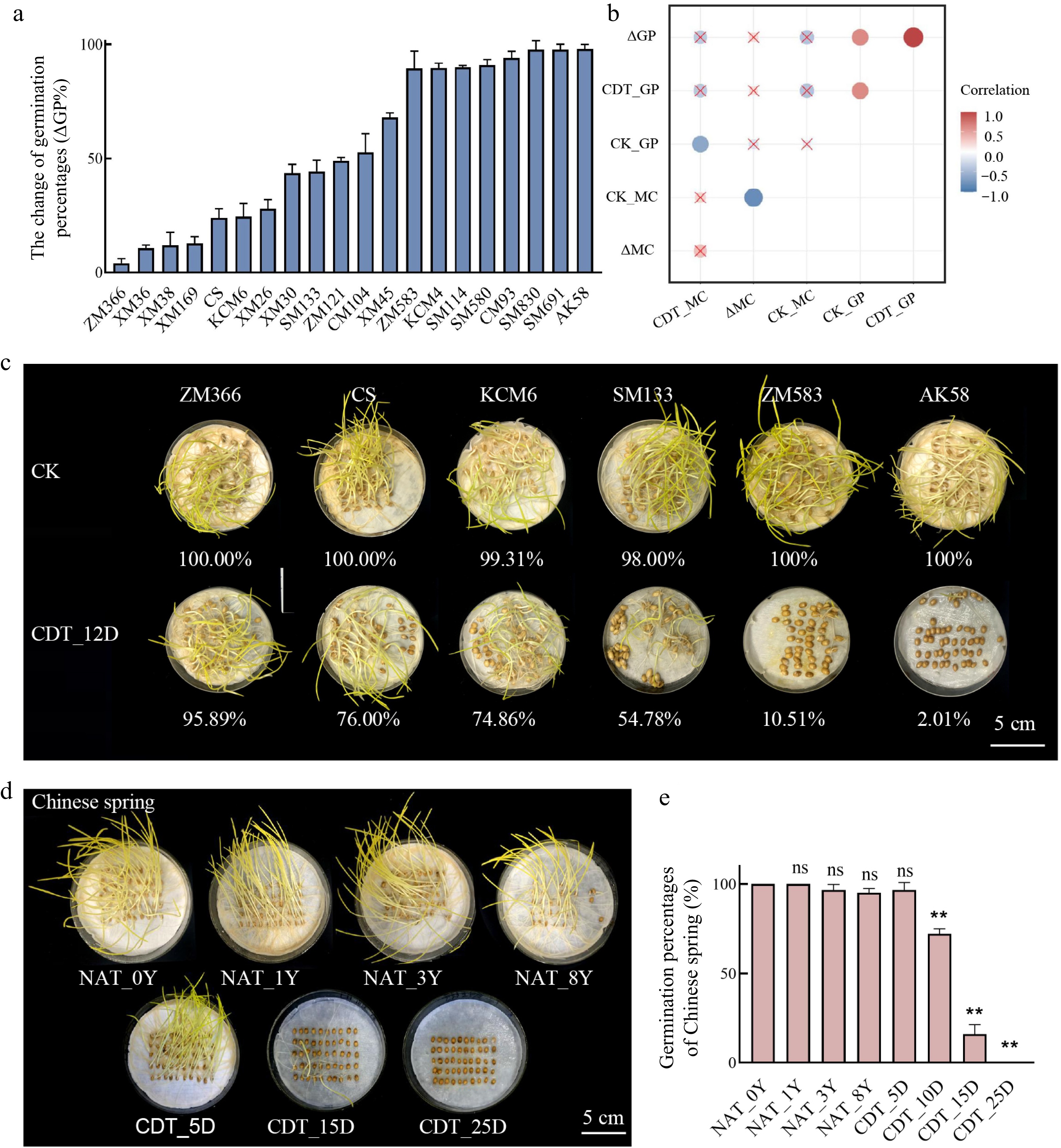
      Figure 1. 

      The germination percentage (GP) of different wheat accessions. (a) The change of germination percentages of 20 wheat varieties after 12 d of controlled deterioration treatment (CDT) (ΔGP%). Data are the means ± standard deviations (SDs) based on three biological replicates. (b) A correlation heatmap between moisture content and GP. CK_GP indicates the GP of non-aged seeds; CDT_GP indicates the GP of seeds that CDT for 12 d; CK_MC indicates the moisture content of seeds before aging; CDT_MC indicates the moisture content of seeds after CDT for 12 d; ΔGP suggests the change of germination during CDT; ΔMC indicates the change in moisture content of seeds during CDT. (c) The GP of six wheat varieties after CDT_12D; bar = 5 cm. (d), (e) The GP of Chinese Spring seeds under NAT and CDT; bar = 5 cm; compared to NAT_0Y seeds; ns, not significant; **, p < 0.01, one-way ANOVA with Tukey's test.

      The varieties with different seed longevity were selected to investigate changes in GP after CDT. Chinese Spring was chosen for subsequent studies due to its wide use in cytogenetic analysis and moderate longevity in our study. In Chinese Spring seeds, the GP of NAT seeds decreased slowly during aging, while the viability of CDT seeds decreased significantly. After 25 d of CDT, the seeds nearly lost their germination capacity (Fig. 1d, e). Therefore, the CDT method lets us obtain seeds of different ages quickly.

    • The relationship between seed longevity and RNA integrity number (RIN) was analyzed using total RNA from the embryo and aleurone layer of Chinese Spring seeds under NAT (0, 1, 3, 5, and 8 years) and CDT (5, 15, and 25 d). The mean RIN was significantly reduced, particularly in the embryo under CDT conditions (Fig. 2a). GP was positively correlated with RIN in both the aleurone layer (R2 = 0.70) and embryo (R2 = 0.45) in Chinese Spring seeds (Fig. 2b). The RIN values were compared between the high longevity cultivar Zhengmai 366 and the low longevity cultivar Aikang 58 after 12 d of CDT, and a reduction in GP was associated with a decrease in RIN value (Supplemental Fig. S2b). These findings suggest that stored mRNAs in embryos and the aleurone layer contribute to seed longevity and germination.

      Figure 2. 

      The RNA integrity (RIN) assays of Chinese Spring seeds. (a) RIN of Chinese Spring seeds (embryo and aleurone) under natural aging treatment (NAT) for 1, 3, and 8 years (compared to NAT_0Y) and controlled deterioration treatment (CDT) for 0, 5, 15, and 25 d (compared to CDT_0D). CDT for 0-d seeds was NAT for 3 years seeds. ns, not significant; *, p < 0.05; **, p < 0.01 one-way ANOVA with Tukey's test. (b) Correlation analysis between RIN and germination percentage of Chinese Spring seeds under NAT (0, 1, 3, 8 years) and CDT (0, 5, 15, 25 d) (Pearson's R2).

    • We used a long-read Nanopore sequencing platform to detect full-length mRNAs in aged seeds. The mRNAs were disrupted into small fragments before Illumina sequencing, so we chose Nanopore sequencing to obtain a more complete picture of the mRNA landscape in aged seeds. We analyzed samples from Chinese Spring seeds that were either NAT_0Y or subjected to CDT_25D. We obtained 205,991 and 115,606 sequences mapped to the reference transcriptome (iwgsc_ refseqv1.1_ annotation_200916 _HCLC_ cds. fa), respectively (Supplemental Table S3). We then kept 40,818 and 20,848 full-length stored mRNAs with identity ≥ 90% (Supplemental Table S3). A significantly higher amount of full-length mRNAs was detected in NAT_0Y (16,787 transcripts) than in the CDT_25D seeds (4,611 transcripts), suggesting that the mRNAs were largely degraded under CDT for 25 d (Supplemental Table S3). We identified 3,083 common full-length transcripts in NAT_0Y and CDT_25D seeds as LLR candidates (Supplemental Table S4, Fig. 3a).
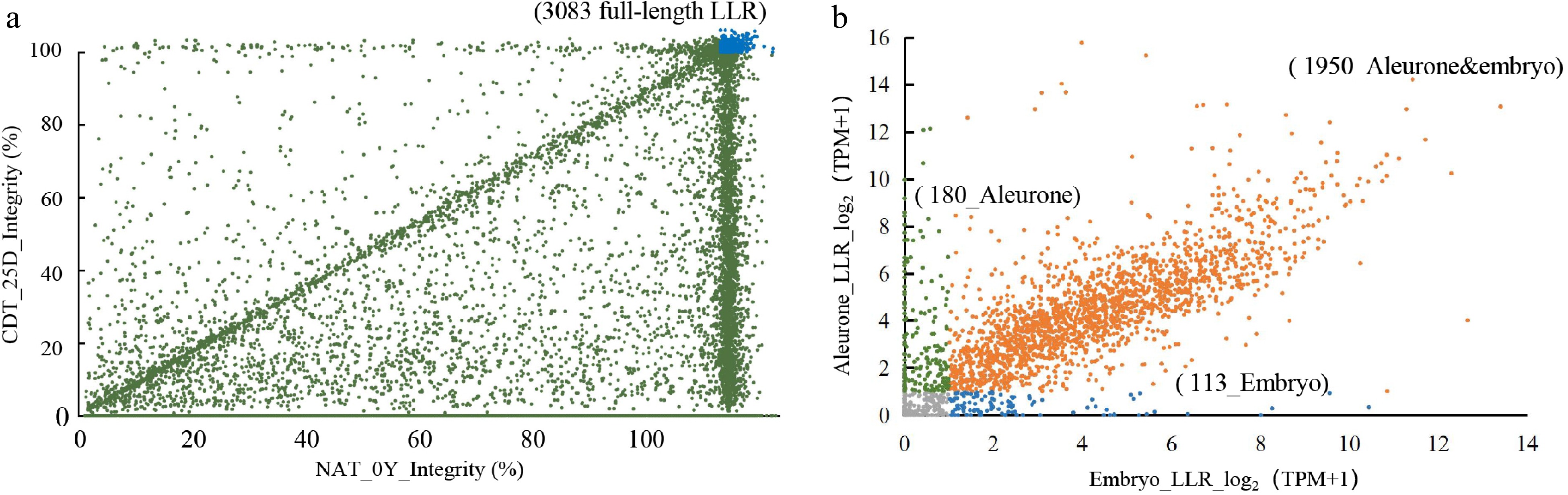
      Figure 3. 

      Identification of full-length long-lived mRNA (LLRs) and validation of transcript levels by Nanopore sequencing and Illumina RNA-seq. (a) Comparative analysis of full-length mRNA in seeds of NAT_0Y and CDT_25D. The blue portion represents 3083 LLRs identified in both samples with at least one full-length transcript. (b) The transcriptomic profile of 3083 full-length LLRs identified by Nanopore sequencing was analyzed. LLRs with a TPM ≥ 1 were considered to have detectable transcript levels. These LLRs were then selected by identifying overlapping genes in all eight aging gradient samples. Aleurone-specific LLRs were annotated with green, embryo-specific LLRs with blue, and LLRs in both tissues were annotated with yellow.

    • To ensure the purity of tissue separation, we selected four genes with distinct expression patterns between embryos and aleurone layers based on the public transcriptome data from Wheatomics[45]. We performed qPCR analysis to confirm a significant difference in relative transcription levels between these two tissues (Supplemental Fig. S1c). We conducted RNA-Seq using the embryo and aleurone layer of Chinese Spring seeds after CDT and NAT to further investigate the transcriptome changes during seed aging. We generated 3.1 billion high-quality clean reads, with Q30 values ranging from 87% to 94%, and the mean GC% content was 55%~61% (Supplemental Table S5). Principal component analysis (PCA) and correlation heatmap showed that these RNA-seq samples could be separated into four categories based on aging treatments and tissues (Supplemental Fig. S1d, S1e). The results showed that gene transcription levels gradually decreased in both the embryo and the aleurone layer during seed aging (Supplemental Fig. S3a, S3b). We identified 19,736 stored mRNAs in the embryo and 21,433 in the aleurone layer with TPM ≥ 1 in both NAT and CDT seeds (Supplemental Fig. S3c; Supplemental Table S6). The transcription of 3083 LLRs detected by Nanopore sequencing was supported by Illumina RNA-seq. We identified 2,130 and 2,064 LLRs in the aleurone layer and embryo, respectively, and 1,950 LLRs were shared in both tissues (Fig. 3b; Supplemental Table S7).

    • To assess the stability of LLRs under NAT (0, 1, 3, 5, and 8 years) and CDT (5, 15, and 25 d) conditions, we measured the gene fold change (FC) using Illumina RNA-seq. In the aleurone layer, there were more LLRs with log2FC ≥ 0 than LLRs with log2FC < 0 under both NAT and CDT conditions (Fig. 4a). However, in the embryo, the number of LLRs with log2FC ≥ 0 decreased during seed aging and was less than that of LLRs with log2FC < 0 in CDT_25D seeds (Fig. 4b). These results suggested that the stability of LLRs differs between the aleurone layer and embryo and that LLRs in the aleurone layer were generally more stable than those in the embryo under aging conditions.
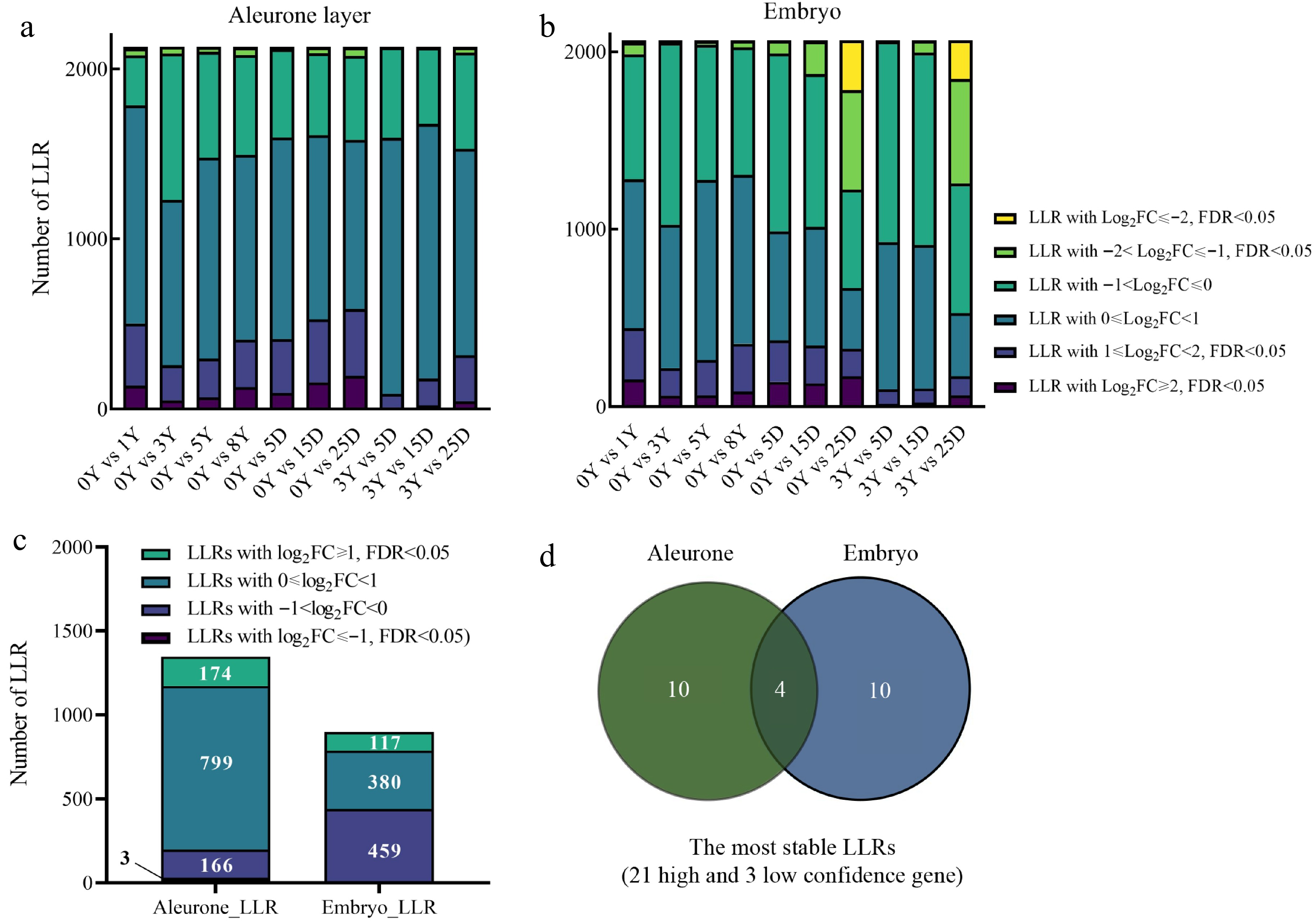
      Figure 4. 

      The degradation pattern of long-lived mRNA (LLRs). (a), (b) LLRs with different fold change (FC) among groups of comparison. (c) This refers to the count of genes that overlap with the same FC across different aging samples from the same tissue. Specifically, the samples include natural aging treatment (NAT) for 0, 1, 3, 5, and 8 years and controlled deterioration treatment (CDT) for 5, 15, and 25 d seeds. (d) Venn diagram comparisons of the most stable LLRs in the embryo and aleurone layer. The most stable LLRs were the overlapping LLRs with log2FC ≥ 1 and FDR < 0.05 between NAT_3Y and CDT (5, 15, and 25 d), which were selected from the overlapping LLRs with log2FC ≥ 1 and FDR < 0.05 (compared to NAT_0Y) as mentioned in Fig. 4c.

      Further analysis demonstrated that the LLRs had different FCs across various seed aging samples and fresh NAT_0Y seeds, likely due to differences in their Transcripts Per Million (TPMs) (Supplemental Table S6, S7). We examined the overlapping LLRs among all aged samples to identify LLRs with similar FC features in aleurone layers or embryos. We found that a significant portion of genes (14.80% from the aleurone layer and 49.00% from the embryo) were LLRs with log2FC < 0. Several LLRs log2FC ≤ 1 and FDR < 0.05 were identified in all overlapping LLR sets (Fig. 4c; Supplemental Table S8). These LLRs with log2FC < 0 were associated with translation and transportation, as shown by Go enrichment analysis (Supplemental Fig. S4a, S4b; Supplemental Table S9). In contrast, LLRs with log2FC ≥ 1, FDR < 0.05 were associated with salt stress, heat, protein folding, reactive oxygen species, protein complex oligomerization, and abscisic acid metabolism (Supplemental Fig. S4c, S4d; Supplemental Table S9). LLRs with log2FC ≥ 1 and FDR < 0.05 may be more stable (Supplemental Table S8) since elevated read counts were obtained in the degraded samples. In addition, the TPMs of LLRs with log2FC ≥ 1 and FDR < 0.05 were relatively higher in NAT_3Y seeds than in NAT_0Y (Supplemental Table S7). Therefore, these LLRs with significantly increased transcripts level that still meets the criteria of log2FC ≥ 1 and FDR < 0.05 when compared to CDT_5D, CDT_15D, and CDT_25D samples with NAT_3Y may have the highest stability during aging. Finally, we identified 24 LLRs with the highest stability during aging (Fig. 4d), among which three were low-confidence genes. So only 21 LLRs were subjected to further analysis. Analysis of these 21 transcripts in embryo and aleurone layer tissues of fresh (NAT_0Y), NAT_3Y, and CDT_25D seeds showed a uniform distribution rather than a gradual increase from 5' to 3' end, eliminating the sequencing bias of Illumina RNA-seq. This result also demonstrated the full-length characteristic of these most stable LLRs (Supplemental Fig. S5).

    • The length of the coding sequence for high-confidence genes varied from 54 to 16,080 bp[45], whereas the 21 most stable LLRs identified in this study had coding sequence lengths of 336−720 bp in the aleurone layer and 195−591 bp in the embryo (Fig. 5ac). Of all high-confidence genes, 74,181 had coding sequence lengths greater than 720 bp. In contrast, the length of 33,711 genes is not longer than 720 bp. Although the number of transcripts with length longer than 720 bp was 2.2 times greater than those shorter than 720 bp, none of them were identified as the most stable LLR, indicating that shorter transcripts may be more stable during seed aging in wheat (Fig. 5ac). Furthermore, the average GC% contents, which is a reason for the stability of transcript, of the LLRs in the aleurone layer and embryo (approximately 75%) was higher than that of all high-confidence genes (approximately 55%), suggesting that the gene with higher GC% content may be more stable during seed aging (Fig. 5d).
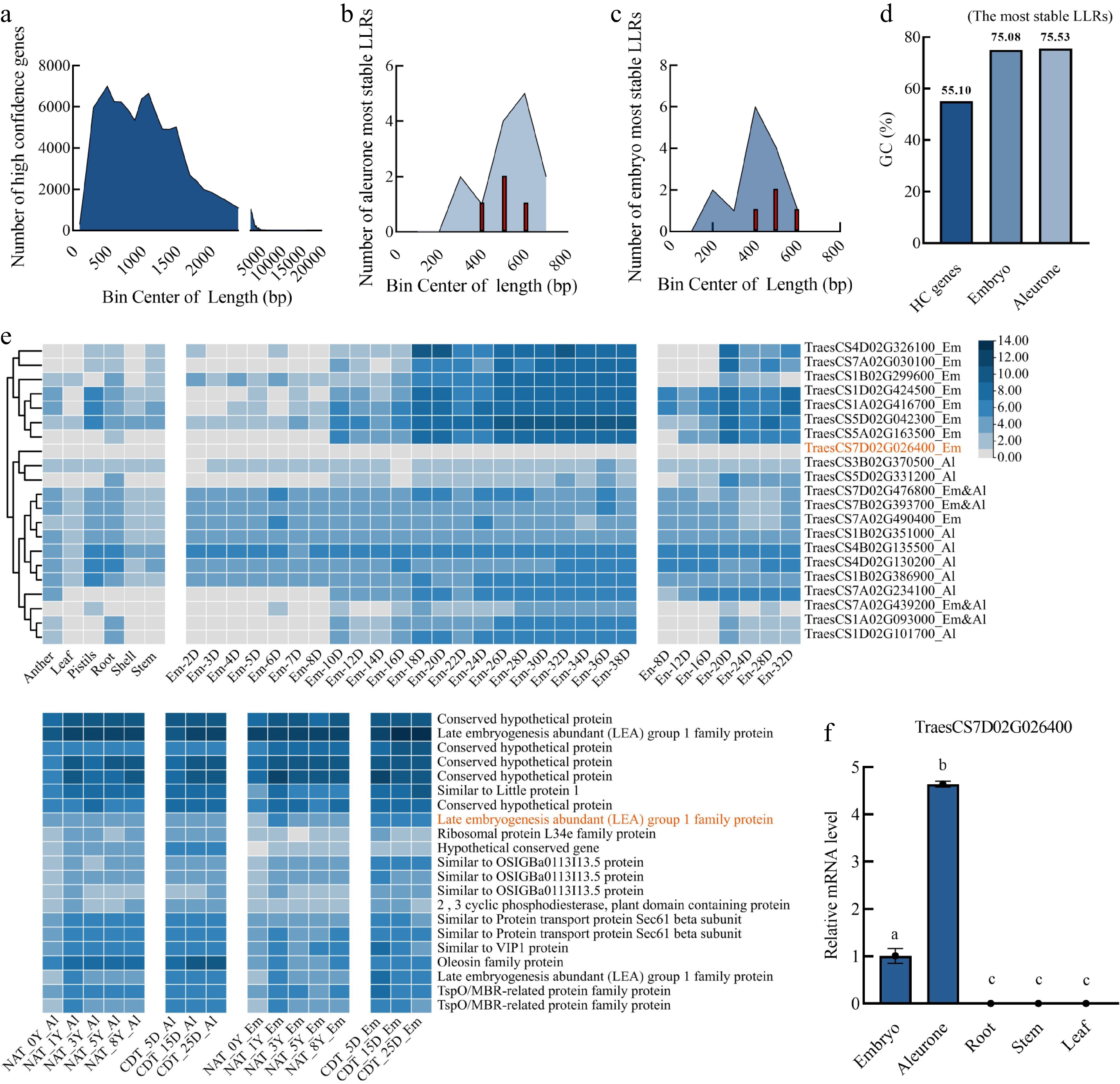
      Figure 5. 

      Characterization of the most stable long-lived mRNAs (LLRs) in the embryo and aleurone layers. (a), (b), (c) Frequency analysis of the coding sequence length (x-axis) and gene numbers (y-axis). (a) Total high confidence genes. (b) The most stable LLRs in the aleurone layer. (c) The most stable LLRs in the embryo. (d) The average GC (%) content of all the high confidence genes in wheat seeds and the most stable LLRs in the wheat embryo and aleurone layer. (e) Heatmap visualization of RNA-seq data (Chinese Spring) from anther, leaf, pistils, root, shell, stem, the developing embryo (2−38 d), developing endosperm (8−32 d), NAT (0, 1, 3, 5, and 8 years) seeds, and CDT (5, 15, and 25 d) seeds. Embryo, EM; Aleurone layer, Al; Endosperm, En. LLRs in both embryo and aleurone layer,m Em&AL. (f) The relative expression levels of TraesCS7D02G026400 in root, stem, and leaf were determined by qPCR using actin as an internal reference gene. Two independent experiments were performed with triplicate samples in each experiment. The relative expression levels were calculated using the TBtools (Simple q-PCR summary) software. Using different lowercase letters to represent significant differences (p < 0.01, one-way ANOVA with Tukey's test).

      The promoters of the 21 most stable LLRs were found to contain motifs associated with various plant hormone responses, including abscisic acid response (ABRE), light response (G-box), and hormone response (Supplemental Fig. S6, Supplemental Table S10). The promoter of 17 genes contains motifs related to cis-acting regulatory elements involved in methyl jasmonate (MeJA) response (CGTCA motif and TGACG motif) (Supplemental Fig. S6). Additionally, motifs related to gibberellin response (P-BOX) and auxin response (TGA elements) were identified in three of the four LLRs that were detected in both the aleurone layer and embryo (Supplemental Fig. S6).

      The four most stable LLRs in the embryo and aleurone layer were annotated as members of the LEA 1 protein family (TraesCS7A02G439200), TSPO/MBR-related protein family (TraesCS1A02G093000), and OSIGBa0113113.5 protein (TraesCS7D02G476800, TraesCS7B02G393700) (Fig. 5e ). The expression analysis in anther, leaf, pistils, root, shell, stem, embryo, endosperm, NAT, and CDT wheat seeds indicated that TraesCS7D02G026400 was a seed-specific gene as well as an embryo-specific stable LLR in RNA seq data (Fig. 5e; Supplemental Table S11). The qPCR results showed a significantly higher relative mRNA level of TraesCS7D02G026400 in the embryo and aleurone layer compared with the root, stem, and leaf (Fig. 5f, Supplemental Table S2). None of the other 20 most stable LLRs was found to be seed-specific (Fig. 5e; Supplemental Table S11).

    • We tried to design primers for the 21 most stable LLRs based on the Chinese Spring genome sequence, but their high GC content brought challenges for primer design and PCR amplification. Therefore, we selected 7 LLRs (LLR 2, 3, 10, 11, 13, 15, and 20) to measure their stability in 18 wheat varieties after aging (Fig. 6a). To evaluate the stability of LLRs, we used three genes previously shown to degrade gradually as wheat seeds aged, and they belonged to the short-lived mRNA (SLR) set in our study[20]. After natural aging for 3 years, SLRs could be amplified in more than ten varieties. However, after CDT for 25 days, all three SLRs were no longer detectable in any of the 18 wheat varieties (Fig. 6b). In contrast, all seven LLRs could be successfully amplified in more than 11 varieties after both aging treatments (Supplemental Fig. S7). These results suggest that LLRs are relatively stable during seed aging, even in different wheat varieties.
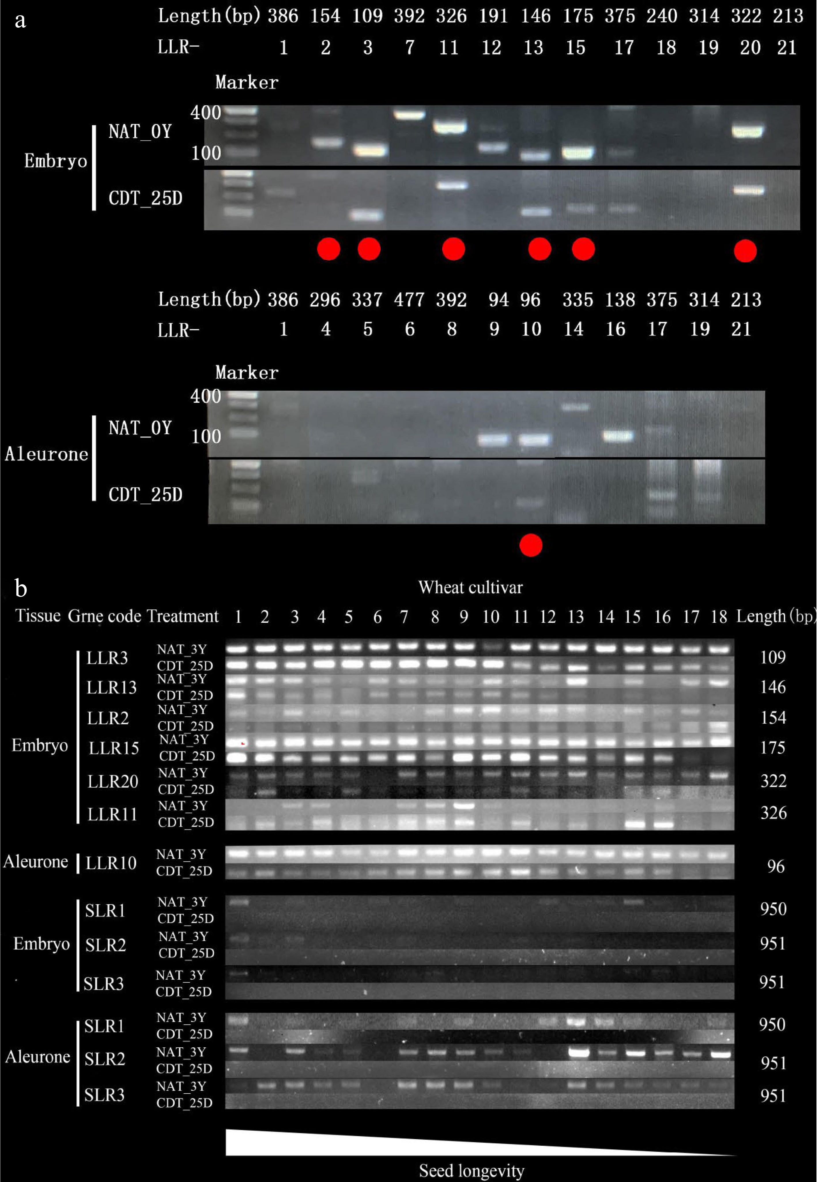
      Figure 6. 

      The transcript levels of the most stable long-lived mRNAs (LLRs) in naturally aged treatment (NAT) and controlled deterioration treatment (CDT) wheat seeds detected by RT-PCR. The number of PCR cycles varied depending on their mRNA abundance in the unaged seeds. PCR products were run in agarose gels. (a) The 21 most stable LLRs in NAT for 0 Y (NAT_0Y) and CDT for 25 d (CDT_25D) Chinese Spring seeds were analyzed, respectively (see Supplemental Table S2 for the gene list). The 10 genes in 18 wheat varieties that NAT for 3 years (NAT_3Y) seeds and CDT_25D seeds were analyzed. (See Supplemental Table S1 for the wheat cultivar code and Supplemental Table S2 for the gene list). The lower wheat cultivar code number indicates higher seed longevity in (b).

      Furthermore, to investigate the variation in seed longevity, we examined the stability of LLR13, LLR15, and LLR20 in four wheat varieties (SM691, AK58, ZM366, and XM36) with different degrees of reduction in seed germination percentage (ΔGP) after 25 d of CDT treatment. The results showed that LLR13, LLR15, and LLR20 degraded significantly in SM691 (ΔGP = 97.79%) and AK58 (ΔGP = 97.99%) but persisted in ZM366 (ΔGP = 4.11%) and XM36 (ΔGP = 10.75%) (Supplemental Fig. S7). These findings suggest that the presence or absence of specific combinations of LLRs can serve as molecular markers to estimate seed longevity, and LLR13, LLR15, and LLR20 may be promising candidates for further investigation into their roles in seed aging and longevity.

    • Seed germination refers to the physiological process culminating in the emergence of the embryo from its enclosing coverings, including the endosperm, perisperm, testa, or pericarp. Starch degradation, initiated by GA secreted by the embryo during germination, is considered a post-germinative event[31,32]. The scutellum, rather than the aleurone epithelium, is mainly responsible for the synthesis of α-amylase during the initial stages of germination in wheat, rye, oats, and maize[47]. However, any malfunctioning embryo or aleurone sections can affect seed germination[48]. Seed GP was correlated with the aleurone layer RIN and the embryo RIN (Fig. 2b). The mutagenic substances formed during aging would act early during the germination of seeds. The deleterious effects of aged endosperm on a young embryo[49] might be related to the decreased activity of antioxidant enzymes, such as catalase, peroxidase, dehydrogenase, and amylase[50]. The accumulation of toxic compounds in the aged endosperm or aleurone can induce chromosomal breakage in young embryos[51]. The response of aleurone layers from normal and aged seeds to heat shock has been investigated. Only aleurone layers from normally germinated seeds could recommence substantial α-amylase synthesis during recovery[52]. One of the LLRs identified in the aleurone layer was an oleosin family protein (TraesCS7A02G234100) (Fig. 5e), which may be involved in oil body mobilization during post-germinative seedling growth and may prevent the coalescence of protein storage vacuoles[5355]. Our study found that the aleurone layer had more stored mRNAs and LLRs in aged seeds than the embryos (Supplemental Fig. S3a, S3b; Fig. 4c). Lipid oxidation has been implicated in seed deterioration, and detailed analyses of the changes in the lipidome during long-term dry storage of a range of genotypes of oilseed rape wheat, barley and Arabidopsis support this claim[5658]. The lipid content of wheat embryo (8%−15%) is higher than that of other seed tissues (bran, aleurone, and endosperm; 6.8%−7.5%)[59]. Additionally, the embryos and endosperms or aleurone layer have different enzymatic patterns, highlighting that the two seed compartments age independently[6]. These differences between embryos and endosperm (aleurone layer) may cause varying degradation percentages of mRNAs (Fig. 4a, b).

    • Our study observed reduced RNA integrity in the embryo and aleurone layer of aged seeds (Fig. 2a), with the lowest RIN being 5.5 (Fig. 2a). Despite a RIN lower than 3, the length of mRNA is still longer than 8000 bp[35]. Although Illumina's TPM and FC can predict mRNA degradation trends[40,44], mRNA fragmentation errors may exist with short-read sequencing. Therefore, it is difficult to determine whether the interruption of mRNAs is caused by seed aging or by the sequencing technology used, as Illumina technology can interrupt mRNAs before sequencing (Supplemental Table S5). By using the NEBNext Poly (A) mRNA magnetic isolation module and cDNA synthesis, Nanopore full-length sequencing was employed in our study to enrich and identify mRNAs that remain intact during aging[41]. Integrating Nanopore and Illumina sequencing enables the identification of LLRs with at least one full-length transcript and predicts mRNA degradation trends in aged seeds. Thus, our approach can effectively exclude the effects of mRNA fragmentation errors, leading to more accurate identification of LLRs. In conclusion, LLRs can be predicted by the FC determined by short-read sequencing[44], and fragment mRNA errors can be excluded by full-length Nanopore sequencing[36], demonstrating the integration of both sequencing technologies is a powerful tool for identifying stable mRNAs in aged seeds.

    • Poly(A) polymerase activity decreases with age, and the translational levels decrease in aged wheat embryos[60,61]. Transcript degradation of the elongation factor EF-1 occurs both in the embryo during NAT and CDT but still exists in the embryo and aleurone layer in CDT_25D (Supplemental Table S6). A longevity-related QTL (Q.Lng.ipk.2A.1) contains a candidate gene similar to the translation elongation factor EFG/EF2 protein[62]. Transcripts related to ribosomal functions, particularly translation, are overrepresented in the stable mRNAs group and may indicate the importance of reconstituting the translational machinery during germination[3]. Among the analyzed mRNAs, 21 selected LLRs were more stable (Fig. 4d). The coding sequence of these LLRs was enriched with three repeats of the sequence TCCTCCTCC, which might be related to transcription factor IIIA and ribosomal protein L5[63]. The ribosomal L34e and preprotein translocase family proteins mRNA were detected as the aleurone layer's most stable LLRs (Fig. 5e). The longevity markers 7D (Wpt-0934) and 7A (wPt-0303) also reveal the relationship between ribosomal proteins and seed longevity[64].

      In the aleurone layer, VIP1 was identified as the most stable LLR. It plays a role in the osmosensory response by binding to the 5'-AGCTGT/G-3' DNA sequence and is found in the promoters of the hypoosmolarity-responsive genes CYP707A1 and CYP707A3[65]. LEA 1, TSPO, and OSIGBa0113113.5 were identified as the most stable LLRs in both embryos and aleurone layers (Fig. 5e). The seed-specific expressed gene (TraesCS7D02G026400) is annotated as an LEA 1 family protein (Fig. 5e). The LEA 1 proteins, which have evolutionary and functional characteristics of an ancestral plant protein group, are also present in other eukaryotes and the Archaea and Bacteria domains[66]. In Arabidopsis, maize, and Medicago, LEA 1 protein is correlated with seed vigor and longevity[6769]. Wheat seed longevity markers on 4B (wPt-1272) have identified some genes described as dehydrin-/LEA group proteins[64]. TSPO (Fig. 5e) expression seems to be correlated with LEA4-5 protein (TraesCS7A02G439200) expression in Arabidopsis[70]. TSPO is a stress-induced, posttranslationally regulated, and early secretory pathway-localized plant cell membrane protein involved in transient intracellular ABA-dependent stress signaling and has roles in apoptosis[71,72]. LLR 13, 15, and 20 were more stable in high longevity varieties than short longevity varieties after aging (Supplemental Fig. S7), suggesting that these stable LLRs may contribute to seed survival[40]. In addition to the 21 most stable mRNAs, several LLRs with log2FC ≥ 0 were identified in both the embryos and the aleurone layer, and they may be necessary for seed longevity. For example, the heat shock protein (HSP) and 1-cysteine peroxiredoxin antioxidant (PER1) were identified as LLRs (Fig. 4c, Supplemental Table S8). The heat shock protein OsHSP18.2 improved seed longevity under CDT[73]. A PER1 protein from Nelumbo nucifera enhances seed longevity and stress tolerance in Arabidopsis, and the PER1 protein is stable in high-vigor wheat after aging treatment[74,75]. A multi-omic study revealed a bZIP23-PER1A–mediated detoxification pathway to enhance seed vigor in rice[27]. These mRNAs existed after NAT and CDT, but the molecular mechanisms responsible for their role in wheat seed longevity and germination have not yet been clarified.

      Seeds translate stored mRNAs during germination using stored ribosomes, and RNA integrity is closely related to seed vigor[19,21]. The germination of dry wheat seeds correlates with the embryo and the living aleurone cell mRNAs[30]. Our study identified specific LLRs related to longevity by comparing high-vigor and low-vigor varieties, and we examined the degradation rates of mRNA by transcriptome profiling[40]. We verified full-length LLRs using Nanopore sequencing[36,44]. While LLRs have a short and high GC content, the protected manner of mRNAs results in mRNAs having variant degradation percentages[23,76]. However, fission due to free radical attacks at random bases is also evident[20,36]. Further investigation is necessary to uncover the complex roles of these LLRs in seed longevity and the mechanism of seed resurrection. Overall, our study provides valuable insights into the mechanisms of plant cell survival and may contribute to developing more effective seed storage and preservation strategies.

      • This research was funded by the Major Program of National Agricultural Science and Technology of China (NK20220607), the National Natural Science Foundation of China (U22A20472), the National Key Research and Development Program of China (2018YFE0112000), the Sichuan Science and Technology Support Project (2021YFH0077; 2021YFYZ0027; 23NSFSC0770), the Science and Technology Support Project of Chengdu (2021-GH03-00002-HZ) and the open research fund of SKL-CGEUSC (SKL-ZD202212).

      • The authors declare that they have no conflict of interest.

      • Supplemental Fig. S1 Tissue purity and correlation between biological replicates in Illumina RNA-seq.
      • Supplemental Fig. S2 The water content and RNA integrity number (RIN) of different varieties.
      • Supplemental Fig. S3 Identification of stored mRNA in aged wheat seeds by Illumina RNA-seq.
      • Supplemental Fig. S4 Gene Ontology (GO) enrichment of overlapping long-lived mRNAs (LLRs).
      • Supplemental Fig. S5 The sequencing depth from 5' to 3' of the most stable long-lived mRNAs (LLRs) and easily degraded mRNAs.
      • Supplemental Fig. S6 Sequence analysis of the most stable long-lived mRNAs (LLRs).
      • Supplemental Fig. S7 The correlation between longevity and the most stable long-lived mRNAs (LLRs).
      • Supplemental Table S1 Wheat accessions used in this study.
      • Supplemental Table S2 Quality of Nanopore-Seq data obtained in this study for NAT_0Y (fresh seeds) and CDT_25D seeds.
      • Supplemental Table S3 Full-length long-lived mRNAs (LLRs) identified by Nanopore-seq data.
      • Supplemental Table S4 Quality of Illumina RNA-seq data.
      • Supplemental Table S5 Identification of stored mRNAs in embryo and aleurone layer using Illumina RNA-seq.
      • Supplemental Table S6 Transcripts Per Million (TPM) analysis of full-length long-lived mRNAs (LLRs) in embryo and aleurone layers using Illumina RNA-seq.
      • Supplemental Table S7 long-lived mRNAs (LLRs) with different fold changes (FC) in the embryos and aleurone layers compared with NAT_0Y.
      • Supplemental Table S8 Gene Ontology (GO) enrichment analysis of overlapping long-lived mRNAs (LLRs) with relatively decreased and significantly increased transcript levels in embryos and aleurone layers.
      • Supplemental Table S9 Motifs in the promoter of the most stable long-lived mRNAs.
      • Supplemental Table S10 Expression profiles of the most stable long-lived mRNAs in different tissues and treatments.
      • Supplemental Table S11 List of genes and primers used for Reverse Transcription-Polymerase Chain Reaction (RT-PCR) and Real-Time Quantitative Polymerase Chain Reaction (qPCR) experiments.
      • Copyright: © 2023 by the author(s). Published by Maximum Academic Press on behalf of Hainan Yazhou Bay Seed Laboratory. This article is an open access article distributed under Creative Commons Attribution License (CC BY 4.0), visit https://creativecommons.org/licenses/by/4.0/.
    Figure (6)  References (76)
  • About this article
    Cite this article
    Liang W, Dong H, Guo X, Rodríguez V, Cheng M, et al. 2023. Identification of long-lived and stable mRNAs in the aged seeds of wheat. Seed Biology 2:14 doi: 10.48130/SeedBio-2023-0014
    Liang W, Dong H, Guo X, Rodríguez V, Cheng M, et al. 2023. Identification of long-lived and stable mRNAs in the aged seeds of wheat. Seed Biology 2:14 doi: 10.48130/SeedBio-2023-0014

Catalog

  • About this article

/

DownLoad:  Full-Size Img  PowerPoint
Return
Return