ARTICLE   Open Access    

Doses of aminoethoxyvinylglycine (AVG) in tomato postharvest storage

More Information
  • Tomatoes are one of the main vegetables found daily in world cuisine and are highly perishable. The aim of this study was to evaluate the quality of 'Débora' tomato fruits over the post-harvest period in refrigerated storage, after immersion in solutions with different concentrations of aminoethoxyvinylglycine (AVG). The experimental design was entirely randomized in a 4 × 5 double factorial scheme, with four treatments, AVG doses 0 (control), 500, 1,000, and 1,500 mg·L−1, and five days of evaluation during the 28 d of refrigerated storage (days 0, 7, 14, 21 and 28) with three replications. Physicochemical analyses were carried out on weight loss, respiration rate, firmness, hydrogen potential (pH), soluble solids, titratable acidity and external and internal fruit color parameters, luminosity, chroma, and °hue. Two-way ANOVA, Tukey's mean test (p < 0.05), and multivariate statistical analysis by principal components (PCA) were carried out. The PCA allowed us to infer that, as weight loss increased, firmness decreased, an effect that was minimized with AVG at a dose of 1,500 mg·L−1, which may be related to the inhibition of the fruit's ethylene production rate. AVG delayed the post-harvest ripening of tomato fruit, reduced the respiration rate of the fruit, and the changes in external and internal chroma. The doses of AVG did not affect the luminosity and pH of the fruit pulp.
  • Crops require a variety of nutrients for growth and nitrogen is particularly important. Nitrogen is the primary factor limiting plant growth and yield formation, and it also plays a significant role in improving product quality[14]. Nitrogen accounts for 1%−3% of the dry weight of plants and is a component of many compounds. For example, it is an important part of proteins, a component of nucleic acids, the skeleton of cell membranes, and a constituent of chlorophyll[5,6]. When the plant is deficient in nitrogen, the synthesis process of nitrogen-containing substances such as proteins decrease significantly, cell division and elongation are restricted, and chlorophyll content decreases, and this leads to short and thin plants, small leaves, and pale color[2,7,8]. If nitrogen in the plant is in excess, a large number of carbohydrates will be used for the synthesis of proteins, chlorophyll, and other substances, so that cells are large and thin-walled, and easy to be attacked by pests and diseases. At the same time, the mechanical tissues in the stem are not well developed and are prone to collapse[3,8,9]. Therefore, the development of new crop varieties with both high yields and improved nitrogen use efficiency (NUE) is an urgently needed goal for more sustainable agriculture with minimal nitrogen demand.

    Plants obtain inorganic nitrogen from the soil, mainly in the form of NH4+ and nitrate (NO3)[1013]. Nitrate uptake by plants occurs primarily in aerobic environments[3]. Transmembrane proteins are required for nitrate uptake from the external environment as well as for transport and translocation between cells, tissues, and organs. NITRATE TRANSPORTER PROTEIN 1 (NRT1)/PEPTIDE TRANSPORTER (PTR) family (NPF), NRT2, CHLORIDE CHANNEL (CLC) family, and SLOW ACTIVATING ANION CHANNEL are four protein families involved in nitrate transport[14]. One of the most studied of these is NRT1.1, which has multiple functions[14]. NRT1.1 is a major nitrate sensor, regulating many aspects of nitrate physiology and developmental responses, including regulating the expression levels of nitrate-related genes, modulating root architecture, and alleviating seed dormancy[1518].

    There is mounting evidence that plant growth and development are influenced by interactions across numerous phytohormone signaling pathways, including abscisic acid, gibberellins, growth hormones, and cytokinins[3,19,20]. To increase the effectiveness of plant nitrogen fertilizer application, it may be possible to tweak the signaling mediators or vary the content of certain phytohormones. Since the 1930s, research on the interplay between growth factors and N metabolism has also been conducted[3]. The Indole acetic acid (IAA) level of plant shoots is shown to decrease in early studies due to N shortage, although roots exhibit the reverse tendency[3,21]. In particular, low NO3 levels caused IAA buildup in the roots of Arabidopsis, Glycine max, Triticum aestivum, and Zea mays, indicating that IAA is crucial for conveying the effectiveness of exogenous nitrogen to the root growth response[20,22,23].

    Studies have shown that two families are required to control the expression of auxin-responsive genes: one is the Auxin Response Factor (ARF) and the other is the Aux/IAA repressor family[2426]. As the transcription factor, the ARF protein regulates the expression of auxin response genes by specifically binding to the TGTCNN auxin response element (AuxRE) in promoters of primary or early auxin response genes[27]. Among them, rice OsARF18, as a class of transcriptional repressor, has been involved in the field of nitrogen utilization and yield[23,28]. In rice (Oryza sativa), mutations in rice salt tolerant 1 (rst1), encoding the OsARF18 gene, lead to the loss of its transcriptional repressor activity and up-regulation of OsAS1 expression, which accelerates the assimilation of NH4+ to Asn and thus increases N utilization[28]. In addition, dao mutant plants deterred the conversion of IAA to OxIAA, thus high levels of IAA strongly activates OsARF18, which subsequently represses the expression of OsARF2 and OsSUT1 by directly binding to the AuxRE and SuRE promoter motifs, resulting in the inhibition of carbohydrate partitioning[23]. As a result, rice carrying the dao has low yields.

    Apples (Malus domestica) are used as a commercially important crop because of their high ecological adaptability, high nutritional value, and annual availability of fruit[29]. To ensure high apple yields, growers promote rapid early fruit yield growth by applying nitrogen. However, the over-application of nitrogen fertilizer to apples during cultivation also produces common diseases and the over-application of nitrogen fertilizer is not only a waste of resources but also harmful to the environment[29]. Therefore, it is of great significance to explore efficient nitrogen-regulated genes to understand the uptake and regulation of nitrogen fertilizer in apples, and to provide reasonable guidance for nitrogen application during apple production[30]. In this study, MdARF18 is identified which is a key transcription factor involved in nitrate uptake and transport in apples and MdARF18 reduces NO3 uptake and assimilation. Further analysis suggests that MdRF18 may inhibit the transcriptional level of MdNRT1.1 promoter by directly binding to its TGTCTT target, thus affecting normal plant growth.

    The protein sequence of apple MdARF18 (MD07G1152100) was obtained from The Apple Genome (https://iris.angers.inra.fr/gddh13/). Mutant of arf18 (GABI_699B09) sequence numbers were obtained from the official TAIR website (www.arabidopsis.org). The protein sequences of ARF18 from different species were obtained from the protein sequence of apple MdARF18 on the NCBI website. Using these data, a phylogenetic tree with reasonably close associations was constructed[31].

    Protein structural domain prediction of ARF18 was performed on the SMART website (https://smart.embl.de/). Motif analysis of ARF18 was performed by MEME (https://meme-suite.org/meme/tools/meme). Clustal was used to do multiple sequence comparisons. The first step was accessing the EBI web server through the Clustal Omega channel. The visualization of the results was altered using Jalview, which may be downloaded from www.jalview.org/download.[32]

    The apple 'Orin' callus was transplanted on MS medium containing 1.5 mg·L−1 6-benzylaminopurine (6-BA) and 0.5 mg·L−1 2,4 dichlorophenoxyacetic acid (2,4-D) at 25 °C, in the dark, at 21-d intervals. 'Royal Gala' apple cultivars were cultured in vermiculite and transplanted at 25 °C every 30 d. The Arabidopsis plants used were of the Columbia (Col-0) wild-type variety. Sowing and germinating Arabidopsis seeds on MS nutrient medium, and Arabidopsis seeds were incubated and grown at 25 °C (light/dark cycle of 16 h/8 h)[33].

    The nutrient solution in the base contained 1.0 mM CaCl2, 1.0 mM KH2PO4, 1.0 mM MgSO4, 0.1 mM FeSO4·7H2O 0.1 mM Na2EDTA·2H2O, 50 μM MnSO4·H2O, 50 μM H3BO3, 0.05 μM CuSO4·5H2O, 0.5 μM Na2MoO4·2H2O, 15 μM ZnSO4·7H2O, 2.5 μM KI, and 0.05 μM CoCl·6H2O, and 0.05 μM CoCl·6H2O, and 0.05 μM CoCl· 6H2O. 2H2O, 15 μM ZnSO4·7H2O, 2.5 μM KI and 0.05 μM CoCl·6H2O, and 0.05 μM CoCl·6H2O, supplemented with 0.5 mM, 2 mM, and 10 mM KNO3 as the sole nitrogen source, and added with the relevant concentrations of KCl to maintain the same K concentration[33,34].

    For auxin treatment, 12 uniformly growing apple tissue-cultured seedlings (Malus domestica 'Royal Gala') were selected from each of the control and treatment groups, apple seedlings were incubated in a nutrient solution containing 1.5 mg·L−1 6-BA, 0.2 mg·L−1 naphthalene acetic acid, and IAA (10 μM) for 50 d, and then the physiological data were determined. Apple seedlings were incubated and grown at 25 °C (light/dark cycle of 16 h/8 h).

    For nitrate treatment, Arabidopsis seedlings were transferred into an MS medium (containing different concentrations of KNO3) as soon as they germinated to test root development. Seven-day-old Arabidopsis were transplanted into vermiculite and then treated with a nutrient solution containing different concentrations of KNO3 (0.5, 2, 10 mM) and watered at 10-d intervals. Apple calli were treated with medium containing 1.5 mg·L−1 6-BA, 0.5 mg·L−1 2,4-D, and varying doses of KNO3 (0.5, 2, and 10 mM) for 25 d, and samples were examined for relevant physiological data. Apple calli were subjected to the same treatment for 1 d for GUS staining[35].

    To obtain MdARF18 overexpression materials, the open reading frame (ORF) of MdARF18 was introduced into the pRI-101 vector. To obtain pMdNRT1.1 material, the 2 kb segment located before the transcription start site of MdNRT1.1 was inserted into the pCAMBIA1300 vector. The Agrobacterium tumefaciens LBA4404 strain was cultivated in lysozyme broth (LB) medium supplemented with 50 mg·L−1 kanamycin and 50 mg·L−1 rifampicin. The MdARF18 overexpression vector and the ProMdNRT1.1::GUS vector were introduced into Arabidopsis and apple callus using the flower dip transformation procedure. The third-generation homozygous transgenic Arabidopsis (T3) and transgenic calli were obtained[36]. Information on the relevant primers designed is shown in Supplemental Table S1.

    Plant DNA and RNA were obtained using the Genomic DNA Kit and the Omni Plant RNA Kit (tDNase I) (Tiangen, Beijing, China)[37].

    cDNA was synthesized for qPCR by using the PrimeScript First Strand cDNA Synthesis Kit (Takara, Dalian, China). The cDNA for qPCR was synthesized by using the PrimeScript First Strand cDNA Synthesis Kit (Takara, Dalian, China). Quantitative real-time fluorescence analysis was performed by using the UltraSYBR Mixture (Low Rox) kit (ComWin Biotech Co. Ltd., Beijing, China). qRT-PCR experiments were performed using the 2−ΔΔCᴛ method for data analysis. The data were analyzed by the 2−ΔΔCᴛ method[31].

    GUS staining buffer contained 1 mM 5-bromo-4-chloro-3-indolyl-β-glutamic acid, 0.01 mM EDTA, 0.5 mM hydrogen ferrocyanide, 100 mM sodium phosphate (pH 7.0), and 0.1% (v/v) Triton X-100 was maintained at 37 °C in the dark. The pMdNRT1.1::GUS construct was transiently introduced into apple calli. To confirm whether MdNRT1.1 is activated or inhibited by MdARF18, we co-transformed 35S::MdARF18 into pMdNRT1.1::GUS is calling. The activity of transgenic calli was assessed using GUS labeling and activity assays[33,38].

    The specimens were crushed into fine particles, combined with 1 mL of ddH2O, and thereafter subjected to a temperature of 100 °C for 30 min. The supernatant was collected in a flow cell after centrifugation at 12,000 revolutions per minute for 10 min. The AutoAnalyzer 3 continuous flow analyzer was utilized to measure nitrate concentrations. (SEAL analytical, Mequon, WI, USA). Nitrate reductase (NR) activity was characterized by the corresponding kits (Solarbio Life Science, Beijing, China) using a spectrophotometric method[31].

    Y1H assays were performed as previously described by Liu et al.[39]. The coding sequence of MdARF18 was integrated into the pGADT7 expression vector, whereas the promoter region of MdNRT1.1 was included in the pHIS2 reporter vector. Subsequently, the constitutive vectors were co-transformed into the yeast monohybrid strain Y187. The individual transformants were assessed on a medium lacking tryptophan, leucine, and histidine (SDT/-L/-H). Subsequently, the positive yeast cells were identified using polymerase chain reaction (PCR). The yeast strain cells were diluted at dilution factors of 10, 100, 1,000, and 10,000. Ten μL of various doses were added to selective medium (SD-T/-L/-H) containing 120 mM 3-aminotriazole (3-AT) and incubated at 28 °C for 2−3 d[37].

    Dual-luciferase assays were performed as described previously[40]. Full-length MdARF18 was cloned into pGreenII 62-SK to produce MdARF18-62-SK. The promoter fragment of MdNRT1.1 was cloned into pGreenII 0800-LUC to produce pMdNRT1.1-LUC. Different combinations were transformed into Agrobacterium tumefaciens LBA4404 and the Agrobacterium solution was injected onto the underside of the leaves of tobacco (Nicotiana benthamiana) leaves abaxially. The Dual Luciferase Reporter Kit (Promega, www.promega.com) was used to detect fluorescence activity.

    Total protein was extracted from wild-type and transgenic apple calli with or without 100 μM MG132 treatment. The purified MdARF18-HIS fusion protein was incubated with total protein[41]. Samples were collected at the indicated times (0, 1, 3, 5, and 7 h).

    Protein gel blots were analyzed using GST antibody. ACTIN antibody was used as an internal reference. All antibodies used in this study were provided by Abmart (www.ab-mart.com).

    Unless otherwise noted, every experiment was carried out independently in triplicate. A one-way analysis of variance (ANOVA) was used to establish the statistical significance of all data, and Duncan's test was used to compare results at the p < 0.05 level[31].

    To investigate whether auxin affects the effective uptake of nitrate in apple, we first externally applied IAA under normal N (5 mM NO3) environment, and this result showed that the growth of Gala apple seedlings in the IAA-treated group were better than the control, and their fresh weights were heavier than the control group (Fig. 1a, d). The N-related physiological indexes of apple seedlings also showed that the nitrate content and NR activity of the root part of the IAA-treated group were significantly higher than the control group, while the nitrate content and NR activity of the shoot part were lower than the control group (Fig. 1b, c). These results demonstrate that auxin could promote the uptake of nitrate and thus promotes growth of plants.

    Figure 1.  Auxin enhances nitrate uptake of Gala seedlings. (a) Phenotypes of apple (Malus domestica 'Royal Gala') seedlings grown nutritionally for 50 d under IAA (10 μM) treatment. (b) Nitrate content of shoot and root apple (Malus domestica 'Royal Gala') seedlings treated with IAA. (c) NR activity in shoot and root of IAA treatment apple (Malus domestica 'Royal Gala') seedlings. (d) Seedling fresh weight under IAA treatment. Bars represent the mean ± SD (n = 3). Different letters above the bars indicate significant differences using the LSD test (p < 0.05).

    To test whether auxin affects the expression of genes related to nitrogen uptake and metabolism. For the root, the expression levels of MdNRT1.1, MdNRT2.1, MdNIA1, MdNIA2, and MdNIR were higher than control group (Supplemental Fig. S1a, f, hj), while the expression levels of MdNRT1.2, MdNRT1.6 and MdNRT2.5 were lower than control group significantly (Supplemental Fig. S1b, d, g). For the shoot, the expression of MdNRT1.1, MdNRT1.5, MdNRT1.6, MdNRT1.7, MdNRT2.1, MdNRT2.5, MdNIA1, MdNIA2, and MdNIR genes were significantly down-regulated (Supplemental Fig. S1a, cj). This result infers that the application of auxin could mediate nitrate uptake in plants by affecting the expression levels of relevant nitrate uptake and assimilation genes.

    Since the auxin signaling pathway requires the regulation of the auxin response factors (ARFs)[25,27], it was investigated whether members of ARF genes were nitrate responsive. Firstly, qPCR quantitative analysis showed that the five subfamily genes of MdARFs (MdARF9, MdARF2, MdARF12, MdARF3, and MdARF18) were expressed at different levels in various organs of the plant (Supplemental Fig. S2). Afterward, the expression levels of five ARF genes were analyzed under different concentrations of nitrate treatment (Fig. 2), and it was concluded that these genes represented by each subfamily responded in different degrees, but the expression level of MdARF18 was up-regulated regardless of low or high nitrogen (Fig. 2i, j), and the expression level of MdARF18 showed a trend of stable up-regulation under IAA treatment (Supplemental Fig. S3). The result demonstrates that MdARFs could affect the uptake of external nitrate by plants and MdARF18 may play an important role in the regulation of nitrate uptake.

    Figure 2.  Relative expression analysis of MdARFs subfamilies in response to different concentrations of nitrate. Expression analysis of representative genes from five subfamilies of MdARF transcription factors. Bars represent the mean ± SD (n = 3). Different letters above the bars indicate significant differences using the LSD test (p < 0.05).

    MdARF18 (MD07G1152100) was predicted through The Apple Genome website (https://iris.angers.inra.fr/gddh13/) and it had high fitness with AtARF18 (AT3G61830). The homologs of ARF18 from 15 species were then identified in NCBI (www.ncbi.nlm.nih.gov) and then constructed an evolutionary tree (Supplemental Fig. S4). The data indicates that MdARF18 was most closely genetically related to MbARF18 (Malus baccata), indicating that they diverged recently in evolution (Supplemental Fig. S4). Conserved structural domain analyses indicated that all 15 ARF18 proteins had highly similar conserved structural domains (Supplemental Fig. S5). In addition, multiple sequence alignment analysis showed that all 15 ARF18 genes have B3-type DNA-binding domains (Supplemental Fig. S6), which is in accordance with the previous reports on ARF18 protein structure[26].

    To explore whether MdARF18 could affect the development of the plant's root system. Firstly, MdARF18 was heterologously expressed into Arabidopsis, and an arf18 mutant (GABI_699B09) Arabidopsis was also obtained (Supplemental Fig. S7). Seven-day-old MdARF18 transgenic Arabidopsis and arf18 mutants were treated in a medium with different nitrate concentrations for 10 d (Fig. 3a, b). After observing results, it was found that under the environment of high nitrate concentration, the primary root of MdARF18 was shorter than arf18 and wild type (Fig. 3c), and the primary root length of arf18 is the longest (Fig. 3c), while there was no significant difference in the lateral root (Fig. 3d). For low nitrate concentration, there was no significant difference in the length of the primary root, and the number of lateral roots of MdARF18 was slightly more than wild type and arf18 mutant. These results suggest that MdARF18 affects root development in plants. However, in general, low nitrate concentrations could promote the transport of IAA by NRT1.1 and thus inhibit lateral root production[3], so it might be hypothesized that MdARF18 would have some effect on MdNRT1.1 thus leading to the disruption of lateral root development.

    Figure 3.  MdARF18 inhibits root development. (a) MdARF18 inhibits root length at 10 mM nitrate concentration. (b) MdARF18 promotes lateral root growth at 0.5 mM nitrate concentration. (c) Primary root length statistics. (d) Lateral root number statistics. Bars represent the mean ± SD (n = 3). Different letters above the bars indicate significant differences using the LSD test (p < 0.05).

    To investigate whether MdARF18 affects the growth of individual plants under different concentrations of nitrate, 7-day-old overexpression MdARF18, and arf18 mutants were planted in the soil and incubated for 20 d. It was found that arf18 had the best growth of shoot, while MdARF18 had the weakest shoot growth at any nitrate concentration (Fig. 4a). MdARF18 had the lightest fresh weight and the arf18 mutant had the heaviest fresh weight (Fig. 4b). N-related physiological indexes revealed that the nitrate content and NR activity of arf18 were significantly higher than wild type, whereas MdARF18 materials were lower than wild type (Fig. 4c, d). More detail, MdARF18 had the lightest fresh weight under low and normal nitrate, while the arf18 mutant had the heaviest fresh weight, and the fresh weight of arf18 under high nitrate concentration did not differ much from the wild type (Fig. 4b). Nitrogen-related physiological indexes showed that the nitrate content of arf18 was significantly higher than wild type, while MdARF18 was lower than wild type. The NR activity of arf18 under high nitrate did not differ much from the wild type, but the NR activity of MdARF18 was the lowest in any treatment (Fig. 4c, d). These results indicate that MdARF18 significantly inhibits plant growth by inhibiting plants to absorb nitrate, and is particularly pronounced at high nitrate concentrations.

    Figure 4.  Ectopic expression of MdARF18 inhibits Arabidopsis growth. (a) Status of Arabidopsis growth after one month of incubation at different nitrate concentrations. (b) Fresh weight of Arabidopsis. (c) Nitrate content of Arabidopsis. (d) NR activity in Arabidopsis. Bars represent the mean ± SD (n = 3). Different letters above the bars indicate significant differences using the LSD test (p < 0.05).

    In addition, to further validate this conclusion, MdARF18 overexpression calli were obtained and treated with different concentrations of nitrate (Supplemental Fig. S8). The results show that the growth of overexpressed MdARF18 was weaker than wild type in both treatments (Supplemental Fig. S9a). The fresh weight of MdARF18 was significantly lighter than wild type (Supplemental Fig. S9b), and its nitrate and NR activity were lower than wild type (Supplemental Fig. S9c, d), which was consistent with the above results (Fig. 4). This result further confirms that MdARF18 could inhibit the development of individual plants by inhibiting the uptake of nitrate.

    Nitrate acts as a signaling molecule that takes up nitrate by activating the NRT family as well as NIAs and NIR[3,34]. To further investigate the pathway by which MdARF18 inhibits plant growth and reduces nitrate content, qRT-PCR was performed on the above plant materials treated with different concentrations of nitrate (Fig. 5). The result shows that the expression levels of AtNRT1.1, AtNIA1, AtNIA2, and AtNIR were all down-regulated in overexpression of MdARF18, and up-regulated in the arf18 mutant (Fig. 5a, hj). There was no significant change in AtNRT1.2 at normal nitrate levels, but AtNRT1.2 expression levels were down-regulated in MdARF18 and up-regulated in arf18 at both high and low nitrate levels (Fig. 5b). This trend in the expression levels of these genes might be consistent with the fact that MdARF18 inhibits the expression of nitrogen-related genes and restricts plant growth. The trend in the expression levels of these genes is consistent with MdARF18 restricting plant growth by inhibiting the expression of nitrogen-related genes. However, AtNRT1.5, AtNRT1.6, AtNRT1.7, AtNRT2.1, and AtNRT2.5 did not show suppressed expression levels in MdARF18 (Fig. 5cg). These results suggest that MdARF18 inhibits nitrate uptake and plant growth by repressing some of the genes for nitrate uptake or assimilation.

    Figure 5.  qPCR-RT analysis of N-related genes. Expression analysis of N-related genes in MdARF18 transgenic Arabidopsis at different nitrate concentrations. Bars represent the mean ± SD (n = 3). Different letters above the bars indicate significant differences using the LSD test (p < 0.05).

    In addition, to test whether different concentrations of nitrate affect the protein stability of MdARF18. However, it was found that there was no significant difference in the protein stability of MdARF18 at different concentrations of nitrate (Supplemental Fig. S10). This result suggests that nitrate does not affect the degradation of MdARF18 protein.

    To further verify whether MdARF18 can directly bind N-related genes, firstly we found that the MdNRT1.1 promoter contains binding sites to ARF factors (Fig. 6a). The yeast one-hybrid research demonstrated an interaction between MdARF18 and the MdNRT1.1 promoter, as shown in Fig. 6b. Yeast cells that were simultaneously transformed with MdNRT1.1-P-pHIS and pGADT7 were unable to grow in selected SD medium. However, cells that were transformed with MdNRT1.1-P-pHIS and MdARF18-pGADT7 grew successfully in the selective medium. The result therefore hypothesizes that MdARF18 could bind specifically to MdNRT1.1 promoter to regulate nitrate uptake in plants.

    Figure 6.  MdARF18 binds directly to the promoter of MdNRT1.1. (a) Schematic representation of MdNRT1.1 promoter. (b) Y1H assay of MdARF18 bound to the MdNRT1.1 promoter in vitro. 10−1, 10−2, 10−3, and 10−4 indicate that the yeast concentration was diluted 10, 100, 1,000, and 10,000 times, respectively. 3-AT stands for 3-Amino-1,2,4-triazole. (c) Dual luciferase assays demonstrate the binding of MdARF18 with MdNRT1.1 promoter. The horizontal bar on the left side of the right indicates the captured signal intensity. Empty LUC and 35S vectors were used as controls. Representative images of three independent experiments are shown here.

    To identify the inhibition or activation of MdNRT1.1 by MdARF18, we analyzed their connections by Dual luciferase assays (Fig. 6c), and also analyzed the fluorescence intensity (Supplemental Fig. S11). It was concluded that the fluorescence signals of cells carrying 35Spro and MdNRT1.1pro::LUC were stronger, but the mixture of 35Spro::MdARF18 and MdNRT1.1pro::LUC injected with fluorescence signal intensity was significantly weakened. Next, we transiently transformed the 35S::MdARF18 into pMdNRT1.1::GUS transgenic calli (Fig. 7). GUS results first showed that the color depth of pMdNRT1.1::GUS and 35S::MdARF18 were significantly lighter than pMdnNRT1.1::GUS alone (Fig. 7a). GUS enzyme activity, as well as GUS expression, also indicated that the calli containing pMdnNRT1.1::GUS alone had a stronger GUS activity (Fig. 7b, c). In addition, the GUS activity of calli containing both pMdNRT1.1:GUS and 35S::MdARF18 were further attenuated under both high and low nitrate concentrations (Fig. 7a). These results suggest that MdARF18 represses MdNRT1.1 expression by directly binding to the MdNRT1.1 promoter region.

    Figure 7.  MdARF18 inhibits the expression of MdNRT1.1. (a) GUS staining experiment of pMdNRT1.1::GUS transgenic calli and transgenic calli containing both pMdNRT1.1::GUS and 35S::MdARF18 with different nitrate treatments. (b) GUS activity assays in MdARF18 overexpressing calli with different nitrate treatments. (c) GUS expression level in MdARF18 overexpressing calli with different nitrate treatments. Bars represent the mean ± SD (n = 3). Different numbers of asterisk above the bars indicate significant differences using the LSD test (*p < 0.05 and **p< 0.01).

    Plants replenish their nutrients by absorbing nitrates from the soil[42,43]. Previous studies have shown that some of the plant hormones such as IAA, GA, and ABA interact with nitrate[25,4445]. The effect of nitrate on the content and transport of IAA has been reported in previous studies, e.g., nitrate supply reduced IAA content in Arabidopsis, wheat, and maize roots and inhibited the transport of IAA from shoot to root[20,21]. In this study, it was found that auxin treatment promoted individual fresh weight gain and growth (Fig. 1a, b). Nitrate content and NR activity were also significantly higher in their root parts (Fig. 1c, d) and also affected the transcript expression levels of related nitrate uptake and assimilation genes (Supplemental Fig. S1). Possibly because IAA can affect plant growth by influencing the uptake of external nitrates by the plant.

    ARFs are key transcription factors to regulate auxin signaling[4649]. We identified five representative genes of the apple MdARFs subfamily and they all had different expression patterns (Supplemental Fig. S2). The transcript levels of each gene were found to be affected to different degrees under different concentrations of nitrate, but the expression level of MdARF18 was up-regulated under both low and high nitrate conditions (Fig. 2). The transcript level of MdARF18 was also activated under IAA treatment (Supplemental Fig. S3), so MdARF18 began to be used in the study of the mechanism of nitrate uptake in plants. In this study, an Arabidopsis AtARF18 homolog was successfully cloned and named MdARF18 (Supplemental Figs S4, S5). It contains a B3-type DNA-binding structural domain consistent with previous studies of ARFs (Supplemental Fig. S6), and arf18 mutants were also obtained and their transcript levels were examined (Supplemental Fig. S7).

    Plants rely on rapid modification of the root system to efficiently access effective nitrogen resources in the soil for growth and survival. The plasticity of root development is an effective strategy for accessing nitrate, and appropriate concentrations of IAA can promote the development of lateral roots[7,44]. The present study found that the length of the primary root was shortened and the number of lateral roots did increase in IAA-treated Gl3 apple seedlings (Supplemental Fig. S12). Generally, an environment with low concentrations of nitrate promotes the transport of IAA by AtNRT1.1, which inhibits the growth of lateral roots[14]. However, in the research of MdARF18 transgenic Arabidopsis, it was found that the lateral roots of MdARF18-OX increased under low concentrations of nitrate, but there was no significant change in the mutant arf18 (Fig. 3). Therefore, it was hypothesized that MdARF18 might repress the expression of the MdNRT1.1 gene or other related genes that can regulate root plasticity, thereby affecting nitrate uptake in plants.

    In rice, several researchers have demonstrated that OsARF18 significantly regulates nitrogen utilization. Loss of function of the Rice Salt Tolerant 1 (RST1) gene (encoding OsARF18) removes its ability to transcriptionally repress OsAS1, accelerating the assimilation of NH4+ to Asn and thereby increasing nitrogen utilization[28]. During soil incubation of MdARF18-OX Arabidopsis, it was found that leaving aside the effect of differences in nitrate concentration, the arf18 mutant grew significantly better than MdARF18-OX and had higher levels of nitrate and NR activity in arf18 than in MdARF18-OX. This demonstrates that MdARF18 may act as a repressor of nitrate uptake and assimilation, thereby inhibiting normal plant development (Fig. 4). Interestingly, an adequate nitrogen environment promotes plant growth, but MdARF18-OX Arabidopsis growth and all physiological indexes were poorer under high nitrate concentration than MdARF18-OX at other concentrations. We hypothesize that MdARF18 may be activated more intensively at high nitrate concentrations, or that MdARF18 suppresses the expression levels of genes for nitrate uptake or assimilation (genes that may play a stronger role at high nitrate concentrations), thereby inhibiting plant growth. In addition, we obtained MdARF18 transgenic calli (Supplemental Fig. S8) and subjected them to high and low concentrations of nitrate, and also found that MdARF18 inhibited the growth of individuals at both concentrations (Supplemental Fig. S9). This further confirms that MdARF18 inhibits nitrate uptake in individuals.

    ARF family transcription factors play a key role in transmitting auxin signals to alter plant growth and development, e.g. osarf1 and osarf24 mutants have reduced levels of OsNRT1.1B, OsNRT2.3a and OsNIA2 transcripts[22]. Therefore, further studies are needed to determine whether MdARF18 activates nitrate uptake through different molecular mechanisms. The result revealed that the transcript levels of AtNRT1.1, AtNIA1, AtNIA2, and AtNIR in MdARF18-OX were consistent with the developmental pattern of impaired plant growth (Fig. 5). Unfortunately, we attempted to explore whether variability in nitrate concentration affects MdARF18 to differ at the protein level, but the two did not appear to differ significantly (Supplemental Fig. S10).

    ARF transcription factors act as trans-activators/repressors of N metabolism-related genes by directly binding to TGTCNN/NNGACA-containing fragments in the promoter regions of downstream target genes[27,50]. The NRT family plays important roles in nitrate uptake, transport, and storage, and NRT1.1 is an important dual-affinity nitrate transporter protein[7,5052], and nitrogen utilization is very important for apple growth[53,54]. We identified binding sites in the promoters of these N-related genes that are compatible with ARF factors, and MdARF18 was found to bind to MdNRT1.1 promoter by yeast one-hybrid technique (Fig. 6a, b). It was also verified by Dual luciferase assays that MdARF18 could act as a transcriptional repressor that inhibited the expression of the downstream gene MdNRT1.1 (Fig. 6c), which inhibited the uptake of nitrate in plants. In addition, the GUS assay was synchronized to verify that transiently expressed pMdNRT1.1::GUS calli with 35S::MdARF18 showed a lighter staining depth and a significant decrease in GUS transcript level and enzyme activity (Fig. 7). This phenomenon was particularly pronounced at high concentrations of nitrate. These results suggest that MdARF18 may directly bind to the MdNRT1.1 promoter and inhibit its expression, thereby suppressing NO3 metabolism and decreasing the efficiency of nitrate uptake more significantly under high nitrate concentrations.

    In conclusion, in this study, we found that MdARF18 responds to nitrate and could directly bind to the TGTCTT site of the MdNRT1.1 promoter to repress its expression. Our findings provide new insights into the molecular mechanisms by which MdARF18 regulates nitrate transport in apple.

    The authors confirm contribution to the paper as follows: study conception and design: Liu GD; data collection: Liu GD, Rui L, Liu RX; analysis and interpretation of results: Liu GD, Li HL, An XH; draft manuscript preparation: Liu GD; supervision: Zhang S, Zhang ZL; funding acquisition: You CX, Wang XF; All authors reviewed the results and approved the final version of the manuscript.

    Data sharing not applicable to this article as no datasets were generated or analyzed during the current study.

    This work was supported by the National Natural Science Foundation of China (32272683), the Shandong Province Key R&D Program of China (2022TZXD008-02), the China Agriculture Research System of MOF and MARA (CARS-27), the National Key Research and Development Program of China (2022YFD1201700), and the National Natural Science Foundation of China (NSFC) (32172538).

  • The authors declare that they have no conflict of interest.

  • Supplemental Fig. S1 Correlations by color map cluster of twelve parameters evaluated in tomato fruit treated with doses of AVG and stored (15 ± 1 °C and RH 90 ± 5 %) for 28 days.
  • [1]

    Laranjeira T, Costa A, Faria-Silva C, Ribeiro D, de Oliveira JMPF, et al. 2022. Sustainable valorization of tomato by-products to obtain bioactive compounds: their potential in inflammation and cancer management. Molecules 27:1701

    doi: 10.3390/molecules27051701

    CrossRef   Google Scholar

    [2]

    Lu Y, Zhu H. 2022. The regulation of nutrient and flavor metabolism in tomato fruit. Vegetable Research 2:5

    doi: 10.48130/VR-2022-0005

    CrossRef   Google Scholar

    [3]

    Teixeira IS, do Socorro Moura Rufino M, de Miranda Pinto C, de Almeida AOG. 2022. Causas de perdas pós-colheita em cultivares de tomates comercializados na Ceasa, Ceará, Brazil. Revista Verde de Agroecologia e Desenvolvimento Sustentável 17:135−42

    doi: 10.18378/rvads.v17i2.9286

    CrossRef   Google Scholar

    [4]

    Nordey T, Léchaudel M, Génard M, Joas J. 2016. Factors affecting ethylene and carbon dioxide concentrations during ripenin: incidence on final dry matter, total soluble solids content and acidity of mango fruit. Journal of Plant Physiology 196–197:70−78

    doi: 10.1016/j.jplph.2016.03.008

    CrossRef   Google Scholar

    [5]

    Yang SF, Hoffman NE. 1984. Ethylene biosynthesis and its regulation in higher plants. Annual Review of Plant Physiology 35:155−89

    doi: 10.1146/annurev.pp.35.060184.001103

    CrossRef   Google Scholar

    [6]

    Capitani G, Hohenester E, Feng L, Storici P, Kirsch JF, et al. 1999. Structure of 1-aminocyclopropane-1-carboxylate synthase, a key enzyme in the biosynthesis of the plant hormone ethylene. Journal of Molecular Biology 294:745−56

    doi: 10.1006/jmbi.1999.3255

    CrossRef   Google Scholar

    [7]

    Toan NV, Hoang LV, Tan LV, Thanh CD, Luan LV. 2011. Effects of AminoethoxyVinylGlycine (AVG) spraying time at preharvest stage to ethylene biosynthesis of cavendish banana (Musa AAA). Journal of Agricultural Science 3:206−11

    doi: 10.5539/jas.v3n1p206

    CrossRef   Google Scholar

    [8]

    Yildiz K, Ozturk B, Ozkan Y. 2012. Effects of aminoethoxyvinylglycine (AVG) on preharvest fruit drop, fruit maturity, and quality of 'Red Chief' apple. Scientia Horticulturae 144:121−24

    doi: 10.1016/j.scienta.2012.07.005

    CrossRef   Google Scholar

    [9]

    He J, Feng Y, Cheng Y, Guan J. 2023. Effects of preharvest aminoethoxyvinylglycine (AVG) treatment on fruit ripening, core browning and related gene expression in 'Huangguan' pear (Pyrus bretschneideri Rehd.). Horticulturae 9:179

    doi: 10.3390/horticulturae9020179

    CrossRef   Google Scholar

    [10]

    Babalık Z. 2021. Effect of aminoethoxyvinylglycine (AVG) on quality, berry set and coloration of 'Alphonse Lavallée' table grapes. Journal of Agricultural Science 159:236−42

    doi: 10.1017/S0021859621000496

    CrossRef   Google Scholar

    [11]

    AOAC. 2005. Official methods of analysis of the association of official analytical chemistry international. 18th Edition. Gaithersburg, MD, USA: Association of Official Analytical Chemistry.

    [12]

    Bleinroth EW, Zuchini AGR, Pompeo RM. 1976. Determinação das características e mecânicas de variedades de abacate e sua conservação pelo frio. Coletânea 7:29−81

    Google Scholar

    [13]

    Minolta K. 1998. Precise color communication: Color control from perception to instrumentation. Ramsey, NJ.

    [14]

    Candir E, Candir A, Sen F. 2017. Effects of aminoethoxyvinylglycine treatment by vacuum infiltration method on postharvest storage and shelf life of tomato fruit. Postharvest Biology and Technology 125:13−25

    doi: 10.1016/j.postharvbio.2016.11.004

    CrossRef   Google Scholar

    [15]

    Garcia RM, 2022. Salicylic acid and aminoethoxyvinylglycine in the refrigerated preservation of apple cv. Eva under organic cultivation. Thesis. São Paulo State University, Brazil. 122 pp.

    [16]

    Ho PL, Tran DT, Hertog MLATM, Nicolaï BM. 2020. Modelling respiration rate of dragon fruit as a function of gas composition and temperature. Scientia Horticulturae 263:109138

    doi: 10.1016/j.scienta.2019.109138

    CrossRef   Google Scholar

    [17]

    Taiz L, Zeiger E, Møller IM, Murphy A. 2017. Plant physiology and development. Sunderland: Sinauer Associates. 858 pp.

    [18]

    Lunardi R, Brackmann A, Steffens C, Zanatta JF, Rombaldi CV, et al. 2004. Effect of the pheharvest treatment with aminoethoxyvinylglycine (AVG) in the juiciness of 'gala' apples stored in controlled atmosphere. Current Agricultural Science and Technology 10:493−97

    Google Scholar

    [19]

    Hussain Z, Singh Z. 2020. Role of aminoethoxyvinylglycine in creasing of sweet orange [Citrus sinensis (L.) Osbeck] fruit. Journal of Pure and Applied Agriculture 5:1−10

    Google Scholar

    [20]

    dos Santos ACN, de Lima MAC, da Trindade DCG, Ribeiro T, de Souza SO, et al. 2007. Aplicação de aminoetoxivinilglicina (AVG) como tratamento pós-colheita em manga 'Kent'. Technical Report 317, Empresa Brasileira de Pesquisa Agropecuária, Petrolina, Brazil. https://ainfo.cnptia.embrapa.br/digital/bitstream/CPATSA/35207/1/OPB1195.pdf

    [21]

    Jeffery D, Smith C, Goodenough P, Prosser I, Grierson D. 1984. Ethylene-independent and ethylene-dependent biochemical changes in ripening tomatoes. Plant Physiology 74:32−38

    doi: 10.1104/pp.74.1.32

    CrossRef   Google Scholar

    [22]

    Aglar E. 2023. Effects of aminoethoxyvinylglycine (AVG) on fruit quality and bioactive content of jujube fruit (Ziziphus Jujuba) harvested at three maturity stages during cold storage. Erwerbs-Obstbau 65:879−88

    doi: 10.1007/s10341-022-00752-0

    CrossRef   Google Scholar

  • Cite this article

    Rocha Lacerda V, Aparecida dos Santos J, Sílvia Angélica de Oliveira H, Gaona Acevedo AF, Lopes Vieites R. 2024. Doses of aminoethoxyvinylglycine (AVG) in tomato postharvest storage. Technology in Horticulture 4: e011 doi: 10.48130/tihort-0024-0008
    Rocha Lacerda V, Aparecida dos Santos J, Sílvia Angélica de Oliveira H, Gaona Acevedo AF, Lopes Vieites R. 2024. Doses of aminoethoxyvinylglycine (AVG) in tomato postharvest storage. Technology in Horticulture 4: e011 doi: 10.48130/tihort-0024-0008

Figures(6)  /  Tables(2)

Article Metrics

Article views(3032) PDF downloads(396)

ARTICLE   Open Access    

Doses of aminoethoxyvinylglycine (AVG) in tomato postharvest storage

Technology in Horticulture  4 Article number: e011  (2024)  |  Cite this article

Abstract: Tomatoes are one of the main vegetables found daily in world cuisine and are highly perishable. The aim of this study was to evaluate the quality of 'Débora' tomato fruits over the post-harvest period in refrigerated storage, after immersion in solutions with different concentrations of aminoethoxyvinylglycine (AVG). The experimental design was entirely randomized in a 4 × 5 double factorial scheme, with four treatments, AVG doses 0 (control), 500, 1,000, and 1,500 mg·L−1, and five days of evaluation during the 28 d of refrigerated storage (days 0, 7, 14, 21 and 28) with three replications. Physicochemical analyses were carried out on weight loss, respiration rate, firmness, hydrogen potential (pH), soluble solids, titratable acidity and external and internal fruit color parameters, luminosity, chroma, and °hue. Two-way ANOVA, Tukey's mean test (p < 0.05), and multivariate statistical analysis by principal components (PCA) were carried out. The PCA allowed us to infer that, as weight loss increased, firmness decreased, an effect that was minimized with AVG at a dose of 1,500 mg·L−1, which may be related to the inhibition of the fruit's ethylene production rate. AVG delayed the post-harvest ripening of tomato fruit, reduced the respiration rate of the fruit, and the changes in external and internal chroma. The doses of AVG did not affect the luminosity and pH of the fruit pulp.

    • Tomato (Solanum lycopersicum L.), is one of the main vegetables found daily in the world's cuisine. It can be eaten both fresh and processed. The heightened demand for this fruit contributes significantly to job creation within the tomato production sector, and it plays a significant role in agribusiness[1]. In addition to its aroma, flavor, and texture, tomatoes are widely accepted for their many benefits for human health. It is a food rich in lycopene, vitamins A and C, and minerals such as potassium, phosphorus, and magnesium, which are important for human nutrition[2].

      Climacteric fruits, like tomatoes, are highly perishable. They show a rapid and significant increase in respiration during ripening, with a series of biochemical and visual changes occurring[3]. Post-harvest losses of fruit and vegetables begin at harvest and continue throughout the marketing stage until consumption, i.e. during packaging, transportation, storage and at the consumer level.

      Ethylene is the main hormone responsible for ripening and its control is one of the main factors in reducing post-harvest losses. There are several inhibitors used in the conservation of climacteric fruits, which control the action and/or synthesis of ethylene, extending the shelf life of the product[4]. One form of control, for example, is the compound aminoethoxyvinylglycine (AVG).

      AVG suppresses ethylene biosynthesis by inhibiting the enzymatic activity responsible for converting S-adenosyl methionine (SAM) to 1-aminocyclopropane-1-carboxylic acid (ACC)[5,6]. Several studies have used AVG to delay ripening and maintain post-harvest quality in climacteric fruits such as bananas[7] and apples[8]. In pears, AVG treatment suppressed the rate of fruit respiration, reduced the loss of firmness, reduced internal browning, senescence disorders, and consequently effectively delayed fruit ripening[9].

      Considering the quality and shelf life of the product, there are several damages caused to tomatoes, including handling, transportation, mechanical damage and exposure to high temperatures[3], resulting in rapid ripening with intensification of the red color. As such, tomatoes require a great deal of care and technology for their preservation.

      As the effect of AVG can vary depending on the dose and the type of fruit[10], and as there are few studies with AVG in tomatoes, more detailed studies are needed to better understand the issues of the feasibility of using AVG at an appropriate dose. In this context, the aim of this work was to evaluate the quality of tomato fruit cultivar 'Débora' over various postharvest times in refrigerated storage, after immersion in solutions with different concentrations of aminoethoxyvinylglycine (AVG).

    • Tomato fruits (Solanum lycopersicum L. Cultivar 'Débora') obtained from commercial cultivation were used. The fruits were harvested at the salad tomato stage of ripeness, green and ripe, and transferred to the Fruit and Vegetable PostHarvest Laboratory at the Universidade Estadual Paulista 'Júlio de Mesquita Filho', Faculdade de Ciências Agronômicas, Campus de Botucatu, São Paulo, Brazil, where they were selected to standardize the batch, eliminating those with physical damage (dented and/or cracked) and biological damage (diseases and/or pests). The fruits were randomly divided, immersed for 15 min in a solution of hypochlorite with 2% active chlorine, diluted to 7%, washed in running water, and dried in the air, remaining at rest for 24 h to remove the field heat.

    • Each treatment consisted of immersing the fruit for 5 min in a solution of AVG at four concentrations: 0 (control), 500, 1,000, and 1,500 mg·L−1. Each treatment consisted of three replicates. The commercial product used was the plant regulator ReTain®.

      After applying the treatments, the fruit was packed in expanded polystyrene trays (two fruits) covered with polyvinyl chloride film (thickness 0.020 mm), stored in a cold room at 15 ± 1 °C and 90% ± 1% RH (average of 31 °C outside) and assessed for quality every 7 d for 28 d.

    • To determine weight loss, a semi-analytical scale (Owa labor model) was used with a maximum load of 2,000 g and a division of 10 mg. The results were calculated as the percentage (%) of weight loss at the start of the experiment and at different intervals during storage using the equation: PM (%) = (Pi ‒ Pj/Pi) × 100, where PP = Weight loss (%); Pi = Initial weight of the fruit (g); Pj = Weight of the fruit in the period following Pi (g)[11].

    • The respiration rate was determined by quantifying CO2 production in a respirometer, according to a methodology adapted from Bleinroth et al.[12], using the equation: TCO2 = 2.2 × (V0 ‒ V1) × 10/P × T, where, TCO2 = Respiration rate (mL CO2 kg−1·h−1); V0 = Volume spent of HCl for potassium hydroxide titration − standard before CO2 absorption (mL); V1 = Volume spent of HCl for potassium hydroxide titration after CO2 absorption from respiration (mL); P = Fruit weight (kg−1); T = Respiration time (h−1); 2.2 = Inherent CO2 (mL) equivalent (44/2), multiplied by the concentration of hydrochloric acid at 0.1 N; 10 = Adjustment for total potassium hydroxide used in CO2 absorption (mL).

    • Firmness was measured at two different points on each fruit and determined using a Texturometer (STEVENS - LFRA Texture Analyzer), with a penetration distance of 20 mm and a speed of 2.0 mm·s−1, using a TA 9/1000 tip. The results were expressed in Newton (N).

    • The hydrogen potential (pH) of the tomato pulp obtained with the aid of a mixer was determined using a tabletop digital pH meter (model DMPH - 2), according to the methodology of AOAC[11].

      The total soluble solids (TSS) content of the extracted tomato pulp was measured using a tabletop digital refractometer (Digital Refractometer DR 202) by direct refractometric reading, according to the methodology of AOAC[11] and the results were expressed in °Brix.

      Titratable acidity (TA) was determined by titrimetry. Three milliliters of extracted tomato juice were diluted to 100 mL with distilled water and titrated with 0.1 N sodium hydroxide solution (NaOH) at pH 8.2. The results were expressed as a percentage of citric acid in the fresh pulp, according to the methodology recommended by AOAC[11]. The calculation was made using the formula: AT (%) = [(V × N × meq)/Y] × 100, where V = Volume of sodium hydroxide used in ml, N = Normality of sodium hydroxide and meq = 0.064, Y = Volume of bulk fruit juice mL[11].

    • The color of the tomato peel and pulp was determined using a Konica Minolta colorimeter (Chroma meter, CR 400) where L*, expressed as a percentage, indicates luminosity values (0% = black and 100% = white), C* is represented by Chroma which defines color intensity. The Hue angle is the value in degrees corresponding to the three-dimensional color diagram and ranges from: 0° to 18° for red-violet, 19° to 54° for red, 55° to 90° for orange, 91° to 126° for yellow, 127° to 162° for yellow-green, 163° to 198° for green, 199° to 234° for blue-green, 235° to 270° for blue, 271° to 306° for blue-violet and 307° to 342° for violet, 343° to 360° red-violet, making 360°[13]. For the approximate reproduction of the color profile, determined by the Konica Minolta colorimeter, the Luminosity, Chroma and Hue angle values of the fruit peel were used to feed into the colorizer.org platform, topic HSL(A), according to the CIELAB diagram.

    • The experimental design was entirely randomized in a 4 × 5 double factorial scheme, with four treatments being the doses of AVG 0 (control), 500, 1,000, and 1,500 mg·L−1 and 5 d of evaluation during the 28 d of refrigerated storage (0, 7, 14, 21, and 28 d) with three repetitions (n = 3).

    • The data obtained was subjected to the Shapiro-Wilk normality test using GraphPad Prism software version 8.0.1 (San Diego, CA, USA). With all the data showing normality, it was subjected to analysis of variance (Two-Way ANOVA), and the means were compared using the Tukey test, p < 0.05; and at p < 0.01; using the Sisvar software version 5.6 (Lavras, MG, Brazil). The results were presented as means with standard deviation (mean ± SD). Principal component analysis and correlations was carried out using JMP 10 statistical software (SAS Institute Inc., USA).

    • The results of the analysis of variance with the doses of AVG (0, 500, 1,000, and 1,500 mg·L−1), storage time (days 0, 7, 14, 21, and 28) and the interaction between these factors for the physicochemical parameters assessed at the post-harvest stage of the tomato fruit are presented in Table 1. Except for pH, all the parameters showed significant differences depending on the doses and storage time. There was an interaction between the factors, except for pH and total soluble solids.

      Table 1.  Results of the analysis of variance (Two-Way ANOVA) of the effect of AVG doses, storage time and the interaction of these factors on the physicochemical parameters evaluated in tomato fruits.

      Cause of variationD.F.Weight lossRespiratory rateFirmnessTotal soluble solidspHTritrable acidity
      Dose30.000**0.000**0.027*0.042*0.778ns0.046*
      Time (d)40.000**0.000**0.000**0.049*0.579ns0.000**
      Dose × time120.004**0.000**0.000**0.219ns0.988ns0.002**
      Residue400.1503.2640.0330.1220.0690.000
      ns represents not significant; * represents p ≤ 0.05 and ** represents p ≤ 0.01.

      There was an increase in weight loss in tomato fruit at all doses during storage (Fig. 1a). The control fruit (0 mg·L−1) showed less weight loss compared to the fruit treated with AVG, and on day 28 the 1,000 and 1,500 mg·L−1 doses showed greater weight loss compared to the other doses. In a similar previous study, the weight loss of 'Grando F1' tomatoes increased with longer storage time and reached 1.38% in the AVG-treated fruit and 1.21% in the control fruit after 20 d at 12 °C[14], so there seems to be no consistent effect of AVG treatments on weight loss. Different results were obtained in apple cultivar 'Eva', where the doses of 500 and 1,500 mg·L−1 were responsible for the lowest weight loss from the 21st and 28th d[15]. Although positive effects have been observed in some fruit varieties, opposite results have been obtained in other fruits[10].
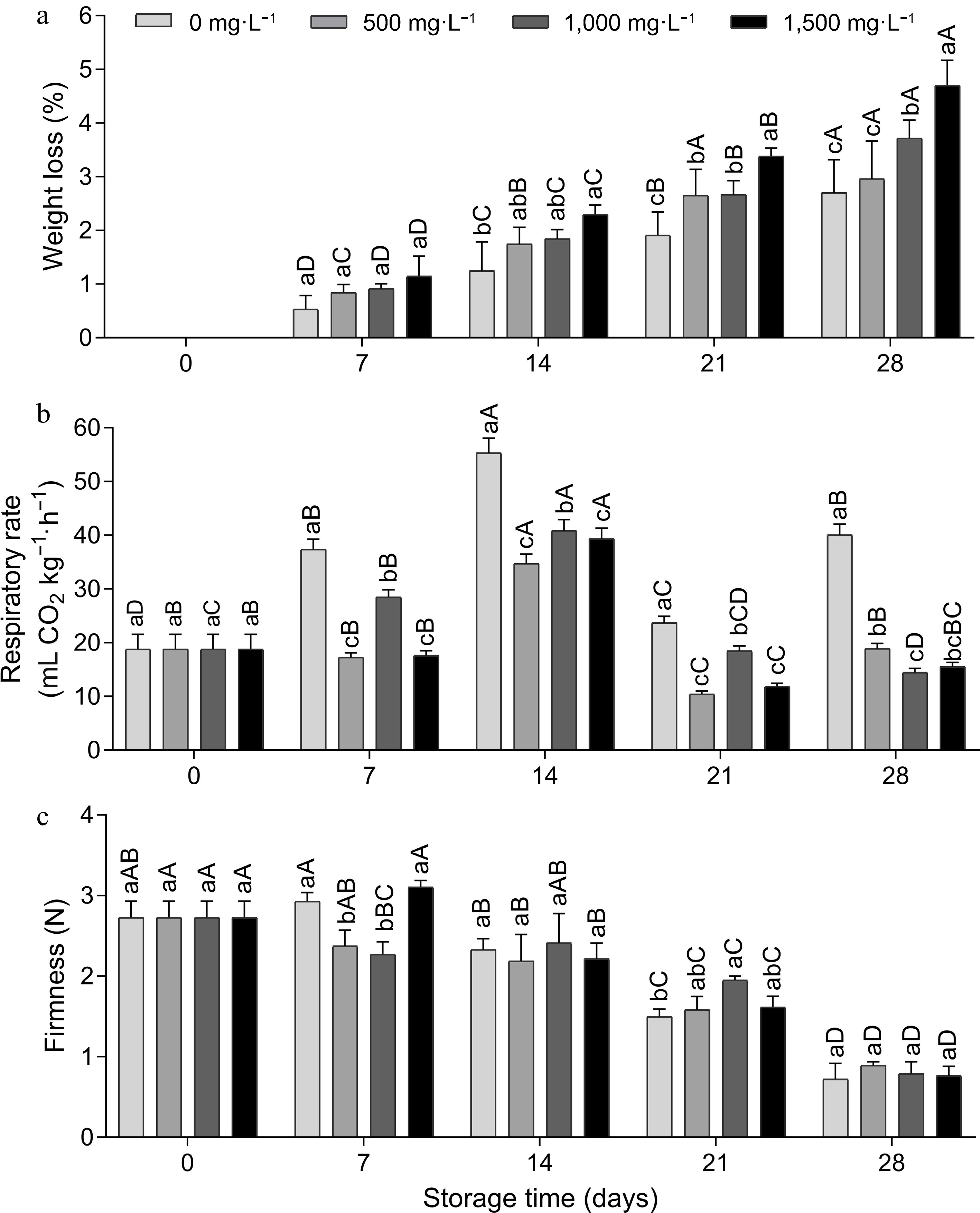
      Figure 1. 

      (a) Weight loss, (b) respiration rate and (c) firmness of tomato fruit treated with doses of aminoethoxyvinylglycine (AVG) and stored (15 ± 1 °C and RH 90% ± 5 %) for 28 d. Lowercase letters differ the doses in a single storage time and capital letters differ the storage times in a single dose, according to the Tukey test (p < 0.05).

      Weight loss is a process that occurs due to the loss of water, stimulated by the process of respiration and transpiration due to the vapor pressure deficit on the surface of the fruit[16]. The loss of water leads to softening of the tissues, affecting the texture, color, and flavor of the fruit, reducing its time on the market. As reported by Taiz et al.[17], AVG also acts to inhibit enzymes that use pyridoxal phosphate as an enzyme cofactor and when applied in high doses, this may have occurred in this study by increasing weight loss, inhibiting various aspects of the plant's metabolism, especially interfering with the rind tissue.

      There was an increase in the respiration rate of the fruit on the 7th day of evaluation, with a gradual increase over time until the maximum climacteric peak of 55.31 mL CO2 kg−1·h−1 on the 14th day for the control fruit, followed by a decline on the other days (Fig. 1b). From the 7th to the 28th day, the control had a significantly higher respiration rate, 55% to 64% higher on the 28th day compared to the AVG-treated fruit, showing a positive effect of this compound. The respiration rate can be reduced using various technologies that reduce the metabolic activity of the fruit, such as refrigerated storage and inhibitors of ethylene action[6], like AVG. The continuation of the metabolic process gradually alters the composition of the product, leading to senescence, which was delayed with AVG in this study.

      Over time, there was a decrease in the firmness of the fruit from day 14 onwards, which was gradual until day 28 (Fig. 1c). AVG provided greater fruit firmness compared to the control at a dose of 1,500 mg·L−1 on day 7 and 1,000 mg·L−1 on day 21, and on the other days there was no difference between the doses. The difference in firmness between treated and untreated fruit disappears over time[18], which probably occurred on day 28. When applied as a postharvest dip treatment, AVG reduced the rate of fruit softening in 'Grando F1' tomatoes[14], 'Huangguan' pears[9] and sweet orange Citrus sinensis (L.)[19] during storage or shelf life, as a consequence of ethylene inhibition.

      The total soluble solids (TSS) content of the control fruit (0 mg·L−1) decreased over time, and for the other doses there was stability, with no significant difference (Fig. 2a). On day 28, the 1,000 mg·L−1 dose provided a 1.0 °Brix increase in SS content compared to the control. The SS content is used as an indirect estimate of sugars, as well as other compounds that are present in the vascular sap, such as vitamins, phenolics and pectin's. Like this study[20], working with 'Tommy Atkins' mangoes at room temperature, observed stability in the SS content when treated with AVG during the 12 d of storage. However, this was probably due to the ripening process of the fruit with the conversion of starch to sugar.
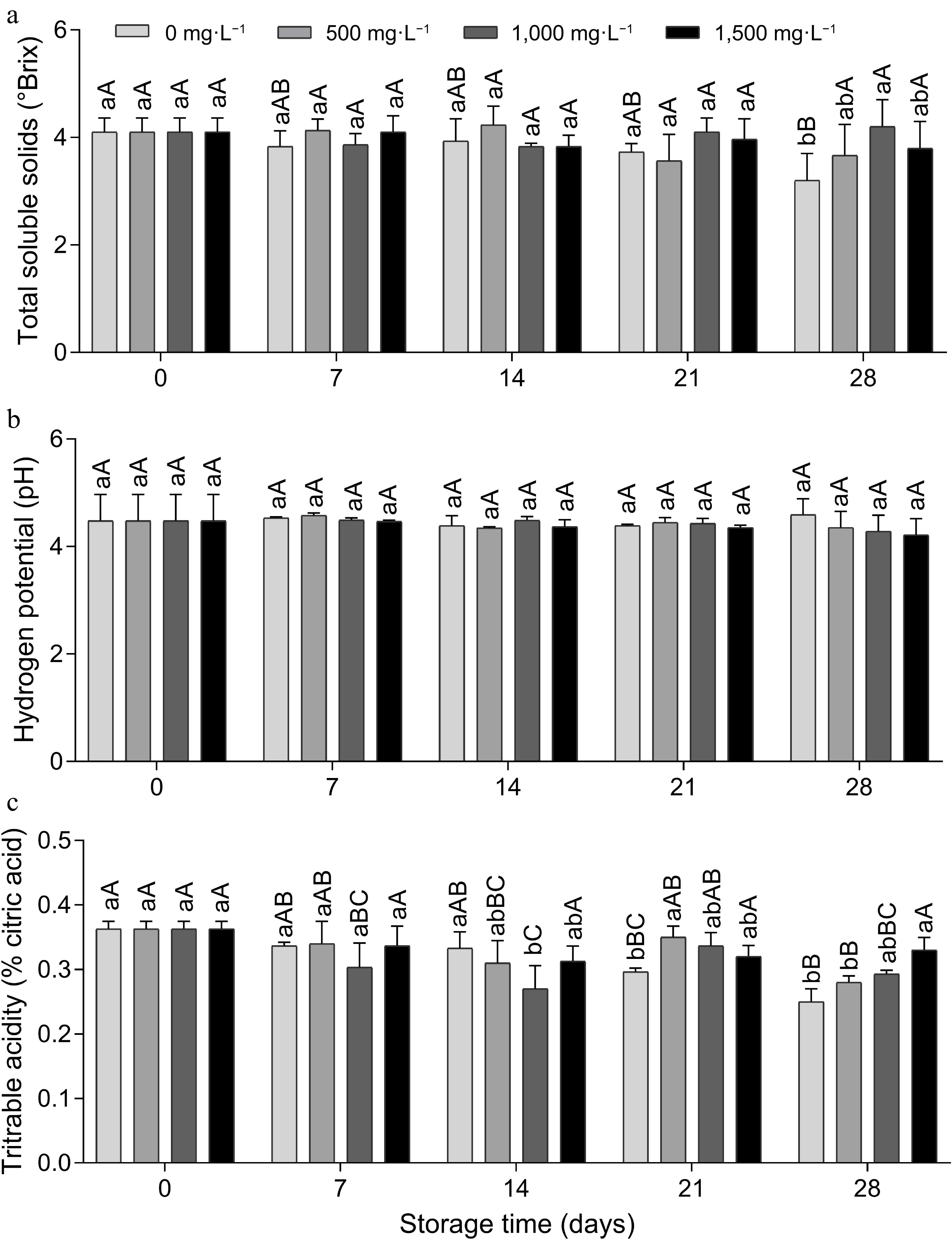
      Figure 2. 

      (a) Total soluble solids, (b) pH and (c) titratable acidity of tomato fruit treated with doses of aminoethoxyvinylglycine and stored (15 ± 1 °C and RH 90% ± 5 %) for 28 d. Lowercase letters differ the doses in a single storage time and capital letters differ the storage times in a single dose, according to the Tukey test (p < 0.05).

      The pH showed no statistical difference between the treatments (Fig. 2b). A previous study,[15] also found no significant difference for the doses of AVG, but found a significant effect for the storage time of the 'Eva' apple cultivar. The author observed higher pH values on the 0th and 7th day of storage, with a decrease in value from the 14th day onwards. This reported decrease was linked to the release of organic acids resulting from the degradation of the cell wall, a fact that did not occur in this study under the conditions in which the experiment was carried out.

      Titratable acidity (TA) remained stable in the fruit treated with the 1,500 mg·L−1 dose during storage; on the other hand, there was a reduction in the fruit treated with the other doses (Fig. 2c). In the fruit treated with the 1,500 mg·L−1 dose on day 28, the acidity was 0.08% and 0.05% higher, respectively, compared to the two lower doses, 0 and 500 mg·L−1. Corroborating this work, dos Santos et al.[20], working with 'Kent' mango, reported that the 300 mg·g−1 concentration of AVG resulted in fruit with a higher acidity content. Titratable acidity analysis is used to quantify acidity through a predominant acid according to the plant material, determining the percentage of organic acids.

      Corroborating the present study, it was previously reported that TA decreased over time and no significant differences were found in the pH of the pulp of 'Grando F1' tomatoes between fruit treated and untreated with 1,000 mg·L−1 AVG at −30 kPa during storage at 12 °C for 20 d and a further 2 d at 20 °C[14]. However, unlike the present study, these authors found no significant differences in SS and TA between fruit treated and not treated with AVG, reporting that changes in organic acids and sugars in tomatoes were not controlled by ethylene[21], a positive effect that certainly occurred in the present study due to the inhibition of ethylene by AVG at a dose of 1,500 mg·L−1, as it kept TA and SS stable.

      The results of the Two-Way ANOVA with the doses of AVG (0, 500, 1,000, and 1,500 mg·L−1), storage time (days 0, 7, 14, 21, and 28) and the interaction between these factors for the external and internal color parameters, assessed at post-harvest of the tomato fruit, are shown in Table 2. Except for the effect of the doses and the interaction between the factors for internal luminosity (pulp), all the other parameters showed significant differences. Storage time was significant for all parameters.

      Table 2.  Results of the analysis of variance (Two-Way ANOVA) of the effect of AVG doses, storage time and the interaction of these factors on the external and internal color parameters evaluated in tomato fruits.

      Cause of variationD.F.L peelC peelh° peelL pulpC pulph° pulp
      Dose30.001**0.000**0.000**0.866ns0.030*0.045 *
      Time (d)40.000**0.000**0.000**0.000**0.000**0.000**
      Dose × time120.038*0.010*0.000**0.695ns0.014*0.000**
      Residue401.7804.4023.2947.0972.50712.640
      ns represents not significant; * represents p ≤ 0.05 and ** represents p ≤ 0.01.

      As for peel color, external luminosity decreased over time (Fig. 3a), and on day 28, the 500 mg·L−1 dose provided significantly lower luminosity compared to the 0 mg·L−1 dose, indicating lower brightness of the fruit periderm (Fig. 3b), but both did not differ from the other doses. There was an increase in external chroma values over time, and on day 28 the AVG doses showed lower chroma compared to the control. Corroborating the present study, in a previous study with 'Grando F1' tomatoes treated with 1,000 mg·L−1 of AVG at −30 kPa during storage at 12 °C for 20 d and a further 2 d at 20 °C after refrigerated storage[14], it was reported that AVG reduced the rate of ethylene production and delayed ripening changes in peel color (L*, C* and h° values, chlorophyll and lycopene content). Therefore, C* values increased while h° and L* values decreased at a lower rate in AVG-treated fruit than in control fruit during storage and shelf life.
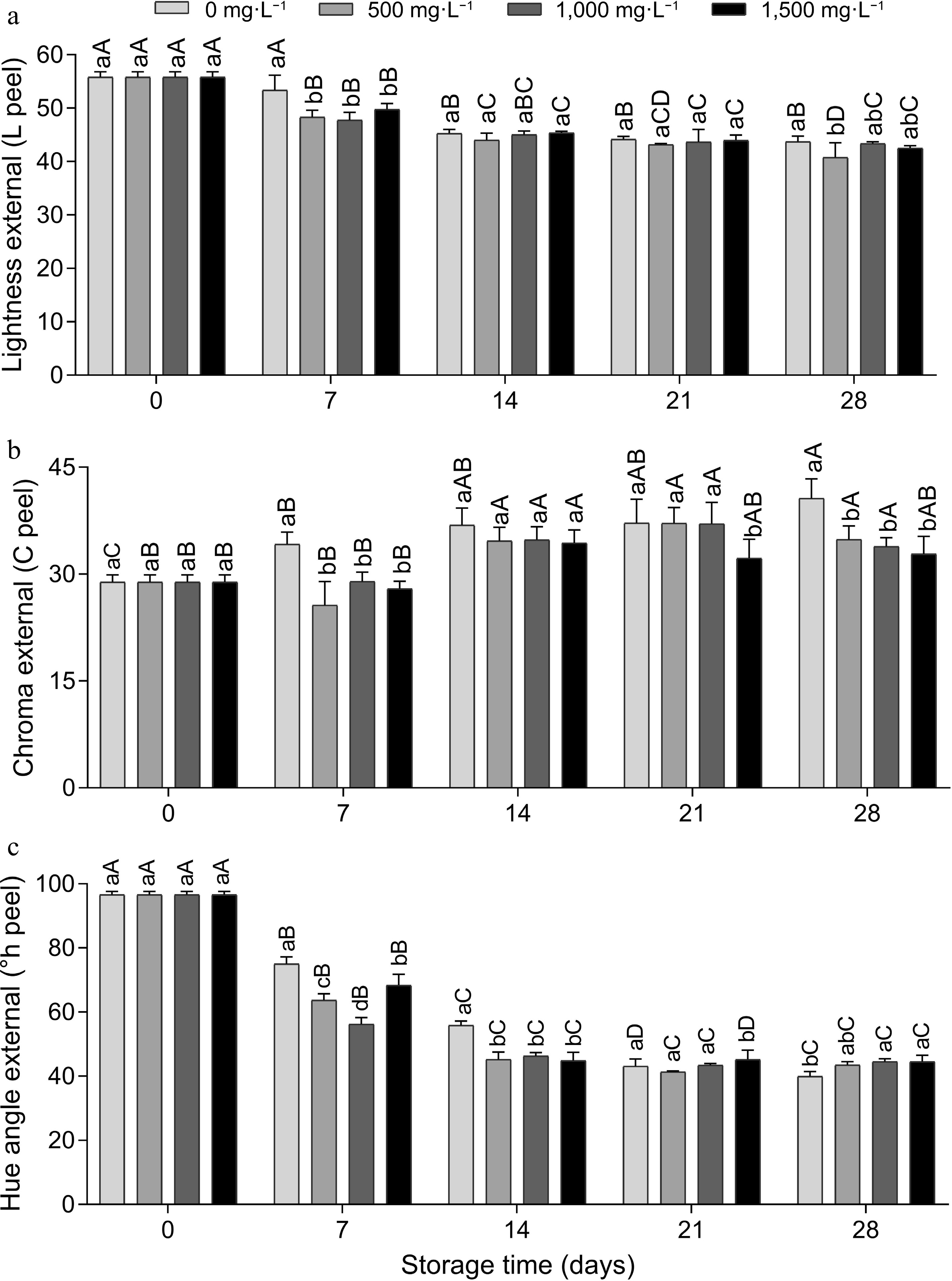
      Figure 3. 

      External instrumental color (peel), (a) luminosity, (b) chroma and (c) hue angle of tomato fruits treated with doses of AVG and stored (15 ± 1 °C and RH 90% ± 5 %) for 28 d. Lowercase letters differ the doses in a single storage time and capital letters differ the storage times in a single dose, according to the Tukey test (p < 0.05).

      The external °hue decreased until day 14 and remained stable for the rest of the time (Fig. 3c). This behavior leads to a change from green to red, which can possibly be attributed to the degradation of chlorophyll and the biosynthesis of lycopene, responsible for the tomato's red color. On day 14, the doses of AVG showed lower °hue compared to the control, and on day 21 there was no difference between the doses, and on day 28 the highest doses (1,000 and 1,500 mg·L−1) provided color maintenance compared to the control (Fig. 4), indicating that AVG reduced fruit ripening.

      Figure 4. 

      Approximate reproduction of the color profile of the peel of tomato fruit treated with doses of AVG and stored (15 ± 1 °C and RH 90% ± 5%) for 28 d, determined using a Konica Minolta colorimeter, with Luminosity, Chroma and °Hue values fed into the colorizer.org platform.

      For internal color, there was no statistical difference between AVG doses only for brightness, showing that there was no negative effect of AVG on fruit brightness. In relation to time, there was a decrease over the days, indicating a loss in the internal brightness of the fruit (Fig. 5a). On the 28th day, the internal chroma was higher in the 0 mg·L−1 control and the doses did not differ (Fig. 5b). There was an increase in color intensity on days 14 and 21. The internal ºhue gradually decreased until day 14 and remained stable on the other days (Fig. 5c), and there was no significant difference between the doses on days 14, 21 and 28. Aglar[22] reported that spraying 225 mg·L−1 of AVG on 'Li' jujube trees pre-harvest and keeping the fruit in cold storage for 45 d at 0 ± 0.5 °C and 90% ± 5% relative humidity (RH) reduced the development of fruit color.
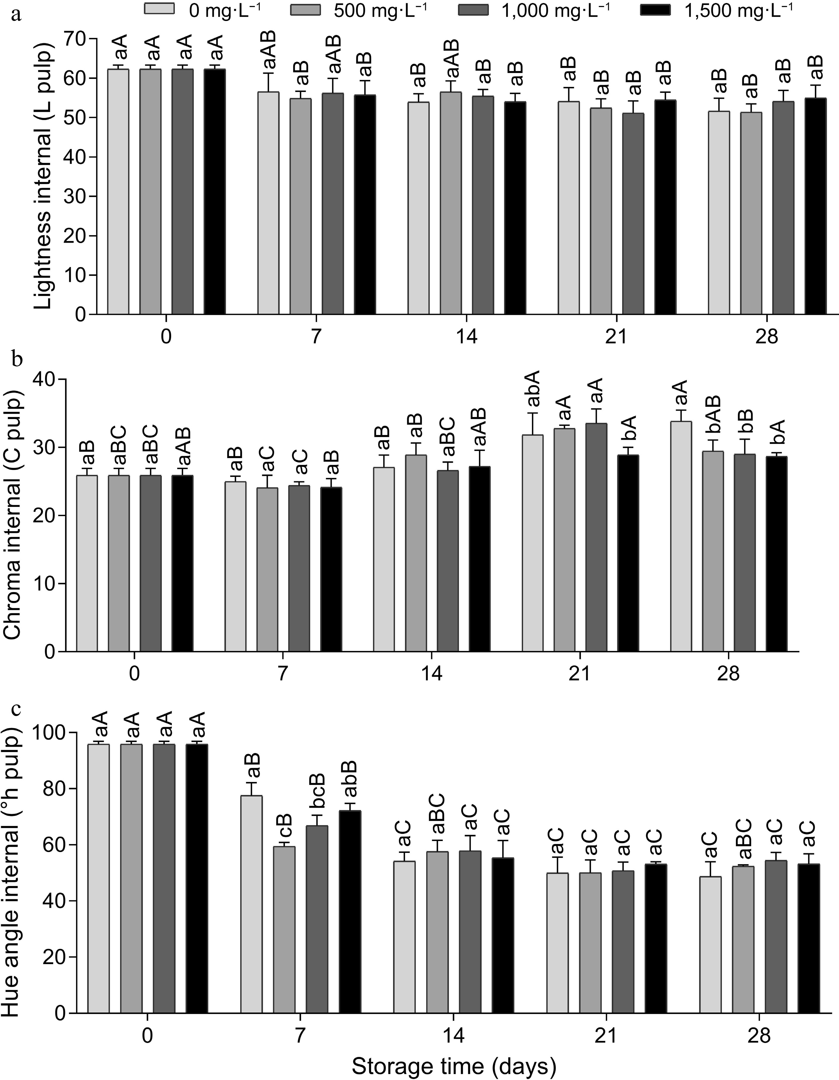
      Figure 5. 

      Internal instrumental color (pulp), (a) luminosity, (b) chroma and (c) hue angle of tomato fruit treated with doses of AVG and stored (15 ± 1 °C and 90% ± 5% RH) for 28 d. Lowercase letters differ the doses in a single storage time and capital letters differ the storage times in a single dose, according to the Tukey test (p < 0.05).

      Principal component analysis (PCA) of 12 parameters evaluated in tomato fruit treated with doses of AVG (0, 500, 1,000, and 1,500 mg·L−1) and stored for 28 d (day 0, 7, 14, 21, and 28), allowed the general observation of the data in a smaller dimension, separated by treatment in different quadrants (Fig. 6). The variability was explained by two principal components (PC) with eigenvalues > 1.0. These two significant PCs (PC1 and PC2) accumulated 76.9% of the total variation.
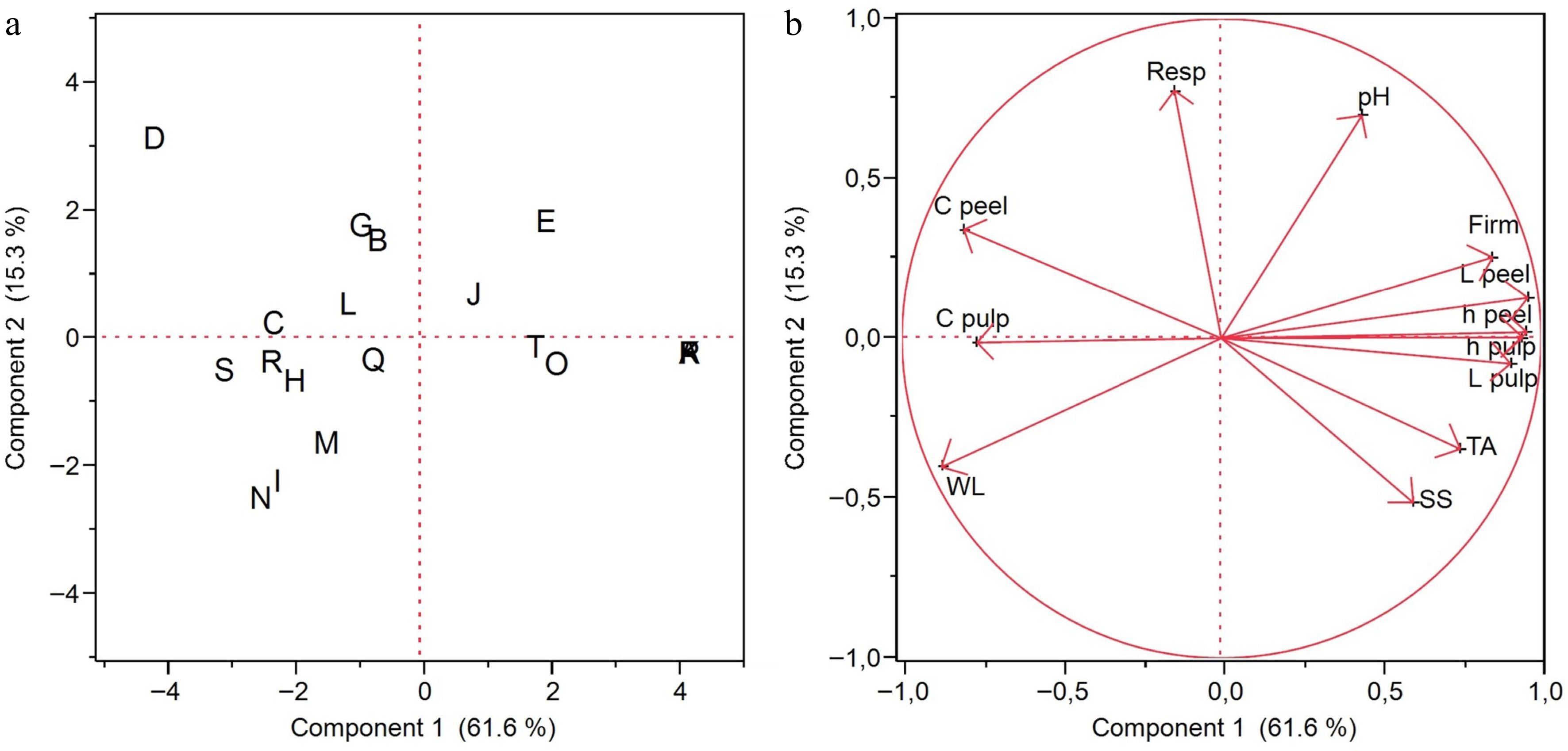
      Figure 6. 

      (a) Score plot and (b) load plot of the principal component analysis of 12 parameters evaluated in tomato fruit treated with doses of AVG and stored (15 ± 1 °C and RH 90% ± 5%) for 28 d. Score graph (a): 0 mg·L−1 dose on days 0 (A), 7 (E), 14 (B), 21 (C) and 28 (D); 500 mg·L−1 dose on days 0 (P), 7 (T), 14 (Q), 21 (R) and 28 (S); 1,000 mg·L−1 dose on days 0 (F), 7 (J), 14 (G), 21 (H) and 28 (I); and 1,500 mg·L−1 dose on days 0 (K), 7 (O), 14 (L), 21 (M) and 28 (N). Load graph (b): weight loss (WL), respiratory rate (Resp), firmness (Firm), total soluble solids (SS), hydrogen potential (pH), titrable acidity (TA), lightness external (L peel), chroma external (C peel), hue angle external (°h peel), lightness internal (L pulp), chroma internal (C pulp) and hue angle internal (°h pulp).

      PC1 was responsible for 61.6% of the total variation and was effective in separating the treatments on days 0 and 7 into positive scores and the treatments on the remaining days (14, 21, and 28) into negative scores (Fig. 6a). Analysis of the PC1 loadings (Fig. 6b) suggests that this separation is mainly due to the analyses of firmness, external and internal luminosity and external and internal °h which have strong positive loadings (> 0.80) and the analyses of external and internal chroma with strong negative loadings in the PCA (< 0.75). The PC1 scores and loadings showed a greater effect for time than for AVG doses.

      PC2 in the score graph (Fig. 6a) was important mainly for separating dose 0 on day 28 (D), since this treatment had a positive score, from doses 1,000 and 1,500 on day 28 (I, N) which had the strongest negative scores, corroborating the respiration rate mean test (Fig. 1b). PC2 represented 15.3% of the total variation and was mainly related to positively charged respiration rate and negatively charged weight loss (Fig. 6b). Therefore, principal component analysis was effective in confirming the results presented here.

      By grouping the dependent variables via correlation, PCA indicated that there were positive correlations between weight loss, external and internal chroma, which correlated negatively with firmness, external and internal luminosity, external and internal hue angle, total soluble solids, and titratable acidity, and all the latter correlated positively with each other (Supplemental Fig. S1). This infers that, as weight loss increased, firmness decreased, an effect that was minimized with AVG, which may be related to the inhibition of the fruit's ethylene production rate and the expression of genes associated with chlorogenic acid metabolism, perception and signal transduction and membrane breakdown in the central tissue, as well as a decrease in malondialdehyde activity and polyphenol oxidase enzyme activity[9].

    • Under the conditions in which the experiment was carried out, it can be concluded that immersing 'Débora' tomato fruit in AVG solution at a dose of 1,500 mg·L−1 delayed ripening, reduced the fruit's respiration rate, and the changes in external and internal chroma, and it did not reduce weight loss. The doses of AVG did not negatively affect the luminosity and pH of the fruit pulp. The untreated fruit became soft and ripe red 7 d earlier than the treated fruit during the 28 d of storage at 15 ± 1 °C and 90% ± 1% RH. The feasibility of using AVG to market tomato fruit to distant markets or to local or retail markets for longer was demonstrated. More studies are needed on the use of AVG in different conditions and with other tomato cultivars.

    • The authors confirm contribution to the paper as follows: study conception and design: Aparecida dos Santos J, Rocha Lacerda V; methodology: Rocha Lacerda V, Aparecida dos Santos J, Gaona Acevedo AF, Lopes Vieites R; software: Rocha Lacerda V; validation: Rocha Lacerda V, Aparecida dos Santos J, Sílvia Angélica de Oliveira H, Gaona Acevedo AF, Rocha Lacerda V; formal analysis: Rocha Lacerda V, Aparecida dos Santos J, Gaona Acevedo AF; investigation: Rocha Lacerda V, Aparecida dos Santos J, Sílvia Angélica de Oliveira H; resources: Rocha Lacerda V; data curation: Rocha Lacerda V, Aparecida dos Santos J; draft manuscript preparation: Rocha Lacerda V, Aparecida dos Santos J, Sílvia Angélica de Oliveira H; final manuscript preparation: Rocha Lacerda V, Sílvia Angélica de Oliveira H, Lopes Vieites R; visualization: Rocha Lacerda V, Aparecida dos Santos J, Sílvia Angélica de Oliveira H, Gaona Acevedo AF; supervision: Lopes Vieites R; project administration: Aparecida dos Santos J; funding acquisition: Lopes Vieites R. All authors reviewed the results and approved the final version of the manuscript.

    • All data generated or analyzed during this study are included in this published article, and are available from the corresponding author on reasonable request.

      • This study was financed in part by the Coordenação de Aperfeiçoamento de Pessoal de Nível Superior - Brazil (CAPES) - Financing Code 001. To the Conselho Nacional de Desenvolvimento Científico e Tecnológico – CNPq, for the financial support for this work.

      • The authors declare that they have no conflict of interest.

      • Supplemental Fig. S1 Correlations by color map cluster of twelve parameters evaluated in tomato fruit treated with doses of AVG and stored (15 ± 1 °C and RH 90 ± 5 %) for 28 days.
      • Copyright: © 2024 by the author(s). Published by Maximum Academic Press, Fayetteville, GA. This article is an open access article distributed under Creative Commons Attribution License (CC BY 4.0), visit https://creativecommons.org/licenses/by/4.0/.
    Figure (6)  Table (2) References (22)
  • About this article
    Cite this article
    Rocha Lacerda V, Aparecida dos Santos J, Sílvia Angélica de Oliveira H, Gaona Acevedo AF, Lopes Vieites R. 2024. Doses of aminoethoxyvinylglycine (AVG) in tomato postharvest storage. Technology in Horticulture 4: e011 doi: 10.48130/tihort-0024-0008
    Rocha Lacerda V, Aparecida dos Santos J, Sílvia Angélica de Oliveira H, Gaona Acevedo AF, Lopes Vieites R. 2024. Doses of aminoethoxyvinylglycine (AVG) in tomato postharvest storage. Technology in Horticulture 4: e011 doi: 10.48130/tihort-0024-0008

Catalog

  • About this article

/

DownLoad:  Full-Size Img  PowerPoint
Return
Return