ARTICLE   Open Access    

Insights into Aquilaria phylogenetics through comparative plastomic resources

  • # Authors contributed equally: Junhu Kan, Liyun Nie, Zenglu Mi

More Information
  • Received: 06 May 2024
    Revised: 06 August 2024
    Accepted: 19 August 2024
    Published online: 04 September 2024
    Forestry Research  4 Article number: e030 (2024)  |  Cite this article
  • The plastid is an essential organelle for its role in photosynthesis and energy production and its genomic information is always employed as important evolutionary markers to explore the relationship among species. Agarwood (Aquilaria), prized for its aromatic blend, finds extensive use in various cultures as incense and perfume. Despite its high economic importance, the phylogenetic status among Aquilaria based on plastomes remains ambiguous due to the lack of available plastomic resources. To bridge this knowledge gap, 22 Aquilaria plastomes were newly sequenced, similar variation patterns in this genus were determined, including a shared 16 bp extension of the rps19 gene and seven highly variable regions. The analysis highlighted the highest prevalence of the A/T motif among simple sequence repeats in these plastomes. Further phylogenetic analysis revealed Aquilaria's phylogenetic implications with an expanded dataset. This comprehensive plastomic resource not only enhances our understanding of Aquilaria evolution but also presents potential molecular markers for DNA barcoding.
  • The tea plant (Camellia sinensis) was first discovered and utilized in China, where its tender leaves were processed into tea. Tea has become the second most popular beverage after water worldwide. Tea contains tea polyphenols, amino acids, vitamins, lipopolysaccharides and other nutrients, as well as potassium, calcium, magnesium, iron, fluorine and other trace elements, which have antioxidant, lipid-lowering, hypoglycaemic, anti-caries, enhance the body's immunity and other physiological regulatory functions[1]. Among them, fluorine is an element widely found in the earth's crust, mainly in the form of fluoride in the environment, is one of the essential trace elements for human beings, and is vital to the growth and development of human bones and teeth[2]. When the human body takes in an appropriate amount of fluorine, it can effectively prevent the formation of dental caries, enhance the absorption of calcium, phosphorus and other elements of the human body. Excessive intake of fluorine however will lead to chronic cumulative poisoning, damaged bone tissue, affect the function of various tissues and organs in the body, and cause harm to health[3]. Tea plants can absorb and accumulate fluoride from air, water and soil, mainly concentrated in the leaves, most of the fluoride in the leaves can be released into the tea soup and be absorbed by the human body, so the fluoride content in tea is closely related to human health[4]. Generally speaking, green tea, black tea, white tea, oolong tea and yellow tea are made from the young buds and shoots of the tea plant, and their fluorine content is low. Some dark tea made from leaves with lower maturity has lower fluorine content. However, the dark tea made from leaves with higher maturity has a high fluorine content, and therefore poses a risk of excessive fluorine[5]. Long-term drinking of dark tea with excessive fluorine content is a cause of tea-drinking fluorosis[6].

    Dark tea, with its smooth taste and digestive benefits, became an indispensable drink in the lives of the Chinese herders, who were mainly meat eaters[7]. Dark tea has also gained popularity in the wider population because of its important health-promoting effects, such as prevention of cardiovascular and cerebrovascular diseases, lowering blood pressure, and promoting weight loss and fat reduction[8]. Long-term consumption of dark tea is likely to cause fluorosis for two reasons: 1) Chinese border ethnic minorities generally use the boiling method to brew dark tea, which increases the leaching rate of fluoride[9] and leads to high fluoride levels in the human body; 2) most of the fresh tea leaves utilized to make dark tea are older and more mature leaves, which contain higher levels of fluoride than younger leaves[10]. The mature leaves of tea plants accumulate a large amount of fluoride, but can grow normally without fluoride poisoning, indicating that tea plants are able to accumulate and tolerate fluoride. Due to the problem of tea-drinking fluorosis, the excessive accumulation of fluoride in tea plants has attracted widespread attention[10]. It is important to investigate the mechanisms related to fluoride absorption, transportation, enrichment, and tolerance in tea plants to develop effective and practical management and control programs to reduce the fluoride content in tea and ensure its safety. Recent research on defluorination measures for tea has included preliminary screening of tea germplasm resources, management measures during tea plant cultivation, processing technologies, and tea brewing methods. In this review, we summarize the results of studies on how fluoride moves from the environment into tea plants and the factors affecting this process, how fluoride is transported in tea plants, the mechanisms of fluoride tolerance in tea plants, and current measures to reduce the fluoride content in tea.

    Tea plants are fluoride-accumulators with the ability to absorb and accumulate fluoride from the surrounding environment. The fluoride content in tea plants is significantly higher than that of other plants under similar growth conditions[11]. After fluoride is absorbed by the roots of tea plants, it is transferred to the above-ground parts, and is also transferred from the leaves downward (Fig. 1), but not from the above-ground parts to the below-ground parts (Fig. 2). External factors such as atmospheric, soil, and water conditions around the tea plant and internal factors can affect the fluoride content in different plant tissues.

    Figure 1.  Fluoride absorption by tea plants. Tea plants absorb fluoride from the atmosphere, soil, and water. Fluoride in the atmosphere is absorbed through the stomata or cuticle of the leaf epidermis. Fluoride ions and fluoride–aluminum complexes in soil and water are absorbed by the roots.
    Figure 2.  Fluoride transportation in tea plants. Fluoride absorbed by the leaves is transferred to the leaf tips and edges, chelates with metal ions, and the complexes are deposited in the leaves. Fluoride can be transferred from old leaves to new tips. Fluoride from soil and water can form complexes with organic acids and aluminum, which are stored in the leaves of tea plants. Fluoride from soil and water can also be individually absorbed and transported for storage in the leaves.

    Fluoride occurs in many forms in nature. Fluoride in the atmosphere mainly exists in the form of hydrogen fluoride, and that in the soil mainly exists in three forms: insoluble, exchangeable, and water-soluble fluoride. Atmospheric fluoride is mainly absorbed through the stomata of tea plant leaves or the cuticle of the epidermis, and its concentration is relatively low[12]. Insoluble fluoride and exchangeable fluoride in the soil cannot be absorbed by tea plants. Water-soluble fluoride is the main form absorbed by the root system of tea plants[13]. Several studies have detected a significant positive correlation between the fluoride content in tea and the water-soluble fluoride content in soil[14,15]. Water-soluble fluoride mainly refers to the fluoride ion (F) or fluoride complexes in soil and water solutions, including free F and fluoride complexed with ions. The water-soluble state has the strongest activity and the highest biological availability, so it is conducive to the migration of fluoride in the environment[16].

    The root system is the main organ responsible for fluoride absorption in tea plants. Water-soluble fluoride can enter the root system by passive or active absorption, depending on its concentration. Fluoride at lower concentrations in solution (0.1–10 mg/L) is mainly absorbed and enriched in the root system of tea plants via active absorption, with a kinetic curve following the Michalis–Menten kinetic model. At higher concentrations (50–100 mg/L), water-soluble fluoride is absorbed at a rate that increases with increasing concentration, and this is achieved via passive absorption[17]. Many studies have shown that the water-soluble fluoride content in soils in most tea-producing regions in China is below the threshold for passive absorption[1820], indicating that fluoride mainly enters tea plants via active absorption by the roots.

    The active uptake of fluoride by tea roots is mediated by ion pump carrier proteins and ABC transporter proteins. Ion pump carrier proteins can transport the substrate across the cell membrane against the electrochemical gradient, and use the energy of ATP hydrolysis to participate in the process of active transport of substances, mainly including proton pump H+-ATPase and calcium ion pump Ca2+-ATPase. ABC transporter proteins can transport ions or heavy metals to vesicles as chelated peptide complexes, thereby reducing toxicity to the cell and improving plant resistance to abiotic stress. Passive fluoride absorption by the root system of tea plants involves water channels and ion channels. Studies on the effects of applying external anion channel inhibitors, cation channel inhibitors, and water channel inhibitors showed that inhibition of external anion channels significantly reduced the absorption of fluoride by roots. This indicated that anion channels are an important pathway for the uptake and trans-membrane transport of fluoride in the root system of tea plants[17,21,22]. The homeostatic flow of ions through channels diffusing along a trans-membrane concentration gradient or potential gradient involves ion channel proteins[23]. Two phylogenetically independent ion channel proteins have recently been identified in tea plants: CLCF-type F/H+ reverse transporter proteins and the FEX (Fluoride export gene) family of small membrane proteins[24]. CLC proteins are involved in the transport of a variety of anions such as chlorine (Cl) and F into and out of the cell. The FEX proteins in tea plants are involved in fluoride absorption via thermodynamic passive electro-diffusion through transmembrane channels[25,26].

    After fluoride is absorbed, it is transported within the tea plant by several different pathways. These include transport after leaf absorption, transport after root absorption, and transfer inside tea plant cells. The fluoride absorbed by leaves can be transferred along the conduit to the leaf tips and edges, accumulating in the top and ipsilateral leaves, but not in the roots. Fluoride in the soil and water environment is taken up by the root system and transported to the xylem via intracellular and intercellular transport. It is then transported upwards via transpiration and eventually accumulates in the leaves[27,28]. Two transport mechanisms have been proposed for the translocation of fluoride via the xylem. One proposed mechanism is that it is translocated in the form of fluoride-aluminum complexes[29]. The other proposed mechanism is that aluminum and fluoride are transported separately and accumulate after reaching the leaves[30].

    The roots are the main organ responsible for fluoride absorption in tea plants. Therefore, most studies have focused on the transport process in roots, especially the transport of fluoride by fluoride-related transporters. It has been found that under acidic conditions, F preferentially forms complexes with Al3+ and these complexes are then absorbed by roots and transported upward in the same state[29,31]. Studies have shown that, compared with F, aluminum-fluoride complexes are more easily absorbed and transported to the new shoots by the root system. This may be related to the elimination of the separate toxic effects of F and Al3+ [29]. Fluoride can also be transported in tea plants by binding to aluminum-organic acid complexes, and then accumulate in the leaves[29]. Both tea plant fluoride transporter proteins, CsFEX1 and CsFEX2, are involved in fluoride transport, but their encoding genes can be differentially expressed among different varieties and depending on the concentration of fluoride. In one study, the expression level of CsFEX1 was consistent among different varieties, while the expression of CsFEX2 was induced under fluoride stress to increase fluoride efflux from tea plants, thereby reducing its accumulation in low-fluoride varieties[32]. In addition, the A–G subfamily of ABC transporters plays a carrier role in the transmembrane transport of F- and Cl- in tea plants[33]. It was found that the expression of the ABC transporter protein CsCL667 was up-regulated in response to fluoride treatment, and its ability to transport fluoride was enhanced, suggesting that CsCL667 functions in fluoride efflux[34]. Another study demonstrated that CsABCB9 localizes in chloroplasts and functions as a fluoride efflux transporter to reduce fluoride-induced damage in leaves and enhance chloroplast activity[35].

    Several factors affect the absorption and transport of fluoride in tea plants, including the absorbable fluoride concentration, soil pH, the presence of other ions, and the activity of ion channels[3638]. Fluoride in nature is present in the atmosphere and soil, and its concentration is the main factor affecting the fluoride content in tea plants. Under normal conditions, tea plants generally absorb fluoride from the soil through the roots, but when the hydrogen fluoride content in the atmosphere is high, tea plants can absorb it through the leaves. The fluoride content in tea plants growing in the same geographical area is similar, mainly because of the soil properties in that area. Tea plants grow in acidic environments, and fluoride in acidic soils is more easily absorbed. During the growth of tea plants, the roots secrete organic acids such as oxalic acid, citric acid, and malic acid, which promote the absorption of fluoride by the roots and its transport to above-ground parts[39]. Other ions such as Ca2+ and Mg2+ combine with F to form precipitates, resulting in lower concentrations of water-soluble fluoride in the soil, which also affects its absorption by tea plant roots[40]. Exogenously applied calcium at low concentrations can change the cell wall structure and membrane permeability in tea plant roots, ultimately leading to reduced fluoride content in tea leaves[36,41,42]. Meanwhile, Al3+ treatment can trigger Ca2+ signaling in tea plant roots, which in turn activates calmodulin and promotes fluoride absorption[43]. The H+ gradient generated by the plasma membrane H+-ATPase can also promote Ca2+ signaling in plants to regulate the transmembrane transport of ions, which affects fluoride absorption[43]. The abundance and activity of H+-ATPase in the plasma membrane of tea plant roots have been found to increase significantly under fluoride stress, and these increases result in improved absorption of fluoride, although this is also affected by the fluoride concentration and temperature[33]. Sodium fluoride was found to induce the expression of genes encoding ABC transporter proteins, resulting in the transmembrane absorption of large amounts of fluoride ions into cells[34]. ABC transporters also transport ions alone or in the form of chelated peptide complexes directly out of the cellular membrane, which improves cellular tolerance to these ions[44]. Some anions with the same valence state also affect the F content in tea plants. For example, the ion channel protein encoded by CLCF is more sensitive to F, more selective for F than for Cl, and functions to export F from the cytoplasm to protect against fluorosis[4547].

    It can be seen that reducing the absorption of fluorine by tea plants and changing the mechanism of fluorine transportation in tea plants can reduce the content of fluorine in different parts of the tea plants. From the mechanism of fluorine absorption, the most effective way is to directly change the form of soil fluorine to reduce the absorption of water-soluble fluorine by roots. On this basis, it is possible to further change the active absorption process of fluorine mediated by ion pump carrier protein and ABC transporter protein in tea roots by molecular techniques. From the perspective of fluorine transport mechanism, the toxic effect of fluorine on tea plants can be reduced mainly by promoting the function of transporter proteins to exclude fluorine from the cell or transport it to the vesicle. The comprehensive application of the above methods to limit fluorine absorption and promote fluorine transport in tea plants can limit the accumulation of fluorine in tea plants.

    The fluoride enrichment characteristics of tea plants are related to various factors, including the tea variety, the organ, and the season. Among different varieties of tea, differences in leaf structure and other physiological characteristics can lead to variations in fluoride absorption and enrichment[48]. Some studies have concluded that the variety is one of the main determinants of the fluoride content in tea leaves[10], and the differences in fluoride content among most varieties reached highly significant levels (Table 1), which can be divided into low-enriched, medium-enriched, and high-enriched germplasm[49]. Various organs of tea plants also show differences in fluoride accumulation. The fluoride content is much higher in leaves than in roots and stems, and significantly higher in old leaves than in new shoots[10,50,51]. The fluoride content can differ widely among tea plants at different developmental stages. In spring, the new leaves begin to accumulate fluoride from the environment, and the fluoride content increases as the leaves age. When the growth rate of tea leaves is slower, they absorb and accumulate more fluoride from the soil and air. When the temperature in summer and autumn is high, the growth rate of tea leaves is fast and the growth period is short, so less fluoride is absorbed and accumulated from the soil and air. This explains why the fluoride content in fresh tea leaves was higher in spring and relatively lower in summer and autumn[4]. Another study found that, in China, the fluoride content in tea leaves was higher in summer than in spring. This may have been related to the maturity level of the tea leaves at harvest and different patterns of fluoride transport[4].

    Table 1.  Fluoride content difference of different tea cultivars.
    Cultivars Province Parts Treatment Years Content (mg/kg) Ref.
    Liannandaye Sichuan Old leaves Drying at 80 °C and boiling water extraction 2006−2007 1,150.79 ± 4.86 [107]
    Mature leaves Drying at 70 °C and hydrochloric acid extraction 2006 1,296.66 ± 12.84 [110]
    Yuenandaye Old leaves Drying at 80 °C and boiling water extraction 2006−2007 1,352.89 ± 12.69 [107]
    Mature leaves Drying at 70 °C and hydrochloric acid extraction 2006 1,560.36 ± 27.10 [110]
    Chenxi NO.4 Old leaves Drying at 80 °C and boiling water extraction 2006−2007 1,865.61 ± 7.46 [107]
    Mature leaves Drying at 70 °C and hydrochloric acid extraction 2006 1,954.93 ± 10.96 [110]
    Meizhan Old leaves Drying at 80 °C and boiling water extraction 2006−2007 2,180.13 ± 14.42 [107]
    Mature leaves Drying at 70 °C and hydrochloric acid extraction 2006 1,732.2 ± 41.2 [110]
    Zhejiang Mature leaves Drying at 80 °C and nitric acid extraction 2002 2,015.48 ± 29.99 [106]
    Fudingdabai Sichuan Old leaves Drying at 80 °C and boiling water extraction 2006−2007 249.64 ± 24.3 [107]
    Guizhou One bud and
    five leaves
    Drying at 80 °C and hydrochloric acid extraction 2010 1,612.3 ± 43.1 [109]
    Fudingdabai Zhejiang Mature leaves Drying at 80 °C and nitric acid extraction 2002 137.1 ± 2.1 [106]
    Fujian Old leaves Drying at 80 °C and hydrochloric acid extraction 2010 282.1 [111]
    Hunan One bud and
    five leaves
    Steaming and boiling water extraction 2011 2,232.05 ± 85.52 [51]
    Zhuyeqi Ya'an and
    surroundings
    Old leaves Drying at 80 °C and boiling water extraction 2006−2007 2,750.16 ± 11.37 [107]
    Ya'an Mature leaves Drying at 70 °C and hydrochloric acid extraction 2006 125.4 [110]
    Hunan One bud and
    five leaves
    Steaming and boiling water extraction 2011 2,330.74 ± 31.39 [51]
    Fujianshuixian Ya'an and
    surroundings
    Old leaves Drying at 80 °C and boiling water extraction 2006−2007 2,548.18 ± 40.97 [107]
    Ya'an Mature leaves Drying at 70 °C and hydrochloric acid extraction 2006 103.7 ± 1.5 [110]
    Fujian Old leaves Drying at 80 °C and hydrochloric acid extraction 2010 1,150.79 ± 4.86 [111]
    Huangyeshuixian Ya'an and
    surroundings
    Old leaves Drying at 80 °C and boiling water extraction 2006−2007 2,424.70 ± 18.85 [107]
    Ya'an Mature leaves Drying at 70 °C and hydrochloric acid extraction 2006 2,950.80 ± 27.73 [110]
    Qianmei 701 Ya'an and
    surroundings
    Old leaves Drying at 80 °C and boiling water extraction 2006−2007 2,522.01 ± 45.33 [107]
    Ya'an Mature leaves Drying at 70 °C and hydrochloric acid extraction 2006 3,693.09 ± 35.12 [110]
    Guizhou One bud and
    five leaves
    Drying at 80 °C and hydrochloric acid extraction 2010 389.95 ± 32.18 [109]
    Guizhou Old leaves Dry samples and hydrochloric acid extraction 2011 2,142.26 ± 16.30 [113]
    Mingshan 130 Ya'an and
    surroundings
    Old leaves Drying at 80 °C and boiling water extraction 2006−2007 2,564.78 ± 51.22 [107]
    Ya'an Mature leaves Drying at 70 °C and hydrochloric acid extraction 2006 3,036.13 ± 31.25 [110]
    Mengshan 9 Ya'an and
    surroundings
    Old leaves Drying at 80 °C and boiling water extraction 2006−2007 2,647.31 ± 70.89 [107]
    Ya'an Mature leaves Drying at 70 °C and hydrochloric acid extraction 2006 3,436.55 ± 20.21 [110]
    Yinghong NO. 2 Ya'an and
    surroundings
    Old leaves Drying at 80 °C and boiling water extraction 2006−2007 2,669.02 ± 799.95 [107]
    Ya'an Mature leaves Drying at 70 °C and hydrochloric acid extraction 2006 3,364.53 ± 51.72 [110]
    Mengshan 11 Ya'an and
    surroundings
    Old leaves Drying at 80 °C and boiling water extraction 2006−2007 2,695.21 ± 59.89 [107]
    Ya'an Mature leaves Drying at 70 °C and hydrochloric acid extraction 2006 3,582.83 ± 9.73 [110]
    Mingshan 311 Ya'an and
    surroundings
    Old leaves Drying at 80 °C and boiling water extraction 2006−2007 2,716.22 ± 42.21 [107]
    Donghuzao 2,731.20 ± 20.78
    Ya'an Mature leaves Drying at 70 °C and hydrochloric acid extraction 2006 3,107.27 ± 54.91 [110]
    Hainandaye Ya'an and
    surroundings
    Old leaves Drying at 80 °C and boiling water extraction 2006−2007 2,746.82 ± 39.71 [107]
    Ya'an Mature leaves Drying at 70 °C and hydrochloric acid extraction 2006 2,961.53 ± 29.94 [110]
    Qianmei 502 Ya'an and
    surroundings
    Old leaves Drying at 80 °C and boiling water extraction 2006−2007 2,878.23 ± 76.94 [107]
    Ya'an Mature leaves Drying at 70 °C and hydrochloric acid extraction 2006 3,881.51 ± 16.48 [110]
    Guizhou One bud and
    five leaves
    Drying at 80 °C and hydrochloric acid extraction 2010 389.95 ± 30.2 [109]
    Old leaves Dry samples and hydrochloric acid extraction 2011 3,260.48 ± 32.12 [113]
    Zisun Ya'an and
    surroundings
    Drying at 80 °C and boiling water extraction 2006−2007 2,904.13 ± 35.40 [107]
    Ya'an Mature leaves Drying at 70 °C and hydrochloric acid extraction 2006 3,140.80 ± 42.86 [110]
    Zhejiang Drying at 80 °C and nitric acid extraction 2002 1,742.7 ± 43.2 [106]
    Qianmei 303 Ya'an and
    surroundings
    Old leaves Drying at 80 °C and boiling water extraction 2006−2007 2,918.13 ± 46.79 [107]
    Ya'an Mature leaves Drying at 70 °C and hydrochloric acid extraction 2006 4,029.11 ± 81.86 [110]
    Guizhou One bud and
    five leaves
    Drying at 80 °C and hydrochloric acid extraction 2010 199.74 ± 16.6 [109]
    Old leaves Dry samples and hydrochloric acid extraction 2011 2,972.79 ± 169.82 [113]
    Anxishuixian Ya'an and
    surroundings
    Old leaves Drying at 80 °C and boiling water extraction 2006−2007 2,924.33 ± 41.39 [107]
    Ya'an Mature leaves Drying at 70 °C and hydrochloric acid extraction 2006 3,454.68 ± 26.29 [110]
    Longjing 43 Ya'an and
    surroundings
    Old leaves Drying at 80 °C and boiling water extraction 2006−2007 3,152.73 ± 27.70 [107]
    Ya'an Mature leaves Drying at 70 °C and hydrochloric acid extraction 2006 4,437.79 ± 26.14 [110]
    Zhejiang Drying at 80 °C and nitric acid extraction 2002 1,377.1 ± 37.0 [106]
    Fujian Old leaves Drying at 80 °C and hydrochloric acid extraction 2010 116.2 ± 0.9 [111]
    Shuyong 307 Ya'an and
    surroundings
    Drying at 80 °C and boiling water extraction 2006−2007 3,223.55 ± 151.43 [107]
    Ya'an Mature leaves Drying at 70 °C and hydrochloric acid extraction 2006 4,296.52 ± 54.98 [110]
    Zhenghedabaicha Ya'an and
    surroundings
    Old leaves Drying at 80 °C and boiling water extraction 2006−2007 3,295.74 ± 27.55 [107]
    Ya'an Mature leaves Drying at 70 °C and hydrochloric acid extraction 2006 3,876.58 ± 21.09 [110]
    Zhejiang Drying at 80 °C and nitric acid extraction 2002 1,373.0 ± 41.9 [106]
    Taiwandaye Ya'an and
    surroundings
    Old leaves Drying at 80 °C and boiling water extraction 2006−2007 3,363.59 ± 456.39 [107]
    Ya'an Mature leaves Drying at 70 °C and hydrochloric acid extraction 2006 4,739.89 ± 58.59 [110]
    Qianmei 419 Ya'an and
    surroundings
    Old leaves Drying at 80 °C and boiling water extraction 2006−2007 3,518.15 ± 76.19 [107]
    Ya'an Mature leaves Drying at 70 °C and hydrochloric acid extraction 2006 4,541.43 ± 28.91 [110]
    Guizhou One bud and
    five leaves
    Drying at 80 °C and hydrochloric acid extraction 2010 133.70 ± 11.2 [109]
    Old leaves Dry samples and hydrochloric acid extraction 2011 2,370.47 ± 11.43 [113]
    Mengshan23 Ya'an and
    surroundings
    Drying at 80 °C and boiling water extraction 2006−2007 3,625.11 ± 86.07 [107]
    Ya'an Mature leaves Drying at 70 °C and hydrochloric acid extraction 2006 4,469.25 ± 40.85 [110]
    Sichuan group species 2,782.59 ± 146.46
    Meitantaicha Guizhou One bud and
    five leaves
    Drying at 80 °C and hydrochloric acid extraction 2010 272.93 ± 27.3 [109]
    Qianmei 101 Drying at 80 °C and hydrochloric acid extraction 2010 175.07 ± 13.5
    Dry samples and hydrochloric acid extraction 2011 3,218.33 ± 57.91 [113]
    Qianmei 601 Drying at 80 °C and hydrochloric acid extraction 2010 267.11 ± 26.31 [109]
    Dry samples and hydrochloric acid extraction 2011 2,823.02 ± 73.36 [113]
    Qianmei 809 Drying at 80 °C and hydrochloric acid extraction 2010 521.48 ± 50.32 [109]
    Dry samples and hydrochloric acid extraction 2011 2,327.91 ± 83.17 [113]
    Qianmei 308 Drying at 80 °C and hydrochloric acid extraction 2010 326.88 ± 29.3 [109]
    Dry samples and hydrochloric acid extraction 2011 3,432.86 ± 159.4 [113]
    Qianmei 415 Drying at 80 °C and hydrochloric acid extraction 2010 186.95 ± 14.2 [109]
    Dry samples and hydrochloric acid extraction 2011 5,090.83 ± 69.56 [113]
    Qiancha NO. 7 Drying at 80 °C and hydrochloric acid extraction 2010 218.81 ± 18.7 [109]
    Qianfu NO. 4 136.82 ± 11.6
    Dry samples and hydrochloric acid extraction 2011 3,066.49 ± 86.35 [113]
    Guiyucha NO. 8 Drying at 80 °C and hydrochloric acid extraction 2010 191.03 ± 18.6 [109]
    Dry samples and hydrochloric acid extraction 2011 2,882.94 ± 195.73 [113]
    Pingyangtezao Drying at 80 °C and hydrochloric acid extraction 2010 125.02 ± 12.1 [109]
    Yuanxiaolv 244.32 ± 20.5
    Nongkangzao 133.70 ± 12.3
    Mingshan 213 106.98 ± 6.74
    Mingke NO. 4 195.29 ± 16.8
    Maolv 174.73 ± 15.8
    Qianmei 412 Old leaves Dry samples and hydrochloric acid extraction 2011 3,396.92 ± 31.61 [113]
    Meitantaicha 2011 3,085.83 ± 101.9
    Zhenong 138 Zhejiang Mature leaves Drying at 80 °C and nitric acid extraction 2002 805.7 ± 6.0 [106]
    Zhenong 12 1,041.2 ± 23.3
    Shuigu 1,123.2 ± 33.5
    Hanlv 1,152.4 ± 2.4
    Zhuzhichun 1,248.2 ± 2.3
    Lvyafoshou 1,298.1 ± 2.0
    Zhenong 139 1,322.4 ± 40.7
    Shuixian 1,323.5 ± 36.1
    Biyun 1,400.9 ± 0.6
    Soubei 1,487.6 ± 29.7
    Maoxie 1,487.7 ± 31.0
    Anhui NO. 9 1,489.7 ± 40.0
    Zhenong 113 1,492.7 ± 43.5
    Yingshuang 1,509.9 ± 7.2
    Zhenong 25 1,521.2 ± 3.2
    Ribenzhong 1,543.8 ± 33.9
    Yunqi 1,549.3 ± 46.9
    Zhenong 23 1,576.7 ± 11.3
    Huangyezao 1,606.7 ± 40.5
    Jinshi 1,662.4 ± 42.4
    Pingyun 1,676.6 ± 44.6
    Zhenong 21 1,678.8 ± 49.6
    Jinfeng 1,705.2 ± 10.3
    Jiukeng 1,779.2 ± 5.0
    Juhuachun 1,993.4 ± 14.5
    Wuniuzao 2,163.2 ± 15.8
    Fujian Old leaves Drying at 80 °C and hydrochloric acid extraction 2010 98.0 ± 1.3 [111]
    Jinguanyin All leaves Drying at 80 °C and nitric acid extraction 2014−2015 536.49 ± 10.41 [112]
    Dangui 2,598.87 ± 24.12
    Old leaves Drying at 80 °C and hydrochloric acid extraction 2010 145.3 ± 0.2 [111]
    Jinmudan All leaves Drying at 80 °C and nitric acid extraction 2014−2015 1,030.21 ± 36.52 [112]
    Ruixiang Drying at 80 °C and nitric acid extraction 1,315.64 ± 21.56
    Xiapu yuanxiao Old leaves Drying at 80 °C and hydrochloric acid extraction 2010 124.6 ± 3.0 [111]
    Jinxuan 103.7 ± 3.5
    Fuandabai 103.0 ± 1.0
    Fuyun NO. 7 104.0 ± 1.1
    Fuyun NO. 6 131.6 ± 1.8
    Fudingdahao 118.0 ± 2.4
    Zaochunhao 107.7 ± 2.8
    Xiapu chunbolv 99.2 ± 1.2
    BaijiguanF1 102.1 ± 1.1
    Huanguanyin Old leaves Drying at 80 °C and hydrochloric acid extraction 99.2 ± 1.3
    Foxiang NO. 1 Yunnan One bud and
    four leaves
    Drying at 60 °C and hydrochloric acid extraction 2011 155.00 ± 6.94 [108]
    Foxiang NO. 2 214.30 ± 5. 94
    Foxiang NO. 4 219. 50 ± 7. 32
    Foxiang NO. 5 190.70 ± 4.09
    Yunkang NO. 10 121. 30 ± 5. 81
    Yunkang NO. 14 198.50 ± 8.49
    Yuncha NO. 1 135.10 ± 4.74
    Baihaozao Hunan One bud and
    five leaves
    Steaming and boiling water extraction 2011 113.2 [51]
    Bixiangzao 121.4
    Taoyuandaye 177.7
    Yulv 165.9
    Jianbohuang NO.13 168.5
    Gaoyaqi 162.8
     | Show Table
    DownLoad: CSV

    In conclusion, selecting low-fluoride tea varieties and reducing the maturity of dark tea raw materials can be used as effective measures to reduce the fluoride content of dark tea.

    Plants can increase their resistance to fluoride through exocytosis and internal tolerance mechanisms. A series of reactions occurs in tea plants to reduce the toxic effects of fluoride and improve tolerance. The results of recent studies indicate that both external and internal factors are involved in fluoride resistance in tea plants. The external factors include the availability of cations that readily chelate fluoride, and the internal factors include the abundance and activity of certain transporters and the capacity of transporter and antioxidant systems (Fig. 3).

    Figure 3.  Mechanisms of fluoride tolerance in tea plants. (A) Metabolites in tea plants reduce the toxic effect of fluoride. Cell wall macromolecular components such as pectin, lignin, cellulose, hemicellulose, polysaccharide, and proteins chelate fluoride. The contents of some metabolites increase during adaptation of tea plants to fluoride. (B) Complexation of cations (Al3+, Ca2+, and Mg2+) with fluoride in tea plants. (C) Roles of the antioxidant system of tea plant in fluoride tolerance. Increases in the activity/abundance of antioxidant enzymes and in the ASA-GSH cycle reduce intracellular levels of reactive oxygen species, leading to increased fluoride tolerance. (D) Roles of transporters in fluoride tolerance. Transporter proteins transport fluoride into the vacuole, and this compartmentalization reduces damage to enzymes and organelles. The CsFEX, CsCLC, and CsABC transporters efflux fluoride from cells, thereby reducing its toxic effects. POD: Peroxidase, CAT: Catalase, SOD: Superoxide dismutase, APX: Ascorbate peroxidase, GR: Glutathione reductase, DHAR: Dehydroascorbate reductase, ABA: Abscisic acid, GA: Gibberellic acid, GHs: Glycoside hydrolases, ASA-GSH: Antioxidant system and the ascorbate-glutathione, ROS: Reactive oxygen species.

    Fluoride ions have a strong ability to form complexes with metal ions. Free F can form complexes with cations such as Al3+, Fe3+, and Ca2+, thereby altering ionic homeostasis and reducing its toxicity to tea plants[52]. Fluoride and aluminum ions form complexes and are enriched in leaves and other organs with a certain proportion. This reduces the toxicity of both Fl- and Al3+, and may be an important physiological mechanism of fluoride enrichment in tea plants[31,53]. Fluoride can also form complexes with Ca2+, so exogenous application of Ca2+ can effectively reduce the fluoride content and enhance the fluoride resistance of tea plants[42]. Fluoride combines with Mg2+, Al3+, and Ca2+ on the surface of tea leaves, and is present on the abaxial and adaxial leaf surfaces in the form of MgF2 and AlF3. The application of a small amount of MgF2 or CaF2 may be a means to reduce the toxicity of fluoride to tea seedlings[40]. Treatment with selenium was shown to reduce the fluoride content in tea, increase the accumulation of fluoride in roots, and reduce the proportion of water-soluble fluoride in tea beverages[54].

    The cell wall is widely involved in plant growth and development and in various stress responses. Fluoride ions can be chelated by the aldehyde, carboxyl, amino, and phosphate groups in polysaccharides, pectin, lignin, proteins, and other components as well as some metal ions adsorbed in the cell wall, which is usually called cell wall fixation[55,56]. Recent studies have shown that fluoride stress activates pathways related to cell wall metabolism, the stress response, signal transduction, and protein degradation, and all of these pathways may contribute to the accumulation/detoxification of fluoride in tea leaves[57,58]. After the application of exogenous fluoride, there are increase in the activities of key enzymes involved in the pectin biosynthetic pathway, in the transcript levels of their encoding genes, and in the pectin polysaccharide content, indicating that treatment with exogenous fluoride promotes pectin biosynthesis. In turn, it promotes the combination of absorbed fluoride with pectin[59]. Lignin is the main component of the plant cell wall, and its amount and the activity of its biosynthetic pathway increase in response to fluoride stress. The lignin content showed the same trend as the fluoride content in leaves, consistent with its important role in alleviating fluoride toxicity in tea plants[15]. Tea polysaccharides can also adsorb and bind fluoride. Compared with polysaccharides in other plants, tea polysaccharides have the highest fluoride content and the strongest fluoride complexation ability. The majority (80%) of fluoride in tea is bound with tea polysaccharides, and the formation of these complexes is one of the factors that enhances tea plants’ fluoride resistance[60]. Studies have found that with increasing fluoride concentration, the F content in the cell wall and its components, the metal ion content in the cell wall, and the contents in total cell wall materials, cellulose, and pectin increased with highly significant positive correlations[59,61].

    To maintain normal plant growth under adverse conditions, a series of metabolic reactions occur to activate defense responses. The formation and transformation of secondary metabolites under fluoride stress may be one way in which tea plants resist fluoride. One study found that as the fluoride concentration increased, catechin was catabolized to produce lignin, the polyphenol content decreased, and the lignin content increased. Thus, leaf lignification promotes stress resistance in tea plants[62]. Organic acids, carbohydrates, and amino acids also play important roles in the fluoride tolerance of tea plants[63]. The contents of free proline and citric acid were found to increase under fluoride stress, and the oxalic acid content in leaves first increased and then decreased as the fluoride concentration increased. These patterns of accumulation suggested that these metabolites were involved in a protective response against fluoride stress in tea plants[64,65]. Another study detected up-regulation of glycoside hydrolases (GHs), primary amine oxidase, and citrate synthetase under fluoride stress, indicating that these enzymes may be involved in the defense response[66]. Plant growth regulators such as abscisic acid and gibberellin play important roles in the response to fluoride stress and in signal transduction[67,68]. However, further studies are required to explore the roles of these and other plant growth regulators in the responses to fluoride stress and in fluoride enrichment in tea plants.

    Subcellular distribution analyses in tea plants have shown that F is concentrated in vacuoles in the cells of tea leaves, indicating that vacuoles are the main site of fluoride accumulation. Fluoride transporters are involved in the vacuole sequestration of fluoride[52]. The fluoride transporter gene FEX in tea plants is expressed in a tissue-specific manner and its product can enhance tolerance to fluoride by reducing the fluoride content in tissues[26]. Studies have shown that fluoride treatment activates the expression of genes encoding receptor-like kinases and MYB and MADS-box transcription factors, thereby regulating fluoride accumulation and fluoride tolerance in tea plants[6971]. Exploring the regulatory mechanism of fluoride transporters is the key to understanding the fluoride enrichment characteristics of tea plants, and is a new direction for molecular research.

    Under fluoride stress, tea plants can eliminate excess reactive oxygen species (ROS) within a certain concentration threshold by regulating their metabolism, thereby protecting themselves against oxidative damage. Under low-level or short-term fluoride treatments, the antioxidant system and the ascorbate-glutathione (ASA-GSH) cycle respond to fluoride stress, and there are increase in the activities of glutathione reductase, ascorbate peroxidase, dehydroascorbate reductase, peroxidase, catalase, and superoxide dismutase. Together, these enzymes remove ROS to reduce the toxicity of fluoride to tea plants. Tea plants that accumulate high levels of fluoride show a stronger ability to remove ROS[72]. Selenium treatment can also modulate fluoride-induced oxidative damage by increasing the activities of superoxide dismutase, peroxidase, and catalase, resulting in reduced malondialdehyde levels[54]. However, as the fluoride concentration increases beyond the detoxification capacity of protective enzymes and non-enzymatic antioxidants in both systems, ROS accumulate to excess levels and cause damage to tea plants[73].

    In conclusion, there are different forms of resistance to fluoride stress in the tea plants, so the adaptability of tea plants to fluorine stress can be improved by enhancing these resistance mechanisms. Appropriate agronomic measures in the tea gardens can enhance the expression level of stress resistance genes in the tea plants, and then increase the content of downstream metabolites to enhance stress resistance. At the same time, the necessary molecular technology can be used as an auxiliary means to carry out a certain aspect of the targeted improvement, and comprehensively enhance the fluorine tolerance of tea plants.

    Fluoride has dual effects on tea plant growth and metabolites related to tea quality. At low concentrations, fluoride has no obvious effect on growth and can promote the normal physiological metabolism of tea plants. High concentrations of fluoride adversely affect tea plants, inhibit growth, and exert toxic effects to reduce the yield. In addition to yield, metabolites related to tea quality are affected by fluoride. At high concentrations, fluoride reduces the synthesis of key secondary metabolites, free amino acids, polyphenols, and caffeine in tea plants, thereby reducing tea quality. In addition, leaf materials with a high fluoride content result in tea beverages with a high fluoride content (Fig. 4). In this way, excessive fluoride seriously affects the quality and safety of tea. Long-term drinking of tea with a high fluoride content can cause skeletal fluorosis, which endangers the health of consumers.

    Figure 4.  Effects of fluoride stress on tea plant growth and tea quality. Fluoride at low concentrations increases the chlorophyll content, photosynthetic rate, and quality-related metabolites in tea plants; and increases the activity of the antioxidant system and the ASA-GSH cycle to remove reactive oxygen species (ROS). Fluoride at high concentrations that exceed the tolerance limit of tea plants decreases the scavenging capacity of the antioxidant system and the ASA-GSH cycle, resulting in ROS accumulation. In addition, damage to chloroplast thylakoid membranes and decreases in chlorophyll content lead to decreases in the photosynthetic rate, stomatal conductance, and carbon assimilation capacity, resulting in decreased biomass and decreased content of quality-related metabolites, as well as leaf yellowing, leaf abscission, and even plant death. POD: Peroxidase, CAT: Catalase, SOD: Superoxide dismutase, APX: Ascorbate peroxidase, GR: Glutathione reductase, DHAR: Dehydroascorbate reductase, ASA-GSH: Antioxidant system and the ascorbate-glutathione, ROS: Reactive oxygen species.

    The growth responses of tea plants to fluoride depend on its concentration. When tea plants were treated with a low concentration of fluoride, the chlorophyll content and photosynthetic rate increased slightly, the initial respiration mode shifted from the glycolysis pathway to the pentose phosphate pathway, and respiration was enhanced. When tea plants were treated with a high concentration of fluoride, the toxic effect was mainly manifested as inhibition of metabolism and damage to cell structure. Excessive fluoride can damage the chloroplasts and cell membrane system of plants. Fluoride can also combine with Mg2+ in chlorophyll, resulting in damage to the chloroplast thylakoid membranes and significant decreases in the leaf photosynthetic rate, chlorophyll content, net photosynthetic rate, and stomatal conductance[7375]. Fluoride can also inhibit the carbon assimilation process by inhibiting the activity of rubisco, and inhibit the activity of ATP synthase on the thylakoid membrane of chloroplasts, thus hindering photophosphorylation[76]. Fluoride significantly inhibits the activities of enzymes involved in respiration, and causes the mitochondria of tea leaves to become vacuolated and degraded. In severe cases, it causes irreversible damage to mitochondria, which in turn leads to a smaller surface area for enzymes to attach to, resulting in weakened cellular respiration. Blocking of sugar metabolism in tea plants reduces respiration, and so ROS accumulate to excess levels[77,78]. Therefore, fluoride at high concentrations can lead to dwarfism, reduced growth, and leaf chlorosis[79,80]. However, few studies have explored the mechanism of fluoride’s effect on tea plant photosynthesis and respiration, and further research is needed.

    Metabolites that contribute to tea quality include polyphenols, amino acids, alkaloids, and aroma substances. The main class of polyphenols is catechins, followed by flavonoids and anthocyanins. Tea polyphenols confer astringency, an important taste quality character. In addition, the oxidation products of tea polyphenols such as theaflavins and thearubigins contribute to the infusion color of fermented teas such as black tea. Most of the flavonols of tea polyphenols are combined with a glycoside to form flavonoid glycosides, which are important contributors to the infusion color of non-fermented teas such as green tea[81]. Tea polyphenols are important antioxidants, and have tumor-inhibiting, anti-inflammatory, and antibacterial activities. Amino acids contribute to the freshness of tea infusions and are an important tea quality parameter. Amino acids can be divided into protein-source amino acids and non-protein-source amino acids. Theanine, a non-protein-source amino acid, is the main amino acid in tea. Theanine contributes to the freshness of tea infusions and offsets the astringency and bitterness of catechin and caffeine. It also has the effect of calming the nerves and promoting sleep in humans[82,83]. The main alkaloid in tea plants is caffeine, which is mainly synthesized and stored in the leaves, and is often stored in the vacuole as a complex with chlorogenic acid. Caffeine affects the quality of tea infusions, and contributes to the bitter taste. It also forms complexes with theaflavins and other substances with a refreshing taste. The quality of tea products is generally positively correlated with the caffeine content[84]. Caffeine has a stimulating effect and promotes blood circulation. Aroma substances in tea confer its unique scent and are important tea quality characters. The aroma of tea is not only an important and pleasant sensory character, but also an important factor in promoting human health.

    Previous studies have shown that fluoride treatments lead to changes in the types and abundance of metabolites such as minor polypeptides, carbohydrates, and amino acids in tea. However, depending on its concentration, fluoride can have dual effects on the physiological metabolism of tea plants. The contents of tea polyphenols, amino acids, caffeine, and water extracts were found to be enhanced by low concentrations of fluoride, but inhibited by fluoride at high concentrations[85]. Similarly, a low-concentration of fluoride was found to increase the contents of the main aroma components in tea and improve tea quality[86,87]. A high fluoride concentration can lead to significant decreases in the amounts of some tea polyphenols, total catechins, protein, theanine, and caffeine, resulting in decreased tea quality[8789]. Aroma is an important quality character of tea, and studies have shown that the amounts of aroma compounds in tea decrease as the fluoride concentration increases. Most aroma compounds show a trend of increasing and then decreasing as the fluoride concentration increases, and only alcohols show the opposite trend. Thus, a high concentration of fluoride adversely affects tea aroma and flavor quality[86,88]. In general, a high fluoride concentration decreases the abundance of important quality metabolites such as tea polyphenols, amino acids, caffeine, and aroma substances, resulting in weakened taste intensity, freshness, and aroma quality. The quality formation of tea is extremely unfavorable under high-fluoride conditions.

    In summary, fluorine stress affects the growth and metabolite content of tea plants, and then affect the safety and quality of tea products. It is necessary to find suitable measures to reduce fluorine in tea garden production, which can increase the content of tea quality metabolites while ensuring or promoting the growth and development of tea plants. On this basis, it is worth studying to further enhance the content of fluoride-tolerant metabolites of tea plants and is a worthwhile research direction.

    Although tea plants have characteristics of polyfluoride and fluoride resistance, excessive fluoride accumulation can still impair their growth and affect tea yield and quality. The long-term consumption of dark tea made from thick, mature leaves can cause tea-drinking fluorosis, and so dark tea has become an important target of tea safety risk research. On the whole, screening for low-fluoride tea varieties, improving soil management measures in tea plantations, and improving tea processing technologies will contribute to reducing the fluoride content in tea and ensuring its quality and safety (Fig. 5).

    Figure 5.  Defluoridation measures for tea plants. (A) Breeding low-fluoride varieties of tea plants. (B) Improving management measures during tea plant cultivation. (C) Improving tea processing technologies. (D) Appropriate brewing methods to prepare tea infusions.

    The fluoride accumulation characteristics vary among tea varieties and are mainly controlled by genotype. Different tea varieties have different fluoride accumulation capabilities. Selecting appropriate low-fluoride varieties is the primary measure to reduce the fluoride content in tea[10]. The differences in fluoride content among varieties are related to differences in leaf structure. Large and thin leaves with well-developed spongy tissue and large intercellular spaces are conducive to absorbing fluoride from the atmosphere, and accumulate a higher fluoride content[86]. Tea plants mainly absorb fluoride through their roots, and there is a significant correlation between root activity and fluoride content in tea plant roots. Therefore, differences in root activity among varieties may explain differences in fluoride uptake. Studies have also shown that fluoride accumulation in tea plants may be affected by the branching angle, a character that is under moderate to strong genetic control[65]. The fluoride content varies widely among different varieties of tea. Breeding and cultivating tea varieties with low fluoride content is an effective way to produce tea beverages with low fluoride concentrations.

    Tea plants can absorb fluoride from the environment. The origin of tea plants and environmental factors directly affect the accumulation of fluoride[90]. Areas where there is a low fluoride content in the soil should be selected for the cultivation of tea plants. The irrigation water should be low-fluoride water, and there should be no fluoride pollution in the air. At the same time, improving soil management measures can effectively reduce the fluoride content in tea leaves. The use of phosphorus fertilizers should be reduced during the planting process, and chemical or organic fertilizers with low fluoride contents should be used to prevent soil pollution. The application of nitrogen fertilizers at appropriate levels combined with root fertilization and foliar spraying can also affect fluoride enrichment in tea plants[91]. Calcium in different forms and concentrations can form CaF2 with fluoride or change the surface charge of soil particles, ion exchange capacity, and the stability of complexes. These changes can alter the soil pH and affect the soil exchangeable fluoride content[37,92]. Competitive adsorption and material chelation reactions in the soil can reduce the absorbable fluoride content. The addition of charcoal from bamboo and other materials can significantly reduce the water-soluble and available fluoride content in tea garden soil, as well as increasing the contents of organically bound fluoride and Fe/Mn-bound fluoride. This method can reduce the absorption and accumulation of fluoride in tea plants without adversely affecting the contents of major secondary metabolites[93,94]. Humic acid aluminum (HAA) adsorbents and low-molecular-weight organic acids can significantly reduce the fluoride content in the soil solution by chelating soluble fluoride, ultimately reducing its absorption by tea plants[95]. Soil defluoridation agents in tea gardens can decrease the soil fluoride content[96], although they do not necessarily decrease the fluoride content in fresh tea leaves.

    The fluoride content in tea mainly depends on the fluoride content in fresh tea leaves, which is affected by the tea genotype and the soil environment. The processing method has a smaller effect on the fluoride content in tea beverages. Compared to green tea, white tea, black tea, yellow tea and oolong tea, the processing of dark tea uses more mature leaves and old leaves, which affects the fluorine content of the finished tea, and there is a risk of excessive fluorine content, so it is necessary to improve dark tea processing technologies to reduce its fluoride content. One study found that appropriate blending of tea raw materials is an effective processing method to reduce the fluoride content in tea leaves. In this method, the fluoride content is measured when selecting raw materials, and fresh tea leaves with different fluoride contents are screened. Blending raw materials with high fluoride content, medium fluoride content, and low fluoride content can effectively control the final fluoride content[39]. During processing, the fluoride content in the tea leaves can be effectively reduced by washing the rolled tea leaves with room-temperature water for 1–2 min, a process that retains the effective components to the greatest extent[97]. Before the dark tea fermentation process, spraying microbial agents while stirring can effectively reduce the fluoride content and improve the quality, aroma, and taste of dark tea. In the processes of tea manufacturing and deep processing, adding different defluorination agents can effectively reduce the fluoride content in tea products without affecting the quality[94,98,99]. Studies have found that Eurotium cristatum is able to phagocytose fluoride. The fluoride content in black tea was effectively reduced using a E. cristatum strain mutagenized by ultraviolet radiation[100].

    Tea beverages are generally prepared by brewing or boiling. The leaching rate of fluoride from tea is affected by factors such as the extraction time, extraction method, and brewing time. Therefore, the brewing method can affect fluoride intake. The fluoride content in matcha depends, in part, on the brewing conditions[9]. The fluoride leaching rate of dark tea was found to be significantly correlated with the brewing method, and was significantly higher in tea prepared using the boiling method than in tea prepared using the ordinary brewing method. The rate of fluoride leaching from tea prepared using the boiling method was also higher with tap water than with pure water. A higher ratio of tea to water, increased water temperatures, and prolonged brewing time also increase the leaching rate of fluoride from tea[101-103]. Therefore, to significantly reduce the intake of fluoride by consumers and prevent fluorosis, it is recommended that tea should be prepared using pure water for brewing, an appropriate tea-water ratio and water temperature, and a shorter brewing time. Adding food-grade nutritional supplements to tea infusions can also reduce the fluoride content below the standard, and does not significantly affect the other bioactive components and quality factors[98].

    The issue of tea safety is an important concern in society. Research on the mechanisms of fluoride enrichment in tea plants and related research on fluoride control and defluorination technologies is of great significance to tea quality and safety, as well as tea genetics and breeding. Recently, some progress has been made in research on the fluoride enrichment and tolerance mechanisms of tea plants, and this has provided a theoretical basis for further research on methods to reduce the fluoride content in tea.

    (1) Although there has been some progress in research on how tea plants adsorb and transport fluoride, the specific mechanisms are still unclear. Further studies should focus on the molecular mechanisms of fluoride ion transport channel proteins (CLC, FEX, and ABC transporters), their interacting proteins, and how they are regulated to control fluoride enrichment. Studies have shown that the deposition of aluminum-fluoride complexes on the cell wall and compartmentalization in the vacuoles are important mechanisms for the detoxification of these ions in tea plants. However, it is still unclear which proteins regulate the absorption, efflux, transport, and storage of these complexes.

    (2) Tea is rich in secondary metabolites such as polyphenols, polysaccharides, and organic acid. Tea polysaccharides can combine with F to form complexes, thereby reducing the toxic effects of F ions[60]. Tea polyphenols contain multiple phenolic hydroxyl groups, have a strong acid-base buffering capacity, and can form complexes with various metal ions to generate ring-shaped chelates. The flavonol content of polyphenols was found to be significantly positively correlated with Al3+ accumulation, and their binding capacity was found to be higher than that of epigallocatechin gallate and proanthocyanidins in the root[104]. Whether polyphenols can further react with F after complexing with Al3+ is worthy of further study. It will be interesting to explore the roles and mechanisms of secondary metabolites in fluoride enrichment and tolerance in tea plants.

    (3) The degree of fluoride stress affects the growth and quality of tea[105]. How to maintain the balance between fluoride content and quality is a problem that needs to be solved in the industry. The reason why dark tea selects leaves with higher maturity is mainly because, in the same amount of leaves, the leaves with higher maturity contain more effective ingredients such as tea polyphenols, amino acids, trace elements and fiber required by the human body, and the finished dark tea with higher leaf maturity has a lower price and is more acceptable to consumers. If the fluorine content of dark tea is reduced by reducing the maturity of the raw materials, the taste will be inappropriate and difficult to be accepted by consumers. Therefore, it is necessary to reduce the fluorine content while maintaining the quality of dark tea, which needs further research .

    (4) There is still a lack of practical and effective fluoride reduction measures in the tea industry, and the development of such measures will be a key breakthrough. In terms of reducing fluoride levels in tea, the first step is to compare fluoride contents among different tea varieties and select varieties with relatively low fluoride content. The next steps are to improve the management of soil in tea plantations, improve tea processing technologies, and recommend appropriate brewing methods. However, there are still no systematic, efficient, and fully effective management measures for reducing the fluoride content in tea. Breeding new low-fluoride varieties of tea plants using traditional breeding methods is long and difficult, and has not yet been achieved using modern molecular breeding technologies. The use of a single defluoridation measure has certain limitations, so it is advisable to combine several strategies to reduce the fluoride content in tea leaves.

    The authors confirm contribution to the paper as follows: study conception and design: Zeng L; data collection: Yang J, Liu C; analysis and interpretation of results: Yang J, Liu C, Zeng L; draft manuscript preparation: Yang J, Liu C, Li J, Zhang Y, Zhu C, Gu D, Zeng L. All authors reviewed and approved the final version of the manuscript.

    The datasets generated during and/or analyzed during the current study are not publicly available due to management requests, but are available from the corresponding author on reasonable request.

    Part of the research aspects carried out by the authors are supported by the financial support from the Key-Area Research and Development Program of Guangdong Province (2023B0202120001), the Guangdong Natural Science Foundation for Distinguished Young Scholar (2023B1515020107), Tea garden standardized production and processing project of Yigong tea farm in Nyingchi City, the South China Botanical Garden, Chinese Academy of Sciences (QNXM-202302), the fund for China Agriculture Research System (CARS-19), Chinese Academy of Sciences Specific Research Assistant Funding Program (2021000064, 2023000030), the Science and Technology Project of Guangzhou (202206010185), the Guangdong Provincial Special Fund for Modern Agriculture Industry Technology Innovation Teams (2023KJ120), and the Science and Technology plan Project of Qingyuan (220804107510735).

  • The authors declare that they have no conflict of interest.

  • Supplemental Table S1 Structural feactures of Aquilaria plastomes.
    Supplemental Fig. S1 Mauve alignment of the 22 Aquilaria samples alliance plastomes, illustrating colliner blocks rearrangement of the accessible gene at the junction of inverted repeat and small-single copy regions. White blocks represent protein-coding gene, black blocks represent transfer RNA (tRNA) genes, and red blocks represent ribosomal RNA (rRNA).
    Supplemental Fig. S2 Comparison of the large single-copy (LSC), small single-copy (SSC), and inverted repeat (IR) region borders of Aquilaria plastomes. The LSC, IR, and SSC regions are depicted with blue, orange, and green blocks, respectively. Gene boxes above the block are transcribed counterclockwise while those below the block are transcribed clockwise.
    Supplemental Fig. S3 Structural comparisons of 37 Aquilaria plastomes with the mVISTA program. X-axis represents the gene position and the y-axis means the con-sistency of the sequences with the numbers ranged from 50% to 100%.
  • [1]

    Eurlings MCM, Gravendeel B. 2005. TrnL-trnF sequence data imply paraphyly of Aquilaria and Gyrinops (Thymelaeaceae) and provide new perspectives for agarwood identification. Plant Systematics and Evolution 254:1−12

    doi: 10.1007/s00606-005-0312-x

    CrossRef   Google Scholar

    [2]

    Hashim YZHY, Kerr PG, Abbas P, Mohd Salleh H. 2016. Aquilaria spp. (agarwood) as source of health beneficial compounds: a review of traditional use, phytochemistry and pharmacology. Journal of Ethnopharmacology 189:331−60

    doi: 10.1016/j.jep.2016.06.055

    CrossRef   Google Scholar

    [3]

    Takamatsu S, Ito M. 2020. Agarotetrol in agarwood: its use in evaluation of agarwood quality. Journal of Natural Medicines 74:98−105

    doi: 10.1007/s11418-019-01349-w

    CrossRef   Google Scholar

    [4]

    López-Sampson A, Page T. 2018. History of use and trade of agarwood. Economic Botany 72:107−29

    doi: 10.1007/s12231-018-9408-4

    CrossRef   Google Scholar

    [5]

    Adam AZ, Lee SY, Mohamed R. 2017. Pharmacological properties of agarwood tea derived from Aquilaria (Thymelaeaceae) leaves: an emerging contemporary herbal drink. Journal of Herbal Medicine 10:37−44

    doi: 10.1016/j.hermed.2017.06.002

    CrossRef   Google Scholar

    [6]

    Liu Y, Chen H, Yang Y, Zhang Z, Wei J, et al. 2013. Whole-tree agarwood-inducing technique: an efficient novel technique for producing high-quality agarwood in cultivated Aquilaria sinensis trees. Molecules 183:3086−106

    doi: 10.3390/molecules18033086

    CrossRef   Google Scholar

    [7]

    Ahmad Mokhtar AM, Mohd Zain HH, Mohammed Muayad TA. 2021. Overview of medicinal properties and toxicities of agarwood species. Educatum Journal of Science, Mathematics and Technology 8:1−16

    doi: 10.37134/ejsmt.vol8.2.1.2021

    CrossRef   Google Scholar

    [8]

    Tsuyuki H, Roberts E. 1965. Zone electrophoretic comparison of muscle myogens and blood proteins of artificial hybrids of Salmonidae with their parental species. Journal of the Fisheries Board of Canada 22:767−73

    doi: 10.1139/f65-067

    CrossRef   Google Scholar

    [9]

    Yin Y, Jiao L, Dong M, Jiang X, Zhang S. 2016. Wood resources, identification, and utilization of agarwood in China. In Agarwood, ed. Mohamed R. Singapore: Springer. pp. 21–38. doi:10.1007/978-981-10-0833-7_2

    [10]

    Wyn L, Anak N. 2010. Wood for the trees: a review of the agarwood (Gaharu) trade in Malaysia. Malaysia: Traffic Southeast Asia. 108 pp.

    [11]

    Uddin MS, Mukul SA, Khan MASA, Alamgir M, Harun MY, et al. 2008. Small-scale agar (Aquilaria agallocha Roxb.) based cottage enterprises in Maulvibazar district of Bangladesh: production, marketing and potential contribution to rural development. Small-scale Forestry 7:139−49

    doi: 10.1007/s11842-008-9046-2

    CrossRef   Google Scholar

    [12]

    Sy EY, Melgar AGB. 2024. The online agarwood trade and seizure analysis in the philippines. Journal of Nature Studies 22(2):12−26

    Google Scholar

    [13]

    Eurlings MCM, van Beek HH, Gravendeel B. 2010. Polymorphic microsatellites for forensic identification of agarwood (Aquilaria crassna). Forensic Science International 197:30−34

    doi: 10.1016/j.forsciint.2009.12.017

    CrossRef   Google Scholar

    [14]

    Naef R. 2011. The volatile and semi-volatile constituents of agarwood, the infected heartwood of Aquilaria species: a review. Flavour and Fragrance Journal 26:73−87

    doi: 10.1002/ffj.2034

    CrossRef   Google Scholar

    [15]

    Dahham SS, Hassan LEA, Ahamed MBK, Majid ASA, Majid AMSA, et al. 2016. In vivo toxicity and antitumor activity of essential oils extract from agarwood (Aquilaria crassna). BMC Complementary and Alternative Medicine 16:236

    doi: 10.1186/s12906-016-1210-1

    CrossRef   Google Scholar

    [16]

    Wu Z, Gu C, Tembrock LR, Zhang D, Ge S. 2017. Characterization of the whole chloroplast genome of Chikusichloa mutica and its comparison with other rice tribe (Oryzeae) species. PLoS One 12:e0177553

    doi: 10.1371/journal.pone.0177553

    CrossRef   Google Scholar

    [17]

    Takamatsu T, Baslam M, Inomata T, Oikawa K, Itoh K, et al. 2018. Optimized method of extracting rice chloroplast DNA for high-quality plastome resequencing and de novo assembly. Frontiers in Plant Science 9:266

    doi: 10.3389/fpls.2018.00266

    CrossRef   Google Scholar

    [18]

    Zhao J, Zhang X, Hong Y, Liu Y. 2016. Chloroplast in plant-virus interaction. Frontiers in Microbiology 7:1565

    doi: 10.3389/fmicb.2016.01565

    CrossRef   Google Scholar

    [19]

    Ding H, Han S, Ye Y, Bi D, Zhang S, et al. 2022. Ten plastomes of Crassula (Crassulaceae) and phylogenetic implications. Biology 11:1779

    doi: 10.3390/biology11121779

    CrossRef   Google Scholar

    [20]

    Zheng S, Poczai P, Hyvönen J, Tang J, Amiryousefi A. 2020. Chloroplot: an online program for the versatile plotting of organelle genomes. Frontiers in Genetics 11:576124

    doi: 10.3389/fgene.2020.576124

    CrossRef   Google Scholar

    [21]

    Sheng J, Yan M, Wang J, Zhao L, Zhou F, et al. 2021. The complete chloroplast genome sequences of five Miscanthus species, and comparative analyses with other grass plastomes. Industrial Crops and Products 162:113248

    doi: 10.1016/j.indcrop.2021.113248

    CrossRef   Google Scholar

    [22]

    Xie C, An W, Liu S, Huang Y, Yang Z, et al. 2021. Comparative genomic study on the complete plastomes of four officinal Ardisia species in China. Scientific Reports 11:22239

    doi: 10.1038/s41598-021-01561-3

    CrossRef   Google Scholar

    [23]

    Yan XL, Kan SL, Wang MX, Li YY, Tembrock LR, et al. 2024. Genetic diversity and evolution of the plastome in allotetraploid cotton (Gossypium spp.). Journal of Systematics and Evolution 00:Early View

    doi: 10.1111/jse.13070

    CrossRef   Google Scholar

    [24]

    Zhang S, Han S, Bi D, Yang J, Ge W, et al. 2024. Intraspecific and intrageneric genomic variation across three Sedum species (Crassulaceae): a plastomic perspective. Genes 15:444

    doi: 10.3390/genes15040444

    CrossRef   Google Scholar

    [25]

    Kan J, Zhang S, Wu Z, Bi D. 2024. Exploring plastomic resources in Sempervivum (Crassulaceae): implications for phylogenetics. Genes 15:441

    doi: 10.3390/genes15040441

    CrossRef   Google Scholar

    [26]

    Wang J, Liao X, Li Y, Ye Y, Xing G, et al. 2023. Comparative plastomes of Curcuma alismatifolia (Zingiberaceae) reveal diversified patterns among 56 different cut-flower cultivars. Genes 14:1743

    doi: 10.3390/genes14091743

    CrossRef   Google Scholar

    [27]

    Wang J, Kan S, Liao X, Zhou J, Tembrock LR, et al. 2024. Plant organellar genomes: much done, much more to do. Trends in Plant Science 29:754−69

    doi: 10.1016/j.tplants.2023.12.014

    CrossRef   Google Scholar

    [28]

    Farah AH, Lee SY, Gao Z, Yao TL, Madon M, et al. 2018. Genome size, molecular phylogeny, and evolutionary history of the tribe Aquilarieae (Thymelaeaceae), the natural source of agarwood. Frontiers in Plant Science 9:712

    doi: 10.3389/fpls.2018.00712

    CrossRef   Google Scholar

    [29]

    Lee SY, Turjaman M, Chaveerach A, Subasinghe S, Fan Q, et al. 2022. Phylogenetic relationships of Aquilaria and Gyrinops (Thymelaeaceae) revisited: evidence from complete plastid genomes. Botanical Journal of the Linnean Society 200:344−59

    doi: 10.1093/botlinnean/boac014

    CrossRef   Google Scholar

    [30]

    Porebski S, Bailey LG, Baum BR. 1997. Modification of a CTAB DNA extraction protocol for plants containing high polysaccharide and polyphenol components. Plant Molecular Biology Reporter 15:8−15

    doi: 10.1007/BF02772108

    CrossRef   Google Scholar

    [31]

    Jin JJ, Yu WB, Yang JB, Song Y, DePamphilis CW, et al. 2020. GetOrganelle: a fast and versatile toolkit for accurate de novo assembly of organelle genomes. Genome Biology 21:241

    doi: 10.1186/s13059-020-02154-5

    CrossRef   Google Scholar

    [32]

    Shi L, Chen H, Jiang M, Wang L, Wu X, et al. 2019. CPGAVAS2, an integrated plastome sequence annotator and analyzer. Nucleic Acids Research 47:W65−W73

    doi: 10.1093/nar/gkz345

    CrossRef   Google Scholar

    [33]

    Greiner S, Lehwark P, Bock R. 2019. OrganellarGenomeDRAW (OGDRAW) version 1.3. 1: expanded toolkit for the graphical visualization of organellar genomes. Nucleic Acids Research 47:W59−W64

    doi: 10.1093/nar/gkz238

    CrossRef   Google Scholar

    [34]

    Li H, Guo Q, Xu L, Gao H, Liu L, et al. 2023. CPJSdraw: analysis and visualization of junction sites of chloroplast genomes. PeerJ 11:e15326

    doi: 10.7717/peerj.15326

    CrossRef   Google Scholar

    [35]

    Darling ACE, Mau B, Blattner FR, Perna NT. 2004. Mauve: multiple alignment of conserved genomic sequence with rearrangements. Genome Research 14:1394−403

    doi: 10.1101/gr.2289704

    CrossRef   Google Scholar

    [36]

    Frazer KA, Pachter L, Poliakov A, Rubin EM, Dubchak I. 2004. VISTA: computational tools for comparative genomics. Nucleic Acids Research 32:W273−W279

    doi: 10.1093/nar/gkh458

    CrossRef   Google Scholar

    [37]

    Rozas J, Ferrer-Mata A, Sánchez-DelBarrio JC, Guirao-Rico S, Librado P, et al. 2017. DnaSP 6: DNA sequence polymorphism analysis of large data sets. Molecular Biology and Evolution 34:3299−302

    doi: 10.1093/molbev/msx248

    CrossRef   Google Scholar

    [38]

    Kurtz S, Choudhuri JV, Ohlebusch E, Schleiermacher C, Stoye J, et al. 2001. REPuter: the manifold applications of repeat analysis on a genomic scale. Nucleic Acids Research 29:4633−42

    doi: 10.1093/nar/29.22.4633

    CrossRef   Google Scholar

    [39]

    Beier S, Thiel T, Münch T, Scholz U, Mascher M. 2017. MISA-web: a web server for microsatellite prediction. Bioinformatics 33:2583−85

    doi: 10.1093/bioinformatics/btx198

    CrossRef   Google Scholar

    [40]

    Xia X. 2017. DAMBE6: new tools for microbial genomics, phylogenetics, and molecular evolution. Journal of Heredity 108:431−37

    doi: 10.1093/jhered/esx033

    CrossRef   Google Scholar

    [41]

    Katoh K, Standley DM. 2013. MAFFT multiple sequence alignment software version 7: improvements in performance and usability. Molecular Biology and Evolution 30:772−80

    doi: 10.1093/molbev/mst010

    CrossRef   Google Scholar

    [42]

    Yang Z. 2007. PAML 4: phylogenetic analysis by maximum likelihood. Molecular Biology and Evolution 24:1586−91

    doi: 10.1093/molbev/msm088

    CrossRef   Google Scholar

    [43]

    Stamatakis A. 2014. RAxML version 8: a tool for phylogenetic analysis and post-analysis of large phylogenies. Bioinformatics 30:1312−13

    doi: 10.1093/bioinformatics/btu033

    CrossRef   Google Scholar

    [44]

    Ronquist F, Teslenko M, Van Der Mark P, Ayres DL, Darling A, et al. 2012. MrBayes 3.2: efficient Bayesian phylogenetic inference and model choice across a large model space. Systematic Biology 61:539−42

    doi: 10.1093/sysbio/sys029

    CrossRef   Google Scholar

    [45]

    Darriba D, Posada D, Kozlov AM, Stamatakis A, Morel B, et al. 2020. ModelTest-NG: a new and scalable tool for the selection of DNA and protein evolutionary models. Molecular Biology and Evolution 37:291−94

    doi: 10.1093/molbev/msz189

    CrossRef   Google Scholar

    [46]

    Rambaut A, Drummond AJ, Xie D, Baele G, Suchard MA. 2018. Posterior summarization in Bayesian phylogenetics using Tracer 1.7. Systematic Biology 67:901−04

    doi: 10.1093/sysbio/syy032

    CrossRef   Google Scholar

    [47]

    Hishamuddin MS, Lee SY, Ng WL, Ramlee SI, Lamasudin DU, et al. 2020. Comparison of eight complete chloroplast genomes of the endangered Aquilaria tree species (Thymelaeaceae) and their phylogenetic relationships. Scientific Reports 10:13034

    doi: 10.1038/s41598-020-70030-0

    CrossRef   Google Scholar

    [48]

    Zheng G, Wei L, Ma L, Wu Z, Gu C, et al. 2020. Comparative analyses of chloroplast genomes from 13 Lagerstroemia (Lythraceae) species: identification of highly divergent regions and inference of phylogenetic relationships. Plant Molecular Biology 102:659−76

    doi: 10.1007/s11103-020-00972-6

    CrossRef   Google Scholar

    [49]

    Abdullah, Mehmood F, Shahzadi I, Waseem S, Mirza B, et al. 2020. Chloroplast genome of Hibiscus rosa-sinensis (Malvaceae): comparative analyses and identification of mutational hotspots. Genomics 112:581−91

    doi: 10.1016/j.ygeno.2019.04.010

    CrossRef   Google Scholar

    [50]

    Wang J, Fu GF, Tembrock LR, Liao XZ, Ge S, et al. 2023. Mutational meltdown or controlled chain reaction: The dynamics of rapid plastome evolution in the hyperdiversity of Poaceae. Journal of Systematics and Evolution 61:328−44

    doi: 10.1111/jse.12854

    CrossRef   Google Scholar

    [51]

    Zhou J, Zhang S, Wang J, Shen H, Ai B, et al. 2021. Chloroplast genomes in Populus (Salicaceae): comparisons from an intensively sampled genus reveal dynamic patterns of evolution. Scientific Reports 11:9471

    doi: 10.1038/s41598-021-88160-4

    CrossRef   Google Scholar

    [52]

    He L, Zhang Y, Lee SY. 2021. Complete plastomes of six species of Wikstroemia (Thymelaeaceae) reveal paraphyly with the monotypic genus Stellera. Scientific Reports 11:13608

    doi: 10.1038/s41598-021-93057-3

    CrossRef   Google Scholar

    [53]

    Qian S, Zhang Y, Lee SY. 2021. Comparative analysis of complete chloroplast genome sequences in Edgeworthia (Thymelaeaceae) and new insights into phylogenetic relationships. Frontiers in Genetics 12:643552

    doi: 10.3389/fgene.2021.643552

    CrossRef   Google Scholar

    [54]

    Bakewell MA, Shi P, Zhang J. 2007. More genes underwent positive selection in chimpanzee evolution than in human evolution. Proceedings of the National Academy of Sciences of the United States of America 104:7489−94

    doi: 10.1073/pnas.0701705104

    CrossRef   Google Scholar

    [55]

    Ai B, Gao Y, Zhang X, Tao J, Kang M, et al. 2015. Comparative transcriptome resources of eleven Primulina species, a group of 'stone plants' from a biodiversity hot spot. Molecular Ecology Resources 15:619−32

    doi: 10.1111/1755-0998.12333

    CrossRef   Google Scholar

    [56]

    Hurst LD. 2002. The Ka/Ks ratio: diagnosing the form of sequence evolution. Trends in Genetics 18:486−87

    doi: 10.1016/S0168-9525(02)02722-1

    CrossRef   Google Scholar

    [57]

    Zhang YH, Huang Y, Li ZM, Zhang SD. 2019. Characterization of the complete chloroplast genome of the vulnerable agarwood tree, Aquilaria yunnanensis (Thymelaeaceae). Conservation Genetics Resources 11:161−64

    doi: 10.1007/s12686-018-0989-0

    CrossRef   Google Scholar

    [58]

    Guo L, Guo S, Xu J, He L, Carlson JE, et al. 2020. Phylogenetic analysis based on chloroplast genome uncover evolutionary relationship of all the nine species and six cultivars of tree peony. Industrial Crops and Products 153:112567

    doi: 10.1016/j.indcrop.2020.112567

    CrossRef   Google Scholar

    [59]

    Sun J, Wang Y, Liu Y, Xu C, Yuan Q, et al. 2020. Evolutionary and phylogenetic aspects of the chloroplast genome of Chaenomeles species. Scientific Reports 10:11466

    doi: 10.1038/s41598-020-67943-1

    CrossRef   Google Scholar

    [60]

    Li E, Liu K, Deng R, Gao Y, Liu X, et al. 2023. Insights into the phylogeny and chloroplast genome evolution of Eriocaulon (Eriocaulaceae). BMC Plant Biology 23:32

    doi: 10.1186/s12870-023-04034-z

    CrossRef   Google Scholar

    [61]

    Liang H, Zhang Y, Deng J, Gao G, Ding C, et al. 2020. The complete chloroplast genome sequences of 14 Curcuma species: insights into genome evolution and phylogenetic relationships within Zingiberales. Frontiers in Genetics 11:802

    doi: 10.3389/fgene.2020.00802

    CrossRef   Google Scholar

    [62]

    Dong WL, Wang RN, Zhang NY, Fan WB, Fang MF, et al. 2018. Molecular evolution of chloroplast genomes of orchid species: insights into phylogenetic relationship and adaptive evolution. International Journal of Molecular Sciences 19:716

    doi: 10.3390/ijms19030716

    CrossRef   Google Scholar

    [63]

    Du F, Li Y, Xu K. 2023. Phylogeny and evolution of Cocconeiopsis (Cocconeidaceae) as revealed by complete chloroplast and mitochondrial genomes. International Journal of Molecular Sciences 25:266

    doi: 10.3390/ijms25010266

    CrossRef   Google Scholar

    [64]

    Ahmad S, Kaul T, Chitkara P, Raza K. 2023. Comparative insight into rice chloroplasts genome: mutational phylogenomics reveals Echinochloa oryzicola as the ongoing progenitor of rice. Genetic Resources and Crop Evolution 70:869−85

    doi: 10.1007/s10722-022-01471-x

    CrossRef   Google Scholar

    [65]

    Tomitani A, Okada K, Miyashita H, Matthijs HCP, Ohno T, et al. 1999. Chlorophyll b and phycobilins in the common ancestor of cyanobacteria and chloroplasts. Nature 400:159−62

    doi: 10.1038/22101

    CrossRef   Google Scholar

    [66]

    Li S, Duan W, Zhao J, Jing Y, Feng M, et al. 2022. Comparative analysis of chloroplast genome in Saccharum spp. and related members of 'Saccharum Complex'. International Journal of Molecular Sciences 23:7661

    doi: 10.3390/ijms23147661

    CrossRef   Google Scholar

    [67]

    Yan L, Wang H, Huang X, Li Y, Yue Y, et al. 2022. Chloroplast genomes of genus Tilia: comparative genomics and molecular evolution. Frontiers in Genetics 13:925726

    doi: 10.3389/fgene.2022.925726

    CrossRef   Google Scholar

  • Cite this article

    Kan J, Nie L, Mi Z, Liu X, Xu D, et al. 2024. Insights into Aquilaria phylogenetics through comparative plastomic resources. Forestry Research 4: e030 doi: 10.48130/forres-0024-0028
    Kan J, Nie L, Mi Z, Liu X, Xu D, et al. 2024. Insights into Aquilaria phylogenetics through comparative plastomic resources. Forestry Research 4: e030 doi: 10.48130/forres-0024-0028

Figures(6)  /  Tables(1)

Article Metrics

Article views(2204) PDF downloads(417)

ARTICLE   Open Access    

Insights into Aquilaria phylogenetics through comparative plastomic resources

Forestry Research  4 Article number: e030  (2024)  |  Cite this article

Abstract: The plastid is an essential organelle for its role in photosynthesis and energy production and its genomic information is always employed as important evolutionary markers to explore the relationship among species. Agarwood (Aquilaria), prized for its aromatic blend, finds extensive use in various cultures as incense and perfume. Despite its high economic importance, the phylogenetic status among Aquilaria based on plastomes remains ambiguous due to the lack of available plastomic resources. To bridge this knowledge gap, 22 Aquilaria plastomes were newly sequenced, similar variation patterns in this genus were determined, including a shared 16 bp extension of the rps19 gene and seven highly variable regions. The analysis highlighted the highest prevalence of the A/T motif among simple sequence repeats in these plastomes. Further phylogenetic analysis revealed Aquilaria's phylogenetic implications with an expanded dataset. This comprehensive plastomic resource not only enhances our understanding of Aquilaria evolution but also presents potential molecular markers for DNA barcoding.

    • Agarwood, a precious aromatic blend, is produced through natural or induced injury to plants within the Aquilaria or Gyrinops genus of the Thymelaeaceae family[1]. As sap seeps from tree wounds mixed with the white wood, a gradual transformation occurs, leading to the development of a distinctive yellow-brown or black-brown wood–oil mixture, thus giving rise to agarwood[2]. Renowned for its extraordinary fragrance, agarwood is widely utilized as both incense and perfume across numerous cultures[3,4]. Agarwood from various varieties exhibits distinct medicinal properties, boasting significant pharmacological effects, clinical efficacy, and health benefits[58]. Its exorbitant value has spurred numerous international trades, with annual trading volumes reaching hundreds of tons and trading scales exceeding millions of dollars[911]. However, its heightened demand has fueled illegal logging and non-selective felling, exacerbated by the scarcity of naturally occurring agarwood trees in the wild[12,13]. At present, Appendix II of the Convention on International Trade in Endangered Species of Wild Fauna and Flora (CITS) lists 23 species of Aquilaria plants and eight species of Gyrinops plants, distributed across countries such as China, Indonesia, Malaysia, India, the Philippines, Cambodia, Vietnam, Laos, Thailand, Papua New Guinea, and Singapore[14]. Currently, the agarwood circulating in the market is primarily sourced from several agarwood plants, notably A. sinensis, A. malaccensis, and A. crassna[15].

      When the morphological differences are evident, utilizing such markers offer advantages in terms of easy recognition and understanding, as well as simplicity and intuitiveness. However, since commercially traded agarwood primarily exists in the form of traditional Chinese patent medicines, simple preparations, and wood, often devoid of key morphological components (especially flowers and fruits), there arises a need for an effective method to distinguish between different agarwood species. Plastids, organelles within plant cells, play important roles in photosynthesis and energy generation[1618]. Typically, the plastome presents a conserved quadripartite circular structure, consisting of two single-copy (SC) regions [large single-copy (LSC) and small single-copy (SSC) regions] and two inverted repeat (IR) regions[1922]. Plastomes serve as effective tools for unveiling phylogenetic relationships among plants[2327]. Despite the rapid advancement of high-throughput sequencing technologies, which has resulted in the release of thousands of plastomes in public databases, the number of plastomes from the Aquilaria genus remains limited. This scarcity of plastomic data has impeded progress in understanding the evolutionary status of Aquilaria.

      Although many previous studies have attempted to determine the phylogenetic status of Aquilaria, the lack of comprehensive genetic data has limited their efficacy. For instance, Farah et al.[28] utilized five chloroplast loci (matK, rbcL, trnL intron, trnL-trnF, and psbC-trnS), along with the internal transcribed spacer (ITS) region, in their analysis of Aquilaria. However, discrepancies emerged between the phylogenetic trees constructed from these two datasets. Notably, while A. agallocha was identified as the sister species to A. sinensis based on chloroplast loci, the ITS data revealed a different scenario, with A. sinensis clustering together with A. yunnanensis. A recent study[29] further revealed a distinct phylogenetic relationship among Aquilaria species, which expanded the analysis by including the plastome of A. rugosa, resulting in notable changes to the relationships among A. sinensis, A. yunnanensis, and A. agallocha. Additionally, the phylogenetic status of A. crassna appeared ambiguous, as different varieties of this species did not cluster together. Specifically, A. crassna (MK779998) exhibited a closer phylogenetic relationship with A. subintegra (MN147871) than to A. crassna (MN125348) varieties. Given these debates and uncertainties, there is a pressing need for a more comprehensive phylogenetic study to provide novel insights into the evolutionary relationships within the Aquilaria genus.

      In this study, novel sequencing of 22 complete plastomes of Aquilaria was conducted, which were combined with 15 published Aquilaria plastomes. Subsequently, this dataset was utilized to conduct comparative genomic and phylogenetic analyses, aiming to achieve three primary objectives: (1) addressing the gap of insufficient plastomic resources within the Aquilaria genus and gain deeper insights into its evolutionary patterns; (2) identification of highly variable regions (HVRs) suitable for DNA barcode-based identification; and (3) performing a phylogenetic analysis to infer the relationships among Aquilaria species using an expanded dataset.

    • Fresh leaves from five A. sinensis samples were collected from the Xishuangbanna Tropical Botanical Garden of the Chinese Academy of Sciences (21°55'55" N, 101°15' 40" E). Additionally, eight A. sinensis samples were collected from Jianfeng, Hainan, China (19°47'48" N, 109°48'37" E), while three samples of A. sinensis were collected from the Guangdong Academy of Forestry (23°11'38" N, 113°22'52" E). Moreover, four A. crassna and two A. rugosa samples were obtained from the Agricultural Genomics Institute, Chinese Academy of Agricultural Sciences (22°32'44" N, 114°03'10" E). Genomic DNA extraction from plant samples was conducted using the cetyl trimethylammonium bromide (CTAB) method[30], followed by the sequencing process by BGI Genomics. For library construction, the Illumina TruSeq DNA PCR-Free Library Prep Kit was utilized, with subsequent sequencing carried out using Illumina HiSeq X Ten system.

    • For the de novo assembly of the 22 newly sequenced plastomes, GetOrganelle v.1.6.4 was utilized[31], with a complete plastome of A. sinensis (MN720647) serving as the reference. Genome annotation was conducted using CPGAVAS2[32], and the annotation results were subsequently verified manually. The annotation files were uploaded in Figshare (doi:10.6084/m9.figshare.25713024). Finally, the circular maps of the Aquilaria plastomes were generated using Chloroplot[20], with visualization facilitated by OGDRAW[33].

    • The analysis of the SC and IR borders among the 22 Aquilaria plastomes was conducted using CPJSdraw v1.0[34] to visualize the expansion and contraction of IRs. Gene rearrangement within these plastomes was examined using Mauve v.2.4.0[35]. To facilitate comparative analyses, 15 complete Aquilaria plastomes were downloaded from GenBank and reannotated (Supplemental Table S1). Subsequently, the mVISTA[36] tool was employed to visualize the similarity among the 37 Aquilaria plastomes. Default parameters were utilized for plastome alignment in Shuffle-LAGAN mode. The number of polymorphic sites and nucleotide variability (π) were further evaluated using DnaSP v.6.12[37], employing a step size of 200 bp and a window length of 600 bp.

    • REPuter (https://bibiserv.cebitec.uni-bielefeld.de/reputer) online program was employed[38] to detect large sequence repeats (LSRs), including forward (F), reverse (R), complement (C), and palindrome (P) sequence repeats. Parameters were set for sequences of n ≥ 30 bp, a sequence identity of ≥ 90%, and a Hamming distance of 3. Additionally, simple sequence repeats (SSRs) were identified using the MISA-web (https://webblast.ipk-gatersleben.de/misa/) online program[39]. Thresholds of seven for mononucleotide repeats, four for dinucleotide repeats, and three for tri-, tetra-, and penta-nucleotide repeats were utilized.

    • In DAMABE v7.3.11[40], 79 protein-coding genes (PCGs) were extracted from 37 plastomes, followed by alignment using MAFFT software (v.7.487)[41]. Subsequently, the CodeML in PAML v.4.9[42] was employed to estimate rates of synonymous (Ks) and non-synonymous (Ka) substitution, along with their ratio (Ka/Ks), utilizing a one-ratio model.

      Phylogenetic relationships among 37 Aquilaria samples were inferred based on complete plastomes with maximum likelihood (ML) and Bayesian inference (BI) methods. To root the trees, three samples from the Daphne genus were selected as outgroups. For the ML analysis, RAxML v.8.2.12[43] with the GTRCAT model was employed, performing 1,000 bootstrap replicates to assess node support. Convergence was evaluated using the '-I autoMRE' parameter. For the BI analysis, MrBayes 3.2.7a[44] was utilized. The best models for nucleotide substitutions were selected using ModelTest-NG[45] based on the Bayesian information criterion (BIC). Two trials with four independent Markov chains were run for 10 million generations each, and convergence was verified using Tracer v.1.7.1[46].

    • In this study, the structural features and plastome organizations of 22 newly sequenced Aquilaria plastomes were investigated (Fig. 1, Supplemental Table S1). The length of these plastomes ranged from 174,804 to 175,046 bp, with an overall GC content ranging between 36.70% and 36.71%. Each plastome exhibited a typical quadripartite structure, comprising IRs ranging from 42,095 to 42,172 bp, an SSC region ranging from 3,344 to 3,347 bp and an LSC region ranging from 87,270 to 87,355 bp in size.
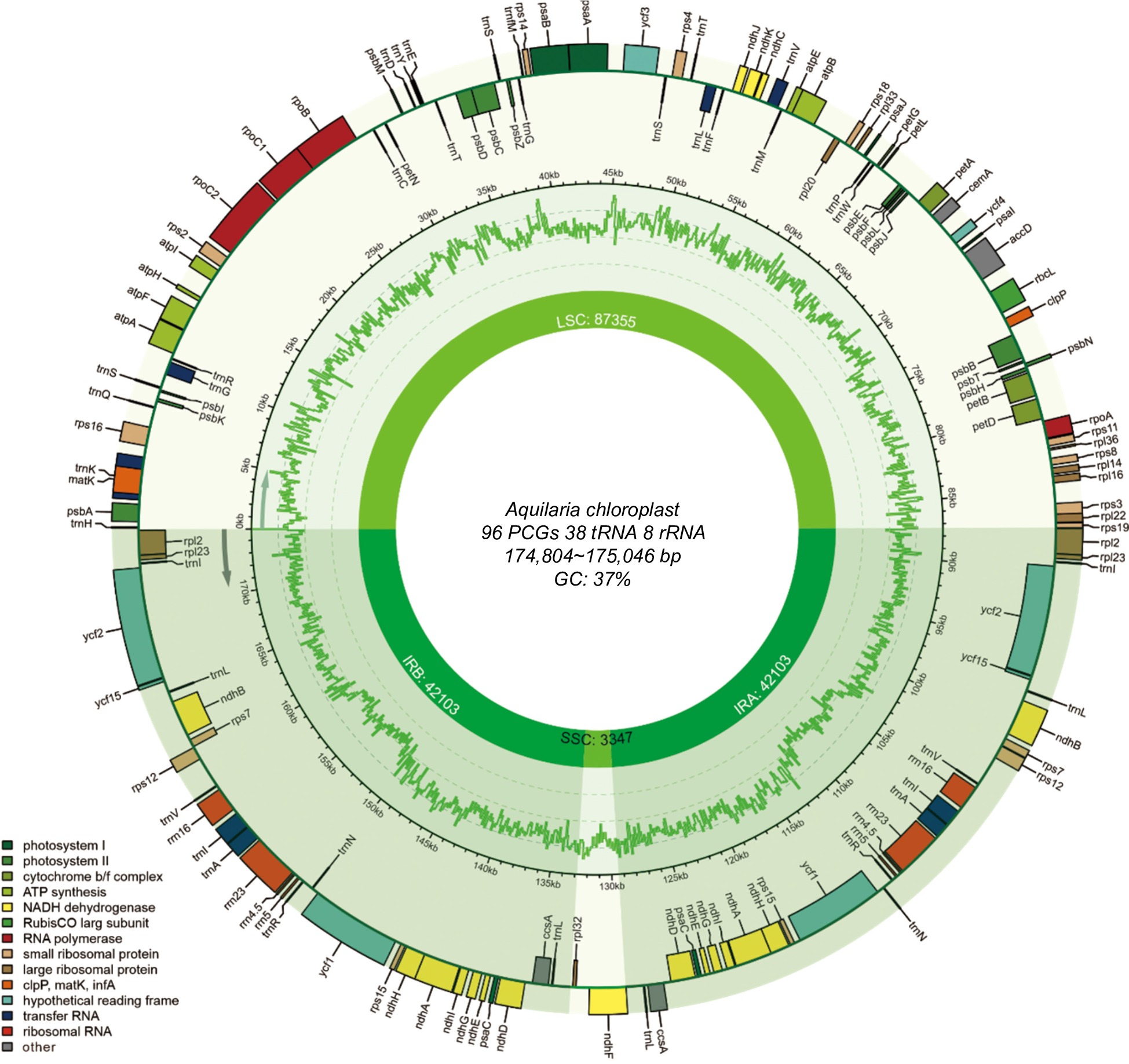
      Figure 1. 

      Annotation map of the Aquilaria plastome. Genes listed inside and outside the circle are transcribed clockwise and counterclockwise, respectively, with genes color-coded based on functional classification.

      The plastomes of these Aquilaria samples encoded an identical set of 142 genes (Table 1), comprising 96 PCGs, 38 transfer RNA (tRNA) genes, and eight ribosomal RNA (rRNA) genes. Among them, 64 single-copy PCGs were identified, with 16 PCGs located in the IR regions. Additionally, four rRNA genes (rrn4.5, rrn23, rrn5, and rrn16) and eight tRNA genes (trnA-UGC, trnI-CAU, trnI-GAU, trnL-CAA, trnL-UAG, trnN-GUU, trnR-ACG, and trnV-GAC) were found to have two copies in IR regions. Introns were detected in 10 PCGs and six tRNA genes. Notably, two PCGs, ycf3, and trans-spliced rps12 (characterized by the first exon located in the LSC region and the other two in the IR regions) contained two introns. These findings underscore a highly conserved genome structure and gene content across Aquilaria plastomes.

      Table 1.  Genes present within Aquilaria plastomes.

      Category Group of genes Names of genes
      Photosynthesis Subunits of photosystem I psaA, psaB, psaC(2), psaI, psaJ
      Subunits of photosystem II psbA, psbB, psbC, psbD, psbE, psbF, psbH, psbI, psbJ, psbK, psbL, psbM, psbN, psbT, psbZ
      Subunits of NADH dehydrogenase ndhA*(2), ndhB*(2), ndhC, ndhD(2), ndhE(2), ndhF, ndhG(2), ndhH(2), ndhI(2), ndhJ, ndhK
      Subunits of cytochrome b/f complex petA, petB*, petD*, petG, petL, petN
      Subunits of ATP synthase atpA, atpB, atpE, atpF*, atpH, atpI
      Large subunit of rubisco rbcL
      Subunits photochlorophyllide reductase
      Self-replication Proteins of large ribosomal subunit rpl14, rpl16, rpl2*(2), rpl20, rpl22, rpl23(2), rpl32, rpl33, rpl36
      Proteins of small ribosomal subunit rps11, rps12**(2), rps14, rps15(2), rps16*, rps18, rps19, rps2, rps3, rps4, rps7(2), rps8
      Subunits of RNA polymerase rpoA, rpoB, rpoC1*, rpoC2
      Ribosomal RNAs rrn16(2), rrn23(2), rrn4.5(2), rrn5(2)
      Transfer RNAs trnA-UGC*(2), trnC-GCA, trnD-GUC, trnE-UUC, trnF-GAA, trnG-GCC, trnG-UCC*, trnH-GUG, trnI-CAU(2), trnI-GAU*(2), trnK-UUU*, trnL-CAA(2), trnL-UAA*, trnL-UAG(2), trnM-CAU, trnN-GUU(2), trnP-UGG, trnQ-UUG, trnR-ACG(2), trnR-UCU, trnS-GCU, trnS-GGA, trnS-UGA, trnT-GGU, trnT-UGU, trnV-GAC(2), trnV-UAC*,
      trnW-CCA, trnY-GUA, trnfM-CAU
      Other genes Maturase matK
      Protease clpP
      Envelope membrane protein cemA
      Acetyl-CoA carboxylase accD
      c-type cytochrome synthesis gene ccsA
      Translation initiation factor
      other
      Genes of unknown function Conserved hypothetical chloroplast ORF ycf1(2), ycf15(2), ycf2(2), ycf3**, ycf4
      Gene*: Gene with one intron; Gene**: Gene with two introns; Gene(2): Number of copies of multi-copy genes.
    • The Mauve alignment results (Supplemental Fig. S1) revealed no gene rearrangements in these newly sequenced plastomes, with the plastome of A. malaccensis serving as the reference. To further explore structure variations within the Aquilaria genus, 15 Aquilaria plastomes were downloaded from GenBank for comparative analysis. The junction sites on the boundaries of genomic regions in Aquilaria species, namely LSC/IRb, SSC/IRb, SSC/IRa, and LSC/IRa, are pivotal in chloroplast genome evolution. These junctions were determined using CPJSdraw. As depicted in Fig. 2 (or Supplemental Fig. S2), the boundaries of genomic regions in Aquilaria samples are highly conserved, with only minor variations observed in the junctions and adjacent genes. The rps19 gene was found across both the LSC and IRb regions, with most of its sequence situated in the LSC region. Furthermore, a 16 bp extension of this gene was consistently observed in the IRb region across all Aquilaria plastomes. This 16 bp extension could potentially serve as a genus-special marker for Aquilaria. Moreover, it was observed that the IRb/SSC junction was located within the ndhF gene. Notably, no genes were found spanning the SSC/IRa and IRa/LSC junctions.
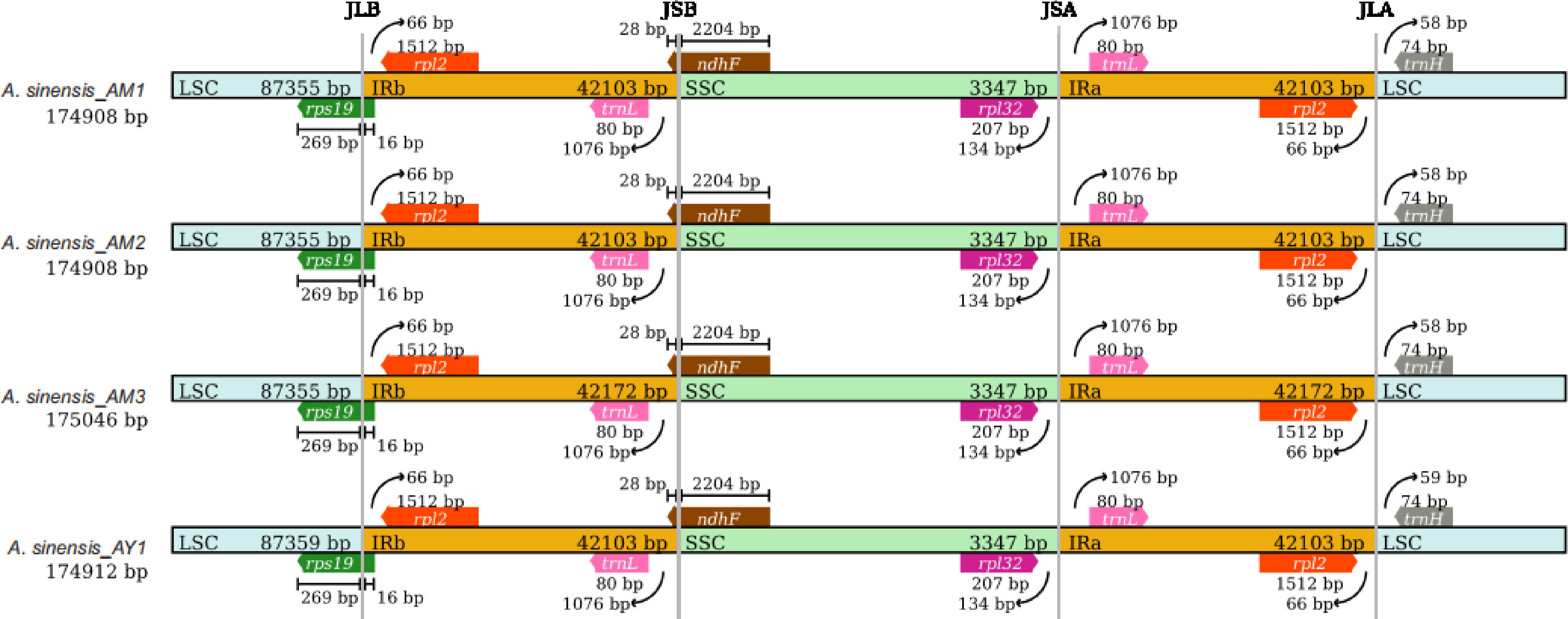
      Figure 2. 

      Comparison of the large single-copy (LSC), small single-copy (SSC), and inverted repeat (IR) region borders of Aquilaria plastomes. The LSC, IR, and SSC regions are depicted with blue, orange, and green blocks, respectively. Gene boxes above the block are transcribed counterclockwise while those below the block are transcribed clockwise. (Representative plastomes from Supplemental Fig. S2).

      The overall organization of the plastomes was analyzed using mVISTA, revealing a high degree of conservation among these Aquilaria plastomes (Supplemental Fig. S3). Interestingly, the SC regions exhibited higher divergence compared to the IR regions. Furthermore, it was observed that PCGs displayed a lower level of variation when compared to non-coding regions.

      Furthermore, we identified some variant hotspot regions, primarily comprising intergenic regions and a few gene coding regions such as rps16 and rpoC1. The nucleotide variability (π) values across the analyzed coding and intergenic sequences of the 37 chloroplast genomes ranged from 0 to 0.03037 (Fig. 3). Notably, variation in the SSC region was higher than that in the IR region, consistent with the mVISTA results. Seven regions with π values higher than 0.005, including rps16, rpoC1, psbM-trnD-GUC, ndhC-trnV-UAC, psbJ-petA, ndhF-rpl32, and rpl32-trnL-UAG, were detected. These regions were identified as HVRs in this study. Among them, the coding region of the rps16 gene exhibited a π value exceeding 0.04, while the rpl32-trnL-UAG inter-region possessed the most informative sites. These HVRs have the potential to serve as barcodes for identifying Aquilaria species.
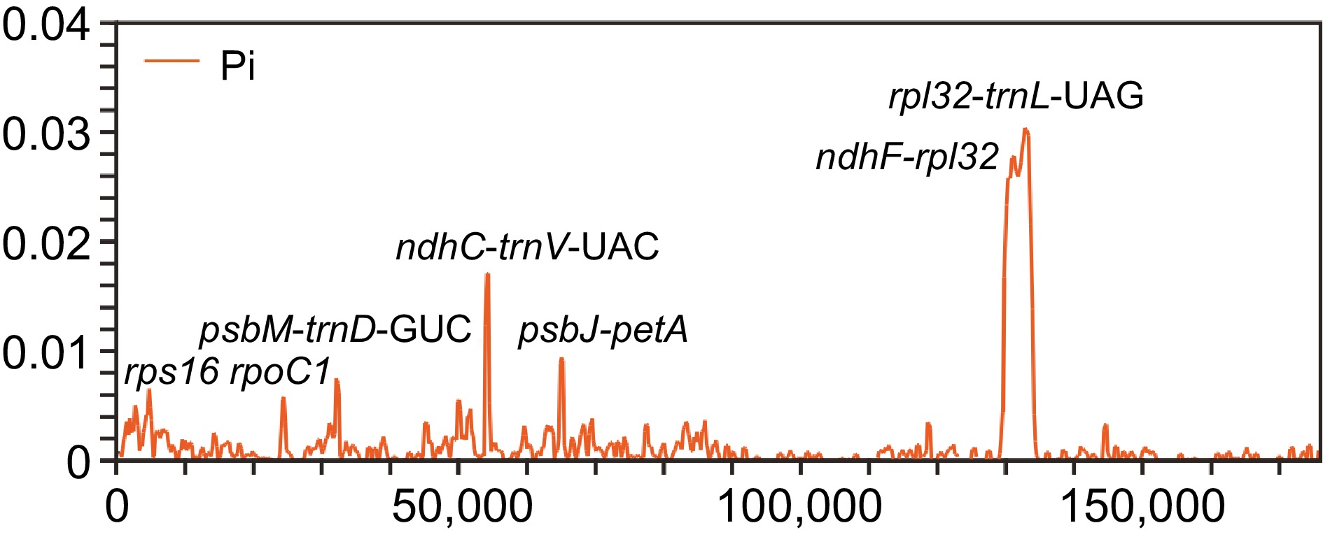
      Figure 3. 

      Comparison of nucleotide variability values among 37 plastomes using window sliding analysis. The x-axis indicates the position of the midpoint of the window, while the y-axis indicates the nucleotide diversity of each window.

    • Nucleotide repeats within chloroplast genomes play a crucial role in plant typing and are widely used as genetic molecular markers in population genetics. Large sequence repeats (LSRs) were identified as repeats with a length of ≥ 30 bp each. A total of 4,765 LSRs were detected across the 37 plastomes, encompassing palindromic repeats (P), forward repeats (F), reverse repeats (R) and complementary repeats (C) (Fig. 4a). Among these, forward repeats were the most prevalent type, with 2,533 occurrences, while complementary repeats were the least common, totaling 46 occurrences.
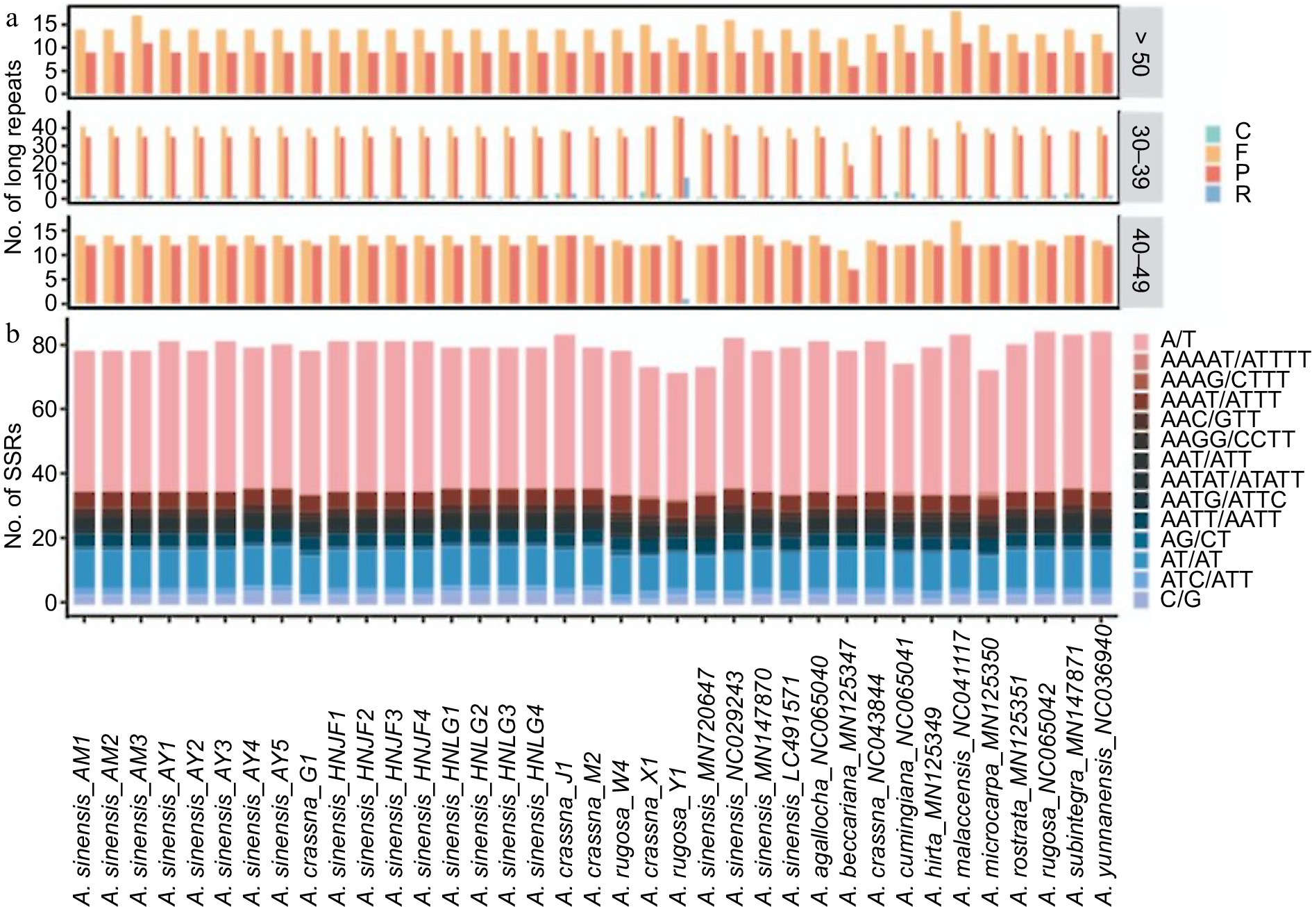
      Figure 4. 

      (a) Distribution of different types of large sequences repeat (LSRs) in Aquilaria plastomes, including complementary (C), forward (F), palindromic (P), and reverse (R) type repeats. (b) Distribution of distinct types of simple sequence repeats (SSRs) in Aquilaria plastomes.

      In the MISA-web analysis, five types of simple sequence repeats (SSRs) were identified, with mononucleotide repeats comprising the majority (1,775, 59.91%), followed by dinucleotide repeats (477, 16.1%), trinucleotide repeats (294, 9.92%), tetranucleotide repeats (409, 13.80%), and pentanucleotide repeats (8, 0.27%) (Fig. 4b). Among these SSRs, repeat units of A/T, AT/AT, AAAT/ATTT, AAAAT/ATTTT, and AAATT/AATTT accounted for 87.55% of the total, indicating a bias towards A/T bases in SSR composition.

    • Ka/Ks is a valuable metric for evaluating whether PCGs have undergone adaptive evolution. Typically, synonymous nucleotide substitutions occur more frequently than non-synonymous substitutions in most genes of organisms, resulting in Ka/Ks values typically being less than 1. In this study, the Ka/Ks values for the PCGs of these plastomes were calculated. Most of the PCGs exhibited Ka/Ks values of less than 1, indicating that these species were subjected to strong purifying selection during the long-term evolutionary process (Fig. 5). Notably, the expression-related accD gene displayed the highest Ka/Ks value (1.54). This finding suggests that the accD gene may have been influenced by positive selection during the evolutionary process.
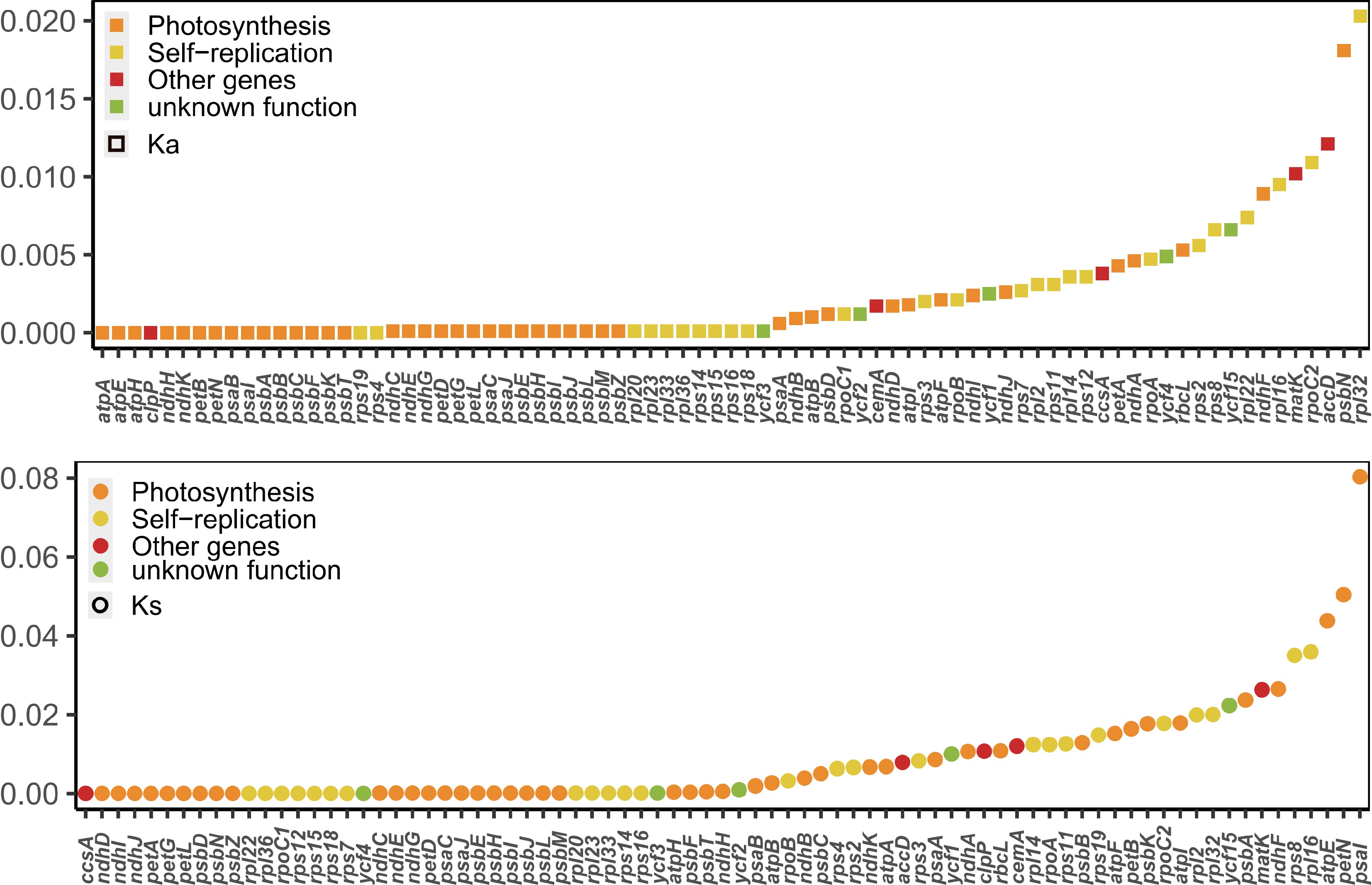
      Figure 5. 

      Values of synonymous (Ks) and non-synonymous (Ka) substitution rates for 79 protein-coding genes in Aquilaria.

      To elucidate the phylogenetic relationships within the Aquilaria genus, 22 newly sequenced Aquilaria plastomes were utilized in conjunction with an additional 15 Aquilaria plastomes to reconstruct the phylogenetic tree using ML and BI methods. Additionally, three Daphne samples were selected as outgroups (Fig. 6). The results revealed the formation of five clades within Aquilaria. The newly sequenced A. sinensis plastomes clustered together with published A. sinensis plastomes, forming a monophyletic clade designated as clade 1 (C1). Clade 2 (C2) comprised three A. rugosa plastomes, which clustered together and formed a sister group to A. yunnanensis with high confidence. Clade 3 (C3) consisted of only one species, identified as A. agallocha.
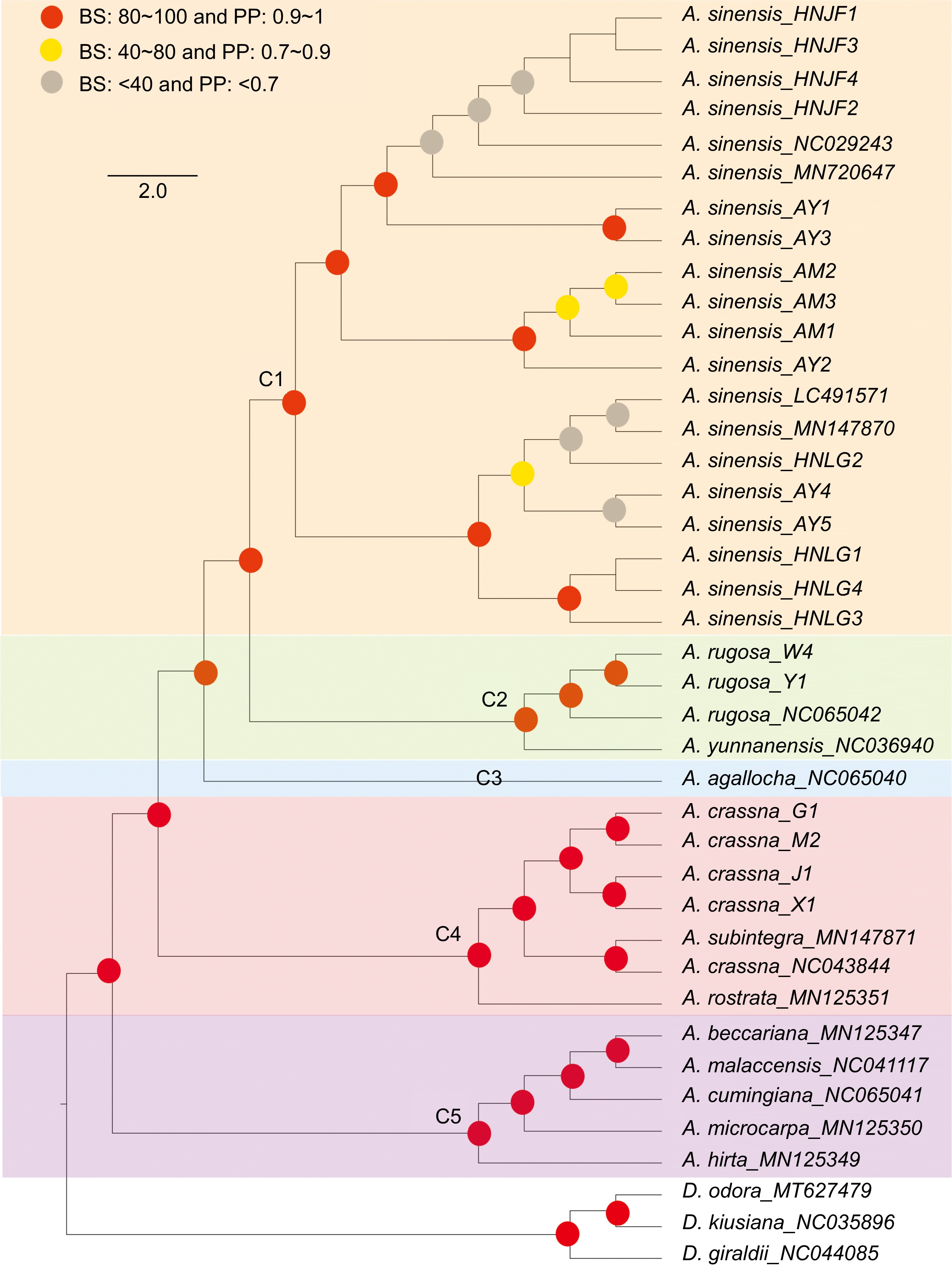
      Figure 6. 

      Phylogenetic tree depicting relationships among Aquilaria species. Red circles indicates bootstrap support (BS) values greater than 80% and posterior probability (PP) values exceeding 1. Yellow circles indicates BS values ranging from 40% to 80%, and PP values ranging from 0.7 to 0.9. Grey circles indicates BS values below 40% and PP values less than 0.7.

      In clade 4 (C4), it was observed that A. crassna (NC043844) clustered together with A. subintegra (MN147871), which aligns with previous research[47]. The newly sequenced A. crassna plastomes also clustered together, demonstrating high consistency. This finding suggests the possibility of hybridization events or misidentification of the published plastome sample (NC043844). It is conceivable that hybridization occurred among these species, with plastomes likely following maternal inheritance, leading to significant disturbance in DNA information within the plastomes. Alternatively, the high similarity in morphology between these species may have contributed to misidentification. Lastly, clade 5 (C5) encompassed five species, mirroring the phylogenetic findings of a previous study[29,47].

    • In this study, the complete plastome sequences of 22 Aquilaria taxa were presented. By integrating these newly sequenced plastomes with an additional 15 Aquilaria published plastomes, a series of comparative analyses within this genus were conducted. The investigation into the location of the SC/IR boundaries offers novel insights into evolutionary studies. The expansion and contraction of IR regions are often associated with various evolutionary events, including gene duplication, loss of gene copies, and the emergence of pseudogenes (such as the ycf1 pseudogene observed in many plants)[48,49]. The substantial variation observed among these boundaries may harbor potential markers for genus/species identification[50,51]. The present research has unveiled a consistent structural feature at the IR/SC boundary in Aquilaria species. Intriguingly, all Aquilaria plastomes analyzed in this study exhibited a 16 bp extension of the rps19 gene in the IRb region. This phenomenon is rarely observed in other species within the Thymelaeaceae family, as it has only been detected in a few species across certain genera[52,53]. In Aquilaria, a consistent 16 bp extension was observed across all examined species, suggesting its potential as a genus-specific marker. Such markers have been utilized for identification previously; for example, the 110 bp expansion of the rps19 gene in the Crassulaceae family serves as a specific family marker[19]. Future studies employing extensive datasets of Thymelaeaceae species may identify additional family-specific identification markers. Furthermore, discrepancies were noted in the plastomes of A. malaccensis (NC041117) and other Aquilaria species, particularly in the positions and transcriptional directions of ndhF and rpl32; their transcribed directions were reversed. These anomalies were primarily attributed to assembly errors. Notably, the same misassembly was found in A. sinensis (NC029243). The loss of rps32 and the incorrect positioning of trnN, ndhF, and ycf1 genes further underscored the erroneous assembly of the plastome. To elucidate the authentic structural patterns of the plastome genome, these inaccurate annotations were substituted with normal data, thereby eliminating potential sources of misinterpretation.

      Furthermore, accD displayed the highest Ka/Ks value of 1.54. Previous research has suggested that adaptive evolution may occur at the molecular level, as evidenced by an elevated Ka/Ks value[54]. Numerous studies have corroborated that a higher Ka/Ks ratio correlates with a more robust positive selection[26,55,56]. Thus, the elevated Ka/Ks ratio observed in accD implies its significant contribution in the adaptation and evolutionary processes within Aquilaria species.

      While previous research has attempted to elucidate the phylogenetic status of the Aquilaria genus, the limited availability of plastomic resources for Aquilaria has left certain issues unresolved. One such point of interest pertains to the relationship among A. sinensis, A. agallocha, and A. yunnanensis. Farah et al.[28] utilized two types of datasets (chloroplast loci and ITS regions) and reported conflicting patterns among these three species. The chloroplast loci data clustered these species into the same clade, with A. agallocha being closer to A. sinensis. Conversely, the evolutionary tree based on ITS regions separated A. sinensis to another clade. Another study[29] also examined these species for phylogenetic analysis, yielding different results. In that study, these species were clustered in the same clade, with A. yunnanensis exhibiting a closer relationship to A. sinensis. Additionally, several studies[1,47,57] have explored phylogenetic relationships in Aquilaria; however, not all of them included all three species of interest. The phylogenetic tree reconstructed in this study mirrored the results of the study conducted by Lee et al.[29] with high confidence. The structure of the phylogenetic tree appeared to be highly influenced by the volume of the dataset; as the number of Aquilaria plastomes were increased, it was anticipated that this result would better elucidate the true relationship among these species.

      Furthermore, of interest was a previous study[47] which revealed that different varieties of A. crassna did not form a monophyletic clade, as A. subintetegra (MN147870) clustered with A. crassna (MK779998, also coded as NC043844). To explore the underlying reason for this discrepancy, four A. crassna plastomes were sequenced. The phylogenetic results unveiled that the newly sequenced A. crassna plastomes in this study clustered together. However, the published A. crassna (NC043844) also clustered with A. subintegra (MN147871), aligning with the findings of the earlier research conducted by Hishamuddin et al.[47]. Chloroplasts, organelles present in plants and some protist cells, possess their own DNA distinct from the nuclear genome. The similarity of the chloroplast genome sequences can unveil phylogenetic relationships and evolutionary history among species[5862], suggesting inheritance from a common ancestor in many cases[6365], as chloroplasts are typically maternally inherited in plants[66,67]. Conversely, similar plastomes may also result from convergent evolution, where analogous chloroplast genome sequences evolve independently on distinct evolutionary trajectories. In the case of A. crassna (NC043844), there is a possibility of incorrect assembly, or even misidentification of the specimen as either A. crassna (NC043844) or A. subintegra (MN147871). Moreover, conducting a pan-plastome study involving A. subintegra and A. crassna could offer a more comprehensive understanding of the relationship between these two species.

      With the augmented dataset of plastomes in this study, a more distinct phylogenetic pattern was unveiled compared to previous research. Besides, although plastomes can provide valuable insights into phylogenetic relationships, the limitations imposed by frequent artificial hybridization are recognized[8]. It is anticipated that future studies integrating data from nuclear genomes, mitogenomes, and plastomes will undoubtedly provide a deeper understanding of the phylogenetic implications within the Aquilaria genus. This study can serve as a valuable example for species identification in trees, highlighting the significance of understanding evolutionary relationships between species or subspecies, given the high similarity among tree species.

    • This study presents the sequencing of 22 Aquilaria plastomes, revealing no gene rearrangements among them. Comparative genomics unveiled a consistent genome size (174,804–175,046 bp) and GC content (36.70%–36.75%) across Aquilaria plastomes. Additionally, seven HVRs were identified, spanning rps16, rpoC1, psbM-trnD-GUC, ndhC-trnV-UAC, psbJ-petA, ndhF-rpl32, and rpl32-trnL-UAG. In examining the IR junction pattern, it was noted that the rps19 gene spans both the LSC and IRb regions, with a 16 bp expansion of the rps19 gene in IRb, potentially serving as a genus-specific marker for Aquilaria. Furthermore, the present analysis identified four types of LSRs and five types of SSRs with a notable A/T bias. The ndhD gene displayed the highest Ka/Ks value of 1.54, indicative of its significant contribution to the adaptation and evolution of Aquilaria species. Finally, the present study elucidated the phylogenetic relationships within Aquilaria and addressed several issues within the genus through an expanded dataset, providing valuable insights into its evolutionary dynamics.

    • The authors confirm contribution to the paper as follows: study conception and design: Wu Z, Hong Z; data collection: Liu X, Xu D, Tembrock LR; analysis and interpretation of results: Kan J, Mi Z, Nie L; draft manuscript preparation: Kan J, Mi Z; organizing and supervising the whole project: Wu Z, Hong Z. All authors reviewed the results and approved the final version of the manuscript.

    • The annotation files of newly assembled Aquilaria plastomes are uploaded to the Figshare website (https://figshare.com/, doi:10.6084/m9.figshare.25713024).

      • This study was funded by Forestry Science and Technology Innovation Project of Guangdong Province (Grant No. 2023KJCX009) and Fundamental Research Funds for the Central Non-profit Research Institution of Chinese Academy of Forestry (Grant No. CAFYBB2021QD003-03). It was also funded by the Science Technology and Innovation Commission of Shenzhen Municipality (Grant No. RCYX20200714114538196) and the Shenzhen Fundamental Research Program (Grant No. JCYJ20220818103212025). This work was also funded by the Guangdong Pearl River Talent Program (Grant No. 2021QN02N792) and the Innovation Program of Chinese Academy of Agricultural Sciences.

      • The authors declare that they have no conflict of interest.

      • # Authors contributed equally: Junhu Kan, Liyun Nie, Zenglu Mi

      • Supplemental Table S1 Structural feactures of Aquilaria plastomes.
      • Supplemental Fig. S1 Mauve alignment of the 22 Aquilaria samples alliance plastomes, illustrating colliner blocks rearrangement of the accessible gene at the junction of inverted repeat and small-single copy regions. White blocks represent protein-coding gene, black blocks represent transfer RNA (tRNA) genes, and red blocks represent ribosomal RNA (rRNA).
      • Supplemental Fig. S2 Comparison of the large single-copy (LSC), small single-copy (SSC), and inverted repeat (IR) region borders of Aquilaria plastomes. The LSC, IR, and SSC regions are depicted with blue, orange, and green blocks, respectively. Gene boxes above the block are transcribed counterclockwise while those below the block are transcribed clockwise.
      • Supplemental Fig. S3 Structural comparisons of 37 Aquilaria plastomes with the mVISTA program. X-axis represents the gene position and the y-axis means the con-sistency of the sequences with the numbers ranged from 50% to 100%.
      • Copyright: © 2024 by the author(s). Published by Maximum Academic Press, Fayetteville, GA. This article is an open access article distributed under Creative Commons Attribution License (CC BY 4.0), visit https://creativecommons.org/licenses/by/4.0/.
    Figure (6)  Table (1) References (67)
  • About this article
    Cite this article
    Kan J, Nie L, Mi Z, Liu X, Xu D, et al. 2024. Insights into Aquilaria phylogenetics through comparative plastomic resources. Forestry Research 4: e030 doi: 10.48130/forres-0024-0028
    Kan J, Nie L, Mi Z, Liu X, Xu D, et al. 2024. Insights into Aquilaria phylogenetics through comparative plastomic resources. Forestry Research 4: e030 doi: 10.48130/forres-0024-0028

Catalog

  • About this article

/

DownLoad:  Full-Size Img  PowerPoint
Return
Return