ARTICLE   Open Access    

Extracts and isolated compounds from the Acorus calamus L. (sweet flag) rhizome showed distinct antimetastatic activity against MDA-MB-231 breast cancer cells

More Information
  • The present work aims to investigate and validate the medicinal potential of Acorus calamus, a plant known for its high therapeutic potential. The focus was identifying biologically active extracts and compounds that exhibit promising anticancer properties. The cytotoxicity of the extracts and compounds extracted from the rhizomes of A. calamus was assessed in three different cancer cell lines: lung carcinoma (A549), colon carcinoma (HCT-116), and breast carcinoma (MDA-MB-231). The application of extracts derived from the rhizome of A. calamus significantly inhibited cell proliferation in various cancer cell lines, including lung, colon, and breast cancer cells. Furthermore, the isolated compounds ACS08 (stearic acid), ACS06 (beta-sitosterol), and ACS02 (β-asarone) exhibited superior anticancer activity against all the tested cancer cell lines, with an inhibition rate of ≥ 50%. Compared with ACS06 and ACS02, ACS08 exhibited greater cytotoxicity. Among the panel of cancer cell lines utilized, compound ACS08 exhibited greater efficacy against the MDA-MB-231 breast cancer cell line, as evidenced by an IC50 estimation of 55.89 μM.
  • Scale insects (Hemiptera: Coccomorpha) with hypogeic habits are considered of high phytosanitary relevance for coffee crops (Rubiaceae: Coffea spp.) in Colombia[1]. A total of 65 species of scale insects associated with coffee roots have been recorded in Colombia[24]. The most species-rich family is the Pseudococcidae with 28 species distributed in nine genera: Dysmicoccus Ferris, 1950 (13 spp.), followed by Pseudococcus Westwood, 1840 (four spp.), Phenacoccus Cockerell, 1893 (three spp.), Planococcus Ferris, 1950, and Spilococcus Ferris, 1950 (two spp. each), and Chorizococcus McKenzie, 1960, Distichlicoccus Ferris, 1950, Ferrisia Fullaway, 1923, and Paraputo Laing, 1929 (one sp. each). For the family Rhizoecidae, 19 species have been recorded in six genera, namely, Rhizoecus Kunckel d'Herculais, 1878 (13 spp.), Pseudorhizoecus Green, 1933 (two spp.), and Capitisetella Hambleton, 1977, Coccidella Hambleton, 1946, Geococcus Green, 1902, and Ripersiella Tinsley, 1899 (one sp. each). Other minor families include Coccidae and Ortheziidae (five spp. in each family), Xenococcidae (three spp.), and Putoidae and Diaspididae (two spp. in each family) and Margarodidae (one sp.). For this study all previous records were re-analysed with the purpose of providing an accurate list of species

    The taxonomic identification of scale insects by a morphological approach is particularly difficult, mainly for two reasons. First, they are small insects (usually < 5 mm) that require the preparation of slide-mounted specimens. Second, the taxonomic keys needed for morphological identifications are primarily designed for adult female specimens[5]. Differing from other insect orders (e.g., Coleoptera, Diptera and Hymenoptera), female scale insects lack well-defined tagmata, as well as sclerites, sutures, or discernible areas. Characters of taxonomic value in scale insects include cuticular processes, such as pores, ducts and, setae[5]. Recognizing these cuticular structures on such small bodies poses a difficult task for non-expert entomologists. To facilitate accessible identification, this manuscript offers an illustrated taxonomic key to scale insect species associated with coffee roots in Colombia and is aimed at users with basic knowledge of scale insect morphology.

    A careful revision of the specimens studied by Caballero et al.[2], preserved in the Scale Insect Collection at the Entomological Museum 'Universidad Nacional Agronomía Bogota' UNAB (Bogotá, Colombia), was carried out to exclude species that are doubtfully recorded from coffee roots in Colombia. This re-assessment allowed the compilation of an accurate list of species that could be included in the taxonomic key. Additional species and information from Caballero[3] and Caballero et al.[4] also were used for construction of the key. List of species recorded for Colombia and c ollection data of specimens analized are in Supplemmental Table S1 and S2 respectivately.

    The illustrated taxonomic key (Table 1) is based on the external morphology of the adult female with a dichotomous structure. Each couplet after the first one is numbered followed by the number of the preceding couplet in parenthesis, e.g. 12(7) means that couplet 12 is derived from couplet 7; the numbers at the end of the couplet indicate the next couplet in order to arrive at the species name that best matches the character states selected by the user. It is illustrated in most of the steps using microphotographs. Acquisition and analysis of images were done with a Lumenera 1-5C camera and the software Image Pro Insight 8.0. Designs were performed with Affinity Photo V 2.1 and Affinity Designer V 2.1 software. The taxonomic keys were structured with some adaptations of published taxonomic keys[615]. The general morphological terminology follows Kondo & Watson[5] with specific terminology for Coccidae[6,16], Margarodidae[17], Ortheziidae[7], Diaspididae[18], Pseudococcidae, Putoidae[8,9], and Rhizoecidae[10,11]. The abdominal segmentation is given as SabdI for abdominal segment 1 to SabdVIII for abdominal segment 8. All microphotographs are of adult female scale insects or their taxonomically important morphological structures.

    Table 1.  Illustrated taxonomic key.
    No.DetailsRef.
    1Abdominal spiracles present (Fig. 1a)2
    Abdominal spiracles absent (Fig. 1b)7
    Fig. 1 Abdominal spiracles (sp) on margin (a) present on Eurhizococcus colombianus, (b) absent on Distichlicoccus takumasai.
    2(1)Anal aperture without pores and setae (Fig. 2a); legs shorter than half of the transversal diameter of body (Fig. 2b); eyespots and mouthparts absentEurhizococcus colombianus

    Jakubski, 1965
    Anal aperture forming a well-developed anal ring with pores and setae (Fig. 2c); legs longer than transversal diameter of body; eyespots and mouthparts present (Fig. 2d)
    3
    Fig. 2 Eurhizococcus colombianus: (a) Anal aperture without pores and setae in the border, (b) section of mid body showing the length of hind leg (lel) and transversal body line (btl). Insignorthezia insignis: (c) Anal aperture with pores (po) and setae (st), (d) section of head with protruding eyespot (es) and labium (lb).
    3(2)Antennae each with eight segments (Fig. 3a)4
    Antennae each with fewer than five segments (Fig. 3b)5
    Fig. 3 (a) Eight-segmented antenna. (b) Four-segmented antenna.
    4(3)Transversal bands of spines absent in ventral region surrounded by an ovisac band (Fig. 4a); dorsal interantennal area without sclerosis (Fig. 4b)Insignorthezia insignis (Browne, 1887)
    Transversal bands of spine plates present in ventral region surrounded by an ovisac band (Fig. 4c); longitudinal sclerosis on dorsum in interantennal area (Fig. 4d)Praelongorthezia praelonga (Douglas, 1891)
    Fig. 4 Insignorthezia insignis: (a) Abdomen without transversal clusters of wax plates, (b) Dorsal interantennal area without sclerosis. Praelongorthezia praelonga: (c) Abdomen with transversal clusters of wax plates marked by dash lines, (d) dorsal interantennal area with a longitudinal sclerotic plate (ep).
    5(3)Antennae each with three segments (Fig. 5a)Newsteadia andreae Caballero, 2021
    Antennae each with four segments (Fig. 5b)6
    Fig. 5 (a) Three-segmented antenna of Newsteadia andreae. Note the presence of pseudosegmentation which gives the appearance of additional segments in the last antennal segment. (b) Four-segmented antenna of Mixorthezia minima.
    6(5)Dorsal area anterior to anal ring with simple pores on protuberances (Fig. 6a); ventral areas surrounding each coxa with a row of wax plate spines (Fig. 6b)Mixorthezia minima Koczné Benedicty & Kozár, 2004
    Dorsal area anterior to anal ring without simple pores or protuberances (Fig. 6c); ventral areas posterior to each coxa without wax plate spines (Fig. 6d)Mixorthezia neotropicalis (Silvestri, 1924)
    Fig. 6 Mixorthezia minima: (a) Dorsum of area anterior to anal ring with close-up of simple pores on protuberances (dash box); (b) ventral area posterior to each coxa with a row of wax plate spines (dash box). Mixorthezia neotropicalis: (c) Close-up of dorsum of area anterior to anal ring lacking simple pores on protuberances (dash box); (d) ventral area posterior to each coxa without associated wax plate spines.
    7(1)Anal plates present (Fig. 7a)8
    Anal plates absent (Fig. 7b)12
    Fig. 7 (a) Anal apparatus of Saissetia coffeae with anal plates (ap) covering the anal aperture (aa). (b) Anal apparatus of Pseudococcus sp. with anal aperture lacking anal plates.
    8(7)Antennae and legs with length similar to or shorter than spiracles (Fig. 8a)9
    Antennae and legs with length at least twice as long as spiracles (Fig. 8b)11
    Fig. 8 (a) Antenna (an) and foreleg (lg) (green lines), and anterior spiracle (sp) (yellow line) of Toumeyella coffeae showing their relative length. Note the similar size of the limbs and spiracle. (b) Antenna (an) and leg (lg) (green lines), and anterior spiracle (sp) (yellow line) of Coccus viridis showing their relative length. Note the relatively smaller size of the spiracle.
    9(8)Ventral tubular macroducts present (Fig. 9)Toumeyella coffeae
    Kondo, 2013
    Ventral tubular macroducts absent10
    Fig. 9 Ventral tubular macroducts (dash box) and close-up of macroducts (photo on right side).
    10(9)Orbicular pores (Fig. 10a) and cribriform platelets present (Fig. 10b); dorsal setae absent; opercular pores absentCryptostigma urichi (Cockerell, 1894)
    Orbicular pores and cribriform platelets absent; dorsal setae present (Fig. 10c); numerous opercular pores present throughout mid areas of dorsum (Fig. 10d)Akermes colombiensis Kondo & Williams, 2004
    Fig. 10 Cryptostigma urichi: (a) Orbicular pore and (b) close-up of a cribriform platelet. Akermes colombiensis: (c) Close-up of a dorsal body setae (dash box) and (d) close-up of opercular pores (arrows).
    11(8)Band of ventral tubular ducts in lateral and submarginal regions absent, ventral tubular ducts of one type; anal plates without discal setae (Fig. 11a); dorsal body setae capitate or clavate (Fig. 11b); perivulvar pores with seven or eight loculi, rarely with 10 loculi (Fig. 11c)Coccus viridis
    (Green, 1889)
    Band of ventral tubular ducts in lateral and submarginal regions present, submarginal region with two types of tubular ducts (Fig. 11d); anal plates with discal setae (Fig. 11e); dorsal body setae spine-like, apically pointed (Fig. 11f); perivulvar pores mostly with 10 loculi (Fig. 11g)Saissetia coffeae
    (Walker, 1852)
    Fig. 11 Coccus viridis: (a) Anal plates without discal setae; (b) dorsal body setae capitate (top) or clavate (below); (c) multilocular disc pores mostly with eight loculi. Saissetia coffeae: (d) Ventral submarginal region with two types of tubular ducts; (e) each anal plate with a discal seta; (f) dorsal body setae acute; (g) multilocular disc pores with mostly 10 loculi.
    12(7)Cerarii present on body margin, at least a pair on each anal lobe (Fig. 12a)13
    Cerarii absent on body margin (Fig. 12b)38
    Fig. 12 Abdominal body margin of (a) Pseudococcus sp. with three cerarii (dash box) and (b) Rhizoecus sp. (dash box) without cerarii.
    13(12)Enlarged oral collar tubular ducts composed of a sclerotized area surrounding the border and a set of flagellated setae (Ferrisia-type oral collar tubular ducts) (Fig. 13a)Ferrisia uzinuri
    Kaydan & Gullan, 2012
    Oral collar tubular ducts simple, not as above (Fig. 13b) or absent14
    Fig. 13 (a) Ferrisia-type oral collar tubular ducts with aperture of tubular duct (ad) surrounded by a sclerotized area (sa) and associated flagellate setae (fs). (b) Oral collar tubular ducts simple (arrows).
    14(12)Antenna with nine segments (Fig. 14a)15
    Antenna with eight segments (Fig. 14b) or fewer (Fig. 14c)19
    Fig. 14 Antenna with (a) nine segments, (b) eight segments and (c) seven segments.
    15(14)Cerarii with more than five conical setae (Fig. 15a); hind trochanter with six sensilla, three on each surface (Fig. 15b)16
    Cerarii with two lanceolate setae (Fig. 15c); hind trochanter with four sensilla, two on each surface (Fig. 15d)17
    Fig. 15 Puto barberi: (a) upper and lateral view of a cerarius, (b) close-up of the surface of trochanter with three sensilla (arrows). Phenacoccus sisalanus: (c) cerarius, (d) trochanter with two sensilla (arrows) on single surface.
    16(15)Cerarii with tubular ducts (Fig. 16a)Puto antioquensis
    (Murillo, 1931)
    Cerarii without tubular ducts (Fig. 16b)Puto barberi
    (Cockerell, 1895)
    Fig. 16 (a) Cerarius associated with tubular ducts (arrows). (b) Cerarius without tubular ducts.
    17(15)Oral collar tubular ducts absentPhenacoccus sisalanus Granara de Willink, 2007
    Oral collar tubular ducts present, at least on venter (Fig. 17)18
    Fig. 17 Ventral surface with oral collar tubular ducts (dash circles).
    18(17)Oral collar tubular ducts restricted to venterPhenacoccus solani
    Ferris, 1918
    Oral collar tubular ducts present on dorsum and venterPhenacoccus parvus Morrison, 1924
    19(14)Oral rim tubular ducts present (Fig. 18)20
    Oral rim tubular ducts absent26
    Fig. 18 Oral rim tubular ducts in upper view (dash circles) and close-up of lateral view.
    20(19)Oral rim tubular ducts present on venter onlyPseudococcus landoi (Balachowsky, 1959)
    Oral rim tubular ducts present on both dorsum and venter21
    21(20)Cerarii restricted to anal lobes (Fig. 19a)Chorizococcus caribaeus Williams & Granara de Willink, 1992
    Cerarii present, at least on the last five abdominal segments (Fig. 19b)22
    Fig. 19 Location of cerarii (dash boxes) on abdominal margin with close-up of cerarius (a) restricted to anal lobes (dash boxes) and (b) cerarii present on the last five abdominal segments.
    22(21)Circulus absent (Fig. 20a)23
    Circulus present (Fig. 20b)24
    Fig. 20 Ventral mid area of abdominal segments III and IV (dash box) of (a) Distichlicoccus takumasai without circulus and (b) Pseudococcus jackbeardsleyi with circulus.
    23(22)Multilocular disc pores present on venter of SabdIV and posterior segments (Fig. 21a); hind coxa with translucent pores and hind femur without translucent pores (Fig. 21b)Spilococcus pressus
    Ferris, 1950
    Multilocular disc pores absent, if some present, not more than three around vulvar opening (i.e. venter of SabdVII or SabdVIII); hind coxa without translucent pores (Fig. 21c) and hind femur with translucent pores (Fig. 21d)Distichlicoccus takumasai Caballero, 2021
    Fig. 21 Spilococcus pressus: (a) Ventral section of abdomen with multilocular disc pores (arrows); (b) hind leg with close-up of coxa with translucent pores (arrows). Distichlicoccus takumasai: (c) Hind coxa without translucent pores; (d) hind femur with translucent pores (arrows).
    24(22)Eyes without discoidal pores nor sclerotized surrounding area (Fig. 22a); circulus with transversal diameter 40 to
    60 µm (Fig. 22b)
    Pseudococcus luciae Caballero, 2021
    Eyes with discoidal pores and sclerotized surrounding area (Fig. 22c); circulus diameter 100 to 200 µm (Fig. 22d)26
    25(24)Oral rim tubular ducts on dorsal abdominal segments numbering three to eight; area between posterior ostiole and cerarius of SabdVII without oral rim tubular ducts (Fig. 23a)Pseudococcus elisae Borchsenius, 1947
    Oral rim tubular ducts on dorsal abdominal segments numbering 14 to 27; area between posterior ostiole and cerarius of SabdVII with an oral rim tubular duct (Fig. 23b)Pseudococcus jackbeardsleyi Gimpel & Miller, 1996
    Fig. 22 Pseudococcus luciae: (a) Eyespot without surrounding sclerotized area nor associated pores; (b) circulus ca. 58 µm wide. Pseudococcus jackbeardsleyi: (a) Eyespot with sclerotized area (sa) and associated pores (po); (d) circulus ca. 154 µm wide.
    Fig. 23 (a) Dorsal margin of abdominal segments VI to VIII, between cerarius of anal lobe (C1), cerarius of SabdVII (C2) and posterior ostiole (os) without oral rim tubular ducts. (b) Dorsal margin of abdominal segments VI to VIII, between cerarius of anal lobe (C1), cerarius of SabdVII (C2) and posterior ostiole (os) with an oral rim tubular duct and/or cerarius adjacent to SabdVII.
    26(19)Oral collar tubular ducts (Fig. 24) on both dorsum and venter27
    Oral collar tubular ducts restricted to venter28
    Fig. 24 Oral collar tubular duct in lateral view.
    27(26)Hind coxa with translucent pores (Fig. 25a); anal lobe with sclerotized bar, not on a sclerotized area (Fig. 25b); multilocular disc pores present posterior to fore coxaPlanococcus citri-minor complex
    Hind coxa without translucent pores (Fig. 25c); anal lobe without sclerotized bar, on a sclerotized area (Fig. 25d); multilocular disc pores absent posterior to fore coxaDysmicoccus quercicolus (Ferris, 1918)
    28(27)Oral collar tubular ducts absent on venter of both head and thorax.29
    Oral collar tubular ducts present on either head or thorax, but not on both areas (Fig. 26)30
    Fig. 25 Planococcus citri-minor complex: (a) Hind coxa with translucent pores (dash box) and (b) anal lobe with a sclerotization forming a bar (ab). Dysmicoccus quercicolus: (c) Hind coxa without translucent pores and (d) anal lobe with irregular broad sclerotized area (sa).
    Fig. 26 Marginal area of Dysmicoccus grassii, lateral to posterior spiracle (ps), with close-up of oral collar tubular ducts (oc) (left side).
    29(28)Translucent pores present on hind coxa, trochanter, femur and tibia (Fig. 27a); marginal clusters of oral collar tubular ducts on venter of SabdVI and SabdVIIDysmicoccus caribensis Granara de Willink, 2009
    Translucent pores restricted to hind femur and tibia (Fig. 27b); marginal clusters of oral collar tubular ducts present on venter of SabdIV to SabdVIIParaputo nasai
    Caballero, 2021
    Fig. 27 (a) Hind leg of Dysmicoccus caribensis with translucent pores on coxa (cx), trochanter (tr) and femur (fm), and tibia (tb). (b) Hind leg of Paraputo nasai with translucent pores restricted to femur (fm) and tibia (tb).
    30(28)Hind coxa with translucent pores (Fig. 28a)Dysmicoccus sylvarum
    Williams & Granara de Willink, 1992
    Hind coxa without translucent pores (Fig. 28b)31
    Fig. 28 (a) Translucent pores on hind coxa. (b) Translucent pores absent on hind coxa.
    31(30)Hind trochanter with translucent pores (Fig. 29a)Dysmicoccus varius
    Granara de Willink, 2009
    Hind trochanter without translucent pores (Fig. 29b)32
    Fig. 29 Translucent pores (a) on hind trochanter, (b) absent from hind trochanter.
    32(31)Oral collar tubular ducts present on margin of thorax (Fig. 30)33
    Oral collar tubular ducts absent from margin of thorax34
    Fig. 30 Prothorax margin of Dysmicoccus grassii with close-up of oral collar tubular ducts.
    33(32)Multilocular disc pores absent on SabdV; dorsal area immediately anterior to anal ring with tuft of flagellate setae; longest flagellate seta as long as diameter of anal ring (Fig. 31a), and discoidal pores larger than trilocular pores (Fig. 31b)Dysmicoccus radicis
    (Green, 1933)
    Multilocular disc pores present on SabdV; dorsal area immediately anterior to anal ring without a tuft of flagellate setae; flagellate setae much shorter than diameter of anal ring (Fig. 31c) and discoidal pores smaller than trilocular pores (Fig. 31d)Dysmicoccus grassii (Leonardi, 1913)
    34(32)Oral collar tubular ducts absent in interantennal area35
    Oral collar tubular ducts present in interantennal area (Fig. 32)36
    35(34)Translucent pores on hind leg restricted to tibia (Fig. 33a)Dysmicoccus perotensis
    Granara de Willink, 2009
    Translucent pores on hind leg present on tibia and femur (Fig. 33b)Dysmicoccus joannesiae-neobrevipes complex
    Fig. 31 Dysmicoccus radicis: (a) Area anterior to anal ring with a cluster of flagellate setae (fs) and anal ring (ar) showing the diameter of the different pores (dash box); (b) discoidal pores (dp) and trilocular pores (tp). Dysmicoccus grassii: (c) Area anterior to anal ring with scattered short flagellate setae (fs) contrasted with anal ring (ar) diameter (dash box); (d) discoidal pores (dp) and trilocular pores (tp) with similar diameter.
    Fig. 32 Interantennal area (dash box) of Dysmicoccus brevipes with close-up of oral collar tubular ducts.
    Fig. 33 (a) Hind leg of Dysmicoccus perotensis with close-up of femur and tibia with translucent pores on tibia only (arrows). (b) Hind leg of Dysmicoccus joannesiae-neobrevipes complex with close-up of femur and tibia with translucent pores (arrows).
    36(34)Hind coxa with translucent pores (see Fig. 28a)Dysmicoccus mackenziei
    Beardsleyi, 1965
    Hind coxa without translucent pores (see Fig. 28b)37
    37(36)Dorsal SabdVIII setae forming a tuft-like group, each seta conspicuously longer than remaining dorsal abdominal setae (Fig. 34a) and setal length similar to anal ring diameter (60–80 µm long)Dysmicoccus brevipes (Cockerell, 1893)
    Dorsal SabdVIII setae evenly distributed, each setae as long as remaining dorsal abdominal setae (Fig. 34b) and length less than half diameter of anal ringDysmicoccus texensis-neobrevipes complex
    38(12)Tritubular ducts absent39
    Tritubular ducts present (Fig. 35a-b)46
    Fig. 34 (a) Abdomen of Dysmicoccus brevipes with dorsal setae on SabdVIII (lfs) longer than setae on anterior segments (sfs). (b) Abdomen of Dysmicoccus texensis-neobrevipes complex with dorsal setae (ufs) along the abdominal segments of uniform length and scattered distribution.
    Fig. 35 (a) Tritubular duct in upper (left) and lateral view (right) with the border of the cuticular ring attached to tubules. (b) Tritubular duct with the border of the cuticular ring widely separated from tubules (arrows).
    39(38)Anal lobes strongly protruded, bulbiform (Fig. 36a) jutting out from margin for a distance equivalent to diameter of anal ring40
    Anal lobes shallow, if protruded, their length never more than half of diameter of anal ring (Fig. 36b)42
    Fig. 36 (a) Abdomen of Neochavesia caldasiae with anal lobes (al) protruding beyond the anal aperture (aa). (b) Abdomen of Ripersiella sp. with anal lobes (al) at the same level as the anal aperture (aa).
    40(39)Anal aperture located at the same level as the base of anal lobes (Fig. 37a); antennae located on ventral margin of headNeochavesia caldasiae (Balachowsky, 1957)
    Anal aperture located anterior to bases of anal lobes (Fig. 37b); antennae located on dorsum of head41
    Fig. 37 (a) Abdomen of Neochavesia caldasiae with anal aperture (aa) positioned between the anal lobes (al), at the same level as the bases of anal lobes (dash line). (b) Abdomen of Neochavesia eversi with anal aperture (aa) situated anterior to the bases of the anal lobes (al) (dash line).
    41(40)Antennae each with five segments, situated on a membranous base (Fig. 38a); length of hind claw less than length of hind tarsus (Fig. 38b)Neochavesia trinidadensis (Beardsley, 1970)
    Antennae each with four segments, situated on a sclerotized base (Fig. 38c); hind claw longer than hind tarsus (Fig. 38d)Neochavesia eversi (Beardsley, 1970)
    Fig. 38 (a) Antenna with four segments and a membranous base (mb). (b) Hind tarsus (green line) longer than the hind claw (red line). (c) Antenna with four segments and a sclerotized base (sb). (d) Hind tarsus (green line) shorter than hind claw (red line).
    42(39)Body setae capitate, at least on one surface (Fig. 39a)43
    Body setae never capitate (Fig. 39b)44
    Fig. 39 (a) Capitate setae. (b) Flagellate setae.
    43(42)Anal aperture without associated cells (Fig. 40a); three-segmented antennae (Fig. 40b); ventral setae in median
    and submedian regions capitate
    Capitisitella migrans
    (Green, 1933)
    Anal aperture surrounded by cells (Fig. 40c); six-segmented antennae (Fig. 40d); ventral setae in medial and submedial regions flagellateWilliamsrhizoecus coffeae
    Caballero & Ramos, 2018
    44(42)Three-segmented antennae (Fig. 41a); circulus present (Fig. 41b)Pseudorhizoecus bari
    Caballero & Ramos, 2018
    Five-segmented antennae (Fig. 41c); circulus absent45
    Fig. 40 Capitisitella migrans: (a) Anal aperture of surrounded only by setae; (b) antenna composed of three segments. Williamsrhizoecus coffeae: (c) Anal aperture of surrounded by setae and cells (flesh); (d) antenna composed of six segments.
    Fig. 41 Pseudorhizoecus bari: (a) Antenna composed of three segments and (b) circulus. (c) Antenna of Pseudorhizoecus proximus composed of five segments.
    45(44)Multilocular disc pores absent; anal aperture ornamented with small protuberances and two to five short setae, each seta never longer than 1/3 diameter of anal aperture, without cells (Fig. 42a)Pseudorhizoecus proximus
    Green, 1933
    Multilocular disc pores present (Fig. 42b); anal aperture not ornamented with small protruberances, ring with well-developed cells and six long setae, each seta as long as diameter of anal ring (Fig. 42c)Ripersiella andensis (Hambleton,
    1946)
    Fig. 42 (a) Anal aperture of Pseudorhizoecus proximus surrounded by protuberances (pr) and a few short setae (st). Ripersiella andensis: (b) Ventral section of abdomen with multilocular disc pores (mp); (c) anal aperture with a ring of cells and six long setae (se).
    46(38)Anal lobes strongly protruded, conical, each one with a stout spine at apex (Fig. 43a)Geococcus coffeae
    Green, 1933
    Anal lobes flat or barely protruded, without spines at apex (Fig. 43b)47
    47(46)Venter of abdomen with clusters of trilocular pores in medial region (Fig. 44a)Coccidella ecuadorina Konczné Benedicty & Foldi, 2004
    Venter of abdomen with trilocular pores evenly dispersed, never forming clusters in medial region (Fig. 44b)48
    Fig. 43 (a) Abdomen of Geococcus coffeae with protruding anal lobe (al) with a stout spine at the apex (sp). (b) Abdomen of Rhizoecus sp. with anal lobe (al) flat, with numerous flagellate setae (fs) at the apex.
    Fig. 44 (a) Ventral surface of Coccidella ecuadorina with clusters of trilocular pores (tc) (dash box) on medial region of abdomen. (b) Ventral surface of Rhizoecus sp. with trilocular pores (tr) scattered on venter of abdomen.
    48(47)Antennae with six well-developed segments (Fig. 45a)51
    Antennae with five well-developed segments (Fig. 45b), apical segment sometimes partially divided (Fig. 45c)49
    Fig. 45 (a) Six-segmented antenna. (b) Five-segmented antenna. (c) Five-segmented antenna with partially divided apical segment (pd). Note: antennal segments numbered in Roman numerals.
    49(48)Antennae length more than 140 µm (Fig. 46a); tritubular ducts of similar diameter to trilocular pores (± 2 µm variation) (Fig. 46b); tritubular ducts with space between ductules and edge as wide as the ductules (Fig. 46c); slender ductule, width/length ratio 1:6Rhizoecus coffeae
    Laing, 1925
    Antennae length less than 130 µm (Fig. 46d); tritubular ducts of diameter nearly twice diameter of trilocular pores (Fig. 46e); tritubular ducts with reduced space or without space between ductules and edge (Fig. 46f); stout ductule, width/length ratio 1:350
    50(49)Tubular ducts present (Fig. 47a); each anal lobe with around 28 dorsal setae of similar length, greater than 30 µm (Fig. 47b, al); and dorsal marginal clusters of setae on SabdVII 20–30 µm long (Fig. 47b, SabdVII)Rhizoecus setosus (Hambleton, 1946)
    Tubular ducts absent; each anal lobe with around 14 dorsal setae, with length less than 15 µm (Fig. 47c, al); dorsal marginal clusters of setae on SabdVII with length less 15 µm (Fig. 47c, SabdVII)Rhizoecus compotor
    Williams & Granara de Willink, 1992
    Fig. 46 (a) Antenna ca. 207 µm long. (b) Tritubular ducts (td) and trilocular pores (tp) with similar diameter. (c) Close-up of a tritubular duct indicating the space between the cuticular ring (mg) and the ductule (dt). (d) Antenna ca. 105 µm long. (e) Each tritubular duct (td) twice the diameter of a trilocular pore (tp). (f) Close-up of tritubular duct without a space between the cuticular ring (mg) and the ductule (dt).
    Fig. 47 Rhizoecus setosus: (a) Tubular ducts (td); (b) anal lobe (al) and abdominal segment (SabdVII) with marginal clusters of setae longer than 30 µm. (c) Abdomen of Rhizoecus compotor with marginal cluster of setae shorter than 20 µm on anal lobe (al) and abdominal segment (SabdVII).
    51(48)Fore tibia with at least one of two internal preapical setae spine-like (Fig. 48a-b)52
    Fore tibia with both internal preapical setae flagellate (Fig. 48c)56
    Fig. 48 Fore legs with preapical setae on tibia (ft): (a) one flagellate (fs) and one spine seta (ss), (b) with a pair of spine setae (ss), (c) with a pair of flagellate setae (fs).
    52(51)Fore tibia with one internal preapical spine-like setae and other seta flagellate (Fig. 48a); anal ring composed of spine-like setae (Fig. 49a); circulus absentRhizoecus spinipes (Hambleton, 1946)
    Fore tibia with both internal preapical setae spine-like (Fig. 48b); anal ring composed of flagellate-like setae (Fig. 49b); at least, one circulus present (Fig. 49c)53
    Fig. 49 (a) Anal ring (ar) of Rhizoecus spinipes with spine-like setae (ss). (b) Anal ring (ar) of Rhizoecus arabicus with flagellate setae (fs). (c) Circulus of Rhizoecus cacticans.
    53(52)Claw digitules setose and short, length less than half length of claw (Fig. 50a)54
    Claw digitules capitate and long, as long as claw (Fig. 50b)55
    Fig. 50 Claw with claw digitule: (a) setose (sd), (b) flagellate (fd).
    54(53)Anal ring with external row composed of 35 cells or more (Fig. 51a, ext); anal ring with external and internal rows separated by a space as wide as a cell of the external row (Fig. 51a, spc); anal ring cells without spicules (Fig. 51a, sp)Rhizoecus variabilis Hambleton, 1978
    Anal ring with external row composed of less than 30 cells (Fig. 51b, ext); anal ring with external and internal rows separated by a narrow space, as wide as half (or less) a cell of the external row (Fig. 51b, spc); anal ring cells with spicules (Fig. 51b, sp)Rhizoecus arabicus Hambleton, 1976
    Fig. 51 (a) Anal ring of Rhizoecus variabilis with external row (ext) of anal ring consisting of over 35 cells; external row separated from the internal row (int) by a similar width as the diameter of a cell (spc). (b) Anal ring of Rhizoecus arabicus with external row (ext) of anal ring with less than 30 cells; external row separated from the internal row (int) by a width less than half the diameter of a cell (spc); cells of the external row with spicules (sp).
    55(53)More than 80 tritubular ducts; circulus with basal diameter at least five times greater than apical diameter (Fig. 52a); stick-like genital chamber, parallel borders and all of similar width and structure, length across about two abdominal segments (169–175 µm long) (Fig. 52b)Rhizoecus atlanticus (Hambleton, 1946)
    Less than 50 tritubular ducts; circulus with basal diameter less than three times the apical diameter (Fig. 52c); genital chamber with basal third two times wider than anterior two-thirds, length across one abdominal segment (43–52 µm long) (Fig. 52d)Rhizoecus cacticans (Hambleton, 1946)
    Fig. 52 Rhizoecus atlanticus: (a) Circulus with diameter at base five times the apical diameter, (b) genital chamber tubular shape, length ca. 150 µm long. Rhizoecus cacticans: (c) Circulus with diameter at base about two times the apical diameter, (d) genital chamber with proximal section basiform and distal section tubular, with arms, length ca. 45 µm long.
    56(51)Multilocular disc pores absent on dorsumRhizoecus mayanus (Hambleton, 1946)
    Multilocular disc pores present on dorsum57
    57(56)Marginal prothoracic setae length greater than 50 µm (Fig. 53a); marginal SabdVII setae length greater than 45 µm (Fig 53b)Rhizoecus colombiensis Ramos-Portilla & Caballero, 2016
    Marginal prothoracic setae length less than 25 µm (Fig. 53c); marginal SabdVII setae length less than 30 µm (Fig. 53d)58
    Fig. 53 Rhizoecus colombiensis: (a) Body margin with a long seta (pts) (> 40 µm), longer than remaining setae in prothorax; (b) margin of abdominal segment VII (SabdVII) (st). with a long seta (pts) (> 40 µm), longer than remaining setae in abdomen. Rhizoecus americanus: (c) Margin of prothorax (pts) with setae of uniform length, shorter than 30 µm; (d) margin of abdominal segment VII (SabdVII) with setae (st) shorter than 30 µm.
    58(57)Tritubular ducts of two sizesRhizoecus caladii
    Green, 1933
    Tritubular ducts of three sizesRhizoecus americanus (Hambleton, 1946)
     | Show Table
    DownLoad: CSV

    The following illustrated taxonomic key (Table 1) is a tool for the identification of adult female scale insects (Hemiptera : Sternorrhyncha : Coccomorpha) associated with coffee roots in Colombia, which includes 59 species from seven families (see Supplemental Table S1).

    The taxonomic key includes 59 species associated with coffee roots. Hemiberlesia sp., Odonaspis sp., Rhizoecus stangei McKenzie, 1962, Spilococcus mamillariae (Bouche, 1844), Planococcus citri (Risso, 1813) and Planococcus minor (Maskell, 1897) were excluded from the key. In the case of the two armoured scale insects, the specimens were found in Berlese funnel samples associated with coffee roots[2], however, there is no evidence of these species feeding on the roots and there are no previous records of association of Hemiberlesia nor Odonaspis species with coffee roots.

    Previous records of single specimens of R. stangei and S. mamillariae by Caballero et al.[2] were determined as misidentifications of Rhizoecus caladii Green, 1933 and Spilococcus pressus Ferris, 1950, respectively. Spilococcus mamillariae is considered as an oligophagous species, but mainly associated with Cactaceae plants and feeding on the aerial parts of plants[19,20]. There are no records of S. mamillariae being found on any plant species of the family Rubiaceae, and hence we have removed this species from the list of species associated with coffee roots. The species R. stangei, which has been recorded only from Mexico and lacks information on its host plant[21] has apparently not been found since its original description[8].

    Planococcus citri and Pl. minor were listed also by Caballero et al.[2] as literature records. the morphological identification of P. citri and P. minor needs to be complemented with molecular and geographical analysis to be more accurate[22]. Therefore, the present key considers only identification to the Planococcus citri-minor complex.

    Furthermore, many specimens of Dysmicoccus collected from coffee roots in Colombia have morphological character states that overlap with Dysmicoccus neobrevipes Beardsley, 1959, Dysmicoccus joannesiae (Costa Lima, 1939) and Dysmicoccus texensis (Tinsley, 1900). The first case is a mix of character states of D. texensis and D. neobrevipes. The number of setae in the abdominal cerarii and the size of oral collar tubular ducts are the most important characters used to differentiate the adult females of Dysmicoccus species[8,23]. Adult females of D. texensis have a consistent pattern of only two setae in all thoracic and abdominal cerarii, along with a uniform size of oral collar tubular ducts (OC). On the other hand, D. neobrevipes varies in the number of setae in the cerarii, ranging from two to seven, accompanied by two distinct sizes of OC. These character states are generally constant among specimens found on the aerial parts of plants. However, among the specimens examined here, while the anal lobe cerarii consistently have two setae on the specimens of D. texensis found on the roots, the remaining cerarii display a variable number of setae, notably ranging from two to five, particularly within the abdominal cerarii. Furthermore, the OC of these specimens all are the same size. Regarding the differences in number of setae in the cerarii, Granara de Willink[23] underlined the need of more comprehensive studies to definitively separate these species.

    The second case involves D. joannesiae and D. neobrevipes. These species exhibit similarities in the number of setae on each cerarius (ranging from two to seven setae per cerarius) and differences in the number of clusters of OC along the abdominal margin; D. joannesiae has more than 25 clusters of OC and D. neobrevipes has fewer than 10 clusters of OC[8]. Granara de Willink also separated these two species by the presence of OC on the thorax and head[23] (present in D. neobrevipes and absent in D. joannesiae). Within the specimens of putative D. neobrevipes studied here, a few had clusters of OC numbering 15 to 20 along the abdominal margin and OC on the thorax and head. The primary challenge with addressing this dilemma lies in the fact that D. joannesiae has only been reported on Joannesia princeps Vell., 1798 (Euphorbiaceae) in Brazil and on Annona muricata (Annonaceae) intercepted in London from Saint Lucia[8,24]. Moreover, there has been no additional morphological variations recorded in the new records of D. joannesiae since its initial description in 1932 by Costa Lima. Therefore, the character states defining D. joannesiae are based on six type specimens. Based on these arguments, the following taxonomic key considers two species complex groups, namely the Dysmicoccus texensis-neobrevipes complex and the D. joannesiae-neobrevipes complex.

    Following article 31.1.2 of the International Commission of Nomenclature (ICZN), herein we make a change in nomenclature for Distichlicoccus takumasae Caballero, 2021. The ending -ae for takumasae is incorrect because the species was dedicated to Dr. Takumasa Kondo (a male coccidologist), and thus the correct ending is -i, hence the species epithet is herein amended to 'takumasai'. The corrected name is Distichlicoccus takumasai Caballero, 2021.

    After reviewing the species of scale insects associated with coffee roots in Colombia, we have compiled a list of 59 species (Supplemental Table S1). Although this study did not focus on the effect of habit (aerial vs underground) or host plant on the morphology of scale insects, we detected significant morphological variation within facultative hypogeal species. Until further studies allow an understanding of the overlap of character states between D. texensisD. neobrevipes and D. joannesiaeD. neobrevipes, we suggest considering these species as a morphological complex for hypogeal specimens. Further ecomorphological studies should be conducted to determine whether the morphology of a species may differ when feeding on the aerial parts compared when feeding on the underground parts of a host and to try to elucidate what factors trigger those changes, especially in species associated with coffee plants. As for the species complex, further collecting, morphological, and molecular studies should help elucidate these taxonomic problems.

    During the literature review performed for this study, we realized that most of the records of species are limited to mentioning the host but not the plant part on which collections were made, however, it is suspected that most species are normally collected from the aerial parts of the plant host. Although this taxonomic key is limited to root-associated species recorded in Colombia, this key could be useful for identifying scale insects associated with coffee in other tropical regions, extending also to species collected from the aerial parts of the hosts.

    The authors confirm contributions to the paper as follows: study conception and design: Caballero A, Kondo T; data collection: Caballero A, Kondo T; analysis and interpretation of results: Caballero A, Kondo T; draft manuscript preparation: Caballero A, Kondo T. Both authors reviewed the results and approved the final version of the manuscript.

    The data (microscopy slides of specimens) that support the findings of this study are available in the Scale insect repository of the entomological museum Universidad Nacional Agronomia Bogota – UNAB, Facultad de Ciencias Agrarias, Colombia. All data generated or analyzed during this study are included in this published article and its supplementary information files.

    The authors thank Dr. Andrea Ramos-Portilla for clarifying some aspects of the morphological variations of Rhizoecus species and Dr. Penny Gullan (Australian National University, Canberra, Australia) for reviewing an earlier version of the manuscript. Many thanks to Erika Valentina Vergara (AGROSAVIA) and Dr. Francisco Serna (Universidad Nacional de Colombia) for their help to access the Museum UNAB. Special thanks to Dr Giuseppina Pellizzari (University of Padova, Italy) for advice on scientific nomenclature. This study was financed by Colciencias (Programa Nacional de Ciencias Básicas [National Program on Basic Sciences]), code 110165843233, contract FP44842-004-2015), by the entomological museum UNAB (Facultad Ciencias Agrarias, Universidad Nacional de Colombia, sede Bogotá) and by Federación Nacional de Cafeteros.

  • The authors declare that they have no conflict of interest.

  • Supplementary Table S1 Voucher specimen numbers of collected plant material submitted at KASH Herbarium, Centre for Biodiversity and Taxonomy, University of Kashmir, India.
    Supplemental Date S1 1H NMR and 13C spectral data of the isolated compounds.
  • [1]

    Siegel RL, Miller KD, Jemal A. 2018. Cancer statistics, 2018. CA: A Cancer Journal for Clinicians 68:7−30

    doi: 10.3322/caac.21442

    CrossRef   Google Scholar

    [2]

    Siegel RL, Giaquinto AN, Jemal A. 2024. Cancer statistics, 2024. CA: A Cancer Journal for Clinicians 74:12−49

    doi: 10.3322/caac.21820

    CrossRef   Google Scholar

    [3]

    Kulothungan V, Ramamoorthy T, Sathishkumar K, Mohan R, Tomy N, et al. 2024. Burden of female breast cancer in India: estimates of YLDs, YLLs, and DALYs at national and subnational levels based on the national cancer registry programme. Breast Cancer Research and Treatment 205:323−32

    doi: 10.1007/s10549-024-07264-3

    CrossRef   Google Scholar

    [4]

    Weigelt B, Geyer FC, Reis-Filho JS. 2010. Histological types of breast cancer: how special are they? Molecular Oncology 4:192−208

    doi: 10.1016/j.molonc.2010.04.004

    CrossRef   Google Scholar

    [5]

    Feng Y, Spezia M, Huang S, Yuan C, Zeng Z, et al. 2018. Breast cancer development and progression: Risk factors, cancer stem cells, signaling pathways, genomics, and molecular pathogenesis. Genes & Diseases 5:77−106

    doi: 10.1016/j.gendis.2018.05.001

    CrossRef   Google Scholar

    [6]

    Yersal O, Barutca S. 2014. Biological subtypes of breast cancer: Prognostic and therapeutic implications. World Journal of Clinical Oncology 5:412−24

    doi: 10.5306/wjco.v5.i3.412

    CrossRef   Google Scholar

    [7]

    Fragomeni SM, Sciallis A, Jeruss JS. 2018. Molecular subtypes and local-regional control of breast cancer. Surgical Oncology Clinics 27:95−120

    doi: 10.1016/j.soc.2017.08.005

    CrossRef   Google Scholar

    [8]

    Pucci C, Martinelli C, Ciofani G. 2019. Innovative approaches for cancer treatment: Current perspectives and new challenges. Ecancermedicalscience 13:961

    doi: 10.3332/ecancer.2019.961

    CrossRef   Google Scholar

    [9]

    Sak K. 2012. Chemotherapy and dietary phytochemical agents. Chemotherapy Research and Practice 2012:282570

    doi: 10.1155/2012/282570

    CrossRef   Google Scholar

    [10]

    Wood DM, Athwal S, Panahloo A. 2004. The advantages and disadvantages of a 'herbal' medicine in a patient with diabetes mellitus: a case report. Diabetic Medicine 21:625−27

    doi: 10.1111/j.1464-5491.2004.01202.x

    CrossRef   Google Scholar

    [11]

    Said EG, El-Saadi MT, Abdelazeem AH, El-Moghazy SM. 2017. Exploring the anticancer and anti-inflammatory activities of novel diphenylthiazole-amino acid conjugates. Journal of Applied Pharmaceutical Science 7:212−17

    doi: 10.7324/JAPS.2017.70731

    CrossRef   Google Scholar

    [12]

    Jaikumar B, Jasmine R. 2016. A review on a few medicinal plants possessing anticancer activity against human breast cancer. International Journal of PharmTech Research 9:333−65

    Google Scholar

    [13]

    Wang HK. 1998. Plant-derived anticancer agents currently in clinical use or in clinical trials. IDrugs: the Investigational Drugs Journal 1:92−102

    Google Scholar

    [14]

    Rastogi T, Hildesheim A, Sinha R. 2004. Opportunities for cancer epidemiology in developing countries. Nature Reviews Cancer 4:909−17

    doi: 10.1038/nrc1475

    CrossRef   Google Scholar

    [15]

    Shrihastini V, Muthuramalingam P, Adarshan S, Sujitha M, Chen J-T, et al. 2021. Plant derived bioactive compounds, their anti-cancer effects and in silico approaches as an alternative target treatment strategy for breast cancer: An updated overview. Cancers 13:6222

    doi: 10.3390/cancers13246222

    CrossRef   Google Scholar

    [16]

    Mittal N, Ginwal H, Varshney V. 2009. Pharmaceutical and biotechnological potential of Acorus calamus Linn.: an indigenous highly valued medicinal plant species. Pharmacognosy Reviews 3:93

    Google Scholar

    [17]

    Zhu M, Tan N, Ji C, Xu J, He W, et al. 2010. Chemical constituents from petroleum ether fraction of ethanol extract of Acorus tatarinowii. China Journal of Chinese Materia Medica 35:173−76

    doi: 10.4268/cjcmm20100211

    CrossRef   Google Scholar

    [18]

    Dong W, Yang D, Lu R. 2010. Chemical constituents from the rhizome of Acorus calamus L. Planta Medica 76:454−57

    doi: 10.1055/s-0029-1186217

    CrossRef   Google Scholar

    [19]

    Khare CP. 2008. Indian medicinal plants: an illustrated dictionary. New York, USA: Springer. doi: 10.1007/978-0-387-70638-2

    [20]

    Lynn NH, Linn TZ, Cui Y, Shimozu Y, Taniguchi S, et al. 2019. 1H Quantitative NMR analyses of β-asarone and related compounds for quality control of Acorus rhizome herbal drugs in terms of the effects of their constituents on in vitro acetylcholine esterase activity. Bioscience, Biotechnology, and Biochemistry 83:892−900

    doi: 10.1080/09168451.2019.1569493

    CrossRef   Google Scholar

    [21]

    Beheshti F, Shabani AA, Akbari Eidgahi MR, Kookhaei P, Vazirian M, et al. 2021. Anticancer activity of Ipomoea purpurea leaves extracts in monolayer and three‐dimensional cell culture. Evidence‐Based Complementary and Alternative Medicine 2021:6666567

    doi: 10.1155/2021/6666567

    CrossRef   Google Scholar

    [22]

    Jonkman JEN, Cathcart JA, Xu F, Bartolini ME, Amon JE, et al. 2014. An introduction to the wound healing assay using live-cell microscopy. Cell Adhesion & Migration 8:440−51

    doi: 10.4161/cam.36224

    CrossRef   Google Scholar

    [23]

    Yarrow JC, Perlman ZE, Westwood NJ, Mitchison TJ. 2004. A high-throughput cell migration assay using scratch wound healing, a comparison of image-based readout methods. BMC Biotechnology 4:21

    doi: 10.1186/1472-6750-4-21

    CrossRef   Google Scholar

    [24]

    Justus CR, Marie MA, Sanderlin EJ, Yang LV. 2023. Transwell in vitro cell migration and invasion assays. In Cell Viability Assays. Methods in Molecular Biology, eds. Friedrich O, Gilbert DF. Vol. 2644. New York: Humana. pp. 349−59. doi: 10.1007/978-1-0716-3052-5_22

    [25]

    Zimmermann-Klemd AM, Konradi V, Steinborn C, Ücker A, Falanga CM, et al. 2019. Influence of traditionally used Nepalese plants on wound healing and immunological properties using primary human cells in vitro. Journal of Ethnopharmacology 235:415−23

    doi: 10.1016/j.jep.2019.02.034

    CrossRef   Google Scholar

    [26]

    Abdelkhalek AA, Sharaf AMMA, Rabie M, El-Subbagh HI. 2017. Derivatives of cucurbitacin-E-glucoside produced by Curvularia lunata NRRL 2178: Anti-inflammatory, antipyretic, antitumor activities, and effect on biochemical parameters. Future Journal of Pharmaceutical Sciences 3:124−30

    doi: 10.1016/j.fjps.2017.04.006

    CrossRef   Google Scholar

    [27]

    Olano C, Méndez C, Salas JA. 2009. Antitumor compounds from marine actinomycetes. Marine Drugs 7:210−48

    doi: 10.3390/md7020210

    CrossRef   Google Scholar

    [28]

    Manzoor MM, Goyal P, Pandotra P, Dar MS, Dar MJ, et al. 2021. Transcriptome-wide identification of squalene epoxidase genes from Glycyrrhiza glabra L.: expression analysis and heterologous expression of GgSQE1 suggest important role in terpenoid biosynthesis. Protoplasma 258:991−1007

    doi: 10.1007/s00709-021-01616-2

    CrossRef   Google Scholar

    [29]

    Rauf A, Imran M, Butt MS, Nadeem M, Peters DG, et al. 2018. Resveratrol as an anti-cancer agent: A review. Critical Reviews in Food Science and Nutrition 58:1428−47

    doi: 10.1080/10408398.2016.1263597

    CrossRef   Google Scholar

    [30]

    Das BK, Swamy AV, Koti BC, Gadad PC. 2019. Experimental evidence for use of Acorus calamus (asarone) for cancer chemoprevention. Heliyon 5:e01585

    doi: 10.1016/j.heliyon.2019.e01585

    CrossRef   Google Scholar

    [31]

    Wickramasinghe NS, Jo H, McDonald JM, Hardy RW. 1996. Stearate inhibition of breast cancer cell proliferation. A mechanism involving epidermal growth factor receptor and G-proteins. The American Journal of Pathology 148(3):987−95

    Google Scholar

    [32]

    Hardy RW, Wickramasinghe NSMD, Ke SC, Wells A. 1997. Fatty acids and breast cancer cell proliferation. In Dietary Fat and Cancer. Advances in Experimental Medicine and Biology. vol 422. Boston, MA: Springer. pp. 57−69. doi: 10.1007/978-1-4757-2670-1_5

    [33]

    Weylandt KH, Chiu CY, Gomolka B, Waechter SF, Wiedenmann B. 2012. Omega-3 fatty acids and their lipid mediators: towards an understanding of resolvin and protectin formation. Prostaglandins & Other Lipid Mediators 97:73−82

    doi: 10.1016/j.prostaglandins.2012.01.005

    CrossRef   Google Scholar

    [34]

    Ravacci GR, Brentani MM, Tortelli T Jr, Torrinhas RSMM, Saldanha T, et al. 2013. Lipid raft disruption by docosahexaenoic acid induces apoptosis in transformed human mammary luminal epithelial cells harboring HER-2 overexpression. The Journal of Nutritional Biochemistry 24:505−15

    doi: 10.1016/j.jnutbio.2012.02.001

    CrossRef   Google Scholar

    [35]

    Lee EJ, Yun UJ, Koo KH, Sung JY, Shim J, et al. 2014. Down-regulation of lipid raft-associated onco-proteins via cholesterol-dependent lipid raft internalization in docosahexaenoic acid-induced apoptosis. Biochimica et Biophysica Acta 1841:190−203

    doi: 10.1016/j.bbalip.2013.10.006

    CrossRef   Google Scholar

    [36]

    Raina R, Parwez S, Verma P, Pankaj N. 2008. Medicinal plants and their role in wound healing. Vet Scan | Online Veterinary Medical Journal 3:21

    Google Scholar

    [37]

    Zong J, Jiang J, Shi P, Liu J, Wang W, et al. 2020. Fatty acid extracts facilitate cutaneous wound healing through activating AKT, ERK, and TGF-β/Smad3 signaling and promoting angiogenesis. American Journal of Translational Research 12(2):478−92

    Google Scholar

    [38]

    Fabian CJ, Kimler BF, Hursting SD. 2015. Omega-3 fatty acids for breast cancer prevention and survivorship. Breast Cancer Research 17:62

    doi: 10.1186/s13058-015-0571-6

    CrossRef   Google Scholar

    [39]

    Siddiqui RA, Zerouga M, Wu M, Castillo A, Harvey K, et al. 2005. Anticancer properties of propofol-docosahexaenoate and propofol-eicosapentaenoate on breast cancer cells. Breast Cancer Research 7:1−10

    doi: 10.1186/bcr1036

    CrossRef   Google Scholar

    [40]

    Khan AA, Husain A, Jabeen M, Mustafa J, Owais M. 2012. Synthesis and characterization of novel n-9 fatty acid conjugates possessing antineoplastic properties. Lipids 47:973−86

    doi: 10.1007/s11745-012-3707-9

    CrossRef   Google Scholar

  • Cite this article

    Akhter S, Manzoor MM, Mir SA, Khaliq T, Sultan P, et al. 2024. Extracts and isolated compounds from the Acorus calamus L. (sweet flag) rhizome showed distinct antimetastatic activity against MDA-MB-231 breast cancer cells. Medicinal Plant Biology 3: e021 doi: 10.48130/mpb-0024-0021
    Akhter S, Manzoor MM, Mir SA, Khaliq T, Sultan P, et al. 2024. Extracts and isolated compounds from the Acorus calamus L. (sweet flag) rhizome showed distinct antimetastatic activity against MDA-MB-231 breast cancer cells. Medicinal Plant Biology 3: e021 doi: 10.48130/mpb-0024-0021

Figures(3)  /  Tables(3)

Article Metrics

Article views(1713) PDF downloads(324)

ARTICLE   Open Access    

Extracts and isolated compounds from the Acorus calamus L. (sweet flag) rhizome showed distinct antimetastatic activity against MDA-MB-231 breast cancer cells

Medicinal Plant Biology  3 Article number: e021  (2024)  |  Cite this article

Abstract: The present work aims to investigate and validate the medicinal potential of Acorus calamus, a plant known for its high therapeutic potential. The focus was identifying biologically active extracts and compounds that exhibit promising anticancer properties. The cytotoxicity of the extracts and compounds extracted from the rhizomes of A. calamus was assessed in three different cancer cell lines: lung carcinoma (A549), colon carcinoma (HCT-116), and breast carcinoma (MDA-MB-231). The application of extracts derived from the rhizome of A. calamus significantly inhibited cell proliferation in various cancer cell lines, including lung, colon, and breast cancer cells. Furthermore, the isolated compounds ACS08 (stearic acid), ACS06 (beta-sitosterol), and ACS02 (β-asarone) exhibited superior anticancer activity against all the tested cancer cell lines, with an inhibition rate of ≥ 50%. Compared with ACS06 and ACS02, ACS08 exhibited greater cytotoxicity. Among the panel of cancer cell lines utilized, compound ACS08 exhibited greater efficacy against the MDA-MB-231 breast cancer cell line, as evidenced by an IC50 estimation of 55.89 μM.

    • Cancer is a prominent cause of mortality worldwide and represents a significant public health concern. The incidence of this disease is experiencing a rapid rise in Africa, Asia, and Central and South America, collectively representing more than 70% of global cancer-related mortality[1]. The most aggressive form of cancer across the globe in females is breast cancer. Approximately 2.6% of women, which is one in every 38, will develop breast cancer during their lifespan. In America, 12% of women (one in eight) may develop aggressive breast cancer[2]. In India, female breast cancer is the leading cause of cancer incidence and mortality, accounting for 13.5% of new cancer cases and 10% of cancer-related deaths in 2020[3]. Breast cancer is an aggressive tumor that can originate from cells in the breast lobule or glandular milk duct. The degree to which a tumor has started to grow within the luminal surface determines whether it is aggressive or non-aggressive. Lobular carcinomas are those breast cancers that originate from lobules of the mammary gland, whereas ductal carcinomas are those that originate from epithelial cells of the duct of the breasts[4]. The tumorous cells in nonmetastatic (noninvasive) breast cancer are confined to the milk ducts or lobules, whereas in metastatic breast cancer (invasive), the altered cells spread to the surrounding tissues and metastasize to the liver, lungs, brain, and bones[5]. Breast cancer is a complex disease consisting of many functionally diverse components with specific medical and therapeutic ramifications[6]. According to several studies, breast cancers with different anatomical and biological features exhibit distinct characteristics that lead to different clinical results and therefore need to be treated with a wide range of therapies[4]. To maximize clinical results, it is crucial to classify patients with breast carcinoma into therapeutically relevant groups. Using cancer immune-histochemical and gene expression analyses, four distinct subgroups of breast cancer have been classified[7]. Extensive research has been conducted on the development of different synthetic chemical agents for cancer therapies[8]. Chemotherapy is a recognized treatment modality for this particular disease, and continual advancements in anticancer medications have significantly enhanced the quality of patient care. Regrettably, conventional chemical drugs have been found to elicit adverse side effects on normal cells and tissues, including bone marrow, resulting in symptoms such as nausea, vomiting, and alopecia[9]. In contrast, natural antioxidants and various phytochemicals have recently been proposed as adjunctive therapies for cancer because of their anti-proliferative and pro-apoptotic properties[10]. In recent years, significant efforts have been dedicated to the development of potential anticancer drugs. For several decades, more than 200 new chemicals have received approval for the treatment of cancer. Notably, 50% of these compounds are structurally original natural compounds that have undergone modifications to increase their efficacy and safety[11]. Owing to their various structural compositions, compounds such as terpenes, flavonoids, fatty acids, alkaloids, lignans, saponins, vitamins, glycosides, oils, and other secondary metabolites play vital roles in selectively inhibiting cell proliferation and inducing programmed cell death in malignant cells[12]. The development of modified and novel anticancer drugs remains crucial for cancer research. However, a significant portion of research in this area has yielded limited promising results, with relatively minimal advancements made in the development of these prototype drugs. There is a need for the incorporation of novel prototypes and formats in the development of potential chemotherapeutic agents. Notably, numerous natural products offer valuable templates in this regard[13]. Therefore, the ongoing exploration of plant-derived anticancer agents has significant potential for identifying safe drugs and mitigating the adverse effects of chemotherapy. This is due to the many benefits offered by natural herbal medications[14]. Secondary metabolites that are active against breast cancer have been reported from different plants, such as Echinacea, Allium sativum, Curcuma longa, Arctium lappa, Synadenium cupulare, Cimicifuga foetida, Cymbopogon citratus, Zingiber officinale, Rhus coriaria, and Ricinus communis L.[15].

      Acorus calamus L. is a perennial monocot belonging to the Acoreacea family. It is primarily distributed in the northern temperate and subtropical regions of Asia, North America, and Europe. The rhizomes of A. calamus have been utilized for the treatment of various medical conditions, including mucus syncope, stroke, epilepsy, amnesia, tinnitus, deafness, dyspepsia-initiating ailments, rheumatic agony, dermatitis, and scabies. Plant-derived extracts and compounds have diverse biological activities, including anti-dementia, antimicrobial, antiepileptic, anti-insecticidal, and antidiabetic effects. Various studies have demonstrated that aqueous and hydroalcoholic concentrates possess lipid scavenging and aerotherapeutic properties[1618]. Additionally, phytochemical analysis of A. calamus revealed the presence of sesquiterpenes, alkaloids, flavones, fatty acids, glycosides, flavonoids, saponins, tannins, polyphenol compounds, mucilage, and volatile oils[19]. The present study aimed to isolate bioactive compounds from A. Calamus, an herb found in the Kashmir Himalayas, and investigate their potential anticancer activity. To date, no studies have been conducted on this species in this particular area of Kashmir. This lack of information has motivated us to initiate a study to gain a better understanding of the subject. Several chemical compounds were isolated and characterized (1H NMR, 13C NMR, DEPT, etc.) from the rhizome extract of A. calamus. The bioactive secondary metabolites were subsequently investigated for their potential role in anticancer activity. The cytotoxic and anti-proliferative effects of the extracted compounds and some crude extracts were tested in lung carcinoma (A549), colon carcinoma (HCT-116), and breast carcinoma (MDA-MB-231) cell lines.

    • A. calamus L. samples were collected from different habitats of Jammu and Kashmir, India, e.g., Singpora, Ganastan, Kawoosa, Najan Aarath, Kakpora Pulwama, and Tuli Rajouri. The samples were washed with clean water and then brought to the CSIR-IIIM laboratory under proper conditions. The samples were identified at the Center for Biodiversity and Taxonomy (CBT), Department of Botany, University of Kashmir. Supplementary Table S1 shows the unique voucher specimen numbers assigned.

    • MTT (Merck Japan, Tokyo, Japan) was used to assess cell viability. Hexane, chloroform, and methanol (Merck Japan, Tokyo, Japan) (HPLC grade) were used for chromatography. TLC plates (silica gel 60) were purchased from Sigma Aldrich. Biolite 96-well multidish plates (Thermo Fisher Scientific) were used for in vitro assays. An analytical balance (Sartorius) was used for weighing. For cell culture, a BOD incubator (Eppendorf) was used. An Evos M7000 microscope (Thermo Fischer Scientific) was used for imaging. Twenty-four-well transwell inserts with 8 micron-pore sizes (Corning, USA) were used for the transwell invasion assay. A spectrophotometer (Perkin Elmer, USA) was used for the MTT assay.

    • The washed and cleaned rhizomes were finely chopped, subjected to air drying at 25 °C, and subsequently pulverized to a fine powder via a mechanical grinding apparatus. The powdered rhizome (1.072 kg) was extracted sequentially by maceration with dichloromethane and methanol (1:1) via a cold extraction method at room temperature. The plant material was dipped into 3 L of a dichloromethane and methanol mixture for 24 h before extraction. Three successive washes of the plant material were carried out to obtain the maximum number of constituents extracted. The solvent was removed under vacuum on a rotary evaporator at 45–45 °C to obtain a brownish-colored residue (144.239 g) of the rhizome of A. calamus. The extract was concentrated via a rotary evaporator and stored at a temperature of 4 °C until use.

      For TLC, hexane-ethyl acetate (7:3, v/v) was used as a solvent system to balance the polarity and optimize compound separation and resolution. The sample volume (2 to 5 μL) was carefully placed on a TLC plate (5 cm × 10 cm). Loaded TLC plates were then placed in a developing chamber with the solvent solution, ensuring that the solvent level was lower than the sample spots to prevent direct washing. The run or development time normally ranges from 2−3 min.

    • Compared with the other extracts, the Kawoosa wetland sample presented a high percentage yield (13.455%). The extract from the Kawoosa wetland was further processed for compound extraction via column chromatography. One hundred grams of rhizome extract of A. calamus was extracted with different organic solvents of increasing polarity. The column was sequentially eluted with different solvents (100% hexane, hexane : chloroform (1:1), 100% chloroform, 10%, 15%, 30%, 50% methanol and 100% methanol). Each eluate was monitored via TLC. The maximum yield (24.8 g) was obtained when the extract was eluted with 10 and 15% methanol, which resulted in similar TLC patterns, which were fractioned using different combinations of hexane and ethyl acetate.

    • The samples were analyzed via the published protocol of Lynn et al., with some modifications[20]. NMR spectra of the isolated compounds were obtained on a 400 MHz Bruker spectrometer in CDCl3 and MeOD with TMS as the internal standard. The experiments involved column chromatography using normal silica gel (60−120 mesh) of Merck grade. Additionally, TLC plates precoated with silica gel 60 F254 (Merck, 0.25 mm) were utilized for monitoring the column chromatography process.

    • A549 (human lung cancer), MIAPaCa (human pancreatic cancer), HCT-116 (human colon cancer), MDA-MB-231, and MDA-MB-68 (human breast cancer) cell lines were purchased from American Type Cell Culture (ATCC), USA. The cells were cultured following the standard protocol with some modifications[21]. Briefly, the cells were incubated at 37 °C in a humidified incubator with the appropriate culture media (HAM'S F12, DMEM, and RPMI-Invitrogen) supplemented with 10% heat-inactivated FBS, penicillin (100 units/ml), and streptomycin (100 mg/ml). Penicillin G, streptomycin, trypsin–EDTA, and HBSS were acquired from Invitrogen Corp. 5-diphenyltetrazolium bromide (MTT), doxorubicin, and DAPI stains were obtained from Sigma Chemical Co. (St. Louis, MO, USA). Cells in the log phase were used for experiments, dimethyl sulfoxide (DMSO) was used as a solvent control, and doxorubicin (10 μM) was used as a standard drug.

    • A549 (lung), MIAPaCa (pancreatic), HCT-116 (colon), MDA-MB-231 and MDA-MB-468 (breast) cell lines were cultivated in 96-well culture plates at a density of 6,000–7,000/200 μl of medium. After 12 h, the cells were treated with various groupings of test compounds for 24 h, ensuring that the final concentration of the DMSO solvent was less than 0.5%. This was followed by termination of the assay with 2.5 mg/ml MTT solution. The formed formazan crystals were dissolved in 150 μl of DMSO per well, and the absorbance was subsequently measured at 570 nm via a microplate reader (Applied Biosystems).

    • Cancer cell migration results from various biological processes, with a specific characteristic observed in their coordination. Wound-healing assays are commonly used methods for investigating cell migration[22,23]. At a seeding density of 7 × 10⁵ cells/well, MDA-MB-231 cells were plated in a 6-well plate for 24 h to achieve monolayer confluence. Monolayer cells were scratched with a clean 10 μl pipette tip in a straight horizontal line, washed with Hanks balanced salt solution (HBSS) to remove displaced cells, and photographed (time 0 h). The cells were finally treated for 24 h with various concentrations of the isolated active compound ACS08 (25, 55, and 100 μM/ml) along with a standard (doxorubicin) or a solvent control (DMSO). Using an Olympus C-7070 microscope, the wounded regions were progressively photographed, and the percentage of wound closure was determined via the following equation.

      %Woundclosure=1Wound area at 0 hWound area at 24 h×100%
    • A transwell invasion assay was performed with Matrigel-coated transwell chambers (pore size of 8.0 μm)[24]. Approximately 1 × 10⁵ cells, together with different concentrations of the isolated active compound and standard drug were resuspended in 200 μl of serum-free RPMI medium and cultivated in the upper compartment of the transwell insert. The lower compartment of the transwell plate contained 800 μl of complete RPMI medium as a chemoattractant for the cells in the upper chamber. The membranes were fixed with paraformaldehyde for 10 min after they were incubated for 24 h at 37 °C in a humidified CO2 incubator.

    • Four fractions, Fr-1, Fr-2, Fr-3, and Fr-4, of the rhizome extract were obtained via the use of pure hexane, hexane–chloroform, and methanol as the eluent with increasing polarity. Purification of Fr-1 yielded two compounds, ACS01 and ACS02. Similarly, ACS03, ACS04, ACS05, and ACS06 were obtained from Fr-2 and Fr-3, respectively. Repeated column chromatography of Fr-4 yielded ACS07 and ACS08 as pure compounds. The structures of the isolated compounds were characterized via spectral data analysis (1H and 13C NMR) in light of the literature (Supplementary Data S1). Thus, the isolated natural products were identified as isoacorone (ACS01), cis-asarone (ACS02), trans-asarone (ACS03), acoric acid (ACS04), acorone (ACS05), β-sitosterol glycoside (ACS06), 2,3,4-trihydoxybutyl tetradecanoate (ACS07), and stearic acid (ACS08) (Fig. 1). All the compounds were isolated for the first time from A. calamus rhizome parts.
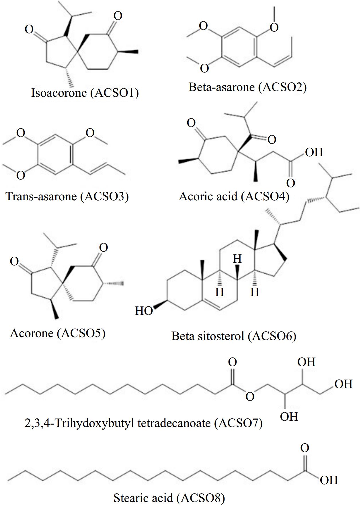
      Figure 1. 

      Structures of the isolated compounds from the rhizome of A. calamus.

    • The various extracts were tested for their initial cytotoxicity at a concentration of 100 μg against three different human cancer cell lines: A-549 (lung), HCT 116 (colon), and MDA-MB-231 (breast). This screening aimed to evaluate the possible cytotoxic effects of the extracts via the MTT assay (Table 1). The extract of AC Kakpora Pulwama (referred to as EXKpME) showed enhanced anti-proliferative properties with significant cytotoxicity against the A-549, HCT 116, and MDA-MB-231 cell lines, resulting in inhibition rates of 91%, 88%, and 94%, respectively. A significant proportion of the extracts exhibited an inhibition rate of ≥ 50% against several experimental cell lines.

      Table 1.  Cytotoxic activity of various extracts at 100 μg/ml concentration.

      S. No. Different A. calamus extracts** Percentage inhibition (%)*
      A549 (Lung) HCT-116 (Colon) MDA-MB- 231 (Breast)
      01 ExKwME 57 55 60
      02 ExKpME 91 88 94
      03 ExGnME 39 50 46
      04 ExKwME 28 30 34
      05 ExArHX 75 82 90
      06 EXArAC 78 85 81
      07 EXArEA 65 82 80
      08 ExNJME 47 45 50
      09 ExNJDC 67 65 80
      10 ExRJME 58 55 60
      * The values are the average of triplicate experiments. ** ExKwME (Kawoosa Methanolic extract); ExKpME (Kakpora Pulwama Methanolic extract); ExGnME (Ganastan Methanolic extract); ExArHX (Aarath Hexane extract); EXArAC (Aarath Acetone extract); EXArEA (Aarath ethylacitate extract); ExNJME (Najan Methanolic extract); ExNJDC (Najan DCM extract); ExRJME (Tuli Rajouri Methanolic extract).

      Similarly, an initial assessment was conducted on isolated pure compounds at 100 μM/ml against the selected cancer cell lines. As shown in Table 2, compounds ACS08, ACS06, and ACS02 showed better anticancer activity against all the cancer cell lines with ≥ 50% inhibition.

      Table 2.  Cytotoxic activity of isolated compounds at 100 μM concentration.

      S. No. Different compounds
      from A. calamus
      Percentage inhibition (%)*
      A549 (Lung) MIAPaCa (Pancreatic) HCT-116 (Colon) MDA-MB-231 (Breast) MDA-MB-468 (Breast)
      01 ACS01 27 22 25 34 30
      02 ACS02 53 59 72 85 80
      03 ACS03 11 13 16 14 12
      04 ACS04 25 37 40 54 38
      05 ACS05 38 30 45 60 55
      06 ACS06 52 45 60 68 72
      07 ACS07 36 40 45 44 40
      08 ACS08 63 52 74 80 64
      * The values are the average of triplicate experiments.

      Compounds showing significant anti-proliferative effects were further screened at five different concentrations (25, 50, 100, 125, and 150 μM) for determination of their IC50 values (Table 3).

      Table 3.  IC50 value (μM) of selected compounds against different cancer cell lines.

      Compounds A549 (Lung) MIAPaCa (Pancreatic) HCT-116 (Colon) MDA-MB-231 (Breast) MDA-MB-468 (Breast)
      ACS02 76.4 ± 2.1 80.56 ± 3.5 85.4 ± 2.1 65.4 ± 3.4 70.5 ± 2.3
      ACS06 82.7 ± 4.5 75.3 ± 2.0 80.7 ± 1.9 69.5 ± 4.0 73.9 ± 2.1
      ACS08 79.7 ± 0.5 65.59 ± 3.9 72.3 ± 2.2 55.89 ± 2.4 65.59 ± 5.0
      The values are the average of triplicate experiments.

      Compared with ACS06 and ACS02, ACS08 displayed more potent cytotoxicity. Among the panel of cancer cell lines, ACS08 was found to be more active against the MDA-MB-231 breast cancer cell line, with an IC50 estimation of 55.89 μM; hence, ACS08 was used for additional examination.

    • To investigate the antimetastatic activity of ACS08, experiments were conducted to examine its impact on cell migration and invasion. The wound healing scratch test and transwell invasion assay were used for this purpose. Substantial inhibition of cell migration and invasion was observed across different doses of ACS08 (Fig. 2).
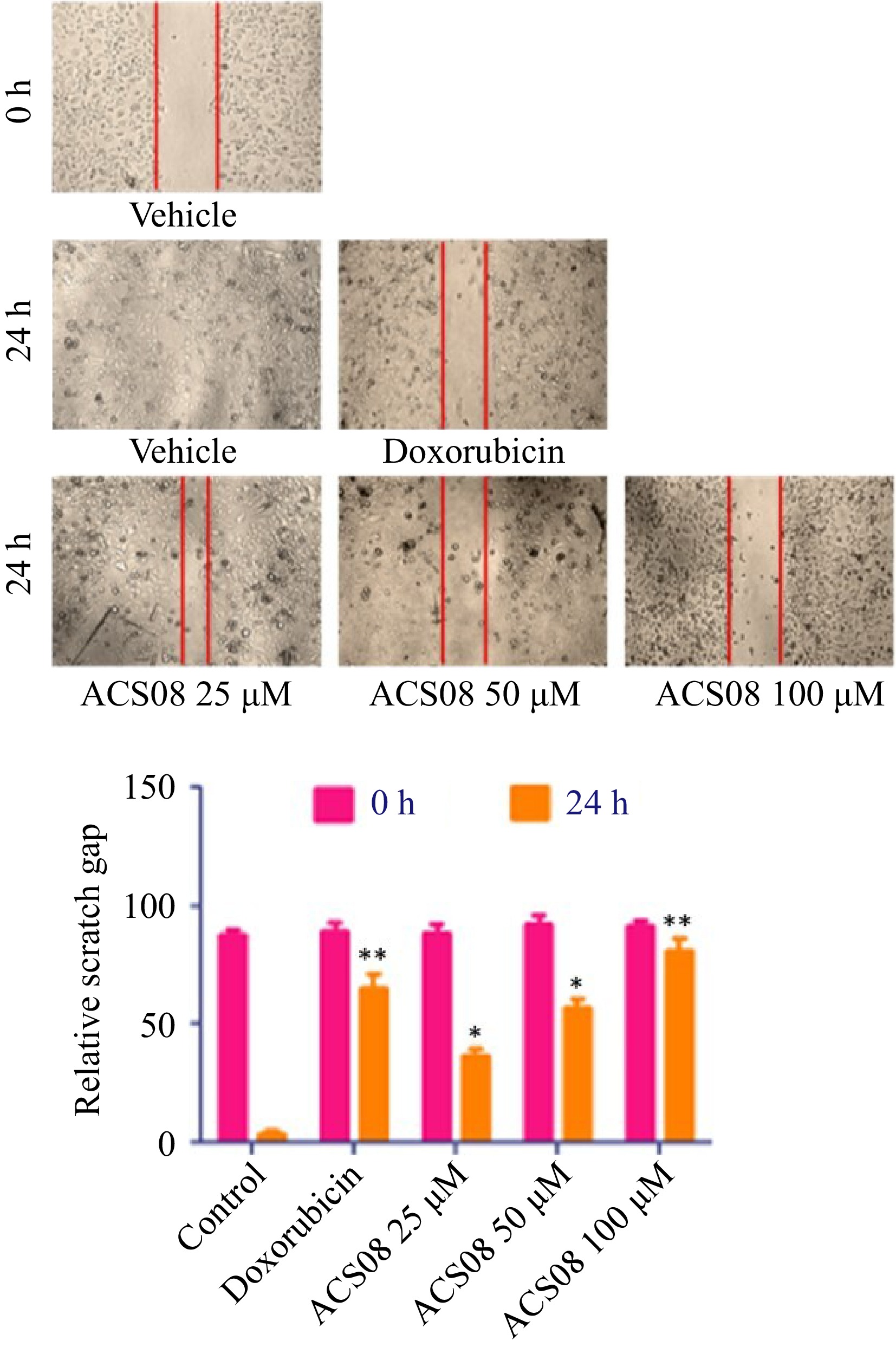
      Figure 2. 

      Wound healing scratch assay experiment of MDA-MB-231 breast cancer cell treated with varying concentrations of ACS08 at different time points. The region of the scratch was estimated at 0 and 24 h time focuses and the migration rate was determined in relation to the corresponding control. Values are expressed as means ± SD representing three independent biological samples, each with three technical replicates. Differences were scored as statistical significance at *** p < 0.0001, ** p < 0.001, and * p < 0.05.

      The dimensions of the scratch zone were assessed at both 0- and 24-h time intervals and the rate of migration was calculated relative to the equivalent control. The results revealed that the application of the IC50 dose (55.89 μM) to MDA-MB-231 cells resulted in a significant reduction in wound closure of approximately 50% in comparison with that in the control group. The degree of inhibition was more pronounced when the cells were subjected to elevated doses (100 μM) of ACS08. In the context of the transwell invasion experiment, approximately 140 cells invaded the bottom surface of the insert membrane in the untreated group. Conversely, a reduced number of 80 cells were found to penetrate following treatment with 25 μM ACS08 (Fig. 3).
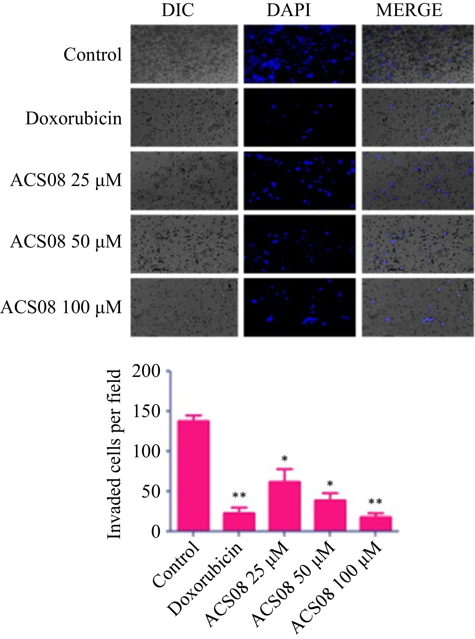
      Figure 3. 

      Histogram and microscopic images showing MDA-MB-231 breast cancer cell migration treated with different concentrations of ACS08. Values are expressed as means ± SD representing three independent biological samples, each with three technical replicates. Differences were scored as statistical significance at *** p < 0.0001, ** p < 0.001, and * p < 0.05.

      The number of invading cells decreased to 50 and 25 cells after exposure to ACS08 at concentrations of 50 and 100 μM, respectively. Collectively, our findings showed that ACS08 has significant antimetastatic effects on MDA-MB-231 breast cancer cells.

    • The application of several extracts derived from the rhizomes of A. calamus to selected cancer cell lines for 48 h resulted in the observed suppression of cell growth. The extracts (ExKwME, ExKpME, ExArHX, EXArAC, EXArEA, ExNJDC, and ExRJME) exhibited notable growth suppression in comparison with the other extracts. Notably, the inhibitory effect on the MDA-MB-231 cell line was substantially greater than that on the other cell lines. We subsequently conducted experiments involving the application of several isolated compounds derived from the same plant to many cancer cell lines, including lung, pancreatic, colon, and breast cancer cells. These compounds were administered at a uniform dosage of 100 μM/ml for 48 h. The results obtained demonstrated a noteworthy reduction in cell growth. Compared with the other isolated compounds, the compounds ACS08, ACS06, and ACS02 exhibited greater activity against the triple-negative breast cancer (MDA-MB 231) cell line.

    • Cancer is a non-communicable ailment that presents considerable obstacles in both emerging and wealthy nations[25]. The therapeutic options available for cancer patients are associated with several limitations, including severe toxicity, the development of drug resistance, and a heightened likelihood of disease recurrence. In recent years, there has been an increasing emphasis on and need for the evaluation of various phytochemicals derived from natural sources, intending to identify superior and more secure alternatives[26,27]. Furthermore, the development of resistance to cancer treatment has compelled researchers to shift their focus toward exploring natural compounds derived from plants and marine sources. Recently, the recognition of the importance of phytochemicals derived from natural sources, such as galantamine, resveratrol, and glycyrrhizin, as noteworthy therapeutic targets in the management of many cancer types has increased[28,29]. A. calamus, together with its two bioactive phytochemicals, alpha (α)-asarone and beta (β)-asarone, are well-recognized substances in traditional medicine. These compounds have been shown to have antitumor and chemopreventive properties, as demonstrated by multiple preclinical investigations conducted both in laboratory settings (in vitro) and in living organisms (in vivo)[30]. The current research aimed to evaluate the cytotoxic potential of extracts from A. calamus via primary cytotoxicity screening against three human malignancy cell lines: A-549 (lung), HCT 116 (colon), and MDA-MB-231 (breast). The extracts were tested at a concentration of 100 μg for fixation via the MTT assay. Cancer has evolved many strategies to evade controlled proliferation and circumvent programmed cell death. Hence, the use of entire plant extracts, which encompass many constituents with diverse potential intracellular targets, may provide a benefit over the utilization of a single isolated plant molecule. The methanolic extract of Kakpora pulwama (EXKPME) had superior anti-proliferative properties and showed significant cytotoxicity against the A-549, HCT 116, and MDA-MB-231 cell lines, resulting in inhibition rates of 91%, 88%, and 94%, respectively. A total of 12 compounds were recovered from the extract of dichloromethane : methanol (1:1) obtained from the subterranean sections of A. calamus. Among the 12 compounds, a total of eight (ACS01-08) were subjected to characterization (Supplemental Data 1). Furthermore, there was a difference in the yield of plant extracts collected from different locations. This variation may be due to varying agroclimatic conditions, such as increased sunshine hours and high temperatures, which led to optimum growth of the plant. The current investigation revealed that the MDA-MB 231 cell line had the highest level of sensitivity toward all the chemicals tested. While the anticancer properties of essential oils derived from A. calamus have been investigated, there is currently a lack of research on the potential anticancer effects of fatty acids isolated from A. calamus. Research has shown the inhibitory impact of stearic acid, a saturated fatty acid often found in dietary sources, on cell proliferation. Research has revealed that stearic acid (ACS08), derived from A. calamus, has inhibitory effects on the migration and invasion of MDA-MB-231 cells. In this study, a wound healing scratch test and a transwell invasion experiment were used to assess the effects of stearic acid on cellular migration and invasion to evaluate its potential as an antimetastatic agent. Inhibitory effects on cell migration and invasion were observed when different doses of stearic acid were applied (Fig. 1).

      Collectively, the present findings provide evidence that stearic acid has discernible antimetastatic properties when tested on MDA-MB-231 breast cancer cells. This finding aligns with prior research, which has shown the inhibitory effects of stearic acid on the growth of breast cancer cells and its ability to cause apoptosis in these cells[31,32]. Eicosatetraenoic acid (EPA) and docosahexaenoic acid (DHA), which are long-chain omega-3 fatty acids, are important for the production of bioactive lipid mediators that play crucial roles in the resolution of inflammation[33]. Fatty acids, which are integral constituents of phospholipid membranes and lipid rafts, play crucial roles in the organization and segregation of molecules. Moreover, they have been implicated in cell signaling processes that are believed to influence the development of breast carcinogenesis[34,35]. Plant-based medicine has been shown to facilitate wound healing and tissue regeneration via several methods that are not only cost-effective but also safe.

      Nevertheless, before its use, scientific validation, standardization, and safety assessment are crucial[36]. The findings of our investigation indicate that the application of ACS08 at the IC50 (55 μM) resulted in a significant reduction in wound closure by approximately 50% in MDA-MB-231 cells compared with that in the control group. The level of inhibition observed was more pronounced when the cells were subjected to elevated doses (100 μM) of ACS08. Zong et al. reported comparable findings, indicating that fatty acid extracts promote the healing of cutaneous wounds by activating AKT, ERK, and TGF-β/Smad3 signaling, as well as facilitating angiogenesis[37]. Previous studies have investigated the associations between the intake ratios of marine omega-3 fatty acids, specifically eicosatetraenoic acid (EPA) and docosahexaenoic acid (DHA), relative to omega-6 arachidonic acid, and the risk of breast cancer in women. Findings from various case‒control and cohort studies have indicated that women with higher intake ratios of EPA and DHA than arachidonic acid tend to have a decreased risk of breast cancer. However, it is important to note that these associations have not been consistently observed across all studies[38]. Fatty acids have been shown to have potential therapeutic and prophylactic effects against breast cancer in both in vitro and in vivo experimental models[39,40]. The present research evaluated the impact on cell viability and proliferation via an MTT assay. The results indicate that all the tested extracts and compounds substantially reduced cell viability and proliferation across one or more of the examined cell lines. Chemicals may have an anti-proliferative effect by potentially inducing cell death and/or arresting the cell cycle. The findings of this study have expanded the possibility of identifying anticancer chemicals in A. calamus, hence opening new opportunities for the future. Further investigation is needed to elucidate the associations between metabolites and the anticancer properties of plant extracts derived from A. calamus.

    • A. calamus, also known as sweet flag, has a rich historical background and is associated with a wide array of traditional and ethnomedicinal uses. The medicinal efficacy of A. calamus is attributed to the presence of secondary metabolites. The findings of the current investigation demonstrated that extracts and compounds derived from the rhizome of A. calamus had anticancer properties. The results outlined in this paper may serve as a potential resource for researchers seeking further investigations related to A. calamus and its bioactive phytochemical components in the context of cancer chemoprevention.

    • This study involved the use of established human cell lines. The cell lines used in this research were obtained from American Type Cell Culture (ATCC), USA and were used in accordance with institutional and national ethical standards. The cell lines have been previously published or validated, and no new human tissues were used in this study.

    • The authors confirm contribution to the paper as follows: conceptualization: Akhter S, Sultan P, Hassan QP; writing — original draft preparation: Hassan QP, Manzoor MM, Akhter S; experimental work: Akhter S, Mir SA, Manzoor MM, Khaliq T; facilities: Hassan QP, Sultan P. All authors have read and agreed to the final version of the manuscript.

    • All the data generated or analyzed during this study are included in this published article and its Supplemental information files.

      • Sabiyah Akhter and Tahirah Khaliq acknowledge the University Grants Commission (UGC) India for the Maulana Azad National Fellowship (MANF) received during this study. Malik Muzafar Manzoor acknowledges the Indian Council of Medical Research (ICMR) India for a Research Associateship.

      • The authors declare that they have no conflict of interest.

      • Supplementary Table S1 Voucher specimen numbers of collected plant material submitted at KASH Herbarium, Centre for Biodiversity and Taxonomy, University of Kashmir, India.
      • Supplemental Date S1 1H NMR and 13C spectral data of the isolated compounds.
      • Copyright: © 2024 by the author(s). Published by Maximum Academic Press, Fayetteville, GA. This article is an open access article distributed under Creative Commons Attribution License (CC BY 4.0), visit https://creativecommons.org/licenses/by/4.0/.
    Figure (3)  Table (3) References (40)
  • About this article
    Cite this article
    Akhter S, Manzoor MM, Mir SA, Khaliq T, Sultan P, et al. 2024. Extracts and isolated compounds from the Acorus calamus L. (sweet flag) rhizome showed distinct antimetastatic activity against MDA-MB-231 breast cancer cells. Medicinal Plant Biology 3: e021 doi: 10.48130/mpb-0024-0021
    Akhter S, Manzoor MM, Mir SA, Khaliq T, Sultan P, et al. 2024. Extracts and isolated compounds from the Acorus calamus L. (sweet flag) rhizome showed distinct antimetastatic activity against MDA-MB-231 breast cancer cells. Medicinal Plant Biology 3: e021 doi: 10.48130/mpb-0024-0021

Catalog

  • About this article

/

DownLoad:  Full-Size Img  PowerPoint
Return
Return