REVIEW   Open Access    

Microspore embryogenesis: in vitro cultivation induced cell reprogramming for plant breeding

  • # Authors contributed equally: Fan Yang, Xinyu Liu, Ying Qiao

More Information
  • Received: 16 August 2024
    Revised: 21 October 2024
    Accepted: 05 November 2024
    Published online: 10 December 2024
    Seed Biology  3 Article number: e021 (2024)  |  Cite this article
  • Microspore embryogenesis facilitates the rapid generation of doubled haploid (DH) plants, providing significant advantages in crop breeding by achieving homozygosity within a single generation. However, its application is often constrained by genotype-specific limitations and inefficiencies. Understanding the mechanisms underlying microspore embryogenesis is essential for enhancing DH induction efficiency and offers insights into cell fate transitions, the acquisition of totipotency, and the initiation of embryogenesis. This review examines recent advances in the regulatory mechanisms of microspore embryogenesis, focusing on the roles of cell wall remodeling, programmed cell death, autophagy, hormonal responses, and epigenetic modifications. In addition, the current challenges of microspore embryogenesis for crop breeding are summarized. These advances not only offer new strategies for improving the efficiency of microspore embryogenesis but also provide valuable directions for future research, ultimately contributing to the improvement of crop breeding.
  • The tea plant (Camellia sinensis) was first discovered and utilized in China, where its tender leaves were processed into tea. Tea has become the second most popular beverage after water worldwide. Tea contains tea polyphenols, amino acids, vitamins, lipopolysaccharides and other nutrients, as well as potassium, calcium, magnesium, iron, fluorine and other trace elements, which have antioxidant, lipid-lowering, hypoglycaemic, anti-caries, enhance the body's immunity and other physiological regulatory functions[1]. Among them, fluorine is an element widely found in the earth's crust, mainly in the form of fluoride in the environment, is one of the essential trace elements for human beings, and is vital to the growth and development of human bones and teeth[2]. When the human body takes in an appropriate amount of fluorine, it can effectively prevent the formation of dental caries, enhance the absorption of calcium, phosphorus and other elements of the human body. Excessive intake of fluorine however will lead to chronic cumulative poisoning, damaged bone tissue, affect the function of various tissues and organs in the body, and cause harm to health[3]. Tea plants can absorb and accumulate fluoride from air, water and soil, mainly concentrated in the leaves, most of the fluoride in the leaves can be released into the tea soup and be absorbed by the human body, so the fluoride content in tea is closely related to human health[4]. Generally speaking, green tea, black tea, white tea, oolong tea and yellow tea are made from the young buds and shoots of the tea plant, and their fluorine content is low. Some dark tea made from leaves with lower maturity has lower fluorine content. However, the dark tea made from leaves with higher maturity has a high fluorine content, and therefore poses a risk of excessive fluorine[5]. Long-term drinking of dark tea with excessive fluorine content is a cause of tea-drinking fluorosis[6].

    Dark tea, with its smooth taste and digestive benefits, became an indispensable drink in the lives of the Chinese herders, who were mainly meat eaters[7]. Dark tea has also gained popularity in the wider population because of its important health-promoting effects, such as prevention of cardiovascular and cerebrovascular diseases, lowering blood pressure, and promoting weight loss and fat reduction[8]. Long-term consumption of dark tea is likely to cause fluorosis for two reasons: 1) Chinese border ethnic minorities generally use the boiling method to brew dark tea, which increases the leaching rate of fluoride[9] and leads to high fluoride levels in the human body; 2) most of the fresh tea leaves utilized to make dark tea are older and more mature leaves, which contain higher levels of fluoride than younger leaves[10]. The mature leaves of tea plants accumulate a large amount of fluoride, but can grow normally without fluoride poisoning, indicating that tea plants are able to accumulate and tolerate fluoride. Due to the problem of tea-drinking fluorosis, the excessive accumulation of fluoride in tea plants has attracted widespread attention[10]. It is important to investigate the mechanisms related to fluoride absorption, transportation, enrichment, and tolerance in tea plants to develop effective and practical management and control programs to reduce the fluoride content in tea and ensure its safety. Recent research on defluorination measures for tea has included preliminary screening of tea germplasm resources, management measures during tea plant cultivation, processing technologies, and tea brewing methods. In this review, we summarize the results of studies on how fluoride moves from the environment into tea plants and the factors affecting this process, how fluoride is transported in tea plants, the mechanisms of fluoride tolerance in tea plants, and current measures to reduce the fluoride content in tea.

    Tea plants are fluoride-accumulators with the ability to absorb and accumulate fluoride from the surrounding environment. The fluoride content in tea plants is significantly higher than that of other plants under similar growth conditions[11]. After fluoride is absorbed by the roots of tea plants, it is transferred to the above-ground parts, and is also transferred from the leaves downward (Fig. 1), but not from the above-ground parts to the below-ground parts (Fig. 2). External factors such as atmospheric, soil, and water conditions around the tea plant and internal factors can affect the fluoride content in different plant tissues.

    Figure 1.  Fluoride absorption by tea plants. Tea plants absorb fluoride from the atmosphere, soil, and water. Fluoride in the atmosphere is absorbed through the stomata or cuticle of the leaf epidermis. Fluoride ions and fluoride–aluminum complexes in soil and water are absorbed by the roots.
    Figure 2.  Fluoride transportation in tea plants. Fluoride absorbed by the leaves is transferred to the leaf tips and edges, chelates with metal ions, and the complexes are deposited in the leaves. Fluoride can be transferred from old leaves to new tips. Fluoride from soil and water can form complexes with organic acids and aluminum, which are stored in the leaves of tea plants. Fluoride from soil and water can also be individually absorbed and transported for storage in the leaves.

    Fluoride occurs in many forms in nature. Fluoride in the atmosphere mainly exists in the form of hydrogen fluoride, and that in the soil mainly exists in three forms: insoluble, exchangeable, and water-soluble fluoride. Atmospheric fluoride is mainly absorbed through the stomata of tea plant leaves or the cuticle of the epidermis, and its concentration is relatively low[12]. Insoluble fluoride and exchangeable fluoride in the soil cannot be absorbed by tea plants. Water-soluble fluoride is the main form absorbed by the root system of tea plants[13]. Several studies have detected a significant positive correlation between the fluoride content in tea and the water-soluble fluoride content in soil[14,15]. Water-soluble fluoride mainly refers to the fluoride ion (F) or fluoride complexes in soil and water solutions, including free F and fluoride complexed with ions. The water-soluble state has the strongest activity and the highest biological availability, so it is conducive to the migration of fluoride in the environment[16].

    The root system is the main organ responsible for fluoride absorption in tea plants. Water-soluble fluoride can enter the root system by passive or active absorption, depending on its concentration. Fluoride at lower concentrations in solution (0.1–10 mg/L) is mainly absorbed and enriched in the root system of tea plants via active absorption, with a kinetic curve following the Michalis–Menten kinetic model. At higher concentrations (50–100 mg/L), water-soluble fluoride is absorbed at a rate that increases with increasing concentration, and this is achieved via passive absorption[17]. Many studies have shown that the water-soluble fluoride content in soils in most tea-producing regions in China is below the threshold for passive absorption[1820], indicating that fluoride mainly enters tea plants via active absorption by the roots.

    The active uptake of fluoride by tea roots is mediated by ion pump carrier proteins and ABC transporter proteins. Ion pump carrier proteins can transport the substrate across the cell membrane against the electrochemical gradient, and use the energy of ATP hydrolysis to participate in the process of active transport of substances, mainly including proton pump H+-ATPase and calcium ion pump Ca2+-ATPase. ABC transporter proteins can transport ions or heavy metals to vesicles as chelated peptide complexes, thereby reducing toxicity to the cell and improving plant resistance to abiotic stress. Passive fluoride absorption by the root system of tea plants involves water channels and ion channels. Studies on the effects of applying external anion channel inhibitors, cation channel inhibitors, and water channel inhibitors showed that inhibition of external anion channels significantly reduced the absorption of fluoride by roots. This indicated that anion channels are an important pathway for the uptake and trans-membrane transport of fluoride in the root system of tea plants[17,21,22]. The homeostatic flow of ions through channels diffusing along a trans-membrane concentration gradient or potential gradient involves ion channel proteins[23]. Two phylogenetically independent ion channel proteins have recently been identified in tea plants: CLCF-type F/H+ reverse transporter proteins and the FEX (Fluoride export gene) family of small membrane proteins[24]. CLC proteins are involved in the transport of a variety of anions such as chlorine (Cl) and F into and out of the cell. The FEX proteins in tea plants are involved in fluoride absorption via thermodynamic passive electro-diffusion through transmembrane channels[25,26].

    After fluoride is absorbed, it is transported within the tea plant by several different pathways. These include transport after leaf absorption, transport after root absorption, and transfer inside tea plant cells. The fluoride absorbed by leaves can be transferred along the conduit to the leaf tips and edges, accumulating in the top and ipsilateral leaves, but not in the roots. Fluoride in the soil and water environment is taken up by the root system and transported to the xylem via intracellular and intercellular transport. It is then transported upwards via transpiration and eventually accumulates in the leaves[27,28]. Two transport mechanisms have been proposed for the translocation of fluoride via the xylem. One proposed mechanism is that it is translocated in the form of fluoride-aluminum complexes[29]. The other proposed mechanism is that aluminum and fluoride are transported separately and accumulate after reaching the leaves[30].

    The roots are the main organ responsible for fluoride absorption in tea plants. Therefore, most studies have focused on the transport process in roots, especially the transport of fluoride by fluoride-related transporters. It has been found that under acidic conditions, F preferentially forms complexes with Al3+ and these complexes are then absorbed by roots and transported upward in the same state[29,31]. Studies have shown that, compared with F, aluminum-fluoride complexes are more easily absorbed and transported to the new shoots by the root system. This may be related to the elimination of the separate toxic effects of F and Al3+ [29]. Fluoride can also be transported in tea plants by binding to aluminum-organic acid complexes, and then accumulate in the leaves[29]. Both tea plant fluoride transporter proteins, CsFEX1 and CsFEX2, are involved in fluoride transport, but their encoding genes can be differentially expressed among different varieties and depending on the concentration of fluoride. In one study, the expression level of CsFEX1 was consistent among different varieties, while the expression of CsFEX2 was induced under fluoride stress to increase fluoride efflux from tea plants, thereby reducing its accumulation in low-fluoride varieties[32]. In addition, the A–G subfamily of ABC transporters plays a carrier role in the transmembrane transport of F- and Cl- in tea plants[33]. It was found that the expression of the ABC transporter protein CsCL667 was up-regulated in response to fluoride treatment, and its ability to transport fluoride was enhanced, suggesting that CsCL667 functions in fluoride efflux[34]. Another study demonstrated that CsABCB9 localizes in chloroplasts and functions as a fluoride efflux transporter to reduce fluoride-induced damage in leaves and enhance chloroplast activity[35].

    Several factors affect the absorption and transport of fluoride in tea plants, including the absorbable fluoride concentration, soil pH, the presence of other ions, and the activity of ion channels[3638]. Fluoride in nature is present in the atmosphere and soil, and its concentration is the main factor affecting the fluoride content in tea plants. Under normal conditions, tea plants generally absorb fluoride from the soil through the roots, but when the hydrogen fluoride content in the atmosphere is high, tea plants can absorb it through the leaves. The fluoride content in tea plants growing in the same geographical area is similar, mainly because of the soil properties in that area. Tea plants grow in acidic environments, and fluoride in acidic soils is more easily absorbed. During the growth of tea plants, the roots secrete organic acids such as oxalic acid, citric acid, and malic acid, which promote the absorption of fluoride by the roots and its transport to above-ground parts[39]. Other ions such as Ca2+ and Mg2+ combine with F to form precipitates, resulting in lower concentrations of water-soluble fluoride in the soil, which also affects its absorption by tea plant roots[40]. Exogenously applied calcium at low concentrations can change the cell wall structure and membrane permeability in tea plant roots, ultimately leading to reduced fluoride content in tea leaves[36,41,42]. Meanwhile, Al3+ treatment can trigger Ca2+ signaling in tea plant roots, which in turn activates calmodulin and promotes fluoride absorption[43]. The H+ gradient generated by the plasma membrane H+-ATPase can also promote Ca2+ signaling in plants to regulate the transmembrane transport of ions, which affects fluoride absorption[43]. The abundance and activity of H+-ATPase in the plasma membrane of tea plant roots have been found to increase significantly under fluoride stress, and these increases result in improved absorption of fluoride, although this is also affected by the fluoride concentration and temperature[33]. Sodium fluoride was found to induce the expression of genes encoding ABC transporter proteins, resulting in the transmembrane absorption of large amounts of fluoride ions into cells[34]. ABC transporters also transport ions alone or in the form of chelated peptide complexes directly out of the cellular membrane, which improves cellular tolerance to these ions[44]. Some anions with the same valence state also affect the F content in tea plants. For example, the ion channel protein encoded by CLCF is more sensitive to F, more selective for F than for Cl, and functions to export F from the cytoplasm to protect against fluorosis[4547].

    It can be seen that reducing the absorption of fluorine by tea plants and changing the mechanism of fluorine transportation in tea plants can reduce the content of fluorine in different parts of the tea plants. From the mechanism of fluorine absorption, the most effective way is to directly change the form of soil fluorine to reduce the absorption of water-soluble fluorine by roots. On this basis, it is possible to further change the active absorption process of fluorine mediated by ion pump carrier protein and ABC transporter protein in tea roots by molecular techniques. From the perspective of fluorine transport mechanism, the toxic effect of fluorine on tea plants can be reduced mainly by promoting the function of transporter proteins to exclude fluorine from the cell or transport it to the vesicle. The comprehensive application of the above methods to limit fluorine absorption and promote fluorine transport in tea plants can limit the accumulation of fluorine in tea plants.

    The fluoride enrichment characteristics of tea plants are related to various factors, including the tea variety, the organ, and the season. Among different varieties of tea, differences in leaf structure and other physiological characteristics can lead to variations in fluoride absorption and enrichment[48]. Some studies have concluded that the variety is one of the main determinants of the fluoride content in tea leaves[10], and the differences in fluoride content among most varieties reached highly significant levels (Table 1), which can be divided into low-enriched, medium-enriched, and high-enriched germplasm[49]. Various organs of tea plants also show differences in fluoride accumulation. The fluoride content is much higher in leaves than in roots and stems, and significantly higher in old leaves than in new shoots[10,50,51]. The fluoride content can differ widely among tea plants at different developmental stages. In spring, the new leaves begin to accumulate fluoride from the environment, and the fluoride content increases as the leaves age. When the growth rate of tea leaves is slower, they absorb and accumulate more fluoride from the soil and air. When the temperature in summer and autumn is high, the growth rate of tea leaves is fast and the growth period is short, so less fluoride is absorbed and accumulated from the soil and air. This explains why the fluoride content in fresh tea leaves was higher in spring and relatively lower in summer and autumn[4]. Another study found that, in China, the fluoride content in tea leaves was higher in summer than in spring. This may have been related to the maturity level of the tea leaves at harvest and different patterns of fluoride transport[4].

    Table 1.  Fluoride content difference of different tea cultivars.
    Cultivars Province Parts Treatment Years Content (mg/kg) Ref.
    Liannandaye Sichuan Old leaves Drying at 80 °C and boiling water extraction 2006−2007 1,150.79 ± 4.86 [107]
    Mature leaves Drying at 70 °C and hydrochloric acid extraction 2006 1,296.66 ± 12.84 [110]
    Yuenandaye Old leaves Drying at 80 °C and boiling water extraction 2006−2007 1,352.89 ± 12.69 [107]
    Mature leaves Drying at 70 °C and hydrochloric acid extraction 2006 1,560.36 ± 27.10 [110]
    Chenxi NO.4 Old leaves Drying at 80 °C and boiling water extraction 2006−2007 1,865.61 ± 7.46 [107]
    Mature leaves Drying at 70 °C and hydrochloric acid extraction 2006 1,954.93 ± 10.96 [110]
    Meizhan Old leaves Drying at 80 °C and boiling water extraction 2006−2007 2,180.13 ± 14.42 [107]
    Mature leaves Drying at 70 °C and hydrochloric acid extraction 2006 1,732.2 ± 41.2 [110]
    Zhejiang Mature leaves Drying at 80 °C and nitric acid extraction 2002 2,015.48 ± 29.99 [106]
    Fudingdabai Sichuan Old leaves Drying at 80 °C and boiling water extraction 2006−2007 249.64 ± 24.3 [107]
    Guizhou One bud and
    five leaves
    Drying at 80 °C and hydrochloric acid extraction 2010 1,612.3 ± 43.1 [109]
    Fudingdabai Zhejiang Mature leaves Drying at 80 °C and nitric acid extraction 2002 137.1 ± 2.1 [106]
    Fujian Old leaves Drying at 80 °C and hydrochloric acid extraction 2010 282.1 [111]
    Hunan One bud and
    five leaves
    Steaming and boiling water extraction 2011 2,232.05 ± 85.52 [51]
    Zhuyeqi Ya'an and
    surroundings
    Old leaves Drying at 80 °C and boiling water extraction 2006−2007 2,750.16 ± 11.37 [107]
    Ya'an Mature leaves Drying at 70 °C and hydrochloric acid extraction 2006 125.4 [110]
    Hunan One bud and
    five leaves
    Steaming and boiling water extraction 2011 2,330.74 ± 31.39 [51]
    Fujianshuixian Ya'an and
    surroundings
    Old leaves Drying at 80 °C and boiling water extraction 2006−2007 2,548.18 ± 40.97 [107]
    Ya'an Mature leaves Drying at 70 °C and hydrochloric acid extraction 2006 103.7 ± 1.5 [110]
    Fujian Old leaves Drying at 80 °C and hydrochloric acid extraction 2010 1,150.79 ± 4.86 [111]
    Huangyeshuixian Ya'an and
    surroundings
    Old leaves Drying at 80 °C and boiling water extraction 2006−2007 2,424.70 ± 18.85 [107]
    Ya'an Mature leaves Drying at 70 °C and hydrochloric acid extraction 2006 2,950.80 ± 27.73 [110]
    Qianmei 701 Ya'an and
    surroundings
    Old leaves Drying at 80 °C and boiling water extraction 2006−2007 2,522.01 ± 45.33 [107]
    Ya'an Mature leaves Drying at 70 °C and hydrochloric acid extraction 2006 3,693.09 ± 35.12 [110]
    Guizhou One bud and
    five leaves
    Drying at 80 °C and hydrochloric acid extraction 2010 389.95 ± 32.18 [109]
    Guizhou Old leaves Dry samples and hydrochloric acid extraction 2011 2,142.26 ± 16.30 [113]
    Mingshan 130 Ya'an and
    surroundings
    Old leaves Drying at 80 °C and boiling water extraction 2006−2007 2,564.78 ± 51.22 [107]
    Ya'an Mature leaves Drying at 70 °C and hydrochloric acid extraction 2006 3,036.13 ± 31.25 [110]
    Mengshan 9 Ya'an and
    surroundings
    Old leaves Drying at 80 °C and boiling water extraction 2006−2007 2,647.31 ± 70.89 [107]
    Ya'an Mature leaves Drying at 70 °C and hydrochloric acid extraction 2006 3,436.55 ± 20.21 [110]
    Yinghong NO. 2 Ya'an and
    surroundings
    Old leaves Drying at 80 °C and boiling water extraction 2006−2007 2,669.02 ± 799.95 [107]
    Ya'an Mature leaves Drying at 70 °C and hydrochloric acid extraction 2006 3,364.53 ± 51.72 [110]
    Mengshan 11 Ya'an and
    surroundings
    Old leaves Drying at 80 °C and boiling water extraction 2006−2007 2,695.21 ± 59.89 [107]
    Ya'an Mature leaves Drying at 70 °C and hydrochloric acid extraction 2006 3,582.83 ± 9.73 [110]
    Mingshan 311 Ya'an and
    surroundings
    Old leaves Drying at 80 °C and boiling water extraction 2006−2007 2,716.22 ± 42.21 [107]
    Donghuzao 2,731.20 ± 20.78
    Ya'an Mature leaves Drying at 70 °C and hydrochloric acid extraction 2006 3,107.27 ± 54.91 [110]
    Hainandaye Ya'an and
    surroundings
    Old leaves Drying at 80 °C and boiling water extraction 2006−2007 2,746.82 ± 39.71 [107]
    Ya'an Mature leaves Drying at 70 °C and hydrochloric acid extraction 2006 2,961.53 ± 29.94 [110]
    Qianmei 502 Ya'an and
    surroundings
    Old leaves Drying at 80 °C and boiling water extraction 2006−2007 2,878.23 ± 76.94 [107]
    Ya'an Mature leaves Drying at 70 °C and hydrochloric acid extraction 2006 3,881.51 ± 16.48 [110]
    Guizhou One bud and
    five leaves
    Drying at 80 °C and hydrochloric acid extraction 2010 389.95 ± 30.2 [109]
    Old leaves Dry samples and hydrochloric acid extraction 2011 3,260.48 ± 32.12 [113]
    Zisun Ya'an and
    surroundings
    Drying at 80 °C and boiling water extraction 2006−2007 2,904.13 ± 35.40 [107]
    Ya'an Mature leaves Drying at 70 °C and hydrochloric acid extraction 2006 3,140.80 ± 42.86 [110]
    Zhejiang Drying at 80 °C and nitric acid extraction 2002 1,742.7 ± 43.2 [106]
    Qianmei 303 Ya'an and
    surroundings
    Old leaves Drying at 80 °C and boiling water extraction 2006−2007 2,918.13 ± 46.79 [107]
    Ya'an Mature leaves Drying at 70 °C and hydrochloric acid extraction 2006 4,029.11 ± 81.86 [110]
    Guizhou One bud and
    five leaves
    Drying at 80 °C and hydrochloric acid extraction 2010 199.74 ± 16.6 [109]
    Old leaves Dry samples and hydrochloric acid extraction 2011 2,972.79 ± 169.82 [113]
    Anxishuixian Ya'an and
    surroundings
    Old leaves Drying at 80 °C and boiling water extraction 2006−2007 2,924.33 ± 41.39 [107]
    Ya'an Mature leaves Drying at 70 °C and hydrochloric acid extraction 2006 3,454.68 ± 26.29 [110]
    Longjing 43 Ya'an and
    surroundings
    Old leaves Drying at 80 °C and boiling water extraction 2006−2007 3,152.73 ± 27.70 [107]
    Ya'an Mature leaves Drying at 70 °C and hydrochloric acid extraction 2006 4,437.79 ± 26.14 [110]
    Zhejiang Drying at 80 °C and nitric acid extraction 2002 1,377.1 ± 37.0 [106]
    Fujian Old leaves Drying at 80 °C and hydrochloric acid extraction 2010 116.2 ± 0.9 [111]
    Shuyong 307 Ya'an and
    surroundings
    Drying at 80 °C and boiling water extraction 2006−2007 3,223.55 ± 151.43 [107]
    Ya'an Mature leaves Drying at 70 °C and hydrochloric acid extraction 2006 4,296.52 ± 54.98 [110]
    Zhenghedabaicha Ya'an and
    surroundings
    Old leaves Drying at 80 °C and boiling water extraction 2006−2007 3,295.74 ± 27.55 [107]
    Ya'an Mature leaves Drying at 70 °C and hydrochloric acid extraction 2006 3,876.58 ± 21.09 [110]
    Zhejiang Drying at 80 °C and nitric acid extraction 2002 1,373.0 ± 41.9 [106]
    Taiwandaye Ya'an and
    surroundings
    Old leaves Drying at 80 °C and boiling water extraction 2006−2007 3,363.59 ± 456.39 [107]
    Ya'an Mature leaves Drying at 70 °C and hydrochloric acid extraction 2006 4,739.89 ± 58.59 [110]
    Qianmei 419 Ya'an and
    surroundings
    Old leaves Drying at 80 °C and boiling water extraction 2006−2007 3,518.15 ± 76.19 [107]
    Ya'an Mature leaves Drying at 70 °C and hydrochloric acid extraction 2006 4,541.43 ± 28.91 [110]
    Guizhou One bud and
    five leaves
    Drying at 80 °C and hydrochloric acid extraction 2010 133.70 ± 11.2 [109]
    Old leaves Dry samples and hydrochloric acid extraction 2011 2,370.47 ± 11.43 [113]
    Mengshan23 Ya'an and
    surroundings
    Drying at 80 °C and boiling water extraction 2006−2007 3,625.11 ± 86.07 [107]
    Ya'an Mature leaves Drying at 70 °C and hydrochloric acid extraction 2006 4,469.25 ± 40.85 [110]
    Sichuan group species 2,782.59 ± 146.46
    Meitantaicha Guizhou One bud and
    five leaves
    Drying at 80 °C and hydrochloric acid extraction 2010 272.93 ± 27.3 [109]
    Qianmei 101 Drying at 80 °C and hydrochloric acid extraction 2010 175.07 ± 13.5
    Dry samples and hydrochloric acid extraction 2011 3,218.33 ± 57.91 [113]
    Qianmei 601 Drying at 80 °C and hydrochloric acid extraction 2010 267.11 ± 26.31 [109]
    Dry samples and hydrochloric acid extraction 2011 2,823.02 ± 73.36 [113]
    Qianmei 809 Drying at 80 °C and hydrochloric acid extraction 2010 521.48 ± 50.32 [109]
    Dry samples and hydrochloric acid extraction 2011 2,327.91 ± 83.17 [113]
    Qianmei 308 Drying at 80 °C and hydrochloric acid extraction 2010 326.88 ± 29.3 [109]
    Dry samples and hydrochloric acid extraction 2011 3,432.86 ± 159.4 [113]
    Qianmei 415 Drying at 80 °C and hydrochloric acid extraction 2010 186.95 ± 14.2 [109]
    Dry samples and hydrochloric acid extraction 2011 5,090.83 ± 69.56 [113]
    Qiancha NO. 7 Drying at 80 °C and hydrochloric acid extraction 2010 218.81 ± 18.7 [109]
    Qianfu NO. 4 136.82 ± 11.6
    Dry samples and hydrochloric acid extraction 2011 3,066.49 ± 86.35 [113]
    Guiyucha NO. 8 Drying at 80 °C and hydrochloric acid extraction 2010 191.03 ± 18.6 [109]
    Dry samples and hydrochloric acid extraction 2011 2,882.94 ± 195.73 [113]
    Pingyangtezao Drying at 80 °C and hydrochloric acid extraction 2010 125.02 ± 12.1 [109]
    Yuanxiaolv 244.32 ± 20.5
    Nongkangzao 133.70 ± 12.3
    Mingshan 213 106.98 ± 6.74
    Mingke NO. 4 195.29 ± 16.8
    Maolv 174.73 ± 15.8
    Qianmei 412 Old leaves Dry samples and hydrochloric acid extraction 2011 3,396.92 ± 31.61 [113]
    Meitantaicha 2011 3,085.83 ± 101.9
    Zhenong 138 Zhejiang Mature leaves Drying at 80 °C and nitric acid extraction 2002 805.7 ± 6.0 [106]
    Zhenong 12 1,041.2 ± 23.3
    Shuigu 1,123.2 ± 33.5
    Hanlv 1,152.4 ± 2.4
    Zhuzhichun 1,248.2 ± 2.3
    Lvyafoshou 1,298.1 ± 2.0
    Zhenong 139 1,322.4 ± 40.7
    Shuixian 1,323.5 ± 36.1
    Biyun 1,400.9 ± 0.6
    Soubei 1,487.6 ± 29.7
    Maoxie 1,487.7 ± 31.0
    Anhui NO. 9 1,489.7 ± 40.0
    Zhenong 113 1,492.7 ± 43.5
    Yingshuang 1,509.9 ± 7.2
    Zhenong 25 1,521.2 ± 3.2
    Ribenzhong 1,543.8 ± 33.9
    Yunqi 1,549.3 ± 46.9
    Zhenong 23 1,576.7 ± 11.3
    Huangyezao 1,606.7 ± 40.5
    Jinshi 1,662.4 ± 42.4
    Pingyun 1,676.6 ± 44.6
    Zhenong 21 1,678.8 ± 49.6
    Jinfeng 1,705.2 ± 10.3
    Jiukeng 1,779.2 ± 5.0
    Juhuachun 1,993.4 ± 14.5
    Wuniuzao 2,163.2 ± 15.8
    Fujian Old leaves Drying at 80 °C and hydrochloric acid extraction 2010 98.0 ± 1.3 [111]
    Jinguanyin All leaves Drying at 80 °C and nitric acid extraction 2014−2015 536.49 ± 10.41 [112]
    Dangui 2,598.87 ± 24.12
    Old leaves Drying at 80 °C and hydrochloric acid extraction 2010 145.3 ± 0.2 [111]
    Jinmudan All leaves Drying at 80 °C and nitric acid extraction 2014−2015 1,030.21 ± 36.52 [112]
    Ruixiang Drying at 80 °C and nitric acid extraction 1,315.64 ± 21.56
    Xiapu yuanxiao Old leaves Drying at 80 °C and hydrochloric acid extraction 2010 124.6 ± 3.0 [111]
    Jinxuan 103.7 ± 3.5
    Fuandabai 103.0 ± 1.0
    Fuyun NO. 7 104.0 ± 1.1
    Fuyun NO. 6 131.6 ± 1.8
    Fudingdahao 118.0 ± 2.4
    Zaochunhao 107.7 ± 2.8
    Xiapu chunbolv 99.2 ± 1.2
    BaijiguanF1 102.1 ± 1.1
    Huanguanyin Old leaves Drying at 80 °C and hydrochloric acid extraction 99.2 ± 1.3
    Foxiang NO. 1 Yunnan One bud and
    four leaves
    Drying at 60 °C and hydrochloric acid extraction 2011 155.00 ± 6.94 [108]
    Foxiang NO. 2 214.30 ± 5. 94
    Foxiang NO. 4 219. 50 ± 7. 32
    Foxiang NO. 5 190.70 ± 4.09
    Yunkang NO. 10 121. 30 ± 5. 81
    Yunkang NO. 14 198.50 ± 8.49
    Yuncha NO. 1 135.10 ± 4.74
    Baihaozao Hunan One bud and
    five leaves
    Steaming and boiling water extraction 2011 113.2 [51]
    Bixiangzao 121.4
    Taoyuandaye 177.7
    Yulv 165.9
    Jianbohuang NO.13 168.5
    Gaoyaqi 162.8
     | Show Table
    DownLoad: CSV

    In conclusion, selecting low-fluoride tea varieties and reducing the maturity of dark tea raw materials can be used as effective measures to reduce the fluoride content of dark tea.

    Plants can increase their resistance to fluoride through exocytosis and internal tolerance mechanisms. A series of reactions occurs in tea plants to reduce the toxic effects of fluoride and improve tolerance. The results of recent studies indicate that both external and internal factors are involved in fluoride resistance in tea plants. The external factors include the availability of cations that readily chelate fluoride, and the internal factors include the abundance and activity of certain transporters and the capacity of transporter and antioxidant systems (Fig. 3).

    Figure 3.  Mechanisms of fluoride tolerance in tea plants. (A) Metabolites in tea plants reduce the toxic effect of fluoride. Cell wall macromolecular components such as pectin, lignin, cellulose, hemicellulose, polysaccharide, and proteins chelate fluoride. The contents of some metabolites increase during adaptation of tea plants to fluoride. (B) Complexation of cations (Al3+, Ca2+, and Mg2+) with fluoride in tea plants. (C) Roles of the antioxidant system of tea plant in fluoride tolerance. Increases in the activity/abundance of antioxidant enzymes and in the ASA-GSH cycle reduce intracellular levels of reactive oxygen species, leading to increased fluoride tolerance. (D) Roles of transporters in fluoride tolerance. Transporter proteins transport fluoride into the vacuole, and this compartmentalization reduces damage to enzymes and organelles. The CsFEX, CsCLC, and CsABC transporters efflux fluoride from cells, thereby reducing its toxic effects. POD: Peroxidase, CAT: Catalase, SOD: Superoxide dismutase, APX: Ascorbate peroxidase, GR: Glutathione reductase, DHAR: Dehydroascorbate reductase, ABA: Abscisic acid, GA: Gibberellic acid, GHs: Glycoside hydrolases, ASA-GSH: Antioxidant system and the ascorbate-glutathione, ROS: Reactive oxygen species.

    Fluoride ions have a strong ability to form complexes with metal ions. Free F can form complexes with cations such as Al3+, Fe3+, and Ca2+, thereby altering ionic homeostasis and reducing its toxicity to tea plants[52]. Fluoride and aluminum ions form complexes and are enriched in leaves and other organs with a certain proportion. This reduces the toxicity of both Fl- and Al3+, and may be an important physiological mechanism of fluoride enrichment in tea plants[31,53]. Fluoride can also form complexes with Ca2+, so exogenous application of Ca2+ can effectively reduce the fluoride content and enhance the fluoride resistance of tea plants[42]. Fluoride combines with Mg2+, Al3+, and Ca2+ on the surface of tea leaves, and is present on the abaxial and adaxial leaf surfaces in the form of MgF2 and AlF3. The application of a small amount of MgF2 or CaF2 may be a means to reduce the toxicity of fluoride to tea seedlings[40]. Treatment with selenium was shown to reduce the fluoride content in tea, increase the accumulation of fluoride in roots, and reduce the proportion of water-soluble fluoride in tea beverages[54].

    The cell wall is widely involved in plant growth and development and in various stress responses. Fluoride ions can be chelated by the aldehyde, carboxyl, amino, and phosphate groups in polysaccharides, pectin, lignin, proteins, and other components as well as some metal ions adsorbed in the cell wall, which is usually called cell wall fixation[55,56]. Recent studies have shown that fluoride stress activates pathways related to cell wall metabolism, the stress response, signal transduction, and protein degradation, and all of these pathways may contribute to the accumulation/detoxification of fluoride in tea leaves[57,58]. After the application of exogenous fluoride, there are increase in the activities of key enzymes involved in the pectin biosynthetic pathway, in the transcript levels of their encoding genes, and in the pectin polysaccharide content, indicating that treatment with exogenous fluoride promotes pectin biosynthesis. In turn, it promotes the combination of absorbed fluoride with pectin[59]. Lignin is the main component of the plant cell wall, and its amount and the activity of its biosynthetic pathway increase in response to fluoride stress. The lignin content showed the same trend as the fluoride content in leaves, consistent with its important role in alleviating fluoride toxicity in tea plants[15]. Tea polysaccharides can also adsorb and bind fluoride. Compared with polysaccharides in other plants, tea polysaccharides have the highest fluoride content and the strongest fluoride complexation ability. The majority (80%) of fluoride in tea is bound with tea polysaccharides, and the formation of these complexes is one of the factors that enhances tea plants’ fluoride resistance[60]. Studies have found that with increasing fluoride concentration, the F content in the cell wall and its components, the metal ion content in the cell wall, and the contents in total cell wall materials, cellulose, and pectin increased with highly significant positive correlations[59,61].

    To maintain normal plant growth under adverse conditions, a series of metabolic reactions occur to activate defense responses. The formation and transformation of secondary metabolites under fluoride stress may be one way in which tea plants resist fluoride. One study found that as the fluoride concentration increased, catechin was catabolized to produce lignin, the polyphenol content decreased, and the lignin content increased. Thus, leaf lignification promotes stress resistance in tea plants[62]. Organic acids, carbohydrates, and amino acids also play important roles in the fluoride tolerance of tea plants[63]. The contents of free proline and citric acid were found to increase under fluoride stress, and the oxalic acid content in leaves first increased and then decreased as the fluoride concentration increased. These patterns of accumulation suggested that these metabolites were involved in a protective response against fluoride stress in tea plants[64,65]. Another study detected up-regulation of glycoside hydrolases (GHs), primary amine oxidase, and citrate synthetase under fluoride stress, indicating that these enzymes may be involved in the defense response[66]. Plant growth regulators such as abscisic acid and gibberellin play important roles in the response to fluoride stress and in signal transduction[67,68]. However, further studies are required to explore the roles of these and other plant growth regulators in the responses to fluoride stress and in fluoride enrichment in tea plants.

    Subcellular distribution analyses in tea plants have shown that F is concentrated in vacuoles in the cells of tea leaves, indicating that vacuoles are the main site of fluoride accumulation. Fluoride transporters are involved in the vacuole sequestration of fluoride[52]. The fluoride transporter gene FEX in tea plants is expressed in a tissue-specific manner and its product can enhance tolerance to fluoride by reducing the fluoride content in tissues[26]. Studies have shown that fluoride treatment activates the expression of genes encoding receptor-like kinases and MYB and MADS-box transcription factors, thereby regulating fluoride accumulation and fluoride tolerance in tea plants[6971]. Exploring the regulatory mechanism of fluoride transporters is the key to understanding the fluoride enrichment characteristics of tea plants, and is a new direction for molecular research.

    Under fluoride stress, tea plants can eliminate excess reactive oxygen species (ROS) within a certain concentration threshold by regulating their metabolism, thereby protecting themselves against oxidative damage. Under low-level or short-term fluoride treatments, the antioxidant system and the ascorbate-glutathione (ASA-GSH) cycle respond to fluoride stress, and there are increase in the activities of glutathione reductase, ascorbate peroxidase, dehydroascorbate reductase, peroxidase, catalase, and superoxide dismutase. Together, these enzymes remove ROS to reduce the toxicity of fluoride to tea plants. Tea plants that accumulate high levels of fluoride show a stronger ability to remove ROS[72]. Selenium treatment can also modulate fluoride-induced oxidative damage by increasing the activities of superoxide dismutase, peroxidase, and catalase, resulting in reduced malondialdehyde levels[54]. However, as the fluoride concentration increases beyond the detoxification capacity of protective enzymes and non-enzymatic antioxidants in both systems, ROS accumulate to excess levels and cause damage to tea plants[73].

    In conclusion, there are different forms of resistance to fluoride stress in the tea plants, so the adaptability of tea plants to fluorine stress can be improved by enhancing these resistance mechanisms. Appropriate agronomic measures in the tea gardens can enhance the expression level of stress resistance genes in the tea plants, and then increase the content of downstream metabolites to enhance stress resistance. At the same time, the necessary molecular technology can be used as an auxiliary means to carry out a certain aspect of the targeted improvement, and comprehensively enhance the fluorine tolerance of tea plants.

    Fluoride has dual effects on tea plant growth and metabolites related to tea quality. At low concentrations, fluoride has no obvious effect on growth and can promote the normal physiological metabolism of tea plants. High concentrations of fluoride adversely affect tea plants, inhibit growth, and exert toxic effects to reduce the yield. In addition to yield, metabolites related to tea quality are affected by fluoride. At high concentrations, fluoride reduces the synthesis of key secondary metabolites, free amino acids, polyphenols, and caffeine in tea plants, thereby reducing tea quality. In addition, leaf materials with a high fluoride content result in tea beverages with a high fluoride content (Fig. 4). In this way, excessive fluoride seriously affects the quality and safety of tea. Long-term drinking of tea with a high fluoride content can cause skeletal fluorosis, which endangers the health of consumers.

    Figure 4.  Effects of fluoride stress on tea plant growth and tea quality. Fluoride at low concentrations increases the chlorophyll content, photosynthetic rate, and quality-related metabolites in tea plants; and increases the activity of the antioxidant system and the ASA-GSH cycle to remove reactive oxygen species (ROS). Fluoride at high concentrations that exceed the tolerance limit of tea plants decreases the scavenging capacity of the antioxidant system and the ASA-GSH cycle, resulting in ROS accumulation. In addition, damage to chloroplast thylakoid membranes and decreases in chlorophyll content lead to decreases in the photosynthetic rate, stomatal conductance, and carbon assimilation capacity, resulting in decreased biomass and decreased content of quality-related metabolites, as well as leaf yellowing, leaf abscission, and even plant death. POD: Peroxidase, CAT: Catalase, SOD: Superoxide dismutase, APX: Ascorbate peroxidase, GR: Glutathione reductase, DHAR: Dehydroascorbate reductase, ASA-GSH: Antioxidant system and the ascorbate-glutathione, ROS: Reactive oxygen species.

    The growth responses of tea plants to fluoride depend on its concentration. When tea plants were treated with a low concentration of fluoride, the chlorophyll content and photosynthetic rate increased slightly, the initial respiration mode shifted from the glycolysis pathway to the pentose phosphate pathway, and respiration was enhanced. When tea plants were treated with a high concentration of fluoride, the toxic effect was mainly manifested as inhibition of metabolism and damage to cell structure. Excessive fluoride can damage the chloroplasts and cell membrane system of plants. Fluoride can also combine with Mg2+ in chlorophyll, resulting in damage to the chloroplast thylakoid membranes and significant decreases in the leaf photosynthetic rate, chlorophyll content, net photosynthetic rate, and stomatal conductance[7375]. Fluoride can also inhibit the carbon assimilation process by inhibiting the activity of rubisco, and inhibit the activity of ATP synthase on the thylakoid membrane of chloroplasts, thus hindering photophosphorylation[76]. Fluoride significantly inhibits the activities of enzymes involved in respiration, and causes the mitochondria of tea leaves to become vacuolated and degraded. In severe cases, it causes irreversible damage to mitochondria, which in turn leads to a smaller surface area for enzymes to attach to, resulting in weakened cellular respiration. Blocking of sugar metabolism in tea plants reduces respiration, and so ROS accumulate to excess levels[77,78]. Therefore, fluoride at high concentrations can lead to dwarfism, reduced growth, and leaf chlorosis[79,80]. However, few studies have explored the mechanism of fluoride’s effect on tea plant photosynthesis and respiration, and further research is needed.

    Metabolites that contribute to tea quality include polyphenols, amino acids, alkaloids, and aroma substances. The main class of polyphenols is catechins, followed by flavonoids and anthocyanins. Tea polyphenols confer astringency, an important taste quality character. In addition, the oxidation products of tea polyphenols such as theaflavins and thearubigins contribute to the infusion color of fermented teas such as black tea. Most of the flavonols of tea polyphenols are combined with a glycoside to form flavonoid glycosides, which are important contributors to the infusion color of non-fermented teas such as green tea[81]. Tea polyphenols are important antioxidants, and have tumor-inhibiting, anti-inflammatory, and antibacterial activities. Amino acids contribute to the freshness of tea infusions and are an important tea quality parameter. Amino acids can be divided into protein-source amino acids and non-protein-source amino acids. Theanine, a non-protein-source amino acid, is the main amino acid in tea. Theanine contributes to the freshness of tea infusions and offsets the astringency and bitterness of catechin and caffeine. It also has the effect of calming the nerves and promoting sleep in humans[82,83]. The main alkaloid in tea plants is caffeine, which is mainly synthesized and stored in the leaves, and is often stored in the vacuole as a complex with chlorogenic acid. Caffeine affects the quality of tea infusions, and contributes to the bitter taste. It also forms complexes with theaflavins and other substances with a refreshing taste. The quality of tea products is generally positively correlated with the caffeine content[84]. Caffeine has a stimulating effect and promotes blood circulation. Aroma substances in tea confer its unique scent and are important tea quality characters. The aroma of tea is not only an important and pleasant sensory character, but also an important factor in promoting human health.

    Previous studies have shown that fluoride treatments lead to changes in the types and abundance of metabolites such as minor polypeptides, carbohydrates, and amino acids in tea. However, depending on its concentration, fluoride can have dual effects on the physiological metabolism of tea plants. The contents of tea polyphenols, amino acids, caffeine, and water extracts were found to be enhanced by low concentrations of fluoride, but inhibited by fluoride at high concentrations[85]. Similarly, a low-concentration of fluoride was found to increase the contents of the main aroma components in tea and improve tea quality[86,87]. A high fluoride concentration can lead to significant decreases in the amounts of some tea polyphenols, total catechins, protein, theanine, and caffeine, resulting in decreased tea quality[8789]. Aroma is an important quality character of tea, and studies have shown that the amounts of aroma compounds in tea decrease as the fluoride concentration increases. Most aroma compounds show a trend of increasing and then decreasing as the fluoride concentration increases, and only alcohols show the opposite trend. Thus, a high concentration of fluoride adversely affects tea aroma and flavor quality[86,88]. In general, a high fluoride concentration decreases the abundance of important quality metabolites such as tea polyphenols, amino acids, caffeine, and aroma substances, resulting in weakened taste intensity, freshness, and aroma quality. The quality formation of tea is extremely unfavorable under high-fluoride conditions.

    In summary, fluorine stress affects the growth and metabolite content of tea plants, and then affect the safety and quality of tea products. It is necessary to find suitable measures to reduce fluorine in tea garden production, which can increase the content of tea quality metabolites while ensuring or promoting the growth and development of tea plants. On this basis, it is worth studying to further enhance the content of fluoride-tolerant metabolites of tea plants and is a worthwhile research direction.

    Although tea plants have characteristics of polyfluoride and fluoride resistance, excessive fluoride accumulation can still impair their growth and affect tea yield and quality. The long-term consumption of dark tea made from thick, mature leaves can cause tea-drinking fluorosis, and so dark tea has become an important target of tea safety risk research. On the whole, screening for low-fluoride tea varieties, improving soil management measures in tea plantations, and improving tea processing technologies will contribute to reducing the fluoride content in tea and ensuring its quality and safety (Fig. 5).

    Figure 5.  Defluoridation measures for tea plants. (A) Breeding low-fluoride varieties of tea plants. (B) Improving management measures during tea plant cultivation. (C) Improving tea processing technologies. (D) Appropriate brewing methods to prepare tea infusions.

    The fluoride accumulation characteristics vary among tea varieties and are mainly controlled by genotype. Different tea varieties have different fluoride accumulation capabilities. Selecting appropriate low-fluoride varieties is the primary measure to reduce the fluoride content in tea[10]. The differences in fluoride content among varieties are related to differences in leaf structure. Large and thin leaves with well-developed spongy tissue and large intercellular spaces are conducive to absorbing fluoride from the atmosphere, and accumulate a higher fluoride content[86]. Tea plants mainly absorb fluoride through their roots, and there is a significant correlation between root activity and fluoride content in tea plant roots. Therefore, differences in root activity among varieties may explain differences in fluoride uptake. Studies have also shown that fluoride accumulation in tea plants may be affected by the branching angle, a character that is under moderate to strong genetic control[65]. The fluoride content varies widely among different varieties of tea. Breeding and cultivating tea varieties with low fluoride content is an effective way to produce tea beverages with low fluoride concentrations.

    Tea plants can absorb fluoride from the environment. The origin of tea plants and environmental factors directly affect the accumulation of fluoride[90]. Areas where there is a low fluoride content in the soil should be selected for the cultivation of tea plants. The irrigation water should be low-fluoride water, and there should be no fluoride pollution in the air. At the same time, improving soil management measures can effectively reduce the fluoride content in tea leaves. The use of phosphorus fertilizers should be reduced during the planting process, and chemical or organic fertilizers with low fluoride contents should be used to prevent soil pollution. The application of nitrogen fertilizers at appropriate levels combined with root fertilization and foliar spraying can also affect fluoride enrichment in tea plants[91]. Calcium in different forms and concentrations can form CaF2 with fluoride or change the surface charge of soil particles, ion exchange capacity, and the stability of complexes. These changes can alter the soil pH and affect the soil exchangeable fluoride content[37,92]. Competitive adsorption and material chelation reactions in the soil can reduce the absorbable fluoride content. The addition of charcoal from bamboo and other materials can significantly reduce the water-soluble and available fluoride content in tea garden soil, as well as increasing the contents of organically bound fluoride and Fe/Mn-bound fluoride. This method can reduce the absorption and accumulation of fluoride in tea plants without adversely affecting the contents of major secondary metabolites[93,94]. Humic acid aluminum (HAA) adsorbents and low-molecular-weight organic acids can significantly reduce the fluoride content in the soil solution by chelating soluble fluoride, ultimately reducing its absorption by tea plants[95]. Soil defluoridation agents in tea gardens can decrease the soil fluoride content[96], although they do not necessarily decrease the fluoride content in fresh tea leaves.

    The fluoride content in tea mainly depends on the fluoride content in fresh tea leaves, which is affected by the tea genotype and the soil environment. The processing method has a smaller effect on the fluoride content in tea beverages. Compared to green tea, white tea, black tea, yellow tea and oolong tea, the processing of dark tea uses more mature leaves and old leaves, which affects the fluorine content of the finished tea, and there is a risk of excessive fluorine content, so it is necessary to improve dark tea processing technologies to reduce its fluoride content. One study found that appropriate blending of tea raw materials is an effective processing method to reduce the fluoride content in tea leaves. In this method, the fluoride content is measured when selecting raw materials, and fresh tea leaves with different fluoride contents are screened. Blending raw materials with high fluoride content, medium fluoride content, and low fluoride content can effectively control the final fluoride content[39]. During processing, the fluoride content in the tea leaves can be effectively reduced by washing the rolled tea leaves with room-temperature water for 1–2 min, a process that retains the effective components to the greatest extent[97]. Before the dark tea fermentation process, spraying microbial agents while stirring can effectively reduce the fluoride content and improve the quality, aroma, and taste of dark tea. In the processes of tea manufacturing and deep processing, adding different defluorination agents can effectively reduce the fluoride content in tea products without affecting the quality[94,98,99]. Studies have found that Eurotium cristatum is able to phagocytose fluoride. The fluoride content in black tea was effectively reduced using a E. cristatum strain mutagenized by ultraviolet radiation[100].

    Tea beverages are generally prepared by brewing or boiling. The leaching rate of fluoride from tea is affected by factors such as the extraction time, extraction method, and brewing time. Therefore, the brewing method can affect fluoride intake. The fluoride content in matcha depends, in part, on the brewing conditions[9]. The fluoride leaching rate of dark tea was found to be significantly correlated with the brewing method, and was significantly higher in tea prepared using the boiling method than in tea prepared using the ordinary brewing method. The rate of fluoride leaching from tea prepared using the boiling method was also higher with tap water than with pure water. A higher ratio of tea to water, increased water temperatures, and prolonged brewing time also increase the leaching rate of fluoride from tea[101-103]. Therefore, to significantly reduce the intake of fluoride by consumers and prevent fluorosis, it is recommended that tea should be prepared using pure water for brewing, an appropriate tea-water ratio and water temperature, and a shorter brewing time. Adding food-grade nutritional supplements to tea infusions can also reduce the fluoride content below the standard, and does not significantly affect the other bioactive components and quality factors[98].

    The issue of tea safety is an important concern in society. Research on the mechanisms of fluoride enrichment in tea plants and related research on fluoride control and defluorination technologies is of great significance to tea quality and safety, as well as tea genetics and breeding. Recently, some progress has been made in research on the fluoride enrichment and tolerance mechanisms of tea plants, and this has provided a theoretical basis for further research on methods to reduce the fluoride content in tea.

    (1) Although there has been some progress in research on how tea plants adsorb and transport fluoride, the specific mechanisms are still unclear. Further studies should focus on the molecular mechanisms of fluoride ion transport channel proteins (CLC, FEX, and ABC transporters), their interacting proteins, and how they are regulated to control fluoride enrichment. Studies have shown that the deposition of aluminum-fluoride complexes on the cell wall and compartmentalization in the vacuoles are important mechanisms for the detoxification of these ions in tea plants. However, it is still unclear which proteins regulate the absorption, efflux, transport, and storage of these complexes.

    (2) Tea is rich in secondary metabolites such as polyphenols, polysaccharides, and organic acid. Tea polysaccharides can combine with F to form complexes, thereby reducing the toxic effects of F ions[60]. Tea polyphenols contain multiple phenolic hydroxyl groups, have a strong acid-base buffering capacity, and can form complexes with various metal ions to generate ring-shaped chelates. The flavonol content of polyphenols was found to be significantly positively correlated with Al3+ accumulation, and their binding capacity was found to be higher than that of epigallocatechin gallate and proanthocyanidins in the root[104]. Whether polyphenols can further react with F after complexing with Al3+ is worthy of further study. It will be interesting to explore the roles and mechanisms of secondary metabolites in fluoride enrichment and tolerance in tea plants.

    (3) The degree of fluoride stress affects the growth and quality of tea[105]. How to maintain the balance between fluoride content and quality is a problem that needs to be solved in the industry. The reason why dark tea selects leaves with higher maturity is mainly because, in the same amount of leaves, the leaves with higher maturity contain more effective ingredients such as tea polyphenols, amino acids, trace elements and fiber required by the human body, and the finished dark tea with higher leaf maturity has a lower price and is more acceptable to consumers. If the fluorine content of dark tea is reduced by reducing the maturity of the raw materials, the taste will be inappropriate and difficult to be accepted by consumers. Therefore, it is necessary to reduce the fluorine content while maintaining the quality of dark tea, which needs further research .

    (4) There is still a lack of practical and effective fluoride reduction measures in the tea industry, and the development of such measures will be a key breakthrough. In terms of reducing fluoride levels in tea, the first step is to compare fluoride contents among different tea varieties and select varieties with relatively low fluoride content. The next steps are to improve the management of soil in tea plantations, improve tea processing technologies, and recommend appropriate brewing methods. However, there are still no systematic, efficient, and fully effective management measures for reducing the fluoride content in tea. Breeding new low-fluoride varieties of tea plants using traditional breeding methods is long and difficult, and has not yet been achieved using modern molecular breeding technologies. The use of a single defluoridation measure has certain limitations, so it is advisable to combine several strategies to reduce the fluoride content in tea leaves.

    The authors confirm contribution to the paper as follows: study conception and design: Zeng L; data collection: Yang J, Liu C; analysis and interpretation of results: Yang J, Liu C, Zeng L; draft manuscript preparation: Yang J, Liu C, Li J, Zhang Y, Zhu C, Gu D, Zeng L. All authors reviewed and approved the final version of the manuscript.

    The datasets generated during and/or analyzed during the current study are not publicly available due to management requests, but are available from the corresponding author on reasonable request.

    Part of the research aspects carried out by the authors are supported by the financial support from the Key-Area Research and Development Program of Guangdong Province (2023B0202120001), the Guangdong Natural Science Foundation for Distinguished Young Scholar (2023B1515020107), Tea garden standardized production and processing project of Yigong tea farm in Nyingchi City, the South China Botanical Garden, Chinese Academy of Sciences (QNXM-202302), the fund for China Agriculture Research System (CARS-19), Chinese Academy of Sciences Specific Research Assistant Funding Program (2021000064, 2023000030), the Science and Technology Project of Guangzhou (202206010185), the Guangdong Provincial Special Fund for Modern Agriculture Industry Technology Innovation Teams (2023KJ120), and the Science and Technology plan Project of Qingyuan (220804107510735).

  • The authors declare that they have no conflict of interest.

  • [1]

    Verdeil JL, Alemanno L, Niemenak N, Tranbarger TJ. 2007. Pluripotent versus totipotent plant stem cells: dependence versus autonomy? Trends in Plant Science 12:245−52

    doi: 10.1016/j.tplants.2007.04.002

    CrossRef   Google Scholar

    [2]

    Li SZ, Wang J, Jia SG, Wang K, Li HJ. 2023. Synthetic apomixis: from genetic basis to agricultural application. Seed Biology 2:10

    doi: 10.48130/seedbio-2023-0010

    CrossRef   Google Scholar

    [3]

    Shen K, Qu M, Zhao P. 2023. The roads to haploid embryogenesis. Plants 12:243

    doi: 10.3390/plants12020243

    CrossRef   Google Scholar

    [4]

    Radoeva T, Vaddepalli P, Zhang Z, Weijers D. 2019. Evolution, initiation, and diversity in early plant embryogenesis. Developmental Cell 50:533−43

    doi: 10.1016/j.devcel.2019.07.011

    CrossRef   Google Scholar

    [5]

    Testillano PS. 2019. Microspore embryogenesis: targeting the determinant factors of stress-induced cell reprogramming for crop improvement. Journal of Experimental Botany 70:2965−78

    doi: 10.1093/jxb/ery464

    CrossRef   Google Scholar

    [6]

    Rodríguez-Sanz H, Solís MT, López MF, Gómez-Cadenas A, Risueño MC, et al. 2015. Auxin biosynthesis, accumulation, action and transport are involved in stress-induced microspore embryogenesis initiation and progression in Brassica napus. Plant and Cell Physiology 56:1401−17

    doi: 10.1093/pcp/pcv058

    CrossRef   Google Scholar

    [7]

    Luo P, Jiang A, Zhou Y, Yang M, Zhou X, et al. 2022. Phospholipase C is a novel regulator at the early stages of microspore embryogenesis in Nicotiana tabacum. Plant Signaling & Behavior 17:2094618

    doi: 10.1080/15592324.2022.2094618

    CrossRef   Google Scholar

    [8]

    Solís MT, El-Tantawy AA, Cano V, Risueño MC, Testillano PS. 2015. 5-azacytidine promotes microspore embryogenesis initiation by decreasing global DNA methylation, but prevents subsequent embryo development in rapeseed and barley. Frontiers in Plant Science 6:472

    doi: 10.3389/fpls.2015.00472

    CrossRef   Google Scholar

    [9]

    Berenguer E, Bárány I, Solís MT, Pérez-Pérez Y, Risueño MC, et al. 2017. Inhibition of histone H3K9 methylation by BIX-01294 promotes stress-induced microspore totipotency and enhances embryogenesis initiation. Frontiers in Plant Science 8:1161

    doi: 10.3389/fpls.2017.01161

    CrossRef   Google Scholar

    [10]

    Bárány I, Berenguer E, Solís MT, Pérez-Pérez Y, Santamaría ME, et al. 2018. Autophagy is activated and involved in cell death with participation of cathepsins during stress-induced microspore embryogenesis in barley. Journal of Experimental Botany 69:1387−402

    doi: 10.1093/jxb/erx455

    CrossRef   Google Scholar

    [11]

    Soriano M, Li H, Boutilier K. 2013. Microspore embryogenesis: establishment of embryo identity and pattern in culture. Plant Reproduction 26:181−96

    doi: 10.1007/s00497-013-0226-7

    CrossRef   Google Scholar

    [12]

    Maraschin SF, de Priester W, Spaink HP, Wang M. 2005. Androgenic switch: an example of plant embryogenesis from the male gametophyte perspective. Journal of Experimental Botany 56:1711−26

    doi: 10.1093/jxb/eri190

    CrossRef   Google Scholar

    [13]

    Maraschin SF, Gaussand G, Pulido A, Olmedilla A, Lamers GEM, et al. 2005. Programmed cell death during the transition from multicellular structures to globular embryos in barley androgenesis. Planta 221:459−70

    doi: 10.1007/s00425-004-1460-x

    CrossRef   Google Scholar

    [14]

    Daghma DS, Kumlehn J, Hensel G, Rutten T, Melzer M. 2012. Time-lapse imaging of the initiation of pollen embryogenesis in barley (Hordeum vulgare L. ). Journal of Experimental Botany 63:6017−21

    doi: 10.1093/jxb/ers254

    CrossRef   Google Scholar

    [15]

    Maraschin SF, Vennik M, Lamers GE, Spaink HP, Wang M. 2005. Time-lapse tracking of barley androgenesis reveals position-determined cell death within pro-embryos. Planta 220:531−40

    doi: 10.1007/s00425-004-1371-x

    CrossRef   Google Scholar

    [16]

    Lian N, Wang X, Jing Y, Lin J. 2021. Regulation of cytoskeleton-associated protein activities: Linking cellular signals to plant cytoskeletal function. Journal of Integrative Plant Biology 63:241−50

    doi: 10.1111/jipb.13046

    CrossRef   Google Scholar

    [17]

    Dubas E, Custers J, Kieft H, Wędzony M, van Lammeren AAM. 2014. Characterization of polarity development through 2-and 3-D imaging during the initial phase of microspore embryogenesis in Brassica napus L. Protoplasma 251:103−13

    doi: 10.1007/s00709-013-0530-y

    CrossRef   Google Scholar

    [18]

    Hause B, Hause G, Pechan P, Van Lammeren AAM. 1993. Cytoskeletal changes and induction of embryogenesis in microspore and pollen cultures of Brassica napus L. Cell Biology International 17:153−68

    doi: 10.1006/cbir.1993.1052

    CrossRef   Google Scholar

    [19]

    Islam SM, Tuteja N. 2012. Enhancement of androgenesis by abiotic stress and other pretreatments in major crop species. Plant Science 182:134−44

    doi: 10.1016/j.plantsci.2011.10.001

    CrossRef   Google Scholar

    [20]

    Gervais C, Newcomb W, Simmonds DH. 2000. Rearrangement of the actin filament and microtubule cytoskeleton during induction of microspore embryogenesis in Brassica napus L. cv. Topas. Protoplasma 213:194−202

    doi: 10.1007/BF01282157

    CrossRef   Google Scholar

    [21]

    Soriano M, Cistué L, Castillo AM. 2008. Enhanced induction of microspore embryogenesis after n-butanol treatment in wheat (Triticum aestivum L. ) anther culture. Plant Cell Reports 27:805−11

    doi: 10.1007/s00299-007-0500-y

    CrossRef   Google Scholar

    [22]

    Dubas E, Castillo AM, Zur I, Żur I, Krzewska M, Vallés MP. 2021. Microtubule organization changes severely after mannitol and n-butanol treatments inducing microspore embryogenesis in bread wheat. BMC Plant Biology 21:586

    doi: 10.1186/s12870-021-03345-3

    CrossRef   Google Scholar

    [23]

    Cosgrove DJ. 2024. Structure and growth of plant cell walls. Nature Reviews Molecular Cell Biology 25:340−58

    doi: 10.1038/s41580-023-00691-y

    CrossRef   Google Scholar

    [24]

    Cosgrove DJ. 2005. Growth of the plant cell wall. Nature Reviews Molecular Cell Biology 6:850−61

    doi: 10.1038/nrm1746

    CrossRef   Google Scholar

    [25]

    Camacho-Fernández C, Seguí-Simarro JM, Mir R, Boutilier K, Corral-Martínez P. 2021. Cell wall composition and structure define the developmental fate of embryogenic microspores in Brassica napus. Frontiers in Plant Science 12:737139

    doi: 10.3389/fpls.2021.737139

    CrossRef   Google Scholar

    [26]

    Pelloux J, Rustérucci C, Mellerowicz EJ. 2007. New insights into pectin methylesterase structure and function. Trends in Plant Science 12:267−77

    doi: 10.1016/j.tplants.2007.04.001

    CrossRef   Google Scholar

    [27]

    Levesque-Tremblay G, Müller K, Mansfield SD, Haughn GW. 2015. HIGHLY METHYL ESTERIFIED SEEDS is a pectin methyl esterase involved in embryo development. Plant Physiology 167:725−37

    doi: 10.1104/pp.114.255604

    CrossRef   Google Scholar

    [28]

    Tang XC, He YQ, Wang Y, Sun MX. 2006. The role of arabinogalactan proteins binding to Yariv reagents in the initiation, cell developmental fate, and maintenance of microspore embryogenesis in Brassica napus L. cv. Topas. Journal of Experimental Botany 57:2639−50

    doi: 10.1093/jxb/erl027

    CrossRef   Google Scholar

    [29]

    Seifert GJ, Roberts K. 2007. The biology of arabinogalactan proteins. Annual Review of Plant Biology 58:137−61

    doi: 10.1146/annurev.arplant.58.032806.103801

    CrossRef   Google Scholar

    [30]

    Tang X, Liu Y, He Y, Ma L, Sun MX. 2013. Exine dehiscing induces rape microspore polarity, which results in different daughter cell fate and fixes the apical–basal axis of the embryo. Journal of Experimental Botany 64:215−28

    doi: 10.1093/jxb/ers327

    CrossRef   Google Scholar

    [31]

    Caffall KH, Mohnen D. 2009. The structure, function, and biosynthesis of plant cell wall pectic polysaccharides. Carbohydrate Research 344:1879−900

    doi: 10.1016/j.carres.2009.05.021

    CrossRef   Google Scholar

    [32]

    Wang C, Zhang P, He Y, Huang F, Wang X, et al. 2023. Exogenous spraying of IAA improved the efficiency of microspore embryogenesis in Wucai (Brassica campestris L.) by affecting the balance of endogenous hormones, energy metabolism, and cell wall degradation. BMC Genomics 24:380

    doi: 10.1186/s12864-023-09483-2

    CrossRef   Google Scholar

    [33]

    Letarte J, Simion E, Miner M, Kasha KJ. 2006. Arabinogalactans and arabinogalactan-proteins induce embryogenesis in wheat (Triticum aestivum L.) microspore culture. Plant Cell Reports 24:691−98

    doi: 10.1007/s00299-005-0013-5

    CrossRef   Google Scholar

    [34]

    Camacho-Fernández C, Corral-Martínez P, Calabuig-Serna A, Arjona-Mudarra P, Sancho-Oviedo D, et al. 2024. The different response of Brassica napus genotypes to microspore embryogenesis induced by heat shock and trichostatin A is not determined by changes in cell wall structure and composition but by different stress tolerance. Physiologia Plantarum 176:e14405

    doi: 10.1111/ppl.14405

    CrossRef   Google Scholar

    [35]

    Li Z, Zhang D, Shi P, Htwe YM, Yu Q, et al. 2023. Cell wall lignification may be necessary for somatic embryogenesis of areca palm (Areca catechu). Scientia Horticulturae 307:111538

    doi: 10.1016/j.scienta.2022.111538

    CrossRef   Google Scholar

    [36]

    Zhao P, Zhou XM, Zhang LY, Wang W, Ma LG, et al. 2013. A bipartite molecular module controls cell death activation in the Basal cell lineage of plant embryos. PLoS Biology 11:e1001655

    doi: 10.1371/journal.pbio.1001655

    CrossRef   Google Scholar

    [37]

    Shi C, Luo P, Du YT, Chen H, Huang X, et al. 2019. Maternal control of suspensor programmed cell death via gibberellin signaling. Nature Communications 10:3484

    doi: 10.1038/s41467-019-11476-3

    CrossRef   Google Scholar

    [38]

    Kacprzyk J, Burke R, Armengot L, Coppola M, Tattrie SB, et al. 2024. Roadmap for the next decade of plant programmed cell death research. New Phytologist 242:1865−75

    doi: 10.1111/nph.19709

    CrossRef   Google Scholar

    [39]

    Touraev A, Vicente O, Heberle-Bors E. 1997. Initiation of microspore embryogenesis by stress. Trends in Plant Science 2:297−302

    doi: 10.1016/S1360-1385(97)89951-7

    CrossRef   Google Scholar

    [40]

    Ali MF, Muday GK. 2024. Reactive oxygen species are signaling molecules that modulate plant reproduction. Plant, Cell & Environment 47:1592−605

    doi: 10.1111/pce.14837

    CrossRef   Google Scholar

    [41]

    Pérez-Pérez ME, Lemaire SD, Crespo JL. 2012. Reactive oxygen species and autophagy in plants and algae. Plant Physiology 160:156−64

    doi: 10.1104/pp.112.199992

    CrossRef   Google Scholar

    [42]

    Pérez-Pérez Y, Bárány I, Berenguer E, Carneros E, Risueño MC, et al. 2019. Modulation of autophagy and protease activities by small bioactive compounds to reduce cell death and improve stress-induced microspore embryogenesis initiation in rapeseed and barley. Plant Signaling & Behavior 14:1559577

    doi: 10.1080/15592324.2018.1559577

    CrossRef   Google Scholar

    [43]

    Żur I, Kopeć P, Surówka E, Dubas E, Krzewska M, et al. 2021. Impact of ascorbate-glutathione cycle components on the effectiveness of embryogenesis induction in isolated microspore cultures of barley and triticale. Antioxidants 10:1254

    doi: 10.3390/antiox10081254

    CrossRef   Google Scholar

    [44]

    Dubas E, Krzewska M, Surówka E, Kopeć P, Springer A, et al. 2024. New prospects for improving microspore embryogenesis induction in highly recalcitrant winter wheat lines. Plants 13:363

    doi: 10.3390/plants13030363

    CrossRef   Google Scholar

    [45]

    Zhao P, Zhou XM, Zhao LL, Cheung AY, Sun MX. 2020. Autophagy-mediated compartmental cytoplasmic deletion is essential for tobacco pollen germination and male fertility. Autophagy 16:2180−92

    doi: 10.1080/15548627.2020.1719722

    CrossRef   Google Scholar

    [46]

    Zhao LL, Chen R, Bai Z, Liu J, Zhang Y, et al. 2024. Autophagy-mediated degradation of integumentary tapetum is critical for embryo pattern formation. Nature Communications 15:2676

    doi: 10.1038/s41467-024-46902-8

    CrossRef   Google Scholar

    [47]

    Green DR, Levine B. 2014. To be or not to be? How selective autophagy and cell death govern cell fate. Cell 157:65−75

    doi: 10.1016/j.cell.2014.02.049

    CrossRef   Google Scholar

    [48]

    Luo P, Zhao Z, Yang F, Zhang L, Li S, et al. 2025. Stress-induced autophagy is essential for microspore cell fate transition to the initial cell of androgenesis. Plant, Cell & Environment 48:421−34

    doi: 10.1111/pce.15158

    CrossRef   Google Scholar

    [49]

    Kohli A, Sreenivasulu N, Lakshmanan P, Kumar PP. 2013. The phytohormone crosstalk paradigm takes center stage in understanding how plants respond to abiotic stresses. Plant Cell Reports 32:945−57

    doi: 10.1007/s00299-013-1461-y

    CrossRef   Google Scholar

    [50]

    Jones B, Ljung K. 2011. Auxin and cytokinin regulate each other’s levels via a metabolic feedback loop. Plant Signaling & Behavior 6:901−4

    doi: 10.4161/psb.6.6.15323

    CrossRef   Google Scholar

    [51]

    Yu Z, Zhang F, Friml J, Ding Z. 2022. Auxin signaling: Research advances over the past 30 years. Journal of Integrative Plant Biology 64:371−92

    doi: 10.1111/jipb.13225

    CrossRef   Google Scholar

    [52]

    Song X, Xiong Y, Kong X, Huang G. 2023. Roles of auxin response factors in rice development and stress responses. Plant, Cell & Environment 46:1075−86

    doi: 10.1111/pce.14494

    CrossRef   Google Scholar

    [53]

    Guha S, Maheshwari SC. 1964. In vitro production of embryos from anthers of Datura. Nature 204:497−97

    doi: 10.1038/204497a0

    CrossRef   Google Scholar

    [54]

    Raghavan V. 2004. Role of 2, 4-dichlorophenoxyacetic acid (2,4-D) in somatic embryogenesis on cultured zygotic embryos of Arabidopsis: cell expansion, cell cycling, and morphogenesis during continuous exposure of embryos to 2,4-D. American Journal of Botany 91:1743−56

    doi: 10.3732/ajb.91.11.1743

    CrossRef   Google Scholar

    [55]

    Lanková M, Smith RS, Pesek B, Kubes M, Zazímalová E, et al. 2010. Auxin influx inhibitors 1-NOA, 2-NOA, and CHPAA interfere with membrane dynamics in tobacco cells. Journal of Experimental Botany 61:3589−98

    doi: 10.1093/jxb/erq172

    CrossRef   Google Scholar

    [56]

    Dollmantel HJ, Reinert J. 1980. Auxin levels, antiauxin(s) and androgenic plantlet formation in isolated pollen cultures of Nicotiana tabacum. Protoplasma 103:155−62

    doi: 10.1007/BF01276672

    CrossRef   Google Scholar

    [57]

    Cistué L, Ramos A, Castillo AM. 1998. Influence of anther pretreatment and culture medium composition on the production of barley doubled haploids from model and low responding cultivars. Plant Cell, Tissue and Organ Culture 55:159−66

    doi: 10.1023/A:1006130028396

    CrossRef   Google Scholar

    [58]

    Zhang Y, Wang A, Liu Y, Wang Y, Feng H. 2011. Effects of the antiauxin PCIB on microspore embryogenesis and plant regeneration in Brassica rapa. Scientia Horticulturae 130:32−37

    doi: 10.1016/j.scienta.2011.06.047

    CrossRef   Google Scholar

    [59]

    Rodríguez-Sanz H, Manzanera J-A, Solís M-T, Gómez-Garay A, Pintos B, et al. 2014. Early markers are present in both embryogenesis pathways from microspores and immature zygotic embryos in cork oak, Quercus suber L. BMC Plant Biology 14:224

    doi: 10.1186/s12870-014-0224-4

    CrossRef   Google Scholar

    [60]

    Prem D, Gupta K, Agnihotri A. 2005. Doubled haploids: a powerful biotechnological tool for genetic enhancement in oilseed brassicas. In Plant Biotechnology and Molecular Markers, eds. Srivastava P, Narula A, Srivastava S. Dordrecht: Springer. pp. 18−30. doi: 10.1007/1-4020-3213-7_2

    [61]

    Rodríguez-Sanz H, Moreno-Romero J, Solís MT, Köhler C, Risueño MC, et al. 2014. Changes in histone methylation and acetylation during microspore reprogramming to embryogenesis occur concomitantly with BnHKMT and BnHAT expression and are associated with cell totipotency, proliferation, and differentiation in Brassica napus. Cytogenetic and Genome Research 143:209−18

    doi: 10.1159/000365261

    CrossRef   Google Scholar

    [62]

    Dubas E, Moravčíková J, Libantová J, Matušíková I, Benková E, et al. 2014. The influence of heat stress on auxin distribution in transgenic B. napus microspores and microspore-derived embryos. Protoplasma 251:1077−87

    doi: 10.1007/s00709-014-0616-1

    CrossRef   Google Scholar

    [63]

    Li SM, Zheng HX, Zhang XS, Sui N. 2021. Cytokinins as central regulators during plant growth and stress response. Plant Cell Reports 40:271−82

    doi: 10.1007/s00299-020-02612-1

    CrossRef   Google Scholar

    [64]

    Żur I, Dubas E, Golemiec E, Szechyńska-Hebda M, Gołębiowska G, et al. 2009. Stress-related variation in antioxidative enzymes activity and cell metabolism efficiency associated with embryogenesis induction in isolated microspore culture of triticale (× Triticosecale Wittm.). Plant Cell Reports 28:1279−87

    doi: 10.1007/s00299-009-0730-2

    CrossRef   Google Scholar

    [65]

    Juzoń-Sikora K, Nowicka A, Plačková L, Doležal K, Żur I. 2023. Hormonal homeostasis associated with effective induction of triticale microspore embryogenesis. Plant Cell, Tissue and Organ Culture 152:583−604

    doi: 10.1007/s11240-022-02433-y

    CrossRef   Google Scholar

    [66]

    Chen K, Li GJ, Bressan RA, Song CP, Zhu JK, et al. 2020. Abscisic acid dynamics, signaling, and functions in plants. Journal of Integrative Plant Biology 62:25−54

    doi: 10.1111/jipb.12899

    CrossRef   Google Scholar

    [67]

    Hoekstra S, van Bergen S, van Brouwershaven IR, Schilperoort RA, Wang M. 1997. Androgenesis in Hordeum vulgare L.: Effects of mannitol, calcium and abscisic acid on anther pretreatment. Plant Science 126:211−18

    doi: 10.1016/S0168-9452(97)00096-4

    CrossRef   Google Scholar

    [68]

    Reynolds TL, Crawford RL. 1996. Changes in abundance of an abscisic acid-responsive, early cysteine-labeled metallothionein transcript during pollen embryogenesis in bread wheat (Triticum aestivum). Plant Molecular Biology 32:823−29

    doi: 10.1007/BF00020480

    CrossRef   Google Scholar

    [69]

    Imamura J, Harada H. 1980. Effects of abscisic acid and water stress on the embryo and plantlet formation in anther culture of Nicotiana tabacum cv. Samsun. Zeitschrift für Pflanzenphysiologie 100:285−89

    doi: 10.1016/s0044-328x(80)80232-7

    CrossRef   Google Scholar

    [70]

    Żur I, Dubas E, Krzewska M, Janowiak F, Hura K, et al. 2014. Antioxidant activity and ROS tolerance in triticale (× Triticosecale Wittm.) anthers affect the efficiency of microspore embryogenesis. Plant Cell, Tissue and Organ Culture 119:79−94

    doi: 10.1007/s11240-014-0515-3

    CrossRef   Google Scholar

    [71]

    Żur I, Dubas E, Krzewska M, Waligórski P, Dziurka M, et al. 2015. Hormonal requirements for effective induction of microspore embryogenesis in triticale (× Triticosecale Wittm.) anther cultures. Plant Cell Reports 34:47−62

    doi: 10.1007/s00299-014-1686-4

    CrossRef   Google Scholar

    [72]

    Ahmadi B, Shariatpanahi ME, Teixeira da Silva JA. 2014. Efficient induction of microspore embryogenesis using abscisic acid, jasmonic acid and salicylic acid in Brassica napus L. Plant Cell, Tissue and Organ Culture 116:343−51

    doi: 10.1007/s11240-013-0408-x

    CrossRef   Google Scholar

    [73]

    Berenguer E, Carneros E, Pérez-Pérez Y, Gil C, Martínez A, Testillano PS. 2021. Small molecule inhibitors of mammalian GSK-3β promote in vitro plant cell reprogramming and somatic embryogenesis in crop and forest species. Journal of Experimental Botany 72:7808−25

    doi: 10.1093/jxb/erab365

    CrossRef   Google Scholar

    [74]

    Youn JH, Kim TW. 2015. Functional insights of plant GSK3-like kinases: multi-taskers in diverse cellular signal transduction pathways. Molecular Plant 8:552−65

    doi: 10.1016/j.molp.2014.12.006

    CrossRef   Google Scholar

    [75]

    Huang X, Zhao P, Peng X, Sun MX. 2023. Seed development in Arabidopsis: what we have learnt in the past 30 years. Seed Biology 2:6

    doi: 10.48130/SeedBio-2023-0006

    CrossRef   Google Scholar

    [76]

    Xiong M, Feng GN, Gao Q, Zhang CQ, Li QF, et al. 2022. Brassinosteroid regulation in rice seed biology. Seed Biology 1:2

    doi: 10.48130/seedbio-2022-0002

    CrossRef   Google Scholar

    [77]

    Wong C, Alabadí D, Blázquez MA. 2023. Spatial regulation of plant hormone action. Journal of Experimental Botany 74:6089−103

    doi: 10.1093/jxb/erad244

    CrossRef   Google Scholar

    [78]

    Haddadi P, Moieni A, Karimzadeh G, Abdollahi MR. 2012. Effects of gibberellin, abscisic acid and embryo desiccation on normal plantlet regeneration, secondary embryogenesis and callogenesis in microspore culture of Brassica napus L. cv. PF704. International Journal of Plant Production 2:153−62

    doi: 10.22069/IJPP.2012.607

    CrossRef   Google Scholar

    [79]

    Ahmadi B, Alizadeh K, Teixeira da Silva JA. 2012. Enhanced regeneration of haploid plantlets from microspores of Brassica napus L. using bleomycin, PCIB, and phytohormones. Plant Cell, Tissue and Organ Culture 109:525−33

    doi: 10.1007/s11240-012-0119-8

    CrossRef   Google Scholar

    [80]

    Hays DB, Yeung EC, Pharis RP. 2002. The role of gibberellins in embryo axis development. Journal of Experimental Botany 53:1747−51

    doi: 10.1093/jxb/erf017

    CrossRef   Google Scholar

    [81]

    Huang H, Chen Y, Wang S, Qi T, Song S. 2023. Jasmonate action and crosstalk in flower development and fertility. Journal of Experimental Botany 74:1186−97

    doi: 10.1093/jxb/erac251

    CrossRef   Google Scholar

    [82]

    Jacquard C, Mazeyrat-Gourbeyre F, Devaux P, Boutilier K, Baillieul F, et al. 2009. Microspore embryogenesis in barley: anther pre-treatment stimulates plant defence gene expression. Planta 229:393−402

    doi: 10.1007/s00425-008-0838-6

    CrossRef   Google Scholar

    [83]

    Zeng H, Bai Y, Wei Y, Reiter RJ, Shi H. 2022. Phytomelatonin as a central molecule in plant disease resistance. Journal of Experimental Botany 73:5874−85

    doi: 10.1093/jxb/erac111

    CrossRef   Google Scholar

    [84]

    Ma B, Ma T, Xian W, Hu B, Chu C. 2023. Interplay between ethylene and nitrogen nutrition: How ethylene orchestrates nitrogen responses in plants. Journal of Integrative Plant Biology 65:399−407

    doi: 10.1111/jipb.13355

    CrossRef   Google Scholar

    [85]

    Binder BM. 2020. Ethylene signaling in plants. Journal of Biological Chemistry 295:7710−25

    doi: 10.1074/jbc.REV120.010854

    CrossRef   Google Scholar

    [86]

    Cho UH, Kasha KJ. 1989. Ethylene production and embryogenesis from anther cultures of barley (Hordeum vulgare). Plant Cell Reports 8:415−17

    doi: 10.1007/BF00270082

    CrossRef   Google Scholar

    [87]

    Prem D, Gupta K, Agnihotri A. 2005. Effect of various exogenous and endogenous factors on microspore embryogenesis in Indian mustard (Brassica juncea (L.) Czern and Coss). In Vitro Cellular & Developmental Biology - Plant 41:266−73

    doi: 10.1079/IVP2005636

    CrossRef   Google Scholar

    [88]

    Prem D, Gupta K, Sarkar G, Agnihotri A. 2008. Activated charcoal induced high frequency microspore embryogenesis and efficient doubled haploid production in Brassica juncea. Plant Cell, Tissue and Organ Culture 93:269−82

    doi: 10.1007/s11240-008-9373-1

    CrossRef   Google Scholar

    [89]

    Leroux B, Carmoy N, Giraudet D, Potin P, Larher F, et al. 2009. Inhibition of ethylene biosynthesis enhances embryogenesis of cultured microspores of Brassica napus. Plant Biotechnology Reports 3:347−53

    doi: 10.1007/s11816-009-0109-4

    CrossRef   Google Scholar

    [90]

    Evans JM, Batty NP. 1994. Ethylene precursors and antagonists increase embryogenesis of Hordeum vulgare L. anther culture. Plant Cell Reports 13:676−78

    doi: 10.1007/BF00231622

    CrossRef   Google Scholar

    [91]

    Kiviharju E, Moisander S, Laurila J. 2005. Improved green plant regeneration rates from oat anther culture and the agronomic performance of some DH lines. Plant Cell, Tissue and Organ Culture 81:1−9

    doi: 10.1007/s11240-004-1560-0

    CrossRef   Google Scholar

    [92]

    Begcy K, Dresselhaus T. 2018. Epigenetic responses to abiotic stresses during reproductive development in cereals. Plant Reproduction 31:343−55

    doi: 10.1007/s00497-018-0343-4

    CrossRef   Google Scholar

    [93]

    Lee K, Seo PJ. 2018. Dynamic epigenetic changes during plant regeneration. Trends in Plant Science 23:235−47

    doi: 10.1016/j.tplants.2017.11.009

    CrossRef   Google Scholar

    [94]

    Ono A, Kinoshita T. 2021. Epigenetics and plant reproduction: Multiple steps for responsibly handling succession. Current Opinion in Plant Biology 61:102032

    doi: 10.1016/j.pbi.2021.102032

    CrossRef   Google Scholar

    [95]

    Ahmad A, Zhang Y, Cao XF. 2010. Decoding the epigenetic language of plant development. Molecular Plant 3:719−28

    doi: 10.1093/mp/ssq026

    CrossRef   Google Scholar

    [96]

    He S, Feng X. 2022. DNA methylation dynamics during germline development. Journal of Integrative Plant Biology 64:2240−51

    doi: 10.1111/jipb.13422

    CrossRef   Google Scholar

    [97]

    El-Tantawy AA, Solís MT, Risueño MC, Testillano PS. 2014. Changes in DNA methylation levels and nuclear distribution patterns after microspore reprogramming to embryogenesis in barley. Cytogenetic and Genome Research 143:200−8

    doi: 10.1159/000365232

    CrossRef   Google Scholar

    [98]

    Li J, Huang Q, Sun M, Zhang T, Li H, et al. 2016. Global DNA methylation variations after short-term heat shock treatment in cultured microspores of Brassica napus cv. Topas. Scientific Reports 6:38401

    doi: 10.1038/srep38401

    CrossRef   Google Scholar

    [99]

    Sun L, Cao Y, Li Z, Liu Y, Yin X, et al. 2023. Conserved H3K27me3-associated chromatin looping mediates physical interactions of gene clusters in plants. Journal of Integrative Plant Biology 65:1966−82

    doi: 10.1111/jipb.13502

    CrossRef   Google Scholar

    [100]

    Nowicka A, Juzoń K, Krzewska M, Dziurka M, Dubas E, et al. 2019. Chemically-induced DNA de-methylation alters the effectiveness of microspore embryogenesis in triticale. Plant Science 287:110189

    doi: 10.1016/j.plantsci.2019.110189

    CrossRef   Google Scholar

    [101]

    Solís MT, Rodríguez-Serrano M, Meijón M, Cañal MJ, Cifuentes A, et al. 2012. DNA methylation dynamics and MET1a-like gene expression changes during stress-induced pollen reprogramming to embryogenesis. Journal of Experimental Botany 63:6431−44

    doi: 10.1093/jxb/ers298

    CrossRef   Google Scholar

    [102]

    Krzewska M, Dubas E, Gołębiowska G, Nowicka A, Janas A, et al. 2021. Comparative proteomic analysis provides new insights into regulation of microspore embryogenesis induction in winter triticale (× Triticosecale Wittm.) after 5-azacytidine treatment. Scientific Reports 11:22215

    doi: 10.1038/s41598-021-01671-y

    CrossRef   Google Scholar

    [103]

    Kong C, Su H, Deng S, Ji J, Wang Y, et al. 2022. Global DNA methylation and mRNA-miRNA variations activated by heat shock boost early microspore embryogenesis in cabbage (Brassica oleracea). International Journal of Molecular Sciences 23:5147

    doi: 10.3390/ijms23095147

    CrossRef   Google Scholar

    [104]

    Pandey R, Müller A, Napoli CA, Selinger DA, Pikaard CS, et al. 2002. Analysis of histone acetyltransferase and histone deacetylase families of Arabidopsis thaliana suggests functional diversification of chromatin modification among multicellular eukaryotes. Nucleic Acids Research 30:5036−55

    doi: 10.1093/nar/gkf660

    CrossRef   Google Scholar

    [105]

    Liu C, Lu F, Cui X, Cao X. 2010. Histone methylation in higher plants. Annual Review of Plant Biology 61:395−420

    doi: 10.1146/annurev.arplant.043008.091939

    CrossRef   Google Scholar

    [106]

    Valero-Rubira I, Castillo AM, Burrell MÁ, Vallés MP. 2023. Microspore embryogenesis induction by mannitol and TSA results in a complex regulation of epigenetic dynamics and gene expression in bread wheat. Frontiers in Plant Science 13:1058421

    doi: 10.3389/fpls.2022.1058421

    CrossRef   Google Scholar

    [107]

    Valero-Rubira I, Vallés MP, Echávarri B, Fustero P, Costar MA, et al. 2024. New epigenetic modifier inhibitors enhance microspore embryogenesis in bread wheat. Plants 13:772

    doi: 10.3390/plants13060772

    CrossRef   Google Scholar

    [108]

    Liu C, Song G, Fang B, Liu Z, Zou J, et al. 2023. Suberoylanilide hydroxamic acid induced microspore embryogenesis and promoted plantlet regeneration in ornamental kale (Brassica oleracea var. acephala). Protoplasma 260:117−29

    doi: 10.1007/s00709-022-01764-z

    CrossRef   Google Scholar

    [109]

    Touraev A, Indrianto A, Wratschko I, Vicente O, Heberle-Bors E. 1996. Efficient microspore embryogenesis in wheat (Triticum aestivum L.) induced by starvation at high temperature. Sexual Plant Reproduction 9:209−15

    doi: 10.1007/BF02173100

    CrossRef   Google Scholar

    [110]

    Olsen FL. 1992. Isolation and cultivation of embryogenic microspores from barley (Hordeum vulgare L.). Hereditas 115:255−66

    doi: 10.1111/j.1601-5223.1992.tb00568.x

    CrossRef   Google Scholar

    [111]

    Barnabas B, Fransz PF, Schel JHN. 1987. Ultrastructural studies on pollen embryogenesis in maize (Zea mays L.). Plant Cell Reports 6:212−15

    doi: 10.1007/BF00268482

    CrossRef   Google Scholar

    [112]

    Gu HH, Hagberg P, Zhou WJ. 2004. Cold pretreatment enhances microspore embryogenesis in oilseed rape (Brassica napus L.). Plant Growth Regulation 42:137−43

    doi: 10.1023/B:GROW.0000017488.29181.fa

    CrossRef   Google Scholar

    [113]

    Perez-Piñar T, Hartmann A, Bössow S, Gnad H, Mock HP. 2024. Metabolic changes during wheat microspore embryogenesis induction using the highly responsive cultivar Svilena. Journal of Plant Physiology 294:154193

    doi: 10.1016/j.jplph.2024.154193

    CrossRef   Google Scholar

    [114]

    Gajecka M, Marzec M, Chmielewska B, Jelonek J, Zbieszczyk J, et al. 2020. Plastid differentiation during microgametogenesis determines green plant regeneration in barley microspore culture. Plant Science 291:110321

    doi: 10.1016/j.plantsci.2019.110321

    CrossRef   Google Scholar

    [115]

    Esteves P, Belzile FJ. 2019. Isolated microspore culture in barley. Methods in Molecular Biology 1900:53−71

    doi: 10.1007/978-1-4939-8944-7_5

    CrossRef   Google Scholar

    [116]

    Vergne P, Gaillard A. 2021. Isolation of staged and viable maize microspores for DH production. Methods in Molecular Biology 2287:281−93

    doi: 10.1007/978-1-0716-1315-3_15

    CrossRef   Google Scholar

    [117]

    Bhowmik P, Dirpaul J, Polowick P, Ferrie AMR. 2011. A high throughput Brassica napus microspore culture system: influence of percoll gradient separation and bud selection on embryogenesis. Plant Cell, Tissue and Organ Culture 106:359−62

    doi: 10.1007/s11240-010-9913-3

    CrossRef   Google Scholar

    [118]

    Winarto B, Teixeira da Silva JA. 2011. Microspore culture protocol for Indonesian Brassica oleracea. Plant Cell, Tissue and Organ Culture 107:305−15

    doi: 10.1007/s11240-011-9981-z

    CrossRef   Google Scholar

    [119]

    Niu L, Shi F, Feng H, Zhang Y. 2019. Efficient doubled haploid production in microspore culture of Zengcheng flowering Chinese cabbage (Brassica campestris L. ssp. chinensis [L.] Makino var. utilis Tsen et Lee). Scientia Horticulturae 245:57−64

    doi: 10.1016/j.scienta.2018.09.076

    CrossRef   Google Scholar

    [120]

    Jia J, Zhang Y, Cui L, Feng H. 2019. Effect of thidiazuron on microspore embryogenesis and plantlet regeneration in Chinese flowering cabbage (Brassica rapa. var. parachinenis). Plant Breeding 138:916−24

    doi: 10.1111/pbr.12738

    CrossRef   Google Scholar

  • Cite this article

    Yang F, Liu X, Qiao Y, Tang X, Luo P. 2024. Microspore embryogenesis: in vitro cultivation induced cell reprogramming for plant breeding. Seed Biology 3: e021 doi: 10.48130/seedbio-0024-0019
    Yang F, Liu X, Qiao Y, Tang X, Luo P. 2024. Microspore embryogenesis: in vitro cultivation induced cell reprogramming for plant breeding. Seed Biology 3: e021 doi: 10.48130/seedbio-0024-0019

Figures(1)

Article Metrics

Article views(1668) PDF downloads(478)

Other Articles By Authors

REVIEW   Open Access    

Microspore embryogenesis: in vitro cultivation induced cell reprogramming for plant breeding

Seed Biology  3 Article number: e021  (2024)  |  Cite this article

Abstract: Microspore embryogenesis facilitates the rapid generation of doubled haploid (DH) plants, providing significant advantages in crop breeding by achieving homozygosity within a single generation. However, its application is often constrained by genotype-specific limitations and inefficiencies. Understanding the mechanisms underlying microspore embryogenesis is essential for enhancing DH induction efficiency and offers insights into cell fate transitions, the acquisition of totipotency, and the initiation of embryogenesis. This review examines recent advances in the regulatory mechanisms of microspore embryogenesis, focusing on the roles of cell wall remodeling, programmed cell death, autophagy, hormonal responses, and epigenetic modifications. In addition, the current challenges of microspore embryogenesis for crop breeding are summarized. These advances not only offer new strategies for improving the efficiency of microspore embryogenesis but also provide valuable directions for future research, ultimately contributing to the improvement of crop breeding.

    • In higher plants, haploid microspores are formed through meiosis in the anther and typically develop into mature pollen for pollination. However, under specific in vitro stress conditions, these microspores can be reprogrammed toward embryogenesis, leading to the formation of embryo. Following induction, a subset of microspores, known as responsive microspores, acquire totipotency and embryogenic competence, while others do not survive[1,2]. This process, known as microspore embryogenesis (or androgenesis), is particularly valuable in plant breeding, as it enables the production of double haploid (DH) plants, which are fully homozygous within a single generation[3].

      Microspore embryogenesis induced by stress also provides a valuable model for studying cell reprogramming and the transition from differentiation to proliferation[4]. However, the efficiency of this process is constrained by various factors, leading to bottlenecks at specific developmental stages. Despite its usefulness in breeding programs, the in vitro system often exhibits low efficiency, particularly in many crops[5]. Further understanding of the induction processes could help identify new targets and develop strategies to enhance the efficiency of in vitro embryogenesis, even in species that are typically resistant to this method.

      Progress in understanding the regulatory mechanisms of microspore embryogenesis induction has been limited by challenges in studying the early stages using biochemical and molecular techniques, as well as the difficulty of applying genetic approaches to embryogenesis-responsive genotypes. These techniques are crucial for localizing key molecules and identifying differential gene expression in embryogenic structures from the initial stages, distinguishing them from non-embryogenic ones through advanced imaging microscopy technologies[6,7]. Recent advances have identified several cellular processes involved in regulating stress-induced microspore embryogenesis, although their exact roles remain incompletely understood.

      This review highlights the key mechanisms influencing the induction of microspore embryogenesis, with a focus on cytoskeleton dynamics, cell wall remodeling, programmed cell death (PCD), autophagy, hormones, and chromatin modifications. In addition, current cell and molecular biology techniques offer new strategies to enhance the efficiency of microspore embryogenesis through pharmacological treatments with small molecules that modulate these processes[810]. These advancements not only deepen our understanding of the mechanisms underlying microspore embryogenesis but also hold significant potential for increasing haploid plant production in crop breeding.

    • Embryo-forming microspore typically exhibit a round shape with a centrally located nucleus and vacuoles[11]. During the gametophyte pathway, mid-late uninucleate microspores contain a large vacuole that occupies most of the cell’s internal space, positioning the nucleus at one side of the spore wall. However, upon exposure to elevated temperatures, microspores transition to an embryogenic state, marked by significant volume expansion. This transition includes the elimination of starch grains, the breakdown of the large central vacuole, the relocation of the nucleus to the center, and a reduction in cytoplasmic volume, resulting in a radial expansion that forms a star-like structure (SLS)[12]. The appearance of this SLS is the first visible sign of microspore embryogenesis[13]. However, this structure is not always a reliable marker of embryogenesis initiation, as it can also appear in cultured microspores that fail to develop into embryos[14,15]. Therefore, identifying a dependable molecular marker that accurately indicates the switch in microspore cell fate toward androgenesis is crucial. Such a marker would provide a valuable tool for studying the molecular mechanisms of androgenesis initiation and could significantly enhance the efficiency of DH induction.

      The microtubule (MT) cytoskeleton plays a key role in responding to stress due to its dynamic nature, which allows it to reorganize in response to environmental and developmental stimuli[16]. Cytoskeletal changes are essential for regulating processes such as cell division, polarity, cell wall formation, intracellular transport, and autophagy[16]. During the stress induction phase of microspore embryogenesis, key events such as microspore enlargement, nucleus migration to the center (forming the SLS), and preprophase band formation before symmetric division all rely on the organization and dynamics of cortical microtubules (CMT) and endoplasmic microtubules (EMT)[17]. In embryogenic cells, the MT cytoskeleton loses its polarity, and microtubules undergo extensive reorganization, likely playing a central role in the newly induced symmetric division[18].

      The involvement of the cytoskeleton in microspore embryogenesis induction has sparked interest in the effects of tubulin-targeting agents such as colchicine, cytochalasin D, and n-butanol[1921]. In wheat (Triticum aestivum), stress treatment with mannitol, followed by the application of n-butanol, a known microtubule-disrupting agent, has been shown to enhance microspore embryogenesis and plant regeneration[22]. In uni-nucleated, mannitol-treated microspores, severe fragmentation of CMT and EMT is observed, although a network of short EMT bundles persists to protect the nucleus. Subsequent treatment with n-butanol leads to further depolymerization of both CMT and EMT, accompanied by the formation of MT aggregates in the perinuclear region[22]. It is possible that these MT bundles facilitate the migration of the nucleus to a central position prior to division. Although further research is needed to fully understand the mechanisms underlying these MT modifications, these studies suggest that targeted treatments affecting MT dynamics could offer new strategies for inducing microspore embryogenesis in species with low responsiveness.

    • Plant cell walls are complex structures composed of polysaccharides, proteins, and aromatic compounds[23]. Beyond their role in maintaining cell shape, cell walls are integral to plant growth, cell differentiation, intercellular communication, water transport, and defense mechanisms[24]. In viable embryos, the cell wall profile is distinct, characterized by thin walls rich in arabinogalactan proteins (AGPs), both highly and low methyl-esterified pectin, and callose. These embryos also feature a subintinal layer with a notably high concentration of callose, which, while not necessarily thick, is crucial for the embryo's structural integrity and function[25]. Pectin, a major component of plant cell walls, is typically found in a highly methylated form[26]. The degree of pectin methylation is regulated by pectin methylesterases (PMEs)[26], which modify the rigidity and flexibility of the cell wall[27]. Studies utilizing monoclonal antibodies and Yariv reagents[28] have shown that AGPs play a role in processes such as cell division, programmed cell death, pollen tube differentiation, and embryonic pattern formation[29].

      Cell wall remodeling is critical during microspore embryogenesis[30]. In Brassica napus, differential expression of pectin PMEs has been observed during this process. Non-esterified pectin is associated with the gametophytic pathway, while esterified pectin signals totipotency and the onset of microspore embryogenesis[31]. This indicates that PME-mediated modifications to the pectin structure are pivotal for microspore embryogenesis, facilitating cell wall remodeling. Auxin, a hormone linked to organogenesis in Arabidopsis thaliana, requires pectin demethylation to reduce cell wall stiffness and promote its function[5]. When microspores were treated with indole-3-acetic acid (IAA), almost all PME genes were upregulated, leading to accelerated pectin decomposition[32]. This results in a significant reduction in pectin content, the degradation of pollen mother cell walls, and faster release of tetrad microspores[32], suggesting that cell wall deterioration may promote microspore advancement.

      Except for pectin esterification, AGP also plays a critical role in microspore embryogenesis. The incorporation of arabinogalactans, an AGP source, in T. aestivum microspore cultivation has been shown to enhance the likelihood of embryogenesis[33]. However, treatments such as heat shock combined with the histone deacetylase inhibitor trichostatin A (TSA) could induce cell walls that are unfavorable for embryogenesis progression, featuring reduced AGP levels, weakened inner wall adhesion, and altered pectin composition[34]. This underscores the importance of both PMEs and AGPs in cell wall remodeling during microspore embryogenesis, as they influence cell rigidity and embryonic development. A recent study also suggests that cell wall lignification may also be necessary for somatic embryogenesis in Areca catechu[35], suggesting that other cell wall components might impact microspore embryogenesis. This is an area that remains largely unexplored.

    • PCD is a gene-regulated process essential for the development and homeostasis of multicellular organisms[36,37]. This mechanism is universally observed, from single-celled prokaryotes to complex eukaryotes[38]. In the context of microspore embryogenesis, PCD plays a crucial role, particularly under stress conditions such as heat shock, starvation, cold, ethanol, and gamma irradiation, which lead to the death of numerous microspore-containing anthers[39].

      Microspore embryogenesis involves two key stages: the formation of multicellular structures (MCS) within the isolated microspore's outer wall and the differentiation of these MCS into embryo-like structures (ELS). PCD occurs during the transition from MCS to EL[13]. For instance, under mannitol stimulation, MCS differentiate into two types of cells: vegetative cells and germ cells. While germ cells undergo PCD and die, vegetative cells continue to develop into ELS, eventually forming globular embryos[12]. In barley (Hordeum vulgare), PCD in vegetative cells often preceded the rupture of the outer wall, facilitating the release and development of globular embryos by removing the outer wall[12].

      Stress conditions also induce an increase of reactive oxygen species (ROS)[40], which are closely associated with autophagy[41]. In H. vulgare microspores, stress treatments lead to elevated ROS levels, which triggered the formation of autophagosomes — structures that contain phagocytic substances and organelles. These autophagosomes activate hydrolytic proteases, contributing to cellular degradation. The application of specific inhibitors has been shown to reduce microspore death and improve rates of microsporogenesis[42]. Effective microspore embryogenesis in H. vulgare has been linked to the accumulation of ascorbate and the high activity of enzymes that regulate its redox status. The most successful treatments involve low temperatures and the exogenous application of mannitol, with or without reduced glutathione[43]. By adjusting culture conditions, including antioxidant levels and nutrient content, researchers can more effectively manage ROS levels, enhancing microspore viability and promoting embryogenesis[44]. Inhibitors of autophagy and cysteine proteases have been shown to reduce cell death, thereby promoting embryogenesis[10].

      While autophagy is closely linked to cell death, it plays a dual role in plants. Autophagy is not only a mechanism for cellular degradation and recycling but is also involved in various essential developmental processes[45,46]. In addition to its role in inducing PCD, autophagy influences cell fate determination[47]. A recent study showed that autophagy plays a role in promoting tobacco (Nicotiana tabacum) microspore cell fate transition, which might be involved in lignin biosynthesis and chromatin decondensation for promoting reprogramming for androgenesis initiation[48].

    • The role of plant growth regulators (PGRs) is a key focus in microspore embryogenesis research. These critical signaling molecules regulate plant growth and development and initiate signal transduction pathways in response to various environmental stimuli[49]. Among PGRs, auxin and cytokinins (CKs) play significant roles, acting synergistically or antagonistically depending on context and relative concentrations[50].

      Auxins, the first plant hormones identified, is essential for cell growth, division, and embryogenesis[51,52]. IAA, the first auxin discovered, is still the most widely utilized for inducing microspore embryogenesis[53]. More stable synthetic auxin analogs, such as 2,4-dichlorophenoxyacetic acid (2,4-D), are often used as culture media supplements for both dicotyledonous and monocotyledonous plants[54]. Interestingly, several inhibitors of auxin biosynthesis (7-azaindole, AZI) or auxin polar transport, such as 2,3,5-triiodobenzoic acid (TIBA), 1-N-naphthylphthalamic acid (NPA), and 1-naphthoxyacetic acid (1-NOA), along with auxin analogues α-(o-chlorophenoxy)-isobutyric acid (OCPIB) and p-Chlorophenoxyisobutyric acid (PCIB), have also been frequently employed for microspore embryogenesis. These compounds disrupt auxin homeostasis and thereby influence embryo development[55]. However, the results can be ambiguous or contradictory. For instance, the inhibitors such as AZI[56], OCPIB[56], TIBA[57], and PCIB[58] have been reported to enhance embryo formation. In contrast, NPA[59] and 1-NOA[55], both potent synthetic auxin inhibitors, have been associated with a negative effect on microspore embryogenesis.

      To date, most studies have focused on exogenous auxin in culture media, with only a few studies on the effects of endogenous auxin on microspore embryogenesis. Previous studies have shown that endogenous auxins accumulated in early microspore embryo cells of B. napus and Quercus suber[60,61]. In the microspore-derived embryos of B. napus, auxin has been monitored by using the DR5 and DR5rev reporter gene systems at cellular resolution[62]. In addition, the involvement of endogenous auxin in microspore reprogramming and in vitro embryo formation has been further revealed, with de novo biosynthesis of endogenous auxin and early accumulation of IAA in proembryo detectable from the first embryogenic divisions[61]. Moreover, the expression of both tryptophan aminotransferase of Arabidopsis 1 (BnTAA1) and Peptidyl-prolyl cis-trans isomerase Pin1 (BnPIN1) is upregulated throughout the initiation and progression of microspore embryogenesis, correlating with increased IAA levels[61]. Altogether, these findings clearly indicate that auxin biosynthesis, activity, and transport are essential for stress-induced microspore embryogenesis.

      CKs, along with auxin, are crucial regulators of plant growth and development[63]. The balance of these two hormones, which usually act antagonistically, is vital for cell division and differentiation. Exogenous CKs have been tested in various anther culture systems, yielding variable results[64]. However, the role of endogenous CKs in microspore embryogenesis remains unclear. A recent study demonstrated that effective embryogenic development of isolated triticale (× Triticosecale Wittm.) microspores requires high concentrations of auxins and moderate levels of CKs, with active trans isomers released by co-cultured ovaries playing a critical role. Notably, high induction efficiency is associated with a significantly lower ratio of active auxin to active CKs[65].

      Abscisic acid (ABA), a known ubiquitous plant stress hormone, plays a role in the strees-signal transduction in inducing microspore embryogenesis[66]. ABA levels of microspores increased in microspore embryogenesis induction in Hordeum vulgare[67] and T. aestivum[68], suggesting the positive influence of ABA accumulation on effectiveness. This effect also has been confirmed by treatments with exogenous ABA or its inhibitor fluridone[68,69]. In contrast, higher levels of endogenous ABA have been shown to significantly reduce regeneration efficiency. Nevertheless, the role of ABA in microspore reprogramming is still complex. On the one hand, it acts as an anti-stress factor that enhances microspore viability during microspore embryogenesis induction. On the other hand, ABA-induced signaling cascades activate various genes, primarily those controlling the synthesis of late embryogenesis abundant (LEA) proteins. In addition, ABA also influences the activity of enzymes, modulates the redox status, and interacts with other PGRs[7072]. Therefore, a specific homeostasis of PGRs and the crosstalk between auxin, CKs, and ABA seems to be more critical for effective microspore embryogenesis than the absolute levels of individual PGRs[71].

      Despite extensive research of phytohormone on microspore culture, the effects of other phytohormones, such as brassinosteroids (BRs), gibberellins (GAs), jasmonic acid (JA), salicylic acid (SA), and ethylene on microspore embryogenesis, are still underexplored. A recent study introduced a novel strategy to enhance microspore embryogenesis in B. napus and H. vulgare using synthetic small molecule inhibitors of mammalian glycogen synthase kinase 3β (GSK-3β)[73]. Plant GSK-3-like plays a key role in hormonal signaling networks during development and stress responses[74]. The inhibitor thiadiazolidinone-8 (TDZD-8) was found to suppress GSK-3 activity in microspore cultures, leading to increased expression of embryogenesis-related genes such as FUSCA3 (FUS3), LEAFY COTYLEDON2 (LEC2), and AGAMOUS-LIKE15 (AGL15)[73]. This suggests that GSK-3 kinases, particularly the BR-INSENSITIVE 2 (BIN2) regulator of BR signaling[32,75,76], are involved in microspore embryogenesis. During this process, genes associated with BR biosynthesis and signaling, including CONSTITUTIVE PHOTOMORPHOGENESIS AND DWARFISM (CPD), GSK-3/BIN2, BRI1-EMS-SUPPRESSOR1 (BES1), and BRASSINAZOLE-RESISTANT1 (BZR1), are upregulated, while the BR catabolic gene PHYB ACTIVATION-TAGGED SUPPRESSOR1 (BAS1) is repressed, indicating activation of BR pathway[73]. TDZD-8's ability to enhance BR signaling components suggests that it may mimic the effect of BR, highlighting the crucial role of BR in microspore embryogenesis[73].

      GA is involved in a wide range of developmental responses in plants[77]. In microspore cultures of B. napus and Solanum tuberosum, GA3 has been shown to enhance plantlet regeneration, primarily by promoting embryo axis elongation and accelerating maturation[78]. Similarly, Ahmadi et al. reported that GA3 treatment resulted in the highest percentage of B. napus plantlet regeneration[79]. The application of the GA-biosynthesis inhibitor uniconazole in significantly inhibited axis elongation of globular embryos in B. napus[80].

      JA regulates stamen development and fertility under basal conditions, affect root growth and trichome formation under stress conditions, and control defense responses against insect herbivores and pathogens[81]. In H. vulgare anther cultures, microspore embryogenesis-induction treatment resulted in higher expression of three genes encoding enzymes involved in JA biosynthesis[82]. Ahmadi et al.[72] claimed that the supplementation of induction medium with JA for 24 h improved embryo yield in microspore cultures of B. napus. Moreover, the addition of JA for 12 h resulted in better plantlet regeneration.

      SA plays a role in the defense mechanisms against biotic and abiotic stress[83]. Several studies have explored the application of SA to culture media to improve microspore embryogenesis efficiency. Ahmadi et al.[72] reported a positive effect of application of SA on the yield of microspore-derived embryos in B. napus. The mechanism of SA action may be linked to its ability to increase the activity of superoxide dismutase (H2O2-producing enzyme) while inhibiting the activities of ascorbate peroxidase and catalase (H2O2-decomposing enzymes). This inhibition leads to endogenous H2O2 accumulation, which is thought to initiate microspore embryogenesis[70].

      Ethylene is a gaseous plant hormone involved in various developmental processes[84], and it plays a role in in vitro callus growth, organogenesis, and embryogenesis[85]. Embryogenesis in H. vulgare can be stimulated by both promoters and antagonists of ethylene, depending on the genotype[86]. It suggests that the response is influenced by the amount of ethylene produced, with an optimal concentration required for the initiation of microspore embryogenesis. Positive effects have often been observed with substances known as inhibitors of ethylene action, such as silver nitrate[87], activated charcoal[88], aminoethoxyvinylglycine (AVG), and cobalt chloride[89]. Conversely, it reported benefits from ethylene precursors such as 1-aminocyclopropane-1-carboxylic acid (ACC) and promoter Ethephon (ETP), which enhance microspore embryogenesis initiation in the anther cultures of H. vulgare[90] and Avena sativa[91].

      Overall, both endogenous and exogenous hormonal regulation are crucial for effective microspore embryogenesis induction. However, most systems do not require exogenous hormones as inducers; instead, transient physical (thermal) or chemical stress is essential for triggering transition in developmental cell fate, likely as a response mediated by endogenous hormones. Therefore, the precise functions and regulatory networks of various endogenous hormones in microspore embryogenesis remain largely unknown, presenting significant opportunities for future research.

    • Emerging studies underscore the increasing recognition of epigenetic processes as pivotal regulators of gene expression and genomic stability during adaptive responses to developmental changes[91]. These epigenetic modifications are particularly significant in reproductive development, responses to abiotic stress, and plant regeneration—all essential to microspore embryogenesis[9294]. The primary epigenetic mechanisms in plants include DNA methylation, histone post-translational modifications (PTMs), and the regulation of small RNA and long noncoding RNA pathways[95].

      DNA methylation is a stable and heritable modification that can change in response to developmental and environmental factors[96]. During microspore embryogenesis, typically conducted under stress conditions, a reduction in DNA methylation has been observed[97,98]. The change in the developmental program and the initiation of embryogenesis influence the functional organization of the nuclear domains, including chromatin condensation state[48]. Open chromatin typically enhances the accessibility of the genome to the transcription machinery, whereas closed chromatin represses gene expression by restricting this accessibility[99]. Quantitative biochemical assays and immunolocalization of 5-methyl-deoxy-cytosine (5mdC) demonstrated that microspore reprogramming and the initiation of embryogenesis involved global DNA hypomethylation. Following induction, early microspore-induced proembryos exhibited a decondensed chromatin pattern characterized by low DNA methylation, as revealed by 5mdC labeling[97]. In B. napus microspore embryogenesis, microspores cultured at 32 °C exhibited DNA hypomethylation[98]. In winter triticale (× T. Wittm.), treatment with DNA methylation inhibitors such as 5-azacytidine (AzaC) and 2’-deoxy-5-azacytidine (DAC) resulted in decreased DNA methylation and increase microspore embryogenesis, suggesting that hypomethylation may enhance the induction of embryogenesis[100]. In contrast, further embryo development is characterized by a progressive increase in global methylation, accompanied by heterochromatization associated with cellular differentiation. In B. napus, the expression of the DNA methyltransferase gene MET1 is upregulated during microspore embryogenesis[101]. When 5-azacytidine (AzaC) is applied at early stages, it induced DNA hypomethylation and promoted the initiation of microspore embryogenesis in both B. napus and H. vulgare[8]. The combination of AzaC and low temperature as an inducing treatment enhanced the expression of genes such as GLUTATHIONE S-TRANSFERASE (GSTF2), TAPETUM DETERMINANT1 (TPD1-like), and SOMATIC EMBRYOGENESIS RECEPTOR KINASE 2 (SERK2), which are involved in regulating microspore embryogenesis[102]. In cabbage (Brassica oleracea), differentially expressed genes targeted by DNA methylation and miRNAs during heat shock were primarily linked to ROS metabolism and ABA signaling, indicating that DNA methylation and miRNA (microRNA) regulation may influence microspore embryogenesis by modulating these pathways[103]. Recent findings further support the idea that DNA hypomethylation is critical for regulating chromatin remodeling and switching the gene expression program during the induction of microspore embryogenesis.

      Histone-tail PTMs, such as acetylation and methylation, directly or indirectly influence the interaction between histones and DNA, thereby affecting transcriptional regulation[95]. Acetylation is typically associated with transcriptional activation, while methylation is linked to repression. Chromatin-modifying enzymes, including histone lysine methyltransferases (HKMTs) and demethylases (LSD1 [Lysine-specific histone demethylase 1] and JmjC [Jumonji-C] families), along with histone acetyltransferases (HATs) and deacetylases (HDACs), are recognized as key modulators of cell reprogramming. These enzymes alter the genome-wide distribution of repressive and permissive histone marks, promoting either open or closed chromatin states[104]. Methylation of histones occurs at different lysine residues in histones H3 and H4; specifically, H3 methylation at K9 and K27 is generally associated with gene silencing, whereas active genes are linked to methylation at K4 and K36[105].

      To investigate the role of histone-tail PTMs in microspore embryogenesis, induction of T. aestivum microspore embryogenesis with mannitol and TSA resulted in hyperacetylation of H3.2, with TSA demonstrating a more pronounced effect. These treatments differentially impacted histone PTMs, underscoring the complex role of acetylation[106]. In addition, inhibitors of histone methylation (chaetocin) and histone phosphorylation (aurora kinase inhibitor II [AUK-II]) were tested across various cultivars, with both chaetocin and AUK-II significantly increasing the percentage of embryogenic structures; notably, AUK-II outperformed TSA[107]. In B. oleracea, the histone deacetylase inhibitor suberoylanilide hydroxamic acid (SAHA) successfully induced embryogenesis[108]. Furthermore, dimethylation of H3K9 (H3K9me2) increased during microspore reprogramming and embryonic development, peaking in globular and torpedo-shaped embryos. The histone methylation inhibitor BIX-01294 was also found to inhibit microspore embryogenesis in B. napus and H. vulgare, linking this epigenetic mark to cellular differentiation[9]. In contrast to H3K9me2, high levels of acetylated histones were observed in vacuolated microspores before embryogenesis induction. The expression pattern of BnHAT closely aligned with the temporal profiles of acetylated histones during this process[61]. Collectively, these studies demonstrate the crucial involvement of histone acetylation and acetyltransferases in activating cell division and proliferation.

      Collectively, these studies signify substantial progress in understanding the epigenetic dynamics that regulate the efficiency of microspore embryogenesis induction, particularly in recalcitrant crops. Although the precise relationship between epigenetic modifications, transcriptional changes, and microspore embryogenesis induction remains largely unknown, analyzing epigenetic dynamics in coordination with gene regulation offers new insights into the mechanisms of microspore reprogramming toward embryogenesis, which is still in its infancy, will certainly advance in the near future.

    • Since the first successful production of haploid embryos from Datura anther cultures in 1964[53], in vitro systems for microspore embryogenesis have been developed for hundreds of plant species across various families, although efficiency varies significantly among them[5]. Microspore embryogenesis has been successfully applied in major crops such as wheat (T. aestivum)[109], barley (H. vulgare)[110], maize (Zea mays)[111], and rape (B. napus)[112]. Despite its widespread application for DH production, microspore embryogenesis remains highly inefficient or even completely ineffective for many economically important crop species.

      The efficiency of microspore embryogenesis in T. aestivum is significantly genotype-dependent. Key limitations include the low number of microspores that successfully undergo developmental reprogramming, the scarcity of high-quality embryos, and the elevated percentage of albino plantlets. However, many genotypes remain unresponsive to microspore embryogenesis. A major challenge is the low rate of spontaneous chromosome doubling, necessitating the use of toxic agents such as colchicine and orizalin, which result in the loss of DH lines and require an extra seed multiplication cycle[107]. Currently, anther culture is a more reliable method for producing doubled haploids in T. aestivum compared to isolated microspore cultures, which tend to yield more albino plantlets[113].

      H. vulgare serves as ideal cereal model for molecular studies on microspore embryogenesis due to its diploid genome, low basic chromosome number of seven, and considerable diversity in microspore-derived plant regeneration capabilities[114]. The size of the resulting embryogenic microspore fraction is significantly influenced by three primary factors: genotype, harvest stage, and type of pretreatment (e.g., its intensity and duration). For specific genotypes and culture conditions, optimizing pretreatment variables to enhance the yield of embryogenic microspores per spike is always desirable[115].

      In Z. mays microspore embryogenesis, significant advancements have been made over the past 20 years; yet several challenges remain. Genotype plays a crucial role in androgenesis responsiveness, with elite germplasm often exhibiting low or no response. Key steps in the process, such as plant regeneration from androgenic embryos and spontaneous chromosome doubling, remain inadequately addressed. While recent studies have advanced our understanding of early cellular and molecular events, effectively applying this knowledge to recalcitrant genotypes remain inadequately addressed. Despite the successful culture protocols, microspore culture has not yet become a major tool in maize breeding. Nevertheless, Vergne & Gaillard recently provide a protocol for isolating maize microspores for DH production[116].

      In B. napus microspore embryogenesis, significant challenges remain despite advancements. Variability in embryo yield among genotypes emphasizes the need to understand the underlying mechanisms influencing microspore competence. Different embryogenic structures exhibit varying viability and developmental fates, with some transitioning into less embryogenic forms. Cell wall composition significantly impacts these structures, as variations in components such as pectin and callose correlate with embryogenic potential. Moreover, HDAC inhibition can promote certain aspects of embryogenesis but may have detrimental effects, particularly in high-responding genotypes such as DH4079[34]. This highlights the complexity of stress signaling pathways and their differential responses among genotypes, necessitating tailored approaches to enhance microspore embryogenesis across diverse Brassica species.

      In summary, the donor plant genotype is particularly crucial for microspore embryogenesis in crop breeding. Other limitations, such as culture conditions and the composition of the culture medium, also affect efficiency[117,118]. For instance, the NLN medium supplemented with 130 g·L−1 sucrose was effective in inducing microspore embryo production in Chinese flowering cabbage (Brassica rapa)[119,120]. These findings underscore the importance of optimizing culture conditions and understanding the underlying mechanisms of microspore embryogenesis to overcome existing limitations. Addressing these challenges can significantly expand the potential application of microspore embryogenesis, enhancing its utility in crop improvement and breeding programs.

    • Microspore embryogenesis is a valuable technique in plant research and plant breeding, providing a powerful means to generate DH plants and offering unique insights into cellular reprogramming as well as the acquisition of totipotency (Fig. 1). This technique leverages stress treatments, such as heat shock, to redirect the developmental pathway of microspores—normally destined to become pollen grains—towards embryogenesis. However, the application of microspore embryogenesis is not without its challenges. The process often suffers from inefficiencies, including low induction rates, high levels of cell death, and the difficulty in distinguishing between responsive and non-responsive microspores. Overcoming these limitations will require a deeper understanding of the underlying mechanisms governing microspore reprogramming, stress response, and cell fate determination. Future research aimed at unraveling the molecular and biochemical pathways that regulate microspore embryogenesis will be crucial for addressing these challenges. Improvements in our ability to manipulate cytoskeleton dynamics, cell wall remodeling, programmed cell death, autophagy, and hormonal responses may lead to more efficient protocols, paving the way for broader application across a range of plant species. These advancements hold great promise for accelerating breeding programs and enhancing crop improvement efforts worldwide.
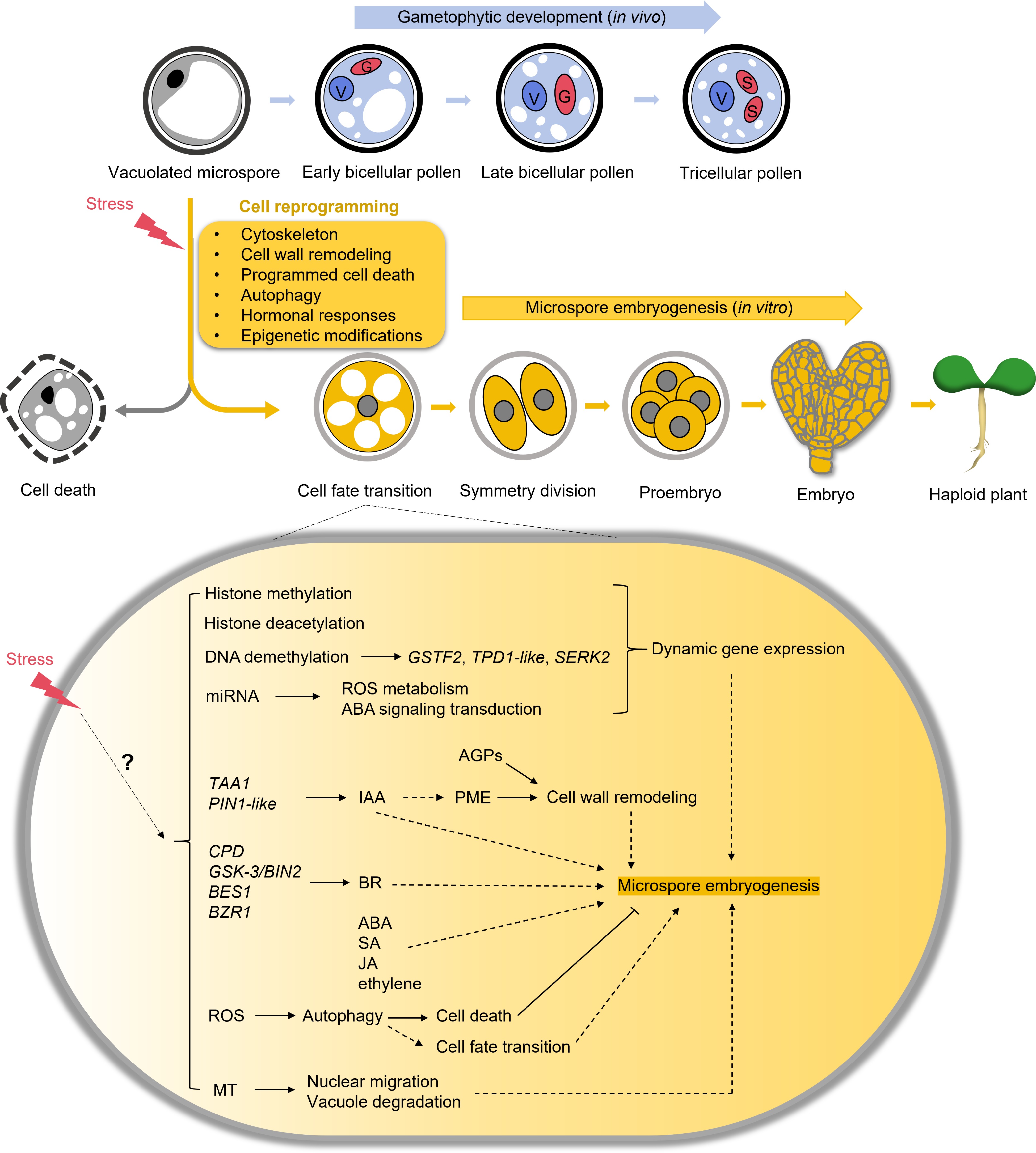
      Figure 1. 

      A schematic of the process of microspore embryogenesis. In the natural gametophytic developmental pathway, the vacuolated microspore undergoes asymmetrical division to produce a bicellular pollen grain, which matures further into a tricellular pollen grain. However, when isolated microspores are subjected to stress in vitro, they undergo reprogramming. This cell reprogramming leads to the microspores dividing to form proembryos, which then develop into full embryos. Several determinant factors govern this process, including cytoskeleton, cell wall remodeling, programmed cell death, autophagy, hormonal responses, and epigenetic modifications. The remaining microspores typically undergo cell death. V, vegetative nucleus. G, generative nucleus. S, sperm nucleus.

    • While microspore embryogenesis presents substantial potential for advancing plant breeding and research, several challenges must be addressed to enhance its efficiency and unravel its underlying mechanisms. Despite recent progress in molecular biology, much of the research remains focused on cellular descriptions using in vitro treatments over extended periods. Therefore, deeper investigations using advanced molecular and cellular techniques are crucial to addressing the following key questions:

      (i) Novel mechanisms of stress-induced microspore cell fate transition: Beyond the mechanisms discussed so far, what additional pathways or molecular signals contribute to the reprogramming of microspores under stress conditions? Identifying novel regulatory networks could provide new insights into how microspores transition to an embryogenic development.

      (ii) Reliable molecular markers for early selection of embryogenic microspores: What molecular markers can be identified that reliably distinguish embryogenic microspores at the earliest stages? Early detection of embryogenic microspores could significantly improve the induction efficiency by allowing for the selective cultivation of these cells.

      (iii) Genes activated or inhibited by epigenetic modification: Which specific genes are de novo activated or repressed by epigenetic modifications during the initiation of microspore embryogenesis? Understanding how epigenetic changes influence gene expression at the onset of embryogenesis could reveal targets for enhancing the process.

      (iv) Innovative methods to increase microspore embryogenesis frequency: What novel approaches could be developed to significantly and broadly enhance the frequency of microspore embryogenesis, particularly for species that are currently recalcitrant to this technique? Innovative strategies might include the use of new chemical treatments, gene editing techniques, or optimized culture conditions.

      (v) The comparative study between zygotic and microspore embryogenesis: Comparative analyses can reveal significant similarities between zygotic and microspore embryogenesis. Identifying and validating of determinant factors that influence embryogenesis may have implications for other forms of embryogenesis. The application of these regulators could be instrumental in reducing cell mortality and enhancing the rate of embryogenesis, particularly in in vitro settings. Furthermore, using microspore embryogenesis as a model to study the initiation of embryogenesis could provide insights into the complexities of zygotic embryogenesis, which is deeply buried in maternal tissues and difficult to access[75].

      Exploring the unanswered questions and overcoming the challenges associated with microspore embryogenesis will deepen our understanding of this complex process and enhance its practical applications in plant breeding. Addressing the obstacles related to low efficiency, high cell death rates, and species-specific limitations is essential for unlocking the full potential of microspore embryogenesis as a reliable tool for producing DH plants. These advancements could accelerate the development of new plant varieties with desirable traits, significantly contributing to agricultural innovation and productivity. By refining techniques and optimizing regulatory mechanisms, microspore embryogenesis could become more effective, enabling its broader application across a wider range of crops species, ultimately supporting global food security and sustainable agricultural practices.

      • This work was supported by the National Natural Science Foundation of China (32000248).

      • The authors confirm contribution to the paper as follows: draft manuscript preparation: Yang F, Liu X, Qiao Y, Tang X, Luo P. All authors approved the final version of the manuscript.

      • Data sharing not applicable to this review as no datasets were generated or analyzed during the current study.

      • The authors declare that they have no conflict of interest.

      • # Authors contributed equally: Fan Yang, Xinyu Liu, Ying Qiao

      • Copyright: © 2024 by the author(s). Published by Maximum Academic Press on behalf of Hainan Yazhou Bay Seed Laboratory. This article is an open access article distributed under Creative Commons Attribution License (CC BY 4.0), visit https://creativecommons.org/licenses/by/4.0/.
    Figure (1)  References (120)
  • About this article
    Cite this article
    Yang F, Liu X, Qiao Y, Tang X, Luo P. 2024. Microspore embryogenesis: in vitro cultivation induced cell reprogramming for plant breeding. Seed Biology 3: e021 doi: 10.48130/seedbio-0024-0019
    Yang F, Liu X, Qiao Y, Tang X, Luo P. 2024. Microspore embryogenesis: in vitro cultivation induced cell reprogramming for plant breeding. Seed Biology 3: e021 doi: 10.48130/seedbio-0024-0019

Catalog

  • About this article

/

DownLoad:  Full-Size Img  PowerPoint
Return
Return