PERSPECTIVE   Open Access    

Perspectives on cultured meat

More Information
  • Cultured meat is a promising alternative protein technology that aims to produce healthier, safer, and more sustainable meat. It is muscle and fat tissue generated in vitro by the culturing and biomanufacturing of animal cells for human consumption. However, producing cultured meat at large scale and low-cost is still a difficult task. Technology, regulation and consumer acceptance are all challenges for cultured meat production. In this perspective, we divided the industrial production of cultured meat into three goals according to the cell types and complexity. We discuss the technologies used to achieve low-cost cultured meat industrial production. We also discuss the regulation and consumer acceptance and how both can accelerate commercialization.
  • Atractylodes macrocephala Koidz. (common names 'Baizhu' in Chinese and 'Byakujutsu' in Japanese) is a diploid (2n = 2x = 24) and out-crossing perennial herb in the Compositae family, and has a long history of cultivation in temperate and subtropical areas of East Asia as it is widely used in traditional herbal remedies with multiple pharmacological activities[13]. The 'Pharmacopoeia of the People's Republic of China' states that 'Baizhu' is the dry rhizome of A. macrocephala Koidz. (Atractylodis Macrocephalae Rhizoma, AMR). However, in Japanese traditional medicine 'Baizhu' can be referred to both: A. japonica or A. macrocephala[4].

    A. macrocephala is naturally endemic to China and cultivated in more than 200 towns in China, belonging to Zhejiang, Hunan, Jiangxi, Anhui, Fujian, Sichuan, Hubei, Hebei, Henan, Jiangsu, Guizhou, Shanxi, and Shaanxi Provinces[3]. A. macrocephala grows to a height of 20–60 cm (Fig. 1). The leaves are green, papery, hairless, and generally foliole with 3–5 laminae with cylindric glabrous stems and branches. The flowers grow and aggregate into a capitulum at the apex of the stem. The corollas are purplish-red, and the florets are 1.7 cm long. The achenes, densely covered with white, straight hairs, are obconic and measure 7.5 mm long. The rhizomes used for medicinal purposes are irregular masses or irregularly curving cylinders about 3–13 cm long and 1.5–7 cm in diameter with an outwardly pale greyish yellow to pale yellowish color or a sparse greyish brown color. The periderm-covered rhizomes are externally greyish brown, often with nodose protuberances and coarse wrinkles. The cross-sections are white with fine dots of light yellowish-brown to brown secretion. Rhizomes are collected from plants that are > 2 years old during the spring. The fibrils are removed, dried, and used for medicinal purposes[5, 6].

    Figure 1.  Plant morphology of A. macrocephala.

    The medicinal properties of AMRs are used for spleen deficiency, phlegm drinking, dizziness, palpitation, edema, spontaneous sweating, benefit Qi, and fetal restlessness[7]. The AMR contains various functional components, among which high polysaccharide content, with a yield close to 30%[8]. Therefore, the polysaccharides of A. macrocephala Koidz. rhizome (AMRP) are essential in assessing the quality control and bioactivity of A. macrocephala. Volatile oil accounts for about 1.4% of AMR, with atractylon and atractylodin as the main components[9]. Atractylon can be converted to atractylenolide I (AT-I), atractylenolide II (AT-II), and atractylenolide III (AT-III) under ambient conditions. AT-III can be dehydrated to AT-II under heating conditions[10, 11]. AMRs, including esters, sesqui-, and triterpenes, have a wide range of biological activities, such as improving immune activity, intestinal digestion, neuroprotective activity, immune anti-inflammatory, and anti-tumor.

    In recent years, research on the pharmacological aspects of AMR has continued to increase. Still, the discovery of the main active components in AMR is in its infancy. The PAO-ZHI processing of AMR is a critical step for AMR to exert its functional effects, but also, in this case, further work is required. Studies on the biosynthesis of bioactive compounds and different types of transcriptomes advanced current knowledge of A. macrocephala, but, as mentioned, required more systematic work. Ulteriorly, an outlook on the future research directions of A. macrocephala was provided based on the advanced technologies currently applied in A. macrocephala (Fig. 2).

    Figure 2.  Current progress of A. macrocephala.

    A. macrocephala is distributed among mountainous regions more than 800 m above sea level along the middle and lower reaches of the Yangtze River (China)[5]. Due to over-exploitation and habitat destruction, natural populations are rare, threatened, and extinct in many locations[1,12]. In contrast to its native range, A. macrocephala is widely cultivated throughout China, in a total area of 2,000–2,500 ha, with a yield of 7,000 t of rhizomes annually[13]. A. macrocephala is mainly produced in Zhejiang, Anhui, and Hebei (China)[14]. Since ancient times, Zhejiang has been the famous producing area and was later introduced to Jiangxi, Hunan, Hebei, and other places[15]. Wild A. macrocephala is currently present in at least 14 provinces in China. It is mainly distributed over three mountain ranges, including the Tianmu and Dapan mountains in Zhejiang Province and the Mufu mountains along the border of Hunan and Jiangxi Provinces. A. macrocephala grows in a forest, or grassy areas on mountain or hill slopes and valleys at an altitude of 600–2,800 m. A. macrocephala grows rapidly at a temperature of 22–28 °C, and favors conditions with total precipitation of 300–400 mm evenly distributed among the growing season[16]. Chen et al. first used alternating trilinear decomposition (ATLD) to characterize the three-dimensional fluorescence spectrum of A. macrocephala[17]. Then they combined the three-dimensional fluorescence spectrum with partial least squares discriminant analysis (PLS-DA) and k-nearest neighbor method (kNN) to trace the origin of Atractylodes samples. The results showed that the classification models established by PLS-DA and kNN could effectively distinguish the samples from three major Atractylodes producing areas (Anhui, Hunan, and Zhejiang), and the classification accuracy rate (CCR) of Zhejiang atractylodes was up to 80%, and 90%, respectively[17]. Zhang et al. compared the characteristics, volatile oil content, and chemical components of attested materials from six producing areas of Zhejiang, Anhui, Hubei, Hunan, Hebei, and Henan. Differences in the shape, size, and surface characteristics were reported, with the content of volatile oil ranging from 0.58% to 1.22%, from high to low, Hunan (1.22%) > Zhejiang (1.20%) > Anhui (1.02%) > Hubei (0.94%) > Henan (0.86%) > Hebei (0.58%)[18]. This study showed that the volatile oil content of A. macrocephala in Hunan, Anhui, and Hubei is not much different from that of Zhejiang, which is around 1%. A. macrocephala is a local herb in Zhejiang, with standardized cultivation techniques, with production used to reach 80%–90% of the country. However, in recent years, the rapid development of Zhejiang's real estate economy has reduced the area planted with Zhejiang A. macrocephala, resulting in a sudden decrease in production. Therefore, neighboring regions, such as Anhui and Hunan, vigorously cultivate A. macrocephala, and the yield and quality of A. macrocephala can be comparable to those of Zhejiang. The results were consistent with the data reports[18]. Guo et al. analyzed the differentially expressed genes of Atractylodes transcripts from different regions by the Illumina HiSeq sequencing platform. It was found that 2,333, 1,846, and 1,239 DEGs were screened from Hubei and Hebei, Anhui and Hubei, and Anhui and Hebei Atrexia, respectively, among which 1,424, 1,091, and 731 DEGs were annotated in the GO database. There were 432, 321, and 208 DEGs annotated in the KEGG database. These DEGs were mainly related to metabolic processes and metabolic pathways of secondary metabolites. The highest expression levels of these genes were found in Hubei, indicating higher terpenoid production in Hubei[19]. Other compounds were differentially accumulated in Atractylodes. Chlorogenic acid from Hebei was 0.22%, significantly higher than that from Zhejiang and Anhui[20]. Moreover, the content of neochlorogenic acid and chlorogenic acid decreased after processing, with the highest effect reported in Zhejiang, with the average transfer rate of neochlorogenic acid and chlorogenic acid reaching 55.68% and 55.05%[20]. All these changes would bring great help in distinguishing the origins of A. macrocephala.

    Medicinal AMR can be divided into raw AMR and cooked AMR. The processing method is PAO-ZHI; the most traditional method is wheat bran frying. The literature compared two different treatment methods, crude A. macrocephala (CA) and bran-processed A. macrocephala, and found that the pharmacological effects of AMR changed after frying with wheat bran, mainly in the anti-tumor, antiviral and anti-inflammatory effects[21]. The anti-inflammatory effect was enhanced, while the anti-tumor and antiviral effects were somewhat weakened, which may be related to the composition changes of the compounds after frying. The study of the content of AT-I, II, and III, and atractyloside A, in rat serum provided helpful information on the mechanism of wheat bran processing[22]. In addition to frying wheat bran, Sun et al. used sulfur fumigation to treat AMR[23]. They found that the concentration of different compounds changed, producing up to 15 kinds of terpenoids. Changes in pharmacological effects were related to treatment and the type of illumination[24,25]. Also, artificial light can improve the various biological functions. A. macrocephala grew better under microwave electrodeless light, with a chlorophyll content of 57.07 ± 0.65 soil and plant analyzer develotrnent (SPAD)[24]. The antioxidant activity of AMR extract treated with light-emitting diode (LED)-red light was the highest (95.3 ± 1.1%) compared with other treatments[24]. The total phenol and flavonoid contents of AMR extract treated with LED-green light were the highest at 24.93 ± 0.3 mg gallic acid equivalents (GAE)/g and 11.2 ± 0.3 mg quercetin equivalents (QE)/g compared with other treatments[24, 25]. Polysaccharides from Chrysanthemun indicum L.[26] and Sclerotium rolfsiisacc[27] can improve AMR's biomass and bioactive substances by stimulating plant defense and thus affect their efficacy. In summary, there are compositional differences between A. macrocephala from different origins. Besides, different treatments, including processing mode, light irradiation, and immune induction factors, which can affect AMR's biological activity, provide some reference for the cultivation and processing of A. macrocephala (Fig. 3).

    Figure 3.  Origin, distribution and processing of A. macrocephala.

    The AMR has been reported to be rich in polysaccharides, sesquiterpenoids (atractylenolides), volatile compounds, and polyacetylenes[3]. These compounds have contributed to various biological activities in AMR, including immunomodulatory effects, improving gastrointestinal function, anti-tumor activity, neuroprotective activity, and anti-inflammatory.

    AMRP has received increasing attention as the main active component in AMR because of its rich and diverse biological activities. In the last five years, nine AMRP have been isolated from AMR. RAMP2 had been isolated from AMR, with a molecular weight of 4.354 × 10³ Da. It was composed of mannose, galacturonic acid, glucose, galactose, and arabinose, with the main linkages of →3-β-glcp-(1→, →3,6-β-glcp-(1→, →6-β-glcp-(1→, T-β-glcp-(1→, →4-α-galpA-(1→, →4-α-galpA-6-OMe-(1→, →5-α-araf-(1→, →4,6-β-manp-(1→ and →4-β-galp-(1→[28]. Three water-soluble polysaccharides AMAP-1, AMAP-2, and AMAP-3 were isolated with a molecular weight of 13.8 × 10⁴ Da, 16.2 × 10⁴ Da, and 8.5 × 10⁴ Da, respectively. Three polysaccharides were deduced to be natural pectin-type polysaccharides, where the homogalacturonan (HG) region consists of α-(1→4)-linked GalpA residues and the ramified region consists of alternating α-(1→4)-linked GalpA residues and α-(1→2)-linked Rhap residues. Besides, three polysaccharides were composed of different ratios of HG and rhamnogalacturonan type I (RG-I) regions[29]. Furthermore, RAMPtp has been extracted from AMR with a molecular weight of 1.867 × 10³ Da. It consists of glucose, mannose, rhamnose, arabinose, and galactose with 60.67%, 14.99%, 10.61%, 8.83%, and 4.90%, connected by 1,3-linked β-D Galp and 1,6-linked β-D Galp residues[30]. Additionally, PAMK was characterized by a molecular weight of 4.1 kDa, consisting of galactose, arabinose, and glucose in a molar ratio of 1:1.5:5, with an alpha structure and containing 96.47% polysaccharide and small amounts of protein, nucleic acid, and uric acid[31]. Another PAMK extracted from AMR had a molecular weight of 2.816 × 10³ Da and consisted of glucose and mannose in molar ratios of 0.582 to 0.418[32]. Guo et al. isolated PAMK with a molecular weight of 4.748 × 10³ g/mol from AMR, consisting of glucose, galactose, arabinose, fructose, and mannose in proportions of 67.01%, 12.32%, 9.89%, 1.18%, and 0.91%, respectively[33]. In addition, AMP1-1 is a neutral polysaccharide fragment with a molecular weight of 1.433 kDa isolated from AMR. It consists of glucose and fructose, and the structure was identified as inulin-type fructose α-D-Glcp-1→(2-β-D-Fruf-1)7[34]. These reports indicated that, in general, polysaccharides are extracted by water decoction, ultrasonic-assisted extraction, enzyme hydrolysis method, and microwave-assisted extraction. The separation and purification are column chromatography, stepwise ethanol precipitation, and ultrafiltration. Their physicochemical properties and structural characterization are generally achieved by determining the molecular weight, determining the monosaccharide composition, analyzing the secondary structure, and glycosidic bond configuration of polysaccharides with Fourier transform infrared (FT-IR) and nuclear magnetic resonance (NMR). The advanced structures of polysaccharides can be identified by high-performance size exclusion chromatography-multiangle laser light scattering (HPSEC-MALLS), transmission electron microscopy (TEM), and scanning electron microscopy (SEM) techniques (Table 1). AMRP has various physiological functions, including immunomodulatory effects, improving gastrointestinal function, and anti-tumor activity. The related biological activities, animal models, monitoring indicators, and results are summarized in Table 1.

    Table 1.  Components and bioactivity of polysaccharides from Atractylodes macrocephala Koidz. Rhizome.
    Pharmacological activitiesDetailed functionPolysaccharides informationModelDoseTest indexResultsRef.
    Immunomodulatory effectsRestore immune
    function
    /Chicken models
    (HS-induced)
    200 mg/kgOxidative index;
    Activities of mitochondrial complexes and ATPases;
    Ultrastructure in chicken spleens;
    Expression levels of cytokines, Mitochondrial dynamics- and apoptosis-related genes
    Alleviated
    the expression of
    IL-1 ↑,TNF-α ↑, IL-2 ↓, IFN- γ ↓; mitochondrial dynamics- and anti-apoptosis-related genes ↓; pro-apoptosis-related genes ↑;
    the activities of mitochondrial complexes and ATPases ↓ caused by HS
    [35]
    Regulate the immune function/Chicken models
    (HS-induced)
    200 mg/kgiNOS–NO activities;
    ER stress-related genes;
    Apoptosis-related genes;
    Apoptosis levels
    Alleviated NO content ↑; activity of iNOS ↑ in the chicken spleen; GRP78, GRP94, ATF4, ATF6, IRE ↑; caspase3 ↑; Bcl-2 ↓ caused by HS[36]
    Relieve immunosuppressionCommercial AMR powder (purity 70%)Geese models
    (CTX-induced)
    400 mg/kgSpleen development;
    Percentages of leukocytes in peripheral blood
    Alleviated the spleen damage;
    T and B cell proliferation ↓; imbalance of leukocytes; disturbances of humoral; cellular immunity caused by CTX
    [37]
    Active the lymphocytesCommercial AMR powder (purity 95%)Geese models
    (CTX-induced)
    400 mg/kgThymus morphology;
    The level of serum GMC-SF, IL-1b, IL-3, IL-5;
    mRNA expression of CD25, novel_mir2, CTLA4 and CD28 signal pathway
    Maintain normal cell morphology of thymus;
    Alleviated GMC-SF ↓, IL-1b ↓, IL-5↓, IL-6↓, TGF-b↓; IL-4 ↑, IL-10 ↑; novel_mir2 ↓, CD25↓, CD28↓ in thymus and lymphocytes caused by CTX
    [38]
    Alleviate immunosuppressionCommercial AMR powder (purity 70%)Geese models
    (CTX-induced)
    400 mg/kgThymus development;
    T cell proliferation rate;
    The level of CD28, CD96, MHC-II;
    IL-2 levels in serum;
    differentially expressed miRNAs
    Alleviated thymus damage;
    T lymphocyte proliferation rate ↓; T cell activation ↓; IL-2 levels ↓ caused by CTX;
    Promoted novel_mir2 ↑; CTLA4 ↓; TCR-NFAT signaling pathway
    [39]
    Alleviates T cell activation declineCommercial AMR powder (purity 95%)BALB/c female mice (CTX-induced)200 mg/kgSpleen index;
    Morphology, death, cytokine concentration of splenocytes;
    Th1/Th2 ratio, activating factors of lymphocytes;
    T cell activating factors;
    mRNA expression level in CD28 signal pathway
    Improved the spleen index;
    Alleviated abnormal splenocytes morphology and death; Balance Th1/Th2 ratio; IL-2 ↑, IL-6 ↑, TNF-α ↑, IFN-γ ↑; mRNA levels of CD28, PLCγ-1, IP3R, NFAT, AP-1 ↑
    [40]
    Immunoregulation and ImmunopotentiationCommercial AMR powder (purity 80%)BMDCs (LPS-induced);
    Female BALB/c mice (ovalbumin as a model antigen)
    /Surface molecule expression of BMDCs;
    Cytokines secreted by dendritic cell supernatants;
    OVA-specific antibodies in serum;
    Cytokines in serum;
    Lymphocyte immunophenotype
    Expression of CD80 and CD86 ↑; IL-1β ↑, IL-12 ↑, TNF-α↑ and IFN-γ ↑; OVA-specific antibodies in serum ↑; Secretion of cytokines ↑; Proliferation rate of spleen lymphocytes ↑; Activation of CD3+CD4+ and CD3+CD8+ lymphocytes[46]
    Increase immune-response capacity of the spleen in miceCommercial AMR powder (purity 70%)BALB/c female mice100, 200, 400 mg/kgSpleen index;
    Concentrations of cytokines;
    mRNA and protein expression levels in TLR4 signaling
    In the medium-PAMK group:
    IL-2, IL-4, IFN-c, TNF-a ↑; mRNA and protein expression of TLR4, MyD88, TRAF6, TRAF3, NF-κB in the spleen ↑
    [41]
    Immunological activityCommercial AMR powder (purity 80%)Murine splenic lymphocytes (LPS or PHA-induced)13, 26, 52, 104, 208 μg/mLT lymphocyte surface markersLymphocyte proliferation ↑;
    Ratio of CD4+/CD8+ T cells ↑
    [47]
    Immunomodulatory activityTotal carbohydrates content 95.66 %Mouse splenocytes
    (Con A or LPS-induced)
    25, 50, 100 μg/mLSplenocyte proliferation;
    NK cytotoxicity;
    Productions of NO and cytokines;
    Transcription factor activity;
    Signal pathways and receptor
    Promoted splenocyte proliferation; Cells enter S and G2/M phases; Ratios of T/B cells ↑; NK cytotoxicity ↑; Transcriptional activities of NFAT ↑; NF-κB, AP-1 ↑; NO, IgG, IL-1α, IL-1β, IL-2, IL-3, IL-4, IL-6, IL-10, IL-12p40, IL-12p70, IL-13, IFN-γ, TNF-α, G-CSF, GM-CSF, KC, MIP-1α, MIP-1β, RANTES, Eotaxin ↑[42]
    Promote the proliferation of thymic epithelial cellsContents of fucrhaara, galactose, glucose, fructose,
    and xylitol: 0.98%, 0.40%, 88.67%, 4.47%, and 5.47%
    MTEC1 cells50 μg/mLCell viability and proliferation;
    lncRNAs, miRNAs, and mRNAs expression profiles in MTEC1 cells
    The differential genes were 225 lncRNAs, 29 miRNAs, and 800 mRNAs; Genes enriched in cell cycle, cell division, NF-κB signaling, apoptotic process, and MAPK signaling pathway[44]
    Immunomodulatory activityMW: 4.354 × 10³ Da;
    Composed of mannose, galacturonic acid, glucose, galactose and arabinose;
    The main linkages are →3-β-glcp-(1→, →3,6-β-glcp-(1→, →6-β-glcp-(1→, T-β-glcp-(1→,
    →4-α-galpA-(1→, →4-α-galpA-6-OMe-(1→, →5-α-araf-(1→, →4,6-β-manp-(1→ and →4-β-galp-(1→
    CD4+ T cell50, 100, 200 μg/mLMolecular weight;
    Monosaccharide composition;
    Secondary structure;
    Surface topography;
    Effect on Treg cells
    Treg cells percentage ↑; mRNA expressions of Foxp3, IL-10 and IL-2 ↑; STAT5 phosphorylation levels ↑; IL-2/STAT5 pathway[28]
    Immunostimulatory activityMW of AMAP-1, AMAP-2, and AMAP-3 were 13.8×10⁴ Da, 16.2×10⁴ Da and 8.5×10⁴ Da;
    HG region consists of α-(1→4)-linked GalpA residues
    RAW264.7 cells (LPS-induced)80, 40, 200 μg/mLMolecular weight;
    Total carbohydrate;
    Uronic acid contents;
    Secondary structure;
    Monosaccharide composition;
    Immunostimulatory activity
    RG-I-rich AMAP-1 and AMAP-2 improved the release of NO[29]
    Immunomodulatory effectMW: 1.867×10³ Da;
    Contents of glucose, mannose, rhamnose,
    arabinose and galactose: 60.67%, 14.99%, 10.61%, 8.83% and 4.90%
    SMLN lymphocytes25
    μg/ml
    Molecular weight;
    Monosaccharide composition;
    Ultrastructure;
    Intracellular Ca2+concentration;
    Target genes;
    Cell cycle distribution
    [Ca2+]i ↑; More cells in S and G2/M phases; IFN-γ ↑, IL-17A ↑; mRNA expressions of IL-4 ↓[30]
    Macrophage activationTotal carbohydrates content 95.66 %RAW264.7 macrophages (LPS-induced)25, 50, 100 μg/mLPinocytic activity;
    Phagocytic uptake;
    Phenotypic characterization;
    Cytokine production;
    Bioinformatics analysis;
    Transcription factor inhibition
    IL-6, IL-10 and TNF-α ↑; CCL2 and CCL5 ↑; Pinocytic and phagocytic activity ↑; CD40, CD80, CD86, MHC-I, MHC-II ↑; NF-κB and Jak-STAT pathway[43]
    Immunomodulatory effectTotal carbohydrates content 95.66 %SMLN lymphocytes25, 50, 100 μg/mLCytokine production;
    CD4+ and CD8+ lymphocytes;
    Target genes;
    Bioinformatics analysis;
    T and B lymphocyte proliferation;
    Receptor binding and blocking
    IFN-γ, IL-1α, IL-21, IFN-α, CCL4, CXCL9, CXCL10 ↑; CD4+ and CD8+subpopulations proportions ↑;
    c-JUN, NFAT4, STAT1, STAT3 ↑;
    67 differentially expressed miRNAs (55 ↑ and
    12 ↓), associated with immune system pathways; Affect T and B lymphocytes
    [45]
    Improving gastrointestinal functionRelieve enteritis and improve intestinal
    flora disorder
    Commercial AMR powder (purity 70%);
    Contents of fucrhaara, galactose, glucose, xylitol, and fructose: 0.98%, 0.40%, 88.67%, 4.47%, and 5.47%
    Goslings (LPS-induced)400 mg/kgSerum CRP, IL-1β, IL-6, and TNF-α levels;
    Positive rate of IgA;
    TLR4, occludin, ZO-1, cytokines, and immunoglobulin mRNA expression;
    Intestinal flora of gosling excrement
    Relieved IL-1β, IL-6, TNF-α levels in serum ↑; the number of IgA-secreting cells ↑; TLR4 ↑; tight junction occludin and ZO-1 ↓; IL-1β mRNA expression in the small intestine ↑; Romboutsia ↓ caused by LPS[48]
    Ameliorate ulcerative colitisMW: 2.391 × 10⁴ Da;
    Composed of mannose, glucuronic acid, glucose and arabinose in a molar ratio of 12.05:6.02:72.29:9.64
    Male C57BL/6J mice (DDS-induced)10, 20, 40 mg/kg bwHistopathological evaluation;
    Inflammatory mediator;
    Composition of gut microbiota;
    Feces and plasma for global metabolites profiling
    Butyricicoccus, Lactobacillus ↑;
    Actinobacteria, Akkermansia, Anaeroplasma, Bifidobacterium, Erysipelatoclostridium, Faecalibaculum, Parasutterella,
    Parvibacter, Tenericutes, Verrucomicrobia ↓;
    Changed 23 metabolites in fecal content; 21 metabolites in plasma content
    [49]
    Attenuate ulcerative colitis/Male SD rats (TNBS-induced);
    Co-culture BMSCs and IEC-6 cells
    540 mg/kg
    (for rats);
    400 μg/mL (for cell)
    Histopathological analysis;
    Cell migration;
    Levels of cytokines
    Potentiated BMSCs’ effect on preventing colitis and homing the injured tissue, regulated cytokines;
    BMSCs and AMP promoted the migration of IEC
    [52]
    Against intestinal mucosal injuryMW: 3.714 × 10³ Da;
    Composed of glucose, arabinose, galactose, galacturonic acid, rhamnose
    and mannose with molar ratios of 59.09:23.22:9.32:4.70:2.07:1.59
    Male C57BL/6 mice (DDS-induced)100 mg/kgIntestinal morphology;
    IL-6, TNF-α and IL-1β in serum;
    mRNA expression;
    Intestinal microbiota
    Alleviated body weight ↓; colon length ↓; colonic damage caused by DSS;
    Over-expression of TNF-α, IL-1β, IL-6 ↓; Infiltration of neutrophils in colon ↓; Mucin 2 ↑;
    Tight junction protein Claudin-1 ↑;
    Harmful bacteria content ↓;
    Beneficial bacteria content ↑
    [50]
    Against intestinal injuryTotal carbohydrates 95.66 %IECs (DDS-induced)5, 25, 50 μg/mLCell proliferation and apoptosis;
    Expression levels of intercellular TJ proteins;
    lncRNA screening
    Proliferation and survival of IECs ↑;
    Novel lncRNA ITSN1-OT1 ↑;
    Blocked the nuclear import of phosphorylated STAT2
    [51]
    Anti-tumor activityInduce apoptosis in transplanted H22 cells in miceMW: 4.1× 10³ Da;
    Neutral heteropolysaccharide composed of galactose, arabinose, and glucose with α-configuration (molar ratio, 1:1.5:5)
    Female Kunming mice100 and 200 mg/kg (for rats)Secondary structure;
    Molecular weight;
    Molecular weight;
    Thymus index and Spleen index;
    Lymphocyte Subpopulation in peripheral blood;
    Cell cycle distribution
    In tumor-bearing mice CD3+, CD4+, CD8+ ↓;
    B cells ↑
    [31]
    Regulate the innate immunity of colorectal cancer cellsCommercial AMR powder (purity 70%)C57BL/6J mice (MC38 cells xenograft model)500 mg/kgExpression of pro-inflammatory cytokines and secretionIL-6, IFN-λ, TNF-α, NO ↑ through MyD88/TLR4-dependent signaling pathway;
    Survival duration of mice with tumors ↑;
    Prevent tumorigenesis in mice
    [54]
    Induce apoptosis of Eca-109 cellsMW: 2.1× 10³ Da;
    Neutral hetero polysaccharide composed
    of arabinose and glucose (molar ratio, 1:4.57) with pyranose rings and α-type and β-type glycosidic linkages
    Eca-109 cells0.25, 0.5, 1, 1.5, 2.00 mg/mLCell morphology;
    Cell cycle arrest;
    Induction of apoptosis
    Accelerate the apoptosis of Eca109 cells[53]
    '/' denotes no useful information found in the study.
     | Show Table
    DownLoad: CSV

    To study the immunomodulatory activity of AMRP, the biological models generally adopted are chicken, goose, mouse, and human cell lines. Experiments based on the chicken model have generally applied 200 mg/kg doses. It was reported that AMRP protected the chicken spleen against heat stress (HS) by alleviating the chicken spleen immune dysfunction caused by HS, reducing oxidative stress, enhancing mitochondrial function, and inhibiting cell apoptosis[35]. Selenium and AMRP could improve the abnormal oxidation and apoptosis levels and endoplasmic reticulum damage caused by HS, and could act synergistically in the chicken spleen to regulate biomarker levels[36]. It indicated that AMRP and the combination of selenium and AMRP could be applied as chicken feed supplementation to alleviate the damage of HS and improve chicken immunity.

    The general application dose in the goose model is also 200 mg/kg, and the main injury inducer is cyclophosphamide (CTX). AMRP alleviated CTX-induced immune damage in geese and provided stable humoral immune protection[37]. Little is known about the role of AMRP in enhancing immunity in geese through the miRNA pathway. It was reported that AMRP alleviated CTX-induced decrease in T lymphocyte activation levels through the novel _mir2/CTLA4/CD28/AP-1 signaling pathway[38]. It was also reported that AMRP might be achieved by upregulating the TCR-NFAT pathway through novel_mir2 targeting of CTLA4, thereby attenuating the immune damage induced by CTX[39]. This indicated that AMRP could also be used as goose feed supplementation to improve the goose's autoimmunity.

    The typical injury inducer for mouse models is CTX, and the effects on mouse spleen tissue are mainly observed. BALB/c female mice were CTX-induced damage. However, AMRP increased cytokine levels and attenuated the CTX-induced decrease in lymphocyte activation levels through the CD28/IP3R/PLCγ-1/AP-1/NFAT signaling pathway[40]. It has also been shown that AMRP may enhance the immune response in the mouse spleen through the TLR4-MyD88-NF-κB signaling pathway[41].

    Various cellular models have been used to study the immune activity of AMRP, and most of these studies have explored the immune activity with mouse splenocytes and lymphocytes. Besides, the commonly used damage-inducing agents are LPS, phytohemagglutinin (PHA), and concanavalin A (Con A).

    In one study, the immunoreactivity of AMRP was studied in cultured mouse splenocytes. LPS and Con A served as controls. Specific inhibitors against mitogen-activated protein kinases (MAPKs) and NF-κB significantly inhibited AMRP-induced IL-6 production. The results suggested that AMRP-induced splenocyte activation may be achieved through TLR4-independent MAPKs and NF-κB signaling pathways[42]. Besides, AMRP isolated from AMR acting on LPS-induced RAW264.7 macrophages revealed that NF-κB and Jak-STAT signaling pathways play a crucial role in regulating immune response and immune function[43]. RAMP2 increased the phosphorylation level of STAT5 in Treg cells, indicating that RAMP2 could increase the number of Treg cells through the IL-2/STAT5 signaling pathway[28]. Furthermore, the relationship between structure and immune activity was investigated. Polysaccharides rich in RG-I structure and high molecular weight improved NO release from RAW264.7 cells. Conversly, polysaccharides rich in HG structure and low molecular weight did not have this ability, indicating that the immunoreactivity of the polysaccharide may be related to the side chain of RG-I region[29]. Moreover, the effect of AMRP on the expression profile of lncRNAs, miRNAs, and mRNAs in MTEC1 cells has also been investigated. The differentially expressed genes include lncRNAs, Neat1, and Limd1. The involved signaling pathways include cell cycle, mitosis, apoptotic process, and MAPK[44].

    Xu et al. found that AMRP affects supramammary lymph node (SMLN) lymphocytes prepared from healthy Holstein cows. Sixty-seven differentially expressed miRNAs were identified based on microRNA sequencing and were associated with immune system pathways such as PI3K-Akt, MAPKs, Jak-STAT, and calcium signaling pathways. AMRP exerted immunostimulatory effects on T and B lymphocytes by binding to T cell receptor (TCR) and membrane Ig alone, thereby mobilizing immune regulatory mechanisms within the bovine mammary gland[45].

    AMRP can also be made into nanostructured lipid carriers (NLC). Nanoparticles as drug carriers can improve the action of drugs in vivo. NLC, as a nanoparticle, has the advantages of low toxicity and good targeting[46]. The optimization of the AMRP-NLC preparation process has been reported. The optimum technologic parameters were: the mass ratio of stearic acid to caprylic/capric triglyceride was 2:1. The mass ratio of poloxamer 188 to soy lecithin was 2:1. The sonication time was 12 min. The final encapsulation rate could reach 76.85%[47]. Furthermore, AMRP-NLC interfered with the maturation and differentiation of bone marrow-derived dendritic cells (BMDCs). Besides, AMRP-NLC, as an adjuvant of ovalbumin (OVA), could affect ova-immunized mice with enhanced immune effects[46].

    AMRP also has the effect of alleviating intestinal damage. They are summarized in Table 1. The common damage-inducing agents are lipopolysaccharide (LPS), dextran sulfate sodium (DDS), and trinitrobenzene sulfonic acid (TNBS). A model of LPS-induced enteritis in goslings was constructed to observe the effect of AMRP on alleviating small intestinal damage. Gosling excrement was analyzed by 16S rDNA sequencing to illuminate the impact of AMRP on the intestinal flora. Results indicated that AMRP could maintain the relative stability of cytokine levels and immunoglobulin content and improve intestinal flora disorder[48]. Feng et al. used DDS-induced ulcerative colitis (UC) in mice and explored the alleviating effects of AMRP on UC with 16S rDNA sequencing technology and plasma metabolomics. The results showed that AMRP restored the DDS-induced disruption of intestinal flora composition, regulated the production of metabolites such as short-chain fatty acids and cadaveric amines, and regulated the metabolism of amino acids and bile acids by the host and intestinal flora[49]. A similar study has reported that AMRP has a protective effect on the damage of the intestinal mucosal barrier in mice caused by DSS. It was found that AMRP increased the expression of Mucin 2 and the tight junction protein Claudin-1. In addition, AMRP decreased the proportion of harmful bacteria and increased the potentially beneficial bacteria content in the intestine[50]. The protective effect of AMRP on DSS-induced damage to intestinal epithelial cells (IECs) has also been investigated. The results showed that AMRP promoted the proliferation and survival of IECs.

    In addition, AMRP induced a novel lncRNA ITSN1-OT1, which blocked the nuclear import of phosphorylated STAT2 and inhibited the DSS-induced reduced expression and structural disruption of tight junction proteins[51]. AMRP can also act in combination with cells to protect the intestinal tract. The ulcerative colitis model in Male Sprague-Dawley (SD) rats was established using TNBS, and BMSCs were isolated. IEC-6 and BMSCs were co-cultured and treated by AMRP. The results showed that AMRP enhanced the prevention of TNBS-induced colitis in BMSCs, promoted the migration of IEC, and affected the expression of various cytokines[52]. These reports indicated that the 16S rDNA sequencing technique could become a standard method to examine the improvement of gastrointestinal function by AMRP.

    AMRP has anti-tumor activity and other biological activities. AMRP can induce apoptosis in Hepatoma-22 (H22) and Eca-109 cells and modulate the innate immunity of MC38 cells. For instance, the anti-tumor effects of AMRP were investigated by constructing a tumor-bearing mouse model of H22 tumor cells. AMRP blocked the S-phase of H22 tumor cells and induced an immune response, inhibiting cell proliferation[31]. In addition, AMRP can inhibit cell proliferation through the mitochondrial pathway and by blocking the S-phase of Eca-109 tumor cells[53]. AMRP affects MC38 tumor cells, and the anti-tumor effect of AMRP was investigated with Toll-like receptor 4 (TLR4) KO C57BL/6 mice and the construction of the MC38 tumor cell xenograft model. AMRP significantly inhibited the development of MC38 cells in mice and prolonged the survival of tumor-bearing mice. AMRP activity was diminished in TLR4 KO mice. Combined with the immunoblotting assay results, it was shown that TLR4 regulated the MyD88-dependent signaling pathway, which has a critical effect on the anti-tumor effect of AMRP[54].

    AMR contains a large number of bioactive compounds. Among them, small molecule compounds include esters, sesquiterpenes, and other compounds. These small molecule compounds have significant pharmacological activities, including anti-tumor, neuroprotective, immunomodulatory, and anti-inflammatory. In the last five years, small molecule compounds have been increasingly identified (Fig. 4), with atractylenolides as the main component of AMR extracts[11]. Atractylenolides are a small group of sesquiterpenoids. Atractylenolides include AT-I, AT-II, and AT-III, lactones isolated from AMR.

    Figure 4.  Structure of small molecule compounds with bioactivities from AMR. Atractylenolide I (1); Atractylenolide II (2); Atractylenolide III (3); 3β-acetoxyl atractylenolide I (4); 4R,5R,8S,9S-diepoxylatractylenolide II (5); 8S,9S-epoxyla-tractylenolide II (6); Atractylmacrols A (7); Atractylmacrols B (8); Atractylmacrols C (9); Atractylmacrols D (10); Atractylmacrols E (11); 2-[(2E)-3,7-dimethyl-2,6-octadienyl]-6-methyl-2,5-cyclohexadiene-1,4-dione (12); 8-epiasterolid (13); (3S,4E,6E,12E)-1-acetoxy-tetradeca-4,6,12-triene-8,10-diyne-3,14-diol (14); (4E,6E,12E)-tetradeca-4,6,12-triene-8,10-diyne-13,14-triol (15); 1-acetoxy-tetradeca-6E,12E-diene-8, 10-diyne-3-ol (16); 1,3-diacetoxy-tetradeca-6E, 12E-diene-8,10-diyne (17); Biatractylenolide II (18); Biepiasterolid (19); Biatractylolide (20).

    The anti-tumor activity was mainly manifested by AT-I and AT-II, especially AT-I (Table 2). Anti-tumor activity has been studied primarily in vivo and in vitro. However, there is a lack of research on the anti-tumor activity of atractylenolide in human clinical trials. The concentration of atractylenolide applied on cell lines was < 400 μM, or < 200 mg/kg on tumor-bearing mice.

    Table 2.  Anti-tumor activity of atractylenolides.
    TypesSubstancesModelIndexDoseSignal pathwayResultsRef.
    Human colorectal cancerAT-IIIHCT-116 cell;
    HCT-116 tumor xenografts bearing in nude mice
    Cell viability;
    Cell apoptotic;
    mRNAs and protein
    expressions of Bax, Bcl-2, caspase-9 and caspase-3
    25, 50, 100, 200 μM (for cell);
    50, 100,
    200 mg/kg (for rats)
    Bax/Bcl-2 signaling pathwayPromoting the expression of proapoptotic related gene/proteins; Inhibiting the expression of antiapoptotic related gene/protein; Bax↑; Caspase-3↓; p53↓; Bcl-2↓[55]
    Human gastric carcinomaAT-IIHGC-27 and AGS cell
    Cell viability;
    Morphological changes;
    Flow cytometry;
    Wound healing;
    Cell proliferation, apoptosis, and motility
    50, 100, 200, 400 μMAkt/ERK signaling pathwayCell proliferation, motility↓; Cell apoptosis↑; Bax↑;
    Bcl-2↓; p-Akt↓; p-ERK↓
    [56]
    Mammary
    tumorigenesis
    AT-IIMCF 10A cell;
    Female SD rats (NMU-induced)
    Nrf2 expression and nuclear accumulation;
    Cytoprotective effects;
    Tumor progression;
    mRNA and protein levels of Nrf2;
    Downstream detoxifying enzymes
    20, 50, 100 μM (for cell);
    100 and 200 mg/kg (for rats)
    JNK/ERK-Nrf2-ARE signaling pathway;
    Nrf2-ARE signaling pathway
    Nrf2 expressing↑; Nuclear translocation↑; Downstream detoxifying enzymes↓; 17β-Estradiol↓; Induced malignant transformation[57]
    Human colon adenocarcinomaAT-IHT-29 cellCell viability;
    TUNEL and Annexin V-FITC/PI double stain;
    Detection of initiator and
    executioner caspases level
    10, 20, 40, 80, 100 μMMitochondria-dependent pathwayPro-survival Bcl-2↓; Bax↑; Bak↑; Bad↑; Bim↑; Bid↑; Puma↑[58]
    Sensitize triple-negative
    TNBC cells to paclitaxel
    AT-IMDA-MB-231 cell;
    HS578T cell;
    Balb/c mice (MDA-MB-231 cells-implanted)
    Cell viability
    Transwell migration
    CTGF expression
    25, 50, 100 μM (for cell);
    50 mg/kg (for rats)
    /Expression and secretion of CTGF↓; CAF markers↓; Blocking CTGF expression and fibroblast activation[59]
    Human ovarian cancerAT-IA2780 cellCell cycle;
    Cell apoptosis;
    Cyclin B1 and CDK1 level
    12.5, 25, 50, 100 and 200 μMPI3K/Akt/mTOR
    signaling pathway
    Cyclin B1, CDK1↓; Bax↑;
    Caspase-9↓; Cleaved caspase-3↓; Cytochrome c↑; AIF↑; Bcl-2↓; Phosphorylation level of PI3K, Akt, mTOR↓
    [60]
    Impaired metastatic properties transfer of CSCsAT-ILoVo-CSCs; HT29-CSCsCell migration
    and invasion;
    miR-200c expression;
    Cell apoptosis
    200 μMPI3K/Akt/mTOR signaling pathwaySuppressing miR-200c activity; Disrupting EV uptake by non-CSCs[61]
    Colorectal cancerAT-IHCT116 cell;
    SW480 cell;
    male BALB/c nude mice (HCT116-implanted)
    Cell viability;
    Cell apoptosis;
    Glucose uptake;
    Lactate Production;
    STAT3 expression;
    Immunohistological analysis
    25, 50, 100, 150, 200 μM (for cell);
    50 mg/kg (for rats)
    JAK2/STAT3 signalingCaspase-3↑; PARP-1↓;
    Bax↑; Bcl-2↓; Rate-limiting glycolytic
    enzyme HK2↓; STAT3 phosphorylation↓
    [62]
    Human lung cancerAT-INSCLC cells (A549 and H1299);
    female nude mice (A549-Luc cells- implanted)
    Cell viability;
    Cell cycle;
    Phosphorylation and protein expression of
    ERK1/2, Stat3,
    PDK1, transcription factor SP1;
    mRNA levels of PDK1 gene
    12.5, 25, 50, 100, 150 μM (for cell);
    25 and 75 mg/kg (for rats)
    /ERK1/2↑; Stat3↓; SP1↓;
    PDK1↓
    [63]
    '/' denotes no useful information found in the study.
     | Show Table
    DownLoad: CSV

    AT-III affects human colorectal cancer. AT-II affects human gastric carcinoma and mammary tumorigenesis. AT-I affects human colon adenocarcinoma, human ovarian cancer, metastatic properties transfer of Cancer stem cells (CSCs), colorectal cancer, and human lung cancer, and enhances the sensitivity of triple-negative breast cancer cells to paclitaxel. Current techniques have made it possible to study the effects of atractylenolide on tumors at the signaling pathway level (Table 2). For instance, AT-III significantly inhibited the growth of HCT-116 cells and induced apoptosis by regulating the Bax/Bcl-2 apoptotic signaling pathway. In the HCT116 xenograft mice model, AT-III could inhibit tumor growth and regulate the expression of related proteins or genes. It indicated that AT-III could potentially treat human colorectal cancer[55]. AT-II significantly inhibited the proliferation and motility of HGC-27 and AGS cells and induced apoptosis by regulating the Akt/ERK signaling pathway. It suggested that AT-II can potentially treat gastric cancer[56]. However, in this study, the anti-tumor effects of AT-II in vivo were not examined. AT-II regulated intracellular-related enzyme expression in MCF 10A cells through the JNK/ERK-Nrf2-ARE signaling pathway. AT-II reduced inflammation and oxidative stress in rat mammary tissue through the Nrf2-ARE signaling pathway. AT-II inhibited tumor growth in the N-Nitroso-N-methyl urea (NMU)-induced mammary tumor mice model, indicating that AT-II can potentially prevent breast cancer[57]. AT-I induced apoptosis in HT-29 cells by activating anti-survival Bcl-2 family proteins and participating in a mitochondria-dependent pathway[58]. It indicated that AT-I is a potential drug effective against HT-29 cells. However, the study was only conducted in vitro; additional in vivo experimental data are needed. AT-I can enhance the sensitivity of triple-negative breast cancer (TNBC) cells to paclitaxel. MDA-MB-231 and HS578T cell co-culture systems were constructed, respectively. AT-I was found to impede TNBC cell migration. It also enhanced the sensitivity of TNBC cells to paclitaxel by inhibiting the conversion of fibroblasts into cancer-associated fibroblasts (CAFs) by breast cancer cells. In the MDA-MB-231 xenograft mice model, AT-I was found to enhance the effect of paclitaxel on tumors and inhibit the metastasis of tumors to the lung and liver[59]. AT-I inhibited the growth of A2780 cells through PI3K/Akt/mTOR signaling pathway, promoting apoptosis and blocking the cell cycle at G2/M phase change, suggesting a potential therapeutic agent for ovarian cancer[60]. However, related studies require in vivo validation trials. CSCs are an important factor in tumorigenesis. CSCs isolated from colorectal cancer (CRC) cells can metastasize to non-CSCs via miR-200c encapsulated in extracellular vesicles (EVs).

    In contrast, AT-I could inhibit the activity and transfer of miR-200c. Meanwhile, interfere with the uptake of EVs by non-CSCs. This finding contributes to developing new microRNA-based natural compounds against cancer[61]. AT-I has the function of treating colorectal cancer. HCT116 and SW480 cells were selected for in vitro experiments, and AT-I was found to regulate STAT3 phosphorylation negatively. The HCT116 xenograft mice model was constructed, and AT-I was found to inhibit the growth of HCT116. AT-I induced apoptosis in CRC cells, inhibited glycolysis, and blocked the JAK2/STAT3 signaling pathway, thus exerting anti-tumor activity[62]. The in vitro experiments were performed with A549 and H1299 cell lines. The in vivo experiments were performed to construct the A549-Luc xenograft mice model. The results showed that AT-I inhibited lung cancer cell growth by activating ERK1/2. AT-I inhibited SP1 protein expression and phosphorylation of Stat3, decreasing PDK1 gene expression. The study showed that AT-I could inhibit lung cancer cell growth and targeting PDK1 is a new direction for lung cancer treatment[63]. The research on the anti-tumor of atractylenolide is relatively complete, and there are various signaling pathways related to its anti-tumor activity. Based on the above information, the anti-tumor mechanism of atractylenolide in the past five years was schemed (Fig. 5).

    Figure 5.  Schematic diagram for the anti-tumor mechanism of atractylenolides.

    In recent years, few studies have been conducted on the neuroprotective activity of esters or sesquiterpenoids from AMR. The neuroprotective effects of AT-III have been studied systematically. Biatractylolide has also been considered to have a better neuroprotective effect. New compounds continue to be identified, and their potential neuroprotective effects should be further explored. The related biological activities, animal models, monitoring indicators, and results are summarized in Table 3. Neuroprotective effects include the prevention and treatment of various diseases, such as Parkinson's, Alzheimer's, anti-depressant anxiety, cerebral ischemic injury, neuroinflammation, and hippocampal neuronal damage. In vivo and in vitro will shed light on the potential effect of sesquiterpenoids from AMR and other medicinal plants.

    Table 3.  Neuroprotective effects of esters and sesquiterpenoids.
    ActivitiesSubstancesModelIndexDoseSignal pathwayResultsRef.
    Establish a PD modelAT-II; AT-I;
    Biepiasterolid;
    Isoatractylenolide I;
    AT-III; 3β-acetoxyl atractylenolide I;
    (4E,6E,12E)- tetradeca-4,6,12-triene-8,10-diyne-13,14-triol;
    (3S,4E,6E,12E)-1-acetoxy-tetradeca-4,6,12-triene-8,10-diyne-3,14-diol
    SH-SY5Y cell (MPP+-induced)Cell viability10, 1, 0.1 μM/All compounds have inhibitory activity on MPP+-
    induced SH-SY5Y cell
    [64]
    /4R,5R,8S,9S-diepoxylatractylenolide II;
    8S,9S-epoxyla-tractylenolide II
    BV-2 microglia cells (LPS-induced)Cell viability;
    NO synthase
    inhibitor;
    IL-6 levels
    6.25, 12.5, 25, 50, 100 μMNF-κB signaling
    pathway
    NO inhibition with IC50 values
    of 15.8, and 17.8 μМ, respectively;
    IL-6 ↓
    [65]
    Protecting Alzheimer’s diseaseBiatractylolidePC12 cell (Aβ25-35-induced);
    Healthy male Wistar rats (Aβ25-35-induced)
    Cell viability;
    Morris water maze model;
    TNF-α, IL-6, and IL-1β
    20, 40, 80 μM (for cells);
    0.1, 0.3, 0.9 mg/kg (for rats)
    NF-κB signaling
    pathway
    Reduce apoptosis; Prevent cognitive decline; Reduce the activation of NF-κB signal pathway[66]
    /BiatractylolidePC12 and SH-SY5Y cell (glutamate-induced)Cell viability;
    Cell apoptosis;
    LDA;
    Protein expression
    10, 15, 20 μMPI3K-Akt-GSK3β-Dependent
    Pathways
    GSK3β protein expression ↓;
    p-Akt protein expression ↑
    [67]
    Parkinson's DiseaseAT-IBV-2 cells (LPS-induced);
    Male C57BL6/J mice (MPTP-intoxicated)
    mRNA and protein levels;
    Immunocytochemistry; Immunohistochemistry;
    25, 50, 100 μM (for cells);
    3, 10, 30 mg/kg/mL (for rats)
    /NF-κB ↓; HO-1 ↑; MnSOD ↑; TH-immunoreactive neurons ↑; Microglial activation ↓[68]
    Anti depressant like effectAT-IMale ICR mice (CUMS induced depressive like behaviors)Hippocampal neurotransmitter levels;
    Hippocampal pro inflammatory cytokine levels;
    NLRP3 inflammasome in the hippocampi
    5, 10, 20 mg/kg/Serotonin ↓;
    Norepinephrine ↓; NLRP3 inflammasome ↓; (IL)-1β ↓
    [69]
    Alzheimer's diseaseBiatractylenolide II/AChE inhibitory activities;
    Molecular docking
    //Biatractylenolide II can interact with PAS and CAS of AChE[70]
    Cerebral ischemic injury and
    neuroinflammation
    AT-IIIMale C57BL/6J mice (MCAO- induced);
    Primary microglia (OGDR
    stimulation)
    Brain infarct size;
    Cerebral blood flow;
    Brain edema;
    Neurological deficits;
    Protein expressions of proinflammatory;
    Anti-inflammatory
    cytokines
    0.01, 0.1, 1, 10, 100 μM (for cells);
    0.1–10 mg/kg
    (for rats)
    JAK2/STAT3/Drp1-dependent mitochondrial fissionBrain infarct size ↓;
    Restored CBF;
    ameliorated brain edema; Improved neurological deficits;
    IL-1β ↓; TNF-α ↓; IL-6 ↓;
    Drp1 phosphorylation ↓
    [71]
    Reduces depressive- and anxiogenic-like behaviorsAT-IIIMale SD rats (LPS-induced and CUMS rat model)Forced swimming test;
    Open field test;
    Sucrose preference test;
    Novelty-suppressed feeding test;
    Proinflammatory cytokines levels
    3, 10, 30 mg/kg/30 mg/kg AT-III produced an anxiolytic-like effect; Prevented depressive- and anxiety-like behaviors; Proinflammatory cytokines levels ↓[72]
    Alleviates
    injury in rat
    hippocampal neurons
    AT-IIIMale SD rats (isoflurane-induced)Apoptosis and autophagy in the hippocampal neurons;
    Inflammatory factors;
    Levels of p-PI3K,
    p-Akt, p-mTOR
    1.2, 2.4, 4.8 mg/kgPI3K/Akt/mTOR signaling pathwayTNF-α ↓; IL-1β ↓; IL-6 ↓; p-PI3K ↑; p-Akt ↑; p-mTOR ↑[73]
    ''/' denotes no useful information found in the study.
     | Show Table
    DownLoad: CSV

    Zhang et al. identified eight compounds from AMR, two newly identified, including 3β-acetoxyl atractylenolide I and (3S,4E,6E,12E)-1-acetoxy-tetradecane-4,6,12-triene-8,10-diyne-3,14-diol. 1-Methyl-4-phenylpyridinium (MPP+) could be used to construct a model of Parkinson's disease. A model of MPP+-induced damage in SH-SY5Y cells was constructed. All eight compounds showed inhibitory effects on MPP+-induced damage[64]. Si et al. newly identified eight additional sesquiterpenoids from AMR. A model of LPS-induced BV-2 cell injury was constructed. 4R, 5R, 8S, 9S-diepoxylatractylenolide II and 8S, 9S-epoxylatractylenolide II had significant anti-neuroinflammatory effects. Besides, the anti-inflammatory effect of 4R, 5R, 8S, 9S-diepoxylatractylenolide II might be related to the NF-κB signaling pathway[65]. Biatractylolide has a preventive effect against Alzheimer's disease. In vitro experiments were conducted by constructing an Aβ25-35-induced PC12 cell injury model. In vivo experiments were conducted by constructing an Aβ25-35-induced mice injury model to examine rats' spatial learning and memory abilities. Biatractylolide reduced hippocampal apoptosis, alleviated Aβ25-35-induced neurological injury, and reduced the activation of the NF-κB signaling pathway. Thus, it can potentially treat Aβ-related lesions in the central nervous system[66]. It has also been shown that biatractylolide has neuroprotective effects via the PI3K-Akt-GSK3β-dependent pathway to alleviate glutamate-induced damage in PC12 and SH-SY5Y cells[67]. The attenuating inflammatory effects of AT-I were examined by constructing in vivo and in vitro Parkinson's disease models. Furthermore, AT-I alleviated LPS-induced BV-2 cell injury by reducing the nuclear translocation of NF-κB. AT-I restored 1-methyl-4-phenyl-1,2,3,6-tetrahydropyridine (MPTP)-induced behavioral impairment in C57BL6/J mice, protecting dopaminergic neurons[68]. AT-I also has anti-depressant effects. Chronic unpredictable mild stress (CUMS) induced depressive behavior in institute of cancer research (ICR) mice, and AT-I achieved anti-depressant function by inhibiting the activation of NLRP3 inflammatory vesicles, thereby reducing IL-1β content levels[69]. Biatractylenolide II is a newly identified sesquiterpene compound with the potential for treating Alzheimer's disease. The AChE inhibitory activity of biatractylenolide II was measured, and molecular simulations were also performed. It was found to interact with the peripheral anion site and active catalytic site of AChE[70]. AT-III has a broader neuroprotective function. The middle cerebral artery (MCAO) mouse model and oxygen-glucose deprivation-reoxygenation (OGDR) microglia model were constructed. AT-III was found to ameliorate brain edema and neurological deficits in MCAO mice. In addition, AT-III suppressed neuroinflammation and reduced ischemia-related complications through JAK2/ STAT3-dependent mitochondrial fission in microglia[71]. In order to investigate the anti-depressant and anti-anxiolytic effects of AT-III, the LPS-induced depression model and CUMS model were constructed. Combined with the sucrose preference test (SPT), novelty-suppressed feeding test (NSFT), and forced swimming test (FST) to demonstrate that AT-III has anti-depressant and anti-anxiolytic functions by inhibiting hippocampal neuronal inflammation[72]. In addition, AT-III also has the effect of attenuating hippocampal neuronal injury in rats. An isoflurane-induced SD rats injury model was constructed. AT-III alleviated apoptosis, autophagy, and inflammation in hippocampal neurons suggesting that AT-III can play a role in anesthesia-induced neurological injury[73]. However, AT-III attenuates anesthetic-induced neurotoxicity is not known.

    Immunomodulatory and anti-inflammatory activities are studied in vivo and in vitro. The construction of an inflammatory cell model in vitro generally uses RAW 264.7 macrophages. Different cells, such as BV2 microglia, MG6 cells, and IEC-6 cells, can also be used. Active compounds' immune and anti-inflammatory activity is generally examined using LPS-induced cell and mouse models. For enteritis, injury induction is performed using TNBS and DSS. Several studies have shown that AT-III has immunomodulatory and anti-inflammatory activities. Other sesquiterpene compounds also exhibit certain activities. The related biological activities, animal models, monitoring indicators, and results are summarized in Table 4. For example, five new sesquiterpene compounds, atractylmacrols A-E, were isolated from AMR. The anti-inflammatory effect of the compounds was examined with LPS-induced RAW264.7 macrophage damage, and atractylmacrols A-E were found to inhibit NO production[74]. Three compounds, 2-[(2E)-3,7-dimethyl-2,6-octadienyl]-6-methyl-2, 5-cyclohexadiene-1, 4-dione (1); 1-acetoxy-tetradeca-6E,12E-diene-8, 10-diyne-3-ol (2); 1,3-diacetoxy-tetradeca-6E, 12E-diene-8,10-diyne (3) were isolated from AMR. All three compounds could inhibit the transcriptional activity and nuclear translocation of NF-κB. The most active compound was compound 1, which reduced pro-inflammatory cytokines and inhibited MAPK phosphorylation[75]. Twenty-two compounds were identified from AMR. LPS-induced RAW 264.7 macrophages and BV2 cell injury models were constructed, respectively. Among them, three compounds, AT-I, AT-II, and 8-epiasterolid showed significant damage protection in both cell models and inhibited LPS-induced cell injury by inactivating the NF-κB signaling pathway[76]. To construct a TNBS-induced mouse colitis model, AT-III regulated oxidative stress through FPR1 and Nrf2 signaling pathways, alleviated the upregulation of FPR1 and Nrf2 proteins, and reduced the abundance of Lactobacilli in injured mice[77]. AT-III also has anti-inflammatory effects in peripheral organs. A model of LPS-injured MG6 cells was constructed. AT-III alleviated LPS injury by significantly reducing the mRNA expression of TLR4 and inhibiting the p38 MAPK and JNK pathways[78]. It indicated that AT-III has the potential as a therapeutic agent for encephalitis. The neuroprotective and anti-inflammatory effects of AT-III were investigated in a model of LPS-induced BV2 cell injury and a spinal cord injury (SCI) mouse model. AT-III alleviated the injury in SCI rats, promoted the conversion of M1 to M2, and attenuated the activation of microglia/macrophages, probably through NF-κB, JNK MAPK, p38 MAPK, and Akt signaling pathways[79]. AT-III has a protective effect against UC. DSS-induced mouse model and LPS-induced IEC-6 cell injury model were constructed. AT-III alleviated DSS and LPS-induced mitochondrial dysfunction by activating the AMPK/SIRT1/PGC-1α signaling pathway[80].

    Table 4.  Immunomodulatory and anti-inflammatory activities of esters and sesquiterpenoids.
    ActivitiesSubstanceModelIndexDoseSignal pathwayResultRef.
    Against LPS-induced NO productionAtractylmacrols A-ERAW264.7 macrophages (LPS-induced)Isolation;
    Structural identification;
    Inhibition activity of
    NO production
    25 μM/Have effects on LPS-induced NO production[74]
    Anti-inflammatory2-[(2E)-3,7-dimethyl-2,6-octadienyl]-6-methyl-2,5-cyclohexadiene-1,
    4-dione;
    1-acetoxy-tetradeca-6E,12E-diene-8, 10-diyne-3-ol;
    1,3-diacetoxy-tetradeca-6E, 12E-diene-8,
    10-diyne
    RAW 264.7
    macrophages (LPS-induced)
    Level of NO and PGE2;
    Level of iNOS, COX-2;
    Levels of pro-inflammatory cytokines;
    Phosphorylation of MAPK(p38, JNK, and ERK1/2)
    2 and 10 μMNF-κB signaling pathwayIL-1β ↓; IL-6 ↓; TNF-α ↓;
    p38 ↓; JNK ↓; ERK1/2 ↓
    [75]
    Anti-inflammatoryAT-I; AT-II;
    8-epiasterolid
    RAW264.7 macrophages;
    BV2 microglial cells (LPS-
    induced)
    Structure identification;
    NO, PGE2 production;
    Protein expression of iNOS, COX-2, and cytokines
    40 and 80 μMNF-κB signaling pathway.NO ↓; PGE2 ↓; iNOS ↓;
    COX-2 ↓; IL-1β ↓; IL-6 ↓; TNF-α ↓
    [76]
    Intestinal inflammationAT-IIIMale C57BL/6 mice (TNBS-induced)Levels of myeloperoxidase;
    Inflammatory factors;
    Levels of the prooxidant markers, reactive oxygen species, and malondialdehyde;
    Antioxidant-related enzymes;
    Intestinal flora
    5, 10, 20 mg/kgFPR1 and Nrf2 pathwaysDisease activity index score ↓; Myeloperoxidase ↓; Inflammatory factors interleukin-1β ↓; Tumor necrosis factor-α ↓; Antioxidant enzymes catalase ↓; Superoxide dismutase ↓; Glutathione peroxidase ↓; FPR1 and Nrf2 ↑; Lactobacilli ↓[77]
    Anti-inflammatoryAT-IIIMG6 cells (LPS-
    induced)
    mRNA and protein levels of TLR4,
    TNF-α, IL-1β, IL-6, iNOS, COX-2;
    Phosphorylation of p38 MAPK and JNK
    100 μMp38 MAPK and JNK signaling pathwaysTNF-α ↓; IL-1β ↓; IL-6 ↓;
    iNOS ↓; COX-2 ↓
    [78]
    Ameliorates spinal cord injuryAT-IIIBV2 microglial (LPS-
    induced);
    Female SD rats (Infinite Horizon impactor)
    Spinal cord lesion area;
    Myelin integrity;
    Surviving neurons;
    Locomotor function;
    Microglia/macrophages;
    Inflammatory factors
    1, 10, 100 μM (for cell);
    5 mg/kg (for rats)
    NF-κB,
    JNK MAPK, p38 MAPK, and Akt pathways
    Active microglia/macrophages;
    Inflammatory mediators ↓
    [79]
    Ulcerative colitisAT-IIIIEC-6 (LPS-induced);
    C57BL/6J male mice (DSS-induced)
    MDA,GSH content;
    SOD activity;
    Intestinal permeability;
    Mitochondrial membrane potential;
    Complex I and complex IV activity
    40 and 80 μM (for cell);
    5 and 10 mg/kg (for rats)
    AMPK/
    SIRT1/PGC-1α signaling pathway
    Disease activity index ↓;
    p-AMPK ↑; SIRT1 ↑;
    PGC-1α ↑;
    Acetylated PGC-1α ↑
    [80]
    '/' denotes no useful information found in the study.
     | Show Table
    DownLoad: CSV

    The biosynthetic pathways for bioactive compounds of A. macrocephala are shown in Fig. 6. The biosynthetic pathways of all terpenes include the mevalonate (MVA) pathway in the cytosol and the 2-C-methyl-D-erythritol 4-phosphate (MEP) pathway in the plastid[81]. The cytosolic MVA pathway is started with the primary metabolite acetyl-CoA and supplies isopentenyl (IPP), and dimethylallyl diphosphate (DMAPP) catalyzed by six enzymatic steps, including acetoacetyl-CoA thiolase (AACT), hydroxymethylglutaryl-CoA synthase (HMGS), hydroxymethylglutaryl-CoA reductase (HMGR), mevalonate kinase (MVK), phosphomevalonate kinase (PMK) and mevalonate 5-phosphate decarboxylase (MVD)[82]. IPP and DMAPP can be reversibly isomerized by isopentenyl diphosphate isomerase (IDI)[83]. In the MEP pathway, D-glyceraldehyde-3-phosphate (GAP) and pyruvate are transformed into IPP and DMAPP over seven enzymatic steps, including 1-deoxy-d-xylulose 5-phosphate synthase (DXS), 1-deoxy-d-xylulose 5-phosphate reductoisomerase (DXR), 2C-methyl-d-erythritol 4-phosphate cytidyltransferase (MECT), 4-(cytidine 5′-diphospho)-2C-methyl-d-erythritol kinase (CMK), 2C-methyl-d-erythritol-2,4-cyclodiphosphate synthase (MECP), 4-hydroxy-3-methylbut-2-enyl diphosphate synthase (HDS) and 4-hydroxy-3-methylbut-2-enyl diphosphate reductase (HDR) were involved in the whole process[84]. The common precursor of sesquiterpenes is farnesyl diphosphate (FPP) synthesized from IPP and DMAPP under the catalysis of farnesyl diphosphate synthase (FPPS)[85]. Various sesquiterpene synthases, such as β-farnesene synthase (β-FS), germacrene A synthase (GAS), β-caryophyllene synthase (QHS), convert the universal precursor FPP into more than 300 different sesquiterpene skeletons in different species[8689]. Unfortunately, in A. macrocephala, only the functions of AmFPPS in the sesquiterpenoid biosynthetic pathway have been validated in vitro[90]. Identifying sesquiterpene biosynthesis in A. macrocephala is difficult due to the lack of: isotope-labeled biosynthetic pathways, constructed genetic transformation system, and high-quality genome.

    Figure 6.  Biosynthetic pathways for bioactive compounds of A. macrocephala.

    With the gradual application of transcriptome sequencing technology in the study of some non-model plants, the study of A. macrocephala has entered the stage of advanced genetics and genomics. Yang et al. determined the sesquiterpene content in the volatile oil of AMR by gas chromatography and mass spectrometry (GC-MS) in A. macrocephala. Mixed samples of leaves, stems, rhizomes, and flowers of A. macrocephala were sequenced by Illumina high throughput sequencing technology[91]. Similarly, compounds' relative content in five A. macrocephala tissue was quantitatively detected by ultra-performance liquid chromatography-tandem mass spectrometry. Sesquiterpenoids accumulations in rhizomes and roots were reported[90]. Seventy-three terpenoid skeleton synthetases and 14 transcription factors highly expressed in rhizomes were identified by transcriptome analysis. At the same time, the function of AmFPPS related to the terpenoid synthesis pathway in A. macrocephala was verified in vitro[90]. In addition to the study of the different tissue parts of A. macrocephala, the different origin of A. macrocephala is also worthy of attention. The AMR from different producing areas was sequenced by transcriptome. Seasonal effects in A. macrocephala were also studied. Interestingly, compared with one-year growth AMR, the decrease of terpenes and polyketone metabolites in three-year growth AMR was correlated with the decreased expression of terpene synthesis genes[92]. Infestation of Sclerotium rolfsii sacc (S. rolfsii) is one of the main threats encountered in producing A. macrocephala[93]. To explore the expression changes of A. macrocephala-related genes after chrysanthemum indicum polysaccharide (CIP) induction, especially those related to defense, the samples before and after treatment were sequenced. The expression levels of defense-related genes, such as polyphenol oxidase (PPO) and phenylalanine ammonia-lyase (PAL) genes, were upregulated in A. macrocephala after CIP treatment[94].

    Traditional Chinese Medicine (TCM), specifically herbal medicine, possesses intricate chemical compositions due to both primary and secondary metabolites that exhibit a broad spectrum of properties, such as acidity-base, polarity, molecular mass, and content. The diverse nature of these components poses significant challenges when conducting quality investigations of TCM[95]. Recent advancements in analytical technologies have contributed significantly to the profiling and characterizing of various natural compounds present in TCM and its compound formulae. Novel separation and identification techniques have gained prominence in this regard. The aerial part of A. macrocephala (APA) has been studied for its anti-inflammatory and antioxidant properties. The active constituents have been analyzed using high-performance liquid chromatography-electrospray ionization-tandem mass spectrometry (HPLC-ESI-MS/MS). The results indicated that APA extracts and all sub-fractions contain a rich source of phenolics and flavonoids. The APA extracts and sub-fractions (particularly ACE 10-containing constituents) exhibited significant anti-inflammatory and antioxidant activity[96]. In another study, a four-dimensional separation approach was employed using offline two-dimensional liquid chromatography ion mobility time-of-flight mass spectrometry (2D-LC/IM-TOF-MS) in combination with database-driven computational peak annotation. A total of 251 components were identified or tentatively characterized from A. macrocephala, including 115 sesquiterpenoids, 90 polyacetylenes, 11 flavonoids, nine benzoquinones, 12 coumarins, and 14 other compounds. This methodology significantly improved in identifying minor plant components compared to conventional LC/MS approaches[97]. Activity-guided separation was employed to identify antioxidant response element (ARE)-inducing constituents from the rhizomes of dried A. macrocephala. The combination of centrifugal partition chromatography (CPC) and an ARE luciferase reporter assay performed the separation. The study's results indicate that CPC is a potent tool for bioactivity-guided purification from natural products[98]. In addition, 1H NMR-based metabolic profiling and genetic assessment help identify members of the Atractylodes genus[99]. Moreover, there were many volatile chemical compositions in A. macrocephala. The fatty acyl composition of seeds from A. macrocephala was determined by GC-MS of fatty acid methyl esters and 3-pyridylcarbinol esters[100]. Fifteen compounds were identified in the essential oil extracted from the wild rhizome of Qimen A. macrocephala. The major components identified through gas chromatography-mass spectrometry (GC-MS) analysis were atractylone (39.22%) and β-eudesmol (27.70%). Moreover, gas purge microsolvent extraction (GP-MSE) combined with GC-MS can effectively characterize three species belonging to the Atractylodes family (A. macrocephala, A. japonica, and A. lancea)[101].

    So far, the research on A. macrocephala has focused on pharmacological aspects, with less scientific attention to biogeography, PAO-ZHI processing, biosynthesis pathways for bioactive compounds, and technology application. The different origins lead to specific differences in appearance, volatile oil content, volatile oil composition, and relative percentage content of A. macrocephala. However, A. macrocephala resources lack a systematic monitoring system regarding origin traceability and quality control, and there is no standardized process for origin differentiation. Besides, the PAO-ZHI processing of A. macrocephala is designed to reduce toxicity and increase effectiveness. The active components will have different changes before and after processing. But current research has not been able to decipher the mechanism by which the processing produces its effects. Adaptation of in vivo and in vitro can facilitate understanding the biological activity. The choice of the models and doses is particularly important. The recent studies that identified AMR bioactivities provided new evidence but are somewhat scattered. For example, in different studies, the same biological activity corresponds to different signaling pathways, but the relationship between the signaling pathways has not been determined. Therefore, a more systematic study of the various activities of AMR is one of the directions for future pharmacological activity research of A. macrocephala. In addition, whether there are synergistic effects among the active components in AMR also deserves further study, but they are also more exhaustive. As for the biosynthesis of bioactive compounds in A. macrocephala, the lack of isotopic markers, mature genetic transformation systems, and high-quality genomic prediction of biosynthetic pathways challenge the progress in sesquiterpene characterization. In recent years, the transcriptomes of different types of A. macrocephala have provided a theoretical basis and research foundation for further exploration of functional genes and molecular regulatory mechanisms but still lack systematicity. Ulteriorly, applying new technologies will gradually unlock the mystery of A. macrocephala.

    This work was supported by the Key Scientific and Technological Grant of Zhejiang for Breeding New Agricultural Varieties (2021C02074), National Young Qihuang Scholars Training Program, National 'Ten-thousand Talents Program' for Leading Talents of Science and Technology Innovation in China, National Natural Science Foundation of China (81522049), Zhejiang Provincial Program for the Cultivation of High level Innovative Health Talents, Zhejiang Provincial Ten Thousands Program for Leading Talents of Science and Technology Innovation (2018R52050), Research Projects of Zhejiang Chinese Medical University (2021JKZDZC06, 2022JKZKTS18). We appreciate the great help/technical support/experimental support from the Public Platform of Pharmaceutical/Medical Research Center, Academy of Chinese Medical Science, Zhejiang Chinese Medical University.

  • The authors declare that they have no conflict of interest.

  • [1] Godfray HCJ, Aveyard P, Garnett T, Hall JW, Key TJ, et al. 2018. Meat consumption, health, and the environment. Science 361:eaam5324 doi: 10.1126/science.aam5324

    CrossRef   Google Scholar

    [2] Post MJ, Levenberg S, Kaplan DL, Genovese N, Fu J, et al. 2020. Scientific, sustainability and regulatory challenges of cultured meat. Nature Food 1:403−15 doi: 10.1038/s43016-020-0112-z

    CrossRef   Google Scholar

    [3] Post MJ. 2014. Cultured beef: medical technology to produce food. Journal of the Science of Food and Agriculture 94:1039−41 doi: 10.1002/jsfa.6474

    CrossRef   Google Scholar

    [4] Kolkmann AM, Post MJ, Rutjens MAM, van Essen ALM, Moutsatsou P. 2020. Serum-free media for the growth of primary bovine myoblasts. Cytotechnology 72:111−20 doi: 10.1007/s10616-019-00361-y

    CrossRef   Google Scholar

    [5] Kuo HH, Gao X, DeKeyser JM, Fetterman KA, Pinheiro EA, et al. 2020. Negligible-cost and weekend-free chemically defined human iPSC culture. Stem Cell Reports 14:256−70 doi: 10.1016/j.stemcr.2019.12.007

    CrossRef   Google Scholar

    [6] Zhang G, Zhao X, Li X, Du G, Zhou J, et al. 2020. Challenges and possibilities for bio-manufacturing cultured meat. Trends in Food Science & Technology 97:443−50 doi: 10.1016/j.jpgs.2020.01.026

    CrossRef   Google Scholar

    [7] Bodiou V, Moutsatsou P, Post MJ. 2020. Microcarriers for upscaling cultured meat production. Frontiers in Nutrition 7:10 doi: 10.3389/fnut.2020.00010

    CrossRef   Google Scholar

    [8] Ben-Arye T, Shandalov Y, Ben-Shaul S, Landau S, Zagury Y, et al. 2020. Textured soy protein scaffolds enable the generation of three-dimensional bovine skeletal muscle tissue for cell-based meat. Nature Food 1:210−20 doi: 10.1038/s43016-020-0046-5

    CrossRef   Google Scholar

    [9] Kang DH, Louis F, Liu H, Shimoda H, Nishiyama Y, et al. 2021. Engineered whole cut meat-like tissue by the assembly of cell fibers using tendon-gel integrated bioprinting. Nature Communications 12:5059 doi: 10.1038/s41467-021-25236-9

    CrossRef   Google Scholar

    [10] Zhang M, Li L, Bai J. 2020. Consumer acceptance of cultured meat in urban areas of three cities in China. Food Control 118:107390 doi: 10.1016/j.foodcont.2020.107390

    CrossRef   Google Scholar

  • Cite this article

    Ding S, Post MJ, Zhou G. 2021. Perspectives on cultured meat. Food Materials Research 1: 3 doi: 10.48130/FMR-2021-0003
    Ding S, Post MJ, Zhou G. 2021. Perspectives on cultured meat. Food Materials Research 1: 3 doi: 10.48130/FMR-2021-0003

Figures(3)

Article Metrics

Article views(8408) PDF downloads(1982)

Other Articles By Authors

PERSPECTIVE   Open Access    

Perspectives on cultured meat

Food Materials Research  1 Article number: 3  (2021)  |  Cite this article

Abstract: Cultured meat is a promising alternative protein technology that aims to produce healthier, safer, and more sustainable meat. It is muscle and fat tissue generated in vitro by the culturing and biomanufacturing of animal cells for human consumption. However, producing cultured meat at large scale and low-cost is still a difficult task. Technology, regulation and consumer acceptance are all challenges for cultured meat production. In this perspective, we divided the industrial production of cultured meat into three goals according to the cell types and complexity. We discuss the technologies used to achieve low-cost cultured meat industrial production. We also discuss the regulation and consumer acceptance and how both can accelerate commercialization.

    • Meat is one of the most important foods globally as it provides proteins, fatty acids and many other nutrients. Due to the rising world population and individual incomes, the demand for meat is increasing year on year, especially in developing countries. The Food and Agriculture Organization of the United Nations (FAO) pointed out that meat production currently relies heavily on livestock, which poses severe implications for the environment, human health and animal welfare[1]. Alternative protein is a promising technology to solve the dilemma of meat consumption and animal husbandry. Among alternative protein technologies, cultured meat has the potential to produce 'real' meat with less implications than livestock farming[2]. As shown in Fig. 1, another advantage of cultured meat is that the production cycle is shorter than that of traditional animal husbandry.

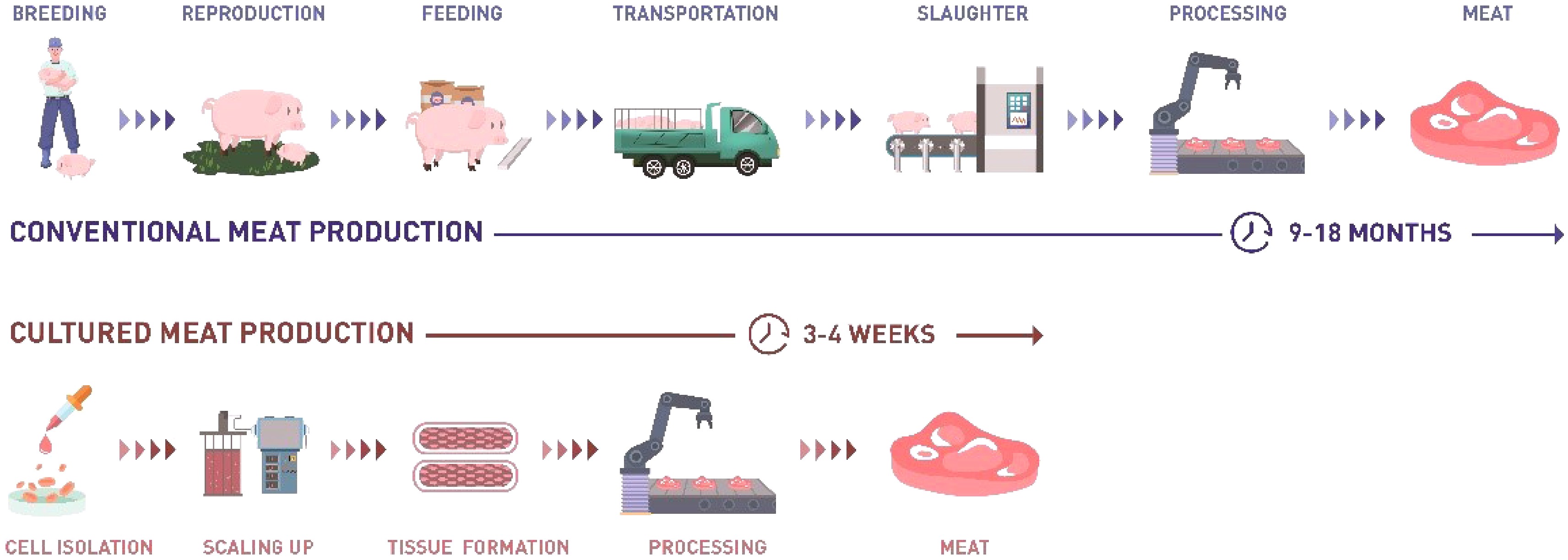
      Figure 1.  Comparison between traditional meat production and cultured meat production.

      Cultured meat uses cultured animal cells to produce meat or meat proteins and fat tissue for human consumption according to the development and regeneration principles of mammalian tissues. The idea was first proposed by British Prime Minister Winston Churchill in 1931. In August 2013, Mark J. Post revealed the world's first cultured beef burger in London, which proved the feasibility of the concept of cultured meat for the first time[3]. Since 2013, societal attention and research on cultured meat have developed rapidly, to date more than 70 startup and scale-up companies are working in this space worldwide. In December 2020, the Singapore Food Agency approved Eat Just's cultured chicken to be sold in Singapore, which is the first regulatory approval for the production and sale of cultured meat globally.

      While there has been a lot of research on technology, regulation and consumer acceptance of cultured meat, more progress is needed before cost-effective cultured meat can be produced commercially. Technically, cultured meat needs to optimize seed cells, cell media, scaffolds, and the biomanufacturing processes. At the regulatory level, although Singapore has approved cultured meat products, this approval only applies to a single product from a single species. Currently, it appears that every individual product has to be approved for each geography. How to form a relatively unified regulatory system needs further consideration.

      Finally, how to get consumers to accept and be satisfied with cultured meat is a necessary part of commercial success. In this paper, we review the methodologies of cultured meat and discuss the technologies to achieve low-cost cultured meat industrial production. We also discuss the regulation and consumer acceptance and how both accelerate commercialization.

    • Cultured meat is muscle tissue and fat tissue generated in vitro by culturing and biomanufacturing animal cells and tissues for human consumption[2]. Depending on cell types and complexity, cultured meat products fall into three main categories, including: 1) cells as an ingredient, 2) muscle and fat organoids, and 3) full thickness complex tissue (Fig. 2). The ultimate goal of cultured meat is to create full thickness complex tissue containing muscle fibers, fat tissue, blood vessels, nerves and extracellular matrix, which is currently difficult. The medium-term goal is to produce cultured meat products that contain large amounts of muscle fibers, that are the basic building blocks of muscle and contain the most important source of protein in meat, accounting for about 80%−90% of the total protein. In addition, it is highly conducive to meat-like organoleptic features to interlace the muscle fibers with fat tissue. The short-term goal of cultured meat is to produce meat-like products, containing cells and an edible scaffold. Either way, cultured meat requires functional seed cell, low-cost serum-free medium, tissue engineering and scaling up processes. The continuous research in these fields can reduce the production cost of cultured meat for large-scale production purposes.

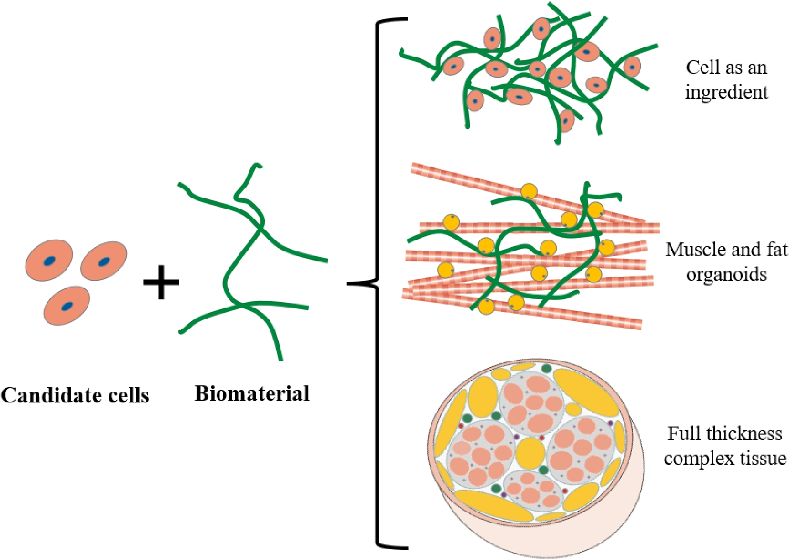
      Figure 2.  Three categories of cultured meat. 1. Direct combination of cells and biomaterials; 2. muscle and fat organoids generated by different cell types, that are combined by biomaterials; 3. complex muscle tissue, including muscle, fat, blood vessels, fibrous tissue, etc.

    • Meat proteins or fat should be the minimum necessary ingredients for cultured meat according to the three goals. Embryonic stem cells (ESC), induced pluripotent stem cells (iPSC) and mesenchymal stem cells (MSC) are pluripotent or multipotent stem cells with the ability to differentiate into muscle cells, stromal cells and fat cells, but they require precise regulation of cell fate. At present, ESC and iPSC are rarely used for cultured meat production and research whereas some startups use MSC or adipose tissue derived stem cells for fat-related products. Muscle stem cells (MuSC) are unipotent adult stem cells that contribute to muscle regeneration and form muscle fibers in vitro and therefore appear to be the most suitable cell source for skeletal muscle fiber based cultured meat. In addition to the cells mentioned above, other muscle tissue can also be consumed as meat, including smooth muscle and heart muscle. Smooth muscle tissue can be produced from smooth muscle cells, and cardiac muscle tissue can be produced from cardiomyocytes derived from embryonic stem cells. Another important protein in muscle tissue is extracellular matrix protein, accounting for about 10%−20%. The main matrix protein in meat is collagen, which can be produced by fibroblasts and smooth muscle cells, or adipose tissue derived stem cells. More research is needed on how to isolate the cell candidates from animal tissues and culture them at low cost and apply them to create different cultured meat products.

      However, primary adult stem cells have the disadvantage that they generally have limited replicative capacity compared to ESCs or iPSCs. In addition to primary adult stem cells, several research institutions and startups have therefore also used spontaneous or gene-edited immortal cell lines, or small molecule modified cells as seed cells for cultured meat production[2]. Cell lines obtained by this method are generally more replicative, which means that the efficiency of future scaling-up is higher, and the cost lower than that for primary cells. However, there are still regulatory and consumer acceptance problems of such schemes that modify cell biology, more so with genetic modification than with small molecule modification.

    • Cell culture medium contains basal culture medium and serum to provide nutrients for cell growth. However, serum of animal origin contains undefined substances and is not sustainable, which doesn’t fit with the purpose of cultured meat. Cell culture medium is one of the main factors contributing to the high cost of cultured meat. Therefore, the development of low-cost serum-free medium will become an important prerequisite for the industrialization of cultured meat. Serum-free media have been developed for ESC and MSC. It is important to develop serum-free formulations suitable for cultured meat seed cells[4].

      Serum-free medium development strategies usually involve optimizing basal media and compounds that affect specific cellular pathways, such as growth factors, hormones and small molecules. DMEM/F12 is a nutrient-rich basal medium, which has been greatly optimized in the field of biopharmaceuticals to reduce cost and be used for the growth of different cells due to its sufficient, possibly excessive, nutrient content. For cell specific pathway molecules, progress in research has led to a deep understanding of cell proliferation and differentiation, e.g. FGF signaling pathway in muscle stem cell proliferation. The rapid development of transcriptomics, proteomics and metabolomics can also help better understand the nutritional requirements and metabolic characteristics of cells, which can improve the efficiency of serum-free medium development. In addition, high-throughput screening tests and high-throughput microscopy allow rapid screening of compounds that regulate self-renewal and differentiation of cells, facilitating serum-free medium development[5]. Several biopharmaceutical media companies have a large array of serum-free formulations, and in-depth analysis can identify a range of common nutrients to help accelerate the development of serum-free media for new cell lines. Finally, the use of high-throughput workstations and rapid cell metabolism detection combined with artificial intelligence for medium optimization and prediction, can further help improve the efficiency of medium optimization.

      After the formulation is developed, the next step will be to generate low-cost food-grade alternatives of these media. Several media companies already have well-established supply chains of basic ingredients. Synthetic biology can be used to produce other functional factors at a low cost and improve the stability of culture media[6]. In addition, the use of plant and microbial hydrolysates to replace the relevant components in the cell culture medium can also contribute to low-cost food-compatible medium. Finally, if some key compounds cannot be removed or replaced by food-grade ingredients, it is necessary to trace their metabolokinetics in cell culture and seek ways to replace them by compounds with similar function.

    • Cultured meat requires a large number of cells to form cultured meat products. How to culture seed cells on a large scale is an important problem restricting the industrialization of cultured meat. The traditional two-dimensional culture method has disadvantages such as the low surface-area-to-volume-ratio, the inability to monitor culture conditions in real time and the cumbersome passaging process, which is not suitable for scaling up.

      It is necessary to consider suspension culture, either microcarrier-based, cell aggregate-based or as single cells, in suitable bioreactors. Suspension culture technologies have been developed for embryonic stem cells, mesenchymal stem cells and fibroblasts. Muscle stem cells are a special kind of stem cell due to their adhesion and cell fusion ability, so microcarrier-based suspension culture seems to be a better choice. However, lessons from biopharmaceutical applications indicate that it is difficult to achieve low-cost culture based on microcarrier suspension culture. It appears inevitable to develop suspension culture for muscle and fat precursor cells that employ inexpensive, biodegradable microcarriers[7] or dispense with microcarriers altogether. Using food-grade microcarriers could potentially realize integration of cell proliferation and differentiation in one bioreactor step. However, it is necessary to consider how to multiply cells more than 106 times on the microcarriers to obtain sufficient scaling up in a bioreactor run.

      The bioreactors used for mammalian scale-up production are mainly stirred tanks and rocking bioreactors. In future, it is necessary to design larger bioreactors to promote the large-scale production of seed cells for cultured meat.

    • In order to produce proper cultured meat for consumption, these cells need to undergo differentiation towards muscle protein forming fibers and proper fat cells or tissues. Tissue engineering techniques can be used to produce meat-related tissue. Scaffold biomaterials are the key components of cultured meat and are used for cell proliferation and differentiation to form tissues. Non-animal sources of polysaccharides, proteins and other materials can be used as scaffolds for cultured meat. The characteristics of scaffolds will also have an impact on the final product of cultured meat. Therefore, it is necessary to combine the seed cells, scaffolds and appropriate tissue engineering techniques to make the most suitable cultured meat products.

      There are two approaches for tissue engineering including bottom-up and top-down, both of which can be used to build cultured meat products. Two recent articles take both approaches respectively. Some studies and startups used a top-down approach to develop cultured meat tissue by adding cells into edible porous scaffolds[8]. In fact, using the top-down approach, it has become a trend in cultured meat field to culture cells into a structured plant scaffold to produce a hybrid culture meat/plant-based product with acceptable texture and taste. This approach can quickly implement the construction of larger muscle tissues, but it has drawbacks including poor tissue function and non-uniform cell distribution. The bottom-up approach can generate tissue by pre-mixing cells and biocompatible materials to form tissue by 3D printing[9] or structured molding. In this way, smaller tissues are generally made, which need a final assembling process in a patty or other defined meat products. Hybrid products may achieve lower-cost cultured meat products and may be easier to industrialize.

      Differentiation of muscle and fat tissue at scale requires the development of novel, dedicated bioreactors where tissues are generated in an automated fashion. Differentiation conditions for muscle and fat tissue are very different and it is easier to keep these processes separated. When complex multi-cell type tissues are cultured, one of the challenges is to define conditions where the cell types can simultaneously differentiate. The aspect of differentiation has not yet received much attention, but is of significant importance for the realization of bona fide cultured meat.

    • Cultured meat is produced in a different way to traditional meat production, so regulation needs to be considered. In December 2020, the Singapore Food Agency (SFA) became the first national regulatory authority globally to approve a cultured meat product. During the regulatory process (which took over two years), the cultured chicken meat produced by Eat Just was approved and used as an ingredient in the company's cultured chicken products. This approval is not an overall approval for cultured meat products, but only for the product and manufacturing process of Eat Just. Safety and nutrition indicators such as microbial content, amino acid composition and other indicators have been rigorously reviewed. Singapore's unique cultured meat regulatory system has also attracted more cultured meat companies to focus on Singapore as first entry into the market.

      In the United States, the Food and Drug Administration (FDA) has conducted many discussions with United States Department of Agriculture (USDA) on the regulation of cultured meat and formulated a regulatory framework (excluding seafood). The FDA will oversee cell collection/storage and all culture inputs and processes until biomass is harvested from bioreactors, and the USDA will oversee subsequent processing and labeling of meat. In the European Union, cultured meat produced without genetic modification, is regulated by the EU's Novel Foods Regulation of the European Union. The company must obtain authorization from the European Commission before its cultured meat product can enter the market. The application should consist of safety information, the identity of the product, its production process, compositional data and specifications, proposed uses, use level and anticipated intake of the product[2]. In China, cultured meat products should be regulated as 'New Food Raw Materials'. Cultured meat companies should submit applications to the National Health Commission, including raw material development reports, safety assessment reports, an analysis of the production process and other materials.

      For the regulation of cultured meat, it will be very important to have uniform safety standards that should include the identity of the product, its production process, compositional data and specifications, proposed uses, use level and anticipated intake of the product. Important safety information contains absorption, distribution, metabolism and excretion (ADME); nutritional and toxicological information and allergenicity of cultured meat product. International consensus on such safety information would greatly simplify the regulatory landscape.

    • As a novel food, the consumers' acceptance of cultured meat will determine commercial success. A considerable number of surveys have been conducted on consumers' acceptance of cultured meat, which provides ample support for the entry of cultured meat into the market. In western countries[2], when consumers receive more information about cultured meat, they are more inclined to try cultured meat products. The social benefits or the similarities to conventional meat are more appealing than the technological innovations around cultured meat. Price is also an important factor, as consumers are more willing to buy cultured meat when the price is lower. The acceptance of cultured meat is higher in men, younger people, and omnivores than in women, older people and vegetarians. Cultured meat is more attractive among American and Asian consumers than in Europe. Our lab in China also produced China’s first cultured pork and held a tasting event in June, 2020 (Fig. 3). According to a recent survey in China, knowledge of cultured meat has increased people's acceptance. More than 70% of respondents were willing to taste or purchase cultured meat[10].

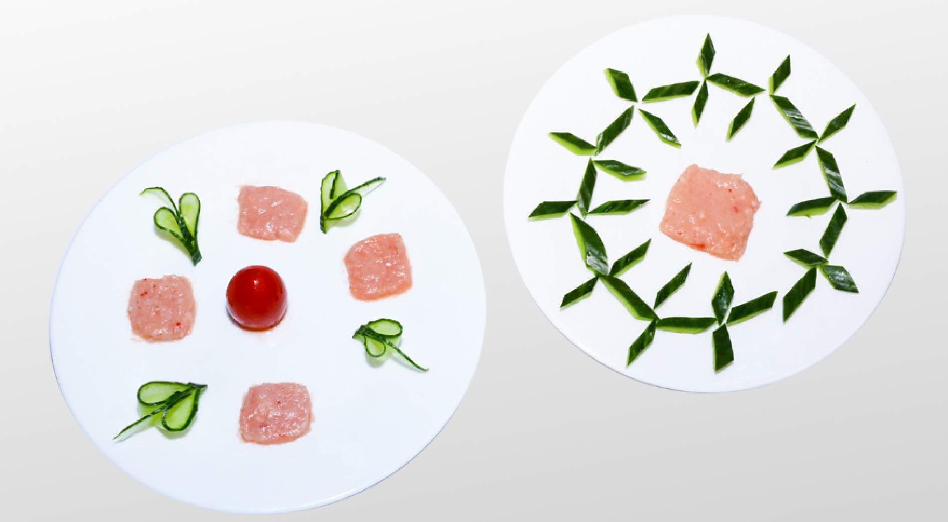
      Figure 3.  On June 6, 2020, cultured pork was tasted in Nanjing Agricultural University.

      Almost all of the current surveys are based on hypothetical cultured meat products. Because there are no commercially available cultured meat products, researchers can't observe consumer preferences in practice. However, these surveys can help cultured meat companies to develop targeted marketing strategies. The focus of future consumer acceptance studies will be in Singapore, as soon as cultured meat products become more widely available. With availability of products, the next level of consumer acceptance studies will provide more insight into the attributes of cultured meat products that are important for consumers.

    • The progress of science and technology, coupled with the attention to livestock meat production, has promoted the industrialization of cultured meat. Although cultured meat products have been approved in Singapore, low-cost cultured meat industrial production remains a challenge. Producing meat-like products, containing cells that express meat proteins within an edible, plant-based scaffold may be easier to commercialize than pure cultured meat products. The seed cells, low-cost serum-free medium, scaling up and cultured meat tissue formation still need more research and development. For the regulation of cultured meat, a uniform safety standard is essential and current consumers' acceptance survey will help cultured meat companies to develop targeted marketing strategies. We believe that the cultured meat industry will develop rapidly with the synergy of technology, regulation and consumer acceptance and bring tastier, healthier and more sustainable meat products for consumers in the future.

      • The authors declare that they have no conflict of interest.
      • Copyright: © 2021 by the author(s). Exclusive Licensee Maximum Academic Press, Fayetteville, GA. This article is an open access article distributed under Creative Commons Attribution License (CC BY 4.0), visit https://creativecommons.org/licenses/by/4.0/.
    Figure (3)  References (10)
  • About this article
    Cite this article
    Ding S, Post MJ, Zhou G. 2021. Perspectives on cultured meat. Food Materials Research 1: 3 doi: 10.48130/FMR-2021-0003
    Ding S, Post MJ, Zhou G. 2021. Perspectives on cultured meat. Food Materials Research 1: 3 doi: 10.48130/FMR-2021-0003

Catalog

  • About this article

/

DownLoad:  Full-Size Img  PowerPoint
Return
Return