ARTICLE   Open Access    

Transcription factor PagLBD21 functions as a repressor of secondary xylem development in Populus

More Information
  • During secondary growth in trees, xylem cells differentiated from cambium cells begin to synthesize secondary cell walls that are primarily composed of cellulose, hemicellulose and lignin, and are deposited between primary cell walls and plasma membranes, leading to wood formation. Identification of regulatory genes functioning during this developmental process is valuable for increasing wood production. In this study, we functionally characterized an LBD (LATERAL ORGAN BOUNDARIES DOMAIN) transcription factor PagLBD21 as a repressor of secondary xylem development in Populus. Compared to wild type plants, transgenic plants overexpressing PagLBD21 (PagLBD21OE) exhibited decreased xylem widths in cross-sections. Consistent with the functional analysis, RNA sequencing (RNA-seq) analysis revealed that genes functioning in xylem development and secondary cell wall biosynthesis pathways were significantly down-regulated in PagLBD21OE plants. We also performed DNA affinity purification followed by sequencing (DAP-seq) to identify genome-wide target genes of PagLBD21. Furthermore, we compared the RNA-seq and DAP-seq datasets of PagLBD21 and PagLBD3, and the results showed that there was a significant overlap between their target genes, suggesting these two LBD transcription factors are functionally redundant during secondary growth.
  • Drought is a major abiotic stress affecting plant growth which becomes even more intensified as water availability for irrigation is limited with current climate changes[1]. Timely detection and identification of drought symptoms are critically important to develop efficient and water-saving irrigation programs and drought-tolerance turfgrasses. However, turfgrass assessments of stress damages have been mainly using the visual rating of turf quality which is subjective in nature and inclined to individual differences in light perception that drives inconsistency in estimating color, texture, and pattern of stress symptoms in grass species[24]. Remote sensing with appropriate imaging technology provides an objective, consistent, and rapid method of detecting and monitoring drought stress in large-scale turfgrass areas, which can be useful for developing precision irrigation programs and high-throughput phenotyping of drought-tolerance species and cultivars in breeding selection[5].

    Spectral reflectance and chlorophyll fluorescence imaging are emerging tools for rapid and non-destructive monitoring of drought effects in crops. These tools combine imaging and spectroscopy modalities to rigorously dissect the structural and physiological status of plants[6,7]. Spectral reflectance imaging captures reflected light (one out of three fates of light: reflect, absorb and transmit when striking leaf) at different wavelengths ranging from visible to near-infrared regions to characterize vegetation traits[8,9]. Within spectral reflectance imaging, multispectral imaging on one hand measures reflected light in three to ten different broad spectral bands in individual pixels[10,11]. Hyperspectral imaging on the other hand captures reflected light in narrow and more than 200 contiguous spectral bands. Some absorbed light by leaf is re-radiated back in the form of fluorescence and fluorescence imaging utilizes those lights in red and far-red regions to capture plant physiological status[12]. When drought progresses, plants start to develop various symptoms (physiological modifications) gradually over time[13]. Some of those symptoms include stomata closure, impediment in gas exchange, change in pigment composition and distribution which result in wilting and associated morphological alteration in leaf color (senescence), shape (leaf curling) and overall plant architecture. As different plant components or properties reflect light differently at different wavelengths and patterns of reflectance and fluorescence change along with plant stress and related symptoms development, spectral reflectance and fluorescence imaging provide accurate, reliable and detailed information for crop drought monitoring. Fluorescence imaging primarily based on fluorescing plant components or chlorophyll complex in photosynthetic antenna and reaction centers and therefore it mainly monitors stress development by tracking changes in overall photosynthetic performance or other metabolism that interfere with photosynthetic operation[9,14]. Multispectral imaging, hyperspectral imaging, or chlorophyll fluorescence has been used in different studies for plant responses to drought stress in various plant species[10,1517]. The comparative approach of multiple imaging technologies could help to find the efficient methods for the evaluation of plant responses and tolerance to drought[18].

    Vegetation indices derived from multispectral or hyperspectral imaging and fluorescence parameters typically are ratio or linear combinations of reflectance and fluorescence emissions from leaves or canopy of plants, respectively[19,20]. Canopy reflectance at different wavelengths and chlorophyll fluorescence varies with canopy color and density and changes with environmental conditions that affect plant growth, including drought stress[14,20,21]. These variations in reflectance and fluorescence are captured by vegetation indices, such as normalized difference vegetation index (NDVI) and fluorescence parameters including the ratio of variable fluorescence to maximum fluorescence (Fv/m) which are commonly used to evaluate environmental impact on plant growth. Other indices reflect physiological health of plants, such as photochemical reflectance index (PRI) has recently been reported to be useful for drought stress assessment in crops[19]. Previous research identified varying sensitivity of PRI and NDVI to detect water stress; for example, Sun et al.[22] found PRI to be a prominent indicator of drought stress whereas Kim et al.[20] discovered NDVI had greater correlation with drought stress development. There are also several conflicting findings on the responsiveness of fluorescence parameters to drought stress. Photochemical efficiency of PSII (Fv/Fm) was found to be greatly related to drought stress by Panigada et al.[23] but Jansen et al.[24] reported Fv/Fm to be relatively insensitive to drought progression. Lu & Zhang[25] identified that coefficient of photochemical quenching (qP) was insensitive to drought stress whereas Moustakas et al.[26] reported that (qP) being the most sensitive indicator of such stress conditions. There is a need for a comprehensive study that examines multiple vegetation indices (both hyperspectral and multispectral indices) and fluorescence parameters, and parallelly assess their sensitivities to reflect plant growth and physiological status during drought stress.

    The objectives of the current study were: (1) to perform comparative analysis of drought responses of vegetation and photosynthetic indices using multispectral, hyperspectral and chlorophyll fluorescence imaging for Kentucky bluegrass (Poa pratensis L.), a cool-season perennial grass species widely used as turfgrass; (2) identify major vegetation and photosynthetic indices from the imaging technologies and correlated to visual turf quality and leaf relative water content from the destructive measurement; and (3) determine the major vegetation and photosynthetic indices that are most responsive or sensitive to the progression of drought stress that may be useful to early detection and monitoring the level of drought stress causing growth and physiological damages in cool-season grass species.

    Sod strips of Kentucky bluegrass cultivar 'Dauntless' were collected from established field plots at the Rutgers Plant Science Research and Extension Farm, Adelphia, NJ, USA. Sods were planted in plastic pots of 18 cm diameter and 20 cm length filled with a mixture of soil (sandy loam, semi-active, mesic Typic Hapludult; pH 6.55; 260 kg·P·ha−1, 300 kg·K·ha−1) and sand in the ratio of 2/1 (v/v). Plants were established for 50-d in a greenhouse with 24/22 °C day/night average temperatures, 12-h average photoperiod and 750 μmol·m−2·s−1 average photosynthetically active radiation (PAR) with natural sunlight and supplemental lightings. Plants were well-watered, trimmed weekly to 100 mm and fertilized weekly with a 24–3.5–10 (N–P–K) fertilizer (Scotts Miracle-Gro) at the rate of 2.6 g·N·m−2 during the establishment period in the greenhouse. Once plants were well-established, they were moved to the controlled environmental growth chamber (GC72, Environmental Growth Chambers, Chagrin Falls, OH, USA). The growth chamber was controlled at 22/18 °C day/night temperature, 60% relative humidity, 12-h photoperiod and 650 μmol·m−2·s−1 PAR at the canopy level. Plants were allowed to acclimate for a week within the growth chamber conditions and then treatments were initiated.

    There were two different treatments: well-watered control and drought stress. For the well-watered control, plants were irrigated once every two days with sufficient water until drainage occurred from the pot bottom or when soil water content reached the field capacity. Drought stress was imposed by withholding irrigation from each pot throughout the experiment period. Each treatment had five replicates. The experimental treatments were arranged as a complete randomized design with plants of both treatments randomly placed and relocated in the chamber twice each week to minimize effects of potential microenvironment variations in the growth chamber.

    A time-domain reflectometry system (Model 6050 × 1; Soil Moisture Equipment, Santa Barbara, CA, USA) installed with 20 cm soil moisture probe was used to measure soil volumetric water content. Volumetric water content was measured every two days in each pot to track soil moisture dynamics in control and drought stress treatments. To assess plant responses at different soil moisture levels, turfgrass quality (TQ) and leaf relative water content (RWC) were evaluated. Turfgrass quality was visually rated on a scale of 1-9 depending upon canopy color, uniformity and density[27]. A rating of 1 indicates discolored and completely dead plants, 9 indicates lush green colored healthy plants and 6 indicates the minimum acceptable turfgrass quality. Leaf RWC was measured by soaking 0.2 g fresh leaves in distilled water overnight at 4 °C[28]. Turgid leaves after overnight soaking were oven dried at 70 °C to a constant dry weight. Leaf RWC was calculated as [(fresh weight – dry weight)/ (turgid weight – dry weight)] × 100.

    Control and drought stress pots were scanned using a close-range benchtop hyperspectral imaging system (Resonon Inc., Bozeman, MT, USA) containing Pika XC2 camera equipped with 23 mm lens. This camera took images in spectral range of 400–1,000 nm with much detailed spectral resolution of 1.9 nm in 447 different spectral channels. The camera provided 1600 spatial pixels and maximum frame rate of 165 frames per second. It had 23.1° field of view and 0.52 milli-radians instantaneous field of view. Resonon hyperspectral imaging systems are line-scan imagers (also referred to as push-broom imagers) that collect spectral data from each pixel on one line at a time. Multiple lines are imaged when an object or pot kept in scanning stage of linear stage assembly underneath the camera is moved by a stage motor. Those line images are assembled to form a complete image. The systems had regulated lights placed above the linear stage assembly to create optimal conditions for performing the scans. Lights were at the same level as the lens on a parallel plane. Distance between lens and the top of grass canopy was maintained at 0.4 m for capturing the best representation of drought progression. All scans were performed using spectronon pro (Resonon Inc., Bozeman, MT, USA) software connected to the camera using a USB cable. Before performing a scan, the lens was appropriately focused, dark current noise was removed and the system was calibrated for reflectance measurement using a white tile provided by the manufacturer. To ensure distortion-free hyperspectral datacube with a unit-aspect-ratio image, benchtop system's swatch settings were adjusted using pixel aspect ratio calibration sheet also provided by the manufacturer. Once the system was ready, controlled- and stressed-pots were scanned individually every two days throughout the experiment. As the lens was focused centrally, obtained images were of the central grass area and were processed using spectronon pro data analysis software. The entire grass image was selected using a selection tool and the spectrum was generated. From each spectrum, vegetation indices were calculated either using built-in plugins or by manually creating algorithms. The list of vegetation indices calculated using image analysis is mentioned in Table 1.

    Table 1.  List of vegetation indices calculated using hyperspectral and multispectral image analysis for drought stress monitoring in Kentucky bluegrass. Name and number in subscript following the letter R in each formula represent the reflectance at individual light and particular wavelength.
    Vegetation indexIndex abbreviation and formula
    Hyperspectral analysisMultispectral analysis
    Structure Independent Pigment IndexSIPI = (R800 – R445) / (R800 + R680)SIPIm = (RNIR840 – RBlue444) / (RNIR840 + RRed668)
    Simple Ratio IndexSRI = R800 / R675SRIm = RNIR840 / RRed668
    Plant Senescence Reflectance IndexPSRI = (R680 –R500) / R750PSRIm = (RRed668 – RBlue475) / RRededge740
    Photochemical Reflectance IndexPRI = (R570 – R531) / (R570 + R531)PRI = (RGreen560 – RGreen531) / (RGreen560 + RGreen531)
    Normalized Difference Vegetation IndexNDVI = (R800 – R680) / (R800 + R680)NDVIm = (RNIR840 – RRed668) / (RNIR840 + RRed668)
    Normalized Difference Red EdgeNDRE = (R750 – R705) / (R750 + R705)NDREm = (RRededge717 – RRed668) / (RRededge717 + RRed668)
     | Show Table
    DownLoad: CSV

    Micasense Rededge-MX dual camera system (AgEagle Sensor Systems Inc., Wichita, KS, USA) was used to collect multispectral images of controlled- and drought stressed-pots placed within a light box (1.2 m × 0.6 m × 0.6 m). The multispectral camera system had 1,280 × 960 resolution, 47.2° field of view and 5.4 mm focal length. The camera captured ten different spectral bands simultaneously on a command (Table 2). To allow the multispectral camera system, which was designed for aerial operation, to work in the light box settings, a downwelling light sensor (DLS) module provided by the manufacturer was installed to the camera system. Images were captured manually through WIFI connection from mobile devices or computer to the multispectral camera system. The sensor layout of the dual camera system, while causing negligible error in aerial condition, led to mismatching between spectral bands in a close distance, therefore, spectral bands needed to be overlapped during post-processing. The captured images of individual spectral bands were stored as separate .jpgf image files and then were used to calculate the relevant vegetation indices. Multispectral image analysis was executed using Python (Version 3.10) code by Rublee et al.[29]. Image analysis aligned ten spectral bands using Oriented FAST and Rotated BRIEF algorithm to achieve complete overlap between spectral band images. The reflectance correction panel provided by the manufacturer was used to account for the illumination condition in light box environment and the correction was reflected in pixel value adjustment for each band in python code; vegetation indices based on the aligned images were then calculated using the corresponding formula (Table 1). Images that included background noise were excluded from analysis.

    Table 2.  Spectral band details (center wavelength and band width) for Micasense Rededge-MX dual camera system.
    Band nameCentral wavelength (nm)Band width (nm)
    Blue44444428
    Blue47547532
    Green53153114
    Green56056027
    Red65065016
    Red66866814
    RE70570510
    RE71771712
    RE74074018
    NIR84084257
     | Show Table
    DownLoad: CSV

    Chlorophyll fluorescence images were taken using a pulse amplitude modulated fluorescence imaging system (FC 800-O/1010, Photon System Instruments, Drasov, Czech Republic). A high-speed charge-coupled device (CCD) camera was mounted on a robotic arm placed in the middle of LED light panels. The camera had 720 × 560 pixels spatial resolution, 50 frames per second frame rate and 12-bit depth. Four different LED light panels each of 20 cm × 20 cm size were equipped with 64 orange-red (617 nm) LEDs in three panels and 64 cool-white LEDs (6,500 k) in the rest of one panel. Before making measurements, plants were dark-adapted for 25 min in a dark room to open all PSII reaction centers. The distance between camera and the top of the grass canopy was maintained at 0.3 m while taking images to ensure optimum quality. Images were acquired following the Kautsky effect measured in a pulse amplitude modulated mode[30,31]. Briefly, dark-adapted plants were first exposed to non-actinic measuring light for 5 s to measure minimum fluorescence at the dark-adapted state (Fo). Plants were immediately exposed to 800 ms saturation pulse of 3,350 µmol·m−2·s−1 to measure maximum fluorescence after dark adaptation (Fm). They were kept under dark relaxation for 17 s and then exposed to actinic light 750 µmol·m−2·s−1 for 70 s. Plants were exposed to a series of saturating pulses at 8 s, 18 s, 28 s, 48 s and 68 s during their exposure to actinic light conditions and maximum fluorescence at different light levels and steady state were measured. They were kept under dark relaxation again for 100 s and irradiated with saturating pulses at 28 s, 58 s and 88 s during dark relaxation for measuring maximum fluorescence during the relaxation. Selected durations for each light and dark relaxation state were preset in default quenching-act2 protocol of the fluorescence imaging system. Fluorescence at different light levels and steady states were used to calculate several fluorescence parameters (Table 3).

    Table 3.  Chlorophyll fluorescence parameters calculated from pulse amplitude modulated fluorescence imaging system.
    Chlorophyll fluorescence parameterFormula
    Maximum photochemical efficiency of PSII (Fv / Fm)(Fm-Fo) / Fm
    Photochemical efficiency of open PSII centers
    (F'v / F'm)
    (F'm – F'o) / F'm
    Actual photochemical quantum yield of PSII centers Y(PSII)(F'm – Fs) / F'm
    Photochemical quenching coefficient (Puddle model; qP)(F'm – Fs) / (F'm – F'o)
    Photochemical quenching coefficient (Lake model; qL)qP × F'o / Fs
    Non-photochemical quenching coefficient (qN)(Fm-F'm) / Fm
    Non-photochemical quenching (NPQ)(Fm-F'm) / F'm
    Chlorophyll fluorescence decrease ratio (Rfd)(Fm-Fs) / Fs
     | Show Table
    DownLoad: CSV

    The two-way repeated measure analysis of variance was performed to determine treatment effects and t-test was performed to compare control and drought stress treatments at a given day of measurement. Correlation analysis using all individual observations (five replications for each control and drought stress treatments) was performed to determine the relationship among all measured traits, vegetation indices and fluorescence parameters. Partial least square regression (PLSR) models were developed in SAS JMP (version 13.2; SAS Institute, Cary, NC, USA) for comparing hyperspectral, multispectral and chlorophyll fluorescence imaging in their overall associations with physiological assessments of drought stress. Vegetation indices and fluorescence parameters from individual imaging technologies were predictor variables, and turfgrass quality and leaf relative water content were response variables. A leave one out cross validation approach was used to develop the best performing partial least square model for each imaging technology. A model was first established with all predictor variables and the variable with the lowest importance was removed from the dataset and the model was rebuilt with the remaining variables. The rebuilt model was re-validated using leave one out cross validation and assessed checking root mean PRESS and percent variation explained for cumulative Y values. From each loop of operation, one variable was removed, and a new model was developed. The whole process ended when the last variable was removed and thus no more models could be developed. Finally, a series of models was obtained, and they were compared to identify a model with the highest accuracy for individual imaging technologies. The best performing model from each imaging technology was used to estimate turfgrass quality and leaf relative water content.

    The initial soil water content prior to drought stress was maintained at the field capacity of 29% and remained at this level in the well-watered control treatment during the entire experimental period (20 d) (Fig. 1a). SWC in the drought treatment significantly decreased to below the well-watered treatment, beginning at 4 d, and declined to 5.8% by 20 d.

    Figure 1.  Drought stress affected turf quality, leaf relative water content and soil volumetric water content during 20 d of stress period in Kentucky bluegrass. * indicates significant difference between control and drought stress treatments (p ≤ 0.05) at each day of measurement. Presented values represent average of five data points.

    Leaf RWC was ≥ 93% in all plants prior to drought stress and declined to a significantly lower level than that of the control plants, beginning at 10 d of treatment when SWC declined to 16% (Fig. 1b). TQ began to decrease to a significantly lower level than the that of the well-watered plants at 10 d of drought stress at RWC of 87% and SWC of 16%, and further declined to the minimally acceptable level of 6.0 at 16 d of drought stress when RWC decreased to 66% and SWC dropped to 8% during drought stress (Fig. 1c).

    Most hyperspectral imaging indices, including SIPI (Fig. 2a), SRI (Fig. 2b), PRI (Fig. 2d), NDVI (Fig. 2e) and NDRE (Fig. 2f) exhibited a declining trend during 20-d drought stress while PSRI (Fig. 2C) showed increases during drought stress. The index value of drought-stressed plants became significantly lower than that of the well-watered plants, beginning at 14 d for SIPI and SRI, 16 d for PRI and PSRI, and 18 d for NDVI and NDRE. The multispectral SIPIm and SRIm did not differ between drought-stressed plants from the control plants until 18 d of treatment (Fig. 3a, b) while NDVIm, NDREm , PRIm , and PSRIm values were significantly lower than those of well-watered control plants at 16 d of drought stress (Fig. 3cf).

    Figure 2.  Vegetation indices generated by hyperspectral sensing and sensitivity of these indices in monitoring drought in Kentucky bluegrass exposed to 20 d of drought stress. * indicates significant difference between control and drought stress treatments (p ≤ 0.05) at each day of measurement. Presented values represent average of five data points.
    Figure 3.  Vegetation indices generated by multispectral image analysis and sensitivity of these indices in monitoring drought in Kentucky bluegrass exposed to 20 d of drought tress. * indicates significant difference between control and drought stress treatments (p ≤ 0.05) at each day of measurement. Presented values represent average of five data points.

    Chlorophyll fluorescence indices detected drought damages in leaf photosynthesis systems, as shown by declines in different indices during drought stress (Fig. 4). Drought-stressed plants exhibited significant lower chlorophyll fluorescence levels than that of the well-watered plants, beginning at 12 d for NPQ (Fig. 4a), 16 d for Fv/Fm (Fig. 4b), and 18 d for F'V/F'm (Fig. 4c), Y(PSII) (Fig. 4d), qP (Fig. 4e), and qL (Fig. 4f). Separation between drought-stressed and well-watered plants were also evident in index- or parameter- generated images (Fig. 5).

    Figure 4.  Chlorophyll fluorescence parameters measured by pulse amplitude modulated fluorescence imaging system and detection of drought by these parameters in Kentucky bluegrass exposed to 20 d of drought stress. * indicates significant difference between control and drought stress treatments (p ≤ 0.05) at each day of measurement. Presented values represent average of five data points. NPQ, Non-photochemical quenching; Fv /Fm, Maximum photochemical efficiency of PSII; F'v/F'm, Photochemical efficiency of open PSII centers; Y(PSII), Actual photochemical quantum yield of PSII centers; qP, Photochemical quenching coefficient (Puddle model); qL, Photochemical quenching coefficient (Lake model); qN, Non-photochemical quenching coefficient; Rfd, Chlorophyll fluorescence decrease ratio.
    Figure 5.  Maps generated by the three most drought sensitive indices and parameters [hyperspectral structure independent pigment index (SIPI), multispectral normalized difference vegetation index (NDVIm) and chlorophyll fluorescence NPQ]. These maps clearly separated control and drought stress after 18 d of treatment when majorities of indices and parameters detected drought stress.

    Leaf RWC and TQ had significant correlation with most of indices and parameters calculated using three different imaging sensors (hyperspectral, multispectral and chlorophyll fluorescence) (Table 4). Among the indices, RWC had the strongest correlations with chlorophyll fluorescence NPQ (r = 0.88) and qL (r = 0.89), hyperspectral PRI (r = 0.94), and multispectral PSRIm (−0.92). TQ was most correlated to chlorophyll fluorescence NPQ (r = 0.89), hyperspectral PSRI (r = −0.90), and multispectral PSRIm (r = −0.85).

    Table 4.  Correlations among several physiological traits, vegetation indices and chlorophyll fluorescence parameters.
    RWCTQFV/FmF'v/F'mY(PSII)NPQqNqPqLRfdSIPISRIPSRIPRINDVINDREWBISIPImPSRImPRImNDVImNDREm
    RWC1.00
    TQ0.95*1.00
    FV/Fm0.87*0.85*1.00
    F'v/F'm0.81*0.77*0.95*1.00
    Y(PSII)0.85*0.74*0.80*0.74*1.00
    NPQ0.88*0.89*0.95*0.84*0.75*1.00
    qN0.84*0.83*0.96*0.84*0.77*0.96*1.00
    qP0.82*0.70*0.73*0.66*0.99*0.69*0.72*1.00
    qL0.89*0.81*0.90*0.86*0.97*0.83*0.86*0.95*1.00
    Rfd0.84*0.82*0.89*0.83*0.77*0.92*0.86*0.72*0.83*1.00
    SIPI0.84*0.71*0.63*0.58*0.51*0.57*0.69*0.48*0.60*0.46*1.00
    SRI0.57*0.62*0.44*0.45*0.330.41*0.45*0.300.400.330.83*1.00
    PSRI−0.83*−0.90*−0.90*−0.86*−0.76*−0.83*−0.87*−0.71*−0.86*−0.76*−0.75*−0.57*1.00
    PRI0.94*0.82*0.80*0.76*0.71*0.79*0.71*0.66*0.77*0.78*0.260.17−0.78*1.00
    NDVI0.53*0.65*0.41*0.43*0.41*0.42*0.400.380.43*0.42*0.50*0.42*−0.54*0.311.00
    NDRE0.64*0.73*0.64*0.63*0.45*0.54*0.64*0.400.56*0.44*0.92*0.85*−0.75*0.330.50*1.00
    SIPIm0.52*0.50*0.56*0.58*0.47*0.52*0.49*0.43*0.52*0.51*0.330.28−0.58*0.61*0.270.39−0.281.00
    PSRIm−0.92*−0.85*−0.85*−0.85*−0.83*−0.80*−0.77*−0.79*−0.88*−0.77*−0.40−0.230.77*−0.82*−0.41−0.400.32−0.52*1.00
    PRIm0.20−0.030.06−0.010.280.140.110.310.200.180.050.100.01−0.040.000.090.060.09−0.041.00
    NDVIm0.75*0.74*0.77*0.78*0.67*0.72*0.68*0.62*0.73*0.70*0.43*0.33−0.76*0.81*0.370.47*−0.350.93*−0.76*−0.051.00
    NDREm0.90*0.89*0.89*0.89*0.81*0.83*0.81*0.76*0.88*0.81*0.52*0.41*−0.87*0.87*0.45*0.53*−0.320.62*−0.87*−0.040.85*1.00
    Details for individual abbreviations of vegetation indices and fluorescence parameters used in this table were previously mentioned in Tables 1 & 3. Some other abbreviations are: RWC, leaf relative water content; and TQ, turfgrass quality. Values followed by * indicate significant correlation at p ≤ 0.05. Correlation analysis was performed using all individual data points (five replications for each control and drought stress treatments).
     | Show Table
    DownLoad: CSV

    Correlations among different vegetation indices and parameters were also significant in many cases. Hyperspectral indices such as PSRI and PRI were significantly correlated with all multispectral indices except PRIm. Multispectral NDVIm and NDREm were significantly correlated with all hyperspectral indices. When hyperspectral and multispectral indices were correlated with chlorophyll fluorescence parameters, majorities of these indices significantly associated with fluorescence parameters with exceptions of multispectral PRIm which had weak and positive (r ranges 0.06 to 0.31) associations with fluorescence parameters.

    Partial least square regression models were developed by integrating all indices from individual imaging technologies which identified the most reliable imaging systems to detect and monitor plant responses to drought stress. The PLSR model developed using hyperspectral imaging indices had improved predictability (root mean PRESS ≤ 0.38 and percent variation explained ≥ 87) compared to such models developed using other imaging systems and associated indices (Table 5). Comparing multispectral imaging with chlorophyll fluorescence imaging, multispectral imaging had slightly better predictability [root mean PRESS = 0.40 (RWC) and 0.44 (TQ) and percent variation explained = 86 (RWC) and 83 (TQ)] considering similar number of predictor variables used for estimating TQ and RWC in all imaging systems.

    Table 5.  Summary of partial least square model showing predictability of individual models using specific numbers of predictor variables (identified by leave one out cross validation) generated by different sensing technologies. Details of individual abbreviations are mentioned in previous tables. Partial least square was performed using all individual data points (five replications for each control and drought stress treatments).
    Sensing technology used for predictionPredicted
    variable
    No. of predictors usedPredictor variablesRoot mean
    PRESS
    Percent variation explained
    for cumulative Y
    Cumulative Q2
    HyperspectralTQ4PRI, PSRI, NDRE, SIPI0.36870.99
    RWC4PRI, PSRI, NDRE, SIPI0.38890.99
    MultispectralTQ3PSRIm, NDVIm, NDREm0.44850.97
    RWC3PSRIm, NDVIm, NDREm0.40860.97
    Chlorophyll fluorescenceTQ4Fv/Fm, NPQ, qN, qL0.46830.95
    RWC3Fv/Fm, NPQ, qL0.59840.93
     | Show Table
    DownLoad: CSV

    The integrated indices from each of the three imaging systems were highly correlated to TQ, with R2 of 0.90, 0.85, and 0.83 for hyperspectral imaging, multispectral imaging, and chlorophyll fluorescence, respectively (Fig. 6). For RWC, the correlation R2 was 0.88, 0.84, and 0.80, respectively with hyperspectral imaging, multispectral imaging, and chlorophyll fluorescence. The hyperspectral imaging was better be able to predict TQ and RWC compared to other imaging systems (Fig. 6).

    Figure 6.  Comparison of predicted turfgrass quality (TQ) and leaf relative water content (RWC) versus their measured values using partial least square regression model. Turfgrass quality and relative water contents were predicted using various indices generated by hyperspectral, multispectral and chlorophyll fluorescence sensing technologies. The dashed line represents the I:I line. Regression analysis was performed using all individual data points (five replications for each control and drought stress treatments).

    Leaf RWC and TQ are the two most widely used parameters or traits to evaluate turfgrass responses to drought stress[28,32,33]. In this study, RWC detected water deficit in leaves at 10 d of drought stress when SWC declined to 16% and TQ declined to below the minimal acceptable level of 6.0 at 16 d of drought stress when RWC decreased to 66% and SWC dropped to 8% during drought stress. These results suggested that RWC was a sensitive trait to detect water stress in plants, which is in agreement with previous research[34,35]. However, leaf RWC is a destructive measurement and TQ is a subjective estimate. Nondestructive and quantitative detection of stress symptoms in plants through assessing changes in phenotypic and physiological responses of plants to drought stress is critical for developing water-saving irrigation programs and breeding selection traits to increase water use efficiency and improve plant tolerance to drought stress. In this study, some of the phenotypic traits assessed by hyperspectral and multispectral imaging analysis and photosynthetic parameters measured by chlorophyll fluorescence were highly correlated to leaf RWC or visual TQ, as discussed in detail below, which could be used as non-destructive indicators or predictors for the level of drought stress in Kentucky bluegrass and other cool-season turfgrass species.

    The strong correlation of integrated indices from each of the three imaging systems with TQ (R2 of 0.90, 0.85, and 0.83, respectively) and RWC (R2 of 0.88, 0.84, and 0.80, respectively) for hyperspectral imaging, multispectral imaging, and chlorophyll fluorescence suggested that all three non-destructive imaging systems could be used as a non-destructive technique to detect and monitor water stress in Kentucky bluegrass. However, the hyperspectral imaging indices had higher predictability to RWC and visual TQ compared to the indices from multispectral imaging and chlorophyll fluorescence based on the PLSR models. Hyperspectral imaging used in this study captured images in 447 different spectral bands and gathered much more details about individual components of entire vegetation as each component has its own spectral signature. Multispectral imaging captures images with ten spectral bands and chlorophyll fluorescence imaging used only emitted red and far-red lights to snap images. Nevertheless, our results suggested that the PLSR models by integrating all indices from each individual imaging technologies identified the most reliable imaging systems to detect and monitor plant responses to drought stress in this study.

    The indices derived from the three imaging systems varied in their correlation to RWC or TQ in Kentucky bluegrass in this study. Among the indices, RWC had the strongest correlations with chlorophyll fluorescence NPQ (r = 0.88) and qL (r = 0.89), hyperspectral PRI (r = 0.94), and multispectral PSRIm (r = −0.92). TQ was most correlated to chlorophyll fluorescence NPQ (r = 0.89), hyperspectral PSRI (r = −0.90), and multispectral PSRIm (r = −0.85). The indices also varied in their sensitivity to drought stress for Kentucky bluegrass, and therefore they detected drought stress in plants at different times of treatment. The hyperspectral SIPI and SRI were the most responsive to drought stress with significant decline at 14 d followed by PRI and PSRI at 16 d while NDVI and NDRE were slowest showing decline (18 d) in response to drought. Multispectral indices exhibited decline later during drought at 16 d of drought stress for NDVIm, NDREm , PRIm , and PSRIm and 18 d for SIPIm and SRIm. Indices SIPI and SRI are related to leaf carotenoid composition and vegetation density and high spectral resolution of hyperspectral system was able to capture subtle changes in pigment concentration and canopy (slight leaf shrinking and rolling) at early phase of drought progression[36,37]. Index PSRI is indicative of the ratio of bulk carotenoids including α- and β-carotenes to chlorophylls and PRI is sensitive to xanthophyll cycle particularly de-epoxidation of zeaxanthin that releases excess energy as heat in order to photoprotection[3840]. Activation of photoprotective mechanisms including xanthophyl cycle require a certain level of stress severity depending on type of abiotic stress and plant species[41]. The PSRI calculated using both hyperspectral and multispectral imaging systems exhibited similar trends, and PSRI and PRI from either imaging system detected drought stress after 16 days of treatment applications. In agreement with our results, Das & Seshasai[42] found that PSRI showed similar trends when its value > −0.2 regardless of whether measured using hyperspectral or multispectral imaging. Both PSRI and PRI were also highly correlated to leaf RWC or TQ in Kentucky bluegrass exposed to drought stress in this study, suggesting that these two indices could be useful parameters to detect and monitor plant responses to drought stress.

    Vegetation index of NDVI has been the most widely used vegetation index in several crops such as wheat (Triticum aestivum L.)[43], cool- and warm-season turfgrass species including perennial ryegrass (Lolium perenne L.), tall fescue (Festuca arundinacea Schreb.), seashore paspalum (Paspalum vaginatum Sw.) and hybrid bermudagrass [Cynodon dactylon (L.) Pers. × C. transvaalensis Burtt-Davy][2, 44, 45]. For example, Bhandari et al.[43] and Badzmierowski et al.[14] found NDVI was correlated to overall turfgrass quality and chlorophyll content under nitrogen and drought stresses in tall fescue and citrus (Citrus spp.) plants. In this study, NDVI and NDRE were also correlated to leaf RWC and TQ, both NDVI and NDRE calculated from hyperspectral or multispectral imaging were least responsive to drought stress or detected drought stress later than other indices. Hong et al.[46] reported that NDVI being a better indicator than NDRE for early drought stress detection in turfgrasses when these indices were measured by handheld multispectral sensor. Eitel et al.[47] utilized broadband satellite images to estimate NDVI and NDRE and identified NDRE being a better option for early detection of stress condition in woodland area. Either NDVI or NDRE could be used as indices for vegetation density, but not sensitive indicators for plant responses to drought stress or for detecting drought damages in plants.

    Chlorophyll fluorescence reflects the integrity and functionality of photosystems in the light reactions of photosynthesis and serves as a good indicator for photochemical activity and efficiency[48]. The Y(PSII) is an effective quantum yield of photochemical energy conversion and estimates the actual proportion of absorbed light that is used for electron transport[49]. The ratio of F'v/F'm is maximum proportion of absorbed light that can be used for electron transport when all possible PSII reaction centers are open under light adapted state. Parameters qP and qL estimate the fraction of open PSII centers based on 'puddle' and 'lake or connected unit' models of photosynthetic antenna complex, respectively[50]. Rfd is an indicator for photosynthetic quantum conversion associated with functionality of the photosynthetic core unit. Overall, these parameters revolve around the operation status and functioning of PSII reaction centers or the core unit that possesses chlorophyll a-P680 in a matrix of proteins[51]. Parameter NPQ indicates non-photochemical quenching of fluorescence via heat dissipation involving xanthophyll cycle and state transition of photosystems[52]. This parameter is mostly associated with xanthophylls and other pigments in light harvesting antenna complex of photosystems but not with the PSII core unit[53]. Li et al.[9] reported that chlorophyll fluorescence imaging parameters including F'V/F'm have a limitation of late drought detection in plants. Shin et al.[54] reported F'V/F'm, Y(PSII), qP, and qL detected stress effects under severe drought when leaves were completely wilted and fresh weights declined in lettuce (Lactuca sativa L.) seedings. In this study, NPQ and Fv/Fm exhibited significant decline earlier (12−16 d of stress treatment) when drought was in mild to moderate level (> 60% leaf water content) compared to other chlorophyll fluorescence indices. The NPQ was strongly correlated to leaf RWC (r = 0.88) and TQ (r = 0.89) for Kentucky bluegrass exposed to drought stress. These results suggested that NPQ is a sensitive indicator of photosynthetic responses to drought stress and could be a useful parameter for evaluating plant tolerance to drought stress and monitoring drought responses.

    The comparative analysis of phenotypic and photosynthetic responses to drought stress using three imaging technologies (hyperspectral, multispectral and chlorophyll fluorescence) using the partial least square modeling demonstrated that the integrated vegetation indices from hyperspectral imaging had higher predictability for detecting turfgrass responses to drought stress relative to those from multispectral imaging and chlorophyll fluorescence. Among individual indices, SIPI and SRI from hyperspectral imaging were able to detect drought stress sooner than others while PSRI and PRI from both hyperspectral and multispectral imaging were also highly correlated to leaf RWC or TQ responses to drought stress, suggesting these indices could be useful parameters to detect and monitor drought stress in cool-season turfgrass. While NDVI or NDRE from both hyperspectral and multispectral imaging could be used as indices for vegetation density, but not sensitive indicators for plant responses to drought stress. Among chlorophyll fluorescence indices, NPQ and Fv/Fm were more closely correlated to RWC or TQ while NPQ was most responsive to drought stress, and therefore NPQ could be a useful indicator for detecting and monitoring cool-season turfgrass response to drought stress. The sensitivity and effectiveness of these indices associated with drought responses in this study could be further testified in other cool-season and warm-season turfgrass species under field conditions. As each imaging technology used in this experiment comes with bulky accessories such as LED panels, mounting tower and support system, capturing images within limited space of controlled environmental chambers are challenging. Future research should be in developing multimodal imaging integrating major features of all three technologies and reducing size and space requirement that would deliver improved decision support for drought monitoring and irrigation management in turfgrasses. Development of advanced algorithms that could incorporate broader spectral details or band reflectance for calculating novel vegetation indices are warranted.

    The research presented in this paper was funded by the United State Department of Agriculture - National Institute of Food and Agriculture (2021-51181-35855).

  • The authors declare that they have no conflict of interest. Bingru Huang is the Editorial Board member of Journal Grass Research who was blinded from reviewing or making decisions on the manuscript. The article was subject to the journal's standard procedures, with peer-review handled independently of this Editorial Board member and her research groups.

  • Supplemental Table S1 Primers used for gene cloning and qPCR.
    Supplemental Table S2 DEGs between PagLBD21OE and WT.
    Supplemental Table S3 Gene Ontology (GO) analysis of the up- or down -regulated DEGs in.
    Supplemental Table S4 Summary of PagLBD21 DAP-seq binding sites and target genes.
    Supplemental Table S5 The overlapped genes between PagLBD21 DAP-seq target genes and.
    Supplemental Table S6 The overlapped target genes in PagLBD21 DAP-seq & PagLBD3 DAP D.
    Supplemental Table S7 The overlapped DEGs in PagLBD21 RNA-seq & PagLBD3 RNA-seq.
    Supplemental Fig. S1 The expression levels of PagLBD21 in Populus trichocarpa. (a) RNA-seq expression data in poplar phloem and xylem. Ph, phloem; Xy, xylem. (b) Expression profiles of the section of poplar cryosection from differentiated phloem to mature xylem in four trees (T1–T4). T, Tree. ⅰ, ⅱ, ⅲ, ⅳ indicate phloem, cambium, expanding xylem, and maturing xylem respectively. Each value is the mean ± standard error(SEM) of three replicates (n=3 technical repetitions).
  • [1]

    Groover AT, Mansfield SD, DiFazio SP, Dupper G, Fontana JR, et al. 2006. The Populus homeobox gene ARBORKNOX1 reveals overlapping mechanisms regulating the shoot apical meristem and the vascular cambium. Plant Molecular Biology 61:917−32

    doi: 10.1007/s11103-006-0059-y

    CrossRef   Google Scholar

    [2]

    Du J, Mansfield SD, Groover AT. 2009. The Populus homeobox gene ARBORKNOX2 regulates cell differentiation during secondary growth. The Plant Journal 60:1000−14

    doi: 10.1111/j.1365-313X.2009.04017.x

    CrossRef   Google Scholar

    [3]

    Zhao Y, Song X, Zhou H, Wei K, Jiang C, et al. 2020. KNAT2/6b, a class I KNOX gene, impedes xylem differentiation by regulating NAC domain transcription factors in poplar. New Phytologist 225:1531−44

    doi: 10.1111/nph.16036

    CrossRef   Google Scholar

    [4]

    Liu L, Zinkgraf M, Petzold HE, Beers EP, Filkov V, et al. 2015. The Populus ARBORKNOX1 homeodomain transcription factor regulates woody growth through binding to evolutionarily conserved target genes of diverse function. New Phytologist 205:682−94

    doi: 10.1111/nph.13151

    CrossRef   Google Scholar

    [5]

    Zhu Y, Song D, Sun J, Wang X, Li L. 2013. PtrHB7, a class III HD-Zip gene, plays a critical role in regulation of vascular cambium differentiation in Populus. Molecular Plant 6:1331−43

    doi: 10.1093/mp/sss164

    CrossRef   Google Scholar

    [6]

    Robischon M, Du J, Miura E, Groover A. 2011. The Populus class III HD ZIP, popREVOLUTA, influences cambium initiation and patterning of woody stems. Plant Physiology 155:1214−25

    doi: 10.1104/pp.110.167007

    CrossRef   Google Scholar

    [7]

    Du J, Miura E, Robischon M, Martinez C, Groover A. 2011. The Populus Class III HD ZIP transcription factor POPCORONA affects cell differentiation during secondary growth of woody stems. PLoS One 6:e17458

    doi: 10.1371/journal.pone.0017458

    CrossRef   Google Scholar

    [8]

    Zhu Y, Song D, Xu P, Sun J, Li L. 2018. A HD-ZIP III gene, PtrHB4, is required for interfascicular cambium development in Populus. Plant Biotechnology Journal 16:808−17

    doi: 10.1111/pbi.12830

    CrossRef   Google Scholar

    [9]

    Zhong R, Lee C, Ye Z. 2010. Functional characterization of poplar wood-associated NAC domain transcription factors. Plant Physiology 152:1044−55

    doi: 10.1104/pp.109.148270

    CrossRef   Google Scholar

    [10]

    Zhong R, McCarthy RL, Lee C, Ye Z. 2011. Dissection of the transcriptional program regulating secondary wall biosynthesis during wood formation in poplar. Plant Physiology 157:1452−68

    doi: 10.1104/pp.111.181354

    CrossRef   Google Scholar

    [11]

    Chen H, Wang JP, Liu H, Li H, Lin YJ, et al. 2019. Hierarchical transcription factor and chromatin binding network for wood formation in black cottonwood (Populus trichocarpa). The Plant Cell 31:602−26

    doi: 10.1105/tpc.18.00620

    CrossRef   Google Scholar

    [12]

    Wang Z, Mao Y, Guo Y, Gao J, Liu X, et al. 2020. MYB transcription factor161 mediates feedback regulation of Secondary wall-associated NAC-Domain1 family genes for wood formation. Plant Physiology 184:1389−406

    doi: 10.1104/pp.20.01033

    CrossRef   Google Scholar

    [13]

    Gui J, Luo L, Zhong Y, Sun J, Umezawa T, Li L. 2019. Phosphorylation of LTF1, an MYB transcription factor in Populus, acts as a sensory switch regulating lignin biosynthesis in wood cells. Molecular Plant 12:1325−37

    doi: 10.1016/j.molp.2019.05.008

    CrossRef   Google Scholar

    [14]

    Jiao B, Zhao X, Lu W, Guo L, Luo K. 2019. The R2R3 MYB transcription factor MYB189 negatively regulates secondary cell wall biosynthesis in Populus. Tree Physiology 39:1187−200

    doi: 10.1093/treephys/tpz040

    CrossRef   Google Scholar

    [15]

    Li C, Wang X, Ran L, Tian Q, Fan D, et al. 2015. PtoMYB92 is a transcriptional activator of the lignin biosynthetic pathway during secondary cell wall formation in Populus tomentosa. Plant and Cell Physiology 56:2436−46

    doi: 10.1093/pcp/pcv157

    CrossRef   Google Scholar

    [16]

    Chai G, Qi G, Cao Y, Wang Z, Yu L, et al. 2014. Poplar PdC3H17 and PdC3H18 are direct targets of PdMYB3 and PdMYB21, and positively regulate secondary wall formation in Arabidopsis and poplar. New Phytologist 203:520−34

    doi: 10.1111/nph.12825

    CrossRef   Google Scholar

    [17]

    Zhang Y, Li Z, Ma B, Hou Q, Wan X. 2020. Phylogeny and functions of LOB domain proteins in plants. International Journal of Molecular Sciences 21:2278

    doi: 10.3390/ijms21072278

    CrossRef   Google Scholar

    [18]

    Xu C, Luo F, Hochholdinger F. 2016. LOB domain proteins: beyond lateral organ boundaries. Trends in Plant Science 21:159−67

    doi: 10.1016/j.tplants.2015.10.010

    CrossRef   Google Scholar

    [19]

    Lee HW, Kim MJ, Park MY, Han KH, Kim J. 2013. The conserved proline residue in the LOB domain of LBD18 is critical for DNA-binding and biological function. Molecular Plant 6:1722−25

    doi: 10.1093/mp/sst037

    CrossRef   Google Scholar

    [20]

    Ye L, Wang X, Lyu M, Siligato R, Eswaran G, et al. 2021. Cytokinins initiate secondary growth in the Arabidopsis root through a set of LBD genes. Current Biology 31:3365−73.E7

    doi: 10.1016/j.cub.2021.05.036

    CrossRef   Google Scholar

    [21]

    Soyano T, Thitamadee S, Machida Y, Chua NH. 2008. ASYMMETRIC LEAVES2-LIKE19/LATERAL ORGAN BOUNDARIES DOMAIN30 and ASL20/LBD18 regulate tracheary element differentiation in Arabidopsis. The Plant Cell 20:3359−73

    doi: 10.1105/tpc.108.061796

    CrossRef   Google Scholar

    [22]

    Jhu MY, Ichihashi Y, Farhi M, Wong C, Sinha NR. 2021. LATERAL ORGAN BOUNDARIES DOMAIN 25 functions as a key regulator of haustorium development in dodders. Plant Physiology 186:2093−110

    doi: 10.1093/plphys/kiab231

    CrossRef   Google Scholar

    [23]

    Yordanov YS, Regan S, Busov V. 2010. Members of the LATERAL ORGAN BOUNDARIES DOMAIN transcription factor family are involved in the regulation of secondary growth in Populus. The Plant Cell 22:3662−77

    doi: 10.1105/tpc.110.078634

    CrossRef   Google Scholar

    [24]

    Han Z, Yang T, Guo Y, Cui W, Yao L, et al. 2021. The transcription factor PagLBD3 contributes to the regulation of secondary growth in Populus. Journal of Experimental Botany 72:7092−106

    doi: 10.1093/jxb/erab351

    CrossRef   Google Scholar

    [25]

    Tian F, Yang D, Meng Y, Jin J, Gao G. 2020. PlantRegMap: charting functional regulatory maps in plants. Nucleic Acids Research 48:D1104−D1113

    doi: 10.1093/nar/gkz1020

    CrossRef   Google Scholar

    [26]

    Jia H, Liu G, Li J, Zhang J, Sun P, et al. 2020. Genome resequencing reveals demographic history and genetic architecture of seed salinity tolerance in Populus euphratica. Journal of Experimental Botany 71:4308−20

    doi: 10.1093/jxb/eraa172

    CrossRef   Google Scholar

    [27]

    Qiu D, Bai S, Ma J, Zhang L, Shao F, et al. 2019. The genome of Populus alba × Populus tremula var. glandulosa clone 84K. DNA Research 26:423−31

    doi: 10.1093/dnares/dsz020

    CrossRef   Google Scholar

    [28]

    Robinson MD, McCarthy DJ, Smyth GK. 2010. edgeR: a bioconductor package for differential expression analysis of digital gene expression data. Bioinformatics 26:139−40

    doi: 10.1093/bioinformatics/btp616

    CrossRef   Google Scholar

    [29]

    Falcon S, Gentleman R. 2007. Using GOstats to test gene lists for GO term association. Bioinformatics 23:257−58

    doi: 10.1093/bioinformatics/btl567

    CrossRef   Google Scholar

    [30]

    Zhu LJ, Gazin C, Lawson ND, Pagès H, Lin SM, et al. 2010. ChIPpeakAnno: a Bioconductor package to annotate ChIP-seq and ChIP-chip data. BMC Bioinformatics 11:237

    doi: 10.1186/1471-2105-11-237

    CrossRef   Google Scholar

    [31]

    Zinkgraf M, Liu L, Groover A, Filkov V. 2017. Identifying gene coexpression networks underlying the dynamic regulation of wood-forming tissues in Populus under diverse environmental conditions. New Phytologist 214:1464−78

    doi: 10.1111/nph.14492

    CrossRef   Google Scholar

    [32]

    Sundell D, Street NR, Kumar M, Mellerowicz EJ, Kucukoglu M, et al. 2017. AspWood: High-spatial-resolution transcriptome profiles reveal uncharacterized modularity of wood formation in Populus tremula. The Plant Cell 29:1585−604

    doi: 10.1105/tpc.17.00153

    CrossRef   Google Scholar

    [33]

    Zhang Y, Liu T, Meyer CA, Eeckhoute J, Johnson DS, et al. 2008. Model-based analysis of ChIP-Seq (MACS). Genome Biology 9:R137

    doi: 10.1186/gb-2008-9-9-r137

    CrossRef   Google Scholar

    [34]

    Immanen J, Nieminen K, Smolander OP, Kojima M, Alonso Serra J, et al. 2016. Cytokinin and auxin display distinct but interconnected distribution and signaling profiles to stimulate cambial activity. Current Biology 26:1990−7

    doi: 10.1016/j.cub.2016.05.053

    CrossRef   Google Scholar

    [35]

    Chen JJ, Wang LY, Immanen J, Nieminen K, Spicer R, et al. 2019. Differential regulation of auxin and cytokinin during the secondary vascular tissue regeneration in Populus trees. New Phytologist 224:188−201

    doi: 10.1111/nph.16019

    CrossRef   Google Scholar

    [36]

    Wang S, Li E, Porth I, Chen J, Mansfield SD, et al. 2014. Regulation of secondary cell wall biosynthesis by poplar R2R3 MYB transcription factor PtrMYB152 in Arabidopsis. Scientific Reports 4:5054

    doi: 10.1038/srep05054

    CrossRef   Google Scholar

    [37]

    Tokunaga H, Kojima M, Kuroha T, Ishida T, Sugimoto K, et al. 2012. Arabidopsis lonely guy (LOG) multiple mutants reveal a central role of the LOG-dependent pathway in cytokinin activation. The Plant Journal 69:355−65

    doi: 10.1111/j.1365-313X.2011.04795.x

    CrossRef   Google Scholar

  • Cite this article

    Li H, Yin S, Wang L, Xu N, Liu L. 2022. Transcription factor PagLBD21 functions as a repressor of secondary xylem development in Populus. Forestry Research 2:19 doi: 10.48130/FR-2022-0019
    Li H, Yin S, Wang L, Xu N, Liu L. 2022. Transcription factor PagLBD21 functions as a repressor of secondary xylem development in Populus. Forestry Research 2:19 doi: 10.48130/FR-2022-0019

Figures(5)  /  Tables(1)

Article Metrics

Article views(5006) PDF downloads(958)

Other Articles By Authors

ARTICLE   Open Access    

Transcription factor PagLBD21 functions as a repressor of secondary xylem development in Populus

Forestry Research  2 Article number: 19  (2022)  |  Cite this article

Abstract: During secondary growth in trees, xylem cells differentiated from cambium cells begin to synthesize secondary cell walls that are primarily composed of cellulose, hemicellulose and lignin, and are deposited between primary cell walls and plasma membranes, leading to wood formation. Identification of regulatory genes functioning during this developmental process is valuable for increasing wood production. In this study, we functionally characterized an LBD (LATERAL ORGAN BOUNDARIES DOMAIN) transcription factor PagLBD21 as a repressor of secondary xylem development in Populus. Compared to wild type plants, transgenic plants overexpressing PagLBD21 (PagLBD21OE) exhibited decreased xylem widths in cross-sections. Consistent with the functional analysis, RNA sequencing (RNA-seq) analysis revealed that genes functioning in xylem development and secondary cell wall biosynthesis pathways were significantly down-regulated in PagLBD21OE plants. We also performed DNA affinity purification followed by sequencing (DAP-seq) to identify genome-wide target genes of PagLBD21. Furthermore, we compared the RNA-seq and DAP-seq datasets of PagLBD21 and PagLBD3, and the results showed that there was a significant overlap between their target genes, suggesting these two LBD transcription factors are functionally redundant during secondary growth.

    • Wood is the world's most abundant renewable resource used for timber, pulp, and energy. The process of wood formation is known as secondary growth, includes cell differentiation, cell expansion, secondary cell wall (SCW) biosynthesis, and programmed cell death. SCW is a specialized cell wall, consisting of three major components, cellulose, lignin, and hemicellulose, deposited inside of primary cell wall, The differentiation of secondary xylem from cambium cells and SCW deposition are key steps in determining wood yield and quality.

      Transcriptional regulation is critical for secondary growth, and many transcription factors have been demonstrated as key regulators of different stages of secondary growth. For instance, several class I KNOX transcription factors in Populus are key regulators of vascular cell maintenance and differentiation during secondary growth[14]. Several class III HD-Zip transcription factors in Populus are important for vascular cambium initiation and xylem differentiation[58]. As shown in Arabidopsis, many NAC and MYB transcription factors have been demonstrated as master regulators of SCW biosynthesis in Populus[916]. Modulating the expression of these regulatory genes could dramatically change the wood property and yield.

      LBD (LATERAL ORGAN BOUNDARIES DOMAIN) proteins belong to a plant-specific transcription factor family that participate in various plant developmental processes[17,18]. The LBD transcription factors contain a conserved LOB (Lateral Organ Boundaries) domain at the N-terminal responsible for DNA binding activity and a variable C-terminal responsible for activation/repression of target gene transcription. The conserved LBD domain can be further divided into three subdomains, including a C block (CX2CX6CX3C), a GAS block (Gly-Ala-Ser), and a leucine-zipper-like coiled-coil motif (LX6LX3LX6L). A conserved proline amino acid in the GAS block is critical for the DNA binding activity and biological function of LBD18 in Arabidopsis[19]. Several studies have demonstrated that LBD transcription factors play important roles in secondary growth. For example, in Arabidopsis root, two pairs of LBD homologous genes (LBD3 and LBD4, LBD1 and LBD11) act redundantly downstream of cytokinin to promote radial growth and function in maintenance of cambial stem cells, overexpression of these LBD genes leads to rapid secondary growth in root[20]. Another two LBD genes, LBD18 and LBD30, positively regulate tracheary element (TE) differentiation in Arabidopsis, overexpression of either LBD18 or LBD30 induce transdifferentiation of nonvascular cells into TE-like cells[21]. CcLBD25 functions as a key regulator of haustorium development in the parasitic plant dodder, and down-regulation of CcLBD25 interferes with the haustorium penetration and formation of vascular connections to host plants[22]. In Populus, PtaLBD1 is a positive regulator of phloem development and overexpression of PtaLBD1 significantly enhances wood growth[23]. Similar to PtaLBD1, another LBD transcription factor PagLBD3 also plays important roles in regulating cambial cell differentiation into secondary phloem and xylem in Populus[24]. There are 58 LBD transcription factors in Populus[24,25], but the function of most is unknown.

      In this study, we functionally characterized an LBD transcription factor, PagLBD21, which acted as a repressor of xylem development in hybrid poplar (Populus alba X P. glandulosa) clone 84K. We also identified differentially expressed genes (DEGs) in PagLBD21OE plants using RNA-seq and PagLBD21 target genes using DAP-seq. Comparative study found that genes regulated by PagLBD21 and PagLBD3 were significantly overlapped during secondary growth. Our results provided valuable information for further dissecting the regulatory network of wood formation.

    • Populus alba x Populus tremula var. glandulosa clone 84K was used in this study. All plants were propagated via tissue culture and transferred to soil for phenotype analysis and sequencing. Plants were grown in a phytotron at 26 °C under a 16 h light/8 h dark photoperiod.

    • The LBD21 cDNA in poplar[26,27] was amplified with primers LBD21-31-KpnI-5' and LBD21-31-XbaI-3' (Supplemental Table S1). The pEASY-BLUNT Simple Cloning Kit (TransGene Biotech) was used to recombine the PCR products. KpnI and XbaI, the restriction endonuclease, were used to digest the recombinant pEASY-BLUNT and PzP211-35S-PolyA vectors. After running agarose gel electrophoresis and collecting two aim sequences, they were connected by T4 ligase (Takara). This construct was introduced in Agrobacterium Tumefaciens (GV3101) and used for transformation by leaves and stem dipping. The OD600 value of the bacterial liquid is 0.3 to 0.45, and the immersion time (15 to 20 min) was the more ideal infection condition. After that, the leaves and stem segments were placed in differentiation medium containing 50 mg/L kanamycin, and 50 mg/L cefotaminate for about one month, with succession transfers every ten days. Until the new shoots grew, the differentiation solid medium containing 0.5 mg/L 6-BA, 0.002 mg/L TDZ, and 0.1 mg/L NAA was replaced with rooting solid medium (0.01 mg/L NAA, 0.1 mg/L IBA). All the plant tissue culture was performed on the half of the Murashige and Skoog medium.

    • All the data was retrieved from Phytozome and DNA sequencing results. Protein alignment was performed using DNAMAN software. Sequences were aligned with Multiple Sequence Aligment.

    • Plants planted in the soil for a month were used for slice observation. The number of internodes was counted starting from the first visible internode and counting down to the surface of the soil. The target internode was cut into thin slices by Gillette blades, placed in 0.1% phloroglucinol solution or 0.05% toluidine blue O (Sangon Biotech) for about 5−10 min, and temporary tablets would be made for microscopic observation (OLYMPUS BX53). 0.1% phloroglucinol solution was configured using anhydrous ethanol 10 mL, concentrated hydrochloric acid 1.6 mL and phloroglucinol 0.01 g.

    • From 2-month-old wild-type (WT) plants, samples of the following tissues were taken in order to analyze the tissue expression pattern of PagLBD3: leaf, leaf petiole, root, whole stem 1st to 4th internodes, whole stem 9th to 12th internodes, bark, secondary phloem, and secondary xylem. To obtain secondary phloem and secondary xylem samples, we peeled the bark and scraped the corresponding side of the phloem or xylem. All materials were ground into a fine powder using liquid nitrogen, and total RNA was extracted using the cetyltrimethylammonium bromide (CTAB) method. Then DNase I (Takara, 2270) and purified columns (Takara MiniBEST Plant RNA Extraction Kit, 9769) were used to get high quality RNA. The RNA purity and concentration were measured by Nanodrop 2000.

      The same amount of RNA was used for cDNA reverse transcription. HiScript II Q Select RT SuperMix (+gDNA wiper) (Vazyme, R223–01) was used to synthesize cDNA, while 2×ChamQ SYBR Color qPCR Master Mix (Vazyme, Q411–02) was used for qPCR. Actin was used as the internal control to normalize gene expression level with the 2−ΔΔCq method. Supplemental Table S1 contains the list of the primers used for qPCR. The qPCR was run with a minimum of three duplicates.

    • The 7th−12th internodes of two-month-old WT and PagLBD21-OE transgenic plants were harvested at 10 am for RNA extraction. Illumina Hiseq X 10 platform was used with paired-end 150 bp mode. Cleaned sequencing reads were mapped to P. trichocarpa v3.0 genome assembly with HISAT2 using default parameters, and normalized to the fragments per kilobase of exon per million mapped fragment (FPKM) by packages edgeR[28]. Differentially expressed genes (DEGs) were identified as previously described[4]

      P. trichocarpa v3.0 Gene Ontology (GO) annotation and the BIOCONDUCTOR package GOstats[29] were used in the analysis, and p-value ≤ 0.01 was seen as significantly enriched.

    • To prepare the genomic DNA library, the genomic DNA of 7th−12th internodes of 2-month-old WT plants were extracted by CTAB methods, sonicated to 200−500 bp, and purified with 3 M sodium acetate. At the same time, glutathione S-transferase (GST) and GST-PagLBD21 proteins were prepared. The pColdIII vector (Takara) ligated with the GST and GST-PagLBD21 amplification products respectively. The correct vectors were transformed into the E.coli BL21 (DE3) cell line to induce protein at 15 °C. Then the proteins were purified and then DNA affinity purification (DAP) was performed with the genomic DNA library. The GST and GST-PagLBD21 protein-bound DNA fragments were eluted and amplified for sequencing. Three biological replicates were prepared. Data analysis was performed with IDR pipeline as previously described[4]. Peak annotation was performed with CHIPPEAKANNO[30].

    • The RNA-seq and DAP-seq assembly of PagLBD21 is available in the CNCB database under accession number CRA007846.

    • We identified a PagLBD21 (Potri.010G186000) gene displaying significantly higher expression in secondary phloem than in secondary xylem from transcriptome analysis[31] (Supplemental Fig. S1). The LBD family was classified into two classes based on sequence analysis and N-terminal domain structure[17]. PagLBD21 belonged to class I, which contains a conserved LOB (LATERAL ORGAN BOUNDARIES) domain in the N-terminal (Fig. 1a). Phylogenetic analysis showed that PagLBD21 had a close distance to the previously reported PagLBD3[24]. To further analyze the expression pattern of PagLBD21 in the wood forming zone, we searched the AspWood database and found it belongs to e1 cluster[32], which displayed high expression from secondary phloem to expanding xylem and declined significantly in matured xylem (Supplemental Fig. S1b). We also performed RT-qPCR to examine the expression of PagLBD21 in different tissues of poplar (Fig. 1b). Consistently, PagLBD21 was expressed highest in the phloem, followed by xylem. Notably, PagLBD21 expression level in the 9th−12th internodes was higher than in the 1st−4th. Together, these results suggested PagLBD21 participating in regulating secondary growth.
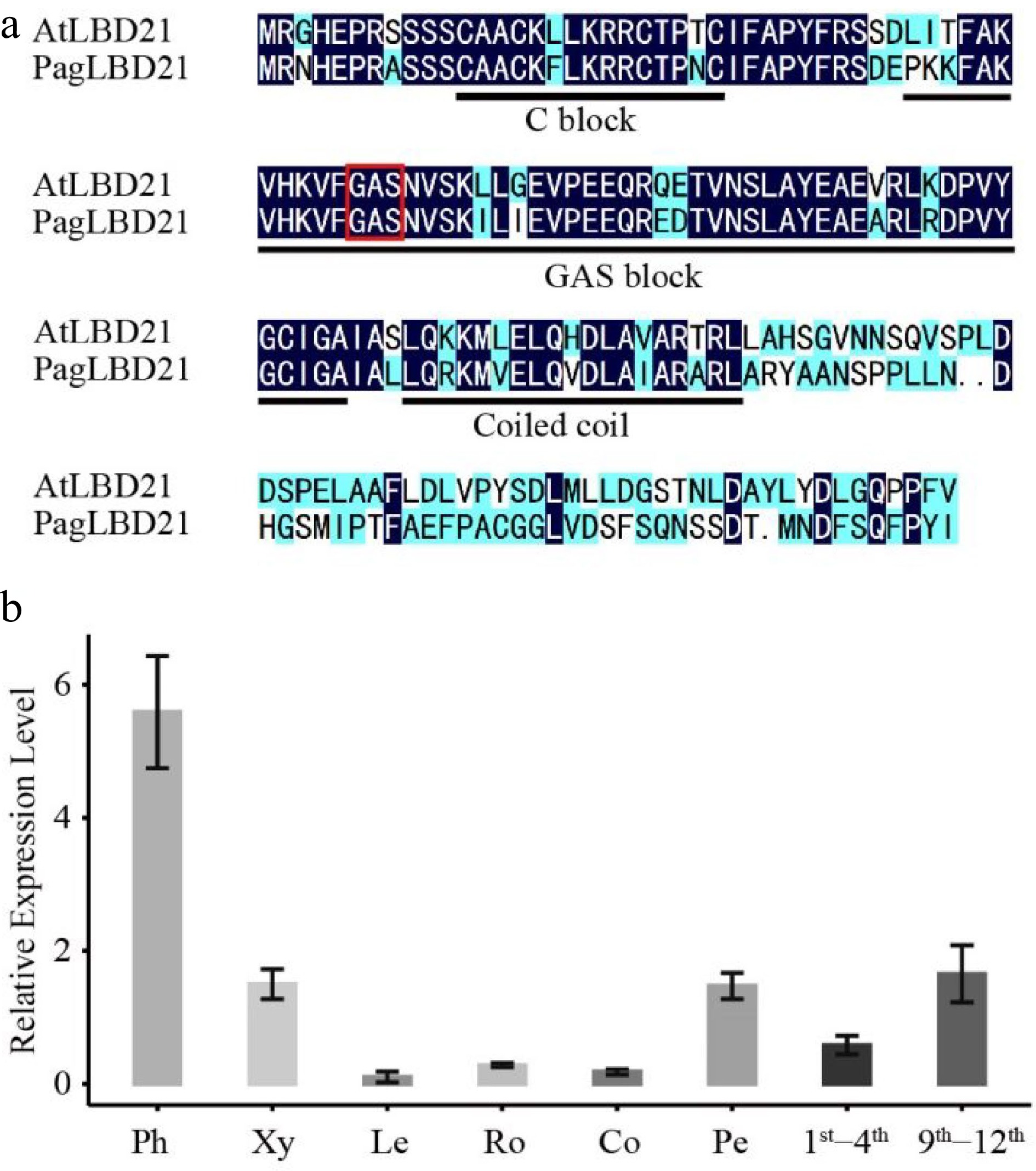
      Figure 1. 

      Characterization of PagLBD21 in Populus. (a) Amino acid sequence alignment of Populus PagLBD21 and Arabidopsis AtLBD21. Black and blue colors indicate identical and similar amino acids, respectively. The red box represents the core amino acid of GAS block. (b) RT-qPCR of PagLBD21 in different tissues using wild type. Each value is the mean ± standard error (SEM) of three replicates (n = 3 technical repetitions). Ph, phloem; Xy, xylem; Le, leaves; Ro, root; Co, cortex; Pe, petiole; 1st−4th, the stem of 1st to 4th internodes; 9th−12th, the stem of 9th to 12th internodes.

    • To determine the function of PagLBD21 in Populus, we generated transgenic plants overexpressing PagLBD21 (PagLBD21OE) in Populus. Eleven independent PagLBD21OE transgenic lines were obtained. We selected two transgenic lines L26 and L36, which displayed mild growth changes, for further analysis. RT-qPCR showed that PagLBD21 expressed significantly higher in both L26 and L36 lines than in non-transgenic plants (WT) (Fig. 2b). Compared to WT, PagLBD21OE plants had shorter stems and smaller stem diameter, and fewer leaves (Fig. 2a & b).
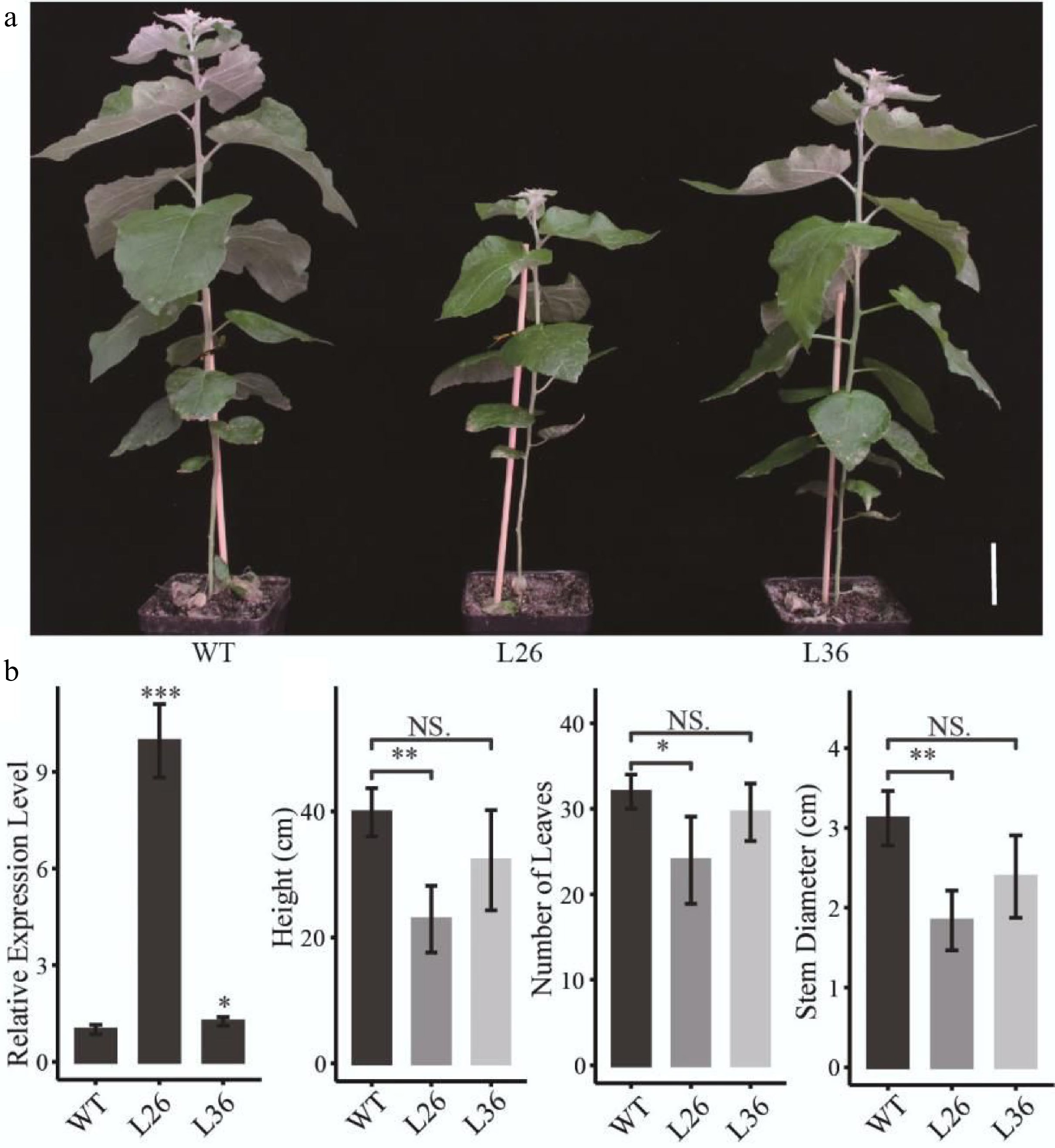
      Figure 2. 

      Effects of PagLBD21 overexpression (PagLBD21OE) in Populus. (a) Morphological phenotypes of 2-month-old PagLBD21OE transgenic lines compared to wild type (WT). Scale bar = 5 cm. The WT, L26 and L36 represent wild-type, the overexpression line of number 26, the overexpression line of number 36, respectively. (b) RT-qPCR of PagLBD21 in PagLBD21OE transgenic lines and WT. (c) The comparison of WT and PagLBD21OE transgenic lines in plants' height, stem diameter, and number of leaves. Each value is the mean ± SD of three replicates (n = 3 biological replicates). Student's t-test was used: *, P < 0.05; **, P < 0.01; NS, nonsignificant.

    • As the PagLBD21OE plants displayed smaller stem diameters, we speculated that PagLBD21 negatively regulated stem secondary growth in plants. To test this hypothesis, we performed freehand sectioning of plant stems and stained with the lignin-specific dye phloroglucinol. We found that xylem development was clearly repressed in PagLBD21OE plants (Fig. 3). In the 7th internode, the WT plants formed a completed xylem ring while the PagLBD21OE plants still displayed discontinuous xylem (Fig. 3a); in the 13th internode, the width of secondary xylem region in PagLBD21OE plants was significantly narrower than in WT (Fig. 3b); the cambium zone did not exhibit clear changes (Fig. 3c). Further quantification analysis showed that the width of the phloem region and number of cambium cells were similar to WT while the xylem region was significantly reduced (Fig. 3df). Correspondingly, the ratio of xylem region in the whole stem (xylem/stem) was significantly reduced (Fig. 3g). Collectively, our results suggested that PagLBD21 is a repressor of xylem development.
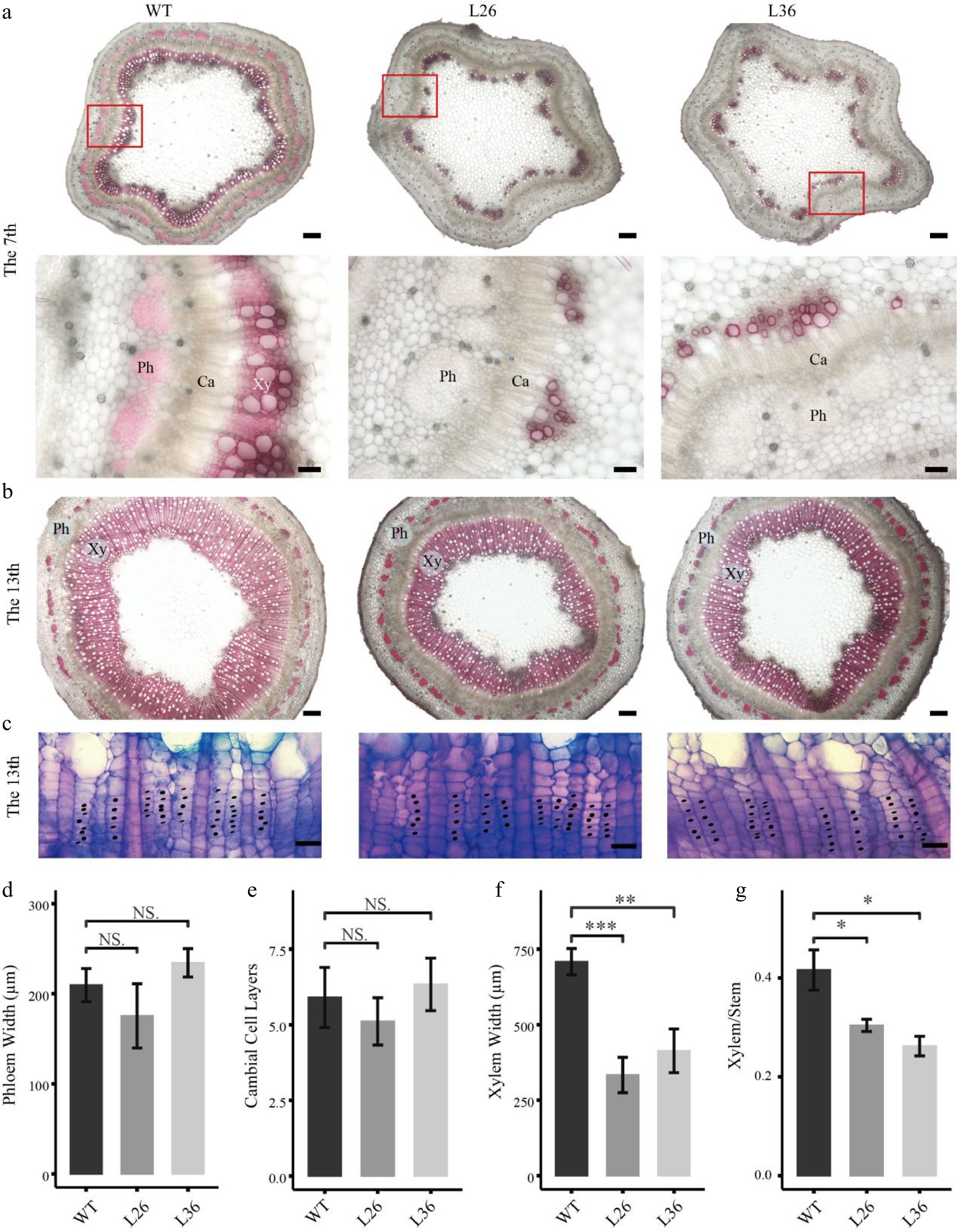
      Figure 3. 

      Effects of PagLBD21 overexpression (PagLBD21OE) on secondary growth in Populus. Stem cross sections of (a) 7th and (b), (c) 13th internodes of WT and PagLBD21OE plants. Scale bar: 200 μm for (b) and the upper panel of (a), 50 μm for the lower panel of (a), 20 μm for (c). The red-boxed area in the upper panels is depicted in detail in the lower panels. Xy, xylem. Ph, phloem. Ca, cambium. Detailed observation of the cambial zone (c) was stained by toluidine blue O, cambial cells were marked with black dots. The cambial cell layers (e) were analyzed according to (c). The phloem widths (d), xylem widths (f), and xylem widths divided by the radius (Xylem/Stem) (g) in 13th internodes were measured by ImageJ. Each value is the mean ± SD of three replicates (n = 3). Student’s t-test was used: *, P < 0.05; **, P < 0.01; ***, P < 0.001; NS, nonsignificant.

    • To profile genes regulated by overexpressing PagLBD21 during secondary growth, we performed RNA-seq with stem internodes 7th to 12th collected from 2-month-old PagLBD21OE (L36) and WT plants (MATERIALS AND METHODS). In total, 1421 significantly differentially expressed genes (DEGs) were identified between PagLBD21OE and WT plants (P-value < 0.05), with 722 and 699 up- and down-regulated DEGs in PagLBD21OE plants, respectively (Supplemental Table S2).

      Gene Ontology (GO) analysis was performed to characterize the function of DEGs. We found that the up-regulated DEGs were significantly enriched in GO categories including plant-type primary cell wall biogenesis, plant-type cell wall loosening, and plant-type cell wall modification (Fig. 4a, Supplemental Table S3); meanwhile, the down-regulated DEGs were significantly enriched in GO categories including cell differentiation, xylem development, and plant-type secondary cell wall biogenesis (Fig. 4b, Supplemental Table S3). These results were consistent with the observation that differentiation of the cambium cells to xylem was inhibited in PagLBD21OE plants.
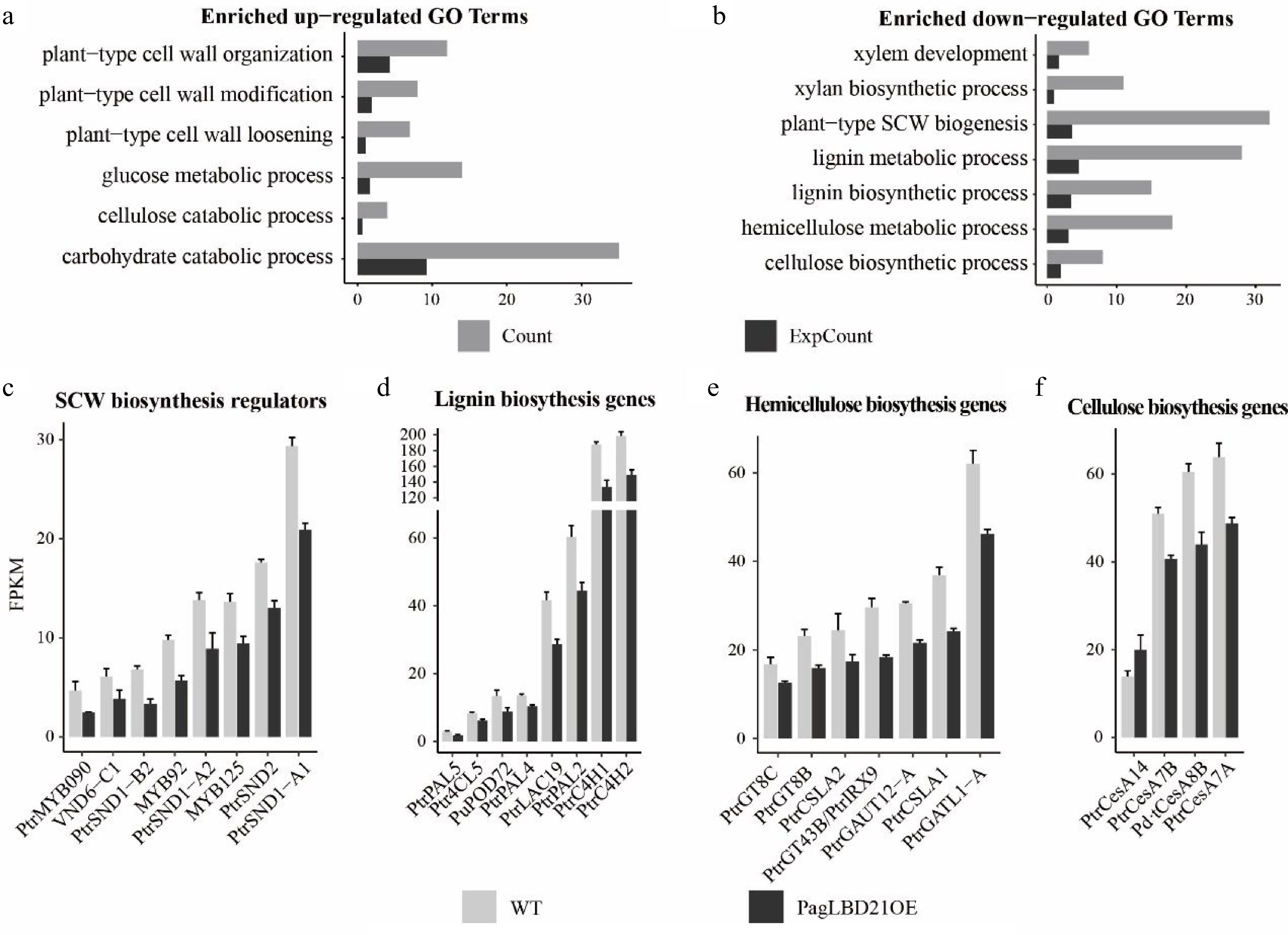
      Figure 4. 

      Transcriptome analysis of PagLBD21OE plant. (a), (b) Enriched Gene Ontology (GO) categories of (a) up-regulated and (b) down-regulated differentially expressed genes (DEGs) in PagLBD21OE line 36. Count, the number of genes in a GO term, ExpCount, the expected gene number. Expression of representative genes that function in (c) SCW biosynthesis regulatory pathway, (d) lignin biosynthesis, (e) hemicellulose biosynthesis, and (f) cellulose biosynthesis. The fragments per kilobase of exon per million mapped fragments (FPKM) for each gene from RNA-seq experiments were shown. Each value is the mean + standard error (SD) of three replicates (n = 3 technical repetitions). (n = 3 biological replicates).

      Consistent with the functional characterization, many genes encoding key regulators and enzymes responsible for secondary cell wall biosynthesis were among the down-regulated DEGs in PagLBD21OE plants. For instance, positive regulators of secondary cell wall biosynthesis including SND1-A1/A2/B2, VND6-C1, SND2, MYB92, MYB090, MYB125 were significantly down-regulated in PagLBD21OE plants (Fig. 4c), and most genes encoding key enzymes responsible for secondary cell wall cellulose, lignin, and hemicellulose biosynthesis were also significantly down-regulated in PagLBD21OE (Fig. 4df). Together, the RNA-seq data suggested that PagLBD21 inhibited xylem development partially through repressing secondary cell wall biosynthesis.

    • To further dissect the molecular function of PagLBD21, we performed DAP-seq to identify its genome-wide binding targets as previously reported[24] (MATERIALS AND METHODS). The genomic 'input' DNA libraries were sequenced in parallel as a control for data analysis. Three biological replicates were prepared for both PagLBD21 DAP-seq and 'input' control.

      We analyzed the DAP-seq data with IDR pipeline and used MACS2 as the peak caller[4,33]. In total, we obtained 1790 peaks as PagLBD21 binding sites which associated with 1639 unique target genes (Supplemental Table S4). The average width of PagLBD21 binding site was 312 bp (Fig. 5a). The genome-wide distribution of PagLBD21 binding sites was enriched around the transcription start site (TSS) of the target gene (Fig. 5b). Further inspection of the location of the binding sites relative to target genes showed that 37.508% was upstream of gene TSS and 3.726% was overlapped with gene TSS (Fig. 5c).
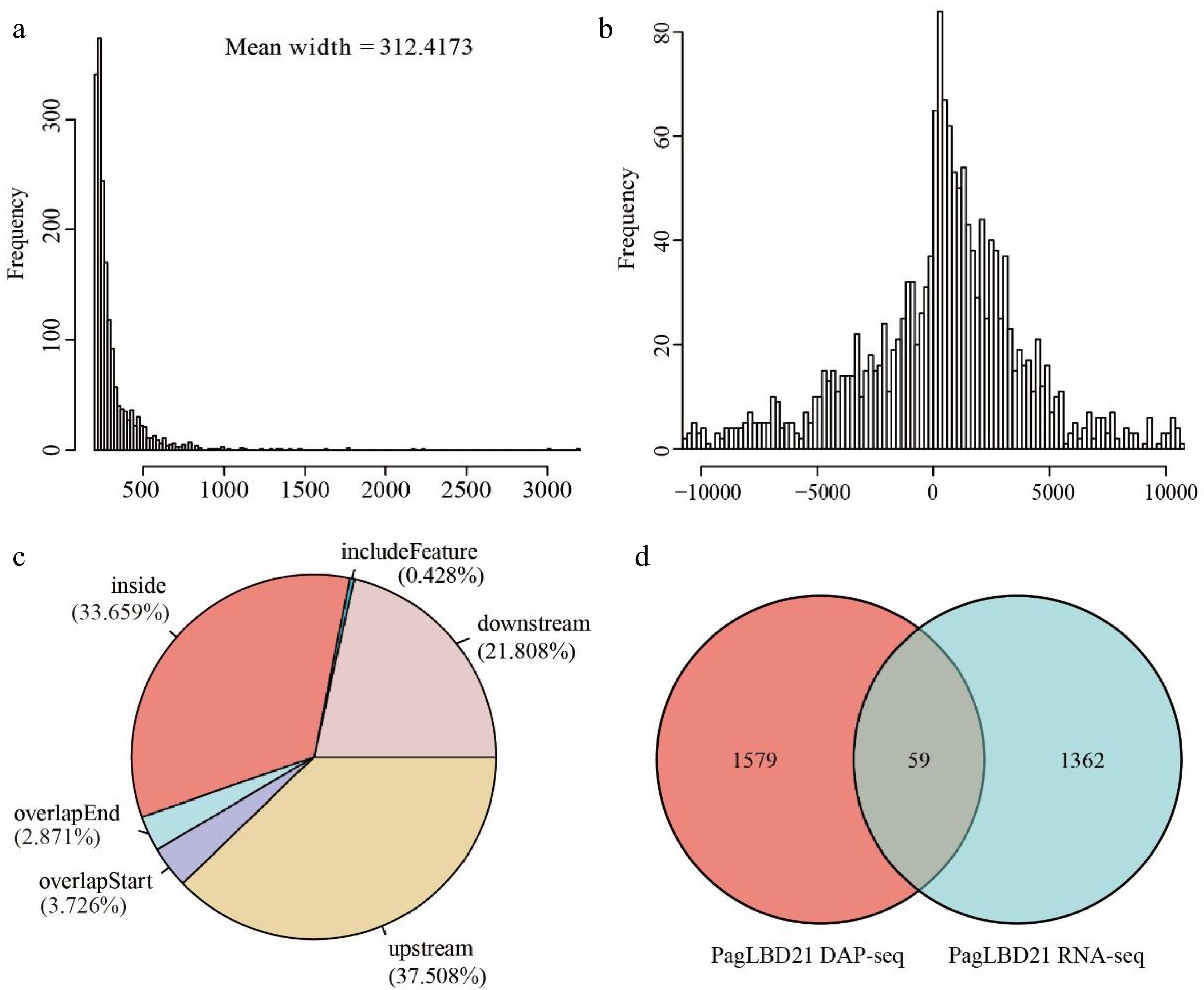
      Figure 5. 

      Analysis of genome-wide targets of PagLBD21 through DAP-seq. (a) The mean width of PagLBD21 binding sites identified in DAP-seq. (b) Genome-wide distribution of PagLBD21 binding sites is centered on gene transcriptional start sites (TSS). (c) The distribution of PagLBD21 binding sites relative to gene features. (d) Venn diagrams of target genes of DAP-seq data (left) and DEGs of RNA-seq data (right) of PagLBD21.

      To identify genes that are possibly regulated by PagLBD21 directly, we compared the DEGs of RNA-seq data and target genes of DAP-seq data. Results showed there were 59 overlapped genes between these two datasets (Fig. 5d), including 25 up-regulated and 34 down-regulated genes (Supplemental Table S5), respectively.

    • We previously reported another LBD gene, PagLBD3, which participated in regulating secondary growth in Populus[24]. Characterization of PagLBD21OE and PagLBD3OE plants showed partially similar phenotype, such as decreased xylem width. We hypothesize that these two LBD genes may target a group of common genes during secondary growth. Therefore, we performed pair-wise comparisons of the RNA-seq and DAP-seq data of PagLBD21 and PagLBD3. As shown in Table 1, there were 793, 260, 373, and 655 overlapped genes between PagLBD21 DAP-seq & PagLBD3 DAP-seq, PagLBD21 DAP-seq & PagLBD3 RNA-seq, PagLBD21 RNA-seq & PagLBD3 DAP-seq, and PagLBD21 RNA-seq & PagLBD3 RNA-seq, respectively. Notably, all these overlaps were significantly higher than random distribution in HYPGEOMDIST test.

      Table 1.  Overlapping study of DAP-seq and RNA-seq related genes between PagLBD21 and PagLBD3.

      LBD21
      DAP-seq
      LBD21
      RNA-seq
      LBD3
      DAP-seq
      LBD3
      RNA-seq
      LBD21 DAP-seq1,638
      LBD21 RNA-seq591,421
      LBD3 DAP-seq7393737,955
      LBD3 RNA-seq2606551,7826,468

      As the number of both PagLBD3 related DEGs and target genes were about five times of that related to PagLBD21, the common genes in PagLBD21 DAP-seq & PagLBD3 DAP-seq and PagLBD21 RNA-seq & PagLBD3 RNA-seq only accounts for 9% (739 out of 7,955) and 10% (655 out of 6,468) of PagLBD3 DAP-seq derived target genes and RNA-seq derived DEGs, respectively. However, the common genes in PagLBD21 DAP-seq & PagLBD3 DAP-seq and PagLBD21 RNA-seq & PagLBD3 RNA-seq accounts for 45% (739 out of 1,638) and 46% (655 out of 1,421) of PagLBD21 DAP-seq derived target genes and RNA-seq derived DEGs, respectively. Detailed inspection found that many key regulators of secondary growth were among the common targets of PagLBD21 DAP-seq and PagLBD3 DAP-seq, such as auxin transporter genes (PtPIN6b, PtAUX8LAX4, and PtPIN3a)[34,35], PtrMYB152 which encodes a positive regulator of lignin biosynthesis[36], and PtLOG7b which encodes a key enzyme during cytokinin biosynthesis[34,37] (Supplemental Table S6). Functional analysis showed that in the common DEGs in PagLBD21 RNA-seq & PagLBD3 RNA-seq, plant-type cell wall loosening, and plant-type cell wall modification categories were enriched in up-regulated genes, while cell differentiation, xylem development, and plant-type secondary cell wall biogenesis categories were enriched in down-regulated genes (Supplemental Table S7). Collectively, these results suggested that PagLBD21 and PagLBD3 targeted a common group of genes during secondary growth.

    • Extensive secondary growth is a prominent feature of forest tree species. The differentiation of secondary phloem and secondary xylem from cambium cells are key steps of secondary growth, and the secondary vascular system plays essential roles in long distance transportation of water and nutrients. In this study, we identified an LBD transcription factor, PagLBD21, as an important regulator of secondary xylem development in Populus. Expression analysis found that PagLBD21 expressed across the secondary phloem, cambium zone, and secondary xylem, but showed a higher level on the phloem side (Supplemental Fig. S1)[31,32]. Overexpression of PagLBD21 (PagLBD21OE) reduced plant height and stem diameter (Fig. 2). Stem cross-section analysis showed that the lignification process in secondary xylem was delayed in PagLBD21OE plants, and the xylem width was significantly reduced in the 13th internode (Fig. 3). Meanwhile, the phloem widths did not change significantly (Fig. 3). These results indicated the xylem development was suppressed by PagLBD21 overexpression, which may cause the suppression of plant growth in general.

      To further investigate the influences of PagLBD21 on secondary growth, we performed RNA-seq to identify differentially expressed genes (DEGs) in PagLBD21OE compared to WT plants. Because the PagLBD21OE L36 plants we used for RNA-seq displayed low expression (Fig. 2, Supplemental Table S2), we only identified 1,421 DEGs between PagLBD21OE and WT plants. However, GO enrichment analysis showed that biological pathways such as cell differentiation, xylem development, and plant-type secondary cell wall biogenesis were significantly enriched in down-regulated DEGs (Fig. 4a & b), furthermore, a group of key regulators and secondary cell wall biosynthesis genes were significantly down-regulated in PagLBD21OE plants, supporting the conclusion that PagLBD21 overexpression suppressed xylem development. We also performed DAP-seq to identify PagLBD21 genome-wide binding sites and identified 1,639 unique target genes associated with PagLBD21 binding sites (Fig. 5). Distribution pattern analysis suggested the PagLBD21 DAP-seq data was specific. However, there were only 59 genes overlapped between DEGs from RNA-seq and target genes from DAP-seq (Fig. 5d), which may due to the low expression level of the transgenic line used for RNA-seq and low sequencing depth of DAP-seq.

      Previously, we have reported another LBD transcription factor, PagLBD3, as an important regulator of secondary growth in Populus[24]. The overexpression plants of PagLBD21 and PagLBD3 displayed similar changes in many aspects, such as dwarf, hard rooting, and repression of xylem development. However, the phenotype of PagLBD21OE plants was not as strong as PagLBD3OE plants. There were also differences between PagLBD21 and PagLBD3 overexpressing plants: PagLBD21OE plants reduced stem diameter while PagLBD3OE plants increased stem diameter; PagLBD21OE plants had only reduced xylem width but regular xylem boundaries while PagLBD3OE plants had reduced xylem width with irregularly lignification and wider phloem and cortex. The phenotype differences may partially be caused by different expression levels of transgenic genes: the PagLBD21OE line used were weak lines with low expression level while PagLBD3OE line used were strong lines with high expression level.

      We also compared the RNA-seq and DAP-seq datasets of PagLBD21 and PagLBD3. Results showed that there was a significant overlap between PagLBD21 DAP-seq & PagLBD3 DAP-seq, PagLBD21 DAP-seq & PagLBD3 RNA-seq, PagLBD21 RNA-seq & PagLBD3 DAP-seq, and PagLBD21 RNA-seq & PagLBD3 RNA-seq (Table 1), respectively. As the datasets related to PagLBD21 were much smaller than that related to PagLBD3, the common genes only account for a small percentage of PagLBD3 datasets. These results made it difficult to compare the differences between PagLBD21 and PagLBD3 signaling pathways. However, many genes encoding key regulators for secondary growth, such as auxin transporters, cytokinin biosynthesis, regulators for lignin biosynthesis, and the majority of SCW biosynthesis genes were targeted or regulated by both PagLBD21 and PagLBD3, indicating that PagLBD21 and PagLBD3 targeted a common group of genes during secondary growth. Our results provided information to investigate the functions of LBD family genes, as well as to further dissect the regulatory mechanisms underlying tree secondary growth and wood formation.

      • This work was supported by the Introduction and Training Plan for Young Scientists in Universities Shandong Province (Research group of forest tree biotechnology), Open Fund of State Key Laboratory of Tree Genetics and Breeding (Chinese Academy of Forestry) (TGB2021006), and National Key R&D Program of China (2021YFD2200800).

      • The authors declare that they have no conflict of interest.

      • Supplemental Table S1 Primers used for gene cloning and qPCR.
      • Supplemental Table S2 DEGs between PagLBD21OE and WT.
      • Supplemental Table S3 Gene Ontology (GO) analysis of the up- or down -regulated DEGs in.
      • Supplemental Table S4 Summary of PagLBD21 DAP-seq binding sites and target genes.
      • Supplemental Table S5 The overlapped genes between PagLBD21 DAP-seq target genes and.
      • Supplemental Table S6 The overlapped target genes in PagLBD21 DAP-seq & PagLBD3 DAP D.
      • Supplemental Table S7 The overlapped DEGs in PagLBD21 RNA-seq & PagLBD3 RNA-seq.
      • Supplemental Fig. S1 The expression levels of PagLBD21 in Populus trichocarpa. (a) RNA-seq expression data in poplar phloem and xylem. Ph, phloem; Xy, xylem. (b) Expression profiles of the section of poplar cryosection from differentiated phloem to mature xylem in four trees (T1–T4). T, Tree. ⅰ, ⅱ, ⅲ, ⅳ indicate phloem, cambium, expanding xylem, and maturing xylem respectively. Each value is the mean ± standard error(SEM) of three replicates (n=3 technical repetitions).
      • Copyright: © 2022 by the author(s). Published by Maximum Academic Press, Fayetteville, GA. This article is an open access article distributed under Creative Commons Attribution License (CC BY 4.0), visit https://creativecommons.org/licenses/by/4.0/.
    Figure (5)  Table (1) References (37)
  • About this article
    Cite this article
    Li H, Yin S, Wang L, Xu N, Liu L. 2022. Transcription factor PagLBD21 functions as a repressor of secondary xylem development in Populus. Forestry Research 2:19 doi: 10.48130/FR-2022-0019
    Li H, Yin S, Wang L, Xu N, Liu L. 2022. Transcription factor PagLBD21 functions as a repressor of secondary xylem development in Populus. Forestry Research 2:19 doi: 10.48130/FR-2022-0019

Catalog

  • About this article

/

DownLoad:  Full-Size Img  PowerPoint
Return
Return