Search
2022 Volume 1
Article Contents
REVIEW   Open Access    

The contributions of sporophytic tapetum to pollen formation

More Information
  • Successful pollen formation is essential for plant reproduction. During anther development, microspore mother cells undergo meiosis to form tetrads. After being released from the tetrad, microspores develop into mature pollen. The tapetum is the innermost layer of anther somatic cells and forms a locule to provide nutrition, enzymes and pollen wall materials for microspore development. Based on the male sterile phenotype, many genes important for tapetum and pollen development have been cloned. In this review, we highlight the genetic pathway of DYT1-TDF1-AMS-MS188-MS1 which acts in tapetal development in Arabidopsis. We also compared this genetic pathway in different species such as Arabidopsis, rice and maize. Based on this pathway, we review recent findings and insights into the contribution of the tapetum to pollen formation at the molecular level. 1) Tapetum provides nutrition for microspore development. 2) Tapetum provides enzymes to dissolve pectin and callose to release microspores from tetrads. 3) Tapetum synthesizes precursors for pollen wall formation via different molecular pathways. 4) Tapetum provides precursors for pollen coat formation. 5) Tapetum provides small RNAs to regulate genic methylation in the germline cells.
  • 加载中
  • [1]

    Scott RJ, Spielman M, Dickinson HG. 2004. Stamen structure and function. The Plant Cell 16:S46−S60

    doi: 10.1105/tpc.017012

    CrossRef   Google Scholar

    [2]

    Goldberg RB, Beals TP, Sanders PM. 1993. Anther development: basic principles and practical applications. The Plant Cell 5:1217−29

    doi: 10.1105/tpc.5.10.1217

    CrossRef   Google Scholar

    [3]

    Sun L, Xiang X, Yang Z, Yu P, Wen X, et al. 2018. OsGPAT3 plays a critical role in anther wall programmed cell death and pollen development in rice. International Journal of Molecular Sciences 10:4017

    doi: 10.3390/ijms19124017

    CrossRef   Google Scholar

    [4]

    Yi J, Moon S, Lee YS, Zhu L, Liang W, et al. 2016. Defective Tapetum Cell Death 1 (DTC1) Regulates ROS Levels by Binding to Metallothionein during Tapetum Degeneration. Plant Physiology 170:1611−23

    doi: 10.1104/pp.15.01561

    CrossRef   Google Scholar

    [5]

    Chen L, Liu YG. 2014. Male sterility and fertility restoration in crops. Annual review of plant biology 65:579−606

    doi: 10.1146/annurev-arplant-050213-040119

    CrossRef   Google Scholar

    [6]

    Wilson ZA, Yang C. 2004. Plant gametogenesis: conservation and contrasts in development. Reproduction (Cambridge, England) 128:483−92

    doi: 10.1530/rep.1.00306

    CrossRef   Google Scholar

    [7]

    Brett CT, Waldron KW. 1990. Physiology and Biochemistry of Plant Cell Walls. Topics in Plant Physiology. eds. Black M, Chapman J. London: Unwin Hyman. 194 pp.

    [8]

    Carpita NC, Gibeaut DM. 1993. Structural models of primary cell walls in flowering plants consistency of molecular structure with the physical properties of the walls during growth. The Plant Journal 3:1−30

    doi: 10.1111/j.1365-313X.1993.tb00007.x

    CrossRef   Google Scholar

    [9]

    Matsuo Y, Arimura S, Tsutsumi N. 2013. Distribution of cellulosic wall in the anthers of Arabidopsis during microsporogenesis. Plant Cell Reports 32:1743−50

    doi: 10.1007/s00299-013-1487-1

    CrossRef   Google Scholar

    [10]

    Shi Q, Lou Y, Shen S, Wang S, Zhou L, et al. 2021. A cellular mechanism underlying the restoration of thermo/photoperiod-sensitive genic male sterility. Molecular Plant 14:2104−14

    doi: 10.1016/j.molp.2021.08.019

    CrossRef   Google Scholar

    [11]

    Heslop-Harrison J. 1963. An ultrastructural study of pollen wall ontogeny in Silene pendula. Grana Palynologica 4:7−24

    doi: 10.1080/00173136309437854

    CrossRef   Google Scholar

    [12]

    Piffanelli P, Ross JHE, Murphy DJ. 1998. Biogenesis and function of the lipidic structures of pollen grains. Sexual Plant Reproduction 11:65−80

    doi: 10.1007/s004970050122

    CrossRef   Google Scholar

    [13]

    Zhou Q, Zhu J, Cui Y, Yang Z. 2015. Ultrastructure analysis reveals sporopollenin deposition and nexine formation at early stage of pollen wall development in Arabidopsis. Science Bulletin 60:273−76

    doi: 10.1007/s11434-014-0723-6

    CrossRef   Google Scholar

    [14]

    Huang L, Cao J, Zhang A, Ye Y, Zhang Y, et al. 2009. The polygalacturonase gene BcMF2 from Brassica campestris is associated with intine development. Journal of Experimental Botany 60:301−13

    doi: 10.1093/jxb/ern295

    CrossRef   Google Scholar

    [15]

    Li J, Yu M, Geng L, Zhao J. 2010. The fasciclin-like arabinogalactan protein gene, FLA3, is involved in microspore development of Arabidopsis. The Plant Journal 64:482−97

    doi: 10.1111/j.1365-313X.2010.04344.x

    CrossRef   Google Scholar

    [16]

    Xu T, Zhang C, Zhou Q, Yang Z. 2016. Pollen wall pattern in Arabidopsis. Science Bulletin 61:832−37

    doi: 10.1007/s11434-016-1062-6

    CrossRef   Google Scholar

    [17]

    Ariizumi T, Toriyama K. 2011. Genetic regulation of sporopollenin synthesis and pollen exine development. Annual Review of Plant Biology 62:437−60

    doi: 10.1146/annurev-arplant-042809-112312

    CrossRef   Google Scholar

    [18]

    Jiang J, Zhang Z, Cao J. 2013. Pollen wall development: the associated enzymes and metabolic pathways. Plant Biology 15:249−63

    doi: 10.1111/j.1438-8677.2012.00706.x

    CrossRef   Google Scholar

    [19]

    Quilichini TD, Grienenberger E, Douglas CJ. 2015. The biosynthesis, composition and assembly of the outer pollen wall: a tough case to crack. Phytochemistry 113:170−82

    doi: 10.1016/j.phytochem.2014.05.002

    CrossRef   Google Scholar

    [20]

    Shi J, Cui M, Yang L, Kim YJ, Zhang D. 2015. Genetic and biochemical mechanisms of pollen wall development. Trends in Plant Science 20:741−53

    doi: 10.1016/j.tplants.2015.07.010

    CrossRef   Google Scholar

    [21]

    Grienenberger E, Quilichini TD. 2021. The toughest material in the plant kingdom: an update on sporopollenin. Frontiers in Plant Science 12:703864

    doi: 10.3389/fpls.2021.703864

    CrossRef   Google Scholar

    [22]

    Pacini E, Franchi GG, Hesse M. 1985. The tapetum: its form, function, and possible phylogeny in Embryophyta. Plant Systematics and Evolution 149:155−85

    doi: 10.1007/BF00983304

    CrossRef   Google Scholar

    [23]

    Mariani C, de Beuckeleer M, Truettner J, Leemans J, Goldberg RB. 1990. Induction of male sterility in plants by a chimaeric ribonuclease gene. Nature 347:737−41

    doi: 10.1038/347737a0

    CrossRef   Google Scholar

    [24]

    Phan HA, Iacuone S, Li SF, Parish RW. 2011. The MYB80 transcription factor is required for pollen development and the regulation of tapetal programmed cell death in Arabidopsis thaliana. The Plant cell 23:2209−24

    doi: 10.1105/tpc.110.082651

    CrossRef   Google Scholar

    [25]

    Zhang D, Liu D, Lv X, Wang Y, Xun Z, et al. 2014. The cysteine protease CEP1, a key executor involved in tapetal programmed cell death, regulates pollen development in Arabidopsis. The Plant Cell 26:2939−61

    doi: 10.1105/tpc.114.127282

    CrossRef   Google Scholar

    [26]

    Xie H, Wan Z, Li S, Zhang Y. 2014. Spatiotemporal production of reactive oxygen species by NADPH oxidase is critical for tapetal programmed cell death and pollen development in Arabidopsis. The Plant Cell 26:2007−23

    doi: 10.1105/tpc.114.125427

    CrossRef   Google Scholar

    [27]

    Cui Y, Zhao Q, Xie H, Wong W, Wang X, et al. 2017. MONENSIN SENSITIVITY1 (MON1)/CALCIUM CAFFEINE ZINC SENSITIVITY1 (CCZ1)-mediated Rab7 activation regulates tapetal programmed cell death and pollen development. Plant Physiology 173:206−18

    doi: 10.1104/pp.16.00988

    CrossRef   Google Scholar

    [28]

    Cheng Z, Guo X, Zhang J, Liu Y, Wang B, et al. 2020. βVPE is involved in tapetal degradation and pollen development by activating proprotease maturation in Arabidopsis thaliana. Journal of Experimental Botany 71:1943−55

    doi: 10.1093/jxb/erz560

    CrossRef   Google Scholar

    [29]

    Sorensen AM, Kröber S, Unte US, Huijser P, Dekker K, et al. 2003. The Arabidopsis ABORTED MICROSPORES (AMS) gene encodes a MYC class transcription factor. The Plant Journal 33:413−23

    doi: 10.1046/j.1365-313X.2003.01644.x

    CrossRef   Google Scholar

    [30]

    Zhang W, Sun Y, Timofejeva L, Chen C, Grossniklaus U, et al. 2006. Regulation of Arabidopsis tapetum development and function by DYSFUNCTIONAL TAPETUM1 (DYT1) encoding a putative bHLH transcription factor. Development 133:3085−95

    doi: 10.1242/dev.02463

    CrossRef   Google Scholar

    [31]

    Xu J, Yang C, Yuan Z, Zhang D, Gondwe M, et al. 2010. The ABORTED MICROSPORES regulatory network is required for postmeiotic male reproductive development in Arabidopsis thaliana. The Plant cell 22:91−107

    doi: 10.1105/tpc.109.071803

    CrossRef   Google Scholar

    [32]

    Zhu J, Chen H, Li H, Gao J, Jiang H, et al. 2008. Defective in tapetal development and function 1 is essential for anther development and tapetal function for microspore maturation in Arabidopsis. The Plant journal:for cell and molecular biology 55:266−77

    doi: 10.1111/j.1365-313X.2008.03500.x

    CrossRef   Google Scholar

    [33]

    Wilson ZA, Morroll SM, Dawson J, Swarup R, Tighe PJ. 2001. The Arabidopsis MALE STERILITY1 (MS1) gene is a transcriptional regulator of male gametogenesis, with homology to the PHD-finger family of transcription factors. The Plant Journal 28:27−39

    doi: 10.1046/j.1365-313X.2001.01125.x

    CrossRef   Google Scholar

    [34]

    Ito T, Shinozaki K. 2002. The MALE STERILITY1 gene of Arabidopsis, encoding a nuclear protein with a PHD-finger motif, is expressed in tapetal cells and is required for pollen maturation. Plant & Cell Physiology 43:1285−92

    doi: 10.1093/pcp/pcf154

    CrossRef   Google Scholar

    [35]

    Zhang Z, Zhu J, Gao J, Wang C, Li H, et al. 2007. Transcription factor AtMYB103 is required for anther development by regulating tapetum development, callose dissolution and exine formation in Arabidopsis. The Plant Journal 52:528−38

    doi: 10.1111/j.1365-313X.2007.03254.x

    CrossRef   Google Scholar

    [36]

    Zhu J, Zhang G, Chang Y, Li X, Yang J, et al. 2010. AtMYB103 is a crucial regulator of several pathways affecting Arabidopsis anther development. Science China. Life Sciences 53:1112−22

    doi: 10.1007/s11427-010-4060-y

    CrossRef   Google Scholar

    [37]

    Vizcay-Barrena G, Wilson ZA. 2006. Altered tapetal PCD and pollen wall development in the Arabidopsis ms1 mutant. Journal of Experimental Botany 57:2709−17

    doi: 10.1093/jxb/erl032

    CrossRef   Google Scholar

    [38]

    Zhu J, Lou Y, Xu X, Yang Z. 2011. A genetic pathway for tapetum development and function inArabidopsis. Journal of Integrative Plant Biology 53:892−900

    doi: 10.1111/j.1744-7909.2011.01078.x

    CrossRef   Google Scholar

    [39]

    Millar AA, Gubler F. 2005. The Arabidopsis GAMYB-like genes, MYB33 and MYB65, are microRNA-regulated genes that redundantly facilitate anther development. The Plant Cell 17:705−21

    doi: 10.1105/tpc.104.027920

    CrossRef   Google Scholar

    [40]

    Zhu E, You C, Wang S, Cui J, Niu B, et al. 2015. The DYT1-interacting proteins bHLH010, bHLH089 and bHLH091 are redundantly required for Arabidopsis anther development and transcriptome. The Plant Journal 83:976−90

    doi: 10.1111/tpj.12942

    CrossRef   Google Scholar

    [41]

    Cui J, You C, Zhu E, Huang Q, Ma H, et al. 2016. Feedback regulation of DYT1 by Interactions with downstream bHLH factors promotes DYT1 nuclear localization and anther development. The Plant Cell 28:1078−93

    doi: 10.1105/tpc.15.00986

    CrossRef   Google Scholar

    [42]

    Gu J, Zhu J, Yu Y, Teng X, Lou Y, et al. 2014. DYT1 directly regulates the expression of TDF1 for tapetum development and pollen wall formation in Arabidopsis. The Plant Journal 80:1005−13

    doi: 10.1111/tpj.12694

    CrossRef   Google Scholar

    [43]

    Lou Y, Zhou H, Han Y, Zeng Q, Zhu J, et al. 2018. Positive regulation of AMS by TDF1 and the formation of a TDF1-AMS complex are required for anther development in Arabidopsis thaliana. The New Phytologist 217:378−91

    doi: 10.1111/nph.14790

    CrossRef   Google Scholar

    [44]

    Lou Y, Xu X, Zhu J, Gu J, Blackmore S, et al. 2014. The tapetal AHL family protein TEK determines nexine formation in the pollen wall. Nature Communications 5:3855

    doi: 10.1038/ncomms4855

    CrossRef   Google Scholar

    [45]

    Ferguson AC, Pearce S, Band LR, Yang C, Ferjentsikova I, et al. 2017. Biphasic regulation of the transcription factor ABORTED MICROSPORES (AMS) is essential for tapetum and pollen development in Arabidopsis. The New Phytologist 213:778−90

    doi: 10.1111/nph.14200

    CrossRef   Google Scholar

    [46]

    Lu J, Xiong S, Yin W, Teng X, Lou Y, et al. 2020. MS1, a direct target of MS188, regulates the expression of key sporophytic pollen coat protein genes in Arabidopsis. Journal of Experimental Botany 71:4877−89

    doi: 10.1093/jxb/eraa219

    CrossRef   Google Scholar

    [47]

    Xiong S, Lu J, Lou Y, Teng X, Gu J, et al. 2016. The transcription factors MS188 and AMS form a complex to activate the expression of CYP703A2 for sporopollenin biosynthesis in Arabidopsis thaliana. The Plant Journal 88:936−46

    doi: 10.1111/tpj.13284

    CrossRef   Google Scholar

    [48]

    Wang K, Guo Z, Zhou W, Zhang C, Zhang Z, et al. 2018. The regulation of sporopollenin biosynthesis genes for rapid pollen wall formation. Plant Physiology 178:283−94

    doi: 10.1104/pp.18.00219

    CrossRef   Google Scholar

    [49]

    Jung KH, Han MJ, Lee YS, Kim YW, Hwang I, et al. 2005. Rice Undeveloped Tapetum1 is a major regulator of early tapetum development. The Plant Cell 17:2705−22

    doi: 10.1105/tpc.105.034090

    CrossRef   Google Scholar

    [50]

    Li N, Zhang D, Liu H, Yin C, Li X, et al. 2006. The rice tapetum degeneration retardation gene is required for tapetum degradation and anther development. The Plant Cell 18:2999−3014

    doi: 10.1105/tpc.106.044107

    CrossRef   Google Scholar

    [51]

    Zhang S, Fang Z, Zhu J, Gao J, Yang Z. 2010. OsMYB103 is required for rice anther development by regulating tapetum development and exine formation. Chinese Science Bulletin 55:3288−97

    doi: 10.1007/s11434-010-4087-2

    CrossRef   Google Scholar

    [52]

    Li H, Yuan Z, Vizcay-Barrena G, Yang C, Liang W, et al. 2011. PERSISTENT TAPETAL CELL1 encodes a PHD-finger protein that is required for tapetal cell death and pollen development in rice. Plant Physiology 156:615−30

    doi: 10.1104/pp.111.175760

    CrossRef   Google Scholar

    [53]

    Cai C, Zhu J, Lou Y, Guo Z, Xiong S, et al. 2015. The functional analysis of OsTDF1 reveals a conserved genetic pathway for tapetal development between rice and Arabidopsis. Science Bulletin 60:1073−82

    doi: 10.1007/s11434-015-0810-3

    CrossRef   Google Scholar

    [54]

    Zhang D, Liang W, Yuan Z, Li N, Shi J, et al. 2008. Tapetum degeneration retardation is critical for aliphatic metabolism and gene regulation during rice pollen development. Molecular plant 1:599−610

    doi: 10.1093/mp/ssn028

    CrossRef   Google Scholar

    [55]

    Pan X, Yan W, Chang Z, Xu Y, Luo M, et al. 2020. OsMYB80 regulates anther development and pollen fertility by targeting multiple biological pathways. Plant and Cell Physiology 61:988−1004

    doi: 10.1093/pcp/pcaa025

    CrossRef   Google Scholar

    [56]

    Han Y, Zhou S, Fan J, Zhou L, Shi Q, et al. 2021. OsMS188 is a key regulator of tapetum development and sporopollenin synthesis in rice. Rice 14:4

    doi: 10.1186/s12284-020-00451-y

    CrossRef   Google Scholar

    [57]

    Jiang Y, An X, Li Z, Yan T, Zhu T, et al. 2021. CRISPR/Cas9-based discovery of maize transcription factors regulating male sterility and their functional conservation in plants. Plant Biotechnology Journal 19:1769−84

    doi: 10.1111/pbi.13590

    CrossRef   Google Scholar

    [58]

    An X, Ma B, Duan M, Dong Z, Liu R, et al. 2020. Molecular regulation of ZmMs7 required for maize male fertility and development of a dominant male-sterility system in multiple species. PNAS 117:23499−509

    doi: 10.1073/pnas.2010255117

    CrossRef   Google Scholar

    [59]

    Zhang D, Wu S, An X, Xie K, Dong Z, et al. 2018. Construction of a multicontrol sterility system for a maize male-sterile line and hybrid seed production based on the ZmMs7 gene encoding a PHD-finger transcription factor. Plant Biotechnology Journal 16:459−71

    doi: 10.1111/pbi.12786

    CrossRef   Google Scholar

    [60]

    Nan G, Zhai J, Arikit S, Morrow D, Fernandes J, et al. 2017. MS23, a master basic helix-loop-helix factor, regulates the specification and development of the tapetum in maize. Development 144:163−72

    doi: 10.1242/dev.140673

    CrossRef   Google Scholar

    [61]

    Albertsen MC, Fox T, Leonard A, Li B, Loveland B, et al. 2016. Patent No. US 2016/0024520

    [62]

    Moon J, Skibbe D, Timofejeva L, Wang CJR, Kelliher T, et al. 2013. Regulation of cell divisions and differentiation by MALE STERILITY32 is required for anther development in maize. The Plant Journal 76:592−602

    doi: 10.1111/tpj.12318

    CrossRef   Google Scholar

    [63]

    Niu N, Liang W, Yang X, Jin W, Wilson ZA, et al. 2013. EAT1 promotes tapetal cell death by regulating aspartic proteases during male reproductive development in rice. Nature Communications 4:1445

    doi: 10.1038/ncomms2396

    CrossRef   Google Scholar

    [64]

    Ji C, Li H, Chen L, Xie M, Wang F, et al. 2013. A novel rice bHLH transcription factor, DTD, acts coordinately with TDR in controlling tapetum function and pollen development. Molecular Plant 6:1715−8

    doi: 10.1093/mp/sst046

    CrossRef   Google Scholar

    [65]

    Fu Z, Yu J, Cheng X, Zong X, Xu J, et al. 2014. The rice basic helix-loop-helix transcription factor TDR INTERACTING PROTEIN2 is a central switch in early anther development. The Plant Cell 26:1512−24

    doi: 10.1105/tpc.114.123745

    CrossRef   Google Scholar

    [66]

    Ko SS, Li MJ, Sun-Ben Ku M, Ho YC, Lin YJ, et al. 2014. The bHLH142 transcription factor coordinates with TDR1 to modulate the expression of EAT1 and regulate pollen development in rice. The Plant Cell 26:2486−504

    doi: 10.1105/tpc.114.126292

    CrossRef   Google Scholar

    [67]

    Cheng Y, Dai X, Zhao Y. 2006. Auxin biosynthesis by the YUCCA flavin monooxygenases controls the formation of floral organs and vascular tissues in Arabidopsis. Genes & Development 20:1790−99

    doi: 10.1101/gad.1415106

    CrossRef   Google Scholar

    [68]

    Yao X, Tian L, Yang J, Zhao Y, Zhu Y, et al. 2018. Auxin production in diploid microsporocytes is necessary and sufficient for early stages of pollen development. PLoS Genetics 14:e1007397

    doi: 10.1371/journal.pgen.1007397

    CrossRef   Google Scholar

    [69]

    Zheng Y, Wang D, Ye S, Chen W, Li G, et al. 2021. Auxin guides germ-cell specification in Arabidopsis anthers. PNAS 118:e2101492118

    doi: 10.1073/pnas.2101492118

    CrossRef   Google Scholar

    [70]

    Yang J, Tian L, Sun M, Huang X, Zhu J, et al. 2013. AUXIN RESPONSE FACTOR17 is essential for pollen wall pattern formation in Arabidopsis. Plant Physiology 162:720−31

    doi: 10.1104/pp.113.214940

    CrossRef   Google Scholar

    [71]

    Wang B, Xue J, Yu Y, Liu S, Zhang J, et al. 2017. Fine regulation of ARF17 for anther development and pollen formation. BMC Plant Biology 17:243

    doi: 10.1186/s12870-017-1185-1

    CrossRef   Google Scholar

    [72]

    Xu X, Wang B, Feng Y, Xue J, Qian X, et al. 2019. AUXIN RESPONSE FACTOR17 directly regulates MYB108 for anther dehiscence. Plant Physiology 181:645−55

    doi: 10.1104/pp.19.00576

    CrossRef   Google Scholar

    [73]

    Ye Q, Zhu W, Li L, Zhang S, Yin Y, et al. 2010. Brassinosteroids control male fertility by regulating the expression of key genes involved in Arabidopsis anther and pollen development. PNAS 107:6100−5

    doi: 10.1073/pnas.0912333107

    CrossRef   Google Scholar

    [74]

    Chen W, Lv M, Wang Y, Wang P, Cui Y, et al. 2019. BES1 is activated by EMS1-TPD1-SERK1/2-mediated signaling to control tapetum development in Arabidopsis thaliana. Nature Communications 10:4164

    doi: 10.1038/s41467-019-12118-4

    CrossRef   Google Scholar

    [75]

    Aya K, Ueguchi-Tanaka M, Kondo M, Hamada K, Yano K, et al. 2009. Gibberellin modulates anther development in rice via the transcriptional regulation of GAMYB. The Plant Cell 21:1453−72

    doi: 10.1105/tpc.108.062935

    CrossRef   Google Scholar

    [76]

    Sakata T, Oda S, Tsunaga Y, Shomura H, Kawagishi-Kobayashi M, et al. 2014. Reduction of gibberellin by low temperature disrupts pollen development in rice. Plant Physiology 164:2011−9

    doi: 10.1104/pp.113.234401

    CrossRef   Google Scholar

    [77]

    Plackett ARG, Ferguson AC, Powers SJ, Wanchoo-Kohli A, Phillips AL, et al. 2014. DELLA activity is required for successful pollen development in the Columbia ecotype of Arabidopsis. New Phytologist 201:825−36

    doi: 10.1111/nph.12571

    CrossRef   Google Scholar

    [78]

    Jin Y, Song X, Chang H, Zhao Y, Cao C, et al. 2021. The GA-DELLA-OsMS188 module controls male reproductive development in rice. New phytologist 233:2629−42

    doi: 10.1111/nph.17939

    CrossRef   Google Scholar

    [79]

    Clément C, Laporte P, Audran JC. 1998. The loculus content and tapetum during pollen development in Lilium. Sexual Plant Reproduction 11:94−106

    doi: 10.1007/s004970050125

    CrossRef   Google Scholar

    [80]

    Clément C, Audran JC. 1995. Anther wall layers control pollen sugar nutrition in Lilium. Protoplasma 187:172−81

    doi: 10.1007/BF01280246

    CrossRef   Google Scholar

    [81]

    Roitsch T, González MC. 2004. Function and regulation of plant invertases: sweet sensations. Trends in Plant Science 9:606−13

    doi: 10.1016/j.tplants.2004.10.009

    CrossRef   Google Scholar

    [82]

    Goetz M, Godt DE, Guivarc'h A, Kahmann U, Chriqui D, et al. 2001. Induction of male sterility in plants by metabolic engineering of the carbohydrate supply. PNAS 98:6522−27

    doi: 10.1073/pnas.091097998

    CrossRef   Google Scholar

    [83]

    Engelke T, Hirsche J, Roitsch T. 2010. Anther-specific carbohydrate supply and restoration of metabolically engineered male sterility. Journal of Experimental Botany 61:2693−706

    doi: 10.1093/jxb/erq105

    CrossRef   Google Scholar

    [84]

    Hirsche J, Engelke T, Völler D, Götz M, Roitsch T. 2009. Interspecies compatibility of the anther specific cell wall invertase promoters from Arabidopsis and tobacco for generating male sterile plants. Theoretical and Applied Genetics 118:235−45

    doi: 10.1007/s00122-008-0892-2

    CrossRef   Google Scholar

    [85]

    Ranwala AP, Miller WB. 1998. Sucrose-cleaving enzymes and carbohydrate pools in Lilium longiflorum floral organs. Physiologia Plantarum 103:541−50

    doi: 10.1034/j.1399-3054.1998.1030413.x

    CrossRef   Google Scholar

    [86]

    Zhang H, Liang W, Yang X, Luo X, Jiang N, et al. 2010. Carbon starved anther encodes a MYB domain protein that regulates sugar partitioning required for rice pollen development. The Plant Cell 22:672−89

    doi: 10.1105/tpc.109.073668

    CrossRef   Google Scholar

    [87]

    Li J, Huang Y, Tan H, Yang X, Tian L, et al. 2015. An endoplasmic reticulum magnesium transporter is essential for pollen development in Arabidopsis. Plant Science 231:212−20

    doi: 10.1016/j.plantsci.2014.12.008

    CrossRef   Google Scholar

    [88]

    Li L, Tutone AF, Drummond RSM, Gardner RC, Luan S. 2001. A novel family of magnesium transport genes in Arabidopsis. The Plant Cell 13:2761−75

    doi: 10.1105/tpc.010352

    CrossRef   Google Scholar

    [89]

    Li L, Sokolov LN, Yang Y, Li D, Ting J, et al. 2008. A mitochondrial magnesium transporter functions in Arabidopsis pollen development. Molecular Plant 1:675−85

    doi: 10.1093/mp/ssn031

    CrossRef   Google Scholar

    [90]

    Chen J, Li L, Liu Z, Yuan Y, Guo L, et al. 2009. Magnesium transporter AtMGT9 is essential for pollen development in Arabidopsis. Cell Research 19:887−98

    doi: 10.1038/cr.2009.58

    CrossRef   Google Scholar

    [91]

    Xu X, Wang B, Lou Y, Han W, Lu J, et al. 2015. Magnesium transporter 5 plays an important role in Mg transport for male gametophyte development in Arabidopsis. The Plant Journal 84:925−36

    doi: 10.1111/tpj.13054

    CrossRef   Google Scholar

    [92]

    Xu X, Qian X, Wang K, Yu Y, Guo Y, et al. 2020. Slowing development facilitates Arabidopsis mgt mutants to accumulate enough magnesium for pollen formation and fertility restoration. Frontiers in Plant Science 11:621338

    doi: 10.3389/fpls.2020.621338

    CrossRef   Google Scholar

    [93]

    Micheli F. 2001. Pectin methylesterases: cell wall enzymes with important roles in plant physiology. Trends Plant Science 6:414−19

    doi: 10.1016/s1360-1385(01)02045-3

    CrossRef   Google Scholar

    [94]

    Ridley BL, O'Neill MA, Mohnen D. 2001. Pectins: structure, biosynthesis, and oligogalacturonide-related signaling. Phytochemistry 57:929−67

    doi: 10.1016/S0031-9422(01)00113-3

    CrossRef   Google Scholar

    [95]

    Wikiera A, Mika M. 2013. Structure and properties of pectin. Postepy biochemii 59:89−94

    Google Scholar

    [96]

    Preuss D, Rhee SY, Davis RW. 1994. Tetrad analysis possible in Arabidopsis with mutation of the QUARTET (QRT) genes. Science 264:1458−60

    doi: 10.1126/science.8197459

    CrossRef   Google Scholar

    [97]

    Rhee SY, Somerville CR. 1998. Tetrad pollen formation in quartet mutants of Arabidopsis thaliana is associated with persistence of pectic polysaccharides of the pollen mother cell wall. The Plant Journal 15:79−88

    doi: 10.1046/j.1365-313X.1998.00183.x

    CrossRef   Google Scholar

    [98]

    Rhee SY, Osborne E, Poindexter PD, Somerville CR. 2003. Microspore separation in the quartet 3 mutants of Arabidopsis is impaired by a defect in a developmentally regulated polygalacturonase required for pollen mother cell wall degradation. Plant Physiology 133:1170−80

    doi: 10.1104/pp.103.028266

    CrossRef   Google Scholar

    [99]

    Francis KE, Lam SY, Copenhaver GP. 2006. Separation of Arabidopsis pollen tetrads is regulated by QUARTET1, a pectin methylesterase gene. Plant Physiology 142:1004−13

    doi: 10.1104/pp.106.085274

    CrossRef   Google Scholar

    [100]

    Ogawa M, Kay P, Wilson S, Swain SM. 2009. ARABIDOPSIS DEHISCENCE ZONE POLYGALACTURONASE1 (ADPG1), ADPG2, and QUARTET2 are polygalacturonases required for cell separation during reproductive development in Arabidopsis. The Plant Cell 21:216−33

    doi: 10.1105/tpc.108.063768

    CrossRef   Google Scholar

    [101]

    Dong X, Hong Z, Sivaramakrishnan M, Mahfouz M, Verma DPS. 2005. Callose synthase (CalS5) is required for exine formation during microgametogenesis and for pollen viability in Arabidopsis. The Plant Journal 42:315−28

    doi: 10.1111/j.1365-313X.2005.02379.x

    CrossRef   Google Scholar

    [102]

    Nishikawa SI, Zinkl GM, Swanson RJ, Maruyama D, Preuss D. 2005. Callose (β-1,3 glucan) is essential for Arabidopsis pollen wall patterning, but not tube growth. BMC Plant Biology 5:22

    doi: 10.1186/1471-2229-5-22

    CrossRef   Google Scholar

    [103]

    Xiong S, Zeng Q, Hou J, Hou L, Zhu J, et al. 2020. The temporal regulation of TEK contributes to pollen wall exine patterning. PLoS Genetics 16:e1008807

    doi: 10.1371/journal.pgen.1008807

    CrossRef   Google Scholar

    [104]

    Frankel R, Izhar S, Nitsan J. 1969. Timing of callase activity and cytoplasmic male sterility inPetunia. Biochemical Genetics 3:451−55

    doi: 10.1007/BF00485605

    CrossRef   Google Scholar

    [105]

    Stieglitz H, Stern H. 1973. Regulation of β-1,3-glucanase activity in developing anthers of Lilium. Developmental Biology 34:169−73

    doi: 10.1016/0012-1606(73)90347-3

    CrossRef   Google Scholar

    [106]

    Stieglitz H. 1977. Role of β-1,3-glucanase in postmeiotic microspore release. Developmental Biology 57:87−97

    doi: 10.1016/0012-1606(77)90356-6

    CrossRef   Google Scholar

    [107]

    Hird DL, Worrall D, Hodge R, Smartt S, Paul W, Scott R. 1993. The anther-specific protein encoded by the Brassica napus and Arabidopsis thaliana A6 gene displays similarity to β-1,3-glucanases. The Plant Journal 4:1023−33

    doi: 10.1046/j.1365-313X.1993.04061023.x

    CrossRef   Google Scholar

    [108]

    Xu J, Ding Z, Vizcay-Barrena G, Shi J, Liang W, et al. 2014. ABORTED MICROSPORES acts as a master regulator of pollen wall formation in Arabidopsis. The Plant Cell 26:1544−56

    doi: 10.1105/tpc.114.122986

    CrossRef   Google Scholar

    [109]

    Dobritsa AA, Geanconteri A, Shrestha J, Carlson A, Kooyers N, et al. 2011. A large-scale genetic screen in Arabidopsis to identify genes involved in pollen exine production. Plant Physiology 157:947−70

    doi: 10.1104/pp.111.179523

    CrossRef   Google Scholar

    [110]

    Suzuki T, Narciso JO, Zeng W, van de Meene A, Yasutomi M, et al. 2017. KNS4/UPEX1: A type II arabinogalactan β-(1,3)-galactosyltransferase required for pollen exine development. Plant Physiology 173:183−205

    doi: 10.1104/pp.16.01385

    CrossRef   Google Scholar

    [111]

    Wang K, Yu Y, Jia X, Zhou S, Zhang F, et al. 2021. Delayed callose degradation restores the fertility of multiple P/TGMS lines in Arabidopsis. Journal of Integrative Plant Biology 64:717−30

    doi: 10.1111/jipb.13205

    CrossRef   Google Scholar

    [112]

    de Azevedo Souza C, Kim SS, Koch S, Kienow L, Schneider K, et al. 2009. A novel fatty Acyl-CoA Synthetase is required for pollen development and sporopollenin biosynthesis in Arabidopsis. The Plant Cell 21:507−25

    doi: 10.1105/tpc.108.062513

    CrossRef   Google Scholar

    [113]

    Morant M, Jørgensen K, Schaller H, Pinot F, Møller BL, et al. 2007. CYP703 is an ancient cytochrome P450 in land plants catalyzing in-chain hydroxylation of lauric acid to provide building blocks for sporopollenin synthesis in pollen. The Plant Cell 19:1473−87

    doi: 10.1105/tpc.106.045948

    CrossRef   Google Scholar

    [114]

    Dobritsa AA, Shrestha J, Morant M, Pinot F, Matsuno M, et al. 2009. CYP704B1 is a long-chain fatty acid ω-hydroxylase essential for sporopollenin synthesis in pollen of Arabidopsis. Plant Physiology 151:574−89

    doi: 10.1104/pp.109.144469

    CrossRef   Google Scholar

    [115]

    Aarts MG, Hodge R, Kalantidis K, Florack D, Wilson ZA, et al. 1997. The Arabidopsis MALE STERILITY 2 protein shares similarity with reductases in elongation/condensation complexes. The Plant Journal 12:615−23

    doi: 10.1046/j.1365-313X.1997.00615.x

    CrossRef   Google Scholar

    [116]

    Chen W, Yu X, Zhang K, Shi J, De Oliveira S, et al. 2011. Male Sterile2 encodes a plastid-localized fatty acyl carrier protein reductase required for pollen exine development in Arabidopsis. Plant Physiology 157:842−53

    doi: 10.1104/pp.111.181693

    CrossRef   Google Scholar

    [117]

    Grienenberger E, Kim SS, Lallemand B, Geoffroy P, Heintz D, et al. 2010. Analysis of TETRAKETIDE α-PYRONE REDUCTASE function in Arabidopsis thaliana reveals a previously unknown, but conserved, biochemical pathway in sporopollenin monomer biosynthesis. The Plant Cell 22:4067−83

    doi: 10.1105/tpc.110.080036

    CrossRef   Google Scholar

    [118]

    Kim SS, Grienenberger E, Lallemand B, Colpitts CC, Kim SY, et al. 2010. LAP6/POLYKETIDE SYNTHASE A and LAP5/POLYKETIDE SYNTHASE B encode hydroxyalkyl alpha-pyrone synthases required for pollen development and sporopollenin biosynthesis in Arabidopsis thaliana. The Plant Cell 22:4045−66

    doi: 10.1105/tpc.110.080028

    CrossRef   Google Scholar

    [119]

    Dobritsa AA, Lei Z, Nishikawa SI, Urbanczyk-Wochniak E, Huhman DV, et al. 2010. LAP5 and LAP6 encode anther-specific proteins with similarity to chalcone synthase essential for pollen exine development in Arabidopsis. Plant physiology 153:937−55

    doi: 10.1104/pp.110.157446

    CrossRef   Google Scholar

    [120]

    Quilichini TD, Friedmann MC, Samuels AL, Douglas CJ. 2010. ATP-binding cassette transporter G26 is required for male fertility and pollen exine formation in Arabidopsis. Plant Physiology 154:678−90

    doi: 10.1104/pp.110.161968

    CrossRef   Google Scholar

    [121]

    Choi H, Jin JY, Choi S, Hwang JU, Kim YY, et al. 2011. An ABCG/WBC-type ABC transporter is essential for transport of sporopollenin precursors for exine formation in developing pollen. The Plant Journal 65:181−93

    doi: 10.1111/j.1365-313X.2010.04412.x

    CrossRef   Google Scholar

    [122]

    Dou XY, Yang KZ, Zhang Y, Wang W, Liu XL, et al. 2011. WBC27, an adenosine tri-phosphate-binding cassette protein, controls pollen wall formation and patterning in Arabidopsis. Journal of Integrative Plant Biology 53:74−88

    doi: 10.1111/j.1744-7909.2010.01010.x

    CrossRef   Google Scholar

    [123]

    Qin P, Tu B, Wang Y, Deng L, Quilichini TD, et al. 2013. ABCG15 encodes an ABC transporter protein, and is essential for post-meiotic anther and pollen exine development in rice. Plant and Cell Physiology 54:138−54

    doi: 10.1093/pcp/pcs162

    CrossRef   Google Scholar

    [124]

    Osthoff KS, Wiermann R. 1987. Phenols as integrated compounds of sporopollenin from pinus pollen. Journal of Plant Physiology 131:5−15

    doi: 10.1016/S0176-1617(87)80262-6

    CrossRef   Google Scholar

    [125]

    Domínguez E, Mercado JA, Quesada MA, Heredia A. 1999. Pollen sporopollenin: degradation and structural elucidation. Sexual Plant Reproduction 12:171−78

    doi: 10.1007/s004970050189

    CrossRef   Google Scholar

    [126]

    Li FS, Phyo P, Jacobowitz J, Hong M, Weng JK. 2019. The molecular structure of plant sporopollenin. Nature Plants 5:41−6

    doi: 10.1038/s41477-018-0330-7

    CrossRef   Google Scholar

    [127]

    Mikhael A, Jurcic K, Schneider C, Karr D, Fisher GL, et al. 2020. Demystifying and unravelling the molecular structure of the biopolymer sporopollenin. Rapid Communications in Mass Spectrometry 34:e8740

    doi: 10.1002/rcm.8740

    CrossRef   Google Scholar

    [128]

    Xue J, Zhang B, Zhan H, Lv Y, Jia X, et al. 2020. Phenylpropanoid derivatives are essential components of sporopollenin in vascular plants. Molecular Plant 13:1644−53

    doi: 10.1016/j.molp.2020.08.005

    CrossRef   Google Scholar

    [129]

    Rozema J, Broekman RA, Blokker P, Meijkamp BB, de Bakker N, et al. 2001. UV-B absorbance and UV-B absorbing compounds (para-coumaric acid) in pollen and sporopollenin: the perspective to track historic UV-B levels. Journal of Photochemistry and Photobiology B: Biology 62:108−17

    doi: 10.1016/S1011-1344(01)00155-5

    CrossRef   Google Scholar

    [130]

    Jia Q, Zhu J, Xu X, Lou Y, Zhang Z, et al. 2015. Arabidopsis AT-hook protein TEK positively regulates the expression of arabinogalactan proteins for nexine formation. Molecular Plant 8:251−60

    doi: 10.1016/j.molp.2014.10.001

    CrossRef   Google Scholar

    [131]

    Preuss D, Lemieux B, Yen G, Davis RW. 1993. A conditional sterile mutation eliminates surface components from Arabidopsis pollen and disrupts cell signaling during fertilization. Genes & Development 7:974−85

    doi: 10.1101/gad.7.6.974

    CrossRef   Google Scholar

    [132]

    Hülskamp M, Kopczak SD, Horejsi TF, Kihl BK, Pruitt RE. 1995. Identification of genes required for pollen-stigma recognition inArabidopsis thaliana. The Plant Journal 8:703−14

    doi: 10.1046/j.1365-313X.1995.08050703.x

    CrossRef   Google Scholar

    [133]

    Piffanelli P, Murphy DJ. 1998. Novel organelles and targeting mechanisms in the anther tapetum. Trends in Plant Science 3:250−52

    doi: 10.1016/S1360-1385(98)01260-6

    CrossRef   Google Scholar

    [134]

    Pacini E, Hesse M. 2002. Types of pollen dispersal units in orchids, and their consequences for germination and fertilization. Annals of Botany 89:653−64

    doi: 10.1093/aob/mcf138

    CrossRef   Google Scholar

    [135]

    Blackmore S, Wortley AH, Skvarla JJ, Rowley JR. 2007. Pollen wall development in flowering plants. New Phytologist 174:483−98

    doi: 10.1111/j.1469-8137.2007.02060.x

    CrossRef   Google Scholar

    [136]

    Wheeler MJ, Franklin-Tong VE, Franklin FCH. 2001. The molecular and genetic basis of pollen-pistil interactions. New Phytologist 151:565−84

    doi: 10.1046/j.0028-646x.2001.00229.x

    CrossRef   Google Scholar

    [137]

    Jia X, Xue J, Zhang F, Yao C, Shen S, et al. 2021. A dye combination for the staining of pollen coat and pollen wall. Plant Reproduction 34:91−101

    doi: 10.1007/s00497-021-00412-5

    CrossRef   Google Scholar

    [138]

    Piffanelli P, Ross JHE, Murphy DJ. 1997. Intra- and extracellular lipid composition and associated gene expression patterns during pollen development in Brassica napus. The Plant Journal 11:549−62

    doi: 10.1046/j.1365-313X.1997.11030549.x

    CrossRef   Google Scholar

    [139]

    Xue Z, Xu X, Zhou Y, Wang X, Zhang Y, et al. 2018. Deficiency of a triterpene pathway results in humidity-sensitive genic male sterility in rice. Nature Communications 9:604

    doi: 10.1038/s41467-018-03048-8

    CrossRef   Google Scholar

    [140]

    Mayfield JA, Fiebig A, Johnstone SE, Preuss D. 2001. Gene families from the Arabidopsis thaliana pollen coat proteome. Science 292:2482−85

    doi: 10.1126/science.1060972

    CrossRef   Google Scholar

    [141]

    Mayfield JA, Preuss D. 2000. Rapid initiation of Arabidopsis pollination requires the oleosin-domain protein GRP17. Nature Cell Biology 2:128−30

    doi: 10.1038/35000084

    CrossRef   Google Scholar

    [142]

    Updegraff EP, Zhao F, Preuss D. 2009. The extracellular lipase EXL4 is required for efficient hydration of Arabidopsis pollen. Sexual Plant Reproduction 22:197−204

    doi: 10.1007/s00497-009-0104-5

    CrossRef   Google Scholar

    [143]

    Jessen D, Olbrich A, Knüfer J, Krüger A, Hoppert M, et al. 2011. Combined activity of LACS1 and LACS4 is required for proper pollen coat formation in Arabidopsis. The Plant Journal 68:715−26

    doi: 10.1111/j.1365-313X.2011.04722.x

    CrossRef   Google Scholar

    [144]

    Koornneef M, Hanhart CJ, Thiel F. 1989. A genetic and phenotypic description of Eceriferum (cer) mutants in Arabidopsis thaliana. Journal of Heredity 80:118−22

    doi: 10.1093/oxfordjournals.jhered.a110808

    CrossRef   Google Scholar

    [145]

    Hannoufa A, Negruk V, Eisner G, Lemieux B. 1996. The CER3 gene of Arabidopsis thaliana is expressed in leaves, stems, roots, flowers and apical meristems. The Plant Journal 10:459−67

    doi: 10.1046/j.1365-313X.1996.10030459.x

    CrossRef   Google Scholar

    [146]

    Zhang Z, Zhan H, Lu J, Xiong S, Yang N, et al. 2021. Tapetal 3-Ketoacyl-Coenzyme a synthases are involved in pollen coat lipid accumulation for pollen-stigma interaction in Arabidopsis. Frontiers in Plant Science 12:770311

    doi: 10.3389/fpls.2021.770311

    CrossRef   Google Scholar

    [147]

    Joubès J, Raffaele S, Bourdenx B, Garcia C, Laroche-Traineau J, et al. 2008. The VLCFA elongase gene family in Arabidopsis thaliana: phylogenetic analysis, 3D modelling and expression profiling. Plant molecular biology 67:547−66

    doi: 10.1007/s11103-008-9339-z

    CrossRef   Google Scholar

    [148]

    Haslam TM, Kunst L. 2013. Extending the story of very-long-chain fatty acid elongation. Plant Science 210:93−107

    doi: 10.1016/j.plantsci.2013.05.008

    CrossRef   Google Scholar

    [149]

    Ariizumi T, Hatakeyama K, Hinata K, Sato S, Kato T, et al. 2003. A novel male-sterile mutant ofArabidopsis thaliana, f aceless pollen-1, produces pollen with a smooth surface and an acetolysis-sensitive exine. Plant Molecular Biology 53:107−16

    doi: 10.1023/B:PLAN.0000009269.97773.70

    CrossRef   Google Scholar

    [150]

    Chen XB, Goodwin SM, Boroff VL, Liu XL, Jenks MA. 2003. Cloning and characterization of the WAX2 gene of Arabidopsis involved in cuticle membrane and wax production. The Plant Cell 15:1170−85

    doi: 10.1105/tpc.010926

    CrossRef   Google Scholar

    [151]

    Rowland O, Lee R, Franke R, Schreiber L, Kunst L. 2007. The CER3 wax biosynthetic gene from Arabidopsis thaliana is allelic to WAX2/YRE/FLP1. FEBS letters 581:3538−44

    doi: 10.1016/j.febslet.2007.06.065

    CrossRef   Google Scholar

    [152]

    Kurata T, Kawabata-Awai C, Sakuradani E, Shimizu S, Okada K, Wada T. 2003. The YORE-YORE gene regulates multiple aspects of epidermal cell differentiation in Arabidopsis. The Plant journal:for cell and molecular biology 36:55−66

    doi: 10.1046/j.1365-313X.2003.01854.x

    CrossRef   Google Scholar

    [153]

    Bernard A, Domergue F, Pascal S, Jetter R, Renne C, et al. 2012. Reconstitution of plant alkane biosynthesis in yeast demonstrates that Arabidopsis ECERIFERUM1 and ECERIFERUM3 are core components of a very-long-chain alkane synthesis complex. The Plant cell 24:3106−18

    doi: 10.1105/tpc.112.099796

    CrossRef   Google Scholar

    [154]

    Ishiguro S, Nishimori Y, Yamada M, Saito H, Suzuki T, et al. 2010. The Arabidopsis FLAKY POLLEN1 gene encodes a 3-hydroxy-3-methylglutaryl-coenzyme A synthase required for development of tapetum-specific organelles and fertility of pollen grains. Plant & Cell Physiology 51:896−911

    doi: 10.1093/pcp/pcq068

    CrossRef   Google Scholar

    [155]

    Suzuki T, Tsunekawa S, Koizuka C, Yamamoto K, Imamura J, et al. 2013. Development and disintegration of tapetum-specific lipid-accumulating organelles, elaioplasts and tapetosomes, in Arabidopsis thaliana and Brassica napus. Plant Science 207:25−36

    doi: 10.1016/j.plantsci.2013.02.008

    CrossRef   Google Scholar

    [156]

    van der Veen JH, Wirtz P. 1968. EMS-induced genic male sterility in Arabidopsis thaliana: a model selection experiment. Euphytica 17:371−77

    doi: 10.1007/BF00056238

    CrossRef   Google Scholar

    [157]

    Wilson Z, Vizcay Barrena G, Yang C. 2007. Arabidopsis male sterility1 regulates programmed cell death in the anther tapetum and pollen wall development. Comparative Biochemistry and Physiology Part A: Molecular & Integrative Physiology 146:S203

    doi: 10.1016/j.cbpa.2007.01.451

    CrossRef   Google Scholar

    [158]

    Rejón JD, Delalande F, Schaeffer-Reiss C, Alché JD, Rodríguez-García MI, et al. 2016. The pollen coat proteome: at the cutting edge of plant reproduction. Proteomes 4:5

    doi: 10.3390/proteomes4010005

    CrossRef   Google Scholar

    [159]

    Doughty J, Dixon S, Hiscock SJ, Willis AC, Parkin IAP, Dickinson HG. 1998. PCP-A1, a defensin-like Brassica pollen coat protein that binds the S locus glycoprotein, is the product of gametophytic gene expression. The Plant Cell 10:1333−47

    doi: 10.1105/tpc.10.8.1333

    CrossRef   Google Scholar

    [160]

    Takayama S, Shiba H, Iwano M, Asano K, Hara M, et al. 2000. Isolation and characterization of pollen coat proteins of Brassica campestris that interact with S locus-related glycoprotein 1 involved in pollen-stigma adhesion. PNAS 97:3765−70

    doi: 10.1073/pnas.97.7.3765

    CrossRef   Google Scholar

    [161]

    Nasrallah JB, Nasrallah ME. 2014. S-locus receptor kinase signalling. Biochemical Society Transactions 42:313−9

    doi: 10.1042/BST20130222

    CrossRef   Google Scholar

    [162]

    Zhan H, Xiong H, Wang S, Yang Z. 2018. Anther endothecium-derived very-long-chain fatty acids facilitate pollen hydration in Arabidopsis. Molecular Plant 11:1101−4

    doi: 10.1016/j.molp.2018.05.002

    CrossRef   Google Scholar

    [163]

    Haslam TM, Mañas-Fernández A, Zhao LF, Kunst L. 2012. Arabidopsis ECERIFERUM2 is a component of the fatty acid elongation machinery required for fatty acid extension to exceptional lengths. Plant Physiology 160:1164−74

    doi: 10.1104/pp.112.201640

    CrossRef   Google Scholar

    [164]

    Haslam TM, Haslam R, Thoraval D, Pascal S, Delude C, et al. 2015. ECERIFERUM2-LIKE proteins have unique biochemical and physiological functions in very-long-chain fatty acid elongation. Plant Physiology 167:682−92

    doi: 10.1104/pp.114.253195

    CrossRef   Google Scholar

    [165]

    Haslam TM, Gerelle WK, Graham SW, Kunst L. 2017. The unique role of the ECERIFERUM2-LIKE clade of the BAHD acyltransferase superfamily in cuticular wax metabolism. Plants-Basel 6:23

    doi: 10.3390/plants6020023

    CrossRef   Google Scholar

    [166]

    Xia Y, Nikolau BJ, Schnable PS. 1997. Developmental and hormonal regulation of the Arabidopsis CER2 gene that codes for a nuclear-localized protein required for the normal accumulation of cuticular waxes. Plant Physiology 115:925−37

    doi: 10.1104/pp.115.3.925

    CrossRef   Google Scholar

    [167]

    Fiebig A, Mayfield JA, Miley NL, Chau S, Fischer RL, et al. 2000. Alterations in CER6, a gene identical to CUT1, differentially affect long-chain lipid content on the surface of pollen and stems. The Plant Cell 12:2001−8

    doi: 10.1105/tpc.12.10.2001

    CrossRef   Google Scholar

    [168]

    Doughty J, Hedderson F, McCubbin A, Dickinson H. 1993. Interaction between a coating-borne peptide of the Brassica pollen grain and stigmatic S (self-incompatibility)-locus-specific glycoproteins. PNAS 90:467−71

    doi: 10.1073/pnas.90.2.467

    CrossRef   Google Scholar

    [169]

    Wang L, Clarke LA, Eason RJ, Parker CC, Qi B, et al. 2017. PCP-B class pollen coat proteins are key regulators of the hydration checkpoint in Arabidopsis thaliana pollen-stigma interactions. New Phytologist 213:764−77

    doi: 10.1111/nph.14162

    CrossRef   Google Scholar

    [170]

    Liu C, Shen L, Xiao Y, Vyshedsky D, Peng C, et al. 2021. Pollen PCP-B peptides unlock a stigma peptide-receptor kinase gating mechanism for pollination. Science 372:171−75

    doi: 10.1126/science.abc6107

    CrossRef   Google Scholar

    [171]

    Wei LQ, Yan LF, Wang T. 2011. Deep sequencing on genome-wide scale reveals the unique composition and expression patterns of microRNAs in developing pollen of Oryza sativa. Genome Biology 12:R53

    doi: 10.1186/gb-2011-12-6-r53

    CrossRef   Google Scholar

    [172]

    Chambers C, Shuai B. 2009. Profiling microRNA expression in Arabidopsis pollen using microRNA array and real-time PCR. BMC Plant Biology 9:87

    doi: 10.1186/1471-2229-9-87

    CrossRef   Google Scholar

    [173]

    Tsuji H, Aya K, Ueguchi-Tanaka M, Shimada Y, Nakazono M, et al. 2006. GAMYB controls different sets of genes and is differentially regulated by microRNA in aleurone cells and anthers. The Plant Journal 47:427−44

    doi: 10.1111/j.1365-313X.2006.02795.x

    CrossRef   Google Scholar

    [174]

    Achard P, Herr A, Baulcombe DC, Harberd NP. 2004. Modulation of floral development by a gibberellin-regulated microRNA. Development 131:3357−65

    doi: 10.1242/dev.01206

    CrossRef   Google Scholar

    [175]

    Sun Y, Xiong X, Wang Q, Zhu L, Wang L, et al. 2021. Integrated analysis of small RNA, transcriptome, and degradome sequencing reveals the MiR156, MiR5488 and MiR399 are involved in the regulation of male sterility in PTGMS rice. International Journal of Molecular Sciences 22:2260

    doi: 10.3390/ijms22052260

    CrossRef   Google Scholar

    [176]

    Wu S, Tan H, Hao X, Xie Z, Wang X, et al. 2019. Profiling miRNA expression in photo-thermo-sensitive male genic sterility line (PTGMS) PA64S under high and low temperature. Plant Signaling & Behavior 14:1679015

    doi: 10.1080/15592324.2019.1679015

    CrossRef   Google Scholar

    [177]

    Li Z, An X, Zhu T, Yan T, Wu S, et al. 2019. Discovering and constructing ceRNA-miRNA-target gene regulatory networks during anther development in maize. International Journal of Molecular Sciences 20:3480

    doi: 10.3390/ijms20143480

    CrossRef   Google Scholar

    [178]

    Omidvar V, Mohorianu I, Dalmay T, Fellner M. 2015. Identification of miRNAs with potential roles in regulation of anther development and male-sterility in 7B-1 male-sterile tomato mutant. BMC Genomics 16:878

    doi: 10.1186/s12864-015-2077-0

    CrossRef   Google Scholar

    [179]

    Slotkin RK, Vaughn M, Borges F, Tanurdžić M, Becker JD, et al. 2009. Epigenetic reprogramming and small RNA silencing of transposable elements in pollen. Cell 136:461−72

    doi: 10.1016/j.cell.2008.12.038

    CrossRef   Google Scholar

    [180]

    Zhai J, Zhang H, Arikit S, Huang K, Nan G, et al. 2015. Spatiotemporally dynamic, cell-type-dependent premeiotic and meiotic phasiRNAs in maize anthers. PNAS 112:3146−51

    doi: 10.1073/pnas.1418918112

    CrossRef   Google Scholar

    [181]

    Teng C, Zhang H, Hammond R, Huang K, Meyers BC, Walbot V. 2020. Dicer-like 5 deficiency confers temperature-sensitive male sterility in maize. Nature Communications 11:2912

    doi: 10.1038/s41467-020-16634-6

    CrossRef   Google Scholar

    [182]

    Long J, Walker J, She W, Aldridge B, Gao H, et al. 2021. Nurse cell-derived small RNAs define paternal epigenetic inheritance in Arabidopsis. Science 373:eabh0556

    doi: 10.1126/science.abh0556

    CrossRef   Google Scholar

    [183]

    Zhou X, Huang K, Teng C, Abdelgawad A, Batish M, et al. 2022. 24-nt phasiRNAs move from tapetal to meiotic cells in maize anthers. New Phytologist 235:488−501

    doi: 10.1111/nph.18167

    CrossRef   Google Scholar

  • Cite this article

    Yao X, Hu W, Yang Z. 2022. The contributions of sporophytic tapetum to pollen formation. Seed Biology 1:5 doi: 10.48130/SeedBio-2022-0005
    Yao X, Hu W, Yang Z. 2022. The contributions of sporophytic tapetum to pollen formation. Seed Biology 1:5 doi: 10.48130/SeedBio-2022-0005

Figures(4)  /  Tables(1)

Article Metrics

Article views(8051) PDF downloads(1554)

Other Articles By Authors

REVIEW   Open Access    

The contributions of sporophytic tapetum to pollen formation

Seed Biology  1 Article number: 5  (2022)  |  Cite this article

Abstract: Successful pollen formation is essential for plant reproduction. During anther development, microspore mother cells undergo meiosis to form tetrads. After being released from the tetrad, microspores develop into mature pollen. The tapetum is the innermost layer of anther somatic cells and forms a locule to provide nutrition, enzymes and pollen wall materials for microspore development. Based on the male sterile phenotype, many genes important for tapetum and pollen development have been cloned. In this review, we highlight the genetic pathway of DYT1-TDF1-AMS-MS188-MS1 which acts in tapetal development in Arabidopsis. We also compared this genetic pathway in different species such as Arabidopsis, rice and maize. Based on this pathway, we review recent findings and insights into the contribution of the tapetum to pollen formation at the molecular level. 1) Tapetum provides nutrition for microspore development. 2) Tapetum provides enzymes to dissolve pectin and callose to release microspores from tetrads. 3) Tapetum synthesizes precursors for pollen wall formation via different molecular pathways. 4) Tapetum provides precursors for pollen coat formation. 5) Tapetum provides small RNAs to regulate genic methylation in the germline cells.

    • The anther is an essential organ for plant reproduction, in which mature pollen grains are produced and released. The wall of anthers consist of four layers, epidermis, endothecium, middle layer and tapetum cell, that surround the reproductive cells[1] (Fig. 1). The epidermis plays a protective role in anther development, and the endothecium is responsible for anther dehiscence to release functional pollen[2]. The tapetum is a cell layer that directly contacts microspores, and undergoes programmed cell death (PCD). It is generally accepted that tapetal cells act as 'nutrition cells' for microspore development. The middle layer exists in seed plants, but its function remains unclear. It has been proposed that the middle cell layer may partially play a similar role as the tapetum to facilitate microspore development via PCD[3]. In flowering plants, dysfunction of the tapetum is often associated with male sterility, highlighting the significance of the tapetal layer in male gametogenesis[4]. In agriculture, male-sterile plants are the necessary materials for hybrid seed production to improve the yields of crops[5].
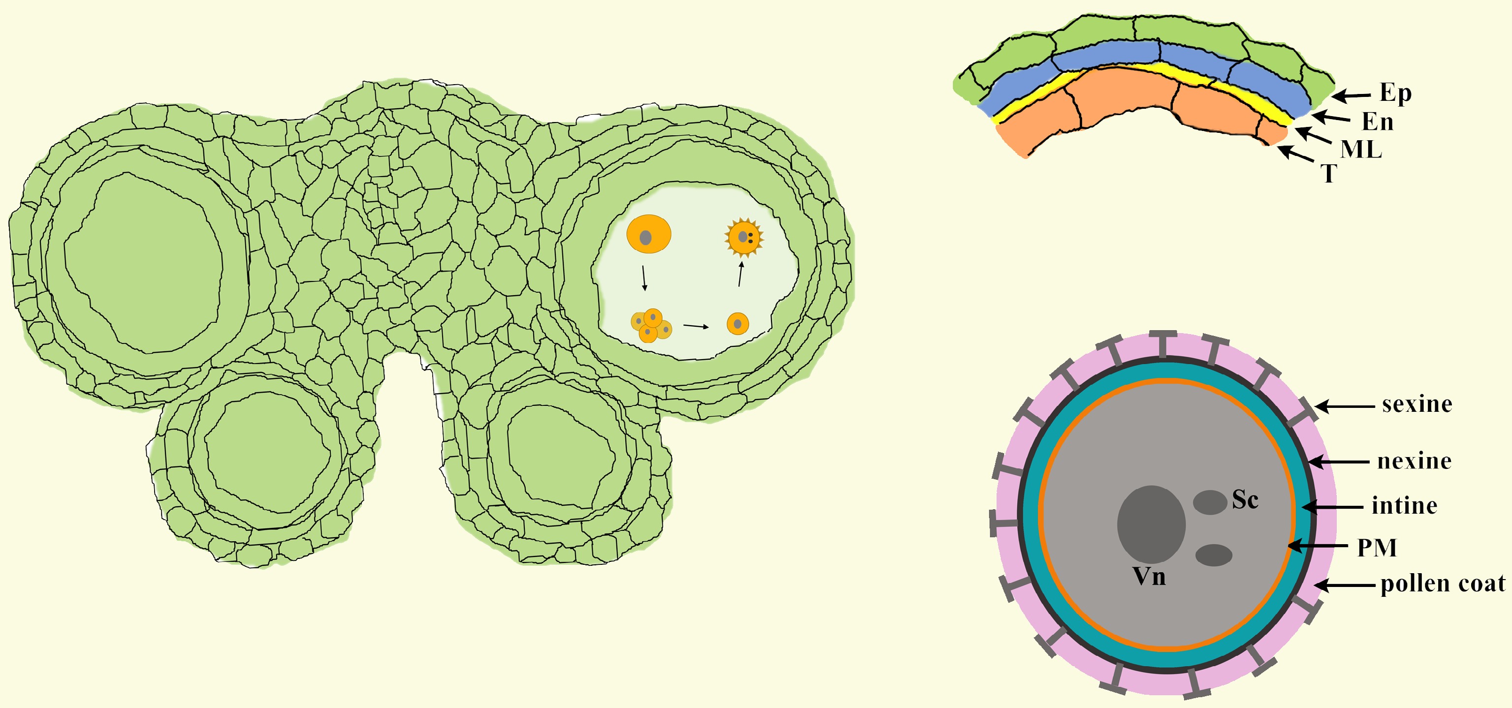
      Figure 1. 

      The structure of anthers and pollen. In Arabidopsis, each anther has four anther locules (pollen sacs), and the anther wall around the anther locule is composed of the epidermis, endothecium, middle layer and tapetum. Mature pollen grains are produced inside the anther locule. A pollen grain has two sperm cells in the cytoplasm of the large vegetative cell and is covered with a complex pollen wall outside of the plasma membrane. Ep, epidermis; En, endothecium; ML, middle layer; T, tapetum; Vn, vegetative nucleus; Sc, sperm cell; PM, plasma membrane.

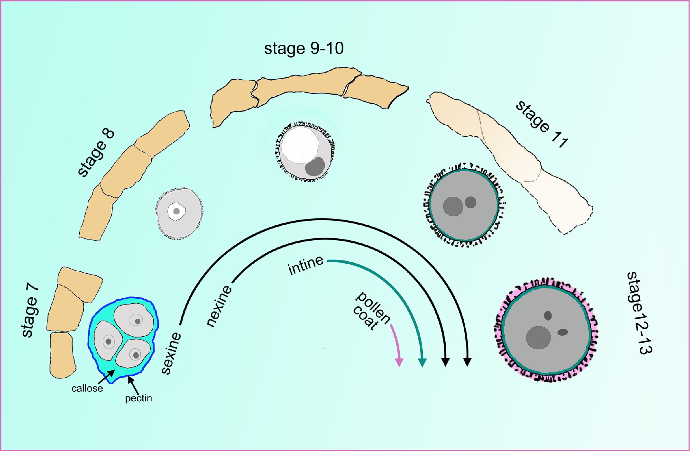
      In the anther locule, the diploid microsporocyte undergoes meiosis to form microspores that are enclosed inside a tetrad. After being released from tetrad, microspores undergo two rounds of mitosis to develop into mature pollen[6]. During these processes, the structure and composition of the cell wall undergo drastic changes (Fig. 2). The primary cell wall surrounding the microsporocyte is mainly composed of cellulose, hemicellulose and pectin[7,8]. Before meiosis, the cellulose of the primary cell wall is degraded, leaving the wall to be mainly composed of pectin. This structure is also termed the pectin wall[9,10]. At the initiation of meiosis, a layer of callose composed of β-1, 3-glucan (callose wall), is formed between the cell membrane and the pectin wall. When meiosis is completed, tetrads are formed with four haploid microspores enclosed inside the thick callose wall and the outer pectin wall[10]. At the late tetrad stage, a layer of matrix, named primexine, that is composed of polysaccharides, cellulose and proteins is deposited between the plasma membrane and the callose wall of individual microspores[11]. It is widely recognized that primexine acts as a scaffold for the formation of pollen exine. The main component of the exine layer is sporopollenin, which is an extremely biochemically resistant material[12]. After the microspore plasma membrane undulates, sporopollenin is assembled at the peak of the undulation to form probacula and is finally shaped into the complete pollen exine[13]. The exine can be further divided into the outer sexine and nexine. When the exine preliminarily forms, an intine is formed beneath the nexine[14,15]. Finally, the sculptured cavities of the sexine are then filled with tryphine, and this structure is named the pollen coat. Mature pollen grains with multiple-layered pollen walls are ready to be released from anthers[12]. Several excellent reviews that focus on the structure of the pollen wall, the biosynthesis of sporopollenin and the formation of the pollen wall have been published[1621].

      Figure 2. 

      The cell wall undergoes a tremendous change during pollen development. The orange quadrilaterals represent the tapetal cells, and the corresponding microspores or pollen at specific anther stages are shown under the tapetum cells. At stage 7, the four microspores are enclosed inside the callose wall and the outer pectin wall. At late stage 7, the sexine and nexine precursors start to deposit outside the membrane. During stages 9 to 10, the tapetum begins to degenerate and becomes spongy. The intine layer appears between the plasma membrane and nexine layer at stage 10. At stage 11, the tapetum evidently degenerates, and the pollen coat precursor start to fill the sculptured cavities of the sexine. At stages 12 and 13, the tapetum cell degenerates completely, and all layers of the pollen wall are established.

      Tapetal cells exist in the microsporangium or anther of all land plants[22]. Ectopic expression of RNase in the tapetum cell leads to male sterility, implying a close connection between the tapetum and pollen formation[23]. As the 'nutrition cells' for the growth of microspores, tapetal cells have evolved several properties with high transcriptional and translational activity. In Arabidopsis, the tapetum layer turns into polar secretory cells at the late development stage. Each tapetal cell contains two nuclei, and its cytoplasm is condensed and packed with abundant plastids, mitochondria and vesicular transport systems. During anther development, the tapetum cell undergoes PCD to provide enzymes and materials for pollen formation[2428]. The contribution of the tapetum to pollen development based on cytological observation has been extensively studied. In recent years, many genes essential for anther development have been discovered in male-sterile plants. Many of these genes are expressed in the tapetum and are essential for pollen formation. Here, we combine the cytological and molecular results of recent progresses in this field to propose a cascade contribution of the tapetum to pollen development, including nutrition supply, microspore release, exine deposition, pollen coat formation, and we also introduce the tapetum function for providing small RNAs to regulate genic methylation in germline cells.

    • In Arabidopsis, five transcription factors specifically expressed in the tapetum have been proven to be critical for pollen formation. DYSFUNCTIONAL TAPETUM 1 (DYT1) and ABORTED MICROSPORE (AMS) are basic helix-loop-helix (bHLH) family members[2931]. DEFECTIVE IN TAPETAL DEVELOPMENT AND FUNCTION 1 (TDF1) and MS188/MYB103/MYB80 encode R2R3 MYB transcription factors[32]. MALE STERILITY 1 (MS1) is a plant homeodomain (PHD)-finger transcription factor[33,34]. In mutants of dyt1, tdf1 and ams, their hypertrophic tapetal cells occupy the locule and crush the microspores[32]. In ms188 and ms1, the tapetal cells are defective, as they have a turgid shape. However, anther locules can form, suggesting their essential roles in late tapetal development[3337]. Based on gene expression, cytological and genetic analyses, a genetic pathway (DYT1-TDF1-AMS- MS188-MS1) was identified[38]. In addition to these five key transcription factors, several other transcription factors are redundantly involved in the development of the tapetum. Two GAMYB-like genes, MYB33 and MYB65, influence the development of the tapetum and pollen. In the myb33 myb65 mutant, the tapetal cells become hypertrophic, leading to pollen abortion. This phenotype is similar to that of tdf1. Under low temperature or high light, the fertility of myb33 myb65 increases, implying that MYB33 and MYB65 play an additional role in modulating fertility at decreased temperatures[39]. Additionally, three bHLH genes in Arabidopsis, bHLH010, bHLH089 and bHLH091 are redundantly required for early tapetum development. The bhlh010 and bhlh089 single mutants display normal fertility. However, the tapetal cells of the bhlh triple mutant were abnormally expanded and irregularly organized, which is similar to the phenotype in dyt1. These three bHLH proteins interact with DYT1 and may influence the function of DYT1. In the bhlh triple mutant, the expression of MYB103, MS1 and MYB99 was reduced. This implies that these three bHLH transcription factors redundantly regulated tapetum development by interacting with DYT1 and affecting the expression of many target genes, such as MYB103, MS1 and MYB99[40,41].

      In the past 10 years, molecular and biochemical evidence has further shown that the five genes of the genetic pathway are sequentially activated during tapetum development. DYT1 directly binds to the promoter of TDF1 to activate its transcription. The expression of TDF1 is driven by the DYT1 promoter and could rescue the transcription of AMS, MS188, MS1 and a series of pollen wall-related genes in the dyt1 background. This indicates that DYT1 regulates pollen wall formation via TDF1[42]. TDF1 directly regulates the expression of AMS[43] which further regulates the expression of MYB80/MS188[44,45]. Finally, MYB80/MS188 regulates the expression of MS1[46] (Fig. 3). Based on this genetic pathway, several feed-forward loops are formed to facilitate the expression of downstream targets (Fig. 3). TDF1 interacts with AMS to activate its regulation of downstream gene expression[43,44]. In addition, AMS and MS188 form a complex and facilitate the expression of sporopollenin synthesis genes[47,48]. These transcription factors, together with feed-forward loops, form a regulatory network that rapidly regulates tapetum development and pollen formation. The detailed downstream factors of these transcription factors are reviewed and summarized in the following sections (Table 1).
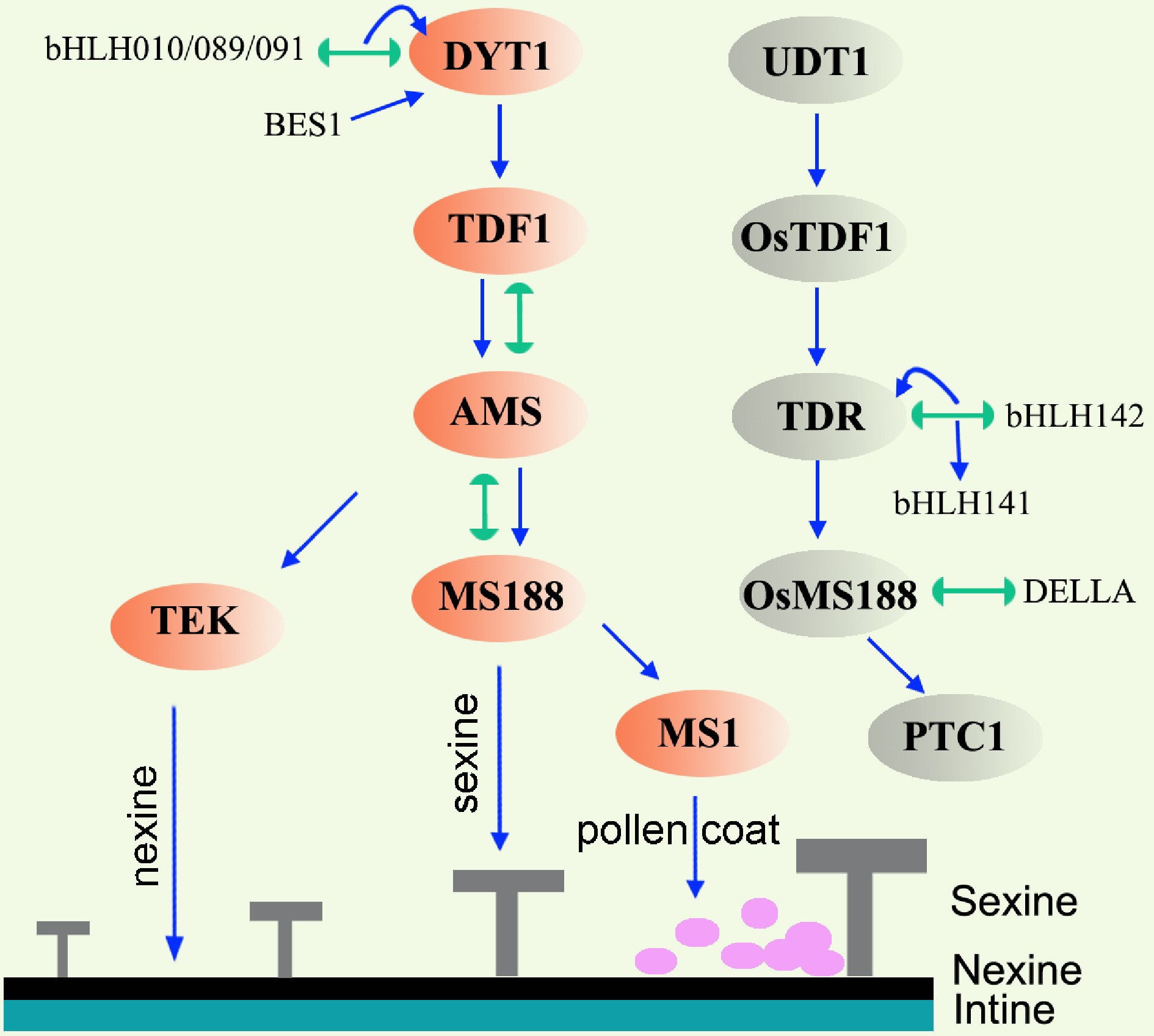
      Figure 3. 

      Gene regulatory network in the tapetum of Arabidopsis and rice. Lines terminating in arrows represent positive regulation, lines with semicircle ends indicate interaction. Orange ovals and grey ovals mark the key transcription factors in Arabidopsis and rice respectively. In Arabidopsis, DYT1-TDF1-AMS are responsible for early tapetum development. AMS regulates nexine and sexine formation via TEK and MS188, respectively. MS1 is responsible for pollen coat formation. Abbreviations: TEK, transposable element silencing via AT-hook; BES1, BRI1 EMS SUPPRESSOR 1; UDT1, UNDEVELOPED TAPETUM 1; TDR, TAPETUM DEGENERATION RETARDATION; PTC1, PERSISTANT TAPETAL CELL 1.

      Table 1.  The summary of the key genes and their functions during anther or pollen development.

      NameIDProteinFunctionReference
      DYT1AT4G21330bHLH transcription factorEarly tapetum development[30]
      TDF1AT3G28470MYB transcription factorEarly tapetum development[32]
      AMSAT2G16910bHLH transcription factorEarly tapetum development[29,31]
      MS188/MYB80/MYB103AT5G56110MYB transcription factorTapetum PCD, microspore release, exine formation[24,35,47]
      MS1AT5G22260PHD-finger transcription factorTapetum PCD, exine and pollen coat formation[33,34]
      OsUDT1Os07g0549600bHLH transcription factorDYT1 ortholog; early tapetum development[49]
      OsTDF1Os03g18480MYB transcription factorTDF1 ortholog; early tapetum development[53]
      OsTDROs02g0120500bHLH transcription factorAMS ortholog; tapetum development[50,54]
      OsMS188/OsMYB80Os04g39470MYB transcription factorMS188 ortholog; tapetum PCD, exine formation[51,55,56]
      OsPTC1/OsMS1Os09g0449000PHD-finger transcriptional factorMS1 ortholog; tapetum PCD, exine formation[50,52,54]
      ZmMs32GRMZM2G163233bHLH transcription factorDYT1 ortholog; tapetum development[21,57,62]
      ZmMs9GRMZM2G308034MYB transcription factorTDF1 ortholog; tapetum development[57,61]
      ZmbHLH51Zm00001d053895bHLH transcription factorAMS ortholog; tapetum development[57]
      ZmMYB84Zm00001d025664MYB transcription factorMS188 ortholog; tapetum development[57]
      ZmMs7GRMZM5G890224PHD-finger transcriptional factorMS1 ortholog; tapetum development[5759]
      MYB33AT5G06100GAMYB transcription factorTapetum and pollen development[39]
      MYB65AT3G11440GAMYB transcription factorTapetum and pollen development[39]
      bHLH010AT2G31220bHLH transcription factorbHLH010, bHLH089 and bHLH091 redundantly required for tapetum and pollen development[40]
      bHLH089AT1G06170bHLH transcription factor[40]
      bHLH091AT2G31210bHLH transcription factor[40]
      EAT1/DTD1/bHLH141Os04g0599300bHLH transcription factorTapetum PCD[63,64]
      TIP2/bHLH142Os01g0293100bHLH transcription factorTapetum PCD[65,66]
      MGT5AT4G28580Transmembrane magnesium transporterTransport Mg from tapetum to anther locule[91]
      QRT3AT4G20050polygalacturonase
      Pectin dissolution[10,98]
      A6AT4G14080β-1,3-glucanaseCallose dissolution[107,110]
      UPEX1/KNS4/RES3AT1G33430Arabinogalactan β-(1,3)-galactosyltransferaseInfluence the secretion of A6 from tapetum[110]
      ACOS5AT1G62940Fatty acyl-CoA synthetaseSporopollenin synthesis[112]
      CYP703A2At1G01280Hemethiolate monooxygenase (P450)Sporopollenin synthesis[47,113]
      CYP704B1AT1G69500Hemethiolate monooxygenase (P450)Sporopollenin synthesis[114]
      PKSAAT1G02050AcyltransferaseSporopollenin synthesis[116,118]
      PKSBAT4G34850AcyltransferaseSporopollenin synthesis[116,118]
      TKPR1AT4G35420Tetraketide alpha-pyrone reductaseSporopollenin synthesis[117]
      TKPR2AT1G68540Tetraketide alpha-pyrone reductaseSporopollenin synthesis[117]
      MS2AT3G11980Fatty acid reductaseSporopollenin synthesis[115,116]
      ABCG26AT3G13220ATP binding cassette transporterSporopollenin transportation[120,121]
      ABCG15AT3G21090ATP binding cassette transporterSporopollenin transportation[123]
      TEKAT2G42940AT-hook nuclear localized (AHL) proteinNexine formation[44,103]
      OsOSC12Os08g0223900Bicyclic triterpene poaceatapetol synthasePollen coat formation[139]
      GRP17AT5G07530Glycine-rich proteinPollen coat protein[140,141]
      EXL4AT1G75910Lipase proteinPollen coat protein[140,142]
      EXL6AT1G75930Lipase proteinPollen coat protein[140]
      CER1AT1G02205DecarbonylasesPollen coat synthesis: very long chain alkane synthesis[152,153]
      CER3/FLP1/WAX2/YREAT5G57800DecarbonylasesPollen coat synthesis: very long chain alkane synthesis[149151,153]
      KCS7AT1G711603-ketoacyl-CoA synthasePollen coat synthesis: fatty acid elongation[146]
      KCS15AT3G521603-ketoacyl-CoA synthasePollen coat synthesis: fatty acid elongation[146]
      KCS21AT5G490703-ketoacyl-CoA synthasePollen coat synthesis: fatty acid elongation[146]
      Dcl5Zm00001eb104810EndoribonucleaseGeneration of 24-nt phasiRNAs in the tapetum in maize[181]
      CLSY3AT1G05490HelicaseGeneration of 24-nt siRNAs in the anther in Arabidopsis[182]

      In rice, the homologies of the five transcription factors in the pathway have been identified[20,4953] (Fig. 3). UNDEVELOPED TAPETUM 1 (UDT1), a bHLH transcription factor that shows high homology with DYT1, plays a major role in the differentiation of tapetal cells[49]. OsTDF1 is an orthologue of Arabidopsis TDF1, and the ostdf1 knockout mutant displays vacuolated and hypertrophic tapetal cells, which is similar to the tdf1 mutant[53]. The TAPETUM DEGENERATION RETARDATION (TDR) gene, an orthologue of AMS, has been proven to be a critical component in regulating tapetum development in rice and is important for aliphatic metabolism in pollen[50,54]. PERSISTANT TAPETAL CELL1 (PTC1)/OsMS1 is the orthologue of Arabidopsis MS1[50,52,54]. The function of OsMS188/OsMYB80 has been reported recently. The osms188/osmyb80 mutant exhibited aberrant degradation of tapetal cells, lack of sexine and microspore degeneration[55,56]. Similar to Arabidopsis, the OsUDT1-OsTDF1-OsTDR-OsMS188-PTC1 genetic pathway is present in the rice tapetum[53,56]. In maize, the homologies of AtDYT1/OsUDT1, AtTDF1/OsTDF1, AtAMS/OsTDR, AtMS188/OsMS188, and AtMS1/OsPTC1 are ZmMs32, ZmMs9, ZmbHLH51, ZmMYB84, and ZmMs7, respectively[5762]. Mutations in all five genes all lead to male sterility[5762]. A relatively conserved genetic pathway was also proposed in maize[57]. It seems that the genetic pathway composed of the five key transcription factors in Arabidopsis, rice, and maize is conserved, which is consistent with the conservative cytological processes in monocotyledons and dicotyledons[20].

      In addition to the five conserved transcription factors, two other bHLH family members have been identified to be essential for tapetal function in rice. ETERNAL TAPETUM 1 (EAT1)/DTD1/bHLH141 positively promotes PCD in tapetal cells by directly regulating the transcription of two aspartic protease-encoding genes, OsAP25 and OsAP37[63,64]. TDR INTERACTING PROTEIN2 (TIP2)/bHLH142 regulated the expression of both TDR and EAT1. TIP2/bHLH42 interacts with TDR to form a heterodimer and regulates the expression of EAT1[65,66]. EAT1 and TIP2 share sequence similarity with bHLH010, bHLH089 and bHLH091. However, unlike the redundant roles of the three bHLH genes for tapetum development in Arabidopsis, both the tip2 and eat1 single mutants display delayed tapetal PCD. This finding indicates that they are both essential regulators of tapetal PCD in rice. In addition to delayed PCD phenotypes, the three inner layers of the anther wall of tip2 but not eat1 remained undifferentiated from stage 7 to stage 8, implying the specific function of TIP2 in the differentiation of these cells[63,65].

      Plant hormones are important for plant growth. Auxin (IAA), gibberellin (GA) and brassinosteroid (BR) hormonal signals are integrated into the tapetum genetic program for anther and pollen development (Fig. 3). Auxin is involved in anther morphogenesis and pollen formation[6769]. ARF17, an auxin response factor, is expressed in microsporocytes, microspores, tapetum, and endothecium[7072]. In the arf17 mutant, the tapetum development is defective, and the pollen wall pattern cannot be formed[70,71]. However, the detailed relationship between ARF17 and the DYT1-TDF1-AMS-MS188-MS1 genetic pathway is unknown. In addition to its function in the tapetum, ARF17 is also involved in callose wall degradation and anther dehiscence[70,72]. BR mutants exhibited abnormal tapetal development, reduced pollen production, and an irregular pollen exine pattern[73,74]. BRI1 EMS SUPPRESSOR 1 (BES1) is a key factor in the BR signalling pathway. BES1 acts upstream of DYT1 to regulate tapetum development in Arabidopsis[73,74]. In addition to the tapetal defects discovered in the GAMYB mutant myb33 myb65 in Arabidopsis, common defects in tapetal PCD and exine formation were found in GA-deficient, GA-insensitive and gamyb mutants in rice[75]. Moreover, the application of exogenous GA rescues the male infertility caused by low temperature stress[76]. These results suggest that GA participates in the regulation of anther/pollen development. DELLA/SLR1 is the central negative regulator of GA signalling. Similar to myb33 myb65, the hypertrophy phenotype in tapetal cells is present in a DELLA loss-of-function double mutant lacking both the DELLA paralogues REPRESSOR OF ga1-3 (RGA) and GA INSENSITIVE (GAI)[77]. Recently, it was reported that rice DELLA/SLR1, is required for tapetum development[78]. In the slr1 mutant in rice, the programmed cell death of the tapetum is premature, and pollen is aborted without exine formation. As an important transcription factor, OsMS188 interacts with SLR1 to cooperatively activate the expression of sporopollenin biosynthesis genes, such as CYP703A3, DEFECTIVE POLLEN WALL (DPW), and POLYKETIDE SYNTHASE (PKS1) and the sporopollenin transport-related gene ABCG15. The activation of these genes may be responsible for subsequent pollen wall formation[78]. Thus, a GA–DELLA–OsMS188 module has been revealed to control the development of the male reproductive system.

    • After being released from tetrads, microspores are immersed in the locule nutritive fluid whose composition fluctuates during anther development. The locular fluid contains sugars, proteins, amino acids and sporopollenin precursors during early microspore growth, and precursors for pollen coat formation during the late pollen maturation stage[79,80]. These substances in the locular fluid are secreted from the tapetum cells to meet the requirement of normal growth microspore development[80]. Extracellular invertase is responsible for sugar hydrolysis[81]. In tobacco, Nin88 encodes an extracellular invertase isoenzyme, and it is specifically expressed in tapetum and pollens. Antisense repression of Nin88 or over-expression of an invertase inhibitor under the Nin88 promoter all results in pollen abortion in tobacco[82,83]. AtcwINV2 is a homologous gene of Nin88 in Arabidopsis and it is specifically expressed in anther. Antisense repression of AtcwINV2 leads to reduced seed setting and pollen germination[84]. All these results indicated that sugars and their hydrolytic products in the anther especially in the tapetum are critical for pollen formation[82,83,85]. In rice, CARBON STARVED ANTHER (CSA) encodes a tapetum-expressed R2R3 MYB transcription factor. It regulates the transcription of MST8, a monosaccharide transporter, for sugar partitioning during anther development[86]. Magnesium is a divalent metal cation essential for living cells. In plants, the magnesium transporter (MGT) is responsible for the absorption and transport of Mg. In Arabidopsis, the magnesium transporter family contains 10 members[87,88]. MGT4, MGT5 and MGT9 are expressed in pollen and have the ability to absorb Mg from anther locule fluid for pollen formation[89,90]. Additionally, MGT5 and MGT6 are also expressed in the tapetum[91,92]. In the mgt5, mgt5+/- and mgt6+/- mutants, pollen mitosis is abnormal, and pollen intine is defective. These effects lead to pollen abortion. AMS directly regulates the expression of MGT5 to export Mg from the tapetum to the locular fluid[91] (Fig. 4). In conclusion, MGT5 plays dual roles as both a sporophytic and gametophytic gene. It not only exports Mg from the tapetum but also absorbs Mg into pollen. In the meantime, other MGTs may play essential or redundant roles in the tapetum or pollen to provide sufficient amounts of Mg for pollen growth.
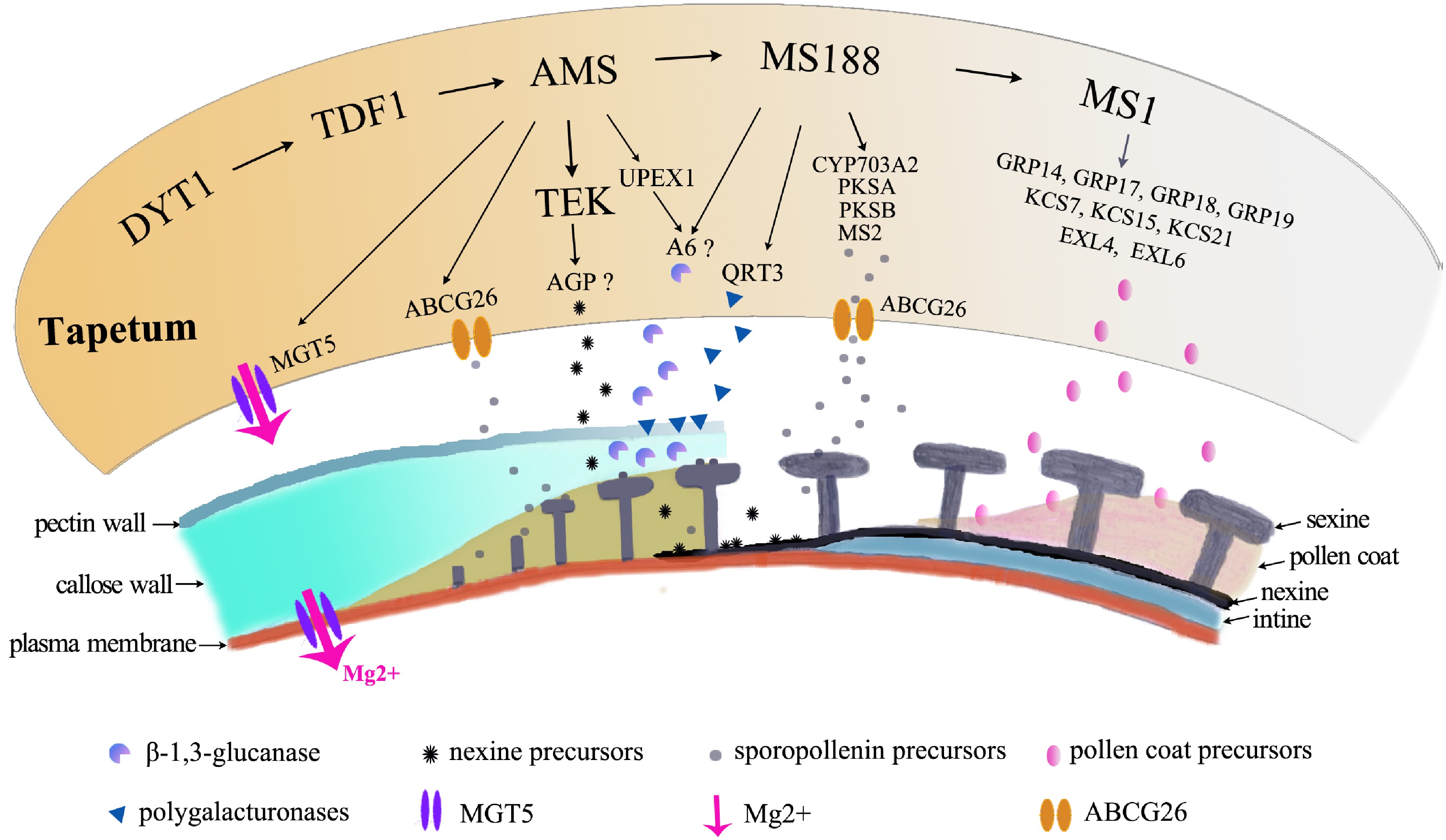
      Figure 4. 

      Molecular pathways in tapetum contribution to pollen formation. The orange irregular shape represents the tapetal cell. The pathway regulates a large number of genes for pollen growth, which are shown below the tapetal cell, to provide Mg for pollen growth, to secrete enzymes for degradation of the pectin wall, for the callose wall to release microspores, to provide precursors of nexine and sexine, and to provide materials for pollen coat formation.

    • In addition to its nutritive function, the tapetum is also responsible for tetrad wall degeneration. The wall of the tetrad is composed of a thin pectin wall and a thick callose wall. The timely degradation of the tetrad wall ensures the release of the individual microspores into the anther locule for further maturation. The pectin wall consists of homogalacturonan, rhamnogalacturonan I and rhamnogalacturonan II. The degradation of pectin requires pectin methylesterases (PMEs) and polygalacturonases (PGs)[9395]. Failure to degrade the pectin layer following meiosis results in the formation of tetrahedral clusters of four pollen grains. This phenotype was observed in the quartet (qrt) mutants in Arabidopsis[10,96,97]. Currently, three QRT genes have been cloned. QRT1 encodes a PME, while QRT2 and QRT3 encode PGs[98100]. Pectin is first demethylated by QRT1 and then degraded by QRT2 and QRT3 to loosen and degrade the pectin wall. All these QRTs are expressed in tapetal cells and are secreted into the locule. MS188 directly regulates QRT3 expression[10](Fig. 4). Premature expression of QRT3 in the tapetum using the A9 promoter leads to irregular exine formation, indicating that the timely degradation of the pectin wall is important for pollen wall formation[10].

      The callose wall is mainly composed of β-1,3-glucan. A decrease or loss of callose synthesis leads to a defective pollen wall pattern, indicating that the callose layer is essential for sporopollenin deposition[35,70,101103]. β-1,3-Glucanase (callase) is secreted from the tapetum cells for the degradation of the callose layer[104106]. In Arabidopsis and Brassica napus, anther-specific protein 6 (A6) is considered to be a β-1,3-glucanase that digests the callose wall[107]. However, in the a6 mutant, the callose wall is degraded normally, implying that other genes encoding β-1,3-glucanases are also involved in callose wall degradation. A6 is specifically expressed in the tapetum and has a sharp peak in activity immediately before microspore release. In the ms188 mutant, the expression of A6 is decreased and the degradation of callose is delayed[35]. In the ams mutant, both the accumulation and dissolution of callose are abnormal, and the expression of A6 is also decreased. AMS and MS188 may determine callose degradation by regulating the expression of A6[35,108] (Fig. 4). UNEVEN PATTERN OF EXINE1 (UPEX1)/KAONASHI (KNS4)/ RESTORER OF REVERSIBLE MALE STERILE 3 (RES3) encodes a glycosyltransferase that is directly regulated by AMS in the tapetum[109111]. In the res3 mutant, the secretion of A6 and other β-1,3-glucanases from the tapetum to the locule was delayed, which further affected the release of microspores from tetrads. It seems that AMS and MS188 regulate A6 and its family members during callose wall degradation. The authors also suggested that the delayed callose degradation in the res3 mutant may be a general mechanism by which fertility can be restored in multiple sterility lines[111], implying its application prospects in hybrid breeding.

    • The outer pollen wall exine is composed of an outer sculptured sexine layer and an inner nexine layer. The major component of sexine was considered to be sporopollenin, which is composed of biopolymers of long-chain fatty acids and aromatics. The sophisticated pathway for the synthesis of long-chain fatty acids for sporopollenin monomer formation has been well documented based on genetic phenotypes and biochemical activity[17,21]. A series of enzymes, such as ACYL-CoA SYNTHETASE5 (ACOS5), CYP703A2, CYP704B1, POLYKETIDE SYNTHASE A (PKSA), PKSB, TETRAKETIDE α-PYRONE REDUCTASE1 (TKPR1), TKPR2 and MALE STERILE 2 (MS2), are involved in this biochemical pathway in the tapetum[17,21]. ACOS5 may play a role as a fatty acyl-CoA[112]. CYP703A2 and CYP704B1 are two members of the cytochrome P450 family that are involved in catalysing the hydroxylation of different long chain fatty acids[113,114]. The hydroxylated products are either converted to fatty alcohols by MS2 or catalysed by PKSA and PKSB into triketide and tetraketide α-pyrones[115,116]. Then, the tetraketide α-pyrones are believed to be reduced by TKPR1 and TKPR2 to form polyhydroxylated tetraketide[117119]. Most of these enzymes are specifically/abundantly expressed in the tapetum cell[47,48,108] (Fig. 4). In ms188, sexine is completely absent[35]. MS188 directly regulates the transcription of these genes for the establishment of sexine[48]. AMS binds to the promoter of several important pollen wall formation genes such as CYP703A2, CYP704B1, PKSB and TKPR1[108]. Furthermore, AMS interacts with MS188. AMS and MS188 may form a feed-forward loop to facilitate the expression of sporopollenin synthesis genes for sexine formation[47,48]. The synthesized sporopollenin precursors are predicted to be transported by members of the ATP-binding cassette transporter superfamily such as ABCG26 or ABCG15, in Arabidopsis and rice, respectively[120123] (Fig. 4). The expression of these ABCGs is also regulated by tapetal transcription factors[56,108]. Overall, both the biosynthesis and export of sporopollenin precursors are primarily regulated by MS188 in the tapetal cells.

      In addition to long-chain fatty acids, phenolics were also reported to be an essential component of sporopollenin. As early as 1987, researchers detected several phenolic materials in sporopollenin[124]. However, conflicting results were obtained via different methods[125]. In 2019, Li and colleagues showed that the sporopollenin of pine is primarily composed of aliphatic-polyketide-derived polyvinyl alcohol units and 7-O-p-coumaroylated C16 aliphatic units[126]. However, in 2020, Mikhael et al., carried out high-resolution X-ray photoelectron spectroscopy (HR-XPS) and showed the absence of aromaticity in the sporopollenin exine of Lycopodium clavatum[127]. Using genetic, biochemical and cell biology techniques, Xue et al., identified that in vascular plants, phenylpropanoid derivatives are another component of sporopollenin. The genes encoding enzymes of the phenylpropanoid synthesis pathway are expressed in the tapetum in Arabidopsis. NMR studies have shown that the sporopollenin composition of ferns and lycophytes is different from that of seed plants[128]. It is known that sporopollenin can absorb UV to protect pollen[129]. Xue et al. demonstrated that phenylpropanoid derivatives are essential for UV protection in pollen[128]. In conclusion, genetic evidence shows that both aliphatic units and phenypropanoid phenolics are essential components of the sporopollenin wall.

    • Nexine is a layer between the sexine and an inner intine. Usually, this cell wall is observed under transmission electronic microscopy in seed plants. As it is difficult to isolate this layer for composition analysis, the current understanding of this layer is quite obscure. In Arabidopsis, TRANSPOSABLE ELEMENT SILENCING VIA AT-hook (TEK) encodes an AT-hook nuclear localized (AHL) protein. The nexine layer is absent in the tek mutant, but sexine is normally formed, indicating that the formation of sexine is independent of the nexine layer[44] (Fig. 4). In the tapetum, AMS directly regulates MS188/MYB80 for sexine formation. TEK is strongly expressed in the tapetum at stage 7 and is also a direct target of AMS[44]. Therefore, AMS directly regulates MS188 for sexine formation and regulates TEK for nexine formation (Fig. 4). TEK was found to regulate the transcription of genes encoding arabinogalactan proteins (AGPs)[130]. However, the presence of AGPs in nexine has not yet been verified.

    • The pollen coat, which covers the surface or fills the sculptured cavities of the sexine, is responsible for pollen stigma interactions and pollen hydration and protects pollen from harsh environmental stress[13,131136]. Recently, two pollen coat-specific staining dyes: pollen-coat-stain (PCS) 52 and PCS 184, were identified. These two pollen coat dyes together with the exine dye basic fuchsin (BF) clearly stain the pollen coat and pollen wall in vivo in angiosperms[137].

      The pollen coat is composed of proteins, lipids, isoprenoids, and glycoconjugates[133,138]. In rice, OsOxidosqualene cyclases 12 (OsOSC12) encodes a bicyclic triterpene synthase and plays a role in the triterpene pathway. It is expressed in tapetal cells. Deficiency of OsOSC12 leads to a defective pollen coat and shows a humidity-sensitive genic male sterility (HGMS) phenotype. These findings imply that the tapetum-synthesized triterpene is an essential component in the pollen coat to prevent dehydration of pollen grains[139]. In Arabidopsis, pollen coat proteome analysis revealed that pollen coat proteins consist mainly of two families: lipid-binding oleosin or glycing-rich protein (GRP) and extracellular lipase (EXL)[140]. GRP17 accounts for the largest proportion of pollen coat proteins in Arabidopsis[140]. Mutations of GRP17 impair pollen hydration and the competitive ability, indicating the importance of this protein in hydration[141]. EXL4 and EXL6 were also identified in the pollen coat[140]. In the exl4 mutant, pollen hydration is slower. As a lipase, it was suggested that EXL4 may change the lipid composition to improve the ability of pollen to absorb water from the stigma[142]. Lipids are another main component of the pollen coat, and are important for pollen stigma communication and pollen hydration. Most of the detected lipids in the pollen coat are derivatives of very-long-chain fatty acids (VLCFAs)[143]. A number of mutants that disturb long chain lipid synthesis, such as eceriferum 1 (cer1), cer3/faceless pollen-1 (flp-1)/wax2/yre, 3-ketoacy-CoA synthase 7 (kcs7) kcs15 kcs21, and long-chain acyl-CoA synthetases 1 (lacs1) lacs4, show pollen coat defects[143146]. 3-Ketoacy-CoA synthase (KCS) catalyses fatty acid elongation[147,148]. CER1 and CER3/FLP1/WAX2/YRE may encode fatty acid hydroxylases and are involved in the synthesis of very long chain alkanes[149153]. It was reported that several pollen coat proteins or lipid synthesis-related enzymes are expressed predominantly or specifically in tapetal cells[46,108,146,154,155], indicating the important role of the tapetum in providing materials for pollen coat formation. ms1 was the earliest reported male sterile mutant in Arabidopsis in 1968[156]. MS1 is a plant homeodomain (PHD)-finger transcription factor[33]. The pollen wall was defective in the ms1 mutant[157]. Recently, it has been found that MS1 regulates the transcription of several pollen coat protein genes, such as GRP14, GRP17, GRP18, GRP19, EXL4, and EXL6, and pollen coat lipid synthesis genes, such as KCS7, KCS15, and KCS21[46,108,146] (Fig 4). Interestingly, it was observed that GRP19, EXL6, KCS20, KCS21 proteins are secreted into the anther locule before tapetal degradation[46,146]. These results suggest that instead of being passively released into the anther locule after tapetal degeneration, pollen coat precursors may be prepared in advance under the regulation of MS1. MS1 is directly regulated by MS188. This indicates that following sporopollenin synthesis and sexine formation mediated by MS188, MS1 subsequently regulates the expression of pollen coat protein genes. This reveals that a regulatory cascade establishes the multiple layers of the pollen wall (Fig. 4).

      The tapetum provides the major components of the pollen coat. A recent investigation showed that endothecium and developing microspores also contribute to pollen coat formation[136,158162]. CER2 and CER2-like proteins are putative BAHD acyltransferases required for VLCFA elongation. CER2, CER2L2, and KCS6 were found to be expressed in the endothecium[162], and cer2 cer2l2 and cer6/kcs6 mutants all show severe pollen coat defects[163167]. It seems that the tapetum first secretes pollen coat proteins and lipids into the anther locule, and after the degeneration of tapetum cells, the endothecium continues to provide pollen coat lipids on the surface of mature pollen for pollen hydration. Pollen-produced cysteine-rich pollen coat proteins are also involved in pollen stigma interactions[159,160,168170]. POLLEN COAT PROTEIN B-class peptides (PCP-Bs) are cysteine-rich pollen coat proteins[169]. It has been recently established that pollen-born PCP-Bs bind to the ANJEA–FERONIA (ANJ–FER) receptor kinase complex, to decrease stigmatic ROS and facilitate pollen hydration[170]. This data indicates that the kinds of PCPs in the pollen coat are produced and provided from different tissues.

    • Small RNAs are important for plant development because they regulate the transcript levels of target genes and the expression of transposons. It has been previously reported that pollen-specific miRNAs exist in Arabidopsis and rice[171,172]. The transcripts of Arabidopsis MYB33/MYB65 and rice OsGAMYB/OsGAMYB-like genes are targeted by miR159[39,173]. Over-expression of miR159 in Arabidopsis and rice all leads to anther defects and results in male sterility, indicating the miR159-GAMYBs module should be strictly controlled for normal anther development[173,174]. ARF17 is a target gene of miR160. 5mARF17 transgenic plants, which avoid miR160-directed ARF17 cleavage, also showed tapetal defects. These results indicate that the fine-tuned expression of ARF17 by miR160 is critical for tapetum development[71]. More and more microRNAome in developing anthers of wild-type plants and male sterile lines in different species were obtained[175178]. In the future, it will be informative to investigate the detailed function of these potential miRNAs during anther and pollen development.

      Genome reprogramming in pollen is guided by small RNAs. In Arabidopsis pollen, transposable elements (TEs) are activated only in vegetative cells. However, TE siRNAs accumulate in pollen and sperm cells, suggesting that siRNA from the vegetative nucleus can target silencing in sperm cells[179]. In maize anthers, there are two classes of phased siRNAs: 21-nt phased siRNAs (phasiRNAs) and 24-nt phasiRNAs. The 24-nt phasiRNAs and their precursors accumulate preferentially in the tapetum and meiocytes. However, tapetal cells but not meiotic cells may be essentially required for 24-nt phasiRNA biogenesis in maize[180]. Dicer-like 5 (Dcl5) is required for the generation of 24-nt phasiRNAs in maize. In the dcl5 mutant, few or no 24-nt phasiRNAs were detected, tapetal cells were defective, and the mutant displayed temperature-sensitive male fertility. These results indicate that DcL5 and 24-nt phasiRNAs are important for normal tapetum development and male fertility and the tapetum is the source for 24-nt phasiRNA biogenesis[181]. Recently, it has been found in Arabidopsis that 24-nt siRNAs are synthesized by tapetal cells through the activity of the chromatin remodeler CLASSY 3 (CLSY3). The tapetum-derived siRNA then governs germline methylation and silences germline transposons[182]. More recently, a similar mechanism was discovered in maize. Zhou et al. reported that the 24-PHAS precursor and Dcl5 primarily accumulated in the tapetum. After synthesis, the 24-nt phasiRNAs may move from the tapetum to meiocytes and other somatic cell layers in the anther wall[183]. In conclusion, in both Arabidopsis and maize, the 24-nt siRNA required for normal anther and germline development is mainly provided from the tapetum and moves into the germline cells.

    • In recent decades, the key transcription factors regulating tapetum development have been identified. In Arabidopsis, the DYT1-TDF1-AMS-MS188-MS1 genetic pathway is not only important for tapetum development, but also provides a cascade regulation for pollen formation. First, DYT1 and TDF1 regulate early tapetum development when microsporocytes are undergoing meiosis in the anther locule. At the tetrad stage, AMS initiates nexine deposition by activating the expression of TEK and promoting sexine formation via MS188 to regulate the synthesis of sporopollenin precursors. Moreover, both AMS and MS188 play critical roles in the degradation of the pectin wall and callose to gradually release the microspores from the tetrad. Last, the downstream member in the genetic pathway, MS1, regulates the transcription of a series of pollen coat related genes for pollen coat formation. Thus, mature pollen grains with multiple-layered pollen walls are ready to be released from anthers. The genetic pathway consists of five key transcriptional factors that are relatively conserved in Arabidopsis, rice and maize. However, functions of other homologous between Arabidopsis and rice are different, such as Arabidopsis bHLH010/bHLH089/bHLH091 and rice bHLH141/bHLH142. Therefore, it is necessary to explore more factors involved in the regulation of tapetum among species, analyze their relationship associated with those key transcription factors, and establish a more comprehensive gene regulatory network for tapetum development.

      In future, the coordination between tapetum development and pollen formation remains to be explored. The composition of sporopollenin still remains to be deciphered. Although nexine is a conserved pollen cell wall layer in seed plants, its chemical composition is still unclear. During anther development, the cell wall of the pollen mother cell is transited to the pollen wall. This transition is critical for pollen formation and plant fertility. The enzymes that dissolve the primary cell wall of microsporocytes and the tetrad callose layer still need to be identified. Further study of these issues in different species will help us to further characterize the relationship between anther sporophytic tissues and microspores/pollens as well as the evolution of the complicated pollen wall. In future, it is also very important to study whether mutations of the key genes essential for tapetum development can also lead to sterile phenotypes in different kinds of crops, and explore the application prospects of these male sterile materials in hybrid breeding.

      • This work was supported by grants from National Science Foundation of China (31970520, 31870296). We thank Dr. Jun Zhu, Dr. Yue Lou and Dr. Jing-Shi Xue from Shanghai Normal University for critical reading and revising of the manuscript. We thank Kengyu Pan, Benshun Zhu and Yu Jiang from Shanghai Normal University for revising of the manuscript.

      • The authors declare that they have no conflict of interest.

      • Copyright: © 2022 by the author(s). Published by Maximum Academic Press on behalf of Hainan Yazhou Bay Seed Laboratory. This article is an open access article distributed under Creative Commons Attribution License (CC BY 4.0), visit https://creativecommons.org/licenses/by/4.0/.
    Figure (4)  Table (1) References (183)
  • About this article
    Cite this article
    Yao X, Hu W, Yang Z. 2022. The contributions of sporophytic tapetum to pollen formation. Seed Biology 1:5 doi: 10.48130/SeedBio-2022-0005
    Yao X, Hu W, Yang Z. 2022. The contributions of sporophytic tapetum to pollen formation. Seed Biology 1:5 doi: 10.48130/SeedBio-2022-0005

Catalog

    /

    DownLoad:  Full-Size Img  PowerPoint
    Return
    Return