ARTICLE   Open Access    

Foliar application of a mixture of putrescine, melatonin, proline, and potassium fulvic acid alleviates high temperature stress of cucumber plants grown in the greenhouse

More Information
  • Putrescine (Put), melatonin (MT), proline (Pro), and potassium fulvic acid (MFA) are widely used as plant growth regulators to enhance stress tolerance. However, the roles of their mixtures in response to stress are largely unknown. Here, we mixed Put with MT, Pro, and MFA (hereafter referred to as Put mixture) with different concentrations and foliar sprayed at different growth stages (seedling, flowering, and fruiting stage) of cucumber (Cucumis sativus L.) to investigate their roles on plant growth, fruit yield, and quality under high temperature stress. The foliar application of the Put mixture promoted cucumber growth, increased chlorophyll and Pro contents and net photosynthesis rate, and reduced the values of relative electrolyte leakage, H2O2 and malondialdehyde contents of cucumber leaves, indicating that treatment with Put mixture reduced the oxidative stress caused by high temperature. Furthermore, Put mixture-treated cucumber plants had lower fruit deformity rate and higher fruit yield compared with control. The contents of vitamin C and soluble solids of cucumber fruit significantly increased and the contents of tannin and organic acid decreased. The most profound effects were found in the plants treated with 8 mmol L−1 Put, 50 µmol L−1 MT, 1.5 mmol L−1 Pro and 0.3 g L−1 MFA every 7 d, three times at the seedling stage, indicating that cucumber seedlings treated with the mixture of Put, MT, Pro, and MFA significantly alleviated the negative effects of high temperature stress.
  • Salvia rosmarinus L. (old name Rosmarinus officinalis), common name Rosemary thrives well in dry regions, hills and low mountains, calcareous, shale, clay, and rocky substrates[1]. Salvia rosmarinus used since ancient times in traditional medicine is justified by its antiseptic, antimicrobial, anti-inflammatory, antioxidant, and antitumorigenic activity[1,2]. The main objective of the study is to evaluate the antimicrobial activity of different extracts of Salvia rosmarinus in vitro, and its compounds related to in silico targeting of enzymes involved in cervical cancer. Since the start of the 20th century, some studies have shown that microbial infections can cause cervical cancers worldwide, infections are linked to about 15% to 20% of cancers[3]. More recently, infections with certain viruses like Human papillomaviruses (HPV) and Human immunodeficiency virus (HIV), bacteria like Chlamydia trachomatis, and parasites like schistosomiasis have been recognized as risk factors for cancer in humans[3]. Then again, cancer cells are a group of diseases characterized by uncontrolled growth and spread of abnormal cells. Many things are known to increase the risk of cancer, including dietary factors, certain infections, lack of physical activity, obesity, and environmental pollutants[4]. Some studies have found that unbalanced common flora Lactobacillus bacteria around the reproductive organ of females increases the growth of yeast species (like Candida albicans) and some studies have found that women whose blood tests showed past or current Chlamydia trachomatis infection may be at greater risk of cervical cancer. It could therefore be that human papillomavirus (HPV) promotes cervical cancer growth[3]. Salvia rosmarinus is traditionally a healer chosen as a muscle relaxant and treatment for cutaneous allergy, tumors, increases digestion, and the ability to treat depressive behavior; mothers wash their bodies to remove bacterial and fungal infections, promote hair growth, and fight bad smells[5] .

    The study of plant-based chemicals, known as phytochemicals, in medicinal plants is gaining popularity due to their numerous pharmacological effects[6] against drug resistance pathogens and cancers. The causes of drug resistance to bacteria, fungi, and cancer are diverse, complex, and only partially understood. The factors may act together to initiate or promote infections and carcinogenesis in the human body is the leading cause of death[7]. Antimicrobial medicines are the cornerstone of modern medicine. The emergence and spread of drug-resistant pathogens like bacteria and fungi threaten our ability to treat common infections and to perform life-saving procedures including cancer chemotherapy and cesarean sections, hip replacements, organ transplantation, and other surgeries[7]. On the other hand, information about the current magnitude of the burden of bacterial and fungal drug resistance, trends in different parts of the world, and the leading pathogen–drug combinations contributing to the microbial burden is crucial. If left unchecked, the spread of drug resistance could make many microbial pathogens much more lethal in the future than they are today. In addition to these, cancers can affect almost any part of the body and have many anatomies and molecular subtypes that each require specific management strategies to avoid or inhibit them. There are more than 200 different types of cancer that have been detected. The world's most common cancers affecting men are lung, prostate, colorectal, stomach, and liver cancers[8]. While breast, cervix, colorectal, lung, and stomach cancers are the most commonly diagnosed among women[8]. Although some cancers said to be preventable they seem to still be one of the causes of death to humans, for example cervical cancer. The need to fill the gap to overcome the problem of searching for antimicrobials and anticancers from one source of Salvia rosmarinus is of importance.

    Cervical cancer is a common cancer in women and a prominent cause of death[9]. In Ethiopia, cervical cancer is a big deal for women aged 15 to 44, coming in as the second most common cancer[9]. Globally, it's the fourth most common prevalent disease for women[10]. Aberrant methylation of tumor-suppressor genes' promoters can shut down their important functions and play a big role in causing cervical tumors[10]. There are various cervical cancer repressor genes (proteins turn off or reduce gene expression from the affected gene), such as CCNA1, CHF, HIT, PAX1, PTEN, SFRP4, and TSC1. The genes play a crucial role in causing cervical cancer by regulating transcription and expression through promoter hypermethylation, leading to precursor lesions during cervical development and malignant transformation[11]. The process of DNA methylation is primarily carried out by a group of enzymes known as DNA methyltransferases (DNMT1). It has been reported that DNMT1 (PDB ID: 4WXX), a protein responsible for DNA methylation can contribute to the development of cervical cancer. DNMT1 inhibits the transcription of tumor suppressor genes, facilitating tumorigenesis, which finally develops into cervical cancer. Tumor suppressor gene transcription is inhibited by DNMT1, which helps cancer grow and eventually leads to cervical cancer. Repressive genes' hypermethylation may be decreased, their expression can be increased, and the phenotype of malignant tumors can be reversed by inhibiting the DNMT1 enzyme.

    On the other hand, infection by the human papilloma virus (HPV) phenotype 16, enzyme 6 (PDB ID: 4XR8) has been correlated with a greatly increased risk of cervical cancer worldwide[12]. Based on variations in the nucleotide sequences of the virus genome, over 100 distinct varieties of the human papilloma virus (HPV) have been identified (e.g. type 1, 2 etc.). Genital warts can result from certain types 6 and 11 of sexually transmitted HPVs. Other HPV strains, still, that can infect the genitalia, do not show any symptoms of infection[8]. Persistent infection with a subset of approximately 13 so-called 'high-risk' sexually transmitted HPVs, including such as types 16, 18, 31, 33, 35, 39, 45, 51, 52, 56, 58, 59, and 68 different from the ones that cause warts may lead to the development of cervical intraepithelial neoplasia (CIN), vulvar intraepithelial neoplasia (VIN), penile intraepithelial neoplasia (PIN), and/or anal intraepithelial neoplasia (AIN). These are precancerous lesions and can progress to invasive cancer. Almost all occurrences of cervical cancer have HPV infection as a required component[13]. Superfluous infection by HPV type 16 E6 (PDB ID: 4XR8) has been correlated with a greatly increased genital risk of precursor cervical cancer worldwide[11]. Scholars more defined in major biochemical and biological activities of HPV type 16 E6 (PDB ID: 4XR8) in high-risk HPV oncogenes and how they may work together in the development of cervical disease and cancer[13].

    One potential approach to treat cervical cancer is to inhibit the activity of the DNMT1 and HPV type 16 E6 enzymes specifically[1316]. Over 50% of clinical drug forms worldwide originate from plant compounds[17]. In the past, developing new drugs was a lengthy and costly process. However, with the emergence of bioinformatics, the use of computer-based tools and methods have become increasingly important in drug discovery. One such method is molecular docking and ADMET profiling which involves using the structure of a drug to screen for potential candidates. This approach is known as structure-based drug design and can save both time and resources during the research process[15]. Structural-based drug designing addresses ligand binding sites with a known protein structure[15]. Using free binding energies, a computational method known as docking examines a large number of molecules and suggests structural theories for impeding the target molecule[17]. Nowadays, due to increasing antibiotic resistance like bacteria, fungi, and cancer cells, natural products remain an important source for discovering antimicrobial compounds and novel drugs for anti-cancers like cervical cancers. Therefore, the purpose of this research is to assess the antimicrobial activity of extracts, molecular docking, ADMET profiling in anticancer properties of compounds isolated from Salvia rosmarinus, on a targeting DNMT1 and HPV type 16 E6 in human cervical cancer. In the present study, various solvent crude extracts obtained from Salvia rosmarinus were used for antimicrobial activity and the isolated compounds 1 and 2 were submitted for in silico study to target the DNMT1 and HPV type 16 E6 enzymes to inhibit the growth of human cervical cancer cells.

    Healthy Salvia rosmarinus leaves were collected in Bacho district, Southwest Showa, Oromia, Ethiopia, during the dry season of November 2022. The plant materials were authenticated by Melaku Wondafrish, Natural Science Department, Addis Ababa University and deposited with a voucher number 3/2-2/MD003-80/8060/15 in Addis Ababa University's National Herbarium.

    The most common organic solvent used in extractions of medicinal plants is 2.5 L of petroleum ether, chloroform/methanol (1:1), and methanol. The test culture medium for microbes was used and performed in sterile Petri dishes (100 mm diameter) containing sterile Muller–Hinton Agar medium (25 mL, pH 7) and Sabouraud Dextrose Agar (SDA) for bacteria and fungi, respectively. A sterile Whatman filter paper (No. 1) disc of 6 mm diameter was used to determine which antibiotics an infective organism is sensitive to prescribed by a minimum zone of inhibition (MZI). Ciprofloxacin antibiotic reference (manufactured by Wellona Pharma Ciprofloxacin tablet made in India) and Ketoconazole 2% (made in Bangladesh) were used as a positive controls for antibacterial and antifungal, respectively and Dimethyl sulfoxide (DMSO) 98.9% was used as a negative control for antimicrobial tests. In the present study, the height of the column was 650 mm and the width was 80 mm. Several studies by previous researchers showed the acceptable efficiency of column chromatography (up to 43.0% w/w recovery) in the fractionation and separation of phenolic compounds from plant samples[18]. In column chromatography, the ideal stationary phase used silica gel 60 (0.200 mm) particles. The 1H-NMR spectrums of the compounds were analyzed using a 600 MHz NMR machine and 150 MHz for 13C NMR. The compounds were dissolved in MeOD for compound 1 and in DMSO for compound 2 for NMR analysis. On the other hand, UV spectroscopy (made in China) used 570 nm ultraviolet light to determine the absorbency of flavonoids (mg·g−1) phytochemicals.

    The samples (extracts) were analyzed to detect the presence of certain chemical compounds such as alkaloids (tested using Wagner's reagents), saponins (tested using the froth test), steroids (tested with Liebermann Burchard's tests), terpenoids (tested with Lidaebermann Burchard's tests), quinones, and flavonoids (tested using Shinoda tests)[19].

    The leaves of Salvia rosmarinus (500 g) were successively extracted using maceration using petroleum ether, chloroform/methanol (1:1), and methanol, every one 2.5 L for 72 h to afford 3.6, 6, and 53 g crude extracts, respectively. The methanol/chloroform (1:1) extract (6 g) was loaded to silica gel (150 g) column chromatography using the increasing polarity of petroleum ether, methanol/chloroform (1:1) solvent system to afford 80 fractions (100 mL each). The fraction obtained from chloroform/methanol 1:1 (3:2) after repeated column chromatography yielded compound 1 (18 mg). Fractions 56-65, eluted with chloroform/methanol (1:1) were combined and purified with column chromatography to give compound 2 (10 mg).

    The microorganisms were obtained from the Ethiopia Biodiversity Institution (EBI). Two gram-positive bacteria namely Staphylococcus aureus serotype (ATCC 25923) and Streptococcus epidermidis (ATCC14990); and three gram-negative bacteria, namely Escherichia coli (ATCC 25922), Pseudomonas aeruginosa (ATCC 5702), and Klebsiella pneumonia (ATCC e13883) were inoculated overnight at 37 °C in Muller–Hinton Agar/MHA culture medium and two fungus strains of Candida albicans (ATCC 16404) and Aspergillus niger (ATCC 11414) were inoculated overnight at 27−30 °C in Sabouraud Dextrose Agar/SDA culture medium[20].

    The antibacterial and antifungal activities of different crude extracts obtained from Salvia rosmarinus plant leaves were evaluated by the disk diffusion method (in accordance with the 13th edition of the CLSI M02 document on hardydiagnostics.com/disk-diffusion). Briefly, the test was performed in sterile Petri dishes (100 mm diameter) containing solid and sterile Muller–Hinton Agar medium (25 mL, pH 7) and Sabouraud Dextrose Agar (SDA) for bacteria and fungi, respectively. The extracts were placed on the surface of the media that had previously been injected with a sterile microbial suspension (one microbe per petri dish) after being adsorbed on sterile paper discs (5 μL per Whatman disc of 6 mm diameter). To prevent test samples from eventually evaporating, all Petri dishes were sealed with sterile laboratory films. They were then incubated at 37 °C for 24 h, and the zone diameter of the inhibition was measured and represented in millimeters. Ciprofloxacin antibiotic reference (manufactured by Wellona Pharma Ciprofloxacin tablet, India) was used as a positive control and DMSO was used as a negative control for antibacterial activity test while Ketoconazole 2% (Bangladesh) was used as a positive control and 10 μL of 0.2% agar as a negative control for antifungal activity tests[20]. The term 'inhibitory concentration' refers to the minimum sample concentration required to kill 99.9% of the microorganisms present[21]. Three repetitions of the crude extract sample were used to precisely measure the inhibitory halo diameter (in mm), which was then expressed as mean ± standard deviation to assess the anti-microbial activity.

    Cervical cancer-causing protein was identified through relevant literature. The protein molecule structure of DNA (cytosine-5)-methyltransferase 1 (DNMT1) (PDB ID: 4WXX)[21] and HPV type 16 E6 (PDB ID: 4XR8)[21] - a protein known to cause cervical cancer - were downloaded from the Protein Data Bank[22]. The stability of the protein molecule was assessed using Rampage[23].

    Phytochemical constituents of Salvia rosmarinus plant leaves were used to select a source of secondary metabolites (ligands). Ligand molecules were obtained through plant extraction, and isolation, and realized with PubChem (https://pubchem.ncbi.nlm.nih.gov/). The ligands were downloaded in Silver diamine fluoride format (SDF) and then converted to PDB format using an online SMILES translator (https://cactus.nci.nih.gov/translate/). The downloaded files were in PDB format, which was utilized for running various tools and software[24].

    The Biovia Discovery Studio Visualizer software was used to analyze the protein molecule. The protein molecule was converted into PDB format and its hierarchy was analyzed by selecting ligands and water molecules. Both the protein molecule and the water molecules lost their attached ligands during the analysis. Finally, the protein's crystal structure was saved in a PDB file[25].

    PyRx software was utilized to screen secondary metabolites and identify those ligands with the lowest binding energy to the protein target. The ligands with the lowest binding energy were further screened for their drug-likeliness property through analysis. It is worth noting that PyRx runs on PDBQT format. To begin using PyRx, it needs to load a protein molecule. This molecule should be converted from PDB to the protein data bank, partial charge (Q), and Atom Type (PDBQT) format. Once the protein molecule is loaded, it can import ligands from a specific folder in Silver diamine fluoride format. The ligand energy was minimized and changed to PDBQT format. The protein was docked with the ligand and screened based on minimum binding energy (https://cactus.nci.nih.gov/translate/).

    The optimal ligand was selected for final docking using AutoDock Vina and Biovia by modifying the reference of Discovery Studio Client 2021 (https://cactus.nci.nih.gov/translate/).

    The protein target from the Protein Data Bank (PDB) was loaded onto the graphical interface of AutoDock Vina. To prepare the protein for docking, water molecules were removed, hydrogen polar atoms were added, and Kollman charges were assigned to the protein molecule. Ultimately, PDBQT format was used to store the protein. After being imported in PDB format, the Ligand molecule was transformed to PDBQT format. Next, a grid box was chosen to represent the docked region. The command prompt was used to run AutoDock Vina and the outcomes were examined (https://cactus.nci.nih.gov/translate/).

    Docking the ligand with the protein target DNMT1(PDB ID: 4WXX)[22] and HPV type 16 E6 (PDB ID: 4XR8)[21] enzymes were performed using Biovia Discovery Studio Client 2021 by loading the protein target first followed by the ligand in PDB format. The charges were attached to the protein molecule, and the energy was minimized for the ligands. Both the protein and ligand molecules were prepared for docking. Once the docking process was complete, the results were analyzed based on several parameters, including absolute energy, clean energy, conf number, mol number, relative energy, and pose number. The interaction between the protein and ligand was analyzed using structure visualization tools, such as Biovia Discovery Studio Visualizer and PyMol (https://cactus.nci.nih.gov/translate/).

    The process of visualizing the structure was carried out using the PyMol tool. PyMol is a freely available software. Firstly, the protein molecule in PDBQT form was loaded on the PyMol graphical screen. Then, the output PDBQT file was added. The docked structure was visualized and the 'molecule' option was changed to 'molecular surface' under the 'shown as' menu (https://cactus.nci.nih.gov/translate/).

    Drug likeliness properties of the screened ligands were evaluated using the SwissADME online server. SMILE notations were obtained from PubChem and submitted to the SwissADME web server for analysis. The drugs were subjected to Lipinski's rule of five[20] for analysis. Lipinski's rules of five were selected for final docking through AutoDock Vina and Biovia Discovery Studio Client 2021. Ligands 1 and 2 were analyzed using Lipinski's rule of five for docking with AutoDock Vina and Biovia Discovery Studio Client 2021.

    The antimicrobial analysis data generated by triplicate measurements reported as mean ± standard deviation, and a bar graph also generated by GraphPad Prism version 8.0.1 (244) for Windows were used to perform the analysis. GraphPad Prism was used and combined with scientific graphing, comprehensive bar graph fitting (nonlinear regression), understandable statistics, and data organization. Prism allows the performance and modification of basic statistical tests commonly used and determined through the statistical applications in microbiology labs (https://graphpad-prism.software.informer.com/8.0/).

    Phytochemical screening of the different extracts for the presence (+) and absence (−) of alkaloids, steroids, glycosides, coumarins, terpenoids, flavonoids, carbohydrates, tannins, and saponins were done. The present study showed that alkaloids, terpenoids, flavonoids, and tannins tests in S. rosmarinus leaves of petroleum ether, chloroform/methanol (1:1), and methanol extracts were high whereas glycoside, coumarins, and carbohydrates had a moderate presence. The extract of S. rosmarinus leaves contain commonly bioactive constituents such as alkaloids, steroids, terpenoids, flavonoids, tannins, and saponins. These bioactive chemicals have active medicinal properties. Phytochemical compounds found in S. rosmarinus leaves have the potential to treat cancer cells and pathogens. The study also found that these flavonoids are related to natural phenolic compounds with anticancer and antimicrobial properties in the human diet (Table 1).

    Table 1.  Phytochemical screening tests result of petroleum ether, chloroform/methanol (1:1) and methanol extracts of Salvia rosmarinus leaves.
    Botanical name Phytochemicals Phytochemical screening tests Different extracts
    Petroleum ether Chloroform/methanol (1:1) Mehanol
    Salvia rosmarinus Alkaloids Wagner's test ++ ++ ++
    Steroids Libermann Burchard test ++ + ++
    Glycoside Keller-Killiani test +
    Coumarins Appirade test + +
    Terpenoids Libermann Burchard test ++ ++ ++
    Flavonoids Shinoda test ++ ++ ++
    Carbohydrate Fehling's test ++ ++
    Tannins Lead acetate test ++ ++ ++
    Saponins Foam test + + +
    + indicates moderate presence, ++ indicates highly present, − indicates absence.
     | Show Table
    DownLoad: CSV

    Two compounds were isolated and characterized using NMR spectroscopic methods (Fig. 1 & Supplementary Fig. S1ac). Compound 1 (10 mg) was isolated as yellow crystals from the methanol/chloroform (1:1) leaf extract of Salvia rosmarinus. The TLC profile showed a spot at Rf 0.42 with methanol/chloroform (3:2) as a mobile phase. The 1H-NMR spectrum (600 MHz, MeOD, Table 2, Supplementary Fig. S1a) of compound 1 showed the presence of one olefinic proton signal at δ 5.3 (t, J = 3.7 Hz, 1H), two deshielded protons at δ 4.7 (m, 1H), and 4.1 (m, 1H) associated with the C-30 exocyclic methylene group, and one O-bearing methine proton at δH 3.2 (m, 1H), and six methyl protons at δ 1.14 (s, 3H), 1.03 (d, J = 6.3 Hz, 3H), 1.00 (s, 3H), 0.98 (s, 3H), 0.87 (s, 3H), and 0.80 (s, 3H). A proton signal at δ 2.22 (d, J = 13.5 Hz, 1H) was attributed to methine proton for H-18. Other proton signals integrate for 20 protons were observed in the range δ 2.2 to 1.2. The proton decoupled 13C-NMR and DEPT-135 spectra (151 MHz, MeOD, Supplementary Fig. S1b & c) of compound 1 revealed the presence of 30 well-resolved carbon signals, suggesting a triterpene skeleton. The analysis of the 13C NMR spectrum displayed signals corresponding to six methyl, nine methylene, seven methine, and eight quaternary carbons. Among them, the signal observed at δ 125.5 (C-12) belongs to olefinic carbons. The methylene carbon showed signals at δC 39.9, 28.5, 18.1, 36.7, 23.9, 30.4, 26.5, 32.9, and 38.6. The quaternary carbons showed a signal at δC 39.4, 41.9, 38.4, 138.2, 41.8, and 47.8. The signals of exocyclic methylene carbon signals appeared at δ 153.1 and 103.9. The spectrum also showed sp3 oxygenated methine carbon at δ 78.3 and carboxyl carbon at δ 180.2. The spectrum revealed signals due to methyl groups at δC 27.4, 16.3, 15.0, 20.2, 22.7, and 16.4. The remaining carbon signals for aliphatic methines were shown at δC 55.3, 55.2, 53.0, and 37.1. The NMR spectral data of compound 1 is in good agreement with data reported for micromeric acid, previously reported from the same species by Abdel-Monem et al.[26]. (Fig. 1, Table 2).

    Figure 1.  Structure of isolated compounds from the leaves of Salvia rosmarinus.
    Table 2.  Comparison of the 13C-NMR spectral data of compound 1 and micromeric acid (MeOD, δ in ppm).
    Position NMR data of compound 1 Abdel-Monem
    et al.[26]
    1H-NMR 13C-NMR 13C-NMR
    1 38.60 39.9
    2 27.8 28.5
    3 3.2 (m, 1H) 78.3 80.3
    4 39.4 39.9
    5 55.3 56.7
    6 18.1 18.3
    7 36.7 34.2
    8 41.9 40.7
    9 53 48.8
    10 38.4 38.2
    11 23.9 24.6
    12 5.3 (t, J = 3.7 Hz, 1H) 125.5 127.7
    13 138.2 138
    14 41.8 43.3
    15 30.4 29.1
    16 26.5 25.6
    17 47.8 48
    18 δ 2.22 (d, J = 13.5 Hz, 1H) 55.2 56.1
    19 37.1 38.7
    20 153.1 152.8
    21 32.9 33.5
    22 39.0 40.1
    23 27.4 29.4
    24 16.3 16.9
    25 15.0 16.6
    26 20.2 18.3
    27 22.7 24.6
    28 180.2 177.8
    29 16.4 17.3
    30 4.7 (m, 1H), and 4.1 (m, 1H) 103.9 106.5
     | Show Table
    DownLoad: CSV

    Compound 2 (18 mg) was obtained as a white amorphous isolated from 40% methanol/chloroform (1:1) in petroleum ether fraction with an Rf value of 0.49. The 1H NMR (600 MHz, DMSO, Supplementary Fig. S2a) spectral-data showed two doublets at 7.79 (d, J = 8.7 Hz, 2H), and 6.90 (d, J = 8.7 Hz, 2H) which are evident for the presence of 1,4-disubstituted aromatic group. The oxygenated methylene and terminal methyl protons were shown at δ 4.25 (q, J = 7.1 Hz, 2H) and 1.29 (t, J = 7.1 Hz, 3H), respectively. The13C-NMR spectrum, with the aid of DEPT-135 (151 MHz, DMSO, Table 3, Supplementary Fig. S2b & c) spectra of compound 2 confirmed the presence of well-resolved seven carbon peaks corresponding to nine carbons including threee quaternary carbons, one oxygenated methylene carbon, one terminal methyl carbon, and two symmetrical aromatic methine carbons. The presence of quaternary carbon signals was shown at δ 120.9 (C-1), 148.2 (C-4), and ester carbonyl at δ 166.0 (C-7). The symmetry aromatic carbons signal was observed at δ 131.4 (C-2, 6), and 116.8 (C-3, 5). The oxygenated methylene and terminal methyl carbons appeared at δC 60.4 (C-8) and 14.7 (C-9), respectively. The spectral results provided above were in good agreement with those for benzocaine in the study by Alotaibi et al.[27]. Accordingly, compound 2 was elucidated to be benzocaine (4-Aminobenzoic acid-ethyl ester) (Table 3, Fig. 1, Supplementary Fig. S2ac), this compound has never been reported before from the leaves of Salvia rosmarinus.

    Table 3.  Comparison of the 1H-NMR, and 13C-NMR spectral data of compound 2 and benzocaine (DMSO, δ in ppm).
    Position NMR data of compound 2 Alotaibi et al.[27]
    1H-NMR 13C-NMR 1H-NMR 13C-NMR
    1 120.9 119
    2 7.79 (d, J = 8.7 Hz, 2H) 131.4 7.86 (d, J = 7.6 Hz) 132
    3 6.90 (d, J = 8.7 Hz, 2H) 116.8 6.83 (d, J = 7.6 Hz) 114
    4 148.2 151
    5 6.90 (d, J = 8.7 Hz, 2H) 116.8 6.83 (d, J = 7.6 Hz) 114
    6 7.79 (d, J = 8.7 Hz, 2H) 131.4 7.86 (d, J = 7.6 Hz) 132
    7 166.0 169
    8 4.3 (q, J = 7.1 Hz, 2H) 60.4 4.3 (q, J = 7.0 Hz) 61
    9 1.3 (t, J = 7.1 Hz, 3H) 14.7 1.36 (t, J = 7.0 Hz) 15
     | Show Table
    DownLoad: CSV

    The extracts and isolated compounds from Salvia rosmarinus were evaluated in vitro against microbes from gram-positive bacteria (S. aureus and S. epidermidis), gram-negative bacteria (E. coli, P. aeruginosa, and K. pneumoniae) and fungi (C. albicans and A. Niger) (Table 4). The petroleum ether extracts exhibited significant activity against all the present study-tested microbes at 100 μg·mL−1, resulting in an inhibition zone ranging from 7 to 21 mm. Chloroform/methanol (1:1) and methanol extracts demonstrated significant activity against all the present study-tested microbes at 100 μg·mL−1 exhibiting inhibition zones from 6 to 14 mm and 6 to 13 mm, respectively (Table 4). The chloroform/methanol (1:1) extracts were significantly active against bacteria of E. coli and K. pneumonia, and A. Niger fungi at 100 μg·mL−1. On the other hand, chloroform/methanol (1:1) extracts were significantly inactive against the S. rosmarinus and P. aeruginosa of bacteria and C. albicans of fungi, and again chloroform/methanol (1:1) extracts overall significantly active produced an inhibition zone of 12 to 14 mm (Table 4). Methanol extracts exhibited significant activity against S. aureus, E. coli bacteria, and A. Niger fungi at 100 μg·mL−1. The inhibition zone was recorded to be 11 to 13 mm. However, methanol extracts exhibited significant inactivity against K. pneumoniae (Table 4). The overall result of our studies shows that Salvia rosmarinus was extracted and evaluated in vitro, exhibiting significant antibacterial and antifungal activity, with inhibition zones recorded between 6 to 21 mm for bacteria and 5 to 21 mm for fungi. In our study, the positive control for ciprofloxacin exhibited antibacterial activity measured at 21.33 ± 1.15 mm, 15.00 ± 0.00 mm, and 14.20 ± 0.50 mm for petroleum ether, chloroform/methanol (1:1), and methanol extracts, respectively. Similarly, the positive control for ketoconazole demonstrated antifungal activity of 22.00 ± 1.00 mm, 13.67 ± 0.58 mm, and 15.00 ± 0.58 mm for petroleum ether, chloroform/methanol (1:1), and methanol extracts, respectively. Additionally, our findings indicated that the mean values of flavonoids (mg/g) tested were 92.2%, 90.4%, and 94.0% for petroleum ether, chloroform/methanol (1:1), and methanol extracts, respectively. This suggests that the groups of phenolic compounds evaluated play a significant role in antimicrobial activities, particularly against antibiotic-resistant strains.

    Table 4.  Comparison of mean zone of inhibition (MZI) leaf extracts of Salvia rosmarinus.
    Type of specimen, and standard antibiotics for
    each sample
    Concentration (μg·mL−1) of extract
    in 99.8% DMSO
    Average values of the zone of inhibition (mm)
    Gram-positive (+) bacteria Gram-negative (−) bacteria Fungai
    S. aurous S. epidermidis E. coli P. aeruginosa K. pneumoniae C. albicans A. niger
    Petroleum ether extracts
    S. rosmarinus 50 18.50 ± 0.50 15.33 ± 0.58 0.00 ± 0.00 0.00 ± 0.00 10.00 ± 0.00 15.93 ± 0.12 4.47 ± 0.50
    75 19.87 ± 0.06 17.00 ± 0.00 9.33 ± 0.29 10.53 ± 0.50 10.93 ± 0.12 18.87 ± 0.23 5.47 ± 0.50
    100 21.37 ± 0.78 17.50 ± 0.50 11.47 ± 0.50 13.17 ± 0.29 12.43 ± 0.51 20.83 ± 0.76 6.70 ± 0.10
    Standard antibiotics Cipro. 21.33 ± 1.15 18.33 ± 0.58 9.33 ± 0.58 12.30 ± 0.52 15.00 ± 0.00
    Ketocon. 22.00 ± 1.00 10.67 ± 0.58
    Chloroform/methanol (1:1) extracts
    50 5.47 ± 0.42 0.00 ± 0.00 10.33 ± 0.00 0.00 ± 0.00 9.70 ± 0.00 0.00 ± 0.12 8.47 ± 0.50
    S. rosmarinus
    75 5.93 ± 0.06 0.00 ± 0.00 11.33 ± 0.29 0.00 ± 0.50 12.50 ± 0.12 0.00 ± 0.23 10.67 ± 0.50
    100 6.47 ± 0.06 0.00 ± 0.00 14.17 ± 0.50 7.33 ± 0.29 14.17 ± 0.51 0.00 ± 0.76 12.67 ± 0.10
    Standard antibiotics Cipro. 15.00 ± 0.00 11.00 ± 1.00 11.33 ± 0.58 10.00 ± 0.52 12.67 ± 0.00
    Ketocon. 7.00 ± 1.00 13.67 ± 0.58
    Methanol extracts
    50 9.17 ± 0.29 5.50 ± 0.50 0.00 ± 0.00 7.50 ± 0.00 0.00 ± 0.00 6.57 ± 0.12 0.00 ± 0.50
    S. rosmarinus
    75 9.90 ± 0.10 6.93 ± 0.12 9.33 ± 0.29 8.50 ± 0.50 0.00 ± 0.00 8.70 ± 0.23 0.00 ± 0.50
    100 11.63 ± 0.55 7.97 ± 0.06 11.47 ± 0.50 9.90 ± 0.10 0.00 ± 0.00 10.83 ± 0.76 13.13 ± 0.10
    Standard antibiotics Cipro. 13.00 ± 0.00 11.50 ± 0.50 14.20 ± 0.58 13.33 ± 0.29 10.00 ± 0.00
    Ketocon. 12.00 ± 1.00 15.00 ± 0.58
    Mean values of flavonoids (mg·g−1) by 570 nm
    S. rosmarinus
    Petroleum ether extracts Chloroform/methanol (1:1) extracts Methanol extracts
    50 0.736 0.797 0.862
    75 0.902 0.881 0.890
    100 0.922 0.904 0.940
    Samples: Antibiotics: Cipro., Ciprofloxacin; Ketocon., ketoconazole (Nizoral); DMSO 99.8%, Dimethyl sulfoxide.
     | Show Table
    DownLoad: CSV

    Determining the three solvent extracts in S. rosmarinus plants resulted in relatively high comparable with positive (+) control. Especially, the S. rosmarinus petroleum ether leaf extracts against drug resistance human pathogenic bacteria S. aureus, S. epidermidis, E. coli, P. aeruginosa, and K. pneumoniae were minimum zone of inhibition (MZI) recorded that 21.37 ± 0.78, 17.50 ± 0.50, 11.47 ± 0.50, 13.17 ± 0.29, and 12.43 ± 0.51 mm, respectively and against human pathogenic fungi C. albicans and A. niger were minimum zone of inhibition (MZI) recorded that 20.83 ± 0.76 and 6.70 ± 0.10 mm, respectively which was used from bacteria against S. aureus MZI recorded that 21.37 ± 0.78 mm higher than the positive control (21.33 ± 1.15 mm). The S. rosmarinus of chloroform/methanol (1:1) extracts were found to be against E. coli (14.17 ± 0.50 mm) and K. pneumoniae (14.17 ± 0.51 mm) higher than the positive control 11.33 ± 0.58 and 12.67 ± 0.00 mm, respectively. The methanol extracts of leaves in the present study plants were found to have overall MZI recorded less than the positive control. The Salvia rosmarinus crude extracts showed better antifungal activities than the gram-negative (−) bacteria (Table 4, Fig 2, Supplementary Fig. S3). Therefore, the three extracts, using various solvents of different polarity indexes, have been attributed to specific biological activities. For example, the antimicrobial activities of Salvia rosmarinus extracts may be due to the presence of alkaloids, terpenoids, flavonoids, tannins, and saponins in natural products (Table 1).

    Figure 2.  Microbes' resistance with drugs relative to standard antibiotics in extracts of Salvia rosmarinus. The figures represent understudy of three extracts derived from Salvia rosmarinus. (a) Petroleum ether, (b) chloroform/methanol (1:1), and (c) methanol extracts tested in Salvia rosmarinus.

    Compounds 1 and 2 were isolated from chloroform/methanol (1:1) extract of Salvia rosmarinus (Fig. 1, Tables 2 & 3). The plant extract exhibited highest antibacterial results recorded a mean inhibition with diameters of 21 and 14 mm at a concentration of 100 mg·mL−1 against S. aureus and E. coli/K. pneumoniae, respectively. After testing, overall it was found that the highly active petroleum ether extract of Salvia rosmarinus was able to inhibit the growth of S. aureus and C. albicans, with inhibition zones of 21 and 20 mm, respectively. The petroleum ether extracts showed good efficacy against all tested microbes, particularly gram-positive bacteria and fungi (Table 4). This is noteworthy because gram-negative bacteria generally exhibit greater resistance to antimicrobial agents. Petroleum ether and chloroform/methanol (1:1) extracts of the leaves were used at a concentration of 100 mg·mL−1, resulting in impressive inhibition zone diameters of 11 and 14 mm for E. coli, 13 and 7 mm for P. aeruginosa, and 12 and 14 mm for K. pneumoniae, respectively.

    The present study found that at a concentration of 50 μg·mL−1, petroleum ether, chloroform/methanol (1:1), and MeOH extracts did not display any significant inhibition zone effects against the tested microbes. This implies that the samples have a dose-dependent inhibitory effect on the pathogens. The leaves of Salvia rosmarinus have been found to possess remarkable antimicrobial properties against gram-negative bacteria in different extracts such as E. coli, P. aeruginosa, and K. pneumoniae with 14.17 ± 0.50 in chloroform/methanol (1:1), 13.17 ± 0.29 in petroleum ether and 14.17 ± 0.51 in chloroform/methanol (1:1), respectively. However, in the present study, Salvia rosmarinus was found to possess remarkable high zones of inhibition with diameters of 21.37 ± 0.78 and 17.50 ± 0.50 mm antimicrobial properties against S. aureus, and S. epidermidis of gram-positive bacteria, respectively (Supplementary Fig. S3). The results are summarized in Fig. 2ac.

    The crystal structure of human DNMT1 (351-1600), classification transferase, resolution: 2.62 Å, PDB ID: 4WXX. Active site dimensions were set as grid size of center X = −12.800500 Å, center Y = 34.654981 Å, center Z = −24.870231 Å (XYZ axis) and radius 59.081291. A study was conducted to investigate the binding interaction of the isolated compounds 1 and 2 of the leaves of Salvia rosmarinus with the binding sites of the DNMT1 enzyme in human cervical cancer (PDB ID: 4WXX), using molecular docking analysis.

    The study also compared the results with those of standard anti-cancer agents Jaceosidin (Table 5 & Fig. 3). The compounds isolated had a final fixing energy extending from −5.3 to −8.4 kcal·mol−1, as shown in Table 4. It was compared to jaceosidin (–7.8 kcal·mol−1). The results of the molecular docking analysis showed that, compound 1 (−8.4 kcal·mol−1) showed the highest binding energy values compared with the standard drugs jaceosidin (–7.8 kcal·mol−1). Compound 2 has shown lower docking affinity (–5.3 kcal·mol−1) but good matching amino acid residue interactions compared to jaceosidin. After analyzing the results, it was found that the isolated compounds had similar residual interactions and docking scores with jaceosidin.

    Table 5.  Molecular docking results of ligand compounds 1 and 2 against DNMT1 enzyme (PDB ID: 4WXX).
    Ligands Binding affinity

    ( kcal·mol−1)
    H-bond Residual interactions
    Hydrophobic/electrostatic Van der Waals
    1 −8.4 ARG778 (2.85249), ARG778 (2.97417), VAL894 (2.42832) Lys-889, Pro-879, Tyr-865, His-795, Cys-893, Gly-760, Val-759, Phe-892, Phe-890, Pro-884, Lys-749
    2 −5.3 ARG596 (2.73996), ALA597 (1.84126), ILE422 (2.99493), THR424 (2.1965), ILE422 (2.93653) Electrostatic Pi-Cation-ARG595 (3.56619), Hydrophobic Alkyl-ARG595 (4.15839), Hydrophobic Pi-Alkyl-ARG595 (5.14967) Asp-423, Glu-428, Gly-425, Ile-427, Trp-464, Phe-556, Gln-560, Gln-594, Glu-559, Gln-598, Ser-563
    Jaceosidin −7.8 ASP571 (2.93566), GLN573 (2.02126), GLU562 (2.42376), GLN573 (3.49555), GLU562 (3.46629) Hydrophobic Alkyl-PRO574 (4.59409), Hydrophobic Alkyl-ARG690 (5.09748), Hydrophobic Pi-Alkyl-PHE576 (5.1314), Hydrophobic Pi-Alkyl-PRO574 (4.97072), Hydrophobic Pi-Alkyl-ARG690 (5.07356) Glu-698, Cys-691, Ala-695, Pro-692, Val-658, Glu-566, Asp-565
     | Show Table
    DownLoad: CSV
    Figure 3.  The 2D and 3D binding interactions of compounds against DNMT1 enzyme (PDB ID: 4WXX). The 2D and 3D binding interactions of compound 1 and 2 represent against DNMT1 enzyme, and jaceosidin (standard) against DNMT1 enzyme.

    Hence, compound 1 might have potential anti-cancer agents. However, anti-cancer in vitro analysis has not yet been performed. Promising in silico results indicate that further research could be beneficial. The 2D and 3D binding interactions of compounds 1 and 2 against human cervical cancer of DNMT1 enzyme (PDB ID: 4WXX) are presented in Fig. 3. The binding interactions between the DNMT1 enzyme (PDB ID: 4WXX), and compound 1 (Fig. 3) and compound 2 (Fig. 3) were displayed in 3D. Compounds and amino acids are connected by hydrogen bonds (green dash lines) and hydrophobic interactions (non-green lines).

    Crystal structure of the HPV16 E6/E6AP/p53 ternary complex at 2.25 Å resolution, classification viral protein, PDB ID: 4XR8. Active site dimensions were set as grid size of center X = −43.202782 Å, center Y = −39.085513 Å, center Z = −29.194115 Å (XYZ axis), R-value observed 0.196, and Radius 65.584122. A study was conducted to investigate the binding interaction of the isolated compounds 1 and 2 of the leaves of Salvia rosmarinus with the binding sites of the enzyme of human papilloma virus (HPV) type 16 E6 (PDB ID: 4XR8), using molecular docking analysis software. The study also compared the results with those of standard anti-cancer agents jaceosidin (Table 6 & Fig. 4). The compounds isolated had a bottom most fixing energy extending from −6.3 to −10.1 kcal·mol−1, as shown in Table 6. It was compared to jaceosidin (–8.8 kcal·mol−1). The results of the molecular docking analysis showed that, compound 1 (−10.1 kcal·mol−1) showed the highest binding energy values compared with the standard drugs jaceosidin (–8.8 kcal·mol−1). Compound 2 has shown lower docking affinity (–6.3 kcal·mol−1) but good matching amino acid residue interactions compared to jaceosidin. After analyzing the results, it was found that the isolated compounds had similar residual interactions and docking scores with jaceosidin.

    Table 6.  Molecular docking results of ligand compounds 1 and 2 against HPV type 16 E6 (PDB ID: 4XR8).
    Ligands Binding affinity
    (kcal·mol−1)
    H-bond Residual interactions
    Hydrophobic/electrostatic Van der Waals
    1 −10.1 ASN101 (2.25622), ASP228 (2.88341) Asp-148, Lys-176, Lys-180, Asp-178, Ile-179, Tyr-177, Ile-334, Glu-382, Gln-336, Pro-335, Gln-73, Arg-383, Tyr-100
    2 −6.5 TRP63 (1.90011), ARG67 (2.16075), ARG67 (2.8181) Hydrophobic Pi-Sigma-TRP341 (3.76182), Hydrophobic Pi-Pi Stacked-TYR156 (4.36581), Hydrophobic Pi-Pi T-shaped-TRP63 (5.16561), Hydrophobic Pi-Pi T-shaped-TRP63 (5.44632), Hydrophobic Alkyl-PRO155 (4.34691), Hydrophobic Pi-Alkyl-TRP341 (4.11391), Hydrophobic Pi-Alkyl-ALA64 (4.61525) Glu-154, Arg-345, Asp-66, Met-331, Glu-112, Lys-16, Trp-231
    Jaceosidin −8.8 ARG146 (2.06941), GLY70 (3.49991), GLN73 (3.38801) Electrostatic Pi-Cation-ARG67 (3.93442), Hydrophobic Pi-Alkyl-PRO49 (5.40012) Tyr-342, Tyr-79, Ser-338, Arg-129, Pro-335, Leu-76, Tyr-81, Ser-74, Tyr-71, Ser-80, Glu-46
     | Show Table
    DownLoad: CSV
    Figure 4.  The 2D and 3D binding interactions of compounds against HPV type 16 E6 (PDB ID: 4XR8). The 2D and 3D binding interactions of compound 1 and 2 represent against HPV type 16 E6 enzyme, and jaceosidin (standard) against HPV type 16 E6 enzyme.

    Hence, compounds 1 and 2 might have potential anti-cancer agents of HPV as good inhibitors. However, anti-cancer in vitro analysis has not been performed yet on HPV that causes cervical cancer agents. Promising in silico results indicate that further research could be beneficial. The 2D and 3D binding interactions of compounds 1 and 2 against human papilloma virus (HPV) type 16 E6 enzyme (PDB ID: 4XR8) are presented in Fig. 4. The binding interactions between the HPV type 16 E6 enzyme (PDB ID: 4XR8) and compound 1 (Fig. 4) and compound 2 (Fig. 4) were displayed in 3D. Compounds and amino acids are connected by hydrogen bonds (magenta lines) and hydrophobic interactions (non-green lines).

    In silico bioactivities of a drug, including drug-likeness and toxicity, predict its oral activity based on the document of Lipinski's Rule[25] was stated and the results of the current study showed that the compounds displayed conform to Lipinski's rule of five (Table 7). Therefore, both compounds 1 and 2 should undergo further investigation as potential anti-cancer agents. Table 8 shows the acute toxicity predictions, such as LD50 values and toxicity class classification (ranging from 1 for toxic, to 6 for non-toxic), for each ligand, revealing that none of them were acutely toxic. Furthermore, they were found to be similar to standard drugs. Isolated compound 1 has shown toxicity class classification 4 (harmful if swallowed), while 2 showed even better toxicity prediction giving results of endpoints such as hepatotoxicity, mutagenicity, cytotoxicity, and irritant (Table 8). All the isolated compounds were predicted to be non-hepatotoxic, non-irritant, and non-cytotoxic. However, compound 1 has shown carcinogenicity and immunotoxicity (Table 9). Hence, based on ADMET prediction analysis, none of the compounds have shown acute toxicity, so they might be proven as good drug candidates.

    Table 7.  Drug-likeness predictions of compounds computed by Swiss ADME.
    Ligands Formula Mol. Wt. (g·mol−1) NRB NHA NHD TPSA (A°2) Log P (iLOGP) Log S (ESOL) Lipinski's rule of five
    1 C30H46O3 454.68 1 3 2 57.53 3.56 −6.21 1
    2 C 9H11NO2 165.19 3 2 1 52.32 1.89 −2.21 0
    Jaceosidin C17H14O7 330.3 3 7 3 105 1.7 1 0
    NHD, number of hydrogen donors; NHA, number of hydrogen acceptors; NRB, number of rotatable bonds; TPSA, total polar surface area; and log P, octanol-water partition coefficients; Log S, turbid metric of solubility.
     | Show Table
    DownLoad: CSV
    Table 8.  Pre ADMET predictions of compounds, computed by Swiss ADME.
    Ligands Formula Skin permeation value
    (logKp - cm·s−1)
    GI
    absorption
    Inhibitor interaction
    BBB permeability Pgp substrate CYP1A2 inhibitor CYP2C19 inhibitor CYP2C9 inhibitor CYP2D6 inhibitor
    1 C30H46O3 −4.44 Low No No No No No No
    2 C 9H11NO2 −5.99 High Yes No No No No No
    Jaceosidin C17H14O7 −6.13 High No No Yes No Yes Yes
    GI, gastrointestinal; BBB, blood brain barrier; Pgp, P-glycoprotein; and CYP, cytochrome-P.
     | Show Table
    DownLoad: CSV
    Table 9.  Toxicity prediction of compounds, computed by ProTox-II and OSIRIS property explorer.
    Ligands Formula LD50
    (mg·kg−1)
    Toxicity
    class
    Organ toxicity
    Hepatotoxicity Carcinogenicity Immunotoxicity Mutagenicity Cytotoxicity Irritant
    1 C30H46O3 2,000 4 Inactive Active Active Inactive Inactive Inactive
    2 C 9H11NO2 NA NA Inactive Inactive Inactive Inactive Inactive Inactive
    Jaceosidin C17H14O7 69 3 Inactive Inactive Inactive Inactive Inactive Inactive
    NA, not available.
     | Show Table
    DownLoad: CSV

    Rosemary is an evergreen perennial plant that belongs to the family Lamiaceae, previously known as Rosmarinus officinalis. Recently, the genus Rosmarinus was combined with the genus Salvia in a phylogenetic study and became known as Salvia rosmarinus[28,29] and it has been used since ancient times for various medicinal, culinary, and ornamental purposes. In the field of food science, rosemary is well known as its essential oil is used as a food preservative, thanks to its antimicrobial and antioxidant properties, rosemary has many other food applications such as cooking, medicinal, and pharmacology uses[30]. According to the study, certain phytochemical compounds found in Salvia rosmarinus leaves have the potential to halt the growth of cancer cells, and pathogens or even kill them[31]. In literature, alkaloids are found mostly in fungi and are known for their strong antimicrobial properties, which make them valuable in traditional medicine[32,33]. However, in the present study, S. rosmarinus species have been shown to possess alkaloids. Most alkaloids have a bitter taste and are used to protect against antimalarial, antiasthma, anticancer, antiarrhythmic, analgesic, and antibacterial[33] also some alkaloids containing nitrogen such as vincristine, are used to treat cancer.

    Steroids occur naturally in the human body. They are hormones that help regulate our body's reaction to infection or injury, the speed of metabolism, and more. On the other hand, steroids are reported to have various biological activities such as chronic obstructive pulmonary disease (COPD), multiple sclerosis, and imitate male sex hormones[34]. It is a natural steroid compound occurring both in plants and animals[35]. Thus, were found in the present study. Terpenoids are derived from mevalonic acid (MVA) which is composed of a plurality of isoprene (C5) structural units. Terpenoids, like mono-terpenes and sesquiterpenes, are widely found in nature and more than 50,000 have been found in plants that reduce tumors and cancers. Many volatile terpenoids, such as menthol and perillyl alcohol, are used as raw materials for spices, flavorings, and cosmetics[36]. In the present study, high levels of these compounds were found in Salvia rosmarinus leaves.

    Flavonoids are a class of phenolic compounds commonly found in fruits and vegetables and are considered excellent antioxidants[37]. Similarly, the results of this study revealed that S. rosmarinus contain flavonoids. According to the literature, these flavonoids, terpenoids, and steroid activities include anti-diabetic, anti-inflammatory, anti-cancer, anti-bacterial, hepatic-protective, and antioxidant effects[36]. Tannins are commonly found in most terrestrial plants[38] and have the potential to treat cancer, and HIV/AIDS as well as to treat inflamed or ulcerated tissues. Similarly, in the present study, tannins were highly found in the presented plant. On the other hand, due to a sudden rise in the number of contagious diseases and the development of antimicrobial resistance against current drugs, drug development studies are vital to discovering novel medicinal compounds[30] and add to these cancer is a complex multi-gene disease[39] as in various cervical cancer repressor genes[11] that by proteins turn off or reduce gene expression from the affected gene to cause cervical cancer by regulating transcription and expression through promoter hypermethylation (DNMT1), leading to precursor lesions during cervical development and malignant transformation.

    In a previous study[40], a good antibacterial result was recorded at a median concentration (65 μg·mL−1). Methanol extract showed a maximum and minimum zone antibacterial result against negative bacteria E. coli 14 + 0.71 and most of the petroleum ether tests show null zone of inhibition. However, in the present study at a concentration of 100 μg·mL−1, the methanol extract demonstrated both maximum and minimum antibacterial zones against E. coli 11.47 ± 0.50. Conversely, the test conducted with petroleum ether exhibited a good zone of inhibition by increasing concentration. Further research may be necessary to determine the optimal concentration for this extract to maximize its efficacy. The results obtained in gram-negative bacteria such as E. coli, P. aeruginosa, and K. pneumoniae are consistent with previous research findings[41]. However, in the present study, Salvia rosmarinus has been found to possess high zones of inhibition with diameters of 21.37 ± 0.78 and 17.50 ± 0.50 mm antimicrobial properties against S. aureus, and S.epidermidis of gram-positive bacteria, respectively (Table 4 & Fig. 2, Supplementary Fig. S3). According to a previous study[42], the ethanolic leaf extract of Salvia rosmarinus did exhibit activity against C. albicans strains. In the present study, the antifungal activity of petroleum ether extracts from Salvia rosmarinus were evaluated against two human pathogenic fungi, namely C. albicans and A. niger. The findings showed that at a concentration of 100 μg·mL−1, the extracts were able to inhibit the growth of C. albicans 20.83 ± 0.76 resulting in a minimum zone of inhibition.

    Antimicrobial agents can be divided into groups based on the mechanism of antimicrobial activity. The main groups are: agents that inhibit cell wall synthesis, depolarize the cell membrane, inhibit protein synthesis, inhibit nucleic acid synthesis, and inhibit metabolic pathways in bacteria. On the other hand, antimicrobial resistance mechanisms fall into four main categories: limiting the uptake of a drug; modifying a drug target; inactivating a drug; and active drug efflux. Because of differences in structure, etc., there is a variation in the types of mechanisms used by gram-negative bacteria vs gram-positive bacteria. Gram-negative bacteria make use of all four main mechanisms, whereas gram-positive bacteria less commonly use limiting the uptake of a drug[43]. The present findings showed similar activity in chloroform/methanol (1:1) and methanol extracts of leaves of Salvia rosmarinus than gram-negative bacteria like P. aeruginosa and Klebsiella pneumoniae. However, Staphylococcus epidermidis of gram-positive bacteria under chloroform/methanol (1:1) extracts have similarly shown antimicrobial résistance. This occurred due to intrinsic resistance that may make use of limiting uptake, drug inactivation, and drug efflux that need further study. The structure of the cell wall thickness and thinners of gram-negative and gram-positive bacteria cells, respectively when exposed to an antimicrobial agent, there happen two main scenarios may occur regarding resistance and persistence. In the first scenario, resistant cells survive after non-resistant ones are killed. When these resistant cells regrow, the culture consists entirely of resistant bacteria. In the second scenario, dormant persistent cells survive. While the non-persistent cells are killed, the persistent cells remain. When regrown, any active cells from this group will still be susceptible to the antimicrobial agent.

    Ferreira et al.[44] explained that with molecular docking, the interaction energy of small molecular weight compounds with macromolecules such as target protein (enzymes), and hydrophobic interactions and hydrogen bonds at the atomic level can be calculated as energy. Several studies have been conducted showing natural products such as epigallocatechin-3-gallate-3-gallate (EGCG), curcumin, and genistein can be used as an inhibitor of DNMT1[4547] . In the literature micromenic (1) is used for antimicrobial activities and for antibiotic-resistance like methicillin-resistant Staphylococcus aureus (MRSA)[48], and benzocaine (2) is used to relieve pain and itching caused by conditions such as sunburn or other minor burns, insect bites or stings, poison ivy, poison oak, poison sumac, minor cuts, or scratches[49]. However, in the present study, Salvia rosmarinus was used as a source of secondary metabolites (ligands) by using chloroform/methanol (1:1) extract of the plant leaves yielded to isolate micromeric (1) and benzocaine (2) in design structure as a candidate for drugs as inhibitors of the DNMT1 enzyme by inhibiting the activity of DNMT1 that prevent the formation of cervical cancer cells.

    Cervical cancer is one of the most dangerous and deadly cancers in women caused by Human papillomaviruses (HPV). Some sexually transmitted HPVs (type 6 owner of E6) may cause genital warts. There are several options for the treatment of early-stage cervical cancer such as surgery, nonspecific chemotherapy, radiation therapy, laser therapy, hormonal therapy, targeted therapy, and immunotherapy, but there is no effective cure for an ongoing HPV infection. In the present study, Salvia rosmarinus leaves extracted and isolated compounds 1 and 2 are one of the therapeutic drugs design structure as a candidate drug for inhibiting HPV type 16 E6 enzyme. Similarly, numerous researchers have conducted studies on the impact of plant metabolites on the treatment of cervical cancer. Their research has demonstrated that several compounds such as jaceosidin, resveratrol, berberin, gingerol, and silymarin may be active in treating the growth of cells[47].

    Small-molecule drugs are still most commonly used in the treatment of cancer[50]. Molecular docking in in silico looks for novel small-molecule (ligands) interacting with genes or DNA or protein structure agents which are still in demand, newly designed compounds are required to have a specific even multi-targeted mechanism of action to anticancer and good selectivity over normal cells. In addition to these, in the literature, anti-cancer drugs are not easily classified into different groups[51]. Thus, drugs have been grouped according to their chemical structure, presumed mechanism of action, and cytotoxic activity related to cell cycle arrest, transcription regulation, modulating autophagy, inhibition of signaling pathways, suppression of metabolic enzymes, and membrane disruption[52]. Another problem for grouping anticancers often encountered is the resistance that may emerge after a brief period of a positive reaction to the therapy or may even occur in drug-naïve patients[50]. In recent years, many studies have investigated the molecular mechanism of compounds affecting cancer cells and results suggest that compounds exert their anticancer effects by providing free electron charge inhibiting some of the signaling pathways that are effective in the progression of cancer cells[53] and numerous studies have shown that plant-based compounds such as phenolic acids and sesquiterpene act as anticancer agents by affecting a wide range of molecular mechanisms related to cancer[53]. The present investigations may similarly support molecular mechanisms provided for the suppression of metabolic enzymes of cervical cancer.

    The main aim of the study was to evaluate the antimicrobial activity of different extracts of Salvia rosmarinus in vitro, and its compounds related to in silico targeting of enzymes involved in cervical cancer. The phytochemical screening tests indicated the presence of phytochemicals such as alkaloids, terpenoids, flavonoids, and tannins in its extracts. The plant also exhibited high antimicrobial activity, with varying efficacy in inhibiting pathogens in a dose-dependent manner (50−100 μg·mL−1). However, this extract exhibited a comparatively high inhibition zone in gram-positive and gram-negative bacteria had lower inhibition zones against E. coli, P. aeruginosa, and K. pneumoniae, respectively, and stronger antifungal activity 20.83 ± 0.76 mm inhibition zone against C. albicans fungi. Molecular docking is a promising approach to developing effective drugs through a structure-based drug design process. Based on the docking results, the in silico study predicts the best interaction between the ligand molecule and the protein target DNMT1 and HPV type 16 E6. Compound 1 (–8.3 kcal·mol−1) and 2 (–5.3 kcal·mol−1) interacted with DNMT1 (PDB ID: 4WXX) and the same compound 1 (–10.1 kcal·mol−1) and 2 (–6.5 kcal·mol−1) interacted with HPV type 16 E6 (PDB ID: 4XR8). Compounds 1 and 2 may have potential as a medicine for treating agents of cancer by inhibiting enzymes DNMT1 and HPV type 16 E6 sites, as well as for antimicrobial activities. None of the compounds exhibited acute toxicity in ADMET prediction analysis, indicating their potential as drug candidates. Further studies are required using the in silico approach to generate a potential drug through a structure-based drug-designing approach.

  • The authors confirm contribution to the paper as follows: all authors designed and comprehended the research work; plant materials collection, experiments performing, data evaluation and manuscript draft: Dejene M; research supervision and manuscript revision: Dekebo A, Jemal K; NMR results generation: Tufa LT; NMR data analysis: Dekebo A, Tegegn G; molecular docking analysis: Aliye M. All authors reviewed the results and approved the final version of the manuscript.

  • All data generated or analyzed during this study are included in this published article.

  • This work was partially supported by Adama Science and Technology University under Grant (ASTU/SP-R/171/2022). We are grateful for the fellowship support from Adama Science and Technology University (ASTU), the identification of plants by Mr. Melaku Wendafrash, and pathogenic strain support from the Ethiopian Biodiversity Institute (EBI). We also thank the technical assistants of the Applied Biology and Chemistry departments of Haramaya University (HU) for their help.

  • The authors declare that they have no conflict of interest.

  • Supplemental Fig. S1 Temperature records registered in the greenhouse where the experiment was performed.
  • [1]

    Liu H, Yin C, Gao Z, Hou L. 2021. Evaluation of cucumber yield, economic benefit and water productivity under different soil matric potentials in solar greenhouses in North China. Agricultural Water Management 243:106442

    doi: 10.1016/j.agwat.2020.106442

    CrossRef   Google Scholar

    [2]

    Niu C, Wang G, Sui J, Liu G, Ma F, et al. 2022. Biostimulants alleviate temperature stress in tomato seedlings. Scientia Horticulturae 293:110712

    doi: 10.1016/j.scienta.2021.110712

    CrossRef   Google Scholar

    [3]

    Szymańska R, Ślesak I, Orzechowska A, Kruk J. 2017. Physiological and biochemical responses to high light and temperature stress in plants. Environmental and Experimental Botany 139:165−77

    doi: 10.1016/j.envexpbot.2017.05.002

    CrossRef   Google Scholar

    [4]

    Zhang J, Li DM, Gao Y, Yu B, Xia CX, et al. 2012. Pretreatment with 5-aminolevulinic acid mitigates heat stress of cucumber leaves. Biologia Plantarum 56:780−4

    doi: 10.1007/s10535-012-0136-9

    CrossRef   Google Scholar

    [5]

    Weng J, Li P, Rehman A, Wang L, Gao X, et al. 2021. Physiological response and evaluation of melon (Cucumis melo L.) germplasm resources under high temperature and humidity stress at seedling stage. Scientia Horticulturae 288:110317

    doi: 10.1016/j.scienta.2021.110317

    CrossRef   Google Scholar

    [6]

    Rosa N, Àvila G, Carbó J, Verjans W, Bonany J, et al. 2022. Response of Malus × domestica Borkh to metamitron and high night temperature: effects on physiology and fruit abscission. Scientia Horticulturae 292:110610

    doi: 10.1016/j.scienta.2021.110610

    CrossRef   Google Scholar

    [7]

    Reva M, Cano C, Herrera M, Bago A. 2021. Arbuscular mycorrhizal inoculation enhances endurance to severe heat stress in three horticultural crops. HortScience 56:396−406

    doi: 10.21273/HORTSCI14888-20

    CrossRef   Google Scholar

    [8]

    Collado-González J, Piñero MC, Otálora G, López-Marín J, del Amor FM. 2021. The effect of foliar putrescine application, ammonium exposure, and heat stress on antioxidant compounds in cauliflower waste. Antioxidants 10:707

    doi: 10.3390/antiox10050707

    CrossRef   Google Scholar

    [9]

    Wang W, Paschalidis K, Feng J, Song J, Liu J. 2019. Polyamine catabolism in plants: a universal process with diverse functions. Frontiers in Plant Science 10:561

    doi: 10.3389/fpls.2019.00561

    CrossRef   Google Scholar

    [10]

    Jahan MS, Hasan MM, Alotaibi FS, Alabdallah NM, Alharbi BM, et al. 2022. Exogenous putrescine increases heat tolerance in tomato seedlings by regulating chlorophyll metabolism and enhancing antioxidant defense efficiency. Plants 11:1038

    doi: 10.3390/plants11081038

    CrossRef   Google Scholar

    [11]

    Das A, Karwa S, Taunk J, Bahuguna RN, Chaturvedi AK, et al. 2021. Putrescine exogenous application alleviates oxidative stress in reproductive tissue under high temperature in rice. Plant Physiology Reports 26:381−91

    doi: 10.1007/s40502-021-00590-4

    CrossRef   Google Scholar

    [12]

    Yuan R, Shu S, Guo S, Sun J, Wu J. 2018. The positive roles of exogenous putrescine on chlorophyll metabolism and xanthophyll cycle in salt-stressed cucumber seedlings. Photosynthetica 56:557−66

    doi: 10.1007/s11099-017-0712-5

    CrossRef   Google Scholar

    [13]

    Pál M, Szalai G, Janda T. 2015. Speculation: polyamines are important in abiotic stress signaling. Plant Science 237:16−23

    doi: 10.1016/j.plantsci.2015.05.003

    CrossRef   Google Scholar

    [14]

    Kolupaev YE, Kokorev AI, Shkliarevskyi MA, Lugovaya AA, Karpets YV, et al. 2021. Role of NO synthesis modification in the protective effect of putrescine in wheat seedlings subjected to heat stress. Applied Biochemistry and Microbiology 57:384−91

    doi: 10.1134/S0003683821030066

    CrossRef   Google Scholar

    [15]

    Piñero MC, Otálora G, Collado J, López-Marín J, del Amor FM. 2021. Foliar application of putrescine before a short-term heat stress improves the quality of melon fruits (Cucumis melo L.). Journal of the Science of Food and Agriculture 101:1428−35

    doi: 10.1002/jsfa.10756

    CrossRef   Google Scholar

    [16]

    Yu Y, Lv Y, Shi Y, Li T, Chen Y, et al. 2018. The role of phyto-melatonin and related metabolites in response to stress. Molecules 23:1887

    doi: 10.3390/molecules23081887

    CrossRef   Google Scholar

    [17]

    Tan DX, Reiter RJ. 2020. An evolutionary view of melatonin synthesis and metabolism related to its biological functions in plants. Journal of Experimental Botany 71:4677−89

    doi: 10.1093/jxb/eraa235

    CrossRef   Google Scholar

    [18]

    Li X, Ahammed GJ, Zhang X, Zhang L, Yan P, et al. 2021. Melatonin-mediated regulation of anthocyanin biosynthesis and antioxidant defense confer tolerance to arsenic stress in Camellia sinensis L. Journal of Hazardous Materials 403:123922

    doi: 10.1016/j.jhazmat.2020.123922

    CrossRef   Google Scholar

    [19]

    Tiwari RK, Lal MK, Naga KC, Kumar R, Chourasia KN, et al. 2020. Emerging roles of melatonin in mitigating abiotic and biotic stresses of horticultural crops. Scientia Horticulturae 272:109592

    doi: 10.1016/j.scienta.2020.109592

    CrossRef   Google Scholar

    [20]

    Jahan MS, Guo S, Baloch AR, Sun J, Shu S, et al. 2020. Melatonin alleviates nickel phytotoxicity by improving photosynthesis, secondary metabolism and oxidative stress tolerance in tomato seedlings. Ecotoxicology and Environmental Safety 197:110593

    doi: 10.1016/j.ecoenv.2020.110593

    CrossRef   Google Scholar

    [21]

    Jahan MS, Shu S, Wang Y, Hasan MM, El-Yazied AA, et al. 2021. Melatonin pretreatment confers heat tolerance and repression of heat-induced senescence in tomato through the modulation of ABA- and GA-mediated pathways. Frontiers in Plant Science 12:650955

    doi: 10.3389/fpls.2021.650955

    CrossRef   Google Scholar

    [22]

    Jahan MS, Guo SR, Sun J, Shu S, Wang Y, et al. 2021. Melatonin-mediated photosynthetic performance of tomato seedlings under high-temperature stress. Plant Physiology and Biochemistry 167:309−20

    doi: 10.1016/j.plaphy.2021.08.002

    CrossRef   Google Scholar

    [23]

    Wang Y, Reiter R, Chan Z. 2018. Phytomelatonin: a universal abiotic stress regulator. Journal of Experimental Botany 69:963−74

    doi: 10.1093/jxb/erx473

    CrossRef   Google Scholar

    [24]

    Jahan MS, Shu S, Wang Y, Chen Z, He M, et al. 2019. Melatonin alleviates heat-induced damage of tomato seedlings by balancing redox homeostasis and modulating polyamine and nitric oxide biosynthesis. BMC Plant Biology 19:414

    doi: 10.1186/s12870-019-1992-7

    CrossRef   Google Scholar

    [25]

    dos Santos AR, Melo YL, de Oliveira LF, Cavalcante IE, de Souza Ferraz RL, et al. 2022. Exogenous silicon and proline modulate osmoprotection and antioxidant activity in cowpea under drought stress. Journal of Soil Science and Plant Nutrition 22:1692−99

    doi: 10.1007/s42729-022-00764-5

    CrossRef   Google Scholar

    [26]

    Hanif S, Saleem MF, Sarwar M, Irshad M, Shakoor A, et al. 2021. Biochemically triggered heat and drought stress tolerance in rice by proline application. Journal of Plant Growth Regulation 40:305−12

    doi: 10.1007/s00344-020-10095-3

    CrossRef   Google Scholar

    [27]

    Hayat K, Khan J, Khan A, Ullah S, Ali S, et al. 2021. Ameliorative effects of exogenous proline on photosynthetic attributes, nutrients uptake, and oxidative stresses under cadmium in pigeon pea (Cajanus cajan L. ). Plants 10:796

    doi: 10.3390/plants10040796

    CrossRef   Google Scholar

    [28]

    Đukić NH, Marković SM, Mastilović JS, Simović P. 2021. Differences in proline accumulation between wheat varieties in response to heat stress. Botanica Serbica 45:61−69

    doi: 10.2298/BOTSERB2101061D

    CrossRef   Google Scholar

    [29]

    Hussain R, Ayyub CM, Shaheen MR, Rashid S, Nafees M, et al. 2021. Regulation of osmotic balance and increased antioxidant activities under heat stress in Abelmoschus esculentus L. triggered by exogenous proline application. Agronomy 11:685

    doi: 10.3390/agronomy11040685

    CrossRef   Google Scholar

    [30]

    Tonhati R, Mello SC, Momesso P, Pedroso RM. 2020. L-proline alleviates heat stress of tomato plants grown under protected environment. Scientia Horticulturae 268:109370

    doi: 10.1016/j.scienta.2020.109370

    CrossRef   Google Scholar

    [31]

    Wang Y, Lin Q, Xiao R, Cheng S, Luo H, et al. 2020. Removal of Cu and Pb from contaminated agricultural soil using mixed chelators of fulvic acid potassium and citric acid. Ecotoxicology and Environmental Safety 206:111179

    doi: 10.1016/j.ecoenv.2020.111179

    CrossRef   Google Scholar

    [32]

    Van Oosten MJ, Pepe O, De Pascale S, Silletti S, Maggio A. 2017. The role of biostimulants and bioeffectors as alleviators of abiotic stress in crop plants. Chemical and Biological Technologies in Agriculture 4:5

    doi: 10.1186/s40538-017-0089-5

    CrossRef   Google Scholar

    [33]

    Jin Q, Zhang Y, Wang Q, Li M, Sun H, et al. 2022. Effects of potassium fulvic acid and potassium humate on microbial biodiversity in bulk soil and rhizosphere soil of Panax ginseng. Microbiological Research 254:126914

    doi: 10.1016/j.micres.2021.126914

    CrossRef   Google Scholar

    [34]

    Gao L, Ren W, Luo B. 2016. Study on the application effect of potassium fulvic acid on heading lettuce. Humic Acid 4:26−9

    Google Scholar

    [35]

    Zhou L, Sun L, Mao H, Dong Q. 2012. Effects of drought-resistant fulvic acid liquid fertilizer on wheat and maize growth. Agricultural Research in the Arid Areas 30:154−8

    Google Scholar

    [36]

    Said EAM, Sallume MO. 2020. Effect of addition potassium and spray (humic and fulvic) acids application on some growth and yield characteristics of squash, Cucurbita pepo L. Biochemical and Cellular Archives 20:769−76

    Google Scholar

    [37]

    Canellas LP, Olivares FL, Aguiar NO, Jones DL, Nebbioso A, et al. 2015. Humic and fulvic acids as biostimulants in horticulture. Scientia Horticulturae 196:15−27

    doi: 10.1016/j.scienta.2015.09.013

    CrossRef   Google Scholar

    [38]

    Jagadish SVK, Way DA, Sharkey TD. 2021. Plant heat stress: concepts directing future research. Plant, Cell & Environment 44:1992−2005

    doi: 10.1111/pce.14050

    CrossRef   Google Scholar

    [39]

    Janni M, Gullì M, Maestri E, Marmiroli M, Valliyodan B, et al. 2020. Molecular and genetic bases of heat stress responses in crop plants and breeding for increased resilience and productivity. Journal of Experimental Botany 71:3780−802

    doi: 10.1093/jxb/eraa034

    CrossRef   Google Scholar

    [40]

    Wu D, Zhu J, Shu Z, Wang W, Sun G. 2020. Physiological and transcriptional response to heat stress in heat-resistant and heat-sensitive maize (Zea mays L.) inbred lines at seedling stage. Protoplasma 257:1615−37

    doi: 10.1007/s00709-020-01538-5

    CrossRef   Google Scholar

    [41]

    Wang L, Liu J, Wang W, Sun Y. 2016. Exogenous melatonin improves growth and photosynthetic capacity of cucumber under salinity-induced stress. Photosynthetica 54:19−27

    doi: 10.1007/s11099-015-0140-3

    CrossRef   Google Scholar

    [42]

    Hassan N, Ebeed H, Aljaarany A. 2020. Exogenous application of spermine and putrescine mitigate adversities of drought stress in wheat by protecting membranes and chloroplast ultra-structure. Physiology and Molecular Biology of Plants 26:233−45

    doi: 10.1007/s12298-019-00744-7

    CrossRef   Google Scholar

    [43]

    Wu X, Shu S, Wang Y, Yuan R, Guo S. 2019. Exogenous putrescine alleviates photoinhibition caused by salt stress through cooperation with cyclic electron flow in cucumber. Photosynthesis Research 141:303−14

    doi: 10.1007/s11120-019-00631-y

    CrossRef   Google Scholar

    [44]

    Shu S, Yuan R, Shen J, Chen J, Wang L, et al. 2019. The positive regulation of putrescine on light-harvesting complex II and excitation energy dissipation in salt-stressed cucumber seedlings. Environmental and Experimental Botany 162:283−94

    doi: 10.1016/j.envexpbot.2019.02.027

    CrossRef   Google Scholar

    [45]

    Imran M, Aaqil Khan M, Shahzad R, Bilal S, Khan M, et al. 2021. Melatonin ameliorates thermotolerance in soybean seedling through balancing redox homeostasis and modulating antioxidant defense, phytohormones and polyamines biosynthesis. Molecules 26:5116

    doi: 10.3390/molecules26175116

    CrossRef   Google Scholar

    [46]

    Buttar ZA, Wu S, Arnao MB, Wang C, Ullah I, et al. 2020. Melatonin suppressed the heat stress-induced damage in wheat seedlings by modulating the antioxidant machinery. Plants 9:809

    doi: 10.3390/plants9070809

    CrossRef   Google Scholar

    [47]

    Ahammed GJ, Xu W, Liu A, Chen S. 2019. Endogenous melatonin deficiency aggravates high temperature-induced oxidative stress in Solanum lycopersicum L. Environmental and Experimental Botany 161:303−11

    doi: 10.1016/j.envexpbot.2018.06.006

    CrossRef   Google Scholar

    [48]

    Anjum SA, Wang L, Farooq M, Xue L, Ali S. 2011. Fulvic acid application improves the maize performance under well-watered and drought conditions. Journal of Agronomy and Crop Science 197:409−17

    doi: 10.1111/j.1439-037X.2011.00483.x

    CrossRef   Google Scholar

    [49]

    Ghosh UK, Islam MN, Siddiqui MN, Cao X, Khan MAR. 2022. Proline, a multifaceted signalling molecule in plant responses to abiotic stress: understanding the physiological mechanisms. Plant Biology 24:227−39

    doi: 10.1111/plb.13363

    CrossRef   Google Scholar

    [50]

    Wei Y, Wang Y, Wu X, Shu S, Sun J, et al. 2019. Redox and thylakoid membrane proteomic analysis reveals the Momordica (Momordica charantia L.) rootstock-induced photoprotection of cucumber leaves under short-term heat stress. Plant Physiology and Biochemistry 136:98−108

    doi: 10.1016/j.plaphy.2019.01.010

    CrossRef   Google Scholar

    [51]

    Muhammad I, Shalmani A, Ali M, Yang Q, Ahmad H, et al. 2021. Mechanisms regulating the dynamics of photosynthesis under abiotic stresses. Frontiers in Plant Science 11:615942

    doi: 10.3389/fpls.2020.615942

    CrossRef   Google Scholar

    [52]

    Hong SW, Lee U, Vierling E. 2003. Arabidopsis hot mutants define multiple functions required for acclimation to high temperatures. Plant Physiology 132:757−67

    doi: 10.1104/pp.102.017145

    CrossRef   Google Scholar

    [53]

    Hodges DM, DeLong JM, Forney CF, Prange RK. 1999. Improving the thiobarbituric acid-reactive-substances assay for estimating lipid peroxidation in plant tissues containing anthocyanin and other interfering compounds. Planta 207:604−11

    doi: 10.1007/s004250050524

    CrossRef   Google Scholar

    [54]

    Bates LS, Waldren RP, Teare ID. 1973. Rapid determination of free proline for water stress studies. Plant Soil 39:205−7

    doi: 10.1007/BF00018060

    CrossRef   Google Scholar

    [55]

    Velikova V, Yordanov I, Edreva A. 2000. Oxidative stress and some antioxidant systems in acid rain-treated bean plants: protective role of exogenous polyamines. Plant Science 151:59−66

    doi: 10.1016/S0168-9452(99)00197-1

    CrossRef   Google Scholar

    [56]

    Arnon DI. 1949. Copper enzymes in isolated chloroplasts. Polyphenoloxidase in Beta Vulgaris. Plant Physiology 24:1−15

    doi: 10.1104/pp.24.1.1

    CrossRef   Google Scholar

    [57]

    Dion P, Jämtgård S, Bertrand A, Thériault M, Pepin S, et al. 2021. Growing medium soluble carbon and nitrogen influence xylem sap and soluble solid contents in greenhouse cucumber fruits. Canadian Journal of Plant Science 101:366−76

    doi: 10.1139/cjps-2020-0097

    CrossRef   Google Scholar

    [58]

    Dyduch-Siemińska M, Najda A, Dyduch J, Gantner M, Klimek K. 2015. The content of secondary metabolites and antioxidant activity of wild strawberry fruit (Fragaria vesca L.). Journal of Analytical Methods in Chemistry 2015:831238

    doi: 10.1155/2015/831238

    CrossRef   Google Scholar

    [59]

    Wang H, Li J, Cheng M, Zhang F, Wang X, et al. 2019. Optimal drip fertigation management improves yield, quality, water and nitrogen use efficiency of greenhouse cucumber. Scientia Horticulturae 243:357−66

    doi: 10.1016/j.scienta.2018.08.050

    CrossRef   Google Scholar

    [60]

    Wang SY, Camp MJ. 2000. Temperatures after bloom affect plant growth and fruit quality of strawberry. Scientia Horticulturae 85:183−99

    doi: 10.1016/S0304-4238(99)00143-0

    CrossRef   Google Scholar

  • Cite this article

    Wang Y, Liu H, Lin W, Jahan MS, Wang J, et al. 2022. Foliar application of a mixture of putrescine, melatonin, proline, and potassium fulvic acid alleviates high temperature stress of cucumber plants grown in the greenhouse. Technology in Horticulture 2:6 doi: 10.48130/TIH-2022-0006
    Wang Y, Liu H, Lin W, Jahan MS, Wang J, et al. 2022. Foliar application of a mixture of putrescine, melatonin, proline, and potassium fulvic acid alleviates high temperature stress of cucumber plants grown in the greenhouse. Technology in Horticulture 2:6 doi: 10.48130/TIH-2022-0006

Figures(6)  /  Tables(1)

Article Metrics

Article views(5262) PDF downloads(827)

ARTICLE   Open Access    

Foliar application of a mixture of putrescine, melatonin, proline, and potassium fulvic acid alleviates high temperature stress of cucumber plants grown in the greenhouse

Technology in Horticulture  2 Article number: 6  (2022)  |  Cite this article

Abstract: Putrescine (Put), melatonin (MT), proline (Pro), and potassium fulvic acid (MFA) are widely used as plant growth regulators to enhance stress tolerance. However, the roles of their mixtures in response to stress are largely unknown. Here, we mixed Put with MT, Pro, and MFA (hereafter referred to as Put mixture) with different concentrations and foliar sprayed at different growth stages (seedling, flowering, and fruiting stage) of cucumber (Cucumis sativus L.) to investigate their roles on plant growth, fruit yield, and quality under high temperature stress. The foliar application of the Put mixture promoted cucumber growth, increased chlorophyll and Pro contents and net photosynthesis rate, and reduced the values of relative electrolyte leakage, H2O2 and malondialdehyde contents of cucumber leaves, indicating that treatment with Put mixture reduced the oxidative stress caused by high temperature. Furthermore, Put mixture-treated cucumber plants had lower fruit deformity rate and higher fruit yield compared with control. The contents of vitamin C and soluble solids of cucumber fruit significantly increased and the contents of tannin and organic acid decreased. The most profound effects were found in the plants treated with 8 mmol L−1 Put, 50 µmol L−1 MT, 1.5 mmol L−1 Pro and 0.3 g L−1 MFA every 7 d, three times at the seedling stage, indicating that cucumber seedlings treated with the mixture of Put, MT, Pro, and MFA significantly alleviated the negative effects of high temperature stress.

    • Cucumber (Cucumis sativus L.) is one of the most important economic crops in China, and its protected cultivation area and yield are increasing year by year[1]. However, high temperature in late spring, summer, and autumn has become one of the main factors restricting protected cucumber production in the middle and lower reaches of the Yangtze River of China. High temperature stress leads to the decline in photosynthetic efficiency by affecting photosynthetic electron transport and related enzyme activities[2,3]. It is found that the contents of soluble protein and proline (Pro) in cucumber seedlings with different high temperature tolerance varieties increase with the enhancement of high temperature tolerance at 28, 38, and 42 °C[4]. Furthermore, the content of reactive oxygen species (ROS) and malondialdehyde (MDA) significantly increases under high temperature, which can damage cells and destroy the stability of the cell membrane[5]. Importantly, high temperature stress will damage the fruit and lead to fruit drop during the fruiting stage[6,7]. Therefore, it is imperative to alleviate the impact of high temperature on plants. Several studies have shown that exogenous spraying of growth regulators on plants can regulate their growth and improve their stress resistance.

      Polyamines (PAs) are aliphatic nitrogenous compounds with small molecular weight. In the physiological pH environment, they are generally positively charged to form polycations, and have high physiological activity[8]. Furthermore, they can covalently combine with other substances to form more stable compounds, which are not easily oxidized. PAs participate in physiological, biochemical, and molecular processes, such as resistance reaction and morphogenesis[9]. Putrescine (Put) is the core substance for the synthesis of other PAs. Exogenous application of Put increases plant stress tolerance through maintaining higher chlorophyll content and photosynthesis rate, enhancing antioxidant activity to scavenge excessive ROS, and inducing the expression of genes involved in stress response[911]. Put significantly increases the content of photosynthetic pigment and promotes the growth of cucumber seedlings under salt stress[12]. Short-term high temperature treatment increases the content of PAs, but long-term high temperature treatment inhibits the synthesis of endogenous PAs[13]. Exogenous application of Put increases high temperature stress tolerance through regulation of NO synthesis[14]. Furthermore, the foliar application of Put before a short-term high temperature stress improves the fruit quality of melon[15]. The role of Put in high temperature stress has been gradually revealed, but its functions in cucumber under high temperature stress are largely unknown.

      Melatonin (MT) is used as a biological regulator to enhance plant stress resistance through increasing chlorophyll content, accelerating photosynthetic carbon assimilation, activating ROS scavenging system, and delaying leaf senescence[1618]. High temperature stress induces the accumulation of MT, which regulates the activities of various downstream enzymes through a series of signal transductions to control the synthesis and decomposition of substances, resulting in improved tolerance of plants to high temperature stress[1923]. Our previous studies showed that exogenous foliar application of 100 µmol L−1 MT significantly enhances the high temperature tolerance of tomato[10,21,22,24].

      Pro is an effective osmoregulation substance, which provides sufficient free water and active substances for physiological and biochemical reactions by protecting the functional structure of biological macromolecules, resulting in improving the adaptability of plants to stress[25,26]. Pro also reduces the damage to the cell membrane caused by stress through maintaining the integrity of the membrane structure[27]. In crops, vegetables, flowers, and other plants, the content of Pro is found to reflect the stress resistance of plants to some extent[28,29]. Varieties with strong resistance to stress often accumulate more Pro[28,29]. Furthermore, the foliar application of 100 mg L-1 Pro inhibits the accumulation of H2O2 and MDA, increases water use efficiency and fruit total soluble solids in tomato under high temperature stress[30].

      Potassium fulvic acid (MFA) is a kind of fulvic acid fertilizer, in which fulvic acid accounts for more than 50%. It has the characteristics of low molecular weight, easy biological absorption and utilization, strong physiological activity, and easy solubility in water[31]. It can increase the content, absorption, and utilization rate of potassium fertilizer, improve crop yield and quality, and enhance crop resistance to environmental stresses[32,33]. Foliar application of MFA significantly promotes the growth and development of heading lettuce[34]. At present, the role of MFA on plants is mainly investigated under drought stress. MFA reduces stomatal opening, promotes root development, and increases chlorophyll content and antioxidant enzyme activity, resulting in enhanced plant drought stress resistance[3537]. Although the sole critical roles of Put, MT, Pro, and MFA have been identified in different plants, their combined functions in alleviation of high temperature stress are unclear. Here, the mixture of Put, MT, Pro, and MFA (hereafter referred to as Put mixture) with different concentrations was sprayed at different growth stages (seedling, flowering, and fruiting stage) of cucumber to investigate their roles on growth, fruit yield, and quality under high temperature stress. The results showed that the foliar application of the Put mixture promoted cucumber growth, and increased yield and quality. These effects were the most profound in the plants treated with the mixture of 8 mmol L−1 Put, 50 µmol L−1 MT, 1.5 mmol L−1 Pro, and 0.3 g L−1 MFA every 7 d, three times at the seedling stage. Therefore, our results suggested that the foliar application of the Put mixture alleviated the damage caused by high temperature stress to cucumber.

    • As shown in Fig. 1, the foliar application of the Put mixture promoted the growth of cucumber plants under high temperature stress, especially in the treatment applied during the seedling stage. The plant height of S-1, S-2, and S-3 treatment at the seedling stage was significantly higher than that of the control (CK), with an increase of 21.4%, 16.9%, and 13.2%, respectively (Fig. 1a). The plant height of Fl-1, Fl-2 and Fl-6 treatment during the flowering stage was significantly higher than that observed in CK (Fig. 1a). However, only the plant height of Fr-5 treatment significantly increased by 8.3% compared with CK at the fruiting stage (Fig. 1a). The stem diameter of S-1, Fl-2, and Fr-6 was 17.1%, 16.4%, and 13.8%, respectively, higher than that in CK (Fig. 1b). Furthermore, the fresh and dry weight of cucumber plants at the seedling stage treatments increased significantly compared with CK (Fig. 1c & d).
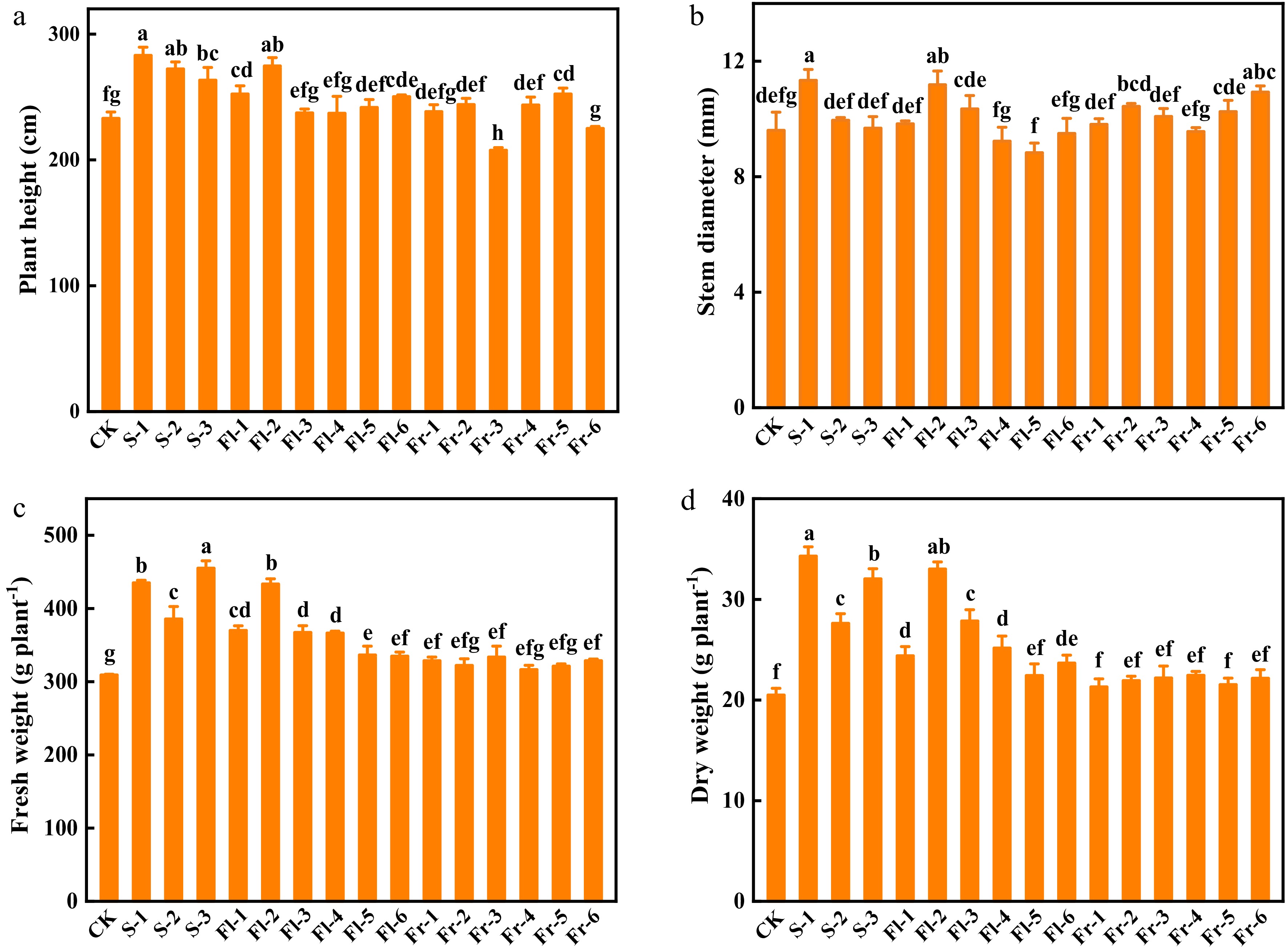
      Figure 1. 

      Effects of the mixture of putrescine (Put), melatonin (MT), proline (Pro), and potassium fulvic acid (MFA) on the growth of cucumber plants under high temperature stress. (a) Plant height. (b) Stem diameter. (c) Fresh weight. (d) Dry weight. CK, cucumber plants without treatment with the mixture of 8 mmol L−1 Put, 50 µmol L−1 MT, 1.5 mmol L−1 Pro, and 0.3 g L−1 MFA (Put mixture, original concentration). S, Fl, and Fr indicated cucumber plants-treated with the Put mixture at seedling stage, flowering stage, and fruiting stage, respectively. 1, 2, and 3 indicated cucumber plants-treated with the original, diluted five times, and diluted ten times concentration of the Put mixture every 7 d, 3 times, respectively. 4, 5, and 6 indicated cucumber plants-treated with the original, diluted five times, and diluted ten times concentration of the Put mixture every 14 d, 3 times, respectively. Data represent as the mean ± SD (n = 3). Different letters indicate significant differences according to Tukey’s test at P ≤ 0.05.

    • To investigate the role of the Put mixture on the cell membrane integrity of cucumber leaves, we analyzed the level of relative electrolyte leakage and MDA content of cucumber leaves after planting for 40 d. The level of relative electrolyte leakage in the Put mixture-treated plants was lower than that in CK (Fig. 2a). The level of relative electrolyte leakage in the treatment at flowering and fruiting stage every 7 d was significantly lower than those in every 14 d treatments (Fig. 2a). In addition, MDA content in all of the Put mixture-treated plants was lower than that in CK (Fig. 2b). The content of MDA in S-1 treatment was the lowest, which was 34.6% lower than that in CK (Fig. 2b). As shown in Fig. 2c, the content of Pro in cucumber leaves at the same treatment stage increased with the increase of spraying concentration. The content of Pro in S-1, Fl-1, Fl-2, and Fr-1 treatments increased by 29.1%, 22.8%, 18.2%, and 16.7%, respectively, compared with CK (Fig. 2c). There was no significant difference in the content of H2O2 between CK and Fl-6 and Fr-3 treatments, but the content of H2O2 in other treatments was significantly lower than that in CK (Fig. 2d).
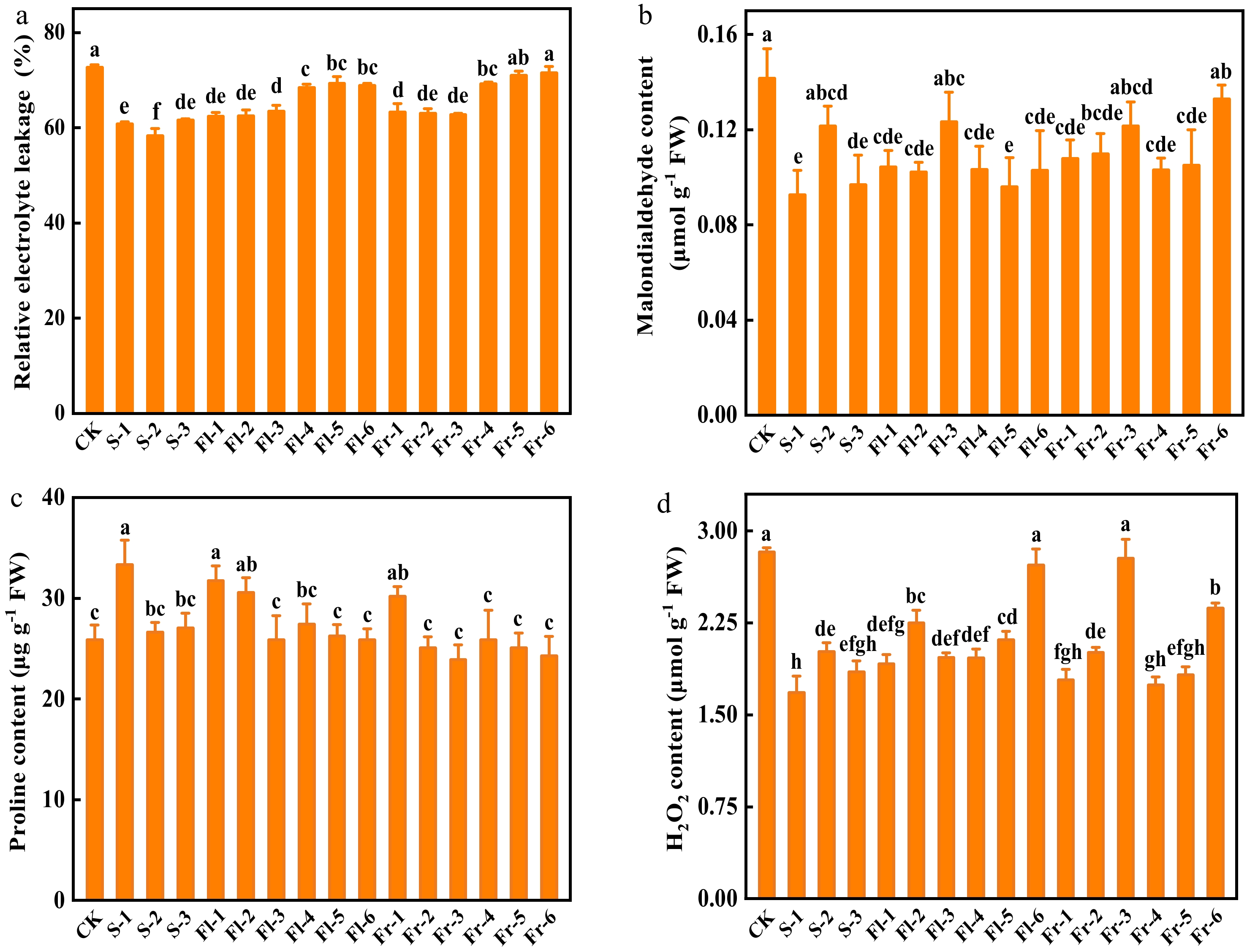
      Figure 2. 

      Effects of the mixture of putrescine (Put), melatonin (MT), proline (Pro), and potassium fulvic acid (MFA) on relative electrolyte leakage, malondialdehyde, Pro, and H2O2 content of cucumber plants under high temperature stress. (a) Relative electrolyte leakage. (b) Malondialdehyde content. (c) Pro content. (d) H2O2 content. CK, cucumber plants without treatment with the mixture of 8 mmol L−1 Put, 50 µmol L−1 MT, 1.5 mmol L−1 Pro, and 0.3 g L−1 MFA (Put mixture, original concentration). S, Fl, and Fr indicated cucumber plants-treated with the Put mixture at seedling stage, flowering stage, and fruiting stage, respectively. 1, 2, and 3 indicated cucumber plants-treated with the original, diluted five times, and diluted ten times concentration of the Put mixture every 7 d, 3 times, respectively. 4, 5, and 6 indicated cucumber plants-treated with the original, diluted five times, and diluted ten times concentration of the Put mixture every 14 d, 3 times, respectively. Data represent as the mean ± SD (n = 3). Different letters indicate significant differences according to Tukey’s test at P ≤ 0.05. FW, fresh weight.

    • As shown in Fig. 3, spraying different concentrations of Put mixture increased the total chlorophyll content of cucumber leaves in comparison to CK. Chlorophyll content in S-1 treatment was the highest, which was 28.2% higher than that in CK (Fig. 3).
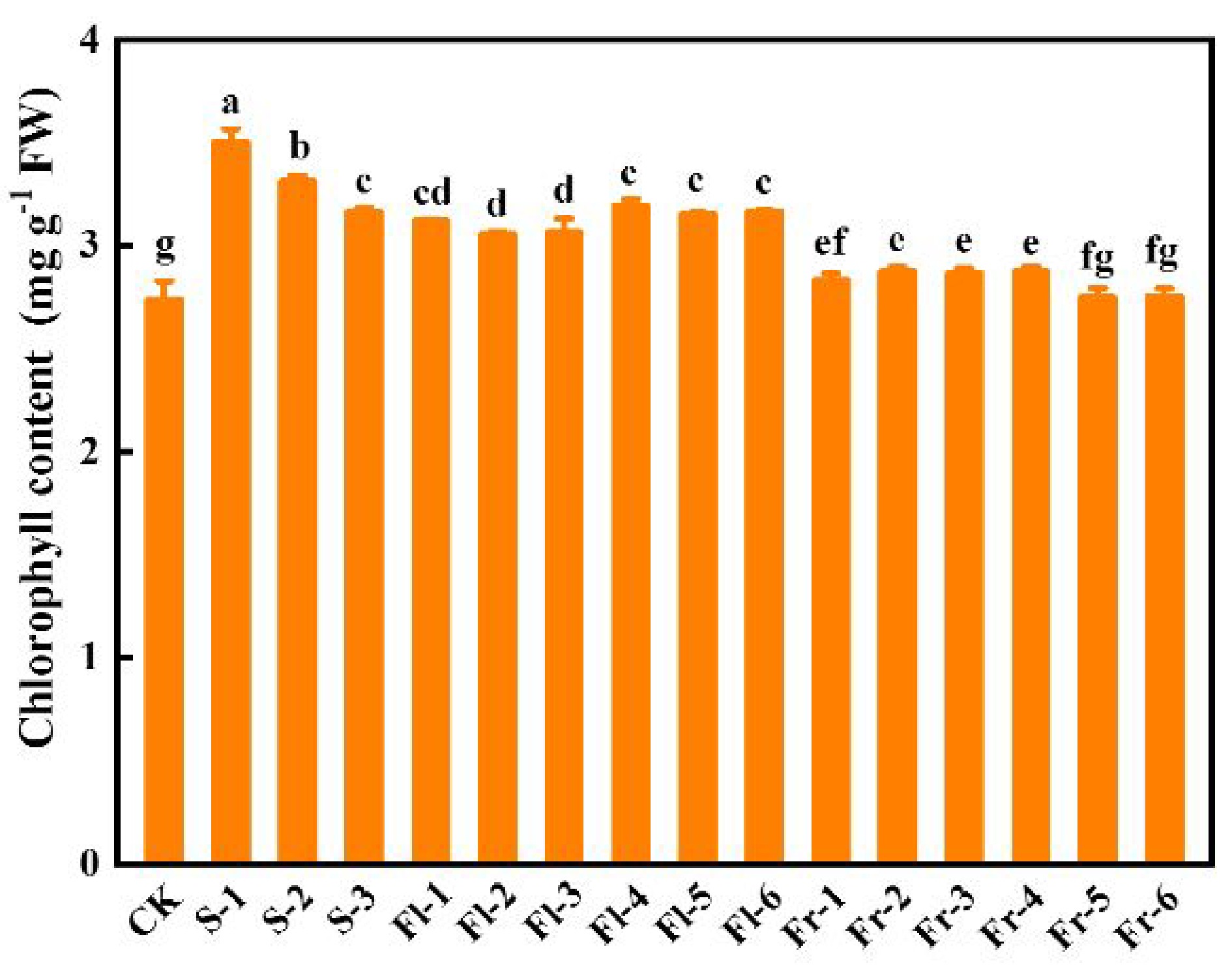
      Figure 3. 

      Effects of the mixture of putrescine (Put), melatonin (MT), proline (Pro), and potassium fulvic acid (MFA) on chlorophyll content of cucumber leaves under high temperature stress. CK, cucumber plants without treatment with the mixture of 8 mmol L−1 Put, 50 µmol L−1 MT, 1.5 mmol L−1 Pro, and 0.3 g L−1 MFA (Put mixture, original concentration). S, Fl, and Fr indicated cucumber plants-treated with the Put mixture at seedling stage, flowering stage, and fruiting stage, respectively. 1, 2, and 3 indicated cucumber plants-treated with the original, diluted five times, and diluted ten times concentration of the Put mixture every 7 d, 3 times, respectively. 4, 5, and 6 indicated cucumber plants-treated with the original, diluted five times, and diluted ten times concentration of the Put mixture every 14 d, 3 times, respectively. Data represent as the mean ± SD (n = 3). Different letters indicate significant differences according to Tukey’s test at P ≤ 0.05. FW, fresh weight.

      The net photosynthetic rate (Pn) of cucumber plants in the treatments at the seedling stage significantly increased compared with CK, and the effect was positively related to the spraying concentration, among which the plants of S-1 treatment had the highest Pn, and increased by 1.03-fold (Fig. 4a). Similarly, the Pn in the treatments at the flowering stage also decreased with the decrease of spraying concentration, and the effect of spraying mixture every 7 d was better than those in every 14 d at the same concentration (Fig. 4a). Except for Fr-6 treatment, the foliar application of the Put mixture significantly increased the transpiration rate (Tr) (Fig. 4b). The intercellular CO2 concentration (Ci) in the plants of S-1 and Fr-1 treatment increased by 28.6% and 32.6%, respectively, compared with CK (Fig. 4c). Except for Fr-5 and Fr-6 treatment, treatment with Put mixture significantly increased the value of stomatal conductance (Gs) in comparison to CK, especially in S-1 treatment, which was 1.85-fold higher than that in CK (Fig. 4d).
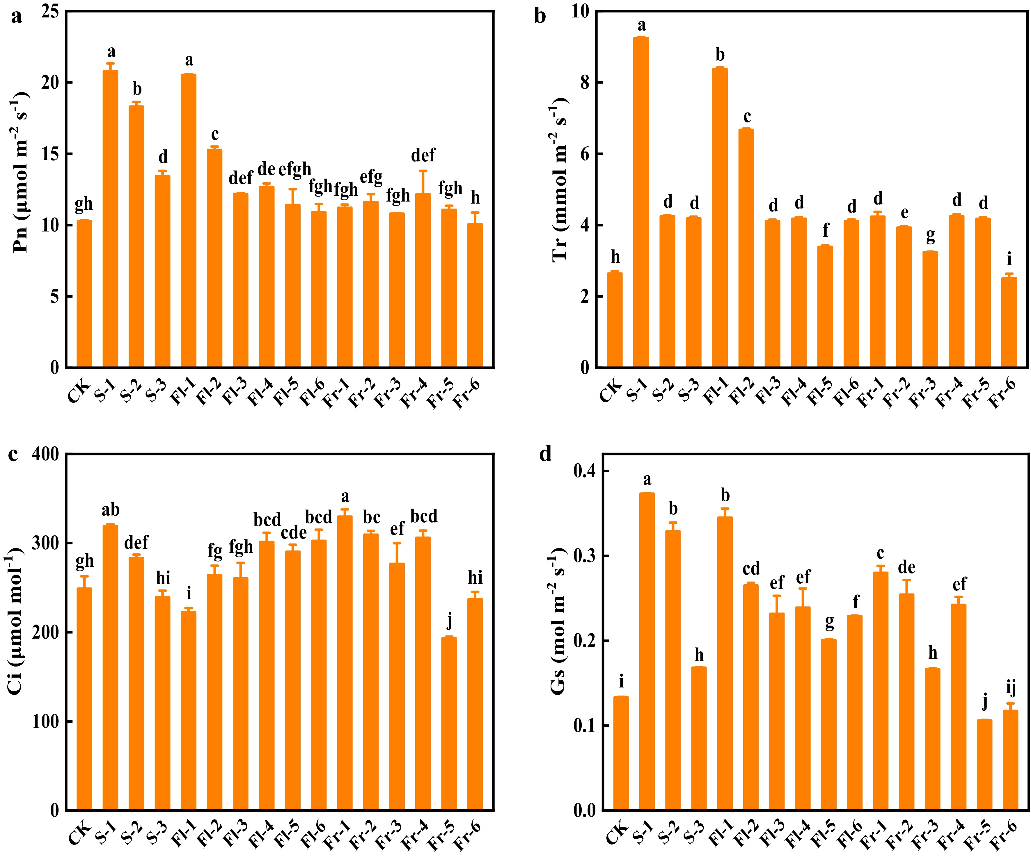
      Figure 4. 

      Effects of the mixture of putrescine (Put), melatonin (MT), proline (Pro), and potassium fulvic acid (MFA) on gas exchange parameters of cucumber under high temperature stress. (a) Net photosynthetic rate (Pn). (b) Transpiration rate (Tr). (c) Intercellular CO2 concentration (Ci). (d) Stomatal conductance (Gs). CK, cucumber plants without treatment with the mixture of 8 mmol L−1 Put, 50 µmol L−1 MT, 1.5 mmol L−1 Pro, and 0.3 g L−1 MFA (Put mixture, original concentration). S, Fl, and Fr indicated cucumber plants-treated with the Put mixture at seedling stage, flowering stage, and fruiting stage, respectively. 1, 2, and 3 indicated cucumber plants-treated with the original, diluted five times, and diluted ten times concentration of the Put mixture every 7 d, 3 times, respectively. 4, 5, and 6 indicated cucumber plants-treated with the original, diluted five times, and diluted ten times concentration of the Put mixture every 14 d, 3 times, respectively. Data represent as the mean ± SD (n = 3). Different letters indicate significant differences according to Tukey’s test at P ≤ 0.05.

    • High temperature stress affects cucumber fruit and causes fruit deformity. Spraying Put mixture significantly reduced deformity rate (Fig. 5a). The deformity rate of cucumber fruit in CK was 16.2%, while it was only 6.3% in S-1 treatment, which decreased by 61.1% (Fig. 5a). Furthermore, the foliar application of the Put mixture also increased single fruit weight and fruit yield per plant, and the effect of S-1 treatment was the best, which increased by 38.1% and 18.1%, respectively, in comparison to CK (Fig. 5b & c).
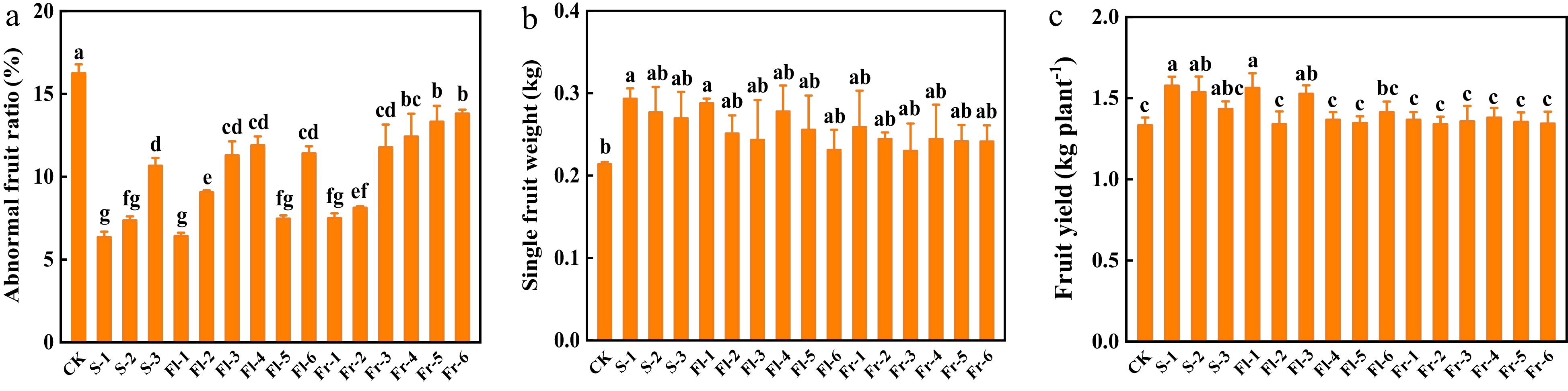
      Figure 5. 

      Effects of the mixture of putrescine (Put), melatonin (MT), proline (Pro), and potassium fulvic acid (MFA) on fruit deformity rate and yield of cucumber. (a) Abnormal fruit ratio. (b) Single fruit weight. (c) Fruit yield per plant. CK, cucumber plants without treatment with the mixture of 8 mmol L−1 Put, 50 µmol L−1 MT, 1.5 mmol L−1 Pro, and 0.3 g L−1 MFA (Put mixture, original concentration). S, Fl, and Fr indicated cucumber plants-treated with the Put mixture at seedling stage, flowering stage, and fruiting stage, respectively. 1, 2, and 3 indicated cucumber plants-treated with the original, diluted five times, and diluted ten times concentration of the Put mixture every 7 d, 3 times, respectively. 4, 5, and 6 indicated cucumber plants-treated with the original, diluted five times, and diluted ten times concentration of the Put mixture every 14 d, 3 times, respectively. Data represent as the mean ± SD (n = 3). Different letters indicate significant differences according to Tukey’s test at P ≤ 0.05.

      Tannin content of the fruit treated with Put mixture was significantly reduced compared with that in CK (Fig. 6a). At the seedling and flowering stage, tannin content decreased with the increase in spray concentration, and the effect of S-1 treatment was the most profound, which was reduced by 20.0% (Fig. 6a). Except for Fr-3 treatment, the content of organic acid decreased significantly compared to CK (Fig. 6b). The content of organic acid in S-1 treatment was the lowest, which decreased by 34.2% (Fig. 6b). During the fruiting stage, with the increase concentration of spray, the content of organic acid in fruit gradually decreased (Fig. 6b). Compared with CK, all treatments significantly increased the content of vitamin C in cucumber fruit (Fig. 6c). The contents of soluble solids in S-1, Fl-1, Fl-2, Fr-4, and Fr-5 treatments were significantly higher than that in CK (Fig. 6d).
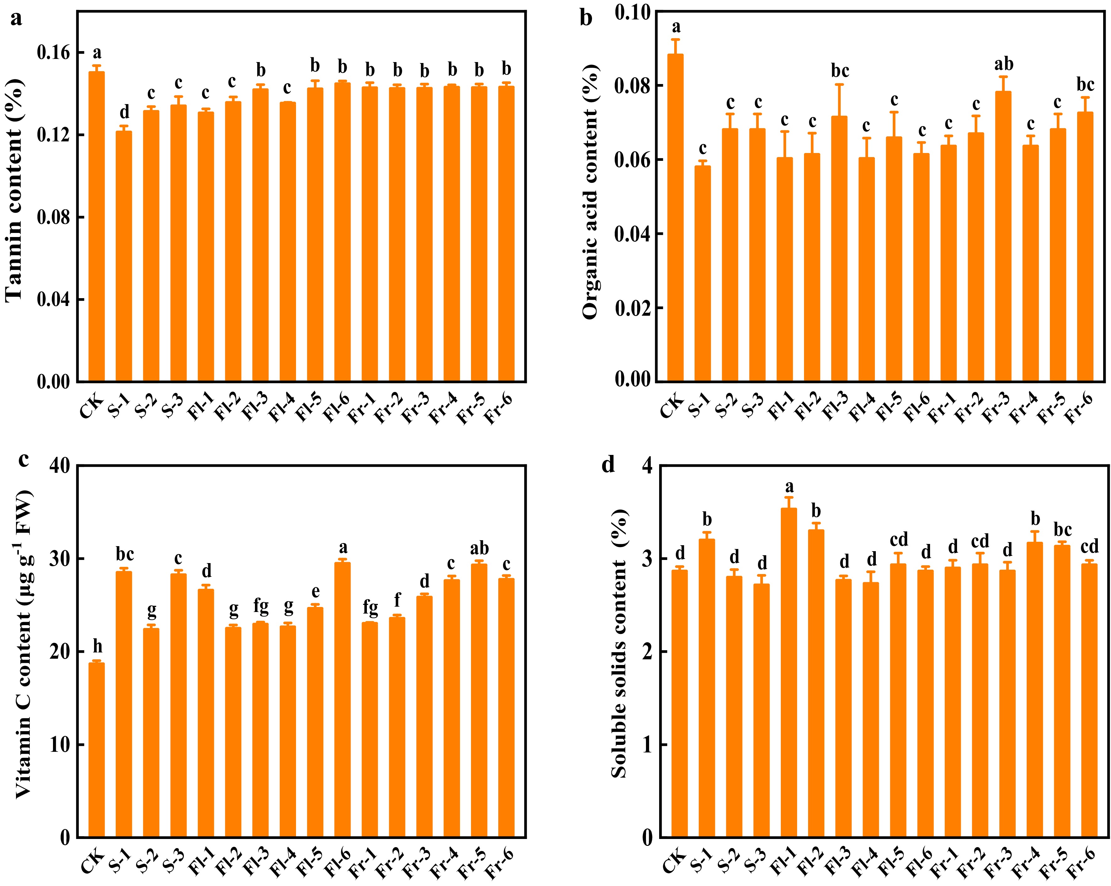
      Figure 6. 

      Effects of the mixture of putrescine (Put), melatonin (MT), proline (Pro), and potassium fulvic acid (MFA) on fruit quality of cucumber. (a) Tannin content. (b) Organic acid content. (c) Vitamin C content. (d) Soluble solids content. CK, cucumber plants without treatment with the mixture of 8 mmol L−1 Put, 50 µmol L−1 MT, 1.5 mmol L−1 Pro, and 0.3 g L−1 MFA (Put mixture, original concentration). S, Fl, and Fr indicated cucumber plants-treated with the Put mixture at seedling stage, flowering stage, and fruiting stage, respectively. 1, 2, and 3 indicated cucumber plants-treated with the original, diluted five times, and diluted ten times concentration of the Put mixture every 7 d, 3 times, respectively. 4, 5, and 6 indicated cucumber plants-treated with the original, diluted five times, and diluted ten times concentration of the Put mixture every 14 d, 3 times, respectively. Data represent as the mean ± SD (n = 3). Different letters indicate significant differences according to Tukey’s test at P ≤ 0.05.

    • High temperature negatively affects plant growth and fruit quality, resulting in leaf wilting, and fruit deformity[38,39]. It has been demonstrated that the foliar application of a moderate concentration of plant growth regulator can ameliorate high temperature stress[2]. In agreement with previous studies, cucumber plants treated with the Put mixture significantly promoted growth under high temperature stress (Fig. 1). Among them, the effect of S-1 treatment was the most obvious on plant height, stem diameter, fresh and dry weight (Fig. 1). In addition, high temperature leads to plant water deficit and oxidative stress, which inhibits plant growth and reduces plant chlorophyll content[40]. The results of this experiment showed that the accumulation of cucumber seedling biomass was inhibited, and the chlorophyll content of CK was lower than that of the Put mixture treatment group under high temperature stress (Figs 1 & 3). Spraying the Put mixture on the leaves alleviated the inhibition of high temperature stress on the growth of cucumber plants.

      High temperature often leads to the accumulation of ROS in plants. ROS peroxide with unsaturated fatty acids on the cell membrane and produce a large amount of MDA. MDA further aggravates the oxidation reaction of cell biofilm and causes the destruction of the biofilm structure[41]. Therefore, the content of MDA in plant cells can indirectly represent the degree of oxidative stress. This study showed that treatment with the Put mixture reduced the levels of electrolyte leakage of cucumber leaves, decreased the contents of H2O2 and MDA (Fig. 2), indicating that application of Put mixture alleviated high temperature stress-induced oxidative stress. Similarly, the foliar application of Put removes free radicals and ROS, and reduces the degree of membrane lipid peroxidation under environmental stresses[9, 10]. In addition, Put enhances the scavenging capacity of ROS in chloroplasts by regulating the coordination of antioxidant enzymes and antioxidants in chloroplasts under environmental stress, alleviating the damage of environmental stress to chloroplast structure and function[4244]. Furthermore, MT is an antioxidant that can scavenge oxygen free radicals and repair its own oxidation products[21,45]. In addition to its direct reaction with ROS, it can enhance the resistance of plants to stress by improving the efficiency of the antioxidant system in plants[21,24,46,47]. Treatment with MFA also improves antioxidant enzyme activity and antioxidant content of plant seedlings under environmental stress to maintain the balance of ROS production and scavenging[48]. Furthermore, Pro maintains the osmotic balance between protoplast and environment through osmotic regulation, reduces water loss, and protects cell membrane structure[49]. This study showed that the foliar application of the Put mixture decreased H2O2 and MDA contents in cucumber leaves (Fig. 2). Therefore, treatment with the Put mixture reduced membrane damage, maintained osmotic balance, reduced oxidative stress, and improved plant high temperature resistance.

      Photosynthesis, one of the most important physiological processes in plants, is very sensitive to temperature changes. High temperature stress affects the activity of chlorophyll synthesis enzymes, reduces the content of photosynthetic pigment[50,51], resulting in a reduction of Pn. Studies have shown that single exogenous spraying of Put, MT, Pro, or MFA alleviates the decline of photosynthesis caused by environmental stress through maintaining the structural stability of chloroplasts, scavenging excessive ROS, inhibiting photosynthesis pigment degradation, and increasing the maximum quantum yield of photosystem II[16,27,29,43]. Similarly, this study showed that spraying Put mixture at the seedling stage significantly improved chlorophyll content, Pn, and Tr of plants under high temperature stress, alleviated the reduction of Gs caused by high temperature stress (Figs 3 & 4). The decrease of Pn under high temperature treatment was accompanied by the decrease of Gs and Ci (Fig. 4), indicating that the inhibition of stomatal factors might be the dominant reason for Pn decrease. However, the foliar application of the Put mixture alleviated the decrease of photosynthesis caused by stomatal restriction.

      High temperature stress seriously affects cucumber fruit, causing fruit deformity, and reducing fruit yield and quality[38,39]. The foliar application of the Put mixture significantly decreased the rate of deformity, improved fruit yield, the contents of vitamin C and soluble solids in cucumber fruit, and decreased the contents of tannins and organic acids (Figs 5 & 6).

    • The foliar application of the Put mixture promoted plant growth, reduced the level of relative electrolyte leakage and the content of H2O2 and MDA, increased the content of Pro and photosynthesis of cucumber plants. Furthermore, the fruit yield and content of vitamin C and soluble solids increased in Put mixture treated plants, but the content of tannins and organic acids decreased. Therefore, spraying Put mixture on the leaves, especially at the seedling stage with 8 mmol L−1 Put, 50 µmol L−1 MT, 1.5 mmol L−1 Pro, and 0.3 g L−1 MFA every 7 d for three times, alleviated the inhibition of high temperature stress on the growth of cucumber plants, improved cucumber plant adaptability to high temperature stress, and increased the yield and quality.

    • Cucumber (Cucumis sativus L. cv Jinchun No. 4) was used as experimental material. Uniform seeds were sterilized with 10% NaClO for 10 min, followed by washing 5 times with deionized water and soaked in deionized water for 4 h, then the seeds were incubated for germination on moistened filter paper in an incubator (Shanghai Zhicheng Analytical Instrument Manufacturing Co., Ltd., Shanghai, China), which was maintained at 28 °C. The germinated seeds were sown in 32-well plastic trays filled with seedling substrates (Jiangsu Xingnong Substrate Technology Co., Ltd., China) and grown in the greenhouse of Baima Teaching and Research Base of Nanjing Agricultural University. The temperature in the greenhouse during the day was controlled at 25−28 °C, the temperature at night was 18−20°C, and the relative humidity was maintained at 75%−80%. When the fourth leaves were fully expanded, the seedlings were selected and planted in coconut coir substrates (Van der Knaap Group of Companies, Wateringen, Netherlands) on Apr. 17, 2021 in the same greenhouse. As shown in Supplemental Fig. S1, the maximum temperature was over 30 °C in the most of the cultivation period, indicating that cucumber plants suffered from high temperature stress.

    • Cucumber seedlings were randomly divided into 16 groups to treat with or without Put mixture, and each group contained 15 plants as one treatment. As shown in Table 1, cucumber plants without treatment with the mixture of 8 mmol L−1 Put, 50 µmol L−1 MT, 1.5 mmol L−1 Pro, and 0.3 g L−1 MFA (Put mixture, original concentration) were used as CK. S-1, S-2, and S-3 indicated cucumber plants treated with the original, diluted five times, and diluted ten times concentration of Put mixture at seedling stage every 7 d, 3 times, respectively. Fl-1, Fl-2, and Fl-3 indicated cucumber plants treated with the original, diluted five times, and diluted ten times concentration of Put mixture at flowering stage every 7 d, 3 times, respectively. Fl-4, Fl-5, and Fl-6 indicated cucumber plants treated with the original, diluted five times, and diluted ten times concentration of Put mixture at flowering stage every 14 d, 3 times, respectively. Fr-1, Fr-2, and Fr-3 indicated cucumber plants treated with the original, diluted five times, and diluted ten times concentration of Put mixture at fruiting stage every 7 d, 3 times, respectively. Fr-4, Fr-5, and Fr-6 indicated cucumber plants treated with the original, diluted five times, and diluted ten times concentration of Put mixture at fruiting stage every 14 d, 3 times, respectively.

      Table 1.  Spraying concentration and stage of cucumber with putrescine mixture.

      TreatmentPutrescine
      concentration
      (mmol L−1)
      Potassium fulvic acid concentration
      (g L−1)
      Proline
      concentration
      (mmol L−1)
      Melatonin
      concentration
      (µmol L−1)
      Spraying interval (d)Spraying stage
      CK
      S-180.31.5507Seedling stage
      S-21.60.060.3107Seedling stage
      S-30.80.030.1557Seedling stage
      Fl-180.31.5507Flowering stage
      Fl-21.60.060.3107Flowering stage
      Fl-30.80.030.1557Flowering stage
      Fl-480.31.55014Flowering stage
      Fl-51.60.060.31014Flowering stage
      Fl-60.80.030.15514Flowering stage
      Fr-180.31.5507Fruiting stage
      Fr-21.60.060.3107Fruiting stage
      Fr-30.80.030.1557Fruiting stage
      Fr-480.31.55014Fruiting stage
      Fr-51.60.060.31014Fruiting stage
      Fr-60.80.030.15514Fruiting stage
    • The plant growth parameters were measured after planting for 40 d. Plant height was measured from the stem base to the growth point with a ruler, and stem diameter was measured with a vernier caliper, 1 cm below the cotyledons. Fresh samples were washed with distilled water and dried with paper, and then fresh weight was weighed with an electronic scale. The samples were dried for 15 min at 105 °C in an oven (Shanghai Yiheng Scientific Instrument Co., Ltd., Shanghai, China), and the temperature was reduced to 75 °C until constant weight was obtained.

    • The Pn, Gs, Ci, and Tr of the fifth fully expanded leaf below the growth point were measured with a portable photosynthesis system (LI-6400; Li-COR, Lincoln, NE, USA) from 9:00 to 11:00 am after planting for 40 d. The measurement parameters were as follows: ambient CO2 concentration was 380 µmol mol−1, the leaf chamber temperature was maintained at 25 °C, and the photosynthetic photo flux density was 800 µmol m−2 s−1.

    • Relative electrolyte leakage was detected according to the method described previously[52]. The content of MDA was determined using the thiobarbituric acid method[53].

    • Fresh leaves were washed, cut into pieces and 0.2 g samples were placed in a 15-ml centrifuge tube. Five millilitres of 3% sulfosalicylic acid solution was added into the tubes, and extracted in a boiling water bath for 10 min (shaken every 5 min). After cooling, the tubes were centrifuged at 3000 r for 10 min and 1 ml of the supernatant was placed in a new tube, adding 1 ml of distilled water, 1 ml of glacial acetic acid, and 2 ml of acidic ninhydrin solution. Subsequently, the tubes were heated in a boiling water bath for 60 min. Four millilitres of toluene was added to the tube after cooling, and vortexed for 30 s. The upper layer of toluene Pro red solution was used to measure Pro concentration at 520 nm using a UV-1800 spectrophotometer (Shanghai Unico Instrument Co., Ltd., Shanghai, China) as previously described[54].

    • Cucumber leaves (0.2 g) were ground to homogenate in 1.6 ml of 0.1% TCA on ice and centrifuged at 12000 r for 20 min. The supernatant (0.2 ml) was added to 1 ml of 1 mol L−1 KI and 0.25 ml of 0.1 mol L−1 potassium phosphate buffer (pH = 7.8) for reaction for 1 h in the dark. The concentration of H2O2 was measured at 390 nm using a spectrophotometer and calculated as previously described[55].

      For the measurement of chlorophyll content, 20 ml of 95% ethanol was added to 0.2 g of fresh leaves and sealed. The tubes were placed in the dark for 24−36 h until the leaves turn white. The chlorophyll content was measured according to the method of Arnon[56].

    • Ten plants were labeled in each treatment for yield measurement. Fruit weight was measured each time after picking, and the yield per plant was calculated after harvest. Six fresh ripe cucumbers were collected from each treatment during the fruit stage, and the fruit soluble solids, tannins, organic acid, and vitamin C content were determined to evaluate the fruit quality.

      The content of soluble solids was detected as previously described[57]. Briefly, the content of soluble solids was determined using an Abbe refractometer (WZ-108, Beijing Wancheng Beizeng Precision Instrument Co., Ltd., Beijing, China). Before determination, the refractometer was calibrated with a standard sample and then the content of soluble solids was analyzed and determined.

      For measuring tannin content, cucumber fruit (5 g) was ground and transferred to a 150-ml conical flask, shaken and extracted for 15 min. Five millilitres of 1 mol L−1 zinc acetate standard solution and 3.5 ml of concentrated ammonia were added into a 100-ml volumetric flask, shaken, and the tannin extraction was slowly transferred into the volumetric flask, keeping it warm in a 35 °C water bath for 30 min with shaking. After cooling, the volume was adjusted to 100 ml with distilled water, fully mixed and filtered. Ten millilitres of filtrate was placed in a 150-ml conical flask, and 40 ml of distilled water, 12.5 ml of NH3-NH4Cl, and 10 drops of chrome black T indicator were added and mixed well. The mixture was titrated with 0.05 mol L−1 EDTA solution until the wine red changed to pure blue. The content of tannin was calculated as previously described[58].

      To measure the content of organic acid, cucumber fruits (5 g) were ground and washed into a 250-ml conical flask with distilled water to make the volume to 100 ml. Organic acids were extracted in a constant temperature water bath at 80 °C for 30 min and shaken continuously. After cooling, the extractions were filtered and the residues were washed with distilled water 3 times; the filtrate was mixed and fixed to 100 ml with distilled water. The organic acid content was titrated with 0.1 mol L−1 sodium hydroxide standard solution as previously described[59].

      The content of vitamin C was determined according to the method previously described[60].

    • All data were statistically analyzed using the SPSS 18.0 version (SPSS Inc., Chicago, IL, USA), and the results are presented as means ± SDs (n = 3). Analysis of variance (ANOVA) was used to test for significance, and the significance between treatments were analyzed with Tukey’s honestly significant difference test (HSD) at P < 0.05.

      • This work was supported by the National Key Research and Development Program of China (2019YFD1001902).

      • The authors declare that they have no conflict of interest.

      • Supplemental Fig. S1 Temperature records registered in the greenhouse where the experiment was performed.
      • Copyright: © 2022 by the author(s). Published by Maximum Academic Press, Fayetteville, GA. This article is an open access article distributed under Creative Commons Attribution License (CC BY 4.0), visit https://creativecommons.org/licenses/by/4.0/.
    Figure (6)  Table (1) References (60)
  • About this article
    Cite this article
    Wang Y, Liu H, Lin W, Jahan MS, Wang J, et al. 2022. Foliar application of a mixture of putrescine, melatonin, proline, and potassium fulvic acid alleviates high temperature stress of cucumber plants grown in the greenhouse. Technology in Horticulture 2:6 doi: 10.48130/TIH-2022-0006
    Wang Y, Liu H, Lin W, Jahan MS, Wang J, et al. 2022. Foliar application of a mixture of putrescine, melatonin, proline, and potassium fulvic acid alleviates high temperature stress of cucumber plants grown in the greenhouse. Technology in Horticulture 2:6 doi: 10.48130/TIH-2022-0006

Catalog

  • About this article

/

DownLoad:  Full-Size Img  PowerPoint
Return
Return