


-
Amaranthus spp. are economically important crop plants valued for their nutritional and horticultural significance[1]. It has many species with great importance in the food, cosmetic, and pharmaceutical industries, as Amaranthus genus contain flavonoids, carotenoids, betalains, and so on[2−4]. They have become one of the most promising crops[2]. When producing flavonoids or other metabolites using plants, raw materals would be in shortage for the environment or climate. However, according to the plant cell totipotency, in vitro culture techniques are useful tools for the continuous production of secondary metabolites[5,6]. Secondary metabolites are continuously extracted by large-scale plant cell culture systems. Resulting in a higher rate of metabolism in cell cultures than in differentiated plants. Meanwhile, the use of technology could shorten biosynthetic cycles.
The researchers established the system of cell cultures and callus induction for the production of secondary metabolites in Cnidium officinale Makino[7]. The success of callus induction is dependent on genotype, the composition of the culture medium and the presence of appropriate combinations and concentrations of hormones in the culture media[8,9]. The genotypes, hormone combination and concentration are decisive factors for Amaranthus spp. callus formation. Callus formation was successfully induced in A. paniculatus[10], A. caudatus, A. hypochondriacus, A. cruentus, A. hybridus[11−13], A. tricolor[14,15] and A. spinosus[15]. However, the hormone combination and concentration of callus induction were different amongst these genotypes. Plant growth regulators, especially auxins and cytokinins, play a key role for cell growth and secondary metabolite biosynthesis in plant cell and tissue culture[16]. The best hormone combinations for callus induction was NAA plus BAP or 2,4-D plus kinetin in four species of Amaranthus[12]. In previous reports, a lower cytokinin:auxin ratio was more suitable for A. tricolor and A. spinosus. The callus induction in Amaranthus spp., using either hypocotyl segments or stem sections with BAP or kinetin and low doses of NAA or 2,4-D[11,12,14]. Additionally, plant growth regulators, especially, combination and concentration of auxin and cytokinine, could affect the biosynthesis and accumulation of flavonoids in cell culture[17,18]. Generally, high auxin levels are often deleterious to secondary metabolite production. However, few reports on callus induction in amaranth[10−15] and flavonoid production using amaranth callus have been published. We performed the research herein to explore the effects of the combination and concentration of auxin and cytokinine on callus induction, proliferation and the content of flavonoids in Amaranthus spp..
-
Seeds of 12 Amaranthus spp. were collected from the publicly available US Department of Agriculture National Plant Germplasm System (USDA-NPGS) Amaranthus germplasm collection (
www.ars-grin.gov ; Table 1). We obtained the sterilized seeds according to the method of Liu et al.[19]. The seeds were maintained in a plant growth chamber at 25 ± 1 °C, 16 h photoperiod and 25 μmol·m−2·sec illuminance provided by light-emitting diode (LED) lights.Table 1. Effects of genotype on induction of callus in Amaranthus spp.
Plant ID Plant name Taxonomy Induction rate (%) Origin PI 606282 Lal Shak Amaranthus blitum subsp. Oleraceus 89.17 ± 1.91 a Bangladesh PI 572261 AMA57/81 Amaranthus powellii subsp. Bouchoni 50.57 ± 3.95 c Germany PI 277269 Lal Sag Amaranthus tricolor 90.67 ± 1.55 a India Ames 18049 Ramdana Amaranthus tricolor 78.63 ± 0.49 b Nepal PI 604669 White leaf Amaranthus tricolor 87.27 ± 1.07 a China, Taiwan Ames 5110 RRC321 Amaranthus tricolor 95.03 ± 2.25 a West Africa Ames 5134 Tampala Amaranthus tricolor 67.07 ± 1.46 bc United States, Pennsylvania Ames 5311 Fota Kira Amaranthus tricolor 87.53 ± 1.27 a India Ames 15328 RRC359 Amaranthus tricolor 69.37 ± 2.67 b United States PI 527321 BAILIUYEXIAN Amaranthus tricolor 77.9 ± 4.36 b China PI 607446 Crystal Amaranthus tricolor 94.67 ± 4.03 a Thailand Ames 2141 RRC228 Amaranthus tricolor 70.8 ± 2.62 b India, Tamil Nadu Means ± STD followed with the same letters are not significantly different using DMRT at α = 0.05. Callus induction
-
To compare the effects of different genotypes on callus induction in Amaranthus, the hypocotyl segments (5−8 mm) from seven-day old in vitro germinated seedlings were placed on the medium (MS + 3.0 mg·L−1 BAP + 0.5 mg·L−1 2,4-D + 0.7% (w/v) agar + 3% (w/v) sucrose, pH 5.6−5.8) to induce callus. Each treatment was carried out with three biological repetitions.
To screen the optimum medium, the hypocotyl segments of A. tricolor 'Lal Sag' were placed on the MS medium supplemented with BAP (1.5 and 3.0 mg·L−1) and 0.5 mg /L NAA or 2,4-D (0.5 and 1.0 mg·L−1), singly or in combination, to induce callus. The hypocotyl segments of A. powellii subsp. bouchoni were placed on the MS medium supplemented with BAP (1.5, 3.0 and 6.0 mg·L−1), 0.5 mg·L−1 NAA or 2,4-D singly or in combination, to induce callus.
Callus growth and biomass yield
-
We placed 0.15 g fresh callus of A. powellii subsp. bouchoni on the MS medium supplemented with 0.5 mg·L−1 2,4-D, 0.7% (w/v) agar, 3% (w/v) sucrose, and different types of cytokinin, including BAP (0, 0.5, 1.0, 3.0, 6.0 and 9.0 mg·L−1), KT (0, 0.1, 0.5, 1.0, 2.0, and 4.0 mg·L−1), and TDZ (0, 0.05, 0.10, 0.50, 1.00, and 2.00 mg·L−1). In addition, the callus was placed on the MS medium supplemented with 3.0 mg·L−1 BAP, 0.7% (w/v) agar, 3% (w/v) sucrose, and different type of auxins, including 2,4-D, NAA, and IAA. The concentration of every auxin was 0, 0.5, 1.0, 1.5, 2.0, and 2.5 mg·L−1. After 30 d, we observed the growth of amaranth callus, and weighed the callus fresh and dry weight.
In addition, we weighed the fresh weight and observed the growth state of amaranth callus from six bottles every 3 d continuously, 12 times to plot the growth curve of amaranth.
Cell suspension growth curve
-
We plotted the growth curve of the amaranth cell suspension culture by following two methods.
(1) 2g amaranth callus was inoculated into 50 mL MS medium supplemented with 3.0 mg·L−1 BAP, 0.5 mg·L−1 2,4-D, and 3% (w/v) sucrose. After filtering the cell suspension culture of Amaranthus and absorbing water with filter paper, we measured the fresh weight and dry weight every 5 d continuously six times to plot the growth curve. Meanwhile, the growth state of fresh amaranth callus was observed.
(2) We used a 2 mm diameter sieve to filter the cell suspension culture of Amaranthus which were cultured in the suspension culture medium for 20 d, then 2.30 g fresh weight suspension cells were transferred into 40 mL of the same medium for further culture. The fresh weight of cell suspension culture of Amaranthus was measured, following drying of the suspension cells to measure the dry weight every 2 d continuously six times. The growth curve of the cell suspension culture of Amaranthus was plotted according to the fresh and dry weight.
Determination of total flavonoid content
-
Total flavonoid content were determined using the method of Li et al.[20] with some modifications. The dried amaranth callus was ground into fine powder, then extracted with 10 mL 60% (v/v) ethanol solution in a conical flask and sonicated (power 100 W) for 60 min at 60 °C. The extracts were centrifuged (15 min, 3,000 rpm), and the supernatant was collected into new tubes. The total flavonoid content was detected at a wavelength of 510 nm in a DU640 spectrophotometer. For quantitation, rutin was as an internal standard for calibration.
Total flavonoid contentt (mg·g−1) = (C × V1 × V3)/(m × V2)
C: Mass concentration of flavonoids (mg·mL−1); M: Mass of sample = 0.3 g; V1: 10 mL 60% ethanol solution for flavonoid extraction; V2: 5 mL testing solution; V3: 10 mL reaction solution.
Flavonoid yield (mg/bottle) = total flavonoid content (mg·g−1) × callus weight (g/bottle).
Statistical analysis
-
The research results were presented in terms of means ± SD of at least three biological replicates. The data were analyzed by one-way analysis of variance (ANOVA) followed by Duncan's test using SPSS version 19.0. The pictures were created using GraphPad Prism 6.0 software and Excel 2016.
-
There are reports that callus induction ability was greatly influenced by the genotype Persian shallot[21]. Callus formation was successfully induced in some Amaranrhus spp.[10−15]. In the research, callus induction rate varied from 50% to 95% for 12 amaranth samples (shown in Table 1). The induction rate of PI 277269, Ames 18049, and PI 604669 hypocotyl was higher than others, and the callus was compact (shown in Fig. 1c). In contrast, the induction rate of PI 572261 was lower than others, but the callus was loose and had little browning. The other amaranths could induce callus, but the callus was easy to brown in a short time after induction (shown in Fig. 1d). The results indicated that the Amaranthus explants have great capacity to form callus[22], and the callus induction ability are greatly influenced by the genotype[13].
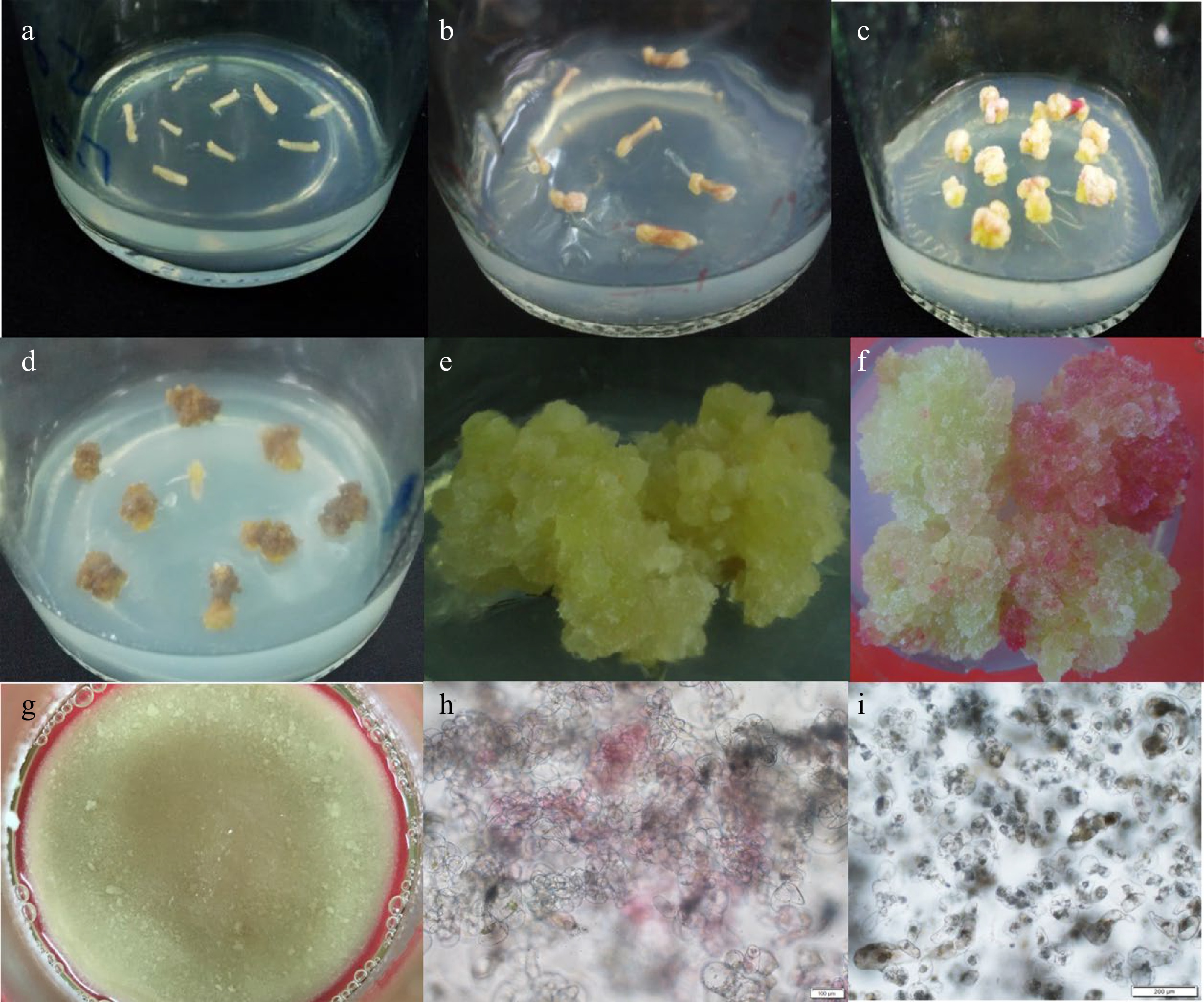
Figure 1.
Callus culture of Amaranthus L. (a) PI 277269 hypocotyl in MS + BAP media could not promote callus formation. (b) PI 277269 hypocotyl in MS 0.5 mg·L−1 2,4-D media could promote callus formation until 60 d. (c) PI 277269 hypocotyl in MS + 3.0 mg·L−1 BAP + 0.5 mg·L−1 2,4-D could promote callus formation and growth in 20 d. (d) Ames 2141 hypocotyl in MS + 3.0 mg·L−1 BAP + 0.5 mg·L−1 2,4-D could induce callus, but these calli began to brown in a short time. (e), (f) The callus proliferation of PI 277269 in MS + 6.0 mg·L−1 BAP + 0.5 mg·L−1 2,4-D medium for 3 years. (g) The suspension cells of PI 277269 in MS + 6.0 mg·L−1 BAP + 0.5 mg·L−1 2,4-D medium for 16 d. Microscopic observation of (h) callus and (i) suspension cells.
Effects of the ratio of auxin-to-cytokinin on induction of callus in Amaranrhus
-
Both the hormones (auxins and cytokinins) play a key role in the initiation of callus at different concentrations[16]. Based on the genotype selection, only BAP in the media could not promote callus formation from PI 277269 hypocotyl (Fig. 1a), and only 2,4-D (0.5 or 1.0 mg·L−1) in the media could promote callus formation until 60 d (Fig. 1b). Explants in MS + 3.0 mg·L−1 BAP + 0.5 mg·L−1 2,4-D or NAA could promote callus formation and growth in 20 d (Fig. 1c). This indicates that the presence of BAP in the medium, along with 2,4-D or NAA, could quickly induce callus growth (showed in Table 2).
Table 2. Effects of hormones on callus induction in Amaranth 'PI 277269'.
Hormones Callus
colorCompaction Induction
rate (%)Induction
time (d)1.5 mg·L−1 BAP − − 0 c − 0.5 mg·L−1 2,4-D Red − 72.33 ± 5.17 b 60 1.0 mg·L−1 2,4-D Red − 66.97 ± 1.40 b 60 3.0 mg·L−1 BAP +
0.5 mg·L−1 NAARed and yellow Loose 93.7 ± 0.72 a 20 3.0 mg·L−1 BAP +
0.5 mg·L−1 2,4-DWhite Compact 92.67 ± 2.41 a 20 Means ± STD followed with the same letters are not significantly different using DMRT at α = 0.05. The optimal medium (MS + 0.5 mg·L−1 2,4-D + 6.0 mg·L−1 BAP) for callus induction from the PI 572261 hypocotyls was also suitable for leaf induction callus. The induction rate was both over 90%. The red callus could be induced indicated by the leaf colour being red.
Effects of the ratio of auxin-to-cytokinin on proliferation of callus in Amaranrhus
-
In general, the ratio of auxin-to-cytokinin could affect the direction of explant morphogenesis. An intermediate ratio of auxin and cytokinin could induce callus. However, a lower cytokinin : auxin ratio was more suitable for A. tricolor and A. spinosus[15]. In contrast, a higher cytokinin:auxin ratio was needed for optimal callus growth in the research. The amaranth callus grew normally without browning on the callus proliferation medium supplemented with BAP (Fig. 2a) and KT (Fig. 2b) could promote the callus proliferation, while TDZ significantly inhibited the proliferation of amaranth callus (Fig. 2c).
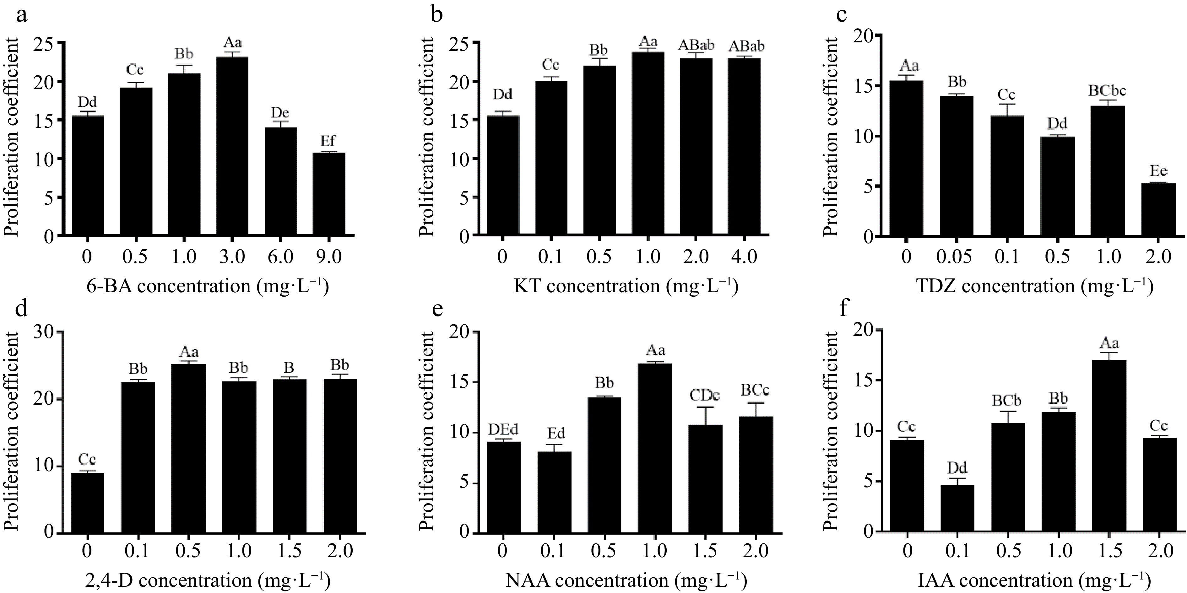
Figure 2.
The effect of different concentrations of cytokinins and auxins on the proliferation of amaranth callus. Upper case letters indicate p < 0.01, lower case letters indicate p < 0.05. The same letter indicates no significant difference.
When the BAP and KT concentration was 3.0 mg·L−1 and 1.0 mg·L−1, respectively, the proliferation coefficient of the callus was the highest, up to 23.13 and 23.79, which was significantly higher than other concentrations. However, with the increase of TDZ concentration, the proliferation coefficient of amaranth callus was a downwards trend, and was significantly lower than that of the control.
At 30 d, the addition of 2,4-D, NAA or IAA could promote the proliferation of amaranth callus (Fig. 2d−f). The optimal concentration of 2,4-D, NAA and IAA for the proliferation of amaranth callus was 0.5, 1.0, and 1.5 mg·L−1, corresponding proliferation coefficient 25.24, 16.88, and 17.03 for 30 d in vitro culture. Meanwhile, the amaranth callus showed partial browning by NAA and IAA treated, and 2,4-D treatment showed no browning.
Through the above analysis, the synergistic effect of auxin and cytokinin on callus induction plays an important role, and a higher cytokinin:auxin ratio was more suitable for Amaranthus, in accordance with A. gangeticus[23]. Unlike the previous reports, a lower cytokinin:auxin ratio was more suitable for A. tricolor and A. spinosus. the callus induction in Amaranthus spp., using either hypocotyl segments or stem sections with BAP or kinetin and low doses of NAA or 2,4-D[11,12,14]. We have maintained the callus proliferation in the MS + 6.0 mg·L−1 BAP + 0.5 mg·L−1 2,4-D medium for 3 years (shown in Fig. 1e, f & h).
The growth curve of amaranth callus is shown in an 'S' curve in Fig. 3a. At 0–3 d culture, fresh weights (FW) did not increase. FW increased slowly and quickly, respectively, from 3 to 6 d and from 6 to 21 d. Subsequently, the growth rate of callus began to slow down, and the fresh weight of callus reached the peak at 33 d. After the callus was cultured for 33 d, it began to brown.
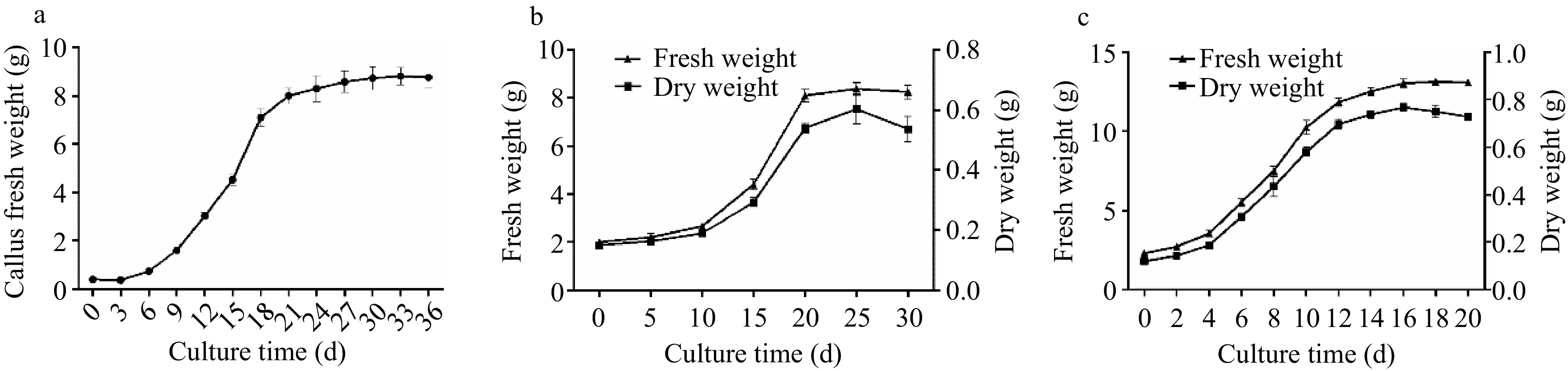
Figure 3.
Growth curve of callus of Amaranthus tricolor L.
Growth curve of cell suspension culture of Amaranthus
-
Amaranth callus (2 g) cultured on solid medium were inoculated into 50 mL MS medium supplemented with 3.0 mg·L−1 BAP, 0.5 mg·L−1 2,4-D, and 3% (w/v) sucrose. Both the growth curves were 'S' type. In the process of suspension cell culture (Fig. 3b), the cell grew slowly in the first 10 d. Then the proliferation, the fresh and dry weight of suspension cells increased rapidly from 11−20 d. The growth speed was decreased during 20−25 d, and the fresh weight and dry weight reached the peak at 25 d. Subsequently, the fresh and dry weight of the suspension cells began to decline at 25−30 d. Based on combination with the growth curve of fresh and dry weight, the best transfer time of cell suspension culture of Amaranthus was 20−25 d after culture.
When the cell suspension culture of Amaranthus was transferred into a fresh liquid medium, the cells grew rapidly and shortened the transfer time (Fig. 3c). The proliferation, the fresh and dry weight of suspension cells increased rapidly from 4 d, and the weight reached the peak at 16 d. Subsequently, the dry weight of the suspension cells began to decline. Combined with the growth curve of fresh and dry weight, the best transfer time of cell suspension culture of Amaranthus was 14−16 d after culture (shown in Fig. 1g & i).
Effect of concentrations of cytokinin on total flavonoid content and yield in amaranth
-
Plant growth regulators could affect the accumulation of flavonoids in cell culture[17]. With the increase of concentration of 6-BA, flavonoid content and yield showed a 'down-up-down' trend in the callus, and they were highest in 3.0 mg·L−1 6-BA, 2.014 mg·g−1 and 0.842 mg/bottle, respectively. The content was less than 2.044 mg·g−1 in the control group without significant difference, but yield is more than 0.729 mg/bottle in the control group with significant differences (Fig. 4a). With the increase of KT concentration, the total flavonoid content in callus showed a downward trend, and lower than in the control group with significant differences. However, the flavonoid yield was the highest at 0.1 mg·L−1 KT with significant difference from the control group (Fig. 4b). With the increase of TDZ concentration, the total flavonoid content in callus showed a trend of 'up-down'. The total flavonoid content was the highest and reached 2.174 mg·g−1 at 0.05 mg·L−1 TDZ, which was significantly different from the control group (Fig. 4c).
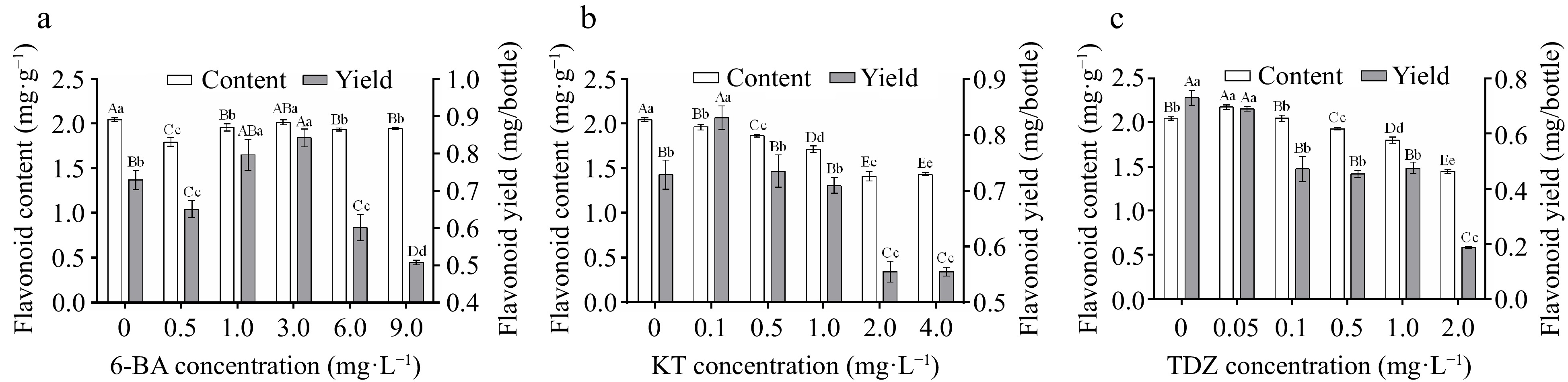
Figure 4.
The effect of different concentrations of cytokinin on total flavonoid content and yield in amaranth callus. Upper case letters indicate p < 0.01, lower case letters indicate p < 0.05. The same letter indicates no significant difference.
In the research, the high concentration of cytokinin in combination inhibited the accumulation of flavonoids in callus. We speculated that the high content of cytokinine promotes the callus proliferation of amaranth and inhibits flavonoid biosynthesis. The calli growth and their production of flavonoids may behave antagonistically. Researchers found similar results on the callus of hawthorn (Crataegus azarolus)[24] and (Rumex pictus)[17].
When supplemented with 2,4-D, NAA or IAA, the total flavonoid content and yield in the callus were significantly higher than those in the control group, and the difference from the control group was extremely significant (Showed in Fig. 5). The 2,4-D concentration ranged from 0.1 to 1.5 mg·L−1 for high total flavonoid content, and the yield was highest at 0.5 mg·L−1 2,4-D than other concentrations and control, with significant difference. The concentration of NAA for highest content and yield of flavonoid was 0.1 mg·L−1 (1.568 mg·g−1) and 1.0 mg·L−1 (0.520 mg/bottle), respectively. When IAA concentration was 1.0 mg·L−1, the flavonoid content (1.366 mg·g−1) and yield (0.438 mg/bottle) was the highest, respectively. Our results showed that 0.5 mg·L−1 2,4-D was most beneficial for the content and yield of flavonoids. The results indicated that the accumulation of flavonoids in amaranth callus necessitates the incorporation of the exogenously added auxin. Similarly, auxins affected flavonoid production in callus culture of Hydrocotyl bonariensis[17]. Furthermore, flavonoid production in callus culture was at a higher rate than in differentiated plants from the various Amaranthus species[4,25], and the callus could continuously produce flavonoids.
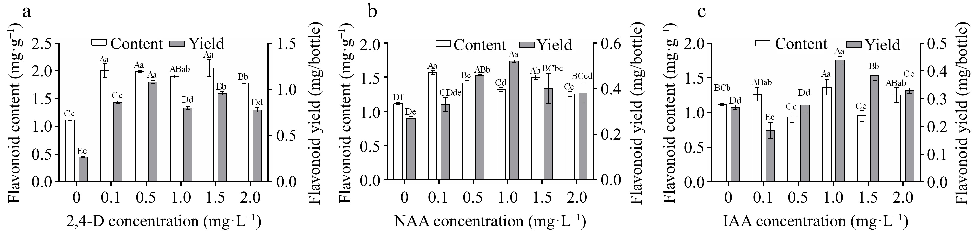
Figure 5.
The effect of different concentrations of auxin on total flavonoid content and yield in amaranth callus. Upper case letters indicate p < 0.01, lower case letters indicate p < 0.05. The same letter indicates no significant difference.
-
The Amaranthus callus induction ability was greatly influenced by the genotype, and a higher cytokinin:auxin ratio was needed for optimal callus induction and proliferation of amaranth. The callus proliferation has been maintained for over 3 years, and the proliferation coefficient was up to 25.24 in the medium (MS + 3.0 mg·L−1 BAP + 0.5 mg·L−1 2,4-D). The growth curve of amaranth callus is an 'S' curve on the solid medium or in the suspension cell culture. MS + 3.0 mg·L−1 BAP + 0.5 mg·L−1 2,4-D was beneficial for the content and yield of flavonoid in the callus.
-
S. Liu conceived and designed the experiments. S. Liu, Y. Xuan, L. Xie, and J. Pan wrote the paper. Y. Xuan, L. Xie, and J. Pan performed the experiment and analyzed the data. All authors read and approved the final version of manuscript.
The financial support for this study was provided by Program for High-level University Construction of the Fujian Agriculture and Forestry University (612014028) and the Natural Science Foundation of Fujian Province (2018J01700).
-
The authors declare that they have no conflict of interest.
-
Received 13 June 2023; Accepted 24 August 2023; Published online 13 September 2023
-
A higher cytokinin: auxin ratio was optimal Amaranthus spp. calluses induction and proliferation.
The growth curve of amaranth callus is ‘S’ curve on the solid medium or in the suspension cell culture.
A optima cytokinin: auxin ratio was beneficial for the content and yield of flavonoid in the callus.
- Copyright: © 2023 by the author(s). Published by Maximum Academic Press on behalf of Hainan University. This article is an open access article distributed under Creative Commons Attribution License (CC BY 4.0), visit https://creativecommons.org/licenses/by/4.0/.
-
About this article
Cite this article
Xuan Y, Liu S, Xie L, Pan J. 2023. Establishment of Amaranthus spp. calluses and cell suspension culture, and the effect of plant growth regulators on total flavonoid content. Tropical Plants 2:15 doi: 10.48130/TP-2023-0015 

Establishment of Amaranthus spp. calluses and cell suspension culture, and the effect of plant growth regulators on total flavonoid content
- Received: 13 June 2023
- Accepted: 24 August 2023
- Published online: 13 September 2023
Abstract: To explore the effects of the combination of auxin and cytokinin on amaranths callus induction, proliferation and the content of flavonoids, Amaranthus spp. was used to carry out the research in this article. The results showed that explants in MS + 3.0 mg·L−1 BAP + 0.5 mg·L−1 2,4-D could promote callus formation and growth. The callus in PI 277269, Ame 18049, and PI 604669 hypocotyl has a high induction rate, but it was compact. The callus in PI 572261 was opposite. The callus proliferation has been maintained for more 3 years, and the proliferation coefficient was up to 25.24 in the medium (MS + 3.0 mg·L−1 BAP + 0.5 mg·L−1 2,4-D). The growth curve of amaranth callus is 'S' curve on the solid medium or in the suspension cell culture. MS + 3.0 mg·L−1 BAP + 0.5 mg·L−1 2,4-D was beneficial for the content and yield of flavonoid in the callus.
-
Key words:
- Amaranthus spp. /
- Hormone /
- Callus /
- Induction /
- Growth curve /
- Flavonoids