ARTICLE   Open Access    

The origin, evolution, and functional divergence of the Dicer-like (DCL) and Argonaute (AGO) gene families in plants

  • # Authors contributed equally: Li-Yao Su, Shan-Shan Li

More Information
  • Received: 12 September 2024
    Revised: 23 October 2024
    Accepted: 30 October 2024
    Published online: 28 November 2024
    Epigenetics Insights  17 Article number: e003 (2024)  |  Cite this article
  • Dicer-like (DCL) and Argonaute (AGO) proteins play a crucial role in plant epigenetics. However, the evolutionary origins and roles of these gene families in plant adaptation, stress response, and development remain unclear. This study explores the origin and functional evolution of DCLs and AGOs across 36 plant species spanning diverse taxonomic groups. Member identification, phylogenetic analysis, evolutionary trajectory analysis, and functional divergence analysis were conducted. The results show that the DCL and AGO originated in Rhodophytes and underwent two major expansions: during algal terrestrialization and the transition from lower to higher plants. In seed plants, DCLs diversified into four classes following two whole-genome duplication (WGD) events, whereas AGOs diversified into seven classes through two WGD events and one tandem duplication event. Expression analyses in Physcomitrium patens, Zea mays, Arabidopsis thaliana, and Fragaria vesca revealed high expression of these gene families in reproductive tissues, with notably lower expression in pollen. Additionally, the expression of these genes exhibits different responses to various environmental stresses in A. thaliana and Z. mays, highlighting their important roles in adaptation to environmental fluctuations. The present research reveals the functional diversification of DCLs and AGOs and their crucial roles in facilitating terrestrial adaptation and rapid land colonization.
  • Atractylodes macrocephala Koidz. (common names 'Baizhu' in Chinese and 'Byakujutsu' in Japanese) is a diploid (2n = 2x = 24) and out-crossing perennial herb in the Compositae family, and has a long history of cultivation in temperate and subtropical areas of East Asia as it is widely used in traditional herbal remedies with multiple pharmacological activities[13]. The 'Pharmacopoeia of the People's Republic of China' states that 'Baizhu' is the dry rhizome of A. macrocephala Koidz. (Atractylodis Macrocephalae Rhizoma, AMR). However, in Japanese traditional medicine 'Baizhu' can be referred to both: A. japonica or A. macrocephala[4].

    A. macrocephala is naturally endemic to China and cultivated in more than 200 towns in China, belonging to Zhejiang, Hunan, Jiangxi, Anhui, Fujian, Sichuan, Hubei, Hebei, Henan, Jiangsu, Guizhou, Shanxi, and Shaanxi Provinces[3]. A. macrocephala grows to a height of 20–60 cm (Fig. 1). The leaves are green, papery, hairless, and generally foliole with 3–5 laminae with cylindric glabrous stems and branches. The flowers grow and aggregate into a capitulum at the apex of the stem. The corollas are purplish-red, and the florets are 1.7 cm long. The achenes, densely covered with white, straight hairs, are obconic and measure 7.5 mm long. The rhizomes used for medicinal purposes are irregular masses or irregularly curving cylinders about 3–13 cm long and 1.5–7 cm in diameter with an outwardly pale greyish yellow to pale yellowish color or a sparse greyish brown color. The periderm-covered rhizomes are externally greyish brown, often with nodose protuberances and coarse wrinkles. The cross-sections are white with fine dots of light yellowish-brown to brown secretion. Rhizomes are collected from plants that are > 2 years old during the spring. The fibrils are removed, dried, and used for medicinal purposes[5, 6].

    Figure 1.  Plant morphology of A. macrocephala.

    The medicinal properties of AMRs are used for spleen deficiency, phlegm drinking, dizziness, palpitation, edema, spontaneous sweating, benefit Qi, and fetal restlessness[7]. The AMR contains various functional components, among which high polysaccharide content, with a yield close to 30%[8]. Therefore, the polysaccharides of A. macrocephala Koidz. rhizome (AMRP) are essential in assessing the quality control and bioactivity of A. macrocephala. Volatile oil accounts for about 1.4% of AMR, with atractylon and atractylodin as the main components[9]. Atractylon can be converted to atractylenolide I (AT-I), atractylenolide II (AT-II), and atractylenolide III (AT-III) under ambient conditions. AT-III can be dehydrated to AT-II under heating conditions[10, 11]. AMRs, including esters, sesqui-, and triterpenes, have a wide range of biological activities, such as improving immune activity, intestinal digestion, neuroprotective activity, immune anti-inflammatory, and anti-tumor.

    In recent years, research on the pharmacological aspects of AMR has continued to increase. Still, the discovery of the main active components in AMR is in its infancy. The PAO-ZHI processing of AMR is a critical step for AMR to exert its functional effects, but also, in this case, further work is required. Studies on the biosynthesis of bioactive compounds and different types of transcriptomes advanced current knowledge of A. macrocephala, but, as mentioned, required more systematic work. Ulteriorly, an outlook on the future research directions of A. macrocephala was provided based on the advanced technologies currently applied in A. macrocephala (Fig. 2).

    Figure 2.  Current progress of A. macrocephala.

    A. macrocephala is distributed among mountainous regions more than 800 m above sea level along the middle and lower reaches of the Yangtze River (China)[5]. Due to over-exploitation and habitat destruction, natural populations are rare, threatened, and extinct in many locations[1,12]. In contrast to its native range, A. macrocephala is widely cultivated throughout China, in a total area of 2,000–2,500 ha, with a yield of 7,000 t of rhizomes annually[13]. A. macrocephala is mainly produced in Zhejiang, Anhui, and Hebei (China)[14]. Since ancient times, Zhejiang has been the famous producing area and was later introduced to Jiangxi, Hunan, Hebei, and other places[15]. Wild A. macrocephala is currently present in at least 14 provinces in China. It is mainly distributed over three mountain ranges, including the Tianmu and Dapan mountains in Zhejiang Province and the Mufu mountains along the border of Hunan and Jiangxi Provinces. A. macrocephala grows in a forest, or grassy areas on mountain or hill slopes and valleys at an altitude of 600–2,800 m. A. macrocephala grows rapidly at a temperature of 22–28 °C, and favors conditions with total precipitation of 300–400 mm evenly distributed among the growing season[16]. Chen et al. first used alternating trilinear decomposition (ATLD) to characterize the three-dimensional fluorescence spectrum of A. macrocephala[17]. Then they combined the three-dimensional fluorescence spectrum with partial least squares discriminant analysis (PLS-DA) and k-nearest neighbor method (kNN) to trace the origin of Atractylodes samples. The results showed that the classification models established by PLS-DA and kNN could effectively distinguish the samples from three major Atractylodes producing areas (Anhui, Hunan, and Zhejiang), and the classification accuracy rate (CCR) of Zhejiang atractylodes was up to 80%, and 90%, respectively[17]. Zhang et al. compared the characteristics, volatile oil content, and chemical components of attested materials from six producing areas of Zhejiang, Anhui, Hubei, Hunan, Hebei, and Henan. Differences in the shape, size, and surface characteristics were reported, with the content of volatile oil ranging from 0.58% to 1.22%, from high to low, Hunan (1.22%) > Zhejiang (1.20%) > Anhui (1.02%) > Hubei (0.94%) > Henan (0.86%) > Hebei (0.58%)[18]. This study showed that the volatile oil content of A. macrocephala in Hunan, Anhui, and Hubei is not much different from that of Zhejiang, which is around 1%. A. macrocephala is a local herb in Zhejiang, with standardized cultivation techniques, with production used to reach 80%–90% of the country. However, in recent years, the rapid development of Zhejiang's real estate economy has reduced the area planted with Zhejiang A. macrocephala, resulting in a sudden decrease in production. Therefore, neighboring regions, such as Anhui and Hunan, vigorously cultivate A. macrocephala, and the yield and quality of A. macrocephala can be comparable to those of Zhejiang. The results were consistent with the data reports[18]. Guo et al. analyzed the differentially expressed genes of Atractylodes transcripts from different regions by the Illumina HiSeq sequencing platform. It was found that 2,333, 1,846, and 1,239 DEGs were screened from Hubei and Hebei, Anhui and Hubei, and Anhui and Hebei Atrexia, respectively, among which 1,424, 1,091, and 731 DEGs were annotated in the GO database. There were 432, 321, and 208 DEGs annotated in the KEGG database. These DEGs were mainly related to metabolic processes and metabolic pathways of secondary metabolites. The highest expression levels of these genes were found in Hubei, indicating higher terpenoid production in Hubei[19]. Other compounds were differentially accumulated in Atractylodes. Chlorogenic acid from Hebei was 0.22%, significantly higher than that from Zhejiang and Anhui[20]. Moreover, the content of neochlorogenic acid and chlorogenic acid decreased after processing, with the highest effect reported in Zhejiang, with the average transfer rate of neochlorogenic acid and chlorogenic acid reaching 55.68% and 55.05%[20]. All these changes would bring great help in distinguishing the origins of A. macrocephala.

    Medicinal AMR can be divided into raw AMR and cooked AMR. The processing method is PAO-ZHI; the most traditional method is wheat bran frying. The literature compared two different treatment methods, crude A. macrocephala (CA) and bran-processed A. macrocephala, and found that the pharmacological effects of AMR changed after frying with wheat bran, mainly in the anti-tumor, antiviral and anti-inflammatory effects[21]. The anti-inflammatory effect was enhanced, while the anti-tumor and antiviral effects were somewhat weakened, which may be related to the composition changes of the compounds after frying. The study of the content of AT-I, II, and III, and atractyloside A, in rat serum provided helpful information on the mechanism of wheat bran processing[22]. In addition to frying wheat bran, Sun et al. used sulfur fumigation to treat AMR[23]. They found that the concentration of different compounds changed, producing up to 15 kinds of terpenoids. Changes in pharmacological effects were related to treatment and the type of illumination[24,25]. Also, artificial light can improve the various biological functions. A. macrocephala grew better under microwave electrodeless light, with a chlorophyll content of 57.07 ± 0.65 soil and plant analyzer develotrnent (SPAD)[24]. The antioxidant activity of AMR extract treated with light-emitting diode (LED)-red light was the highest (95.3 ± 1.1%) compared with other treatments[24]. The total phenol and flavonoid contents of AMR extract treated with LED-green light were the highest at 24.93 ± 0.3 mg gallic acid equivalents (GAE)/g and 11.2 ± 0.3 mg quercetin equivalents (QE)/g compared with other treatments[24, 25]. Polysaccharides from Chrysanthemun indicum L.[26] and Sclerotium rolfsiisacc[27] can improve AMR's biomass and bioactive substances by stimulating plant defense and thus affect their efficacy. In summary, there are compositional differences between A. macrocephala from different origins. Besides, different treatments, including processing mode, light irradiation, and immune induction factors, which can affect AMR's biological activity, provide some reference for the cultivation and processing of A. macrocephala (Fig. 3).

    Figure 3.  Origin, distribution and processing of A. macrocephala.

    The AMR has been reported to be rich in polysaccharides, sesquiterpenoids (atractylenolides), volatile compounds, and polyacetylenes[3]. These compounds have contributed to various biological activities in AMR, including immunomodulatory effects, improving gastrointestinal function, anti-tumor activity, neuroprotective activity, and anti-inflammatory.

    AMRP has received increasing attention as the main active component in AMR because of its rich and diverse biological activities. In the last five years, nine AMRP have been isolated from AMR. RAMP2 had been isolated from AMR, with a molecular weight of 4.354 × 10³ Da. It was composed of mannose, galacturonic acid, glucose, galactose, and arabinose, with the main linkages of →3-β-glcp-(1→, →3,6-β-glcp-(1→, →6-β-glcp-(1→, T-β-glcp-(1→, →4-α-galpA-(1→, →4-α-galpA-6-OMe-(1→, →5-α-araf-(1→, →4,6-β-manp-(1→ and →4-β-galp-(1→[28]. Three water-soluble polysaccharides AMAP-1, AMAP-2, and AMAP-3 were isolated with a molecular weight of 13.8 × 10⁴ Da, 16.2 × 10⁴ Da, and 8.5 × 10⁴ Da, respectively. Three polysaccharides were deduced to be natural pectin-type polysaccharides, where the homogalacturonan (HG) region consists of α-(1→4)-linked GalpA residues and the ramified region consists of alternating α-(1→4)-linked GalpA residues and α-(1→2)-linked Rhap residues. Besides, three polysaccharides were composed of different ratios of HG and rhamnogalacturonan type I (RG-I) regions[29]. Furthermore, RAMPtp has been extracted from AMR with a molecular weight of 1.867 × 10³ Da. It consists of glucose, mannose, rhamnose, arabinose, and galactose with 60.67%, 14.99%, 10.61%, 8.83%, and 4.90%, connected by 1,3-linked β-D Galp and 1,6-linked β-D Galp residues[30]. Additionally, PAMK was characterized by a molecular weight of 4.1 kDa, consisting of galactose, arabinose, and glucose in a molar ratio of 1:1.5:5, with an alpha structure and containing 96.47% polysaccharide and small amounts of protein, nucleic acid, and uric acid[31]. Another PAMK extracted from AMR had a molecular weight of 2.816 × 10³ Da and consisted of glucose and mannose in molar ratios of 0.582 to 0.418[32]. Guo et al. isolated PAMK with a molecular weight of 4.748 × 10³ g/mol from AMR, consisting of glucose, galactose, arabinose, fructose, and mannose in proportions of 67.01%, 12.32%, 9.89%, 1.18%, and 0.91%, respectively[33]. In addition, AMP1-1 is a neutral polysaccharide fragment with a molecular weight of 1.433 kDa isolated from AMR. It consists of glucose and fructose, and the structure was identified as inulin-type fructose α-D-Glcp-1→(2-β-D-Fruf-1)7[34]. These reports indicated that, in general, polysaccharides are extracted by water decoction, ultrasonic-assisted extraction, enzyme hydrolysis method, and microwave-assisted extraction. The separation and purification are column chromatography, stepwise ethanol precipitation, and ultrafiltration. Their physicochemical properties and structural characterization are generally achieved by determining the molecular weight, determining the monosaccharide composition, analyzing the secondary structure, and glycosidic bond configuration of polysaccharides with Fourier transform infrared (FT-IR) and nuclear magnetic resonance (NMR). The advanced structures of polysaccharides can be identified by high-performance size exclusion chromatography-multiangle laser light scattering (HPSEC-MALLS), transmission electron microscopy (TEM), and scanning electron microscopy (SEM) techniques (Table 1). AMRP has various physiological functions, including immunomodulatory effects, improving gastrointestinal function, and anti-tumor activity. The related biological activities, animal models, monitoring indicators, and results are summarized in Table 1.

    Table 1.  Components and bioactivity of polysaccharides from Atractylodes macrocephala Koidz. Rhizome.
    Pharmacological activitiesDetailed functionPolysaccharides informationModelDoseTest indexResultsRef.
    Immunomodulatory effectsRestore immune
    function
    /Chicken models
    (HS-induced)
    200 mg/kgOxidative index;
    Activities of mitochondrial complexes and ATPases;
    Ultrastructure in chicken spleens;
    Expression levels of cytokines, Mitochondrial dynamics- and apoptosis-related genes
    Alleviated
    the expression of
    IL-1 ↑,TNF-α ↑, IL-2 ↓, IFN- γ ↓; mitochondrial dynamics- and anti-apoptosis-related genes ↓; pro-apoptosis-related genes ↑;
    the activities of mitochondrial complexes and ATPases ↓ caused by HS
    [35]
    Regulate the immune function/Chicken models
    (HS-induced)
    200 mg/kgiNOS–NO activities;
    ER stress-related genes;
    Apoptosis-related genes;
    Apoptosis levels
    Alleviated NO content ↑; activity of iNOS ↑ in the chicken spleen; GRP78, GRP94, ATF4, ATF6, IRE ↑; caspase3 ↑; Bcl-2 ↓ caused by HS[36]
    Relieve immunosuppressionCommercial AMR powder (purity 70%)Geese models
    (CTX-induced)
    400 mg/kgSpleen development;
    Percentages of leukocytes in peripheral blood
    Alleviated the spleen damage;
    T and B cell proliferation ↓; imbalance of leukocytes; disturbances of humoral; cellular immunity caused by CTX
    [37]
    Active the lymphocytesCommercial AMR powder (purity 95%)Geese models
    (CTX-induced)
    400 mg/kgThymus morphology;
    The level of serum GMC-SF, IL-1b, IL-3, IL-5;
    mRNA expression of CD25, novel_mir2, CTLA4 and CD28 signal pathway
    Maintain normal cell morphology of thymus;
    Alleviated GMC-SF ↓, IL-1b ↓, IL-5↓, IL-6↓, TGF-b↓; IL-4 ↑, IL-10 ↑; novel_mir2 ↓, CD25↓, CD28↓ in thymus and lymphocytes caused by CTX
    [38]
    Alleviate immunosuppressionCommercial AMR powder (purity 70%)Geese models
    (CTX-induced)
    400 mg/kgThymus development;
    T cell proliferation rate;
    The level of CD28, CD96, MHC-II;
    IL-2 levels in serum;
    differentially expressed miRNAs
    Alleviated thymus damage;
    T lymphocyte proliferation rate ↓; T cell activation ↓; IL-2 levels ↓ caused by CTX;
    Promoted novel_mir2 ↑; CTLA4 ↓; TCR-NFAT signaling pathway
    [39]
    Alleviates T cell activation declineCommercial AMR powder (purity 95%)BALB/c female mice (CTX-induced)200 mg/kgSpleen index;
    Morphology, death, cytokine concentration of splenocytes;
    Th1/Th2 ratio, activating factors of lymphocytes;
    T cell activating factors;
    mRNA expression level in CD28 signal pathway
    Improved the spleen index;
    Alleviated abnormal splenocytes morphology and death; Balance Th1/Th2 ratio; IL-2 ↑, IL-6 ↑, TNF-α ↑, IFN-γ ↑; mRNA levels of CD28, PLCγ-1, IP3R, NFAT, AP-1 ↑
    [40]
    Immunoregulation and ImmunopotentiationCommercial AMR powder (purity 80%)BMDCs (LPS-induced);
    Female BALB/c mice (ovalbumin as a model antigen)
    /Surface molecule expression of BMDCs;
    Cytokines secreted by dendritic cell supernatants;
    OVA-specific antibodies in serum;
    Cytokines in serum;
    Lymphocyte immunophenotype
    Expression of CD80 and CD86 ↑; IL-1β ↑, IL-12 ↑, TNF-α↑ and IFN-γ ↑; OVA-specific antibodies in serum ↑; Secretion of cytokines ↑; Proliferation rate of spleen lymphocytes ↑; Activation of CD3+CD4+ and CD3+CD8+ lymphocytes[46]
    Increase immune-response capacity of the spleen in miceCommercial AMR powder (purity 70%)BALB/c female mice100, 200, 400 mg/kgSpleen index;
    Concentrations of cytokines;
    mRNA and protein expression levels in TLR4 signaling
    In the medium-PAMK group:
    IL-2, IL-4, IFN-c, TNF-a ↑; mRNA and protein expression of TLR4, MyD88, TRAF6, TRAF3, NF-κB in the spleen ↑
    [41]
    Immunological activityCommercial AMR powder (purity 80%)Murine splenic lymphocytes (LPS or PHA-induced)13, 26, 52, 104, 208 μg/mLT lymphocyte surface markersLymphocyte proliferation ↑;
    Ratio of CD4+/CD8+ T cells ↑
    [47]
    Immunomodulatory activityTotal carbohydrates content 95.66 %Mouse splenocytes
    (Con A or LPS-induced)
    25, 50, 100 μg/mLSplenocyte proliferation;
    NK cytotoxicity;
    Productions of NO and cytokines;
    Transcription factor activity;
    Signal pathways and receptor
    Promoted splenocyte proliferation; Cells enter S and G2/M phases; Ratios of T/B cells ↑; NK cytotoxicity ↑; Transcriptional activities of NFAT ↑; NF-κB, AP-1 ↑; NO, IgG, IL-1α, IL-1β, IL-2, IL-3, IL-4, IL-6, IL-10, IL-12p40, IL-12p70, IL-13, IFN-γ, TNF-α, G-CSF, GM-CSF, KC, MIP-1α, MIP-1β, RANTES, Eotaxin ↑[42]
    Promote the proliferation of thymic epithelial cellsContents of fucrhaara, galactose, glucose, fructose,
    and xylitol: 0.98%, 0.40%, 88.67%, 4.47%, and 5.47%
    MTEC1 cells50 μg/mLCell viability and proliferation;
    lncRNAs, miRNAs, and mRNAs expression profiles in MTEC1 cells
    The differential genes were 225 lncRNAs, 29 miRNAs, and 800 mRNAs; Genes enriched in cell cycle, cell division, NF-κB signaling, apoptotic process, and MAPK signaling pathway[44]
    Immunomodulatory activityMW: 4.354 × 10³ Da;
    Composed of mannose, galacturonic acid, glucose, galactose and arabinose;
    The main linkages are →3-β-glcp-(1→, →3,6-β-glcp-(1→, →6-β-glcp-(1→, T-β-glcp-(1→,
    →4-α-galpA-(1→, →4-α-galpA-6-OMe-(1→, →5-α-araf-(1→, →4,6-β-manp-(1→ and →4-β-galp-(1→
    CD4+ T cell50, 100, 200 μg/mLMolecular weight;
    Monosaccharide composition;
    Secondary structure;
    Surface topography;
    Effect on Treg cells
    Treg cells percentage ↑; mRNA expressions of Foxp3, IL-10 and IL-2 ↑; STAT5 phosphorylation levels ↑; IL-2/STAT5 pathway[28]
    Immunostimulatory activityMW of AMAP-1, AMAP-2, and AMAP-3 were 13.8×10⁴ Da, 16.2×10⁴ Da and 8.5×10⁴ Da;
    HG region consists of α-(1→4)-linked GalpA residues
    RAW264.7 cells (LPS-induced)80, 40, 200 μg/mLMolecular weight;
    Total carbohydrate;
    Uronic acid contents;
    Secondary structure;
    Monosaccharide composition;
    Immunostimulatory activity
    RG-I-rich AMAP-1 and AMAP-2 improved the release of NO[29]
    Immunomodulatory effectMW: 1.867×10³ Da;
    Contents of glucose, mannose, rhamnose,
    arabinose and galactose: 60.67%, 14.99%, 10.61%, 8.83% and 4.90%
    SMLN lymphocytes25
    μg/ml
    Molecular weight;
    Monosaccharide composition;
    Ultrastructure;
    Intracellular Ca2+concentration;
    Target genes;
    Cell cycle distribution
    [Ca2+]i ↑; More cells in S and G2/M phases; IFN-γ ↑, IL-17A ↑; mRNA expressions of IL-4 ↓[30]
    Macrophage activationTotal carbohydrates content 95.66 %RAW264.7 macrophages (LPS-induced)25, 50, 100 μg/mLPinocytic activity;
    Phagocytic uptake;
    Phenotypic characterization;
    Cytokine production;
    Bioinformatics analysis;
    Transcription factor inhibition
    IL-6, IL-10 and TNF-α ↑; CCL2 and CCL5 ↑; Pinocytic and phagocytic activity ↑; CD40, CD80, CD86, MHC-I, MHC-II ↑; NF-κB and Jak-STAT pathway[43]
    Immunomodulatory effectTotal carbohydrates content 95.66 %SMLN lymphocytes25, 50, 100 μg/mLCytokine production;
    CD4+ and CD8+ lymphocytes;
    Target genes;
    Bioinformatics analysis;
    T and B lymphocyte proliferation;
    Receptor binding and blocking
    IFN-γ, IL-1α, IL-21, IFN-α, CCL4, CXCL9, CXCL10 ↑; CD4+ and CD8+subpopulations proportions ↑;
    c-JUN, NFAT4, STAT1, STAT3 ↑;
    67 differentially expressed miRNAs (55 ↑ and
    12 ↓), associated with immune system pathways; Affect T and B lymphocytes
    [45]
    Improving gastrointestinal functionRelieve enteritis and improve intestinal
    flora disorder
    Commercial AMR powder (purity 70%);
    Contents of fucrhaara, galactose, glucose, xylitol, and fructose: 0.98%, 0.40%, 88.67%, 4.47%, and 5.47%
    Goslings (LPS-induced)400 mg/kgSerum CRP, IL-1β, IL-6, and TNF-α levels;
    Positive rate of IgA;
    TLR4, occludin, ZO-1, cytokines, and immunoglobulin mRNA expression;
    Intestinal flora of gosling excrement
    Relieved IL-1β, IL-6, TNF-α levels in serum ↑; the number of IgA-secreting cells ↑; TLR4 ↑; tight junction occludin and ZO-1 ↓; IL-1β mRNA expression in the small intestine ↑; Romboutsia ↓ caused by LPS[48]
    Ameliorate ulcerative colitisMW: 2.391 × 10⁴ Da;
    Composed of mannose, glucuronic acid, glucose and arabinose in a molar ratio of 12.05:6.02:72.29:9.64
    Male C57BL/6J mice (DDS-induced)10, 20, 40 mg/kg bwHistopathological evaluation;
    Inflammatory mediator;
    Composition of gut microbiota;
    Feces and plasma for global metabolites profiling
    Butyricicoccus, Lactobacillus ↑;
    Actinobacteria, Akkermansia, Anaeroplasma, Bifidobacterium, Erysipelatoclostridium, Faecalibaculum, Parasutterella,
    Parvibacter, Tenericutes, Verrucomicrobia ↓;
    Changed 23 metabolites in fecal content; 21 metabolites in plasma content
    [49]
    Attenuate ulcerative colitis/Male SD rats (TNBS-induced);
    Co-culture BMSCs and IEC-6 cells
    540 mg/kg
    (for rats);
    400 μg/mL (for cell)
    Histopathological analysis;
    Cell migration;
    Levels of cytokines
    Potentiated BMSCs’ effect on preventing colitis and homing the injured tissue, regulated cytokines;
    BMSCs and AMP promoted the migration of IEC
    [52]
    Against intestinal mucosal injuryMW: 3.714 × 10³ Da;
    Composed of glucose, arabinose, galactose, galacturonic acid, rhamnose
    and mannose with molar ratios of 59.09:23.22:9.32:4.70:2.07:1.59
    Male C57BL/6 mice (DDS-induced)100 mg/kgIntestinal morphology;
    IL-6, TNF-α and IL-1β in serum;
    mRNA expression;
    Intestinal microbiota
    Alleviated body weight ↓; colon length ↓; colonic damage caused by DSS;
    Over-expression of TNF-α, IL-1β, IL-6 ↓; Infiltration of neutrophils in colon ↓; Mucin 2 ↑;
    Tight junction protein Claudin-1 ↑;
    Harmful bacteria content ↓;
    Beneficial bacteria content ↑
    [50]
    Against intestinal injuryTotal carbohydrates 95.66 %IECs (DDS-induced)5, 25, 50 μg/mLCell proliferation and apoptosis;
    Expression levels of intercellular TJ proteins;
    lncRNA screening
    Proliferation and survival of IECs ↑;
    Novel lncRNA ITSN1-OT1 ↑;
    Blocked the nuclear import of phosphorylated STAT2
    [51]
    Anti-tumor activityInduce apoptosis in transplanted H22 cells in miceMW: 4.1× 10³ Da;
    Neutral heteropolysaccharide composed of galactose, arabinose, and glucose with α-configuration (molar ratio, 1:1.5:5)
    Female Kunming mice100 and 200 mg/kg (for rats)Secondary structure;
    Molecular weight;
    Molecular weight;
    Thymus index and Spleen index;
    Lymphocyte Subpopulation in peripheral blood;
    Cell cycle distribution
    In tumor-bearing mice CD3+, CD4+, CD8+ ↓;
    B cells ↑
    [31]
    Regulate the innate immunity of colorectal cancer cellsCommercial AMR powder (purity 70%)C57BL/6J mice (MC38 cells xenograft model)500 mg/kgExpression of pro-inflammatory cytokines and secretionIL-6, IFN-λ, TNF-α, NO ↑ through MyD88/TLR4-dependent signaling pathway;
    Survival duration of mice with tumors ↑;
    Prevent tumorigenesis in mice
    [54]
    Induce apoptosis of Eca-109 cellsMW: 2.1× 10³ Da;
    Neutral hetero polysaccharide composed
    of arabinose and glucose (molar ratio, 1:4.57) with pyranose rings and α-type and β-type glycosidic linkages
    Eca-109 cells0.25, 0.5, 1, 1.5, 2.00 mg/mLCell morphology;
    Cell cycle arrest;
    Induction of apoptosis
    Accelerate the apoptosis of Eca109 cells[53]
    '/' denotes no useful information found in the study.
     | Show Table
    DownLoad: CSV

    To study the immunomodulatory activity of AMRP, the biological models generally adopted are chicken, goose, mouse, and human cell lines. Experiments based on the chicken model have generally applied 200 mg/kg doses. It was reported that AMRP protected the chicken spleen against heat stress (HS) by alleviating the chicken spleen immune dysfunction caused by HS, reducing oxidative stress, enhancing mitochondrial function, and inhibiting cell apoptosis[35]. Selenium and AMRP could improve the abnormal oxidation and apoptosis levels and endoplasmic reticulum damage caused by HS, and could act synergistically in the chicken spleen to regulate biomarker levels[36]. It indicated that AMRP and the combination of selenium and AMRP could be applied as chicken feed supplementation to alleviate the damage of HS and improve chicken immunity.

    The general application dose in the goose model is also 200 mg/kg, and the main injury inducer is cyclophosphamide (CTX). AMRP alleviated CTX-induced immune damage in geese and provided stable humoral immune protection[37]. Little is known about the role of AMRP in enhancing immunity in geese through the miRNA pathway. It was reported that AMRP alleviated CTX-induced decrease in T lymphocyte activation levels through the novel _mir2/CTLA4/CD28/AP-1 signaling pathway[38]. It was also reported that AMRP might be achieved by upregulating the TCR-NFAT pathway through novel_mir2 targeting of CTLA4, thereby attenuating the immune damage induced by CTX[39]. This indicated that AMRP could also be used as goose feed supplementation to improve the goose's autoimmunity.

    The typical injury inducer for mouse models is CTX, and the effects on mouse spleen tissue are mainly observed. BALB/c female mice were CTX-induced damage. However, AMRP increased cytokine levels and attenuated the CTX-induced decrease in lymphocyte activation levels through the CD28/IP3R/PLCγ-1/AP-1/NFAT signaling pathway[40]. It has also been shown that AMRP may enhance the immune response in the mouse spleen through the TLR4-MyD88-NF-κB signaling pathway[41].

    Various cellular models have been used to study the immune activity of AMRP, and most of these studies have explored the immune activity with mouse splenocytes and lymphocytes. Besides, the commonly used damage-inducing agents are LPS, phytohemagglutinin (PHA), and concanavalin A (Con A).

    In one study, the immunoreactivity of AMRP was studied in cultured mouse splenocytes. LPS and Con A served as controls. Specific inhibitors against mitogen-activated protein kinases (MAPKs) and NF-κB significantly inhibited AMRP-induced IL-6 production. The results suggested that AMRP-induced splenocyte activation may be achieved through TLR4-independent MAPKs and NF-κB signaling pathways[42]. Besides, AMRP isolated from AMR acting on LPS-induced RAW264.7 macrophages revealed that NF-κB and Jak-STAT signaling pathways play a crucial role in regulating immune response and immune function[43]. RAMP2 increased the phosphorylation level of STAT5 in Treg cells, indicating that RAMP2 could increase the number of Treg cells through the IL-2/STAT5 signaling pathway[28]. Furthermore, the relationship between structure and immune activity was investigated. Polysaccharides rich in RG-I structure and high molecular weight improved NO release from RAW264.7 cells. Conversly, polysaccharides rich in HG structure and low molecular weight did not have this ability, indicating that the immunoreactivity of the polysaccharide may be related to the side chain of RG-I region[29]. Moreover, the effect of AMRP on the expression profile of lncRNAs, miRNAs, and mRNAs in MTEC1 cells has also been investigated. The differentially expressed genes include lncRNAs, Neat1, and Limd1. The involved signaling pathways include cell cycle, mitosis, apoptotic process, and MAPK[44].

    Xu et al. found that AMRP affects supramammary lymph node (SMLN) lymphocytes prepared from healthy Holstein cows. Sixty-seven differentially expressed miRNAs were identified based on microRNA sequencing and were associated with immune system pathways such as PI3K-Akt, MAPKs, Jak-STAT, and calcium signaling pathways. AMRP exerted immunostimulatory effects on T and B lymphocytes by binding to T cell receptor (TCR) and membrane Ig alone, thereby mobilizing immune regulatory mechanisms within the bovine mammary gland[45].

    AMRP can also be made into nanostructured lipid carriers (NLC). Nanoparticles as drug carriers can improve the action of drugs in vivo. NLC, as a nanoparticle, has the advantages of low toxicity and good targeting[46]. The optimization of the AMRP-NLC preparation process has been reported. The optimum technologic parameters were: the mass ratio of stearic acid to caprylic/capric triglyceride was 2:1. The mass ratio of poloxamer 188 to soy lecithin was 2:1. The sonication time was 12 min. The final encapsulation rate could reach 76.85%[47]. Furthermore, AMRP-NLC interfered with the maturation and differentiation of bone marrow-derived dendritic cells (BMDCs). Besides, AMRP-NLC, as an adjuvant of ovalbumin (OVA), could affect ova-immunized mice with enhanced immune effects[46].

    AMRP also has the effect of alleviating intestinal damage. They are summarized in Table 1. The common damage-inducing agents are lipopolysaccharide (LPS), dextran sulfate sodium (DDS), and trinitrobenzene sulfonic acid (TNBS). A model of LPS-induced enteritis in goslings was constructed to observe the effect of AMRP on alleviating small intestinal damage. Gosling excrement was analyzed by 16S rDNA sequencing to illuminate the impact of AMRP on the intestinal flora. Results indicated that AMRP could maintain the relative stability of cytokine levels and immunoglobulin content and improve intestinal flora disorder[48]. Feng et al. used DDS-induced ulcerative colitis (UC) in mice and explored the alleviating effects of AMRP on UC with 16S rDNA sequencing technology and plasma metabolomics. The results showed that AMRP restored the DDS-induced disruption of intestinal flora composition, regulated the production of metabolites such as short-chain fatty acids and cadaveric amines, and regulated the metabolism of amino acids and bile acids by the host and intestinal flora[49]. A similar study has reported that AMRP has a protective effect on the damage of the intestinal mucosal barrier in mice caused by DSS. It was found that AMRP increased the expression of Mucin 2 and the tight junction protein Claudin-1. In addition, AMRP decreased the proportion of harmful bacteria and increased the potentially beneficial bacteria content in the intestine[50]. The protective effect of AMRP on DSS-induced damage to intestinal epithelial cells (IECs) has also been investigated. The results showed that AMRP promoted the proliferation and survival of IECs.

    In addition, AMRP induced a novel lncRNA ITSN1-OT1, which blocked the nuclear import of phosphorylated STAT2 and inhibited the DSS-induced reduced expression and structural disruption of tight junction proteins[51]. AMRP can also act in combination with cells to protect the intestinal tract. The ulcerative colitis model in Male Sprague-Dawley (SD) rats was established using TNBS, and BMSCs were isolated. IEC-6 and BMSCs were co-cultured and treated by AMRP. The results showed that AMRP enhanced the prevention of TNBS-induced colitis in BMSCs, promoted the migration of IEC, and affected the expression of various cytokines[52]. These reports indicated that the 16S rDNA sequencing technique could become a standard method to examine the improvement of gastrointestinal function by AMRP.

    AMRP has anti-tumor activity and other biological activities. AMRP can induce apoptosis in Hepatoma-22 (H22) and Eca-109 cells and modulate the innate immunity of MC38 cells. For instance, the anti-tumor effects of AMRP were investigated by constructing a tumor-bearing mouse model of H22 tumor cells. AMRP blocked the S-phase of H22 tumor cells and induced an immune response, inhibiting cell proliferation[31]. In addition, AMRP can inhibit cell proliferation through the mitochondrial pathway and by blocking the S-phase of Eca-109 tumor cells[53]. AMRP affects MC38 tumor cells, and the anti-tumor effect of AMRP was investigated with Toll-like receptor 4 (TLR4) KO C57BL/6 mice and the construction of the MC38 tumor cell xenograft model. AMRP significantly inhibited the development of MC38 cells in mice and prolonged the survival of tumor-bearing mice. AMRP activity was diminished in TLR4 KO mice. Combined with the immunoblotting assay results, it was shown that TLR4 regulated the MyD88-dependent signaling pathway, which has a critical effect on the anti-tumor effect of AMRP[54].

    AMR contains a large number of bioactive compounds. Among them, small molecule compounds include esters, sesquiterpenes, and other compounds. These small molecule compounds have significant pharmacological activities, including anti-tumor, neuroprotective, immunomodulatory, and anti-inflammatory. In the last five years, small molecule compounds have been increasingly identified (Fig. 4), with atractylenolides as the main component of AMR extracts[11]. Atractylenolides are a small group of sesquiterpenoids. Atractylenolides include AT-I, AT-II, and AT-III, lactones isolated from AMR.

    Figure 4.  Structure of small molecule compounds with bioactivities from AMR. Atractylenolide I (1); Atractylenolide II (2); Atractylenolide III (3); 3β-acetoxyl atractylenolide I (4); 4R,5R,8S,9S-diepoxylatractylenolide II (5); 8S,9S-epoxyla-tractylenolide II (6); Atractylmacrols A (7); Atractylmacrols B (8); Atractylmacrols C (9); Atractylmacrols D (10); Atractylmacrols E (11); 2-[(2E)-3,7-dimethyl-2,6-octadienyl]-6-methyl-2,5-cyclohexadiene-1,4-dione (12); 8-epiasterolid (13); (3S,4E,6E,12E)-1-acetoxy-tetradeca-4,6,12-triene-8,10-diyne-3,14-diol (14); (4E,6E,12E)-tetradeca-4,6,12-triene-8,10-diyne-13,14-triol (15); 1-acetoxy-tetradeca-6E,12E-diene-8, 10-diyne-3-ol (16); 1,3-diacetoxy-tetradeca-6E, 12E-diene-8,10-diyne (17); Biatractylenolide II (18); Biepiasterolid (19); Biatractylolide (20).

    The anti-tumor activity was mainly manifested by AT-I and AT-II, especially AT-I (Table 2). Anti-tumor activity has been studied primarily in vivo and in vitro. However, there is a lack of research on the anti-tumor activity of atractylenolide in human clinical trials. The concentration of atractylenolide applied on cell lines was < 400 μM, or < 200 mg/kg on tumor-bearing mice.

    Table 2.  Anti-tumor activity of atractylenolides.
    TypesSubstancesModelIndexDoseSignal pathwayResultsRef.
    Human colorectal cancerAT-IIIHCT-116 cell;
    HCT-116 tumor xenografts bearing in nude mice
    Cell viability;
    Cell apoptotic;
    mRNAs and protein
    expressions of Bax, Bcl-2, caspase-9 and caspase-3
    25, 50, 100, 200 μM (for cell);
    50, 100,
    200 mg/kg (for rats)
    Bax/Bcl-2 signaling pathwayPromoting the expression of proapoptotic related gene/proteins; Inhibiting the expression of antiapoptotic related gene/protein; Bax↑; Caspase-3↓; p53↓; Bcl-2↓[55]
    Human gastric carcinomaAT-IIHGC-27 and AGS cell
    Cell viability;
    Morphological changes;
    Flow cytometry;
    Wound healing;
    Cell proliferation, apoptosis, and motility
    50, 100, 200, 400 μMAkt/ERK signaling pathwayCell proliferation, motility↓; Cell apoptosis↑; Bax↑;
    Bcl-2↓; p-Akt↓; p-ERK↓
    [56]
    Mammary
    tumorigenesis
    AT-IIMCF 10A cell;
    Female SD rats (NMU-induced)
    Nrf2 expression and nuclear accumulation;
    Cytoprotective effects;
    Tumor progression;
    mRNA and protein levels of Nrf2;
    Downstream detoxifying enzymes
    20, 50, 100 μM (for cell);
    100 and 200 mg/kg (for rats)
    JNK/ERK-Nrf2-ARE signaling pathway;
    Nrf2-ARE signaling pathway
    Nrf2 expressing↑; Nuclear translocation↑; Downstream detoxifying enzymes↓; 17β-Estradiol↓; Induced malignant transformation[57]
    Human colon adenocarcinomaAT-IHT-29 cellCell viability;
    TUNEL and Annexin V-FITC/PI double stain;
    Detection of initiator and
    executioner caspases level
    10, 20, 40, 80, 100 μMMitochondria-dependent pathwayPro-survival Bcl-2↓; Bax↑; Bak↑; Bad↑; Bim↑; Bid↑; Puma↑[58]
    Sensitize triple-negative
    TNBC cells to paclitaxel
    AT-IMDA-MB-231 cell;
    HS578T cell;
    Balb/c mice (MDA-MB-231 cells-implanted)
    Cell viability
    Transwell migration
    CTGF expression
    25, 50, 100 μM (for cell);
    50 mg/kg (for rats)
    /Expression and secretion of CTGF↓; CAF markers↓; Blocking CTGF expression and fibroblast activation[59]
    Human ovarian cancerAT-IA2780 cellCell cycle;
    Cell apoptosis;
    Cyclin B1 and CDK1 level
    12.5, 25, 50, 100 and 200 μMPI3K/Akt/mTOR
    signaling pathway
    Cyclin B1, CDK1↓; Bax↑;
    Caspase-9↓; Cleaved caspase-3↓; Cytochrome c↑; AIF↑; Bcl-2↓; Phosphorylation level of PI3K, Akt, mTOR↓
    [60]
    Impaired metastatic properties transfer of CSCsAT-ILoVo-CSCs; HT29-CSCsCell migration
    and invasion;
    miR-200c expression;
    Cell apoptosis
    200 μMPI3K/Akt/mTOR signaling pathwaySuppressing miR-200c activity; Disrupting EV uptake by non-CSCs[61]
    Colorectal cancerAT-IHCT116 cell;
    SW480 cell;
    male BALB/c nude mice (HCT116-implanted)
    Cell viability;
    Cell apoptosis;
    Glucose uptake;
    Lactate Production;
    STAT3 expression;
    Immunohistological analysis
    25, 50, 100, 150, 200 μM (for cell);
    50 mg/kg (for rats)
    JAK2/STAT3 signalingCaspase-3↑; PARP-1↓;
    Bax↑; Bcl-2↓; Rate-limiting glycolytic
    enzyme HK2↓; STAT3 phosphorylation↓
    [62]
    Human lung cancerAT-INSCLC cells (A549 and H1299);
    female nude mice (A549-Luc cells- implanted)
    Cell viability;
    Cell cycle;
    Phosphorylation and protein expression of
    ERK1/2, Stat3,
    PDK1, transcription factor SP1;
    mRNA levels of PDK1 gene
    12.5, 25, 50, 100, 150 μM (for cell);
    25 and 75 mg/kg (for rats)
    /ERK1/2↑; Stat3↓; SP1↓;
    PDK1↓
    [63]
    '/' denotes no useful information found in the study.
     | Show Table
    DownLoad: CSV

    AT-III affects human colorectal cancer. AT-II affects human gastric carcinoma and mammary tumorigenesis. AT-I affects human colon adenocarcinoma, human ovarian cancer, metastatic properties transfer of Cancer stem cells (CSCs), colorectal cancer, and human lung cancer, and enhances the sensitivity of triple-negative breast cancer cells to paclitaxel. Current techniques have made it possible to study the effects of atractylenolide on tumors at the signaling pathway level (Table 2). For instance, AT-III significantly inhibited the growth of HCT-116 cells and induced apoptosis by regulating the Bax/Bcl-2 apoptotic signaling pathway. In the HCT116 xenograft mice model, AT-III could inhibit tumor growth and regulate the expression of related proteins or genes. It indicated that AT-III could potentially treat human colorectal cancer[55]. AT-II significantly inhibited the proliferation and motility of HGC-27 and AGS cells and induced apoptosis by regulating the Akt/ERK signaling pathway. It suggested that AT-II can potentially treat gastric cancer[56]. However, in this study, the anti-tumor effects of AT-II in vivo were not examined. AT-II regulated intracellular-related enzyme expression in MCF 10A cells through the JNK/ERK-Nrf2-ARE signaling pathway. AT-II reduced inflammation and oxidative stress in rat mammary tissue through the Nrf2-ARE signaling pathway. AT-II inhibited tumor growth in the N-Nitroso-N-methyl urea (NMU)-induced mammary tumor mice model, indicating that AT-II can potentially prevent breast cancer[57]. AT-I induced apoptosis in HT-29 cells by activating anti-survival Bcl-2 family proteins and participating in a mitochondria-dependent pathway[58]. It indicated that AT-I is a potential drug effective against HT-29 cells. However, the study was only conducted in vitro; additional in vivo experimental data are needed. AT-I can enhance the sensitivity of triple-negative breast cancer (TNBC) cells to paclitaxel. MDA-MB-231 and HS578T cell co-culture systems were constructed, respectively. AT-I was found to impede TNBC cell migration. It also enhanced the sensitivity of TNBC cells to paclitaxel by inhibiting the conversion of fibroblasts into cancer-associated fibroblasts (CAFs) by breast cancer cells. In the MDA-MB-231 xenograft mice model, AT-I was found to enhance the effect of paclitaxel on tumors and inhibit the metastasis of tumors to the lung and liver[59]. AT-I inhibited the growth of A2780 cells through PI3K/Akt/mTOR signaling pathway, promoting apoptosis and blocking the cell cycle at G2/M phase change, suggesting a potential therapeutic agent for ovarian cancer[60]. However, related studies require in vivo validation trials. CSCs are an important factor in tumorigenesis. CSCs isolated from colorectal cancer (CRC) cells can metastasize to non-CSCs via miR-200c encapsulated in extracellular vesicles (EVs).

    In contrast, AT-I could inhibit the activity and transfer of miR-200c. Meanwhile, interfere with the uptake of EVs by non-CSCs. This finding contributes to developing new microRNA-based natural compounds against cancer[61]. AT-I has the function of treating colorectal cancer. HCT116 and SW480 cells were selected for in vitro experiments, and AT-I was found to regulate STAT3 phosphorylation negatively. The HCT116 xenograft mice model was constructed, and AT-I was found to inhibit the growth of HCT116. AT-I induced apoptosis in CRC cells, inhibited glycolysis, and blocked the JAK2/STAT3 signaling pathway, thus exerting anti-tumor activity[62]. The in vitro experiments were performed with A549 and H1299 cell lines. The in vivo experiments were performed to construct the A549-Luc xenograft mice model. The results showed that AT-I inhibited lung cancer cell growth by activating ERK1/2. AT-I inhibited SP1 protein expression and phosphorylation of Stat3, decreasing PDK1 gene expression. The study showed that AT-I could inhibit lung cancer cell growth and targeting PDK1 is a new direction for lung cancer treatment[63]. The research on the anti-tumor of atractylenolide is relatively complete, and there are various signaling pathways related to its anti-tumor activity. Based on the above information, the anti-tumor mechanism of atractylenolide in the past five years was schemed (Fig. 5).

    Figure 5.  Schematic diagram for the anti-tumor mechanism of atractylenolides.

    In recent years, few studies have been conducted on the neuroprotective activity of esters or sesquiterpenoids from AMR. The neuroprotective effects of AT-III have been studied systematically. Biatractylolide has also been considered to have a better neuroprotective effect. New compounds continue to be identified, and their potential neuroprotective effects should be further explored. The related biological activities, animal models, monitoring indicators, and results are summarized in Table 3. Neuroprotective effects include the prevention and treatment of various diseases, such as Parkinson's, Alzheimer's, anti-depressant anxiety, cerebral ischemic injury, neuroinflammation, and hippocampal neuronal damage. In vivo and in vitro will shed light on the potential effect of sesquiterpenoids from AMR and other medicinal plants.

    Table 3.  Neuroprotective effects of esters and sesquiterpenoids.
    ActivitiesSubstancesModelIndexDoseSignal pathwayResultsRef.
    Establish a PD modelAT-II; AT-I;
    Biepiasterolid;
    Isoatractylenolide I;
    AT-III; 3β-acetoxyl atractylenolide I;
    (4E,6E,12E)- tetradeca-4,6,12-triene-8,10-diyne-13,14-triol;
    (3S,4E,6E,12E)-1-acetoxy-tetradeca-4,6,12-triene-8,10-diyne-3,14-diol
    SH-SY5Y cell (MPP+-induced)Cell viability10, 1, 0.1 μM/All compounds have inhibitory activity on MPP+-
    induced SH-SY5Y cell
    [64]
    /4R,5R,8S,9S-diepoxylatractylenolide II;
    8S,9S-epoxyla-tractylenolide II
    BV-2 microglia cells (LPS-induced)Cell viability;
    NO synthase
    inhibitor;
    IL-6 levels
    6.25, 12.5, 25, 50, 100 μMNF-κB signaling
    pathway
    NO inhibition with IC50 values
    of 15.8, and 17.8 μМ, respectively;
    IL-6 ↓
    [65]
    Protecting Alzheimer’s diseaseBiatractylolidePC12 cell (Aβ25-35-induced);
    Healthy male Wistar rats (Aβ25-35-induced)
    Cell viability;
    Morris water maze model;
    TNF-α, IL-6, and IL-1β
    20, 40, 80 μM (for cells);
    0.1, 0.3, 0.9 mg/kg (for rats)
    NF-κB signaling
    pathway
    Reduce apoptosis; Prevent cognitive decline; Reduce the activation of NF-κB signal pathway[66]
    /BiatractylolidePC12 and SH-SY5Y cell (glutamate-induced)Cell viability;
    Cell apoptosis;
    LDA;
    Protein expression
    10, 15, 20 μMPI3K-Akt-GSK3β-Dependent
    Pathways
    GSK3β protein expression ↓;
    p-Akt protein expression ↑
    [67]
    Parkinson's DiseaseAT-IBV-2 cells (LPS-induced);
    Male C57BL6/J mice (MPTP-intoxicated)
    mRNA and protein levels;
    Immunocytochemistry; Immunohistochemistry;
    25, 50, 100 μM (for cells);
    3, 10, 30 mg/kg/mL (for rats)
    /NF-κB ↓; HO-1 ↑; MnSOD ↑; TH-immunoreactive neurons ↑; Microglial activation ↓[68]
    Anti depressant like effectAT-IMale ICR mice (CUMS induced depressive like behaviors)Hippocampal neurotransmitter levels;
    Hippocampal pro inflammatory cytokine levels;
    NLRP3 inflammasome in the hippocampi
    5, 10, 20 mg/kg/Serotonin ↓;
    Norepinephrine ↓; NLRP3 inflammasome ↓; (IL)-1β ↓
    [69]
    Alzheimer's diseaseBiatractylenolide II/AChE inhibitory activities;
    Molecular docking
    //Biatractylenolide II can interact with PAS and CAS of AChE[70]
    Cerebral ischemic injury and
    neuroinflammation
    AT-IIIMale C57BL/6J mice (MCAO- induced);
    Primary microglia (OGDR
    stimulation)
    Brain infarct size;
    Cerebral blood flow;
    Brain edema;
    Neurological deficits;
    Protein expressions of proinflammatory;
    Anti-inflammatory
    cytokines
    0.01, 0.1, 1, 10, 100 μM (for cells);
    0.1–10 mg/kg
    (for rats)
    JAK2/STAT3/Drp1-dependent mitochondrial fissionBrain infarct size ↓;
    Restored CBF;
    ameliorated brain edema; Improved neurological deficits;
    IL-1β ↓; TNF-α ↓; IL-6 ↓;
    Drp1 phosphorylation ↓
    [71]
    Reduces depressive- and anxiogenic-like behaviorsAT-IIIMale SD rats (LPS-induced and CUMS rat model)Forced swimming test;
    Open field test;
    Sucrose preference test;
    Novelty-suppressed feeding test;
    Proinflammatory cytokines levels
    3, 10, 30 mg/kg/30 mg/kg AT-III produced an anxiolytic-like effect; Prevented depressive- and anxiety-like behaviors; Proinflammatory cytokines levels ↓[72]
    Alleviates
    injury in rat
    hippocampal neurons
    AT-IIIMale SD rats (isoflurane-induced)Apoptosis and autophagy in the hippocampal neurons;
    Inflammatory factors;
    Levels of p-PI3K,
    p-Akt, p-mTOR
    1.2, 2.4, 4.8 mg/kgPI3K/Akt/mTOR signaling pathwayTNF-α ↓; IL-1β ↓; IL-6 ↓; p-PI3K ↑; p-Akt ↑; p-mTOR ↑[73]
    ''/' denotes no useful information found in the study.
     | Show Table
    DownLoad: CSV

    Zhang et al. identified eight compounds from AMR, two newly identified, including 3β-acetoxyl atractylenolide I and (3S,4E,6E,12E)-1-acetoxy-tetradecane-4,6,12-triene-8,10-diyne-3,14-diol. 1-Methyl-4-phenylpyridinium (MPP+) could be used to construct a model of Parkinson's disease. A model of MPP+-induced damage in SH-SY5Y cells was constructed. All eight compounds showed inhibitory effects on MPP+-induced damage[64]. Si et al. newly identified eight additional sesquiterpenoids from AMR. A model of LPS-induced BV-2 cell injury was constructed. 4R, 5R, 8S, 9S-diepoxylatractylenolide II and 8S, 9S-epoxylatractylenolide II had significant anti-neuroinflammatory effects. Besides, the anti-inflammatory effect of 4R, 5R, 8S, 9S-diepoxylatractylenolide II might be related to the NF-κB signaling pathway[65]. Biatractylolide has a preventive effect against Alzheimer's disease. In vitro experiments were conducted by constructing an Aβ25-35-induced PC12 cell injury model. In vivo experiments were conducted by constructing an Aβ25-35-induced mice injury model to examine rats' spatial learning and memory abilities. Biatractylolide reduced hippocampal apoptosis, alleviated Aβ25-35-induced neurological injury, and reduced the activation of the NF-κB signaling pathway. Thus, it can potentially treat Aβ-related lesions in the central nervous system[66]. It has also been shown that biatractylolide has neuroprotective effects via the PI3K-Akt-GSK3β-dependent pathway to alleviate glutamate-induced damage in PC12 and SH-SY5Y cells[67]. The attenuating inflammatory effects of AT-I were examined by constructing in vivo and in vitro Parkinson's disease models. Furthermore, AT-I alleviated LPS-induced BV-2 cell injury by reducing the nuclear translocation of NF-κB. AT-I restored 1-methyl-4-phenyl-1,2,3,6-tetrahydropyridine (MPTP)-induced behavioral impairment in C57BL6/J mice, protecting dopaminergic neurons[68]. AT-I also has anti-depressant effects. Chronic unpredictable mild stress (CUMS) induced depressive behavior in institute of cancer research (ICR) mice, and AT-I achieved anti-depressant function by inhibiting the activation of NLRP3 inflammatory vesicles, thereby reducing IL-1β content levels[69]. Biatractylenolide II is a newly identified sesquiterpene compound with the potential for treating Alzheimer's disease. The AChE inhibitory activity of biatractylenolide II was measured, and molecular simulations were also performed. It was found to interact with the peripheral anion site and active catalytic site of AChE[70]. AT-III has a broader neuroprotective function. The middle cerebral artery (MCAO) mouse model and oxygen-glucose deprivation-reoxygenation (OGDR) microglia model were constructed. AT-III was found to ameliorate brain edema and neurological deficits in MCAO mice. In addition, AT-III suppressed neuroinflammation and reduced ischemia-related complications through JAK2/ STAT3-dependent mitochondrial fission in microglia[71]. In order to investigate the anti-depressant and anti-anxiolytic effects of AT-III, the LPS-induced depression model and CUMS model were constructed. Combined with the sucrose preference test (SPT), novelty-suppressed feeding test (NSFT), and forced swimming test (FST) to demonstrate that AT-III has anti-depressant and anti-anxiolytic functions by inhibiting hippocampal neuronal inflammation[72]. In addition, AT-III also has the effect of attenuating hippocampal neuronal injury in rats. An isoflurane-induced SD rats injury model was constructed. AT-III alleviated apoptosis, autophagy, and inflammation in hippocampal neurons suggesting that AT-III can play a role in anesthesia-induced neurological injury[73]. However, AT-III attenuates anesthetic-induced neurotoxicity is not known.

    Immunomodulatory and anti-inflammatory activities are studied in vivo and in vitro. The construction of an inflammatory cell model in vitro generally uses RAW 264.7 macrophages. Different cells, such as BV2 microglia, MG6 cells, and IEC-6 cells, can also be used. Active compounds' immune and anti-inflammatory activity is generally examined using LPS-induced cell and mouse models. For enteritis, injury induction is performed using TNBS and DSS. Several studies have shown that AT-III has immunomodulatory and anti-inflammatory activities. Other sesquiterpene compounds also exhibit certain activities. The related biological activities, animal models, monitoring indicators, and results are summarized in Table 4. For example, five new sesquiterpene compounds, atractylmacrols A-E, were isolated from AMR. The anti-inflammatory effect of the compounds was examined with LPS-induced RAW264.7 macrophage damage, and atractylmacrols A-E were found to inhibit NO production[74]. Three compounds, 2-[(2E)-3,7-dimethyl-2,6-octadienyl]-6-methyl-2, 5-cyclohexadiene-1, 4-dione (1); 1-acetoxy-tetradeca-6E,12E-diene-8, 10-diyne-3-ol (2); 1,3-diacetoxy-tetradeca-6E, 12E-diene-8,10-diyne (3) were isolated from AMR. All three compounds could inhibit the transcriptional activity and nuclear translocation of NF-κB. The most active compound was compound 1, which reduced pro-inflammatory cytokines and inhibited MAPK phosphorylation[75]. Twenty-two compounds were identified from AMR. LPS-induced RAW 264.7 macrophages and BV2 cell injury models were constructed, respectively. Among them, three compounds, AT-I, AT-II, and 8-epiasterolid showed significant damage protection in both cell models and inhibited LPS-induced cell injury by inactivating the NF-κB signaling pathway[76]. To construct a TNBS-induced mouse colitis model, AT-III regulated oxidative stress through FPR1 and Nrf2 signaling pathways, alleviated the upregulation of FPR1 and Nrf2 proteins, and reduced the abundance of Lactobacilli in injured mice[77]. AT-III also has anti-inflammatory effects in peripheral organs. A model of LPS-injured MG6 cells was constructed. AT-III alleviated LPS injury by significantly reducing the mRNA expression of TLR4 and inhibiting the p38 MAPK and JNK pathways[78]. It indicated that AT-III has the potential as a therapeutic agent for encephalitis. The neuroprotective and anti-inflammatory effects of AT-III were investigated in a model of LPS-induced BV2 cell injury and a spinal cord injury (SCI) mouse model. AT-III alleviated the injury in SCI rats, promoted the conversion of M1 to M2, and attenuated the activation of microglia/macrophages, probably through NF-κB, JNK MAPK, p38 MAPK, and Akt signaling pathways[79]. AT-III has a protective effect against UC. DSS-induced mouse model and LPS-induced IEC-6 cell injury model were constructed. AT-III alleviated DSS and LPS-induced mitochondrial dysfunction by activating the AMPK/SIRT1/PGC-1α signaling pathway[80].

    Table 4.  Immunomodulatory and anti-inflammatory activities of esters and sesquiterpenoids.
    ActivitiesSubstanceModelIndexDoseSignal pathwayResultRef.
    Against LPS-induced NO productionAtractylmacrols A-ERAW264.7 macrophages (LPS-induced)Isolation;
    Structural identification;
    Inhibition activity of
    NO production
    25 μM/Have effects on LPS-induced NO production[74]
    Anti-inflammatory2-[(2E)-3,7-dimethyl-2,6-octadienyl]-6-methyl-2,5-cyclohexadiene-1,
    4-dione;
    1-acetoxy-tetradeca-6E,12E-diene-8, 10-diyne-3-ol;
    1,3-diacetoxy-tetradeca-6E, 12E-diene-8,
    10-diyne
    RAW 264.7
    macrophages (LPS-induced)
    Level of NO and PGE2;
    Level of iNOS, COX-2;
    Levels of pro-inflammatory cytokines;
    Phosphorylation of MAPK(p38, JNK, and ERK1/2)
    2 and 10 μMNF-κB signaling pathwayIL-1β ↓; IL-6 ↓; TNF-α ↓;
    p38 ↓; JNK ↓; ERK1/2 ↓
    [75]
    Anti-inflammatoryAT-I; AT-II;
    8-epiasterolid
    RAW264.7 macrophages;
    BV2 microglial cells (LPS-
    induced)
    Structure identification;
    NO, PGE2 production;
    Protein expression of iNOS, COX-2, and cytokines
    40 and 80 μMNF-κB signaling pathway.NO ↓; PGE2 ↓; iNOS ↓;
    COX-2 ↓; IL-1β ↓; IL-6 ↓; TNF-α ↓
    [76]
    Intestinal inflammationAT-IIIMale C57BL/6 mice (TNBS-induced)Levels of myeloperoxidase;
    Inflammatory factors;
    Levels of the prooxidant markers, reactive oxygen species, and malondialdehyde;
    Antioxidant-related enzymes;
    Intestinal flora
    5, 10, 20 mg/kgFPR1 and Nrf2 pathwaysDisease activity index score ↓; Myeloperoxidase ↓; Inflammatory factors interleukin-1β ↓; Tumor necrosis factor-α ↓; Antioxidant enzymes catalase ↓; Superoxide dismutase ↓; Glutathione peroxidase ↓; FPR1 and Nrf2 ↑; Lactobacilli ↓[77]
    Anti-inflammatoryAT-IIIMG6 cells (LPS-
    induced)
    mRNA and protein levels of TLR4,
    TNF-α, IL-1β, IL-6, iNOS, COX-2;
    Phosphorylation of p38 MAPK and JNK
    100 μMp38 MAPK and JNK signaling pathwaysTNF-α ↓; IL-1β ↓; IL-6 ↓;
    iNOS ↓; COX-2 ↓
    [78]
    Ameliorates spinal cord injuryAT-IIIBV2 microglial (LPS-
    induced);
    Female SD rats (Infinite Horizon impactor)
    Spinal cord lesion area;
    Myelin integrity;
    Surviving neurons;
    Locomotor function;
    Microglia/macrophages;
    Inflammatory factors
    1, 10, 100 μM (for cell);
    5 mg/kg (for rats)
    NF-κB,
    JNK MAPK, p38 MAPK, and Akt pathways
    Active microglia/macrophages;
    Inflammatory mediators ↓
    [79]
    Ulcerative colitisAT-IIIIEC-6 (LPS-induced);
    C57BL/6J male mice (DSS-induced)
    MDA,GSH content;
    SOD activity;
    Intestinal permeability;
    Mitochondrial membrane potential;
    Complex I and complex IV activity
    40 and 80 μM (for cell);
    5 and 10 mg/kg (for rats)
    AMPK/
    SIRT1/PGC-1α signaling pathway
    Disease activity index ↓;
    p-AMPK ↑; SIRT1 ↑;
    PGC-1α ↑;
    Acetylated PGC-1α ↑
    [80]
    '/' denotes no useful information found in the study.
     | Show Table
    DownLoad: CSV

    The biosynthetic pathways for bioactive compounds of A. macrocephala are shown in Fig. 6. The biosynthetic pathways of all terpenes include the mevalonate (MVA) pathway in the cytosol and the 2-C-methyl-D-erythritol 4-phosphate (MEP) pathway in the plastid[81]. The cytosolic MVA pathway is started with the primary metabolite acetyl-CoA and supplies isopentenyl (IPP), and dimethylallyl diphosphate (DMAPP) catalyzed by six enzymatic steps, including acetoacetyl-CoA thiolase (AACT), hydroxymethylglutaryl-CoA synthase (HMGS), hydroxymethylglutaryl-CoA reductase (HMGR), mevalonate kinase (MVK), phosphomevalonate kinase (PMK) and mevalonate 5-phosphate decarboxylase (MVD)[82]. IPP and DMAPP can be reversibly isomerized by isopentenyl diphosphate isomerase (IDI)[83]. In the MEP pathway, D-glyceraldehyde-3-phosphate (GAP) and pyruvate are transformed into IPP and DMAPP over seven enzymatic steps, including 1-deoxy-d-xylulose 5-phosphate synthase (DXS), 1-deoxy-d-xylulose 5-phosphate reductoisomerase (DXR), 2C-methyl-d-erythritol 4-phosphate cytidyltransferase (MECT), 4-(cytidine 5′-diphospho)-2C-methyl-d-erythritol kinase (CMK), 2C-methyl-d-erythritol-2,4-cyclodiphosphate synthase (MECP), 4-hydroxy-3-methylbut-2-enyl diphosphate synthase (HDS) and 4-hydroxy-3-methylbut-2-enyl diphosphate reductase (HDR) were involved in the whole process[84]. The common precursor of sesquiterpenes is farnesyl diphosphate (FPP) synthesized from IPP and DMAPP under the catalysis of farnesyl diphosphate synthase (FPPS)[85]. Various sesquiterpene synthases, such as β-farnesene synthase (β-FS), germacrene A synthase (GAS), β-caryophyllene synthase (QHS), convert the universal precursor FPP into more than 300 different sesquiterpene skeletons in different species[8689]. Unfortunately, in A. macrocephala, only the functions of AmFPPS in the sesquiterpenoid biosynthetic pathway have been validated in vitro[90]. Identifying sesquiterpene biosynthesis in A. macrocephala is difficult due to the lack of: isotope-labeled biosynthetic pathways, constructed genetic transformation system, and high-quality genome.

    Figure 6.  Biosynthetic pathways for bioactive compounds of A. macrocephala.

    With the gradual application of transcriptome sequencing technology in the study of some non-model plants, the study of A. macrocephala has entered the stage of advanced genetics and genomics. Yang et al. determined the sesquiterpene content in the volatile oil of AMR by gas chromatography and mass spectrometry (GC-MS) in A. macrocephala. Mixed samples of leaves, stems, rhizomes, and flowers of A. macrocephala were sequenced by Illumina high throughput sequencing technology[91]. Similarly, compounds' relative content in five A. macrocephala tissue was quantitatively detected by ultra-performance liquid chromatography-tandem mass spectrometry. Sesquiterpenoids accumulations in rhizomes and roots were reported[90]. Seventy-three terpenoid skeleton synthetases and 14 transcription factors highly expressed in rhizomes were identified by transcriptome analysis. At the same time, the function of AmFPPS related to the terpenoid synthesis pathway in A. macrocephala was verified in vitro[90]. In addition to the study of the different tissue parts of A. macrocephala, the different origin of A. macrocephala is also worthy of attention. The AMR from different producing areas was sequenced by transcriptome. Seasonal effects in A. macrocephala were also studied. Interestingly, compared with one-year growth AMR, the decrease of terpenes and polyketone metabolites in three-year growth AMR was correlated with the decreased expression of terpene synthesis genes[92]. Infestation of Sclerotium rolfsii sacc (S. rolfsii) is one of the main threats encountered in producing A. macrocephala[93]. To explore the expression changes of A. macrocephala-related genes after chrysanthemum indicum polysaccharide (CIP) induction, especially those related to defense, the samples before and after treatment were sequenced. The expression levels of defense-related genes, such as polyphenol oxidase (PPO) and phenylalanine ammonia-lyase (PAL) genes, were upregulated in A. macrocephala after CIP treatment[94].

    Traditional Chinese Medicine (TCM), specifically herbal medicine, possesses intricate chemical compositions due to both primary and secondary metabolites that exhibit a broad spectrum of properties, such as acidity-base, polarity, molecular mass, and content. The diverse nature of these components poses significant challenges when conducting quality investigations of TCM[95]. Recent advancements in analytical technologies have contributed significantly to the profiling and characterizing of various natural compounds present in TCM and its compound formulae. Novel separation and identification techniques have gained prominence in this regard. The aerial part of A. macrocephala (APA) has been studied for its anti-inflammatory and antioxidant properties. The active constituents have been analyzed using high-performance liquid chromatography-electrospray ionization-tandem mass spectrometry (HPLC-ESI-MS/MS). The results indicated that APA extracts and all sub-fractions contain a rich source of phenolics and flavonoids. The APA extracts and sub-fractions (particularly ACE 10-containing constituents) exhibited significant anti-inflammatory and antioxidant activity[96]. In another study, a four-dimensional separation approach was employed using offline two-dimensional liquid chromatography ion mobility time-of-flight mass spectrometry (2D-LC/IM-TOF-MS) in combination with database-driven computational peak annotation. A total of 251 components were identified or tentatively characterized from A. macrocephala, including 115 sesquiterpenoids, 90 polyacetylenes, 11 flavonoids, nine benzoquinones, 12 coumarins, and 14 other compounds. This methodology significantly improved in identifying minor plant components compared to conventional LC/MS approaches[97]. Activity-guided separation was employed to identify antioxidant response element (ARE)-inducing constituents from the rhizomes of dried A. macrocephala. The combination of centrifugal partition chromatography (CPC) and an ARE luciferase reporter assay performed the separation. The study's results indicate that CPC is a potent tool for bioactivity-guided purification from natural products[98]. In addition, 1H NMR-based metabolic profiling and genetic assessment help identify members of the Atractylodes genus[99]. Moreover, there were many volatile chemical compositions in A. macrocephala. The fatty acyl composition of seeds from A. macrocephala was determined by GC-MS of fatty acid methyl esters and 3-pyridylcarbinol esters[100]. Fifteen compounds were identified in the essential oil extracted from the wild rhizome of Qimen A. macrocephala. The major components identified through gas chromatography-mass spectrometry (GC-MS) analysis were atractylone (39.22%) and β-eudesmol (27.70%). Moreover, gas purge microsolvent extraction (GP-MSE) combined with GC-MS can effectively characterize three species belonging to the Atractylodes family (A. macrocephala, A. japonica, and A. lancea)[101].

    So far, the research on A. macrocephala has focused on pharmacological aspects, with less scientific attention to biogeography, PAO-ZHI processing, biosynthesis pathways for bioactive compounds, and technology application. The different origins lead to specific differences in appearance, volatile oil content, volatile oil composition, and relative percentage content of A. macrocephala. However, A. macrocephala resources lack a systematic monitoring system regarding origin traceability and quality control, and there is no standardized process for origin differentiation. Besides, the PAO-ZHI processing of A. macrocephala is designed to reduce toxicity and increase effectiveness. The active components will have different changes before and after processing. But current research has not been able to decipher the mechanism by which the processing produces its effects. Adaptation of in vivo and in vitro can facilitate understanding the biological activity. The choice of the models and doses is particularly important. The recent studies that identified AMR bioactivities provided new evidence but are somewhat scattered. For example, in different studies, the same biological activity corresponds to different signaling pathways, but the relationship between the signaling pathways has not been determined. Therefore, a more systematic study of the various activities of AMR is one of the directions for future pharmacological activity research of A. macrocephala. In addition, whether there are synergistic effects among the active components in AMR also deserves further study, but they are also more exhaustive. As for the biosynthesis of bioactive compounds in A. macrocephala, the lack of isotopic markers, mature genetic transformation systems, and high-quality genomic prediction of biosynthetic pathways challenge the progress in sesquiterpene characterization. In recent years, the transcriptomes of different types of A. macrocephala have provided a theoretical basis and research foundation for further exploration of functional genes and molecular regulatory mechanisms but still lack systematicity. Ulteriorly, applying new technologies will gradually unlock the mystery of A. macrocephala.

    This work was supported by the Key Scientific and Technological Grant of Zhejiang for Breeding New Agricultural Varieties (2021C02074), National Young Qihuang Scholars Training Program, National 'Ten-thousand Talents Program' for Leading Talents of Science and Technology Innovation in China, National Natural Science Foundation of China (81522049), Zhejiang Provincial Program for the Cultivation of High level Innovative Health Talents, Zhejiang Provincial Ten Thousands Program for Leading Talents of Science and Technology Innovation (2018R52050), Research Projects of Zhejiang Chinese Medical University (2021JKZDZC06, 2022JKZKTS18). We appreciate the great help/technical support/experimental support from the Public Platform of Pharmaceutical/Medical Research Center, Academy of Chinese Medical Science, Zhejiang Chinese Medical University.

  • The authors declare that they have no conflict of interest.

  • Supplementary Table S1 Genome data used in this study.
    Supplementary Table S2 Source of gene expression data.
    Supplementary Fig. S1 The ML tree of DCLs identified with 1KP transcriptomic data. Branches are color-coded to denote different plant groups: black for rhodophytes, yellow for chlorophytes, red for charophytes, green for ferns and bryophytes, and blue for seed plants.
    Supplementary Fig. S2 The ML tree of AGOs identified with 1KP transcriptomic data. Branches are color-coded to denote different plant groups: black for rhodophytes, yellow for chlorophytes, red for charophytes, green for ferns and bryophytes, and blue for seed plants.
    Supplementary Fig. S3 RNase III domain alignment of four groups of DCL proteins.
    Supplementary Fig. S4 MID domain alignment of three groups of AGOs.
    Supplementary Fig. S5 PAZ domain alignment of three groups of AGOs.
    Supplementary Fig. S6 Expression of DCLs and AGOs in different tissues of F. vesca. (A) Comparative expression profiles of DCL gene family members. (B) Comparative expression profiles of AGO gene family members. Dashed lines demarcate distinct clades, with the heatmap displaying relative expression levels from low (blue) to high (red).
  • [1]

    Liu P, Liu R, Xu Y, Zhang C, Niu Q, et al. 2023. DNA cytosine methylation dynamics and functional roles in horticultural crops. Horticulture Research 10:d170

    doi: 10.1093/hr/uhad170

    CrossRef   Google Scholar

    [2]

    Paudel L, Kerr S, Prentis P, Tanurdžić M, Papanicolaou A, et al. 2022. Horticultural innovation by viral-induced gene regulation of carotenogenesis. Horticulture Research 9:uhab008

    doi: 10.1093/hr/uhab008

    CrossRef   Google Scholar

    [3]

    Shi M, Wang C, Wang P, Yun F, Liu Z, et al. 2023. Role of methylation in vernalization and photoperiod pathway: a potential flowering regulator? Horticulture Research 10:uhad17

    doi: 10.1093/hr/uhad174

    CrossRef   Google Scholar

    [4]

    Cuerda-Gil D, Slotkin RK. 2016. Non-canonical RNA-directed DNA methylation. Nature Plants 2:16163

    doi: 10.1038/nplants.2016.163

    CrossRef   Google Scholar

    [5]

    Margis R, Fusaro AF, Smith NA, Curtin SJ, Watson JM, et al. 2006. The evolution and diversification of Dicers in plants. FEBS Letters 580:2442−50

    doi: 10.1016/j.febslet.2006.03.072

    CrossRef   Google Scholar

    [6]

    Kurihara Y, Watanabe Y. 2004. Arabidopsis micro-RNA biogenesis through Dicer-like 1 protein functions. Proceedings of the National Academy of Sciences of the United States of America 101:12753−58

    doi: 10.1073/pnas.0403115101

    CrossRef   Google Scholar

    [7]

    Jia J, Ji R, Li Z, Yu Y, Nakano M, et al. 2020. Soybean DICER-LIKE2 regulates seed coat color via production of primary 22-nucleotide small interfering RNAs from long inverted repeats. The Plant Cell 32:3662−73

    doi: 10.1105/tpc.20.00562

    CrossRef   Google Scholar

    [8]

    Taochy C, Gursanscky NR, Cao J, Fletcher SJ, Dressel U, et al. 2017. A genetic screen for impaired systemic rnai highlights the crucial role of DICER-LIKE 2. Plant Physiology 175:1424−1437

    doi: 10.1104/pp.17.01181

    CrossRef   Google Scholar

    [9]

    Wu YY, Hou BH, Lee WC, Lu SH, Yang CJ, et al. 2017. DCL2- and RDR6-dependent transitive silencing of SMXL4 and SMXL5 in Arabidopsis dcl4 mutants causes defective phloem transport and carbohydrate over-accumulation. The Plant Journal 90:1064−78

    doi: 10.1111/tpj.13528

    CrossRef   Google Scholar

    [10]

    Matzke MA, Mosher RA. 2014. RNA-directed DNA methylation: an epigenetic pathway of increasing complexity. Nature Reviews Genetics 15:394−408

    doi: 10.1038/nrg3683

    CrossRef   Google Scholar

    [11]

    Wang Q, Xue Y, Zhang L, Zhong Z, Feng S, et al. 2021. Mechanism of siRNA production by a plant Dicer-RNA complex in dicing-competent conformation. Science 374:1152−57

    doi: 10.1126/science.abl4546

    CrossRef   Google Scholar

    [12]

    Liu Y, Teng C, Xia R, Meyers BC. 2020. PhasiRNAs in plants: their biogenesis, genic sources, and roles in stress responses, development, and reproduction. The Plant Cell 32:3059−80

    doi: 10.1105/tpc.20.00335

    CrossRef   Google Scholar

    [13]

    Teng C, Zhang H, Hammond R, Huang K, Meyers BC, Walbot V. 2020. Dicer-like 5 deficiency confers temperature-sensitive male sterility in maize. Nature Communications 11:2912

    doi: 10.1038/s41467-020-16634-6

    CrossRef   Google Scholar

    [14]

    Carbonell A, Carrington JC. 2015. Antiviral roles of plant ARGONAUTES. Current Opinion in Plant Biology 27:111−17

    doi: 10.1016/j.pbi.2015.06.013

    CrossRef   Google Scholar

    [15]

    Fang X, Qi Y. 2016. RNAi in plants: an argonaute-centered view. The Plant Cell 28:272−85

    doi: 10.1105/tpc.15.00920

    CrossRef   Google Scholar

    [16]

    Li Z, Li W, Guo M, Liu S, Liu L, et al. 2022. Origin, evolution and diversification of plant ARGONAUTE proteins. The Plant Journal 109:1086−97

    doi: 10.1111/tpj.15615

    CrossRef   Google Scholar

    [17]

    Zhang H, Xia R, Meyers BC, Walbot V. 2015. Evolution, functions, and mysteries of plant ARGONAUTE proteins. Current Opinion in Plant Biology 27:84−90

    doi: 10.1016/j.pbi.2015.06.011

    CrossRef   Google Scholar

    [18]

    Garcia-Ruiz H, Carbonell A, Hoyer JS, Fahlgren N, Gilbert KB, et al. 2015. Roles and programming of arabidopsis argonaute proteins during Turnip mosaic virus infection. PLOS Pathogens 11:e1004755

    doi: 10.1371/journal.ppat.1004755

    CrossRef   Google Scholar

    [19]

    Wang XB, Jovel J, Udomporn P, Wang Y, Wu Q, et al. 2011. The 21-Nucleotide, but not 22-nucleotide, viral secondary small interfering rnas direct potent antiviral defense by two cooperative argonautes in Arabidopsis thaliana. The Plant Cell 23:1625−38

    doi: 10.1105/tpc.110.082305

    CrossRef   Google Scholar

    [20]

    Brosseau C, Moffett P. 2015. Functional and genetic analysis identify a role for arabidopsis argonaute5 in antiviral RNA silencing. The Plant Cell 27:1742−54

    doi: 10.1105/tpc.15.00264

    CrossRef   Google Scholar

    [21]

    Tucker MR, Okada T, Hu Y, Scholefield A, Taylor JM, et al. 2012. Somatic small RNA pathways promote the mitotic events of megagametogenesis during female reproductive development in Arabidopsis. Development 139:1399−404

    doi: 10.1242/dev.075390

    CrossRef   Google Scholar

    [22]

    Yu Y, Ji L, Le BH, Zhai J, Chen J, et al. 2021. Correction: ARGONAUTE10 promotes the degradation of miR165/6 through the SDN1 and SDN2 exonucleases in Arabidopsis. PLOS Biology 19:e3001120

    doi: 10.1371/journal.pbio.3001120

    CrossRef   Google Scholar

    [23]

    Zhu H, Hu F, Wang R, Zhou X, Sze SH, et al. 2011. Arabidopsis argonaute10 specifically sequesters MIR166/165 to regulate shoot apical meristem development. Cell 145:242−56

    doi: 10.1016/j.cell.2011.03.024

    CrossRef   Google Scholar

    [24]

    Gao M, Wei W, Li MM, Wu YS, Ba Z, et al. 2014. Ago2 facilitates Rad51 recruitment and DNA double-strand break repair by homologous recombination. Cell Research 24:532−41

    doi: 10.1038/cr.2014.36

    CrossRef   Google Scholar

    [25]

    Schuck J, Gursinsky T, Pantaleo V, Burgyán J, Behrens SE. 2013. AGO/RISC-mediated antiviral RNA silencing in a plant in vitro system. Nucleic Acids Research 41:5090−103

    doi: 10.1093/nar/gkt193

    CrossRef   Google Scholar

    [26]

    Zhang X, Zhao H, Gao S, Wang WC, Katiyar-Agarwal S, et al. 2011. Arabidopsis Argonaute 2 regulates innate immunity via miRNA393-mediated silencing of a golgi-localized SNARE gene, MEMB12. Molecular Cell 42:356−66

    doi: 10.1016/j.molcel.2011.04.010

    CrossRef   Google Scholar

    [27]

    Zhang Z, Liu X, Guo X, Wang XJ, Zhang X. 2016. Arabidopsis AGO3 predominantly recruits 24-nt small RNAs to regulate epigenetic silencing. Nature Plants 2:16049

    doi: 10.1038/nplants.2016.49

    CrossRef   Google Scholar

    [28]

    Howell MD, Fahlgren N, Chapman EJ, Cumbie JS, Sullivan CM, et al. 2007. Genome-wide analysis of the RNA-DEPENDENT RNA POLYMERASE6/DICER-LIKE4 pathway in Arabidopsis reveals dependency on miRNA- and tasiRNA-directed targeting. The Plant Cell 19:926−42

    doi: 10.1105/tpc.107.050062

    CrossRef   Google Scholar

    [29]

    Duan CG, Zhang H, Tang K, Zhu X, Qian W, et al. 2014. Specific but interdependent functions for Arabidopsis AGO4 and AGO6 in RNA-directed DNA methylation. The EMBO Journal 34:581−92

    doi: 10.15252/embj.201489453

    CrossRef   Google Scholar

    [30]

    Olmedo-Monfil V, Durán-Figueroa N, Arteaga-Vázquez M, Demesa-Arévalo E, Autran D, et al. 2010. Control of female gamete formation by a small RNA pathway in Arabidopsis. Nature 464:628−32

    doi: 10.1038/nature08828

    CrossRef   Google Scholar

    [31]

    Zheng X, Zhu J, Kapoor A, Zhu JK. 2007. Role of Arabidopsis AGO6 in siRNA accumulation, DNA methylation and transcriptional gene silencing. The EMBO Journal 26:1691−701

    doi: 10.1038/sj.emboj.7601603

    CrossRef   Google Scholar

    [32]

    Hernández-Lagana E, Rodríguez-Leal D, Lúa J, Vielle-Calzada J. 2016. A multigenic network of argonaute4 clade members controls early megaspore formation in Arabidopsis. Genetics 204:1045−56

    doi: 10.1534/genetics.116.188151

    CrossRef   Google Scholar

    [33]

    Bélanger S, Zhan J, Meyers BC. 2023. Phylogenetic analyses of seven protein families refine the evolution of small RNA pathways in green plants. Plant Physiology 192:1183−203

    doi: 10.1093/plphys/kiad141

    CrossRef   Google Scholar

    [34]

    Wang S, Liang H, Xu Y, Li L, Wang H, et al. 2021. Genome-wide analyses across Viridiplantae reveal the origin and diversification of small RNA pathway-related genes. Communications Biology 4:412

    doi: 10.1038/s42003-021-01933-5

    CrossRef   Google Scholar

    [35]

    Li S, Wei L, Gao Q, Xu M, Wang Y, et al. 2024. Molecular and phylogenetic evidence of parallel expansion of anion channels in plants. Plant Physiology 194:2533−48

    doi: 10.1093/plphys/kiad687

    CrossRef   Google Scholar

    [36]

    Su L, Zhang T, Yang B, Dong T, Liu X, et al. 2023. Different evolutionary patterns of TIR1/AFBs and AUX/IAAs and their implications for the morphogenesis of land plants. BMC Plant Biology 23:265

    doi: 10.1186/s12870-023-04253-4

    CrossRef   Google Scholar

    [37]

    Wu Y, Wen J, Xia Y, Zhang L, Du H. 2022. Evolution and functional diversification of R2R3-MYB transcription factors in plants. Horticulture Research 9:uhac058

    doi: 10.1093/hr/uhac058

    CrossRef   Google Scholar

    [38]

    Nishiyama T, Sakayama H, de Vries J, Buschmann H, Saint-Marcoux D, et al. 2018. The chara genome: secondary complexity and implications for plant terrestrialization. Cell 174:448−64

    doi: 10.1016/j.cell.2018.06.033

    CrossRef   Google Scholar

    [39]

    Rensing SA. 2018. Great moments in evolution: the conquest of land by plants. Current Opinion in Plant Biology 42:49−54

    doi: 10.1016/j.pbi.2018.02.006

    CrossRef   Google Scholar

    [40]

    Alaba S, Piszczalka P, Pietrykowska H, Pacak AM, Sierocka I, et al. 2015. The liverwort Pellia endiviifolia shares microtranscriptomic traits that are common to green algae and land plants. New Phytologist 206:352−67

    doi: 10.1111/nph.13220

    CrossRef   Google Scholar

    [41]

    Axtell MJ, Snyder JA, Bartel DP. 2007. Common functions for diverse small RNAs of land plants. The Plant Cell 19:1750−69

    doi: 10.1105/tpc.107.051706

    CrossRef   Google Scholar

    [42]

    Fattash I, Voß B, Reski R, Hess WR, Frank W. 2007. Evidence for the rapid expansion of microRNA-mediated regulation in early land plant evolution. BMC Plant Biology 7:13

    doi: 10.1186/1471-2229-7-13

    CrossRef   Google Scholar

    [43]

    Lin PC, Lu CW, Shen BN, Lee GZ, Bowman JL, et al. 2016. Identification of miRNAs and their targets in the liverwort Marchantia polymorpha by integrating RNA-Seq and degradome analyses. Plant and Cell Physiology 57:339−58

    doi: 10.1093/pcp/pcw020

    CrossRef   Google Scholar

    [44]

    Dong Q, Hu B, Zhang C. 2022. MicroRNAs and their roles in plant development. Frontiers in Plant Science 13:824240

    doi: 10.3389/fpls.2022.824240

    CrossRef   Google Scholar

    [45]

    Zhan J, Meyers BC. 2023. Plant Small RNAs: Their biogenesis, regulatory roles, and functions. Annual Review of Plant Biology 74:21−51

    doi: 10.1146/annurev-arplant-070122-035226

    CrossRef   Google Scholar

    [46]

    Conant GC, Birchler JA, Pires JC. 2014. Dosage, duplication, and diploidization: clarifying the interplay of multiple models for duplicate gene evolution over time. Current Opinion in Plant Biology 19:91−98

    doi: 10.1016/j.pbi.2014.05.008

    CrossRef   Google Scholar

    [47]

    Liu S, Liu Y, Yang X, Tong C, Edwards D, et al. 2014. The Brassica oleracea genome reveals the asymmetrical evolution of polyploid genomes. Nature Communications 5:3930

    doi: 10.1038/ncomms4930

    CrossRef   Google Scholar

    [48]

    Panchy N, Lehti-Shiu M, Shiu SH. 2016. Evolution of gene duplication in plants. Plant Physiology 171:2294−316

    doi: 10.1104/pp.16.00523

    CrossRef   Google Scholar

    [49]

    He S, Feng X. 2022. DNA methylation dynamics during germline development. Journal of Integrative Plant Biology 64:2240−51

    doi: 10.1111/jipb.13422

    CrossRef   Google Scholar

    [50]

    Melnyk CW, Molnar A, Bassett A, Baulcombe DC. 2011. Mobile 24 nt small RNAs direct transcriptional gene silencing in the root meristems of Arabidopsis thaliana. Current Biology 21:1678−83

    doi: 10.1016/j.cub.2011.08.065

    CrossRef   Google Scholar

    [51]

    Nielsen CPS, Arribas-Hernández L, Han L, Reichel M, Woessmann J, et al. 2024. Evidence for an RNAi-independent role of Arabidopsis DICER-LIKE2 in growth inhibition and basal antiviral resistance. The Plant Cell 36:2289−309

    doi: 10.1093/plcell/koae067

    CrossRef   Google Scholar

    [52]

    Parent JS, Bouteiller N, Elmayan T, Vaucheret H. 2015. Respective contributions of Arabidopsis DCL2 and DCL4 to RNA silencing. The Plant Journal 81:223−32

    doi: 10.1111/tpj.12720

    CrossRef   Google Scholar

    [53]

    Wu H, Li B, Iwakawa HO, Pan Y, Tang X, et al. 2020. Plant 22-nt siRNAs mediate translational repression and stress adaptation. Nature 581:89−93

    doi: 10.1038/s41586-020-2231-y

    CrossRef   Google Scholar

    [54]

    Havecker ER, Wallbridge LM, Hardcastle TJ, Bush MS, Kelly KA, et al. 2010. The Arabidopsis RNA-directed dna methylation argonautes functionally diverge based on their expression and interaction with target loci. The Plant Cell 22:321−34

    doi: 10.1105/tpc.109.072199

    CrossRef   Google Scholar

    [55]

    Ortiz-Vasquez Q, León-Martínez G, Barragán-Rosillo C, González-Orozco E, Deans S, et al. 2023. Genomic methylation patterns in pre-meiotic gynoecia of wild-type and RdDM mutants of Arabidopsis. Frontiers in Plant Science 14:1123211

    doi: 10.3389/fpls.2023.1123211

    CrossRef   Google Scholar

    [56]

    He F, Xu C, Fu X, Shen Y, Guo L, et al. 2018. The MicroRNA390/TRANS-ACTING SHORT INTERFERING RNA3 module mediates lateral root growth under salt stress via the auxin pathway. Plant Physiology 177:775−91

    doi: 10.1104/pp.17.01559

    CrossRef   Google Scholar

    [57]

    Yin W, Xiao Y, Niu M, Meng W, Li L, et al. 2020. ARGONAUTE2 enhances grain length and salt tolerance by activating BIG GRAIN3 to modulate cytokinin distribution in rice. The Plant Cell 32:2292−306

    doi: 10.1105/tpc.19.00542

    CrossRef   Google Scholar

    [58]

    Birchler JA, Yang H. 2022. The multiple fates of gene duplications: Deletion, hypofunctionalization, subfunctionalization, neofunctionalization, dosage balance constraints, and neutral variation. The Plant Cell 34:2466−74

    doi: 10.1093/plcell/koac076

    CrossRef   Google Scholar

    [59]

    Paysan-Lafosse T, Blum M, Chuguransky S, Grego T, Pinto BL, et al. 2023. InterPro in 2022. Nucleic Acids Research 51:D418−D427

    doi: 10.1093/nar/gkac993

    CrossRef   Google Scholar

    [60]

    Price MN, Dehal PS, Arkin AP. 2010. FastTree 2 - approximately maximum-likelihood trees for large alignments. PLoS One 5:e9490

    doi: 10.1371/journal.pone.0009490

    CrossRef   Google Scholar

    [61]

    Minh BQ, Schmidt HA, Chernomor O, Schrempf D, Woodhams MD, et al. 2020. IQ-TREE 2: new models and efficient methods for phylogenetic inference in the genomic era. Molecular Biology and Evolution 37:1530−34

    doi: 10.1093/molbev/msaa015

    CrossRef   Google Scholar

    [62]

    Tang H, Bowers JE, Wang X, Ming R, Alam M, et al. 2008. Synteny and collinearity in plant genomes. Science 320:486−88

    doi: 10.1126/science.1153917

    CrossRef   Google Scholar

    [63]

    Chen C, Chen H, Zhang Y, Thomas HR, Frank MH, et al. 2020. TBtools: an integrative toolkit developed for interactive analyses of big biological data. Molecular Plant 13:1194−202

    doi: 10.1016/j.molp.2020.06.009

    CrossRef   Google Scholar

  • Cite this article

    Su LY, Li SS, Liu H, Cheng ZM, Xiong AS. 2024. The origin, evolution, and functional divergence of the Dicer-like (DCL) and Argonaute (AGO) gene families in plants. Epigenetics Insights 17: e003 doi: 10.48130/epi-0024-0005
    Su LY, Li SS, Liu H, Cheng ZM, Xiong AS. 2024. The origin, evolution, and functional divergence of the Dicer-like (DCL) and Argonaute (AGO) gene families in plants. Epigenetics Insights 17: e003 doi: 10.48130/epi-0024-0005

Figures(6)

Article Metrics

Article views(979) PDF downloads(214)

ARTICLE   Open Access    

The origin, evolution, and functional divergence of the Dicer-like (DCL) and Argonaute (AGO) gene families in plants

Epigenetics Insights  17 Article number: e003  (2024)  |  Cite this article

Abstract: Dicer-like (DCL) and Argonaute (AGO) proteins play a crucial role in plant epigenetics. However, the evolutionary origins and roles of these gene families in plant adaptation, stress response, and development remain unclear. This study explores the origin and functional evolution of DCLs and AGOs across 36 plant species spanning diverse taxonomic groups. Member identification, phylogenetic analysis, evolutionary trajectory analysis, and functional divergence analysis were conducted. The results show that the DCL and AGO originated in Rhodophytes and underwent two major expansions: during algal terrestrialization and the transition from lower to higher plants. In seed plants, DCLs diversified into four classes following two whole-genome duplication (WGD) events, whereas AGOs diversified into seven classes through two WGD events and one tandem duplication event. Expression analyses in Physcomitrium patens, Zea mays, Arabidopsis thaliana, and Fragaria vesca revealed high expression of these gene families in reproductive tissues, with notably lower expression in pollen. Additionally, the expression of these genes exhibits different responses to various environmental stresses in A. thaliana and Z. mays, highlighting their important roles in adaptation to environmental fluctuations. The present research reveals the functional diversification of DCLs and AGOs and their crucial roles in facilitating terrestrial adaptation and rapid land colonization.

    • Epigenetics refers to heritable changes in gene expression that do not alter the DNA sequence but affect gene activity, such as DNA methylation, histone modifications, and non-coding RNAs. In plants, the dicer-like (DCL) and argonaute (AGO) gene families not only participate in non-coding RNA production and function but also in RNA-directed DNA methylation (RdDM)[13]. RNA interference (RNAi) is a critical biological process that involves both post-transcriptional gene silencing (PTGS) and transcriptional gene silencing (TGS) mediated by small RNAs. This process begins with the generation of small RNAs, which are then incorporated into the RNA-induced silencing complex (RISC). The generation of mature small RNAs (sRNAs) is primarily facilitated by DCL proteins, whereas the AGO proteins play a pivotal role as carriers, guiding sRNAs to recognize and base-pair with target mRNA sequences, ultimately regulating gene expression[2]. Beyond transcriptional and post-transcriptional silencing, DNA methylation represents another critical regulatory mechanism in various plant growth and developmental processes[1,3]. RdDM, often referred to as the canonical RdDM pathway, is a widespread epigenetic regulatory mechanism in plants. Both canonical and non-canonical RdDM pathways heavily rely on the functions of DCLs and AGOs[4].

      DCLs function as molecular factories for processing plants small RNAs (sRNAs), serving highly conserved roles across plant biology. These proteins typically contain several domains, including DExD, Helicase-C, DUF283, PAZ, RNase III, and dsRNA-binding, all belonging to the ribonuclease III family[5]. The Arabidopsis thaliana (A. thaliana) genome contains four DCL genes, designated as DCL1 through DCL4, each playing a unique role in RNA silencing and plant physiological responses. For example, DCL1 is primarily responsible for the biogenesis of microRNAs, indirectly affecting normal plant development and environmental adaptation[6]. DCL2 mainly produces small interfering RNAs (siRNAs), which are crucial for plant defense mechanisms and developmental processes[79]. DCL3 is predominantly involved in the synthesis of 24-nucleotide siRNAs and is essential for the RdDM pathway, maintenance of genomic stability, regulation of gene expression, and responses to environmental stimuli[10,11]. DCL4 produces 21-nucleotide siRNAs, which play key roles in post-transcriptional gene silencing, especially in antiviral defense mechanisms[12]. Additionally, DCL5 (previously known as DCL3b) is found in monocots; it enhances the activity of DCL3 and plays a specialized role in reproductive processes[13].

      In plants, the AGO family genes interact with sRNAs to form RISC, which act as specific regulators of gene expression across various biological processes. AGO proteins modulate gene expression through several mechanisms including transcript cleavage, suppression of PTGS, and influencing DNA methylation through RdDM, along with other specialized functions[14,15]. The AGO family exhibits significant evolutionary diversity and can be categorized into three main phylogenetic groups: AGO1/5/10, AGO2/3/7, and AGO4/6/8/9[16,17]. AGO1 is a widely expressed member that plays a central role in multiple sRNA-mediated silencing pathways, especially those associated with PTGS[18,19]. The function of AGO5 is less well understood but it is thought to be involved in gene silencing during viral infections[20,21]. AGO10 selectively binds to 21-nt siRNAs and is involved in transcriptional gene silencing pathways[22,23]. AGO2 is recognized for its role in defense against viruses, it also binds to 21-nt siRNAs to participate in PTGS[2426]. AGO3 and AGO7, despite being phylogenetically close to AGO2, display functional divergence—AGO3 binds to 24-nt siRNAs and primarily participates in RdDM to maintain genomic and transposon stability[27], whereas AGO7 interacts with miR390 to trigger the production of trans-acting siRNAs from TAS3 transcripts[28]. AGO4, a core component of the RdDM pathway, guides 24-nt siRNAs to DNA sites to promote DNA methylation, thereby silencing their target genes[1]. AGO6 shares functional similarities with AGO4, often acting as its functional complement. Additionally, AGO9 has been shown to also participate in RdDM[2931]. The specific roles of AGO8 and its associated siRNAs remain unclear. However, AGO8, along with its paralogs AGO4, AGO6, and AGO9, is crucial for early megaspore formation[30,32].

      Although extensive research has detailed the evolution and function of DCLs and AGOs in plants, most studies have focused on their classification and diversity, with less attention given to their potential functional divergence during evolution[33,34]. In the present study, the distribution, evolution, and expansion of DCLs and AGOs were examined across a wide range of species. By constructing phylogenetic trees, their possible evolutionary trajectories within angiosperms were inferred. Their expression profiles were further analyzed in various tissues and under different stress conditions to explore the potential functions of these two gene families. The findings significantly advance the understanding of the functional evolution of DCLs and AGOs in angiosperms, and offer valuable insights that could inform future breeding strategies aimed at developing improved plant varieties.

    • To investigate the origins and evolutionary histories of DCL and AGO genes in plants, this study used Arabidopsis DCL and AGO genes as seed sequences. BLAST software was employed to identify homologous sequences in 36 plant species, spanning groups such as rhodophytes, chlorophytes, charophytes, bryophytes, ferns, gymnosperms, basal angiosperms, monocots, and eudicots. Additional validation with InterProScan confirmed the presence of requisite domains in the identified sequences. This screening process resulted in the identification of 113 DCLs and 334 AGOs across the 36 species.

      To delineate the evolutionary relationships among the DCL and AGO genes, phylogenetic trees were constructed using the maximum likelihood method. The phylogenetic analysis of DCLs revealed two main branches, which can be further divided into four clades (Fig. 1a). This tree suggests that the DCL genes originated in rhodophytes and remained relatively stable in chlorophytes and charophytes. Notably, a significant expansion of DCLs occurred in bryophytes, marked by the emergence of the DCL1, DCL3, and DCL4 clades, with seed plants exhibiting widespread representation across all four clades. The AGO gene family tree consists of three main branches and seven clades (Fig. 1b), with phylogenetic evidence indicating that the ancestors of the AGO4/6/8/9 and AGO2/3/7 groups were present in algae. These genes underwent further expansion in bryophytes and ferns and fully evolved in seed plants. Taken together, these results highlight a largely consistent evolutionary history for DCLs and AGOs, suggesting synchronous evolution among these gene families. Additionally, the identification of DCL and AGO members were expanded using transcriptome-based gene annotations from the 1KP database, which covers over 1,000 plant species (Supplementary Figs S1 & S2). The results from this broader analysis are consistent with those derived from the initial 36 species. Based on sequence homology and phylogenetic insights, the origins and evolutionary trajectories of the DCL and AGO gene families across various plant lineages have been inferred, providing a comprehensive overview of their development through evolutionary history.
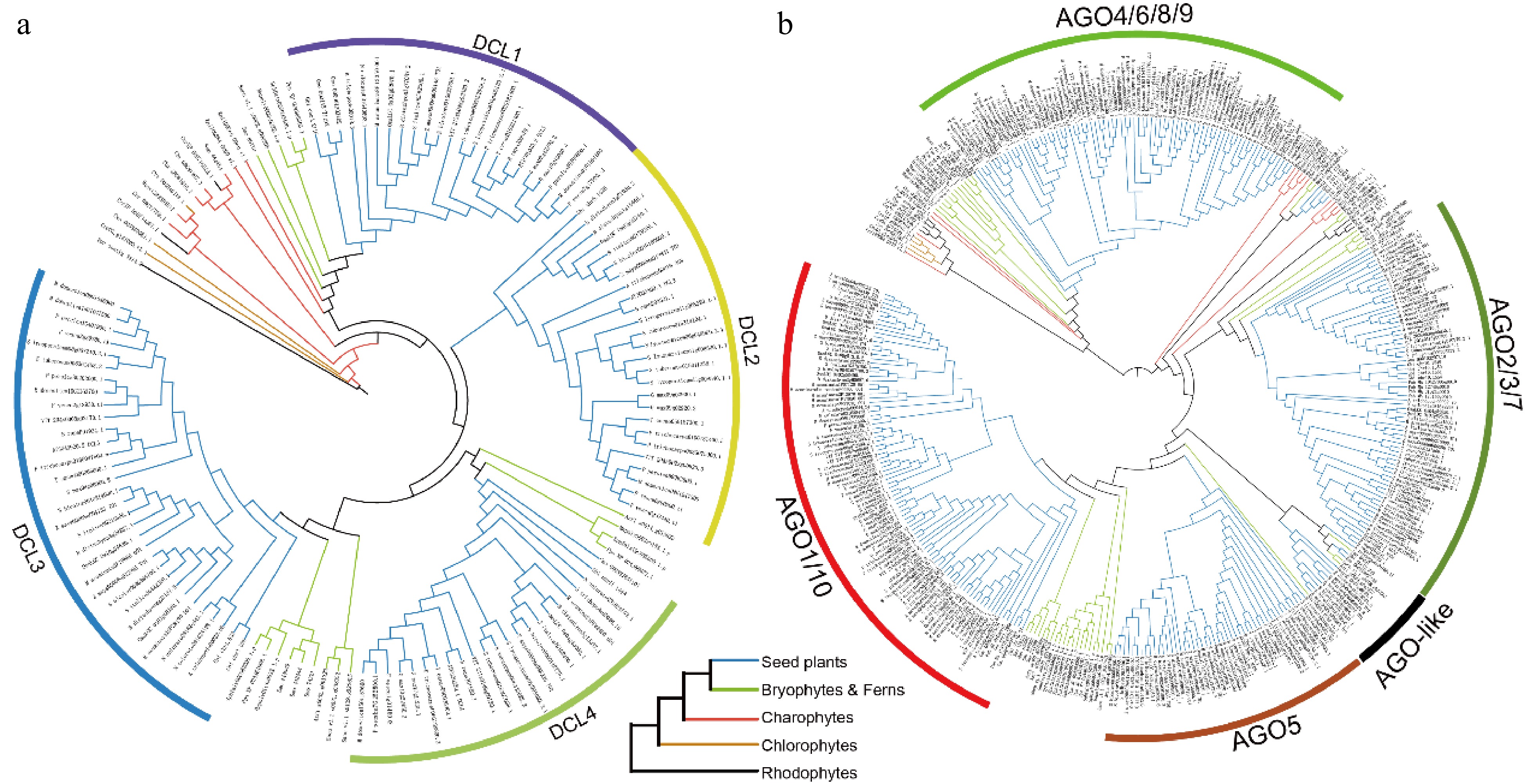
      Figure 1. 

      Phylogenetic trees of the (a) DCL, and (b) AGO gene families across 36 plant species. Branches are color-coded to denote different plant groups: black for rhodophytes, yellow for chlorophytes, red for charophytes, green for ferns and bryophytes, and blue for seed plants.

      Multiple sequence alignments of sRNA-related functional domains in DCLs and AGOs were performed. Overall, the RNase III domains of DCLs in all four branches are highly conserved at both the C- and N-termini. Additionally, there are variations among the RNase III domains between different branches, with those in higher plants being more conserved (Supplementary Fig. S3). This conservation may reflect adaptations to diverse environmental pressures through more sophisticated RNA regulatory mechanisms, leading to the synthesis of a wider variety of sRNAs. Furthermore, we aligned the MID and PAZ domains of AGOs across different branches. Members of the AGO2/3/7 and AGO4/6/8/9 branches have largely lost the MID domain, and those that retain it show less conservation. In contrast, the AGO1/5/10 branch retains a highly conserved MID domain (Supplementary Fig. S4). Similarly, the PAZ domain shows significant differences among branches, with high conservation in the AGO1/5/10 branch, including in lower plants, while the other two branches exhibit lower conservation (Supplementary Fig. S5). Highly conserved MID and PAZ domains are typically associated with fundamental miRNA processing, whereas less conserved domains may relate to specific functional requirements and adaptive changes.

      In plants, the copy number of DCL genes does not appreciably vary across different evolutionary branches (Fig. 2), ranging from 25 to 40 across the four groups and predominantly existing as single-copy genes throughout plant evolution. The primary factor influencing this gene copy number variation among different plants is attributed to whole-genome duplication (WGD) events during specific evolutionary processes. By contrast, the copy number of AGO genes exhibit considerable variation across branches. Specifically, the major clades of AGO4/6/8/9, AGO2/3/7, and AGO1/5/10 contain 95, 93, and 173 genes, respectively (Fig. 2). The number of AGO genes notably exceeds that of DCL genes and shows diversification into more clades, suggesting that the AGO genes are more frequently retained during duplication events. Additionally, AGO4 and AGO6, which encode key enzymes in plant methylation through siRNA processing were analyzed separately (Fig. 2). Results show that AGO4 is prevalent in basal angiosperms, whereas AGO6 is restricted to monocots and dicots. Based on these findings, it is hypothesized that the evolution of AGO4 and AGO6 may be linked to significant shifts in reproductive strategies and the development of floral organs during the transition from gymnosperms to angiosperms. Given the unique and critical roles of AGO4 and AGO6 in methylation processes, the expansion and loss of these genes was investigated within the AGO4/6/8/9 clade across 36 species (Fig. 3). Phylogenetic analysis reveals that these genes exist as single copies in algae, ferns, gymnosperms, and basal angiosperms. Following the ε duplication event, both AGO4 and AGO6 were retained in embryophytes. Unlike AGO6, which did not undergo significant expansion after its formation, AGO4 experienced multiple duplication events. In the Brassicaceae, the α and β duplication events led to the emergence of AGO8 and AGO9. Similarly, duplication events in crops such as potatoes, tomatoes, and monocots also contributed to the expansion of AGO4.
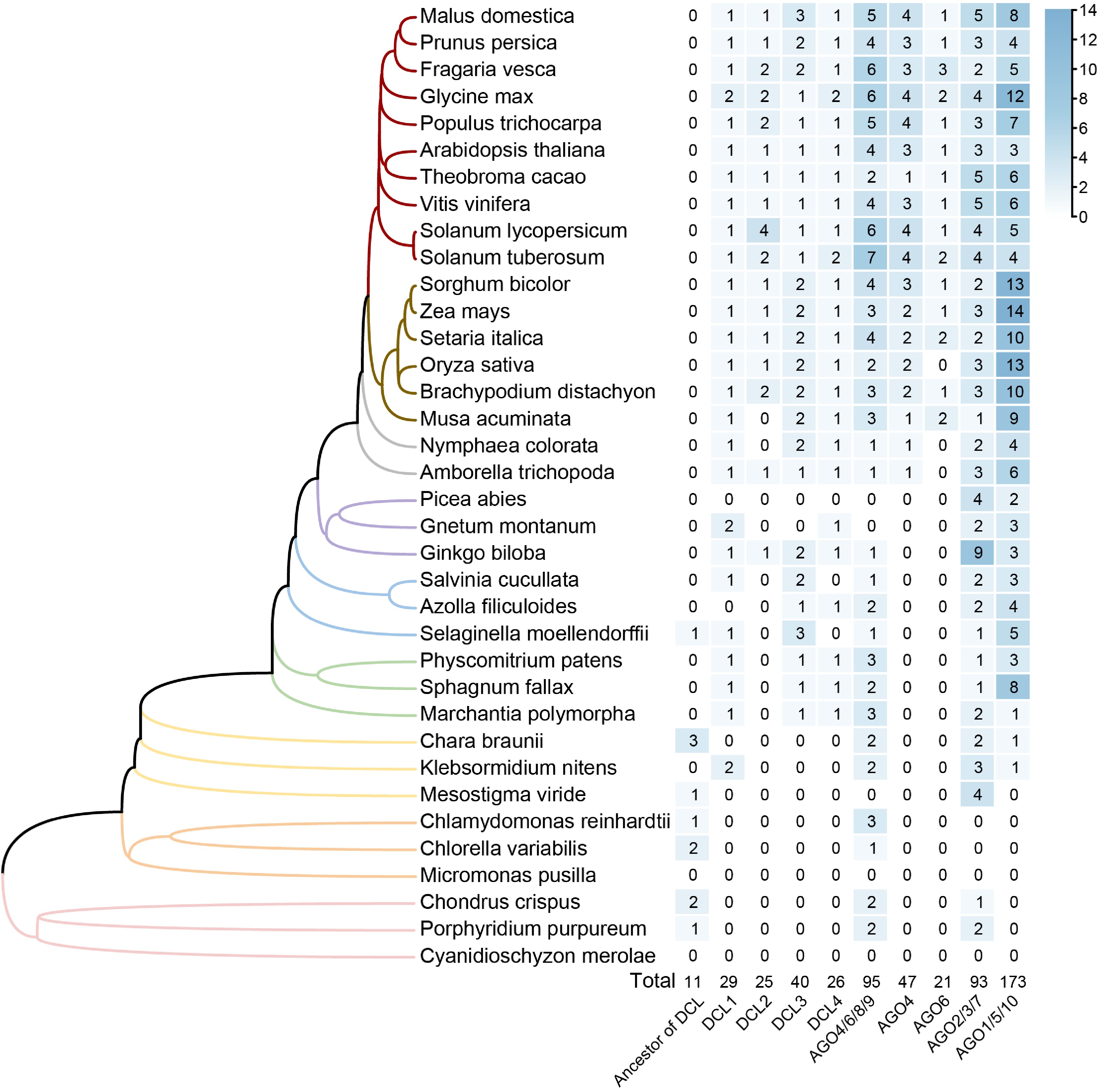
      Figure 2. 

      Phylogenetic distribution and gene copy number analysis of DCL and AGO gene families across 36 plant species. The phylogenetic tree on the left represents the evolutionary relationships of species investigated, with branches colored to represent different groups. The heatmap on the right displays gene copy numbers for each clade of gene family across the species, with higher numbers represented by darker shades. The total counts for each clade across all species are provided at the bottom of the heatmap.
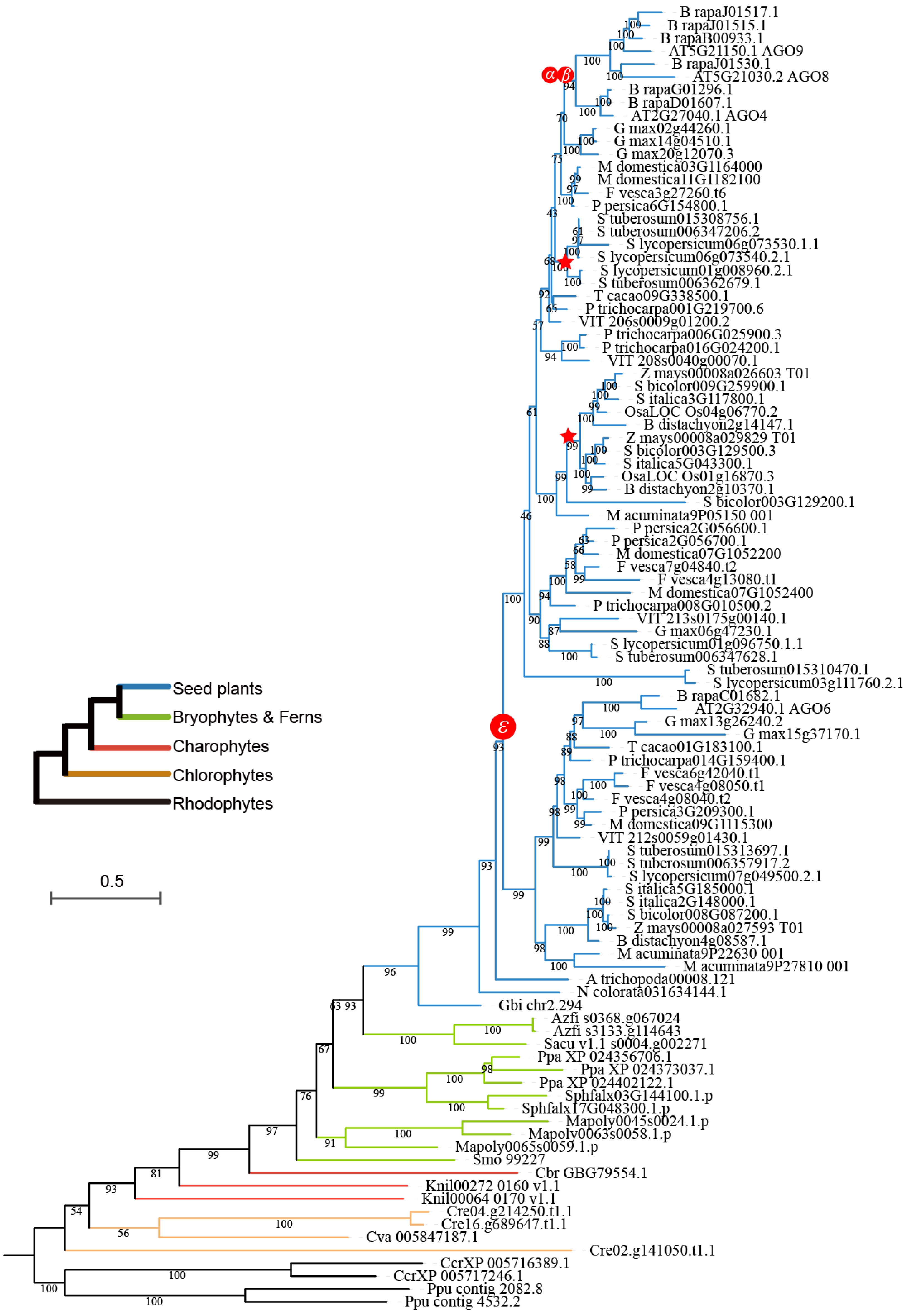
      Figure 3. 

      Phylogeny of the AGO4/6/8/9 clade within the AGO gene family. Different colored branches represent distinct plant groups. The symbols ε, α, and β represent the epsilon angiosperm-wide WGD event, the alpha duplication event, and the beta duplication event, respectively. Red stars along the branches indicate specific whole-genome duplication events.

    • To elucidate the evolutionary differences between DCLs and AGOs in plants, a collinearity network analysis was conducted on 18 plants, including both monocots and dicots, based on their phylogenetic relationships. The analysis identified 542 syntenic gene pairs, grouping the DCL genes into four clusters that represent four distinct evolutionary trajectories. Additionally, four WGD events and three tandem duplication pairs were detected in apple, soybean, and tomato, suggesting lineage-specific expansions of the DCLs during evolution (Fig. 4a). Therefore, we integrated both phylogenetic and collinearity data were integrated (Fig. 4b) and the evolutionary history of DCL genes was reconstructed. It is proposed that two ancestral DCL genes existed before the emergence of seed plants, which subsequently underwent two WGD events. This process resulted in the loss of three branches, leaving four extant DCL groups.
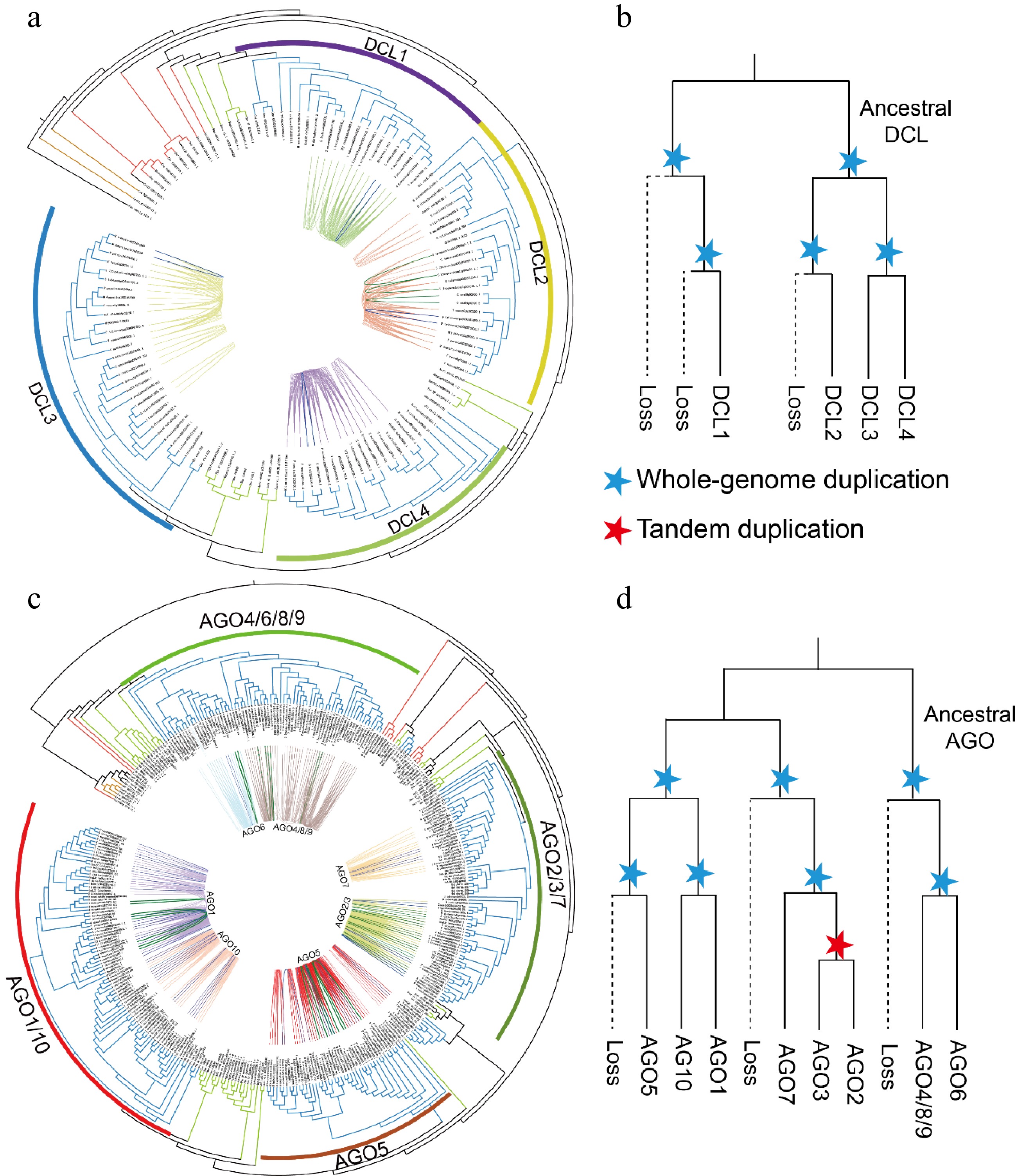
      Figure 4. 

      Phylogenetic analysis and synteny identification of the DCL and AGO genes. (a), (c) Phylogenetic and syntenic relationships of the DCLs and AGOs. The blue and green lines indicate gene pairs resulting from WGD and tandem duplication in the DCLs and AGOs, respectively. (b, d) Schematic representation of the proposed evolutionary histories of the DCL and AGO gene families. The dashed lines indicate gene loss. Blue stars mark either the ancient seed plant-wide or angiosperm-wide genome duplication events. Red stars represent tandem duplication events of genes.

      In the AGO gene family, 1,090 syntenic gene pairs were identified and subsequently clustered into seven groups (Fig. 4c). Notably, AGO4, AGO8, and AGO9 formed a cluster, demonstrating their evolutionary homology. A similar homologous relationship is observed between AGO2 and AGO3. Additionally, 43 syntenic gene pairs resulting from intraspecific duplications across various AGO groups were found. Moreover, 23 tandem duplication pairs were identified, they distributed across the syntenic gene clusters of AGO1, AGO4/8/9, AGO5, and AGO6, with most tandem duplications occurring within AGO2/3. Based on these findings, it is inferred that the AGO family originated from three ancestral genes before the emergence of seed plants, with clusters retained through two WGD events. Furthermore, AGO2 and AGO3 appear to have arisen from tandem duplications (Fig. 4d).

    • To explore the functional differences between DCLs and AGOs throughout plant evolution, expression patterns were analyzed using publicly available data from various tissues of P. patens, Z. may, A. thaliana, and F. vesca. Among these species, F. vesca exhibits the highest number of DCL members, totaling six. The expression profiles of various tissues were largely consistent across the four species, with DCLs showing high expression in reproductive tissues (Fig. 5a, Supplementary Fig. S6a). In F. vesca, the DCL2 and DCL3 branches each contain two gene members, however, in each branch, only one gene exhibits high expression. This contrasts with Z. mays, where the expression patterns of the two DCL3 members are similar. In P. patens, DCL3 is more prominently expressed in vegetative tissues, whereas in A. thaliana, Z. may, and F. vesca, it shows high expression in reproductive tissues. For the AGOs, the overall expression profiles are similar to those of the DCLs, with high expression in reproductive tissues (Fig. 5b, Supplementary Fig. S6b). The AGO4/6/8/9 groups demonstrate functional complementation in reproductive tissues in ancestral species, whereas in A. thaliana, Z. mays, and F. vesca, AGOs are ubiquitously expressed in all reproductive tissues. In P. patens, AGO1 group members are highly expressed only in reproductive tissues in P. patens, however, they maintain high expression levels across all tissues in A. thaliana and F. vesca. Interestingly, despite their overall low expression levels in pollen both DCLs and AGOs from various groups remain active in reproductive organs in A. thaliana and F. vesca.
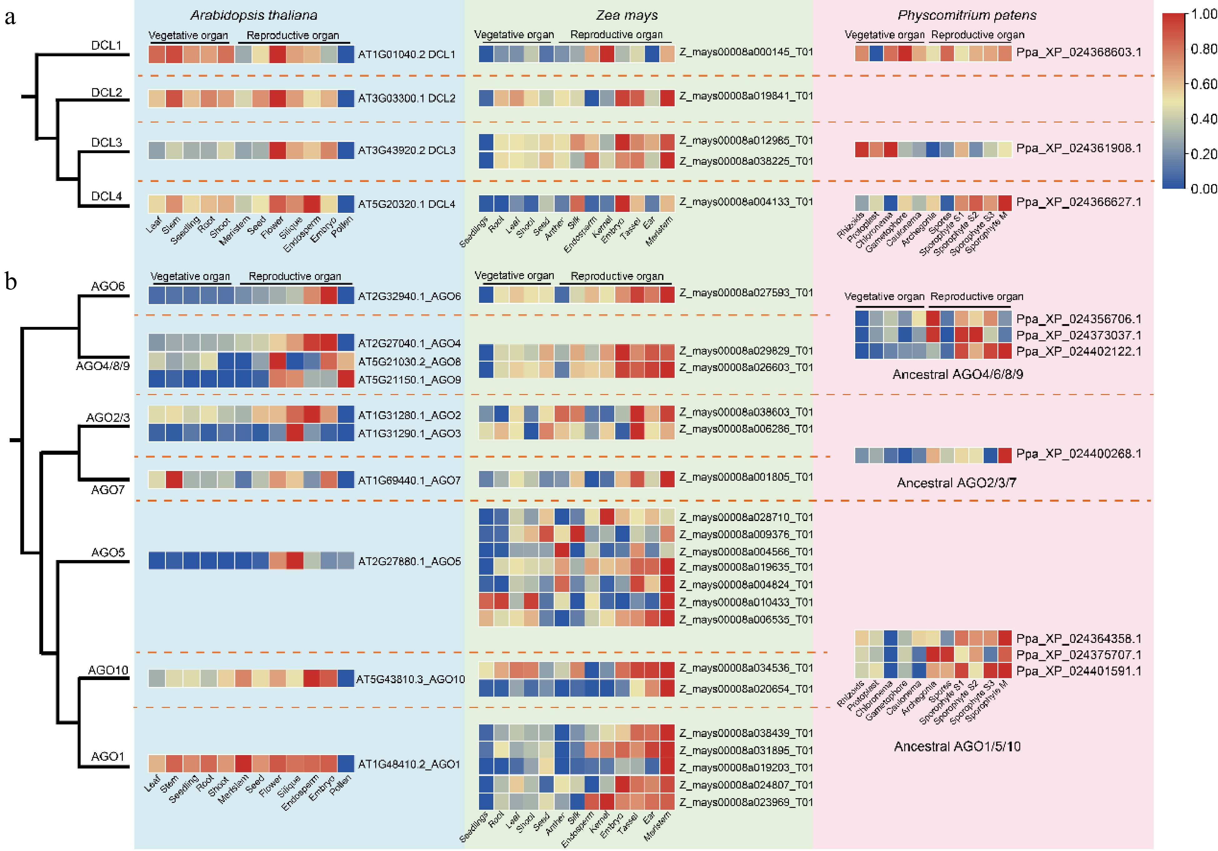
      Figure 5. 

      Expression profiles of DCLs and AGOs in different tissues of A. thaliana, Z. may, and P. patens. (a) Comparative expression profiles of DCL gene family members. (b) Comparative expression profiles of AGO gene family members. Dashed lines demarcate distinct clades, with the heatmap displaying relative expression levels from low (blue) to high (red).

      The expression of DCLs and AGOs in response to various stresses in A. thaliana and Z. may were further analyzed. The results indicate that A. thaliana DCLs respond to all stressors except irradiation, whereas Z. may DCLs primarily respond to heat, salt, drought, and nutrient deficiency (Fig. 6a). The stress responses of AGOs vary across different groups (Fig. 6b). For example, in A. thaliana, AGO6, AGO7, and AGO10 are involved in most stress pathways. In Z. mays, AGO6, AGO4/8/9, AGO2/3, and AGO10 participate in multiple stress responses. In both A. thaliana and Z. mays, AGO5 exhibits minimal responsiveness to stress, whereas AGO4 responds to similar stresses, including heat, salt, drought, cold, shade, and nutrient deficiency. This comprehensive analysis underscores the specificity and variability of DCL and AGO responses to environmental stresses, highlighting their essential adaptive functions in plant stress physiology.
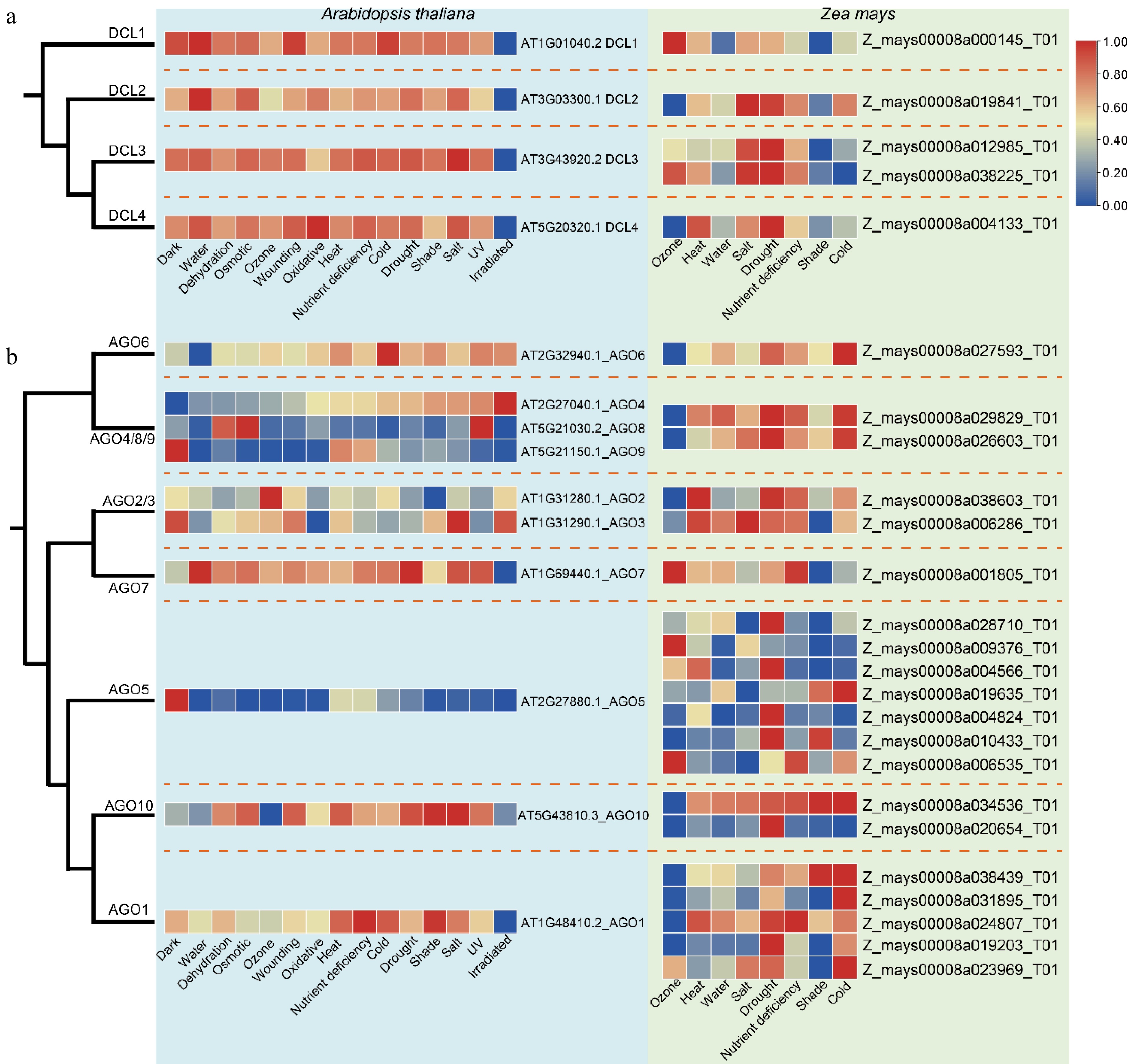
      Figure 6. 

      Expression of the DCLs and AGOs under different stress conditions in A. thaliana and Z. mays. (a) Comparative expression profiles of DCL gene family members. (b) Comparative expression profiles of AGO gene family members. Dashed lines denote distinct clades, with the heatmap displaying relative expression levels from low (blue) to high (red).

    • As plants transitioned from aquatic to terrestrial environments, they encountered more variable habitats and increased exposure to air. This shift prompted the expansion of numerous gene families to adapt to these diverse environmental challenges[3537]. The present findings align with previous studies, revealing that DCLs are predominantly classified into four groups, with DCL2 being exclusive to seed plants (Fig. 1a). The origin of DCLs was traced back to rhodophytes, detecting homologs in Chondrus crispus and Porphyridium purpureum, a finding supported by data from the 1KP database (Supplementary Fig. S1). Similarly, the present analysis suggests that AGOs also originated from rhodophytes, demonstrating the conservation of the RNAi pathway across plant species. Contrary to Li et al., who reported a single ancestral lineage for AGO, the present study identifies ancestral positions for AGO4/6/8/9 and AGO2/3/7 in Porphyridium purpureum[16]. Furthermore, a clear differentiation of AGOs into two distinct groups in rhodophytes, chlorophytes, and charophytes was observed (Fig. 1b). The expansion of the AGO family from charophytes to bryophytes likely represents an evolutionary adaptation crucial for terrestrial colonization[38,39]. Furthermore, these findings underscore the significant role of epigenetics in the terrestrial adaptation of plants.

      DCLs exhibit a single ancestral branch in all algae, however, their expansion during the transition from aquatic to terrestrial environments coincides with that of the AGOs. This expansion continuous as lower plants evolved into higher plants, leading to the present diversification of these gene families. As key components of the RNAi mechanism, DCLs, and AGOs are crucial for the generation and function of miRNA. Specific miRNAs in algae that are conserved in seed plants, such as miR167, miR172, miR395, miR414, miR418, and miR419, are missing in mosses and ferns[4043]. These miRNAs are essential for flower development, stress resistance, and root development in higher plants[44,45]. Their absence in mosses and ferns highlights differences in the miRNA-mediated gene silencing pathways between lower and higher plants and underscores the adaptive changes during plant evolution, reflecting species-specific survival strategies and developmental needs in diverse environments. Further analysis of the evolutionary trajectories of DCLs and AGOs in seed plants revealed that DCLs underwent two rounds of WGDs in land plants without a significant increase in their numbers (Figs 2 & 4). By contrast, AGOs experienced two WGD events and one tandem duplication (Fig. 4), and maintained specific expansions within different species (Fig. 3). These findings reveal distinct evolutionary paths of these gene families and their crucial roles in adapting RNAi mechanisms for plant survival and development across diverse ecological settings.

      WGD and various forms of gene duplication are the primary mechanisms that drive the expansion of gene families. The retention of duplicated genes throughout evolution has facilitated better adaptation in plant growth and development[4648]. Extensive research has established the functions of DCLs and AGOs. This study discovers that most DCL and AGO genes in P. patens are highly expressed in meristematic tissues, a pattern that is conserved in A. thaliana, Z. mays, and F. vesca (Fig. 5 & Supplementary Fig. S3). This expression profile is likely attributed to active DNA methylation and RNAi regulation within these tissues[10,49,50], underscoring the intricate genetic regulation essential for plant development. Additionally, DCL3 and DCL4 in P. patens show high expression in vegetative and meristematic tissues, respectively, suggesting the specialization of their functions (Figs 5 & Supplementary Fig. S3). In Arabidopsis, F. vesca, and Z. mays, the DCL2/3/4 genes are highly expressed in various tissues and response to stress (Fig. 6). The sRNAs they produce vary, indicating that diverse types of sRNAs are extensively involved in the life cycles of plants. This diversity enables plants to adapt to environmental fluctuations and supports their growth and development[5153]. Additionally, DCL serves as the factory for manufacturing and producing miRNAs. Its tissue-specific expression, along with upregulated expression under stress conditions, is closely associated with the miRNAs it produces. For example, the miRNA156/SPL module can participate in root development and vegetative growth while also enhancing the plant's tolerance to abiotic stress. miR169 targets different members of the NF-YA gene family, which is involved in multiple developmental processes and stress responses. Moreover, miR159, miR397, and miR393 possess diverse functions in plant growth, development, and stress tolerance[44,45]. By contrast, the functions of the AGO4/6/8/9 clade have remained largely unchanged throughout evolution, with no special functions emerging from their expansion in Z. may and A. thaliana (Figs 5, 6, & Supplementary Fig. S3). Previous studies have demonstrated functional complementarity among AGO4/6/8/9 proteins[10,54,55], which is essential for maintaining critical biological processes under varying conditions. AGO2/3/7 appear to be predominantly involved in stress responses (Fig. 6), consistent with findings of previous studies[56,57]. In A. thaliana, F. vesca, and Z. mays, AGO1/5/10 exhibit significant functional divergence. Specifically, in A. thaliana, AGO1 and AGO5 exist as single-copy genes. However, AGO5 has largely lost its regulatory functions in tissue development and stress response, whereas AGO1 retains all these functions (Figs 5 & 6). In Z. mays, although there is a significant expansion of members within the AGO1/5/10 group, their functions remain largely similar to those observed in P. patens. These results suggest that subfunctionalization and neofunctionalization are two potential evolutionary outcomes of gene duplication[58], and also demonstrate the role of epigenetic regulation in directing species-specific evolutionary trajectories in plants.

    • Genomic data for 36 plant species used in this study were obtained from databases such as Phytozome (Supplementary Table S1). Gene screening and alignment of the 1KP transcriptome data were conducted using the ONEKP online platform (https://db.cngb.org/onekp). Protein sequences of the four DCLs and ten AGOs from A. thaliana served as query sequences for BLASTP analysis against the proteomic data of the remaining 35 plant genomes, using an e-value threshold of 1e-20. The resulting sequences were then analyzed using InterProScan to identify and annotate conserved domains using the Pfam, PANTHER, and SMART databases[59]. Only sequences that contained domains consistent with those found in A. thaliana were selected for constructing a phylogenetic tree using FastTree. Branches exhibiting abnormal lengths were manually removed to ensure the accuracy of the inferred phylogenetic relationships[60].

    • First, multiple sequence alignment of all DCL and AGO protein sequences was performed using MAFFT. Gap positions were then removed from the aligned sequences using Phyutility with a cutoff parameter of 0.5. Next, ProtTest was used to predict the best-fit substitution models for constructing the DCL and AGO phylogenetic trees. The DCL and AGO trees were constructed using IQ-TREE with the JTT + F + R6 and LG + I + G + F models, respectively, with 1000 Bootstrap replicates[61]. For genes indexed in the 1KP database, phylogenetic trees were constructed using FastTree. Additionally, collinearity blocks were identified by comparing coding sequences across species using the Python version of MCScan[62].

    • Expression data for different tissues of P. patens and F. vesca were obtained from the Physcomitrium eFP Browser and the Strawberry eFP Browser, respectively (http://bar.utoronto.ca). For A. thaliana and Z. mays, tissue-specific and stress-induced expression profiles were downloaded from https://plantrnadb.com. Detailed data sources are provided in Supplementary Table S2. The expression of DCL and AGO genes across these species was visualized using TBtools[63].

      • The research was supported by the Key Research and Development Program of Jiangsu (BE2023350) and the Priority Academic Program Development of Jiangsu Higher Education Institutions Project (PAPD), and the high-performance computing platform at the Bioinformatics Center of Nanjing Agricultural University. We would like to thank A&L Scientific Editing (www.alpublish.com) for their linguistic assistance during the preparation of this manuscript.

      • The authors confirm contribution to the paper as follows: study conception and design: Xiong AS, Cheng ZM, Su LY; data analysis, draft manuscript preparation: Su LY, Li SS, Liu H. All authors reviewed the results and approved the final version of the manuscript.

      • All data generated or analyzed during this study are included in this published article.

      • The authors declare that they have no conflict of interest.

      • # Authors contributed equally: Li-Yao Su, Shan-Shan Li

      • Supplementary Table S1 Genome data used in this study.
      • Supplementary Table S2 Source of gene expression data.
      • Supplementary Fig. S1 The ML tree of DCLs identified with 1KP transcriptomic data. Branches are color-coded to denote different plant groups: black for rhodophytes, yellow for chlorophytes, red for charophytes, green for ferns and bryophytes, and blue for seed plants.
      • Supplementary Fig. S2 The ML tree of AGOs identified with 1KP transcriptomic data. Branches are color-coded to denote different plant groups: black for rhodophytes, yellow for chlorophytes, red for charophytes, green for ferns and bryophytes, and blue for seed plants.
      • Supplementary Fig. S3 RNase III domain alignment of four groups of DCL proteins.
      • Supplementary Fig. S4 MID domain alignment of three groups of AGOs.
      • Supplementary Fig. S5 PAZ domain alignment of three groups of AGOs.
      • Supplementary Fig. S6 Expression of DCLs and AGOs in different tissues of F. vesca. (A) Comparative expression profiles of DCL gene family members. (B) Comparative expression profiles of AGO gene family members. Dashed lines demarcate distinct clades, with the heatmap displaying relative expression levels from low (blue) to high (red).
      • © 2024 by the author(s). Published by Maximum Academic Press, Fayetteville, GA. This article is an open access article distributed under Creative Commons Attribution License (CC BY 4.0), visit https://creativecommons.org/licenses/by/4.0/.
    Figure (6)  References (63)
  • About this article
    Cite this article
    Su LY, Li SS, Liu H, Cheng ZM, Xiong AS. 2024. The origin, evolution, and functional divergence of the Dicer-like (DCL) and Argonaute (AGO) gene families in plants. Epigenetics Insights 17: e003 doi: 10.48130/epi-0024-0005
    Su LY, Li SS, Liu H, Cheng ZM, Xiong AS. 2024. The origin, evolution, and functional divergence of the Dicer-like (DCL) and Argonaute (AGO) gene families in plants. Epigenetics Insights 17: e003 doi: 10.48130/epi-0024-0005

Catalog

  • About this article

/

DownLoad:  Full-Size Img  PowerPoint
Return
Return