REVIEW   Open Access    

Global research landscape and trends of plant-based meat analogs: a bibliometric analysis

More Information
  • Plant-based meat analogs have become an important topic in recent years. To scientifically understand the research situation of plant-based meat analogs, we analyzed 2,595 publications from January 2000 to October 2023 by bibliometric method based on the PubMed database. The results showed a gradual rise in the number of annual publications, with the fastest growth rate of 58.5% in 2021. The country with the most publications was the United States (685, 24.87%), followed by China (242, 8.79%) and the United Kingdom (196, 7.12%). The University of Helsinki, Texas A&M University and the University of California were core research institutions. Popular and important journals were mainly Foods, Nutrients, Meat Science, Food Research International, and Critical Reviews in Food Science and Nutrition. Current research topics focused on alternative proteins and their functional and nutritional characteristics, as well as sustainable development. The research interests have gradually expanded from quality characteristics to nutritional characteristics. Further improving the quality, controlling natural toxin contamination, as well as systematically investigating the effects on health were future research trends. The effects of plant-based meat analogs on metabolic pathways and diseases were important clues in the study of nutritional health. This bibliometric analysis comprehensively and quantitatively presents the research landscape and hotspots, and further suggests future research directions. These findings can benefit researchers in selecting appropriate journals and finding potential collaborators to achieve in-depth research in this field.
  • Atractylodes macrocephala Koidz. (common names 'Baizhu' in Chinese and 'Byakujutsu' in Japanese) is a diploid (2n = 2x = 24) and out-crossing perennial herb in the Compositae family, and has a long history of cultivation in temperate and subtropical areas of East Asia as it is widely used in traditional herbal remedies with multiple pharmacological activities[13]. The 'Pharmacopoeia of the People's Republic of China' states that 'Baizhu' is the dry rhizome of A. macrocephala Koidz. (Atractylodis Macrocephalae Rhizoma, AMR). However, in Japanese traditional medicine 'Baizhu' can be referred to both: A. japonica or A. macrocephala[4].

    A. macrocephala is naturally endemic to China and cultivated in more than 200 towns in China, belonging to Zhejiang, Hunan, Jiangxi, Anhui, Fujian, Sichuan, Hubei, Hebei, Henan, Jiangsu, Guizhou, Shanxi, and Shaanxi Provinces[3]. A. macrocephala grows to a height of 20–60 cm (Fig. 1). The leaves are green, papery, hairless, and generally foliole with 3–5 laminae with cylindric glabrous stems and branches. The flowers grow and aggregate into a capitulum at the apex of the stem. The corollas are purplish-red, and the florets are 1.7 cm long. The achenes, densely covered with white, straight hairs, are obconic and measure 7.5 mm long. The rhizomes used for medicinal purposes are irregular masses or irregularly curving cylinders about 3–13 cm long and 1.5–7 cm in diameter with an outwardly pale greyish yellow to pale yellowish color or a sparse greyish brown color. The periderm-covered rhizomes are externally greyish brown, often with nodose protuberances and coarse wrinkles. The cross-sections are white with fine dots of light yellowish-brown to brown secretion. Rhizomes are collected from plants that are > 2 years old during the spring. The fibrils are removed, dried, and used for medicinal purposes[5, 6].

    Figure 1.  Plant morphology of A. macrocephala.

    The medicinal properties of AMRs are used for spleen deficiency, phlegm drinking, dizziness, palpitation, edema, spontaneous sweating, benefit Qi, and fetal restlessness[7]. The AMR contains various functional components, among which high polysaccharide content, with a yield close to 30%[8]. Therefore, the polysaccharides of A. macrocephala Koidz. rhizome (AMRP) are essential in assessing the quality control and bioactivity of A. macrocephala. Volatile oil accounts for about 1.4% of AMR, with atractylon and atractylodin as the main components[9]. Atractylon can be converted to atractylenolide I (AT-I), atractylenolide II (AT-II), and atractylenolide III (AT-III) under ambient conditions. AT-III can be dehydrated to AT-II under heating conditions[10, 11]. AMRs, including esters, sesqui-, and triterpenes, have a wide range of biological activities, such as improving immune activity, intestinal digestion, neuroprotective activity, immune anti-inflammatory, and anti-tumor.

    In recent years, research on the pharmacological aspects of AMR has continued to increase. Still, the discovery of the main active components in AMR is in its infancy. The PAO-ZHI processing of AMR is a critical step for AMR to exert its functional effects, but also, in this case, further work is required. Studies on the biosynthesis of bioactive compounds and different types of transcriptomes advanced current knowledge of A. macrocephala, but, as mentioned, required more systematic work. Ulteriorly, an outlook on the future research directions of A. macrocephala was provided based on the advanced technologies currently applied in A. macrocephala (Fig. 2).

    Figure 2.  Current progress of A. macrocephala.

    A. macrocephala is distributed among mountainous regions more than 800 m above sea level along the middle and lower reaches of the Yangtze River (China)[5]. Due to over-exploitation and habitat destruction, natural populations are rare, threatened, and extinct in many locations[1,12]. In contrast to its native range, A. macrocephala is widely cultivated throughout China, in a total area of 2,000–2,500 ha, with a yield of 7,000 t of rhizomes annually[13]. A. macrocephala is mainly produced in Zhejiang, Anhui, and Hebei (China)[14]. Since ancient times, Zhejiang has been the famous producing area and was later introduced to Jiangxi, Hunan, Hebei, and other places[15]. Wild A. macrocephala is currently present in at least 14 provinces in China. It is mainly distributed over three mountain ranges, including the Tianmu and Dapan mountains in Zhejiang Province and the Mufu mountains along the border of Hunan and Jiangxi Provinces. A. macrocephala grows in a forest, or grassy areas on mountain or hill slopes and valleys at an altitude of 600–2,800 m. A. macrocephala grows rapidly at a temperature of 22–28 °C, and favors conditions with total precipitation of 300–400 mm evenly distributed among the growing season[16]. Chen et al. first used alternating trilinear decomposition (ATLD) to characterize the three-dimensional fluorescence spectrum of A. macrocephala[17]. Then they combined the three-dimensional fluorescence spectrum with partial least squares discriminant analysis (PLS-DA) and k-nearest neighbor method (kNN) to trace the origin of Atractylodes samples. The results showed that the classification models established by PLS-DA and kNN could effectively distinguish the samples from three major Atractylodes producing areas (Anhui, Hunan, and Zhejiang), and the classification accuracy rate (CCR) of Zhejiang atractylodes was up to 80%, and 90%, respectively[17]. Zhang et al. compared the characteristics, volatile oil content, and chemical components of attested materials from six producing areas of Zhejiang, Anhui, Hubei, Hunan, Hebei, and Henan. Differences in the shape, size, and surface characteristics were reported, with the content of volatile oil ranging from 0.58% to 1.22%, from high to low, Hunan (1.22%) > Zhejiang (1.20%) > Anhui (1.02%) > Hubei (0.94%) > Henan (0.86%) > Hebei (0.58%)[18]. This study showed that the volatile oil content of A. macrocephala in Hunan, Anhui, and Hubei is not much different from that of Zhejiang, which is around 1%. A. macrocephala is a local herb in Zhejiang, with standardized cultivation techniques, with production used to reach 80%–90% of the country. However, in recent years, the rapid development of Zhejiang's real estate economy has reduced the area planted with Zhejiang A. macrocephala, resulting in a sudden decrease in production. Therefore, neighboring regions, such as Anhui and Hunan, vigorously cultivate A. macrocephala, and the yield and quality of A. macrocephala can be comparable to those of Zhejiang. The results were consistent with the data reports[18]. Guo et al. analyzed the differentially expressed genes of Atractylodes transcripts from different regions by the Illumina HiSeq sequencing platform. It was found that 2,333, 1,846, and 1,239 DEGs were screened from Hubei and Hebei, Anhui and Hubei, and Anhui and Hebei Atrexia, respectively, among which 1,424, 1,091, and 731 DEGs were annotated in the GO database. There were 432, 321, and 208 DEGs annotated in the KEGG database. These DEGs were mainly related to metabolic processes and metabolic pathways of secondary metabolites. The highest expression levels of these genes were found in Hubei, indicating higher terpenoid production in Hubei[19]. Other compounds were differentially accumulated in Atractylodes. Chlorogenic acid from Hebei was 0.22%, significantly higher than that from Zhejiang and Anhui[20]. Moreover, the content of neochlorogenic acid and chlorogenic acid decreased after processing, with the highest effect reported in Zhejiang, with the average transfer rate of neochlorogenic acid and chlorogenic acid reaching 55.68% and 55.05%[20]. All these changes would bring great help in distinguishing the origins of A. macrocephala.

    Medicinal AMR can be divided into raw AMR and cooked AMR. The processing method is PAO-ZHI; the most traditional method is wheat bran frying. The literature compared two different treatment methods, crude A. macrocephala (CA) and bran-processed A. macrocephala, and found that the pharmacological effects of AMR changed after frying with wheat bran, mainly in the anti-tumor, antiviral and anti-inflammatory effects[21]. The anti-inflammatory effect was enhanced, while the anti-tumor and antiviral effects were somewhat weakened, which may be related to the composition changes of the compounds after frying. The study of the content of AT-I, II, and III, and atractyloside A, in rat serum provided helpful information on the mechanism of wheat bran processing[22]. In addition to frying wheat bran, Sun et al. used sulfur fumigation to treat AMR[23]. They found that the concentration of different compounds changed, producing up to 15 kinds of terpenoids. Changes in pharmacological effects were related to treatment and the type of illumination[24,25]. Also, artificial light can improve the various biological functions. A. macrocephala grew better under microwave electrodeless light, with a chlorophyll content of 57.07 ± 0.65 soil and plant analyzer develotrnent (SPAD)[24]. The antioxidant activity of AMR extract treated with light-emitting diode (LED)-red light was the highest (95.3 ± 1.1%) compared with other treatments[24]. The total phenol and flavonoid contents of AMR extract treated with LED-green light were the highest at 24.93 ± 0.3 mg gallic acid equivalents (GAE)/g and 11.2 ± 0.3 mg quercetin equivalents (QE)/g compared with other treatments[24, 25]. Polysaccharides from Chrysanthemun indicum L.[26] and Sclerotium rolfsiisacc[27] can improve AMR's biomass and bioactive substances by stimulating plant defense and thus affect their efficacy. In summary, there are compositional differences between A. macrocephala from different origins. Besides, different treatments, including processing mode, light irradiation, and immune induction factors, which can affect AMR's biological activity, provide some reference for the cultivation and processing of A. macrocephala (Fig. 3).

    Figure 3.  Origin, distribution and processing of A. macrocephala.

    The AMR has been reported to be rich in polysaccharides, sesquiterpenoids (atractylenolides), volatile compounds, and polyacetylenes[3]. These compounds have contributed to various biological activities in AMR, including immunomodulatory effects, improving gastrointestinal function, anti-tumor activity, neuroprotective activity, and anti-inflammatory.

    AMRP has received increasing attention as the main active component in AMR because of its rich and diverse biological activities. In the last five years, nine AMRP have been isolated from AMR. RAMP2 had been isolated from AMR, with a molecular weight of 4.354 × 10³ Da. It was composed of mannose, galacturonic acid, glucose, galactose, and arabinose, with the main linkages of →3-β-glcp-(1→, →3,6-β-glcp-(1→, →6-β-glcp-(1→, T-β-glcp-(1→, →4-α-galpA-(1→, →4-α-galpA-6-OMe-(1→, →5-α-araf-(1→, →4,6-β-manp-(1→ and →4-β-galp-(1→[28]. Three water-soluble polysaccharides AMAP-1, AMAP-2, and AMAP-3 were isolated with a molecular weight of 13.8 × 10⁴ Da, 16.2 × 10⁴ Da, and 8.5 × 10⁴ Da, respectively. Three polysaccharides were deduced to be natural pectin-type polysaccharides, where the homogalacturonan (HG) region consists of α-(1→4)-linked GalpA residues and the ramified region consists of alternating α-(1→4)-linked GalpA residues and α-(1→2)-linked Rhap residues. Besides, three polysaccharides were composed of different ratios of HG and rhamnogalacturonan type I (RG-I) regions[29]. Furthermore, RAMPtp has been extracted from AMR with a molecular weight of 1.867 × 10³ Da. It consists of glucose, mannose, rhamnose, arabinose, and galactose with 60.67%, 14.99%, 10.61%, 8.83%, and 4.90%, connected by 1,3-linked β-D Galp and 1,6-linked β-D Galp residues[30]. Additionally, PAMK was characterized by a molecular weight of 4.1 kDa, consisting of galactose, arabinose, and glucose in a molar ratio of 1:1.5:5, with an alpha structure and containing 96.47% polysaccharide and small amounts of protein, nucleic acid, and uric acid[31]. Another PAMK extracted from AMR had a molecular weight of 2.816 × 10³ Da and consisted of glucose and mannose in molar ratios of 0.582 to 0.418[32]. Guo et al. isolated PAMK with a molecular weight of 4.748 × 10³ g/mol from AMR, consisting of glucose, galactose, arabinose, fructose, and mannose in proportions of 67.01%, 12.32%, 9.89%, 1.18%, and 0.91%, respectively[33]. In addition, AMP1-1 is a neutral polysaccharide fragment with a molecular weight of 1.433 kDa isolated from AMR. It consists of glucose and fructose, and the structure was identified as inulin-type fructose α-D-Glcp-1→(2-β-D-Fruf-1)7[34]. These reports indicated that, in general, polysaccharides are extracted by water decoction, ultrasonic-assisted extraction, enzyme hydrolysis method, and microwave-assisted extraction. The separation and purification are column chromatography, stepwise ethanol precipitation, and ultrafiltration. Their physicochemical properties and structural characterization are generally achieved by determining the molecular weight, determining the monosaccharide composition, analyzing the secondary structure, and glycosidic bond configuration of polysaccharides with Fourier transform infrared (FT-IR) and nuclear magnetic resonance (NMR). The advanced structures of polysaccharides can be identified by high-performance size exclusion chromatography-multiangle laser light scattering (HPSEC-MALLS), transmission electron microscopy (TEM), and scanning electron microscopy (SEM) techniques (Table 1). AMRP has various physiological functions, including immunomodulatory effects, improving gastrointestinal function, and anti-tumor activity. The related biological activities, animal models, monitoring indicators, and results are summarized in Table 1.

    Table 1.  Components and bioactivity of polysaccharides from Atractylodes macrocephala Koidz. Rhizome.
    Pharmacological activitiesDetailed functionPolysaccharides informationModelDoseTest indexResultsRef.
    Immunomodulatory effectsRestore immune
    function
    /Chicken models
    (HS-induced)
    200 mg/kgOxidative index;
    Activities of mitochondrial complexes and ATPases;
    Ultrastructure in chicken spleens;
    Expression levels of cytokines, Mitochondrial dynamics- and apoptosis-related genes
    Alleviated
    the expression of
    IL-1 ↑,TNF-α ↑, IL-2 ↓, IFN- γ ↓; mitochondrial dynamics- and anti-apoptosis-related genes ↓; pro-apoptosis-related genes ↑;
    the activities of mitochondrial complexes and ATPases ↓ caused by HS
    [35]
    Regulate the immune function/Chicken models
    (HS-induced)
    200 mg/kgiNOS–NO activities;
    ER stress-related genes;
    Apoptosis-related genes;
    Apoptosis levels
    Alleviated NO content ↑; activity of iNOS ↑ in the chicken spleen; GRP78, GRP94, ATF4, ATF6, IRE ↑; caspase3 ↑; Bcl-2 ↓ caused by HS[36]
    Relieve immunosuppressionCommercial AMR powder (purity 70%)Geese models
    (CTX-induced)
    400 mg/kgSpleen development;
    Percentages of leukocytes in peripheral blood
    Alleviated the spleen damage;
    T and B cell proliferation ↓; imbalance of leukocytes; disturbances of humoral; cellular immunity caused by CTX
    [37]
    Active the lymphocytesCommercial AMR powder (purity 95%)Geese models
    (CTX-induced)
    400 mg/kgThymus morphology;
    The level of serum GMC-SF, IL-1b, IL-3, IL-5;
    mRNA expression of CD25, novel_mir2, CTLA4 and CD28 signal pathway
    Maintain normal cell morphology of thymus;
    Alleviated GMC-SF ↓, IL-1b ↓, IL-5↓, IL-6↓, TGF-b↓; IL-4 ↑, IL-10 ↑; novel_mir2 ↓, CD25↓, CD28↓ in thymus and lymphocytes caused by CTX
    [38]
    Alleviate immunosuppressionCommercial AMR powder (purity 70%)Geese models
    (CTX-induced)
    400 mg/kgThymus development;
    T cell proliferation rate;
    The level of CD28, CD96, MHC-II;
    IL-2 levels in serum;
    differentially expressed miRNAs
    Alleviated thymus damage;
    T lymphocyte proliferation rate ↓; T cell activation ↓; IL-2 levels ↓ caused by CTX;
    Promoted novel_mir2 ↑; CTLA4 ↓; TCR-NFAT signaling pathway
    [39]
    Alleviates T cell activation declineCommercial AMR powder (purity 95%)BALB/c female mice (CTX-induced)200 mg/kgSpleen index;
    Morphology, death, cytokine concentration of splenocytes;
    Th1/Th2 ratio, activating factors of lymphocytes;
    T cell activating factors;
    mRNA expression level in CD28 signal pathway
    Improved the spleen index;
    Alleviated abnormal splenocytes morphology and death; Balance Th1/Th2 ratio; IL-2 ↑, IL-6 ↑, TNF-α ↑, IFN-γ ↑; mRNA levels of CD28, PLCγ-1, IP3R, NFAT, AP-1 ↑
    [40]
    Immunoregulation and ImmunopotentiationCommercial AMR powder (purity 80%)BMDCs (LPS-induced);
    Female BALB/c mice (ovalbumin as a model antigen)
    /Surface molecule expression of BMDCs;
    Cytokines secreted by dendritic cell supernatants;
    OVA-specific antibodies in serum;
    Cytokines in serum;
    Lymphocyte immunophenotype
    Expression of CD80 and CD86 ↑; IL-1β ↑, IL-12 ↑, TNF-α↑ and IFN-γ ↑; OVA-specific antibodies in serum ↑; Secretion of cytokines ↑; Proliferation rate of spleen lymphocytes ↑; Activation of CD3+CD4+ and CD3+CD8+ lymphocytes[46]
    Increase immune-response capacity of the spleen in miceCommercial AMR powder (purity 70%)BALB/c female mice100, 200, 400 mg/kgSpleen index;
    Concentrations of cytokines;
    mRNA and protein expression levels in TLR4 signaling
    In the medium-PAMK group:
    IL-2, IL-4, IFN-c, TNF-a ↑; mRNA and protein expression of TLR4, MyD88, TRAF6, TRAF3, NF-κB in the spleen ↑
    [41]
    Immunological activityCommercial AMR powder (purity 80%)Murine splenic lymphocytes (LPS or PHA-induced)13, 26, 52, 104, 208 μg/mLT lymphocyte surface markersLymphocyte proliferation ↑;
    Ratio of CD4+/CD8+ T cells ↑
    [47]
    Immunomodulatory activityTotal carbohydrates content 95.66 %Mouse splenocytes
    (Con A or LPS-induced)
    25, 50, 100 μg/mLSplenocyte proliferation;
    NK cytotoxicity;
    Productions of NO and cytokines;
    Transcription factor activity;
    Signal pathways and receptor
    Promoted splenocyte proliferation; Cells enter S and G2/M phases; Ratios of T/B cells ↑; NK cytotoxicity ↑; Transcriptional activities of NFAT ↑; NF-κB, AP-1 ↑; NO, IgG, IL-1α, IL-1β, IL-2, IL-3, IL-4, IL-6, IL-10, IL-12p40, IL-12p70, IL-13, IFN-γ, TNF-α, G-CSF, GM-CSF, KC, MIP-1α, MIP-1β, RANTES, Eotaxin ↑[42]
    Promote the proliferation of thymic epithelial cellsContents of fucrhaara, galactose, glucose, fructose,
    and xylitol: 0.98%, 0.40%, 88.67%, 4.47%, and 5.47%
    MTEC1 cells50 μg/mLCell viability and proliferation;
    lncRNAs, miRNAs, and mRNAs expression profiles in MTEC1 cells
    The differential genes were 225 lncRNAs, 29 miRNAs, and 800 mRNAs; Genes enriched in cell cycle, cell division, NF-κB signaling, apoptotic process, and MAPK signaling pathway[44]
    Immunomodulatory activityMW: 4.354 × 10³ Da;
    Composed of mannose, galacturonic acid, glucose, galactose and arabinose;
    The main linkages are →3-β-glcp-(1→, →3,6-β-glcp-(1→, →6-β-glcp-(1→, T-β-glcp-(1→,
    →4-α-galpA-(1→, →4-α-galpA-6-OMe-(1→, →5-α-araf-(1→, →4,6-β-manp-(1→ and →4-β-galp-(1→
    CD4+ T cell50, 100, 200 μg/mLMolecular weight;
    Monosaccharide composition;
    Secondary structure;
    Surface topography;
    Effect on Treg cells
    Treg cells percentage ↑; mRNA expressions of Foxp3, IL-10 and IL-2 ↑; STAT5 phosphorylation levels ↑; IL-2/STAT5 pathway[28]
    Immunostimulatory activityMW of AMAP-1, AMAP-2, and AMAP-3 were 13.8×10⁴ Da, 16.2×10⁴ Da and 8.5×10⁴ Da;
    HG region consists of α-(1→4)-linked GalpA residues
    RAW264.7 cells (LPS-induced)80, 40, 200 μg/mLMolecular weight;
    Total carbohydrate;
    Uronic acid contents;
    Secondary structure;
    Monosaccharide composition;
    Immunostimulatory activity
    RG-I-rich AMAP-1 and AMAP-2 improved the release of NO[29]
    Immunomodulatory effectMW: 1.867×10³ Da;
    Contents of glucose, mannose, rhamnose,
    arabinose and galactose: 60.67%, 14.99%, 10.61%, 8.83% and 4.90%
    SMLN lymphocytes25
    μg/ml
    Molecular weight;
    Monosaccharide composition;
    Ultrastructure;
    Intracellular Ca2+concentration;
    Target genes;
    Cell cycle distribution
    [Ca2+]i ↑; More cells in S and G2/M phases; IFN-γ ↑, IL-17A ↑; mRNA expressions of IL-4 ↓[30]
    Macrophage activationTotal carbohydrates content 95.66 %RAW264.7 macrophages (LPS-induced)25, 50, 100 μg/mLPinocytic activity;
    Phagocytic uptake;
    Phenotypic characterization;
    Cytokine production;
    Bioinformatics analysis;
    Transcription factor inhibition
    IL-6, IL-10 and TNF-α ↑; CCL2 and CCL5 ↑; Pinocytic and phagocytic activity ↑; CD40, CD80, CD86, MHC-I, MHC-II ↑; NF-κB and Jak-STAT pathway[43]
    Immunomodulatory effectTotal carbohydrates content 95.66 %SMLN lymphocytes25, 50, 100 μg/mLCytokine production;
    CD4+ and CD8+ lymphocytes;
    Target genes;
    Bioinformatics analysis;
    T and B lymphocyte proliferation;
    Receptor binding and blocking
    IFN-γ, IL-1α, IL-21, IFN-α, CCL4, CXCL9, CXCL10 ↑; CD4+ and CD8+subpopulations proportions ↑;
    c-JUN, NFAT4, STAT1, STAT3 ↑;
    67 differentially expressed miRNAs (55 ↑ and
    12 ↓), associated with immune system pathways; Affect T and B lymphocytes
    [45]
    Improving gastrointestinal functionRelieve enteritis and improve intestinal
    flora disorder
    Commercial AMR powder (purity 70%);
    Contents of fucrhaara, galactose, glucose, xylitol, and fructose: 0.98%, 0.40%, 88.67%, 4.47%, and 5.47%
    Goslings (LPS-induced)400 mg/kgSerum CRP, IL-1β, IL-6, and TNF-α levels;
    Positive rate of IgA;
    TLR4, occludin, ZO-1, cytokines, and immunoglobulin mRNA expression;
    Intestinal flora of gosling excrement
    Relieved IL-1β, IL-6, TNF-α levels in serum ↑; the number of IgA-secreting cells ↑; TLR4 ↑; tight junction occludin and ZO-1 ↓; IL-1β mRNA expression in the small intestine ↑; Romboutsia ↓ caused by LPS[48]
    Ameliorate ulcerative colitisMW: 2.391 × 10⁴ Da;
    Composed of mannose, glucuronic acid, glucose and arabinose in a molar ratio of 12.05:6.02:72.29:9.64
    Male C57BL/6J mice (DDS-induced)10, 20, 40 mg/kg bwHistopathological evaluation;
    Inflammatory mediator;
    Composition of gut microbiota;
    Feces and plasma for global metabolites profiling
    Butyricicoccus, Lactobacillus ↑;
    Actinobacteria, Akkermansia, Anaeroplasma, Bifidobacterium, Erysipelatoclostridium, Faecalibaculum, Parasutterella,
    Parvibacter, Tenericutes, Verrucomicrobia ↓;
    Changed 23 metabolites in fecal content; 21 metabolites in plasma content
    [49]
    Attenuate ulcerative colitis/Male SD rats (TNBS-induced);
    Co-culture BMSCs and IEC-6 cells
    540 mg/kg
    (for rats);
    400 μg/mL (for cell)
    Histopathological analysis;
    Cell migration;
    Levels of cytokines
    Potentiated BMSCs’ effect on preventing colitis and homing the injured tissue, regulated cytokines;
    BMSCs and AMP promoted the migration of IEC
    [52]
    Against intestinal mucosal injuryMW: 3.714 × 10³ Da;
    Composed of glucose, arabinose, galactose, galacturonic acid, rhamnose
    and mannose with molar ratios of 59.09:23.22:9.32:4.70:2.07:1.59
    Male C57BL/6 mice (DDS-induced)100 mg/kgIntestinal morphology;
    IL-6, TNF-α and IL-1β in serum;
    mRNA expression;
    Intestinal microbiota
    Alleviated body weight ↓; colon length ↓; colonic damage caused by DSS;
    Over-expression of TNF-α, IL-1β, IL-6 ↓; Infiltration of neutrophils in colon ↓; Mucin 2 ↑;
    Tight junction protein Claudin-1 ↑;
    Harmful bacteria content ↓;
    Beneficial bacteria content ↑
    [50]
    Against intestinal injuryTotal carbohydrates 95.66 %IECs (DDS-induced)5, 25, 50 μg/mLCell proliferation and apoptosis;
    Expression levels of intercellular TJ proteins;
    lncRNA screening
    Proliferation and survival of IECs ↑;
    Novel lncRNA ITSN1-OT1 ↑;
    Blocked the nuclear import of phosphorylated STAT2
    [51]
    Anti-tumor activityInduce apoptosis in transplanted H22 cells in miceMW: 4.1× 10³ Da;
    Neutral heteropolysaccharide composed of galactose, arabinose, and glucose with α-configuration (molar ratio, 1:1.5:5)
    Female Kunming mice100 and 200 mg/kg (for rats)Secondary structure;
    Molecular weight;
    Molecular weight;
    Thymus index and Spleen index;
    Lymphocyte Subpopulation in peripheral blood;
    Cell cycle distribution
    In tumor-bearing mice CD3+, CD4+, CD8+ ↓;
    B cells ↑
    [31]
    Regulate the innate immunity of colorectal cancer cellsCommercial AMR powder (purity 70%)C57BL/6J mice (MC38 cells xenograft model)500 mg/kgExpression of pro-inflammatory cytokines and secretionIL-6, IFN-λ, TNF-α, NO ↑ through MyD88/TLR4-dependent signaling pathway;
    Survival duration of mice with tumors ↑;
    Prevent tumorigenesis in mice
    [54]
    Induce apoptosis of Eca-109 cellsMW: 2.1× 10³ Da;
    Neutral hetero polysaccharide composed
    of arabinose and glucose (molar ratio, 1:4.57) with pyranose rings and α-type and β-type glycosidic linkages
    Eca-109 cells0.25, 0.5, 1, 1.5, 2.00 mg/mLCell morphology;
    Cell cycle arrest;
    Induction of apoptosis
    Accelerate the apoptosis of Eca109 cells[53]
    '/' denotes no useful information found in the study.
     | Show Table
    DownLoad: CSV

    To study the immunomodulatory activity of AMRP, the biological models generally adopted are chicken, goose, mouse, and human cell lines. Experiments based on the chicken model have generally applied 200 mg/kg doses. It was reported that AMRP protected the chicken spleen against heat stress (HS) by alleviating the chicken spleen immune dysfunction caused by HS, reducing oxidative stress, enhancing mitochondrial function, and inhibiting cell apoptosis[35]. Selenium and AMRP could improve the abnormal oxidation and apoptosis levels and endoplasmic reticulum damage caused by HS, and could act synergistically in the chicken spleen to regulate biomarker levels[36]. It indicated that AMRP and the combination of selenium and AMRP could be applied as chicken feed supplementation to alleviate the damage of HS and improve chicken immunity.

    The general application dose in the goose model is also 200 mg/kg, and the main injury inducer is cyclophosphamide (CTX). AMRP alleviated CTX-induced immune damage in geese and provided stable humoral immune protection[37]. Little is known about the role of AMRP in enhancing immunity in geese through the miRNA pathway. It was reported that AMRP alleviated CTX-induced decrease in T lymphocyte activation levels through the novel _mir2/CTLA4/CD28/AP-1 signaling pathway[38]. It was also reported that AMRP might be achieved by upregulating the TCR-NFAT pathway through novel_mir2 targeting of CTLA4, thereby attenuating the immune damage induced by CTX[39]. This indicated that AMRP could also be used as goose feed supplementation to improve the goose's autoimmunity.

    The typical injury inducer for mouse models is CTX, and the effects on mouse spleen tissue are mainly observed. BALB/c female mice were CTX-induced damage. However, AMRP increased cytokine levels and attenuated the CTX-induced decrease in lymphocyte activation levels through the CD28/IP3R/PLCγ-1/AP-1/NFAT signaling pathway[40]. It has also been shown that AMRP may enhance the immune response in the mouse spleen through the TLR4-MyD88-NF-κB signaling pathway[41].

    Various cellular models have been used to study the immune activity of AMRP, and most of these studies have explored the immune activity with mouse splenocytes and lymphocytes. Besides, the commonly used damage-inducing agents are LPS, phytohemagglutinin (PHA), and concanavalin A (Con A).

    In one study, the immunoreactivity of AMRP was studied in cultured mouse splenocytes. LPS and Con A served as controls. Specific inhibitors against mitogen-activated protein kinases (MAPKs) and NF-κB significantly inhibited AMRP-induced IL-6 production. The results suggested that AMRP-induced splenocyte activation may be achieved through TLR4-independent MAPKs and NF-κB signaling pathways[42]. Besides, AMRP isolated from AMR acting on LPS-induced RAW264.7 macrophages revealed that NF-κB and Jak-STAT signaling pathways play a crucial role in regulating immune response and immune function[43]. RAMP2 increased the phosphorylation level of STAT5 in Treg cells, indicating that RAMP2 could increase the number of Treg cells through the IL-2/STAT5 signaling pathway[28]. Furthermore, the relationship between structure and immune activity was investigated. Polysaccharides rich in RG-I structure and high molecular weight improved NO release from RAW264.7 cells. Conversly, polysaccharides rich in HG structure and low molecular weight did not have this ability, indicating that the immunoreactivity of the polysaccharide may be related to the side chain of RG-I region[29]. Moreover, the effect of AMRP on the expression profile of lncRNAs, miRNAs, and mRNAs in MTEC1 cells has also been investigated. The differentially expressed genes include lncRNAs, Neat1, and Limd1. The involved signaling pathways include cell cycle, mitosis, apoptotic process, and MAPK[44].

    Xu et al. found that AMRP affects supramammary lymph node (SMLN) lymphocytes prepared from healthy Holstein cows. Sixty-seven differentially expressed miRNAs were identified based on microRNA sequencing and were associated with immune system pathways such as PI3K-Akt, MAPKs, Jak-STAT, and calcium signaling pathways. AMRP exerted immunostimulatory effects on T and B lymphocytes by binding to T cell receptor (TCR) and membrane Ig alone, thereby mobilizing immune regulatory mechanisms within the bovine mammary gland[45].

    AMRP can also be made into nanostructured lipid carriers (NLC). Nanoparticles as drug carriers can improve the action of drugs in vivo. NLC, as a nanoparticle, has the advantages of low toxicity and good targeting[46]. The optimization of the AMRP-NLC preparation process has been reported. The optimum technologic parameters were: the mass ratio of stearic acid to caprylic/capric triglyceride was 2:1. The mass ratio of poloxamer 188 to soy lecithin was 2:1. The sonication time was 12 min. The final encapsulation rate could reach 76.85%[47]. Furthermore, AMRP-NLC interfered with the maturation and differentiation of bone marrow-derived dendritic cells (BMDCs). Besides, AMRP-NLC, as an adjuvant of ovalbumin (OVA), could affect ova-immunized mice with enhanced immune effects[46].

    AMRP also has the effect of alleviating intestinal damage. They are summarized in Table 1. The common damage-inducing agents are lipopolysaccharide (LPS), dextran sulfate sodium (DDS), and trinitrobenzene sulfonic acid (TNBS). A model of LPS-induced enteritis in goslings was constructed to observe the effect of AMRP on alleviating small intestinal damage. Gosling excrement was analyzed by 16S rDNA sequencing to illuminate the impact of AMRP on the intestinal flora. Results indicated that AMRP could maintain the relative stability of cytokine levels and immunoglobulin content and improve intestinal flora disorder[48]. Feng et al. used DDS-induced ulcerative colitis (UC) in mice and explored the alleviating effects of AMRP on UC with 16S rDNA sequencing technology and plasma metabolomics. The results showed that AMRP restored the DDS-induced disruption of intestinal flora composition, regulated the production of metabolites such as short-chain fatty acids and cadaveric amines, and regulated the metabolism of amino acids and bile acids by the host and intestinal flora[49]. A similar study has reported that AMRP has a protective effect on the damage of the intestinal mucosal barrier in mice caused by DSS. It was found that AMRP increased the expression of Mucin 2 and the tight junction protein Claudin-1. In addition, AMRP decreased the proportion of harmful bacteria and increased the potentially beneficial bacteria content in the intestine[50]. The protective effect of AMRP on DSS-induced damage to intestinal epithelial cells (IECs) has also been investigated. The results showed that AMRP promoted the proliferation and survival of IECs.

    In addition, AMRP induced a novel lncRNA ITSN1-OT1, which blocked the nuclear import of phosphorylated STAT2 and inhibited the DSS-induced reduced expression and structural disruption of tight junction proteins[51]. AMRP can also act in combination with cells to protect the intestinal tract. The ulcerative colitis model in Male Sprague-Dawley (SD) rats was established using TNBS, and BMSCs were isolated. IEC-6 and BMSCs were co-cultured and treated by AMRP. The results showed that AMRP enhanced the prevention of TNBS-induced colitis in BMSCs, promoted the migration of IEC, and affected the expression of various cytokines[52]. These reports indicated that the 16S rDNA sequencing technique could become a standard method to examine the improvement of gastrointestinal function by AMRP.

    AMRP has anti-tumor activity and other biological activities. AMRP can induce apoptosis in Hepatoma-22 (H22) and Eca-109 cells and modulate the innate immunity of MC38 cells. For instance, the anti-tumor effects of AMRP were investigated by constructing a tumor-bearing mouse model of H22 tumor cells. AMRP blocked the S-phase of H22 tumor cells and induced an immune response, inhibiting cell proliferation[31]. In addition, AMRP can inhibit cell proliferation through the mitochondrial pathway and by blocking the S-phase of Eca-109 tumor cells[53]. AMRP affects MC38 tumor cells, and the anti-tumor effect of AMRP was investigated with Toll-like receptor 4 (TLR4) KO C57BL/6 mice and the construction of the MC38 tumor cell xenograft model. AMRP significantly inhibited the development of MC38 cells in mice and prolonged the survival of tumor-bearing mice. AMRP activity was diminished in TLR4 KO mice. Combined with the immunoblotting assay results, it was shown that TLR4 regulated the MyD88-dependent signaling pathway, which has a critical effect on the anti-tumor effect of AMRP[54].

    AMR contains a large number of bioactive compounds. Among them, small molecule compounds include esters, sesquiterpenes, and other compounds. These small molecule compounds have significant pharmacological activities, including anti-tumor, neuroprotective, immunomodulatory, and anti-inflammatory. In the last five years, small molecule compounds have been increasingly identified (Fig. 4), with atractylenolides as the main component of AMR extracts[11]. Atractylenolides are a small group of sesquiterpenoids. Atractylenolides include AT-I, AT-II, and AT-III, lactones isolated from AMR.

    Figure 4.  Structure of small molecule compounds with bioactivities from AMR. Atractylenolide I (1); Atractylenolide II (2); Atractylenolide III (3); 3β-acetoxyl atractylenolide I (4); 4R,5R,8S,9S-diepoxylatractylenolide II (5); 8S,9S-epoxyla-tractylenolide II (6); Atractylmacrols A (7); Atractylmacrols B (8); Atractylmacrols C (9); Atractylmacrols D (10); Atractylmacrols E (11); 2-[(2E)-3,7-dimethyl-2,6-octadienyl]-6-methyl-2,5-cyclohexadiene-1,4-dione (12); 8-epiasterolid (13); (3S,4E,6E,12E)-1-acetoxy-tetradeca-4,6,12-triene-8,10-diyne-3,14-diol (14); (4E,6E,12E)-tetradeca-4,6,12-triene-8,10-diyne-13,14-triol (15); 1-acetoxy-tetradeca-6E,12E-diene-8, 10-diyne-3-ol (16); 1,3-diacetoxy-tetradeca-6E, 12E-diene-8,10-diyne (17); Biatractylenolide II (18); Biepiasterolid (19); Biatractylolide (20).

    The anti-tumor activity was mainly manifested by AT-I and AT-II, especially AT-I (Table 2). Anti-tumor activity has been studied primarily in vivo and in vitro. However, there is a lack of research on the anti-tumor activity of atractylenolide in human clinical trials. The concentration of atractylenolide applied on cell lines was < 400 μM, or < 200 mg/kg on tumor-bearing mice.

    Table 2.  Anti-tumor activity of atractylenolides.
    TypesSubstancesModelIndexDoseSignal pathwayResultsRef.
    Human colorectal cancerAT-IIIHCT-116 cell;
    HCT-116 tumor xenografts bearing in nude mice
    Cell viability;
    Cell apoptotic;
    mRNAs and protein
    expressions of Bax, Bcl-2, caspase-9 and caspase-3
    25, 50, 100, 200 μM (for cell);
    50, 100,
    200 mg/kg (for rats)
    Bax/Bcl-2 signaling pathwayPromoting the expression of proapoptotic related gene/proteins; Inhibiting the expression of antiapoptotic related gene/protein; Bax↑; Caspase-3↓; p53↓; Bcl-2↓[55]
    Human gastric carcinomaAT-IIHGC-27 and AGS cell
    Cell viability;
    Morphological changes;
    Flow cytometry;
    Wound healing;
    Cell proliferation, apoptosis, and motility
    50, 100, 200, 400 μMAkt/ERK signaling pathwayCell proliferation, motility↓; Cell apoptosis↑; Bax↑;
    Bcl-2↓; p-Akt↓; p-ERK↓
    [56]
    Mammary
    tumorigenesis
    AT-IIMCF 10A cell;
    Female SD rats (NMU-induced)
    Nrf2 expression and nuclear accumulation;
    Cytoprotective effects;
    Tumor progression;
    mRNA and protein levels of Nrf2;
    Downstream detoxifying enzymes
    20, 50, 100 μM (for cell);
    100 and 200 mg/kg (for rats)
    JNK/ERK-Nrf2-ARE signaling pathway;
    Nrf2-ARE signaling pathway
    Nrf2 expressing↑; Nuclear translocation↑; Downstream detoxifying enzymes↓; 17β-Estradiol↓; Induced malignant transformation[57]
    Human colon adenocarcinomaAT-IHT-29 cellCell viability;
    TUNEL and Annexin V-FITC/PI double stain;
    Detection of initiator and
    executioner caspases level
    10, 20, 40, 80, 100 μMMitochondria-dependent pathwayPro-survival Bcl-2↓; Bax↑; Bak↑; Bad↑; Bim↑; Bid↑; Puma↑[58]
    Sensitize triple-negative
    TNBC cells to paclitaxel
    AT-IMDA-MB-231 cell;
    HS578T cell;
    Balb/c mice (MDA-MB-231 cells-implanted)
    Cell viability
    Transwell migration
    CTGF expression
    25, 50, 100 μM (for cell);
    50 mg/kg (for rats)
    /Expression and secretion of CTGF↓; CAF markers↓; Blocking CTGF expression and fibroblast activation[59]
    Human ovarian cancerAT-IA2780 cellCell cycle;
    Cell apoptosis;
    Cyclin B1 and CDK1 level
    12.5, 25, 50, 100 and 200 μMPI3K/Akt/mTOR
    signaling pathway
    Cyclin B1, CDK1↓; Bax↑;
    Caspase-9↓; Cleaved caspase-3↓; Cytochrome c↑; AIF↑; Bcl-2↓; Phosphorylation level of PI3K, Akt, mTOR↓
    [60]
    Impaired metastatic properties transfer of CSCsAT-ILoVo-CSCs; HT29-CSCsCell migration
    and invasion;
    miR-200c expression;
    Cell apoptosis
    200 μMPI3K/Akt/mTOR signaling pathwaySuppressing miR-200c activity; Disrupting EV uptake by non-CSCs[61]
    Colorectal cancerAT-IHCT116 cell;
    SW480 cell;
    male BALB/c nude mice (HCT116-implanted)
    Cell viability;
    Cell apoptosis;
    Glucose uptake;
    Lactate Production;
    STAT3 expression;
    Immunohistological analysis
    25, 50, 100, 150, 200 μM (for cell);
    50 mg/kg (for rats)
    JAK2/STAT3 signalingCaspase-3↑; PARP-1↓;
    Bax↑; Bcl-2↓; Rate-limiting glycolytic
    enzyme HK2↓; STAT3 phosphorylation↓
    [62]
    Human lung cancerAT-INSCLC cells (A549 and H1299);
    female nude mice (A549-Luc cells- implanted)
    Cell viability;
    Cell cycle;
    Phosphorylation and protein expression of
    ERK1/2, Stat3,
    PDK1, transcription factor SP1;
    mRNA levels of PDK1 gene
    12.5, 25, 50, 100, 150 μM (for cell);
    25 and 75 mg/kg (for rats)
    /ERK1/2↑; Stat3↓; SP1↓;
    PDK1↓
    [63]
    '/' denotes no useful information found in the study.
     | Show Table
    DownLoad: CSV

    AT-III affects human colorectal cancer. AT-II affects human gastric carcinoma and mammary tumorigenesis. AT-I affects human colon adenocarcinoma, human ovarian cancer, metastatic properties transfer of Cancer stem cells (CSCs), colorectal cancer, and human lung cancer, and enhances the sensitivity of triple-negative breast cancer cells to paclitaxel. Current techniques have made it possible to study the effects of atractylenolide on tumors at the signaling pathway level (Table 2). For instance, AT-III significantly inhibited the growth of HCT-116 cells and induced apoptosis by regulating the Bax/Bcl-2 apoptotic signaling pathway. In the HCT116 xenograft mice model, AT-III could inhibit tumor growth and regulate the expression of related proteins or genes. It indicated that AT-III could potentially treat human colorectal cancer[55]. AT-II significantly inhibited the proliferation and motility of HGC-27 and AGS cells and induced apoptosis by regulating the Akt/ERK signaling pathway. It suggested that AT-II can potentially treat gastric cancer[56]. However, in this study, the anti-tumor effects of AT-II in vivo were not examined. AT-II regulated intracellular-related enzyme expression in MCF 10A cells through the JNK/ERK-Nrf2-ARE signaling pathway. AT-II reduced inflammation and oxidative stress in rat mammary tissue through the Nrf2-ARE signaling pathway. AT-II inhibited tumor growth in the N-Nitroso-N-methyl urea (NMU)-induced mammary tumor mice model, indicating that AT-II can potentially prevent breast cancer[57]. AT-I induced apoptosis in HT-29 cells by activating anti-survival Bcl-2 family proteins and participating in a mitochondria-dependent pathway[58]. It indicated that AT-I is a potential drug effective against HT-29 cells. However, the study was only conducted in vitro; additional in vivo experimental data are needed. AT-I can enhance the sensitivity of triple-negative breast cancer (TNBC) cells to paclitaxel. MDA-MB-231 and HS578T cell co-culture systems were constructed, respectively. AT-I was found to impede TNBC cell migration. It also enhanced the sensitivity of TNBC cells to paclitaxel by inhibiting the conversion of fibroblasts into cancer-associated fibroblasts (CAFs) by breast cancer cells. In the MDA-MB-231 xenograft mice model, AT-I was found to enhance the effect of paclitaxel on tumors and inhibit the metastasis of tumors to the lung and liver[59]. AT-I inhibited the growth of A2780 cells through PI3K/Akt/mTOR signaling pathway, promoting apoptosis and blocking the cell cycle at G2/M phase change, suggesting a potential therapeutic agent for ovarian cancer[60]. However, related studies require in vivo validation trials. CSCs are an important factor in tumorigenesis. CSCs isolated from colorectal cancer (CRC) cells can metastasize to non-CSCs via miR-200c encapsulated in extracellular vesicles (EVs).

    In contrast, AT-I could inhibit the activity and transfer of miR-200c. Meanwhile, interfere with the uptake of EVs by non-CSCs. This finding contributes to developing new microRNA-based natural compounds against cancer[61]. AT-I has the function of treating colorectal cancer. HCT116 and SW480 cells were selected for in vitro experiments, and AT-I was found to regulate STAT3 phosphorylation negatively. The HCT116 xenograft mice model was constructed, and AT-I was found to inhibit the growth of HCT116. AT-I induced apoptosis in CRC cells, inhibited glycolysis, and blocked the JAK2/STAT3 signaling pathway, thus exerting anti-tumor activity[62]. The in vitro experiments were performed with A549 and H1299 cell lines. The in vivo experiments were performed to construct the A549-Luc xenograft mice model. The results showed that AT-I inhibited lung cancer cell growth by activating ERK1/2. AT-I inhibited SP1 protein expression and phosphorylation of Stat3, decreasing PDK1 gene expression. The study showed that AT-I could inhibit lung cancer cell growth and targeting PDK1 is a new direction for lung cancer treatment[63]. The research on the anti-tumor of atractylenolide is relatively complete, and there are various signaling pathways related to its anti-tumor activity. Based on the above information, the anti-tumor mechanism of atractylenolide in the past five years was schemed (Fig. 5).

    Figure 5.  Schematic diagram for the anti-tumor mechanism of atractylenolides.

    In recent years, few studies have been conducted on the neuroprotective activity of esters or sesquiterpenoids from AMR. The neuroprotective effects of AT-III have been studied systematically. Biatractylolide has also been considered to have a better neuroprotective effect. New compounds continue to be identified, and their potential neuroprotective effects should be further explored. The related biological activities, animal models, monitoring indicators, and results are summarized in Table 3. Neuroprotective effects include the prevention and treatment of various diseases, such as Parkinson's, Alzheimer's, anti-depressant anxiety, cerebral ischemic injury, neuroinflammation, and hippocampal neuronal damage. In vivo and in vitro will shed light on the potential effect of sesquiterpenoids from AMR and other medicinal plants.

    Table 3.  Neuroprotective effects of esters and sesquiterpenoids.
    ActivitiesSubstancesModelIndexDoseSignal pathwayResultsRef.
    Establish a PD modelAT-II; AT-I;
    Biepiasterolid;
    Isoatractylenolide I;
    AT-III; 3β-acetoxyl atractylenolide I;
    (4E,6E,12E)- tetradeca-4,6,12-triene-8,10-diyne-13,14-triol;
    (3S,4E,6E,12E)-1-acetoxy-tetradeca-4,6,12-triene-8,10-diyne-3,14-diol
    SH-SY5Y cell (MPP+-induced)Cell viability10, 1, 0.1 μM/All compounds have inhibitory activity on MPP+-
    induced SH-SY5Y cell
    [64]
    /4R,5R,8S,9S-diepoxylatractylenolide II;
    8S,9S-epoxyla-tractylenolide II
    BV-2 microglia cells (LPS-induced)Cell viability;
    NO synthase
    inhibitor;
    IL-6 levels
    6.25, 12.5, 25, 50, 100 μMNF-κB signaling
    pathway
    NO inhibition with IC50 values
    of 15.8, and 17.8 μМ, respectively;
    IL-6 ↓
    [65]
    Protecting Alzheimer’s diseaseBiatractylolidePC12 cell (Aβ25-35-induced);
    Healthy male Wistar rats (Aβ25-35-induced)
    Cell viability;
    Morris water maze model;
    TNF-α, IL-6, and IL-1β
    20, 40, 80 μM (for cells);
    0.1, 0.3, 0.9 mg/kg (for rats)
    NF-κB signaling
    pathway
    Reduce apoptosis; Prevent cognitive decline; Reduce the activation of NF-κB signal pathway[66]
    /BiatractylolidePC12 and SH-SY5Y cell (glutamate-induced)Cell viability;
    Cell apoptosis;
    LDA;
    Protein expression
    10, 15, 20 μMPI3K-Akt-GSK3β-Dependent
    Pathways
    GSK3β protein expression ↓;
    p-Akt protein expression ↑
    [67]
    Parkinson's DiseaseAT-IBV-2 cells (LPS-induced);
    Male C57BL6/J mice (MPTP-intoxicated)
    mRNA and protein levels;
    Immunocytochemistry; Immunohistochemistry;
    25, 50, 100 μM (for cells);
    3, 10, 30 mg/kg/mL (for rats)
    /NF-κB ↓; HO-1 ↑; MnSOD ↑; TH-immunoreactive neurons ↑; Microglial activation ↓[68]
    Anti depressant like effectAT-IMale ICR mice (CUMS induced depressive like behaviors)Hippocampal neurotransmitter levels;
    Hippocampal pro inflammatory cytokine levels;
    NLRP3 inflammasome in the hippocampi
    5, 10, 20 mg/kg/Serotonin ↓;
    Norepinephrine ↓; NLRP3 inflammasome ↓; (IL)-1β ↓
    [69]
    Alzheimer's diseaseBiatractylenolide II/AChE inhibitory activities;
    Molecular docking
    //Biatractylenolide II can interact with PAS and CAS of AChE[70]
    Cerebral ischemic injury and
    neuroinflammation
    AT-IIIMale C57BL/6J mice (MCAO- induced);
    Primary microglia (OGDR
    stimulation)
    Brain infarct size;
    Cerebral blood flow;
    Brain edema;
    Neurological deficits;
    Protein expressions of proinflammatory;
    Anti-inflammatory
    cytokines
    0.01, 0.1, 1, 10, 100 μM (for cells);
    0.1–10 mg/kg
    (for rats)
    JAK2/STAT3/Drp1-dependent mitochondrial fissionBrain infarct size ↓;
    Restored CBF;
    ameliorated brain edema; Improved neurological deficits;
    IL-1β ↓; TNF-α ↓; IL-6 ↓;
    Drp1 phosphorylation ↓
    [71]
    Reduces depressive- and anxiogenic-like behaviorsAT-IIIMale SD rats (LPS-induced and CUMS rat model)Forced swimming test;
    Open field test;
    Sucrose preference test;
    Novelty-suppressed feeding test;
    Proinflammatory cytokines levels
    3, 10, 30 mg/kg/30 mg/kg AT-III produced an anxiolytic-like effect; Prevented depressive- and anxiety-like behaviors; Proinflammatory cytokines levels ↓[72]
    Alleviates
    injury in rat
    hippocampal neurons
    AT-IIIMale SD rats (isoflurane-induced)Apoptosis and autophagy in the hippocampal neurons;
    Inflammatory factors;
    Levels of p-PI3K,
    p-Akt, p-mTOR
    1.2, 2.4, 4.8 mg/kgPI3K/Akt/mTOR signaling pathwayTNF-α ↓; IL-1β ↓; IL-6 ↓; p-PI3K ↑; p-Akt ↑; p-mTOR ↑[73]
    ''/' denotes no useful information found in the study.
     | Show Table
    DownLoad: CSV

    Zhang et al. identified eight compounds from AMR, two newly identified, including 3β-acetoxyl atractylenolide I and (3S,4E,6E,12E)-1-acetoxy-tetradecane-4,6,12-triene-8,10-diyne-3,14-diol. 1-Methyl-4-phenylpyridinium (MPP+) could be used to construct a model of Parkinson's disease. A model of MPP+-induced damage in SH-SY5Y cells was constructed. All eight compounds showed inhibitory effects on MPP+-induced damage[64]. Si et al. newly identified eight additional sesquiterpenoids from AMR. A model of LPS-induced BV-2 cell injury was constructed. 4R, 5R, 8S, 9S-diepoxylatractylenolide II and 8S, 9S-epoxylatractylenolide II had significant anti-neuroinflammatory effects. Besides, the anti-inflammatory effect of 4R, 5R, 8S, 9S-diepoxylatractylenolide II might be related to the NF-κB signaling pathway[65]. Biatractylolide has a preventive effect against Alzheimer's disease. In vitro experiments were conducted by constructing an Aβ25-35-induced PC12 cell injury model. In vivo experiments were conducted by constructing an Aβ25-35-induced mice injury model to examine rats' spatial learning and memory abilities. Biatractylolide reduced hippocampal apoptosis, alleviated Aβ25-35-induced neurological injury, and reduced the activation of the NF-κB signaling pathway. Thus, it can potentially treat Aβ-related lesions in the central nervous system[66]. It has also been shown that biatractylolide has neuroprotective effects via the PI3K-Akt-GSK3β-dependent pathway to alleviate glutamate-induced damage in PC12 and SH-SY5Y cells[67]. The attenuating inflammatory effects of AT-I were examined by constructing in vivo and in vitro Parkinson's disease models. Furthermore, AT-I alleviated LPS-induced BV-2 cell injury by reducing the nuclear translocation of NF-κB. AT-I restored 1-methyl-4-phenyl-1,2,3,6-tetrahydropyridine (MPTP)-induced behavioral impairment in C57BL6/J mice, protecting dopaminergic neurons[68]. AT-I also has anti-depressant effects. Chronic unpredictable mild stress (CUMS) induced depressive behavior in institute of cancer research (ICR) mice, and AT-I achieved anti-depressant function by inhibiting the activation of NLRP3 inflammatory vesicles, thereby reducing IL-1β content levels[69]. Biatractylenolide II is a newly identified sesquiterpene compound with the potential for treating Alzheimer's disease. The AChE inhibitory activity of biatractylenolide II was measured, and molecular simulations were also performed. It was found to interact with the peripheral anion site and active catalytic site of AChE[70]. AT-III has a broader neuroprotective function. The middle cerebral artery (MCAO) mouse model and oxygen-glucose deprivation-reoxygenation (OGDR) microglia model were constructed. AT-III was found to ameliorate brain edema and neurological deficits in MCAO mice. In addition, AT-III suppressed neuroinflammation and reduced ischemia-related complications through JAK2/ STAT3-dependent mitochondrial fission in microglia[71]. In order to investigate the anti-depressant and anti-anxiolytic effects of AT-III, the LPS-induced depression model and CUMS model were constructed. Combined with the sucrose preference test (SPT), novelty-suppressed feeding test (NSFT), and forced swimming test (FST) to demonstrate that AT-III has anti-depressant and anti-anxiolytic functions by inhibiting hippocampal neuronal inflammation[72]. In addition, AT-III also has the effect of attenuating hippocampal neuronal injury in rats. An isoflurane-induced SD rats injury model was constructed. AT-III alleviated apoptosis, autophagy, and inflammation in hippocampal neurons suggesting that AT-III can play a role in anesthesia-induced neurological injury[73]. However, AT-III attenuates anesthetic-induced neurotoxicity is not known.

    Immunomodulatory and anti-inflammatory activities are studied in vivo and in vitro. The construction of an inflammatory cell model in vitro generally uses RAW 264.7 macrophages. Different cells, such as BV2 microglia, MG6 cells, and IEC-6 cells, can also be used. Active compounds' immune and anti-inflammatory activity is generally examined using LPS-induced cell and mouse models. For enteritis, injury induction is performed using TNBS and DSS. Several studies have shown that AT-III has immunomodulatory and anti-inflammatory activities. Other sesquiterpene compounds also exhibit certain activities. The related biological activities, animal models, monitoring indicators, and results are summarized in Table 4. For example, five new sesquiterpene compounds, atractylmacrols A-E, were isolated from AMR. The anti-inflammatory effect of the compounds was examined with LPS-induced RAW264.7 macrophage damage, and atractylmacrols A-E were found to inhibit NO production[74]. Three compounds, 2-[(2E)-3,7-dimethyl-2,6-octadienyl]-6-methyl-2, 5-cyclohexadiene-1, 4-dione (1); 1-acetoxy-tetradeca-6E,12E-diene-8, 10-diyne-3-ol (2); 1,3-diacetoxy-tetradeca-6E, 12E-diene-8,10-diyne (3) were isolated from AMR. All three compounds could inhibit the transcriptional activity and nuclear translocation of NF-κB. The most active compound was compound 1, which reduced pro-inflammatory cytokines and inhibited MAPK phosphorylation[75]. Twenty-two compounds were identified from AMR. LPS-induced RAW 264.7 macrophages and BV2 cell injury models were constructed, respectively. Among them, three compounds, AT-I, AT-II, and 8-epiasterolid showed significant damage protection in both cell models and inhibited LPS-induced cell injury by inactivating the NF-κB signaling pathway[76]. To construct a TNBS-induced mouse colitis model, AT-III regulated oxidative stress through FPR1 and Nrf2 signaling pathways, alleviated the upregulation of FPR1 and Nrf2 proteins, and reduced the abundance of Lactobacilli in injured mice[77]. AT-III also has anti-inflammatory effects in peripheral organs. A model of LPS-injured MG6 cells was constructed. AT-III alleviated LPS injury by significantly reducing the mRNA expression of TLR4 and inhibiting the p38 MAPK and JNK pathways[78]. It indicated that AT-III has the potential as a therapeutic agent for encephalitis. The neuroprotective and anti-inflammatory effects of AT-III were investigated in a model of LPS-induced BV2 cell injury and a spinal cord injury (SCI) mouse model. AT-III alleviated the injury in SCI rats, promoted the conversion of M1 to M2, and attenuated the activation of microglia/macrophages, probably through NF-κB, JNK MAPK, p38 MAPK, and Akt signaling pathways[79]. AT-III has a protective effect against UC. DSS-induced mouse model and LPS-induced IEC-6 cell injury model were constructed. AT-III alleviated DSS and LPS-induced mitochondrial dysfunction by activating the AMPK/SIRT1/PGC-1α signaling pathway[80].

    Table 4.  Immunomodulatory and anti-inflammatory activities of esters and sesquiterpenoids.
    ActivitiesSubstanceModelIndexDoseSignal pathwayResultRef.
    Against LPS-induced NO productionAtractylmacrols A-ERAW264.7 macrophages (LPS-induced)Isolation;
    Structural identification;
    Inhibition activity of
    NO production
    25 μM/Have effects on LPS-induced NO production[74]
    Anti-inflammatory2-[(2E)-3,7-dimethyl-2,6-octadienyl]-6-methyl-2,5-cyclohexadiene-1,
    4-dione;
    1-acetoxy-tetradeca-6E,12E-diene-8, 10-diyne-3-ol;
    1,3-diacetoxy-tetradeca-6E, 12E-diene-8,
    10-diyne
    RAW 264.7
    macrophages (LPS-induced)
    Level of NO and PGE2;
    Level of iNOS, COX-2;
    Levels of pro-inflammatory cytokines;
    Phosphorylation of MAPK(p38, JNK, and ERK1/2)
    2 and 10 μMNF-κB signaling pathwayIL-1β ↓; IL-6 ↓; TNF-α ↓;
    p38 ↓; JNK ↓; ERK1/2 ↓
    [75]
    Anti-inflammatoryAT-I; AT-II;
    8-epiasterolid
    RAW264.7 macrophages;
    BV2 microglial cells (LPS-
    induced)
    Structure identification;
    NO, PGE2 production;
    Protein expression of iNOS, COX-2, and cytokines
    40 and 80 μMNF-κB signaling pathway.NO ↓; PGE2 ↓; iNOS ↓;
    COX-2 ↓; IL-1β ↓; IL-6 ↓; TNF-α ↓
    [76]
    Intestinal inflammationAT-IIIMale C57BL/6 mice (TNBS-induced)Levels of myeloperoxidase;
    Inflammatory factors;
    Levels of the prooxidant markers, reactive oxygen species, and malondialdehyde;
    Antioxidant-related enzymes;
    Intestinal flora
    5, 10, 20 mg/kgFPR1 and Nrf2 pathwaysDisease activity index score ↓; Myeloperoxidase ↓; Inflammatory factors interleukin-1β ↓; Tumor necrosis factor-α ↓; Antioxidant enzymes catalase ↓; Superoxide dismutase ↓; Glutathione peroxidase ↓; FPR1 and Nrf2 ↑; Lactobacilli ↓[77]
    Anti-inflammatoryAT-IIIMG6 cells (LPS-
    induced)
    mRNA and protein levels of TLR4,
    TNF-α, IL-1β, IL-6, iNOS, COX-2;
    Phosphorylation of p38 MAPK and JNK
    100 μMp38 MAPK and JNK signaling pathwaysTNF-α ↓; IL-1β ↓; IL-6 ↓;
    iNOS ↓; COX-2 ↓
    [78]
    Ameliorates spinal cord injuryAT-IIIBV2 microglial (LPS-
    induced);
    Female SD rats (Infinite Horizon impactor)
    Spinal cord lesion area;
    Myelin integrity;
    Surviving neurons;
    Locomotor function;
    Microglia/macrophages;
    Inflammatory factors
    1, 10, 100 μM (for cell);
    5 mg/kg (for rats)
    NF-κB,
    JNK MAPK, p38 MAPK, and Akt pathways
    Active microglia/macrophages;
    Inflammatory mediators ↓
    [79]
    Ulcerative colitisAT-IIIIEC-6 (LPS-induced);
    C57BL/6J male mice (DSS-induced)
    MDA,GSH content;
    SOD activity;
    Intestinal permeability;
    Mitochondrial membrane potential;
    Complex I and complex IV activity
    40 and 80 μM (for cell);
    5 and 10 mg/kg (for rats)
    AMPK/
    SIRT1/PGC-1α signaling pathway
    Disease activity index ↓;
    p-AMPK ↑; SIRT1 ↑;
    PGC-1α ↑;
    Acetylated PGC-1α ↑
    [80]
    '/' denotes no useful information found in the study.
     | Show Table
    DownLoad: CSV

    The biosynthetic pathways for bioactive compounds of A. macrocephala are shown in Fig. 6. The biosynthetic pathways of all terpenes include the mevalonate (MVA) pathway in the cytosol and the 2-C-methyl-D-erythritol 4-phosphate (MEP) pathway in the plastid[81]. The cytosolic MVA pathway is started with the primary metabolite acetyl-CoA and supplies isopentenyl (IPP), and dimethylallyl diphosphate (DMAPP) catalyzed by six enzymatic steps, including acetoacetyl-CoA thiolase (AACT), hydroxymethylglutaryl-CoA synthase (HMGS), hydroxymethylglutaryl-CoA reductase (HMGR), mevalonate kinase (MVK), phosphomevalonate kinase (PMK) and mevalonate 5-phosphate decarboxylase (MVD)[82]. IPP and DMAPP can be reversibly isomerized by isopentenyl diphosphate isomerase (IDI)[83]. In the MEP pathway, D-glyceraldehyde-3-phosphate (GAP) and pyruvate are transformed into IPP and DMAPP over seven enzymatic steps, including 1-deoxy-d-xylulose 5-phosphate synthase (DXS), 1-deoxy-d-xylulose 5-phosphate reductoisomerase (DXR), 2C-methyl-d-erythritol 4-phosphate cytidyltransferase (MECT), 4-(cytidine 5′-diphospho)-2C-methyl-d-erythritol kinase (CMK), 2C-methyl-d-erythritol-2,4-cyclodiphosphate synthase (MECP), 4-hydroxy-3-methylbut-2-enyl diphosphate synthase (HDS) and 4-hydroxy-3-methylbut-2-enyl diphosphate reductase (HDR) were involved in the whole process[84]. The common precursor of sesquiterpenes is farnesyl diphosphate (FPP) synthesized from IPP and DMAPP under the catalysis of farnesyl diphosphate synthase (FPPS)[85]. Various sesquiterpene synthases, such as β-farnesene synthase (β-FS), germacrene A synthase (GAS), β-caryophyllene synthase (QHS), convert the universal precursor FPP into more than 300 different sesquiterpene skeletons in different species[8689]. Unfortunately, in A. macrocephala, only the functions of AmFPPS in the sesquiterpenoid biosynthetic pathway have been validated in vitro[90]. Identifying sesquiterpene biosynthesis in A. macrocephala is difficult due to the lack of: isotope-labeled biosynthetic pathways, constructed genetic transformation system, and high-quality genome.

    Figure 6.  Biosynthetic pathways for bioactive compounds of A. macrocephala.

    With the gradual application of transcriptome sequencing technology in the study of some non-model plants, the study of A. macrocephala has entered the stage of advanced genetics and genomics. Yang et al. determined the sesquiterpene content in the volatile oil of AMR by gas chromatography and mass spectrometry (GC-MS) in A. macrocephala. Mixed samples of leaves, stems, rhizomes, and flowers of A. macrocephala were sequenced by Illumina high throughput sequencing technology[91]. Similarly, compounds' relative content in five A. macrocephala tissue was quantitatively detected by ultra-performance liquid chromatography-tandem mass spectrometry. Sesquiterpenoids accumulations in rhizomes and roots were reported[90]. Seventy-three terpenoid skeleton synthetases and 14 transcription factors highly expressed in rhizomes were identified by transcriptome analysis. At the same time, the function of AmFPPS related to the terpenoid synthesis pathway in A. macrocephala was verified in vitro[90]. In addition to the study of the different tissue parts of A. macrocephala, the different origin of A. macrocephala is also worthy of attention. The AMR from different producing areas was sequenced by transcriptome. Seasonal effects in A. macrocephala were also studied. Interestingly, compared with one-year growth AMR, the decrease of terpenes and polyketone metabolites in three-year growth AMR was correlated with the decreased expression of terpene synthesis genes[92]. Infestation of Sclerotium rolfsii sacc (S. rolfsii) is one of the main threats encountered in producing A. macrocephala[93]. To explore the expression changes of A. macrocephala-related genes after chrysanthemum indicum polysaccharide (CIP) induction, especially those related to defense, the samples before and after treatment were sequenced. The expression levels of defense-related genes, such as polyphenol oxidase (PPO) and phenylalanine ammonia-lyase (PAL) genes, were upregulated in A. macrocephala after CIP treatment[94].

    Traditional Chinese Medicine (TCM), specifically herbal medicine, possesses intricate chemical compositions due to both primary and secondary metabolites that exhibit a broad spectrum of properties, such as acidity-base, polarity, molecular mass, and content. The diverse nature of these components poses significant challenges when conducting quality investigations of TCM[95]. Recent advancements in analytical technologies have contributed significantly to the profiling and characterizing of various natural compounds present in TCM and its compound formulae. Novel separation and identification techniques have gained prominence in this regard. The aerial part of A. macrocephala (APA) has been studied for its anti-inflammatory and antioxidant properties. The active constituents have been analyzed using high-performance liquid chromatography-electrospray ionization-tandem mass spectrometry (HPLC-ESI-MS/MS). The results indicated that APA extracts and all sub-fractions contain a rich source of phenolics and flavonoids. The APA extracts and sub-fractions (particularly ACE 10-containing constituents) exhibited significant anti-inflammatory and antioxidant activity[96]. In another study, a four-dimensional separation approach was employed using offline two-dimensional liquid chromatography ion mobility time-of-flight mass spectrometry (2D-LC/IM-TOF-MS) in combination with database-driven computational peak annotation. A total of 251 components were identified or tentatively characterized from A. macrocephala, including 115 sesquiterpenoids, 90 polyacetylenes, 11 flavonoids, nine benzoquinones, 12 coumarins, and 14 other compounds. This methodology significantly improved in identifying minor plant components compared to conventional LC/MS approaches[97]. Activity-guided separation was employed to identify antioxidant response element (ARE)-inducing constituents from the rhizomes of dried A. macrocephala. The combination of centrifugal partition chromatography (CPC) and an ARE luciferase reporter assay performed the separation. The study's results indicate that CPC is a potent tool for bioactivity-guided purification from natural products[98]. In addition, 1H NMR-based metabolic profiling and genetic assessment help identify members of the Atractylodes genus[99]. Moreover, there were many volatile chemical compositions in A. macrocephala. The fatty acyl composition of seeds from A. macrocephala was determined by GC-MS of fatty acid methyl esters and 3-pyridylcarbinol esters[100]. Fifteen compounds were identified in the essential oil extracted from the wild rhizome of Qimen A. macrocephala. The major components identified through gas chromatography-mass spectrometry (GC-MS) analysis were atractylone (39.22%) and β-eudesmol (27.70%). Moreover, gas purge microsolvent extraction (GP-MSE) combined with GC-MS can effectively characterize three species belonging to the Atractylodes family (A. macrocephala, A. japonica, and A. lancea)[101].

    So far, the research on A. macrocephala has focused on pharmacological aspects, with less scientific attention to biogeography, PAO-ZHI processing, biosynthesis pathways for bioactive compounds, and technology application. The different origins lead to specific differences in appearance, volatile oil content, volatile oil composition, and relative percentage content of A. macrocephala. However, A. macrocephala resources lack a systematic monitoring system regarding origin traceability and quality control, and there is no standardized process for origin differentiation. Besides, the PAO-ZHI processing of A. macrocephala is designed to reduce toxicity and increase effectiveness. The active components will have different changes before and after processing. But current research has not been able to decipher the mechanism by which the processing produces its effects. Adaptation of in vivo and in vitro can facilitate understanding the biological activity. The choice of the models and doses is particularly important. The recent studies that identified AMR bioactivities provided new evidence but are somewhat scattered. For example, in different studies, the same biological activity corresponds to different signaling pathways, but the relationship between the signaling pathways has not been determined. Therefore, a more systematic study of the various activities of AMR is one of the directions for future pharmacological activity research of A. macrocephala. In addition, whether there are synergistic effects among the active components in AMR also deserves further study, but they are also more exhaustive. As for the biosynthesis of bioactive compounds in A. macrocephala, the lack of isotopic markers, mature genetic transformation systems, and high-quality genomic prediction of biosynthetic pathways challenge the progress in sesquiterpene characterization. In recent years, the transcriptomes of different types of A. macrocephala have provided a theoretical basis and research foundation for further exploration of functional genes and molecular regulatory mechanisms but still lack systematicity. Ulteriorly, applying new technologies will gradually unlock the mystery of A. macrocephala.

    This work was supported by the Key Scientific and Technological Grant of Zhejiang for Breeding New Agricultural Varieties (2021C02074), National Young Qihuang Scholars Training Program, National 'Ten-thousand Talents Program' for Leading Talents of Science and Technology Innovation in China, National Natural Science Foundation of China (81522049), Zhejiang Provincial Program for the Cultivation of High level Innovative Health Talents, Zhejiang Provincial Ten Thousands Program for Leading Talents of Science and Technology Innovation (2018R52050), Research Projects of Zhejiang Chinese Medical University (2021JKZDZC06, 2022JKZKTS18). We appreciate the great help/technical support/experimental support from the Public Platform of Pharmaceutical/Medical Research Center, Academy of Chinese Medical Science, Zhejiang Chinese Medical University.

  • The authors declare that they have no conflict of interest.

  • Supplemental Table S1 Recommended literature based on citations, impact factor, time of publication, and relevance to keywords.
    Supplemental Table S2 Classic literature screened based on the co-citation relationship of recommended literature.
    Supplemental Table S3 Core documents based on overlapping references between recommended and classic literature.
  • [1]

    He J, Evans NM, Liu H, Shao S. 2020. A review of research on plant-based meat alternatives: Driving forces, history, manufacturing, and consumer attitudes. Comprehensive Reviews in Food Science and Food Safety 19(5):2639−56

    doi: 10.1111/1541-4337.12610

    CrossRef   Google Scholar

    [2]

    Wang Y, Lyu B, Fu H, Li J, Ji L, et al. 2023. The development process of plant-based meat alternatives: Raw material formulations and processing strategies. Food Research International 167:112689

    doi: 10.1016/j.foodres.2023.112689

    CrossRef   Google Scholar

    [3]

    Kyriakopoulou K, Keppler JK, van der Goot AJ. 2021. Functionality of ingredients and additives in plant-based meat analogues. Foods 10(3):600

    doi: 10.3390/foods10030600

    CrossRef   Google Scholar

    [4]

    Joseph P, Searing A, Watson C, McKeague J. 2020. Alternative proteins: market research on consumer trends and emerging landscape. Meat and Muscle Biology 4(2):1−11

    doi: 10.22175/mmb.11225

    CrossRef   Google Scholar

    [5]

    McClements DJ. 2023. Ultraprocessed plant-based foods: Designing the next generation of healthy and sustainable alternatives to animal-based foods. Comprehensive Reviews in Food Science and Food Safety 22(5):3531−59

    doi: 10.1111/1541-4337.13204

    CrossRef   Google Scholar

    [6]

    Shan S, Teng C, Chen D, Campanella O. 2023. Insights into protein digestion in plant-based meat analogs. Current Opinion in Food Science 52:101043

    doi: 10.1016/j.cofs.2023.101043

    CrossRef   Google Scholar

    [7]

    Donthu N, Kumar S, Mukherjee D, Pandey N, Lim WM. 2021. How to conduct a bibliometric analysis: An overview and guidelines. Journal of Business Research 133:285−96

    doi: 10.1016/j.jbusres.2021.04.070

    CrossRef   Google Scholar

    [8]

    Liu Y, Xu Y, Cheng X, Lin Y, Jiang S, et al. 2022. Research trends and most influential clinical studies on anti-PD1/PDL1 immunotherapy for cancers: A bibliometric analysis. Frontiers in Immunology 13:862084

    doi: 10.3389/fimmu.2022.862084

    CrossRef   Google Scholar

    [9]

    Liu Y, Li J, Cheng X, Zhang X. 2021. Bibliometric analysis of the top-cited publications and research trends for stereotactic body radiotherapy. Frontiers in Oncology 11:795568

    doi: 10.3389/fonc.2021.795568

    CrossRef   Google Scholar

    [10]

    Kumar R, Rani S, Awadh MA. 2022. Exploring the application sphere of the Internet of things in industry 4.0: A review, bibliometric and content analysis. Sensors 22:4276

    doi: 10.3390/s22114276

    CrossRef   Google Scholar

    [11]

    Kumar R, Goel P. 2022. Exploring the domain of interpretive structural modelling (ISM) for sustainable future panorama: A bibliometric and content analysis. Archives of Computational Methods in Engineering 29:2781−810

    doi: 10.1007/s11831-021-09675-7

    CrossRef   Google Scholar

    [12]

    Liu Y, Jiang S, Lin Y, Yu H, Yu L, et al. 2022. Research landscape and trends of lung cancer radiotherapy a bibliometric analysis. Frontiers in Oncology 12:1066557

    doi: 10.3389/fonc.2022.1066557

    CrossRef   Google Scholar

    [13]

    Ni Q, Amalfitano N, Biasioli F, Gallo L, Tagliapietra F, et al. 2022. Bibliometric review on the volatile organic compounds in meat. Foods 11(22):3574

    doi: 10.3390/foods11223574

    CrossRef   Google Scholar

    [14]

    Moreira MNB, da Veiga CP, da Veiga CRP, Reis GG, Pascuci LM. 2022. Reducing meat consumption: Insights from a bibliometric analysis and future scopes. Future Foods 5:100120

    doi: 10.1016/j.fufo.2022.100120

    CrossRef   Google Scholar

    [15]

    Wild F, Czerny M, Janssen AM, Kole A, Zunabovic M, et al. 2014. The evolution of a plant-based alternative to meat: From niche markets to widely accepted meat alternatives. Agro Food Industry Hi Tech 25:45−49

    Google Scholar

    [16]

    Dekkers BL, Boom RM, van der Goot AJ. 2018. Structuring processes for meat analogues. Trends in Food Science & Technology 81:25−36

    doi: 10.1016/j.jpgs.2018.08.011

    CrossRef   Google Scholar

    [17]

    Wang Y, Cai W, Li L, Gao Y, Lai KH. 2023. Recent advances in the processing and manufacturing of plant-based meat. Journal of Agricultural and Food Chemistry 71(3):1276−90

    doi: 10.1021/acs.jafc.2c07247

    CrossRef   Google Scholar

    [18]

    Godfray HCJ, Aveyard P, Garnett T, Hall JW, Key TJ, et al. 2018. Meat consumption, health, and the environment. Science 361:eaam5324

    doi: 10.1126/science.aam5324

    CrossRef   Google Scholar

    [19]

    Daszkiewicz T. 2022. Food production in the context of global developmental challenges. Agriculture 12(6):832

    doi: 10.3390/agriculture12060832

    CrossRef   Google Scholar

    [20]

    Pimentel D, Pimentel M. 2003. Sustainability of meat-based and plant-based diets and the environment. The American Journal of Clinical Nutrition 78:660S−663S

    doi: 10.1093/ajcn/78.3.660S

    CrossRef   Google Scholar

    [21]

    Georgiadis NJ, Olwero JgN, Ojwang' G, Romañach SS. 2007. Savanna herbivore dynamics in a livestock-dominated landscape: I. Dependence on land use, rainfall, density, and time. Biological Conservation 137(3):461−72

    doi: 10.1016/j.biocon.2007.03.005

    CrossRef   Google Scholar

    [22]

    Farouk MM, Pufpaff KM, Amir M. 2016. Industrial halal meat production and animal welfare: A review. Meat Science 120:60−70

    doi: 10.1016/j.meatsci.2016.04.023

    CrossRef   Google Scholar

    [23]

    Micha R, Wallace SK, Mozaffarian D. 2010. Red and processed meat consumption and risk of incident coronary heart disease, stroke, and diabetes mellitus: A systematic review and meta-analysis. Circulation 121(21):2271−83

    doi: 10.1161/CIRCULATIONAHA.109.924977

    CrossRef   Google Scholar

    [24]

    Pan A, Sun Q, Bernstein AM, Schulze MB, Manson JE, et al. 2012. Red meat consumption and mortality: results from 2 prospective cohort studies. Archives of Internal Medicine 172(7):555−63

    doi: 10.1001/archinternmed.2011.2287

    CrossRef   Google Scholar

    [25]

    Bernstein AM, Sun Q, Hu FB, Stampfer MJ, Manson JE, et al. 2010. Major dietary protein sources and risk of coronary heart disease in women. Circulation 122(9):876−83

    doi: 10.1161/CIRCULATIONAHA.109.915165

    CrossRef   Google Scholar

    [26]

    Melina V, Craig W, Levin S. 2016. Position of the academy of nutrition and dietetics: Vegetarian diets. Journal of the Academy of Nutrition and Dietetics 116(12):1970−80

    doi: 10.1016/j.jand.2016.09.025

    CrossRef   Google Scholar

    [27]

    Satija A, Bhupathiraju SN, Rimm EB, Spiegelman D, Chiuve SE, et al. 2016. Plant-based dietary patterns and incidence of type 2 diabetes in US men and women: Results from three prospective cohort studies. PLOS Medicine 13(6):e1002039

    doi: 10.1371/journal.pmed.1002039

    CrossRef   Google Scholar

    [28]

    Dinu M, Abbate R, Gensini GF, Casini A, Sofi F. 2017. Vegetarian, vegan diets and multiple health outcomes: A systematic review with meta-analysis of observational studies. Critical Reviews in Food Science and Nutrition 57(17):3640−49

    doi: 10.1080/10408398.2016.1138447

    CrossRef   Google Scholar

    [29]

    Tonstad S, Butler T, Yan R, Fraser GE. 2009. Type of vegetarian diet, body weight, and prevalence of type 2 diabetes. Diabetes Care 32(5):791−96

    doi: 10.2337/dc08-1886

    CrossRef   Google Scholar

    [30]

    Flint M, Bowles S, Lynn A, Paxman JR. 2023. Novel plant-based meat alternatives: future opportunities and health considerations. Proceedings of the Nutrition Society 82(3):370−85

    doi: 10.1017/S0029665123000034

    CrossRef   Google Scholar

    [31]

    Chantanuson R, Nagamine S, Kobayashi T, Nakagawa K. 2022. Preparation of soy protein-based food gels and control of fibrous structure and rheological property by freezing. Food Structure 32:100258

    doi: 10.1016/j.foostr.2022.100258

    CrossRef   Google Scholar

    [32]

    Zhang X, Zhao Y, Zhao X, Sun P, Zhao D, et al. 2023. The texture of plant protein-based meat analogs by high moisture extrusion: A review. Journal of Texture Studies 54(3):351−64

    doi: 10.1111/jtxs.12697

    CrossRef   Google Scholar

    [33]

    Kaczmarska K, Taylor M, Piyasiri U, Frank D. 2021. Flavor and metabolite profiles of meat, meat substitutes, and traditional plant-based high-protein food products available in Australia. Foods 10(4):801

    doi: 10.3390/foods10040801

    CrossRef   Google Scholar

    [34]

    Wang Y, Tuccillo F, Lampi AM, Knaapila A, Pulkkinen M, et al. 2022. Flavor challenges in extruded plant-based meat alternatives: A review. Comprehensive Reviews in Food Science and Food Safety 21(3):2898−929

    doi: 10.1111/1541-4337.12964

    CrossRef   Google Scholar

    [35]

    Hu FB, Otis BO, McCarthy G. 2019. Can plant-based meat alternatives be part of a healthy and sustainable diet? Journal of the American Medical Association (JAMA) 322(16):1547−48

    doi: 10.1001/jama.2019.13187

    CrossRef   Google Scholar

    [36]

    Tso R, Forde CG. 2021. Unintended consequences: nutritional impact and potential pitfalls of switching from animal-to plant-based foods. Nutrients 13(8):2527

    doi: 10.3390/nu13082527

    CrossRef   Google Scholar

    [37]

    Sayd T, Chambon C, Santé-Lhoutellier V. 2016. Quantification of peptides released during in vitro digestion of cooked meat. Food Chemistry 197:1311−23

    doi: 10.1016/j.foodchem.2015.11.020

    CrossRef   Google Scholar

    [38]

    Xie Y, Wang C, Zhao D, Zhou G, Li, C. 2020. Processing method altered mouse intestinal morphology and microbial composition by affecting digestion of meat proteins. Frontiers in Microbiology 11:511

    doi: 10.3389/fmicb.2020.00511

    CrossRef   Google Scholar

    [39]

    Drulyte D, Orlien V. 2019. The effect of processing on digestion of legume proteins. Foods 8(6):224

    doi: 10.3390/foods8060224

    CrossRef   Google Scholar

    [40]

    Xie Y, Cai L, Zhao D, Liu H, Xu X, et al. 2022. Real meat and plant-based meat analogues have different in vitro protein digestibility properties. Food Chemistry 387:132917

    doi: 10.1016/j.foodchem.2022.132917

    CrossRef   Google Scholar

    [41]

    Xie Y, Cai L, Huang Z, Shan K, Xu X, et al. 2022. Plant-based meat analogues weaken gastrointestinal digestive function and show less digestibility than real meat in mice. Journal of Agricultural and Food Chemistry 70(39):12442−55

    doi: 10.1021/acs.jafc.2c04246

    CrossRef   Google Scholar

    [42]

    Costa-Catala J, Toro-Funes N, Comas-Basté O, Hernández-Macias S, Sánchez-Pérez S, et al. 2023. Comparative assessment of the nutritional profile of meat products and their plant-based analogues. Nutrients 15(12):2807

    doi: 10.3390/nu15122807

    CrossRef   Google Scholar

    [43]

    Cole E, Goeler-Slough N, Cox A, Nolden A. 2022. Examination of the nutritional composition of alternative beef burgers available in the United States. International Journal of Food Sciences and Nutrition 73(4):425−32

    doi: 10.1080/09637486.2021.2010035

    CrossRef   Google Scholar

    [44]

    Curtain F, Grafenauer S. 2019. Plant-based meat substitutes in the flexitarian age: an audit of products on supermarket shelves. Nutrients 11(11):2603

    doi: 10.3390/nu11112603

    CrossRef   Google Scholar

    [45]

    Yang Y, Zheng Y, Ma W, Zhang Y, Sun C, et al. 2023. Meat and plant-based meat analogs: Nutritional profile and in vitro digestion comparison. Food Hydrocolloids 143:108886

    doi: 10.1016/j.foodhyd.2023.108886

    CrossRef   Google Scholar

    [46]

    Bohrer BM. 2019. An investigation of the formulation and nutritional composition of modern meat analogue products. Food Science and Human Wellness 8(4):320−29

    doi: 10.1016/j.fshw.2019.11.006

    CrossRef   Google Scholar

    [47]

    Rizzolo-Brime L, Orta-Ramirez A, Puyol Martin Y, Jakszyn P. 2023. Nutritional assessment of plant-based meat alternatives: A comparison of nutritional information of plant-based meat alternatives in Spanish Supermarkets. Nutrients 15(6):1325

    doi: 10.3390/nu15061325

    CrossRef   Google Scholar

    [48]

    Poti JM, Braga B, Qin B. 2017. Ultra-processed food intake and obesity: What really matters for health-processing or nutrient content? Current Obesity Reports 6:420−31

    doi: 10.1007/s13679-017-0285-4

    CrossRef   Google Scholar

    [49]

    Srour B, Fezeu LK, Kesse-Guyot E, Allès B, Méjean C, et al. 2019. Ultra-processed food intake and risk of cardiovascular disease: prospective cohort study (NutriNet-Santé). BMJ 365:l1451

    doi: 10.1136/bmj.l1451

    CrossRef   Google Scholar

    [50]

    Andreani G, Sogari G, Marti A, Froldi F, Dagevos H, et al. 2023. Plant-based meat alternatives: Technological, nutritional, environmental, market, and social challenges and opportunities. Nutrients 15(2):452

    doi: 10.3390/nu15020452

    CrossRef   Google Scholar

    [51]

    Xie Y, Cai L, Ding M, Shan K, Zhao D, et al. 2023. Plant-based meat analogues enhance the gastrointestinal motility function and appetite of mice by specific volatile compounds and peptides. Food Research International 174:113551

    doi: 10.1016/j.foodres.2023.113551

    CrossRef   Google Scholar

    [52]

    Xie Y, Cai L, Huang Z, Shan K, Xu X, et al. 2024. Plant-based meat analogues aggravated lipid accumulation by regulating lipid metabolism homeostasis in mice. Food Science and Human Wellness 13:946−60

    doi: 10.26599/FSHW.2022.9250081

    CrossRef   Google Scholar

    [53]

    Augustin Mihalache O, Carbonell-Rozas L, Cutroneo S, Dall'Asta C. 2023. Multi-mycotoxin determination in plant-based meat alternatives and exposure assessment. Food Research International 168:112766

    doi: 10.1016/j.foodres.2023.112766

    CrossRef   Google Scholar

    [54]

    Augustin Mihalache O, Dellafiora L, Dall'Asta C. 2022. A systematic review of natural toxins occurrence in plant commodities used for plant-based meat alternatives production. Food Research International 158:111490

    doi: 10.1016/j.foodres.2022.111490

    CrossRef   Google Scholar

    [55]

    Knaapila A, Michel F, Jouppila K, Sontag-Strohm T, Piironen V. 2022. Millennials' consumption of and attitudes toward meat and plant-based meat alternatives by consumer segment in Finland. Foods 11(3):456

    doi: 10.3390/foods11030456

    CrossRef   Google Scholar

    [56]

    Safdar B, Zhou H, Li H, Cao J, Zhang T, et al. 2022. Prospects for plant-based meat: current standing, consumer perceptions, and shifting trends. Foods 11(23):3770

    doi: 10.3390/foods11233770

    CrossRef   Google Scholar

    [57]

    Hoek AC, Luning PA, Weijzen P, Engels W, Kok FJ, et al. 2011. Replacement of meat by meat substitutes: A survey on person- and product-related factors in consumer acceptance. Appetite 56(3):662−73

    doi: 10.1016/j.appet.2011.02.001

    CrossRef   Google Scholar

    [58]

    Boukid F, Hassoun A, Zouari A, Tülbek MÇ, Mefleh M, et al. 2023. Fermentation for designing innovative plant-based meat and dairy alternatives. Foods 12(5):1005

    doi: 10.3390/foods12051005

    CrossRef   Google Scholar

    [59]

    Ou M, Lou J, Lao L, Guo Y, Pan D, et al. 2023. Plant-based meat analogue of soy proteins by the multi-strain solid-state mixing fermentation. Food Chemistry 414:135671

    doi: 10.1016/j.foodchem.2023.135671

    CrossRef   Google Scholar

    [60]

    Kircali Ata S, Shi JK, Yao X, Hua XY, Haldar S, et al. 2023. Predicting the textural properties of plant-based meat analogs with machine learning. Foods 12(2):344

    doi: 10.3390/foods12020344

    CrossRef   Google Scholar

    [61]

    Bouvard V, Loomis D, Guyton KZ, Grosse Y, El Ghissassi F, et al. 2015. Carcinogenicity of consumption of red and processed meat. Lancet Oncology 16(16):1599−600

    doi: 10.1016/S1470-2045(15)00444-1

    CrossRef   Google Scholar

    [62]

    Zhao S, Wang L, Hu W, Zheng Y. 2023. Meet the meatless: Demand for new generation plant-based meat alternatives. Applied Economic Perspectives and Policy 45(1):4−21

    doi: 10.1002/aepp.13232

    CrossRef   Google Scholar

    [63]

    Vural Y, Ferriday D, Rogers PJ. 2023. Consumer attitudes towards alternative meat products: Expectations about taste and the role of disgust. Appetite 181:106394

    doi: 10.1016/j.appet.2022.106394

    CrossRef   Google Scholar

    [64]

    Lai Y, Suo S, Wang R, Kong X, Hu Y, et al. 2018. Trends involving monoclonal antibody (mAb) research and commercialization: A scientometric analysis of IMS lifecycle R&D focus database (1980−2016). Human Vaccines & Immunotherapeutics 14:847−55

    doi: 10.1080/21645515.2017.1420445

    CrossRef   Google Scholar

    [65]

    Deng Z, Wang H, Chen Z, Wang T. 2020. Bibliometric analysis of dendritic epidermal T Cell (DETC) research from 1983 to 2019. Frontiers in Immunology 11:259

    doi: 10.3389/fimmu.2020.00259

    CrossRef   Google Scholar

    [66]

    Xie Y, Wang C, Zhao D, Zhou C, Li C. 2020. Long-term intake of pork meat proteins altered the composition of gut microbiota and host-derived proteins in the gut contents of mice. Molecular Nutrition & Food Research 64(17):e2000291

    doi: 10.1002/mnfr.202000291

    CrossRef   Google Scholar

    [67]

    Xie Y, Zhou G, Wang C, Xu X, Li C. 2019. Specific microbiota dynamically regulate the bidirectional gut-brain axis communications in mice fed meat protein diets. Journal of Agricultural and Food Chemistry 67(3):1003−17

    doi: 10.1021/acs.jafc.8b05654

    CrossRef   Google Scholar

    [68]

    Xie Y, Wang C, Zhao D, Wang C, Li C. 2020. Dietary proteins regulate serotonin biosynthesis and catabolism by specific gut microbes. Journal of Agricultural and Food Chemistry 68(21):5880−90

    doi: 10.1021/acs.jafc.0c00832

    CrossRef   Google Scholar

    [69]

    Pabst O, Hornef MW, Schaap FG, Cerovic V, Clavel T, et al. 2023. Gut-liver axis: barriers and functional circuits. Nature Reviews Gastroenterology & Hepatology 20:447−61

    doi: 10.1038/s41575-023-00771-6

    CrossRef   Google Scholar

  • Cite this article

    Xie Y, Cai L, Zhou G, Li C. 2024. Global research landscape and trends of plant-based meat analogs: a bibliometric analysis. Food Materials Research 4: e020 doi: 10.48130/fmr-0024-0011
    Xie Y, Cai L, Zhou G, Li C. 2024. Global research landscape and trends of plant-based meat analogs: a bibliometric analysis. Food Materials Research 4: e020 doi: 10.48130/fmr-0024-0011

Figures(8)

Article Metrics

Article views(3062) PDF downloads(638)

Other Articles By Authors

REVIEW   Open Access    

Global research landscape and trends of plant-based meat analogs: a bibliometric analysis

Food Materials Research  4 Article number: e020  (2024)  |  Cite this article

Abstract: Plant-based meat analogs have become an important topic in recent years. To scientifically understand the research situation of plant-based meat analogs, we analyzed 2,595 publications from January 2000 to October 2023 by bibliometric method based on the PubMed database. The results showed a gradual rise in the number of annual publications, with the fastest growth rate of 58.5% in 2021. The country with the most publications was the United States (685, 24.87%), followed by China (242, 8.79%) and the United Kingdom (196, 7.12%). The University of Helsinki, Texas A&M University and the University of California were core research institutions. Popular and important journals were mainly Foods, Nutrients, Meat Science, Food Research International, and Critical Reviews in Food Science and Nutrition. Current research topics focused on alternative proteins and their functional and nutritional characteristics, as well as sustainable development. The research interests have gradually expanded from quality characteristics to nutritional characteristics. Further improving the quality, controlling natural toxin contamination, as well as systematically investigating the effects on health were future research trends. The effects of plant-based meat analogs on metabolic pathways and diseases were important clues in the study of nutritional health. This bibliometric analysis comprehensively and quantitatively presents the research landscape and hotspots, and further suggests future research directions. These findings can benefit researchers in selecting appropriate journals and finding potential collaborators to achieve in-depth research in this field.

    • A variety of factors, including meat resource scarcity, health concerns, environmental and animal welfare issues, have driven the emergence and development of meat alternatives[1]. Plant-based meat analogs are prepared from plant proteins, carbohydrates, oils, fragrances, gums and adhesives through a series of processing techniques[2]. At present, there are main meat substitutes that have a similar flavor, texture, and appearance to meat[3,4]. Plant-based meat analogs have become a rapidly growing area of research, with hundreds of articles published each year. It is necessary but challenging for researchers to stay on top of research trends and monitor the latest important advances. Although some literature reviews and meta-analyses on plant-based meat analogs have provided a lot of useful information and reliable findings[5,6], there are still some shortcomings. Systematic literature reviews are intended to provide a qualitative summary of progress on a particular research topic. They are limited by the knowledge and perspectives of the authors and cannot evaluate a large number of publications. A meta-analysis can summarize evidence from multiple, homogeneous studies to address a particular question. However, these two methods cannot present the current situation and emerging trend of knowledge structure in the whole research field. Therefore, a new method is needed to conduct a comprehensive and quantitative analysis of plant-based meat analogs. The method needs systematically summarize important advances, show current research hotspots, and propose future research directions.

      Bibliometric analysis is a quantitative and objective analysis method based on thousands of publications. It uses mathematical and statistical methods to study the distribution structure, quantitative relations, and their change rules[79]. This can comprehensively present the research status and predict the development trend of the whole field. Moreover, bibliometric analysis can visualize information and intuitively present the characteristics of the research field[10,11]. The results can help researchers identify advances in the field, and research directions, and select collaborators or target journals[12]. Bibliometric analysis is an important method for identifying active researchers and potential collaborators, sorting out hot topics, describing dynamic trends, and identifying future research frontiers in specific fields. In recent years, bibliometric analysis has become more and more popular in food research[13,14]. However, there are few bibliometric analyses about plant-based meat analogs. With the increasing research on plant-based meat analogs, rigorous, in-depth, and useful bibliometric analysis is needed to help relevant scholars understand the global research development in this field and better grasp the latest research trends.

      Therefore, this study conducted a bibliometric analysis of literature related to plant-based meat analogs published between January 2000 and October 2023. The research situation of plant-based meat analogs were summarized from the perspectives of publication time, number of national publications, research institutions, journals, keywords, and so on. The main objectives are: (1) to understand the development and evolution of historical research; (2) identify key contributors to the field of plant-based meat analogs, including countries, institutions and authors; (3) indicate the main published journals; (4) discuss current research hotspots; (5) predict the future research trend in this field.

    • PubMed is one of the commonly used databases for bibliometric analysis. Based on the PubMed database, this analysis was conducted on October 3, 2023. Literature on plant-based meat analogs published since 2000 were searched. The search terms were set to plant-based meat or plant meat to include all current descriptions of plant-based meat analogs such as plant-based meat analogs, plant-based meat analogs, plant-based meat substitutes, plant-based meat alternatives. The literature language was limited to English. The literature types were restricted to articles and reviews.

    • Software such as VOSviewer 1.6.16 (Leiden University, the Netherlands), CiteSpace V 5.8.R3 (Drexel University, the USA), Bibliometrix (University of Naples Federico II, Italy) as well as Citexs platform were integrated to complete literature data mining. Data were analyzed from the perspectives of countries, institutions, authors, journals, keywords, citation relationships, associated genes, and biological pathways. The general trend, distribution, and hot spot changes in the field were visualized. According to the input keywords, based on bibliographic coupling, the number of citations, impact factor (IF), publication time, and other conditions, we recommended the literature highly related to the keywords. The classic literature was further summarized based on the sharing relationship of recommended literature. The overlapped and co-cited parts of recommended literature and classical literature were defined as core literature. The BioBERT biomedical language representation model was used to mine and statistically analyze the disease entity words in the abstract of literature. Gene Ontology (GO) analysis and Kyoto Encyclopedia of Genes and Genomes (KEGG) pathway analysis were conducted by OmicsBean platform. Microsoft Office Excel 2019 software (Microsoft, Redmond, WA, USA) was used for descriptive statistical analysis, curve fitting, and to produce tables and figures. Select the best fitting model according to the size of the correlation coefficient (R2). The annual growth rate of the number of publications is calculated according to the following formula: Growth rate = [(number of publications in the next year − number of publications in the last year) / number of publications in the last year] × 100. The GraphPad Prism 9 software (Dotmatics, San Diego, CA, USA) was applied to make plot histograms and bubble diagrams.

    • The time distribution of literature publication generally shows the development characteristics of research field. In this study, 2,595 publications on plant-based meat analogs were retrieved. As shown in Fig. 1a, the number of publications each year showed an overall trend of rapid growth. At the early stage, the number of publications on plant-based meat analogs between 2000 and 2015 remained very small (no more than 90). However, there was a steady rise in the publications from 2015 to 2020. Since 2017, the number has exceeded 100. Annual publications increased dramatically from 2020 to 2023, of which 2021 has the fastest growth rate of 58.5%. Overall, more than 70% of the literature was published after 2015, and more than 50% was published after 2020. As of the search date (2023-10-03), 304 publications have been published worldwide in 2023, accounting for 11.7% of the total number of publications. The cumulative publication trend of plant meat analogs followed this formula: y = 68.665e0.1874x (R2 = 0.994, X is the year and Y is the cumulative number of publications). By fitting the growth curve, it was predicted that the cumulative number of publications in this field would exceed 3,500 by 2024. These results indicated that the research in this field was in a rapidly rising stage and was expected to continue to grow in a certain period.
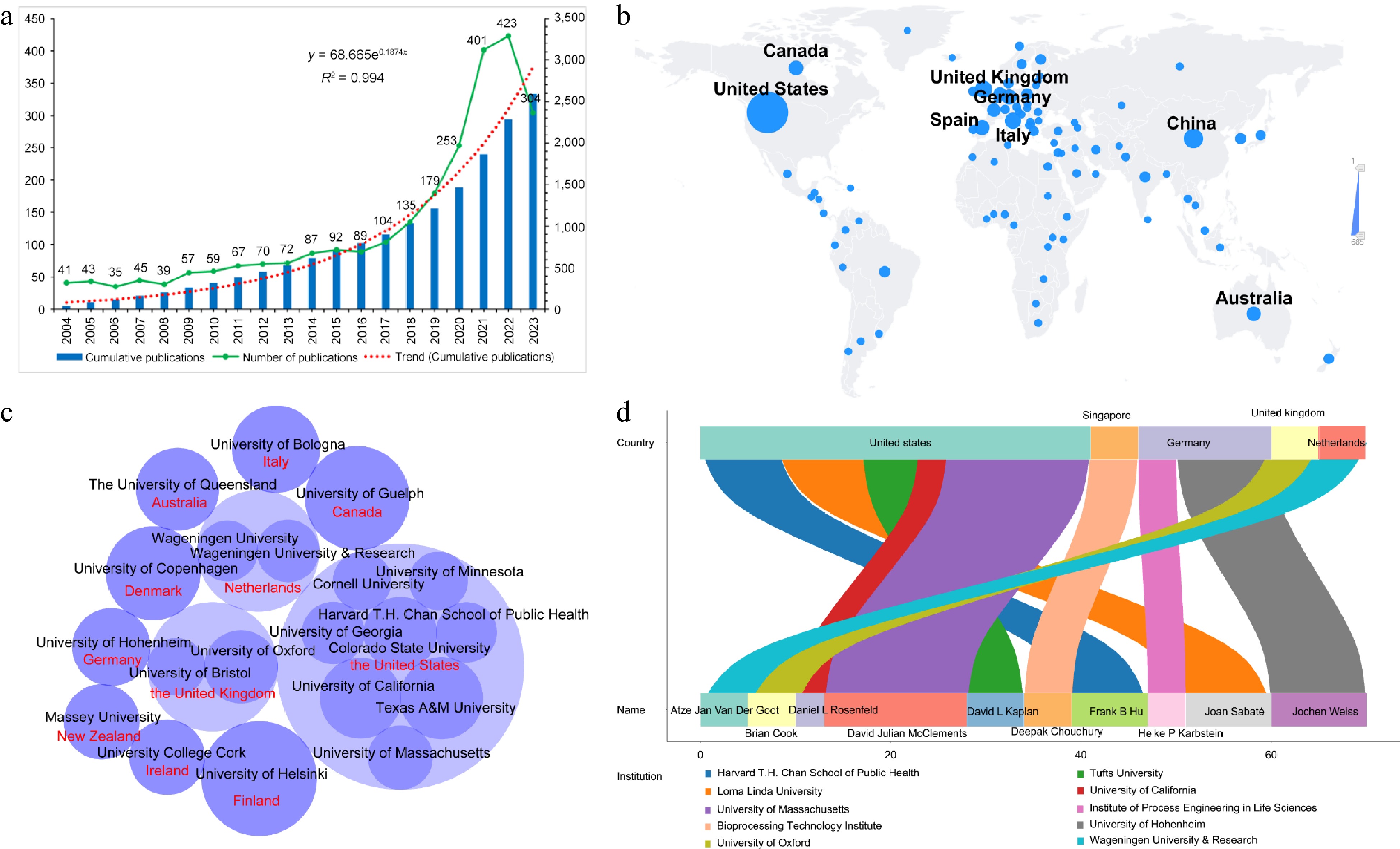
      Figure 1. 

      Overview of research related to plant-based meat analogs from 2000 to 2023. (a) The specific number of annual publications; (b) Visualization world map of publications; (c) Distribution of core research institutions; (d) Active authors.

    • The amount of literature can reflect the activity of scientific research. Therefore, the total number of documents published by one country is an important indicator of the output and productivity of this country. From 2000-01 to 2023-10, research on plant-based meat analogs has been carried out in hundreds of countries around the world. As shown in Fig. 1b, the United States had significantly more research publications than other countries. At present, a total of 685 papers have been published in the United States, accounting for 24.87% of the total. China and the United Kingdom ranked second and third, with 242 and 196 papers accounting for 8.79% and 7.12%, respectively. Countries such as Italy, Spain, Germany, Canada, and Australia also belonged to the list of countries with high literature production.

      Further statistical analysis showed that the top 20 research institutions based on the number of published documents were distributed in 11 countries (Fig. 1c). Eight of these institutions belong to the United States, with two each in the United Kingdom and New Zealand. The University of Helsinki (Finland) and Texas A&M University (the United States) took the top two spots with 33 and 28 publications, respectively. The University of California (the United States) and the University of Guelph (Canada) followed, and both published 27 papers. Other institutions with more than 20 publications were the University of Oxford (the United Kingdom, 26), Colorado State University (the United States, 22) and the University of Copenhagen (Denmark, 22).

      The statistical results of author publications showed that the top 10 authors in this field came from 10 research institutions in five countries (Fig. 1d). David Julian McClements at the University of Massachusetts in the United States currently published 15 papers in the field, which was the highest output of the author. In second place was Jochen Weiss at University of Hohenheim in Germany, who published 10 papers on the subject. Joan Sabaté and Frank B Hu at Loma Linda University and Harvard T.H. Chan School of Public Health in the United States followed, who respectively had nine and 10 publications. On the whole, the research of plant-based meat analogs has attracted global attention.

    • To comprehensively understand the relevant journals in this field, the information of the top 30 journals, including the JCR category, category rank, category quartile, impact factor in 2022 and the 5-year impact factor, were systematically collated. As shown in Fig. 2, research on plant-based meat analogs have been published in the fields of agricultural science, biochemistry, biotechnology, molecular biology, microbiology, food science, food chemistry, nutrition, and environmental science. Among them, the field of food science published the most papers, a total of 414. This was followed by nutrition and agricultural science, with 278 and 184 publications, respectively.
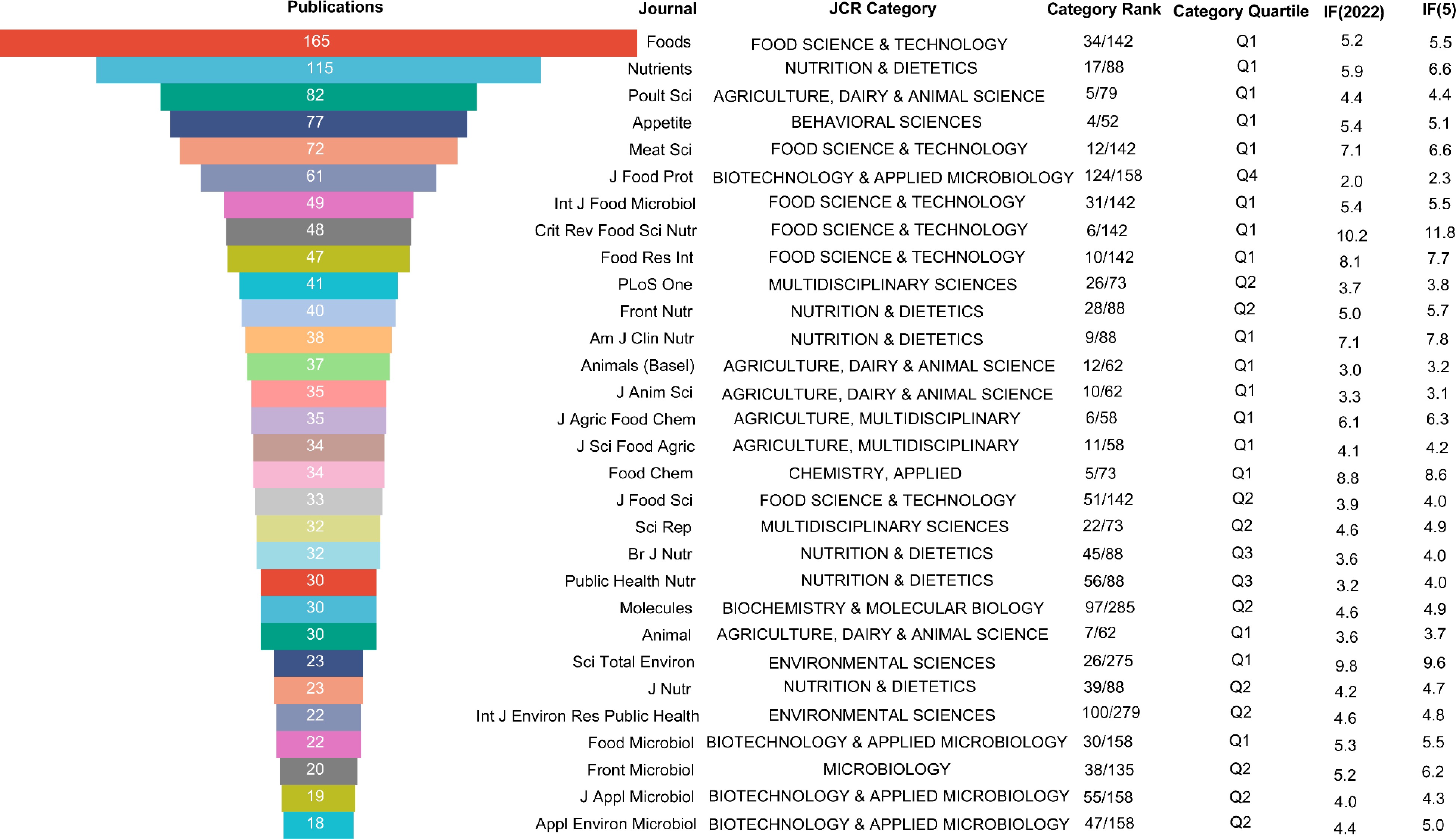
      Figure 2. 

      Top 30 most productive journals in the plant-based meat analogs field.

      According to the JCR category, 17 of these 30 journals were Q1 partitions, accounting for 56.7% of the total. There were 10 journals in the Q2 division, accounting for 30% of the total. The journal that published the most literature on plant-based meat analogs was Foods, with a total of 165, which was significantly higher than other journals. The journal is ranked 34th in the food science and technology category and is located in the Q1 region. The journal's latest IF is 5.2 and its 5-year IF is 5.5. Foods was founded in 2012, covering a wide range of topics including food science and technology, physical and chemical properties of food, food safety, food microbiology, functional food and health. The annual publication volume of the journal has increased rapidly in recent years. The journal with the next largest amount of literature on plant-based meat analogs was Nutrients. The latest IF of the journal is 5.9 and its 5-year IF is 6.6. It ranks 17th in nutrition and was classified as area Q1. Other journals that published more than 40 papers included Poultry Science (82), Appetite (77), Meat Science (72), Journal of Food Protection (61), International Journal of Food Microbiology (49), Critical Reviews in Food Science and Nutrition (48), Food Research International (47), and PLoS One (41). Among them, Meat Science, Food Research International and Critical Reviews in Food Science and Nutrition rank 12th, 10th, and 6th respectively in food science and technology journals, occupying a very important position. Meat Science is an international journal that features comprehensive research on engineering-food technology. It was founded in 1977 by the publisher Elsevier. The latest IF and 5-year IF are 7.1 and 6.6, respectively. Food Research International is also published by Elsevier, covering the field of food science and technology. It has an IF of 8.1 for 2022. Critical Reviews in Food Science and Nutrition is an international journal featuring food science and nutrition research. It was first published in 1980 by Taylor & Francis Inc. The journal has a 5-year IF of 11.8. These popular and important journals deserve special attention from researchers involved in plant-based meat analogs to get the latest advances in the field.

    • Keywords are usually the most representative terms used to explain the research topic. They are highly concise and generalized about the research purpose, research object, and research method. In order to explore the evolution trend and research hotspot of plant-based meat analogs, word frequency analysis of keywords was first performed. As shown in Fig. 3, meat appeared the most frequently, appearing 89 times in total. In addition, diet (76), sustainability (74), nutrition (71), plant-based (60), plant protein (54), vegetarian (52), cultured meat (46), meat quality (46), and protein (46) were all high-frequency words.
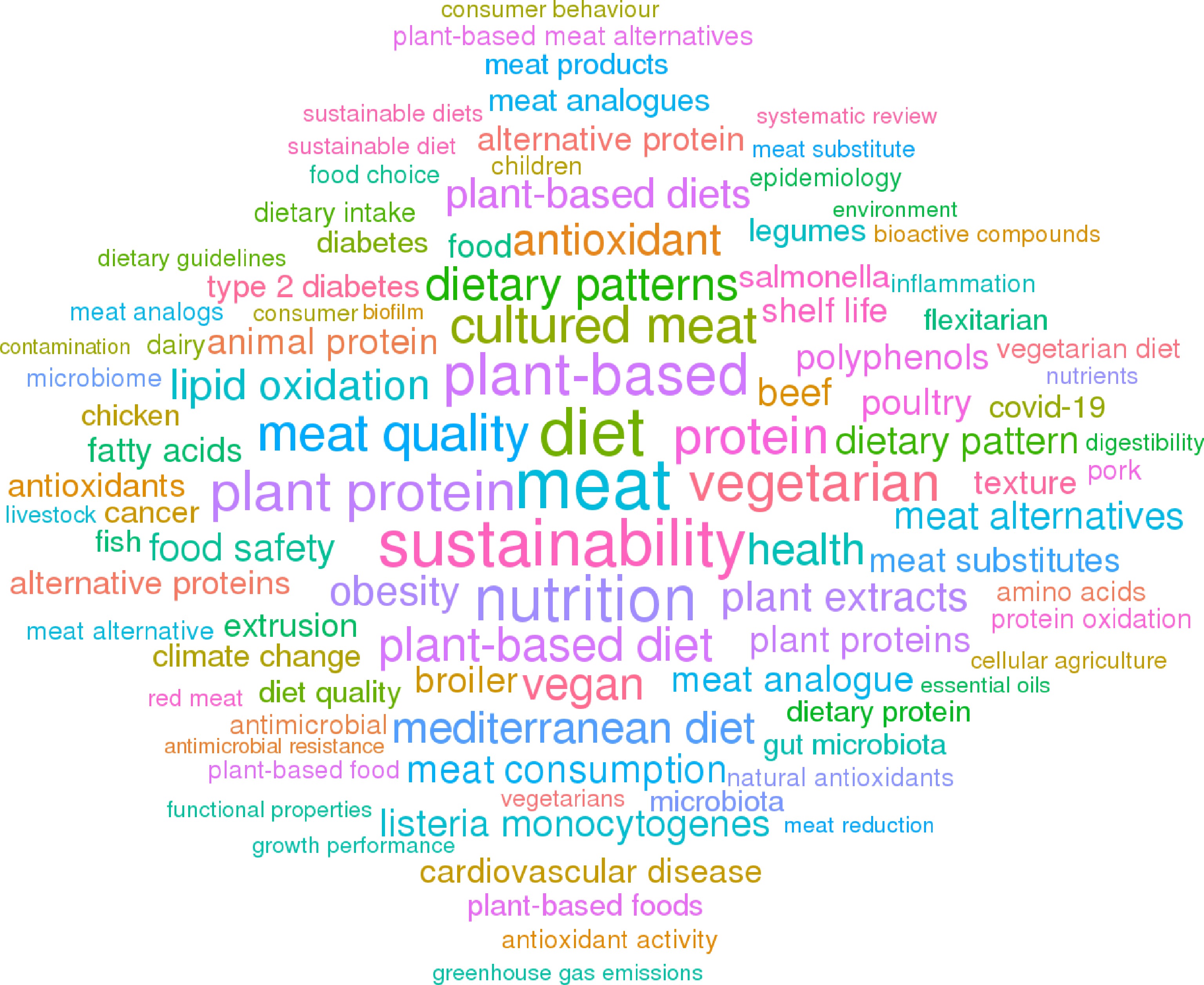
      Figure 3. 

      Analysis of keyword frequency related to plant-based meat analogs from 2000 to 2023.

      Then, 2000-01 to 2023-10 were divided into the following four periods: 2000.01 to 2014.12, 2015.01 to 2018.12, 2019.01 to 2020.12, and 2021.01 to 2023.10. The popularity ranking and ranking change of keyword word frequency in period were analyzed. As shown in Fig. 4, in the first stage, keywords directly related to plant-based meat analogs were not prominent. However, when it comes to the second stage, the keywords surrounding plant-based meat analogs gradually became clear and diversified. At this stage, the main keywords were allergenicity, alternative proteins, and flavor. In the third and fourth stages, the major keywords were cultured meat, plant-based meat analogs, animal welfare, and sustainability. These changes demonstrated that plant-based meat analogs have attracted the attention of more and more researchers.
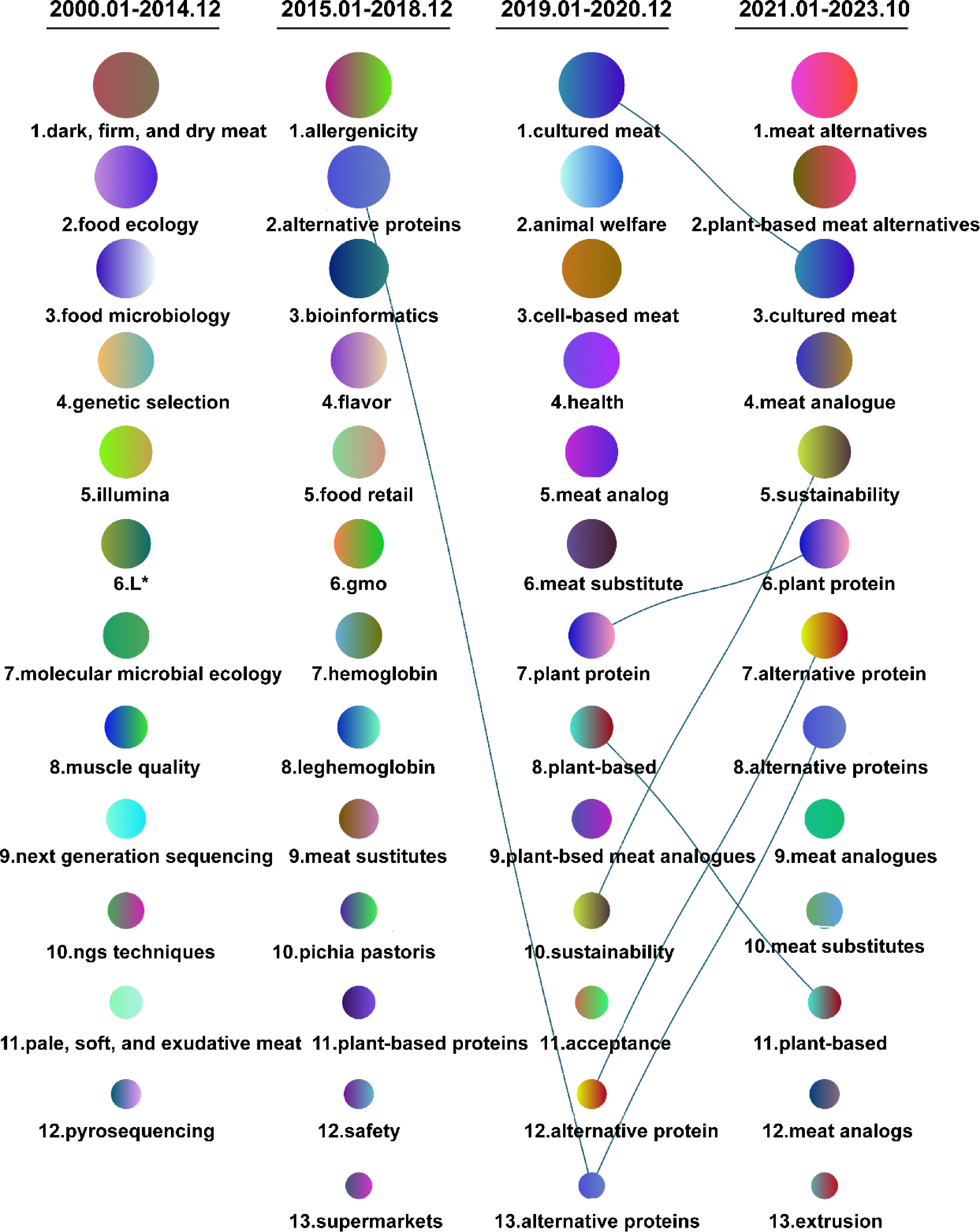
      Figure 4. 

      Changes in keywords related to plant-based meat analogs over time. The larger the circular area, the higher the degree of attention of keywords.

      To better evaluate current research hotspots, we conducted a separate analysis of keyword frequency in the last 3 years. As shown in Fig. 5, the top five most frequent keywords in 2021 from high to low were meat, plant-based diet, nutrition, sustainability, and plant protein, while in 2022 the top 5 changed to nutrition, sustainability, meat, plant-based, and diet. So far, the prefix 'plant-based' has been the most frequent keyword in 2023, followed by sustainability, plant protein, cultured meat, and alternative protein. A co-occurrence analysis showed the interrelated knowledge network of the research topics in the three years. The results showed that current hot topics related to plant-based meat analogs include alternative proteins and their functional and nutritional characteristics, as well as sustainable development (Fig. 6).
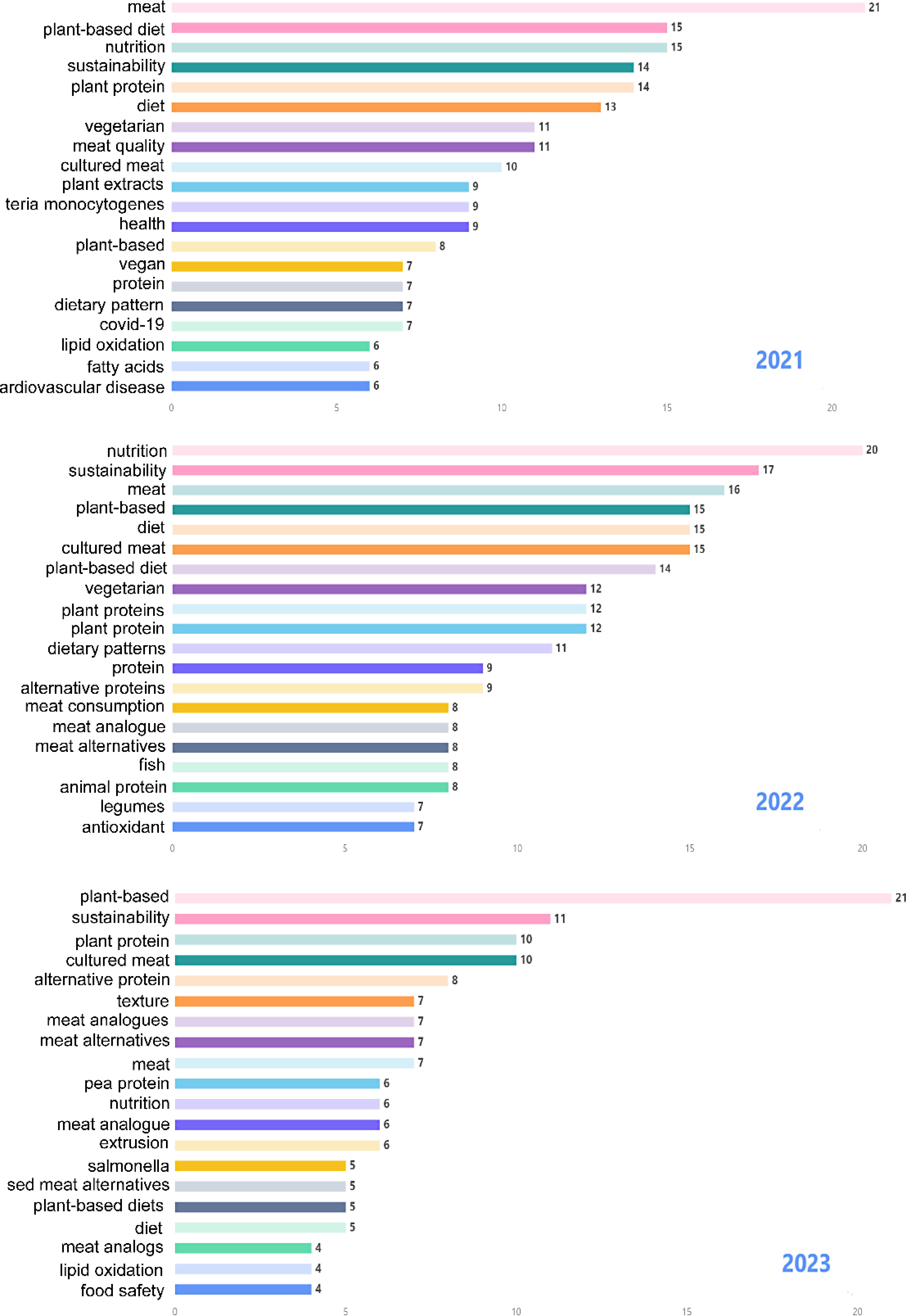
      Figure 5. 

      Hot keywords related to plant-based meat analogs from the past three years.
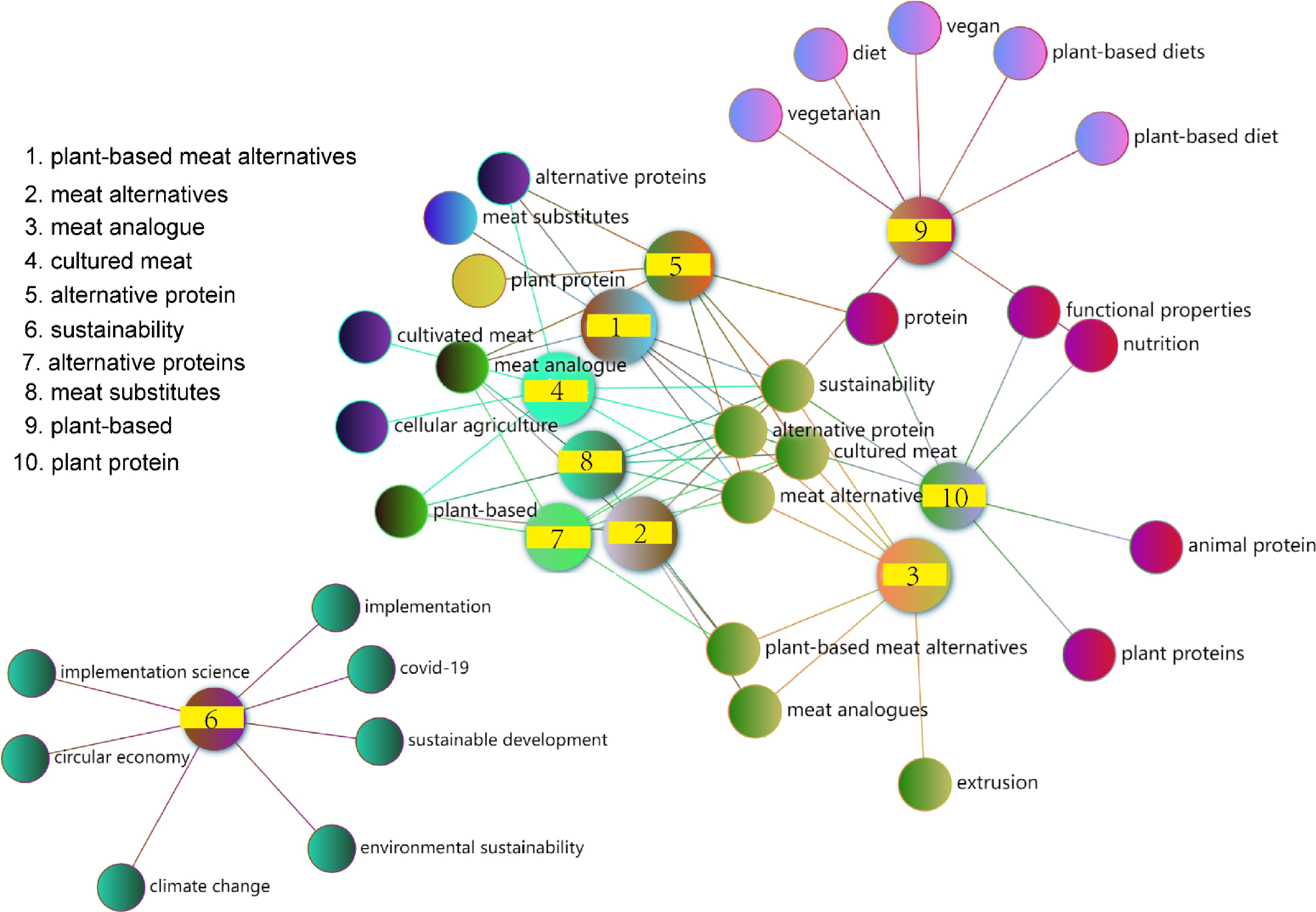
      Figure 6. 

      Knowledge networks associated with hot research topics in the past three years.

    • To have a more detailed and in-depth understanding of the current research status and future research direction of plant-based meat analogs, the citation relationships of current relevant papers in this field were analyzed (Fig. 7a, Supplemental Tables S1S3). The recommended literature indicated the latest progress of research on plant-based meat analogs (Supplemental Table S1). The classic literature was groundbreaking research in the field, by analyzing these documents, researchers can quickly grasp the historical process of research on plant-based meat analogs (Supplemental Table S2). The core literature suggested the ongoing focus of researchers in the field (Supplemental Table S3). From the systematic analysis, it is clear that the idea of processing plant-based ingredients for protein foods is not a new concept, because many products, such as tofu and tempeh, have been consumed in Asia for centuries[15]. Differently, these products are mainly aimed at vegetarians or vegans and have not attracted global attention. To date, some emerging technologies, such as extrusion, have not been used to develop plant-based foods, making plant-based proteins possess meat-like structures[16,17]. These plant-based meat analogs, which have similar texture, flavor, and color to meat, become a hot topic in the food and research communities as one of the meat substitutes.
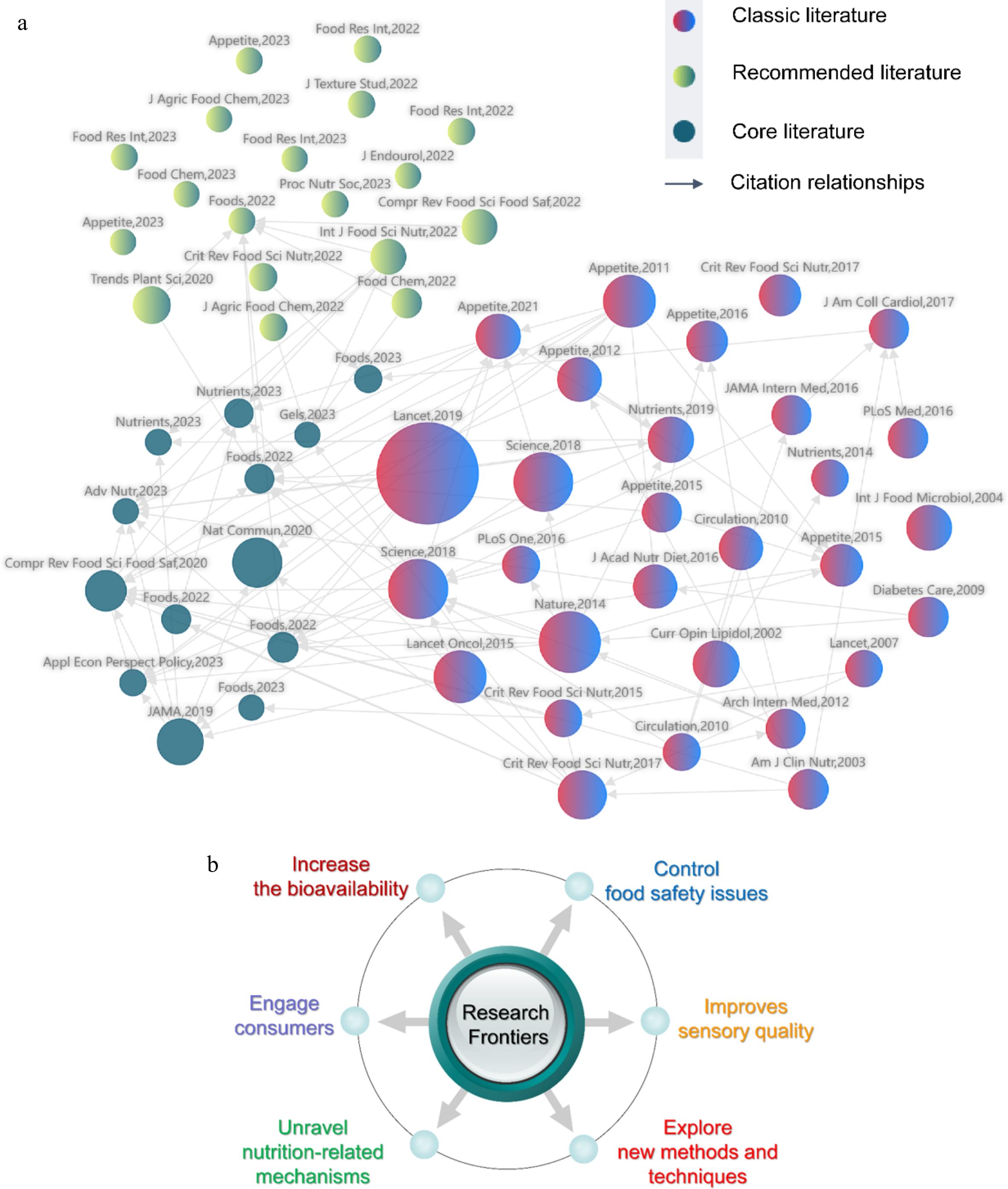
      Figure 7. 

      Analysis of the research status and trends in the field of plant-based meat analogs. (a) Citation relationships between published papers; (b) Possible research trends. Each ball represents an article. The size of the sphere is positively correlated with the number of co-citations.

    • Meat is an important source of protein with high biological value. However, the modern relationship among meat, environment and health is very complex. With the rapid growth of the world population, the demand for meat is increasing, and the supply of meat is most likely to be insufficient[18,19]. Furthermore, increased awareness of sustainable development has made consumers gradually realize that animal rearing for meat production consumes a lot of resources and creates environmental problems such as greenhouse gas emissions[20,21]. In addition, meat production involves animal welfare issues[22]. What's more, several studies have linked excessive meat intake to chronic diseases such as obesity, diabetes, and cardiovascular disease[2325]. These factors drive more and more people to look for meat alternatives. At the same time, some studies have shown that plant-based diets are more environmentally sustainable than diets rich in animal products. Vegetarians and vegans also have a lower risk of health conditions such as ischemic heart disease, type 2 diabetes, hypertension, and obesity[2629]. Together, these circumstances have driven a global transition from animal protein to plant-based protein, further fueling the rise of plant-based meat analogs[30].

    • Some studies have focused on quality characteristics such as texture[31,32] and flavor[33,34] of plant-based meat analogs. With the increasing supply and sales, more and more people pay attention to the nutritional properties of plant-based meat analogs[35,36]. The composition of plant-based meat analogs is similar to that of real meat products, but the ingredients come from different sources. Plant-based meat analogs are made from various plant components through a series of processing processes such as extrusion, high temperature, and cooling. Many studies have pointed out that different processing may lead to changes in protein structure, and the changes will further affect its digestive characteristics[3739]. However, both in vitro and in vivo studies have indicated that the digestion and absorption characteristics of proteins in plant-based meat analogs are not as good as those in meat[40,41]. How to improve the bioavailability of nutrients of plant-based meat analogs is a question worth considering.

      Recently, a large number of studies have also pointed out that there were certain differences in the nutrient content between plant-based meat analogs and real meat[42,43]. Most plant-based meat analogs have lower protein, cholesterol, and vitamin B12 content but higher carbohydrate, dietary fiber, and sodium content than real meat. In addition, the fat content varies greatly among products. The differences in carbohydrate (dietary fiber, sugar, and starch), fat, and sodium content between real meat and plant-based meat analogs drew particular attention from the researchers[44,45]. Moreover, in order to simulate the special properties of meat, the formulation of plant-based meat analogs includes many food additives, such as colors, flavorings, and adhesives, in addition to purified or semi-purified plant proteins[46]. The use of refined raw materials and multiple ingredients has led to most current plant-based meat analogs being classified as ultra-processed products[47]. Consumption of ultra-processed foods may increase the associated risk of obesity, metabolic syndrome, and hypertension[48,49]. As a result, there is a growing awareness that plant-based meat analogs cannot be directly equated with plant-based foods and cannot be considered equally beneficial to health. Whether these plant-based meat analogs can be used as good alternatives to meat has aroused consideration[30,50].

    • However, there are relatively few studies on the nutritional value and physiological effects of plant-based meat analogs. For the first time, Xie et al. directly took plant-based meat analogs as research objects in a mouse model to explore the differences between them and real meat in terms of gastrointestinal digestive function, appetite regulation, and liver metabolism[41,51,52]. They found that compared with real meat, plant-based meat analogs reduced gastrointestinal digestive function in mice by down-regulating the expression of gastrointestinal nitrogen nutrient sensors[41]. Specific digestive peptides and flavor substances in plant-based meat analogs increased the food intake of mice by altering the balance between appetite regulators[51]. This further affected lipid metabolism in mice, resulting in increased body weight and lipid accumulation[52]. These findings revealed the differences in nutritional function and related metabolic mechanisms between real meat and plant-based meat analogs, and provided a theoretical reference and scientific basis for a rational diet. Besides, recent studies have found that multiple plant-based meat analogue products were contaminated with one or mixtures of up to seven mycotoxins[53]. That suggests high risks for consumers. To better understand the risks associated with switching to a plant-based meat diet, future systematic studies of natural toxin contamination of plant-based meat alternatives are needed. Policymakers also need to consider dealing with these natural toxins in plant-based meat analogs to ensure consumer safety[54].

      Notably, consumer acceptance of plant-based meat analogs and how to increase consumption and attract new consumers have long been the focus of academia, industry and business[50,55,56]. Consumer acceptance largely depends on attitudes and beliefs about meat substitutes and food neophobia[57]. In order to make meat alternatives more attractive to meat consumers, improving their sensory quality and the similarity to meat has been a common practice. At present, some effective techniques have been applied to the production of plant-based meat analogs. For example, fermentation is booming as an effective technology for improving the sensory, nutritional, and functional properties of plant-based meat analogs[58,59]. Also, machine learning-based methods have been proposed to predict the structural properties of meat analogs[60], which provides convenience for optimizing product development cycles and reducing costs.

      In conclusion, although a lot of studies have been carried out on the quality characteristics of plant-based meat analogs, how to further improve the quality and attract more consumers is still a hot topic. Moreover, it is the future trend to systematically investigate and control the safety issues such as pollution of natural toxins, as well as explore the health effects and related mechanisms. More methods and techniques need to be explored and developed to optimize the quality and nutritional characteristics of plant-based meat analogs (Fig. 7b).

    • The genetic entity words in the abstract of 2595 publications were mined and analyzed using the BioBERT biomedical language representation model. As shown in Fig. 8, a total of 23 genes were discovered. Among them, the maximum number of literature involving INS was 162, and there were 58, 54 and 52 publications related to CRP, C10orf90 and FAT1, respectively. Other genes covered in at least 30 publications included IL6 (37), TNF (34), and NDUFB3 (30) (Fig. 8a). Enrichment analysis of all genes found that these genes were significantly enriched in terms of molecular function (MF), cellular component (CP) and biological process (BP). In terms of BP, these genes were mainly involved in peptidyl-serine/tyrosine phosphorylation, protein kinase B signaling, nitric oxide biosynthetic process, smooth muscle cell proliferation, and protein, glucose, and lipid metabolic process. In terms of CC, these genes were mainly concentrated in extracellular space, extracellular region, extracellular exosome, secretory granule lumen, external side of plasma membrane, cytoplasm, interleukin-6 receptor complex, insulin-like growth factor binding protein complex, alphav-beta3 integrin-IGF-1-IGF1R complex, and insulin-like growth factor ternary complex. In terms of MF, these genes mainly have hormone, cytokine, and N-acylglucosamine 2-epimerase activity, as well as identical protein, insulin-like growth factor receptor, insulin receptor, protease, glucagon receptor, TFIIIC-class transcription factor complex, and bradykinin receptor binding functions (Fig. 8b). KEGG pathway analysis showed that these genes were mainly involved in alcoholic/non-alcoholic fatty liver disease, type II diabetes mellitus, hypertrophic cardiomyopathy, adipocytokine, AMPK, mTOR, PI3K-Akt, C-type lectin receptor, TNF, toll-like receptor, HIF-1 signaling pathway, insulin resistance, antifolate resistance, and longevity regulating pathway (Fig. 8c). These results suggested that plant-based meat analogs were mainly involved in metabolically related pathways and diseases, providing important clues for future health research.
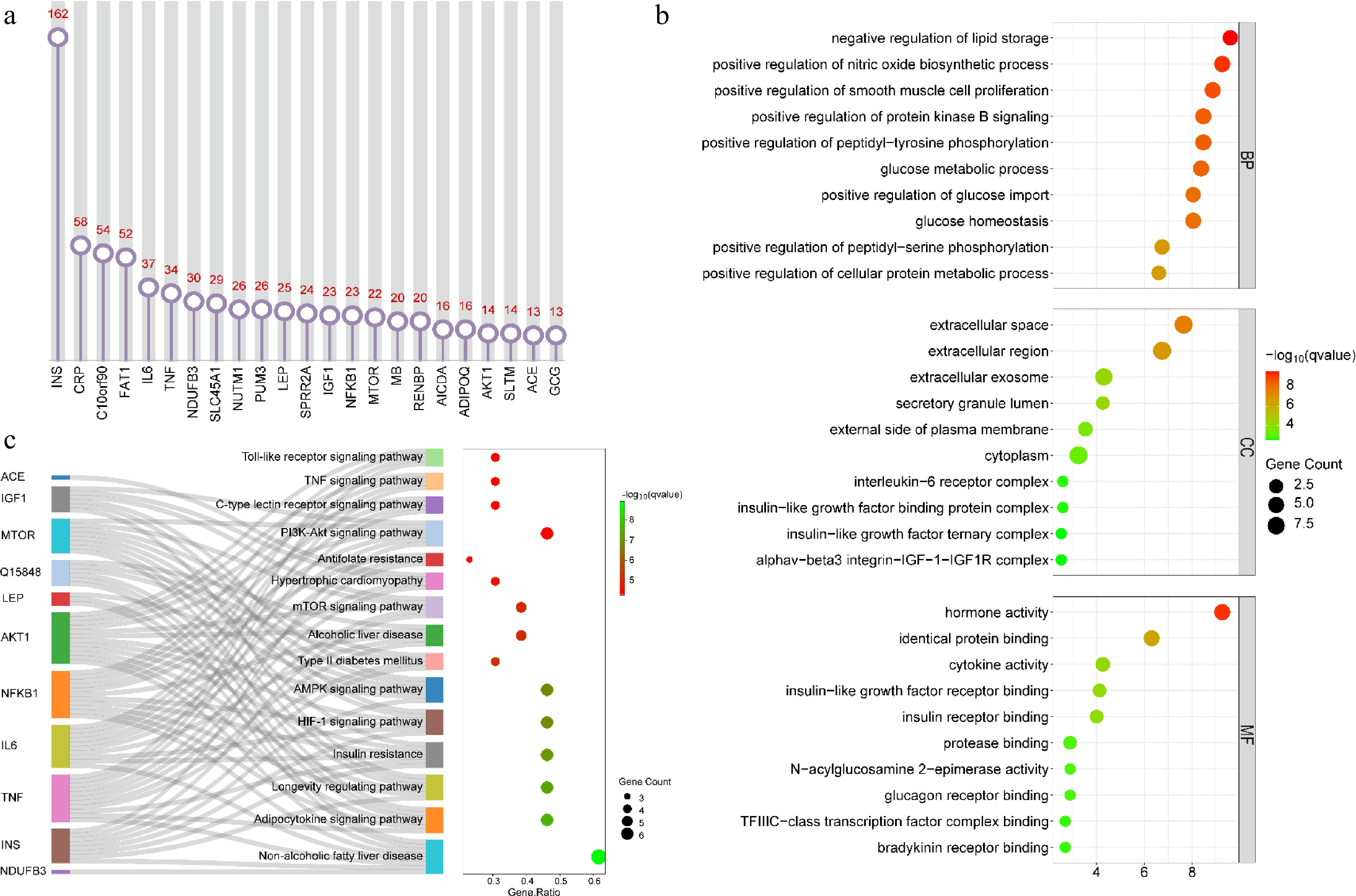
      Figure 8. 

      Analysis of associated genes and biological pathways. (a) Major associated genes; (b) GO enrichment analysis of major associated genes; (c) KEGG pathway analysis of major associated genes. For (b) and (c), the top 10 and 15 pathways were presented, respectively.

    • Plant-based meat analogs have been one of the hot topics in recent years. The present analysis summarized the current state of research, research hotspots, and future trends in the field from the micro level (authors, institutions, and keywords) to the macro level (world, country, and topic). This is of vital reference value for further research.

      The number of annual publications can directly reflect the research scale and attention of a certain field, and predict its future development trend. Based on the PubMed database, the temporal distribution characteristics of the number of publications related to plant-based meat analogs were analyzed. The results showed that this field did not attract much attention from researchers before 2015. Since then, more and more attention and interest have been paid to this field. This may be related to the fact that the World Health Organization classified processed meat as 'carcinogenic to humans' and red meat as 'probably carcinogenic' in 2015[61]. Meat-related health concerns, coupled with a growing awareness of sustainability, have prompted the search for meat alternatives. In the following years, the market for plant-based meat analogs expanded[4,44,62], attracting more and more researchers. At present, plant-based meat products are not only favored by vegetarian consumers but also attract many meat lovers[63]. Under these circumstances, the research on plant-based meat analogs will continue to develop rapidly in the next few years and will gradually go deeper.

      Research on plant-based meat analogs has been carried out in hundreds of countries around the world. Multidimensional analysis of research can help to understand the degree of academic support and recognition for the research field. This is also conducive to the harmonious development of the field and the cooperation between relevant institutions and authors. Journals are important carriers for displaying academic information and disseminating knowledge. However, few researchers have a comprehensive view of all relevant journals in a field. The hot journals not only represent the recognition and choice of most researchers, but are also an important way to obtain the hot research content in this field. The analysis of published journals can guide researchers to select key publications to read and understand the main research results and latest progress in this field. At the same time, it is also helpful for researchers to accurately locate their research results and quickly find suitable journals for submission.

      Keyword and reference analysis are one of the important methods and indicators of bibliometrics. Based on keyword co-occurrence analysis and literature co-citation analysis, it can reveal the evolution process, main research direction, and hot frontier of a specific field[64,65]. Systematic analysis showed that plant-based meat analogs were widely accepted, in part because they were considered an environmentally friendly alternative to meat[1]. However, the situation of energy consumption, land occupation, and water resource utilization in the actual production process remains to be clarified. As an alternative to meat, plant-based meat analogs not only need to mimic the texture, color, and flavor of meat but also have considerable biological value. At present, a series of processing and ultra-long formulations have also caused nutritionists to worry about their nutritional functions. Some researchers have investigated the differences in nutritional function between plant-based meat analogs and real meat from the aspects of substance composition, nutrient content, and bioavailability[41,45]. But the health implications of these differences were poorly explored. The undigested portion of the diet goes into the large intestine and is used by gut microbiota. Different nutrient substrates will shape different intestinal microecology[38,66]. Gut microbiota is not only important to the health of the gut but also affects many aspects. A large number of studies have uncovered the bidirectional communication between gut microbiota and extra-intestinal organs such as the brain (gut-brain axis), liver (gut-liver axis), kidney (gut-kidney axis), heart (gut-heart axis), and so on[6769]. Therefore, it is important to clarify the nutritional characteristics of plant-based meat analogs for scientific dietary guidance and maintenance health.

      In addition, BioBERT is a language representation model for biomedical text mining and training. It has been proven to be very effective in many biomedical text-mining tasks. The important genes and key biological pathways were found through this method, which can provide important implications for the subsequent research of plant-based meat analogs in the field of nutrition. In short, the research of plant-based meat analogs has initially formed a diversified research perspective. However, it is still necessary to improve the acceptance of products and reveal their nutritional characteristics through multi-dimensional and in-depth research.

    • This work is the first bibliometric analysis to map and describe the knowledge landscape of plant-based meat-analogs related research from 2000−2023. The integration of various bibliometrics software and tools for analysis and visualization increases the richness of the results. However, there are certain limitations in this work. The literature search is only based on the PubMed database, and some literature could be omitted, which may introduce bias in paper selection and lead to the omission of important studies. Nevertheless, we believe our results remain a valid global research profile in the field of plant-based meat analogs. This work can provide an important reference for the further development of research on plant-based meat analogs.

    • The authors confirm contribution to the paper as follows: study conception: Xie Y, Li C; data collection and analysis: Xie Y, Cai L, Zhou G, Li C; draft manuscript preparation: Xie Y; manuscript revision: Cai L, Zhou G, Li C. All authors read and approved the final version.

    • The datasets generated during and/or analyzed during the current study are available from the corresponding author on reasonable request.

      • This work was supported by Jiangsu Innovative Group of Meat Nutrition, Health and Biotechnology, and National Natural Science Foundation (grant number: 32072211).

      • The authors declare that they have no conflict of interest. Guanghong Zhou and Chunbao Li are the Editorial Board members of Food Materials Research who were blinded from reviewing or making decisions on the manuscript. The article was subject to the journal's standard procedures, with peer-review handled independently of these Editorial Board members and their research groups.

      • Supplemental Table S1 Recommended literature based on citations, impact factor, time of publication, and relevance to keywords.
      • Supplemental Table S2 Classic literature screened based on the co-citation relationship of recommended literature.
      • Supplemental Table S3 Core documents based on overlapping references between recommended and classic literature.
      • Copyright: © 2024 by the author(s). Published by Maximum Academic Press on behalf of Nanjing Agricultural University. This article is an open access article distributed under Creative Commons Attribution License (CC BY 4.0), visit https://creativecommons.org/licenses/by/4.0/.
    Figure (8)  References (69)
  • About this article
    Cite this article
    Xie Y, Cai L, Zhou G, Li C. 2024. Global research landscape and trends of plant-based meat analogs: a bibliometric analysis. Food Materials Research 4: e020 doi: 10.48130/fmr-0024-0011
    Xie Y, Cai L, Zhou G, Li C. 2024. Global research landscape and trends of plant-based meat analogs: a bibliometric analysis. Food Materials Research 4: e020 doi: 10.48130/fmr-0024-0011

Catalog

  • About this article

/

DownLoad:  Full-Size Img  PowerPoint
Return
Return