ARTICLE   Open Access    

Bioactive components and antimicrobial potential of extracts from Artemisia species and their repellent activities against Aphid (Macrosiphoniella sanborni)

More Information
  • Species from the Artemisia genus frequently have high resistance to pests and pathogens due to their being rich in secondary metabolites. Therefore, identifying bioactive components from Artemisia plants is essential for developing botanical pesticides and selecting parents for breeding resistant varieties of cultivated Chrysanthemum morifolium. This study investigated the resistance of four Artemisia species to aphids (Macrosiphoniella sanborni) and the antimicrobial properties of their extracts. Choice and no-choice assays showed that the tested four species had strong repellent and antifeedant effects on aphids compared with chrysanthemum. The antimicrobial activity of ethyl acetate extracts from different tissues against four pathogenic fungi was tested by disc diffusion assay. Among them, the extracts from Artemisia maximowicziana showed the strongest antimicrobial effect. The inhibition rates of Alternaria alternata, Colletotrichum siamense, and Phoma sp. caused by leaf extracts from A. maximowicziana were 53.8%, 54.95%, and 61.46%, respectively. And the inhibition increased to 75.44%, 51.65%, and 79.51%, respectively, using the stem extracts. However, the root extracts of Artemisia spp. showed only up to 25% to Fusarium solani. GC-MS analysis showed that the volatiles of Artemisia spp. were mostly abundant in terpenoids, but the components and contents were remarkably different among species. Further analysis of Orthogonal Partial Least Squares Discriminant Analysis (OPLS-DA) showed the most contributed component among all potentially antimicrobial bioactive components was (−)-thujol. In this study, A. maximowicziana was identified as the material with potential value as a parent for crossbreeding, and its primary volatile compound (−)-thujol with potential resistant active is worth further investigation.
  • Stroke remains a major global health issue, characterized by high incidence rates, significant disability, and substantial mortality. Ischemic stroke (IS), in particular, is a leading cause of death, accounting for ~3.71 million fatalities worldwide per year. The highest mortality rates are observed in Central Asia and Eastern Europe[1]. The 2021 Global Burden of Disease Study reported that IS was responsible for 70.4 million disability-adjusted life years, and 7.81 million new cases, and a troubling 88.0% increase in incident cases since 1990, highlighting the growing impact and urgency of addressing this condition[2].

    IS is a complex health condition influenced by numerous factors. While traditional risk factors such as cardiometabolic disorders, lifestyle, and psychosocial issues are well-documented, the precise mechanisms that drive IS remain incompletely understood. Recent advances in epigenetic research are shedding light on these complexities. Epigenetics involves modifications to DNA and histones that impact gene expression and chromatin structure, without altering the DNA sequence itself. Key epigenetic mechanisms include DNA methylation and histone modification. These modifications are dynamic, reversible, and responsive to environmental influences, making them vital for both normal cellular function and disease states[3].

    DNA methylation is a critical epigenetic mechanism where methyl groups are added to cytosine residues, where they may influence gene expression without changing the genetic code. Research has shown that alterations in DNA methylation patterns are associated with IS. For example, long interspersed nuclear element-1 (LINE-1), the sole autonomous retrotransposon in the human genome, can reshape chromatin and alter genome structure and gene expression. Lower levels of LINE-1 methylation have been linked to a higher prevalence of IS, with this association remaining significant in males even after adjusting for risk factors[4]. Systematic reviews have also provided evidence for reduced methylation in the promoter regions of key genes such as tumor necrosis factor α, estrogen receptor α, and microRNA-223 in stroke patients, and conversely, increased methylation of genes encoding cystathionine β-synthase and ATP-binding cassette G1 (ABCG1)[5]. Differential methylation of methylenetetrahydrofolate reductase (MTHFR), encoding a critical enzyme in one-carbon metabolism, is also correlated with risk for IS[6]. At the chromatin level, gene expression is regulated by histone modifications, such as those mediated by histone deacetylases (HDACs) which remove acetyl groups from histones. Notably, higher methylation levels of HDAC9 have been associated with a reduced risk of IS[7]. These findings highlight the critical role of epigenetic mechanisms in understanding and addressing IS.

    Bibliometrics involves the systematic application of statistical methods to analyze and quantify scientific and technical outputs across researchers, institutions, and countries. This approach combines qualitative and quantitative evaluations to highlight key metrics such as leading countries, institutions, journals, authors, and keywords over time[8]. By evaluating the current research landscape and trends, bibliometrics provides valuable insights into future research directions, aids in profiling research fields, trend analysis, impact measurement, and identifies emerging areas and collaboration opportunities[8]. While bibliometric analysis has been applied to various aspects of IS, studies specifically focusing on epigenetics in IS are limited. This study seeks to address this gap by employing bibliometric techniques on extensive datasets related to epigenetics in IS. It aims to address recent research, and identify key topics and emerging trends, thereby providing a foundational framework for future research.

    As the first bibliometric analysis of its kind, this study aims to explore the following research questions: (i) What are the trends and patterns of advancement in epigenetics research related to IS up to 2024? (ii) Which documents, authors, and countries have made the most significant contributions to the study of epigenetics in IS? (iii) What are the key thematic structures and research themes identified in epigenetics research concerning IS? (iv) What are the anticipated future research directions and emerging trends in the field of epigenetics and IS?

    The Web of Science Core Collection was used to identify 98 studies on epigenetics in IS as of July 31, 2024. Limiting the search to English-language articles, editorials, abstracts, proceedings, and reviews were excluded. The relevant records-covering countries, institutions, journals, authors, and more, using scientometric tools where then analyzed for a comprehensive evaluation.

    After preparing the data, R Studio was utilized with the Bibliometrix R package, an open-source tool for quantitative scientometric and bibliometric analysis. Bibliometrix offers advanced statistical and visualization tools and is complemented by Biblioshiny, a web-based platform for enhanced literature analysis. Using these tools, in-depth analyses were conducted to track research trends, emerging themes, and forecast future directions[8]. The academic impact was assessed with h-index and total citation (TC) metrics, key publications identified by citation analysis and Bradford's Law and Lotka's Law applied to evaluate journal productivity and author output.

    VOSviewer 1.6.20[9] and CiteSpace 5.8[10] were utilized to analyze and visualize bibliometric data. VOSviewer created color-coded maps of keyword co-occurrence, highlighting prolific authors, co-cited journals, and key references after refining the dataset[9]. CiteSpace, known for its advanced document analysis features, provided dynamic views of co-citation networks, clustering publications by conceptual themes and revealing research trends over time[10]. While VOSviewer offered clear visualizations, CiteSpace's burst detection and cluster analysis were crucial for identifying significant developments. Together, these tools provided a comprehensive overview of the research landscape, unveiling influential publications and emerging trends.

    This study provides an in-depth assessment of research on epigenetics in IS, analyzing 47 papers from 37 journals published between 2014 and 2024. Key findings include an annual publication growth rate of 14.9%, an average document age of 4.43 years, and an average of 19.21 citations per document. Among the 383 authors, no single author worked alone, with international collaboration at 31.9% and an average of 10 co-authors per paper (Supplementary Fig. S1a). While there has been a consistent increase in global publications on epigenetics, peaking in 2022 with eight papers, citation rates fell sharply from an average of 58.5 per article in 2017 to 15 in 2018 (Supplementary Figs S1b, S1c). The Sankey diagram highlights key metrics, with 'Stroke' as a leading journal and Spain as the top contributor, particularly recognizing Israel Fernandez-Cadenas from Spain as a prominent author (Supplementary Fig. S1d).

    Supplementary Table S1 ranks the most cited journals in epigenetics research related to IS based on h-index, Bradford's Law, and TC. The top 10 journals have h-index values ranging from 1 to 3 and TC from 5 to 206. Clinical Epigenetics, Molecular Neurobiology, and Stroke, all in Zone 1 of Bradford's Law, have the highest h-index of 3. Molecular Neurobiology leads in TC, indicating these journals are pivotal in the field.

    Institutional contributions reveal that the Hospital Del Mar Research Institute in Spain leads with 12 publications, followed by Autonomous University of Barcelona with 10, and the State University of New York Buffalo with eight (Supplementary Table S2a). Among the 383 authors of 47 research papers, 94.6% have published fewer than two articles in the past decade, in line with Lotka's Law. The top authors by h-index are Fernandez-Cadenas Israel, Jimenez-Conde Jordi, and Roquer Jaume, all from Spain, each with six publications (Supplementary Table S2b).

    Supplementary Table 2c summarizes country contributions and collaborations. China leads with 32% of the total publications, followed by Spain at 18%, and India at 10%. While China has the highest publication count (128 total, average 8.0), Spain leads in TC (205, average 22.8) and India also shows high citation rates (103, average 20.6). Supplementary Fig. S1e illustrates the global distribution of publications, with navy blue indicating the highest output, blue for moderate activity, and gray for no publications. Key collaborations include Spain-UK and Spain-USA, each with four co-authored papers, and USA-Sweden with three. Supplementary Table S3 highlights the most cited documents: Patnala et al.[11] in Molecular Neurobiology lead with 161 citations.

    Scientific mapping with VOSviewer was used to analyze keyword frequency and co-citation patterns, revealing core knowledge and intellectual structures in the field. Co-citation analysis identified key publications and clusters, highlighting integration patterns and central topics. This approach is crucial for understanding the intersection of epigenetics and IS research, offering insights to guide future research and development.

    Figure 1 illustrates the co-citation analysis of 47 journal articles and 2,624 references, identifying 14 key papers with at least five citations each. This analysis uncovered three prominent clusters, connected by 73 links, with a total link strength (TLS) of 130. Each cluster, differentiated by color and node size indicating citation volume, represents a distinct research stream in the field. Red Cluster 1 reveals key insights into IS, including the identification of LINE-1 methylation as a cardiovascular risk biomarker[12], significant genetic factors associated with stroke subtypes[13], and the impact of DNA methylation on clopidogrel response and stroke recovery[14]. It also emphasizes the role of DNA methylation in stroke pathology[15], its effects on recovery outcomes and stroke subtypes[16,17], regional stroke disparities[18], and its involvement in atherosclerosis[19]. Green Cluster 2 reveals key advancements in methodology, including the TOAST classification for IS subtypes[20], improved normalization techniques for Illumina 450K data preprocessing[21], and breakthroughs in the characterization of the human DNA methylome and histone variants[22]. Blue Cluster 3 focuses on the significant role of HDAC9 in large-vessel stroke. The studies reveal a connection between HDAC9 genetic variants and an increased risk of large-vessel IS[23]. Additionally, elevated HDAC9 expression is noted in carotid plaque and intima-media thickness[24], highlighting the importance of further validating these findings and conducting detailed stroke subtyping[25].

    Figure 1.  Co-citation analysis of cited references.

    Bibliographic coupling examines citation patterns to reveal related research. Of the 47 documents reviewed, 43 met the citation threshold, forming a major cluster with 42 interconnected nodes. This cluster, detailed in Fig. 2a, is divided into eight sub-clusters with 199 connections and a TLS of 358, highlighting significant field interrelationships. Network analysis shows that larger nodes indicate higher citation weights, while larger labels represent higher PageRank values[9]. Figure 2b illustrates the overlay network, mapping publication years and influence. The analysis identified eight sub-research clusters with conceptual similarities and five key themes, offering insights, and suggesting future research directions.

    Figure 2.  Bibliographic coupling analysis. (a) Network visualization. (b) Overlay visualization.

    Based on Fig. 2a, Light Green Cluster 1, encompassing 11 articles, focuses on mitochondrial metabolism and epigenetics[26], HDAC9 polymorphisms[27], histone modifications[28], epigenetic regulation in immune cells[29], and the therapeutic potential of epigenetic modifications[11]. Red Cluster 2 encompasses 10 studies discussing various epigenetic factors in IS, including DNA methylation[7], miRNAs[30], and circular RNAs such as circRap1b[31]. It also covers pathways and regulators such as the corin-B type natriuretic peptide (BNP)-neprilysin (NEP) pathway[32], RNA methylation regulators[33], and various types of methylation, including allele-specific methylation, miR-335 and miR-495[34], calmodulin-binding transcription activator 1 (CAMTA1) methylation[35], and DNA methylation related to cardiometabolic traits[36]. Blue Cluster 3 highlights five studies on epigenetic modifications in IS. These studies examine the connection between DNA methylation and the ABCG1 gene[5], reinforcing the significance of DNA methylation research, explore tumor necrosis factor receptor-associated factor 3 (TRAF3) methylation and vascular recurrence[14], investigate protein phosphatase Mg2+/Mn2+ dependent 1A (PPM1A) gene methylation[37], and address hypomethylation of the MTRNR2L8 gene[38]. Purple Cluster 4 includes four key studies focusing on genetic and epigenetic factors related to vascular and neurological diseases. These studies: identified genes associated with atherosclerosis using advanced chromatin capture techniques[39]; mapped stroke-related single nucleotide polymorphisms (SNPs) to specific genomic regions; reported a GWAS on plasma levels of tissue factor pathway inhibitors, revealing significant SNPs[40]; and highlighted the importance of large-scale genomics in advancing the diagnosis and treatment of neurological disorders. Green Cluster 5 includes three studies focusing on the relationship between biological age (b-Age) and stroke[41], b-Age and white matter hyperintensities[42], and the effectiveness of b-Age as a predictor for stroke recurrence[43]. Light Blue Cluster 6, comprising three studies, highlights that: elevated tumor protein p53 (TP53) promoter methylation is observed in IS patients[44]; global DNA methylation levels do not vary significantly across IS subtypes[16]; and significant promoter methylation changes are present in the autoimmune regulator (AIRE1) and arachidonate 12-lipoxygenase (ALOX12) genes within atherosclerotic plaques[45]. Orange Cluster 7 includes three studies, i.e. development of a rapid pyrosequencing assay to analyze DNA methylation in the MTHFR gene[3], identification of lncRNA_H19 as essential for neurogenesis and stroke recovery, and discovery of ankyrin repeat and SOCS box containing 10 (ASB10) and tetratricopeptide repeat domain 37 (TTC37) methylation sites as potential biomarkers for stroke risk[46]. Lastly, Light Red-Violet Cluster 8 focuses on advanced research methods and their implications for IS, including epigenome-wide association studies (EWAS), genome editing, advanced sequencing, and single-cell analysis (Fig. 2a).

    CiteSpace was used to map the evolution of research on epigenetics in IS, highlighting key areas and trends. Figure 3a displays the main keywords, while Fig. 3b presents a thematic analysis of nine research clusters organized by density and centrality. Themes such as 'genome-wide association' and 'coronary-artery-disease' are less central, whereas 'cerebral-ischemia', 'methylation', and 'inhibition' are more relevant. Core, but less dense themes such as 'model' and 'focal cerebral-ischemia' show high centrality. Emerging niche areas in the upper left quadrant, like 'cholesterol' and 'histone deacetylase inhibitors', exhibit high density but lower centrality. Dominant themes in the upper right quadrant, including 'expression' and 'gene-expression', have both high density and centrality, outshining 'mechanisms' and 'brain'. Overall, central concepts such as 'ischemic stroke', 'DNA methylation', and 'epigenetics' are crucial, with 'clopidogrel' and 'management' emerging as key themes in the evolving research landscape. Supplementary Table S4 highlights the evolution of key topics in epigenetics and IS from 2014 to 2024. 'Methylation' emerged prominently in early 2016, mid-2018, and late 2020, while 'epigenetics' gained attention in early 2017, mid-2020, and late 2022. The TreeMap visualization in Fig. 3c 'ischemic stroke' as the most frequently used term (14%), followed by 'DNA methylation' and 'epigenetics' (11% each). The TreeMap's color-coded rectangles represent these keywords, with rectangle size indicating the frequency of their occurrence in publications.

    Figure 3.  Research hotspots and future trends. (a) Word frequency, (b) thematic analysis, and (c) TreeMap diagram.

    In CiteSpace, color-coded clusters reveal the emergence of common reference links within specific domains, with prominent works indicated by large nodes due to their high h-index. Figure 4 illustrates the citation network from 2014 to 2024, featuring 13 clusters with parameters set at LRF = 2.5, LBY = 5, L/N = 10, and e = 1.0. CiteSpace analysis shows a highly modular network with a modularity score of 0.78, signifying distinct clusters. The mean silhouette value of 0.94 indicates excellent cluster quality, while a harmonic mean of 0.85 reflects high accuracy and recall. The network, comprising 249 nodes and 806 edges across 47 articles, has a density of 0.03, with no pruning applied to the initial 25 references.

    Figure 4.  Panoramic overview of the common keywords and co-cited references.

    Among the 13 clusters shown in Figs 4 and 5, the timeline for 10 clusters from 2014 to 2024 reveals their evolving themes. Cluster 0, focused on 'cerebral ischemic preconditioning', is the most prominent, while Cluster 9, related to 'transcription' is the smallest. All clusters have been active for at least five years, with Cluster 9 emerging in the past two years. Of which, Cluster 0, with a silhouette value of 0.93, includes 34 members, and Cluster 1, with a silhouette value of 0.99, has 29 members (Supplementary Table S5). This analysis highlights persistent research engagement and thematic concentration in epigenetics.

    Figure 5.  Timeline analysis and citation burst analysis.

    Citation bursts reveal periods of rapid citation growth, highlighting significant research trends and emerging hotspots. Analyzing these bursts, including factors like author details, publication dates, and burst intensity, helps identify shifts in research focus. Among the 10 bursts analyzed, Pidsley et al.'s publication[21], denoted as a red node in Cluster 3, achieved the highest burst strength of 1.76, marking it as a key influential work with a significant citation surge since 2018 (Fig. 5).

    Co-words analysis, which examines keyword frequency to reflect article content, was performed using VOSviewer on 47 articles, identifying 75 distinct keywords. This analysis generated seven clusters, with 687 links and a TLS of 987 (Fig. 6a). In the visualization, larger nodes and thicker lines represent higher keyword frequency and stronger relationships. The key clusters identified include: 'epigenetics' (36 occurrences, TLS 210), 'DNA methylation' (19 occurrences, TLS 101), 'stroke' (13 occurrences, TLS 94), 'ischemic stroke' (13 occurrences, TLS 78), and 'risk' (10 occurrences, TLS 69). The overlay visualization demonstrates keyword trends over time: 'ischemic stroke' emerged in 2018, 'epigenetics' in mid-2019, and both 'DNA methylation' and 'stroke' from 2020 onwards (Fig. 6b).

    Figure 6.  Co-words analysis. (a) Network visualization, (b) overlay visualization.

    The field of epigenetics in IS has seen substantial growth, attracting significant interest from clinicians, especially neurologists, as well as researchers in neuroscience and medical sciences. This heightened focus has led to a burgeoning body of literature, including diverse articles[14,15], clinical studies[34,36], systematic reviews, and meta-analyses[6] on various aspects of IS. Although the concept of epigenetics emerged decades ago, substantial research in this area, particularly relating to IS, only began to accelerate around 2015. Despite this progress, comprehensive bibliometric analyses of this niche remains sparse, positioning this study as a trailblazer in providing a detailed bibliometric assessment.

    The present analysis reveals a steady increase in publications on epigenetics and IS, starting with early works by Turunen et al.[47] and Baccarelli et al.[12]. Since 2022, there has been a consistent rise in the volume of publications, though at a moderate pace. This study offers a foundational bibliometric review, leveraging the epigenetics framework to map out key authors, influential journals, institutions, and countries involved in IS research. By encompassing all relevant publications up to July 31, 2024, the study delivers a comprehensive snapshot of the field’s evolution, highlighting key contributors and emerging trends.

    The bibliometric data indicates a steady growth in publication volume, with an annual increase of 14.9%. Papers in this field average 4.43 years in age and garner approximately 19.2 citations each. The trend towards collaborative research is evident, with no solo authors and international collaboration comprising 31.9% of co-authorships. The Sankey diagram analysis underscores 'Stroke' as a leading journal and identifies prominent contributors such as Israel Fernandez-Cadenas from Spain. Spain is recognized as a key player, though China leads with 16 publications. The publication of Patnala et al.[11] in Molecular Neurobiology has had a significant impact, and top contributing institutions to the topic of epigenetics and IS include Hospital Del Mar Research Institute, Autonomous University of Barcelona, and State University of New York Buffalo.

    This study employs CiteSpace to visualize and analyze trends in epigenetics research. By mapping key themes and intellectual structures, it identifies 10 major thematic clusters and examines their evolution over time. The analysis includes timeline and burst detection, which reveal significant trends in epigenetics related to IS, highlighting both the persistence and development of these research areas.

    Over the past decade, research into epigenetics and IS has advanced significantly (Table 1), unveiling complex relationships between genetic and environmental factors[13,47,48]. Early studies primarily focused on how epigenetic modifications, such as DNA methylation, influence stroke pathology[16,17]. Recent research has highlighted the crucial role of these modifications in both stroke risk[6] and recovery[48], particularly through mechanisms like cerebral ischemic preconditioning[29]. This approach holds promise for developing novel treatments by inducing a protective state against severe ischemic events[17]. For instance, the discovery of altered global DNA methylation in stroke patients and its association with various stroke subtypes and atherosclerosis highlights the potential for subtype-specific therapies[16]. Moreover, advancements in epigenetic research, including innovations in mapping DNA methylation and histone modifications, provide essential tools for developing and refining these therapeutic strategies[12,24,26]. By integrating these epigenetic insights with strategies like cerebral ischemic preconditioning, researchers can advance treatment methods and improve stroke management. This approach, informed by ongoing epigenetic research, promises to enhance our ability to protect against and treat severe ischemic events.

    Table 1.  Genetic loci associated with ischemic stroke identified in research articles published by the top 10 most cited leading authors.
    Authors (reference) Types of epigenetic modifications Location of epigenetic modifications Epigenetic regulatory mechanisms
    Fernandez-Cadenas I; Caty C; Natalia C;
    Jerzy K; Joan M;
    Elena M[14]
    Site specific DNA methylation CpG sites within the TRAF gene Reduced DNA methylation of TRAF3, which activates immune responses, was closely linked to vascular recurrence and increased platelet aggregation in IS patients.
    Fernandez-Cadenas I; Jiménez-Conde J; Jaume R; Elisa CG; Natalia C; Eva GS[15] Site specific DNA methylation CpG sites within the THBS2, ZFP57, ALOX12, ABI3, ALLC genes Reduced DNA methylation at a CpG site in the THBS2 gene was linked to adverse stroke outcomes at three months. Furthermore, four differentially methylated regions (DMRs) were also associated with stroke outcome at the ZFP57, ALOX12, ABI3, and ALLC genes, which have all been implicated in atherogenesis and cognitive impairment.
    Fernandez-Cadenas I; Jiménez-Conde J; Jaume R; Caty C;
    Elisa CG; Eva GS;
    Joan M[16]
    Global DNA methylation Genome-wide No global methylation differences were observed among IS subtypes large-artery atherosclerosis, small-artery disease, or cardio-aortic embolism.
    Fernandez-Cadenas I; Caty C;Natalia C;
    Jerzy K; Joan M;
    Elena M[37]
    Site specific DNA methylation CpG sites within the PPM1A gene PPM1A methylation, influencing TGF-β1 signaling and the transcription of plasminogen activator inhibitor-1, was linked to vascular recurrence in aspirin-treated patients.
    Fernandez-Cadenas I; Jiménez-Conde J; Jaume R; Jerzy K;
    Joan M; Elena M[49]
    Global DNA methylation CpG sites within the ZFHX3 and MAP3K1 genes Methylation of ZFHX3 and MAP3K1 regulates stroke subtype risk by altering gene expression. Hypomethylation of ZFHX3, involved in myogenic and neuronal differentiation, increases cardioembolic stroke risk, while MAP3K1 shows methylation changes linked to stroke. Mendelian randomization identifies these modifications as key drivers of cardioembolic, atherothrombotic, and lacunar stroke risk.
    Jiménez-Conde J;
    Elisa CG[41]
    Global DNA methylation Genome-wide IS patients were found to be biologically older than their chronological age, especially in younger individuals, and exhibit significant increases in the Hannum DNA methylation clock.
    Jiménez-Conde J; Jaume R; Elisa CG;
    Eva GS[42]
    Global DNA methylation Genome-wide The epigenetic clock, determined by DNA methylation, significantly accounts for the burden of white matter hyperintensities, independent of chronological age.
    Jiménez-Conde J; Jaume R; Elisa CG; Eva GS[43] Global DNA methylation Genome-wide b-Age is a crucial biomarker for predicting stroke recurrence, with higher b-age associated with elevated risk.
    * These top 10 most cited authors were extracted from Supplementary Table S2b. ABI3: ABI family member 3, ALLC: allantoicase, ALOX12: arachidonate 12-lipoxygenase 12S type, b-Age: biological age, MAP3K1: mitogen-activated protein kinase kinase kinase 1, PPM1A: protein phosphatase, Mg2+/Mn2+ dependent 1A, THBS2: thrombospondin-2, TRAF3: tumor necrosis factor receptor-associated factor 3, protein phosphatase, ZFP57: zinc finger protein 57 homolog, TGF-β1: transforming growth factor beta 1, ZFHX3: Zinc finger homeobox 3.
     | Show Table
    DownLoad: CSV

    Likewise, advancing our understanding of global DNA methylation patterns is crucial for improving research and treatment strategies for IS. The ENCODE project has provided invaluable insights into genomic features, including transcription regions and chromatin structures, revealing functional elements across 80% of the human genome[50]. These insights demonstrate how non-coding variants, particularly those active during fetal development, impact gene regulation and are associated with IS[38,50]. Shen et al.[38], identified 1,012 differentially methylated CpG sites related to large-artery atherosclerotic stroke, which highlights the significance of DNA methylation as a diagnostic and therapeutic target. Additionally, the findings of Meng et al.[51] on the effect of electroacupuncture in modulating gene expression through histone acetylation further highlights the importance of epigenetic modifications in stroke recovery. Advances in DNA methylation analysis, including the development of biomarkers such as b-Age for predicting stroke recurrence[43], emphasize the need to integrate these epigenetic insights into stroke prevention and management strategies. Key epigenetic markers, such as allele-specific DNA methylation affecting genes like ALOX5AP, have been identified as modulators of stroke phenotypes, suggesting potential targets for intervention[34]. Additionally, hypermethylation of specific genes, including major histocompatibility complex, class II (HLA)-DR beta 1 (DRB1) and HLA-DQ beta 1 (DQB1), has been linked to IS pathology, offering potential biomarkers for disease prognosis and targeted therapies[52].

    The integration of epigenetic research with non-invasive brain stimulation techniques is also showing promise in enhancing post-stroke rehabilitation[48]. Methods like transcranial magnetic stimulation and transcranial direct-current stimulation are being investigated for their potential to improve recovery from stroke-induced dysphagia. Although results are mixed, this area of research highlights the need for further validation and exploration[48]. Moreover, there is growing interest in how environmental factors and lifestyle interventions, such as vitamin supplementation[53], impact epigenetic modifications and stroke risk[6]. These developments highlight the increasing recognition of epigenetics as a critical factor in stroke research, opening new avenues for personalized treatments and preventive strategies. The trend toward integrating epigenetic insights into clinical practice reflects a broader shift toward more precise and effective approaches in stroke management and prevention.

    Future research in IS should prioritize the development of targeted therapies that leverage specific epigenetic modifications. Emphasis should be placed on exploring the potential of HDAC inhibitors[11,24-26] and miRNA-based treatments to craft personalized interventions[30,41]. By tailoring therapies to individual epigenetic profiles, these approaches hold promise for more effective and targeted therapeutic outcomes. In addition to therapy development, there is a pressing need for the validation of new biomarkers that can accurately predict stroke risk and progression[28,31]. Research should focus on confirming the diagnostic and prognostic value of various epigenetic markers, including specific DNA methylation sites and circRNAs[31]. This validation process will require studies involving larger and more diverse cohorts to ensure reliability and applicability[28]. Integrating epigenomic data with existing genomic and clinical information presents an opportunity to enhance stroke risk prediction and treatment strategies[15,38,42,44,45]. Employing advanced methodologies such as EWAS and chromatin capture techniques will be crucial for identifying and mapping new stroke-associated genes and pathways[47]. This integration could lead to more precise risk assessments and tailored treatment approaches. The use of advanced technological tools should also be a priority in future research. Innovations such as genome editing, single-cell sequencing, and rapid pyrosequencing offer the potential to provide deeper insights into the molecular mechanisms underlying stroke[3,47]. These technologies could facilitate the discovery of novel therapeutic targets and improve our overall understanding of stroke pathology. Another important area for exploration is the use of b-Age as a biomarker for stroke risk[42,44]. Investigating how DNA methylation can determine b-Age may improve stroke risk assessment and help develop targeted preventative strategies. This approach is particularly relevant for younger individuals who may be biologically older than their chronological age, indicating a higher risk despite their youth[43]. Finally, a comprehensive investigation into global DNA methylation patterns and specific gene methylation changes is essential[17]. Establishing detailed epigenetic profiles could reveal additional factors relevant to IS and guide the development of personalized treatment and monitoring strategies. In summary, integrating insights from epigenetic research with cutting-edge technologies and biomarker discovery is crucial for advancing stroke prevention, diagnosis, and treatment. By adopting a personalized and targeted approach, future research can lead to more effective and individualized interventions, ultimately improving outcomes for patients with IS.

    One limitation of this review reflects the intricate nature of epigenetic modifications and their interactions with other biological factors, which complicates the isolation of specific effects. Although histone modifications and DNA methylation have been identified as key players, they may work interdependently, and their roles are also influenced by a multitude of variables, making it challenging to pinpoint their exact contributions to IS. Additionally, various studies have relied on cross-sectional data, which restricts our ability to establish causal relationships between epigenetic changes and IS. Longitudinal research is needed to better understand how epigenetic modifications evolve and their impact on stroke risk and progression. Likewise, the concept of b-Age as a biomarker for stroke risk, while promising, presents complexities in measurement and interpretation. For example, variability in b-Age assessment methods and a lack of standardization may affect the consistency and clinical relevance of findings. Furthermore, many potential biomarkers, such as specific DNA methylation sites and circRNAs, require validation in larger, more diverse populations to confirm their diagnostic and prognostic value. Thus, while the study offers valuable insights into the role of epigenetics in IS, the limitations such as methodological variability, and the need for further validation underscore the need for future research. Addressing these limitations through larger, more diverse studies and standardized methodologies will be essential for advancing the field and translating findings into effective clinical applications.

    This bibliometric analysis has concluded critical advancements in IS research, offering a comprehensive view of emerging trends, influential authors, and key institutions. Notably, Fernandez-Cadenas Israel has emerged as a pivotal figure in this field, with Spain recognized as a leading contributor, particularly through impactful publications in the esteemed journal Stroke. Evolving research, from unraveling the intricate interplay between genetic and environmental factors to focusing on site-specific and global epigenetic modifications such as DNA methylation, has set the stage for innovative stroke subtype-specific therapies and the identification of crucial molecular biomarkers. Recent studies have highlighted the significance of b-Age and vitamin supplementation in stroke research, alongside an increased emphasis on transcriptional regulation. Investigating how DNA methylation affects b-Age could revolutionize stroke risk assessment and enable the formulation of personalized preventative strategies. This study has compiled recent work demonstrating the impact of epigenetic alterations on stroke pathology, and promising therapeutic targets and biomarkers. Furthermore, the integration of epigenomic data with genomic and clinical insights is essential for advancing stroke risk prediction and treatment methodologies. This research outlines the transformative potential of epigenetics in reshaping our understanding of IS, paving the way for more effective prevention and treatment strategies. By focusing on personalized, targeted approaches, future research can leverage these insights to develop more effective interventions, ultimately enhancing patient outcomes in stroke management.

    Ethical approval was not required because no living organisms or identifiable images are used for this study.

  • The authors confirm contribution to the paper as follows: Study conception and design, draft manuscript preparation, resources, analysis and interpretation of results: Wei LK; resources, manuscript editing & revision: Sutherland HG, Griffiths LR; study supervision: Griffiths LR. All authors reviewed the results and approved the final version of the manuscript.

  • All data generated or analyzed are included in this published article.

  • We would like to acknowledge the Ministry of Higher Education Fundamental Research Grant Scheme [FRGS/1/2021/SKK0/UTAR/02/4].

  • The authors declare that they have no conflict of interest.

  • Supplemental Table S1 Components in leaf, stem and root of A. keiskeana.
    Supplemental Table S2 Components in leaf, stem and root of A. viridisquama.
    Supplemental Table S3 Components in leaf, stem and root of A. maximowicziana.
    Supplemental Table S4 Components in leaf, stem and root of A. sacrorum.
  • [1]

    Ivănescu B, Burlec AF, Crivoi F, Roșu C, Corciovă A. 2021. Secondary metabolites from Artemisia genus as biopesticides and innovative nano-based application strategies. Molecules 26:3061

    doi: 10.3390/molecules26103061

    CrossRef   Google Scholar

    [2]

    Abad MJ, Bedoya LM, Apaza L, Bermejo P. 2012. The Artemisia L. genus: a review of bioactive essential oils. Molecules 17:2542−66

    doi: 10.3390/molecules17032542

    CrossRef   Google Scholar

    [3]

    Bora KS, Sharma A. 2011. The genus Artemisia: a comprehensive review. Pharmaceutical Biology 49:101−9

    doi: 10.3109/13880209.2010.497815

    CrossRef   Google Scholar

    [4]

    Pandey AK, Singh P. 2017. The Genus Artemisia: a 2012-2017 literature review on chemical composition, antimicrobial, insecticidal and antioxidant activities of essential oils. Medicines 4:68

    doi: 10.3390/medicines4030068

    CrossRef   Google Scholar

    [5]

    Huang X, Ge S, Liu J, Wang Y, Liang X, et al. 2018. Chemical composition and bioactivity of the essential oil from Artemisia lavandulaefolia (Asteraceae) on Plutella xylostella (Lepidoptera: Plutellidae). Florida Entomologist 101:44−48

    doi: 10.1653/024.101.0109

    CrossRef   Google Scholar

    [6]

    Anshul N, Kalra A, Singh D. 2014. Biological effect of sweet wormwood, Artemisia annua methanol extracts and essential oil against Helicoverpa armigera Hub. (Lepidoptera: Noctuidae). Journal of Entomology and Zoology Studies 2:304−07

    Google Scholar

    [7]

    Ma Y, Chen C, Li Q, Xu F, Cheng Y, et al. 2019. Monitoring antifungal agents of Artemisia annua against Fusarium oxysporum and Fusarium solani, associated with Panax notoginseng root-rot disease. Molecules 24:213

    doi: 10.3390/molecules24010213

    CrossRef   Google Scholar

    [8]

    Zhang W, Gao T, Li P, Tian C, Song A, et al. 2020. Chrysanthemum CmWRKY53 negatively regulates the resistance of chrysanthemum to the aphid Macrosiphoniella sanborni. Horticulture Research 7:109

    doi: 10.1038/s41438-020-0334-0

    CrossRef   Google Scholar

    [9]

    Badawy MEI, Abdelgaleil SAM. 2014. Composition and antimicrobial activity of essential oils isolated from Egyptian plants against plant pathogenic bacteria and fungi. Industrial Crops and Products 52:776−82

    doi: 10.1016/j.indcrop.2013.12.003

    CrossRef   Google Scholar

    [10]

    Sun Y, Guan Z, Chen S, Fang W, Chen F. 2012. Identification of aphid resistance in eleven species from Dendranthema and Artemisia at seedling stage. Acta Ecologica Sinica 32:319−25

    doi: 10.5846/stxb201011151632

    CrossRef   Google Scholar

    [11]

    Deng Y. 2010. Study on chrysanthemum resistant germplasm innovation by intergeneric hybridization. Thesis. Nanjing Agricultural University, Nanjing. pp. 90−98

    [12]

    Yuan HD, Yuan HY, Chung SH, Jin GZ, Piao GC. 2010. An active part of Artemisia sacrorum Ledeb. attenuates hepatic lipid accumulation through activating AMP-activated protein kinase in human HepG2 cells. Bioscience, Biotechnology, and Biochemistry 74:322−28

    doi: 10.1271/bbb.90651

    CrossRef   Google Scholar

    [13]

    Choi E, Park H, Lee J, Kim G. 2013. Anticancer, antiobesity, and anti-inflammatory activity of Artemisia species in vitro. Journal of Traditional Chinese Medicine 33:92−97

    doi: 10.1016/S0254-6272(13)60107-7

    CrossRef   Google Scholar

    [14]

    Yuan H, Lu X, Ma Q, Li D, Xu G, et al. 2016. Flavonoids from Artemisia sacrorum Ledeb. and their cytotoxic activities against human cancer cell lines. Experimental and Therapeutic Medicine 12:1873−78

    doi: 10.3892/etm.2016.3556

    CrossRef   Google Scholar

    [15]

    Suleimen EM, Sisengalieva GG, Adilkhanova AA, Dudkin RV, Gorovoi PG, et al. 2019. Composition and biological activity of essential oil from Artemisia keiskeana. Chemistry of Natural Compounds 55:154−56

    doi: 10.1007/s10600-019-02641-7

    CrossRef   Google Scholar

    [16]

    Lee JH. 2016. Antibacterial activity against oral pathogens and anti–oral cancer activity of Artemisia species in vitro. Journal of Herbs, Spices & Medicinal Plants 22:130−38

    doi: 10.1080/10496475.2015.1091424

    CrossRef   Google Scholar

    [17]

    Sun H, Zhang F, Chen S, Guan Z, Jiang J, et al. 2015. Effects of aphid herbivory on volatile organic compounds of Artemisia annua and Chrysanthemum morifolium. Biochemical Systematics and Ecology 60:225−33

    doi: 10.1016/j.bse.2015.04.023

    CrossRef   Google Scholar

    [18]

    Mun HY, Jeong JY, Kim CJ, Lee HB. 2012. First report of chrysanthemum (Chrysanthemum morifolium) crown rot caused by Fusarium solani in Korea. The Plant Pathology Journal 28:49−54

    doi: 10.5423/PPJ.NT.10.2011.0195

    CrossRef   Google Scholar

    [19]

    Cao Z. 2022. Identification, research development on fungicide-resistant monitoring and control of important pathogen of chrysanthemum. Thesis. Nanjing Agricultural University, Nanjing. pp. 15−20

    [20]

    Bouwmeester H, Schuurink RC, Bleeker PM, Schiestl F. 2019. The role of volatiles in plant communication. The Plant Journal 100:892−907

    doi: 10.1111/tpj.14496

    CrossRef   Google Scholar

    [21]

    Simon AL, Caulfield JC, Hammond-Kosack KE, Field LM, Aradottir GI. 2021. Identifying aphid resistance in the ancestral wheat Triticum monococcum under field conditions. Scientific Reports 11:13495

    doi: 10.1038/s41598-021-92883-9

    CrossRef   Google Scholar

    [22]

    Hussein HS, Tawfeek ME, Abdelgaleil SAM. 2021. Chemical composition, aphicidal and antiacetylcholinesterase activities of essential oils against Aphis nerii Boyer de Fonscolombe (Hemiptera: Aphididae). Journal of Asia-Pacific Entomology 24:259−65

    doi: 10.1016/j.aspen.2021.02.001

    CrossRef   Google Scholar

    [23]

    Wang F, Park YL, Gutensohn M. 2020. Glandular trichome-derived sesquiterpenes of wild tomato accessions (Solanum habrochaites) affect aphid performance and feeding behavior. Phytochemistry 180:112532

    doi: 10.1016/j.phytochem.2020.112532

    CrossRef   Google Scholar

    [24]

    Wang F, Park YL, Gutensohn M. 2021. Glandular trichome-derived mono- and sesquiterpenes of tomato have contrasting roles in the interaction with the potato aphid Macrosiphum euphorbiae. Journal of Chemical Ecology 47:204−14

    doi: 10.1007/s10886-021-01243-4

    CrossRef   Google Scholar

    [25]

    Li J, Hu H, Mao J, Yu L, Stoopen G, et al. 2019. Defense of pyrethrum flowers: repelling herbivores and recruiting carnivores by producing aphid alarm pheromone. New Phytologist 223:1607−20

    doi: 10.1111/nph.15869

    CrossRef   Google Scholar

    [26]

    Zhong J, Guo Y, Shi H, Liang Y, Guo Z, et al. 2022. Volatiles mediated an eco-friendly aphid control strategy of Chrysanthemum genus. Industrial Crops and Products 180:114734

    doi: 10.1016/j.indcrop.2022.114734

    CrossRef   Google Scholar

    [27]

    Sampietro DA, Lizarraga EF, Ibatayev ZA, Omarova AB, Suleimen YM, Catalan CA. 2016. Chemical composition and antimicrobial activity of essential oils from Acantholippia deserticola, Artemisia proceriformis, Achillea micrantha and Libanotis buchtormensis against phytopathogenic bacteria and fungi. Natural Product Research 30:1950−55

    doi: 10.1080/14786419.2015.1091453

    CrossRef   Google Scholar

    [28]

    Czerniewicz P, Chrzanowski G, Sprawka I, Sytykiewicz H. 2018. Aphicidal activity of selected Asteraceae essential oils and their effect on enzyme activities of the green peach aphid, Myzus persicae (Sulzer). Pesticide Biochemistry and Physiology 145:84−92

    doi: 10.1016/j.pestbp.2018.01.010

    CrossRef   Google Scholar

    [29]

    Julio LF, Burillo J, Giménez C, Cabrera R, Díaz CE, et al. 2015. Chemical and biocidal characterization of two cultivated Artemisia absinthium populations with different domestication levels. Industrial Crops and Products 76:787−92

    doi: 10.1016/j.indcrop.2015.07.041

    CrossRef   Google Scholar

    [30]

    Cheng Z, Duan H, Zhu X, Fan F, Li R, et al. 2020. Effects of patchouli and wormwood oils on the bioassays and behaviors of Tetranychus cinnabarinus (Boisduval) (Acari: Tetranychidae). International Journal of Pest Management 66:271−78

    doi: 10.1080/09670874.2019.1636155

    CrossRef   Google Scholar

    [31]

    Rizvi SAH, Ling S, Tian F, Xie F, Zeng X. 2018. Toxicity and enzyme inhibition activities of the essential oil and dominant constituents derived from Artemisia absinthium L. against adult Asian citrus psyllid Diaphorina citri Kuwayama (Hemiptera: Psyllidae). Industrial Crops and Products 121:468−75

    doi: 10.1016/j.indcrop.2018.05.031

    CrossRef   Google Scholar

  • Cite this article

    Yang M, Li M, Chen F, Chen S. 2024. Bioactive components and antimicrobial potential of extracts from Artemisia species and their repellent activities against Aphid (Macrosiphoniella sanborni). Ornamental Plant Research 4: e025 doi: 10.48130/opr-0024-0021
    Yang M, Li M, Chen F, Chen S. 2024. Bioactive components and antimicrobial potential of extracts from Artemisia species and their repellent activities against Aphid (Macrosiphoniella sanborni). Ornamental Plant Research 4: e025 doi: 10.48130/opr-0024-0021

Figures(5)  /  Tables(3)

Article Metrics

Article views(1990) PDF downloads(325)

Other Articles By Authors

ARTICLE   Open Access    

Bioactive components and antimicrobial potential of extracts from Artemisia species and their repellent activities against Aphid (Macrosiphoniella sanborni)

Ornamental Plant Research  4 Article number: e025  (2024)  |  Cite this article

Abstract: Species from the Artemisia genus frequently have high resistance to pests and pathogens due to their being rich in secondary metabolites. Therefore, identifying bioactive components from Artemisia plants is essential for developing botanical pesticides and selecting parents for breeding resistant varieties of cultivated Chrysanthemum morifolium. This study investigated the resistance of four Artemisia species to aphids (Macrosiphoniella sanborni) and the antimicrobial properties of their extracts. Choice and no-choice assays showed that the tested four species had strong repellent and antifeedant effects on aphids compared with chrysanthemum. The antimicrobial activity of ethyl acetate extracts from different tissues against four pathogenic fungi was tested by disc diffusion assay. Among them, the extracts from Artemisia maximowicziana showed the strongest antimicrobial effect. The inhibition rates of Alternaria alternata, Colletotrichum siamense, and Phoma sp. caused by leaf extracts from A. maximowicziana were 53.8%, 54.95%, and 61.46%, respectively. And the inhibition increased to 75.44%, 51.65%, and 79.51%, respectively, using the stem extracts. However, the root extracts of Artemisia spp. showed only up to 25% to Fusarium solani. GC-MS analysis showed that the volatiles of Artemisia spp. were mostly abundant in terpenoids, but the components and contents were remarkably different among species. Further analysis of Orthogonal Partial Least Squares Discriminant Analysis (OPLS-DA) showed the most contributed component among all potentially antimicrobial bioactive components was (−)-thujol. In this study, A. maximowicziana was identified as the material with potential value as a parent for crossbreeding, and its primary volatile compound (−)-thujol with potential resistant active is worth further investigation.

    • The genus Artemisia, comprising small herbs and shrubs, contains over 500 species distributed worldwide[1]. As the largest genus in the family Asteraceae, many Artemisia species have been traditionally used as medicinal plants. They contain abundant secondary metabolites including volatiles, flavonoids, organic acids, and lactones, and have shown therapeutic potential for treating diseases such as malaria, hepatitis, cancer, and inflammation[2,3].

      Most Artemisia species produce intense aromas due to their volatile organic compounds (VOCs) abundance. These VOCs can effectively deter herbivores and inhibit pathogenic microbes, conferring a selective advantage on plants[3]. Numerous studies have identified the insecticidal and antimicrobial potential of essential oils and extracts from the Artemisia species[4]. The essential oil of Artemisia lavandulaefolia exhibited contact and fumigant toxicity against Plutella xylostella adults[5]. The methanol leaf extracts and essential oil of Artemisia annua significantly reduced the body weight of cotton bollworm larvae at 2% concentration and strongly inhibited the growth of Fusarium oxysporum and F. solani[6,7]. However, the bioactivity of plant volatiles depends on genotype, tissue, environment, types of pests and diseases, etc. Therefore, more Artemisia species should be identified and evaluated for their insecticidal and antimicrobial potential assay. Exploring Artemisia germplasm diversity is critical for discovering novel bioactive compounds and developing biopesticides. Furthermore, Artemisia can provide potential parents for breeding resistant varieties of closely related species such as chrysanthemum.

      Chrysanthemum (Chrysanthemum morifolium), one of the four traditionally famous flowers in China and the second most essential cut flowers globally, is of extremely high ornamental and economic value. Chrysanthemum is vulnerable to various pests and pathogens. The chrysanthemum aphid, Macrosiphoniella sanborni, is the most damaging pest, which not only hinders vegetative growth but also impacts the ornamental quality of flowers[8]. For pathogens, Alternaria alternata, Colletotrichum siamense, Phoma sp., and Fusarium solani are the primary pathogens of chrysanthemums that infect the aerial part or roots, causing chrysanthemums to wither, rot, and even die. At present, the control of these pests and diseases mainly relies on the use of chemical pesticides, which usually lead to environmental pollution, pesticide residue, and produce a negative impact on human health. As a related species of chrysanthemum, many Artemisia species are reportedly resistant to aphids and pathogens. For example, the essential oils from Artemisia monosperma strongly inhibited the mycelium growth of A. alternata and F. solani[9]. The present researchers previously found that Artemisia vulgaris, Artemisia japonica, Artemisia scoparia, and A. annua showed a strong repellent and antifeedant effect after inoculating aphids[10]. An intergeneric hybridization between aphid-susceptible Chrysanthemum 'Zhongshanjingui' (maternal parent) and A. vulgaris (paternal parent) showed much higher resistance to chrysanthemum aphid than maternal chrysanthemum[11]. Therefore, Artemisia plants have broad prospects in integrated pest and disease control and provide materials and new strategies for chrysanthemum green control and the breeding of pest or pathogen-resistant chrysanthemum cultivars.

      The four Artemisia species, including A. keiskeana, A. viridisquama, A. maximowicziana, and A. sacrorum, distributed widely in China, are traditional Chinese medicinal plants used to treat many diseases (www.iplant.cn). Current research mainly focuses on their pharmacological activities, while their biological activities against pathogens and pests of plants are rarely reported[1216]. This study aimed to evaluate the aphid resistance and antimicrobial properties of extracts from four Artemisia species. The repellent effects against aphids (M. sanborni) were assessed using choice and no-choice experiments, compared to C. morifolium 'Jinba'. The antimicrobial activity of ethyl acetate extracts of their different tissues was compared using the disc diffusion method against four selected pathogenic microorganisms. To identify potential active components, we analyzed the extracts by gas chromatography-mass spectroscopy (GC-MS) and then evaluated them using the OPLS-DA method.

    • A. keiskeana, A. viridisquama, A. maximowicziana, A. sacrorum, and C. morifolium 'Jinba' were obtained from the Chrysanthemum Germplasm Resource Preserving Center, Nanjing Agricultural University, Nanjing, China. All plants were propagated from cuttings and grown in a greenhouse at 25 ± 2 °C, 70% relative humidity, and a 16 h/8 h light/dark photoperiod. The light intensity was 100 μmol·m−2·s−1. One-month-old plants were used for the following experiments.

    • Aphids reared on C. morifolium 'Jinba' at 25 ± 2 °C, 70% relative humidity, and a 16 h/8 h (light/dark) photoperiod was used. Aphid performance on Artemisia species and C. morifolium 'Jinba' were tested using a no-choice assay according to previous methods with slight modification[10]. Each assay contained 12 individual plants with three biological replications. Ten adult aphids were inoculated on the terminal bud of plants after 4 h starvation. Then, each plant was placed in a 25 cm × 12 cm polyester cylinder capped with gauze. Total aphid numbers were counted every other day.

    • Aphid olfactory responses were measured using a Y-tube olfactometer (25 cm × 15 cm × 15 cm, 1 cm diam, 75° angle) based on previous methods with slight modification[17]. The arms were connected by Teflon tubes to chambers containing the odor sources. Charcoal-filtered air was pumped through each chamber at a velocity of 100 mL·min−1. Adult aphids starved for 4 h were placed at one end of the Y-tube. The aphids that moved into one of the arms up to 3 cm and stayed over the 30 s were recorded as making an effective choice, and those that did not choose within 5 min were deemed as 'no choice'. Each assay contains ten individual aphids with four biological replications. The Y-tube was rotated 180° to change the odor source every 5 min. The olfactometer tube was washed with 70% ethanol and dried at 100 °C for 5 min.

    • The second to fourth leaves from the plant apex, stems, and roots were harvested for volatile extraction. 200 mg of tissue was ground in liquid nitrogen and extracted with 1 mL ethyl acetate containing 0.002% (w/v) nonyl acetate as an internal standard. The extraction was performed under shaking at 200 rpm for 1 h at room temperature. After centrifugation at 12,000 rpm for 15 min, the supernatant of extracts was collected and stored at −80 °C for further analysis and the antimicrobial assay in vitro.

      The extracts were analyzed by GC-MS (Agilent 9000 B-7000D) and equipped with an HP-5 MS capillary column (30 m × 0.25 mm × 0.25 μm film thickness). The GC procedure was set as follows: 1 μl of each sample was injected at 250 °C with 5:1 split mode. The oven temperature was increased at 5 °C·min−1 from 40 to 80 °C and held for 2 min, then increased to 160 °C at 5 °C·min−1 and held for 2 min, followed by increasing to 250 °C at 10 °C·min−1. The flow rate of helium carrier gas was set at 1 mL·min−1. All MS data was collected from 40 to 400 m/z.

      The extracts' components were identified by matching retention time and mass spectra to libraries (National Institute of Standards and Technology, NIST-2008). The content of each component was calculated using the internal standard method. The calculation formula is as follows: the content of each component (μg·g−1 FW) = [(peak area of each constituent × content of internal standard)/peak area of internal standard]/sample fresh weight, where FW represents fresh weight.

    • Four frequently occurring pathogenic microorganisms were employed, including A. alternata, C. siamense, Phoma sp., and F. solani, isolated from infected chrysanthemums. The in vitro antimicrobial activity of ethyl acetate extracts was evaluated by the agar diffusion method. Fungal plates cultured for five days were selected. Then, an agar mycelium block with a diameter of 0.6 cm was placed in the center of the potato agar medium with evenly spread 200 μL of extracts. 200 μL of ethyl acetate was used as the negative control. The plates were incubated at 28 °C for 4 d. The colony diameter of the fungi was measured with a caliper and photographed. Each treatment was repeated five times.

      Antimicrobial activity was evaluated by calculating the inhibition percentage of colony diameter compared to the negative control as follows: Inhibition rate (IR) = (M − T)/M × 100, where M = diameter of negative control colonies and T = diameter of test colonies.

    • SPSS v 20.0 was used for all statistical analyses. Data were expressed as mean ± standard deviation (SD). Duncan's multiple range test (p < 0.05) and one-way analysis of variance were performed to determine the significance of the data. Orthogonal Partial Least Squares Discriminant Analysis (OPLS-DA) was conducted using SIMCA 14.1. GraphPad Prism 10 was used to cluster heat map analysis.

    • Aphid populations were monitored every other day for 7 d following the inoculation of ten adults onto each plant. Aphid populations on Artemisia spp. gradually decreased, reaching almost zero on the 7th day. However, the number of aphids on C. morifolium 'Jinba' gradually increased, reaching about 5.9 times on the 7th day (Table 1; Fig. 1). The results suggest that the tested Artemisia spp. exhibited apparent antifeedant effects on aphids compared to C. morifolium 'Jinba'.

      Table 1.  Identification of resistance of Artemisia species to aphids.

      Plants No. of aphids at different days after inoculation Multiplication rate
      1 d 3 d 5 d 7 d
      C. morifolium 'Jinba' 10.0 ± 0.00c 15.9 ± 2.15d 31.7 ± 5.43e 59.1 ± 7.29f 5.9 ± 0.73
      A. keiskeana 10.0 ± 0.00c 2.6 ± 1.51b 0.6 ± 0.79a 0.0 ± 0.00a 0.00 ± 0.00
      A. viridisquama 10.0 ± 0.00c 3.5 ± 1.73 b 0.4 ± 0.67a 0.0 ± 0.00a 0.00 ± 0.00
      A. maximowicziana 10.0 ± 0.00c 3.3 ± 1.95 b 0.2 ± 0.63a 0.0 ± 0.00a 0.00 ± 0.00
      A. sacrorum 10.0 ± 0.00c 2.7 ± 0.98b 0.1 ± 0.29a 0.0 ± 0.00a 0.00 ± 0.00
      Values (given as mean ± SD) labelled with different letters represent significant differences (p < 0.01).
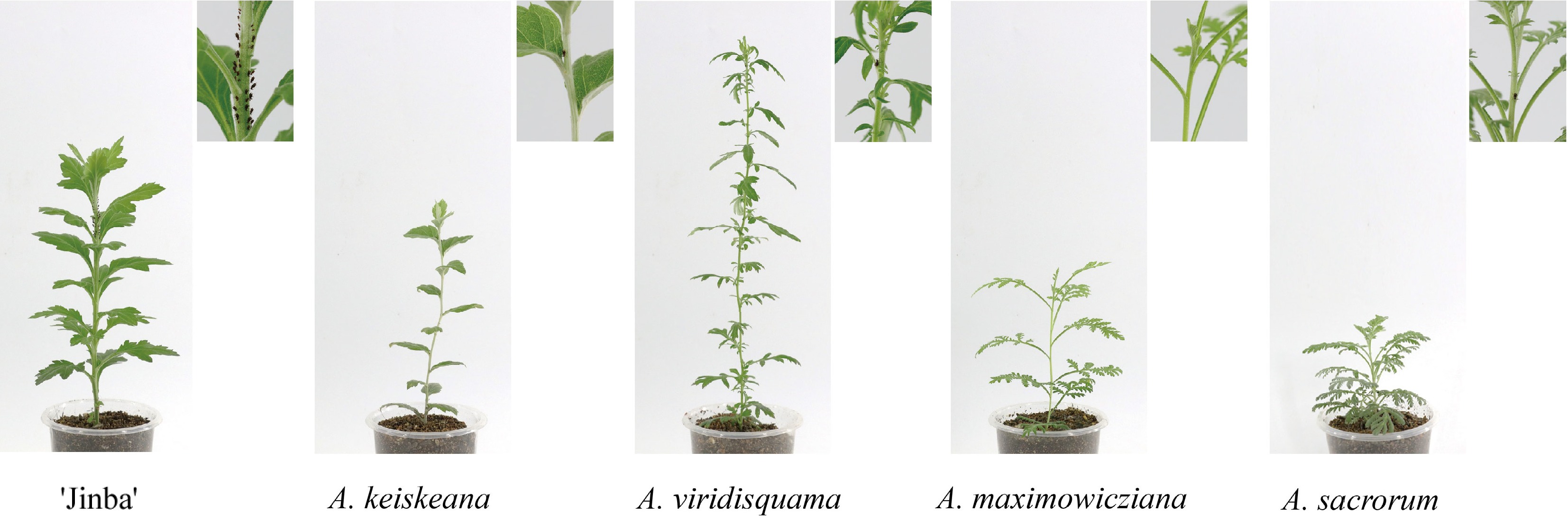
      Figure 1. 

      Aphid density on C. morifolium 'jinba' and four Artemisia species at 5 d after aphid inoculation.

    • To determine the repellent effects of Artemisia spp. volatiles on aphids, dual-choice assays were performed using a Y-tube olfactometer. Each Artemisia species coupled with C. morifolium 'Jinba' as a pair of odor sources in the assay. Compared to C. morifolium 'Jinba', all tested Artemisia species elicited obvious avoidance responses to aphids (Fig. 2). In particular, A. sacrorum has the strongest repellent effect on aphids, followed by A. viridisquama.
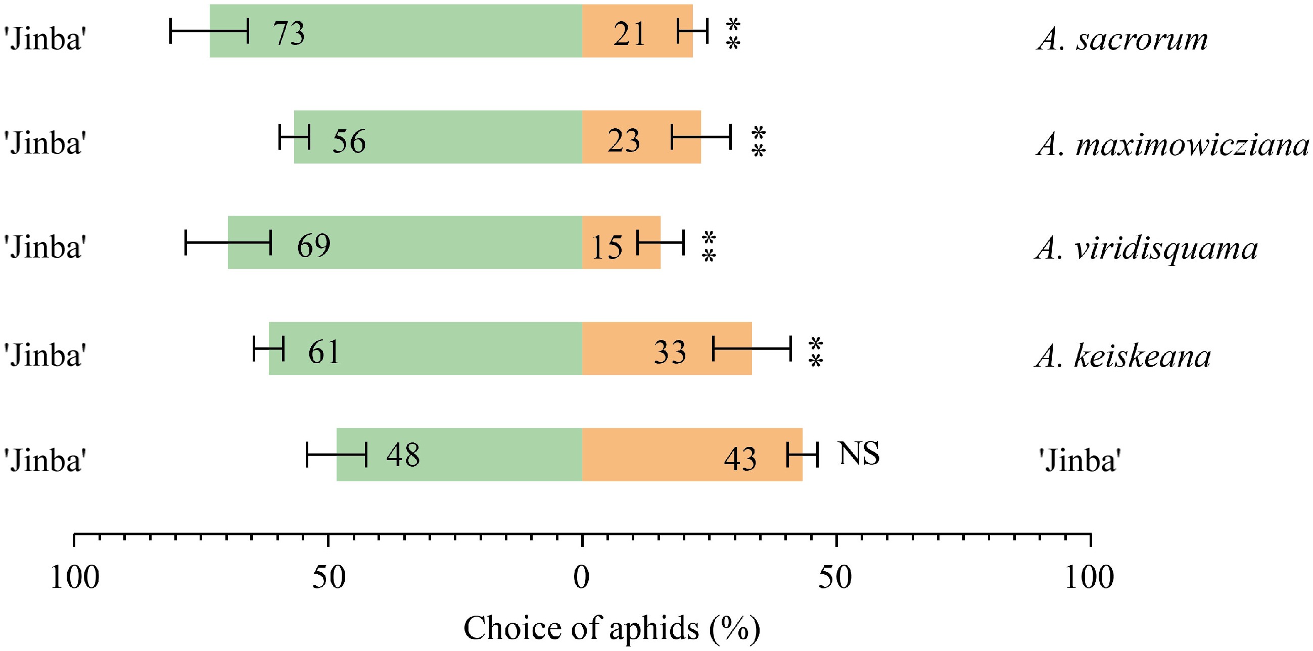
      Figure 2. 

      Choices of aphid to Artemisia species and C. morifolium 'Jinba' in the Y-tube olfactometer. The asterisks with the choice bars indicate significant preference. **p < 0.01.

    • A. alternata, C. siamense, and Phoma sp. commonly infect aerial tissues including the leaf and stem of chrysanthemum. Therefore, to track antifungal agents of aerial tissues of Artemisia spp., leaf and stem extracts were prepared and tested for antifungal activity against the three fungi.

      For inhibition activity assay of the extracts on A. alternata, leaf extracts of A. keiskeana and stem extracts of A. keiskeana, A. viridisquama, and A. maximowicziana strongly inhibited the growth of A. alternata with inhibition rates of 71.35%, 71.93%, 74.85%, and 75.44%, respectively. In comparison, A. sacrorum showed the weakest inhibition, with rates of 29.24% (Table 2; Fig. 3a).

      Table 2.  Inhibitory effect of volatiles from leaves and stems of four kinds of Artemisia on the growth of A. alternata, C. siamense, and Phoma sp.

      Plants Tissue Inhibition rate (IR)
      A. alternata C. siamense Phoma sp.
      A. keiskeana L 71.35 ± 1.17 57.14 ± 6.08 40.00 ± 2.40
      S 71.93 ± 1.50 35.71 ± 3.02 19.51 ± 3.72
      A. viridisquama L 33.00 ± 6.43 25.82 ± 1.92 12.20 ± 2.18
      S 74.85 ± 3.31 42.86 ± 6.87 20.00 ± 1.86
      A. maximowicziana L 53.80 ± 3.80 54.95 ± 2.91 61.46 ± 2.84
      S 75.44 ± 3.31 51.65 ± 7.14 79.51 ± 2.56
      A. sacrorum L 29.24 ± 3.80 19.78 ± 7.93 13.66 ± 3.28
      S 29.24 ± 2.34 38.00 ± 4.12 15.12 ± 1.86
      Values were given as mean ± SD. L, leaf; S, stem.
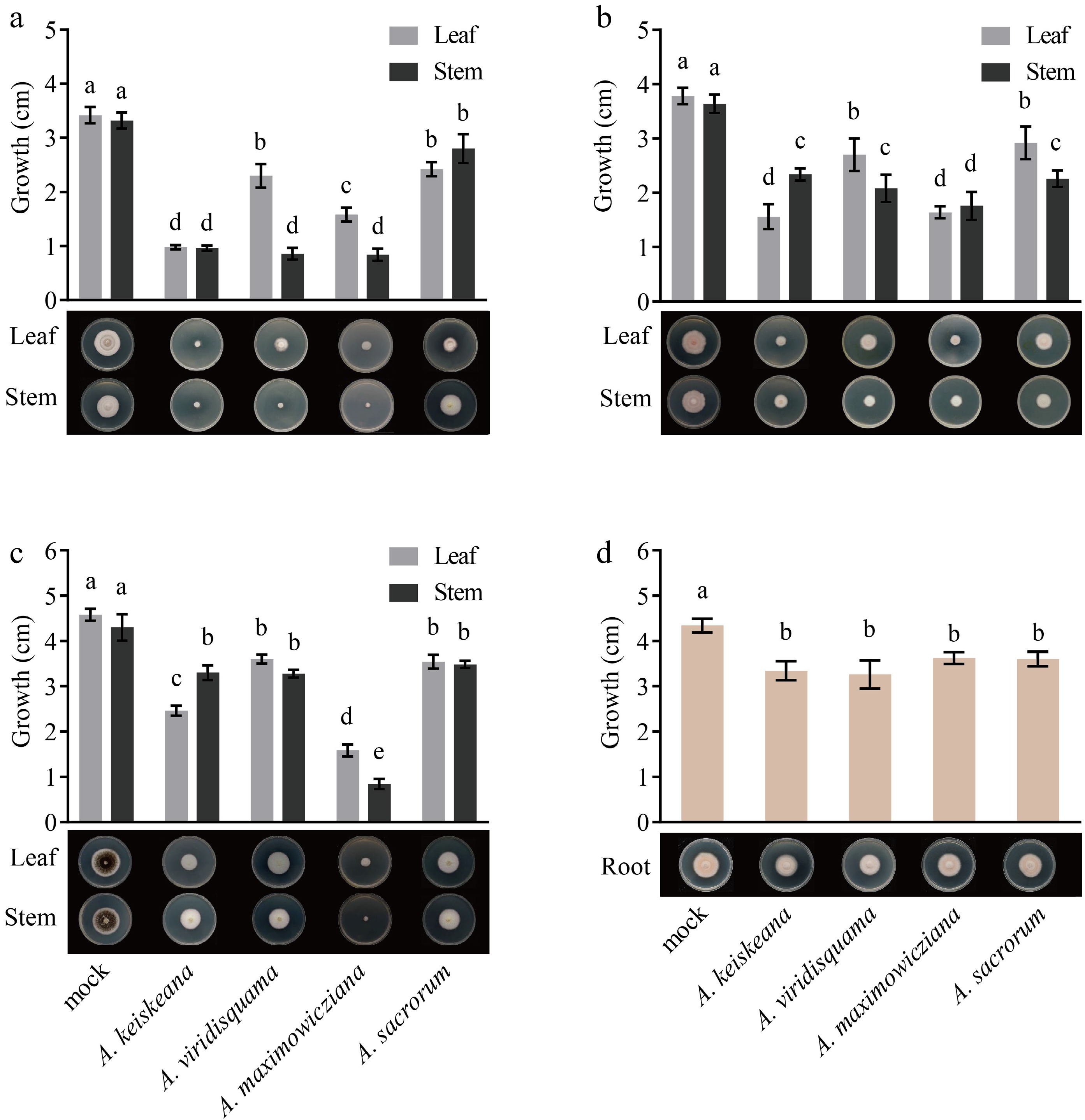
      Figure 3. 

      Inhibitory effect of different tissue extracts from four Artemisia plants on mycelia growth. (a), (b) and (c) colony diameter (cm) of A. alternata, C. siamense, and Phoma sp. after treatment with leaf and stem extracts for 4 d, respectively; (d) growth (cm) of F. solani after treatment with root extracts for 4 d. Different letters represent significant differences (p < 0.05).

      For inhibition activity assay of the extracts on C. siamense, the best inhibitory activity against C. siamense was found in leaf extracts of A. keiskeana and leaf and stem extracts of A. maximowicziana (57.14%, 54.95%, and 51.65%, respectively), followed by stem extracts of A. viridisquama, A. sacrorum, and A. keiskeana, with inhibitory rates of 42.86%, 38%, and 35.71%, respectively (Table 2; Fig. 3b).

      For inhibition activity assay of the extracts on Phoma sp., leaf and stem extracts of A. maximowicziana showed superior inhibition, and the inhibitory rate was 61.46% and 79.51%, respectively. Stem extracts of A. keiskeana showed moderate inhibition with an inhibition rate of 40%. The rest had slight inhibition on Phoma sp. with rates below 20% (Table 2; Fig. 3c).

    • F. solani is the primary pathogen causing chrysanthemum root rot[18,19]. The antimicrobial activity of root volatiles of Artemisia spp. was tested against the mycelial growth of F. solani. The result indicated that these root extracts showed an inhibitory effect. Among them, the inhibitory activity of root extracts from A. keiskeana, A. viridisquama, A. maximowicziana, and A. sacrorum was 23.04%, 24.88%, 16.59% and 17.05%, respectively (Table 3; Fig. 3d).

      Table 3.  Inhibitory effect of volatiles from roots of four kinds of Artemisia on the growth of F. solani.

      Plants Tissue Inhibition rate (IR)
      A. keiskeana R 23.04 ± 4.84
      A. viridisquama R 24.88 ± 7.14
      A. maximowicziana R 16.59 ± 2.99
      A. sacrorum R 17.05 ± 3.68
      Values were given as mean ± SD. R, root.
    • To further investigate the potential bioactive components that confer resistance against aphids and pathogens, root, leaf, and stem extracts from Artemisia spp. were analyzed by GC-MS. Constituents were identified by matching mass spectra to the NIST library. Volatile content was quantified relative to an internal standard (Supplemental Tables S1S4).

      Nineteen compounds were identified in A. keiskeana (Supplemental Table S1). (−)-Alpha-pinene and camphene occurred across all tissues, with the highest levels in leaves followed by that in stems. The major leaf volatiles were (+)-2-bornanone (126.3 μg·g−1 FW), caryophyllene (135.6 μg·g−1 FW), and phytyl acetate (205.6 μg·g−1 FW). (−)-Beta-elemene (109.2 μg·g−1 FW) and aromadendrene (101.4 μg·g−1 FW) predominated in the roots (Supplemental Table S1).

      Fifteen volatiles were found in A. viridisquama (Supplemental Table S2). Phytyl acetate (221.0 μg·g−1 FW), eucalyptol (47.1 μg·g−1 FW) and neophytadiene (67.1 μg·g−1 FW) were most abundant.

      Twenty-five compounds were detected in A. maximowicziana, including six bioactive oxidized monoterpenes like (E)-thujone and (−)-thujol (Supplemental Table S3). (E)-thujone and (−)-thujol were the main components in leaf and stem extracts, the content of which in leaves was up to 174.4 μg·g−1 FW and 498.7 μg·g−1 FW, respectively.

      A. sacrorum contained 26 volatile compounds (Supplemental Table S4), dominated by monoterpenes, sesquiterpenes, and diterpenes. (+)-2-Bornanone (193.3 μg·g−1 FW) and (−)-zingiberene (131.2 μg·g−1 FW) predominated in leaves.

      Overall, 43 volatiles were detected across all Artemisia species (Fig. 4), and terpenoids were the predominant volatiles across all species and tissues. Camphene, gamma-elemene, phytyl acetate, and caryophyllene occurred in all species. Especially certain oxygenated monoterpenes (cis-4-thujanol, (E)-thujone, (−)-thujol, etc.) were unique to A. maximowicziana. Some sesquiterpenes, like (−)-beta-elemene, are only found in the root extracts of A. keiskeana. However, which components of these extracts played a vital role in the antifungal activity remains to be further analyzed.
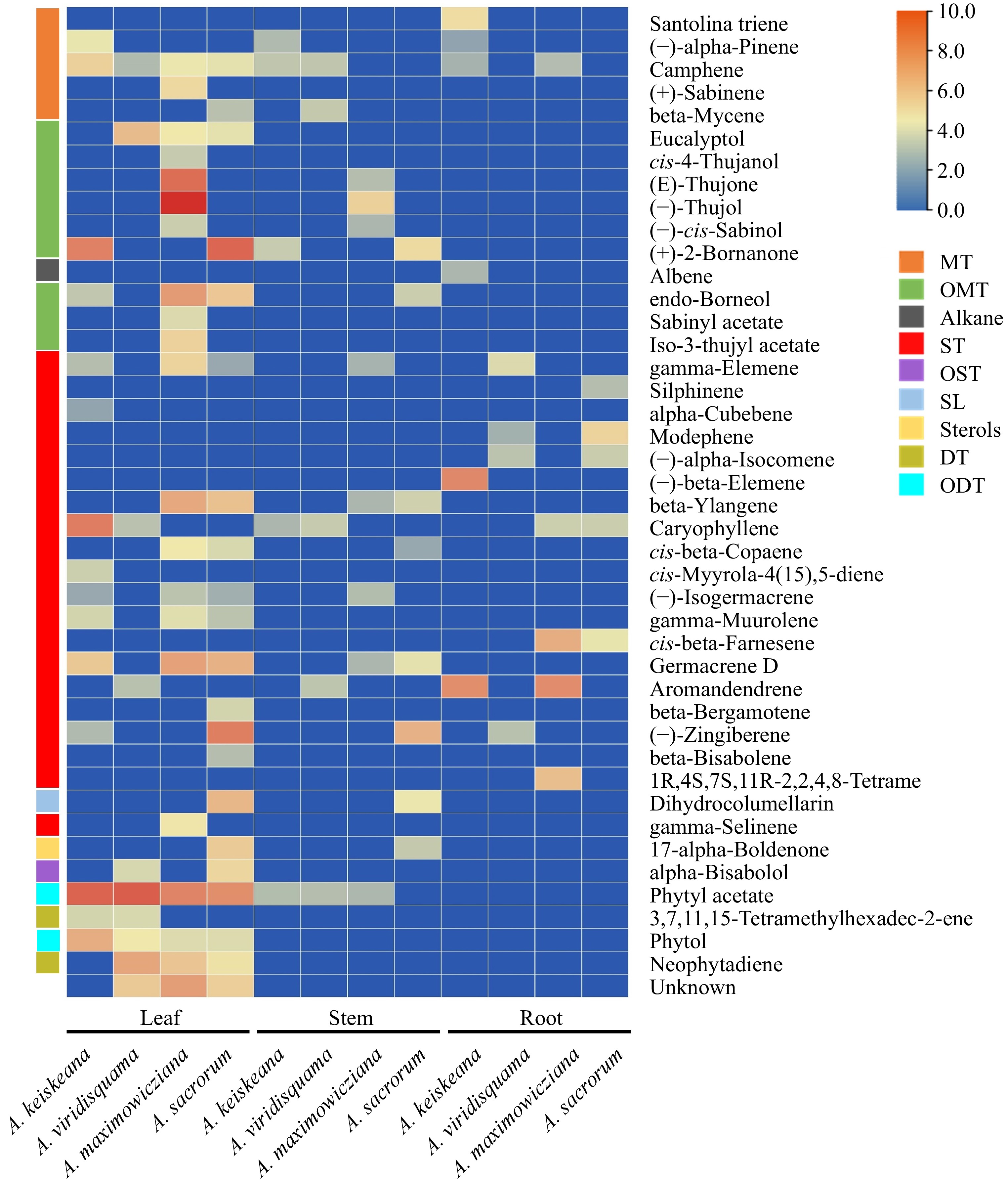
      Figure 4. 

      Main components (above 1% of total volatiles present in chromatograms) of ethyl acetate extracts from the leaf, stem and root of four Artemisia species. Colors reflect the VOC's average relative content, n = 3. MT: monoterpenes; OMT: oxygenated monoterpenes; ST: sesquiterpenes; OST: oxygenated sesquiterpenes; SL: sesquiterpene lactones; DT: diterpenes; ODT: oxygenated diterpenes.

    • The contribution of each volatile compound of extracts to antimicrobial activity was assessed by Orthogonal Partial Least Squares Discriminant Analysis (OPLS-DA) based on the variable's importance in the project (VIP). VIP values were used to describe the overall contribution of each variable to the model. VIP ≥ 1 commonly used as the screening criterion showed that the variable has a significant influence on the model. Among the 35 components in stem and leaf extracts from four Artemisia species, 12 compounds including (−)-thujol, (+)-2-bornanone, phytyl acetate, (−)-zingiberene, (E)-thujone, unknown (unidentified compound), germacrene D, caryophyllene, endo-borneol, beta-ylangene, dihydrocolumellarin and neophytadiene, the VIP values of which were greater than 1, inhibited the growth of A. alternata hypha (Fig. 5a). Ten components-(−)-thujol, (E)-thujone, phytyl acetate, (+)-2-bornanone, endo-borneol, unknow (unidentified compound), germacrene D, caryophyllene, (−)-zingiberene and beta-ylangene contributed to the inhibition of C. siamense mycelial growth (Fig. 5b). For Phoma sp., the mycelial growth was inhibited by 12 key active compounds including (−)-thujol, phytyl acetate, (+)-2-bornanone, caryophyllene, (E)-thujone, (−)-zingiberene, germacrene D, neophytadiene, unknown (unidentified compound), endo-borneol, phytol, and beta-ylangene (Fig. 5c). The primary antifungal agents in the extracts varied among different pathogens, with (−)-thujol exhibiting the most potent influence. Regarding soil-borne pathogenic fungi F. solani, (−)-beta-elemene, aromandendrene, cis-beta-farnesene, 1R,4S,7S,11R-2,2,4,8-tetrame, santolina triene, modephene and caryophyllene from root extracts were effective constituents that could inhibit the growth of the hypha of F. solani, of which (−)-beta-elemene showed the greatest inhibitory effect on F. solani (Fig. 5d).
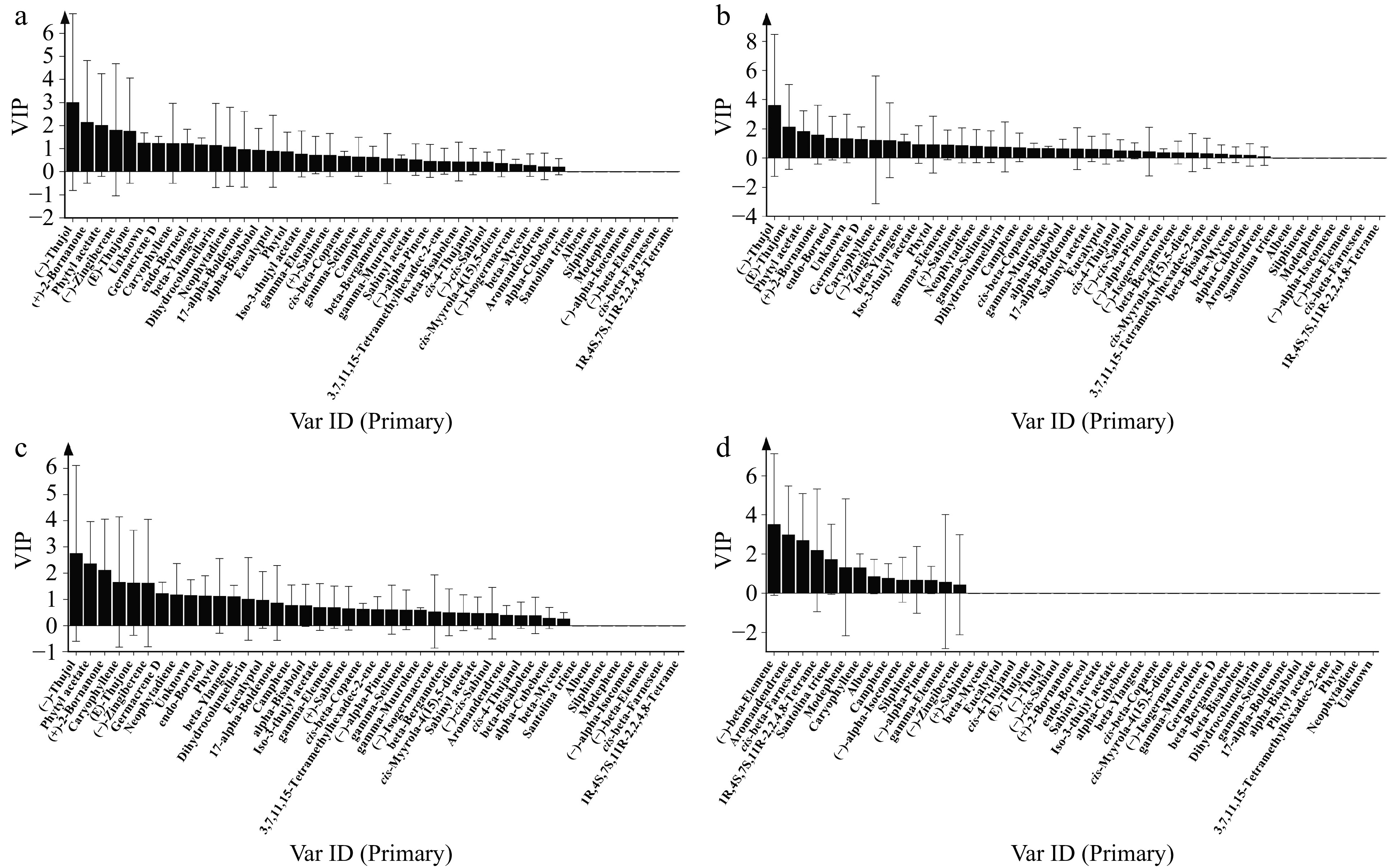
      Figure 5. 

      Screening of key bioactive components of extracts with antifungal effect against (a) A. alternata, (b) C. siamense, (c) Phoma sp. and (d) F. solani by OPLS-DA. VIP ≥ 1 was used as the screening criterion.

    • Artemisia species are of great value due to their abundant bioactive compounds with pharmaceutical and pesticide applications. They also serve as parent materials for intergeneric hybrids with crops like chrysanthemum. Here, the aphid (M. sanborni) resistance of four Artemisia species and the antimicrobial properties of their extracts were investigated, to screen out excellent germplasm materials and active compounds.

      Plant volatiles defend against aphids in direct and indirect ways, the former usually by direct contact or fumigation to repel and keep them from feeding, and even be toxic[20,21]. The essential oils of Artemisia monosperma were the most potent toxicants to Aphis nerii, displaying an LC50 value of 0.06 mg·L−1[22]. Sesquiterpenes from Solanum habrochaites have been verified to not only have a direct avoidance or anti-feeding effect on potato aphids but also affect their longevity and fecundity[23,24]. In this study, the four Artemisia species tested contained abundant bioactive terpenoids like caryophyllene, beta-farnesene, beta-bergamotene, beta-bisabolene, cis-4-thujanol, etc., known to deter aphids[23,25,26], likely contributing to the observed antifeeding and repellent effects. It suggests that all four Artemisia species are promising aphid-resistant germplasm with prospective bioactive volatiles.

      The antifungal activity of Artemisia extracts depends on the extraction solvent, plant part, and pathogen species. Antifungal effects of A. annua leaf methanol extracts on F. oxysporum and F. solani increased with concentration, whereas root and stem extracts showed no inhibition[7]. Artemisia proceriformis oil more strongly inhibited Septoria glycines and Septoria tritici (MIC100 = 2.7 mg·mL−1) versus Fusarium graminearum and Fusarium verticillioides (MIC100 = 10.6 mg·mL−1)[27]. Here, differences in the antifungal activity of Artemisia extracts were also found. A. keiskeana leaf and stem extracts inhibited A. alternata over 70% but had minimal effects on Phoma sp. (Table 2). The inhibitory effect of stem extract from A. viridisquama on A. alternata was significantly higher than that of leaf extract. Moreover, we also noticed that A. maximowicziana extracts strongly inhibited all three aerial pathogens by over 50%. Thus, it represents a promising broad-spectrum botanical fungicide candidate.

      GC-MS profiling of volatiles revealed that, as expected, terpenoids dominated the volatile profile, aligning with findings in other Artemisia species[1]. It mainly consisted of 1, 8-cineole, beta-pinene, thujone, artemisia ketone, camphor, caryophyllene, camphene, and germacrene D[4]. However, there were great differences in main compounds with some special species. For example, A. maximowicziana studied in this paper, whose major component was (−)-thujol, contained about three times as much as (E)-thujone. In addition, phytyl acetate, which was not mentioned in the previously reported Artemisia, was found to have a high content in all four tested Artemisia species.

      Different pests and pathogens have different susceptibility to varied active compounds. The essential oil and extracts of Artemisia absinthium have demonstrated anti-settling, antifeedant and antimicrobial activity against various pests and pathogens such as Myzus persicae, Spodoptera littoralis, Tetranychus cinnabarinus, Diaphorina citri, Fusarium spp. and Botrytis cinerea[2831]. Specifically, (−)-cis-chrysanthenol and linalool have been identified as the primary antifungal agents of extracts against Fusarium spp. and B. cinerea[29], while carvacrol, (−)-α-bisabolol and chamazulene were toxic constituents of volatile oils against D. citri[29,31]. In this study, the antifungal effects of (−)-thujol were potentially broad spectrum. For root pathogens, L-camphor, DL-camphor, β-caryophyllene, and camphene were reported to have antifungal efficacy against F. oxysporum and F. solani comparable to the synthetic fungicides flutriafol, and hymexazol[7]. Here, however, the VIP value of camphene was less than 1; probably, its content in the root was too low to be effective for the resistance. Based on previous reports and our findings, the four Artemisia plants contained 15 bioactive constituents-(−)-thujol, (+)-2-bornanone, phytyl acetate, (−)-zingiberene, (E)-thujone, unknown (unidentified compound), germacrene D, caryophyllene, endo-borneol and beta-ylangene, camphene, cis-beta-farnesene, beta-bergamotene, beta-bisabolene, cis-4-thujanol—may mediate the antifungal and anti-insect effects. However, the active ingredients individually and in combinations against aphids and the tested pathogens should be further studied.

    • In conclusion, the four Artemisia species tested showed certain repellent and antifeedant effects on aphids, and antifungal effects on A. alternata, C. siamense, Phoma sp., and F. solani. Their volatile extracts contain multiple bioactive compounds with the potential to be biofungicides. These findings provide a material and theoretical basis for developing new biopesticides and breeding of pest- and disease-resistant chrysanthemum cultivars.

    • The authors confirm contribution to the paper as follows: study conception and design: Chen S, Chen F; data collection: Yang M, Li M; analysis and interpretation of results: Yang M, Li M; draft manuscript preparation: Yang M. All authors reviewed the results and approved the final version of the manuscript.

    • All data generated or analyzed during this study are included in this published article and its supplementary information files.

      • This work was supported by the National Natural Science Foundation of China (32372749), Hainan Provincial Natural Science Foundation of China (323CXTD386), the Program for Key Research and Development, Jiangsu, China (BE2023367, BE2022417), A Project Funded by the Priority Academic Program Development of Jiangsu Higher Education Institutions, PAPD. We thank Dr. Yuehua Ma (Central Laboratory of College of Horticulture, Nanjing Agricultural University) for assistance in using GC-MS (Agilent 9000 B-7000D).

      • The authors declare that they have no conflict of interest.

      • Copyright: © 2024 by the author(s). Published by Maximum Academic Press, Fayetteville, GA. This article is an open access article distributed under Creative Commons Attribution License (CC BY 4.0), visit https://creativecommons.org/licenses/by/4.0/.
    Figure (5)  Table (3) References (31)
  • About this article
    Cite this article
    Yang M, Li M, Chen F, Chen S. 2024. Bioactive components and antimicrobial potential of extracts from Artemisia species and their repellent activities against Aphid (Macrosiphoniella sanborni). Ornamental Plant Research 4: e025 doi: 10.48130/opr-0024-0021
    Yang M, Li M, Chen F, Chen S. 2024. Bioactive components and antimicrobial potential of extracts from Artemisia species and their repellent activities against Aphid (Macrosiphoniella sanborni). Ornamental Plant Research 4: e025 doi: 10.48130/opr-0024-0021

Catalog

  • About this article

/

DownLoad:  Full-Size Img  PowerPoint
Return
Return