ARTICLE   Open Access    

Paternal miRNA biogenesis contributes to seed development in Arabidopsis

More Information
  • Received: 25 July 2024
    Revised: 25 September 2024
    Accepted: 21 October 2024
    Published online: 30 October 2024
    Seed Biology  3 Article number: e017 (2024)  |  Cite this article
  • miRNAs are key regulators of gene expression and play important roles in various developmental processes. The development of the plant male gametophyte begins with a microspore, which undergoes two rounds of pollen mitosis to produce mature pollen, consisting of a vegetative nucleus and two sperm cells. Although many miRNAs are known to accumulate in mature pollen, it remains unclear how miRNA biogenesis is regulated during pollen mitosis and whether miRNA biogenesis in mature pollen is necessary for seed development. Here, we focus on DCL1, the major enzyme for miRNA biogenesis in Arabidopsis. Hand-pollination using dcl1-7 mutant pollen results in severe seed development defects, characterized by shortened siliques and approximately 80% aborted seeds. While miRNA genes are primarily transcribed at early stages of pollen development, the core factors of the miRNA biogenesis machinery are expressed throughout pollen development, with preferential expression in the vegetative nucleus. Using an artificial miRNA strategy to conditionally knock down DCL1 in the vegetative cell and sperm cells, respectively, we demonstrate that miRNA biogenesis in both cell types contributes to fertility control, but results in distinct defects in seed development. Collectively, these results show that miRNA biogenesis in mature pollen plays a significant role in regulating fertility and seed development.
  • Wheat stands as one of the world’s most crucial staple food crops, furnishing 20% of the global population’s calorie intake and holding a pivotal role in ensuring food security worldwide. Wheat yield is mainly determined by three factors: thousand grain weight (TGW), spike number per unit area, and grain number per spike[1]. The optimization of these three components is of great significance for improving yield. Among these, increasing the grain weight emerges as a particularly significant avenue for boosting wheat productivity. Traits shaping grain morphology, including grain length, grain width, and grain thickness, directly affect grain size, which in turn affects grain weight.

    The development of seeds significantly influences grain weight. The mature seeds of angiosperms are composed of the embryo, endosperm, and seed coat. The maternal and zygotic tissues jointly participate in the regulation of the growth and development of seeds as well as control of the synergistic growth of embryo, endosperm, and seed coat. As grain development advances, seed coat cells perpetually undergo division and expansion, accompanied by continuous carbohydrate accumulation in the endosperm[2]. Genes pertinent to transport, carbohydrate metabolism, and starch synthesis also become active during development. Starch is the main storage component of the wheat endosperm, and its content is a key regulator of grain weight. In addition, plant hormone contents exhibit significant changes during seed development, and genes related to metabolism participate extensively in seed development.

    In this review, we summarize recent research on key regulators of wheat grain weight including transcriptional regulatory factors, post-translation modification factors, the G-protein signaling pathway, and phytohormone signalings to understand the regulatory mechanisms of wheat grain weight (Fig. 1, Table 1).

    Figure 1.  Regulatory networks involved in grain weight in wheat. Several genes involved in transcriptional regulatory factors, post-translation modification factors, the G-protein signaling pathway, and phytohormone signalings participate in wheat grain weight regulation. Broken lines indicate inhibitory regulation. Arrowheads represent positive regulation. Elliptical overlaps represent interactions.
    Table 1.  Genes involved in wheat grain weight regulation.
    Protein nameGene IDProtein categoryPositive(+)/negative(−)
    regulator
    Elite haplotype for
    high grain weight
    Reference
    Starch synthesis-related genes reported to be involved in wheat grain weight
    TaCwi-A1TraesCS2A03G0736600Cell wall invertase+TaCwi-A1a[3]
    TaCWI-5DTraesCS5D03G1216700Cell wall invertase+Hap-5D-C[4]
    TaSUT1-ATraesCS4A03G0027400Sucrose transporters+TaSUT1 in Kauz[5]
    TaSUT1-BTraesCS4B03G0758500Sucrose transporters+TaSUT1 in Kauz[5]
    TaSUT1-DTraesCS4D03G0679400Sucrose transporters+TaSUT1 in Kauz[5]
    TaSus1-ATraesCS7A03G0375000Sucrose synthase+TaSus1-7A-Hap-1[6, 7]
    TaSus1-BTraesCS7B03G0171900Sucrose synthase+TaSus1-7B-Hap-T[6, 7]
    TaSus1-DTraesCS7D03G0358200Sucrose synthase+[6, 7]
    TaSus2-ATraesCS2A03G0349200Sucrose synthase+TaSus2-2A-Hap-A[6, 8]
    TaSus2-BTraesCS2B03G0468900Sucrose synthase+TaSus2-2B-Hap-H[6, 8]
    TaSus2-DTraesCS2D03G0366700Sucrose synthase+[6, 8]
    TaBT1-ATraesCS6A03G0433200Sucrose transporter+[9]
    TaBT1-BTraesCS6B03G0559700Sucrose transporter+Hap1 and Hap2[9]
    TaBT1-DTraesCS6D03G0376900Sucrose transporter+[9]
    TaAGPL1-BTraesCS1B03G1206000Large subunit gene of the AGPase+TaAGP-L-1B-Hap-I[10, 11]
    TaAGPS-1TraesCS7A03G0682600Small subunit gene of the AGPase+TaAGP-S1-7A-Hap-I[10]
    TaSBEIII-ATraesCS7A03G0826800Starch-branching enzyme+Allele-T[12]
    TaSSIV-ATraesCS1A03G0866200Starch synthases+Hap-2-1A[13, 14]
    TaSSIV-BTraesCS1B03G1004700Starch synthases+Hap-3-1B[13, 14]
    TaSSIV-DTraesCS1D03G0838700Starch synthases+[13, 14]
    GWD-ATraesCS6A03G0662800Glucan, water dikinase[15]
    GWD-BTraesCS6B03G0813900Glucan, water dikinase[15]
    GWD-DTraesCS6D03G0552200Glucan, water dikinase[15]
    Transcriptional regulatory factors
    TaNAC019-ATraesCS3A03G0172000NAC transcription factor+[16]
    TaNAC019-BTraesCS3B03G0216600NAC transcription factor+TaNAC019-BI[16]
    TaNAC019-DTraesCS3D03G0154500NAC transcription factor+[16]
    TaNAC100-ATraesCS2A03G0808100NAC transcription factor+TaNAC100-2A-H1[17]
    TaNAC100-BTraesCS2B03G0891700NAC transcription factor+[17]
    TaNAC100-DTraesCS2D03G0746900NAC transcription factor+[17]
    TaPGS1TraesCS1D03G0219000bHLH transcription factor+[18]
    TaPGS1TraesCS1D03G0219700bHLH transcription factor+[18]
    TaFI3TraesCS3A03G1169900PLATZ transcription factor+[18]
    TaGSNETraesCS5B03G0668000WRKY transcription factor+TaGSNE-Hap-2[19]
    TaHDZ-A34TraesCS7A03G0760400HD-Zip transcription factor+Hap-ABD[20]
    TaHDZ-B34TraesCS7B03G0590000HD-Zip transcription factor+Hap-ABD[20]
    TaHDZ-D34TraesCS7D03G0729900HD-Zip transcription factor+Hap-ABD[20]
    TaGW8-B1TraesCS7B03G0430500SPL transcription factor+ TaGW8-B1a[21]
    TaSPL14-ATraesCS5A03G0658100SPL transcription factor+[22]
    TaSPL14-BTraesCS5B03G0692900SPL transcription factor+[22]
    TaSPL14-DTraesCS5D03G0627900SPL transcription factor+[22]
    TaSPL14-7ATraesCS7A03G0567100SPL transcription factor+TaSPL14-7A-Hap1/2[23]
    TaSPL14-7BTraesCS7B03G0393600SPL transcription factor+[23]
    TaSPL14-7DTraesCS7D03G0548900SPL transcription factor+[23]
    Post-Translational Modifications (PTMs)
    Ubiquitin–proteasome pathway
    TaGW2-6ATraesCS6A03G0480200RING-type E3 ubiquitin ligaseHap-A[24, 25]
    TaGW2-6BTraesCS6B03G0578500RING-type E3 ubiquitin ligaseTaGW2-B-HapI/II[24, 25]
    TaGW2-6DTraesCS6D03G0404800RING-type E3 ubiquitin ligase[24, 25]
    TaDA1-ATraesCSU03G0004100LCUbiquitin receptorTaDA1-A-HapI[26]
    TaDA1-BTraesCS2B03G0048000Ubiquitin receptor[26]
    TaDA1-DTraesCS2D03G0031900Ubiquitin receptor[26]
    TaSDIR1-4ATraesCS4A03G0197400RING-type E3 ubiquitin ligaseTaSDIR1-4A-2[27]
    TaPUB1-ATraesCS5A03G1197700U-box E3 ligase+[28]
    TaPUB1-BTraesCS4B03G0885300U-box E3 ligase+[28]
    TaPUB1-DTraesCS4D03G0783100U-box E3 ligase+[28]
    ZnF-ATraesCS4A03G0701600RING-type E3 ubiquitin ligase+[29]
    ZnF-BTraesCS4B03G0092600RING-type E3 ubiquitin ligase+[29]
    ZnF-DTraesCS4D03G0066800RING-type E3 ubiquitin ligase+[29]
    SnRK and phosphatase pathways
    TaSnRK2.3-ATraesCS1A03G0569000Sucrose non-fermenting 1 (SNF1)-related protein kinaseHap-1A-1[30]
    TaSnRK2.3-BTraesCS1B03G0660500Sucrose non-fermenting 1 (SNF1)-related protein kinaseHap-1B-1[30]
    TaSnRK2.9-ATraesCS5A03G0177100Sucrose non-fermenting 1 (SNF1)-related protein kinase Hap-5A-1/2[31]
    TaSnRK2.9-BTraesCS5B03G0188000Sucrose non-fermenting 1 (SNF1)-related protein kinase[31]
    TaSnRK2.9-DTraesCS5D03G0195600Sucrose non-fermenting 1 (SNF1)-related protein kinase[31]
    TaSnRK2.10-ATraesCS4A03G0621500Sucrose non-fermenting 1 (SNF1)-related protein kinaseHap-4A-H[32]
    TaSnRK2.10-BTraesCS4B03G0179500Sucrose non-fermenting 1 (SNF1)-related protein kinase[32]
    TaSnRK2.10-DTraesCS4D03G0149100Sucrose non-fermenting 1 (SNF1)-related protein kinase[32]
    TaPSTOLTraesCS5A03G0115500LCPhosphate Starvation Tolerance 1+[33]
    TaGL3-5ATraesCS5A03G0897900PPKL family—Ser/Thr phosphatase+TaGL3-5A-G[1]
    TaGL3-5BTraesCS5B03G0943200PPKL family—Ser/Thr phosphatase+[1]
    TaGL3-5DTraesCS5D03G0859400PPKL family—Ser/Thr phosphatase+[1]
    TaGL3.3-ATraesCS5A03G0073900PPKL family—Ser/Thr phosphatase+[34]
    TaGL3.3-BTraesCS5B03G0068000PPKL family—Ser/Thr phosphatase+TaGL3.3-5B-C[34]
    TaGL3.3-DTraesCS5D03G0098300PPKL family—Ser/Thr phosphatase+[34]
    TaTPP-7ATraesCS7A03G0422300Trehalose-6-phosphate phosphatase+TaTPP-7A-HapI[35]
    TaTPP-7BTraesCS7B03G0228800Trehalose-6-phosphate phosphatase+[35]
    TaTPP-7DTraesCS7D03G0410500Trehalose-6-phosphate phosphatase+[35]
    Asparagine N-glycosylation pathway
    TaSTT3b-2ATraesCS2A03G1282700Catalytic subunit of oligosaccharyltransferase+[36]
    TaSTT3b-2BTraesCS2B03G1473200Catalytic subunit of oligosaccharyltransferase+[36]
    TaSTT3b-2DTraesCS2D03G1245300Catalytic subunit of oligosaccharyltransferase+[36]
    G-protein signaling pathway
    TaGS3-4ATraesCS4A03G1194500Gγ subunit[37, 38]
    TaGS3-7ATraesCS7A03G0037700Gγ subunit[37, 38]
    TaGS3-7DTraesCS7D03G0033100Gγ subunit[37, 38]
    TaDEP1-ATraesCS5A03G0545300Gγ subunit+TaDEP1-Hap1[39]
    TaDEP1-BTraesCS5B03G0555000Gγ subunit+[39]
    TaDEP1-DTraesCS5D03G0509000Gγ subunit+[39]
    Phytohormone signalings
    CK
    TaCKX2TraesCS3A03G0298200Cytokinin oxidase/dehydrogenase (CKX) enzymes+TaCKX2A-2[40]
    TaCKX4TraesCS3A03G1128900Cytokinin oxidase/dehydrogenase (CKX) enzymes+TaCKX4A-2[40]
    TaCKX5TraesCS3A03G0763900Cytokinin oxidase/dehydrogenase (CKX) enzymes+TaCKX5A-3[40]
    TaCKX9TraesCS1A03G0609600Cytokinin oxidase/dehydrogenase (CKX) enzymes+TaCKX9A-2[40]
    TaCKX6a02TraesCS3D03G0306000Cytokinin oxidase/dehydrogenase (CKX) enzymes+TaCKX6a02-D1a[41]
    TaCKX6-D1TraesCS3D03G0305400Cytokinin oxidase/dehydrogenase (CKX) enzymesTaCKX6-D1-a[42]
    GA
    TaGASR7-ATraesCS7A03G0485700Gibberellin-regulated proteinH1c[43, 44]
    TaGASR7-BTraesCS7B02G115300Gibberellin-regulated protein[43, 44]
    TaGASR7-DTraesCS7D02G210500Gibberellin-regulated protein[43, 44]
    Auxin
    TaTGW-7ATraesCS7A03G0542800Involved in the tryptophan biosynthetic pathway+TaTGW-7Aa[45]
    TaTGW-7BTraesCS7B03G0358400Involved in the tryptophan biosynthetic pathway+[45]
    TaTGW-7DTraesCS7D03G0520200Involved in the tryptophan biosynthetic pathway+[45]
    TaTGW6TraesCS7D03G0173900IAA–glucose (IAA-Glc) hydrolase activity+[46]
    TaIAA21-ATraesCS7A03G0816300Auxin/indole acetic acid repressorHap2, Hap3, Hap5
    TaIAA21-BTraesCS7B03G0674700Auxin/indole acetic acid repressor
    TaIAA21-DTraesCS7D03G0801000Auxin/indole acetic acid repressor
    TaARF25-ATraesCS5A03G0098100Auxin response factor (ARF) protein+
    TaARF25-BTraesCS5B03G0104300Auxin response factor (ARF) protein+
    TaARF25-DTraesCS5D03G0114800Auxin response factor (ARF) protein+
    BR
    TaD11-2ATraesCS2A03G0818100BR biosynthesis enzymes+TaD11-2A-HapI[47]
    TaD11-2BTraesCS2B03G0904700BR biosynthesis enzymes+[47]
    TaD11-2DTraesCS2D03G0759600BR biosynthesis enzymes+[47]
    Tasg-D1TraesCS3D03G0288900STKc_GSK3 Kinase[48]
    ABA
    TaPYL1-1ATraesCS1A03G0514200Abscisic acid (ABA) receptor PYL+[49]
    TaPYL1-1BTraesCS1B03G0603200Abscisic acid (ABA) receptor PYL+TaPYL1-1BIn-442[49]
    TaPYL1-1DTraesCS1D03G0499200Abscisic acid (ABA) receptor PYL+[49]
    TaMYB70-ATraesCS5A03G0432900MYB transcription factor+[49]
    TaMYB70-BTraesCS5B03G0428700MYB transcription factor+[49]
    TaMYB70-DTraesCS5D03G0401500MYB transcription factor+[49]
    TaABI5-ATraesCS3A03G0880400Basic/region leucine zipper transcription factor[28]
    TaABI5-BTraesCS3B03G1006600Basic/region leucine zipper transcription factor[28]
    TaABI5-DTraesCS3D03G0808000Basic/region leucine zipper transcription factor[28]
    JA
    KAT-2BTraesCS6B03G1211100Keto-acyl thiolase 2B+[50]
    TaPAP6-ATraesCS2A03G0298800Fibrillin family member+[51]
    TaPAP6-BTraesCS2B03G0419200Fibrillin family member+[51]
    TaPAP6-DTraesCS2D03G0317100Fibrillin family member+[51]
    TaGL1-B1TraesCS1B03G0239600Carotenoid isomerase gene+TaGL1-B1b[51]
    Other regulators
    TaCYP78A3-ATraesCS7A03G0630800Cytochrome P450(CYP) 78A3 protein+[52]
    TaCYP78A3-BTraesCS7B03G0455800Cytochrome P450(CYP) 78A3 protein+[52]
    TaCYP78A3-DTraesCS7D03G0611800Cytochrome P450(CYP) 78A3 protein+[52]
    TaGW7-ATraesCS2A03G0367000TONNEAU1-recruiting motif (TRM1) proteinH1a[53]
    TaGW7-BTraesCS2B03G0488200TONNEAU1-recruiting motif (TRM1) proteinH1b[53]
    TaGW7-DTraesCS2D03G0384600TONNEAU1-recruiting motif (TRM1) proteinH1d[53]
    TaFlo2-A1TraesCS2A03G1201700FLOURY ENDOSPERM2 (Flo2) gene+TaFlo2-A1b[54, 55]
    TaGS5-3ATraesCS3A03G0396700LCSerine carboxypeptidases+TaGS5-3A-T[56]
    TaMGD-ATraesCS6A03G0937800Monogalactosyl diacylglycerol+[57]
    TaMGD-BTraesCS6B03G1143600Monogalactosyl diacylglycerol+[57]
    TaMGD-DTraesCS6D03G0814200Monogalactosyl diacylglycerol+[57]
     | Show Table
    DownLoad: CSV

    Grain weight hinges on both grain size and endosperm constituents. Within monocot plants, the endosperm has a pivotal role in determining seed size and weight. This prominence arises because the endosperm occupies most of the volume of a mature grain. Consequently, endosperm components exert defining influences on grain weight. In general, among seeds of comparable size, those with higher oil contents have lower seed weights, and those with higher starch contents have higher seed weights. Notably, starch is an important component of wheat grains, accounting for approximately 70% of the dry weight[58]. Starch synthesis and accumulation are related to the development of wheat endosperm and contribute directly to grain weight[59]. Furthermore, the starch content within grains of the same variety exhibits a significantly positive correlation with grain size. The filling process and endosperm development also affect the accumulation, conversion, and starch synthesis of photosynthetic products. Several starch synthesis-related genes have important roles in controlling size and weight in wheat grains. These include the cell wall invertase genes TaCwi-A1 and TaCWI-5D, the sucrose transporter gene TaSUT1, sucrose synthase genes TaSus1 and TaSus2, ADP-Glc pyrophosphorylase genes TaAGPL1/ TaLSU1, BRITTLE1 (BT1), and TaBT16B, starch synthase TaSSIV, starch branching enzyme TaSBEIII-A, and Glucan, Water-Dikinase gene GWD; these genes play vital roles in starch accumulation and are all associated with TGW ( Table 1)[312,15].

    The division and elongation of seed coat cells affect the volume of the cavity wherein both the embryo and the endosperm develop, and they determine traits related to the final grain size, including grain length, width, and thickness. Several signaling pathways have been shown to control seed size by regulating the growth of maternal tissues in wheat. We summarize the grain weight regulatory pathways in wheat from the following aspects: transcriptional regulatory factors, post-translation modification factors, the G-protein signaling pathway, and phytohormone signalings.

    Transcription factors (TFs) are general regulators of functional genes that bind to specific motifs of target gene promoters, thereby activating or suppressing transcription. Numerous TFs have been identified as participants in the intricate orchestration of wheat grain weight.

    Notably, several SQUAMOSA PROMOTER-BINDING PROTEIN-LIKE (SPL) family TFs are associated with grain weight. OsSPL16 positively regulates grain weight by enhancing cell proliferation and grain filling in rice[60]. Its ortholog, TaSPL16, also known as TaGW8, is reported to have a similar function to OsSPL16 in wheat grain weight regulation and is regulated by miR156[21,53]. Correlation analysis between TaGW8-B1a, TaGW8-B1b alleles and agronomic traits showed that wheat cultivars with the allele TaGW8-B1a exhibit a significantly larger grain size and higher TGW compared to those with TaGW8-B1b, because TaGW8-B1b possesses a 276-bp indel in its first intron[21]. Knockdown lines of TaGW7, the ortholog of GRAIN WIDTH7 (OsGW7), showed increases in grain width and weight but reductions in grain length by regulating cell division and organ growth[53]. OsSPL16 directly interacts with the promoter of OsGW7, and represses OsGW7’s expression[61]. Therefore, it is possible that TaSPL16 could bind directly to the promoter of TaGW7 to regulate wheat grain weight. MiR156 cleaves TaSPL14 mRNA, with knockout lines exhibiting a reduced TGW[22]. Another SPL TF, TaSPL14-7A, has a similar function, and its elite alleles, TaSPL14-7A-Hap1/2, are significantly correlated with a higher TGW; expression levels are higher for TaSPL14-7A-Hap1/2 than for TaSPL14-7A-Hap3 and the locus underwent positive selection during global wheat breeding over the last century[23]. Given the conservation of SPL family TF binding motifs and miR156-regulated SPLs, the miR156-SPLs-TaGW7 pathway emerges as a potential regulator of wheat grain weight.

    NAC TFs belong to a plant-specific TF family. As one of the largest TF families, its members are widely involved in the regulation of many biological processes in plants, including stress responses, seed development, and nutrient accumulation. Recently, NAC TFs have been reported to participate in grain weight regulation. For example alterations in TaNAC019 and TaNAC100 could affect TaSus expression, thereby affecting grain starch content and grain size[16,17]. Remarkably, OsNAC20 and OsNAC26 in rice and ZmNAC128 and ZmNAC130 in maize have been recently reported to regulate starch synthesis-related genes to impact grain size and weight[62,63]. Notably, these NAC genes are specifically expressed in endosperm tissue, except for TaNAC100.

    Ectopic overexpression of the basic helix-loop-helix (bHLH) TF TaPGS1 (T. aestivum Positive Regulator of Grain Size 1) within the wheat endosperm yields increases carbohydrate and total protein levels, thereby increasing grain weight[18]. The plant AT-rich zinc-binding proteins (PLATZ), OsFI3 and ZmFI3, which are orthologs of TaFI3 in wheat, are associated with a high TGW, grain width, and grain length in rice and maize[18,64]. TaPGS1 regulates TaFI3 expression in wheat and the PGS1-Fl3 regulatory system is conserved in different cereals[18].

    Grain Size and Number Enhancer (TaGSNE) encodes a WRKY TF and has the highest expression in young roots at the flowering stage[19]. TaGSNE not only governs root length but also adeptly balances the trade-off between grain size and number in wheat[19]. Further, TaGSNE displays responsive behavior to abscisic acid (ABA) and environmental cues. As evaluated using a generalized linear model, the TaGSNE-Hap-2 allele exhibits a significant positive correlation with TGW in three environments[19]. TaGSNE is a candidate gene for breeding high-yield, abiotic-stress-resistant wheat varieties.

    The homeodomain-leucine zipper (HD-Zip) TF, TaHDZ34, plays an important role in modulating wheat TGW. TaHDZ34 can be classified into eight haplotype combinations: Hap-ABD, Hap-Abd, Hap-aBd, Hap-AbD, Hap-aBD, Hap-abD, Hap-ABd, and Hap-abd. A correlation analysis based on two populations (172 lines and162 lines) and eight haplotype combinations of TaHDZ34 showed that the Hap-ABD allele is associated with a higher TGW than those of the other seven haplotype combinations, revealing that it is a superior haplotype for wheat breeding[20]. The regulatory mechanism of TaHDZ34 warrants comprehensive exploration in future studies.

    Post-translation modifications (PTMs) constitute a cornerstone in plant development’s regulatory landscape, and are flexibly responsive to plant signals through protein ubiquitination, phosphorylation, glycosylation, and methylation. These modifications exert influence over gene expression and protein stabilization. Within this intricate framework, the OST pathway, and sucrose non-fermentation-1-related protein kinases (SnRKs) pathway, PPKL family Ser/Thr phosphatase protein phosphatases pathway, collectively contribute to grain weight regulation.

    The ubiquitin–proteasome pathway plays a critical role in seed development by ubiquitinating and degrading proteins. This ubiquitination reaction requires a series of special enzymes: ubiquitin-activating enzymes (E1s), ubiquitin-conjugating enzymes (E2s), and ubiquitin ligases (E3s)[65]. Notably, the ubiquitin–proteasome pathway assumes a conserved role in crop grain weight regulation. We summarize some new recently reported genes involved in this pathway in wheat and regulatory networks that differ from those of other crops.

    TaGW2, a well-known negative regulator of grain weight, encodes an E3 RING ubiquitin ligase and has a similar function to that of its ortholog OsGW2 in rice. In Arabidopsis, rice, and wheat, the ubiquitin receptor DA1, a conserved component of the ubiquitin–proteasome pathway, restricts the proliferation of maternal pericarp cells and in wheat, TaDA1 has an additive effect on TaGW2 by physically interacting with TaGW2, which shares significant sequence similarity with DA2 in Arabidopsis[26,66]. TaDA1 and TaGW2 function in partially overlapping but relatively independent regulatory networks because the abundance of downstream proteins in lines with TaGW2 silencing and lines with TaDA1 silencing differ[26]. In wheat, TaGW2 ubiquitinates TaAGPS via the 26S proteasome pathway and is a negative regulator of TGW[24,25]. Meanwhile, TaGW2-6A has a negative correlation with cytokinin (CK) and gibberellin (GA) synthesis genes, thereby leading to negative control of endosperm cell elongation and division during grain filling[59,67].

    The RING-type E3 ubiquitin ligase TaSDIR1-4A also negatively regulates grain size in common wheat, and Hap-4A-2, a elite allele of TaSDIR1-4A, is associated with a higher TGW because its expression is repressed by the ethylene response factor TaERF3[27]. Overexpression of the E3 ligase TaPUB1 results in a larger seed size and higher TGW than those of WT lines[28]. A recent report published in Nature showed that ZnF-B, a zinc-finger RING-type E3 ligase, ubiquitinates the brassinosteroid (BR) signaling repressor BRI1 kinase inhibitor 1 (TaBKI1), and degrade it to affect wheat plant height and yield. The loss of ZnF stabilizes TaBKI1 to block BR signal transduction to reduce plant height and improve grain size and weight[29].

    Protein phosphorylation, mediated by protein kinases, is one of the most important post-translational modifications and is critically involved in almost every biological process, including defense responses, sugar synthesis, seed dormancy, and germination. Reported functions of protein kinases are mainly focus on their responses to biotic and abiotic stresses, while few studies have focused on seed traits in wheat. A notable exception lies in the SnRKs, which have been reported to be associated with wheat grain traits. Whereas mitogen-activated protein kinases (MAPKs) have been reported to play important roles in regulating the grain size in other plants.

    The SnRK family is a class of Ser/Thr protein kinases; according to sequence homology and protein structural characteristics, it can be divided into three families: SnRK1, SnRK2, and SnRK3[68]. SnRK1 exhibits an important role in carbon metabolism regulation, and SnRK2 and SnRK3 are related to ABA-mediated signaling pathways[69].

    Trehalose-6-phosphate (T6P), a signal hub for sucrose abundance and carbon availability, is important in the regulation of plant growth, development, and yield in major cereal crops[70]. During early grain development stages, T6P directly inhibits SnRK1 activity in response to sucrose availability and promots carbon biosynthesis in wheat grains[71,72]. With a dramatic decrease in T6P levels, SnRK1 activity is activated, and many genes dependent on SnRK1 and related to starch synthesis are triggered to initiate grain filling and maturation[35]. Additionally, ABA is involved in SnRK-related sugar signaling and promotes starch accumulation during grain development. TaTPP-7A encodes the functional T6P dephosphorylation enzyme[35]. In TaTPP-7A overexpression lines, SnRK1-dependent gene (PYL3-7D, PP2C-7D, and SnRK2-1B) expression levels, as well as the expression levels of NCED, a key rate-limiting enzyme coding gene in the ABA biosynthetic pathway, were higher than those in WT[35]. Thus, TaTPP-7A enhances starch synthesis and grain filling mainly through the T6P–SnRK1 pathway and sugar–ABA interaction[35]. A haplotype association analysis show that varieties with HapI of TaTPP-7A have a high TGW and long grain length, whereas those with HapII show a low TKW and short grains. Therefore, HapI is the elite allele for TGW[35].

    SnRK2 is a plant-specific protein kinase family, and is instrumental in the regulation of carbon metabolism[73]. TaSnRK2.3-1A and TaSnRK2.3-1B affect TGW in different environments[30]. Hap-1A-1 and Hap-1B-1, which are associated with a higher TGW, are considered elite haplotypes. Hap-5A-1/2 of TaSnRK2.9-5A and Hap-4A-H of TaSnRK2.10-4A are significantly associated with a higher TGW[31,32]. Regulatory relationships between these SnRK2 haplotypes and TGW were found by association analyses. However, studies on the TGW regulatory mechanisms of SnRK2s are lacking. These regulatory mechanisms are worthy of further exploration.

    The SnRK pathway is a new pathway that regulates grain weight in wheat. Other protein kinases are also involved in regulating grain weight. TaPSTOL (Phosphate Starvation Tolerance 1) is a putative kinase gene that promotes flowering time and seed size, and these traits are correlated with the expression of TaPSTOL under different P concentrations in wheat[33]. However, the regulatory mechanism linking TaPSTOL to grain weight is still unclear. Owing to the functional versatility of protein kinases, the regulation of these genes on grain weight may have indirect or secondary effects. Precise regulatory mechanisms need to be determined.

    Protein phosphatases and kinases have opposing functions and regulate the reversible phosphorylation of proteins[74]. The qGL3 gene encodes the phosphatase kelch (PPKL) family Ser/Thr phosphatase and is associated with a higher grain size and yield in rice[75,76]. GL3.1 directly dephosphorylates Cyclin-T1;3 in rice and results in a shorter grain[75]. TaGL3-5A, an ortholog of GL3.1 in wheat, and the other PPKL-related gene TaGL3.3 are significantly associated with a higher TGW in common wheat[1,34]. These regulatory mechanisms may be conserved, but still need to be verified.

    Asparagine N-glycosylation is one of the most abundant post-translational protein modifications in eukaryotic cells. This biochemical process is catalyzed by the oligosaccharyltransferase (OST) complex and plays a pivotal role in various biological processes in plant development[77,78]. The STAUROSPORINE AND TEMPERATURE SENSITIVE3 (STT3) subunit is a subunit of the OST complex and is important for the catalytic activity of OST[79]. Overexpression of TaSTT3b-2B significantly increases wheat grain weight by affecting the expression of a series of starch synthase, sucrose synthase, and jasmonate (JA) biosynthesis related genes[36]. These recent findings support the role of the OST pathway in the regulation of grain weight in wheat.

    The G-protein signaling pathway is one of the most crucial pathways for grain weight regulation in rice[80]. And this regulatory mechanism is also conserved in wheat. Heterotrimeric G-proteins, comprising Gα, Gβ, and Gγ subunits, could transmit signals from transmembrane receptors to target proteins[37]. A plant-specific organ size regulation (OSR) domain exists at the N-terminus of the G-protein γ-subunit[81]. OsGS3, a Gγ subunit in rice, is identified as a negative regulator of grain weight and length[82]. Correspondingly, in wheat, TaGS3, an ortholog of OsGS3, negatively regulates grain weight and size[38]. However, TaGS3 has five splicing variants, among which GS3.1 is a negative regulator and GS3.5 is a positive regulator because of their different OSR domains[37]. The TaGS3.1 variant can bind to WGB1 to form a functional Gβγ heterodimer and regulate grain weight and size, while TaGS3.5 with an incomplete OSR domain does not interact with WGB1[37].

    DENSE AND ERECT PANICLE 1 (DEP1) was identified a genomic loci associated with grain thickness by genome-wide association study (GWAS)[39]. TaDEP1, which encodes the G-protein γ-subunit, is essential for wheat grain development, and its knockout lines exhibit decreased grain size and TGW[39]. HapI is the elite allele of TaDEP1 and manifests as a major factor with a grain-weight-improving effect of 32%[39]. The SKP1 gene encodes a critical component of the DELLA protein degradation complex within the GA pathway, and it is downregulated in TaDEP1 mutants[39]. This observation hints at an interaction between the G-protein pathway and the GA pathway. This finding provides a novel insight intowheat grain weight regulation, even though the G-protein signaling pathway is conserved in wheat and rice. Nonetheless, the mechanisms by which TaDEP1 regulates grain weight and the interaction between the G-protein pathway and GA pathway are still unclear and should be elucidated in further studies.

    Plant hormones play significant roles in seed development[83,84]. The concentrations of many hormones show large transient changes during grain filling and development. CK, GA, auxin, BR, ABA, and JA are involved in wheat grain weight regulation.

    CK is a classic plant hormone with crucial roles in plant growth and development. Recent studies in model plants have unveiled its pivotal role in regulating the number of endosperm cells and grain-filling patterns by modulating CK metabolic genes' expression; this affects the size and weight of wheat grains and significantly affects the wheat grain yield[85,86]. Cytokinin oxidase/dehydrogenase (CKX) enzymes impact plant growth and development by catalyzing the irreversible degradation of CKs[87]. The TaCKX gene family is linked to TGW and plant height in common wheat. Haplotype variants such as TaCKX2A_2, TaCKX4A_2, TaCKX5A_3, and TaCKX9A_2 show significantly associated with a higher TGW and shorter plant height in both Chinese wheat micro-core collection and GWAS open population[40]. Haplotype variants TaCKX6a02-D1a of TaCKX6a02 (TaCKX2.1)and TaCKX6-D1-a of TaCKX6-D1 (TaCKX2.2) are associated with higher filling rates and grain sizes[41,42]. While numerous CKXs regulators have been reported in rice, relatively little is known about their roles in wheat, necessitating further exploration.

    GA plays a crucial role in plant growth and is associated with seed development. TaGW2-6A negatively regulates GA synthesis and GA response genes. TaGW2-6A allelic variant TaGW2-6ANIL31 regulates GA synthesis via regulating GA 3-oxidases, thereby leading to higher expression of GASA4 and promoting endosperm cell elongation and division during grain filling[67]. TaGASR7, a gibberellin-regulated gene, is identified as a negative regulator of wheat grain weight[44]. However, the regulatory mechanism has not been studied yet in both rice and wheat.

    Auxin, the first plant hormone discovered, contributes substantially to plant growth and development. Auxins exhibit polar transport characteristics, and their concentrations have important effects on plant morphogenesis. Auxin/INDOLE-3-ACETIC ACID (Aux/IAA) repressors and the AUXIN RESPONSE FACTOR (ARF) TFs are two core components of the auxin signaling pathway[88]. Aux/IAA repressors negatively regulate auxin signal transduction and often form dimers with ARF TFs to prevent their transcriptional activation functions of ARFs to their targets[89]. TaIAA21 encodes an Aux/IAA repressor, and mutation in this gene increases grain length, grain width, and grain weight significantly by restricting maternal cell elongation in wheat grains[90]. TaIAA21 interacts with TaARF25, which can directly regulate TaERF3, thereby regulating grain size and weight[90]. The Aux/IA-ARF-ERF regulatory module is relatively conserved in rice and wheat, but target genes of ARFs are different between rice and wheat[90].

    Grain carbohydrates primarily arise from pre-heading and post-heading photosynthesis-derived carbohydrates[91]. In rice, THOUSAND-GRAIN WEIGHT 6 (TGW6) encodes a protein with IAA-glucose hydrolase activity[91]. Loss of function of TGW6 can increase the grain length and grain weight by controlling the IAA supply and increasing the accumulation of carbohydrates before heading[91]. In contrast, Kabir & Nonhebel[46] gave a different viewpoint, declaring that TaTGW6 and OsTGW6 do not regulate grain size via the hydrolysis of IAA-glucose because developing wheat grains do not express an IAA-glucose synthase and have undetectable levels of TaTGW6 and OsTGW6[46]. This is a controversial result and requires further study.

    TaTGW-7A has an N-terminal domain of sigma 54-dependent transcriptional activators[45]. TaTGW-7A is positively correlated with TGW because it encodes a key enzyme in auxin biosynthesis[45,46].TaTGW-7Aa is associated with a high TGW and is the predominant allele[45].

    BRs are a class of plant steroid hormones. Despite their low content, they have high activity and play key roles in the growth and development of plants[92]. BR content is positively correlated with grain weight. Genetic networks of BR level or BR sensitivity to improve rice yield has established in rice, but BR’s impact on wheat remains less understood.

    TaGS5-3A encodes a putative serine carboxypeptidase and is a positive regulator of grain size[56]. TaGS5-3A-T is an elite haplotype and is significantly correlated with a larger grain size and higher TGW[56]. In rice, GS5 regulates grain width by interacting with OsBAK1-7 to affect endocytosis and enhance BR signaling, thereby promoting cell proliferation and palea/lemma expansion[93]. GS5’s role in grain weight regulation of crop might be conserved, because ZmGS5 in maize has similar function with GS5 in rice.

    TaD11, the ortholog of D11 in rice, encodes a enzyme involved in BR biosynthesis, and the expression of TaD11 is significantly suppressed by exogenous BR (24-epiBL)[47]. Overexpressing TaD11-2A in rice could increase endogenous BR levels and improve grain weight. The tad11-2a-1 mutant exhibited a lower grain size than that of the WT. TaD11-2A-HapI is the elite allele and positively selected with wheat breeding development[47]. Tasg-D1, an ortholog of OsGSK2, encodes a Ser/Thr protein kinase glycogen synthase kinase3 and negatively regulates BR signaling, resulting in a reduced TGW[48]. As mentioned above, ZnF-B is a BR signaling activator that regulates the BR signaling pathway to affect wheat grain size[29].

    ABA plays a pivotal role in plant growth, development, and other processes, like grain development, seed dormancy, germination, and seedling establishment. The ABA signal transduction pathway is regulated by a variety of factors. In the presence of ABA, soluble pyrabactin resistance 1 (PYR1)/PYR1-like (PYL)/regulatory components of ABA receptors bind ABA and undergo conformational changes[94]. They can then interact with clade A type 2C protein phosphatases (PP2Cs) and release SnRK2s, which are inhibited by PP2Cs[95,96]. SnRK2s could phosphorylate the downstream ABA-responsive proteins AREB/ABFs[97,98]. TaPYL1-1B encodes an ABA receptor[49]. TaPYL1-1B overexpression lines show higher ABA sensitivity, larger grain sizes, and higher grain yields, water-use efficiency, and drought tolerance than those of WT lines[49]. The TaPYL1-1BIn-442 allele is targeted by TaMYB70 and associated with larger kernel size and higher TGW[49]. The wheat E3 ligase TaPUB1 acts as a negative regulator of the ABA signaling pathway by mediating TaABI5 degradation and positively controlling seed TGW in wheat[28].

    JA has a significant impact on crop growth and defense. Overexpression of the ketoacyl thiolase 2B gene (KAT-2B), which is involved in oxidation during JA synthesis, increases grain weight, thereby enhancing yield[50]. TaPAP6 could promote the accumulation of JA contents by suppressing the jasmonic acid-amino synthetase (JAR) gene[51]. TaGL1-B1 encodes a carotenoid isomerase[51]. The interaction relationship between TaGL1-B1 and TaPAP6 could increase JA accumulation, carotenoid contents, and photosynthesis, thereby increasing wheat grain weight[51]. TaSTT3b-2B impacts grain weight through regulating the expression of JA biosynthesis genes[36].

    Cytochrome P450 (CYP) 78A protein (CYP78A) belongs to a plant-specific gene family. Several cytochrome P450s have been reported to be involved in seed weight regulation in in rice and Arapidopsis. In wheat, the activity of TaCYP78A3 is positively correlated with the final seed size by affecting the cell number in the seed coat[52].

    The function of FLOURY ENDOSPERM2 (Flo2) is conserved across plants. OsFlo2 is positively correlated with the amylose content and grain weight by influencing the expression of starch synthesis-related genes in rice[55,99]. In wheat, TaFlo2-A1, an ortholog of rice OsFlo2, exhibits the same function; furthermore the haplotype TaFlo2-A1b, which is highly expressed levels, is an elite haplotype associated with a high TGW[54].

    Monogalactosyl diacylglycerol (MGDG) is the major glycolipid of the amyloplast membrane and is essential for chloroplast photosynthesis[57]. Overexpressing MGDG synthase gene TaMGD could increase the expression of most starch synthesis-related genes, therefore increasing starch accumulation and grain weight[57].

    Wheat grain weight is regulated by multiple signaling pathways. These signaling pathways are relatively conserved across crops and involve the transcriptional regulation, post-translational modifications, G-protein signaling pathway, and phytohormone signalings. Due to the large and complex genome of wheat, the moleculer basis of wheat grain weight cannot be directly compared with that of rice grain weight. For example, while a number of rice genes has been studied to regulate grain weight through the BR signaling pathway, wheat research has only revealed two such genes. The regulation pathways of grain weight are conserved among different crops. Many grain weight regulatory genes in wheat are orthologous to genes identified in rice. For example, TaGS3, a Gγ subunit identified as a negative regulator of grain weight and length in wheat, is the ortholog of OsGS3. The huge genome and redundant gene functions in wheat make it difficult to explore functions of such orthologous genes. With the development of biotechnology, it is becoming easier to knock out multiple genes simultaneously and explore function of genes in wheat. Moreover, wheat-specific genes, like those in the OST pathway, important candidates for functional studies. The wheat genome is hexaploid with high heterozygosity, presenting substantial opportunities for discovering new grain weight-regulated genes, and for overcoming yield bottlenecks.

    Despite numerous studies on wheat grain weight, the regulatory mechanisms of wheat gain weight genes have not been systematically analyzed. Starch synthesis-related genes are regulated by lots of factors in many pathways related to grain weight. Plant hormones vary substantially across time and post-translational modifications are often involved in hormone signal transduction. SKP1 is downregulated in the TaDEP1 mutant, and this observation suggests that there is an interaction between the G-protein pathway and GA pathway. Pathways contributing to the regulation grain weight are related. However, the interrelationships between regulatory pathways still need to be systematically studied. Most genes affect not only grain weight but also other functional traits. Future challenges in wheat grain weight research involve unraveling the molecular mechanisms of identified regulators, identifying novel regulators, and enhancing grain weight without compromising other traits by establishing appropriate genetic frameworks. The work described in this review provide an important basis for enhancing grain weight through multi-gene-based breeding strategies.

    The authors confirm contribution to the paper as follows: Gao Y, Li Y wrote the article; Dai D and Xia W collected the data; Dai Y, Wang Y, Ma Haigang and Ma Hongxiang modified the manuscript. All authors have reviewed and approved the final version of the manuscript.

    Data sharing not applicable to this article as no datasets were generated or analyzed during current study.

    This work was funded by Natural Science Foundation of Jiangsu Province (BK20220568), Jiangsu Key Project for the Research and Development (BE2022346), Natural Science Fund for Colleges and Universities in Jiangsu Province (22KJB210018), National Natural Science Foundation of China (32201772).

  • The authors declare that they have no conflict of interest.

  • Supplementary Table S1 Primer sequence.
    Supplemental Fig. S1 The dcl1-7 homozygous plant showed disrupted anther development but normal pollen mitoses.
    Supplemental Fig. S2 Male germ cell-overexpressed MIR159a partially rescued fertility defects of the mir159abc mutant.
    Supplemental Fig. S3 miRNA levels and expression of main miRNA biogenesis factors are adapted from publicly available data during pollen development.
    Supplemental Fig. S4 Pollen germination and embryo development in conditionally knockdown plants of dcl1 in pollen.
  • [1]

    Wang J, Mei J, Ren G. 2019. Plant microRNAs: biogenesis, homeostasis, and degradation. Frontiers in Plant Science 10:360

    doi: 10.3389/fpls.2019.00360

    CrossRef   Google Scholar

    [2]

    Xu Y, Chen X. 2023. microRNA biogenesis and stabilization in plants. Fundamental Research 3:707−17

    doi: 10.1016/j.fmre.2023.02.023

    CrossRef   Google Scholar

    [3]

    Park W, Li J, Song R, Messing J, Chen X. 2002. CARPEL FACTORY, a Dicer homolog, and HEN1, a novel protein, act in microRNA metabolism in Arabidopsis thaliana. Current Biology 12:1484−95

    doi: 10.1016/S0960-9822(02)01017-5

    CrossRef   Google Scholar

    [4]

    Han MH, Goud S, Song L, Fedoroff N. 2004. The Arabidopsis double-stranded RNA-binding protein HYL1 plays a role in microRNA-mediated gene regulation. Proceedings of the National Academy of Sciences of the United States of America 101:1093−98

    doi: 10.1073/pnas.0307969100

    CrossRef   Google Scholar

    [5]

    Yang L, Liu Z, Lu F, Dong A, Huang H. 2006. SERRATE is a novel nuclear regulator in primary microRNA processing in Arabidopsis. The Plant Journal 47:841−50

    doi: 10.1111/j.1365-313X.2006.02835.x

    CrossRef   Google Scholar

    [6]

    Yu B, Yang Z, Li J, Minakhina S, Yang M, et al. 2005. Methylation as a crucial step in plant microRNA biogenesis. Science 307:932−35

    doi: 10.1126/science.1107130

    CrossRef   Google Scholar

    [7]

    Nodine MD, Bartel DP. 2010. MicroRNAs prevent precocious gene expression and enable pattern formation during plant embryogenesis. Genes & Development 24:2678−92

    doi: 10.1101/gad.1986710

    CrossRef   Google Scholar

    [8]

    Seefried WF, Willmann MR, Clausen RL, Jenik PD. 2014. Global regulation of embryonic patterning in Arabidopsis by microRNAs. Plant Physiology 165:670−87

    doi: 10.1104/pp.114.240846

    CrossRef   Google Scholar

    [9]

    Armenta-Medina A, Lepe-Soltero D, Xiang D, Datla R, Abreu-Goodger C, et al. 2017. Arabidopsis thaliana miRNAs promote embryo pattern formation beginning in the zygote. Developmental Biology 431:145−51

    doi: 10.1016/j.ydbio.2017.09.009

    CrossRef   Google Scholar

    [10]

    Robinson-Beers K, Pruitt RE, Gasser CS. 1992. Ovule development in wild-type Arabidopsis and two female-sterile mutants. The Plant Cell 4:1237−49

    doi: 10.2307/3869410

    CrossRef   Google Scholar

    [11]

    Wei SJ, Chai S, Zhu RM, Duan CY, Zhang Y, et al. 2020. HUA ENHANCER1 mediates ovule development. Frontiers in Plant Science 11:397

    doi: 10.3389/fpls.2020.00397

    CrossRef   Google Scholar

    [12]

    Hafidh S, Honys D. 2021. Reproduction multitasking: the male gametophyte. Annual Review of Plant Biology 72:581−614

    doi: 10.1146/annurev-arplant-080620-021907

    CrossRef   Google Scholar

    [13]

    Calarco JP, Borges F, Donoghue MTA, Van Ex F, Jullien PE, et al. 2012. Reprogramming of DNA methylation in pollen guides epigenetic inheritance via small RNA. Cell 151:194−205

    doi: 10.1016/j.cell.2012.09.001

    CrossRef   Google Scholar

    [14]

    Ibarra CA, Feng X, Schoft VK, Hsieh TF, Uzawa R, et al. 2012. Active DNA demethylation in plant companion cells reinforces transposon methylation in gametes. Science 337:1360−64

    doi: 10.1126/science.1224839

    CrossRef   Google Scholar

    [15]

    He S, Vickers M, Zhang J, Feng X. 2019. Natural depletion of histone H1 in sex cells causes DNA demethylation, heterochromatin decondensation and transposon activation. eLife 8:e42530

    doi: 10.7554/eLife.42530

    CrossRef   Google Scholar

    [16]

    Borg M, Jacob Y, Susaki D, LeBlanc C, Buendía D, et al. 2020. Targeted reprogramming of H3K27me3 resets epigenetic memory in plant paternal chromatin. Nature Cell Biology 22:621−29

    doi: 10.1038/s41556-020-0515-y

    CrossRef   Google Scholar

    [17]

    Osakabe A, Jamge B, Axelsson E, Montgomery SA, Akimcheva S, et al. 2021. The chromatin remodeler DDM1 prevents transposon mobility through deposition of histone variant H2A. W. Nature Cell Biology 23:391−400

    doi: 10.1038/s41556-021-00658-1

    CrossRef   Google Scholar

    [18]

    Slotkin RK, Vaughn M, Borges F, Tanurdžić M, Becker JD, et al. 2009. Epigenetic reprogramming and small RNA silencing of transposable elements in pollen. Cell 136:461−72

    doi: 10.1016/j.cell.2008.12.038

    CrossRef   Google Scholar

    [19]

    Martínez G, Panda K, Köhler C, Slotkin RK. 2016. Silencing in sperm cells is directed by RNA movement from the surrounding nurse cell. Nature Plants 2:16030

    doi: 10.1038/nplants.2016.30

    CrossRef   Google Scholar

    [20]

    Wu W, Zheng B. 2019. Intercellular delivery of small RNAs in plant gametes. New Phytologist 224:86−90

    doi: 10.1111/nph.15854

    CrossRef   Google Scholar

    [21]

    Wu W, Li L, Zhao Y, Zhao Y, Jiang T, et al. 2021. Heterochromatic silencing is reinforced by ARID1-mediated small RNA movement in Arabidopsis pollen. New Phytologist 229:3269−80

    doi: 10.1111/nph.16871

    CrossRef   Google Scholar

    [22]

    Pachamuthu K, Simon M, Borges F. 2024. Targeted suppression of siRNA biogenesis in Arabidopsis pollen promotes triploid seed viability. Nature Communications 15:4612

    doi: 10.1038/s41467-024-48950-6

    CrossRef   Google Scholar

    [23]

    Creasey KM, Zhai J, Borges F, Van Ex F, Regulski M, et al. 2014. miRNAs trigger widespread epigenetically activated siRNAs from transposons in Arabidopsis. Nature 508:411−15

    doi: 10.1038/nature13069

    CrossRef   Google Scholar

    [24]

    Martinez G, Köhler C. 2017. Role of small RNAs in epigenetic reprogramming during plant sexual reproduction. Current Opinion in Plant Biology 36:22−28

    doi: 10.1016/j.pbi.2016.12.006

    CrossRef   Google Scholar

    [25]

    Oliver C, Annacondia ML, Wang Z, Jullien PE, Slotkin RK, et al. 2022. The miRNome function transitions from regulating developmental genes to transposable elements during pollen maturation. The Plant Cell 34:784−801

    doi: 10.1093/plcell/koab280

    CrossRef   Google Scholar

    [26]

    Borges F, Parent JS, van Ex F, Wolff P, Martínez G, et al. 2018. Transposon-derived small RNAs triggered by miR845 mediate genome dosage response in Arabidopsis. Nature Genetics 50:186−92

    doi: 10.1038/s41588-017-0032-5

    CrossRef   Google Scholar

    [27]

    Zheng B, Chen X, McCormick S. 2011. The anaphase-promoting complex is a dual integrator that regulates both microRNA-mediated transcriptional regulation of Cyclin B1 and degradation of Cyclin B1 during Arabidopsis male gametophyte development. The Plant Cell 23:1033−46

    doi: 10.1105/tpc.111.083980

    CrossRef   Google Scholar

    [28]

    Chen W, Jia PF, Yang WC, Li HJ. 2020. Plasma membrane H+-ATPases-mediated cytosolic proton gradient regulates pollen tube growth. Journal of Integrative Plant Biology 62:1817−22

    doi: 10.1111/jipb.12981

    CrossRef   Google Scholar

    [29]

    Del Toro-De León G, García-Aguilar M, Gillmor CS. 2014. Non-equivalent contributions of maternal and paternal genomes to early plant embryogenesis. Nature 514:624−27

    doi: 10.1038/nature13620

    CrossRef   Google Scholar

    [30]

    Zhao Y, Wang S, Wu W, Li L, Jiang T, et al. 2018. Clearance of maternal barriers by paternal miR159 to initiate endosperm nuclear division in Arabidopsis. Nature Communications 9:5011

    doi: 10.1038/s41467-018-07429-x

    CrossRef   Google Scholar

    [31]

    Okada T, Endo M, Singh MB, Bhalla PL. 2005. Analysis of the histone H3 gene family in Arabidopsis and identification of the male-gamete-specific variant AtMGH3. The Plant Journal 44:557−68

    doi: 10.1111/j.1365-313X.2005.02554.x

    CrossRef   Google Scholar

    [32]

    Misra CS, Sousa AGG, Barros PM, Kermanov A, Becker JD. 2023. Cell-type-specific alternative splicing in the Arabidopsis germline. Plant Physiology 192:85−101

    doi: 10.1093/plphys/kiac574

    CrossRef   Google Scholar

    [33]

    Fang Y, Spector DL. 2007. Identification of nuclear dicing bodies containing proteins for microRNA biogenesis in living Arabidopsis plants. Current Biology 17:818−23

    doi: 10.1016/j.cub.2007.04.005

    CrossRef   Google Scholar

    [34]

    Grant-Downton R, Kourmpetli S, Hafidh S, Khatab H, Le Trionnaire G, et al. 2013. Artificial microRNAs reveal cell-specific differences in small RNA activity in pollen. Current Biology 23:R599−601

    doi: 10.1016/j.cub.2013.05.055

    CrossRef   Google Scholar

    [35]

    Bayer M, Nawy T, Giglione C, Galli M, Meinnel T, et al. 2009. Paternal control of embryonic patterning in Arabidopsis thaliana. Science 323:1485−88

    doi: 10.1126/science.1167784

    CrossRef   Google Scholar

    [36]

    Gòdia M, Swanson G, Krawetz SA. 2018. A history of why fathers' RNA matters. Biology of Reproduction 99:147−59

    doi: 10.1093/biolre/ioy007

    CrossRef   Google Scholar

    [37]

    Borg M, Berger F. 2015. Chromatin remodelling during male gametophyte development. The Plant Journal 83:177−88

    doi: 10.1111/tpj.12856

    CrossRef   Google Scholar

    [38]

    Borges F, Pereira PA, Slotkin RK, Martienssen RA, Becker JD. 2011. MicroRNA activity in the Arabidopsis male germline. Journal of Experimental Botany 62:1611−20

    doi: 10.1093/jxb/erq452

    CrossRef   Google Scholar

  • Cite this article

    Yang H, Zhao Y, Lin Z, Jiang T, Hu Q, et al. 2024. Paternal miRNA biogenesis contributes to seed development in Arabidopsis. Seed Biology 3: e017 doi: 10.48130/seedbio-0024-0017
    Yang H, Zhao Y, Lin Z, Jiang T, Hu Q, et al. 2024. Paternal miRNA biogenesis contributes to seed development in Arabidopsis. Seed Biology 3: e017 doi: 10.48130/seedbio-0024-0017

Figures(4)

Article Metrics

Article views(1516) PDF downloads(295)

ARTICLE   Open Access    

Paternal miRNA biogenesis contributes to seed development in Arabidopsis

Seed Biology  3 Article number: e017  (2024)  |  Cite this article

Abstract: miRNAs are key regulators of gene expression and play important roles in various developmental processes. The development of the plant male gametophyte begins with a microspore, which undergoes two rounds of pollen mitosis to produce mature pollen, consisting of a vegetative nucleus and two sperm cells. Although many miRNAs are known to accumulate in mature pollen, it remains unclear how miRNA biogenesis is regulated during pollen mitosis and whether miRNA biogenesis in mature pollen is necessary for seed development. Here, we focus on DCL1, the major enzyme for miRNA biogenesis in Arabidopsis. Hand-pollination using dcl1-7 mutant pollen results in severe seed development defects, characterized by shortened siliques and approximately 80% aborted seeds. While miRNA genes are primarily transcribed at early stages of pollen development, the core factors of the miRNA biogenesis machinery are expressed throughout pollen development, with preferential expression in the vegetative nucleus. Using an artificial miRNA strategy to conditionally knock down DCL1 in the vegetative cell and sperm cells, respectively, we demonstrate that miRNA biogenesis in both cell types contributes to fertility control, but results in distinct defects in seed development. Collectively, these results show that miRNA biogenesis in mature pollen plays a significant role in regulating fertility and seed development.

    • MicroRNAs (miRNAs) are endogenous non-coding RNAs, 20−24 nucleotides in length, which play a crucial role in regulating gene expression. They are pivotal modulators of developmental processes and responses to environmental stimuli in eukaryotes. The biogenesis of plant miRNAs consists of three major steps[1,2]. Endogenous MIRNA (MIR) genes are transcribed as independent units by Pol II, generating stem-loop structured pri-miRNAs. These pri-miRNAs are then processed into a miRNA/miRNA* duplex with 2-nucleotide overhangs at the 3' ends by the dicing complex, which consists of the nuclear endonuclease DICER-LIKE 1 (DCL1), the double-stranded RNA-binding protein HYPONASTIC LEAVES 1 (HYL1), and the zinc-finger protein Serrate (SE)[35]. Next, the nascent miRNA/miRNA* duplex undergoes 2’-O-methylation at the 3’ ends by HUA ENHANCER1 (HEN1)[6]. In Arabidopsis, mutants with complete miRNA biogenesis defects are embryo-lethal. Embryos lacking DCL1 arrest early in development and exhibit aberrant patterning in most regions of the embryo[7,8]. Similarly, embryos of serrate and hyl1 also show aberrant phenotypes at the beginning of the zygote development[9]. These abnormal phenotypes in an embryo could be partially attributed to the ovule development defects and reduced female fertility caused by mutations of miRNA biogenesis factors[10,11]. However, whether and to what extent the miRNA biogenesis factors in male gametophytes contribute to male fertility and early seed development are still not well understood.

      In flowering plants, the male gametophyte microspores undergo two rounds of mitosis. The first asymmetric mitotic division (PMI) produces two daughter cells with different cell fates: a larger vegetative cell (VC) and a smaller generative cell (GC). The VC exits its cell cycle while the GC undergoes another mitotic division is known as pollen mitosis II (PMII) to produce two sperm cells (SCs). This process results in a mature pollen with the two sperm cells embedded in the cytoplasm of the vegetative cell[12]. Increasing evidence has revealed the extreme heterogeneity between two cell types in mature pollen, characterized by distinct epigenetic controls[1317]. Interestingly, substantial communication persists between the VC and the GC/SCs, particularly involving the movement of small RNA reported during pollen development[1822]. miRNAs also play a crucial role in these processes by regulating gene and transposable element (TE) transcripts. It has been reported that pollen miRNAs serve dual functions: one is to target genes related to pollen development and germination, while the other is to mediate the biogenesis of TE-derived secondary epigenetically activated siRNAs (easiRNAs), thereby initiating sperm silencing of TEs[2325]. One typical example of the latter is the accumulation of miR845-directed 21, 22-nt easiRNAs in a dose-dependent manner, which mediates hybridization barriers between diploid seed parents and tetraploid pollen parents (triploid block)[26].

      The complexity of pollen development and the communication between two types of cells highlight the distinct biological functions of miRNA biogenesis in the VC and the GC/SC. In this study, we explored the spatiotemporal pattern of miRNA biogenesis across various stages of pollen development, focusing on the dynamics of MIR gene transcription and the distribution of miRNA biogenesis factors. Using an artificial miRNA strategy driven by cell type-specific promoters, DCL1 was selectively knocked down in the VC and GC/SC. Our findings underscored the critical roles of miRNA biogenesis in these two cell types of pollen, emphasizing their contributions to fertility control and seed development.

    • Columbia-0 (Col-0) ecotype was used in this study as wild-type plants. The Arabidopsis mutant lines used in this study, including dcl1-7 (CS3089) and the myb33 myb65 double mutant created by introducing a Cas9 deletion of MYB65 into the myb33 background (CS851168), are all in the Col-0 background. The reporter lines of proMIR845a::GFP, proMIR845b::GFP, proMIR164a::GFP, and proMIR2939::GFP were constructed by our lab, the proMIR159a::GFP and proMIR159b::GFP lines were described in a previous study[27]. Transgenic plants of proVCK1::amiR_DCL1 and proHTR10::amiR_DCL1 were generated in this study. All plants were grown in the soil with a humidity of 65% under a 16 h light/8 h dark photoperiod at 22 °C.

    • To generate the constructs of MIRNA reporter lines of proMIR845a::GFP, proMIR845b::GFP, proMIR164a::GFP, and proMIR2939::GFP, the promoter region of MIRNA genes was amplified from Col-0 genomic DNA, cloned into pENTR-D/TOPO, and then transferred into proGW::NLS-2 × GFP by LR reaction. For proVCK1::amiR_DCL1 and proHTR10::amiR_DCL1, the artificial miRNA were designed by the WMD3 website (http://wmd3.weigelworld.org) , and the amiR_DCL1 fragments were amplified from the pRS300-miR319a backbone and cloned to pB7WG2-HTR10 or pB7WG2-VCK1. For myb65-cas9 mutant, the web tool CHOPCHOP (http://chopchop.cbu.uib.no) was used to design the sgRNAs. The primers of MYB65-DT1-BsF, MYB65-DT1-F0, MYB65-DT2-R0, MYB65-DT2-BsR were used to amplify the fragment using plasmid pCBC-DT1T2 as a template, then the amplified fragment was digested with BsaI and inserted into pHEE401E. All primers are listed in Supplementary Table S1.

    • Flowers at stage 12 were emasculated and pistils were left to grow for ~12 h for maturation. Then pistils were hand-pollinated with pollen grains of Col-0 or the dcl1-7 mutant. To measure the length and seed number of F1 siliques, and pistils at 7 days after pollination (DAP) were dissected and the seed numbers were counted under a Leica dissecting microscope. To calculate the proportion of viable seeds in different T1 lines of the transgenic plants, and self-pollinated pistils at 8 DAP were dissected under a microscope. To observe the pollen tubes growth in vitro, pollen grains were spread on the solid pollen germination medium (1 mM CaCl2, 1 mM Ca(NO3)2, 1 mM MgSO4, 1.62 mM H3BO3, 18% (w/v) sucrose, 1% (w/v) low melting agarose, adjusted to pH 7.0 with 0.1 M KOH) and incubated in a humid chamber at 28 °C for 6 h[28]. To examine embryo development with differential interference contrast (DIC) microscopy, the seeds at 7 DAP were mounted in clearing solution (chloral hydrate : water : glycerol, w/v/v, 8:3:1) for DIC imaging with UPlanFLN × 20 objective. Images were further processed using Adobe Photoshop and Image J.

    • To visualize the promoter-reporter of MIRNA genes, fluorescence microscopy analysis was carried out with an Olympus BX53 microscope (image acquisition software: QCapture Pro7; objectives: UPlanFLN 40×). DAPI staining of pollen was examined under a UV channel using UPlanFLN 40 × objectives. To visualize the localization of miRNA biogenesis factors, pollen grains were observed by the Leica Stellaris 5 WLL confocal microscope with the lightning mode. Images were processed using LAS X and Image J.

    • All raw data were imported into GraphPad Prism version 9.5 (GraphPad Software) and then analyzed by Student’s t-test, one-way ANOVA, or Chi-square test. A value of p < 0.05 was considered as statistically significant. ****, ***, **, and * indicate p < 0.0001, 0.001, 0.01, and 0.05, respectively. ns, no significant difference.

    • It has been reported that several dcl1 mutants, such as dcl1-3, dcl1-4, dcl1-5, dcl1-10, dcl1-7, and dcl1-15, display abnormalities in embryo development[9]. Moreover, maternal defects of several dcl1 alleles significantly contribute to the defects of embryo development[10,11]. However, it has been shown that paternal DCL1 from wild-type pollen can partially rescue the embryo-lethal phenotype when dcl1-5/+ mutants are used as females[29]. This finding indicates that paternal DCL1-mediated miRNA biogenesis potentially contributes to embryo development. In this study, a weak allele, dcl1-7 (CS3089) was used, which allowed for the production of viable homozygous mutant plants, to perform the hand-pollination assay. The dcl1-7 homozygous plants displayed defects in anther development (Supplementary Fig. S1a) and a large proportion of pollen grain with low viability (Supplementary Fig. S1b). Notably, at least 50 pollen grains per anther retained normal viability. Furthermore, DAPI staining revealed that these viable pollen grains in the dcl1-7 mutant contained a vegetative nucleus and two sperm nuclei (Supplementary Fig. S1c & d, also shown below), similar to wild-type pollen grains. This suggests that the non-viable pollen grains are likely a result of impaired anther (sporophyte) development, rather than defects in male gametophyte development itself. Given that some pollen grains remained viable in the dcl1-7 mutant, we aimed to investigate whether paternal miRNA biogenesis plays a role in subsequent processes such as double fertilization and early seed development.

      To investigate this question, we assessed seed development by pollinating wild-type pistils with excess pollen from both Col-0 wild-type pollen and the dcl1-7 mutant pollen. Compared with F1 siliques from ♀Col-0 × ♂Col-0 crosses, F1 siliques from ♀Col-0 × ♂dcl1-7 crosses showed significantly shortened siliques (Fig. 1b) and severe defects in seed development, resulting in only 20%−30% viable seeds (Fig. 1c). These observations indicate that paternal miRNA biogenesis machinery in mature pollen contributes to subsequent seed development.
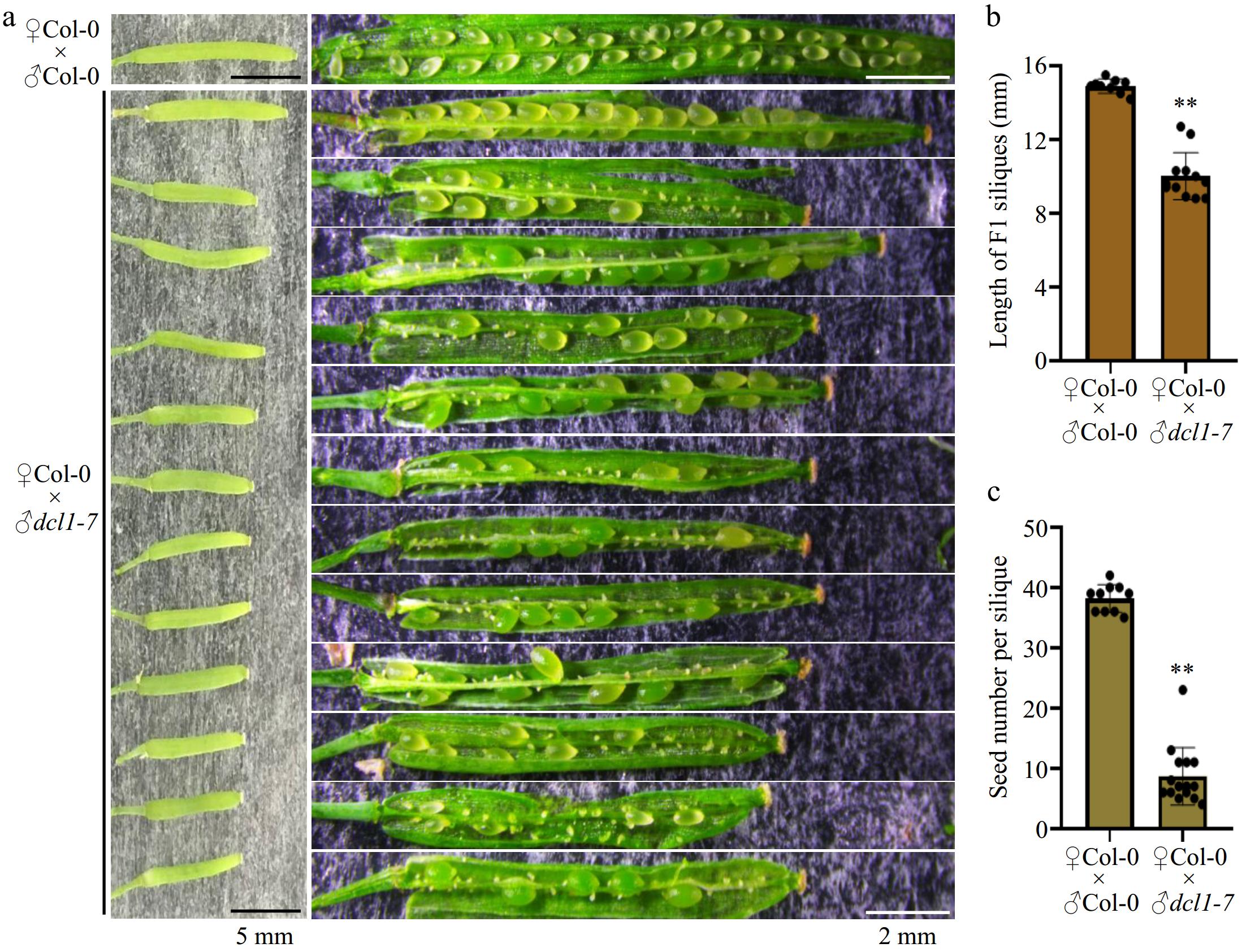
      Figure 1. 

      Hand-pollination by the dcl1-7 mutant pollen caused defects in seed production. (a) Representative F1 siliques from ♀Col-0 × ♂Col-0 and ♀Col-0 × ♂dcl1-7 respectively. The dcl1-7 mutant pollen as the male causes aborted seeds. Black bars indicate 5 mm length, and the white bars indicate 2 mm length. (b), (c) Statistical analysis showing the (b) length, and (c) the total number of the F1 siliques. Each point indicates one silique, p-value were calculated using an unpaired, two-sided Student t-test (** p < 0.01). Scale bars are shown.

      To conclusively demonstrate these results, we followed our previous study, where sperm-delivered miR159 was shown to promote seed development by removing maternal roadblock targets[30]. Given the developmental defects observed from early vegetative growth stages in the mir159abc triple mutant, we investigated the contribution of paternal-generated miR159 to the phenotypes of shortened siliques and aborted seeds. Consistent with the male germ cell-specific expression of HTR10[31], the over-expression of MIR159a driven by the HTR10 promoter did not complement the vegetative growth phenotypes (Supplementary Fig. S2a), but partially rescued the defects on silique length and seed setting rate of the mir159abc triple mutant (Supplementary Fig. S2bd). The defects in endosperm nuclear divisions fertilized by miR159abc pollen were attributed to the retention of miR159 targets MYB33 and MYB65 in the central cell after fertilization[30]. Moreover, the decreased proportion of viable seeds in F1 siliques obtained by mir159abc pollen as male could be restored when using the myb33 myb65 double mutant as female instead of Col-0 (Supplementary Fig. S2eg), although the myb33 myb65 double mutant exhibited partial shortened silique phenotype in self-pollination (Supplementary Fig. S2f). These results further confirmed our previous observation that sperm-delivered miR159 promotes seed development by repressing maternal MYB33 and MYB65. Collectively, it was conclusively demonstrated that paternal miRNA biogenesis is essential for fertilization and viable seed production.

    • A recent study has analyzed the miRNA profiles at the microspore, bicellular pollen, and mature pollen stages, thereby providing an atlas of the miRNome during pollen development[25]. The overall levels of total miRNAs are similar among microspore, bicellular pollen, and tricellular pollen stages, but slightly increased in mature pollen (Supplementary Fig. S3a). This suggests that MIR transcription and/or miRNA biogenesis may not exhibit significant dynamics during pollen development. To investigate whether this is the case, the timing and location of MIR gene transcription during pollen mitoses was first examined, focusing on several well-known abundant miRNAs in mature pollen. Several transgenic lines with GFP reporters driven by endogenous promoters of pollen-enriched miRNAs were constructed, including MIR159a, MIR159b, MIR164a, MIR845a, MIR845b, and MIR2939. The present findings show that all these MIR genes initiated their transcription primarily early in the microspore stage before PMI (Fig. 2a). Following PMI, transcription of these tested MIR genes continued in both VC GC, with a slightly stronger signal observed in the VC-based on GFP intensity (Fig. 2b). However, the transcriptional signal of five out of the six tested MIR genes was completely absent in both VC and GC at the mature pollen stage (Fig. 2c). The exception was proMIR2939::GFP, which exhibited weak but robust expression signals in sperm cells (Fig. 2c). Considering that all four miRNAs are relatively steadily produced across pollen development (Supplementary Fig. S3b)[25], the phenomena of MIR transcription at early stages of pollen development indicate that miRNAs detected in mature pollen are obtained by either local processing of early transcribed pri-miRNA or inherited from bicellular pollen via PMII.
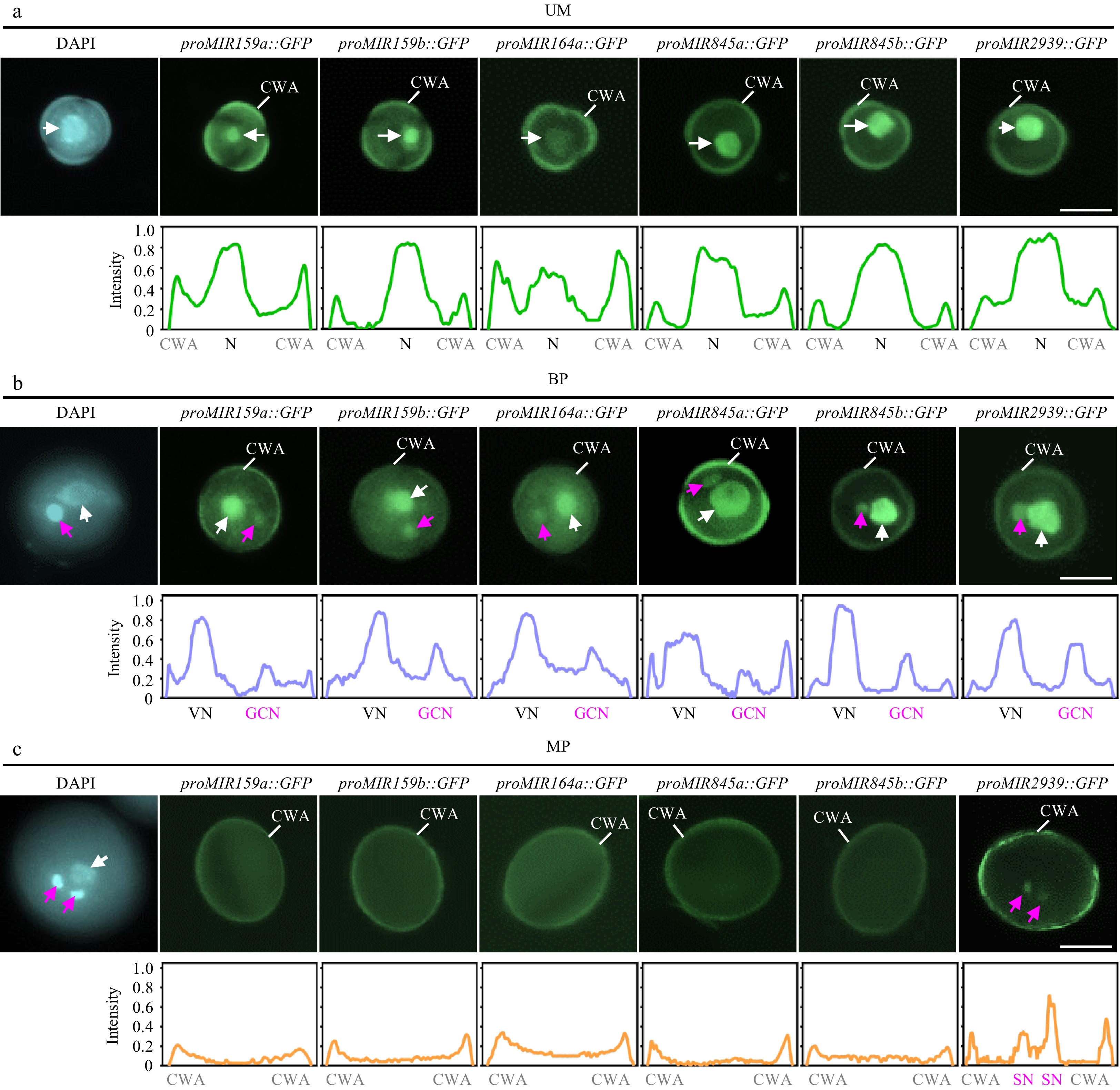
      Figure 2. 

      Spatiotemporal patterns of MIR transcription during pollen development. The expression of proMIR159a::GFP, proMIR159b::GFP, proMIR164a::GFP, proMIR845a::GFP, proMIR845b::GFP, and proMIR2939::GFP was observed during pollen development by fluorescence microscopy. Representative pollen grains show DAPI fluorescence (left, blue) and GFP fluorescence (right, green) at unicellular microspore [(a), UM], bicellular pollen [(b), BP], and mature pollen [(c), MP] stages. White arrowhead indicates N or VN; magenta arrowhead indicates GCN or SN. Relative intensity curve of the GFP signal were shown below the images. VN, vegetative nucleus; GCN, generative cell nucleus, SN, sperm cell nuclei; CWA, cell wall autofluorescence. Scale bar = 10 μm.

    • Since the transcription of MIR genes mainly occurs before the second pollen mitosis, does miRNA biogenesis also exhibit similar properties? RNA-seq data from the vegetative cells and sperm cells of mature pollen were first analyzed[32], focusing on the expression of several main factors involved in miRNA biogenesis. The results show that while DCL1, SE, and HEN1 are detectable in both sperm cells and the vegetative cell, the overall mRNA levels of these genes are low in mature pollen (Supplementary Fig. S3c). Notably, mRNA of HYL1 is barely detected in mature pollen (Supplementary Fig. S3c). These analyses indicate that the local activity of the intact miRNA biogenesis machinery in mature pollen is low.

      To further elucidate the spatiotemporal pattern of miRNA biogenesis during pollen mitosis, we investigated the subcellular localization of DCL1, HYL1, and HEN1 throughout pollen development using corresponding transgenic plants expressing DCL1-YFP, HYL1-YFP, and HEN1-YFP, each driven by their native promoters[33]. The results show that all three proteins are highly detected in the microspore (Fig. 3ad, top panels), indicating the potential for local processing of pri-miRNA. After the first pollen mitosis, there were no significant differences in DCL1-YFP, HYL1-YFP, and HEN1-YFP signal intensity between the vegetative cell and the generative cell at the bicellular pollen stage (Fig. 3a-d, the middle panels). However, the intensities of DCL1-YFP and HYL1-YFP were decreased in sperm cells at the mature pollen stage, while the intensity of HEN1-YFP in sperm cells remained at the same level as in the vegetative cell (Fig. 3ad, the middle panels). These results indicate that miRNA biogenesis factors persist in the vegetative cell throughout pollen development. Instead, miRNA biogenesis activity may be compromised in sperm cells due to a much lower accumulation of DCL1, suggesting that the miRNAs enriched in sperm cells may be inherited from bicellular pollen or transferred from the vegetative cell at the mature pollen stage. The high signal of HEN1-YFP suggests that sperm-enriched small RNAs may require HEN1-mediated 3’ end methylation. Notably, while the miRNA biogenesis machinery has been found to localize in dicing bodies, no nuclear punctate structures were observed for all three proteins in pollen. This indicates that the subcellular territory of miRNA biogenesis in pollen may differ from that in somatic cells.
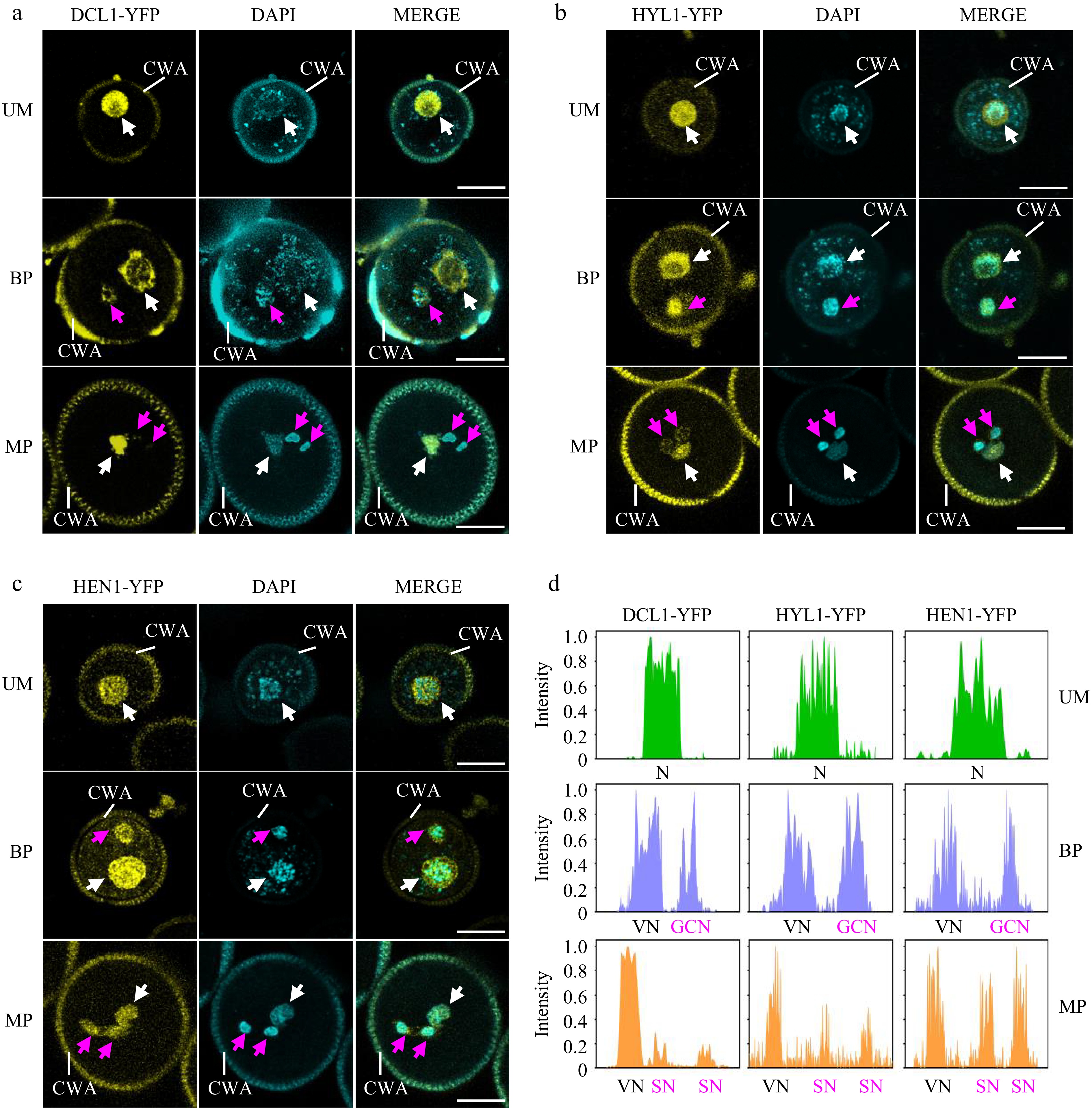
      Figure 3. 

      Spatiotemporal patterns of miRNA biogenesis factors during pollen development. (a)−(c) The localization of (a) DCL1-YFP, (b) HYL1-YFP, and (c) HEN1-YFP was observed during pollen development by confocal microscopy. Representative pollen grains show YFP fluorescence (right, yellow) and DAPI fluorescence (right, cyan) at unicellular microspore (UM), bicellular pollen (BP), and mature pollen (MP) stages. White arrowhead indicates N or VN; magenta arrowhead indicates GCN or SN. VN, vegetative nucleus; GCN, generative cell nucleus, SN, sperm cell nuclei; CWA, cell wall autofluorescence. Scale bar = 10 μm. (d) Relative intensity curve of YFP signal in (a) DCL1-YFP, (b) HYL1-YFP, and (c) HEN1-YFP.

    • As the transcription of MIR genes is mostly completed before the second pollen mitosis (Fig. 2), what is the biological significance of the persistence of the miRNA biogenesis machinery in mature pollen? To investigate the role of miRNA biogenesis factors detected in the vegetative cell and sperm cells of mature pollen, two types of transgenic lines were generated with cell-type-specific knockdown of DCL1 in the VC and GC/SC, respectively. Expression of the artificial miRNA targeting DCL1 was achieved by using the late VC-specific promoter VCK1[34] and the GC/SC-specific promoter HTR10[31], respectively. These constructs were transformed into the proDCL1::DCL1-YFP reporter line to primarily evaluate the efficiency of knockdown by the artificial miRNA (Supplementary Fig. S4a). As shown in Supplementary Fig. S4a, the introduction of the VC-expressed artificial miRNA targeting DCL1 significantly attenuated the signal intensity of DCL1-YFP in the vegetative cell. Similarly, the introduction of the GC/SC-expressed artificial miRNA targeting DCL1 specifically attenuated the signal intensity of DCL1-YFP in the sperm cells, while having no effect on the signal in the vegetative cell. When proVCK1::amiR_DCL1 was introduced, the faint signal of DCL1 in the vegetative nucleus could be still observed, indicating a possible leaky expression. Whether the reduced expression of DCL1 in the vegetative cell and sperm cells affects double fertilization and subsequent seed development were then examined. In eight independent transgenic T1 lines expressing proVCK1::amiR_DCL1 in the vegetative cell, we found that these plants exhibited noticeably shortened siliques, reduced seed numbers, and approximately half of the seeds were unfertilized (Fig. 4a, c), which resembled the phenotypes observed in the F1 silique of ♀Col-0 × ♂dcl1-7 (Fig. 1a). Interestingly, in ten individual transgenic T1 lines expressing proHTR10::amiR_DCL1 in the generative cell and sperm cells, it was observed that approximately 30% of seeds exhibited a typical embryo lethal phenotype (Fig. 4b). Additionally, these plants produced an average of about 30 seeds per silique (Fig. 4d), compared to the Col-0 plants, which produce approximately 50 seeds per silique on average. These results indicating that DCL1 expressed in sperm is involved in early embryogenesis after double fertilization.
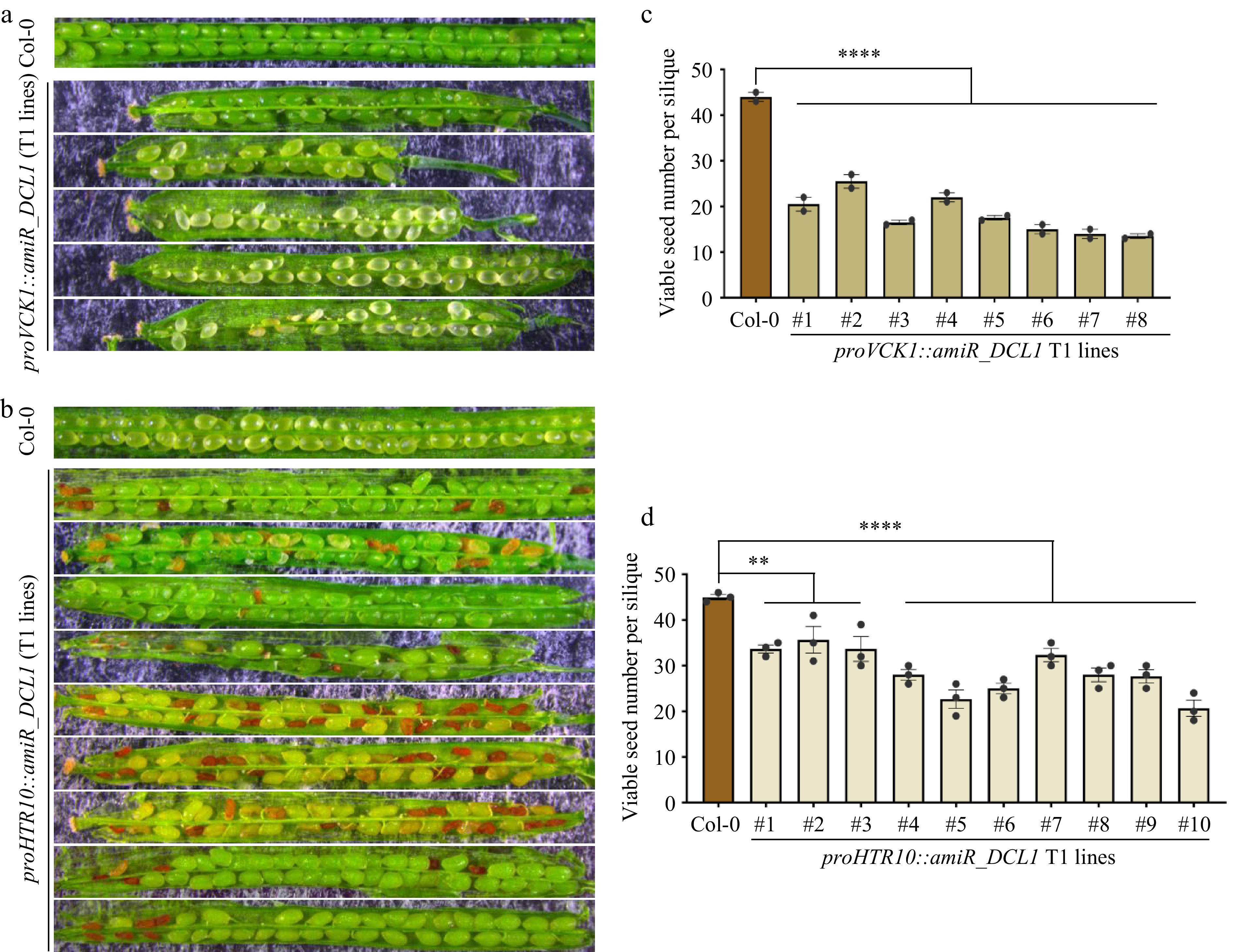
      Figure 4. 

      Both VC and GC/SC-knockdown of DCL1 caused compromised but distinctive defects in seed development. (a) Seed development defects of proVCK1::amiR_DCL1 transgenic T1 lines, representative siliques are shown. (b) Seed development defects of proHTR10::amiR_DCL1 transgenic T1 lines, representative siliques are shown. (c), (d) Statistical analyses of viable seed number per silique in (c) proVCK1::amiR_DCL1, and (d) proHTR10::amiR_DCL1 transgenic T1 lines. Each column indicates one individual T1 line. p-values were calculated by one-way ANOVA Dunnett’s multiple comparison, ** p < 0.01, **** p < 0.0001.

      Given the nature of phenotypes in seed development of plants with conditional knockdown of DCL1 in mature pollen, we hypothesized that the seed development defects in proVCK1::amiR_DCL1 plants should be attributed to the fertilization failure rather than post-fertilization impairment. To test this, the in vitro pollen germination rate of Col-0 and proVCK1::amiR_DCL1 heterozygous plants were preliminarily assessed. Compared to Col-0, five transgenic lines of proVCK1::amiR_DCL1 showed a significantly reduced pollen germination rate (Supplementary Fig. S4b & c), indicating that VC-expressed DCL1 plays an important role in pollen germination. In contrast, the transgenic lines expressing proHTR10::amiR_DCL1 exhibit approximately 30% aborted seeds (Fig. 4b & d). To further investigate the arrested stage of the abnormal seeds in the proHTR10::amiR_DCL1 transgenic lines, we observed the embryo development of normal and abnormal seeds in a same proHTR10::amiR_DCL1 silique at 7 DAP, when the abnormal seeds had not been completely dried out. DIC microscopy analyses showed that the embryos of the abnormal seeds were arrested at the globular stage, while mature embryos had been developed in the normal seeds of the same silique (Supplementary Fig. S4d). The arrest of embryos at the globular stage is consistent with the embryo abnormalities observed in several dcl1 mutant alleles[79,29], demonstrating that paternal DCL1 contributes to early embryo patterning during seed development. Together, it is concluded that mature pollen-expressed DCL1 is necessary for pollen germination and early embryo development.

    • The embryo lethal phenotype of dcl1 mutant alleles highlights the remarkable role of miRNA biogenesis during seed development[8,9,29]. Due to the size discrepancy between sperm cells and the egg cell, it is generally believed that substances in egg cells play a major role in early development. However, it is increasingly clear that the male gamete carries additional coding or non-coding RNAs necessary for early development in different species[35,36]. Our previous work shows that paternal miR159 is required to promote endosperm nuclear divisions during early seed development[30]. In this study, we further demonstrate that mature pollen-expressed miRNA biogenesis factors contribute to both the fertilization process and early embryo development.

      The spatiotemporal dynamics of MIR transcription and miRNA biogenesis factors through pollen mitosis indicate that the biogenesis of most pollen-enriched miRNAs is completed at early developmental stages before PMII. Furthermore, the miRNA biogenesis patterns between the vegetative cell and male germ cells exhibit both similarities and differences, supported by these observations: (i) pollen-expressed MIR genes preferentially complete their transcription before the second pollen mitosis in both the vegetative cell and the generative cell; (ii) miRNA biogenesis factors are localized in both the vegetative cell and generative cell at early developmental stages; (iii) the abundance of DCL1 shows a striking decrease in sperm cells compared to the vegetative cell. The differences in the dicing complex between the vegetative cell and sperm cells might be attributed to the de-condensed chromatin in the vegetative cell and the gradually increasing chromatin condensation in sperm cells[37]. Notably, previous study on miRNA profiling in vegetative and sperm cells have shown that specific miRNAs are distinctly enriched between these two cell types[38], indicating that miRNA distribution within mature pollen is tightly regulated. Based on the vegetative cell-preferred localization of DCL1 and the transcription of MIR genes before sperm cell formation, the specificity of miRNA distribution in the two cell types within mature pollen may not be determined by the localization of miRNA biogenesis machinery. Instead, likely specific miRNAs are actively transferred from the vegetative cell to the sperm cells[1821]. One typical example is miR159, where the transcription of three MIR159 genes occur before the second pollen mitosis, but mature miR159 is significantly enriched in sperm cells[38].

      Unlike specific miRNAs that function in pollen by inhibiting target genes expressed either in pollen itself or on the maternal side, the fact that reduced expression of DCL1 in both the vegetative cell and sperm cells causes distinct defects in seed development indicates that mature pollen-expressed DCL1 is important for specific miRNA biogenesis in pollen and the pollen tube, or is delivered into female gametes via fertilization to promote early miRNA biogenesis. The effects of vegetative cell-expressed DCL1 on pollen germination and sperm cell-expressed DCL1 on embryo development align with the default functions of the vegetative cells and sperm cells, respectively. The present data showed that a significantly reduced pollen germination rate in vegetative-cell dcl1 knockdown plants, indicating that the seed development defects in proVCK1::amiR_DCL1 plants are likely due to fertilization failure rather than post-fertilization impairment. We suggest that DCL1-mediated miRNA biogenesis in the vegetative cell may also play a role in pollen tube targeting or other processes required for successful double fertilization. Meanwhile, the embryo development defects in proHTR10::amiR_DCL1 transgenic lines resemble the embryo abnormalities in dcl1 mutants[9,29], indicating that not only maternal but also paternal DCL1 contributes to early embryo patterning.

      • This work was supported by the National Natural Science Foundation of China (32025005, 31830045, M-0398 to BZ).

      • The authors declare that they have no conflict of interest. Binglian Zheng is the Editorial Board member of Seed Biology who was blinded from reviewing or making decisions on the manuscript. The article was subject to the journal's standard procedures, with peer-review handled independently of this Editorial Board member and the research groups.

      • Supplementary Table S1 Primer sequence.
      • Supplemental Fig. S1 The dcl1-7 homozygous plant showed disrupted anther development but normal pollen mitoses.
      • Supplemental Fig. S2 Male germ cell-overexpressed MIR159a partially rescued fertility defects of the mir159abc mutant.
      • Supplemental Fig. S3 miRNA levels and expression of main miRNA biogenesis factors are adapted from publicly available data during pollen development.
      • Supplemental Fig. S4 Pollen germination and embryo development in conditionally knockdown plants of dcl1 in pollen.
      • Copyright: © 2024 by the author(s). Published by Maximum Academic Press on behalf of Hainan Yazhou Bay Seed Laboratory. This article is an open access article distributed under Creative Commons Attribution License (CC BY 4.0), visit https://creativecommons.org/licenses/by/4.0/.
    Figure (4)  References (38)
  • About this article
    Cite this article
    Yang H, Zhao Y, Lin Z, Jiang T, Hu Q, et al. 2024. Paternal miRNA biogenesis contributes to seed development in Arabidopsis. Seed Biology 3: e017 doi: 10.48130/seedbio-0024-0017
    Yang H, Zhao Y, Lin Z, Jiang T, Hu Q, et al. 2024. Paternal miRNA biogenesis contributes to seed development in Arabidopsis. Seed Biology 3: e017 doi: 10.48130/seedbio-0024-0017

Catalog

  • About this article

/

DownLoad:  Full-Size Img  PowerPoint
Return
Return