REVIEW   Open Access    

Omics technologies accelerating research progress in yams

More Information
  • Yams, belonging to Dioscorea species, are abundant in nutrients and bioactive compounds, contributing to their swiftly expanding share in the global market. Over the past 20 years, worldwide production of yams has seen a twofold increase. Particularly in Africa, yams are a staple food for millions, significantly contributing to food security and sustenance. The development of omics technologies provides an effective means for mining functional genes and exploring related molecular mechanisms in yams. This review summarizes the current research progress on the yam genome, plastome, transcriptome, proteome, and metabolome, to facilitate further genetic research and molecular breeding in yams.
  • Plants are continuously subjected to unpredictable environmental conditions and encounter a multitude of stressors throughout their growth and development, posing a significant challenge to global crop production and food security[1]. Heat and drought are undoubtedly the two most important stresses that have a huge impact on crops. Both elicit a wide array of biochemical, molecular, and physiological alterations and responses, impacting diverse cellular processes and ultimately influencing crop yield and quality[2].

    A primary physiological consequence of both stresses is the diminished photosynthetic capacity, partially resulting from the degradation of chlorophyll due to leaf senescence under stress conditions. Chlorophyll accumulation was diminished in numerous plants subjected to drought or heat stress conditions[3,4]. Various environmental stresses prompt excessive generation of reactive oxygen species (ROS), initiating oxidative damage that compromises lipids, and proteins, and poses a serious threat to cellular functions[2]. To mitigate oxidative stress and minimize damage, plants have developed various protective mechanisms to neutralize ROS. Several antioxidant enzymes, such as SOD, POD, and CAT, are integral to cellular antioxidative defense mechanisms. Additionally, antioxidants such as anthocyanins and proline serve as crucial ROS scavengers[5,6]. The elevation in temperature typically induces the transient synthesis of heat shock proteins (Hsps), which function as molecular chaperones in protecting proteins from denaturation and aggregation, with their activity primarily regulated at the transcriptional level by heat shock factors (Hsfs)[7]. The significance of Hsps and Hsfs in all organisms, including plants, has been assessed in various stress conditions that could disrupt cellular homeostasis and result in protein dysfunction[7]. Drought stress can also trigger the transcription of a suite of marker genes, including RD29A, RD29B, NCED3, AREB1, Rab18, etc., which assist plants in mitigating cellular damage during dehydration and bolstering their resilience to stress[810].

    Previous research efforts focusing on the regulatory control of stress-related genes have largely centered around protein-coding genes. In recent years, non-protein-coding transcripts have emerged as important regulatory factors in gene expression. Among them, long non-coding RNAs (lncRNAs) lncRNAs have been identified as implicated in various abiotic stresses[11,12]. LncRNAs are a class of non-coding RNAs (ncRNAs) exceeding 200 nucleotides in length. They possess minimal or no protein-coding potential[13]. In plants, lncRNAs are specifically transcribed by RNA polymerases Pol IV, Pol V, Pol II, and Pol III[14,15]. LncRNAs exhibit low abundance and display strong tissue and cellular expression specificity relative to mRNAs. Moreover, sequence conservation of lncRNAs is was very poor across different plant species[13,16,17]. The widespread adoption of high-throughput RNA sequencing technology has revealed lncRNAs as potential regulators of plant development and environmental responses. In cucumber, RNA-seq analysis has predicted 2,085 lncRNAs to be heat-responsive, with some potentially acting as competitive endogenous RNAs (ceRNAs) to execute their functions[18]. In radish, a strand-specific RNA-seq (ssRNA-seq) technique identified 169 lncRNAs that were differentially expressed following heat treatment[19]. In Arabidopsis, asHSFB2a, the natural antisense transcript of HSFB2a was massively induced upon heat stress and exhibited a counteracted expression trend relative to HSFB2a. Overexpression of asHSFB2a entirely suppressed the expression of HSFB2a and impacted the plant's response to heat stress[20]. For drought stress resistance, 244 lncRNAs were predicted in tomatoes to be drought responsive probably by interacting with miRNAs and mRNAs[21]. Under drought stress and rehydration, 477 and 706 lncRNAs were differentially expressed in drought-tolerant Brassica napus Q2 compared to drought-sensitive B. napus, respectively[22]. In foxtail millet and maize, 19 and 644 lncRNAs, respectively, were identified as drought-responsive[23,24]. Despite the identification of numerous lncRNAs by high-throughput sequencing, which suggests their potential involvement in various abiotic stress processes, only a minority have been experimentally validated for function.

    In our previous study, we characterized 1,229 differentially expressed (DE) lncRNAs in Chinese cabbage as heat-responsive, and subsequent bioinformatics analysis reduced this number to 81, which are more likely associated with heat resistance[25]. lnc000283 and lnc012465 were selected from among them for further functional investigation. The findings indicated that both lnc000283 and lnc012465 could be promptly induced by heat shock (HS). Overexpression of either lnc000283 or lnc012465 in Arabidopsis plants enhanced their capacity to tolerate heat stress. Additionally, both lnc000283 and lnc012465 conferred drought tolerance to transgenic Arabidopsis.

    The lncRNA sequences examined in this study were from Chiifu-401-42 Chinese cabbage and all Arabidopsis plants were of the Col-0 background. Transgenic plants expressing lnc000283 and lnc012465 were generated using the Agrobacterium tumefaciens-mediated floral dip method[26]. Single-copy and homozygous T3 plants were identified through genetic segregation on an agar medium supplemented with kanamycin. The T3 generation plants, or their homozygous progeny, were utilized in the experiments.

    For phenotypic assessment, Arabidopsis seeds were initially sown on filter paper moistened with ddH2O and placed in a 4 °C freezer for 2 d. Subsequently, they were evenly planted in nutrient-rich soil and transferred to a growth chamber operating a 16-h day/8-h night cycle, with day/night temperatures of 22 °C/18 °C and a light intensity of 250 μmol·m−2·s−1. After 10 d of growth, Arabidopsis plants with uniform growth were transferred to 50-hole plates. Arabidopsis plants grown in Petri dishes were firstly seed-sterilized and then sown on 1/2 MS medium supplemented with 10 g·L−1 sucrose. The seeds were then placed in a 4 °C refrigerator for 2 d in the dark before transferring them to a light incubator. The day/night duration was set to 16 h/8 h, the day/night temperature to 21 °C/18 °C, and the light intensity to 100 μmol·m−2·s−1.

    For heat treatment, 3-week-old seedlings were subjected to 38 °C for 4 d within a light incubator, subsequently transferred to their original growth conditions under the same light/dark cycles. For drought treatment, 3-week-old Arabidopsis seedlings were deprived of water for 10 d, followed by rehydration to facilitate a 2-d recovery period. Plants were photographed and surveyed both before and after treatment.

    The lncRNA sequences (lnc000283 and lnc012465) were chemically synthesized based on RNA-seq data, with restriction sites for BamH1 and Kpn1 engineered upstream and downstream. The resultant lncRNA constructs were subcloned into the pCambia2301 binary vector, incorporating a cauliflower mosaic virus (CaMV) 35S promoter. The recombinant vectors were transformed into Escherichia coli TOP10 competent cells (Clontech), incubated at 37 °C overnight, after which single clones were selected for PCR verification, and the confirmed positive colonies were submitted for sequencing. Following verification, the correct plasmids were introduced into A. tumefaciens strain GV3101 using the freeze-thaw method and subsequently transformed into Arabidopsis wild-type (Col) plants.

    To quantify the chlorophyll content, the aerial portions of wild-type and transgenic Arabidopsis plants, grown in Petri dishes were weighed, minced, and then subjected to boiling in 95% ethanol until fully decolorized. Aliquots of 200 μL from the extract were transferred to a 96-well plate and the absorbance at 663 nm and 645 nm was measured via spectrophotometry by a microplate reader (Multiskan GO, Thermo Scientific, Waltham, MA, USA). Three biological replicates were analyzed for WT and each transgenic line. Chlorophyll content was determined according to the formula of the Arnon method[27]: Chlorophyll a = (12.72A663 − 2.59A645) v/w, Chlorophyll b = (22.88A645 − 4.67A663) v/w, Total chlorophyll = (20.29A645 + 8.05A663) v/w.

    The quantification of anthocyanin was performed as follows: aerial parts of wild-type and transgenic Arabidopsis plants, cultivated in Petri dishes, were weighed and ground to powder in liquid nitrogen. Subsequently, the samples were incubated in 600 μL of acidified methanol (containing 1% HCl) at 70 °C for 1 h. Following this, 1 mL of chloroform was added, and the mixture was vigorously shaken to remove chlorophyll. The mixture was then centrifuged at 12,000 rpm for 5 min, after which the absorbance of the aqueous phase was determined at 535 nm using a spectrophotometer (Shimadzu, Kyoto, Japan). Three biological replicates were analyzed for WT and each transgenic line. The relative anthocyanin content was calculated according to anthocyanin concentration and extraction solution volume. One anthocyanin unit is defined as an absorption unit at a wavelength of 535 nm in 1 mL of extract solution. In the end, the quantity was normalized to the fresh weight of each sample.

    Three-week-old transgenic and WT A. thaliana plants, subjected to normal conditions or varying durations of heat or drought stress, were utilized for subsequent physiological assessments. All assays were performed in accordance with the method described by Chen & Zhang[28]. In brief, 0.1 g of fresh leaf tissue was homogenized in 500 μL of 100 mM PBS (pH 7.8) while chilled on ice. The homogenate was then centrifuged at 4 °C, and the resultant supernatant was employed for further analysis. For the determination of MDA content, 100 μL of the supernatant was combined with 500 μL of a 0.25% thiobarbituric acid (TBA) solution (which was prepared by dissolving 0.125 g of TBA in 5 mL of 1 mol·L−1 NaOH before being added to 45 mL of 10% TCA) and boiled for 15 min. Following a 5 min cooling period on ice, the absorbance was measured at 532 nm and 600 nm. The activity of POD was determined as follows: initially, 28 μL of 0.2% guaiacol and 19 μL of 30% H2O2 were sequentially added to 50 mL of 10mM PBS (pH 7.0), after thorough heating and mixing, 1 mL was transferred into a cuvette, then 50 μL of the supernatant was added to the cuvette and the absorbance at 470 nm was monitored every 15 s for 1 min. To determine the proline content, a reaction solution was prepared by mixing 3% sulfosalicylic acid, acetic acid, and 2.5% acidic ninhydrin in a ratio of 1:1:2, then 50 μL of the supernatant was added to 1 mL of the reaction solution, which was then subjected to a boiling water bath for 15 min (the solution turned red after the boiling water bath). Following cooling on ice, the absorbance at 520 nm was recorded. For the quantification of proline, an L-proline standard curve was prepared by dissolving 0, 5, 10, 15, 20, 25, and 30 μg of L-proline in 0.5 mL of ddH2O, followed by the addition of 1 mL of the reaction solution and measuring the absorbance at 520 nm. The proline content in the samples was then determined based on the L-proline standard curve.

    Total RNA was isolated from the aerial parts of Arabidopsis using the TaKaRa MiniBEST Plant RNA Extraction Kit, followed by purification and reverse transcription using the PrimeScript RT reagent Kit with gDNA Eraser (Takara). The cDNA product was diluted 10 times and real-time PCR was conducted in triplicate for each biological replicate using SYBR PCR Master Mix (Applied Biosystems) on the ABI 7500 system under the following conditions: 98 °C for 3 min, followed by 40 cycles of 98 °C for 2 s and 60 °C for 30 s. The relative expression levels of each gene were normalized against the transcript abundance of the endogenous control UBC30 (At5g56150) and calculated using the 2−ΔCᴛ method. The specific primers employed for qRT-PCR are detailed in Supplemental Table S1.

    In our prior investigation, dozens of lncRNAs associated with the heat stress response in Chinese cabbage were identified through informatics analysis. Two lncRNAs (lnc000283 and lnc012465) were chosen for genetic transformation in Arabidopsis to elucidate their functions comprehensively. Transcriptome data analysis indicated that the expression of lnc000283 and lnc012465 in Chinese cabbage were both induced by HS. To verify the accuracy, the expression patterns of lnc000283 and lnc012465 were confirmed through quantitative real-time PCR (qRT-PCR), and the results from qRT-PCR were consistent with those obtained from RNA-seq (Fig. 1a). The corresponding homologous genes in Arabidopsis were identified as CNT2088434 and CNT2088742, exhibiting sequence similarities of 88% and 87%, respectively (Supplemental Fig. S1). Subcellular localization predictions using the lnclocator database (www.csbio.sjtu.edu.cn/bioinf/lncLocator) suggested that both lncRNAs are localized within the nucleus (Supplemental Table S2). Bioinformatics analysis was conducted using the CPC tool (http://cpc.cbi.pku.edu.cn/) indicated that lnc000283 and lnc012465 are noncoding sequences, with coding probabilities of 0.0466805 and 0.0432148, respectively comparable to the well-characterized lncRNAs COLDAIR and Xist, but significantly lower than those of the protein-coding genes UBC10 and ACT2 (Fig. 1b).

    Figure 1.  Characteristics of lnc000283 and lnc012465. (a) Expression level of lnc000283 and lnc012465 in Chinese cabbage leaves treated at 38 °C at different time points, as determined by qRT-PCR and RNA-seq. CK is a representative plant before heating, and T1, T4, T8, and T12 denote plants that were subjected to 38 °C for 1, 4, 8, and 12 h, respectively. The expression levels were normalized to the expression level of Actin. (b) Analysis of coding potential for lnc000283 and lnc012465. The coding potential scores were calculated using the CPC program. UBC10 (At5g53300) and ACT2 (At3g18780) are positive controls that encode proteins. COLDAIR (HG975388) and Xist (L04961) serve as negative controls, exhibiting minimal protein-coding potential.

    To elucidate the role of lnc000283 and lnc012465 in response to abiotic stress, overexpression vectors were constructed for these lncRNAs, driven by the CaMV 35S promoter, and they were introduced into Arabidopsis thaliana (Col-0 ecotype). Through PCR identification and generational antibiotic screening, two homozygous positive lines for lnc012465 and lnc000283 were obtained. The relative expression levels of these lncRNAs were assessed using qRT-PCR (Fig. 2a). When plants were grown in 1/2 MS medium, with the consumption of nutrients, and reduction of water, the leaves of WT began to turn yellow, but the lnc000283 and lnc012465 overexpression lines developed a deep purple color of leaf veins (Fig. 2b). Examination of chlorophyll and anthocyanin contents in the plants revealed that both overexpression lines had higher levels of chlorophyll and anthocyanin compared to the WT, suggesting that the transgenic plants might possess enhanced resistance to nutritional or water stress (Fig. 2c, d).

    Figure 2.  Arabidopsis plants overexpressing lnc000283 and lnc012465 had higher anthocyanins and chlorophyll content. (a) The relative expression level of lnc000283 and lnc012465 in WT and different transgenic lines. UBC10 (At5g53300) was used as an internal control. Each value is mean ± sd (n = 3). (b) The phenotype of WT and Arabidopsis overexpressing lnc000283 or lnc012465 grown on 1/2 MS medium 50 d after sowing. The (c) anthocyanin and (d) chlorophyll content of WT and transgenic Arabidopsis overexpressing lnc000283 or lnc012465. The asterisks above the bars indicate statistical significance using Student's t-test (*, p < 0.05; **, p < 0.01).

    Given that lnc000283 and lnc012465 were highly induced by heat, the thermotolerance of the overexpressing (OE) plants were compared to that of the wild type. Arabidopsis plants were initially exposed to a an HS treatment at 38 °C for 4 d, followed by recovery at room temperature. The death caused by HS was processive. Post-severe HS challenge for 4 d, OE plants initially appeared similar to WT, but upon recovery, their leaves started to fold or curl, followed by a transition to yellow, white, and eventually drying out (Fig. 3a). OE lnc000283 and OE lnc012465 plants exhibited enhanced thermotolerance compared to WT, with lnc012465 showing particularly strong tolerance (Fig. 3a; Supplemental Fig. S2a). After 5 d of recovery, leaf coloration indicated that transgenic plants maintained a significantly higher percentage of green leaves and a lower percentage of bleached leaves compared to WT (Fig. 3b; Supplemental Fig. S2b). Under non-heat-stress conditions, WT and OE plants possessed comparable water content. However, following heat stress, the fresh-to-dry weight ratio of OE lnc000283 and lnc012465 lines was significantly greater than that of WT (Fig. 3c; Supplemental Fig. S2c). Abiotic stresses frequently trigger the production of excessive reactive oxygen species (ROS), which are believed to cause lipid peroxidation of membrane lipids, leading to damage to macromolecules. Leaf MDA content is commonly used as an indicator of lipid peroxidation under stress conditions; therefore, the MDA content in both transgenic and WT plants was assessed. Figure 3d shows that the MDA content in WT plants progressively increased after heat treatment, whereas in the two lines overexpressing lnc012465, the MDA content increased only slightly and remained significantly lower than that in WT at all time points. In plants overexpressing lnc000283, the MDA content did not significantly differ from that of WT before heat stress. However, after 4 d of heat treatment, the MDA content was significantly lower compared to WT (Supplemental Fig. S2d). The results suggested that the expression of both lnc012465 and lnc000283 can mitigate injury caused by membrane lipid peroxidation under heat-stress conditions. Peroxidase (POD) is a crucial antioxidant enzyme involved in ROS scavenging. Figure 3e and Supplemental Fig. S2e demonstrate that POD activity increased in both transgenic and WT plants after heat treatment. However, the increase in WT plants was modest, whereas OE lnc000283 and OE lnc012465 plants exhibited consistently higher POD activity. As anticipated, proline levels were induced in response to stress in all studied plants (Fig. 3f; Supplemental Fig. S2f). However, under normal conditions and 2 d post-heat stress treatment, the proline content in OE lnc000283 and OE lnc012465 plants did not exhibit significant changes compared to WT (Fig. 3f; Supplemental Fig. S2f). Moreover, after 4 d of heat stress, the proline content in OE lnc012465 lines was significantly lower than in WT, and the OE lnc000283 transgenic line 12-6 also showed a marked decrease in proline content compared to WT (Fig. 3f; Supplemental Fig. S2f). The results indicated that the thermotolerance of plants overexpressing either lnc000283 or lnc012465 was independent of proline accumulation.

    Figure 3.  Overexpressing lnc012465 lines are more tolerant to heat stress. (a) Phenotypes of WT and OE lnc012465 plants were assessed before and after exposure to heat stress. The heat treatment was applied to 25-day-old Arabidopsis plants. (b) The percentage of leaves with different colors in Arabidopsis after heat treatment and recovery for 5 d. (c) The fresh-to-dry weight ratio of Arabidopsis leaves was measured before and after 38 °C heat treatment. (d)−(f) depict the MDA content, POD activity, and proline content in Arabidopsis leaves at varying durations of heat stress. The asterisks above the bars indicate statistical significance using Student's t-test (*, p < 0.05; **, p < 0.01; ***, p < 0.001).

    To elucidate the molecular mechanisms by which lncRNAs enhance thermotolerance in Arabidopsis, the expression of the Hsf gene HsfA7a and three Hsps (Hsp25.3, Hsa32, and Hsp18.1-CI) in OE lnc000283, OE lnc012465, and WT Arabidopsis plants were investigated at various time points following heat treatment. As shown in Fig. 4 and Supplemental Fig. S3, both Hsf and Hsps exhibited a rapid response to heat stress with strong induction. Notably, the transcripts of HsfA7a and Hsp25.3 were significantly upregulated at 1 h after heat exposure, then experienced a sharp decrease. Hsa32 and Hsp18.1-CI were highly induced at 1 h and, unlike the other proteins, sustained high expression levels at 3 h (Fig. 4; Supplemental Fig. S3). At 1 h post-heat treatment, the transcript levels of Hsa32 and HsfA7a in OE lnc000283 did not significantly differ from those in WT. However, by 3 h, Hsa32 expression was roughly 50% of the WT level, while HsfA7a expression was approximately double that of WT (Supplemental Fig. S3). The overexpression of lnc000283 did not significantly affect the transcript level of Hsp25.3 at any of the tested time points. Notably, Hsp18.1-CI expression in both lines overexpressing lnc000283 was significantly induced at all three detection points post-heat treatment, reaching approximately 4-9-fold higher levels than in the WT (Supplemental Fig. S3). In Arabidopsis plants with elevated expression of lnc012465, the expression patterns of all Hsp and Hsf genes were similar to those in plants overexpressing lnc000283, with the notable exception of Hsa32. Unlike the WT, Hsa32 did not show a trend of down-regulation at 3 h post-heat treatment (Fig. 4). The findings suggest that the substantial induction of Hsp18.1-CI may play a role in enhancing the thermotolerance of Arabidopsis plants overexpressing lnc000283 and lnc012465.

    Figure 4.  The expression of HSF and HSP genes in lnc012465 overexpressing lines before and after different heat treatment times. Gene expression levels were quantified using RT-qPCR and normalized to UBC10 (At5g53300). Each value represents the mean ± standard deviation (n = 3). The asterisks above the bars indicate statistical significance using Student's t-test (*, p < 0.05; **, p < 0.01; ***, p < 0.001).

    Prior research has implicated a significant proportion of genes in conferring resistance to various abiotic stresses. To elucidate the functions of lnc000283 and lnc012465 more thoroughly, WT and transgenic plants were subjected to drought stress by depriving them of water for 9 d. It was noted that the majority of leaves in WT plants withered and dried, whereas the OE lnc000283 and OE lnc012465 plants exhibited reduced withering, with only a minority displaying dryness (Fig. 5a; Supplemental Fig. S4a). Eight days post-rewatering, a negligible fraction of WT seedlings exhibited recovery, whereas the overwhelming majority of transgenic plants regained vigorous growth (Fig. 5a; Supplemental Fig. S4a). The transgenic plants demonstrated a significantly higher survival rate compared to the WT plants. Following 9 d of water deficit treatment, less than 40% of the WT plants survived, whereas the OE 012465 lines 8-7 and 9-1 exhibited survival rates of 100% and 95%, respectively, and the OE 000283 lines 11-10 and 12-6 had survival rates of 87% each. (Fig. 5b; Supplemental Fig. S4b). Water loss serves as a critical metric for assessing plant drought tolerance, hence the fresh-to-dry weight ratio of excised leaves was assessed via desiccation analysis. Following 4 d of drought treatment, the fresh-to-dry weight ratio for WT plants was reduced to 43%, whereas for OE lnc000283 lines 11-10 and 12-6, it was reduced to 73% and 75%, respectively. For OE 012465 lines 8-7 and 9-1, the ratios were reduced to 67% and 62%, respectively (Fig. 5c; Supplemental Fig. S4c). The findings indicated that lnc000283 and lnc012465 endow the transgenic plants with drought tolerance.

    Figure 5.  Overexpressing lnc012465 lines are more tolerant to drought stress. (a) Phenotype of WT and OE lnc012465 plants before and after subjecting to drought stress. Drought treatment was carried out on 20-day-old Arabidopsis plants. (b) The percentage of leaves with different colors in Arabidopsis after heat treatment and recovery for 5 d. (c) The fresh weight to dry weight ratio of Arabidopsis leaves before and after undergoing 38 °C heat treatment. (d)−(f) MDA content, POD activity, and proline content in Arabidopsis leaves under different times of heat stress. The asterisks above the bars indicate statistical significance using Student's t-test (*, p < 0.05; **, p < 0.01; ***, p < 0.001)

    MDA content in leaves is a standard biomarker for assessing the extent of drought stress-induced damage. Prior to drought stress exposure, MDA levels in WT and transgenic plants were comparable. However, following 7 and 9 d of water deficit, the MDA content in the transgenic plants was markedly reduced compared to the WT, suggesting a less severe degree of membrane lipid peroxidation in the transgenic plants (Fig. 5d; Supplemental Fig. S4d). Oxidative stress frequently coincides with drought stress, hence the activity of POD was assessed to evaluate the ROS scavenging ability. The findings indicated that as the duration of drought treatment increased, POD activity progressively rose. Before drought exposure, the POD activity in lines 11-10 and 12-6 of OE 000283 was 2.4-fold and 2.2-fold higher than that of the WT, respectively (refer to Supplemental Fig. S4e). Following drought treatment, the POD activity in the transgenic lines remained significantly elevated compared to the wild type, although the enhancement was less pronounced than before the treatment (Supplemental Fig. S4e). In the OE 012465 plants, the POD activity in lines 8-7 and 9-1 significantly surpassed that of the wild type, with the discrepancy being more pronounced during drought stress (Fig. 5e). The proline content in WT and OE 000283 plants exhibited no significant differences before and after 7 d of treatment. However, after 9 d of drought, the proline content in OE 000283 plants was significantly lower compared to that in the WT (Supplemental Fig. S4f). OE 000465 plants showed no significant difference from the wild type before and after drought treatment (Fig. 5f). The findings were consistent with those under heat stress, indicating that the enhanced stress resistance due to the overexpression of lnc000283 and lnc012465 in Arabidopsis is not reliant on proline accumulation.

    Following drought stress treatment, the expression levels of drought-related genes such as RD29A, RD29B, NCED3, AREB1, and Rab18 were significantly elevated in plants overexpressing lnc000283 and lnc012465 compared to WT plants. These findings suggest that lnc000283 and lnc012465 modulate Arabidopsis drought tolerance by regulating the expression of genes associated with the drought stress response (Fig. 6; Supplemental Fig. S5).

    Figure 6.  The expression of drought-responsive genes in lnc012465 overexpressing lines before and after different drought treatment time. Gene expression levels were determined by qRT-PCR normalized against UBC10 (At5g53300). Each value is mean ± sd (n = 3). The asterisks above the bars indicate statistical significance using Student's t-test (*, p < 0.05; **, p < 0.01; ***, p < 0.001).

    The integrity of global food security is under threat due to the confluence of rapid population expansion and profound climatic shifts[29]. Amidst the shifting climatic landscape, heat and drought stress have emerged as primary limitations to crop yield and global food security. Understanding how plants detect stress cues and acclimate to challenging conditions is a pivotal biological inquiry. Moreover, enhancing plant resilience to stress is essential for maintaining agricultural productivity and fostering environmental sustainability[2]. Concurrently, the advancement of next-generation sequencing (NGS) technology has led to the identification of a substantial number of lncRNAs that participate in diverse stress responses, with functional analyses having been conducted on several of these molecules.[30] For instance, in the case of potatoes, the lncRNA StFLORE has been identified to modulate water loss through its interaction with the homologous gene StCDF1[31]. LncRNA TCONS_00021861 can activate the IAA biosynthetic pathway, thereby endowing rice with resistance to drought stress[32]. In wheat, the expression of TalnRNA27 and TalnRNA5 was upregulated in response to heat stress[33]. Our prior investigation identified a total of 81 lncRNAs in Chinese cabbage that engage in intricate interactions with their respective mRNA targets across various phases of heat treatment[25]. Two lncRNAs, lnc000283 and lnc012465, were chosen for subsequent functional analysis. Findings confirmed that these lncRNAs endow transgenic Arabidopsis plants with enhanced tolerance to both heat and drought, thereby offering novel resources for enhancing stress resistance through genetic engineering.

    Abiotic stresses frequently trigger the synthesis of anthocyanins, serving as natural antioxidants that mitigate oxidative damage by neutralizing surplus reactive oxygen species (ROS), thereby protecting plants from growth inhibition and cell death, allowing plants to adapt to abiotic stresses[34,35]. For instance, during chilling stress, the accumulation of anthocyanins within leaves can mitigate oxidative damage, thereby enhancing the photosynthetic rate[36]. Consequently, the level of abiotic stress tolerance can be inferred from the concentration of anthocyanins. The reduction of photosynthetic ability is one of the key physiological phenomena of stresses, which is partly due to the degradation of chlorophyll caused by leaf senescence during stress. The reduced accumulation of chlorophyll in the plants was seen in many plants when exposed to drought or heat stress conditions. The current investigation revealed that lncRNA-overexpressing plants cultivated in Petri dishes exhibited increased accumulation of both chlorophyll and anthocyanins in advanced growth phases, indicating that these transgenic plants, overexpressing lnc000283 and lnc012465, demonstrated enhanced stress tolerance and superior growth performance relative to WT (Fig. 2c, d).

    Upon exposure to heat stress, there is a marked induction of transcription for numerous genes that encode molecular chaperones in plants, with the vast majority of these genes contributing to the prevention of protein denaturation-related damage and the augmentation of thermotolerance[3739]. The present investigation identified multiple heat-inducible genes in plants overexpressing lnc000283 and lnc012465, as well as in WT (Fig. 4; Supplemental Fig. S3). The findings indicated that of the four HSP or HSF genes examined, Hsp18.1-CI exhibited a significantly greater abundance in both OE lnc000283 and OE lnc012465 plants compared to the WT following heat treatment for several days. Hsp18.1-CI, formerly referred to as Hsp18.2 has been the subject of investigation since 1989.[40] Following the fusion of the 5' region of Hsp18.2 in frame with the uidA gene of Escherichia coli, the activity of GUS, serving as the driver gene was observed to increase upon exposure to HS[40]. The Arabidopsis hsfA2 mutant exhibited diminished thermotolerance after heat acclimation, with the transcript levels of Hsp18.1-CI being substantially reduced compared to those in wild-type plants following a 4-h recovery period[41]. The findings revealed that the upregulation of Hsp18.1-CI protein is a critical mechanism by which plants achieve enhanced protection against heat stress in adverse environmental conditions, thereby bolstering their thermotolerance.

    Plants cultivated in natural settings are often subjected to concurrent multiple abiotic stresses, which can exacerbate threats to their routine physiological functions, growth, and developmental processes[42,43]. Elucidating the molecular mechanisms underlying plant responses to abiotic stress is crucial for the development of new crop varieties with enhanced tolerance to multiple abiotic stresses. Previous research has indicated that the overexpression of certain protein-coding genes can endow plants with resistance to a variety of abiotic stresses. For instance, tomatoes with robust expression of ShCML44 demonstrated significantly enhanced tolerance to drought, cold, and salinity stresses[44]. Overexpression of PeCBF4a in poplar plants confers enhanced tolerance to a range of abiotic stresses[45]. With respect to lncRNAs, transgenic Arabidopsis plants that overexpress lncRNA-DRIR displayed marked increased tolerance to salt and drought stresses compared to the wild-type[46]. In the present study, both overexpression lines of lnc000283 and lnc012465 exhibited resistance to heat and drought stresses, thereby contributing to the enhancement of plant resilience against multiple stresses (Figs 3, 5; Supplemental Figs S2, S4).

    The number of genes implicated in plant drought resistance is regulated by both ABA-dependent and ABA-independent pathways[47,48]. It is well established that the expression of RD29A exhibits a high level of responsiveness to drought stress, operating through both ABA-dependent and ABA-independent mechanisms[49]. RD29B, AREB1, and RAB18 are governed by an ABA-dependent regulatory pathway[10,49,50]. NCED3 is involved in ABA biosynthesis[51]. In the present study, the transcript levels of RD29A, RD29B, NCED3, AREB1, and RAB18 were significantly elevated in OE lnc000283 and OE lnc012465 plants compared to those in the WT plants (Fig. 6; Supplemental Fig. S5). The findings indicated that the drought tolerance imparted by OE lnc000283 and OE lnc012465 plants is contingent upon an ABA-dependent mechanism.

    Prior research has indicated that certain long non-coding RNAs (lncRNAs) can assume analogous roles across diverse biological contexts. For example, the lncRNA bra-miR156HG has been shown to modulate leaf morphology and flowering time in both B. campestris and Arabidopsis[52]. Heterogeneous expression of MSL-lncRNAs in Arabidopsis has been associated with the promotion of maleness, and similarly, it is implicated in the sexual lability observed in female poplars[53]. In the present study, lnc000283 and lnc012465 were induced by heat in Chinese cabbage, and their heterologous expression was found to confer heat tolerance in Arabidopsis. Additionally, sequences homologous to lnc000283 and lnc012465 were identified in Arabidopsis (Supplemental Fig. S1). The data suggest that these sequences may share a comparable function to that of heat-inducible sequences, potentially accounting for the conservation of lnc000283 and lnc012465'os functionality across various species.

    In conclusion, the functions of two heat-inducible lncRNAs, lnc000283 and lnc012465 have been elucidated. Transgenic Arabidopsis lines overexpressing these lncRNAs accumulated higher levels of anthocyanins and chlorophyll at a later stage of growth compared to the WT when grown on Petri dishes. Furthermore, under heat and drought stress conditions, these OE plants exhibited enhanced stress tolerance, with several genes related to the stress resistance pathway being significantly upregulated. Collectively, these findings offer novel insights for the development of new varieties with tolerance to multiple stresses.

    The authors confirm contribution to the paper as follows: study conception and supervision: Li N, Song X; experiment performing: Wang Y, Sun S; manuscript preparation and revision: Wang Y, Feng X, Li N. All authors reviewed the results and approved the final version of the manuscript.

    All data generated or analyzed during this study are included in this published article and its supplementary information files.

    This work was supported by the National Natural Science Foundation of China (32172583), the Natural Science Foundation of Hebei (C2021209019), the Natural Science Foundation for Distinguished Young Scholars of Hebei (C2022209010), and the Basic Research Program of Tangshan (22130231H).

  • The authors declare that they have no conflict of interest.

  • [1]

    Wilkin P, Schols P, Chase MW, Chayamarit K, Furness CA, et al. 2005. A plastid gene phylogeny of the yam genus, Dioscorea: roots, fruits and Madagascar. Systematic Botany 30:736−49

    doi: 10.1600/036364405775097879

    CrossRef   Google Scholar

    [2]

    Darkwa K, Olasanmi B, Asiedu R, and Asfaw A. 2020. Review of empirical and emerging breeding methods and tools for yam (Dioscorea spp.) improvement: status and prospects. Plant Breeding 139:474−97

    doi: 10.1111/pbr.12783

    CrossRef   Google Scholar

    [3]

    Bredeson JV, Lyons JB, Oniyinde IO, Okereke NR, Kolade O, et al. 2022. Chromosome evolution and the genetic basis of agronomically important traits in greater yam. Nature Communications 13:2001

    doi: 10.1038/s41467-022-29114-w

    CrossRef   Google Scholar

    [4]

    Zannou A, Ahanchédé A, Struik PC, Richards P, Zoundjihékpon J, et al. 2004. Yam and cowpea diversity management by farmers in the Guinea-Sudan transition zone of Benin. NJAS: Wageningen Journal of Life Sciences 52:393−420

    doi: 10.1016/S1573-5214(04)80023-X

    CrossRef   Google Scholar

    [5]

    Epping J, Laibach N. 2020. An underutilized orphan tuber crop—Chinese yam: a review. Planta 252:58

    doi: 10.1007/s00425-020-03458-3

    CrossRef   Google Scholar

    [6]

    Mabhaudhi T, Chimonyo VGP, Hlahla S, Massawe F, Mayes S, et al. 2019. Prospects of orphan crops in climate change. Planta 250:695−708

    doi: 10.1007/s00425-019-03129-y

    CrossRef   Google Scholar

    [7]

    Cao T, Wang S, Ali A, Shan N, Sun J, et al. 2023. Transcriptome and metabolome analysis reveals the potential mechanism of tuber dynamic development in yam (Dioscorea polystachya Turcz.). LWT 181:114764

    doi: 10.1016/j.lwt.2023.114764

    CrossRef   Google Scholar

    [8]

    Zhang X, Zhang Y, Guo Y, Xue P, Xue Z, et al. 2023. Research progress of diosgenin extraction from Dioscorea zingiberensis C. H. Wright: inspiration of novel method with environmental protection and efficient characteristics. Steroids 192:109181

    doi: 10.1016/j.steroids.2023.109181

    CrossRef   Google Scholar

    [9]

    Tamiru M, Natsume S, Takagi H, White B, Yaegashi H, et al. 2017. Genome sequencing of the staple food crop white Guinea yam enables the development of a molecular marker for sex determination. BMC Biology 15:86

    doi: 10.1186/s12915-017-0419-x

    CrossRef   Google Scholar

    [10]

    Sasaki T, Burr B. 2000. International Rice Genome Sequencing Project: the effort to completely sequence the rice genome. Current Opinion in Plant Biology 3:138−41

    doi: 10.1016/S1369-5266(99)00047-3

    CrossRef   Google Scholar

    [11]

    Goff SA, Ricke D, Lan TH, Presting G, Wang R, et al. 2002. A draft sequence of the rice genome (Oryza sativa L. ssp. japonica). Science 296:92−100

    doi: 10.1126/science.1068275

    CrossRef   Google Scholar

    [12]

    Sugihara Y, Darkwa K, Yaegashi H, Natsume S, Shimizu M, et al. 2020. Genome analyses reveal the hybrid origin of the staple crop white Guinea yam (Dioscorea rotundata). Proceedings of the National Academy of Sciences of the United States of America 117:31987−92

    doi: 10.1073/pnas.2015830117

    CrossRef   Google Scholar

    [13]

    Siadjeu C, Pucker B, Viehöver P, Albach DC, Weisshaar B. 2020. High contiguity de novo genome sequence assembly of Trifoliate yam (Dioscorea dumetorum) using long read sequencing. Genes 11:274

    doi: 10.3390/genes11030274

    CrossRef   Google Scholar

    [14]

    Cheng J, Chen J, Liu X, Li X, Zhang W, et al. 2021. The origin and evolution of the diosgenin biosynthetic pathway in yam. Plant Communications 2:100079

    doi: 10.1016/j.xplc.2020.100079

    CrossRef   Google Scholar

    [15]

    Li Y, Tan C, Li Z, Guo J, Li S, et al. 2022. The genome of Dioscorea zingiberensis sheds light on the biosynthesis, origin and evolution of the medicinally important diosgenin saponins. Horticulture Research 9:uhac165

    doi: 10.1093/hr/uhac165

    CrossRef   Google Scholar

    [16]

    Natsume S, Yaegashi H, Sugihara Y, Abe A, Shimizu M, et al. 2022. Whole genome sequencing of a wild yam species Dioscorea tokoro reveals a genomic region associated with sex. bioRxiv

    doi: 10.1101/2022.06.11.495741

    CrossRef   Google Scholar

    [17]

    Cormier F, Lawac F, Maledon E, Gravillon MC, Nudol E, et al. 2019. A reference high-density genetic map of greater yam (Dioscorea alata L.). Theoretical and Applied Genetics 132:1733−44

    doi: 10.1007/s00122-019-03311-6

    CrossRef   Google Scholar

    [18]

    Cormier F, Martin G, Vignes H, Lachman L, Cornet D, et al. 2021. Genetic control of flowering in greater yam (Dioscorea alata L.). BMC Plant Biology 21:163

    doi: 10.1186/s12870-021-02941-7

    CrossRef   Google Scholar

    [19]

    Marker RE, Tsukamoto T, Turner DL. 1940. Sterols. C. Diosgenin. Journal of the American Chemical Society 62:2525−32

    doi: 10.1021/ja01866a072

    CrossRef   Google Scholar

    [20]

    Hansen DR, Dastidar SG, Cai Z, Penaflor C, Kuehl JV, et al. 2007. Phylogenetic and evolutionary implications of complete chloroplast genome sequences of four early-diverging angiosperms: Buxus (Buxaceae), Chloranthus (Chloranthaceae), Dioscorea (Dioscoreaceae), and Illicium (Schisandraceae). Molecular Phylogenetics and Evolution 45:547−63

    doi: 10.1016/j.ympev.2007.06.004

    CrossRef   Google Scholar

    [21]

    Mariac C, Scarcelli N, Pouzadou J, Barnaud A, Billot C, et al. 2014. Cost-effective enrichment hybridization capture of chloroplast genomes at deep multiplexing levels for population genetics and phylogeography studies. Molecular Ecology Resources 14:1103−13

    doi: 10.1111/1755-0998.12258

    CrossRef   Google Scholar

    [22]

    Wu L, Wang B, Yang J, Song C, Wang P, et al. 2016. The chloroplast genome sequence of an important medicinal plant Dioscorea nipponica. Mitochondrial DNA Part A 27:2559−60

    doi: 10.3109/19401736.2015.1038803

    CrossRef   Google Scholar

    [23]

    Zhou W, Chen C, Hua WP, Wang ZZ. 2016. The complete chloroplast genome sequence of Dioscorea zingiberensis (Dioscoreceae). Mitochondrial DNA Part A 27:2730−31

    doi: 10.3109/19401736.2015.1046168

    CrossRef   Google Scholar

    [24]

    Zhao Z, Wang X, Yu Y, Yuan S, Jiang D, et al. 2018. Complete chloroplast genome sequences of Dioscorea: characterization, genomic resources, and phylogenetic analyses. PeerJ 6:e6032

    doi: 10.7717/peerj.6032

    CrossRef   Google Scholar

    [25]

    Magwé-Tindo J, Wieringa JJ, Sonké B, Zapfack L, Vigouroux Y, et al. 2019. Complete plastome sequences of 14 African yam species (Dioscorea spp.). Mitochondrial DNA Part B 4:74−76

    doi: 10.1080/23802359.2018.1536466

    CrossRef   Google Scholar

    [26]

    Cao T, Zhu Q, Chen X, Wang P, Shan N, et al. 2020. The complete chloroplast genome sequence of the Dioscorea persimilis Prain et Burkill (Dioscoreaceae). Mitochondrial DNA Part B 5:451−52

    doi: 10.1080/23802359.2019.1704645

    CrossRef   Google Scholar

    [27]

    Chen X, Cai L, Zhang Y, Su W, Li B, et al. 2020. The complete chloroplast genome sequence of the Dioscorea esculenta (Lour.) Burkill (Dioscoreaceae). Mitochondrial DNA Part B 5:3786−88

    doi: 10.1080/23802359.2020.1832593

    CrossRef   Google Scholar

    [28]

    Hu N, Gong J, Zhang B. 2021. Characterization of the complete chloroplast genome of Dioscorea polystachya Turcz. Mitochondrial DNA Part B 6:1652−53

    doi: 10.1080/23802359.2021.1927222

    CrossRef   Google Scholar

    [29]

    Wonok W, Sudmoon R, Tanee T, Lee SY, Chaveerach A. 2023. Complete chloroplast genome of four Thai native Dioscorea species: structural, comparative and phylogenetic analyses. Genes 14:703

    doi: 10.3390/genes14030703

    CrossRef   Google Scholar

    [30]

    Weldemichael MY, Gebremedhn HM. 2023. Omics technologies towards sesame improvement: a review. Molecular Biology Reports 50:6885−99

    doi: 10.1007/s11033-023-08551-w

    CrossRef   Google Scholar

    [31]

    Li J, Zhao X, Dong Y, Li S, Yuan J, et al. 2020. Transcriptome analysis reveals key pathways and hormone activities involved in early microtuber formation of Dioscorea opposita. BioMed Research International 2020:8057929

    doi: 10.1155/2020/8057929

    CrossRef   Google Scholar

    [32]

    Sarah G, Homa F, Pointet S, Contreras S, Sabot F, et al. 2017. A large set of 26 new reference transcriptomes dedicated to comparative population genomics in crops and wild relatives. Molecular Ecology Resources 17:565−80

    doi: 10.1111/1755-0998.12587

    CrossRef   Google Scholar

    [33]

    Girma G, Natsume S, Carluccio AV, Takagi H, Matsumura H, et al. 2019. Identification of candidate flowering and sex genes in white Guinea yam (D. rotundata Poir.) by SuperSAGE transcriptome profiling. PLoS One 14:e0216912

    doi: 10.1371/journal.pone.0216912

    CrossRef   Google Scholar

    [34]

    Zhou Y, Luo S, Hameed S, Xiao D, Zhan J, et al. 2020. Integrated mRNA and miRNA transcriptome analysis reveals a regulatory network for tuber expansion in Chinese yam (Dioscorea opposita). BMC Genomics 21:117

    doi: 10.1186/s12864-020-6492-5

    CrossRef   Google Scholar

    [35]

    Wu Z, Jiang W, Tao Z, Pan X, Yu W, et al. 2020. Morphological and stage-specific transcriptome analyses reveal distinct regulatory programs underlying yam (Dioscorea alata L.) bulbil growth. Journal of Experimental Botany 71:1899−914

    doi: 10.1093/jxb/erz552

    CrossRef   Google Scholar

    [36]

    Hou D, Wang Y, Zhang R, Zhao X, Ma Z, et al. 2023. Leaf and rhizome transcriptome assembly and expression analysis of genes involved in terpene biosynthesis in Dioscorea opposita. Journal of Plant Biochemistry and Biotechnology 32:63−75

    doi: 10.1007/s13562-022-00781-6

    CrossRef   Google Scholar

    [37]

    Zhang Y, Guo S, Shao Y, Zhao L, Xing L, et al. 2021. Transciptome analysis molecular mechanism of starch synthesis during tuber development in Chinese yam (Dioscorea opposita). Journal of Biobased Materials and Bioenergy 15:589−97

    doi: 10.1166/jbmb.2021.2103

    CrossRef   Google Scholar

    [38]

    Wu Z, Jiang W, Mantri N, Bao X, Chen S, et al. 2015. Transciptome analysis reveals flavonoid biosynthesis regulation and simple sequence repeats in yam (Dioscorea alata L.) tubers. BMC Genomics 16:346

    doi: 10.1186/s12864-015-1547-8

    CrossRef   Google Scholar

    [39]

    Yan L, Yang H, Ye Q, Huang Z, Zhou H, et al. 2022. Metabolome and transcriptome profiling reveal regulatory network and mechanism of flavonoid biosynthesis during color formation of Dioscorea cirrhosa L. PeerJ 10:e13659

    doi: 10.7717/peerj.13659

    CrossRef   Google Scholar

    [40]

    Hong S, Wu H, Zhong L, Xiong S, Luo X, et al. 2018. Transcriptome analysis of Dioscorea bulbifera L. microtubers conserved in vitro at low temperature. Bulletin of Botannical Research 38:100−09

    doi: 10.7525/j.issn.1673-5102.2018.01.012

    CrossRef   Google Scholar

    [41]

    Siadjeu C, Mayland-Quellhorst E, Pande S, Laubinger S, Albach DC. 2021. Transcriptome sequence reveals candidate genes involving in the post-harvest hardening of Trifoliate yam Dioscorea dumetorum. Plants 10:787

    doi: 10.3390/plants10040787

    CrossRef   Google Scholar

    [42]

    Hua S, Chen Z, Li L, Lin KH, Zhang Y, et al. 2021. Differences in immunity between pathogen-resistant and -susceptible yam cultivars reveal insights into disease prevention underlying ethylene supplementation. Journal of Plant Biochemistry and Biotechnology 30:254−64

    doi: 10.1007/s13562-020-00582-9

    CrossRef   Google Scholar

    [43]

    Graves PR, Haystead TAJ. 2002. Molecular biologist's guide to proteomics. Microbiology and Molecular Biology Reviews 66:39−63

    doi: 10.1128/mmbr.66.1.39-63.2002

    CrossRef   Google Scholar

    [44]

    Sharma S, Deswal R. 2021. Dioscorea Alata tuber proteome analysis uncovers differentially regulated growth-associated pathways of tuber development. Plant and Cell Physiology 62:191−204

    doi: 10.1093/pcp/pcaa151

    CrossRef   Google Scholar

    [45]

    Sun D, Hong Y, Chen H, Gao X. 2021. Analysis of Chinese yam proteins and the itch-causing components. Journal of China Medical University 50:1069−75

    Google Scholar

    [46]

    Guo S, Wang D, Ma Y, Zhang Y, Zhao X. 2021. Combination of RNA-Seq transcriptomics and iTRAQ proteomics reveal the mechanism involved in fresh-cut yam yellowing. Scientific Reports 11:7755

    doi: 10.1038/s41598-021-87423-4

    CrossRef   Google Scholar

    [47]

    Zeng X, Liu D, Huang L. 2021. Metabolome profiling of eight Chinese yam (Dioscorea polystachya Turcz.) varieties reveals metabolite diversity and variety specific uses. Life 11:687

    doi: 10.3390/life11070687

    CrossRef   Google Scholar

    [48]

    Price EJ, Bhattacharjee R, Lopez-Montes A, Fraser PD. 2017. Metabolite profiling of yam (Dioscorea spp.) accessions for use in crop improvement programmes. Metabolomics 13:144

    doi: 10.1007/s11306-017-1279-7

    CrossRef   Google Scholar

    [49]

    Price EJ, Drapal M, Perez-Fons L, Amah D, Bhattacharjee R, et al. 2020. Metabolite database for root, tuber, and banana crops to facilitate modern breeding in understudied crops. The Plant Journal 101:1258−68

    doi: 10.1111/tpj.14649

    CrossRef   Google Scholar

    [50]

    Drapal M, Perez-Fons L, Price EJ, Amah D, Bhattacharjee R, et al. 2022. Datasets from harmonised metabolic phenotyping of root, tuber and banana crop. Data Brief 42:108041

    doi: 10.1016/j.dib.2022.108041

    CrossRef   Google Scholar

    [51]

    Han Y, Wang J, Zhang X, Song Z, Ma F, et al. 2023. A 1H NMR-based metabolomics approach for the identification of differential metabolites between Chinese yam tubers and yam bulbils. Journal of Food Composition and Analysis 116:105097

    doi: 10.1016/j.jfca.2022.105097

    CrossRef   Google Scholar

    [52]

    Adaramola TF, Sonibare MA, Sartie A, Lopez-Montes A, Franco J, et al. 2016. Integration of ploidy level, secondary metabolite profile and morphological traits analyses to define a breeding strategy for trifoliate yam (Dioscorea dumetorum (Kunth) Pax). Plant Genetic Resources 14:1−10

    doi: 10.1017/S1479262114000975

    CrossRef   Google Scholar

    [53]

    Barman P, Bhat KV, Geeta R. 2018. Phylogenetic analysis of Indian Dioscorea and comparison of secondary metabolite content with sampling across the tree. Genetic Resources and Crop Evolution 65:1003−12

    doi: 10.1007/s10722-017-0591-4

    CrossRef   Google Scholar

    [54]

    Lebot V, Malapa R, Abraham K, Molisalé T, Van Kien N, et al. 2018. Secondary metabolites content may clarify the traditional selection process of the greater yam cultivars (Dioscorea alata L.). Genetic Resources and Crop Evolution 65:1699−709

    doi: 10.1007/s10722-018-0647-0

    CrossRef   Google Scholar

    [55]

    Chen M, Sun X, Xue J, Zhou Y, Hang Y. 2022. Evolution of reproductive traits and implications for adaptation and diversification in the yam genus Dioscorea L. Diversity 14:349

    doi: 10.3390/d14050349

    CrossRef   Google Scholar

  • Cite this article

    Chen Y, Tariq H, Shen D, Liu J, Dou D. 2024. Omics technologies accelerating research progress in yams. Vegetable Research 4: e014 doi: 10.48130/vegres-0024-0014
    Chen Y, Tariq H, Shen D, Liu J, Dou D. 2024. Omics technologies accelerating research progress in yams. Vegetable Research 4: e014 doi: 10.48130/vegres-0024-0014

Figures(4)  /  Tables(2)

Article Metrics

Article views(2821) PDF downloads(451)

REVIEW   Open Access    

Omics technologies accelerating research progress in yams

Vegetable Research  4 Article number: e014  (2024)  |  Cite this article

Abstract: Yams, belonging to Dioscorea species, are abundant in nutrients and bioactive compounds, contributing to their swiftly expanding share in the global market. Over the past 20 years, worldwide production of yams has seen a twofold increase. Particularly in Africa, yams are a staple food for millions, significantly contributing to food security and sustenance. The development of omics technologies provides an effective means for mining functional genes and exploring related molecular mechanisms in yams. This review summarizes the current research progress on the yam genome, plastome, transcriptome, proteome, and metabolome, to facilitate further genetic research and molecular breeding in yams.

    • Yams (Dioscorea spp.), an important class of horticulture crops are monocotyledonous plants that contain more than 600 species[1]. D. alata, D. cayenensis and D. rotundata are by far the major cultivated species worldwide, with D. rotundata contributing the most to production and D. alata being the most widely grown[2]. Yam tubers are rich in carbohydrates, proteins, vitamin C, and are storable for months after harvesting[3]. In some African countries, also known as the 'Yam belt', such as Nigeria, Benin, Ghana, Togo, and Guinea, yams are a staple food for millions of people[3,4]. In other regions of Asia, the Pacific and Latin America, yams are an important source of income for around 300 million people[5]. Despite the local significance, yams have long been regarded as orphan crops that have been overlooked by researchers. However, global production of yam has doubled in the past two decades, driven by the need to combat climate change and dietary diversity[6]. The rise in global production of yam is not as a result of increased yield per unit area such as such as rice and corn, but rather the result from the expansion of the overall planting area (FAOSTAT 2020). In recent years, to maintain a sustained increase in yam production, the genetic and molecular breeding research in yams are gradually strengthened globally. From the beginning of empirical breeding to other molecular markers such as QTL that are now more promising[2].

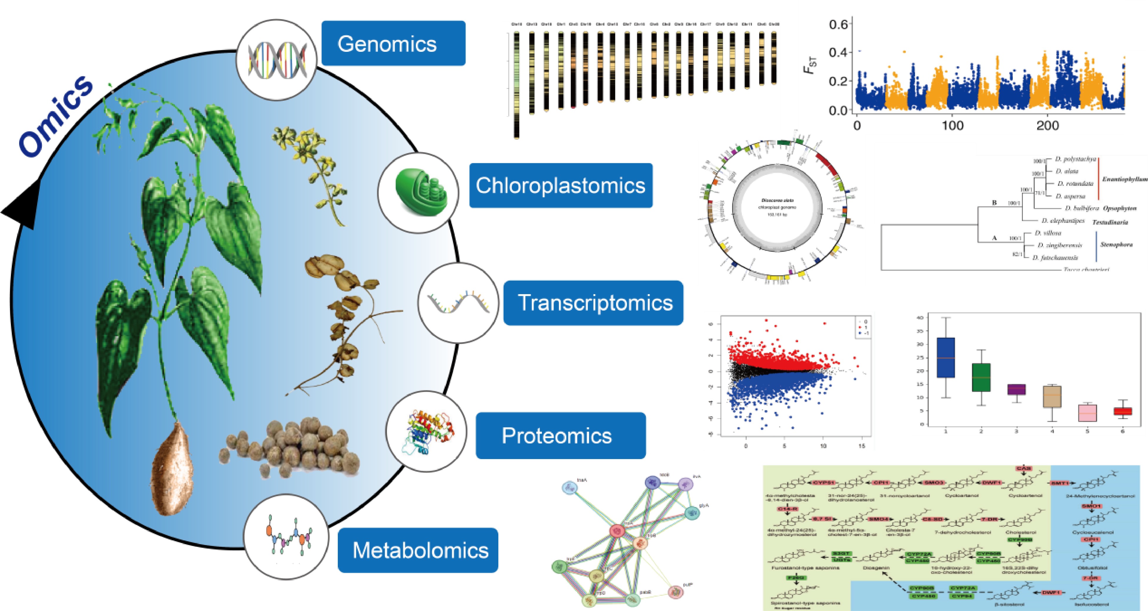
      Comprehensive understanding of genetic basics and conducting genetic improvement requires the interpretation of molecular intricacy and variations at multiple levels such as genome, plastome, transcriptome, proteome, and metabolome. With the advent of sequencing technology, biology research has become increasingly dependent on datasets generated at these levels for model organisms. However, as orphan crops, yams have received little attention in omics levels, with research mainly focusing on the components of the tubers, the germplasm resource classification, and extraction of specific components, and medicinal effects[7]. For example, pollution of the environment during the extraction of Diosgenin elements[8]. When systematically controlling diseases and pests in yams, it is necessary to analyze their genetic diversity. However, standard genetic analysis methods are not applicable due to the limited number of genetic markers available and the high heterozygosity associated with their obligate outcrossing nature. Therefore, the first species of the genus Dioscorea was subjected to whole-genome sequencing, resulting in the release of the first genome of a Dioscorea species, which ushered in the era of functional genomics of Dioscorea[9]. In recent years, significant progress has been made in the genomics of the Dioscorea genus. Scientists have conducted extensive and in-depth studies on Dioscorea species from various perspectives, including genomics, transcriptomics, proteomics, and metabolic networks (Fig. 1). This review provides the current state of genomic research on the Dioscorea genus, hoping to aid in furthering in-depth studies of the genus.

      Figure 1. 

      Omics technologies in yam research.

    • Although crop traits are regulated by genes, the sequencing of all genes alone provides insufficient information on which to base crop improvements such as greater yield and disease resistance[10]. Understanding the precise locations of all genes within a genome enhances the practicality of molecular marker technology. This knowledge enables the pinpointing of specific candidate genes responsible for particular traits, thereby extending the technology's utility. In the model species, such as rice and corn, it has witnessed the significant roles of genomics sequencing and analysis in the genetic improvement of crops[11]. To date, the genomes of five yam species have been sequenced and the genome assemblies of four species reached the chromosome level (Table 1). The assembled genome size ranged from 440 to 629 Mb, and the number of annotated coding genes ranged from 25,000 to 35,000. From these two sets of data, it can be seen that there is great genetic diversity among yam species. These genomics data and related analyses provide references for yam basic biology and molecular breeding.

      Table 1.  List of sequenced Dioscorea species genomes.

      SpeciesAssembly size (Mb)Assembly levelN50 (Mb)Gene numberRef.
      D. rotundata594Chromosome2.1226,198[9]
      D. rotundata*584Chromosome23.434,550[12]
      D. dumetorum485Contig3.235,269[13]
      D. alata479Chromosome2425,189[3]
      D. zingiberensis#480Chromosome44.526,022[14]
      D. zingiberensis#629Chromosome55.830,322[15]
      D. tokoro443Chromosome2429,084[16]
      * The improved genome assembly for D. rotundata; # The two assemblies from different D. zingiberensis strains.

      The sex determination mechanism is crucial in crop breeding, while this problem in yam has not been solved for a long time. In 2017, scientists sequenced and assembled the first genome of the Guinea yam (D. rotundata), marking a significant milestone in Dioscorea genomics[9]. Phylogenetic analysis of conserved genes illuminated the distinct nature of the Dioscorea lineage within monocotyledons, setting it apart from other groups such as Poales (rice), Arecales (palm), and Zingiberales (banana). With the genome tool, an approach was developed to conduct whole-genome resequencing of grouped segregants using F1 progeny that exhibited segregation of male and female D. rotundata plants. By the genomics analysis, a genomic region linked to female heterogametic sex determination (male = ZZ, female = ZW) was identified, and this discovery was further refined and transformed into a molecular marker for sex identification of Guinea yam plants at the seedling stage. Similarly, through genomic sequencing and analysis, the sex determination mechanism of D. tokoro was located in the middle of pseudochromosome 3, with a male heterogametic sex determination (XY) system[16].

      The dissection of yam domestication history plays an important role in the interpretation of the genetic mechanisms of important agronomic trait formation. An improved version of Guinea yam reference genome, together with more than 330 accessions and its wild relatives was used to investigate its origin[12]. The analysis results revealed that diploid D. rotundata was likely evolved from a hybrid of D. abyssinica and D. praehensilis. The assessment of the genomic contributions uncovered a pronounced presence of the D. abyssinica genome within the sex chromosome of D. rotundata and a clear signature of widespread introgression within the SWEETIE gene located on chromosome 17. To explore the chromosome evolution of D. alata, the yam research community generated a highly contiguous genome assembly and a dense genetic map from African breeding populations[3,17,18]. The genomic analysis results suggest that there was an ancient allotetraploidization in the Dioscorea lineage, subsequently with extensive genome-wide reorganization. Moreover, some QTLs (quantitative trait loci) were detected for resistance to anthracnose and tuber quality traits using the genomic tools.

      Although sapogenin saponins were isolated from the rhizomes of D. tokoro in the 1930s[19], its biosynthetic pathway has been a mystery. The comparative genomic analysis suggests that tandem duplication coupled with whole-genome duplication events provided key evolutionary resources for the diosgenin saponin biosynthetic pathway in D. zingiberensis[15]. Combined with transcriptome and metabolite analysis among 13 yam species, some gene expression patterns in specific metabolic pathways were found to be associated with the evolution of the diosgenin saponin biosynthetic pathway. These genes mainly involve in CYP450 family, such as CYP90B, CYP72A and CYP94. Further investigations revealed that the increased concentration of diosgenin in the yam lineage is governed by CpG islands. These islands have evolved to modulate gene expression within the diosgenin pathway, playing a crucial role in balancing the carbon flux between the biosynthesis of diosgenin and starch[14].

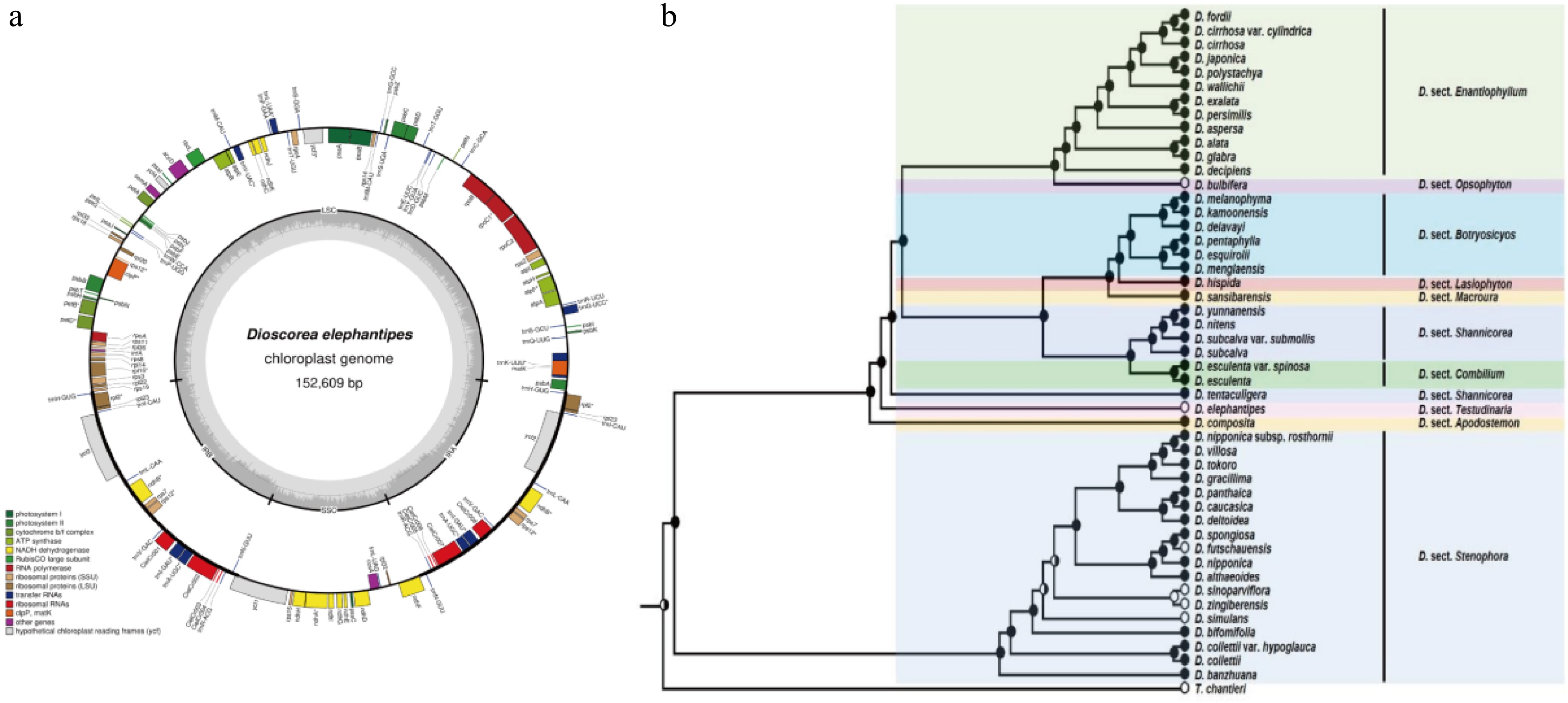
    • Compared to the nuclear genome, plastome, especially chloroplast genome sequences of the Dioscorea spp. are more easily sequenced and frequently used for the identification of kinship. The first chloroplast genome of yam is from D. elephantipes. As an important representative branch, it was employed to estimate phylogenetic relationships among angiosperms[20]. The chloroplast genome is only 152,609 base pairs in size, including 129 genes, 4 rRNAs, 38 tRNAs, as well as 16.72% inverted repeat (IR), 54.24% large single copy (SSC) and 12.32% small single copy (SSC) (Fig. 2a). Although chloroplast genomes are small and simple, it is still challenging to obtain complete chloroplast genomes for large-scale population genetics or phylogeographic studies. Therefore, in 2014, Mariac et al. developed an in-solution enrichment hybridization capture scheme suitable for deep multiplexing of chloroplast genomes, greatly aiding in the large-scale acquisition of complete chloroplast genome series for species[21]. Numerous Dioscorea species chloroplast genomes are stored in GenomeTrakrCP, greatly aiding subsequent research.

      Figure 2. 

      (a) Content of the D. elephantipes chloroplast genome[20]. (b) Phylogenetic tree of 48 Dioscorea species based on the concatenated matrix of matK, rbcL, tmL-F, psbA-tmH, rpl36-rps8, nad1, rps3 and 7 DNA[55].

      To ascertain the phylogenetics relationship among Dioscorea spp., more and more complete chloroplast genomes of yams were generated. In 2016, after sequencing the complete chloroplast genomes of Discorea species, a phylogenetic tree was constructed with other species and D. elephantipes and D. rotundata, showing closer phylogenetic relationships among the three D. species[22,23]. In 2018, high throughput technology was used to sequence the complete chloroplast genomes of D. aspersa, D. alata, D. bulbifera, D. futschauensis, and D. polystachya[24] and compared them with four previously studied species, finding that the chloroplast gene features and structures of these nine Dioscorea species are similar, with only slight differences in sequence length. Based on the species' chloroplast whole genomes, a phylogenetic tree was constructed, revealing the kinship relationships among the Dioscorea species. In the evolution tree, the nine species were divided into two branches. In the one branch, D. polystachya, D. alata, D. aspersa, and D. rotundata have a closer evolutionary relationship, while D. bulbifera and D. elephantipes are in a separate kinship relationship compared to the four species. In the other branch, D. villosa, D. futschauensis, and D. zingiberensis are closely related. In 2019, Magwé-Tindo et al. reconstructed complete chloroplast genomes for 14 African yam species and built a phylogenetic tree with D. rotundata as reference and D. elephantipes as outgroup species, enriching the evolutionary relationships among Dioscorea species[25]. In the following two years, different scientists continued refining the chloroplast genomes of different species. They performed phylogenetic analysis with Dioscorea species, determining the phylogenetic position. Cao et al. sequenced the whole chloroplast genome of D. persimilis and determined that D. persimilis is closely related to D. alata and D. polystachya but distantly related to D. rotundata[26], validating previous findings. Chen et al. sequenced the chloroplast genome of D. esculenta and constructed a phylogenetic tree, showing that the species is closer to D. sansibarensis[27]. Hu et al. sequenced the chloroplast genome of D. polystachya, compared it with 15 Dioscorea species, and found that D. polystachya is closely related to D. alata and D. aspersa[28]. Wonok et al. conducted structural, comparative, and evolutionary analysis of the chloroplast genomes of four native Thai Dioscorea species, providing the chloroplast genome structure and complete chloroplast gene sequences of D. depauperata, D. glabra, D. pyrifolia, and D. brevipetiolata. Phylogenetic analysis revealed that D. brevipetiolata, D. depauperata, and D. glabra are closely related to D. alata, while D. pyrifolia is more evolutionarily similar to D. aspersa[29].

      In addition to the complete chloroplast genome, chloroplast DNA markers can also be used for phylogenetic analysis. A largest phylogenetic tree that consisted of 48 Dioscorea species was constructed based on the concatenated matrix of seven markers including matK, rbcL, tmL-F, psbA-tmH, rpl36-rps8, nad1, rps3 and 7 DNA (Fig. 2b)[3]. This evolutionary tree provided molecular evidence for morphological classification, and for the first time, explored the evolution of four forms—bulbils, inflorescence openness, flower color, and inflorescence structure—based on the phylogenetic tree. This study provides evidence to support classification based on morphological traits and confirms that late differentiation of bulbils in Dioscorea species can improve reproductive efficiency and enhance adaptability.

    • Transcriptomics is the study of gene regulation and its expression at the RNA level[30], which developed from expressed sequence tag (EST) sequencing to the now widely used RNA sequencing (RNA-Seq). RNA-Seq is a method for transcript quantification that allows the more precise measurement of transcript levels and their isoforms compared to other approaches. Because of the high sensitivity in the detection of gene expression, RNA-Seq is often used to provide expression evidence for gene annotation in genome sequencing projects. For the species without reference genome, RNA-seq can be independently employed to construct reference transcriptome and investigate gene regulatory functions in various biological processes.

      The growth and development of yam are complex, from sprouting, stem, and leaf growth, to the final expansion of the tuber, flowering, and fruiting. The genes involved in their formation and their functions are still unknown. RNA-Seq was used to study the changes in the transcriptome during the formation of D. opposita microtubers and it was found that the development of microtubers is closely related to primary metabolisms, such as starch and sucrose metabolism[31], which has important implications for the study of germination. Although transcriptomic data during stem and leaf growth have been less studied, they will be sequenced collaboratively in the construction of the reference transcriptome of yam, thereby improving its accuracy[32]. The majority of yams do not flower or flower for a very short period, and transcriptome changes in flowers during early developmental stages were identified when analyzing transcriptome data from male, female, and dioecious individuals of D. rotundata[33]. Male plants were found to flower more intensely, similar to flowering-determining genes in other species, with a conserved flowering mechanism. Based on transcriptome analysis of microtubers, regulatory genes for phytohormones were also identified and ABA was found to positively regulate tuberous growth. On this foundation, it revealed that the metabolic pathways of tuber chemicals such as flavonoids are closely related to tuber development by transcriptome analysis[7]. Tuber size is strongly associated with yam yield, and the identification of gene functions related to the tuber expansion process can help improve. It turns out that SuSy and AGPase genes regulate the conversion of sucrose to starch in storage roots, which in turn positively regulates tuber enlargement (Fig. 3a)[34]. Similar to the mechanism of tuber influence, transcriptional analysis of the developmental stages of yam bulbs in conjunction with morphological analysis revealed growth hormone, CK, and sucrose as bulb initiation signals[35].
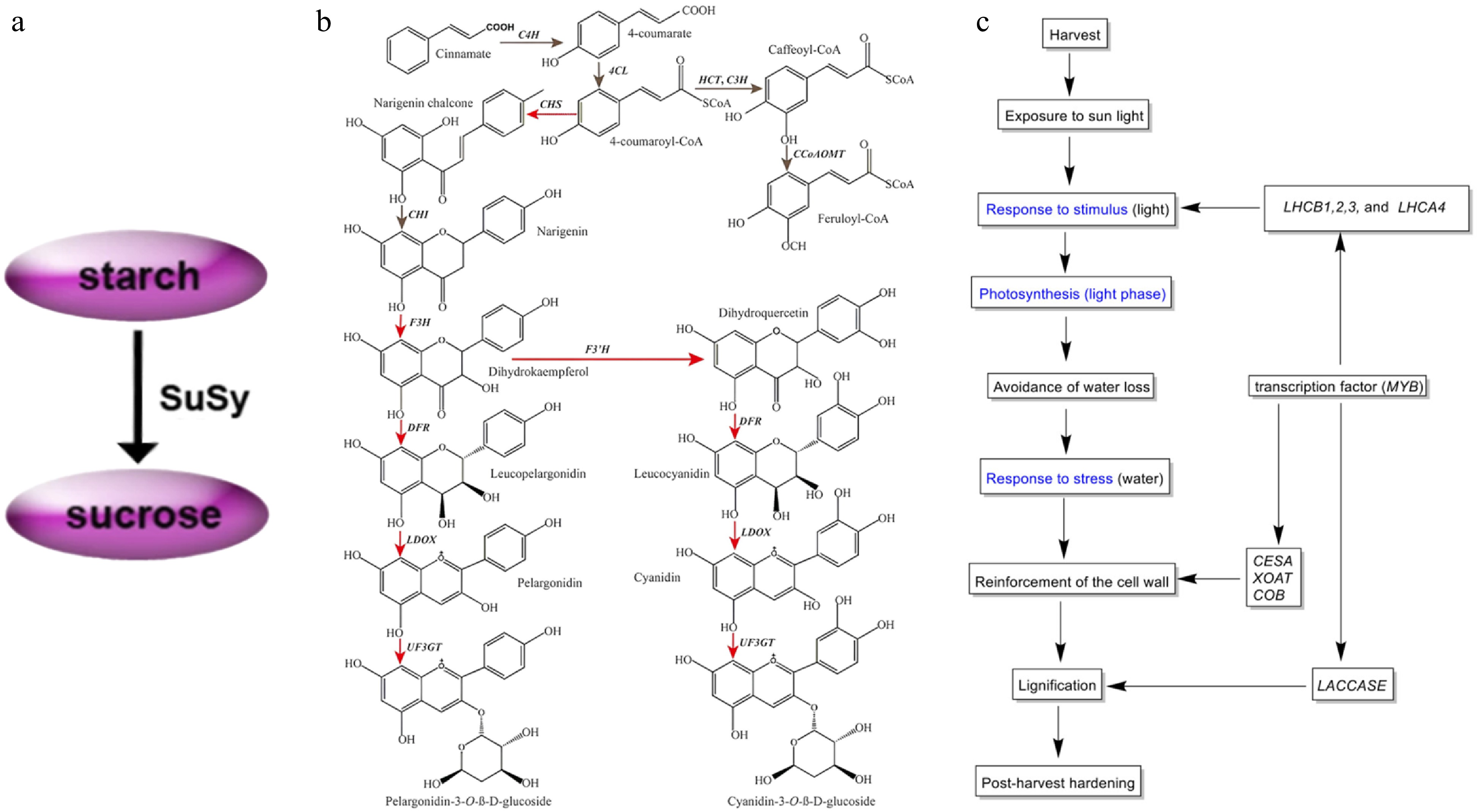
      Figure 3. 

      (a) Modeling the conversion between sucrose and starch during tuber amplification[34]. (b) Simplified diagram depicting the flavonoid biosynthesis pathway in yam tubers. Red arrows indicate genes that were significantly up-regulated in the purple-fleshed yam tuber. Gray indicates no change in gene expression between two tuber types[38]. (c) Putative mechanism of the PHH in D. dumetorum, blue represents GO annotation[41].

      The main constituent of yam tubers is starch, which is also an excellent source of flavonoids, diosgenin elements, and other medicinal constituents, but their biosynthetic pathways are not yet known. Sequencing of transcriptome libraries of D. polystachya leaf and rhizome tissues enriched the transcriptome data of the species while revealing differences in the expression of relevant genes involved in terpene synthesis, which in turn probed the molecular mechanisms of the biosynthetic pathway[36]. After reporting the whole genome sequence of D. zingiberensis, the transcriptome data of this species, which is rich in saponins, were analyzed in comparison with those of 13 saponin-containing species of the Dioscorea spp. Specific gene expression patterns of biosynthetic pathway genes were found to promote differential evolution of the saponin biosynthesis pathway in D. zingiberensis[15]. The yam domestication process has resulted in an increasing percentage of starch, and significant differences in the expression levels of related synthetic genes between the two substances were also found during the study. Starch is a key component affecting the yield and nutrition of yam tubers. Zhang et al.'s transcriptome analysis of D. polystachya species at various stages after sowing concluded that 135 d after sowing was the critical period for starch accumulation, and also verified the conclusion of the previous study on tuber dilation[37]. Flavonoids mainly affect color change in yam tubers, but the mechanism of color change is not fully understood so far. Comparative transcriptome analysis of white-meat and purple-meat varieties of D. alata species revealed significant differences in the expression of a large number of genes[38]. By identifying functional genes in the flavonoid biosynthesis pathway (FBP) of this species, it was found that genes encoding enzymes related to this pathway were significantly up-regulated in purple flesh varieties (Fig. 3b). D. cirrhosa, which is used as a natural dye and medicinal plant because of its reddish-brown tubers, was analyzed and 67 candidate genes related to the flavonoid biosynthesis pathway were identified[39].

      The storage of yam tubers is crucial, but relatively few studies of this kind have been conducted, and current research is directed towards the preservation of microtubers before sowing and severe post-harvest hardening (PHH). D. bulbifera field plantings usually use microtubers as propagation material, but they are highly susceptible to harboring pathogens that cause softening and rotting. Placement at low temperatures prolongs preservation, so transcriptome data of D. bulbifera at 4 °C were explored[39]. Look for preservation mechanisms and mining of related genes yielded a large amount of information related to microtubers preserved in vitro at low temperatures[40]. In response to the issue of hardening, which is defined as beginning within 24 h of harvest and becoming progressively unfit for human consumption, D. dumetorum is particularly pronounced[41]. In the first transcriptomic study of D. dumetorum transcriptomic data from three sclerotized and one non-sclerotized germplasm were analyzed to identify genes involved in the PHH phenomenon[41]. The study discovered that PHH involve the combined action of several genes, encoding cell wall polysaccharide components were significantly up-regulated, suggesting that they directly contribute to tuber hardening (Fig. 3c).

      Yam is an important cash crop in China, but it is susceptible to diseases caused by fungal infections, leading to a decline in quality and output of yam, so it is necessary to characterize its disease resistance. Gray mold is a common disease of yam that is sensitive to defensive hormones such as ethylene (ET), and yam-related transcription factors (TF) are involved in their synthesis and breakdown[42]. To explore the differences in hormone accumulation and gene expression patterns between resistant and susceptible yam varieties, Minghuai 3 (MH3), a highly susceptible variety to Bortrytis cinerea, and Minghuai 1 (MH1), a highly resistant variety, was selected to perform a comparative transcriptome analysis, which provided a basis for unraveling the regulatory mechanisms of the different varieties to the pathogen. Comparison of gene expression after inoculation of both varieties revealed that genes involved in ET signaling plays a key role in the antimicrobial mechanism. Further validation for this result was done by analyzing the gene expression of MH1 and MH3 after vinblastine treatment, and it was found that vinblastine treatment significantly increased the resistance of highly susceptible varieties to the pathogen.

    • Many types of information cannot be gleaned from the study of genes alone; the end products of genes are inherently more complex and closer to function than the genes themselves. Therefore, only through the study of proteins can we determine the functions of proteins that are profoundly affected by post-translational modifications[43]. The study of proteomics is also becoming more and more important, using techniques that are becoming more and more accurate in analyzing proteins as technology develops. There are fewer proteomic studies on yam, and the main research methods are gel-based mass spectrometry and mass spectrometry-based isotope tagging for relative and absolute quantification (iTRAQ). The main focus is on tuber research, such as the growth and development of tubers, and their chemical composition analysis.

      Studies on the growth and development of yam tubers at the gene level have been described in detail, but studies on the protein level have been less involved. In 2021, Sharma & Deswal reported a comprehensive tuber protein dataset for D. alata and analyzed the differences in protein levels at different growth stages[44]. Stage-specific gel-free proteomic analyses were performed for four different morphological stages: tuber sprouting (S1), degraded tuber (S2), new tuber formation (S3) and tuber maturation (S4). This research provided growth-specific markers for S1 and S3, revealing differences in protein expression at each developmental stage (Fig. 4). When people handle yam tubers, the sap always causes itching leading to allergy when it comes in contact with the skin unavoidably, so the study of the chemical composition of yam needs to be paid attention to. Mass spectrometry (MS) and protein qualitative histology analysis of yam proteins and each fraction of proteins after isolation[45]. Yam proteins were isolated and purified and found to have itchogenic properties in some fractions, which were further analyzed, and CCP2 was preliminarily hypothesized to be the itchogenic active ingredient. Fresh-cut yam is a good solution for juice allergy problems, but it tends to turn yellow during processing and storage, which affects product quality[46]. Combined with transcriptomics studies, the mechanism of yellowing in fresh-cut yam was elucidated and the cause of yellowing was explained.
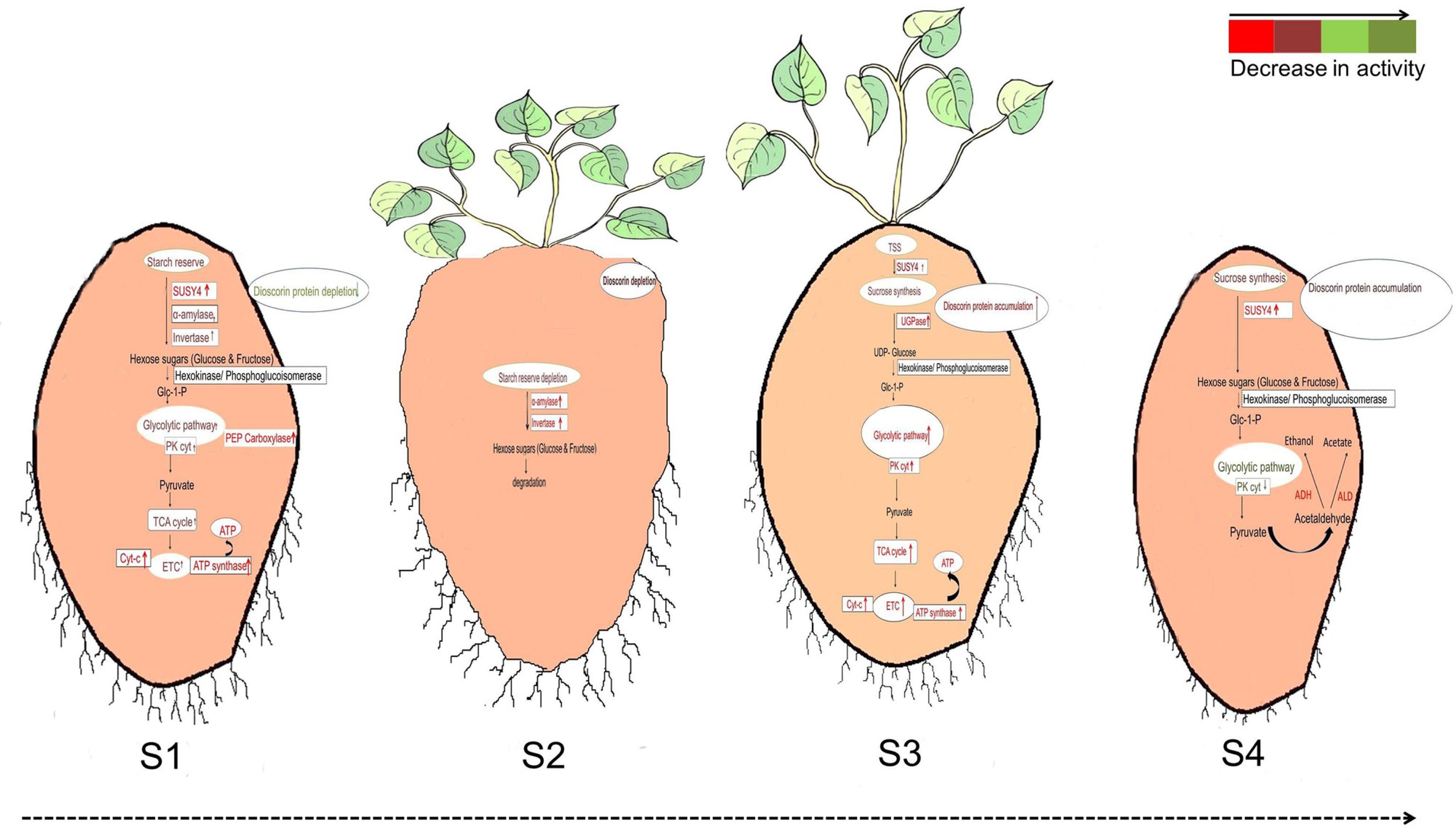
      Figure 4. 

      Differences in the regulation of glycolysis during (S1) tuber sprouting, (S2) degraded tuber, (S3) new tuber formation and (S4) tuber maturation[44].

    • Metabolomics is characterized by end effect and amplification compared to upstream proteomics and genomics, and metabolomics can provide a direct mapping of target metabolites[47]. Therefore, to further explore the abundance of nutritional and functional components, it is necessary to discover the metabolic profile of yam tubers from different species. As yet, the corresponding metabolite profiles or databases of six species have been constructed, which are of great significance to the breeding aspect of yams[47,48] (Table 2). The metabolomic data of yam leaves were provided in the construction of the crop metabolic database, which further improved the metabolic database of yam[49,50]. Furthermore, many traits of interest such as tuber growth and development maybe directly related to metabolite composition, and therefore genomics and metabolomics have been studied more in combination.

      Table 2.  List of mapped Dioscorea species metabolites.

      SpeciesMetabolites numberGermplasm number
      D. rotundata99−11610
      D. cayennensis96−1034
      D. dumetorum111−13025
      D. alata104−1145
      D. bulbifera107−1175
      D. polystachya4318

      Transcriptomic data suggest that yam microtuber formation is adjusted by a variety of hormones[31]. Endogenous levels of ABA was measured at different stages and found that it has a positive role in regulating microtuber formation. Metabolite assays targeting the tuber development process revealed that 400 metabolites accumulated during development[7]. Bulbs have the ability to reproduce as well as tubers, and according to ancient medical records, the clinical health effects of bulbs are superior to those of yam tubers[51]. D. polystachya species was used as a material, and its tubers and bulbs were subjected to boiling treatment and air-drying control, respectively, to compare and analyze the difference in metabolites between them, and it was found that yam bulbs had more nutrients than tubers. As a result, the mechanism of growth and development of yam bulbs is also very important. The metabolites of bulb during growth and development were analyzed, the regulation of growth hormones, CKs, ABA, and sucrose were detected to lead to bulb initiation and growth, with localized production of growth hormones being necessary to trigger the transient of formation.

      Genomic analysis revealed that the genome of Dioscorea spp. contains many genes encoding secondary metabolites. Thus has the potential to synthesize many secondary metabolites, including important compounds such as diosgenin elements and flavonoid phytohormones. Identification of changes in the saponin content of saponin-rich D. zingiberensis validates the hypothesis of a saponin biosynthesis pathway obtained by transcriptomics[15]. Combination with transcriptome analysis also revealed that proanthocyanidins (PAs), a downstream metabolite of the flavonoid biosynthesis pathway maybe a key metabolite in tuber color formation, and a mechanism by which flavonoids affect tuber color was discovered[39]. All metabolites in D. dumetorum contain saponins, alkaloids, and flavonoids and the high content of saponins serves as its chemical taxonomic marker[52]. To study the difference of phenolic and antioxidant potential among six species of Indian yam, the contents of flavonoids and other substances were identified[53]. Targeting saponins and catechins in D. alata metabolites that affect tuber quality provide a basis for breeding hybrids with low saponin and catechin content[54].

    • Combining current research hotspots of scholars both domestically and internationally and addressing the gaps in Dioscorea genomics studies, future work in the field needs to be envisioned as follows:

    • Expanding whole-genome sequencing to other Dioscorea species is an important direction for future genomic research in the genus. Utilizing comparative genomics to integrate interactions between individuals and populations within the genus can help identify key genes and genetic pathways in these interactions, elucidating the adaptive evolutionary mechanisms and genetic mechanisms. Additionally, research in Dioscorea genomics should also focus on genetic diversity, taxonomic identification, and stress responses such as DNA methylation.

    • More high-quality transcriptome will be constructed with reference to other tissues, in addition to the common yam, inter-tissue transcriptome differences in other Dioscorea species have yet to be carried out. At the same time, RNAs with mechanisms that regulate growth and development, tissue specificity and flowering need to be studied more extensively.

    • The development of Dioscorea proteomics has been limited, with fewer techniques used, targeting fewer species and scientific issues. Therefore, the application of the latest proteomic technologies in Dioscorea research need to be expanded to more species and scientific questions.

    • Multiple Dioscorea species contain a variety of important secondary metabolites. Currently, constructing metabolite maps or establishing metabolite databases focus on a limited number of species, so exploring the differences in metabolites among different species is crucial. Additionally, scientific issues explored at the metabolic level should be broader, moving beyond just tuber tissues to consider others.

    • The research aspect is not only limited to the study of yam itself, but with the updating of sequencing technology and the accumulation of histological data, it is possible to overcome the difficulties encountered in other research, such as the extraction of special components of yam.

    • The authors confirm contribution to the paper as follows: study conception and management: Dou D, Liu J; writing the manuscript: Chen Y, Tariq H, Shen D, Liu J, Dou D. All authors reviewed the results and approved the final version of the manuscript.

    • Data sharing not applicable to this article as no datasets were generated or analyzed during the current study.

      • This study was supported by grants from the China Agriculture Research System (CARS-21), the National Natural Science Foundation of China (32270208 and 32230089) and the Fundamental Research Funds for the Central Universities (KYCXJC2023001 and KYQN2023039).

      • The authors declare that they have no conflict of interest.

      • Copyright: © 2024 by the author(s). Published by Maximum Academic Press, Fayetteville, GA. This article is an open access article distributed under Creative Commons Attribution License (CC BY 4.0), visit https://creativecommons.org/licenses/by/4.0/.
    Figure (4)  Table (2) References (55)
  • About this article
    Cite this article
    Chen Y, Tariq H, Shen D, Liu J, Dou D. 2024. Omics technologies accelerating research progress in yams. Vegetable Research 4: e014 doi: 10.48130/vegres-0024-0014
    Chen Y, Tariq H, Shen D, Liu J, Dou D. 2024. Omics technologies accelerating research progress in yams. Vegetable Research 4: e014 doi: 10.48130/vegres-0024-0014

Catalog

  • About this article

/

DownLoad:  Full-Size Img  PowerPoint
Return
Return