ARTICLE   Open Access    

Regeneration and Agrobacterium-mediated genetic transformation of twelve Eucalyptus species

More Information
  • Eucalyptus is a genus of over 900 species and hybrids, and many of them are valuable fast-growing hardwoods. Due to its economic importance, Eucalyptus is one of the early tree species whose genomes were deciphered. However, the lack of efficient genetic transformation systems severely restricts the functional genomic research on the plant. The success of Eucalyptus regeneration and transformation depends greatly on the genotypes and explants. In this study, we systematically screened 26 genotypes from 12 Eucalyptus species in an attempt to obtain Eucalyptus genotypes with high regeneration potential. We developed two common regeneration media that can be applied to most tested Eucalyptus genotypes for both seeding hypocotyls and cloned internodes as explants. We then implemented DsRed2 as a visual marker for genetic transformation efficiency test. Our results suggest that E. camaldulen and E. robusta are amenable for genetic transformation. Finally, we successfully set up a stable Agrobacterium-mediated genetic transformation procedure for both E. camaldulen and E. robusta using seeding hypocotyls and cloned internodes respectively. Taken together, our study provides valuable means for vegetative propagation, gene transformation, CRISPR based gene mutagenesis, activation and suppression, as well as functional characterization of genes in Eucalyptus.
  • Aquaporin’s (AQPs) are small (21–34 kD) channel-forming, water-transporting trans-membrane proteins which are known as membrane intrinsic proteins (MIPs) conspicuously present across all kingdoms of life. In addition to transporting water, plant AQPs act to transport other small molecules including ammonia, carbon dioxide, glycerol, formamide, hydrogen peroxide, nitric acid, and some metalloids such as boron and silicon from the soil to different parts of the plant[1]. AQPs are typically composed of six or fewer transmembrane helices (TMHs) coupled by five loops (A to E) and cytosolic N- and C-termini, which are highly conserved across taxa[2]. Asparagine-Proline-Alanine (NPA) boxes and makeup helices found in loops B (cytosolic) and E (non-cytosolic) fold back into the protein's core to form one of the pore's two primary constrictions, the NPA region[1]. A second filter zone exists at the pore's non-cytosolic end, where it is called the aromatic/arginine (ar/R) constriction. The substrate selectivity of AQPs is controlled by the amino acid residues of the NPA and ar/R filters as well as other elements of the channel[1].

    To date, the AQP gene families have been extensively explored in the model as well as crop plants[39]. In seed plants, AQP distributed into five subfamilies based on subcellular localization and sequence similarities: the plasma membrane intrinsic proteins (PIPs; subgroups PIP1 and PIP2), the tonoplast intrinsic proteins (TIPs; TIP1-TIP5), the nodulin26-like intrinsic proteins (NIPs; NIP1-NIP5), the small basic intrinsic proteins (SIPs; SIP1-SIP2) and the uncategorized intrinsic proteins (XIPs; XIP1-XIP3)[2,10]. Among them, TIPs and PIPs are the most abundant and play a central role in facilitating water transport. SIPs are mostly found in the endoplasmic reticulum (ER)[11], whereas NIPs homologous to GmNod26 are localized in the peribacteroid membrane[12].

    Several studies reported that the activity of AQPs is regulated by various developmental and environmental factors, through which water fluxes are controlled[13]. AQPs are found in all organs such as leaves, roots, stems, flowers, fruits, and seeds[14,15]. According to earlier studies, increased AQP expression in transgenic plants can improve the plants' tolerance to stresses[16,17]. Increased root water flow caused by upregulation of root aquaporin expression may prevent transpiration[18,19]. Overexpression of Tamarix hispida ThPIP2:5 improved osmotic stress tolerance in Arabidopsis and Tamarix plants[20]. Transgenic tomatoes having apple MdPIP1;3 ectopically expressed produced larger fruit and improved drought tolerance[21]. Plants over-expressing heterologous AQPs, on the other hand, showed negative effects on stress tolerance in many cases. Overexpression of GsTIP2;1 from G. soja in Arabidopsis plants exhibited lower resistance against salt and drought stress[22].

    A few recent studies have started to establish a link between AQPs and nanobiology, a research field that has been accelerating in the past decade due to the recognition that many nano-substances including carbon-based materials are valuable in a wide range of agricultural, industrial, and biomedical activities[23]. Carbon nanotubes (CNTs) were found to improve water absorption and retention and thus enhance seed germination in tomatoes[24,25]. Ali et al.[26] reported that Carbon nanoparticles (CTNs) and osmotic stress utilize separate processes for AQP gating. Despite lacking solid evidence, it is assumed that CNTs regulate the aquaporin (AQPs) in the seed coats[26]. Another highly noticed carbon-nano-molecule, the fullerenes, is a group of allotropic forms of carbon consisting of pure carbon atoms[27]. Fullerenes and their derivatives, in particular the water-soluble fullerols [C60(OH)20], are known to be powerful antioxidants, whose biological activity has been reduced to the accumulation of superoxide and hydroxyl[28,29]. Fullerene/fullerols at low concentrations were reported to enhance seed germination, photosynthesis, root growth, fruit yield, and salt tolerance in various plants such as bitter melon and barley[3032]. In contrast, some studies also reported the phytotoxic effect of fullerene/fullerols[33,34]. It remains unknown if exogenous fullerene/fullerol has any impact on the expression or activity of AQPs in the cell.

    Garden pea (P. sativum) is a cool-season crop grown worldwide; depending on the location, planting may occur from winter until early summer. Drought stress in garden pea mainly affects the flowering and pod filling which harm their yield. In the current study, we performed a genome-wide identification and characterization of the AQP genes in garden pea (P. sativum), the fourth largest legume crop worldwide with a large complex genome (~4.5 Gb) that was recently decoded[35]. In particular, we disclose, for the first time to our best knowledge, that the transcriptional regulations of AQPs by osmotic stress in imbibing pea seeds were altered by fullerol supplement, which provides novel insight into the interaction between plant AQPs, osmotic stress, and the carbon nano-substances.

    The whole-genome sequence of garden pea ('Caméor') was retrieved from the URGI Database (https://urgi.versailles.inra.fr/Species/Pisum). Protein sequences of AQPs from two model crops (Rice and Arabidopsis) and five other legumes (Soybean, Chickpea, Common bean, Medicago, and Peanut) were used to identify homologous AQPs from the garden pea genome (Supplemental Table S1). These protein sequences, built as a local database, were then BLASTp searched against the pea genome with an E-value cutoff of 10−5 and hit a score cutoff of 100 to identify AQP orthologs. The putative AQP sequences of pea were additionally validated to confirm the nature of MIP (Supplemental Table S2) and transmembrane helical domains through TMHMM (www.cbs.dtu.dk/services/TMHMM/).

    Further phylogenetic analysis was performed to categorize the AQPs into subfamilies. The pea AQP amino acid sequences, along with those from Medicago, a cool-season model legume phylogenetically close to pea, were aligned through ClustalW2 software (www.ebi.ac.uk/Tools/msa/clustalw2) to assign protein names. The unaligned AQP sequences to Medicago counterparts were once again aligned with the AQP sequences of Arabidopsis, rice, and soybean. Based on the LG model, unrooted phylogenetic trees were generated via MEGA7 and the neighbor-joining method[36], and the specific name of each AQP gene was assigned based on its position in the phylogenetic tree.

    By using the conserved domain database (CDD, www.ncbi.nlm.nih.gov/Structure/cdd/cdd.shtml), the NPA motifs were identified from the pea AQP protein sequences[37]. The software TMHMM (www.cbs. dtu.dk/services/TMHMM/)[38] was used to identify the protein transmembrane domains. To determine whether there were any alterations or total deletion, the transmembrane domains were carefully examined.

    Basic molecular properties including amino acid composition, relative molecular weight (MW), and instability index were investigated through the online tool ProtParam (https://web.expasy.org/protparam/). The isoelectric points (pI) were estimated by sequence Manipulation Suite version 2 (www.bioinformatics.org/sms2)[39]. The subcellular localization of AQP proteins was predicted using Plant-mPLoc[40] and WoLF PSORT (www.genscript.com/wolf-psort.html)[ 41] algorithms.

    The gene structure (intron-exon organization) of AQPs was examined through GSDS ver 2.0[42]. The chromosomal distribution of the AQP genes was illustrated by the software MapInspect (http://mapinspect.software.informer.com) in the form of a physical map.

    To explore the tissue expression patterns of pea AQP genes, existing NGS data from 18 different libraries covering a wide range of tissue, developmental stage, and growth condition of the variety ‘Caméor’ were downloaded from GenBank (www.ncbi.nlm.nih.gov/bioproject/267198). The expression levels of the AQP genes in each tissue and growth stage/condition were represented by the FPKM (Fragments Per Kilobase of transcript per Million fragments mapped) values. Heatmaps of AQPs gene were generated through Morpheus software (https://software.broadinstitute.org/morpheus/#).

    Different solutions, which were water (W), 0.3 M mannitol (M), and fullerol of different concentrations dissolved in 0.3 M mannitol (MF), were used in this study. MF solutions with the fullerol concentration of 10, 50, 100, and 500 mg/L were denoted as MF1, MF2, MF3, and MF4, respectively. Seeds of 'SQ-1', a Chinese landrace accession of a pea, were germinated in two layers of filter paper with 30 mL of each solution in Petri dishes (12 cm in diameter) each solution, and the visual phenotype and radicle lengths of 150 seeds for each treatment were analyzed 72 h after soaking. The radicle lengths were measured using a ruler. Multiple comparisons for each treatment were performed using the SSR-Test method with the software SPSS 20.0 (IBM SPSS Statistics, Armonk, NY, USA).

    Total RNA was extracted from imbibing embryos after 12 h of seed soaking in the W, M, and MF3 solution, respectively, by using Trizol reagent (Invitrogen, Carlsbad, CA, USA). The quality and quantity of the total RNA were measured through electrophoresis on 1% agarose gel and an Agilent 2100 Bioanalyzer respectively (Agilent Technologies, Santa Rosa, USA). The TruSeq RNA Sample Preparation Kit was utilized to construct an RNA-Seq library from 5 µg of total RNA from each sample according to the manufacturer's instruction (Illumina, San Diego, CA, USA). Next-generation sequencing of nine libraries were performed through Novaseq 6000 platform (Illumina, San Diego, CA, USA).

    First of all, by using SeqPrep (https://github.com/jstjohn/SeqPrep) and Sickle (https://github.com/najoshi/sickle) the raw RNA-Seq reads were filtered and trimmed with default parameters. After filtering, high-quality reads were mapped onto the pea reference genome (https://urgi.versailles.inra.fr/Species/Pisum) by using TopHat (V2.1.0)[43]. Using Cufflinks, the number of mapped reads from each sample was determined and normalised to FPKM for each predicted transcript (v2.2.1). Pairwise comparisons were made between W vs M and W vs M+F treatments. The DEGs with a fold change ≥ 1.5 and false discovery rate (FDR) adjusted p-values ≤ 0.05 were identified by using Cuffdiff[44].

    qPCR was performed by using TOROGGreen® qPCR Master Mix (Toroivd, Shanghai, China) on a qTOWER®3 Real-Time PCR detection system (Analytik Jena, Germany). The reactions were performed at 95 °C for 60 s, followed by 42 cycles of 95 °C for 10 s and 60 °C for 30 s. Quantification of relative expression level was achieved by normalization against the transcripts of the housekeeping genes β-tubulin according to Kreplak et al.[35]. The primer sequences for reference and target genes used are listed in Supplemental Table S3.

    The homology-based analysis identifies 41 putative AQPs in the garden pea genome. Among them, all but two genes (Psat0s3550g0040.1, Psat0s2987g0040.1) encode full-length aquaporin-like sequences (Table 1). The conserved protein domain analysis later validated all of the expected AQPs (Supplemental Table S2). To systematically classify these genes and elucidate their relationship with the AQPs from other plants' a phylogenetic tree was created. It clearly showed that the AQPs from pea and its close relative M. truncatula formed four distinct clusters, which represented the different subfamilies of AQPs i.e. TIPs, PIPs, NIPs, and SIPs (Fig. 1a). However, out of the 41 identified pea AQPs, 4 AQPs couldn't be tightly aligned with the Medicago AQPs and thus were put to a new phylogenetic tree constructed with AQPs from rice, Arabidopsis, and soybean. This additional analysis assigned one of the 4 AQPs to the XIP subfamily and the rest three to the TIP or NIP subfamilies (Fig. 1b). Therefore, it is concluded that the 41 PsAQPs comprise 11 PsTIPs, 15 PsNIPs, 9 PsPIPs, 5 PsSIPs, and 1 PsXIP (Table 2). The PsPIPs formed two major subgroups namely PIP1s and PIP2s, which comprise three and six members, respectively (Table 1). The PsTIPs formed two major subgroups TIPs 1 (PsTIP1-1, PsTIP1-3, PsTIP1-4, PsTIP1-7) and TIPs 2 (PsTIP2-1, PsTIP2-2, PsTIP2-3, PsTIP2-6) each having four members (Table 2). Detailed information such as gene/protein names, accession numbers, the length of deduced polypeptides, and protein structural features are presented in Tables 1 & 2

    Table 1.  Description and distribution of aquaporin genes identified in the garden pea genome.
    Chromosome
    S. NoGene NameGene IDGene length
    (bp)
    LocationStartEndTranscription length (bp)CDS length
    (bp)
    Protein length
    (aa)
    1PsPIP1-1Psat5g128840.32507chr5LG3231,127,859231,130,365675675225
    2PsPIP1-2Psat2g034560.11963chr2LG149,355,95849,357,920870870290
    3PsPIP1-4Psat2g182480.11211chr2LG1421,647,518421,648,728864864288
    4PsPIP2-1Psat6g183960.13314chr6LG2369,699,084369,702,397864864288
    5PsPIP2-2-1Psat4g051960.11223chr4LG486,037,44686,038,668585585195
    6PsPIP2-2-2Psat5g279360.22556chr5LG3543,477,849543,480,4042555789263
    7PsPIP2-3Psat7g228600.22331chr7LG7458,647,213458,649,5432330672224
    8PsPIP2-4Psat3g045080.11786chr3LG5100,017,377100,019,162864864288
    9PsPIP2-5Psat0s3550g0040.11709scaffold0355020,92922,63711911191397
    10PsTIP1-1Psat3g040640.12021chr3LG589,426,47389,428,493753753251
    11PsTIP1-3Psat3g184440.12003chr3LG5393,920,756393,922,758759759253
    12PsTIP1-4Psat7g219600.12083chr7LG7441,691,937441,694,019759759253
    13PsTIP1-7Psat6g236600.11880chr6LG2471,659,417471,661,296762762254
    14PsTIP2-1Psat1g005320.11598chr1LG67,864,8107,866,407750750250
    15PsTIP2-2Psat4g198360.11868chr4LG4407,970,525407,972,392750750250
    16PsTIP2-3Psat1g118120.12665chr1LG6230,725,833230,728,497768768256
    17PsTIP2-6Psat2g177040.11658chr2LG1416,640,482416,642,139750750250
    18PsTIP3-2Psat6g054400.11332chr6LG254,878,00354,879,334780780260
    19PsTIP4-1Psat6g037720.21689chr6LG230,753,62430,755,3121688624208
    20PsTIP5-1Psat7g157600.11695chr7LG7299,716,873299,718,567762762254
    21PsNIP1-1Psat1g195040.21864chr1LG6346,593,853346,595,7161863645215
    22PsNIP1-3Psat1g195800.11200chr1LG6347,120,121347,121,335819819273
    23PsNIP1-5Psat7g067480.12365chr7LG7109,420,633109,422,997828828276
    24PsNIP1-6Psat7g067360.12250chr7LG7109,270,462109,272,711813813271
    25PsNIP1-7Psat1g193240.11452chr1LG6344,622,606344,624,057831831277
    26PsNIP2-1-2Psat3g197520.1669chr3LG5420,092,382420,093,050345345115
    27PsNIP2-2-2Psat3g197560.1716chr3LG5420,103,168420,103,883486486162
    28PsNIP3-1Psat2g072000.11414chr2LG1133,902,470133,903,883798798266
    29PsNIP4-1Psat7g126440.11849chr7LG7209,087,362209,089,210828828276
    30PsNIP4-2Psat5g230920.11436chr5LG3463,340,575463,342,010825825275
    31PsNIP5-1Psat6g190560.11563chr6LG2383,057,323383,058,885867867289
    32PsNIP6-1Psat5g304760.45093chr5LG3573,714,868573,719,9605092486162
    33PsNIP6-2Psat7g036680.12186chr7LG761,445,34161,447,134762762254
    34PsNIP6-3Psat7g259640.12339chr7LG7488,047,315488,049,653918918306
    35PsNIP7-1Psat6g134160.24050chr6LG2260,615,019260,619,06840491509503
    36PsSIP1-1Psat3g091120.13513chr3LG5187,012,329187,015,841738738246
    37PsSIP1-2Psat1g096840.13609chr1LG6167,126,599167,130,207744744248
    38PsSIP1-3Psat7g203280.12069chr7LG7401,302,247401,304,315720720240
    39PsSIP2-1-1Psat0s2987g0040.1706scaffold02987177,538178,243621621207
    40PsSIP2-1-2Psat3g082760.13135chr3LG5173,720,100173,723,234720720240
    41PsXIP2-1Psat7g178080.12077chr7LG7335,167,251335,169,327942942314
    bp: base pair, aa: amino acid.
     | Show Table
    DownLoad: CSV
    Figure 1.  Phylogenetic analysis of the identified AQPs from pea genome. (a) The pea AQPs proteins aligned with those from the cool-season legume Medicago truncatual. (b) The four un-assigned pea AQPs in (a) (denoted as NA) were further aligned with the AQPs of rice, soybean, and Arabidopsis by using the Clustal W program implemented in MEGA 7 software. The nomenclature of PsAQPs was based on homology with the identified aquaporins that were clustered together.
    Table 2.  Protein information, conserved amino acid residues, trans-membrane domains, selectivity filter, and predicted subcellular localization of the 39 full-length pea aquaporins.
    S. NoAQPsGeneLengthTMHNPANPAar/R selectivity filterpIWoLF PSORTPlant-mPLoc
    LBLEH2H5LE1LE2
    Plasma membrane intrinsic proteins (PIPs)
    1PsPIP1-1Psat5g128840.32254NPA0F0008.11PlasPlas
    2PsPIP1-2Psat2g034560.12902NPANPAFHTR9.31PlasPlas
    3PsPIP1-4Psat2g182480.12886NPANPAFHTR9.29PlasPlas
    4PsPIP2-1Psat6g183960.12886NPANPAFHT08.74PlasPlas
    5PsPIP2-2-1Psat4g051960.1195300FHTR8.88PlasPlas
    6PsPIP2-2-2Psat5g279360.22635NPANPAFHTR5.71PlasPlas
    7PsPIP2-3Psat7g228600.22244NPA0FF006.92PlasPlas
    8PsPIP2-4Psat3g045080.12886NPANPAFHTR8.29PlasPlas
    Tonoplast intrinsic proteins (TIPs)
    1PsTIP1-1Psat3g040640.12517NPANPAHIAV6.34PlasVacu
    2PsTIP1-3Psat3g184440.12536NPANPAHIAV5.02Plas/VacuVacu
    3PsTIP1-4Psat7g219600.12537NPANPAHIAV4.72VacuVacu
    4PsTIP1-7Psat6g236600.12546NPANPAHIAV5.48Plas/VacuVacu
    5PsTIP2-1Psat1g005320.12506NPANPAHIGR8.08VacuVacu
    6PsTIP2-2Psat4g198360.12506NPANPAHIGR5.94Plas/VacuVacu
    7PsTIP2-3Psat1g118120.12566NPANPAHIAL6.86Plas/VacuVacu
    8PsTIP2-6Psat2g177040.12506NPANPAHIGR4.93VacuVacu
    9PsTIP3-2Psat6g054400.12606NPANPAHIAR7.27Plas/VacuVacu
    10PsTIP4-1Psat6g037720.22086NPANPAHIAR6.29Vac/ plasVacu
    11PsTIP5-1Psat7g157600.12547NPANPANVGC8.2Vacu /plasVacu/Plas
    Nodulin-26 like intrisic proteins (NIPs)
    1PsNIP1-1Psat1g195040.22155NPA0WVF06.71PlasPlas
    2PsNIP1-3Psat1g195800.12735NPANPVWVAR6.77PlasPlas
    3PsNIP1-5Psat7g067480.12766NPANPVWVAN8.98PlasPlas
    4PsNIP1-6Psat7g067360.12716NPANPAWVAR8.65Plas/VacuPlas
    5PsNIP1-7Psat1g193240.12776NPANPAWIAR6.5Plas/VacuPlas
    6PsNIP2-1-2Psat3g197520.11152NPAOG0009.64PlasPlas
    7PsNIP2-2-2Psat3g197560.116230NPA0SGR6.51PlasPlas
    8PsNIP3-1Psat2g072000.12665NPANPASIAR8.59Plas/VacuPlas
    9PsNIP4-1Psat7g126440.12766NPANPAWVAR6.67PlasPlas
    10PsNIP4-2Psat5g230920.12756NPANPAWLAR7.01PlasPlas
    11PsNIP5-1Psat6g190560.12895NPSNPVAIGR7.1PlasPlas
    12PsNIP6-1Psat5g304760.41622NPA0I0009.03PlasPlas
    13PsNIP6-2Psat7g036680.1254000G0005.27ChloPlas/Nucl
    14PsNIP6-3Psat7g259640.13066NPANPVTIGR8.32PlasPlas
    15PsNIP7-1Psat6g134160.25030NLK0WGQR8.5VacuChlo/Nucl
    Small basic intrinsic proteins (SIPs)
    1PsSIP1-1Psat3g091120.12466NPTNPAVLPN9.54PlasPlas/Vacu
    2PsSIP1-2Psat1g096840.12485NTPNPAIVPL9.24VacuPlas/Vacu
    3PsSIP1-3Psat7g203280.12406NPSNPANLPN10.32ChloPlas
    4PsSIP2-1-2Psat3g082760.12404NPLNPAYLGS10.28PlasPlas
    Uncharacterized X intrinsic proteins (XIPs)
    1PsXIP2-1Psat7g178080.13146SPVNPAVVRM7.89PlasPlas
    Length: protein length (aa); pI: Isoelectric point; Trans-membrane helicase (TMH) represents for the numbers of Trans-membrane helices predicted by TMHMM Server v.2.0 tool; WoLF PSORT and Plant-mPLoc: best possible cellualr localization predicted by the WoLF PSORT and Plant-mPLoc tool, respectively (Chlo Chloroplast, Plas Plasma membrane, Vacu Vacuolar membrane, Nucl Nucleus); LB: Loop B, L: Loop E; NPA: Asparagine-Proline-Alanine; H2 represents for Helix 2, H5 represents for Helix 5, LE1 represents for Loop E1, LE2 represents for Loop E2, Ar/R represents for Aromatic/Arginine.
     | Show Table
    DownLoad: CSV

    To understand the genome distribution of the 41 PsAQPs, we mapped these genes onto the seven chromosomes of a pea to retrieve their physical locations (Fig. 2). The greatest number (10) of AQPs were found on chromosome 7, whereas the least (2) on chromosome 4 (Fig. 2 and Table 1). Chromosomes 1 and 6 each contain six aquaporin genes, whereas chromosomes 2, 3, and 5 carry four, seven, and four aquaporin genes, respectively (Fig. 2). The trend of clustered distribution of AQPs was seen on specific chromosomes, particularly near the end of chromosome 7.

    Figure 2.  Chromosomal localization of the 41 PsAQPs on the seven chromosomes of pea. Chr1-7 represents the chromosomes 1 to 7. The numbers on the right of each chromosome show the physical map positions of the AQP genes (Mbp). Blue, green, orange, brown, and black colors represent TIPs, NIPs, PIPs, SIPs, and XIP, respectively.

    The 39 full-length PsAQP proteins have a length of amino acid ranging from 115 to 503 (Table 1) and Isoelectric point (pI) values ranging from 4.72 to 10.35 (Table 2). As a structural signature, transmembrane domains were predicted to exist in all PsAQPs, with the number in individual AQPs varying from 2 to 6. By subfamilies, TIPs harbor the greatest number of TM domains in total, followed by PIPs, NIPs, SIPs, and XIP (Table 2). Exon-intron structure analysis showed that most PsAQPs (16/39) having two introns, while ten members had three, seven members had four, and five members had only one intron (Fig. 3). Overall, PsAQPs exhibited a complex structure with varying intron numbers, positions, and lengths.

    Figure 3.  The exon-intron structures of the AQP genes in pea. Upstream/downstream region, exon, and intron are represented by a blue box, yellow box, and grey line, respectively.

    As aforementioned, generally highly conserved two NPA motifs generate an electrostatic repulsion of protons in AQPs to form the water channel, which is essential for the transport of substrate molecules[15]. In order to comprehend the potential physiological function and substrate specificity of pea aquaporins, NPA motifs (LB, LE) and residues at the ar/R selectivity filter (H2, H5, LE1, and LE2) were examined. (Table 2). We found that all PsTIPs and most PsPIPs had two conserved NPA motifs except for PsPIP1-1, PsPIP2-2-1, and PsPIP2-3, each having a single NPA motif. Among PsNIPs, PsNIP1-6, PsNIP1-6, PsNIP1-7, PsNIP3-1, PsNIP4-1 and PSNIP4-2 had two NPA domains, while PsNIP1-1, PsNIP2-1-2, PsNIP2-2-2 and PsNIP6-1 each had a single NPA motif. In the PsNIP sub-family, the first NPA motif showed an Alanine (A) to Valine (V) substitution in three PsNIPs (PsNIP1-3, PsNIP1-5, and PsNIP6-3) (Table 2). Furthermore, the NPA domains of all members of the XIP and SIP subfamilies were different. The second NPA motif was conserved in PsSIP aquaporins, however, all of the first NPA motifs had Alanine (A) replaced by Leucine (L) (PsSIP2-1-1, PsSIP2-1-2) or Threonine (T) (PsSIP1-1). In comparison to other subfamilies, this motif variation distinguishes water and solute-transporting aquaporins[45].

    Compared to NPA motifs, the ar/R positions were more variable and the amino acid composition appeared to be subfamily-dependent. The majority of PsPIPs had phenylalanine at H2, histidine at H5, threonine at LE1, and arginine at LE2 selective filter (Table 2). All of the PsTIP1 members had a Histidine-Isoleucine-Alanine-Valine structure at this position, while all PsTIP2 members but PsTIP2-3 harbored Histidine-Isoleucine-Glycine-Arginine. Similarly, PsNIPs, PsSIPs and PsXIP also showed subgroup-specific variation in ar/R selectivity filter (Table 2). Each of these substitutions partly determines the function of transporting water[46].

    Sequence-based subcellular localization analysis using WoLF PSORT predicted that all PsPIPs localized in the plasma membrane, which is consistent with their subfamily classification (Table 2). Around half (5/11) of the PsTIPs (PsTIP1-4, PsTIP2-1, PsTIP2-6, PsTIP4-1, and PsTIP5-1) were predicted to localize within vacuoles. However, several TIP members (PsTIP1-1, PsTIP1-3, PsTIP1-7, PsTIP2-2, PsTIP2-3 and PsTIP3-2) were predicted to localize in plasma membranes. We then further investigated their localizations by using another software (Plant-mPLoc, Table 2), which predicted that all the PsTIPs localize within vacuoles, thus supporting that they are tonoplast related. An overwhelming majority of PsNIPs (14/15) and PsXIP were predicted to be found only in plasma membranes., which was also expected (Table 2). Collectively, the versatility in subcellular localization of the pea AQPs is implicative of their distinct roles in controlling water and/or solute transport in the context of plant cell compartmentation.

    Tissue expression patterns of genes are indicative of their functions. Since there were rich resources of RNA-Seq data from various types of pea tissues in the public database, they were used for the extraction of expression information of PsAQP genes as represented by FPKM values. A heat map was generated to show the expression patterns of PsAQP genes in 18 different tissues/stages and their responses to nitrate levels (Fig. 4). According to the heat map, PsPIP1-2, PsPIP2-3 were highly expressed in root and nodule G (Low-nitrate), whereas PsTIP1-4, PsTIP2-6, and PsNIP1-7 were only expressed in roots in comparison to other tissues. The result also demonstrated that PsPIP1-1 and PsNIP3-1 expressed more abundantly in leaf, tendril, and peduncle, whereas PsPIP2-2-2 and PsTIP1-1 showed high to moderate expressions in all the samples except for a few. Interestingly, PsTIP1-1 expression in many green tissues seemed to be oppressed by low-nitrate. In contrast, some AQPs such as PsTIP1-3, PsTIP1-7, PsTIP5-1, PsNIP1-5, PsNIP4-1, PsNIP5-1, and PsSIP2-1-1 showed higher expression only in the flower tissue. There were interesting developmental stage-dependent regulations of some AQPs in seeds (Fig. 4). For example, PsPIP2-1, PsPIP2-2-1, PsNIP1-6, PsSIP1-1, and PsSIP1-2 were more abundantly expressed in the Seed_12 dap (days after pollination;) tissue than in the Seed_5 dai (days after imbibition) tissue; reversely, PsPIP2-2-2, PsPIP2-4, PsTIP2-3, and PsTIP3-2 showed higher expression in seed_5 dai in compare to seed_12 dap tissues (Fig. 4). The AQP genes may have particular functional roles in the growth and development of the pea based on their tissue-specific expression.

    Figure 4.  Heatmap analysis of the expression of pea AQP gene expressions in different tissues using RNA-seq data (PRJNA267198). Normalized expression of aquaporins in terms of reads per kilobase of transcript per million mapped reads (RPKM) showing higher levels of PIPs, NIPs, TIPs SIPs, and XIP expression across the different tissues analyzed. (Stage A represents 7-8 nodes; stage B represents the start of flowering; stage D represents germination, 5 d after imbibition; stage E represents 12 d after pollination; stage F represents 8 d after sowing; stage G represents 18 d after sowing, LN: Low-nitrate; HN: High-nitrate.

    Expressions of plant AQPs in vegetative tissues under normal and stressed conditions have been extensively studied[15]; however, little is known about the transcriptional regulation of AQP genes in seeds/embryos. To provide insights into this specific area, wet-bench RNA-Seq was performed on the germinating embryo samples isolated from water (W)-imbibed seeds and those treated with mannitol (M, an osmotic reagent), mannitol, and mannitol plus fullerol (F, a nano-antioxidant). The phenotypic evaluation showed that M treatment had a substantial inhibitory effect on radicle growth, whereas the supplement of F significantly mitigated this inhibition at all concentrations, in particular, 100 mg/mL in MF3, which increased the radicle length by ~33% as compared to that under solely M treatment (Fig. 5). The expression values of PsAQP genes were removed from the RNA-Seq data, and pairwise comparisons were made within the Group 1: W vs M, and Group 2: W vs MF3, where a total of ten and nince AQPs were identified as differentially expressed genes (DEGs), respectively (Fig. 6). In Group 1, six DEGs were up-regulated and four DEGs down-regulated, whereas in Group 2, six DEGs were up-regulated and three DEGs down-regulated. Four genes viz. PsPIPs2-5, PsNIP6-3, PsTIP2-3, and PsTIP3-2 were found to be similarly regulated by M or MF3 treatment (Fig. 6), indicating that their regulation by osmotic stress couldn't be mitigated by fullerol. Three genes, all being PsNIPs (1-1, 2-1-2, and 4-2), were up-regulated only under mannitol treatment without fullerol, suggesting that their perturbations by osmotic stress were migrated by the antioxidant activities. In contrast, four other genes namely PsTIP2-2, PsTIP4-1, PsNIP1-5, and PsSIP1-3 were only regulated under mannitol treatment when fullerol was present.

    Figure 5.  The visual phenotype and radicle length of pea seeds treated with water (W), 0.3 M mannitol (M), and fullerol of different concentrations dissolved in 0.3 M mannitol (MF). MF1, MF2, MF3, and MF4 indicated fullerol dissolved in 0.3 M mannitol at the concentration of 10, 50, 100, and 500 mg/L, respectively. (a) One hundred and fifty grains of pea seeds each were used for phenotype analysis at 72 h after treatment. Radicle lengths were measured using a ruler in three replicates R1, R2, and R3 in all the treatments. (b) Multiple comparison results determined using the SSR-Test method were shown with lowercase letters to indicate statistical significance (P < 0.05).
    Figure 6.  Venn diagram showing the shared and unique differentially expressed PsAQP genes in imbibing seeds under control (W), Mannitol (M) and Mannitol + Fullerol (MF3) treatments. Up-regulation (UG): PsPIP2-5, PsNIP1-1, PsNIP2-1-2, PsNIP4-2, PsNIP6-3, PsNIP1-5, PsTIP2-2, PsTIP4-1, PsSIP1-3, PsXIP2-1; Down-regulation (DG): PsTIP2-3, PsTIP3-2, PsNIP1-7, PsNIP5-1, PsXIP2-1.

    As a validation of the RNA-Seq data, eight genes showing differential expressions in imbibing seeds under M or M + F treatments were selected for qRT-PCR analysis, which was PsTIP4-1, PsTIP2-2, PsTIP2-3, PsTIP3-2, PsPIP2-5, PsXIP2-1, PsNIP6-3 and PsNIP1-5 shown in Fig 6, the expression modes of all the selected genes but PsXIP2-1 were well consistent between the RNA-Seq and the qRT-PCR data. PsXIP2-1, exhibiting slightly decreased expression under M treatment according to RNA-Seq, was found to be up-regulated under the same treatment by qRT-PCR (Fig. 7). This gene was therefore removed from further discussions.

    Figure 7.  The expression patterns of seven PsAQPs in imbibing seeds as revealed by RNA-Seq and qRT-PCR. The seeds were sampled after 12 h soaking in three different solutions, namely water (W), 0.3 M mannitol (M), and 100 mg/L fullerol dissolved in 0.3 M mannitol (MF3) solution. Error bars are standard errors calculated from three replicates.

    This study used the recently available garden pea genome to perform genome-wide identification of AQPs[35] to help understand their functions in plant growth and development. A total of 39 putative full-length AQPs were found in the garden pea genome, which is very similar to the number of AQPs identified in many other diploid legume crops such as 40 AQPs genes in pigeon pea, chickpea, common bean[7,47,48], and 44 AQPs in Medicago[49]. On the other hand, the number of AQP genes in pea is greater compared to diploid species like rice (34)[4], Arabidopsis thaliana (35)[3], and 32 and 36 in peanut A and B genomes, respectively[8]. Phylogenetic analysis assigned the pea AQPs into all five subfamilies known in plants, whereas the presence of only one XIP in this species seems less than the number in other diploid legumes which have two each in common bean and Medicago[5,48,49]. The functions of the XIP-type AQP will be of particular interest to explore in the future.

    The observed exon-intron structures in pea AQPs were found to be conserved and their phylogenetic distribution often correlated with these structures. Similar exon-intron patterns were seen in PIPs and TIPs subfamily of Arabidopsis, soybean, and tomato[3,6,50]. The two conserved NPA motifs and the four amino acids forming the ar/R SF mostly regulate solute specificity and transport of the substrate across AQPs[47,51]. According to our analysis, all the members of each AQP subfamilies in garden pea showed mostly conserved NPA motifs and a similar ar/R selective filter. Interestingly, most PsPIPs carry double NPA in LB and LE and a hydrophilic ar/R SF (F/H/T/R) as observed in three legumes i.e., common bean[48], soybean[5] chickpea[7], showing their affinity for water transport. All the TIPs of garden pea have double NPA in LB and LE and wide variation at selectivity filters. Most PsTIP1s (1-1, 1-3, 1-4, and 1-7) were found with H-I-A-V ar/R selectivity filter similar to other species such as Medicago, Arachis, and common bean, that are reported to transport water and other small molecules like boron, hydrogen peroxide, urea, and ammonia[52]. Compared with related species, the TIPs residues in the ar/R selectivity filter were very similar to those in common bean[48], Medicago[49], and Arachis[8]. In the present study, the NIPs, NIP1s (1-3, 1-5, 1-6, and1-7), and NIP2-2-2 genes have G-S-G-R selectivity. Interestingly, NIP2s with a G-S-G-R selectivity filter plays an important role in silicon influx (Si) in many plant species such as Soybean and Arachis[6,8]. It was reported that Si accumulation protects plants against various types of biotic and abiotic stresses[53].

    The subcellular localization investigation suggested that most of the PsAQPs were localized to the plasma membrane or vacuolar membrane. The members of the PsPIPs, PsNIPs, and PsXIP subfamilies were mostly located in the plasma membrane, whereas members of the PsTIPs subfamily were often predicted to localize in the vacuolar membrane. Similar situations were reported in many other legumes such as common bean, soybean, and chickpea[5,7,48]. Apart from that, PsSIPs subfamily were predicted to localize to the plasma membrane or vacuolar membrane, and some AQPs were likely to localize in broader subcellular positions such as the nucleus, cytosol, and chloroplast, which indicates that AQPs may be involved in various molecular transport functions.

    AQPs have versatile physiological functions in various plant organs. Analysis of RNA-Seq data showed a moderate to high expression of the PsPIPs in either root or green tissues except for PsPIP2-4, indicating their affinity to water transport. In several other species such as Arachis[8], common bean[48], and Medicago[49], PIPs also were reported to show high expressions and were considered to play an important role to maintain root and leaf hydraulics. Also interestingly, PsTIP2-3 and PsTIP3-2 showed high expressions exclusively in seeds at 5 d after imbibition, indicating their specific roles in seed germination. Earlier, a similar expression pattern for TIP3s was reported in Arabidopsis during the initial phase of seed germination and seed maturation[54], soybean[6], canola[55], and Medicago[49], suggesting that the main role of TIP3s in regulating seed development is conserved across species.

    Carbon nanoparticles such as fullerol have a wide range of potential applications as well as safety concerns in agriculture. Fullerol has been linked to plant protection from oxidative stress by influencing ROS accumulation and activating the antioxidant system in response to drought[56]. The current study revealed that fullerol at an adequate concentration (100 mg/L), had favorable effects on osmotic stress alleviation. In this study, the radical growth of germinating seeds was repressed by the mannitol treatment, and many similar observations have been found in previous studies[57]. Furthermore, mannitol induces ROS accumulation in plants, causing oxidative stress[58]. Our work further validated that the radical growth of germinating seeds were increased during fullerol treatment. Fullerol increased the length of roots and barley seeds, according to Panova et al.[32]. Fullerol resulted in ROS detoxification in seedlings subjected to water stress[32].

    Through transcriptomic profiling and qRT-PCR, several PsAQPs that responded to osmotic stress by mannitol and a combination of mannitol and fullerol were identified. Most of these differentially expressed AQPs belonged to the TIP and NIP subfamilies. (PsTIP2-2, PsTIP2-3, and PsTIP 3-2) showed higher expression by mannitol treatment, which is consistent with the fact that many TIPs in other species such as GmTIP2;3 and Eucalyptus grandis TIP2 (EgTIP2) also showed elevated expressions under osmotic stress[54,59]. The maturation of the vacuolar apparatus is known to be aided by the TIPs, which also enable the best possible water absorption throughout the growth of embryos and the germination of seeds[60]. Here, the higher expression of PsTIP (2-2, 2-3, and 3-2) might help combat water deficiency in imbibing seeds due to osmotic stress. The cellular signals triggering such transcriptional regulation seem to be independent of the antioxidant system because the addition of fullerol didn’t remove such regulation. On the other hand, the mannitol-induced regulation of most PsNIPs were eliminated when fullerol was added, suggesting either a response of these NIPs to the antioxidant signals or being due to the mitigated cellular stress. Based on our experimental data and previous knowledge, we propose that the fullerol-induced up- or down-regulation of specific AQPs belonging to different subfamilies and locating in different subcellular compartments, work coordinatedly with each other, to maintain the water balance and strengthen the tolerance to osmotic stress in germinating pea seeds through reduction of ROS accumulation and enhancement of antioxidant enzyme levels. Uncategorized X intrinsic proteins (XIPs) Aquaporins are multifunctional channels that are accessible to water, metalloids, and ROS.[32,56]. Due likely to PCR bias, the expression data of PsXIP2-1 from qRT-PCR and RNA-Seq analyses didn’t match well, hampering the drawing of a solid conclusion about this gene. Further studies are required to verify and more deeply dissect the functions of each of these PsAQPs in osmotic stress tolerance.

    A total of 39 full-length AQP genes belonging to five sub-families were identified from the pea genome and characterized for their sequences, phylogenetic relationships, gene structures, subcellular localization, and expression profiles. The number of AQP genes in pea is similar to that in related diploid legume species. The RNA-seq data revealed that PsTIP (2-3, 3-2) showed high expression in seeds for 5 d after imbibition, indicating their possible role during the initial phase of seed germination. Furthermore, gene expression profiles displayed that higher expression of PsTIP (2-3, 3-2) in germinating seeds might help maintain water balance under osmotic stress to confer tolerance. Our results suggests that the biological functions of fullerol in plant cells are exerted partly through the interaction with AQPs.

    Under Bio project ID PRJNA793376 at the National Center for Biotechnology Information, raw data of sequencing read has been submitted. The accession numbers for the RNA-seq raw data are stored in GenBank and are mentioned in Supplemental Table S4.

    This study is supported by the National Key Research & Development Program of China (2022YFE0198000) and the Key Research Program of Zhejiang Province (2021C02041).

  • Pei Xu is the Editorial Board member of journal Vegetable Research. He was blinded from reviewing or making decisions on the manuscript. The article was subject to the journal's standard procedures, with peer-review handled independently of this Editorial Board member and his research group.

  • Supplemental Table S1 Shoot induction and multiplication media (SIM) used for regeneration test.
    Supplemental Table S2 The media used in this study.
    Supplemental Table S3 Primers used in this study.
  • [1]

    Chen ZZ, Ho CK, Ahn IS, Chiang VL. 2006. Eucalyptus. USA: Humana Press. pp. 125–34

    [2]

    Byrne M. 2008. Phylogeny, diversity and evolution of eucalypts. In Plant genome: Biodiversity and evolution, ed. Sharma A. Enfield, NH, USA: Science. pp. 303–46

    [3]

    Girijashankar V. 2011. Genetic transformation of eucalyptus. Physiology and Molecular Biology of Plants 17:9−23

    doi: 10.1007/s12298-010-0048-0

    CrossRef   Google Scholar

    [4]

    Yin Y, Wang C, Xiao D, Liang Y, Wang Y. 2021. Advances and Perspectives of Transgenic Technology and Biotechnological Application in Forest Trees. Frontiers in Plant Science 12:786328

    doi: 10.3389/fpls.2021.786328

    CrossRef   Google Scholar

    [5]

    Myburg AA, Grattapaglia D, Tuskan GA, Hellsten U, Hayes RD, et al. 2014. The genome of Eucalyptus grandis. Nature 510:356−62

    doi: 10.1038/nature13308

    CrossRef   Google Scholar

    [6]

    Mullins KV, Llewellyn DJ, Hartney VJ, Strauss S, Dennis ES. 1997. Regeneration and transformation of Eucalyptus camaldulensis. Plant Cell Reports 16:787−91

    doi: 10.1007/s002990050321

    CrossRef   Google Scholar

    [7]

    Ouyang L, Li L. 2016. Effects of an inducible aiiA gene on disease resistance in Eucalyptus urophylla × Eucalyptus grandis. Transgenic Research 25:441−52

    doi: 10.1007/s11248-016-9940-x

    CrossRef   Google Scholar

    [8]

    Wang X, Luo P, Qiu Z, Li X, Zeng B, et al. 2022. Adventitious bud regeneration and Agrobacterium tumefaciens-mediated genetic transformation of Eucalyptus urophylla × E. tereticornis interspecific hybrid. In Vitro Cellular & Developmental Biology - Plant 58:416−26

    doi: 10.1007/s11627-021-10240-x

    CrossRef   Google Scholar

    [9]

    Wang Z, Li L, Ouyang L. 2021. Efficient genetic transformation method for Eucalyptus genome editing. PLoS One 16:e0252011

    doi: 10.1371/journal.pone.0252011

    CrossRef   Google Scholar

    [10]

    de la Torre F, Rodríguez R, Jorge G, Villar B, Álvarez-Otero R, et al. 2014. Genetic transformation of Eucalyptus globulus using the vascular-specific EgCCR as an alternative to the constitutive CaMV35S promoter. Plant Cell, Tissue and Organ Culture (PCTOC) 117:77−84

    doi: 10.1007/s11240-013-0422-z

    CrossRef   Google Scholar

    [11]

    Prakash MG, Gurumurthi K. 2005. Agrobacterium-mediated genetic transformation and regeneration of transgenic plants in Eucalyptus tereticornis Sm. Plant Cell Biotechnology and Molecular Biology 6:23−28

    Google Scholar

    [12]

    Chauhan RD, Veale A, Ma C, Strauss SH, Myburg AA. 2014. Genetic Transformation of Eucalyptus-Challenges and Future Prospects. In Tree Biotechnology, ed. Ramawat KG, Mérillon JM, Ahuja MR. Boca Raton: CRC Press. pp. 392–445. https://doi.org/10.1201/b16714

    [13]

    Plasencia A, Soler M, Dupas A, Ladouce N, Silva-Martins G, et al. 2016. Eucalyptus hairy roots, a fast, efficient and versatile tool to explore function and expression of genes involved in wood formation. Plant Biotechnology Journal 14:1381−93

    doi: 10.1111/pbi.12502

    CrossRef   Google Scholar

    [14]

    Dai Y, Hu GJ, Dupas A, Medina L, Blandels N, et al. 2020. Implementing the CRISPR/Cas9 technology in Eucalyptus hairy roots using wood-related genes. International Journal of Molecular Sciences21

    doi: 10.3390/ijms21103408

    CrossRef   Google Scholar

    [15]

    Jefferson RA, Kavanagh TA, Bevan MW. 1987. GUS fusions: Beta-glucuronidase as a sensitive and versatile gene fusion marker in higher-plants. The EMBO Journal 6:3901−7

    doi: 10.1002/j.1460-2075.1987.tb02730.x

    CrossRef   Google Scholar

    [16]

    Shimomura O, Johnson FH, Saiga Y. 1962. Extraction, purification and properties of aequorin, a bioluminescent protein from the luminous hydromedusan, Aequorea. Journal of Cellular and Comparative Physiology 59:223−39

    doi: 10.1002/jcp.1030590302

    CrossRef   Google Scholar

    [17]

    Stewart C. 2001. The utility of green fluorescent protein in transgenic plants. Plant Cell Reports 20:376−82

    doi: 10.1007/s002990100346

    CrossRef   Google Scholar

    [18]

    Ow DW, Wood KV, Deluca M, De Wet JR, Helinski DR, et al. 1986. Transient and stable expression of the firefly luciferase gene in plant cells and transgenic plants. Science 234:856−59

    doi: 10.1126/science.234.4778.856

    CrossRef   Google Scholar

    [19]

    Aggarwal D, Kumar A, Sudhakara Reddy M. 2011. Agrobacterium tumefaciens mediated genetic transformation of selected elite clone(s) of Eucalyptus tereticornis. Acta Physiologiae Plantarum 33:1603−11

    doi: 10.1007/s11738-010-0695-3

    CrossRef   Google Scholar

    [20]

    de França Bettencourt GM, Soccol CR, Giovanella TS, Franciscon L, Kestring DR, et al. 2018. Agrobacterium tumefaciens-mediated transformation of Eucalyptus urophylla clone BRS07-01. Journal of Forestry Research 31:507−19

    doi: 10.1007/s11676-018-0777-4

    CrossRef   Google Scholar

    [21]

    Tör M, Mantell SH, Ainsworth C. 1992. Endophytic bacteria expressing β-glucuronidase cause false positives in transformation of Dioscorea species. Plant Cell Reports 11:452−56

    doi: 10.1007/BF00232689

    CrossRef   Google Scholar

    [22]

    Hu W, Cheng CL. 1995. Expression of Aequorea green fluorescent protein in plant cells. FEBS Letters 369:331−34

    doi: 10.1016/0014-5793(95)00776-6

    CrossRef   Google Scholar

    [23]

    Davis SJ, Vierstra RD. 1998. Soluble, highly fluorescent variants of green fluorescent protein (GFP) for use in higher plants. Plant Molecular Biology 36:521−28

    doi: 10.1023/A:1005991617182

    CrossRef   Google Scholar

    [24]

    Ansari AM, Ahmed AK, Matsangos AE, Lay F, Born LJ, et al. 2016. Cellular GFP toxicity and immunogenicity: Potential confounders in in vivo cell tracking experiments. Stem Cell Reviews and Reports 12:553−59

    doi: 10.1007/s12015-016-9670-8

    CrossRef   Google Scholar

    [25]

    Haseloff J, Amos B. 1995. GFP in plants. Trends in Genetics 11:328−29

    doi: 10.1016/0168-9525(95)90186-8

    CrossRef   Google Scholar

    [26]

    Crameri A, Whitehorn EA, Tate E, Stemmer WPC. 1996. Improved green fluorescent protein by molecular evolution using DNA shuffling. Nature Biotechnology 14:315−19

    doi: 10.1038/nbt0396-315

    CrossRef   Google Scholar

    [27]

    Millar AJ, Short SR, Chua NH, Kay SA. 1992. A novel circadian phenotype based on firefly luciferase expression in transgenic plants. The Plant Cell 4:1075−87

    doi: 10.1105/tpc.4.9.1075

    CrossRef   Google Scholar

    [28]

    Baird GS, Zacharias DA, Tsien RY. 2000. Biochemistry, mutagenesis, and oligomerization of DsRed, a red fluorescent protein from coral. PNAS 97:11984−89

    doi: 10.1073/pnas.97.22.11984

    CrossRef   Google Scholar

    [29]

    Matz MV, Fradkov AF, Labas YA, Savitsky AP, Zaraisky AG, et al. 1999. Fluorescent proteins from nonbioluminescent Anthozoa species. Nature Biotechnology 17:969−73

    doi: 10.1038/13657

    CrossRef   Google Scholar

    [30]

    Campbell RE, Tour O, Palmer AE, Steinbach PA, Baird GS, et al. 2002. A monomeric red fluorescent protein. PNAS 99:7877−82

    doi: 10.1073/pnas.082243699

    CrossRef   Google Scholar

    [31]

    Sun L, Alariqi M, Zhu Y, Li J, Li Z, et al. 2018. Red fluorescent protein (DsRed2), an ideal reporter for cotton genetic transformation and molecular breeding. The Crop Journal 6:366−76

    doi: 10.1016/j.cj.2018.05.002

    CrossRef   Google Scholar

    [32]

    Jach G, Binot E, Frings S, Luxa K, Schell J. 2001. Use of red fluorescent protein from Discosoma sp. (dsRED) as a reporter for plant gene expression. The Plant Journal 28:483−91

    doi: 10.1046/j.1365-313X.2001.01153.x

    CrossRef   Google Scholar

    [33]

    Kumar C, Wing R, Sundaresan V. 2006. Efficient insertional mutagenesis in rice using the maize En/Spm elements. The Plant Journal 44:879−92

    doi: 10.1111/j.1365-313x.2005.02570.x

    CrossRef   Google Scholar

    [34]

    Nishizawa K, Kita Y, Kitayama M, Ishimoto M. 2006. A red fluorescent protein, DsRed2, as a visual reporter for transient expression and stable transformation in soybean. Plant Cell Reports 25:1355−61

    doi: 10.1007/s00299-006-0210-x

    CrossRef   Google Scholar

    [35]

    Zhang Q, Walawage SL, Tricoli DM, Dandekar AM, Leslie CA. 2015. A red fluorescent protein (DsRED) from Discosoma sp. as a reporter for gene expression in walnut somatic embryos. Plant Cell Reports 34:861−69

    doi: 10.1007/s00299-015-1749-1

    CrossRef   Google Scholar

    [36]

    Kausch AP, Nelson-Vasilchik K, Hague J, Mookkan M, Quemada H, et al. 2019. Edit at will: Genotype independent plant transformation in the era of advanced genomics and genome editing. Plant Science 281:186−205

    doi: 10.1016/j.plantsci.2019.01.006

    CrossRef   Google Scholar

    [37]

    Maren NA, Duan H, Da K, Yencho GC, Ranney TG, et al. 2022. Genotype-independent plant transformation. Horticulture Research 9:uhac047

    doi: 10.1093/hr/uhac047

    CrossRef   Google Scholar

    [38]

    Zhang Y, Wang X. 2021. Geographical spatial distribution and productivity dynamic change of eucalyptus plantations in China. Scientific Reports 11:19764

    doi: 10.1038/s41598-021-97089-7

    CrossRef   Google Scholar

    [39]

    Sartoretto LM, Cid LPB, Brasileiro ACM. 2002. Biolistic transformation of Eucalyptus grandis × E. urophylla callus. Functional Plant Biology 29:917

    doi: 10.1071/PP01153

    CrossRef   Google Scholar

    [40]

    Deepika R, Veale A, Ma C, Strauss SH, Myburg AA. 2011. Optimization of a plant regeneration and genetic transformation protocol for Eucalyptus clonal genotypes. BMC Proceedings 5:P132

    doi: 10.1186/1753-6561-5-s7-p132

    CrossRef   Google Scholar

    [41]

    Karimi M, Inzé D, Depicker A. 2002. GATEWAY™ vectors for Agrobacterium-mediated plant transformation. Trends in Plant Science 7:193−95

    doi: 10.1016/S1360-1385(02)02251-3

    CrossRef   Google Scholar

    [42]

    Murashige T, Skoog F. 1962. A revised medium for rapid growth and bio assay with tobacco tissue culture. Physiologia Plantarum 15:473−97

    Google Scholar

  • Cite this article

    Zhou T, Lin Y, Lin Y, Luo J, Ding J. 2022. Regeneration and Agrobacterium-mediated genetic transformation of twelve Eucalyptus species. Forestry Research 2:15 doi: 10.48130/FR-2022-0015
    Zhou T, Lin Y, Lin Y, Luo J, Ding J. 2022. Regeneration and Agrobacterium-mediated genetic transformation of twelve Eucalyptus species. Forestry Research 2:15 doi: 10.48130/FR-2022-0015

Figures(4)  /  Tables(4)

Article Metrics

Article views(8052) PDF downloads(1565)

Other Articles By Authors

ARTICLE   Open Access    

Regeneration and Agrobacterium-mediated genetic transformation of twelve Eucalyptus species

Forestry Research  2 Article number: 15  (2022)  |  Cite this article

Abstract: Eucalyptus is a genus of over 900 species and hybrids, and many of them are valuable fast-growing hardwoods. Due to its economic importance, Eucalyptus is one of the early tree species whose genomes were deciphered. However, the lack of efficient genetic transformation systems severely restricts the functional genomic research on the plant. The success of Eucalyptus regeneration and transformation depends greatly on the genotypes and explants. In this study, we systematically screened 26 genotypes from 12 Eucalyptus species in an attempt to obtain Eucalyptus genotypes with high regeneration potential. We developed two common regeneration media that can be applied to most tested Eucalyptus genotypes for both seeding hypocotyls and cloned internodes as explants. We then implemented DsRed2 as a visual marker for genetic transformation efficiency test. Our results suggest that E. camaldulen and E. robusta are amenable for genetic transformation. Finally, we successfully set up a stable Agrobacterium-mediated genetic transformation procedure for both E. camaldulen and E. robusta using seeding hypocotyls and cloned internodes respectively. Taken together, our study provides valuable means for vegetative propagation, gene transformation, CRISPR based gene mutagenesis, activation and suppression, as well as functional characterization of genes in Eucalyptus.

    • Eucalypts (Eucalyptus, Myrtaceae) are the world's most valuable fast-growing, broad-leaved hardwood trees. They are native to Australia and have been widely introduced around the world due to their economic importance, such as serving as a source of timber, paper pulp, and essential oils[1,2]. The genus Eucalyptus comprises more than 900 species and hybrids, which provide a rich genetic diversity base for Eucalyptus genetic breeding[2]. However, the traditional genetic breeding of Eucalyptus is limited by a long period of breeding cycles, high levels of heterozygosity, and difficulty of hybridization[3]. The genetic modification (GM)-based transgene technology not only enables the introduction of specific traits of interest into a desirable genotype in a single generation, but also enables overcoming reproductive barriers by transferring selected genes across the genus[4]. Furthermore, with the release of the Eucalyptus grandis genome, transgenic technology has become increasingly important to study the basic questions of plant biology in woody perennials[5]. However, the lack of efficient genetic transformation seriously restricts functional genomic research on the plant[3].

      In the past two decades, various research groups have reported different genetic transformation protocols and attempted to develop transgenic eucalypts. Although genetic transformation protocols have been established for several Eucalyptus species[611], the transformation system is unstable, and the efficiency is very low. The successful transformation of eucalypts depends on many factors, such as genotypes, explant types, Agrobacterium strains, culture media, and growth conditions[12]. However, most publications are confined only to one or two genotypes, and the protocols vary greatly, making the research hard to replicate[12]. This could explain why only few functional studies have been performed in transgenic eucalypts[3]. Recently, a hairy roots transformation system for E. grandis was set up[13]. Two wood-related genes were studied, which provides an optional tool to functionally characterize eucalypt genes[14]. However, this system is limited to organ-specific contexts and suitable for investigating root-specific processes and interactions[14]. Thus, we still need to make efforts to establish a stable and more efficient genetic transformation system of eucalypts.

      The establishment of genetic transformation depends on an effective reporter system, which allows visual detection of transformed tissues. Numerous reporter genes including those encoding β-glucuronidase (GUS)[15], fluorescent proteins (FPs)[16,17], and luciferase (LUC)[18] have been widely employed to visualize target gene expression or identify transgenic lines in diverse plant species. All of them have their own advantages and limitations. For instance, the GUS gene is one of the most widely used reporter genes in many plant species, including Eucalyptus[19,20]. It can be simply detected by histochemical staining but material is consumable due to the destructive nature of the staining and de-staining procedure. Moreover, high frequency results in false-positive happens in earlier callus selection due to its GUS activities in Agrobacterium[21]. Green fluorescent protein (GFP) from the Auquorea victoria jellyfish is another reporter widely used in several living organisms[16]. It can be measured simply using a fluorescence detector without additional substrates or cofactors[17,22]. However, it has high autofluorescence[23], and cytotoxicity and immunogenicity due to GFP aggregates[ 17, 2426]. The LUC genes mainly appliable to monitor real-time gene expression[27]. Its measurement relies on additional substrate and bioluminescence, and depends largely on the local cell environment. Thus, it is rarely applied in the genetic transformation reporter system[18,26]. DsRed2 (Discosoma red fluorescent protein 2) is a DsRed mutant form of the oral disk of coral (Discosoma sp.), and its spectral characteristics are significantly different from those of GFP, with a much higher extinction coefficient and yield of fluorescence quantum[2830]. DsRed2 is mostly used in animal imaging. Recently, DsRed2 has been used in various plant transgenic studies, such as studies on cotton[31], tobacco[32], rice[33], soybean[34], and walnut[35]. Its transient expression and stable transformation had no negative effects on plant development and morphogenesis[32,33]. Thus, DsRed2 might be a better alternative reporter for genetic transformation studies.

      In fact, the regenerative capacity and response to culture conditions are highly genotype-dependent[36,37]. Thus, screening of eucalypt genotypes favorable for Agrobacterium infection and tissue regeneration is the most important step to establishing an efficient genetic transformation system. In this study, the organogenesis efficiency of 12 eucalypt species (including 26 genotypes) was systematically tested with sown hypocotyls and cloned internodes as explants. These eucalypt species were successfully introduced to China and have been widely used for conventional Eucalyptus breeding[38]. Their transformation efficiency was tested by using DsRed2 as a reporter. Based on the regeneration and transformation efficiency studies, we focused on E. camaldulen and E. robusta genotypes, and finally developed an efficient and stable Agrobacterium-mediated genetic transformation method for both.

    • Tissue regeneration capability is a crucial step in establishing an efficient genetic transformation system, while the regeneration capability is highly genotype-dependent. By integrating previous studies on eucalyptus regeneration media, four different shoot induction and multiplication media (SIM) were designed for initial regeneration screening in E. camaldulen, a eucalypt species with relatively high regeneration efficiency reported in previous studies[3,12] (Supplemental Table S1). The results showed that E. camaldulen displayed high regeneration ability in two of the SIMs (SIM1 and SIM3). Thus, we used these two SIM media for further regeneration tests of other eucalypt species. Seeding hypocotyls of 12 eucalypt species (including 26 genotypes) were used as explants for the regeneration capability test. Following 4 weeks of culturing in SIM media, multiple shoots with tiny leaflets started to proliferate and enlarge from the nodal segment (Fig. 1a). The regeneration efficiency was then calculated. Most tested genotypes could be successfully regenerated in the two SIM media, except E. variegate and E. citriodora (Fig. 1b, Table 1). However, the regeneration efficiency varied greatly among species and even among different genotypes of the same species (Fig. 1b, Table 1). The regeneration efficiencies of many genotypes, including E. urophyla (UR1), E. robusta (RO1), E. grandis (GR1), and E. camaldulen (CA1), were up to 80% (Fig. 1b, Table 1), which were considered as good candidates for genetic transformation system establishment. Although the regeneration efficiency in SIM1 was slightly higher than that in SIM3, both SIM media were considered suitable regeneration media and could be applicable to multiple eucalypt species.
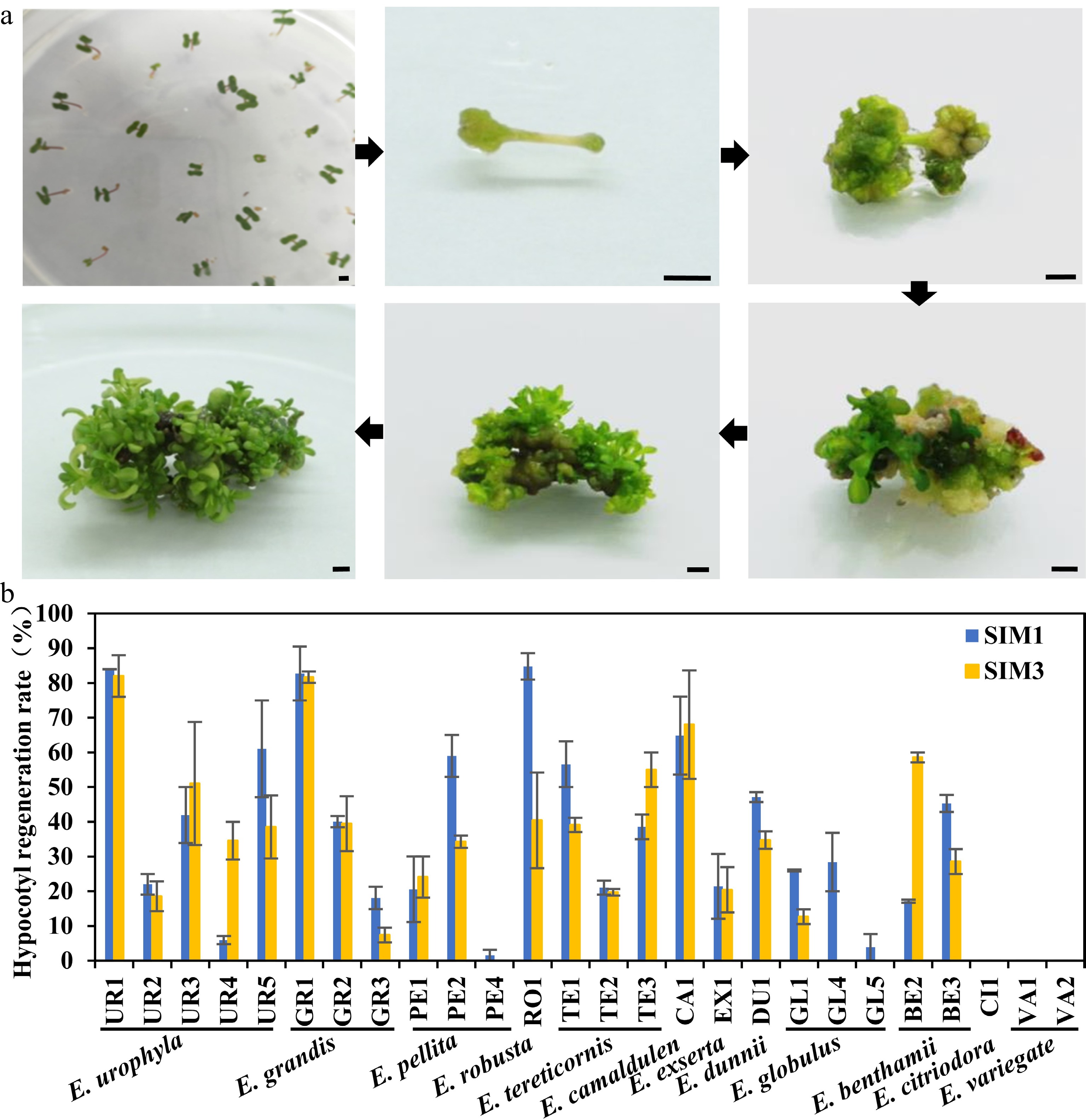
      Figure 1. 

      Regeneration efficiency of different eucalypt species and genotypes using seeding hypocotyl as explants. (a) Representative image of hypocotyl explants regeneration on SIM media at different stages. Photo were taken every other week. Bar = 2 mm. (b) Regeneration efficiency was recorded based on the regeneration ratio. The regeneration rate is defined as a/b × 100%, where a is the number of explants forming shoots after four weeks of screening, and b is the number of explants before the screening. Data shown are mean values from two biological replicates. Error bars ± SE.

      Table 1.  Regeneration rate of shoots induced by hypocotyl as explants in different eucalyptus species or genotypes.

      SpeciesCloneSIM1SIM3
      TotalShoot inductionRegeneration efficiency (%)Differentiation amountTotalShoot inductionRegeneration efficiency (%)Differentiation amount
      E.urophyllaUR1816883.96 ± 0.04a***504182.00 ± 6.00a***
      UR241922.02 ± 2.98ghi*561118.57 ± 4.29ijkl*
      UR3782941.94 ± 8.06def***672851.04 ± 17.71bcde***
      UR43525.95 ± 1.19ijk*441534.58 ± 5.42efghi**
      UR5332061.03 ± 13.97bc*381538.52 ± 9.10defg*
      E.grandisGR1534382.74 ± 7.74a***494081.67 ± 1.67a**
      GR2873540.06 ± 1.60def**384739.47 ± 7.89cdef*
      GR31622818.08 ± 3.23hij*8067.39 ± 2.13kl*
      E.pellitaPE128520.56 ± 9.44hi**21524.09 ± 5.91fghijk**
      PE2714058.97 ± 6.03bc**652234.25 ± 1.75efghi**
      PE44311.56 ± 1.56jk*6600.00 ± 0.00l
      E.robustaRO11129684.76 ± 3.81a***874340.42 ± 13.75cdef***
      E.tereticornisTE1472656.58 ± 6.58bcd***441739.11 ± 2.07defg**
      TE2601321.06 ± 2.01hi**611219.72 ± 0.97hijk*
      TE3391538.55 ± 3.55efg**402255.00 ± 5.00bcd***
      E.camaldulenCA11026564.83 ± 11.26b***765768.01 ± 15.63ab***
      E.exsertaEX1852021.45 ± 9.32hi*621220.41 ± 6.52ghijk**
      E.dunniiDU11868847.13 ± 1.45cde**1926734.74 ± 2.52efghi**
      E.globulusGL1541426.02 ± 0.30fgh*65812.67 ± 2.14jkl*
      GL41073128.42 ± 8.42fgh*9900.00 ± 0.00l
      GL59853.85 ± 3.85jk*9200.00 ± 0.00l
      E.benthamiiBE21903317.12 ± 0.45hij**271658.57 ± 1.43bc**
      BE3793645.29 ± 2.44cde***882628.57 ± 3.57fghij**
      E.citriodoraCI112200.00 ± 0.00k9200.00 ± 0.00l
      E.variegateVA112300.00 ± 0.00k10900.00 ± 0.00l
      VA212500.00 ± 0.00k11600.00 ± 0.00l
      Mean values of two independent experiments ( ± ) with standard errors. Values with the same letter within columns are not significantly different according to Duncan’s Multiple Range Test (DMRT) at a 5% level. Shoots number of each explant were counted and classified (*, < five shoots; five shoots < ** < ten shoots; ***, > ten shoots).
    • Seeding tissues, such as hypocotyl or cotyledon, are usually good explants for in vitro regeneration. However, since Eucalyptus is an outcrossing tree species, the offspring have great genetic variation, which will make transformation unstable and count against future transgenic analysis. Thus, clones from the seven highest regeneration efficiency eucalypt species or genotypes were chosen and sub-cultured. The internodes of these clones were used as explants for the regeneration efficiency test (Fig. 2a). The results showed that all test clones could be successfully regenerated (Fig. 2b, Table 2). In general, we found that the regeneration efficiency of internode explants was lower compared to that of hypocotyl explants (Fig. 2b, Table 2). Among them, E. urophyla (UR1), E. pellita (PE2), E. robusta (RO1), and E. camaldulen (CA1) displayed better regeneration ability, with regeneration efficiency over 50%.
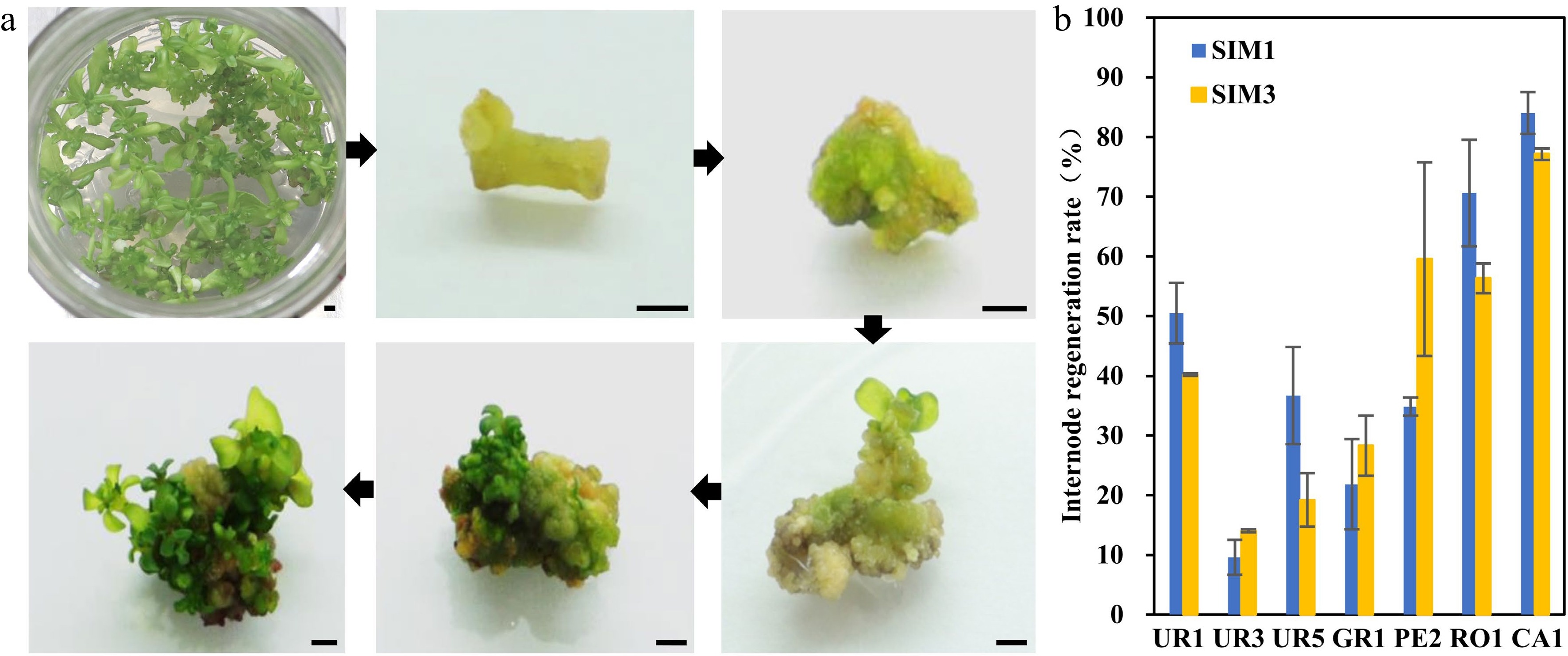
      Figure 2. 

      Regeneration efficiency of different eucalyptus species and genotypes using clonal internode as explants. (a) Representative image of clonal internode explants regeneration on SIM media at different stages. Photos were taken every other week. Bar = 2 mm. (b) Regeneration efficiency was recorded based on the regeneration ratio. The regeneration rate is defined as a/b × 100%, where a is the number of explants forming shoots after four weeks of screening, and b is the number of explants before the screening. Data shown are mean values from two biological replicates. Error bars ± SE.

      Table 2.  Regeneration rate of shoots induced by stem segments as explants in different eucalypt species or genotypes.

      SpeciesCloneSIM1SIM3
      TotalShoot inductionRegeneration efficiency (%)Differentiation amountTotalShoot inductionRegeneration efficiency (%)Differentiation amount
      E.urophyllaUR1804050.51 ± 5.05b***923740.19 ± 0.19bc**
      UR34649.58 ± 2.92d**36514.04 ± 0.25d*
      UR5572136.70 ± 8.13bc*711419.20 ± 4.49cd*
      E.grandisGR1411121.85 ± 7.56cd**491228.29 ± 5.04cd***
      E.pellitaPE2702434.85 ± 1.52bc**633859.55 ± 16.21ab**
      E.robustaRO11258570.63 ± 8.91a**1116456.33 ± 2.49ab***
      E.camaldulenCA11179784.01 ± 3.49a***745777.16 ± 0.97a**
      Mean values of two independent experiments ( ± ) with standard errors. Values with the same letter within columns are not significantly different according to Duncan's Multiple Range Test (DMRT) at a 5% level. Shoots number of each explant were counted and classified (*, < five shoots; five shoots < ** < ten shoots; ; ***, > ten shoots).
    • Based on previous studies, transformation efficiency is largely dependent on genotypes. To access the Agrobacterium susceptibility of different eucalypt genotypes, A. tumefaciens strain GV3101 (pMP90) that harbored the binary vector pCAMBIA2300 containing the DsRed2 reporter gene under the control of the 35S was used for transformation (35S::DsRed2, Fig. 3a). DsRed2 encodes a red fluorescent protein that can be detected under fluorescence microscopy and has been widely used in genetic transformation[3135]. We first performed the Agrobacterium-mediated genetic transformation test of the seven highest regeneration efficiency eucalypt species with hypocotyls as explants. The transformation efficiency was calculated based on DsRed2 fluorescence on the callus (Fig. 3b). The results showed that the transformation efficiency varied greatly among different eucalypt species (Fig. 3c, Table 3). E. urophylla (UR1 and UR5), E. grandis (GR1), and E. pellita (PE2) displayed quite low transformation efficiency, with few visible red fluorescent calli. By contrast, more than half of the RO1 and CA1 explants had fluorescent calli (Fig. 3c, Table 3), suggesting a higher susceptibility to Agrobacterium. We further used internodes from established clones of RO1 and CA1 as transformation explants. However, the transformation efficiency of clonal internodes decreased to 20%, with half the efficiency compared to that using seeding hypocotyls as explants (Fig. 3d, Table 3). These results suggest that the transformation efficiency is highly dependent on the explant types.
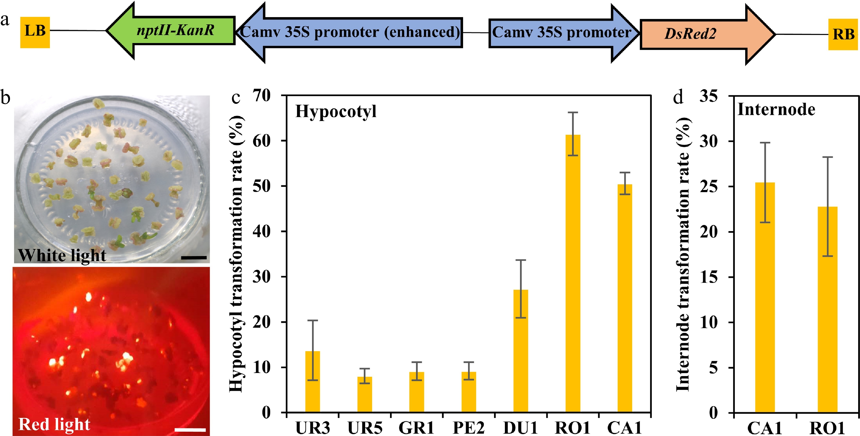
      Figure 3. 

      Transformation efficiency of different eucalyptus species and genotypes. (a) T-DNA region of pCAMBIA2300::35S::DsRed2 vector for genetic transformation. The chimeric neomycin phosphotransferase II (NPT II) selection marker and the reporter gene DsRed2 were driven by cauliflower mosaic virus 35S promoter. LB and RB indicate T-DNA left and right border, respectively. Arrows indicate the direction of transcription. (b) Fluorescence observation of callus. Callus induction one month after Agrobacterium infection were observed at white light and red light with the fluorescence stereo-microscope. Bar = 1 cm. (c), (d) Transformation efficiency calculation of different eucalyptus species and genotypes using seeding hypocotyl and clonal internode as explants, respectively. Transformation efficiency was recorded based on the fluorescence callus of each explant. The transformation rate is defined as a/b × 100%, where a is the number of explants having fluorescence callus after one month of screening, and b is the total number of explants. Data shown are mean values from two biological replicates. Error bars ± SE.

      Table 3.  Transformation efficiency test of different eucalypt species by monitoring the red fluorescence rate of the callus after transformed with the reporter DsRed2.

      SpeciesCloneExplantTotalRed fluorescenceTransformation efficiency (%)
      E. robustaRO1Hypocotyl1778961.48 + 4.76a
      E. camaldulenCA1Hypocotyl713150.55 + 2.40a
      CA2Hypocotyl631929.68 + 2.76b
      E. dunniiDU1Hypocotyl1594527.30 + 6.40
      E. urophyllaUR3Hypocotyl731313.75 + 6.60c
      UR4Hypocotyl9388.07 + 1.62c
      E. pellitaPE2Hypocotyl7369.19 + 1.92c
      E. grandisGR1Hypocotyl4149.13 + 1.98c
      E. robustaRO1Internode481122.78 + 5.47a
      E. camaldulenCA1Internode47512425.46 + 4.40a
      Mean values of two independent experiments (±) with standard errors. Values with the same letter within columns are not significantly different according to Duncan’s Multiple Range Test (DMRT) at a 5% level.
    • Combined with the results of the regeneration and transformation tests, we selected CA1 and RO1 for stable transformation system establishment. Seeding hypocotyls of CA1 and RO1 were used as explants and were transformed by A. tumefaciens strain GV3101 (pMP90) harboring the DsRed2 overexpression construct. It has been shown that pre-culture and co-culture before and after Agrobacterium infection will improve transformation efficiency[12]. In our early trials, the combination of 3-day pre-culture prior to inoculation and 4-day co-cultivation after inoculation was ideal for Agrobacterium infection (Fig. 4a). After 8 weeks on selection and shoot regeneration medium (SIM1) and a further 4 weeks on shoot elongation medium (SE), adventitious shoots formed. The adventitious shoots were then transferred to the rooting media (RM) to obtain complete transgenic plants (Fig. 4a). The positive plants were screened based on DsRed2 fluorescence and PCR tests (Fig. 4b, c). We were able to obtain positive transgenic plants in all three test batches (Fig. 4b, Table 4). Furthermore, we applied this transformation system by overexpressing the eucalypt flowering time gene EgFT using clonal internode segments as explants. We obtained positive plants in every single transformation batch (Table 4). This result indicated that we successfully established a stable transformation system of both E. camaldulen and E. robusta using either hypocotyls or stem segments as explants. Although the transformation efficiency was still low, such a stable transformation system will help us optimize the transformation procedure in the future.
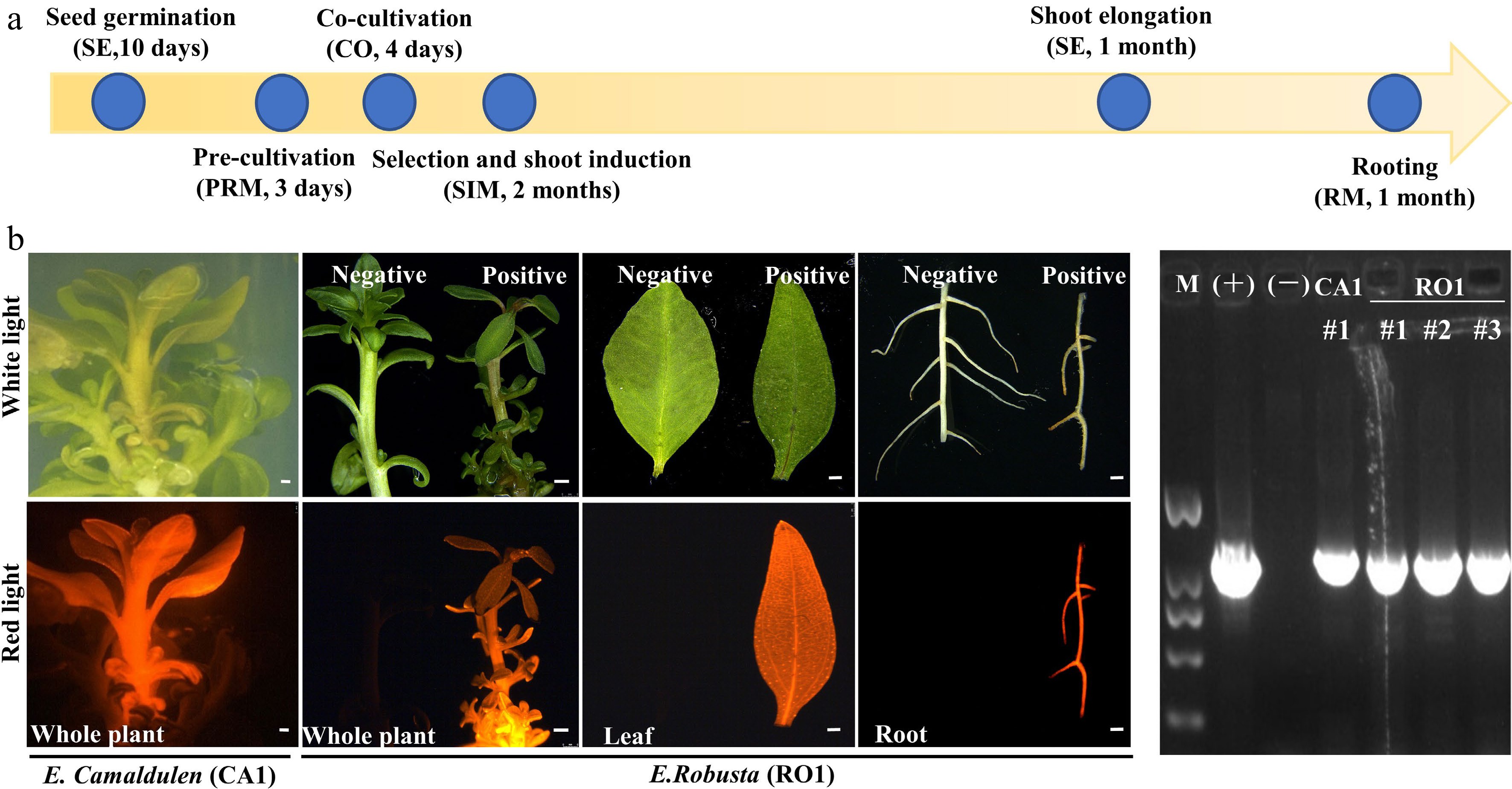
      Figure 4. 

      Agrobacterium-mediated eucalyptus genic transformation with DsRed2 as reporter gene. (a) Flow diagram for agrobacterium-mediated eucalyptus genic transformation. The yellow arrow represents the process direction of transformation. The blue circles with notes represent important transformation steps and time required (the media information of each step is listed in Supplemental Table S2). (b) DsRed2 gene expression in different tissue and organs in transgenic plants. Photos were taken at white light and red light with the fluorescence stereo-microscope. E. robusta (RO1) plants that untransformed were used as a negative control (Negative). Successful transformed plants were marked with Positive. Bar = 1 mm. (c) Positive test of DsRed2 transgenic plants using PCR amplification. M, DNA marker; (+), Positive control with transformation vector as the PCR template; (−), wild type plants; CA1 and RO1, genotypes of E. camaldulen and E. robusta respectively used for transformation donor in this study. The number represents the independent transgenic line.

      Table 4.  Transformation efficiency test of E.robusta and E.camaldulen using hypocotyl and internode as explants respectively.

      GenotypeConstructExplant typeExplant numberPositive plantsTransformation efficiency (%)
      E. robusta (RO1)35S::DsRedHypocotyl7733.9
      E. robusta (RO1)35S::DsRedHypocotyl6711.5
      E. camaldulen (CA1)35S::DsRedHypocotyl5511.9
      E. robusta (RO1)35S::EgFTInternode5123.9
      E. camaldulen (CA1)35S::EgFTInternode6911.5
    • Stable transformation protocols were successfully established for several Eucalyptus species by various research groups around the world[3,19,39,40]. However, the genetic transformation of Eucalyptus is still in its infancy. Except for a few commercial genotypes, there still lack widely applicable protocols for eucalypt transformation as it is too time-consuming and has low efficiency. For these reasons, very few functional studies have been performed in transgenic Eucalyptus (reviewed in Girijishankar[3]). Successful plant stable genetic transformation depends on various factors, such as genotype, type of explant, medium composition, culture conditions, and so on. Among them, genotype is the most influential factor in Eucalyptus genetic transformation. To our knowledge, there are rare reports focused on genotype selection in eucalypt transformation tests. In this study, we screened as many as 26 eucalypt species or genotypes to find eucalypt species or genotypes harboring both high efficiency of regeneration and transformation. Based on this large-scale screening, we developed common regeneration media that were suitable for testing on most eucalypt species, either using hypocotyls or clonal internodes as explants. We also obtained eucalypt genotypes that were highly susceptible to Agrobacterium, which improves transformation efficiency. Our results showed that E. camaldulen and E. robusta display the best regeneration and transformation performance among all testing eucalypt species. In addition, although the regeneration or transformation efficiency of E. dunnii (DU1) is not the best, it has great potential to be a genetic transformation receptor (Fig. 1b & 3c). In this study, besides E. camaldulen, which has been studied previously, we first reported that E. robusta had great potential for further Eucalyptus transformation study. Furthermore, we successfully set up a stable genetic transformation procedure for both E. camaldulen and E. robusta using seeding hypocotyls or cloned internodes. The procedure is stable and efficient, with the highest transformation frequency of up to 3.9%. Overall, our study provides a valuable tool for the study of Eucalyptus functional genomics and molecular breeding.

      Besides genotypes and explants, we noticed that a resistance selection agent is crucial for shoot regeneration after transformation. We used neomycin phosphotransferase (NPT II) selection systems in our study. In previous studies, the screening concentration of kanamycin varied from 10 mg L−1 to 90 mg L−1 due to different antibiotic tolerance among Eucalyptus species[6,11]. In this study, shoots were almost completely prevented when treated with 40 mg L−1 kanamycin, while many shoots were regenerated from explants treated with 30 mg L−1 kanamycin. Thus, we set 30 mg L−1 kanamycin as the screening pressure in our study. However, although E. camaldulen and E. robusta have as high as 80% shoot regeneration efficiency and up to 50% transformation rates, the regeneration rate dramatically decreased after agrobacterial transformation and kanamycin selection. Only few explants regenerated putatively transformed shoots. In the future, optimization of selection conditions could prove to be an effective method of improving E. camaldulen and E. robusta transformation efficiency.

      An appropriate reporter gene can effectively help in the detection of transformed cells. In Eucalyptus, the GUS gene has been used as a reporter gene. However, it depends on histochemical staining, which demands tissue destruction. Moreover, it was reported that transgenic callus with GUS expression could not regenerate into shoots in Eucalyptus[40]. In this study, we applied DsRed2 as a reporter in eucalyptus transformation efficiency screening and stable transformation system setup. It can visually distinguish transgenic from non-transgenic callus of Eucalyptus at an early stage (Fig. 3b) and is expressed in almost all plant tissues without organ preference (Fig. 4b). The application of DsRed2 in this study suggests that DsRed2 is an ideal morphological reporter for Eucalyptus genetic transformation establishment and further transformation efficiency improvement.

    • In this study, we systematically screened the regeneration and Agrobacterium-mediated transformation competence of eucalypt species or genotypes on a large scale. We developed common regeneration media that are suitable for testing on most eucalypt species. We also obtained high Agrobacterium susceptibility amenable eucalypt genotypes, which will help to improve transformation efficiency. Finally, we set up a stable and efficient Agrobacterium-mediated genetic transformation procedure for both E. camaldulen and E. robusta using both hypocotyls and clonal internodes as explants. Overall, our study provides powerful means for eucalyptus propagation, genomics research and molecular breeding.

    • Seeds of Eucalyptus in this study were provided by the China Eucalypt Research Centre (Zhanjiang, China). A. tumefaciens GV3101 and competent cells of E. coil DH5α were homemade in this laboratory. The pCAMBIA2300::35S::DsRed2 vector plasmids were kindly provided by Professor Shuangxia Jin, Huazhong Agricultural University[31]. For the 35S::EgFT construct, full-length cDNA of EgFT (Eucgr.B01458) was amplified from E. grandis. The fragments were then transformed to the destination vector pK2GW7[41].

    • The Murashige and Skoog (MS) basal medium with 20 g L−1 sucrose and 0.8% (w/v) bacteriological agar was used in all media in this study[42]. The pH of the medium was adjusted to 5.8 and autoclaved at 121 °C for 20 min. Filter-sterilized phytohormones were added to the medium after autoclaving. For shoot regeneration, cultures were maintained under a photoperiod of 16 h light and 8 h dark with a light intensity of 12 μmol m−2 s −1 provided using white fluorescent tube lights. For shoot elongation and rooting, the light intensity was increased to 55 μmol m−2 s −1. The room temperature was maintained at 24 ± 2 °C during the whole culture process.

    • For hypocotyl explants, seeds were washed three times in distilled water, surface-sterilized in 70% (v/v) ethanol for 1 min and 1.5% (v/v) NaClO for 15 min with constant stirring, followed by four washes in sterile distilled water. Washed seeds were protected from light in Germination Medium (GM) at 25 °C for 3 d and then transferred to a 16 h light and 8 h dark growth chamber. Hypocotyls were cut for the regeneration experiment. For internode explants, shoot clones regenerated from hypocotyls of different species or genotypes were sub-cultured in SIM. Internodes from the shoots were then cut for the regeneration and transformation experiments.

    • A. tumefaciens strain GV3101 harboring the pCAMBIA2300::35S::DsRed2 binary vector was used for the transformation experiments. The strains were inoculated in liquid YEB (Yeast Mannitol Medium) medium containing 25 mg L−1 rifampicin and 100 mg L−1 spectinomycin and grown at a temperature of 28 °C on a shaker at 200 rpm until OD600 = 0.5–1.0. The bacteria were resuspended in liquid pre-cultivation medium (PRM). The explants of hypocotyls or internodes were cut into 5-mm slices and dipped in the bacterial suspension for 30 min. The explants were dried on filter paper and subsequently transferred onto a co-cultivation medium. The plates were incubated for 3 days in dark conditions at 24 °C. For selection, the explants were transferred to a solidified selection and SIM. Two months later, the regenerated shoots were transferred into a shoot elongation (SE) medium until they were suitable for rooting. Elongated shoots were excised and placed in a rooting medium (RM). After 2 weeks, the roots were fully developed, and the plants could eventually be transplanted in the pots grown in the greenhouse. The media used in this study are listed in Supplemental Table S2.

    • For the regeneration efficiency test, the explants were incubated in different SIMs (SIM1 and SIM3, Supplemental Table S1). After incubation for 28 d in the culture room, the number of shoots per explant, and the percentage (%) of explants forming shoots were measured and recorded. The regeneration rate is defined as a/b × 100%, where a is the number of explants forming shoots after 4 weeks of screening, and b is the number of explants before the screening. The regeneration capability was also monitored by counting the number of shoots of each explant and classified into three types (i.e., high, > ten shoots; ten shoots > medium > five shoots; and low, < five shoots).

      In this study, we used the red fluorescent protein DsRed2 as a reporter to check eucalyptus transformation efficiency. Calli from the regenerated explants were observed under white light and red light with a fluorescence stereomicroscope (Olympus, Tokyo, Japan). The red light uses a filter set for excitation at 530−550 nm and emission at 575 nm. The transformation rate is defined as a/b × 100%, where a is the number of explants that contain red florescence light after four weeks of screening, and b is the number of explants before the screening.

    • Genomic DNA was extracted from young leaves of putative transgenic shoots and wild eucalypt shoots using a Plant Genomic DNA Kit (Tiangen Biotech, Beijing, China). Gene-specific primers of DsRed2 and EgFT were designed and used for positive checking. The construction vectors used for transformation served as a positive control, while DNA from untransformed plants served as a negative control. The amplification products were separated by electrophoresis on a 1.0% (w/v) agarose gel and visualized using a UV transilluminator. Primers used in this study are listed in Supplemental Table S3.

      • This work was supported by the National Natural Science Foundation of China (31971676), the Opening Project of State Key Laboratory of Tree Genetics and Breeding (K2020103), and the Fundamental Research Funds for Central Universities (2662019PY007). We would like to thank Dr. Shuangxia Jin (Huazhong Agricultural University) for providing pCAMBIA2300::35S::DsRed2 vector, and as well as his kind suggestions about this project. We also thank Dr. Siming Gan (China Eucalypt Research Centre, CERC) for kindly providing E. camaldulen seeds.

      • The authors declare that they have no conflict of interest.

      • Copyright: © 2022 by the author(s). Published by Maximum Academic Press, Fayetteville, GA. This article is an open access article distributed under Creative Commons Attribution License (CC BY 4.0), visit https://creativecommons.org/licenses/by/4.0/.
    Figure (4)  Table (4) References (42)
  • About this article
    Cite this article
    Zhou T, Lin Y, Lin Y, Luo J, Ding J. 2022. Regeneration and Agrobacterium-mediated genetic transformation of twelve Eucalyptus species. Forestry Research 2:15 doi: 10.48130/FR-2022-0015
    Zhou T, Lin Y, Lin Y, Luo J, Ding J. 2022. Regeneration and Agrobacterium-mediated genetic transformation of twelve Eucalyptus species. Forestry Research 2:15 doi: 10.48130/FR-2022-0015

Catalog

  • About this article

/

DownLoad:  Full-Size Img  PowerPoint
Return
Return