2023 Volume 3
Article Contents
About this article
ARTICLE   Open Access    

Characterization of flavor frame in grape wines detected by HS-SPME-GC-MS coupled with HPLC, electronic nose, and electronic tongue

More Information
  • To analyze the flavor components in 17 commercially available wine samples from seven grape varieties (Cabernet Sauvignon, Cabernet Gernischt, Shiraz, Merlot, Pinot Noir, Tempranillo and Chardonnay), comprehensive flavor characterization, volatile and non-volatile compounds of grape wines were evaluated by headspace solid phase micro-extraction (HS-SPME) coupled with gas chromatography-mass spectrometry (GC-MS), electronic nose (E-nose), electronic tongue (E-tongue), high performance liquid chromatography (HPLC) and automatic amino acids analyzer. According to GC-MS analysis, a total of 86 volatile compounds were identified, mainly including alcohols, esters, phenols, terpenes and norisoprenoids. Results showed that significant differences of contents of free amino acids and radar fingerprint chart of E-tongue technology were recorded for the 17 grape wines. Moreover, principal component analysis (PCA) of E-nose and E-tongue were used to distinguish the different grape wines effectively, with the cumulative contribution rate accounting for 92.33% and 91.78%, respectively. The results prove that sensors W2S and W1W in the E-nose for wines have a higher influence in the current pattern file. The most abundant phenol in 17 wine samples is catechin. The differences in species and contents of volatile and non-volatile substances give the unique flavor of different grape wines. The results demonstrated that the above mentioned equipment are useful for in-depth grape wine flavor analysis.
  • Columnar cacti are plants of the Cactaceae family distributed across arid and semi-arid regions of America, with ecological, economic, and cultural value[1]. One trait that makes it possible for the columnar cactus to survive in the desert ecosystem is its thick epidermis covered by a hydrophobic cuticle, which limits water loss in dry conditions[1]. The cuticle is the external layer that covers the non-woody aerial organs of land plants. The careful control of cuticle biosynthesis could produce drought stress tolerance in relevant crop plants[2]. In fleshy fruits, the cuticle maintains adequate water content during fruit development on the plant and reduces water loss in fruit during postharvest[3]. Efforts to elucidate the molecular pathway of cuticle biosynthesis have been carried out for fleshy fruits such as tomato (Solanum lycopersicum)[4], apple (Malus domestica)[5], sweet cherry (Prunus avium)[6], mango (Mangifera indica)[7], and pear (Pyrus 'Yuluxiang')[8].

    The plant cuticle is formed by the two main layers cutin and cuticular waxes[3]. Cutin is composed mainly of oxygenated long-chain (LC) fatty acids (FA), which are synthesized by cytochrome p450 (CYP) enzymes. CYP family 86 subfamily A (CYP86A) enzymes carry out the terminal (ω) oxidation of LC-FA[9]. Then, CYP77A carries out the mid-chain oxidation to synthesize the main cutin monomers. In Arabidopsis, AtCYP77A4 and AtCYP77A6 carry out the synthesis of mid-chain epoxy and mid-chain dihydroxy LC-FA, respectively[10,11]. AtCYP77A6 is required for the cutin biosynthesis and the correct formation of floral surfaces[10]. The expression of CYP77A19 (KF410855) and CYP77A20 (KF410856) from potato (Solanum tuberosum) restored the petal cuticular impermeability in Arabidopsis null mutant cyp77a6-1, tentatively by the synthesis of cutin monomers[12]. In eggplant (Solanum torvum), the over-expression of StoCYP77A2 leads to resistance to Verticillium dahlia infection in tobacco plants[13]. Although the function of CYP77A2 in cutin biosynthesis has not yet been tested, gene expression analysis suggests that CaCYP77A2 (A0A1U8GYB0) could play a role in cutin biosynthesis during pepper fruit development[14].

    It has been hypothesized that the export of cuticle precursors is carried out by ATP binding cassette subfamily G (ABCG) transporters. ABCG11/WBC11, ABCG12, and ABCG13 are required for the load of cuticle lipids in Arabidopsis[1517], but ABCG13 function appears to be specific to the flower epidermis[18]. The overexpression of TsABCG11 (JQ389853) from Thellungiella salsugineum increases cuticle amounts and promotes tolerance to different abiotic stresses in Arabidopsis[19].

    Once exported, the cutin monomers are polymerized on the surface of epidermal cells. CD1 code for a Gly-Asp-Ser-Leu motif lipase/esterase (GDSL) from tomato required for the cutin formation through 2-mono(10,16-dihydroxyhexadecanoyl)glycerol esterification[20]. GDSL1 from tomato carries out the ester bond cross-links of cutin monomers located at the cuticle layers and is required for cuticle deposition in tomato fruits[21]. It has been shown that the transcription factor MIXTA-like reduces water loss in tomato fruits through the positive regulation of the expression of CYP77A2, ABCG11, and GDSL1[22]. Despite the relevant role of cuticles in maintaining cactus homeostasis in desert environments[1], the molecular mechanism of cuticle biosynthesis has yet to be described for cactus fruits.

    Stenocereus thurberi is a columnar cactus endemic from the Sonoran desert (Mexico), which produces an ovoid-globose fleshy fruit named sweet pitaya[23]. In its mature state, the pulp of sweet pitaya contains around 86% water with a high content of antioxidants and natural pigments such as betalains and phenolic compounds, which have nutraceutical and industrial relevance[23]. Due to the arid environment in which pitaya fruit grows, studying its molecular mechanism of cuticle biosynthesis can generate new insights into understanding species' adaptation mechanisms to arid environments. Nevertheless, sequences of transcripts from S. thurberi in public databases are scarce.

    RNA-sequencing technology (RNA-seq) allows the massive generation of almost all the transcripts from non-model plants, even if no complete assembled genome is available[24]. Recent advances in bioinformatic tools has improved our capacity to identify long non-coding RNA (lncRNA), which have been showed to play regulatory roles in relevant biological processes, such as the regulation of drought stress tolerance in plants[25], fruit development, and ripening[2629].

    In this study, RNA-seq data were obtained for the de novo assembly and characterization of the S. thurberi fruit peel transcriptome. As a first approach, three transcripts, StCYP77A, StABCG11, and StGDSL1, tentatively involved in cuticle biosynthesis, were identified and quantified during sweet pitaya fruit development. Due to no gene expression analysis having been carried out yet for S. thurberi, stably expressed constitutive genes were identified for the first time.

    Sweet pitaya fruits (S. thurberi) without physical damage were hand harvested from plants in a native conditions field located at Carbó, Sonora, México. They were collocated in a cooler containing dry ice and transported immediately to the laboratory. The superficial part of the peels (~1 mm deep) was removed carefully from the fruits using a scalpel. Peel samples from three fruits were pooled according to their tentative stage of development defined by their visual characteristics, frozen in liquid nitrogen, and pulverized to create a single biological replicate. Four samples belonging to four different plants were analyzed. All fruits harvested were close to the ripening stage. Samples named M1 and M2 were turning from green to ripe [~35−40 Days After Flowering (DAF)], whereas samples M3 and M4 were turning from ripe to overripe (~40−45 DAF).

    Total RNA was isolated from the peels through the Hot Borate method[30]. The concentration and purity of RNA were determined in a spectrophotometer Nanodrop 2000 (Thermo Fisher) by measuring the 260/280 and 260/230 absorbance ratios. RNA integrity was evaluated through electrophoresis in agarose gel 1% and a Bioanalyzer 2100 (Agilent). Pure RNA was sequenced in the paired-end mode in an Illumina NextSeq 500 platform at the University of Arizona Genetics Core Facility. Four RNA-seq libraries, each of them from each sample, were obtained, which include a total of 288,199,704 short reads with a length of 150 base pairs (bp). The resulting sequence data can be accessed at the Sequence Read Archive (SRA) repository of the NCBI through the BioProject ID PRJNA1030439. Libraries are named corresponding to the names of samples M1, M2, M3, and M4.

    FastQC software (www.bioinformatics.babraham.ac.uk/projects/fastqc) was used for short reads quality analysis. Short reads with poor quality were trimmed or eliminated by Trimmomatic (www.usadellab.org/cms/?page=trimmomatic) with a trailing and leading of 25, a sliding window of 4:25, and a minimum read length of 80 bp. A total of 243,194,888 reads with at least a 25 quality score on the Phred scale were used to carry out the de novo assembly by Trinity (https://github.com/trinityrnaseq/trinityrnaseq/wiki) with the following parameters: minimal k-mer coverage of 1, normalization of 50, and minimal transcript length of 200 bp.

    Removal of contaminating sequences and ribosomal RNA (rRNA) was carried out through SeqClean. To remove redundancy, transcripts with equal or more than 90% of identity were merged through CD-hit (www.bioinformatics.org/cd-hit/). Alignment and quantification in terms of transcripts per million (TPM) were carried out through Bowtie (https://bowtie-bio.sourceforge.net/index.shtml) and RSEM (https://github.com/deweylab/RSEM), respectively. Transcripts showing a low expression (TPM < 0.01) were discarded. Assembly quality was evaluated by calculating the parameters N50 value, mean transcript length, TransRate score, and completeness. The statistics of the transcriptome were determined by TrinityStats and TransRate (https://hibberdlab.com/transrate/). The transcriptome completeness was determined through a BLASTn alignment (E value < 1 × 10⁻³) by BUSCO (https://busco.ezlab.org/) against the database of conserved orthologous genes from Embryophyte.

    To predict the proteins tentatively coded in the S. thurberi transcriptome, the best homology match of the assembled transcripts was found by alignment to the Swiss-Prot, RefSeq, nr-NCBI, PlantTFDB, iTAK, TAIR, and ITAG databases using the BLAST algorithm with an E value threshold of 1 × 10⁻¹⁰ for the nr-NCBI database and of 1 × 10⁻⁵ for the others[3134]. An additional alignment was carried out to the protein databases of commercial fruits Persea americana, Prunus persica, Fragaria vesca, Citrus cinensis, and Vitis vinifera to proteins of the cactus Opuntia streptacantha, and the transcriptomes of the cactus Hylocereus polyrhizus, Pachycereus pringlei, and Selenicereus undatus. The list of all databases and the database websites of commercial fruits and cactus are provided in Supplementary Tables S1 & S2. The open reading frame (ORF) of the transcripts and the protein sequences tentative coded from the sweet pitaya transcriptome was predicted by TransDecoder (https://github.com/TransDecoder/TransDecoder/wiki), considering a minimal ORF length of 75 amino acids (aa). The search for protein domains was carried out by the InterPro database (www.ebi.ac.uk/interpro). Functional categorization was carried out by Blast2GO based on GO terms and KEGG metabolic pathways[35].

    LncRNA were identified based on the methods reported in previous studies[25,29,36]. Transcripts without homology to any protein from Swiss-Prot, RefSeq, nr-NCBI, PlantTFDB, iTAK, TAIR, ITAG, P. americana, P. persica, F. vesca, C. cinensis, V. vinifera, and O. streptacantha databases, without a predicted ORF longer than 75 aa, and without protein domains in the InterPro database were selected to identify tentative lncRNA.

    Transcripts coding for signal peptide or transmembrane helices were identified by SignalP (https://services.healthtech.dtu.dk/services/SignalP-6.0/) and TMHMM (https://services.healthtech.dtu.dk/services/TMHMM-2.0/), respectively, and discarded. Further, transcripts corresponding to other non-coding RNAs (ribosomal RNA and transfer RNA) were identified through Infernal by using the Rfam database[37] and discarded. The remaining transcripts were analyzed by CPC[38], and CPC2[39] to calculate their coding potential. Transcripts with a coding potential score lower than −1 for CPC and a coding probability lower than 0.1 for CPC2 were considered lncRNA. To characterize the identified lncRNA, the length and abundance of coding and lncRNA were calculated. Bowtie and RSEM were used to align and quantify raw counts, respectively. The edgeR package[40] was used to normalize raw count data in terms of counts per million (CPM) for both coding and lncRNA.

    To obtain the transcript's expression, the aligning of short reads and quantifying of transcripts were carried out through Bowtie and RSEM software, respectively. A differential expression analysis was carried out between the four libraries by edgeR package in R Studio. Only the transcripts with a count equal to or higher than 0.5 in at least one sample were retained for the analysis. Transcripts with log2 Fold Change (log2FC) between +1 and −1 and with a False Discovery Rate (FDR) lower than 0.05 were taken as not differentially expressed (NDE).

    For the identification of the tentative reference genes two strategies were carried out as described below: i) The NDE transcripts were aligned by BLASTn (E value < 1 × 10⁻⁵) to 43 constitutive genes previously reported in fruits from the cactus H. polyrhizus, S. monacanthus, and S. undatus[4143] to identify possible homologous constitutive genes in S. thurberi. Then, the homologous transcripts with the minimal coefficient of variation (CV) were selected; ii) For all the NDE transcripts, the percentile 95 value of the mean CPM and the percentile 5 value of the CV were used as filters to recover the most stably expressed transcripts, based on previous studies[44]. Finally, transcripts to be tested by quantitative reverse transcription polymerase chain reaction (qRT-PCR) were selected based on their homology and tentative biological function.

    The fruit harvesting was carried out as described above. Sweet pitaya fruit takes about 43 d to ripen, therefore, open flowers were tagged, and fruits with 10, 20, 30, 35, and 40 DAF were collected to cover the pitaya fruit development process (Supplementary Fig. S1). The superficial part of the peels (~1 mm deep) was removed carefully from the fruits using a scalpel. Peel samples from three fruits were pooled according to their stage of development defined by their DAF, frozen in liquid nitrogen, and pulverized to create a single biological replicate. One biological replicate consisted of peels from three fruits belonging to the same plant. Two to three biological replicates were evaluated for each developmental stage. Two technical replicates were analyzed for each biological replicate. RNA extraction, quantification, RNA purity, and RNA integrity analysis were carried out as described above.

    cDNA was synthesized from 100 ng of RNA by QuantiTect Reverse Transcription Kit (QIAGEN). Primers were designed using the PrimerQuest™, UNAFold, and OligoAnalyzer™ tools from Integrated DNA Technologies (www.idtdna.com/pages) and following the method proposed by Thornton & Basu[45]. Transcripts quantification was carried out in a QIAquant 96 5 plex according to the PowerUp™ SYBR™ Green Master Mix protocol (Applied Biosystems), with a first denaturation step for 2 min at 95 °C, followed by 40 cycles of denaturation step at 95 °C for 15 s, annealing and extension steps for 30 s at 60 °C.

    The Cycle threshold (Ct) values obtained from the qRT-PCR were analyzed through the algorithms BestKeeper, geNorm, NormFinder, and the delta Ct method[46]. RefFinder (www.ciidirsinaloa.com.mx/RefFinder-master/) was used to integrate the stability results and to find the most stable expressed transcripts in sweet pitaya fruit peel during development. The pairwise variation value (Vn/Vn + 1) was calculated through the geNorm algorithm in R Studio software[47].

    An alignment of 17 reported cuticle biosynthesis genes from model plants were carried out by BLASTx against the predicted proteins from sweet pitaya. Two additional alignments of 17 charaterized cuticle biosynthesis proteins from model plants against the transcripts and predicted proteins of sweet pitaya were carried out by tBLASTn and BLASTp, respectively. An E value threshold of 1 × 10⁻⁵ was used, and the unique best hits were recovered for all three alignments. The sequences of the 17 characterized cuticle biosynthesis genes and proteins from model plants are showed in Supplementary Table S3. The specific parameters and the unique best hits for all the alignments carried out are shown in Supplementary Tables S4S8.

    Cuticle biosynthesis-related transcripts tentatively coding for a cytochrome p450 family 77 subfamily A (CYP77A), a Gly-Asp-Ser-Leu motif lipase/esterase 1 (GDSL1), and an ATP binding cassette transporter subfamily G member 11 (ABCG11) were identified by best bi-directional hit according to the functional annotation described above. Protein-conserved domains, signal peptide, and transmembrane helix were predicted through InterProScan, SignalP 6.0, and TMHMM, respectively. Alignment of the protein sequences to tentative orthologous of other plant species was carried out by the MUSCLE algorithm[48]. A neighbor-joining (NJ) phylogenetic tree with a bootstrap of 1,000 replications was constructed by MEGA11[49].

    Fruit sampling, primer design, RNA extraction, cDNA synthesis, and transcript quantification were performed as described above. Relative expression was calculated according to the 2−ΔΔCᴛ method[50]. The sample corresponding to 10 DAF was used as the calibrator. The transcripts StEF1a, StTUA, StUBQ3, and StEF1a + StTUA were used as normalizer genes.

    Normality was assessed according to the Shapiro-Wilk test. Significant differences in the expression of the cuticle biosynthesis-related transcripts between fruit developmental stages were determined by one-way ANOVA based on a completely randomized sampling design and a Tukey honestly significant difference (HSD) test, considering a p-value < 0.05 as significant. Statistical analysis was carried out through the stats package in R Studio.

    RNA was extracted from the peels of ripe sweet pitaya fruits (S. thurberi) from plants located in the Sonoran Desert, Mexico. Four cDNA libraries were sequenced in an Illumina NextSeq 500 platform at the University of Arizona Genetics Core Facility. A total of 288,199,704 reads with 150 base pairs (bp) in length were sequenced in paired-end mode. After trimming, 243,194,888 (84.38%) cleaned short reads with at least 29 mean quality scores per read in the Phred scale and between 80 to 150 bp in length were obtained to carry out the assembly. After removing contaminating sequences, redundancy, and low-expressed transcripts, the assembly included 174,449 transcripts with an N50 value of 2,110 bp. Table 1 shows the different quality variables of the S. thurberi fruit peel transcriptome. BUSCO score showed that 85.4% are completed transcripts, although out of these, 37.2% were found to be duplicated. The resulting sequence data can be accessed at the SRA repository of the NCBI through the BioProject ID PRJNA1030439.

    Table 1.  Quality metrics of the Stenocereus thurberi fruit peel transcriptome.
    Metric Data
    Total transcripts 174,449
    N50 2,110
    Smallest transcript length (bp) 200
    Largest transcript length (bp) 19,114
    Mean transcript length (bp) 1,198.69
    GC (%) 41.33
    Total assembled bases 209,110,524
    TransRate score 0.05
    BUSCO score (%) C: 85.38 (S:48.22, D:37.16),
    F: 10.69, M: 3.93.
    Values were calculated through the TrinityStats function of Trinity and TransRate software. Completeness analysis was carried out through BUSCO by aligning the transcriptome to the Embryophyte database through BLAST with an E value threshold of 1 × 10⁻³. Complete (C), single (S), duplicated (D), fragmented (F), missing (M).
     | Show Table
    DownLoad: CSV

    A summary of the homology search in the main public protein database for the S. thurberi transcriptome is shown in Supplementary Table S1. From these databases, the higher homologous transcripts were found in RefSeq with 93,993 (53.87 %). Based on the E value distribution, for 41,685 (44%) and 68,853 (49%) of the hits, it was found a strong homology (E value lower than 1 × 10−50) to proteins in the Swiss-Prot and RefSeq databases, respectively (Supplementary Fig. S2a & b). On the other hand, 56,539 (52.34%) and 99,599 (71.11%) of the matches showed a percentage of identity higher than 60% in the Swiss-Prot and RefSeq databases, respectively (Supplementary Fig. S2c & d).

    Figure 1 shows the homology between transcripts from S. thurberi and proteins of commercial fruits, as well as proteins and transcripts of cacti. Transcripts from S. thurberi homologous to proteins from fruits of commercial interest avocado (P. americana), peach (P. persica), strawberry (F. vesca), orange (C. sinensis), and grapefruit (V. vinifera) ranged from 77,285 (44.30%) to 85,421 (48.96%), with 70,802 transcripts homologous to all the five fruit protein databases (Fig. 1a).

    Transcripts homologous to transcripts or proteins from the cactus dragon fruit (H. polyrhizus), prickly pear cactus (O. streptacantha), Mexican giant cardon (P. pringlei), and pitahaya (S. undatus) ranged from 76,238 (43.70%) to 114,933 (65.88%), with 64,009 transcripts homologous to all the four cactus databases (Fig. 1b). Further, out of the total of transcripts, 44,040 transcripts (25.25%) showed homology only to sequences from cactus, but not for model plants Arabidopsis, tomato, or the commercial fruits included in this study (Fig. 1c).

    Figure 1.  Venn diagram of the homology search results against model plants databases, commercial fruits, and cactus. The number in the diagram corresponds to the number of transcripts from S. thurberi homologous to sequences from that plant species. (a) Homologous to sequences from Fragaria vesca (Fa), Persea americana (Pa), Prunus persica (Pp), Vitis vinifera (Vv), and Citrus sinensis (Cs). (b) Homologous to sequences from Opuntia streptacantha (Of), Selenicereus undatus (Su), Hylocereus polyrhizus (Hp), and Pachycereus pringlei (Pap). (c) Homologous to sequences from Solanum lycopersicum (Sl), Arabidopsis thaliana (At), from the commercial fruits (Fa, Pa, Pp, Vv, and Cs), or the cactus included in this study (Of, Su, Hp, and Pap). Homologous searching was carried out by BLAST alignment (E value < 1 × 10⁻⁵). The Venn diagrams were drawn by ggVennDiagram in R Studio.

    A total of 45,970 (26.35%), 58,704 (33.65%), and 48,186 (27.65%) transcripts showed homology to transcription factors, transcriptional regulators, and protein kinases in the PlantTFDB, iTAK-TR, and iTAK-PK databases, respectively (Supplementary Tables S1, S9S11). For the PlantTFDB, the homologous transcripts belong to 57 transcriptional factors (TF) families (Fig. 2 & Supplementary Table S9), from which, the most frequent were the basic-helix-loop-helix (bHLH), myeloblastosis-related (MYB-related), NAM, ATAF, and CUC (NAC), ethylene responsive factor (ERF), and the WRKY domain families (WRKY) (Fig. 2).

    Figure 2.  Transcription factor (TF) families distribution of S. thurberi fruit peel transcriptome. The X-axis indicates the number of transcripts with hits to each TF family. Alignment to the PlantTFDB database by BLASTx was carried out with an E value threshold of 1 × 10⁻⁵. The bar graph was drawn by ggplot2 in R Studio.

    Based on the homology found and the functional domain searches, gene ontology terms (GO) were assigned to 68,559 transcripts (Supplementary Table S12). Figure 3 shows the top 20 GO terms assigned to the S. thurberi transcriptome, corresponding to the Biological Processes (BP) and Molecular Function (MF) categories. For BP, organic substance metabolic processes, primary metabolic processes, and cellular metabolic processes showed a higher number of transcripts (Supplementary Table S13). Further, for MF, organic cyclic compound binding, heterocyclic compound binding, and ion binding were the processes with the higher number of transcripts. S. thurberi transcripts were classified into 142 metabolic pathways from the KEGG database (Supplementary Table S14). The pathways with the higher number of transcripts recorded were pyruvate metabolism, glycerophospholipid metabolism, glycolysis/gluconeogenesis, and citrate cycle. Further, among the top 20 KEEG pathways, the cutin, suberin, and wax biosynthesis include more than 30 transcripts (Fig. 4).

    Figure 3.  Top 20 Gene Ontology (GO) terms assigned to the S. thurberi fruit peel transcriptome. Bars indicate the number of transcripts assigned to each GO term. Assignment of GO terms was carried out by Blast2GO with default parameters. BP and MF mean Biological Processes and Molecular Functions GO categories, respectively. The graph was drawn by ggplot2 in R Studio.
    Figure 4.  Top 20 KEGG metabolic pathways distribution in the S. thurberi fruit peel transcriptome. Bars indicate the number of transcripts assigned to each KEGG pathway. Assignment of KEGG pathways was carried out in the Blast2GO suite. The bar graph was drawn by ggplot2 in R Studio.

    Out of the total of transcripts, 43,391 (24.87%) were classified as lncRNA (Supplementary Tables S15 & S16). Figure 5 shows a comparison of the length (Fig. 5a) and expression (Fig. 5b) of lncRNA and coding RNA. Both length and expression values were higher in coding RNA than in lncRNA. In general, coding RNA ranged from 201 to 18,629 bp with a mean length of 1,507.18, whereas lncRNA ranged from 200 to 5,198 bp with a mean length of 481.51 (Fig. 5a). The higher expression values recorded from coding RNA and lncRNA were 12.83 and 9.45 log2(CPM), respectively (Fig. 5b).

    Figure 5.  Comparison of coding RNA and long non-coding RNA (lncRNA) from S. thurberi transcriptome. (a) Box plot of transcript length distribution. The Y-axis indicates the length of each transcript in base pairs. (b) Box plot of expression levels. The Y-axis indicates the log2 of the count per million of reads (log2(CPM)) recorded for each transcript. Expression levels were calculated by the edgeR package in R studio. (a), (b) The lines inside the boxes indicate the median. The higher and lower box limits represent the 75th and 25th percentiles, respectively. The box plots were drawn by ggplot2 in R Studio.

    To identify the transcripts without significant changes in expression between the four RNA-seq libraries, a differential expression analysis was carried out. Of the total of transcripts, 4,980 were not differentially expressed (NDE) at least in one paired comparison between the libraries (Supplementary Tables S17S20). Mean counts per million of reads (CPM) and coefficient of variation (CV)[44] were calculated for these NDE transcripts. Transcripts with a CV value lower than 0.113, corresponding with the percentile 5 of the CV, and a mean CPM higher than 1,138.06, corresponding with the percentile 95 of the mean CPM were used as filters to identify the most stably expressed transcripts (Supplementary Table S21). Based on its homology and its tentative biological function, five transcripts were selected to be tested as tentative reference genes. Besides, three NDE transcripts homologous to previously identified stable expressed reference genes in other species of cactus fruit[4143] were selected (Supplementary Table S22). Homology metrics for the eight tentative reference genes selected are shown in Supplementary Table S23. The primer sequences used to amplify the transcripts by qRT-PCR and their nucleotide sequence are shown in Supplementary Tables S24 & S25, respectively.

    The amplification specificity of the eight candidate reference genes determined by melting curves analysis is shown in Supplementary Fig. S3. For the eight tentative reference transcripts selected, the cycle threshold (Ct) values were recorded during sweet pitaya fruit development by qRT-PCR (Supplementary Table S26). The Ct values obtained ranged from 16.85 to 30.26 (Fig. 6a). Plastidic ATP/ADP-transporter (StTLC1) showed the highest Ct values with a mean of 27.34 (Supplementary Table S26). Polyubiquitin 3 (StUBQ3) showed the lowest Ct values in all five sweet pitaya fruit developmental stages (Fig. 6a).

    Figure 6.  Expression stability analysis of tentative reference genes. (a) Box plot of cycle threshold (Ct) distribution of candidate reference genes during sweet pitaya fruit development (10, 20, 30, 35, and 40 d after flowering). The black line inside the box indicates the median. The higher and lower box limits represent the 75th and 25th percentiles, respectively. (b) Bar chart of the geometric mean (geomean) of ranking values calculated by RefFinder for each tentative reference gene (X-axis). The lowest values indicate the best reference genes. (c) Bar chart of the pairwise variation analysis and determination of the optimal number of reference genes by the geNorm algorithm. A pairwise variation value lower than 0.15 indicates that the use of Vn/Vn + 1 reference genes is reliable for the accurate normalization of qRT-PCR data. The Ct data used in the analysis were calculated by qRT-PCR in a QIAquant 96 5 plex (QIAGEN) according to the manufacturer's protocol. The box plot and the bar graphs were drawn by ggplot2 and Excel programs, respectively. Abbreviations: Actin 7 (StACT7), alpha-tubulin (StTUA), elongation factor 1-alpha (StEF1a), COP1-interactive protein 1 (StCIP1), plasma membrane ATPase 4 (StPMA4), BEL1-like homeodomain protein 1 (StBLH1), polyubiquitin 3 (StUBQ3), and plastidic ATP/ADP-transporter (StTLC1).

    The best stability values calculated by NormFinder were 0.45, 0.51, 0.97, and 0.99, corresponding to the transcripts elongation factor 1-alpha (StEF1a), alpha-tubulin (StTUA), plastidic ATP/ADP-transporter (StTLC1), and actin 7 (StACT7), respectively (Supplementary Table S27). For BestKeeper, the most stable expressed transcripts were StUBQ3, StTUA, and StEF1a, with values of 0.72, 0.75, and 0.87, respectively. In the case of the delta Ct method[51], the transcripts StEF1a, StTUA, and StTLC1 showed the best stability.

    According to geNorm analysis, the most stable expressed transcripts were StTUA, StEF1a, StUBQ3, and StACT7, with values of 0.74, 0.74, 0.82, and 0.96, respectively. All the pairwise variation values (Vn/Vn + 1) were lower than 0.15, ranging from 0.019 for V2/V3 to 0.01 for V6/V7 (Fig. 6c). The V value of 0.019 obtained for V2/V3 indicates that the use of the best two reference genes StTUA and StEF1a is reliable enough for the accurate normalization of qRT-PCR data, therefore no third reference gene is required[47]. Except for BestKeeper analysis, StEF1a and StTUA were the most stable transcripts for all of the methods carried out in this study (Supplementary Table S27). The comprehensive ranking analysis indicates that StEF1a, followed by StTUA and StUBQ3, are the most stable expressed genes and are stable enough to be used as reference genes in qRT-PCR analysis during sweet pitaya fruit development (Fig. 6b).

    Three cuticle biosynthesis-related transcripts TRINITY_DN17030_c0_g1_i2, TRINITY_DN15394_c0_g1_i1, and TRINITY_DN23528_c1_g1_i1 tentatively coding for the enzymes cytochrome p450 family 77 subfamily A (CYP77A), Gly-Asp-Ser-Leu motif lipase/esterase 1 (GDSL1), and an ATP binding cassette transporter subfamily G member 11 (ABCG11/WBC11), respectively, were identified and quantified. The nucleotide sequence and predicted amino acid sequences of the three transcripts are shown in Supplementary File 1. The best homology match for StCYP77A (TRINITY_DN17030_c0_g1_i2) was for AtCYP77A4 (AT5G04660) from Arabidopsis and SmCYP77A2 (P37124) from eggplant (Solanum melongena) in the TAIR and Swiss-Prot databases, respectively (Supplementary Table S23).

    TransDecoder, InterPro, and TMHMM analysis showed that StCYP77A codes a polypeptide of 518 amino acids (aa) in length that comprises a cytochrome P450 E-class domain (IPR002401) and a transmembrane region (residues 10 to 32). The phylogenetic tree constructed showed that StCYP77A is grouped in a cluster with all the CYP77A2 proteins included in this analysis, being closer to CYP77A2 (XP_010694692) from B. vulgaris and Cgig2_012892 (KAJ8441854) from Carnegiea gigantean (Supplementary Fig. S4).

    StGDSL1 (TRINITY_DN15394_c0_g1_i1) alignment showed that it is homologous to a GDSL esterase/lipase from Arabidopsis (Q9LU14) and tomato (Solyc03g121180) (Supplementary Table S23). TransDecoder, InterPro, and SignalP analysis showed that StGDSL1 codes a polypeptide of 354 aa in length that comprises a GDSL lipase/esterase domain IPR001087 and a signal peptide with a cleavage site between position 25 and 26 (Supplementary Fig. S5).

    Supplementary Figure S6 shows the analysis carried out on the predicted amino acid sequence of StABCG11 (TRINITY_DN23528_c1_g1_i1). The phylogenetic tree constructed shows three clades corresponding to the ABCG13, ABCG12, and ABCG11 protein classes with bootstrap support ranging from 40% to 100% (Supplementary Fig. S6a). StABCG11 is grouped with all the ABCG11 transporters included in this study in a well-separated clade, being closely related to its tentative ortholog from C. gigantean Cgig2_004465 (KAJ8441854). InterPro and TMHMM results showed that the StABCG11 sequence contains an ABC-2 type transporter transmembrane domain (IPR013525; PF01061.27) with six transmembrane helices (Supplementary Fig. S6b).

    The predicted protein sequence of StABCG11 is 710 aa in length, holding the ATP binding domain (IPR003439; PF00005.30) and the P-loop containing nucleoside triphosphate hydrolase domain (IPR043926; PF19055.3) of the ABC transporters of the G family. Multiple sequence alignment shows that the Walker A and B motif sequence and the ABC signature[15] are conserved between the ABCG11 transporters from Arabidopsis, tomato, S. thurberi, and C. gigantean (Supplementary Fig. S6c).

    According to the results of the expression stability analysis (Fig. 6), four normalization strategies were tested to quantify the three cuticle biosynthesis-related transcripts during sweet pitaya fruit development. The four strategies consist of normalizing by StEF1a, StTUA, StUBQ3, or StEF1a+StTUA. Primer sequences used to quantify the transcripts StCYP77A (TRINITY_DN17030_c0_g1_i2), StGDSL1 (TRINITY_DN15394_c0_g1_i1), and StABCG11 (TRINITY_DN23528_c1_g1_i1) by qRT-PCR during sweet pitaya fruit development are shown in Supplementary Table S24.

    The three cuticle biosynthesis-related transcripts showed differences in expression during sweet pitaya fruit development (Supplementary Table S28). The same expression pattern was recorded for the three cuticle biosynthesis transcripts when normalization was carried out by StEF1a, StTUA, StUBQ3, or StEF1a + StTUA (Fig. 7). A higher expression of StCYP77A and StGDSL1 are shown at the 10 and 20 DAF, showing a decrease at 30, 35, and 40 DAF. StABCG11 showed a similar behavior, with a higher expression at 10 and 20 DAF and a reduction at 30 and 35 DAF. Nevertheless, unlike StCYP77A and StGDSL1, a significant increase at 40 DAF, reaching the same expression as compared with 10 DAF, is shown for StABCG11 (Fig. 7).

    Figure 7.  Expression analysis of cuticle biosynthesis-related transcripts StCYP77A, StGDSL1, and StABCG11 during sweet pitaya (Stenocereus thurberi) fruit development. Relative expression was calculated through the 2−ΔΔCᴛ method using elongation factor 1-alpha (StEF1a), alpha-tubulin (StTUA), polyubiquitin 3 (StUBQ3), or StEF1a + StTUA as normalizing genes at 10, 20, 30, 35, and 40 d after flowering (DAF). The Y-axis and error bars represent the mean of the relative expression ± standard error (n = 4−6) for each developmental stage in DAF. The Ct data for the analysis was recorded by qRT-PCR in a QIAquant 96 5 plex (QIAGEN) according to the manufacturer's protocol. The graph line was drawn by ggplot2 in R Studio. Abbreviations: cytochrome p450 family 77 subfamily A (StCYP77A), Gly-Asp-Ser-Leu motif lipase/esterase 1 (StGDSL1), and ATP binding cassette transporter subfamily G member 11 (StABCG11).

    Characteristics of a well-assembled transcriptome include an N50 value closer to 2,000 bp, a high percentage of conserved transcripts completely assembled (> 80%), and a high proportion of reads mapping back to the assembled transcripts[52]. In the present study, the first collection of 174,449 transcripts from S. thurberi fruit peel are reported. The generated transcriptome showed an N50 value of 2,110 bp, a TransRate score of 0.05, and a GC percentage of 41.33 (Table 1), similar to that reported for other de novo plant transcriptome assemblies[53]. According to BUSCO, 85.4% of the orthologous genes from the Embryophyta databases completely matched the S. thurberi transcriptome, and only 3.9% were missing (Table 1). These results show that the S. thurberi transcriptome generated is not fragmented, and it is helpful in predicting the sequence of almost all the transcripts expressed in sweet pitaya fruit peel[24].

    The percentage of transcripts homologous found, E values, and identity distribution (Supplementary Tables S1 & S2; Supplementary Fig. S2) were similar to that reported in the de novo transcriptome assembly for non-model plants and other cactus fruits[4143,54] and further suggests that the transcriptome assembled of S. thurberi peel is robust[52]. Of the total of transcripts, 70,802 were common to all the five commercial fruit protein databases included in this study, which is helpful for the search for conserved orthologous involved in fruit development and ripening (Fig. 2a). A total of 34,513 transcripts (20%) show homology only to sequences in the cactus's databases, but not in the others (Supplementary Tables S1 & S2; Fig. 1c). This could suggest that a significant conservation of sequences among plants of the Cactaceae family exists which most likely are to have a function in this species adaptation to desert ecosystems.

    To infer the biological functionality represented by the S. thurberi fruit peel transcriptome, gene ontology (GO) terms and KEGG pathways were assigned. Of the main metabolic pathways assigned, 'glycerolipid metabolism' and 'cutin, suberine, and wax biosynthesis' suggests an active cuticle biosynthesis in pitaya fruit peel (Fig. 4). In agreement with the above, the main GO terms assigned for the molecular function (MF) category were 'organic cyclic compound binding', 'transmembrane transporter activity', and 'lipid binding' (Fig. 3). For the biological processes (BP) category, the critical GO terms for the present research are 'cellular response to stimulus', 'response to stress', 'anatomical structure development', and 'transmembrane transport', which could suggest the active development of the fruit epidermis and cuticle biosynthesis for protection to stress.

    The most frequent transcription factors (TF) families found in S. thurberi transcriptome were NAC, WRKY, bHLH, ERF, and MYB-related (Fig. 2), which had been reported to play a function in the tolerance to abiotic stress in plants[55,56]. Although the role of NAC, WRKY, bHLH, ERF, and MYB TF in improving drought tolerance in relevant crop plants has been widely documented[57,58], their contribution to the adaptation of cactus to arid ecosystems has not yet been elucidated and further experimental pieces of evidence are needed.

    It has been reported that the heterologous expression of ERF TF from Medicago truncatula induces drought tolerance and cuticle wax biosynthesis in Arabidopsis leaf[59]. In tomato fruits, the gene SlMIXTA-like which encodes a MYB transcription factor avoids water loss through the positive regulation of genes related to the biosynthesis and transport of cuticle compounds[22]. Despite the relevant role of cuticles in maintaining cactus physiology in desert environments, experimental evidence showing the role of the different TF-inducing cuticle biosynthesis has yet to be reported for cactus fruits.

    Out of the transcripts, 43,391 were classified as lncRNA (Supplementary Tables S15 & S16). This is the first report of lncRNA identification for the species S. thurberi. In fruits, 3,679 lncRNA has been identified from tomato[26], 3,330 from peach (P. persica)[29], 3,857 from melon (Cucumis melo)[28], 2,505 from hot pepper (Capsicum annuum)[27], and 3,194 from pomegranate (Punica granatum)[36]. Despite the stringent criteria to classify the lncRNA of sweet pitaya fruit (S. thurberi), a higher number of lncRNAs are shown when compared with previous reports. This finding is most likely due to the higher level of redundancy found during the transcriptome analysis. To reduce this redundancy, further efforts to achieve the complete genome assembly of S. thurberi are needed.

    Previous studies showed that lncRNA is shorter and has lower expression levels than coding RNA[6062]. In agreement with those findings, both the length and expression values of lncRNA from S. thurberi were lower than coding RNA (Fig. 5). It has been suggested that lncRNA could be involved in the biosynthesis of cuticle components in cabbage[61] and pomegranate[36] and that they could be involved in the tolerance to water deficit through the regulation of cuticle biosynthesis in wild banana[60]. Nevertheless, the molecular mechanism by which lncRNA may regulate the cuticle biosynthesis in S. thurberi fruits has not yet been elucidated.

    A relatively constant level of expression characterizes housekeeping genes because they are involved in essential cellular functions. These genes are not induced under specific conditions such as biotic or abiotic stress. Because of this, they are very useful as internal reference genes for qRT-PCR data normalization[63]. Nevertheless, their expression could change depending on plant species, developmental stages, and experimental conditions[64]. Reliable reference genes for a specific experiment in a given species must be identified to carry out an accurate qRT-PCR data normalization[63]. An initial screening of the transcript expression pattern through RNA-seq improves the identification of stably expressed transcripts by qRT-PCR[44,64].

    Identification of stable expressed reference transcripts during fruit development has been carried out in blueberry (Vaccinium bracteatum)[65], kiwifruit (Actinidia chinensis)[66], peach (P. persica)[67], apple (Malus domestica)[68], and soursop (Annona muricata)[69]. These studies include the expression stability analysis through geNorm, NormFinder, and BestKeeper algorithms[68,69], some of which are supported in RNA-seq data[65,66]. Improvement of expression stability analysis by RNA-seq had led to the identification of non-previously reported reference genes with a more stable expression during fruit development than commonly known housekeeping genes in grapevine (V. vinifera)[44], pear (Pyrus pyrifolia and P. calleryana)[64], and pepper (C. annuum)[70].

    For fruits of the Cactaceae family, only a few studies identifying reliable reference genes have been reported[4143]. Mainly because gene expression analysis has not been carried out previously for sweet pitaya (S. thurberi), the RNA-seq data generated in this work along with geNorm, NormFinder, BestKeeper, and RefFinder algorithms were used to identify reliable reference genes. The comprehensive ranking analysis showed that out of the eight candidate genes tested, StEF1a followed by StTUA and StUBQ3 were the most stable (Fig. 6b). All the pairwise variation values (Vn/Vn + 1) were lower than 0.15 (Fig. 6c), which indicates that StEF1a, StTUA, and StUBQ3 alone or the use of StEF1a and StTUA together are reliable enough to normalize the gene expression data generated by qRT-PCR.

    The genes StEF1a, StTUA, and StUBQ3 are homologous to transcripts found in the cactus species known as dragonfruit (Hylocereus monacanthus and H. undatus)[41], which have been tested as tentative reference genes during fruit development. EF1a has been proposed as a reliable reference gene in the analysis of changes in gene expression of dragon fruit (H. monacanthus and H. undatus)[41], peach (P. persica)[67], apple (M. domestica)[68], and soursop (A. muricata)[69]. According to the expression stability analysis carried out in the present study (Fig. 6) four normalization strategies were designed. The same gene expression pattern was recorded for the three target transcripts evaluated when normalization was carried out by the genes StEF1a, StTUA, StUBQ3, or StEF1a + StTUA (Fig. 7). Further, these data indicates that these reference genes are reliable enough to be used in qRT-PCR experiments during fruit development of S. thurberi.

    The plant cuticle is formed by two main layers: the cutin, composed mainly of mid-chain oxygenated LC fatty acids, and the cuticular wax, composed mainly of very long-chain (VLC) fatty acids, and their derivates VLC alkanes, VLC primary alcohols, VLC ketones, VLC aldehydes, and VLC esters[3]. In Arabidopsis CYP77A4 and CYP77A6 catalyze the synthesis of midchain epoxy and hydroxy ω-OH long-chain fatty acids, respectively[10,11], which are the main components of fleshy fruit cuticle[3].

    The functional domain search carried out in the present study showed that StCYP77A comprises a cytochrome P450 E-class domain (IPR002401) and a membrane-spanning region from residues 10 to 32 (Supplementary Fig. S4). This membrane-spanning region has been previously characterized in CYP77A enzymes from A. thaliana and Brassica napus[11,71]. It suggests that the protein coded by StCYP77A could catalyze the oxidation of fatty acids embedded in the endoplasmic reticulum membrane of the epidermal cells of S. thurberi fruit. Phylogenetic analysis showed that StCYP77A was closer to proteins from its phylogenetic-related species B. vulgaris (BvCYP772; XP_010694692) and C. gigantea (Cgig2_012892) (Supplementary Fig. S4). StCYP77A, BvCYP77A2, and Cgig2_012892 were closer to SlCYP77A2 and SmCYP77A2 than to CYP77A4 and CYP77A6 proteins, suggesting that StCYP77A (TRINITY_DN17030_c0_g1_i2) could correspond to a CYP77A2 protein.

    Five CYP77A are present in the Arabidopsis genome, named CYP77A4, CYP77A5, CYP77A6, CYP77A7, and CYP77A9, but their role in cuticle biosynthesis has only been reported for CYP77A4 and CYP77A6[72]. It has been suggested that CYP77A2 from eggplant (S. torvum) could contribute to the defense against fungal phytopathogen infection by the synthesis of specific compounds[13]. In pepper fruit (C. annuum), the expression pattern of CYP77A2 (A0A1U8GYB0) and ABCG11 (LOC107862760) suggests a role of CYP77A2 and ABCG11 in cutin biosynthesis at the early stages of pepper fruit development[14].

    In the case of the protein encoded by StGDSL1 (354 aa), the length found in this work is similar to the length of its homologous from Arabidopsis (AT3G16370) and tomato (Solyc03g121180) (Supplementary Fig. S5). A GDSL1 protein named CD1 polymerizes midchain oxygenated ω-OH long-chain fatty acids to form the cutin polyester in the extracellular space of tomato fruit peel[20,21]. It has been suggested that the 25-amino acid N-signal peptide found in StGDSL1 (Supplementary Fig. S5), previously reported in GDSL1 from Arabidopsis, B. napus, and tomato, plays a role during the protein exportation to the extracellular space[21,73].

    A higher expression of StCYP77A, StGDSL1, and StABCG11 is shown at the 10 and 20 DAF of sweet pitaya fruit development (Fig. 7), suggesting the active cuticle biosynthesis at the early stages of sweet pitaya fruit development. In agreement with that, two genes coding for GDSL lipase/hydrolases from tomato named SGN-U583101 and SGN-U579520 are highly expressed in the early stages and during the expansion stages of tomato fruit development, but their expression decreases in later stages[74]. It has been shown that the expression of GDSL genes, like CD1 from tomato, is higher in growing fruit[20,21]. Like tomato, the increase in expression of StCYP77A and StGDSL1 shown in pitaya fruit development could be due to an increase in cuticle deposition during the expansion of the fruit epidermis[20].

    The phylogenetic analysis, the functional domains, and the six transmembrane helices found in the StABCG11 predicted protein (Supplementary Fig. S6), suggests that it is an ABCG plasma membrane transporter of sweet pitaya[15]. Indeed, an increased expression of StABCG11 at 40 DAF was recorded in the present study (Fig. 7). Further, this data strongly suggests that it could be playing a relevant role in the transport of cuticle components at the beginning and during sweet pitaya fruit ripening.

    In Arabidopsis, ABCG11 (WBC11) exports cuticular wax and cutin compounds from the plasma membrane[15,75]. It has been reported that a high expression of the ABC plasma membrane transporter from mango MiWBC11 correlates with a higher cuticle deposition during fruit development[7]. The expression pattern for StABCG11, StCYP77A, and StGDSL1 suggests a role of StABCG11 as a cutin compound transporter in the earlier stages of sweet pitaya fruit development (Fig. 7). Further, its increase at 40 DAF suggests that it could be transporting cuticle compounds other than oxygenated long-chain fatty acids, or long-chain fatty acids that are not synthesized by StCYP77A and StGDSL1 in the later stages of fruit development.

    Like sweet pitaya, during sweet cherry fruit (Prunus avium) development, the expression of PaWCB11, homologous to AtABCG11 (AT1G17840), increases at the earlier stages of fruit development decreases at the intermediate stages, and increases again at the later stages[76]. PaWCB11 expression correlated with cuticle membrane deposition at the earlier and intermediate stages of sweet cherry fruit development but not at the later[76]. The increased expression of StABCG11 found in the present study could be due to the increased transport of cuticular wax compounds, such as VLC fatty acids and their derivates, in the later stages of sweet pitaya development[15,75].

    Cuticular waxes make up the smallest amount of the fruit cuticle. Even so, they mainly contribute to the impermeability of the fruit's epidermis[3]. An increase in the transport of cuticular waxes at the beginning of the ripening stage carried out by ABCG transporters could be due to a greater need to avoid water loss and to maintain an adequate amount of water during the ripening of the sweet pitaya fruit. Nevertheless, further expression analysis of cuticular wax biosynthesis-related genes, complemented with chemical composition analysis of cuticles could contribute to elucidating the molecular mechanism of cuticle biosynthesis in cacti and their physiological contribution during fruit development.

    In this study, the transcriptome of the sweet pitaya (S. thurberi) fruit peel was assembled for the first time. The reference genes found here are a helpful tool for further gene expression analysis in sweet pitaya fruit. Transcripts tentatively involved in cuticle compound biosynthesis and transport are reported for the first time in sweet pitaya. The results suggest a relevant role of cuticle compound biosynthesis and transport at the early and later stages of fruit development. The information generated will help to improve the elucidation of the molecular mechanism of cuticle biosynthesis in S. thurberi and other cactus species in the future. Understanding the cuticle's physiological function in the adaptation of the Cactaceae family to harsh environmental conditions could help design strategies to increase the resistance of other species to face the increase in water scarcity for agricultural production predicted for the following years.

    The authors confirm contribution to the paper as follows: study conception and design: Tiznado-Hernández ME, Tafolla-Arellano JC, García-Coronado H, Hernández-Oñate MÁ; data collection: Tiznado-Hernández ME, Tafolla-Arellano JC, García-Coronado H, Hernández-Oñate MÁ; analysis and interpretation of results: Tiznado-Hernández ME, García-Coronado H, Hernández-Oñate MÁ, Burgara-Estrella AJ; draft manuscript preparation: Tiznado-Hernández ME, García-Coronado H. All authors reviewed the results and approved the final version of the manuscript.

    All data generated or analyzed during this study are included in this published article and its supplementary information files. The sequence data can be accessed at the Sequence Read Archive (SRA) repository of the NCBI through the BioProject ID PRJNA1030439.

    The authors wish to acknowledge the financial support of Consejo Nacional de Humanidades, Ciencias y Tecnologías de México (CONAHCYT) through project number 579: Elucidación del Mecanismo Molecular de Biosíntesis de Cutícula Utilizando como Modelo Frutas Tropicales. We appreciate the University of Arizona Genetics Core and Illumina for providing reagents and equipment for library sequencing. The author, Heriberto García-Coronado (CVU 490952), thanks the CONAHCYT (acronym in Spanish) for the Ph.D. scholarship assigned (749341). The author, Heriberto García-Coronado, thanks Dr. Edmundo Domínguez-Rosas for the technical support in bioinformatics for identifying long non-coding RNA.

  • The authors declare that they have no conflict of interest.

  • Supplemental Table S1 Relative contents of volatile compounds of grape wines from different varieties using HS-SPME-GC-MS.
  • [1]

    Garaguso I, Nardini M. 2015. Polyphenols content phenolics profile and antioxidant activity of organic red wines produced without sulfur dioxide/sulfites addition in comparison to conventional red wines. Food Chemistry 179:336−42

    doi: 10.1016/j.foodchem.2015.01.144

    CrossRef   Google Scholar

    [2]

    Wang YR, Luo RM, Wang SL. 2022. Water distribution and key aroma compounds in the process of beef roasting. Frontiers in Nutrition 9:978622

    doi: 10.3389/fnut.2022.978622

    CrossRef   Google Scholar

    [3]

    Styger G, Prior B, Bauer FF. 2011. Wine flavor and aroma. Journal of Industrial Microbiology & Biotechnology 38(9):1145

    doi: 10.1007/s10295-011-1018-4

    CrossRef   Google Scholar

    [4]

    García M, Aleixandre M, Gutiérrez J, Horrillo MC. 2006. Electronic nose for wine discrimination. Sensors and Actuators B - Chemical 113:911−16

    doi: 10.1016/j.snb.2005.03.078

    CrossRef   Google Scholar

    [5]

    Aleixandre M, Santos JP, Sayago I, Cabellos JM, Arroyo T, et al. 2015. A wireless and portable electronic nose to differentiate musts of different ripeness degree and grape varieties. Sensors 15:8429−43

    doi: 10.3390/s150408429

    CrossRef   Google Scholar

    [6]

    Keyzers RA, Boss PK. 2010. Changes in the volatile compound production of fermentations made from musts with increasing grape content. Journal of Agricultural and Food Chemistry 58:1153−64

    doi: 10.1021/jf9023646

    CrossRef   Google Scholar

    [7]

    Forde CG, Cox A, Williams ER, Boss PK. 2011. Associations between the sensory attributes and volatile composition of Cabernet Sauvignon wines and the volatile composition of the grapes used for their production. Journal of Agricultural and Food Chemistry 59:2573−83

    doi: 10.1021/jf103584u

    CrossRef   Google Scholar

    [8]

    Sáenz-Navajas MP, Avizcuri JM, Ballester J, Fernández-Zurbano P, Ferreira V, et al. 2015. Sensory-active compounds influencing wine experts' and consumers' perception of red wine intrinsic quality. LWT - Food Science And Technology 60:400−11

    doi: 10.1016/j.lwt.2014.09.026

    CrossRef   Google Scholar

    [9]

    Tempere S, Pérès S, Espinoza AF, Darriet P, Giraud-Héraud E, et al. 2019. Consumer preferences for different red wine styles and repeated exposure effects. Food Quality and Preference 73:110−16

    doi: 10.1016/j.foodqual.2018.12.009

    CrossRef   Google Scholar

    [10]

    Zhang X, Wang K, Gu X, Sun X, Jin G, et al. 2022. Flavor Chemical Profiles of Cabernet Sauvignon Wines: Six Vintages from 2013 to 2018 from the Eastern Foothills of the Ningxia Helan Mountains in China. Foods 11(1):22

    doi: 10.3390/foods11010022

    CrossRef   Google Scholar

    [11]

    Marigliano LE, Yu R, Torres N, Medina-Plaza C, Oberholster A, et al. 2023. Overhead photoselective shade films mitigate effects of climate change by arresting flavonoid and aroma composition degradation in wine. Frontiers in Plant Science 14:1085939

    doi: 10.3389/fpls.2023.1085939

    CrossRef   Google Scholar

    [12]

    Polásková P, Herszage J, Ebeler SE. 2008. Wine flavor: chemistry in a glass. Chemical Society Reviews 37:2478−89

    doi: 10.1039/b714455p

    CrossRef   Google Scholar

    [13]

    Parker M, Barker A, Black CA, Hixson J, Williamson P, et al. 2019. Don’t miss the marc: phenolic-free glycosides from white grape marc increase flavor of wine. Australian Journal of Grape Wine Research 25:212−23

    doi: 10.1111/ajgw.12390

    CrossRef   Google Scholar

    [14]

    Antalick G, Šuklje K, Blackman JW, Meeks C, Deloire A, et al. 2015. Influence of grape composition on red wine ester profile: Comparison between cabernet sauvignon and shiraz cultivars from Australian warm climate. Journal of Agricultural and Food Chemistry 63:4664−72

    doi: 10.1021/acs.jafc.5b00966

    CrossRef   Google Scholar

    [15]

    Yang Y, Jin GJ, Wang XJ, Kong CL, Liu JB, et al. 2019. Chemical profiles and aroma contribution of terpene compounds in Meili (Vitis vinifera L.) grape and wine. Food Chemistry 284:155−61

    doi: 10.1016/j.foodchem.2019.01.106

    CrossRef   Google Scholar

    [16]

    Delgado Cuzmar P, Salgado E, Ribalta-Pizarro C, Olaeta JA, López E, et al. 2018. Phenolic composition and sensory characteristics of Cabernet Sauvignon wines: effect of water stress and harvest date. International Journal of Food Science and Technology 53(7):1726−35

    doi: 10.1111/ijfs.13757

    CrossRef   Google Scholar

    [17]

    Fretz CB, Luisier JL, Tominaga, T, Amadò R. 2005. 3-mercaptohexanol: An aroma impact compound of Petite Arvine wine. American Journal of Enology and Viticulture 56(4):407−10

    doi: 10.5344/ajev.2005.56.4.407

    CrossRef   Google Scholar

    [18]

    Liang Z, Zhang P, Zeng X, Fang Z. 2021. The art of flavored wine: Tradition and future. Trends in Food Science and Technology 116:130−45

    doi: 10.1016/j.jpgs.2021.07.020

    CrossRef   Google Scholar

    [19]

    Biagi M, Bertelli AAE. 2015. Wine alcohol and pills:What future for the French paradox? Life Sciences 131:19−22

    doi: 10.1016/j.lfs.2015.02.024

    CrossRef   Google Scholar

    [20]

    Liu G, Xu S, Wang X, Jin Q, Xu X, et al. 2016. Analysis of the volatile components of tea seed oil (Camellia sinensis O. Ktze) from China using HS-SPME-GC/MS. International Journal of Food Science And Technology 51:2591−602

    doi: 10.1111/ijfs.13244

    CrossRef   Google Scholar

    [21]

    Saha B, Longo R, Torley P, Saliba A, Schmidtke L. 2018. SPME method optimized by Box-Behnken design for impact odorants in reduced alcohol wines. Foods 7(8):127

    doi: 10.3390/foods7080127

    CrossRef   Google Scholar

    [22]

    Liu YQ, Liu HX, Lin WL, Xue YZ, Liu MQ, et al. 2022. SPME-GC–MS combined with chemometrics to assess the impact of fermentation time on the components, flavor, and function of Laoxianghuang. Frontiers in Nutrition 10:3389

    doi: 10.3389/fnut.2022.915776

    CrossRef   Google Scholar

    [23]

    Fan X, Liu G, Qiao Y, Zhang Y, Leng C, et al. 2019. Characterization of volatile compounds by SPME-GC-MS during the ripening of Kedong Sufu a typical Chinese traditional bacteria-fermented soybean product. Journal of Food Science 84:2441−48

    doi: 10.1111/1750-3841.14760

    CrossRef   Google Scholar

    [24]

    Capone S, Tufariello M, Francioso L, Montagna G, Casino F, et al. 2013. Aroma analysis by GC/MS and electronic nose dedicated to Negroamaro and Primitivo typical Italian Apulian wines. Sensors and Actuators B: Chemical 179:259−69

    doi: 10.1016/j.snb.2012.10.142

    CrossRef   Google Scholar

    [25]

    Tan JZ, Xu J. 2020. Applications of electronic nose (e-nose) and electronic tongue (e-tongue) in food quality-related properties determination: A review. Artificial Intelligence in Agriculture 4:104−15

    doi: 10.1016/j.aiia.2020.06.003

    CrossRef   Google Scholar

    [26]

    Di Rosa AR, Leone F, Cheli F, Chiofalo V. 2017. Fusion of electronic nose electronic tongue and computer vision for animal source food authentication and quality assessment-A review. Journal of Food Engineering 210:62−75

    doi: 10.1016/j.jfoodeng.2017.04.024

    CrossRef   Google Scholar

    [27]

    Shi H, Zhang M, Adhikari B. 2018. Advances of electronic nose and its application in fresh foods: A review. Critical Reviews in Food Science and Nutrition 58(16):2700−10

    doi: 10.1080/10408398.2017.1327419

    CrossRef   Google Scholar

    [28]

    Jiang H, Zhang M, Bhandari B, Adhikari B. 2018. Application of electronic tongue for fresh foods quality evaluation: a review. Food Reviews International 34:746−69

    doi: 10.1080/87559129.2018.1424184

    CrossRef   Google Scholar

    [29]

    López de Lerma MDLN, Bellincontro A, García-Martínez T, Mencarelli F, Moreno JJ. 2013. Feasibility of an electronic nose to differentiate commercial Spanish wines elaborated from the same grape variety. Food Research International 51:790−796

    doi: 10.1016/j.foodres.2013.01.036

    CrossRef   Google Scholar

    [30]

    Han F, Zhang D, Aheto JH, Feng F, Duan T. 2020. Integration of a low-cost electronic nose and a voltammetric electronic tongue for red wines identification. Food Science & Nutrition 8:4330−39

    doi: 10.1002/fsn3.1730

    CrossRef   Google Scholar

    [31]

    Xiang X, Wang Y, Yu Z, Ma M, Zhu ZH, et al. 2019. Non-destructive characterization of egg odor and fertilization status by SPME/GC-MS coupled with electronic nose. Journal of the Science of Food and Agriculture 99:3264−75

    doi: 10.1002/jsfa.9539

    CrossRef   Google Scholar

    [32]

    Pérez-Navarro J, Izquierdo-Cañas PM, Mena-Morales A, Martínez-Gascueña J, Chacón-Vozmediano JL, et al. 2019. First chemical and sensory characterization of Moribel and Tinto Fragoso wines using HPLC-DAD-ESI-MS/MS GC-MS, and Napping® techniques: comparison with Tempranillo. Journal of the Science of Food and Agriculture 99:2108−23

    doi: 10.1002/jsfa.9403

    CrossRef   Google Scholar

    [33]

    Campos MP, Sousa R, Pereira AC, Reis MS. 2017. Advanced predictive methods for wine age prediction: Part II - A comparison study of multiblock regression approaches. Talanta 171:132−42

    doi: 10.1016/j.talanta.2017.04.064

    CrossRef   Google Scholar

    [34]

    Astray G, Mejuto JC, Martínez-Martínez V, Nevares I, Alamo-Sanza M, et al. 2019. Prediction models to control aging time in red wine. Molecules 24:826

    doi: 10.3390/molecules24050826

    CrossRef   Google Scholar

    [35]

    Lopez de Lerma N, Bellincontro A, Mencarelli F, Moreno J, Peinado RA. 2012. Use of electronic nose validated by GC-MS, to establish the optimum off-vine dehydration time of wine grapes. Food Chemistry 130:447−52

    doi: 10.1016/j.foodchem.2011.07.058

    CrossRef   Google Scholar

    [36]

    Ziółkowska A, Wąsowicz E, Jeleń HH. 2016. Differentiation of wines according to grape variety and geographical origin based on volatiles profiling using SPME-MS and SPME-GC/MS methods. Food Chemistry 213:714−720

    doi: 10.1016/j.foodchem.2016.06.120

    CrossRef   Google Scholar

    [37]

    Picard M, de Revel G, Marchand S, Marchand S. 2017. First identification of three p-menthane lactones and their potential precursor menthofuran, in red wines. Food Chemistry 217:294−302

    doi: 10.1016/j.foodchem.2016.08.070

    CrossRef   Google Scholar

    [38]

    Shao Y, Xu F, Sun X, Bao J, Beta T. 2014. Phenolic acids anthocyanins, and antioxidant capacity in rice (Oryza sativa L.) grains at four stages of development after flowering. Food Chemistry 143:90−96

    doi: 10.1016/j.foodchem.2013.07.042

    CrossRef   Google Scholar

    [39]

    Xia Q, Wang L, Xu C, Mei J, Li Y. 2017. Effects of germination and high hydrostatic pressure processing on mineral elements amino acids and antioxidants in vitro bioaccessibility, as well as starch digestibility in brown rice (Oryza sativa L.). Food Chemistry 214:533−42

    doi: 10.1016/j.foodchem.2016.07.114

    CrossRef   Google Scholar

    [40]

    Men H, Shi Y, Fu S, Jiao Y, Qiao Y, et al. 2017. Mining feature of data fusion in the classification of beer flavor information using E-tongue and E-nose. Sensors 17:1656

    doi: 10.3390/s17071656

    CrossRef   Google Scholar

    [41]

    Cao Y, Wu Z, Weng P. 2020. Comparison of bayberry fermented wine aroma from different cultivars by GC-MS combined with electronic nose analysis. Food Science and Nutrition 8:830−40

    doi: 10.1002/fsn3.1343

    CrossRef   Google Scholar

    [42]

    Karimali D, Kosma I, Badeka A. 2020. Varietal classification of red wine samples from four native Greek grape varieties based on volatile compound analysis color parameters and phenolic composition. European Food Research and Technology 246:41−53

    doi: 10.1007/s00217-019-03398-7

    CrossRef   Google Scholar

    [43]

    Krstonošić MA, Hogervorst JC, Mikulić M, Gojković-Bukarica L. 2020. Development of HPLC method for determination of phenolic compounds on a core shell column by direct injection of wine samples. Acta Chromatographica 32:134−38

    doi: 10.1556/1326.2019.00611

    CrossRef   Google Scholar

    [44]

    Wang P, Ma J, Meng X, Li X, Liu Y, et al. 2014. Changes in Flavor characteristics and bacterial diversity during traditional fermentation of Chinese rice wines from Shaoxing region. Food Control 44:58−63

    doi: 10.1016/j.foodcont.2014.03.018

    CrossRef   Google Scholar

    [45]

    Li B, Kimatu BM, Pei F, Chen S, Feng X, et al. 2017. Non-volatile flavour components in Lentinus edodes after hot water blanching and microwave blanching. International Journal of Food Properties 20:S2532−S2542

    doi: 10.1080/10942912.2017.1373667

    CrossRef   Google Scholar

    [46]

    Ghasemi-Varnamkhasti M, Apetrei C, Lozano J, Anyogu A. 2018. Potential use of electronic noses electronic tongues and biosensors as multisensor systems for spoilage examination in foods. Trends in Food Science & Technology 80:71−92

    doi: 10.1016/j.jpgs.2018.07.018

    CrossRef   Google Scholar

    [47]

    Yu H, Wang J. 2007. Discrimination of LongJing green-tea grade by electronic nose. Sensors and Actuators B: Chemical 122:134−40

    doi: 10.1016/j.snb.2006.05.019

    CrossRef   Google Scholar

    [48]

    Liu M, Han X, Tu K, Pan L, Tu J, et al. 2012. Application of electronic nose in Chinese spirits quality control and flavour assessment. Food Control 26:564−70

    doi: 10.1016/j.foodcont.2012.02.024

    CrossRef   Google Scholar

    [49]

    Dong WJ, Hu RS, Long YZ, Li HH, Zhang YJ, et al. 2019. Comparative evaluation of the volatile profiles and taste properties of roasted coffee beans as affected by drying method and detected by electronic nose electronic tongue, and HS-SPME-GC-MS. Food Chemistry 272:723−31

    doi: 10.1016/j.foodchem.2018.08.068

    CrossRef   Google Scholar

    [50]

    Narayan Y. 2021. Hb vsEMG signal classification with time domain and Frequency domain features using LDA and ANN classifier. Materials Today: Proceedings 37:3226−30

    doi: 10.1016/j.matpr.2020.09.091

    CrossRef   Google Scholar

    [51]

    Gómez AH, Hu G, Wang J, Pereira AG. 2006. Evaluation of tomato maturity by electronic nose. Computers and Electronics in Agriculture 54:44−52

    doi: 10.1016/j.compag.2006.07.002

    CrossRef   Google Scholar

    [52]

    Fang D, Yang W, Kimatu BM, Zhao L, An X, et al. 2017. Comparison of flavour qualities of mushrooms (Flammulina velutipes) packed with different packaging materials. Food Chemistry 232:1−9

    doi: 10.1016/j.foodchem.2017.03.161

    CrossRef   Google Scholar

    [53]

    Merkyte V, Morozova K, Boselli E, Scampicchio M. 2018. Fast and simultaneous determination of antioxidant activity total phenols and bitterness of red wines by a multichannel amperometric electronic tongue. Electroanalysis 30:314−19

    doi: 10.1002/elan.201700652

    CrossRef   Google Scholar

    [54]

    Parra V, Arrieta ÁA, Fernández-Escudero JA, García H, Apetrei C, et al. 2006. E-tongue based on a hybrid array of voltammetric sensors based on phthalocyanines perylene derivatives and conducting polymers:Discrimination capability towards red wines elaborated with different varieties of grapes. Sensors and Actuators B: Chemical 115:54−61

    doi: 10.1016/j.snb.2005.08.040

    CrossRef   Google Scholar

  • Cite this article

    Fan X, Pan L, Chen R. 2023. Characterization of flavor frame in grape wines detected by HS-SPME-GC-MS coupled with HPLC, electronic nose, and electronic tongue. Food Materials Research 3:9 doi: 10.48130/FMR-2023-0009
    Fan X, Pan L, Chen R. 2023. Characterization of flavor frame in grape wines detected by HS-SPME-GC-MS coupled with HPLC, electronic nose, and electronic tongue. Food Materials Research 3:9 doi: 10.48130/FMR-2023-0009

Figures(3)  /  Tables(3)

Article Metrics

Article views(4275) PDF downloads(538)

Other Articles By Authors

ARTICLE   Open Access    

Characterization of flavor frame in grape wines detected by HS-SPME-GC-MS coupled with HPLC, electronic nose, and electronic tongue

Food Materials Research  3 Article number: 9  (2023)  |  Cite this article

Abstract: To analyze the flavor components in 17 commercially available wine samples from seven grape varieties (Cabernet Sauvignon, Cabernet Gernischt, Shiraz, Merlot, Pinot Noir, Tempranillo and Chardonnay), comprehensive flavor characterization, volatile and non-volatile compounds of grape wines were evaluated by headspace solid phase micro-extraction (HS-SPME) coupled with gas chromatography-mass spectrometry (GC-MS), electronic nose (E-nose), electronic tongue (E-tongue), high performance liquid chromatography (HPLC) and automatic amino acids analyzer. According to GC-MS analysis, a total of 86 volatile compounds were identified, mainly including alcohols, esters, phenols, terpenes and norisoprenoids. Results showed that significant differences of contents of free amino acids and radar fingerprint chart of E-tongue technology were recorded for the 17 grape wines. Moreover, principal component analysis (PCA) of E-nose and E-tongue were used to distinguish the different grape wines effectively, with the cumulative contribution rate accounting for 92.33% and 91.78%, respectively. The results prove that sensors W2S and W1W in the E-nose for wines have a higher influence in the current pattern file. The most abundant phenol in 17 wine samples is catechin. The differences in species and contents of volatile and non-volatile substances give the unique flavor of different grape wines. The results demonstrated that the above mentioned equipment are useful for in-depth grape wine flavor analysis.

    • Grape wine is one of the most global widely known and appreciated alcoholic beverages. Moderate consumption may have some beneficial effects on human health due to the high antioxidant activity of wine[1]. Aroma, taste, and appearance are three important indicators to evaluate food quality[2]. Among them, the aroma profile of wine is one of the key factors influencing its quality[3]. Understanding consumer preferences and predicting their behavior is a difficult task for the wine industry. Previous studies[410] have documented the organoleptic characteristic such as aroma appreciated by wine consumers. Grape wine is a complex matrix consisting of a wide range of volatile and non-volatile compounds[11]. Although the overall composition of most grape cultivars is very similar, there are distinct aroma and flavor differences between most varieties. These differences can mostly be attributed to relatively minor variations in the proportion of the compounds that constitute the aroma profile of the grape[12]. Especially, the varietal component derived from grape aroma and aromatic precursors, impart specific aroma depending on the cultivars characteristics[13,14] Further, wine flavor is also dependent on fermentation process, storage and aging. The most important aroma substances of wine have been identified as alcohols, esters, aldehydes, ketones, acids, terpenes[15], ethers, lactones, pyrazines, phenolic compounds[16] and sulfur containing compounds. These sulfur-containing compounds can have either a positive or negative impact on the aroma and flavor of wine, compounds such as 3-mercaptohexanol can impart fruity flavors to a wine[17]. Although some of these compounds are present at low concentrations in the grape wine, they normally have a huge impact on the overall aroma profile[18].

      Grape wine is well-known for its health benefits, and most of them are, at least partially, attributed to the presence of phenolic compounds. It has been reported that moderate consumption of alcoholic beverages, especially wine, could protect from cardiovascular disease. This phenomenon defined as the French paradox was proposed for the first time by Serge Renaud[19]. The phenolic compounds originate from original grape and/or formed during alcohol fermentation. Additionally, volatile substances present in concentrations at below their perception threshold may contribute to the final wine aroma and flavor palette by interactive effects with each other in various ways other compounds in wine[20]. Studies also showed that when the ethanol concentration in wine was lowered to 7%, a significant increase in the intensities of the fruity, flowery, and acid flavors and aromas was seen[21].

      Flavor is responsible for the overall distinctive sensory properties of grape wine, and is vital in the evaluation of quality. The subtle differences that distinguish one varietal wine from another may depend on the concentration and types of the volatile and non-volatile substances. The quality of wine can be evaluated through both chemical and sensory analysis. The most widely accepted chemical analytical method to detect, identify and quantify flavor compounds is GC-MS combined with HS-SPME for its high selectivity, sensitivity and precision[2224]. Equipments such as electronic nose and electronic tongue consisting of an array of sensors are widely applied to detect flavor of food by simulating the olfaction and taste of humans with the advantages of excellent selectivity, high sensitivity, less time-consuming and relatively lower price[25]. Among them, gas sensor arrays are referred to as electronic nose, with partial specificity and an appropriate pattern-recognition system, while chemical sensor arrays are defined as electronic tongue, identifying the five basic tastes (sweet, salty, sour, bitter, and umami)[26]. Depending on the sensing materials, gas sensors of E-nose can be classified into several types including, metal-oxide semiconductor (MOS), conducting polymers (CP), quartz crystal microbalance (QCM), and surface acoustic wave (SAW) sensors[27]. Among them, MOS gas sensor is most widely used for E-nose, it was reported that MOS sensors are sensitive to hydrogen and unsaturated hydrocarbons or solvent vapors containing hydrogen atoms[25]. The common E-tongue has the following types: potentiometry, voltammetry, and impedance spectroscopy[28]. E-tongue can detect the overall taste of food, they cannot identify specific compounds. Taste-active compounds, such as free amino acids (FAAs), were responsible for the characteristic taste of grape wines and also act as precursors to the formation of aromas. Thus, the individual taste compounds can be determined by amino acid detection. Currently, E-nose and E-tongue have been widely researched on quality evaluation of red wine. The E-Nose was revealed like a powerful tool for the objective differentiation of the wines obtained from the authorized grape variety in a Protected Denomination of Origin[29]. A multi-sensor fusion technology based on a novel low-cost E-nose and a voltammetric E-tongue was developed to classify red wines that differ in geographical origins, brands, and grape varieties[30]. Compared to GC-MS, E-noses do not provide information on the quantity of the individual volatile compounds but rather a global analysis of the volatile chemical profile so-called 'fingerprints', which is more similar to the human olfactory perception[31,32].

      There is a growing interest in developing rapid methods for the analysis of organoleptic properties of grape wine such as aroma and taste which play a crucial role in consumer preferences and choices[33]. Therefore, accurately and efficiently identifying different wines are of particular importance. In addition, it is important for quality control, storage, and brand recognition as well. In the literature, different methods for wine age prediction[34,35], the influence of grape maturity on wine volatiles and the optimum drying time of the grape to produce sweet wines of higher aromatic quality[36] were investigated. However, there are no systematic studies describing the combined application of HS-SPME-GC-MS, E-nose, E-tongue, HPLC and amino acids analyzer in grape wines flavor studies. Hence, we set up a comprehensive method to analyze the flavor of commercially available grape wines (Cabernet Sauvignon, Cabernet Gernischt, Shiraz, Merlot, Pinot Noir, Tempranillo and Chardonnay). Principal component analysis (PCA) of E-nose and E-tongue was applied to analyze the difference in volatile and non-volatile organic compounds of grape wines. The combination of flavor chemistry with sensory analysis techniques could provide a comprehensive odor and taste characterization of wines, which could provide an effective method for consumers to choose their preferred grape wines. The information obtained in this study would have important referential value for the flavor research of grape wines.

    • By researching the types of grape wines sold in the local supermarket in Nanjing, China, 17 commercially available grape wines from seven different grape varieties (Cabernet Sauvignon, Cabernet Gernischt, Shiraz, Merlot, Pinot Noir, Tempranillo and Chardonnay) were studied as experimental samples (Table 1). HPLC grade methanol, acetic acid, ethyl acetate and phenolic acid standards (gallic acid, protocatechuic acid, vanillic acid, catechin, caffeic acid, syringic acid, p-coumaric acid and ferulic acid) were purchased from Sigma-Aldrich Chemical Company (St. Louis, MO, USA). Water was purified on Simplicity system (Millipore) to prepare the aqueous solutions.

      Table 1.  The details of the grape wines utilized in the experiment.

      Sample numberGrape wine varietiesCountry of originAlcohol content
      (V/V %)
      1Cabernet Sauvignon-AChina12.0
      2Cabernet Sauvignon-BChina13.0
      3Cabernet Sauvignon-CChina12.5
      4Cabernet Sauvignon-DChina12.0
      5Cabernet Sauvignon-EFrance12.0
      6Cabernet Gernischt-AChina12.5
      7Cabernet Gernischt-BChina12.5
      8Shiraz-AChina13.0
      9Shiraz-BAustralia14.5
      10Pinot Noir-AChina13.0
      11Pinot Noir-BChina12.0
      12Merlot-AAustralia13.5
      13Merlot-BAustralia14.0
      14Merlot-CAustralia13.8
      15Merlot-DChina12.5
      16TempranilloSpain13.0
      17ChardonnayAustralia13.0
    • The volatile compounds of grape wine were determined using HS-SPME-GC-MS according to the reported methods[37] with slight modification. The methods have been proved to develop a derivatization protocol for untargeted GC-MS analysis.

      Grape wine (10 mL) was mixed with 2.0 g sodium chloride. The mixture was placed in a 20 mL headspace vial, and stirred at 40 °C for 30 min. To extract volatile compounds from grape wine, a 50/30 µm (DVB/CAR/PDMS) fibre (Supelco, Bellefonte, USA) was used which was preconditioned at 250 °C for 10 min. The fibre was exposed to the sample headspace and extracted at 40 °C for 40 min. After extraction, the fibre was inserted into the splitless injector of the GC-MS (7890A-5975C, Agilent, USA) to identify the volatile compounds. The gas chromatograph was equipped with a 5% phenylmethyl silicone capillary column (HP-5, 30 m × 0.25 mm × 0.25 μm, Agilent, USA). The injector temperature was 250 °C. The carrier gas was helium at a constant flow rate of 1.0 mL/min. Analysis was carried out in the electronic impact mode at 70 eV. The temperature of ionization source and quadrupole was 250 °C and 150 °C, respectively. Detection was performed in full scan mode, from 29 aum to 550 aum. The identification was determined using the NIST.08 libraries and the minimum matching requirement was 80%. The relative content was calculated on the basis of peak area percentage. Each sample was measured in triplicate.

    • The extraction method of phenolic compounds referred to Caceres-Mella et al.[16]. Phenol analysis was carried out with HPLC (LC-20AD, Shimadzu, Japan).The HPLC system consists of a diode array detector (SPD-M20A), autosampler (SIL-20A) and a column oven (CTO-20A). HPLC assay was conducted as described by Beta et al.[38] with some modifications. Their analysis results verify the validity and universality of the method. 250 mm × 4.6 mm, 5 µm ZORBAX SB-C18 (Agilent, USA) was used for separation. The mobile phase consisted of A (0.1% acetic acid in water) and B (0.1% acetic acid in methanol), and the flow rate was 0.9 mL/min. The contents of phenolic compounds were quantified using external calibration curves. The gradient elution program was as follow: 91%–86% A for 0–11 min, 86%–85% A for 11–17 min, 85%–81% A for 17–28 min, 81%–72% A for 28–38 min, 72%–60% A for 38–46 min, 60%–30% A for 46–65 min, and 30%–91% A for 65–75 min. The column oven temperature was held at 30 °C. The injection volume was set to 20 μL and detection wavelength was 280 nm. Analyses were performed in triplicate.

    • The procedures were conducted according to the published literature by Xia et al.[39]. Ten mL grape wine sample was mixed with 10 mL sulfosalicylic acid (10%) to precipitate protein and then centrifuged at 4 °C for 20 min (10,000 rpm/min). Subsequently, the supernatants were filtered with a 0.45 µm micro-pore filter membrane. The content of free amino acids in grape wines was detected by automatic amino acid analyzer (L-8900, Hitachi Ltd., Tokyo, Japan) with a column packed with Hitachi custom ion-exchange resin 2622 (4.6 mm × 60 mm, particle size 5 μm) and then calculated by calibrating with standard amino acids (0.1 μmol/mL). Twenty µL sample solution was injected into the automatic analyzer to obtain the peak area of each amino acids in grape wine. Each sample was measured in triplicate. Quantitation was analyzed by an external standard method and the content of amino acids in the sample was calculated by the formula as follows:

      Mi=Xi×(VW+VS)V0×Vw

      Where Mi (mg/L) is the content of amino acid 'i' in samples, Vs (mL) is the volume of sulfosalicylic acid, Xi (ng) is the concentration of amino acid 'i' detected by the instrument, V0 (μL) is the injection volume, and Vw (mL) is the volume of the wine sample.

    • The analysis of grape wine was performed with a portable electronic nose PEN 3, (Airsense Analytics GmbH, Germany) which was composed of an array of 10 metal oxide semiconductors (MOS). The response characteristics of each sensor were shown as follows: W1C (aromatic compounds); W5S (nitrogen oxide); W3C (ammonia and aromatic compounds); W6S (hydrogen); W5C (olefin and aromatic compounds); W1S (hydrocarbons); W1W (hydrogen sulphide); W2S (alcohols and partially aromatic compounds); W2W (aromatic compounds and organic sulphides); W3S (alkanes (methane, etc.). E-nose was applied to identify different volatile species. The pattern recognition software (Win Muster v.1.6.2) was used for data recording and elaboration.

      The E-nose analysis was conducted according to a method of Liu et al.[40], 10 mL grape wine was injected into a headspace vial of 40 mL volume and equilibrated at 25 ± 2 °C for 30 min to reach a steady state. The headspace gas was pumped through the sensor array for 80 s (injection time) with a flow rate of 300 mL/min. After sample analysis, the system was purged for 100 s with filtered air to enable the signals to return to the baseline. Each sample was measured in triplicate.

    • This experiment was conducted with the Taste-Sensing System SA402B (Intelligent Sensor Technology Co. Ltd. Japan) according to the method from Liu et al.[40]. This E-tongue system was comprised of reference electrodes (Ag/AgCl), auto-sampler, and sensor array. Taste sensors used in this experiment include sourness, bitterness, astringency, umami and saltiness. In this experiment, all the wine bottles were opened on the same day, and samples were stored at a constant temperature of 25 °C before measurement. After centrifugation at 12,000 rpm for 15 min, 80 mL grape wine was filtrated, and the supernatant was gained for electronic tongue determination. Each sample was repeated four times, and the last three stable sets of data were retained.

    • All the assays were performed in triplicate for each of grape wine and the experimental data was expressed as mean values. The PCA data were organized by Origin 95. Radar fingerprint chart was organized by Excel. Electronic nose measurement of grape wine sample was performed using Win Muster software (Winmuster1.6.2) for loading analysis. Least significant difference (LSD, defined when P < 0.05) were used to analyze the significant differences among 17 wine samples via SAS (V8.0, the SAS Institute, USA).

    • A total of 86 volatile flavor compounds were identified in 17 samples from seven kinds of grape wines using HS-SPME-GC-MS, including 10 alcohols, 44 esters, 14 terpenes and norisoprenoids, eight hydrocarbons, five acids, one aldehyde, two phenols, and two other compounds (Supplemental Table S1). About 46, 41, 45, 45, 59, 16 and 13 kinds of volatile compounds were identified on Cabernet Sauvignon, Cabernet Gernischt, Shiraz, Merlot, Pinot Noir, Tempranillo and Chardonnay, respectively. As shown in Fig. 1, the sum content of esters and alcohols made up the most of total volatile content. Alcohols were the predominant flavor substances in Cabernet Sauvignon-A, Cabernet Sauvignon-D, Cabernet Gernischt-A, Merlot-A, Merlot-B and Tempranillo with relative contents of 58.16%, 55.96%, 51.13%, 75.44%, 74.61% and 66.57%, respectively. However, in Cabernet Sauvignon-B, Cabernet Sauvignon-C, Cabernet Sauvignon-E, Cabernet Gernischt-B, Shiraz-A, Shiraz-B, Merlot-C, Merlot-D, Pinot Noir-A, Pinot Noir-B and Chardonnay, esters were found to be the main volatile compounds. The most abundant volatile compounds of 17 samples were 3-methyl-1-butanol, phenylethyl alcohol, butanedioic acid diethyl ester, hexanoic acid ethyl ester and octanoic acid ethyl ester, decanoic acid, ethyl ester. 3-methyl-1-butanol is major contributor to the alcoholic fraction and it is formed by the deamination and decarboxylation of leucine. 2-Phenylethanol, an alcohol that gives a pleasant rose aroma can be considered as a component of the primary aroma. The esters are the largest class of volatile compounds present in wine. They are responsible for the secondary and the tertiary aroma of wines. The main volatile compounds in Cabernet Sauvignon-A and Cabernet Sauvignon-D were 3-methyl-1-butanol, hexanoic acid ethyl ester (apple, fruity, sweetish notes), butanedioic acid diethyl ester. 3-methyl-1-butanol and octanoic acid ethyl ester (ripe fruits, pear, sweety notes) were found to be the major volatile compounds contributing to the flavor of Cabernet Gernischt-A. Hexanoic acid, 2-methylpropyl ester and 1-Isopropyl-2-methoxy-4-methylbenzene were the two unique flavor compounds of Shiraz-A. 3-methyl-1-butanol accounts for a relatively high proportion in Merlot-A, Merlot-B and Tempranillo and high levels of 3-methyl-1-butanol (smokey and unpleasant aroma) might contribute negatively to the grape wine aroma profile. Terpenoids and norisoprenoids have great benefits for the human body and they contribute to some highly desirable descriptors such as floral and citrus notes[41]. In the present study, 14 different terpenoids and norisoprenoids were identified for the seventeen samples. 6, 5, 5, 6 and 6 kinds of terpenoids and norisoprenoids were identified on Cabernet Sauvignon-A, Shiraz-A, Merlot-C, Pinot Noir-A and Pinot Noir-B, respectively. 1,2-dihydronaphthalene-1,1,6-trimethyl (TDN) which is described as petroleum, kerosene and diesel was generally detected in all samples except Cabernet Sauvignon-B. Among 17 grape wines, the variety of volatile compounds in Pinot Noir was the most abundant. Hydrocarbons in wine result from the waxy components of the grape surface, appear in very small quantities and participate in the varietal aroma but without any special organoleptic significance[42]. Phenolic compounds play a key role in defining the quality of a red wine, because they participate directly in color, the antioxidant properties, astringency and bitterness of the wine[16]. 3-ethylphenol and 2,4-bis(1,1-dimethylethyl)phenol were detected among the 17 wines. The proportion between the different volatile compounds is fundamental in order to impress a harmonious equilibrium to the grape wine profile. For example, the presence of alcohols in too high concentrations could be a negative feature since they may hide the positive contribution of esters or aldehydes (floral and fruity).

      Figure 1. 

      The relative contents of volatile compounds classes of seventeen wine samples.

    • Phenolic acids also contribute to the taste of grape wines. In this study, eight phenolic acids including gallic acid, protocatechuic acid, vanillic acid, catechin, caffeic acid, syringic acid, p-coumaric acid and ferulic acid were analyzed and the quantitative results were shown in Table 2. In total, the highest concentration of phenolic compounds was observed in Pinot Noir-A (167.743 ± 2.395 mg/L), while the lowest was in Chardonnay (48.321 ± 1.628 mg/L). The most abundant phenols in sixteen red wine samples were gallic acid and catechin. Wine made from Pinot Noir grape variety had the highest concentration of catechin compared to the other sixteen wine samples, which is in accordance with the results published by Krstonosic et al.[43]. Concerning other abundant phenols, protocatechuic acid was detected in a relatively high concentration (8.658−27.230 mg/L) in seventeen wines. The observed differences in the phenolic content could be attributed to many factors, including terroirs, grape maturity, and varietal characteristics, as well as the applied winemaking technology.

      Table 2.  Phenolic acids in seventeen grape wines using HPLC.

      Phenolic acidsContents of phenolic acids (mg/L)
      Cabernet Sauvignon-ACabernet Sauvignon-BCabernet Sauvignon-CCabernet Sauvignon-DCabernet Sauvignon-ECabernet Gernischt-ACabernet Gernischt-BShiraz-AShiraz-BMerlot-AMerlot-BMerlot-CMerlot-DPinot
      Noir-A
      Pinot
      Noir-B
      TempranilloChardonnay
      gallic acid22.322 ± 2.408fg19.224 ± 2.945h19.187 ± 0.882h30.553 ± 4.171cd34.722 ± 0.512b22.912 ± 0.691f18.660 ± 1.355h39.721 ± 1.848a32.877 ± 0.162bc23.673 ± 0.179f27.293 ± 0.031e28.391 ± 0.055de31.806 ± 0.626c41.739 ± 1.206a15.424 ± 0.126i19.900 ± 0.039gh3.108 ±
      0.068j
      protocatechuic acid21.171 ± 1.289bc9.788 ± 1.216kl12.985 ± 0.209fgh19.860 ± 2.938cd10.796 ± 0.168jk11.607 ± 0.127hijk14.315 ± 1.597f10.949 ± 1.008ijk27.230 ± 1.376a8.658 ± 0.114l17.858 ± 0.134e18.319 ± 0.021de26.639 ± 0.207a22.171 ± 0.631b12.739 ± 0.985fghi13.551 ± 0.056fg12.295 ± 0.163ghij
      vanillic acid6.851 ±
      0.202h
      8.362 ±
      0.201g
      9.792 ±
      0.756f
      2.150 ±
      0.151j
      4.939 ±
      0.042i
      2.117 ± 0.368j9.495 ± 1.301f2.832 ± 0.225j14.841 ± 0.188d2.980 ± 0.150j22.551 ± 0.327a16.620 ± 0.201c17.987 ± 1.514b17.272 ± 0.975bc9.006 ± 0.515fg11.034 ± 0.133e5.999 ± 0.705h
      catechin34.540 ± 2.871h20.991 ± 1.712l29.309 ±
      1.03i
      33.886 ± 1.693h55.238 ± 0.983c45.555 ± 0.632d24.244 ± 1.062k46.814 ± 0.439d40.728 ± 0.448ef34.660 ± 0.965h42.315 ± 0.987e37.839 ± 0.482g39.583 ± 1.280fg58.853 ± 0.347b63.725 ± 1.595a26.547 ± 0.947j13.135 ± 0.109m
      caffeic acid1.565 ±
      0.293i
      1.498 ±
      0.106ij
      3.313 ±
      0.029h
      9.050 ±
      0.149b
      11.735 ± 0.008a6.262 ± 0.557d5.050 ± 0.430f7.807 ± 0.480c3.991 ± 0.281g7.644 ± 0.026c5.588 ± 0.469e7.342 ± 0.243c4.098 ± 0.249g6.198 ± 0.201d1.026 ± 0.149j6.008 ± 0.428de2.010 ±
      0.045i
      syringic acid10.954 ± 1.501d9.369 ± 0.537ef10.348 ± 1.127de15.011 ± 0.497b8.751 ±
      0.007f
      17.211 ± 0.249a12.294 ± 0.786c13.370 ± 0.877c9.908 ± 0.359edf5.360 ± 0.019g5.755 ± 0.133g9.205 ± 0.283ef8.966 ± 0.623f14.902 ± 0.713b3.925 ± 0.372h5.416 ± 0.070g6.217 ± 0.683g
      p-coumaric acid6.128 ± 0.305cde4.327 ± 0.495fg5.560 ±
      0.233e
      7.353 ±
      0.480b
      6.309 ± 0.024cd6.351 ± 0.657cd4.311 ± 0.306fg7.897 ± 0.469b1.420 ± 0.301h5.828 ± 0.143de8.792 ± 0.055a4.733 ± 0.079f4.879 ± 0.013f4.229 ± 0.622fg6.697 ± 0.630c3.848 ± 0.087g0.741 ±
      0.042i
      ferulic acid1.444 ±
      0.250f
      0.643 ±
      0.101h
      0.956 ±
      0.078g
      0.954 ±
      0.075g
      1.023 ± 0.022g1.616 ± 0.079ef1.612 ± 0.164ef1.876 ± 0.131de1.596 ± 0.035f3.080 ± 0.177b1.903 ± 0.172d2.063 ± 0.054d2.011 ± 0.030d2.377 ± 0.171c0.999 ± 0.070g2.517 ± 0.191c4.817 ± 0.347a
      Total104.975 ± 3.850g74.204 ± 5.231j91.450 ± 2.602i118.816 ± 0.709e133.513 ± 0.701bc113.631 ± 1.378f89.982 ± 3.098i131.267 ± 3.470c132.591 ± 1.132bc99.539 ± 1.254h132.055 ± 1.331bc124.512 ± 0.298d135.969 ± 1.285b167.743 ± 2.395a113.542 ± 2.107f88.821 ± 1.140i48.321 ± 1.628k
      Each value is expressed as mean ± SD (n=3) and data in the same row with different letters are significantly different (P < 0.05).
    • The amino acids can not only provide nitrogen for the growth of microorganisms, but also they can bring nice color for the wine[44]. As one of the essential components of grape wine, amino acids supply diverse tastes which were umami (monosodium glutamate, MSG)-like (including Asp and Glu), bitter (including Val, Met, Ile, Leu, Phe, His and Arg) and sweet (including Thr, Ser, Gly and Ala)[45]. In this study, 17 kinds of free amino acids (FAAs) in seventeen grape wines were detected. The total content of amino acids varied from 144.702 ± 8.589 to 510.153 ± 6.708 mg/L as shown in Table 3. The top five grape wines with the highest total amino acids were Pinot Noir-A, Tempranillo, Cabernet Sauvignon-A, Pinot Noir-B and Merlot-B. There was a significant difference (P < 0.05) in MSG-like amino acids content among Pinot Noir-A, Chardonnay, Cabernet Sauvignon-D, Cabernet Sauvignon-B, Cabernet Gernischt-B and Shiraz-B. However, no notable difference in bitter amino acids was observed among Cabernet Sauvignon-A, Cabernet Sauvignon-D and Cabernet Sauvignon-E. The content of essential amino acids among Merlot-D, Pinot Noir-A and Chardonnay were significantly different from each other. Cabernet Sauvignon-E, Shiraz-A, Merlot-B and Pinot Noir-B had little difference in the content of sweet amino acids. Further, our results revealed that among these amino acids, glutamic acid, proline, lysine, arginine and alanine predominated. Glutamic acid which has the umami taste can improve the taste of grape infusions. Pinot Noir-A had the highest content of total amino acids, taste-active amino acids (MSG-like, bitter and sweet components) and essential amino acids among seventeen grape wines. Since free amino acids are precursors of flavor compounds, the different contents of free amino acids were highly correlated to the complex synthesis of flavor compounds in grape wines. Free amino acids are closely related to the taste of the grape wines, which determines the quality of the grape wines.

      Table 3.  Comparison of free amino acids (FAAs) in different kinds of grape wines.

      FAAsContents of FAAs (mg/L)
      Cabernet Sauvignon-ACabernet Sauvignon-BCabernet Sauvignon-CCabernet Sauvignon-DCabernet Sauvignon-ECabernet Gernischt-ACabernet Gernischt-BShiraz-AShiraz-BMerlot-AMerlot-BMerlot-CMerlot-DPinot
      Noir-A
      Pinot
      Noir-B
      TempranilloChardonnay
      Aspartic acid (Asp)21.525 ±
      1.519c
      13.433 ±
      1.031f
      17.666 ±
      0.575d
      14.921 ±
      0.762e
      25.437 ±
      0.502b
      15.846 ±
      1.601e
      11.094 ±
      0.09g
      15.391 ±
      0.05d
      13.668 ±
      0.06f
      8.076 ±
      0.168h
      22.347 ±
      0.339c
      7.850 ±
      0.210h
      6.347 ±
      0.451i
      33.736 ±
      0.464a
      24.744 ±
      0.267b
      7.291 ±
      0.152hi
      24.332 ±
      1.113b
      Threonine (Thr*)16.269 ±
      1.056c
      12.063 ±
      0.853ef
      10.578 ±
      0.714g
      11.488 ±
      0.611gf
      13.503 ±
      0.262d
      12.406 ±
      0.507ef
      8.693 ±
      1.461h
      13.749 ±
      0.110d
      9.002 ±
      0.039h
      8.181 ±
      0.056h
      13.022 ±
      0.231de
      5.339 ±
      0.144i
      3.458 ±
      0.251j
      27.163 ±
      0.401b
      13.234 ±
      0.121de
      6.562 ±
      0.139i
      31.053 ±
      2.011a
      Serine (Ser)16.291 ±
      0.906c
      10.945 ±
      0.827g
      12.117 ±
      1.050f
      10.472 ±
      0.498g
      14.156 ±
      0.282d
      12.004 ±
      0.791f
      8.572 ±
      0.105h
      13.099 ±
      0.113e
      7.701 ±
      0.038hi
      6.924 ±
      0.119i
      13.434 ±
      0.189de
      6.768 ±
      0.180i
      4.393 ±
      0.314j
      22.375 ±
      0.321b
      13.233 ±
      0.142de
      5.119 ±
      0.095j
      37.086 ±
      1.283a
      Glutamic acid (Glu)49.592 ±
      1.962c
      29.761 ±
      1.331i
      37.523 ±
      0.552f
      33.494 ±
      1.637h
      42.911 ±
      0.879d
      36.752 ±
      1.665e
      29.229 ±
      0.262i
      40.975 ±
      0.0918de
      22.722 ±
      0.097j
      24.139 ±
      0.344j
      35.406 ±
      0.557g
      22.510 ±
      0.547j
      19.312 ±
      1.210k
      82.522 ±
      0.901a
      35.544 ±
      0.415g
      17.984 ±
      0.270k
      78.470 ±
      1.532b
      Proline (Pro)47.364 ±
      1.523c
      44.537 ±
      1.459d
      41.971 ±
      1.562e
      36.592 ±
      0.454f
      12.211 ±
      0.253j
      40.114 ±
      1.500e
      36.604 ±
      0.997f
      40.966 ±
      0.212e
      35.376 ±
      1.341fg
      33.374 ±
      0.677g
      45.784 ±
      2.334cd
      37.380 ±
      1.495f
      22.142 ±
      0.251i
      65.491 ±
      0.840a
      33.837 ±
      1.868g
      30.493 ±
      0.151h
      59.197 ±
      1.503b
      Glycine (Gly)23.421 ±
      2.099b
      14.103 ±
      0.939f
      16.887 ±
      0.242de
      16.110 ±
      0.835e
      18.429 ±
      0.352c
      15.579 ±
      1.300e
      13.557 ±
      0.137f
      16.509 ±
      0.188e
      12.292 ±
      0.054g
      10.829 ±
      0.127h
      18.142 ±
      0.337cd
      10.299 ±
      0.289h
      10.842 ±
      0.771h
      32.777 ±
      0.752a
      18.313 ±
      0.188c
      7.6452 ±
      0.142i
      18.943 ±
      0.631c
      Alanine (Ala)39.205 ±
      1.851bc
      26.972 ±
      1.254g
      30.702 ±
      1.026e
      35.004 ±
      1.763d
      38.996 ±
      1.659bc
      34.357 ±
      1.659d
      28.894 ±
      0.451f
      38.139 ±
      0.197c
      27.647 ±
      0.134fg
      22.450 ±
      0.235h
      40.208 ±
      1.078b
      20.594 ±
      0.587i
      18.195 ±
      1.326j
      75.277 ±
      0.950a
      37.903 ±
      0.413c
      16.628 ±
      0.304j
      16.820 ±
      0.008j
      Cysteine (Cys)5.540 ±
      0.104cd
      1.783 ±
      0.082g
      1.354 ±
      0.095h
      2.178 ±
      0.084f
      1.465 ±
      0.021hg
      5.280 ±
      0.511ed
      1.401 ±
      0.084hg
      5.873 ±
      0.100c
      1.281 ±
      0.025h
      5.126 ±
      0.084e
      1.352 ±
      0.148h
      1.131 ±
      0.061h
      1.132 ±
      0.62h
      7.094 ±
      0.151b
      1.467 ±
      0.039hg
      5.095 ±
      0.041e
      9.678 ±
      0.677a
      Valine (Val*)17.734 ±
      0.779b
      11.333 ±
      0.920g
      10.746 ±
      0.591g
      11.000 ±
      0.547g
      12.718 ±
      0.219ef
      12.414 ±
      0.624f
      8.266 ±
      0.139h
      14.721 ±
      0.071c
      7.854 ±
      0.042h
      8.522 ±
      0.194h
      13.401 ±
      0.334de
      6.770 ±
      0.183i
      4.781 ±
      0.329j
      27.156 ±
      0.393a
      13.798 ±
      0.141d
      8.534 ±
      0.065h
      17.712 ±
      0.611b
      Methionine (Met*)8.801 ±
      0.302b
      4.274 ±
      0.253f
      3.833 ±
      0.311g
      4.509 ±
      0.208ef
      5.550 ±
      0.272d
      2.976 ±
      0.332hi
      3.393 ±
      0.090h
      7.962 ±
      0.039c
      2.733 ±
      0.011ij
      2.167 ±
      0.603k
      4.914 ±
      0.144e
      2.118 ±
      0.055k
      1.543 ±
      0.11l
      11.846 ±
      0.131a
      4.765 ±
      0.056e
      2.429 ±
      0.026jk
      5.465 ±
      0.466d
      Isoleucine (Ile*)7.964 ±
      0.353b
      5.198 ±
      0.235hg
      5.155 ±
      0.258hg
      4.997 ±
      0.249hg
      6.017 ±
      0.111de
      5.337 ±
      0.677fg
      3.611 ±
      0.044i
      5.623 ±
      0.045ef
      3.388 ±
      0.026i
      1.805 ±
      0.066k
      6.385 ±
      0.113cd
      2.878 ±
      0.081j
      1.708 ±
      0.118k
      9.591 ±
      0.158a
      6.544 ±
      0.083c
      1.776 ±
      0.003k
      4.836 ±
      0.329h
      Leucine (Leu*)17.252 ±
      0.553d
      16.345 ±
      0.735e
      14.193 ±
      0.407g
      15.124 ±
      0.783f
      21.805 ±
      0.425b
      11.825 ±
      0.750h
      10.729 ±
      0.104i
      14.612 ±
      0.025fg
      11.344 ±
      0.057hi
      5.575 ±
      0.073k
      18.994 ±
      0.339c
      8.527 ±
      0.262j
      5.383 ±
      0.400k
      25.117 ±
      0.451a
      17.209 ±
      0.215d
      5.225 ±
      0.162k
      17.531 ±
      0.642d
      Tyrosine (Tyr)ND13.138 ±
      0.663d
      12.427 ±
      0.839e
      10.907 ±
      0.445g
      9.98 ±
      0.156h
      ND16.776 ±
      0.129c
      12.025 ±
      0.019e
      6.011 ±
      0.012k
      7.871 ±
      0.062i
      23.243 ±
      0.342b
      6.382 ±
      0.130k
      7.041 ±
      0.463j
      ND10.059 ±
      0.06g
      4.621 ±
      0.338l
      28.734 ±
      0.508a
      Phenylalanine (Phe*)17.956 ±
      0.721d
      15.889 ±
      0.609f
      14.612 ±
      0.535g
      16.983 ±
      0.871e
      23.974 ±
      0.416c
      14.005 ±
      0.601g
      11.978 ±
      0.104h
      17.690 ±
      0.060de
      12.123 ±
      0.083h
      10.386 ±
      0.083i
      18.076 ±
      0.293d
      10.289 ±
      0.263i
      5.070 ±
      0.367j
      29.293 ±
      0.341b
      18.266 ±
      0.184
      10.089 ±
      0.107i
      33.473 ±
      1.008a
      Lysine (Lys*)25.432 ±
      0.979b
      17.212 ±
      0680f
      18.807 ±
      0.749
      19.639 ±
      0.963e
      26.293 ±
      0.475b
      21.102 ±
      0.684d
      14.359 ±
      0.082h
      23.706 ±
      0.062c
      15.486 ±
      0.053g
      14.371 ±
      0.189h
      23.719 ±
      0.378c
      11.691 ±
      0.339i
      9.576 ±
      0.613j
      35.700 ±
      0.679a
      21.965 ±
      0.276d
      12.052 ±
      0.061i
      25.346 ±
      0.916b
      Histidine (His)3.439 ±
      0.038j
      3.164 ±
      0.157j
      3.604 ±
      0.145j
      6.410 ±
      0.276g
      11.158 ±
      0.181b
      1.513 ±
      0.130l
      2.186 ±
      0.10k
      10.079 ±
      0.033c
      5.502 ±
      0.022h
      6.711 ±
      0.060fg
      4.287 ±
      0.052i
      7.488 ±
      0.195ed
      7.033 ±
      0.423ef
      4.784 ±
      0.137i
      7.797 ±
      0.091d
      3.409 ±
      0.076j
      27.230 ±
      1.189a
      Arginine (Arg)21.602 ±
      1.500f
      23.530 ±
      0.928e
      14.266 ±
      0.564h
      34.958 ±
      1.719b
      12.218 ±
      0.282j
      23.194 ±
      1.510e
      31.221 ±
      0.515c
      17.041 ±
      0.141h
      25.002 ±
      0.318d
      23.730 ±
      0.317de
      17.294 ±
      0.242h
      10.844 ±
      0.304j
      16.746 ±
      1.144h
      20.205 ±
      0.315g
      46.065 ±
      0.512a
      13.638 ±
      0.313i
      16.389 ±
      0.768h
      Essential amino acids111.389 ±
      3.63c
      82.317 ±
      3.414ef
      77.924 ±
      3.549f
      83.739 ±
      1.636e
      109.860 ±
      1.636c
      80.0643 ±
      3.377ef
      61.030 ±
      2.024g
      98.063 ±
      0.178d
      61.931 ±
      0.305g
      51.007 ±
      0.474h
      98.511 ±
      1.831d
      47.612 ±
      1.328h
      31.519 ±
      2.177i
      165.887 ±
      2.473a
      95.782 ±
      1.076d
      46.668 ±
      0.543h
      135.415 ±
      5.299b
      MSG-like71.118 ±
      3.171c
      43.195 ±
      2.346h
      55.189 ±
      1.059ef
      48.415 ±
      2.400g
      68.348 ±
      1.382c
      52.597 ±
      3.263f
      40.323 ±
      0.356i
      56.365 ±
      0.142e
      36.390 ±
      0.159j
      32.216 ±
      0.492k
      57.753 ±
      0.896de
      30.363 ±
      0.757k
      25.659 ±
      1.661l
      116.256 ±
      1.241a
      60.288 ±
      0.682d
      25.275 ±
      0.420l
      102.806 ±
      2.626b
      Bitter94.728 ±
      3.420d
      80.185 ±
      3.159f
      66.410 ±
      3.045h
      93.981 ±
      4.683d
      93.439 ±
      1.361d
      71.264 ±
      4.026g
      71.264 ±
      4.026g
      87.728 ±
      0.060e
      67.946 ±
      0.342gh
      58.895 ±
      0.430i
      83.351 ±
      1.516f
      48.914 ±
      1.343j
      42.264 ±
      2.879k
      128.010 ±
      1.877a
      114.444 ±
      1.282c
      45.101 ±
      0.744jk
      122.636 ±
      3.935b
      Sweet95.186 ±
      5.489c
      64.083 ±
      3.794f
      70.283 ±
      1.930e
      73.073 ±
      3.707e
      85.084 ±
      1.674d
      74.346 ±
      4.020e
      59.716 ±
      2.155fg
      81.496 ±
      0.587d
      56.643 ±
      0.264g
      48.384 ±
      0.521h
      84.384 ±
      1.834d
      43.001 ±
      1.200i
      36.888 ±
      2.661j
      157.592 ±
      2.405a
      82.684 ±
      0.863d
      35.961 ±
      0.672j
      103.903 ±
      3.889b
      Total339.369 ±
      13.086c
      264.133 ±
      8.793h
      266.442 ±
      6.253h
      284.785 ±
      11.827g
      296.221 ±
      5.321fg
      264.703 ±
      12.407h
      240.567 ±
      4.807i
      308.159 ±
      0.947ef
      219.133 ±
      0.527j
      200.237 ±
      1.429k
      320.009 ±
      7.448de
      178.862 ±
      5.324l
      144.702 ±
      8.589n
      510.153 ±
      6.708a
      324.744 ±
      1.333d
      158.597 ±
      2.238m
      452.296 ±
      13.074b
      Each value is expressed as mean ± SD (n = 3) and data in the same row with different letters are significantly different (P < 0.05). a ND: not detected. * Means essential amino acids.
    • The E-nose was a good method to analyze aroma, as it could offer a fast and non-destructive method to sense volatile substances[46]. PCA was a statistical tool that explained the differentiation between samples as well as the relationship between the objects[4749]. A clear separation of the samples into 17 groups was found according to the PCA plot of the different grape varieties as shown in Fig. 2a. The principal components PC1 and PC2 represented 73.58% and 18.75% of the total variance, respectively, with the cumulative contribution rate accounting for 92.33%. In general, when the accumulated contribution of certain principal compounds (PCs) is over 85%, the PCs can represent the original data. The clusters of the data were divided into three groups labeled A, B and C. Group A was composed of ten subclusters without being overlapped. However, they were closer to each other, indicating that similar volatile ingredients existing in these ten samples (Cabernet Sauvignon-B, Cabernet Sauvignon-C, Cabernet Sauvignon-D, Cabernet Sauvignon-E, Merlot-B, Merlot-C, Merlot-D, Cabernet Gernischt-B, Shiraz-B and Pinot Noir-B). Related to HS-SPME-GC-MS analysis, the difference in the aroma profile among Chardonnay and other wines, Chardonnay located in the left-bottom area labeled B and clearly isolated from the other 16 red wines in PCA plot of E-nose. Group C was made up of six wine samples including Cabernet Sauvignon-A, Shiraz-A, Pinot Noir-A, Cabernet Gernischt-A, Merlot-A, and Tempranillo, clearly isolated from each other. All of them generate special aroma because of their unique fermentation process and raw materials. E-nose is sensitive for obtaining smell information, and slight changes of flavor could result in different sensors response. The results illustrate that this non-destructive method by E-nose is an effective assay for grape wine discrimination.
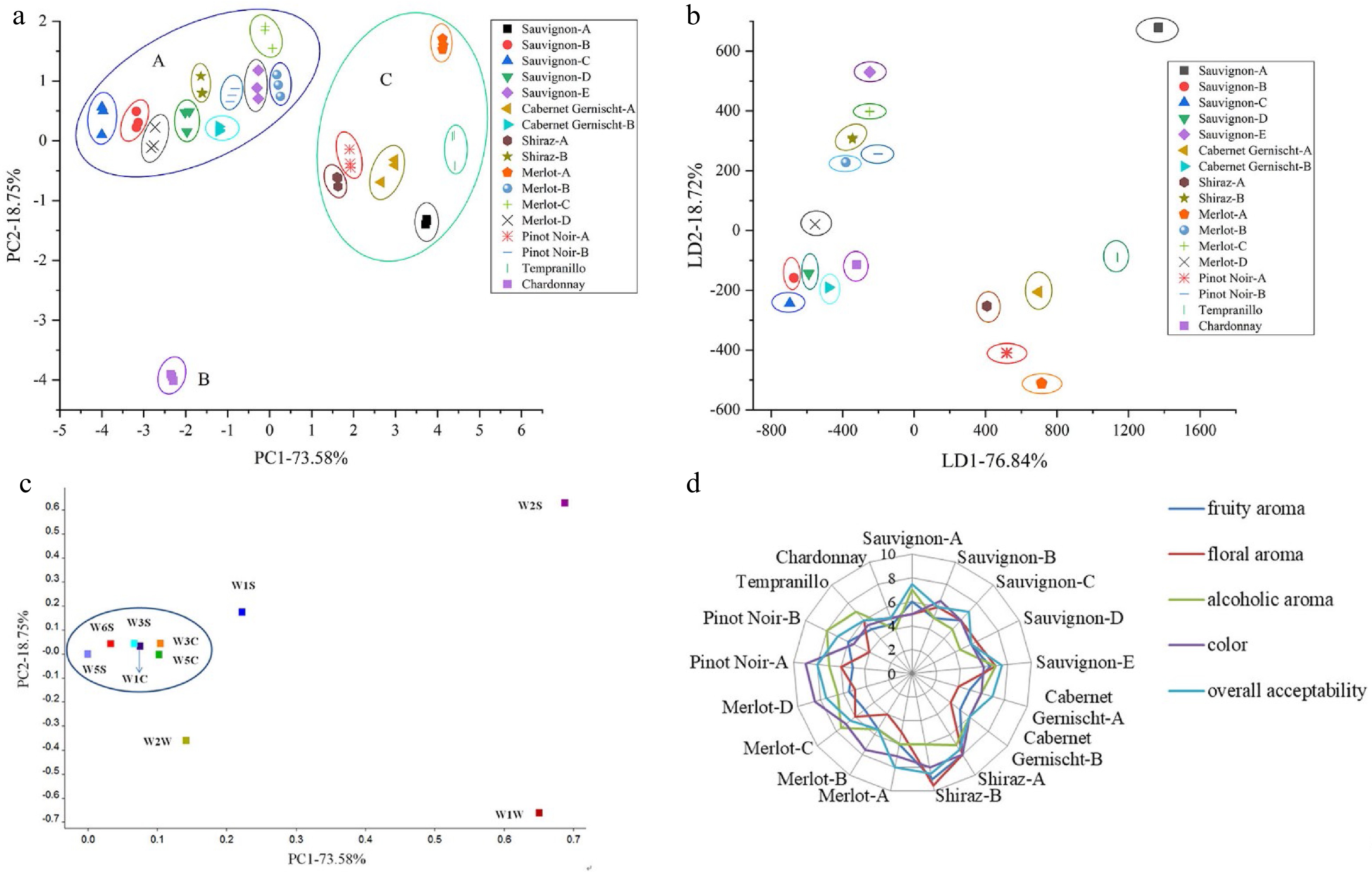
      Figure 2. 

      (a) PCA plot, (b) LDA plot, (c) loading analysis of E-nose for 17 grape wines and (d) graph of sensory scores of the 17 grape wines.

      Linear discriminant analysis (LDA) is frequently used for classification in food field and its main aim is to find the linear combinations of noted attributes that can well separate two or more than two classes of objects[50]. The classification results of the 17 grape wines on the coordinates based on first linear discriminant (LD1) and second linear discriminant (LD2) represented 76.84% and 18.72% of the total variance, respectively, with the discriminant accuracy accounting for 95.56% are shown in Fig. 2b. The results indicate that the PEN 3 E-nose with LDA is an effective instrument to distinguish the 17 grape wines via their odors.

      Loading analysis is useful to check for the influence of a sensor on the distribution of data sets. The loading factor associated to PC1 and PC2 for each sensor is represented in Fig. 2c. The points in the plot represent the sensors used in the experiment. The sensor with loading parameters close to zero for a particular principal component has a low contribution to the total response of the array, whereas high values indicates a discriminating sensor[51]. It is shown that sensors W2S and W1W have a higher influence in the current pattern file while sensors W1S and W2W have relatively low influence. The detectable compounds by sensors W2S and W1W were alcohols and sulfur-containing organics. Sensors W1C, W3S, W5C, W3C, W5S and W6S have closer influence so that they might be represented by one of the group member and this group has a minor influence in the current pattern file.

      Fifty mL grape wine was put into a beaker of 250 mL volume, and they were randomly offered to panelists, the aroma descriptors of samples were recorded by panelists. Panelists agreed that the aroma of grape wine samples could be described using five attributes: fruity aroma, floral aroma, alcoholic aroma, color, and overall acceptability. The intensities of the aroma attributes were scored using a scale from 0 to 10, the higher scores, the stronger intensities. Each sample was evaluated three times by each panelist. Data were expressed as mean. A trained panel quantitated the intensity of the aroma attributes of tea samples were evaluated by ten panelists (six females and four males), with aged between 20 and 30 years old. Panelists were trained by a series of important grape wine aroma compounds.

      The scores of human aroma sensory evaluation analysis are plotted on the radar chart and shown in Fig. 2d. The result analysis demonstrated that fruity aroma, alcoholic aroma and overall acceptability showed significant differences among Chardonnay and other 16 kinds of red wines in the sensory evaluation scores. This is consistent with the results analyzed by E-nose. Shiraz-B exhibited higher level of floral aroma and fruity aroma which may be related to high relative content of higher alcohols and esters detected by HS-SPME-GC-MS. In addition, Pinot Noir-A and Merlot-D have beautiful color, which perhaps due to their relatively high phenolic contents detected by HPLC.

    • The radar fingerprint chart of E-tongue with different grape varieties was presented in Fig. 3a. The mainly typical taste of grape wine includes astringency and sourness. Significant difference (P < 0.05) was observed in aftertaste-B and sour taste. However, there was a minor difference on the flavor from bitterness, astringency, aftertaste-A richness and umami among 17 grape wines. Further, there was a great correlation between the E-tongue and the human sensory evaluation score[52]. The values of saltiness and richness of Tempranillo and Chardonnay were lower than 15 other grape wines. Astringency taste intensity based on the E-tongue measurement ranged from 1.147 ± 0.045 to 4.400 ± 0.046. The bitterness and astringency of grape wines are mainly correlated to the phenol profile[16, 53]. Pinot Noir-A exhibited the highest sourness intensity with a score of −9.733 ± 0.207. Cabernet Sauvignon-E with the highest umami taste levels also appeared to have the highest saltiness scores. It can therefore be concluded that E-tongue could be a rapid method for taste evaluation in wineries. Consumers can easily choose their preferred grape wines according to the satisfactory taste results offered by E- tongue.
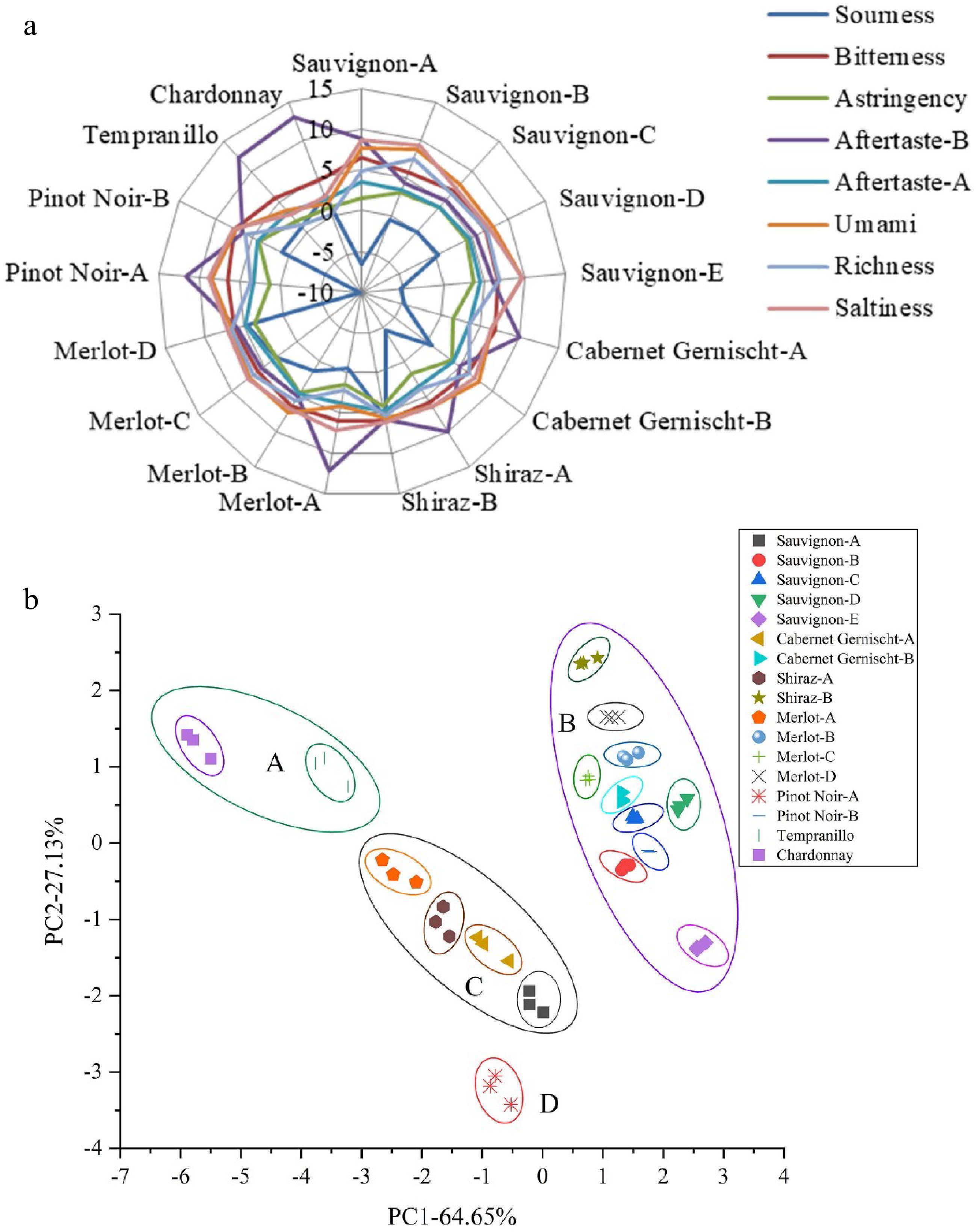
      Figure 3. 

      (a) Radar fingerprint chart of the sensory score, (b) PCA plot of E-tongue data for 17 grape wines.

      For variable reduction and separation into classed, PCA was used applied[54]. PCA of non-volatile compounds of 17 samples from seven kinds of grape varieties were presented in Fig. 3b. It was observed that variance contribution rates of PC1 and PC2 were 64.65% and 27.13%, respectively. The accumulative variance contribution rate of the first two PCs was 91.78% (> 85%), which were considered most information to represent the entire samples. In the PCA plot, a better separation effect of 17 grape wines was shown. Tempranillo, Chardonnay, Shiraz-B, Cabernet Sauvignon-E and Pinot Noir-A were clearly separated from other wines. Shiraz-A, Merlot-A, Cabernet Gernischt-A and Cabernet Sauvignon-A were slightly clustered in the centre of the PCA plot. A group comprised of Cabernet Sauvignon-B, Cabernet Sauvignon-C, Cabernet Sauvignon-D, Pinot Noir-B, Merlot-B, Merlot-C, Merlot-D and Cabernet Gernischt-B was located close together, all eight samples had positive score values at PC1. For E-tongue results, the PCA was able to distinguish the 17 wines from seven grape types completely.

    • In this study, the volatile and non-volatile flavor components of grape wines were analyzed by HS-SPME-GC-MS, E-nose, E-tongue, HPLC, and automatic amino acids analyzer techniques. The phenolic substances detected by HPLC are related to the color of the wine and the content of amino acids and phenols affect the taste of the wine, such as bitterness and astringency detected by E-tongue. Meanwhile, the combined use of HS-SPME-GC-MS and electronic nose technology analyzes the volatile flavor of 17 wines. The floral aroma and fruity aroma of the wine are closely related to alcohols and esters. Pinot Noir-A had the highest content of bitter amino acids, phenols and it was clearly separated from other wines in the PCA plot of E-tongue. The flavor and taste of Chardonnay showed great significance compared to 16 other kinds of red wines. Shiraz-B exhibited higher scores of floral aroma and fruity aroma in sensory evaluation, which may be related to its relative amount of volatile aroma substance. A total of 86 volatile compounds were identified among the 17 samples from seven kinds of wine samples. Alcohols and esters were the main flavor substances. The results clearly show that it is possible to classify grape wines from seven varieties by using E-nose and E-tongue. Sensors W2S and W1W in the E-nose for wines have a higher influence in the current pattern file. In addition, the PCA results of E-nose and E-tongue were obtained with the cumulative contribution rate accounting for 92.33% and 91.78%, respectively. Additionally, the content of free amino acids especially the taste-active amino acids, exhibited significant difference (P < 0.05). Gallic acid and catechin made up a large percentage of the grape wines. This study highlighted the usefulness of combining aroma and taste analysis techniques of grape wines, which could effectively instruct consumers to choose their preferred wines. Meanwhile, this research also provided some efficient methods to monitor grape wine quality in the actual process of industrialization. The beverage industry can certainly follow the protocols and parameters presented in this work in order to make use and apply the techniques immediately.

      • The work was financially supported by the Fundamental Research Funds for the Central Universities (KJQN201944).

      • The authors declare that they have no conflict of interest.

      • Supplemental Table S1 Relative contents of volatile compounds of grape wines from different varieties using HS-SPME-GC-MS.
      • Copyright: © 2023 by the author(s). Published by Maximum Academic Press on behalf of Nanjing Agricultural University. This article is an open access article distributed under Creative Commons Attribution License (CC BY 4.0), visit https://creativecommons.org/licenses/by/4.0/.
    Figure (3)  Table (3) References (54)
  • About this article
    Cite this article
    Fan X, Pan L, Chen R. 2023. Characterization of flavor frame in grape wines detected by HS-SPME-GC-MS coupled with HPLC, electronic nose, and electronic tongue. Food Materials Research 3:9 doi: 10.48130/FMR-2023-0009
    Fan X, Pan L, Chen R. 2023. Characterization of flavor frame in grape wines detected by HS-SPME-GC-MS coupled with HPLC, electronic nose, and electronic tongue. Food Materials Research 3:9 doi: 10.48130/FMR-2023-0009

Catalog

  • About this article

/

DownLoad:  Full-Size Img  PowerPoint
Return
Return