ARTICLE   Open Access    

Comparative analysis of flavones from six commonly used Scutellaria species

More Information
  • Scutellaria plants have been used for thousands of years for medicinal purposes, and flavones are the main bioactive compounds with properties such as anti-cancer, anti-viral, and anti-inflammatory. Although the pharmacological effects of active components and specialized metabolism in S. baicalensis are well-understood, few studies have been conducted on other Scutellaria species. In this study, we investigated the patterns of flavone accumulations in roots, leaves, and hairy roots of S. baicalensis, S. barbata, S. indica, S. strigilosa, S. obtusifolia, and S. altissima. Among the six species, S. baicalensis roots contained the highest concentrations of baicalin and wogonoside, while S. indica leaves accumulated the highest level of scutellarin. In addition, S. strigilosa leaves were rich in baicalin and wogonoside. Among the six hairy roots, S. baicalensis hairy roots had the highest contents of baicalin, baicalein, and wogonin, while S. barbata hairy roots produced the highest level of wogonoside. S. indica hairy roots contained the highest concentration of scutellarin. Compared to natural roots, the hairy roots of S. barbata and S. indica had stronger ability to selectively produce specific active flavones. Overall, this study provides a foundation for investigating diverse specialized metabolism in Scutellaria species.
  • Scale insects (Hemiptera: Coccomorpha) with hypogeic habits are considered of high phytosanitary relevance for coffee crops (Rubiaceae: Coffea spp.) in Colombia[1]. A total of 65 species of scale insects associated with coffee roots have been recorded in Colombia[24]. The most species-rich family is the Pseudococcidae with 28 species distributed in nine genera: Dysmicoccus Ferris, 1950 (13 spp.), followed by Pseudococcus Westwood, 1840 (four spp.), Phenacoccus Cockerell, 1893 (three spp.), Planococcus Ferris, 1950, and Spilococcus Ferris, 1950 (two spp. each), and Chorizococcus McKenzie, 1960, Distichlicoccus Ferris, 1950, Ferrisia Fullaway, 1923, and Paraputo Laing, 1929 (one sp. each). For the family Rhizoecidae, 19 species have been recorded in six genera, namely, Rhizoecus Kunckel d'Herculais, 1878 (13 spp.), Pseudorhizoecus Green, 1933 (two spp.), and Capitisetella Hambleton, 1977, Coccidella Hambleton, 1946, Geococcus Green, 1902, and Ripersiella Tinsley, 1899 (one sp. each). Other minor families include Coccidae and Ortheziidae (five spp. in each family), Xenococcidae (three spp.), and Putoidae and Diaspididae (two spp. in each family) and Margarodidae (one sp.). For this study all previous records were re-analysed with the purpose of providing an accurate list of species

    The taxonomic identification of scale insects by a morphological approach is particularly difficult, mainly for two reasons. First, they are small insects (usually < 5 mm) that require the preparation of slide-mounted specimens. Second, the taxonomic keys needed for morphological identifications are primarily designed for adult female specimens[5]. Differing from other insect orders (e.g., Coleoptera, Diptera and Hymenoptera), female scale insects lack well-defined tagmata, as well as sclerites, sutures, or discernible areas. Characters of taxonomic value in scale insects include cuticular processes, such as pores, ducts and, setae[5]. Recognizing these cuticular structures on such small bodies poses a difficult task for non-expert entomologists. To facilitate accessible identification, this manuscript offers an illustrated taxonomic key to scale insect species associated with coffee roots in Colombia and is aimed at users with basic knowledge of scale insect morphology.

    A careful revision of the specimens studied by Caballero et al.[2], preserved in the Scale Insect Collection at the Entomological Museum 'Universidad Nacional Agronomía Bogota' UNAB (Bogotá, Colombia), was carried out to exclude species that are doubtfully recorded from coffee roots in Colombia. This re-assessment allowed the compilation of an accurate list of species that could be included in the taxonomic key. Additional species and information from Caballero[3] and Caballero et al.[4] also were used for construction of the key. List of species recorded for Colombia and c ollection data of specimens analized are in Supplemmental Table S1 and S2 respectivately.

    The illustrated taxonomic key (Table 1) is based on the external morphology of the adult female with a dichotomous structure. Each couplet after the first one is numbered followed by the number of the preceding couplet in parenthesis, e.g. 12(7) means that couplet 12 is derived from couplet 7; the numbers at the end of the couplet indicate the next couplet in order to arrive at the species name that best matches the character states selected by the user. It is illustrated in most of the steps using microphotographs. Acquisition and analysis of images were done with a Lumenera 1-5C camera and the software Image Pro Insight 8.0. Designs were performed with Affinity Photo V 2.1 and Affinity Designer V 2.1 software. The taxonomic keys were structured with some adaptations of published taxonomic keys[615]. The general morphological terminology follows Kondo & Watson[5] with specific terminology for Coccidae[6,16], Margarodidae[17], Ortheziidae[7], Diaspididae[18], Pseudococcidae, Putoidae[8,9], and Rhizoecidae[10,11]. The abdominal segmentation is given as SabdI for abdominal segment 1 to SabdVIII for abdominal segment 8. All microphotographs are of adult female scale insects or their taxonomically important morphological structures.

    Table 1.  Illustrated taxonomic key.
    No.DetailsRef.
    1Abdominal spiracles present (Fig. 1a)2
    Abdominal spiracles absent (Fig. 1b)7
    Fig. 1 Abdominal spiracles (sp) on margin (a) present on Eurhizococcus colombianus, (b) absent on Distichlicoccus takumasai.
    2(1)Anal aperture without pores and setae (Fig. 2a); legs shorter than half of the transversal diameter of body (Fig. 2b); eyespots and mouthparts absentEurhizococcus colombianus

    Jakubski, 1965
    Anal aperture forming a well-developed anal ring with pores and setae (Fig. 2c); legs longer than transversal diameter of body; eyespots and mouthparts present (Fig. 2d)
    3
    Fig. 2 Eurhizococcus colombianus: (a) Anal aperture without pores and setae in the border, (b) section of mid body showing the length of hind leg (lel) and transversal body line (btl). Insignorthezia insignis: (c) Anal aperture with pores (po) and setae (st), (d) section of head with protruding eyespot (es) and labium (lb).
    3(2)Antennae each with eight segments (Fig. 3a)4
    Antennae each with fewer than five segments (Fig. 3b)5
    Fig. 3 (a) Eight-segmented antenna. (b) Four-segmented antenna.
    4(3)Transversal bands of spines absent in ventral region surrounded by an ovisac band (Fig. 4a); dorsal interantennal area without sclerosis (Fig. 4b)Insignorthezia insignis (Browne, 1887)
    Transversal bands of spine plates present in ventral region surrounded by an ovisac band (Fig. 4c); longitudinal sclerosis on dorsum in interantennal area (Fig. 4d)Praelongorthezia praelonga (Douglas, 1891)
    Fig. 4 Insignorthezia insignis: (a) Abdomen without transversal clusters of wax plates, (b) Dorsal interantennal area without sclerosis. Praelongorthezia praelonga: (c) Abdomen with transversal clusters of wax plates marked by dash lines, (d) dorsal interantennal area with a longitudinal sclerotic plate (ep).
    5(3)Antennae each with three segments (Fig. 5a)Newsteadia andreae Caballero, 2021
    Antennae each with four segments (Fig. 5b)6
    Fig. 5 (a) Three-segmented antenna of Newsteadia andreae. Note the presence of pseudosegmentation which gives the appearance of additional segments in the last antennal segment. (b) Four-segmented antenna of Mixorthezia minima.
    6(5)Dorsal area anterior to anal ring with simple pores on protuberances (Fig. 6a); ventral areas surrounding each coxa with a row of wax plate spines (Fig. 6b)Mixorthezia minima Koczné Benedicty & Kozár, 2004
    Dorsal area anterior to anal ring without simple pores or protuberances (Fig. 6c); ventral areas posterior to each coxa without wax plate spines (Fig. 6d)Mixorthezia neotropicalis (Silvestri, 1924)
    Fig. 6 Mixorthezia minima: (a) Dorsum of area anterior to anal ring with close-up of simple pores on protuberances (dash box); (b) ventral area posterior to each coxa with a row of wax plate spines (dash box). Mixorthezia neotropicalis: (c) Close-up of dorsum of area anterior to anal ring lacking simple pores on protuberances (dash box); (d) ventral area posterior to each coxa without associated wax plate spines.
    7(1)Anal plates present (Fig. 7a)8
    Anal plates absent (Fig. 7b)12
    Fig. 7 (a) Anal apparatus of Saissetia coffeae with anal plates (ap) covering the anal aperture (aa). (b) Anal apparatus of Pseudococcus sp. with anal aperture lacking anal plates.
    8(7)Antennae and legs with length similar to or shorter than spiracles (Fig. 8a)9
    Antennae and legs with length at least twice as long as spiracles (Fig. 8b)11
    Fig. 8 (a) Antenna (an) and foreleg (lg) (green lines), and anterior spiracle (sp) (yellow line) of Toumeyella coffeae showing their relative length. Note the similar size of the limbs and spiracle. (b) Antenna (an) and leg (lg) (green lines), and anterior spiracle (sp) (yellow line) of Coccus viridis showing their relative length. Note the relatively smaller size of the spiracle.
    9(8)Ventral tubular macroducts present (Fig. 9)Toumeyella coffeae
    Kondo, 2013
    Ventral tubular macroducts absent10
    Fig. 9 Ventral tubular macroducts (dash box) and close-up of macroducts (photo on right side).
    10(9)Orbicular pores (Fig. 10a) and cribriform platelets present (Fig. 10b); dorsal setae absent; opercular pores absentCryptostigma urichi (Cockerell, 1894)
    Orbicular pores and cribriform platelets absent; dorsal setae present (Fig. 10c); numerous opercular pores present throughout mid areas of dorsum (Fig. 10d)Akermes colombiensis Kondo & Williams, 2004
    Fig. 10 Cryptostigma urichi: (a) Orbicular pore and (b) close-up of a cribriform platelet. Akermes colombiensis: (c) Close-up of a dorsal body setae (dash box) and (d) close-up of opercular pores (arrows).
    11(8)Band of ventral tubular ducts in lateral and submarginal regions absent, ventral tubular ducts of one type; anal plates without discal setae (Fig. 11a); dorsal body setae capitate or clavate (Fig. 11b); perivulvar pores with seven or eight loculi, rarely with 10 loculi (Fig. 11c)Coccus viridis
    (Green, 1889)
    Band of ventral tubular ducts in lateral and submarginal regions present, submarginal region with two types of tubular ducts (Fig. 11d); anal plates with discal setae (Fig. 11e); dorsal body setae spine-like, apically pointed (Fig. 11f); perivulvar pores mostly with 10 loculi (Fig. 11g)Saissetia coffeae
    (Walker, 1852)
    Fig. 11 Coccus viridis: (a) Anal plates without discal setae; (b) dorsal body setae capitate (top) or clavate (below); (c) multilocular disc pores mostly with eight loculi. Saissetia coffeae: (d) Ventral submarginal region with two types of tubular ducts; (e) each anal plate with a discal seta; (f) dorsal body setae acute; (g) multilocular disc pores with mostly 10 loculi.
    12(7)Cerarii present on body margin, at least a pair on each anal lobe (Fig. 12a)13
    Cerarii absent on body margin (Fig. 12b)38
    Fig. 12 Abdominal body margin of (a) Pseudococcus sp. with three cerarii (dash box) and (b) Rhizoecus sp. (dash box) without cerarii.
    13(12)Enlarged oral collar tubular ducts composed of a sclerotized area surrounding the border and a set of flagellated setae (Ferrisia-type oral collar tubular ducts) (Fig. 13a)Ferrisia uzinuri
    Kaydan & Gullan, 2012
    Oral collar tubular ducts simple, not as above (Fig. 13b) or absent14
    Fig. 13 (a) Ferrisia-type oral collar tubular ducts with aperture of tubular duct (ad) surrounded by a sclerotized area (sa) and associated flagellate setae (fs). (b) Oral collar tubular ducts simple (arrows).
    14(12)Antenna with nine segments (Fig. 14a)15
    Antenna with eight segments (Fig. 14b) or fewer (Fig. 14c)19
    Fig. 14 Antenna with (a) nine segments, (b) eight segments and (c) seven segments.
    15(14)Cerarii with more than five conical setae (Fig. 15a); hind trochanter with six sensilla, three on each surface (Fig. 15b)16
    Cerarii with two lanceolate setae (Fig. 15c); hind trochanter with four sensilla, two on each surface (Fig. 15d)17
    Fig. 15 Puto barberi: (a) upper and lateral view of a cerarius, (b) close-up of the surface of trochanter with three sensilla (arrows). Phenacoccus sisalanus: (c) cerarius, (d) trochanter with two sensilla (arrows) on single surface.
    16(15)Cerarii with tubular ducts (Fig. 16a)Puto antioquensis
    (Murillo, 1931)
    Cerarii without tubular ducts (Fig. 16b)Puto barberi
    (Cockerell, 1895)
    Fig. 16 (a) Cerarius associated with tubular ducts (arrows). (b) Cerarius without tubular ducts.
    17(15)Oral collar tubular ducts absentPhenacoccus sisalanus Granara de Willink, 2007
    Oral collar tubular ducts present, at least on venter (Fig. 17)18
    Fig. 17 Ventral surface with oral collar tubular ducts (dash circles).
    18(17)Oral collar tubular ducts restricted to venterPhenacoccus solani
    Ferris, 1918
    Oral collar tubular ducts present on dorsum and venterPhenacoccus parvus Morrison, 1924
    19(14)Oral rim tubular ducts present (Fig. 18)20
    Oral rim tubular ducts absent26
    Fig. 18 Oral rim tubular ducts in upper view (dash circles) and close-up of lateral view.
    20(19)Oral rim tubular ducts present on venter onlyPseudococcus landoi (Balachowsky, 1959)
    Oral rim tubular ducts present on both dorsum and venter21
    21(20)Cerarii restricted to anal lobes (Fig. 19a)Chorizococcus caribaeus Williams & Granara de Willink, 1992
    Cerarii present, at least on the last five abdominal segments (Fig. 19b)22
    Fig. 19 Location of cerarii (dash boxes) on abdominal margin with close-up of cerarius (a) restricted to anal lobes (dash boxes) and (b) cerarii present on the last five abdominal segments.
    22(21)Circulus absent (Fig. 20a)23
    Circulus present (Fig. 20b)24
    Fig. 20 Ventral mid area of abdominal segments III and IV (dash box) of (a) Distichlicoccus takumasai without circulus and (b) Pseudococcus jackbeardsleyi with circulus.
    23(22)Multilocular disc pores present on venter of SabdIV and posterior segments (Fig. 21a); hind coxa with translucent pores and hind femur without translucent pores (Fig. 21b)Spilococcus pressus
    Ferris, 1950
    Multilocular disc pores absent, if some present, not more than three around vulvar opening (i.e. venter of SabdVII or SabdVIII); hind coxa without translucent pores (Fig. 21c) and hind femur with translucent pores (Fig. 21d)Distichlicoccus takumasai Caballero, 2021
    Fig. 21 Spilococcus pressus: (a) Ventral section of abdomen with multilocular disc pores (arrows); (b) hind leg with close-up of coxa with translucent pores (arrows). Distichlicoccus takumasai: (c) Hind coxa without translucent pores; (d) hind femur with translucent pores (arrows).
    24(22)Eyes without discoidal pores nor sclerotized surrounding area (Fig. 22a); circulus with transversal diameter 40 to
    60 µm (Fig. 22b)
    Pseudococcus luciae Caballero, 2021
    Eyes with discoidal pores and sclerotized surrounding area (Fig. 22c); circulus diameter 100 to 200 µm (Fig. 22d)26
    25(24)Oral rim tubular ducts on dorsal abdominal segments numbering three to eight; area between posterior ostiole and cerarius of SabdVII without oral rim tubular ducts (Fig. 23a)Pseudococcus elisae Borchsenius, 1947
    Oral rim tubular ducts on dorsal abdominal segments numbering 14 to 27; area between posterior ostiole and cerarius of SabdVII with an oral rim tubular duct (Fig. 23b)Pseudococcus jackbeardsleyi Gimpel & Miller, 1996
    Fig. 22 Pseudococcus luciae: (a) Eyespot without surrounding sclerotized area nor associated pores; (b) circulus ca. 58 µm wide. Pseudococcus jackbeardsleyi: (a) Eyespot with sclerotized area (sa) and associated pores (po); (d) circulus ca. 154 µm wide.
    Fig. 23 (a) Dorsal margin of abdominal segments VI to VIII, between cerarius of anal lobe (C1), cerarius of SabdVII (C2) and posterior ostiole (os) without oral rim tubular ducts. (b) Dorsal margin of abdominal segments VI to VIII, between cerarius of anal lobe (C1), cerarius of SabdVII (C2) and posterior ostiole (os) with an oral rim tubular duct and/or cerarius adjacent to SabdVII.
    26(19)Oral collar tubular ducts (Fig. 24) on both dorsum and venter27
    Oral collar tubular ducts restricted to venter28
    Fig. 24 Oral collar tubular duct in lateral view.
    27(26)Hind coxa with translucent pores (Fig. 25a); anal lobe with sclerotized bar, not on a sclerotized area (Fig. 25b); multilocular disc pores present posterior to fore coxaPlanococcus citri-minor complex
    Hind coxa without translucent pores (Fig. 25c); anal lobe without sclerotized bar, on a sclerotized area (Fig. 25d); multilocular disc pores absent posterior to fore coxaDysmicoccus quercicolus (Ferris, 1918)
    28(27)Oral collar tubular ducts absent on venter of both head and thorax.29
    Oral collar tubular ducts present on either head or thorax, but not on both areas (Fig. 26)30
    Fig. 25 Planococcus citri-minor complex: (a) Hind coxa with translucent pores (dash box) and (b) anal lobe with a sclerotization forming a bar (ab). Dysmicoccus quercicolus: (c) Hind coxa without translucent pores and (d) anal lobe with irregular broad sclerotized area (sa).
    Fig. 26 Marginal area of Dysmicoccus grassii, lateral to posterior spiracle (ps), with close-up of oral collar tubular ducts (oc) (left side).
    29(28)Translucent pores present on hind coxa, trochanter, femur and tibia (Fig. 27a); marginal clusters of oral collar tubular ducts on venter of SabdVI and SabdVIIDysmicoccus caribensis Granara de Willink, 2009
    Translucent pores restricted to hind femur and tibia (Fig. 27b); marginal clusters of oral collar tubular ducts present on venter of SabdIV to SabdVIIParaputo nasai
    Caballero, 2021
    Fig. 27 (a) Hind leg of Dysmicoccus caribensis with translucent pores on coxa (cx), trochanter (tr) and femur (fm), and tibia (tb). (b) Hind leg of Paraputo nasai with translucent pores restricted to femur (fm) and tibia (tb).
    30(28)Hind coxa with translucent pores (Fig. 28a)Dysmicoccus sylvarum
    Williams & Granara de Willink, 1992
    Hind coxa without translucent pores (Fig. 28b)31
    Fig. 28 (a) Translucent pores on hind coxa. (b) Translucent pores absent on hind coxa.
    31(30)Hind trochanter with translucent pores (Fig. 29a)Dysmicoccus varius
    Granara de Willink, 2009
    Hind trochanter without translucent pores (Fig. 29b)32
    Fig. 29 Translucent pores (a) on hind trochanter, (b) absent from hind trochanter.
    32(31)Oral collar tubular ducts present on margin of thorax (Fig. 30)33
    Oral collar tubular ducts absent from margin of thorax34
    Fig. 30 Prothorax margin of Dysmicoccus grassii with close-up of oral collar tubular ducts.
    33(32)Multilocular disc pores absent on SabdV; dorsal area immediately anterior to anal ring with tuft of flagellate setae; longest flagellate seta as long as diameter of anal ring (Fig. 31a), and discoidal pores larger than trilocular pores (Fig. 31b)Dysmicoccus radicis
    (Green, 1933)
    Multilocular disc pores present on SabdV; dorsal area immediately anterior to anal ring without a tuft of flagellate setae; flagellate setae much shorter than diameter of anal ring (Fig. 31c) and discoidal pores smaller than trilocular pores (Fig. 31d)Dysmicoccus grassii (Leonardi, 1913)
    34(32)Oral collar tubular ducts absent in interantennal area35
    Oral collar tubular ducts present in interantennal area (Fig. 32)36
    35(34)Translucent pores on hind leg restricted to tibia (Fig. 33a)Dysmicoccus perotensis
    Granara de Willink, 2009
    Translucent pores on hind leg present on tibia and femur (Fig. 33b)Dysmicoccus joannesiae-neobrevipes complex
    Fig. 31 Dysmicoccus radicis: (a) Area anterior to anal ring with a cluster of flagellate setae (fs) and anal ring (ar) showing the diameter of the different pores (dash box); (b) discoidal pores (dp) and trilocular pores (tp). Dysmicoccus grassii: (c) Area anterior to anal ring with scattered short flagellate setae (fs) contrasted with anal ring (ar) diameter (dash box); (d) discoidal pores (dp) and trilocular pores (tp) with similar diameter.
    Fig. 32 Interantennal area (dash box) of Dysmicoccus brevipes with close-up of oral collar tubular ducts.
    Fig. 33 (a) Hind leg of Dysmicoccus perotensis with close-up of femur and tibia with translucent pores on tibia only (arrows). (b) Hind leg of Dysmicoccus joannesiae-neobrevipes complex with close-up of femur and tibia with translucent pores (arrows).
    36(34)Hind coxa with translucent pores (see Fig. 28a)Dysmicoccus mackenziei
    Beardsleyi, 1965
    Hind coxa without translucent pores (see Fig. 28b)37
    37(36)Dorsal SabdVIII setae forming a tuft-like group, each seta conspicuously longer than remaining dorsal abdominal setae (Fig. 34a) and setal length similar to anal ring diameter (60–80 µm long)Dysmicoccus brevipes (Cockerell, 1893)
    Dorsal SabdVIII setae evenly distributed, each setae as long as remaining dorsal abdominal setae (Fig. 34b) and length less than half diameter of anal ringDysmicoccus texensis-neobrevipes complex
    38(12)Tritubular ducts absent39
    Tritubular ducts present (Fig. 35a-b)46
    Fig. 34 (a) Abdomen of Dysmicoccus brevipes with dorsal setae on SabdVIII (lfs) longer than setae on anterior segments (sfs). (b) Abdomen of Dysmicoccus texensis-neobrevipes complex with dorsal setae (ufs) along the abdominal segments of uniform length and scattered distribution.
    Fig. 35 (a) Tritubular duct in upper (left) and lateral view (right) with the border of the cuticular ring attached to tubules. (b) Tritubular duct with the border of the cuticular ring widely separated from tubules (arrows).
    39(38)Anal lobes strongly protruded, bulbiform (Fig. 36a) jutting out from margin for a distance equivalent to diameter of anal ring40
    Anal lobes shallow, if protruded, their length never more than half of diameter of anal ring (Fig. 36b)42
    Fig. 36 (a) Abdomen of Neochavesia caldasiae with anal lobes (al) protruding beyond the anal aperture (aa). (b) Abdomen of Ripersiella sp. with anal lobes (al) at the same level as the anal aperture (aa).
    40(39)Anal aperture located at the same level as the base of anal lobes (Fig. 37a); antennae located on ventral margin of headNeochavesia caldasiae (Balachowsky, 1957)
    Anal aperture located anterior to bases of anal lobes (Fig. 37b); antennae located on dorsum of head41
    Fig. 37 (a) Abdomen of Neochavesia caldasiae with anal aperture (aa) positioned between the anal lobes (al), at the same level as the bases of anal lobes (dash line). (b) Abdomen of Neochavesia eversi with anal aperture (aa) situated anterior to the bases of the anal lobes (al) (dash line).
    41(40)Antennae each with five segments, situated on a membranous base (Fig. 38a); length of hind claw less than length of hind tarsus (Fig. 38b)Neochavesia trinidadensis (Beardsley, 1970)
    Antennae each with four segments, situated on a sclerotized base (Fig. 38c); hind claw longer than hind tarsus (Fig. 38d)Neochavesia eversi (Beardsley, 1970)
    Fig. 38 (a) Antenna with four segments and a membranous base (mb). (b) Hind tarsus (green line) longer than the hind claw (red line). (c) Antenna with four segments and a sclerotized base (sb). (d) Hind tarsus (green line) shorter than hind claw (red line).
    42(39)Body setae capitate, at least on one surface (Fig. 39a)43
    Body setae never capitate (Fig. 39b)44
    Fig. 39 (a) Capitate setae. (b) Flagellate setae.
    43(42)Anal aperture without associated cells (Fig. 40a); three-segmented antennae (Fig. 40b); ventral setae in median
    and submedian regions capitate
    Capitisitella migrans
    (Green, 1933)
    Anal aperture surrounded by cells (Fig. 40c); six-segmented antennae (Fig. 40d); ventral setae in medial and submedial regions flagellateWilliamsrhizoecus coffeae
    Caballero & Ramos, 2018
    44(42)Three-segmented antennae (Fig. 41a); circulus present (Fig. 41b)Pseudorhizoecus bari
    Caballero & Ramos, 2018
    Five-segmented antennae (Fig. 41c); circulus absent45
    Fig. 40 Capitisitella migrans: (a) Anal aperture of surrounded only by setae; (b) antenna composed of three segments. Williamsrhizoecus coffeae: (c) Anal aperture of surrounded by setae and cells (flesh); (d) antenna composed of six segments.
    Fig. 41 Pseudorhizoecus bari: (a) Antenna composed of three segments and (b) circulus. (c) Antenna of Pseudorhizoecus proximus composed of five segments.
    45(44)Multilocular disc pores absent; anal aperture ornamented with small protuberances and two to five short setae, each seta never longer than 1/3 diameter of anal aperture, without cells (Fig. 42a)Pseudorhizoecus proximus
    Green, 1933
    Multilocular disc pores present (Fig. 42b); anal aperture not ornamented with small protruberances, ring with well-developed cells and six long setae, each seta as long as diameter of anal ring (Fig. 42c)Ripersiella andensis (Hambleton,
    1946)
    Fig. 42 (a) Anal aperture of Pseudorhizoecus proximus surrounded by protuberances (pr) and a few short setae (st). Ripersiella andensis: (b) Ventral section of abdomen with multilocular disc pores (mp); (c) anal aperture with a ring of cells and six long setae (se).
    46(38)Anal lobes strongly protruded, conical, each one with a stout spine at apex (Fig. 43a)Geococcus coffeae
    Green, 1933
    Anal lobes flat or barely protruded, without spines at apex (Fig. 43b)47
    47(46)Venter of abdomen with clusters of trilocular pores in medial region (Fig. 44a)Coccidella ecuadorina Konczné Benedicty & Foldi, 2004
    Venter of abdomen with trilocular pores evenly dispersed, never forming clusters in medial region (Fig. 44b)48
    Fig. 43 (a) Abdomen of Geococcus coffeae with protruding anal lobe (al) with a stout spine at the apex (sp). (b) Abdomen of Rhizoecus sp. with anal lobe (al) flat, with numerous flagellate setae (fs) at the apex.
    Fig. 44 (a) Ventral surface of Coccidella ecuadorina with clusters of trilocular pores (tc) (dash box) on medial region of abdomen. (b) Ventral surface of Rhizoecus sp. with trilocular pores (tr) scattered on venter of abdomen.
    48(47)Antennae with six well-developed segments (Fig. 45a)51
    Antennae with five well-developed segments (Fig. 45b), apical segment sometimes partially divided (Fig. 45c)49
    Fig. 45 (a) Six-segmented antenna. (b) Five-segmented antenna. (c) Five-segmented antenna with partially divided apical segment (pd). Note: antennal segments numbered in Roman numerals.
    49(48)Antennae length more than 140 µm (Fig. 46a); tritubular ducts of similar diameter to trilocular pores (± 2 µm variation) (Fig. 46b); tritubular ducts with space between ductules and edge as wide as the ductules (Fig. 46c); slender ductule, width/length ratio 1:6Rhizoecus coffeae
    Laing, 1925
    Antennae length less than 130 µm (Fig. 46d); tritubular ducts of diameter nearly twice diameter of trilocular pores (Fig. 46e); tritubular ducts with reduced space or without space between ductules and edge (Fig. 46f); stout ductule, width/length ratio 1:350
    50(49)Tubular ducts present (Fig. 47a); each anal lobe with around 28 dorsal setae of similar length, greater than 30 µm (Fig. 47b, al); and dorsal marginal clusters of setae on SabdVII 20–30 µm long (Fig. 47b, SabdVII)Rhizoecus setosus (Hambleton, 1946)
    Tubular ducts absent; each anal lobe with around 14 dorsal setae, with length less than 15 µm (Fig. 47c, al); dorsal marginal clusters of setae on SabdVII with length less 15 µm (Fig. 47c, SabdVII)Rhizoecus compotor
    Williams & Granara de Willink, 1992
    Fig. 46 (a) Antenna ca. 207 µm long. (b) Tritubular ducts (td) and trilocular pores (tp) with similar diameter. (c) Close-up of a tritubular duct indicating the space between the cuticular ring (mg) and the ductule (dt). (d) Antenna ca. 105 µm long. (e) Each tritubular duct (td) twice the diameter of a trilocular pore (tp). (f) Close-up of tritubular duct without a space between the cuticular ring (mg) and the ductule (dt).
    Fig. 47 Rhizoecus setosus: (a) Tubular ducts (td); (b) anal lobe (al) and abdominal segment (SabdVII) with marginal clusters of setae longer than 30 µm. (c) Abdomen of Rhizoecus compotor with marginal cluster of setae shorter than 20 µm on anal lobe (al) and abdominal segment (SabdVII).
    51(48)Fore tibia with at least one of two internal preapical setae spine-like (Fig. 48a-b)52
    Fore tibia with both internal preapical setae flagellate (Fig. 48c)56
    Fig. 48 Fore legs with preapical setae on tibia (ft): (a) one flagellate (fs) and one spine seta (ss), (b) with a pair of spine setae (ss), (c) with a pair of flagellate setae (fs).
    52(51)Fore tibia with one internal preapical spine-like setae and other seta flagellate (Fig. 48a); anal ring composed of spine-like setae (Fig. 49a); circulus absentRhizoecus spinipes (Hambleton, 1946)
    Fore tibia with both internal preapical setae spine-like (Fig. 48b); anal ring composed of flagellate-like setae (Fig. 49b); at least, one circulus present (Fig. 49c)53
    Fig. 49 (a) Anal ring (ar) of Rhizoecus spinipes with spine-like setae (ss). (b) Anal ring (ar) of Rhizoecus arabicus with flagellate setae (fs). (c) Circulus of Rhizoecus cacticans.
    53(52)Claw digitules setose and short, length less than half length of claw (Fig. 50a)54
    Claw digitules capitate and long, as long as claw (Fig. 50b)55
    Fig. 50 Claw with claw digitule: (a) setose (sd), (b) flagellate (fd).
    54(53)Anal ring with external row composed of 35 cells or more (Fig. 51a, ext); anal ring with external and internal rows separated by a space as wide as a cell of the external row (Fig. 51a, spc); anal ring cells without spicules (Fig. 51a, sp)Rhizoecus variabilis Hambleton, 1978
    Anal ring with external row composed of less than 30 cells (Fig. 51b, ext); anal ring with external and internal rows separated by a narrow space, as wide as half (or less) a cell of the external row (Fig. 51b, spc); anal ring cells with spicules (Fig. 51b, sp)Rhizoecus arabicus Hambleton, 1976
    Fig. 51 (a) Anal ring of Rhizoecus variabilis with external row (ext) of anal ring consisting of over 35 cells; external row separated from the internal row (int) by a similar width as the diameter of a cell (spc). (b) Anal ring of Rhizoecus arabicus with external row (ext) of anal ring with less than 30 cells; external row separated from the internal row (int) by a width less than half the diameter of a cell (spc); cells of the external row with spicules (sp).
    55(53)More than 80 tritubular ducts; circulus with basal diameter at least five times greater than apical diameter (Fig. 52a); stick-like genital chamber, parallel borders and all of similar width and structure, length across about two abdominal segments (169–175 µm long) (Fig. 52b)Rhizoecus atlanticus (Hambleton, 1946)
    Less than 50 tritubular ducts; circulus with basal diameter less than three times the apical diameter (Fig. 52c); genital chamber with basal third two times wider than anterior two-thirds, length across one abdominal segment (43–52 µm long) (Fig. 52d)Rhizoecus cacticans (Hambleton, 1946)
    Fig. 52 Rhizoecus atlanticus: (a) Circulus with diameter at base five times the apical diameter, (b) genital chamber tubular shape, length ca. 150 µm long. Rhizoecus cacticans: (c) Circulus with diameter at base about two times the apical diameter, (d) genital chamber with proximal section basiform and distal section tubular, with arms, length ca. 45 µm long.
    56(51)Multilocular disc pores absent on dorsumRhizoecus mayanus (Hambleton, 1946)
    Multilocular disc pores present on dorsum57
    57(56)Marginal prothoracic setae length greater than 50 µm (Fig. 53a); marginal SabdVII setae length greater than 45 µm (Fig 53b)Rhizoecus colombiensis Ramos-Portilla & Caballero, 2016
    Marginal prothoracic setae length less than 25 µm (Fig. 53c); marginal SabdVII setae length less than 30 µm (Fig. 53d)58
    Fig. 53 Rhizoecus colombiensis: (a) Body margin with a long seta (pts) (> 40 µm), longer than remaining setae in prothorax; (b) margin of abdominal segment VII (SabdVII) (st). with a long seta (pts) (> 40 µm), longer than remaining setae in abdomen. Rhizoecus americanus: (c) Margin of prothorax (pts) with setae of uniform length, shorter than 30 µm; (d) margin of abdominal segment VII (SabdVII) with setae (st) shorter than 30 µm.
    58(57)Tritubular ducts of two sizesRhizoecus caladii
    Green, 1933
    Tritubular ducts of three sizesRhizoecus americanus (Hambleton, 1946)
     | Show Table
    DownLoad: CSV

    The following illustrated taxonomic key (Table 1) is a tool for the identification of adult female scale insects (Hemiptera : Sternorrhyncha : Coccomorpha) associated with coffee roots in Colombia, which includes 59 species from seven families (see Supplemental Table S1).

    The taxonomic key includes 59 species associated with coffee roots. Hemiberlesia sp., Odonaspis sp., Rhizoecus stangei McKenzie, 1962, Spilococcus mamillariae (Bouche, 1844), Planococcus citri (Risso, 1813) and Planococcus minor (Maskell, 1897) were excluded from the key. In the case of the two armoured scale insects, the specimens were found in Berlese funnel samples associated with coffee roots[2], however, there is no evidence of these species feeding on the roots and there are no previous records of association of Hemiberlesia nor Odonaspis species with coffee roots.

    Previous records of single specimens of R. stangei and S. mamillariae by Caballero et al.[2] were determined as misidentifications of Rhizoecus caladii Green, 1933 and Spilococcus pressus Ferris, 1950, respectively. Spilococcus mamillariae is considered as an oligophagous species, but mainly associated with Cactaceae plants and feeding on the aerial parts of plants[19,20]. There are no records of S. mamillariae being found on any plant species of the family Rubiaceae, and hence we have removed this species from the list of species associated with coffee roots. The species R. stangei, which has been recorded only from Mexico and lacks information on its host plant[21] has apparently not been found since its original description[8].

    Planococcus citri and Pl. minor were listed also by Caballero et al.[2] as literature records. the morphological identification of P. citri and P. minor needs to be complemented with molecular and geographical analysis to be more accurate[22]. Therefore, the present key considers only identification to the Planococcus citri-minor complex.

    Furthermore, many specimens of Dysmicoccus collected from coffee roots in Colombia have morphological character states that overlap with Dysmicoccus neobrevipes Beardsley, 1959, Dysmicoccus joannesiae (Costa Lima, 1939) and Dysmicoccus texensis (Tinsley, 1900). The first case is a mix of character states of D. texensis and D. neobrevipes. The number of setae in the abdominal cerarii and the size of oral collar tubular ducts are the most important characters used to differentiate the adult females of Dysmicoccus species[8,23]. Adult females of D. texensis have a consistent pattern of only two setae in all thoracic and abdominal cerarii, along with a uniform size of oral collar tubular ducts (OC). On the other hand, D. neobrevipes varies in the number of setae in the cerarii, ranging from two to seven, accompanied by two distinct sizes of OC. These character states are generally constant among specimens found on the aerial parts of plants. However, among the specimens examined here, while the anal lobe cerarii consistently have two setae on the specimens of D. texensis found on the roots, the remaining cerarii display a variable number of setae, notably ranging from two to five, particularly within the abdominal cerarii. Furthermore, the OC of these specimens all are the same size. Regarding the differences in number of setae in the cerarii, Granara de Willink[23] underlined the need of more comprehensive studies to definitively separate these species.

    The second case involves D. joannesiae and D. neobrevipes. These species exhibit similarities in the number of setae on each cerarius (ranging from two to seven setae per cerarius) and differences in the number of clusters of OC along the abdominal margin; D. joannesiae has more than 25 clusters of OC and D. neobrevipes has fewer than 10 clusters of OC[8]. Granara de Willink also separated these two species by the presence of OC on the thorax and head[23] (present in D. neobrevipes and absent in D. joannesiae). Within the specimens of putative D. neobrevipes studied here, a few had clusters of OC numbering 15 to 20 along the abdominal margin and OC on the thorax and head. The primary challenge with addressing this dilemma lies in the fact that D. joannesiae has only been reported on Joannesia princeps Vell., 1798 (Euphorbiaceae) in Brazil and on Annona muricata (Annonaceae) intercepted in London from Saint Lucia[8,24]. Moreover, there has been no additional morphological variations recorded in the new records of D. joannesiae since its initial description in 1932 by Costa Lima. Therefore, the character states defining D. joannesiae are based on six type specimens. Based on these arguments, the following taxonomic key considers two species complex groups, namely the Dysmicoccus texensis-neobrevipes complex and the D. joannesiae-neobrevipes complex.

    Following article 31.1.2 of the International Commission of Nomenclature (ICZN), herein we make a change in nomenclature for Distichlicoccus takumasae Caballero, 2021. The ending -ae for takumasae is incorrect because the species was dedicated to Dr. Takumasa Kondo (a male coccidologist), and thus the correct ending is -i, hence the species epithet is herein amended to 'takumasai'. The corrected name is Distichlicoccus takumasai Caballero, 2021.

    After reviewing the species of scale insects associated with coffee roots in Colombia, we have compiled a list of 59 species (Supplemental Table S1). Although this study did not focus on the effect of habit (aerial vs underground) or host plant on the morphology of scale insects, we detected significant morphological variation within facultative hypogeal species. Until further studies allow an understanding of the overlap of character states between D. texensisD. neobrevipes and D. joannesiaeD. neobrevipes, we suggest considering these species as a morphological complex for hypogeal specimens. Further ecomorphological studies should be conducted to determine whether the morphology of a species may differ when feeding on the aerial parts compared when feeding on the underground parts of a host and to try to elucidate what factors trigger those changes, especially in species associated with coffee plants. As for the species complex, further collecting, morphological, and molecular studies should help elucidate these taxonomic problems.

    During the literature review performed for this study, we realized that most of the records of species are limited to mentioning the host but not the plant part on which collections were made, however, it is suspected that most species are normally collected from the aerial parts of the plant host. Although this taxonomic key is limited to root-associated species recorded in Colombia, this key could be useful for identifying scale insects associated with coffee in other tropical regions, extending also to species collected from the aerial parts of the hosts.

    The authors confirm contributions to the paper as follows: study conception and design: Caballero A, Kondo T; data collection: Caballero A, Kondo T; analysis and interpretation of results: Caballero A, Kondo T; draft manuscript preparation: Caballero A, Kondo T. Both authors reviewed the results and approved the final version of the manuscript.

    The data (microscopy slides of specimens) that support the findings of this study are available in the Scale insect repository of the entomological museum Universidad Nacional Agronomia Bogota – UNAB, Facultad de Ciencias Agrarias, Colombia. All data generated or analyzed during this study are included in this published article and its supplementary information files.

    The authors thank Dr. Andrea Ramos-Portilla for clarifying some aspects of the morphological variations of Rhizoecus species and Dr. Penny Gullan (Australian National University, Canberra, Australia) for reviewing an earlier version of the manuscript. Many thanks to Erika Valentina Vergara (AGROSAVIA) and Dr. Francisco Serna (Universidad Nacional de Colombia) for their help to access the Museum UNAB. Special thanks to Dr Giuseppina Pellizzari (University of Padova, Italy) for advice on scientific nomenclature. This study was financed by Colciencias (Programa Nacional de Ciencias Básicas [National Program on Basic Sciences]), code 110165843233, contract FP44842-004-2015), by the entomological museum UNAB (Facultad Ciencias Agrarias, Universidad Nacional de Colombia, sede Bogotá) and by Federación Nacional de Cafeteros.

  • The authors declare that they have no conflict of interest.

  • Supplemental Fig. S1 HPLC chromatograms of nine flavones standard. Compounds identified in chromatographic peaks: 1, Scutellarin; 2, Scutellarein; 3, Baicalin; 4, Wogonoside; 5, Apigenin; 6, Baicalein; 7, Wogonin; 8, Chrysin; 9, Oxoylin A.
    Supplemental Fig. S2 HPLC chromatograms of leaves and roots of six Scutellaria species. Compounds identified in chromatographic peaks: 1, Scutellarin; 2, Scutellarein; 3, Baicalin; 4, Wogonoside; 5, Apigenin; 6, Baicalein; 7, Wogonin; 8, Chrysin; 9, Oxoylin A.
    Supplemental Fig. S3 Six Scutellaria hairy roots cultured in B5 solid medium. (a) S.baicalensis hairy roots; (b) S.indica hairy roots; (c) S.barbata hairy roots; (d) S.strigilosa hairy roots; (e) S.obtusifolia hairy roots; (f) S.altissima hairy roots.
    Supplemental Fig. S4 Chromatograms of nine flavones standard analyzed by LC-MS. Compounds identified in chromatographic peaks: 1, Scutellarin; 2, Scutellarein; 3, Baicalin; 4, Wogonoside; 5, Apigenin; 6, Baicalein; 7, Wogonin; 8, Chrysin; 9, Oxoylin A.
    Supplemental Fig. S5 Secondary mass spectrum of nine flavones standard.
    Supplemental Tables S1 Characteristics of standard substance of nine flavones by UPLC-ESI-MS/MS.
    Supplemental Tables S2 Characteristics of flavones from six Scutellaria hairy roots by UPLC-ESI-MS/MS.
  • [1]

    Joshee N, Tascan A, Medina-Bolivar F, Parajuli P, Rimando AM, et al. 2013. Scutellaria: Biotechnology, phytochemistry and its potential as a commercial medicinal crop. In Biotechnology for Medicinal Plants, eds. Chandra S, Lata H, Varma A. Heidelberg: Springer Berlin. pp. 69–99. https://doi.org/10.1007/978-3-642-29974-2_3

    [2]

    Shang X, He X, He X, Li M, Zhang R, et al. 2010. The genus Scutellaria an ethnopharmacological and phytochemical review. Journal of Ethnopharmacology 128:279−313

    doi: 10.1016/j.jep.2010.01.006

    CrossRef   Google Scholar

    [3]

    Wang L, Chen W, Li M, Zhang F, Chen K, et al. 2020. A review of the ethnopharmacology, phytochemistry, pharmacology, and quality control of Scutellaria barbata D. Don. Journal of Ethnopharmacology 254:112260

    doi: 10.1016/j.jep.2019.112260

    CrossRef   Google Scholar

    [4]

    Wang ZL, Wang S, Kuang Y, Hu ZM, Qiao X, et al. 2018. A comprehensive review on phytochemistry, pharmacology, and flavonoid biosynthesis of Scutellaria baicalensis. Pharmaceutical Biology 56:465−84

    doi: 10.1080/13880209.2018.1492620

    CrossRef   Google Scholar

    [5]

    Min LW. 2009. New therapeutic aspects of flavones: The anticancer properties of Scutellaria and its main active constituents Wogonin, Baicalein and Baicalin. Cancer Treatment Reviews 35:57−68

    doi: 10.1016/j.ctrv.2008.09.005

    CrossRef   Google Scholar

    [6]

    Zhao Q, Zhang Y, Wang G, Hill L, Weng JK, et al. 2016. A specialized flavone biosynthetic pathway has evolved in the medicinal plant, Scutellaria baicalensis. Science Advances 2:e1501780

    doi: 10.1126/sciadv.1501780

    CrossRef   Google Scholar

    [7]

    Zhao Q, Yang J, Cui MY, Liu J, Fang Y, et al. 2019. The reference genome sequence of Scutellaria baicalensis provides insights into the evolution of wogonin biosynthesis. Molecular Plant 12:935−50

    doi: 10.1016/j.molp.2019.04.002

    CrossRef   Google Scholar

    [8]

    Liu H, Ye F, Sun Q, Liang H, Li C, et al. 2021. Scutellaria baicalensis extract and baicalein inhibit replication of SARS-CoV-2 and its 3C-like protease in vitro. Journal of Enzyme Inhibition and Medicinal Chemistry 36:497−503

    doi: 10.1080/14756366.2021.1873977

    CrossRef   Google Scholar

    [9]

    Zhao Q, Cui MY, Levsh O, Yang D, Liu J, et al. 2018. Two CYP82D Enzymes Function as Flavone Hydroxylases in the Biosynthesis of Root-Specific 4′-Deoxyflavones in Scutellaria baicalensis. Molecular Plant 11:135−48

    doi: 10.1016/j.molp.2017.08.009

    CrossRef   Google Scholar

    [10]

    Fang Y, Liu J, Zheng M, Zhu S, Pei T, et al. 2022. SbMYB3 transcription factor promotes root-specific flavone biosynthesis in Scutellaria baicalensis. Horticulture Research 10:uhac266

    doi: 10.1093/hr/uhac266

    CrossRef   Google Scholar

    [11]

    Miyaichi Y, Imoto Y, Tomimori T, Lin CC. 1987. Studies on the constituents of Scutellaria Species. IX. on the flavonoid constituents of the root of Scutellaria indica L. Chemical & Pharmaceutical Bulletin 35:3720−25

    doi: 10.1248/cpb.35.3720

    CrossRef   Google Scholar

    [12]

    Li Y, Ooi LSM, Wang H, But PPH, Ooi VEC. 2004. Antiviral activities of medicinal herbs traditionally used in southern mainland China. Phytotherapy Research 18:718−22

    doi: 10.1002/ptr.1518

    CrossRef   Google Scholar

    [13]

    Kim SW, Cuong TD, Hung TM, Ryoo S, Lee JH, et al. 2013. Arginase II inhibitory activity of flavonoid compounds from Scutellaria indica. Archives of Pharmacal Research 36:922−26

    doi: 10.1007/s12272-013-0125-3

    CrossRef   Google Scholar

    [14]

    Huang ST, Chen Y, Chang WC, Chen HF, Lai HC, et al. 2021. Scutellaria barbata D. Don Inhibits the Main Proteases (Mpro and TMPRSS2) of Severe Acute Respiratory Syndrome Coronavirus 2 (SARS-CoV-2) Infection. Viruses 13:826

    doi: 10.3390/v13050826

    CrossRef   Google Scholar

    [15]

    Dai ZJ, Lu WF, Gao J, Kang HF, Ma YG, et al. 2013. Anti-angiogenic effect of the total flavonoids in Scutellaria barbata D. Don. BMC Complementary and Alternative Medicine 13:150

    doi: 10.1186/1472-6882-13-150

    CrossRef   Google Scholar

    [16]

    Xu Z, Gao R, Pu X, Xu R, Wang J, et al. 2020. Comparative genome analysis of Scutellaria baicalensis and Scutellaria barbata reveals the evolution of active flavonoid biosynthesis. Genomics, Proteomics & Bioinformatics 18:230−40

    doi: 10.1016/j.gpb.2020.06.002

    CrossRef   Google Scholar

    [17]

    Li J, Wang H, Shi X, Zhao L, Lv T, et al. 2019. Anti-proliferative and anti-migratory effects of Scutellaria strigillosa Hemsley extracts against vascular smooth muscle cells. Journal of Ethnopharmacology 235:155−63

    doi: 10.1016/j.jep.2019.02.016

    CrossRef   Google Scholar

    [18]

    Gao C, Zhou Y, Jiang Z, Zhao Y, Zhang D, et al. 2017. Cytotoxic and chemosensitization effects of Scutellarin from traditional Chinese herb Scutellaria altissima L. in human prostate cancer cells. Oncology Reports 38:1491−99

    doi: 10.3892/or.2017.5850

    CrossRef   Google Scholar

    [19]

    Grzegorczyk-Karolak I, Kuźma Ł, Wysokińska H. 2017. The influence of cytokinins on proliferation and polyphenol accumulation in shoot cultures of Scutellaria altissima L. Phytochemistry Letters 20:449−55

    doi: 10.1016/j.phytol.2016.12.029

    CrossRef   Google Scholar

    [20]

    He C, Peng Y, Xiao W, Xiao P. 2012. The ethnopharmacological investigation of Chinese Scutellaria plants. Modern Chinese Medicine 14:16−20

    doi: 10.13313/j.issn.1673-4890.2012.01.016

    CrossRef   Google Scholar

    [21]

    Miyaichi Y, Morimoto T, Yaguchi K, Kizu H. 2006. Studies on the constituents of Scutellaria species (XXI): constituents of the leaves of Scutellaria strigillosa Hemsley. Journal of Natural Medicines 60:157−58

    doi: 10.1007/s11418-005-0023-1

    CrossRef   Google Scholar

    [22]

    Kovács G, Kuzovkina IN, Szoke É, Kursinszki L. 2004. HPLC Determination of Flavonoids in Hairy-Root Cultures of Scutellaria baicalensis Georgi. Chromatographia 60:S81−S85

    Google Scholar

    [23]

    Gharari Z, Bagheri K, Danafar H, Sharafi A. 2020. Enhanced flavonoid production in hairy root cultures of Scutellaria bornmuelleri by elicitor induced over-expression of MYB7 and FNSII2 genes. Plant Physiology and Biochemistry 148:35−44

    doi: 10.1016/j.plaphy.2020.01.002

    CrossRef   Google Scholar

    [24]

    Wilczańska-Barska A, Królicka A, Głód D, Majdan M, Kawiak A, et al. 2012. Enhanced accumulation of secondary metabolites in hairy root cultures of Scutellaria lateriflora following elicitation. Biotechnology Letters 34:1757−63

    doi: 10.1007/s10529-012-0963-y

    CrossRef   Google Scholar

    [25]

    Stepanova AY, Solov'eva AI, Malunova MV, Salamaikina SA, Panov YM, et al. 2021. Hairy Roots of Scutellaria spp. (Lamiaceae) as Promising Producers of Antiviral Flavones. Molecules 26:3927

    doi: 10.3390/molecules26133927

    CrossRef   Google Scholar

    [26]

    Fang Y, Hou Z, Zhang X, Yang D, Liang Z. 2018. Diverse specialized metabolism and their responses to lactalbumin hydrolysate in hairy root cultures of Salvia miltiorrhiza Bunge and Salvia castanea Diels f. tomentosa Stib. Biochemical Engineering Journal 131:58−69

    doi: 10.1016/j.bej.2017.12.007

    CrossRef   Google Scholar

    [27]

    Pei T, Yan M, Li T, Li X, Yin Y, et al. 2022. Characterization of UDP-glycosyltransferase family members reveals how major flavonoid glycoside accumulates in the roots of Scutellaria baicalensis. BMC Genomics 23:169

    doi: 10.1186/s12864-022-08391-1

    CrossRef   Google Scholar

  • Cite this article

    Zheng M, Fang Y, Zhao Q. 2023. Comparative analysis of flavones from six commonly used Scutellaria species. Medicinal Plant Biology 2:12 doi: 10.48130/MPB-2023-0012
    Zheng M, Fang Y, Zhao Q. 2023. Comparative analysis of flavones from six commonly used Scutellaria species. Medicinal Plant Biology 2:12 doi: 10.48130/MPB-2023-0012

Figures(5)  /  Tables(1)

Article Metrics

Article views(4243) PDF downloads(647)

Other Articles By Authors

ARTICLE   Open Access    

Comparative analysis of flavones from six commonly used Scutellaria species

Medicinal Plant Biology  2 Article number: 12  (2023)  |  Cite this article

Abstract: Scutellaria plants have been used for thousands of years for medicinal purposes, and flavones are the main bioactive compounds with properties such as anti-cancer, anti-viral, and anti-inflammatory. Although the pharmacological effects of active components and specialized metabolism in S. baicalensis are well-understood, few studies have been conducted on other Scutellaria species. In this study, we investigated the patterns of flavone accumulations in roots, leaves, and hairy roots of S. baicalensis, S. barbata, S. indica, S. strigilosa, S. obtusifolia, and S. altissima. Among the six species, S. baicalensis roots contained the highest concentrations of baicalin and wogonoside, while S. indica leaves accumulated the highest level of scutellarin. In addition, S. strigilosa leaves were rich in baicalin and wogonoside. Among the six hairy roots, S. baicalensis hairy roots had the highest contents of baicalin, baicalein, and wogonin, while S. barbata hairy roots produced the highest level of wogonoside. S. indica hairy roots contained the highest concentration of scutellarin. Compared to natural roots, the hairy roots of S. barbata and S. indica had stronger ability to selectively produce specific active flavones. Overall, this study provides a foundation for investigating diverse specialized metabolism in Scutellaria species.

    • Scutellaria is a perennial genus comprising about 350 species that belong to the Lamiaceae family. New species are continually being discovered[1]. It is the second-largest genus in the Lamiaceae after Salvia and is widely distributed in tropical mountains and temperate latitudes[1,2]. Many Scutellaria species are used as medicines and ornamental plants[1]. Most plants of the genus Scutellaria have medicinal values, with extracts or monomer compounds exhibiting antitumor, hepatoprotective, antioxidant, anti-inflammatory, anticonvulsant, antibacterial, and antiviral effects[2]. Over 295 compounds, including flavonoids, diterpenes, triterpenoids, phenylethanoids glycosides, alkaloids, phytosterols, polysaccharides, and iridoid glycosides, have been isolated from Scutellaria[24]. Flavones are essential natural active compounds in the genus Scutellaria that has significant pharmacological activities on the human body, including anti-oxidation, anti-cancer, anti-inflammatory, anti-malignant cell proliferation, anti-angiogenesis, and anti-estrogen effects, with no or few toxic side effects from ingestion[5,6]. Baicalin, baicalein, wogonin and wogonoside are the main components with anti-tumor activity[4].

      Scutellaria baicalensis Georgi, also known as 'Huang Qin' in China, has been used in traditional Chinese medicine (TCM) for over 2,000 years to treat headaches, infections, cancer, allergies, and inflammation[7]. The roots of S. baicalensis are the major medicinal part, but the total flavonoid extract of leaves and stems are also used for medicines[4]. S. baicalensis is the most studied plant in the genus Scutellaria in terms of pharmacological research and specialized metabolism. Baicalein from S. baicalensis has been shown to inhibit replication of SARS-CoV-2 by binding its 3C-like protease[8]. The biosynthetic pathways of baicalein and wogonin in S. baicalensis have been fully elucidated[6,7,9]. Moreover, SbMYB3 transcription factor was found to activate the biosynthesis of root-specific flavones[10]. The genome of S. baicalensis has been reported[7], providing a basis for the study of specialized metabolism and genome evolution of Scutellaria.

      The whole herb of Scutellaria indica L. (Chinese name Han Xin Cao) is used for clearing heat and detoxifying, promoting blood circulation and hemostasis, dispersing depression, and deswelling[11]. Modern pharmacological studies have demonstrated the antiviral activities of S. indica and its therapeutic effects on cardiovascular diseases[12,13]. Sculellaria barbata D. Don (Chinese name Ban Zhi Lian), is widely used in traditional Chinese and Korean medicine. The whole plant of S. barbata clears heat and detoxifies, removes blood stasis, and promotes diuresis in the TCM system[3,14]. S. barbata has pharmacological activities, including anti-microbial, anticancer, anti-angiogenesis, antioxidant, and anti-inflammation[3,15], with neo-clerodane diterpenoids and flavones being its major bioactive compounds[3,14]. S. barbata can also effectively prevent SARS-CoV-2 infection and replication by inhibiting Mpro and TMPRSS2 protease activities[14]. The chromosome-level genome assembly of S. barbata has also been completed[16]. Scutellaria strigillosa Hemsl. is recorded to clear heat and damp, and it accumulates abundant flavones such as baicalin and wogonoside. S. strigillosa extracts also have anti-proliferative and anti-migratory properties[17]. The whole plant of Scutellaria obtusifolia Hemsl. is used to treat colds, bruises, diarrhea, and snakebites. Scutellaria altissima L. is used to treat pneumonia, high blood pressure and upper respiratory infection[18]. It contains scutellarin with an anti-tumor effect[18] and diterpenoids with antifungal and anti-feedants properties[19].

      In this study, we elucidated flavone compositions in the roots and leaves of six Sculellaria species, namely S. baicalensis, S. barbata, S. indica, S. strigilosa, S. obtusifolia, and S. altissima. Additionally, we established hairy root cultures of these six species and studied their growth rates and flavone compositions. The flavone accumulation patterns in both natural roots and hairy roots of the six species were compared. Our data showed that hairy roots of S. barbata and S. indica possess advantages over natural roots in specific flavone production.

    • To determine the flavone contents in the roots and leaves of the six Scutellaria species, namely S. baicalensis, S. barbata, S. indica, S. strigilosa, S. obtusifolia, and S. altissima, LC-MS was applied to detect chrysin, baicalein, baicalin, wogonin, wogonoside, scutellarein, scutellarin, apigenin, and oroxylin A. The results revealed that the main flavones are baicalin, wogonoside, baicalein, wogonin, and scutellarin. However, lower levels of apigenin, chrysin, and oroxylin A were found, and they were only present in the roots of a few species. Scutellarein was not detected in any tissue of the species (Table 1, Supplemental Fig. S1 & S2), hence it may exist in the glycoside form, as scutellarin was found in all the samples.

      Table 1.  Contents of flavones in roots and leaves of six Scutellaria species.

      SpeciesBaicalinWogonosideBaicaleinWogoninScutellarinScutellareinApigeninChrysinOroxylin A
      S. baicalensis
      Leaves2.28 ± 1.2420.59 ± 7.68
      Roots124.96 ± 36.5927.06 ± 2.518.68 ± 5.971.74 ± 1.052.18 ± 0.620.62 ± 0.71
      S.barbata
      Leaves7.94 ± 1.340.12 ± 0.0829.50 ± 10.30
      Roots18.44 ± 2.310.78 ± 1.961.48 ± 0.070.68 ± 0.072.35 ± 0.630.57 ± 0.190.05 ± 0.02
      S. indica
      Leaves0.23 ± 0.0435.66 ± 3.02
      Roots3.12 ± 0.664.7 ± 1.920.71 ± 0.051.34 ± 0.742.02 ± 0.56
      S. strigilosa
      Leaves97.53 ± 17.607.21 ± 1.061.38 ± 0.336.97 ± 4.05
      Roots15.59 ± 2.168.39 ± 2.001.15 ± 0.463.04 ± 1.120.28 ± 0.180.42 ± 0.37
      S. obtusifolia
      Leaves0.80 ± 0.37
      Roots6.45 ± 4.053.00 ± 0.400.89 ± 0.242.03 ± 0.200.87 ± 0.37
      S. altissima
      Leaves0.07 ± 0.0330.16 ± 6.89
      Roots31.41 ± 8.639.48 ± 2.440.29 ± 0.170.81 ± 0.08

      Previous studies showed that S. baicalensis roots mainly accumulated baicalein, baicalin, wogonin, and wogonoside[6]. Our results indicated that all the six species had baicalin and wogonoside in their roots, with S. baicalensis roots having the highest levels of baicalin at 124.96 mg/g dry weight (DW), while S. indica roots had the lowest levels. Interestingly, higher concentrations of baicalin and wogonoside were present in the leaves of S. strigilosa, but only small amounts were found in the leaves of other species. This implies that S. strigilosa might have evolved different regulatory mechanism to synthesize flavones to adapt to environmental challenges. Scutellarin levels in the leaves were higher than that in the roots of all species except S. obtusifolia. Among the leaves of the six species, S. indica had the highest levels of scutellarin, while S. obtusifolia had the lowest (Table 1, Supplemental Fig. S1 & S2). These data reveal the different patterns of flavone accumulation in the roots and leaves of these six Scutellaria species.

    • Hairy root cultures were established for S. baicalensis, S. barbata, S. indica, S. strigilosa, S. obtusifolia, and S. altissima by infecting their leaves with Agrobacterium rhizogenes strain A4. All six species' hairy roots grew successfully in B5 liquid or solid medium (Fig. 1a & Supplemental Fig. S3). The hairy roots were cultured for 40 d to compare their growth rates. S. baicalensis and S. barbata hairy roots had the fastest growth rates, while S. indica, S. strigilosa, and S. obtusifolia hairy roots grew more slowly (Fig. 1b). However, the growth rate of S. altissima hairy roots cultured for 40 d was too slow and not comparable to other species. These results suggest that the six hairy roots have different growth characteristics.
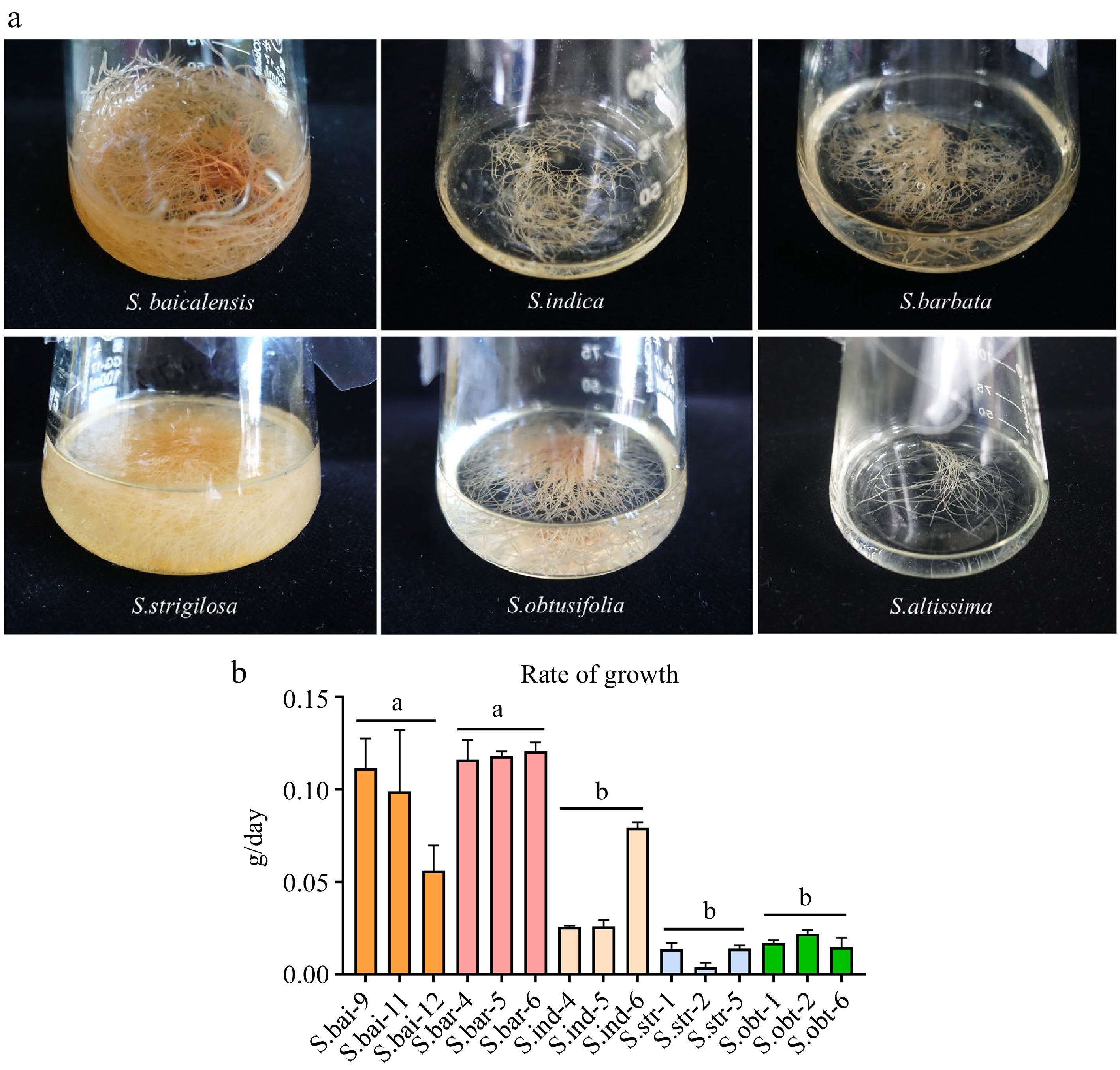
      Figure 1. 

      The growth condition of six Scutellaria hairy roots. (a) Six Scutellaria hairy roots cultured in B5 liquid medium. (b) The growth rate of the five hairy roots. S.bar: S. barbata hairy roots; S.bai: S. baicalensis hairy roots; S.ind: S. indica hairy roots; S.str: S. strigilosa hairy roots; S.obt: S. obtusifolia hairy roots. Five hairy roots were cultured for 40 d. Growth rate (g/day) = (fresh weight of hairy roots cultured for 40 d − fresh weight of hairy roots at initial inoculation)/40. Significance was determined by tukey, and different letters above the bars indicate significantly different values (p < 0.05).

    • LC-MS was used to analyze the metabolites extracted from the six hairy roots to detect the major flavones. By using retention time of standard substances and mass spectrum data, the presence of these flavones were determined. Among the six hairy roots, S. indica hairy roots were found to have eight flavones, which included scutellarin, scutellarein, baicalin, wogonoside, baicalein, wogonin, chrysin, and apigenin. Both S. baicalensis and S. barbata hairy roots had five flavones: scutellarin, baicalin, wogonoside, baicalein, and wogonin. S. obtusifolia hairy roots also had five flavones: scutellarin, baicalin, wogonoside, wogonin, but very low chrysin content was detected. Scutellarin, baicalin, wogonoside, and wogonin were detected in S. altissima hairy roots, while only baicalin, wogonoside, and wogonin could be detected in S. strigilosa hairy roots. However, oroxylin A was not detected in all hairy roots (Fig. 2, Supplemental Fig. S4, S5, Supplemental Tables S1 & S2).

      Figure 2. 

      Chromatograms of flavones in the hairy roots of (a) S. barbata, (b) S. strigilosa, (c) S. baicalensis, (d) S. obtusifolia, (e) S. indica, and (f) S. altissima. Compounds identified in chromatographic peaks: 1, Scutellarin; 2, Scutellarein; 3, Baicalin; 4, Wogonoside; 5, Apigenin; 6, Baicalein; 7, Wogonin; 8, Chrysin. S.bai: S. baicalensis hairy roots; S.bar: S. barbata hairy roots; S.alt: S. altissima hairy roots; S.ind: S. indica hairy roots; S.obt: S. obtusifolia hairy roots; S.str: S. strigilosa hairy roots.

      HPLC was used to quantify the flavones in the six hairy roots. First, the contents of different flavones in the same hairy roots were compared. Hairy roots of S. baicalensis, S. barbata, S. obtusifolia, and S. altissima had the highest levels of baicalin among other flavones, reaching 54.8, 24.28, 12.46, and 7.48 mg/g DW, respectively. However, in hairy roots of S. indica and S. strigilosa, scutellarin and wogonoside had the highest levels, with 19.75 and 17.3 mg/g DW, respectively (Fig. 3). Next, the contents of the same flavone were compared in the hairy roots of different Scutellaria species. Among the six hairy roots, S. baicalensis hairy roots accumulated the highest level of baicalin, followed by S. barbata, while baicalin contents in the other four hairy roots were almost the same. Hairy roots of S. barbata had the highest content of wogonoside, followed by S. strigilosa, S. baicalensis, S. indica, S. obtusifolia, and S. altissima. Scutellarin level in S. indica hairy roots was the highest, whereas S. altissima hairy roots had the lowest. The highest level of baicalein occurred in S. baicalensis hairy roots, followed by S. barbata and S. indica hairy roots. Hairy roots of S. strigilosa produced the lowest level of wogonin, while the highest wogonin content was observed in S. baicalensis hairy roots (Fig. 4).
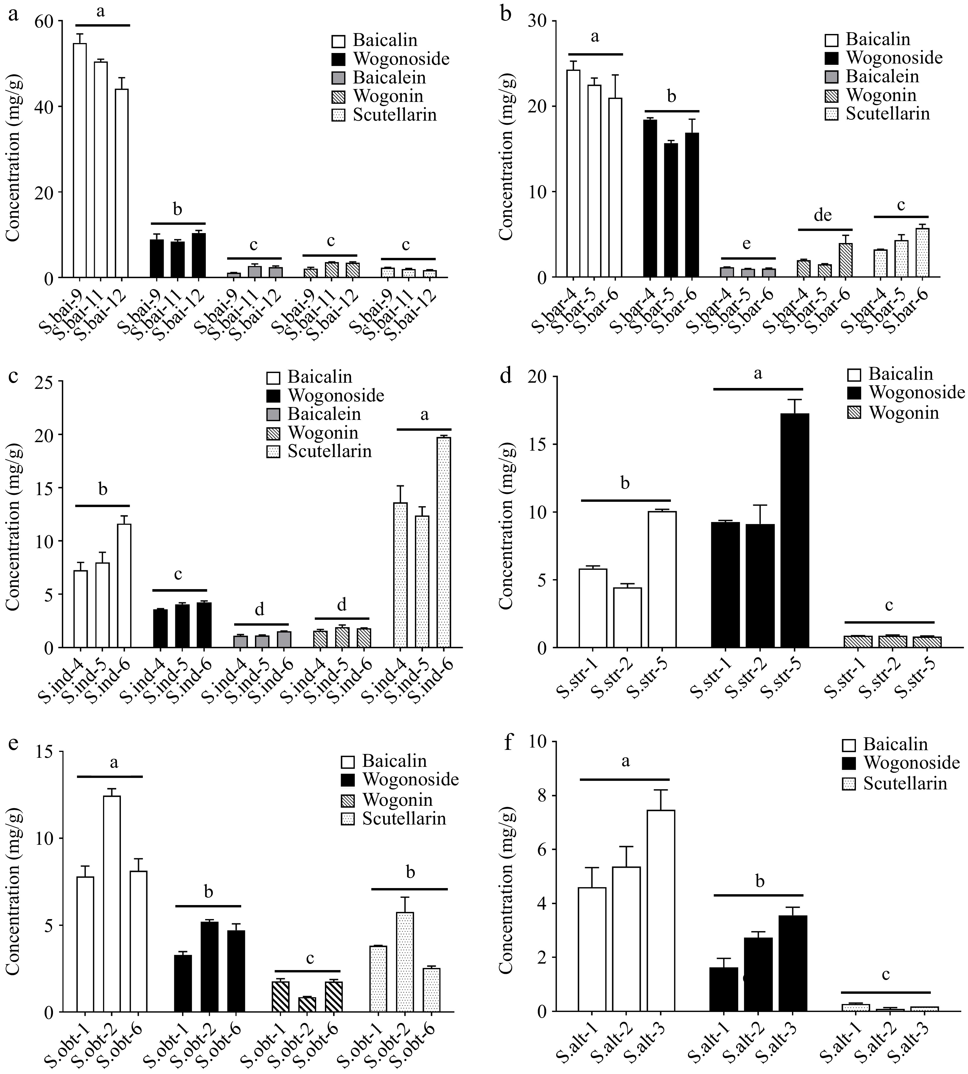
      Figure 3. 

      Comparison of the contents of different flavones in the hairy roots of (a) S. baicalensis, (b) S. barbata, (c) S. indica, (d) S. strigilosa, (e) S. obtusifolia, and (f) S. altissima. S.bai: S. baicalensis hairy roots; S.bar: S. barbata hairy roots; S.alt: S. altissima hairy roots; S.ind: S. indica hairy roots; S.obt: S. obtusifolia hairy roots; S.str: S. strigilosa hairy roots. Significance was determined by tukey, and different letters above the bars indicate significantly different values (p < 0.05).

      Figure 4. 

      Comparison of contents of the same flavones among different hairy roots. Contents of (a) baicalin, (b) wogonoside, (c) baicalein, (d) wogonin, and (e) scutellarin in the different Scutellaria hairy roots. S.bai: S. baicalensis hairy roots; S.bar: S. barbata hairy roots; S.alt: S. altissima hairy roots; S.ind: S. indica hairy roots; S.obt: S. obtusifolia hairy roots; S.str: S. strigilosa hairy roots. 'Bin', 'Bein', 'Wde', 'Win', and 'Sin' represent baicalin, baicalein, wogonoside, wogonin, and scutellarin, respectively. Significance was determined by tukey, and different letters above the bars indicate significantly different values (p < 0.05).

    • To further clarify the patterns of flavone accumulation, we compared flavone levels between the six Scutellaria hairy roots and their natural roots. The roots of S. baicalensis, S. strigilosa, and S. altissima produced higher levels of baicalin than their respective hairy roots. Furthermore, wogonoside levels in the roots of S. baicalensis and S. altissima were also higher than that in their hairy roots. Compared to their natural roots, S. barbata hairy roots produced higher levels of wogonoside and scutellarin, whereas S. indica hairy roots accumulated higher contents of baicalin and scutellarin (Fig. 5). These data suggest that the hairy roots of S. barbata and S. indica have great potential for the rapid production of the bioactive flavones.

      Figure 5. 

      Comparison of flavone contents between the six Scutellaria hairy roots and their natural roots. Flavone levels in the hairy roots and natural roots of (a) S. baicalensis, (b) S. barbata, (c) S. indica, (d) S. strigilosa, (e) S. obtusifolia, and (f) S.altissima. Significance was determined by Student's t test; * 0.01 < p < 0.05 and ** p < 0.01 were considered to indicate significant and highly significant levels, respectively.

    • With the increasing demand for TCM and the degradation of germplasm resources of medicinal plants, it is necessary to use alternative methods for producing bioactive components. Hairy roots are excellent chassis for producing active components, and metabolic engineering can generate more abundant compounds. Flavones are common in Scutellaria species and are important medicinal compounds[2]. While the flavone biosynthetic pathway in S. baicalensis is well elucidated, the pathway in other Scutellaria species remains unclear. Elucidation of the pathways is limited by the difficulty of achieving stable inheritance in these medicinal plants. In this study, we elucidated the flavonoid compositions and flavone accumulation patterns in roots and leaves of S. baicalensis, S. barbata, S. indica, S. strigilosa, S. obtusifolia, and S. altissima. Furthermore, we established hairy root cultures of the six species and investigated their growth rates and different patterns of flavone accumulation.

      Previous study have shown that the main components of Scutellaria are flavonoids[2]. Therefore, we analyzed the flavonoid composition of six common Scutellaria species. Although the flavone accumulation patterns in the roots and leaves of the six Scutellaria species were different, baicalin and wogonoside were predominantly accumulated in most of the Scutellaria roots, and scutellarin was mainly accumulated in the leaves (Table 1). This distribution of flavones is similar to that observed in S. baicalensis[6], suggesting that the regulatory mechanism of flavone synthesis in some Scutellaria species may be conservative. Interestingly, baicalin and wogonoside were highly accumulated in S. strigilosa leaves (Table 1), indicating that S. strigilosa may evolve new pathways to regulate flavone biosynthesis that differ from the known mechanisms. Previous reports have also shown that S. strigilosa is rich in wogonoside and baicalin[20,21], and extracts containing flavones from S. strigilosa have demonstrated anti-proliferative and anti-migratory effects[17]. This suggests that S. strigilosa leaves may have great potential to be developed into medicine.

      Hairy roots were successfully generated from several Scutellaria plant, including S. baicalensis[22], S. bornmuelleri[23], S. lateriflora[24], S. przewalskii and S. pycnoclada[25]. The hairy roots liquid cultures produced active flavones. In this study, we established hairy root cultures of the six Scutellaria species, all of which were capable of accumulating flavones. Although all the hairy roots produced baicalin and wogonoside, they exhibited different flavone accumulation patterns (Figs 2, 3 & 4). Similarly, there are diverse accumulation patterns of tanshinones and phenolic acids in the two Salvia species[26]. This phenomena may be a combination of environmental and genetic effects. Furthermore, hairy roots of S. barbata and S. indica had a greater capacity to produce specific flavones compared to natural roots (Fig. 5), suggesting that the two hairy roots have the potential to yield specific flavones on an industrial scale.

    • Among the six species, S. baicalensis roots had the highest levels of baicalin and wogonoside, while S. indica leaves accumulated the highest content of scutellarin. Of the six hairy roots, S. baicalensis and S. barbata exhibited the fastest growth rates, followed by S. indica, S. strigilosa, and S. obtusifolia. S. baicalensis hairy roots accumulated the highest contents of baicalin, baicalein, and wogonin, while S. barbata hairy roots produced the highest level of wogonoside. However, S. indica hairy roots accumulated the highest concentration of scutellarin. Compared to natural roots, hairy roots of S. barbata and S. indica showed greater potential to selectively produce specific active flavones. This study provides a foundation for studying the specialized metabolism of Scutellaria species, screening species with high-quality flavones, and metabolic engineering of active flavones.

    • Plants of S. baicalensis, S. barbata, S. indica, S. strigilosa, S. obtusifolia, and S. altissima were grown in the greenhouse of Shanghai Chenshan Plant Science Research Center, Shanghai, China, and their leaves and roots were collected with three biological duplicates for metabolic analysis.

    • The vector pK7WG2R carrying the dsRed marker gene was transformed into Agrobacterium rhizogene strain A4. The positive Agrobacterium transformant was cultured in TY medium overnight until OD600 reached 0.6−0.8. Then TY medium was removed by centrifugation, and Agrobacterium precipitation was re-suspended by MS medium with the equal volume. Acetosyringone was added to the Agrobacterium suspensions to a concentration of 50 μM. The leaves of six Scutellaria species were sterilized with 10% sodium hypochlorite for 8−10 min, and subsequently washed with sterile water three times. On a clean bench, the sterile leaves were cut with a lancet dipped in Agrobacterium solution. These leaves were then cultured in the MS medium with 50 μM acetosyringone for 3 d. After 3 d, these leaves were exchanged into MS medium with 400 mg/L cefotaxime sodium for dark culture. About 2−3 weeks later, hairy roots formed at the infected part of the leaves. When the hairy roots grew to 3−5 cm, a single root was cut and cultured in B5 medium. Positive lines of hairy roots were confirmed by red fluorescence inspection.

    • Based on a previous method[6], freeze-dried powders of the natural roots, leaves, and hairy roots of six Scutellaria species were ultrasonically extracted with 70% methanol for 2 h. Then, flavone extracts were filtered with 0.22-μm filters and analyzed by the Agilent 1260 Infinity II HPLC system. Flavones were detected at 280 nm. The flavone standards (baicalin, baicalein, scutellarein, scutellarin, wogonin, wogonoside, oroxylin A, chrysin and apigenin) were purchased from Sigma-Aldrich. Based on the retention time of flavone standards and standard curves, each flavone was confirmed and measured. Moreover, the retention time of scutellarin, scutellarein, baicalin, wogonoside, apigenin, baicalein, wogonin, chrysin and oroxylin A was 11.94, 16.72, 17.34, 20.96, 22.06, 23.29, 27.71, 28.65, and 29.31 min, respectively.

      LC-MS/MS was operated by Thermo Q Exactive Plus, and the procedure is the same as in our previous study[27]. A Phenomenex Luna C18 (2) column (100 mm × 2 mm 3μ) was used to separate flavones. The injection volume was 10 μL. Mass spectra were equipped with a heated ESl source and operated in negative and positive ion modes. The parameters were as follows: aus. Gas flow,10 l/min; aus. Gas heater, 350 °C; sheath gas flow, 40 l/min; spray voltage, 3.5 kV; capillary temperature, 320 °C. For full-scan MS/data-dependent (ddMS2) analysis, spectra were recorded in the m/z range of 50−750 at a resolution of 17,500 with automatic gain control (AGC) targets of 1 × 10⁶ and 2 × 10⁵.

      • This work is sponsored by Natural Science Foundation of Shanghai (22ZR1479500), Special Fund for Scientific Research of Shanghai Landscaping & City Appearance Administrative Bureau (G212401), Ministry of Science and Technology of China (YDZX20223100001003) and Youth Innovation Promotion Association, Chinese Academy of Sciences. QZ is also supported by SANOFI-SIBS Scholarship.

      • The authors declare that they have no conflict of interest.

      • Supplemental Fig. S1 HPLC chromatograms of nine flavones standard. Compounds identified in chromatographic peaks: 1, Scutellarin; 2, Scutellarein; 3, Baicalin; 4, Wogonoside; 5, Apigenin; 6, Baicalein; 7, Wogonin; 8, Chrysin; 9, Oxoylin A.
      • Supplemental Fig. S2 HPLC chromatograms of leaves and roots of six Scutellaria species. Compounds identified in chromatographic peaks: 1, Scutellarin; 2, Scutellarein; 3, Baicalin; 4, Wogonoside; 5, Apigenin; 6, Baicalein; 7, Wogonin; 8, Chrysin; 9, Oxoylin A.
      • Supplemental Fig. S3 Six Scutellaria hairy roots cultured in B5 solid medium. (a) S.baicalensis hairy roots; (b) S.indica hairy roots; (c) S.barbata hairy roots; (d) S.strigilosa hairy roots; (e) S.obtusifolia hairy roots; (f) S.altissima hairy roots.
      • Supplemental Fig. S4 Chromatograms of nine flavones standard analyzed by LC-MS. Compounds identified in chromatographic peaks: 1, Scutellarin; 2, Scutellarein; 3, Baicalin; 4, Wogonoside; 5, Apigenin; 6, Baicalein; 7, Wogonin; 8, Chrysin; 9, Oxoylin A.
      • Supplemental Fig. S5 Secondary mass spectrum of nine flavones standard.
      • Supplemental Tables S1 Characteristics of standard substance of nine flavones by UPLC-ESI-MS/MS.
      • Supplemental Tables S2 Characteristics of flavones from six Scutellaria hairy roots by UPLC-ESI-MS/MS.
      • Copyright: © 2023 by the author(s). Published by Maximum Academic Press, Fayetteville, GA. This article is an open access article distributed under Creative Commons Attribution License (CC BY 4.0), visit https://creativecommons.org/licenses/by/4.0/.
    Figure (5)  Table (1) References (27)
  • About this article
    Cite this article
    Zheng M, Fang Y, Zhao Q. 2023. Comparative analysis of flavones from six commonly used Scutellaria species. Medicinal Plant Biology 2:12 doi: 10.48130/MPB-2023-0012
    Zheng M, Fang Y, Zhao Q. 2023. Comparative analysis of flavones from six commonly used Scutellaria species. Medicinal Plant Biology 2:12 doi: 10.48130/MPB-2023-0012

Catalog

  • About this article

/

DownLoad:  Full-Size Img  PowerPoint
Return
Return