2024 Volume 4
Article Contents
About this article
ARTICLE   Open Access    

Identification and characterization of grape VAP27 gene family and their roles in disease resistance

  • # Authors contributed equally: Ruonan Li, Bianbian Wang

More Information
  • Received: 24 January 2024
    Revised: 03 April 2024
    Accepted: 24 April 2024
    Published online: 17 May 2024
    Fruit Research  4 Article number: e019 (2024)  |  Cite this article
  • Vesicle-associated membrane protein (VAMP)-associated proteins (VAP27s), which are widely expressed in plants and animals, play an important role in metabolism, physiology, growth, and development, disease resistance, and immunity. While the function of this family has been elucidated in model plants like Arabidopsis thaliana and tomato, its role in grapevine remains unclear. In this present study, 12 vesicle-associated protein-membrane protein genes were identified in the grapevine genome by bioinformatics, designated as the VAP27 gene family. A phylogenetic tree, encompassing 53 genes from three model plants, Arabidopsis thaliana, Oryza sativa, and Solanum lycopersicum, revealed the subdivision of the VAP27 gene family into three subfamilies, each presumably serving different functions, besides localizing in endoplasmic reticulum, individual members also localize in nucleus. Additionally, we compared the transcriptional levels and subcellular localizations of the VvVAP27 family members across different plant tissues (flower, leaf, seed, root, fruit, tendril, and stem), indicating site-specific functionalities for different gene members. To investigate the responsiveness of the VAP27 gene family to pathogen infection, particularly Plasmopara viticola on host plants, we analyzed the expression patterns of VAP27 genes post-infection. Our findings revealed divergent expression profiles among different members at different stages of infection. The gene family responded to the infection of downy mildew on grapevine and could inhibit the spread of Phytophthora capsici lesions in Nicotiana benthamiana. These results provide an important basis for further studies delving into the functions of the VAP27 gene family in plant growth and disease resistance.
  • Aquaporins (AQPs) constitute a large family of transmembrane channel proteins that function as regulators of intracellular and intercellular water flow[1,2]. Since their first discovery in the 1990s, AQPs have been found not only in three domains of life, i.e., bacteria, eukaryotes, and archaea, but also in viruses[3,4]. Each AQP monomer is composed of an internal repeat of three transmembrane helices (i.e., TM1–TM6) as well as two half helixes that are formed by loop B (LB) and LE through dipping into the membrane[5]. The dual Asn-Pro-Ala (NPA) motifs that are located at the N-terminus of two half helixes act as a size barrier of the pore via creating an electrostatic repulsion of protons, whereas the so-called aromatic/arginine (ar/R) selectivity filter (i.e., H2, H5, LE1, and LE2) determines the substrate specificity by rendering the pore constriction site diverse in both size and hydrophobicity[59]. Based on sequence similarity, AQPs in higher plants could be divided into five subfamilies, i.e., plasma membrane intrinsic protein (PIP), tonoplast intrinsic protein (TIP), NOD26-like intrinsic protein (NIP), X intrinsic protein (XIP), and small basic intrinsic protein (SIP)[1017]. Among them, PIPs, which are typically localized in the cell membrane, are most conserved and play a central role in controlling plant water status[12,1822]. Among two phylogenetic groups present in the PIP subfamily, PIP1 possesses a relatively longer N-terminus and PIP2 features an extended C-terminus with one or more conserved S residues for phosphorylation modification[5,15,17].

    Tigernut (Cyperus esculentus L.), which belongs to the Cyperaceae family within Poales, is a novel and promising herbaceous C4 oil crop with wide adaptability, large biomass, and short life period[2327]. Tigernut is a unique species accumulating up to 35% oil in the underground tubers[2830], which are developed from stolons and the process includes three main stages, i.e., initiation, swelling, and maturation[3133]. Water is essential for tuber development and tuber moisture content maintains a relatively high level of approximately 85% until maturation when a significant drop to about 45% is observed[28,32]. Thereby, uncovering the mechanism of tuber water balance is of particular interest. Despite crucial roles of PIPs in the cell water balance, to date, their characterization in tigernut is still in the infancy[21]. The recently available genome and transcriptome datasets[31,33,34] provide an opportunity to address this issue.

    In this study, a global characterization of PIP genes was conducted in tigernut, including gene localizations, gene structures, sequence characteristics, and evolutionary patterns. Moreover, the correlation of CePIP mRNA/protein abundance with water content during tuber development as well as subcellular localizations were also investigated, which facilitated further elucidating the water balance mechanism in this special species.

    PIP genes reported in Arabidopsis (Arabidopsis thaliana)[10] and rice (Oryza sativa)[11] were respectively obtained from TAIR11 (www.arabidopsis.org) and RGAP7 (http://rice.uga.edu), and detailed information is shown in Supplemental Table S1. Their protein sequences were used as queries for tBLASTn[35] (E-value, 1e–10) search of the full-length tigernut transcriptome and genome sequences that were accessed from CNGBdb (https://db.cngb.org/search/assembly/CNA0051961)[31,34]. RNA sequencing (RNA-seq) reads that are available in NCBI (www.ncbi.nlm.nih.gov/sra) were also adopted for gene structure revision as described before[13], and presence of the conserved MIP (major intrinsic protein, Pfam accession number PF00230) domain in candidates was confirmed using MOTIF Search (www.genome.jp/tools/motif). To uncover the origin and evolution of CePIP genes, a similar approach was also employed to identify homologs from representative plant species, i.e., Carex cristatella (v1, Cyperaceae)[36], Rhynchospora breviuscula (v1, Cyperaceae)[37], and Juncus effusus (v1, Juncaceae)[37], whose genome sequences were accessed from NCBI (www.ncbi.nlm.nih.gov). Gene structure of candidates were displayed using GSDS 2.0 (http://gsds.gao-lab.org), whereas physiochemical parameters of deduced proteins were calculated using ProtParam (http://web.expasy.org/protparam). Subcellular localization prediction was conducted using WoLF PSORT (www.genscript.com/wolf-psort.html).

    Nucleotide and protein multiple sequence alignments were respectively conducted using ClustalW and MUSCLE implemented in MEGA6[38] with default parameters, and phylogenetic tree construction was carried out using MEGA6 with the maximum likelihood method and bootstrap of 1,000 replicates. Systematic names of PIP genes were assigned with two italic letters denoting the source organism and a progressive number based on sequence similarity. Conserved motifs were identified using MEME Suite 5.5.3 (https://meme-suite.org/tools/meme) with optimized parameters as follows: Any number of repetitions, maximum number of 15 motifs, and a width of 6 and 250 residues for each motif. TMs and conserved residues were identified using homology modeling and sequence alignment with the structure resolved spinach (Spinacia oleracea) SoPIP2;1[5].

    Synteny analysis was conducted using TBtools-II[39] as described previously[40], where the parameters were set as E-value of 1e-10 and BLAST hits of 5. Duplication modes were identified using the DupGen_finder pipeline[41], and Ks (synonymous substitution rate) and Ka (nonsynonymous substitution rate) of duplicate pairs were calculated using codeml in the PAML package[42]. Orthologs between different species were identified using InParanoid[43] and information from synteny analysis, and orthogroups (OGs) were assigned only when they were present in at least two species examined.

    Plant materials used for gene cloning, qRT-PCR analysis, and 4D-parallel reaction monitoring (4D-PRM)-based protein quantification were derived from a tigernut variety Reyan3[31], and plants were grown in a greenhouse as described previously[25]. For expression profiling during leaf development, three representative stages, i.e., young, mature, and senescing, were selected and the chlorophyll content was checked using SPAD-502Plus (Konica Minolta, Shanghai, China) as previously described[44]. Young and senescing leaves are yellow in appearance, and their chlorophyll contents are just half of that of mature leaves that are dark green. For diurnal fluctuation regulation, mature leaves were sampled every 4 h from the onset of light at 8 a.m. For gene regulation during tuber development, fresh tubers at 1, 5, 10, 15, 20, 25, and 35 d after tuber initiation (DAI) were collected as described previously[32]. All samples with three biological replicates were quickly frozen with liquid nitrogen and stored at −80 °C for further use. For subcellular localization analysis, tobacco (Nicotiana benthamiana) plants were grown as previously described[20].

    Tissue-specific expression profiles of CePIP genes were investigated using Illumina RNA-seq samples (150 bp paired-end reads) with three biological replicates for young leaf, mature leaf, sheath of mature leaf, shoot apex, root, rhizome, and three stages of developmental tuber (40, 85, and 120 d after sowing (DAS)), which are under the NCBI accession number of PRJNA703731. Raw sequence reads in the FASTQ format were obtained using fastq-dump, and quality control was performed using fastQC (www.bioinformatics.babraham.ac.uk/projects/fastqc). Read mapping was performed using HISAT2 (v2.2.1, https://daehwankimlab.github.io/hisat2), and relative gene expression level was presented as FPKM (fragments per kilobase of exon per million fragments mapped)[45].

    For qRT-PCR analysis, total RNA extraction and synthesis of the first-strand cDNA were conducted as previously described[24]. Primers used in this study are shown in Supplemental Table S2, where CeUCE2 and CeTIP41[25,33] were employed as two reference genes. PCR reaction in triplicate for each biological sample was carried out using the SYBR-green Mix (Takara) on a Real-time Thermal Cycler Type 5100 (Thermal Fisher Scientific Oy). Relative gene abundance was estimated with the 2−ΔΔCᴛ method and statistical analysis was performed using SPSS Statistics 20 as described previously[13].

    Raw proteomic data for tigernut roots, leaves, freshly harvested, dried, rehydrated for 48 h, and sprouted tubers were downloaded from ProteomeXchange/PRIDE (www.proteomexchange.org, PXD021894, PXD031123, and PXD035931), which were further analyzed using Maxquant (v1.6.15.0, www.maxquant.org). Three dominant members, i.e., CePIP1;1, -2;1, and -2;8, were selected for 4D-PRM quantification analysis, and related unique peptides are shown in Supplemental Table S3. Protein extraction, trypsin digestion, and LC-MS/MS analysis were conducted as described previously[46].

    For subcellular localization analysis, the coding region (CDS) of CePIP1;1, -2;1, and -2;8 were cloned into pNC-Cam1304-SubN via Nimble Cloning as described before[30]. Then, recombinant plasmids were introduced into Agrobacterium tumefaciens GV3101 with the helper plasmid pSoup-P19 and infiltration of 4-week-old tobacco leaves were performed as previously described[20]. For subcellular localization analysis, the plasma membrane marker HbPIP2;3-RFP[22] was co-transformed as a positive control. Fluorescence observation was conducted using confocal laser scanning microscopy imaging (Zeiss LMS880, Germany): The wavelength of laser-1 was set as 730 nm for RFP observation, where the fluorescence was excited at 561 nm; the wavelength of laser-2 was set as 750 nm for EGFP observation, where the fluorescence was excited at 488 nm; and the wavelength of laser-3 was set as 470 nm for chlorophyll autofluorescence observation, where the fluorescence was excited at 633 nm.

    As shown in Table 1, a total of 14 PIP genes were identified from eight tigernut scaffolds (Scfs). The CDS length varies from 831 to 882 bp, putatively encoding 276–293 amino acids (AA) with a molecular weight (MW) of 29.16–31.59 kilodalton (kDa). The theoretical isoelectric point (pI) varies from 7.04 to 9.46, implying that they are all alkaline. The grand average of hydropathicity (GRAVY) is between 0.344 and 0.577, and the aliphatic index (II) ranges from 94.57 to 106.90, which are consistent with the hydrophobic characteristic of AQPs[47]. As expected, like SoPIP2;1, all CePIPs include six TMs, two typical NPA motifs, the invariable ar/R filter F-H-T-R, five conserved Froger's positions Q/M-S-A-F-W, and two highly conserved residues corresponding to H193 and L197 in SoPIP2;1 that were proven to be involved in gating[5,48], though the H→F variation was found in CePIP2;9, -2;10, and -2;11 (Supplemental Fig. S1). Moreover, two S residues, corresponding to S115 and S274 in SoPIP2;1[5], respectively, were also found in the majority of CePIPs (Supplemental Fig. S1), implying their posttranslational regulation by phosphorylation.

    Table 1.  Fourteen PIP genes identified in C. esculentus.
    Gene name Locus Position Intron no. AA MW (kDa) pI GRAVY AI TM MIP
    CePIP1;1 CESC_15147 Scf9:2757378..2759502(–) 3 288 30.76 8.82 0.384 95.28 6 47..276
    CePIP1;2 CESC_04128 Scf4:3806361..3807726(–) 3 291 31.11 8.81 0.344 95.95 6 46..274
    CePIP1;3 CESC_15950 Scf54:5022493..5023820(+) 3 289 31.06 8.80 0.363 94.57 6 49..278
    CePIP2;1 CESC_15350 Scf9:879960..884243(+) 3 288 30.34 8.60 0.529 103.02 6 33..269
    CePIP2;2 CESC_00011 Scf30:4234620..4236549(+) 3 293 31.59 9.27 0.394 101.57 6 35..268
    CePIP2;3 CESC_00010 Scf30:4239406..4241658(+) 3 291 30.88 9.44 0.432 98.97 6 31..266
    CePIP2;4 CESC_05080 Scf46:307799..309544(+) 3 285 30.44 7.04 0.453 100.32 6 28..265
    CePIP2;5 CESC_05079 Scf46:312254..314388(+) 3 286 30.49 7.04 0.512 101.68 6 31..268
    CePIP2;6 CESC_05078 Scf46:316024..317780(+) 3 288 30.65 7.68 0.475 103.06 6 31..268
    CePIP2;7 CESC_05077 Scf46:320439..322184(+) 3 284 30.12 8.55 0.500 100.00 6 29..266
    CePIP2;8 CESC_14470 Scf2:4446409..4448999(+) 3 284 30.37 8.30 0.490 106.90 6 33..263
    CePIP2;9 CESC_02223 Scf1:2543928..2545778(–) 3 283 30.09 9.46 0.533 106.47 6 31..262
    CePIP2;10 CESC_10007 Scf27:1686032..1688010(–) 3 276 29.16 9.23 0.560 106.05 6 26..256
    CePIP2;11 CESC_10009 Scf27:1694196..1696175(–) 3 284 29.71 9.10 0.577 105.49 6 33..263
    AA: amino acid; AI: aliphatic index; GRAVY: grand average of hydropathicity; kDa: kilodalton; MIP: major intrinsic protein; MW: molecular weight; pI: isoelectric point; PIP: plasma membrane intrinsic protein; Scf: scaffold; TM: transmembrane helix.
     | Show Table
    DownLoad: CSV

    To uncover the evolutionary relationships, an unrooted phylogenetic tree was constructed using the full-length protein sequences of CePIPs together with 11 OsPIPs and 13 AtPIPs. As shown in Fig. 1a, these proteins were clustered into two main groups, corresponding to PIP1 and PIP2 as previously defined[10,49], and each appears to have evolved into several subgroups. Compared with PIP1s, PIP2s possess a relatively shorter N-terminal but an extended C-terminal with one conserved S residue (Supplemental Fig. S1). Interestingly, a high number of gene repeats were detected, most of which seem to be species-specific, i.e., AtPIP1;1/-1;2/-1;3/-1;4/-1;5, AtPIP2;1/-2;2/-2;3/-2;4/-2;5/-2;6, AtPIP2;7/-2;8, OsPIP1;1/-1;2/-1;3, OsPIP2;1/-2;4/-2;5, OsPIP2;2/-2;3, CePIP1;1/-1;2, CePIP2;2/-2;3, CePIP2;4/-2;5/-2;6/-2;7, and CePIP2;9/-2;10/-2;11, reflecting the occurrence of more than one lineage-specific whole-genome duplications (WGDs) after their divergence[50,51]. In Arabidopsis that experienced three WGDs (i.e. γ, β, and α) after the split with the monocot clade[52], AtPIP1;5 in the PIP1 group first gave rise to AtPIP1;1 via the γ WGD shared by all core eudicots[50], which latter resulted in AtPIP1;3, -1;4, and -1;2 via β and α WGDs; AtPIP2;1 in the PIP2 group first gave rise to AtPIP2;6 via the γ WGD, and they latter generated AtPIP2;2, and -2;5 via the α WGD (Supplemental Table S1). In rice, which also experienced three WGDs (i.e. τ, σ, and ρ) after the split with the eudicot clade[51], OsPIP1;2 and -2;3 generated OsPIP1;1 and -2;2 via the Poaceae-specific ρ WGD, respectively. Additionally, tandem, proximal, transposed and dispersed duplications also played a role on the gene expansion in these two species (Supplemental Table S1).

    Figure 1.  Structural and phylogenetic analysis of PIPs in C. esculentus, O. sativa, and A. thaliana. (a) Shown is an unrooted phylogenetic tree resulting from full-length PIPs with MEGA6 (maximum likelihood method and bootstrap of 1,000 replicates), where the distance scale denotes the number of amino acid substitutions per site. (b) Shown are the exon-intron structures. (c) Shown is the distribution of conserved motifs among PIPs, where different motifs are represented by different color blocks as indicated and the same color block in different proteins indicates a certain motif. (At: A. thaliana; Ce: C. esculentus; PIP: plasma membrane intrinsic protein; Os: O. sativa).

    Analysis of gene structures revealed that all CePIP and AtPIP genes possess three introns and four exons in the CDS, in contrast to the frequent loss of certain introns in rice, including OsPIP1;2, -1;3, -2;1, -2;3, -2;4, -2;5, -2;6, -2;7, and -2;8 (Fig. 1b). The positions of three introns are highly conserved, which are located in sequences encoding LB (three residues before the first NPA), LD (one residue before the conserved L involved in gating), and LE (18 residues after the second NPA), respectively (Supplemental Fig. S1). The intron length of CePIP genes is highly variable, i.e., 109–993 bp, 115–1745 bp, and 95–866 bp for three introns, respectively. By contrast, the exon length is relatively less variable: Exons 2 and 3 are invariable with 296 bp and 141 bp, respectively, whereas Exons 1 and 4 are of 277–343 bp and 93–132 bp, determining the length of N- and C-terminus of PIP1 and PIP2, respectively (Fig. 1b). Correspondingly, their protein structures were shown to be highly conserved, and six (i.e., Motifs 1–6) out of 15 motifs identified are broadly present. Among them, Motif 3, -2, -6, -1, and -4 constitute the conserved MIP domain. In contrast to a single Motif 5 present in most PIP2s, all PIP1s possess two sequential copies of Motif 5, where the first one is located at the extended N-terminal. In CePIP2;3 and OsPIP2;7, Motif 5 is replaced by Motif 13; in CePIP2;2, it is replaced by two copies of Motif 15; and no significant motif was detected in this region of CePIP2;10. PIP1s and PIP2s usually feature Motif 9 and -7 at the C-terminal, respectively, though it is replaced by Motif 12 in CePIP2;6 and OsPIP2;8. PIP2s usually feature Motif 8 at the N-terminal, though it is replaced by Motif 14 in CePIP2;2 and -2;3 or replaced by Motif 11 in CePIP2;10 and -2;11 (Fig. 1c).

    As shown in Fig. 2a, gene localization of CePIPs revealed three gene clusters, i.e., CePIP2;2/-2;3 on Scf30, CePIP2;4/-2;5/-2;6/-2;7 on Scf46, and CePIP2;10/-2;11 on Scf27, which were defined as tandem repeats for their high sequence similarities and neighboring locations. The nucleotide identities of these duplicate pairs vary from 70.5% to 91.2%, and the Ks values range from 0.0971 to 1.2778 (Table 2), implying different time of their birth. According to intra-species synteny analysis, two duplicate pairs, i.e., CePIP1;1/-1;2 and CePIP2;2/-2;4, were shown to be located within syntenic blocks (Fig. 2b) and thus were defined as WGD repeats. Among them, CePIP1;1/-1;2 possess a comparable Ks value to CePIP2;2/-2;3, CePIP1;1/-1;3, and CePIP2;4/-2;8 (1.2522 vs 1.2287–1.2778), whereas CePIP2;2/-2;4 harbor a relatively higher Ks value of 1.5474 (Table 2), implying early origin or fast evolution of the latter. While CePIP1;1/-1;3 and CePIP2;1/-2;8 were characterized as transposed repeats, CePIP2;1/-2;2, CePIP2;9/-2;10, and CePIP2;8/-2;10 were characterized as dispersed repeats (Fig. 2a). The Ks values of three dispersed repeats vary from 0.8591 to 3.0117 (Table 2), implying distinct times of origin.

    Figure 2.  Duplication events of CePIP genes and synteny analysis within and between C. esculentus, O. sativa, and A. thaliana. (a) Duplication events detected in tigernut. Serial numbers are indicated at the top of each scaffold, and the scale is in Mb. Duplicate pairs identified in this study are connected using lines in different colors, i.e., tandem (shown in green), transposed (shown in purple), dispersed (shown in gold), and WGD (shown in red). (b) Synteny analysis within and between C. esculentus, O. sativa, and A. thaliana. (c) Synteny analysis within and between C. esculentus, C. cristatella, R. breviuscula, and J. effusus. Shown are PIP-encoding chromosomes/scaffolds and only syntenic blocks that contain PIP genes are marked, i.e., red and purple for intra- and inter-species, respectively. (At: A. thaliana; Cc: C. cristatella; Ce: C. esculentus; Je: J. effusus; Mb: megabase; PIP: plasma membrane intrinsic protein; Os: O. sativa; Rb: R. breviuscula; Scf: scaffold; WGD: whole-genome duplication).
    Table 2.  Sequence identity and evolutionary rate of homologous PIP gene pairs identified in C. esculentus. Ks and Ka were calculated using PAML.
    Duplicate 1 Duplicate 2 Identity (%) Ka Ks Ka/Ks
    CePIP1;1 CePIP1;3 78.70 0.0750 1.2287 0.0610
    CePIP1;2 CePIP1;1 77.20 0.0894 1.2522 0.0714
    CePIP2;1 CePIP2;4 74.90 0.0965 1.7009 0.0567
    CePIP2;3 CePIP2;2 70.50 0.1819 1.2778 0.1424
    CePIP2;4 CePIP2;2 66.50 0.2094 1.5474 0.1353
    CePIP2;5 CePIP2;4 87.30 0.0225 0.4948 0.0455
    CePIP2;6 CePIP2;5 84.90 0.0545 0.5820 0.0937
    CePIP2;7 CePIP2;6 78.70 0.0894 1.0269 0.0871
    CePIP2;8 CePIP2;4 72.90 0.1401 1.2641 0.1109
    CePIP2;9 CePIP2;10 76.40 0.1290 0.8591 0.1502
    CePIP2;10 CePIP2;8 64.90 0.2432 3.0117 0.0807
    CePIP2;11 CePIP2;10 91.20 0.0562 0.0971 0.5783
    Ce: C. esculentus; Ka: nonsynonymous substitution rate; Ks: synonymous substitution rate; PIP: plasma membrane intrinsic protein.
     | Show Table
    DownLoad: CSV

    According to inter-species syntenic analysis, six out of 14 CePIP genes were shown to have syntelogs in rice, including 1:1, 1:2, and 2:2 (i.e. CePIP1;1 vs OsPIP1;3, CePIP1;3 vs OsPIP1;2/-1;1, CePIP2;1 vs OsPIP2;4, CePIP2;2/-2;4 vs OsPIP2;3/-2;2, and CePIP2;8 vs OsPIP2;6), in striking contrast to a single one found in Arabidopsis (i.e. CePIP1;2 vs AtPIP1;2). Correspondingly, only OsPIP1;2 in rice was shown to have syntelogs in Arabidopsis, i.e., AtPIP1;3 and -1;4 (Fig. 2b). These results are consistent with their taxonomic relationships that tigernut and rice are closely related[50,51], and also imply lineage-specific evolution after their divergence.

    As described above, phylogenetic and syntenic analyses showed that the last common ancestor of tigernut and rice is more likely to possess only two PIP1s and three PIP2s. However, it is not clear whether the gene expansion observed in tigernut is species-specific or Cyperaceae-specific. To address this issue, recently available genomes were used to identify PIP subfamily genes from C. cristatella, R. breviuscula, and J. effuses, resulting in 15, 13, and nine members, respectively. Interestingly, in contrast to a high number of tandem repeats found in Cyperaceae species, only one pair of tandem repeats (i.e., JePIP2;3 and -2;4) were identified in J. effusus, a close outgroup species to Cyperaceae in the Juncaceae family[36,37]. According to homologous analysis, a total of 12 orthogroups were identified, where JePIP genes belong to PIP1A (JePIP1;1), PIP1B (JePIP1;2), PIP1C (JePIP1;3), PIP2A (JePIP2;1), PIP2B (JePIP2;2), PIP2F (JePIP2;3 and -2;4), PIP2G (JePIP2;5), and PIP2H (JePIP2;6) (Table 3). Further intra-species syntenic analysis revealed that JePIP1;1/-1;2 and JePIP2;2/-2;3 are located within syntenic blocks, which is consistent with CePIP1;1/-1;2, CePIP2;2/-2;4, CcPIP1;1/-1;2, CcPIP2;3/-2;4, RbPIP1;1/-1;2, and RbPIP2;2/-2;5 (Fig. 2c), implying that PIP1A/PIP1B and PIP2B/PIP2D were derived from WGDs occurred sometime before Cyperaceae-Juncaceae divergence. After the split with Juncaceae, tandem duplications frequently occurred in Cyperaceae, where PIP2B/PIP2C and PIP2D/PIP2E/PIP2F retain in most Cyperaceae plants examined in this study. By contrast, species-specific expansion was also observed, i.e., CePIP2;4/-2;5, CePIP2;10/-2;11, CcPIP1;2/-1;3, CcPIP2;4/-2;5, CcPIP2;8/-2;9, CcPIP2;10/-2;11, RbPIP2;3/-2;4, and RbPIP2;9/-2;10 (Table 3 & Fig. 2c).

    Table 3.  Twelve proposed orthogroups based on comparison of representative plant species.
    Orthogroup C. esculentus C. cristatella R. breviuscula J. effusus O. sativa A. thaliana
    PIP1A CePIP1;1 CcPIP1;1 RbPIP1;1 JePIP1;1 OsPIP1;3 AtPIP1;1, AtPIP1;2,
    AtPIP1;3, AtPIP1;4,
    AtPIP1;5
    PIP1B CePIP1;2 CcPIP1;2, CcPIP1;3 RbPIP1;2 JePIP1;2
    PIP1C CePIP1;3 CcPIP1;4 RbPIP1;3 JePIP1;3 OsPIP1;1, OsPIP1;2
    PIP2A CePIP2;1 CcPIP2;1 RbPIP2;1 JePIP2;1 OsPIP2;1, OsPIP2;4,
    OsPIP2;5
    AtPIP2;1, AtPIP2;2,
    AtPIP2;3, AtPIP2;4,
    AtPIP2;5, AtPIP2;6
    PIP2B CePIP2;2 CcPIP2;2 RbPIP2;2 JePIP2;2 OsPIP2;2, OsPIP2;3
    PIP2C CePIP2;3 CcPIP2;3 RbPIP2;3, RbPIP2;4
    PIP2D CePIP2;4, CePIP2;5 CcPIP2;4, CcPIP2;5 RbPIP2;5
    PIP2E CePIP2;5 CcPIP2;5 RbPIP2;6
    PIP2F CePIP2;6 CcPIP2;6
    PIP2G CePIP2;7 CcPIP2;7 RbPIP2;7 JePIP2;3, JePIP2;4
    PIP2H CePIP2;8 CcPIP2;8, CcPIP2;9 RbPIP2;8 JePIP2;5 OsPIP2;6 AtPIP2;7, AtPIP2;8
    PIP2I CePIP2;9, CePIP2;10,
    CePIP2;11
    CcPIP2;10, CcPIP2;11 RbPIP2;9, RbPIP2;10 JePIP2;6 OsPIP2;7, OsPIP2;8
    At: A. thaliana; Cc: C. cristatella; Ce: C. esculentus; Je: J. effuses; Os: O. sativa; Rb: R. breviuscula; PIP: plasma membrane intrinsic protein.
     | Show Table
    DownLoad: CSV

    Tissue-specific expression profiles of CePIP genes were investigated using transcriptome data available for young leaf, mature leaf, sheath, root, rhizome, shoot apex, and tuber. As shown in Fig. 3a, CePIP genes were mostly expressed in roots, followed by sheaths, moderately in tubers, young leaves, rhizomes, and mature leaves, and lowly in shoot apexes. In most tissues, CePIP1;1, -2;1, and -2;8 represent three dominant members that contributed more than 90% of total transcripts. By contrast, in rhizome, these three members occupied about 80% of total transcripts, which together with CePIP1;3 and -2;4 contributed up to 96%; in root, CePIP1;1, -1;3, -2;4, and -2;7 occupied about 84% of total transcripts, which together with CePIP2;1 and -2;8 contributed up to 94%. According to their expression patterns, CePIP genes could be divided into five main clusters: Cluster I includes CePIP1;1, -2;1, and -2;8 that were constitutively and highly expressed in all tissues examined; Cluster II includes CePIP2;2, -2;9, and -2;10 that were lowly expressed in all tested tissues; Cluster III includes CePIP1;2 and -2;11 that were preferentially expressed in young leaf and sheath; Cluster IV includes CePIP1;3 and -2;4 that were predominantly expressed in root and rhizome; and Cluster V includes remains that were typically expressed in root (Fig. 3a). Collectively, these results imply expression divergence of most duplicate pairs and three members (i.e. CePIP1;1, -2;1, and -2;8) have evolved to be constitutively co-expressed in most tissues.

    Figure 3.  Expression profiles of CePIP genes in various tissues, different stages of leaf development, and mature leaves of diurnal fluctuation. (a) Tissue-specific expression profiles of 14 CePIP genes. The heatmap was generated using the R package implemented with a row-based standardization. Color scale represents FPKM normalized log2 transformed counts, where blue indicates low expression and red indicates high expression. (b) Expression profiles of CePIP1;1, -2;1, and -2;8 at different stages of leaf development. (c) Expression profiles of CePIP1;1, -2;1, and -2;8 in mature leaves of diurnal fluctuation. Bars indicate SD (N = 3) and uppercase letters indicate difference significance tested following Duncan's one-way multiple-range post hoc ANOVA (p< 0.01). (Ce: C. esculentus; FPKM: Fragments per kilobase of exon per million fragments mapped; PIP: plasma membrane intrinsic protein)

    As shown in Fig. 3a, compared with young leaves, transcriptome profiling showed that CePIP1;2, -2;3, -2;7, -2;8, and -2;11 were significantly down-regulated in mature leaves, whereas CePIP1;3 and -2;1 were up-regulated. To confirm the results, three dominant members, i.e., CePIP1;1, -2;1, and -2;8, were selected for qRT-PCR analysis, which includes three representative stages, i.e., young, mature, and senescing leaves. As shown in Fig. 3b, in contrast to CePIP2;1 that exhibited a bell-like expression pattern peaking in mature leaves, transcripts of both CePIP1;1 and -2;8 gradually decreased during leaf development. These results were largely consistent with transcriptome profiling, and the only difference is that CePIP1;1 was significantly down-regulated in mature leaves relative to young leaves. However, this may be due to different experiment conditions used, i.e., greenhouse vs natural conditions.

    Diurnal fluctuation expression patterns of CePIP1;1, -2;1, and -2;8 were also investigated in mature leaves and results are shown in Fig. 3c. Generally, transcripts of all three genes in the day (8, 12, 16, and 20 h) were higher than that in the night (24 and 4 h). During the day, both CePIP1;1 and -2;8 exhibited an unimodal expression pattern that peaked at 12 h, whereas CePIP2;1 possessed two peaks (8 and 16 h) and their difference was not significant. Nevertheless, transcripts of all three genes at 20 h (onset of night) were significantly lower than those at 8 h (onset of day) as well as 12 h. In the night, except for CePIP2;1, no significant difference was observed between the two stages for both CePIP1;1 and -2;8. Moreover, their transcripts were comparable to those at 20 h (Fig. 3c).

    To reveal the expression patterns of CePIP genes during tuber development, three representative stages, i.e., 40 DAS (early swelling stage), 85 DAS (late swelling stage), and 120 DAS (mature stage), were first profiled using transcriptome data. As shown in Fig. 4a, except for rare expression of CePIP1;2, -2;2, -2;9, and -2;10, most genes exhibited a bell-like expression pattern peaking at 85 DAS, in contrast to a gradual decrease of CePIP2;3 and -2;8. Notably, except for CePIP2;4, other genes were expressed considerably lower at 120 DAS than that at 40 DAS. For qRT-PCR confirmation of CePIP1;1, -2;1, and -2;8, seven stages were examined, i.e., 1, 5, 10, 15, 20, 25, and 35 DAI, which represent initiation, five stages of swelling, and maturation as described before[32]. As shown in Fig. 4b, two peaks were observed for all three genes, though their patterns were different. As for CePIP1;1, compared with the initiation stage (1 DAI), significant up-regulation was observed at the early swelling stage (5 DAI), followed by a gradual decrease except for the appearance of the second peak at 20 DAI, which is something different from transcriptome profiling. As for CePIP2;1, a sudden drop of transcripts first appeared at 5 DAI, then gradually increased until 20 DAI, which was followed by a gradual decrease at two late stages. The pattern of CePIP2;8 is similar to -1;1, two peaks appeared at 5 and 20 DAI and the second peak was significantly lower than the first. The difference is that the second peak of CePIP2;8 was significantly lower than the initiation stage. By contrast, the second peak (20 DAI) of CePIP2;1 was significantly higher than that of the first one (1 DAI). Nevertheless, the expression patterns of both CePIP2;1 and -2;8 are highly consistent with transcriptome profiling.

    Figure 4.  Transcript and protein abundances of CePIP genes during tuber development. (a) Transcriptome-based expression profiling of 14 CePIP genes during tuber development. The heatmap was generated using the R package implemented with a row-based standardization. Color scale represents FPKM normalized log2 transformed counts, where blue indicates low expression and red indicates high expression. (b) qRT-PCR-based expression profiling of CePIP1;1, -2;1, and -2;8 in seven representative stages of tuber development. (c) Relative protein abundance of CePIP1;1, -2;1, and -2;8 in three representative stages of tuber development. Bars indicate SD (N = 3) and uppercase letters indicate difference significance tested following Duncan's one-way multiple-range post hoc ANOVA (p < 0.01). (Ce: C. esculentus; DAI: days after tuber initiation; DAS: days after sowing; FPKM: Fragments per kilobase of exon per million fragments mapped; PIP: plasma membrane intrinsic protein).

    Since protein abundance is not always in agreement with the transcript level, protein profiles of three dominant members (i.e. CePIP1;1, -2;1, and -2;8) during tuber development were further investigated. For this purpose, we first took advantage of available proteomic data to identify CePIP proteins, i.e., leaves, roots, and four stages of tubers (freshly harvested, dried, rehydrated for 48 h, and sprouted). As shown in Supplemental Fig. S2, all three proteins were identified in both leaves and roots, whereas CePIP1;1 and -2;8 were also identified in at least one of four tested stages of tubers. Notably, all three proteins were considerably more abundant in roots, implying their key roles in root water balance.

    To further uncover their profiles during tuber development, 4D-PRM-based protein quantification was conducted in three representative stages of tuber development, i.e., 1, 25, and 35 DAI. As expected, all three proteins were identified and quantified. In contrast to gradual decrease of CePIP2;8, both CePIP1;1 and -2;1 exhibited a bell-like pattern that peaked at 25 DAI, though no significant difference was observed between 1 and 25 DAI (Fig. 4c). The trends are largely in accordance with their transcription patterns, though the reverse trend was observed for CePIP2;1 at two early stages (Fig. 4b & Fig. 4c).

    As predicted by WoLF PSORT, CePIP1;1, -2;1, and -2;8 may function in the cell membrane. To confirm the result, subcellular localization vectors named pNC-Cam1304-CePIP1;1, pNC-Cam1304-CePIP2;1, and pNC-Cam1304-CePIP2;8 were further constructed. When transiently overexpressed in tobacco leaves, green fluorescence signals of all three constructs were confined to cell membranes, highly coinciding with red fluorescence signals of the plasma membrane marker HbPIP2;3-RFP (Fig. 5).

    Figure 5.  (a) Schematic diagram of overexpressing constructs, (b) subcellular localization analysis of CePIP1;1, -2;1, and -2;8 in N. benthamiana leaves. (35S: cauliflower mosaic virus 35S RNA promoter; Ce: C. esculentus; EGFP: enhanced green fluorescent protein; kb: kilobase; NOS: terminator of the nopaline synthase gene; RFP: red fluorescent protein; PIP: plasma membrane intrinsic protein).

    Water balance is particularly important for cell metabolism and enlargement, plant growth and development, and stress responses[2,19]. As the name suggests, AQPs raised considerable interest for their high permeability to water, and plasma membrane-localized PIPs were proven to play key roles in transmembrane water transport between cells[1,18]. The first PIP was discovered in human erythrocytes, which was named CHIP28 or AQP1, and its homolog in plants was first characterized in Arabidopsis, which is known as RD28, PIP2c, or AtPIP2;3[3,7,53]. Thus far, genome-wide identification of PIP genes have been reported in a high number of plant species, including two model plants Arabidopsis and rice[10,11,1317,5456]. By contrast, little information is available on Cyperaceae, the third largest family within the monocot clade that possesses more than 5,600 species[57].

    Given the crucial roles of water balance for tuber development and crop production, in this study, tigernut, a representative Cyperaceae plant producing high amounts of oil in underground tubers[28,30,32], was employed to study PIP genes. A number of 14 PIP genes representing two phylogenetic groups (i.e., PIP1 and PIP2) or 12 orthogroups (i.e., PIP1A, PIP1B, PIP1C, PIP2A, PIP2B, PIP2C, PIP2D, PIP2E, PIP2F, PIP2G, PIP2H, and PIP2I) were identified from the tigernut genome. Though the family amounts are comparative or less than 13–21 present in Arabidopsis, cassava (Manihot esculenta), rubber tree (Hevea brasiliensis), poplar (Populus trichocarpa), C. cristatella, R. breviuscula, banana (Musa acuminata), maize (Zea mays), sorghum (Sorghum bicolor), barley (Hordeum vulgare), and switchgrass (Panicum virgatum), they are relatively more than four to 12 found in eelgrass (Zostera marina), Brachypodium distachyon, foxtail millet (Setaria italic), J. effuses, Aquilegia coerulea, papaya (Carica papaya), castor been (Ricinus communis), and physic nut (Jatropha curcas) (Supplemental Table S4). Among them, A. coerulea represents a basal eudicot that didn't experience the γ WGD shared by all core eudicots[50], whereas eelgrass is an early diverged aquatic monocot that didn't experience the τ WGD shared by all core monocots[56]. Interestingly, though both species possess two PIP1s and two PIP2s, they were shown to exhibit complex orthologous relationships of 1:1, 2:2, 1:0, and 0:1 (Supplemental Table S5). Whereas AcPIP1;1/AcPIP1;2/ZmPIP1;1/ZmPIP1;2 and ZmPIP2;1/AcPIP2;1 belong to PIP1A and PIP2A identified in this study, AcPIP2;2 and ZmPIP2;2 belong to PIP2H and PIP2I, respectively (Supplemental Table S5), implying that the last common ancestor of monocots and eudicots possesses only one PIP1 and two PIP2s followed by clade-specific expansion. A good example is the generation of AtPIP1;1 and -2;6 from AtPIP1;5 and -2;1 via the γ WGD, respectively[17].

    In tigernut, extensive expansion of the PIP subfamily was contributed by WGD (2), transposed (2), tandem (5), and dispersed duplications (3). It's worth noting that, two transposed repeats (i.e., CePIP1;1/-1;3 and CePIP2;1/-2;8) are shared by rice, implying their early origin that may be generated sometime after the split with the eudicot clade but before Cyperaceae-Poaceae divergence. By contrast, two WGD repeats (i.e., CePIP1;1/-1;2 and CePIP2;2/-2;4) are shared by C. cristatella, R. breviuscula, and J. effusus but not rice and Arabidopsis, implying that they may be derived from WGDs that occurred sometime after Cyperaceae-Poaceae split but before Cyperaceae-Juncaceae divergence. The possible WGD is the one that was described in C. littledalei[58], though the exact time still needs to be studied. Interestingly, compared with Arabidopsis (1) and rice (2), tandem/proximal duplications played a more important role in the expansion of PIP genes in tigernut (5) as well as other Cyperaceae species tested (5–6), which were shown to be Cyperaceae-specific or even species-specific. These tandem repeats may play a role in the adaptive evolution of Cyperaceae species as described in a high number of plant species[14,41]. According to comparative genomics analyses, tandem duplicates experienced stronger selective pressure than genes formed by other modes (WGD, transposed duplication, and dispersed duplication) and evolved toward biased functional roles involved in plant self-defense[41].

    As observed in most species such as Arabidopsis[10,1417], PIP genes in all Cyperaceae and Juncaceae species examined in this study, i.e., tigernut, C. cristatella, R. breviuscula, and J. effuses, feature three introns with conserved positions. By contrast, zero to three introns was not only found in rice but also in other Poaceae species such as maize, sorghum, foxtail millet, switchgrass, B. distachyon, and barley[54,55], implying lineage/species-specific evolution.

    Despite the extensive expansion of PIP genes (PIP2) in tigernut even after the split with R. breviuscula, CePIP1;1, -2;1, and -2;8 were shown to represent three dominant members in most tissues examined in this study, i.e., young leaf, mature leaf, sheath, rhizome, shoot apex, and tuber, though the situation in root is more complex. CePIP1;1 was characterized as a transposed repeat of CePIP1;3, which represents the most expressed member in root. Moreover, its recent WGD repeat CePIP1;2 was shown to be lowly expressed in most tested tissues, implying their divergence. The ortholog of CePIP1;1 in rice is OsPIP1;3 (RWC-3), which was shown to be preferentially expressed in roots, stems, and leaves, in contrast to constitutive expression of OsPIP1;1 (OsPIP1a) and -1;2[5961], two recent WGD repeats. Injecting the cRNA of OsPIP1;3 into Xenopus oocytes could increase the osmotic water permeability by 2–3 times[60], though the activity is considerably lower than PIP2s such as OsPIP2;2 and -2;2[6163]. Moreover, OsPIP1;3 was shown to play a role in drought avoidance in upland rice and its overexpression in lowland rice could increase root osmotic hydraulic conductivity, leaf water potential, and relative cumulative transpiration at the end of 10 h PEG treatment[64]. CePIP2;8 was characterized as a transposed repeat of CePIP2;1. Since their orthologs are present in both rice and Arabidopsis (Supplemental Table S3), the duplication event is more likely to occur sometime before monocot-eudicot split. Interestingly, their orthologs in rice, i.e., OsPIP2;1 (OsPIP2a) and -2;6, respectively, are also constitutively expressed[61], implying a conserved evolution with similar functions. When heterologously expressed in yeast, OsPIP2;1 was shown to exhibit high water transport activity[62,6466]. Moreover, root hydraulic conductivity was decreased by approximately four folds in OsPIP2;1 RNAi knock-down rice plants[64]. The water transport activity of OsPIP2;6 has not been tested, however, it was proven to be an H2O2 transporter that is involved in resistance to rice blast[61]. More work especially transgenic tests may improve our knowledge of the function of these key CePIP genes.

    Leaf is a photosynthetic organ that regulates water loss through transpiration. In tigernut, PIP transcripts in leaves were mainly contributed by CePIP1;1, -2;1, and -2;8, implying their key roles. During leaf development, in contrast to gradual decrease of CePIP1;1 and -2;8 transcripts in three stages (i.e. young, mature, and senescing) examined in this study, CePIP2;1 peaked in mature leaves. Their high abundance in young leaves is by cell elongation and enlargement at this stage, whereas upregulation of CePIP2;1 in mature leaves may inform its possible role in photosynthesis[67]. Thus far, a high number of CO2 permeable PIPs have been identified, e.g., AtPIP2;1, HvPIP2;1, HvPIP2;2, HvPIP2;3, HvPIP2;5, and SiPIP2;7[6870]. Moreover, in mature leaves, CePIP1;1, -2;1, and -2;8 were shown to exhibit an apparent diurnal fluctuation expression pattern that was expressed more in the day and usually peaked at noon, which reflects transpiration and the fact that PIP genes are usually induced by light[11,7173]. In rice, OsPIP2;4 and -2;5 also showed a clear diurnal fluctuation in roots that peaked at 3 h after the onset of light and dropped to a minimum 3 h after the onset of darkness[11]. Notably, further studies showed that temporal and dramatic induction of OsPIP2;5 around 2 h after light initiation was triggered by transpirational demand but not circadian rhythm[74].

    As an oil-bearing tuber crop, the main economic goal of tigernut cultivation is to harvest underground tubers, whose development is highly dependent on water available[32,75]. According to previous studies, the moisture content of immature tigernut tubers maintains more than 80.0%, followed by a seed-like dehydration process with a drop of water content to less than 50% during maturation[28,32]. Thereby, the water balance in developmental tubers must be tightly regulated. Like leaves, the majority of PIP transcripts in tubers were shown to be contributed by CePIP1;1, -2;1, and -2;8, which was further confirmed at the protein level. In accordance with the trend of water content during tuber development, mRNA, and protein abundances of CePIP1;1, -2;1, and -2;8 in initiation and swelling tubers were considerably higher than that at the mature stage. High abundances of CePIP1;1, -2;1, and -2;8 at the initiation stage reflects rapid cell division and elongation, whereas upregulation of CePIP1;1 and -2;1 at the swelling stage is in accordance with cell enlargement and active physiological metabolism such as rapid oil accumulation[28,30]. At the mature stage, downregulation of PIP transcripts and protein abundances resulted in a significant drop in the moisture content, which is accompanied by the significant accumulation of late embryogenesis-abundant proteins[23,32]. The situation is highly distinct from other tuber plants such as potato (Solanum tuberosum), which may contribute to the difference in desiccation resistance between two species[32,76]. It's worth noting that, in one study, CePIP2;1 was not detected in any of the four tested stages, i.e., freshly harvested, dried, rehydrated for 48 h, and sprouted tubers[23]. By contrast, it was quantified in all three stages of tuber development examined in this study, i.e., 1, 25, and 35 DAI (corresponding to freshly harvested tubers), which represent initiation, swelling, and maturation. One possible reason is that the protein abundance of CePIP2;1 in mature tubers is not high enough to be quantified by nanoLC-MS/MS, which is relatively less sensitive than 4D-PRM used in this study[30,46]. In fact, nanoLC-MS/MS-based proteomic analysis of 30 samples representing six tissues/stages only resulted in 2,257 distinct protein groups[23].

    Taken together, our results imply a key role of CePIP1;1, -2;1, and -2;8 in tuber water balance, however, the mechanism underlying needs to be further studied, e.g., posttranslational modifications, protein interaction patterns, and transcriptional regulators.

    To our knowledge, this is the first genome-wide characterization of PIP genes in tigernut, a representative Cyperaceae plant with oil-bearing tubers. Fourteen CePIP genes representing two phylogenetic groups or 12 orthogroups are relatively more than that present in two model plants rice and Arabidopsis, and gene expansion was mainly contributed by WGD and transposed/tandem duplications, some of which are lineage or even species-specific. Among these genes, CePIP1;1, -2;1, and -2;8 have evolved to be three dominant members that are constitutively expressed in most tissues, including leaf and tuber. Transcription of these three dominant members in leaves are subjected to development and diurnal regulation, whereas in tubers, their mRNA and protein abundances are positively correlated with the moisture content during tuber development. Moreover, their plasma membrane-localization was confirmed by subcellular localization analysis, implying that they may function in the cell membrane. These findings shall not only provide valuable information for further uncovering the mechanism of tuber water balance but also lay a solid foundation for genetic improvement by regulating these key PIP members in tigernut.

    The authors confirm contribution to the paper as follows: study conception and design, supervision: Zou Z; analysis and interpretation of results: Zou Z, Zheng Y, Xiao Y, Liu H, Huang J, Zhao Y; draft manuscript preparation: Zou Z, Zhao Y. All authors reviewed the results and approved the final version of the manuscript.

    All the relevant data is available within the published article.

    This work was supported by the Hainan Province Science and Technology Special Fund (ZDYF2024XDNY171 and ZDYF2024XDNY156), China; the National Natural Science Foundation of China (32460342, 31971688 and 31700580), China; the Project of Sanya Yazhou Bay Science and Technology City (SCKJ-JYRC-2022-66), China. The funders had no role in study design, data collection and analysis, decision to publish, or preparation of the manuscript.

  • The authors declare that they have no conflict of interest.

  • [1]

    Wang P, Richardson C, Hawkins TJ, Sparkes I, Hawes C, et al. 2016. Plant VAP27 proteins: domain characterization, intracellular localization and role in plant development. New Phytologist 210:1311−26

    doi: 10.1111/nph.13857

    CrossRef   Google Scholar

    [2]

    Nishimura Y, Hayashi M, Inada H, Tanaka T. 1999. Molecular cloning and characterization of mammalian homologues of vesicle-associated membrane protein-associated (VAMP-associated) proteins. Biochemical and Biophysical Research Communications 254:21−26

    doi: 10.1006/bbrc.1998.9876

    CrossRef   Google Scholar

    [3]

    Skehel PA, Martin KC, Kandel ER, Bartsch D. 1995. A VAMP-binding protein from Aplysia required for neurotransmitter release. Science 269:1580−83

    doi: 10.1126/science.7667638

    CrossRef   Google Scholar

    [4]

    Laurent F, Labesse G, de Wit P. 2000. Molecular cloning and partial characterization of a plant VAP33 homologue with a major sperm protein domain. Biochemical and Biophysical Research Communications 270:286−92

    doi: 10.1006/bbrc.2000.2387

    CrossRef   Google Scholar

    [5]

    Wang P, Hawkins TJ, Richardson C, Cummins I, Deeks MJ, et al. 2014. The plant cytoskeleton, NET3C, and VAP27 mediate the link between the plasma membrane and endoplasmic reticulum. Current Biology 24:1397−405

    doi: 10.1016/j.cub.2014.05.003

    CrossRef   Google Scholar

    [6]

    Zhang L, Zhang H, Liu P, Hao H, Jin J, et al. 2011. Arabidopsis R-SNARE Proteins VAMP721 and VAMP722 are required for cell plate formation. PLoS ONE 6:e26129

    doi: 10.1371/journal.pone.0026129

    CrossRef   Google Scholar

    [7]

    Zhang T, Li Y, Li C, Zang J, Gao E, et al. 2023. Exo84c interacts with VAP27 to regulate exocytotic compartment degradation and stigma senescence. Nature Communications 14:4888

    doi: 10.1038/s41467-023-40729-5

    CrossRef   Google Scholar

    [8]

    Stefano G, Renna L, Wormsbaecher C, Gamble J, Zienkiewicz K, et al. 2018. Plant endocytosis requires the ER membrane-anchored proteins VAP27-1 and VAP27-3. Cell Reports 23:2299−307

    doi: 10.1016/j.celrep.2018.04.091

    CrossRef   Google Scholar

    [9]

    Sparkes IA, Ketelaar T, De Ruijter NCA, Hawes C. 2009. Grab a Golgi: laser trapping of Golgi bodies reveals in vivo interactions with the endoplasmic reticulum. Traffic 10:567−71

    doi: 10.1111/j.1600-0854.2009.00891.x

    CrossRef   Google Scholar

    [10]

    Kamemura K, Chihara T. 2019. Multiple functions of the ER-resident VAP and its extracellular role in neural development and disease. The Journal of Biochemistry 165:391−400

    doi: 10.1093/jb/mvz011

    CrossRef   Google Scholar

    [11]

    Kuster A, Nola S, Dingli F, Vacca B, Gauchy C, et al. 2015. The Q-soluble N-ethylmaleimide-sensitive factor attachment protein receptor (Q-SNARE) SNAP-47 regulates trafficking of selected vesicle-associated membrane proteins (VAMPs). Journal of Biological Chemistry 290:28056−69

    doi: 10.1074/jbc.M115.666362

    CrossRef   Google Scholar

    [12]

    Codjoe JM, Richardson RA, McLoughlin F, Vierstra RD, Haswell ES. 2022. Unbiased proteomic and forward genetic screens reveal that mechanosensitive ion channel MSL10 functions at ER–plasma membrane contact sites in Arabidopsis thaliana. eLife 11:e80501

    doi: 10.7554/eLife.80501

    CrossRef   Google Scholar

    [13]

    Reyes-Impellizzeri S, Moreno AA. 2021. The endoplasmic reticulum role in the plant response to abiotic stress. Frontiers in Plant Science 12:755447

    doi: 10.3389/fpls.2021.755447

    CrossRef   Google Scholar

    [14]

    Ung KL, Schulz L, Kleine-Vehn J, Pedersen BP, Hammes UZ, et al. 2023. Auxin transport at the endoplasmic reticulum: roles and structural similarity of PIN-FORMED and PIN-LIKES. Journal of Experimental Botany 74:6893−903

    doi: 10.1093/jxb/erad192

    CrossRef   Google Scholar

    [15]

    Sparkes I, Hawes C, Frigerio L. 2011. FrontiERs: movers and shapers of the higher plant cortical endoplasmic reticulum. Current Opinion in Plant Biology 14:658−65

    doi: 10.1016/j.pbi.2011.07.006

    CrossRef   Google Scholar

    [16]

    Saravanan RS, Slabaugh E, Singh VR, Lapidus LJ, Haas T, et al. 2009. The targeting of the oxysterol-binding protein ORP3a to the endoplasmic reticulum relies on the plant VAP33 homolog PVA12. The Plant Journal 58:817−30

    doi: 10.1111/j.1365-313X.2009.03815.x

    CrossRef   Google Scholar

    [17]

    Sanderfoot AA, Raikhel NV. 1999. The specificity of vesicle trafficking: coat proteins and SNAREs. The Plant Cell 11:629−41

    doi: 10.1105/tpc.11.4.629

    CrossRef   Google Scholar

    [18]

    El Kasmi F, Krause C, Hiller U, Stierhof YD, Mayer U, et al. 2013. SNARE complexes of different composition jointly mediate membrane fusion in Arabidopsis cytokinesis. Molecular Biology of the Cell 24:1593−601

    doi: 10.1091/mbc.e13-02-0074

    CrossRef   Google Scholar

    [19]

    Ichikawa M, Hirano T, Enami K, Fuselier T, Kato N, et al. 2014. Syntaxin of plant proteins SYP123 and SYP132 mediate root hair tip growth in Arabidopsis thaliana. Plant and Cell Physiology 55:790−800

    doi: 10.1093/pcp/pcu048

    CrossRef   Google Scholar

    [20]

    Kim S, Choi Y, Kwon C, Yun HS. 2019. Endoplasmic reticulum stress-induced accumulation of VAMP721/722 requires CALRETICULIN 1 and CALRETICULIN 2 in Arabidopsis. Journal of Integrative Plant Biology 61:974−80

    doi: 10.1111/jipb.12728

    CrossRef   Google Scholar

    [21]

    Kwon C, Neu C, Pajonk S, Yun HS, Lipka U, et al. 2008. Co-option of a default secretory pathway for plant immune responses. Nature 451:835−40

    doi: 10.1038/nature06545

    CrossRef   Google Scholar

    [22]

    Yun HS, Kwaaitaal M, Kato N, Yi C, Park S, et al. 2013. Requirement of vesicle-associated membrane protein 721 and 722 for sustained growth during immune responses in Arabidopsis. Molecules and Cells 35:481−88

    doi: 10.1007/s10059-013-2130-2

    CrossRef   Google Scholar

    [23]

    Tao K, Waletich JR, Arredondo F, Tyler BM. 2019. Manipulating endoplasmic reticulum-plasma membrane tethering in plants through fluorescent protein complementation. Frontiers in Plant Science 10:635

    doi: 10.3389/fpls.2019.00635

    CrossRef   Google Scholar

    [24]

    Siao W, Wang P, Voigt B, Hussey PJ, Baluska F. 2016. Arabidopsis SYT1 maintains stability of cortical endoplasmic reticulum networks and VAP27-1-enriched endoplasmic reticulum–plasma membrane contact sites. Journal of Experimental Botany 67:6161−71

    doi: 10.1093/jxb/erw381

    CrossRef   Google Scholar

    [25]

    Huang G, Liu Z, Gu B, Zhao H, Jia J, et al. 2019. An RXLR effector secreted by Phytophthora parasitica is a virulence factor and triggers cell death in various plants. Molecular Plant Pathology 20:356−71

    doi: 10.1111/mpp.12760

    CrossRef   Google Scholar

    [26]

    Lan X, Liu Y, Song S, Yin L, Xiang J, et al. 2019. Plasmopara viticola effector PvRXLR131 suppresses plant immunity by targeting plant receptor-like kinase inhibitor BKI1. Molecular Plant Pathology 20:765−83

    doi: 10.1111/mpp.12790

    CrossRef   Google Scholar

    [27]

    Gessler C, Pertot I, Perazzolli M. 2011. Plasmopara viticola: a review of knowledge on downy mildew of grapevine and effective disease management. Phytopathologia Mediterranea 50:3−44

    Google Scholar

    [28]

    Fasoli M, Dal Santo S, Zenoni S, Tornielli GB, Farina L, et al. 2012. The grapevine expression atlas reveals a deep transcriptome shift driving the entire plant into a maturation program. The Plant Cell 24:3489−505

    doi: 10.1105/tpc.112.100230

    CrossRef   Google Scholar

    [29]

    Li M, Jiao Y, Wang Y, Zhang N, Wang B, et al. 2020. CRISPR/Cas9-mediated VvPR4b editing decreases downy mildew resistance in grapevine (Vitis vinifera L.). Horticulture Research 7:149

    doi: 10.1038/s41438-020-00371-4

    CrossRef   Google Scholar

    [30]

    Liu G, Wang B, Lecourieux D, Li M, Liu M, et al. 2021. Proteomic analysis of early-stage incompatible and compatible interactions between grapevine and P. viticola. Horticulture Research 8:100

    doi: 10.1038/s41438-021-00533-y

    CrossRef   Google Scholar

    [31]

    Liu R, Chen T, Yin X, Xiang G, Peng J, et al. 2021. A Plasmopara viticola RXLR effector targets a chloroplast protein PsbP to inhibit ROS production in grapevine. The Plant Journal 106:1557−70

    doi: 10.1111/tpj.15252

    CrossRef   Google Scholar

    [32]

    Toledo-Ortiz G, Huq E, Quail PH. 2003. The Arabidopsis basic/helix-loop-helix transcription factor family. The Plant Cell 15:1749−70

    doi: 10.1105/tpc.013839

    CrossRef   Google Scholar

    [33]

    Heim MA, Jakoby M, Werber M, Martin C, Weisshaar B, et al. 2003. The basic helix-loop-helix transcription factor family in plants: a genome-wide study of protein structure and functional diversity. Molecular Biology and Evolution 20:735−47

    doi: 10.1093/molbev/msg088

    CrossRef   Google Scholar

    [34]

    Zhang L, Ma J, Liu H, Yi Q, Wang Y, et al. 2021. SNARE proteins VAMP721 and VAMP722 mediate the post-Golgi trafficking required for auxin-mediated development in Arabidopsis. The Plant Journal 108:426−40

    doi: 10.1111/tpj.15450

    CrossRef   Google Scholar

    [35]

    Baena G, Xia L, Waghmare S, Karnik R. 2022. SNARE SYP132 mediates divergent traffic of plasma membrane H+-ATPase AHA1 and antimicrobial PR1 during bacterial pathogenesis. Plant Physiology 189:1639−61

    doi: 10.1093/plphys/kiac149

    CrossRef   Google Scholar

    [36]

    Yi C, Park S, Yun HS, Kwon C. 2013. Vesicle-associated membrane proteins 721 and 722 are required for unimpeded growth of Arabidopsis under ABA application. Journal of Plant Physiology 170:529−33

    doi: 10.1016/j.jplph.2012.11.001

    CrossRef   Google Scholar

    [37]

    Liu Y, Lan X, Song S, Yin L, Dry IB, et al. 2018. In planta functional analysis and subcellular localization of the oomycete pathogen Plasmopara viticola candidate RXLR effector repertoire. Frontiers in Plant Science 9:286

    doi: 10.3389/fpls.2018.00286

    CrossRef   Google Scholar

  • Cite this article

    Li R, Wang B, Zha M, Zhang K, Li M, et al. 2024. Identification and characterization of grape VAP27 gene family and their roles in disease resistance. Fruit Research 4: e019 doi: 10.48130/frures-0024-0019
    Li R, Wang B, Zha M, Zhang K, Li M, et al. 2024. Identification and characterization of grape VAP27 gene family and their roles in disease resistance. Fruit Research 4: e019 doi: 10.48130/frures-0024-0019

Figures(6)  /  Tables(1)

Article Metrics

Article views(2960) PDF downloads(379)

ARTICLE   Open Access    

Identification and characterization of grape VAP27 gene family and their roles in disease resistance

Fruit Research  4 Article number: e019  (2024)  |  Cite this article

Abstract: Vesicle-associated membrane protein (VAMP)-associated proteins (VAP27s), which are widely expressed in plants and animals, play an important role in metabolism, physiology, growth, and development, disease resistance, and immunity. While the function of this family has been elucidated in model plants like Arabidopsis thaliana and tomato, its role in grapevine remains unclear. In this present study, 12 vesicle-associated protein-membrane protein genes were identified in the grapevine genome by bioinformatics, designated as the VAP27 gene family. A phylogenetic tree, encompassing 53 genes from three model plants, Arabidopsis thaliana, Oryza sativa, and Solanum lycopersicum, revealed the subdivision of the VAP27 gene family into three subfamilies, each presumably serving different functions, besides localizing in endoplasmic reticulum, individual members also localize in nucleus. Additionally, we compared the transcriptional levels and subcellular localizations of the VvVAP27 family members across different plant tissues (flower, leaf, seed, root, fruit, tendril, and stem), indicating site-specific functionalities for different gene members. To investigate the responsiveness of the VAP27 gene family to pathogen infection, particularly Plasmopara viticola on host plants, we analyzed the expression patterns of VAP27 genes post-infection. Our findings revealed divergent expression profiles among different members at different stages of infection. The gene family responded to the infection of downy mildew on grapevine and could inhibit the spread of Phytophthora capsici lesions in Nicotiana benthamiana. These results provide an important basis for further studies delving into the functions of the VAP27 gene family in plant growth and disease resistance.

    • The vesicle-associated membrane protein (VAMP)-associated protein (VAPs) family has been identified as a highly conserved group of proteins identified both in plants[1] and animals[2]. It was first identified in animals participating in the transportation of related substances by forming SNARE protein complexes[3]. Their plant homologues are named VAP27 because the first member identified had a molecular weight of 27 kDa[4]. The structure of VAPs usually contains an N-terminal major sperm domain (aa 1–129), a coiled-coil domain (aa 178–234), and a C-terminal transmembrane domain (aa 234–253)[4]. It is reported that the N-terminal sperm domain is crucial for the interaction between VAP27-1 and NET3C, and the fixation of VAP27-1 in the ER-PM contact site[5]. In Arabidopsis, 10 VAP homologs have been identified[1]. To date, numerous research on VAPs have been reported in the plant kingdom, and increasing proteins relevant to VAPs have also been discovered[58]. For example, NET3C and VAP27 form homo-dimers or homo-oligomers that function in mediating the communication of PM and ER via interacting with actin and microtubules for lipid transport, calcium influx, and other vital biological processes[5]. VAP27-1 and VAP27-3 have been revealed to interact with clathrin and play a central role in maintaining clathrin homeostatic dynamics at endocytic membranes and regulating endocytosis[8].

      According to existing studies, VAP proteins family localize to the endoplasmic reticulum (ER) and ER/plasma membrane (PM) contact sites and are tightly linked to the cytoskeleton that plays a supporting role[912]. The endoplasmic reticulum (ER), as one of the important components of the intimal system, plays an indispensable role in protein synthesis, folding and quality control, protein secretion, lipid biosynthesis, and calcium storage[13,14]. The ER is also actively involved in endocytosis with mechanisms that have not been clearly defined[15]. The transportation and translocation of various proteins, steroids, lipids, and other synthesized molecules typically depend on ER-centered traditional vesicular trafficking pathways[16,17]. The close association between the ER and the PM, facilitated by proteins like VAPs, is essential for vesicle trafficking from the ER. Given the intimate connection between the VAP family and the plasma membrane, researchers propose VAP interactions with proteins involved in plant development and maintaining structural stability. This explains the requirement of VAPs for growth, cell division, and abiotic stress responses[1822]. Recent research in Arabidopsis thaliana also supports this idea, revealing that SYT1, an ER-resident protein[23], plays a vital role in stabilizing the ER network and connection between VAP27-1-enriched ER and plasma membrane[24]. VAMP721/722 are components of the default secretory pathway and can transport substances required for cell growth, suggesting a potential role in plant autoimmune regulation.

      There have been studies demonstrating that the plasma membrane participates in the secretion of immune protein for the activation of plant immune defense against pathogen invasion. For instance, the antimicrobial proteins secreted through vesicle trafficking was targeted and destroyed by the RxLR effector of Phytophthora brassicae by working together with host RABA-type GTPase, subsequently compromising the immune system[25]. There are also reports indicating that the VAP protein family could influence the development of various plants and defense networks. The immune mechanism of the VAP protein family in Arabidopsis thaliana and tomato has been confirmed[20,22].

      Grapevine (Vitis vinifera L.) is distinguished as one economically valuable fruit, appreciated both for fresh consumption and the production of various processed items such as wine and grape juice. Environmental stresses can seriously affect grapevine growth and development in cropland. For instance, high humidity on prolonged rainy days during critical maturation stages can compromise the quality of grapevine, while drought conditions can drastically reduce fruit yields. Additionally, biotic stresses, such as downy mildew, powdery mildew, anthracnose, and others[26,27], pose threats by impeding normal leaf growth and causing yield losses. Given its substantial economic importance, grapevine cultivation is widespread across various countries. The identification of significant functional genes becomes of utmost interest.

      At present, research on the VAP27 protein is limited, particularly in the context of grapevine. Therefore, in this study, we identified and analyzed the VAP27 protein family through bioinformatic analysis of genomic and transcriptomic data. The structure and function of the VAP27 gene were preliminarily analyzed, laying the foundation for further study of gene functionality.

    • The grape genome sequences of Vitis vinifera cv. 'Pinot Noir' (PN40024.v4) were downloaded from Ensemble Plants (https://plants.ensembl.org/Vitis_vinifera/Info/Index). Initial identification involved querying the grape genome database using the Arabidopsis VAPs protein sequences through BLAST. Next, an HMM file was constructed using the seed alignment file for the VAP domain (PF00635) obtained from the Pfam database, utilizing the HMMER3 software package. HMM searches were then performed against local protein databases of grape sequencing using HMMER3. To ensure accuracy, the physical localizations of all candidate Vitis vinifera VAP27s (VvVAP27s) on chromosomes were examined, and redundant sequences with identical chromosome locations were excluded. All obtained VAP27 protein sequences were subjected to Pfam analysis (http://pfam.xfam.org/) to verify the DBD domain. The presence of DBD domains and coiled-coil structures was confirmed using SMART (http://toolkit.tuebingen.mpg.de/marcoil) and MARCOIL (http://toolkit.tuebingen.mpg.de/marcoil). Sequences lacking the DBD domain or a coiled-coil structure were eliminated from further analysis.

    • Genome sequences, CDS sequences, and protein sequences of the VAP27 family were downloaded for analysis. An unrooted phylogenetic tree was constructed for sequences from grapes, Arabidopsis thaliana, tomatoes, and rice using the Neighbor-Joining (NJ) method with the bootstrap test replicated 1,000 times. The software used for creating these phylogenetic trees was MEGA5. The exons and introns of grape VAP27 genes were determined based on alignments of transcribed sequences and corresponding genomic sequences, and the visualization of VAP27 gene structures was performed with the online Gene Structure Display Server 2.0. Conserved motifs and domains of grape VAP27 genes were identified using MEME 4.11.2 (http://meme-suite.org/tools/meme) and SMART (http://toolkit.tuebingen.mpg.de/marcoil) software.

    • Using published data[28], the expression patterns of VvVAP27 gene family at 54 stages of grape plant development were analyzed using the average expression values of three biological replicates. Clustering analysis plots from RNA-seq datasets were created using FPKM (fragments mapped per kilobase read per million times) values. The expression heatmap of the grape VvVAP27 gene family was drawn using TBtools.

    • The grapevine materials used in this study are Vitis vinifera 'Pinot Noir' and Vitis piasezkii 'Liuba-8', cultured in the Grape Repository of Northwest A&F University, Yangling, Shaanxi, China. The P. viticola population was collected from the susceptible Vitis plants as per previous studies[2931]. Briefly, infected leaves were collected and washed in sterile distilled water three times. The leaves were positioned with the abaxial side facing up on sterile moist filter papers in trays and incubated overnight at room temperature to allow P. viticola sporulation. Leaves on which fresh sporangia developed were transferred into a large Petri dish and washed gently with sterile distilled water. The sporangium suspension was filtered with three-layer sterilized gauze. The concentration was adjusted to 5 × 10⁴ sporangia/mL using a hemocytometer under a light microscope.

      For inoculation, the third to fifth fully expanded leaves from the top were detached and washed three times in sterile distilled water, and inoculated with 10 μL drops of the sporangia suspension on the abaxial leaf surface. The inoculated leaves were placed on sterile Petri dishes (90 mm in diameter) containing three-layer sterile moist filter papers and incubated in an incubator at 23 ± 1 °C, 90% relative humidity, and a photoperiod of 16 h light and 8 h dark. Samples were collected at 0, 6, 12, 24, 48, 96, and 120 h post-inoculation (hpi), with 0 hpi as the control samples. The collected samples were promptly frozen in liquid nitrogen and stored at −80 °C. Each biological replicate was a pool of three independent leaves. The tobacco plant material, N. benthamiana, was routinely grown at 25/20 °C in a greenhouse under white light (18 h light/6 h dark).

    • Total RNA was extracted from grapevine leaves using the RNA Mini Kit (Omega, USA) following the manufacturer's instructions. The EasyScript® One-Step gDNA Removal and cDNA Synthesis SuperMix kit (TransGen Biotech, China) was used to perform reverse transcription and synthesize double-stranded cDNAs. In the reverse transcription, 500 ng RNA was used with the Anchored Oligo (dT) 18 as the primer. The remaining reaction components included 10 μL of 2× ES Reaction Mix, 1.0 μL of gDNA Remover, 1.0 μL of EasyScript® RT Enzyme Mix, and sterile distilled H2O were added to reach a final volume of 20 μL. The reaction was carried out at 42 °C for 15 min, 85 °C for 10 s. Quantitative PCR was performed on an Applied Biosystems QuantStudio 6 (Thermo Fisher Scientific, USA) with PerfectStart® Green qPCR SuperMix (TransGen Biotech, China), according to the recommended protocol. In brief, each reaction mixture contained 10 μL of 2× TransStart Top Green qPCR SuperMix, 2.0 μL of cDNA template, 0.5 μL of each primer, and 8.0 μL of sterile distilled H2O. Cycling parameters included an initial step at 50 °C for 2 min and 94 °C for 30 s, followed by 45 cycles at 95 °C for 5 s, 59 °C for 15 s, and 72 °C for 30 s. Melt-curve analyses were performed with a program starting at 95 °C for 15 s and then a constant increase from 60 to 95 °C. Data were analyzed by the 2−ΔΔCT method for calculating gene relative expression levels with three biological replicates. Gene-specific primers were designed using Primer Premier 5.0 software and gene transcripts were normalized to VvActin as internal standards.

    • The pCambia2300-VAP27s-GFP construct was introduced into the A. tumefaciens GV3101 strain, and recombinant colonies were verified through growth on a selective medium and PCR analysis. For Agrobacterium-mediated transient transformation assays, bacterial cells were collected by centrifugation and then resuspended in infiltration buffer (10 mM MgCl2, 10 mM MES, 200 μM acetosyringone). The bacterial suspension was diluted to a concentration of OD600 = 0.6 and incubated for 3 h at 28 °C before infiltration. Infiltration on tobacco leaves was carried out using a syringe without a needle.

    • Laser confocal microscopy was employed to determine the localization of the VAP27 gene family in plants. The VAP27 gene was inserted into the pCambia2300-GFP vector and the recombinant plasmid was confirmed by Sanger sequencing. Then the recombinant plasmid was transformed into the Agrobacterium strain GV3101. The monoclonal plaque was amplified in liquid culture and confirmed by PCR analysis. For visualization, the ER-RK marker was co-injected with VAP27-GFP into the N. benthamiana leaves. The transformed N. benthamiana leaves were observed using confocal microscopy (TCS SP8 of Leica). The excitation wavelength for green fluorescent protein was set to 488 nm.

    • The mycelium of P. capsici was first cultivated on 10% V8 juice agar medium and then was transferred into 10% liquid V8 medium and cultured in 25 °C darkness for 5 d. The developed hyphae were collected and resuspended in sterile water at 4 °C for 30 min, followed by incubation at room temperature for 30 min to allow the release of zoospores from sporangia. The resulting sporangial suspension was adjusted to 1.0 × 10⁴ sporangia/mL.

      For the P. capsica infection experiment, the A. tumefaciens containing certain plasmids were injected into N. benthamiana. At 48 h post-infection, the inoculated leaves were detached, and their petioles were first wrapped in sterile cotton and then wrapped in two layers of sterile wet filter paper. Then the leaves were first treated using 0.1% Tween-20, followed by inoculation with 30 μL zoospore suspension of P. capsica. The infected leaf samples were kept in the dark at 25 °C to allow the P. capsica development. The lesion area was statistically analyzed.

    • A total of 12 VAP27 genes in the grape genome were identified, designated as Vitis vinifera VAP27 (VvVAP27)1−12 according to their chromosomal positions (Table 1). The genomic distribution revealed an uneven mapping of VAP27s on eight out of the 19 grape chromosomes. Specifically, VvVAP27-1, VvVAP27-2, VvVAP27-3, VvVAP27-4, VvVAP27-7, and VvVAP27-10 were located on Chromosome 2, Chromosome 5, Chromosome 8, Chromosome 12, Chromosome 14, and Chromosome 19, respectively. VvVAP27-5 and VvVAP27-6 were situated on Chromosome 13, while VvVAP27-8 and VvVAP27-9 were positioned on Chromosome 15. VvVAP27-11 and VvVAP27-12 were putatively located on the 'Chromosome Unknown'. Further study will delve into unraveling the biological functions of these 12 VAP27 genes.

      Table 1.  Chromosome distribution of identified 12 grapevine VAP27 genes. Detailed information, including gene locus, gene symbol, length, chromosome, and site is available in the Ensembl Plants Database.

      Protein nameGene IDChrLength (aa)Annotation
      VvVAP27-1Vitvi02g005452238PREDICTED: vesicle-associated protein 1-1
      VvVAP27-2Vitvi05g003605293PREDICTED: vesicle-associated protein 1-2
      VvVAP27-3Vitvi08g001378532PREDICTED: ankyrin-1
      VvVAP27-4Vitvi12g0063812259PREDICTED: vesicle-associated protein 4-1
      VvVAP27-5Vitvi13g0009913470PREDICTED: ankyrin repeat, PH and SEC7 domain containing protein secG
      VvVAP27-6Vitvi13g0085013136PREDICTED: hypothetical protein VITISV_015240
      VvVAP27-7Vitvi14g0034714336PREDICTED: vesicle-associated protein 2-2
      VvVAP27-8Vitvi15g0067715239PREDICTED: vesicle-associated protein 1-2
      VvVAP27-9Vitvi15g0071315239PREDICTED: vesicle-associated protein 1-3
      VvVAP27-10Vitvi19g0030419264PREDICTED: vesicle-associated protein 4-1
      VvVAP27-11Vitvi10g04245Un264PREDICTED: vesicle-associated protein 4-2
      VvVAP27-12Vitvi00g04146Un348PREDICTED: LOW QUALITY PROTEIN: vesicle-associated protein
    • To elucidate the evolutionary relationships within the VAP27 gene family, we conducted a comprehensive analysis involving 53 VAPs, including 10 from Arabidopsis, 17 from rice, 15 from tomato, and 12 from grape, and the result was visualized by constructing a phylogenetic tree (Fig. 1). The 53 VAP27 members across these four species fell into three distinct groups (Fig. 1 Clade I−III). Clade I, consisted of VvVAP27-1, VvVAP27-6, VvVAP27-8 and VvVAP27-9 gene. Clade II contained 3 VvVAP27 members: VvVAP27-2, VvVAP27-7 and VvVAP27-12. Clade III emerged as the most populated, encompassing five VvVAP27 members: VvVAP27-3, VvVAP27-4, VvVAP27-5, VvVAP27-10, and VvVAP27-11.
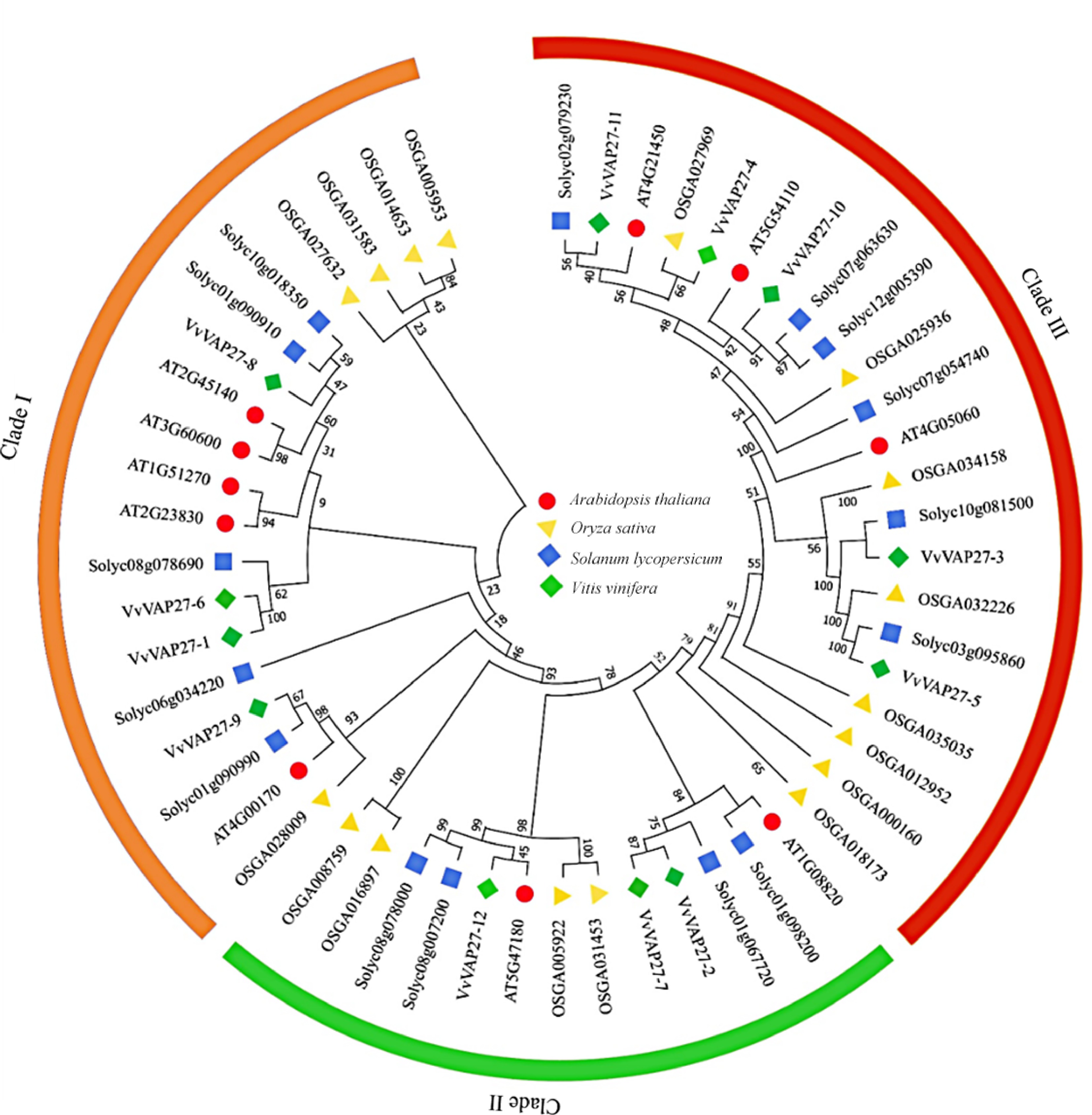
      Figure 1. 

      Unrooted phylogenetic tree of VAP27s in grape, Arabidopsis, rice, and tomato. Vv: Vitis vinifera L. grape species; AT: Arabidopsis thaliana; OS: Oryza sativa; Soly: Solanum lycopersicum.

      The clustering patterns suggest a closer evolutionary proximity of the VAP family in grapes to that of dicotyledon tomatoes compared to rice. This supports the reliability of the analysis results.

    • The gene structures of the 12 grapevine VAP27s were explored through a comprehensive examination of exon/intron boundaries. The varying length and splicing patterns observed among the 12 VAP27s are depicted in Fig. 2a. The structural analysis showed a range of intron numbers from 1 to 7. Notably, VvVAP27-3, VvVAP27-6, and VvVAP27-7 were absent of introns, while VvVAP27-5 exhibited a singular intron. The remaining VAP27 member's genes had between six and seven introns (Fig. 2a). The results revealed significant diversity within the VAP27 family.
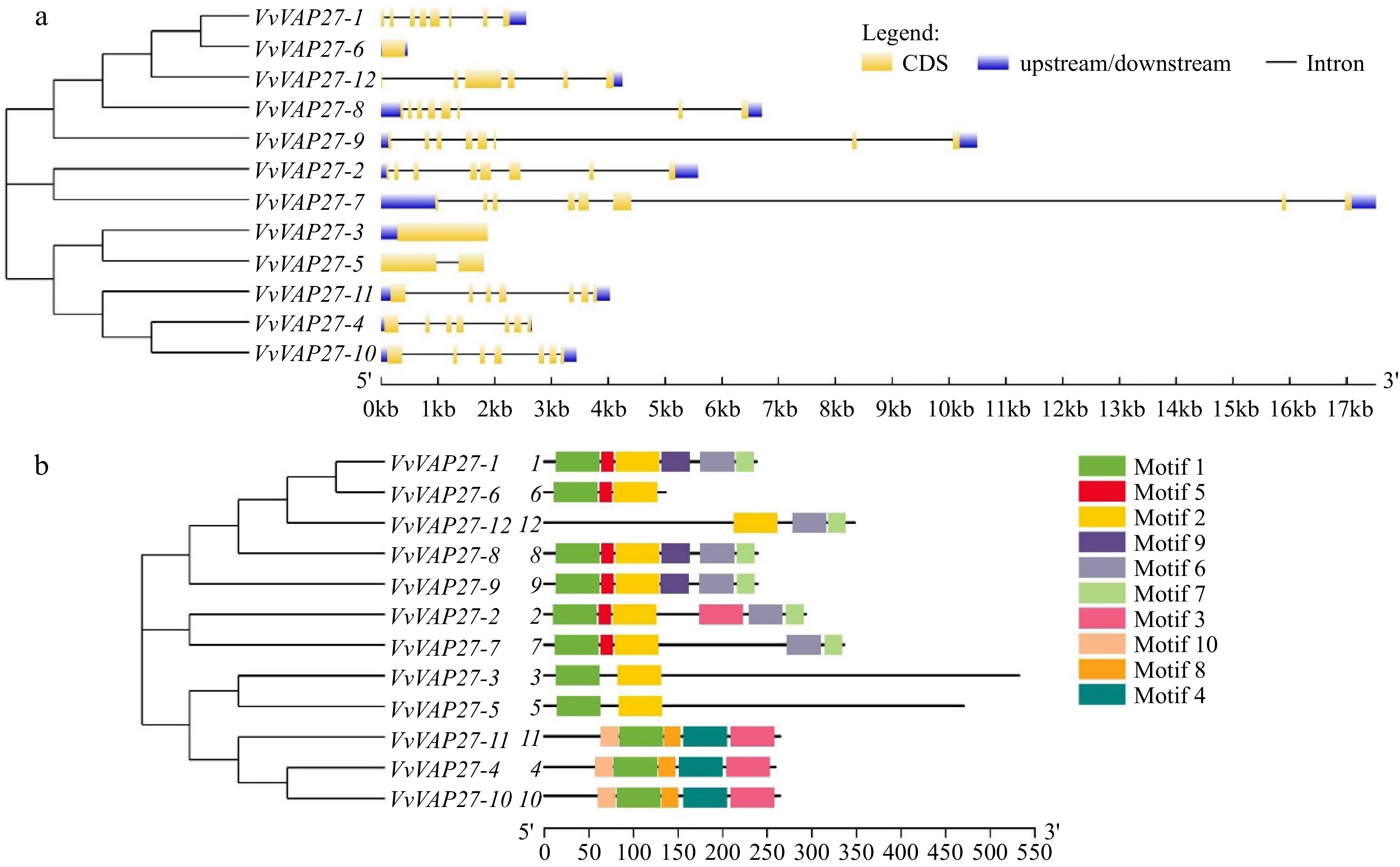
      Figure 2. 

      (a) Intron-exon structure and (b) conserved motifs of VvVAP27.

      According to previous studies, motifs recognized as playing an important role in interaction and signal transduction within the transcriptional complex[32,33] were analyzed using MEME for the 12 conservative VAP27 genes. This was conducted also because these motifs are closely related to gene classification. Among these VAP27s, a total of 10 motifs were identified (Fig. 2b), with Motif 1 present in all 11 members except VAP27-12, which indicates its high conservation within the VAP27 gene family. Motif 8 and Motif 9 were the least conserved, found only in VvVAP27-4, VvVAP27-10, and VvVAP27-11 (Motif 8), and VvVAP27-1, VvVAP27-8, and VvVAP27-9 (Motif 9). The high sequence similarity among genes within the same branch suggests shared functions and roles in plants. The analysis of motif and gene structure analysis enrich our understanding of the VAP27 family's classification, providing a robust theoretical foundation.

    • VAP27 RNAi induces various defects in plant morphology, pollen, seed, and root development in Arabidopsis[1], we anticipated a similar involvement of the VvVAP27 gene in grape growth and development. To investigate this, we examined the expression profiles of the 12 VAP27 genes across different tissues of grapevine (Fig. 3). These tissues represented distinct growth and development stages of grapevine, including root, young stem, leaf, inflorescence, skin, veraison berry, and tendril. Examination of transcriptome data from the VvVAP27 family revealed significant variations across different tissues. The majority of family members (VvVAP27-1 to VvVAP27-10) exhibited comparable expression levels in tissues including flowers, berries, leaves, stems, seeds, and shoots, suggesting their involvement throughout various stages of plant growth and development. Only a subset of genes (VvVAP27-11 and VvVAP27-12) showed significant differences in expression among tissues. Expression of the VvVAP27-11 gene was higher in the berries than in the other tissues that maintained relatively consistent levels. In contrast, VvVAP27-12 showed transcriptional peaks exclusively in seeds and flowers, suggesting a potential association of VvVAP27-12 with flowering and fruit development.
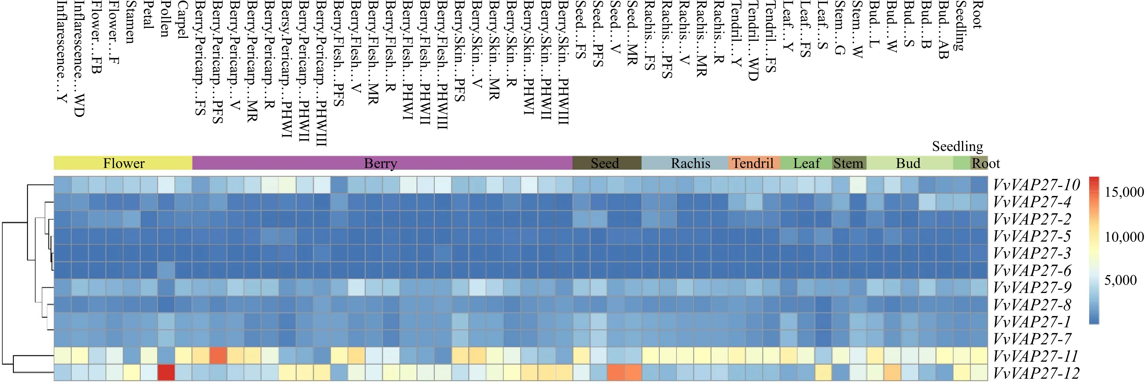
      Figure 3. 

      Expression profiles of the grape VvVAP27s gene. Hierarchical clustering of expression profiles of grape VAP27 genes across different tissues.

    • It has been documented that the VAP27 gene family is involved in regulating plant disease resistance against external pathogen infection[6,1822]. Therefore, we explored whether the VAP27 family exhibits similar functionality in grapevine downy mildew resistance. Our investigation focused on the expression levels of 12 VAP27 gene members at eight time points post-downy mildew inoculation (0, 6, 12, 24, 48, 72, 96, and 120 hpi). Utilizing RT-qPCR, we assessed whether the VAP27 gene responded to the induction of Grape downy mildew (Fig. 4). Vvactin1 was used as a grapevine internal reference gene for normalization[25].
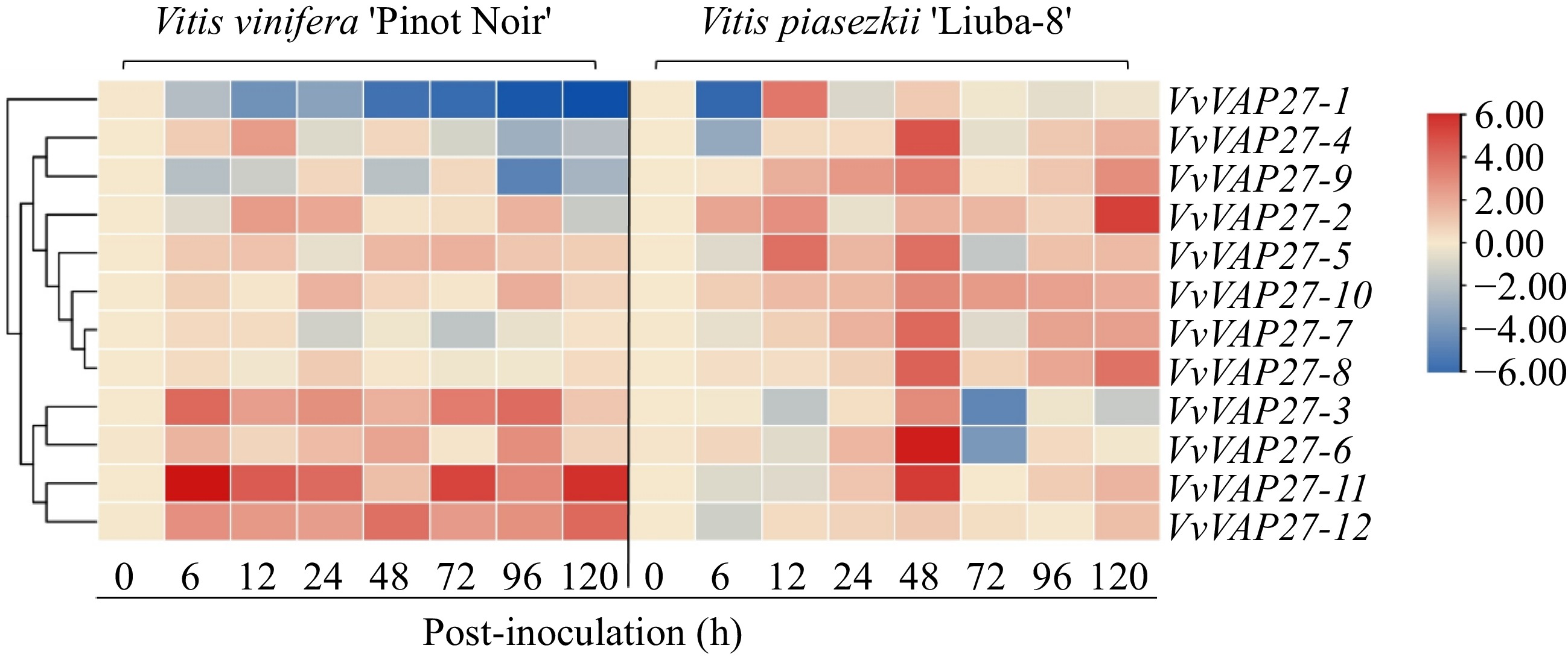
      Figure 4. 

      Heat map showing the expression profiles of VvVAP27 genes at different time points post-downy mildew infection. The color scale represents expression levels, with red indicating high expression level and blue indicating low expression level. The expression was normalized and the data are displayed as log2 values.

      We discovered that genes in the VvVAP27 family genes exhibited specificity in responses to downy mildew induction (Fig. 4). There were a few members significantly overexpressed at the early stage of downy mildew infection in 'Liuba-8'. Only VvVAP27-6 had the highest expression level at 48 hpi compared to other time points. VvVAP27-2 was highly expressed at 120 hpi, with no difference found at other time points. VvVAP27-3, VvVAP27-6, VvVAP27-11, and VvVAP27-12 genes were highly expressed throughout the downy mildew infection period in 'Pinot Noir'. This indicated that these four genes were positively responsive to the induction of downy mildew, suggesting an important role in the grapevine's defense against downy mildew invasion. The expression levels of the other eight genes remained unchanged across different infection periods. We postulate that the varying expression patterns among different members may be related to the regulation of VAP27-mediated plant disease resistance, possibly involving distinct mechanisms of immunity. However, further experimental verification is needed to substantiate these hypotheses.

    • To better explore the function of the VvVAP27 gene family, subcellular localization analysis was conducted on some selected VvVAP27 genes. By detecting GFP-tagged proteins, we found that most of the genes were localized to the endoplasmic reticulum (ER). This localization aligns with previous literature reports indicating that membrane proteins of vesicle-associated proteins function by participating in the formation and regulation of plant cell membranes. The endoplasmic reticulum participates in the formation of cell membranes and is closely related to secretory vesicles that function in transporting secretory proteins to various parts of the plant to contribute to plant growth, development, and disease resistance[34,35]. VvVAP27-2, VvVAP27-4, VvVAP27-6, and VvVAP27-9 are all mapped to the endoplasmic reticulum. However, VvVAP27-2, VvVAP27-4, and VvVAP27-6 are also detected in the nucleus in addition to the ER (Fig. 5). Based on the analysis of the expression pattern induced by downy mildew and the Phytophthora capsici infection experiment, we speculate that the subcellular localization may affect the gene expression.
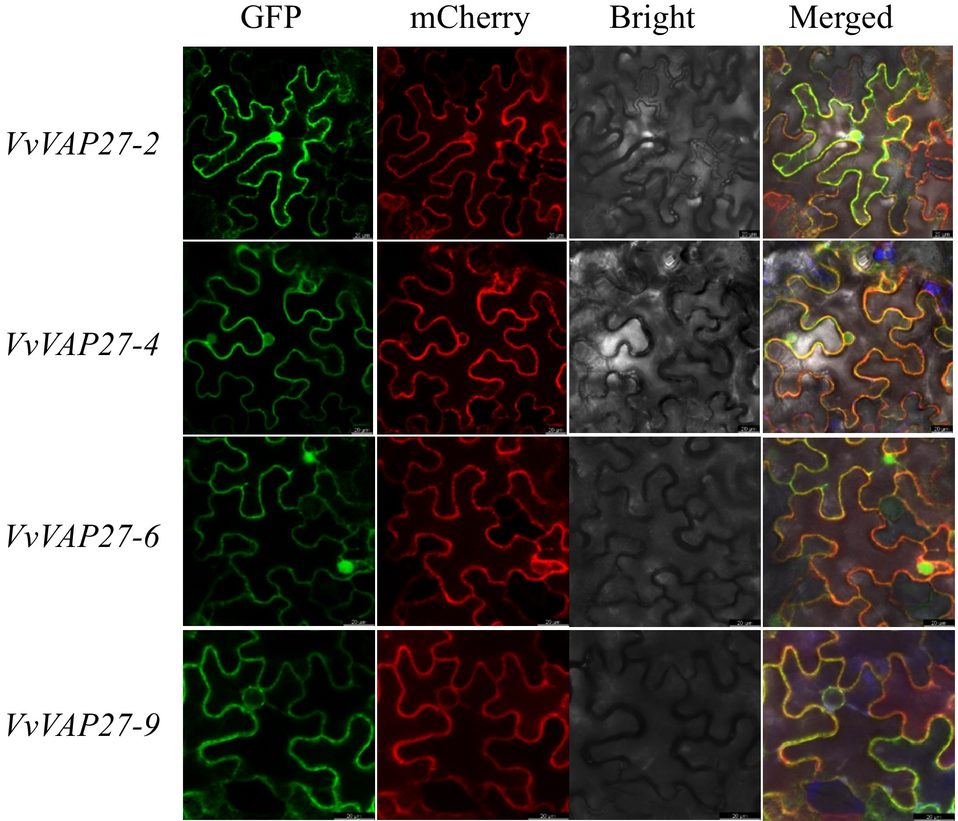
      Figure 5. 

      Subcellular localization analysis of VvVAP27 members.

    • To further study the role of VAP27 genes in disease resistance, the VvVAP27 genes that were induced by grape downy mildew in tobacco leaves were screened. These tobacco leaves were transiently transformed by A. tumefaciens that carry a high-level expression vector with an individual VvVAP27 gene insert before being inoculated with P.capsici spore suspension. The findings revealed that VAP27 gene members inhibited the occurrence of the pathogenicity and significantly enhanced the resistance of tobacco leaves to the pathogen. However, the efficacy of pathogen inhibition varied among different VAP27 members. This result is consistent with previous studies on gene responses to downy mildew infection.

      Specifically, both VvVAP27-6 and VvVAP27-9 exhibited a consistent phenotype, inhibiting infection by pathogens (Fig. 6a). Leaf lesion areas were smaller in VvVAP27-6 and VvVAP27-9 expressing leaves compared with controls, suggesting that they effectively promoted plant immunity (Fig. 6b) and that VvVAP27-6 had a higher inhibitory capacity than VvVAP27-9. VvVAP27-2 and VvVAP27-4, did not differ significantly in the size of the lesion area compared with empty-carrier controls. The heterogeneous functions within this family underscore the need for further experimental studies to elucidate the roles of the remaining genes.
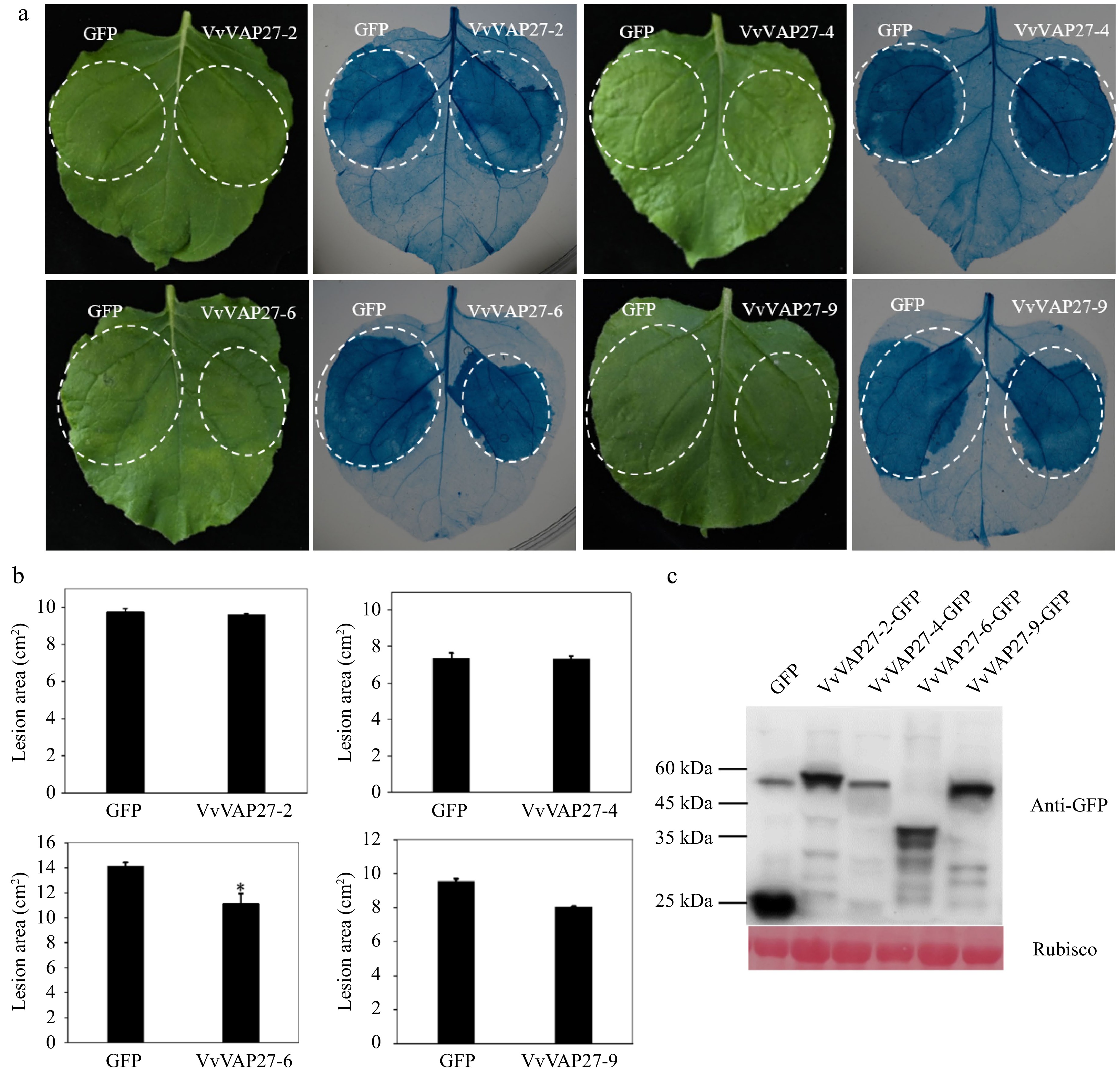
      Figure 6. 

      Responses of VvVAP27 induced by Phytophthora capsici infection. The inoculation of Phytophthora capsici was conducted on Nicotiana benthamiana leaves transiently transformed with VvVAP27. (a) Observation of the phenotypes of VvVAP27 family members and the control after inoculation with Phytophthora capsici, visualized by trypan blue staining. (b) Statistical analysis of the lesion areas caused by Phytophthora capsici infection on Nicotiana benthamiana leaves transiently transformed by VvVAP27. (c) The expression of VvVAP27 family members and GFP protein was detected by Western blot. The experiment was repeated three times and asterisks represent the level of significant differences (* p < 0.05, ** p < 0.01).

    • Vesicle-associated membrane proteins (VAMP-associated proteins) (VAPs) are a family of proteins widely expressed in plants, which play a key role in plant defense against both biotic and abiotic stresses. In this study, a family of 12 VAP27 genes were identified in grapes using bioinformatic methods. Consistent with previous studies, the VAMP gene family has demonstrated multifaceted involvement in diverse defense processes across different plant tissues. VAP27-1 and VAP27-3, as non-plant VAP homologs[5], have been localized extensively to the ER and EPCS[1,5,16]. These proteins were identified to promote plant endocytosis and play a role in endocytosis. In Arabidopsis thaliana, VAMP721/722 have been identified as essential factors for growth, cell division, and responses to abiotic stress[1822]. The PEN1-SNAP33-VAMP721/722 pathway in Arabidopsis thaliana facilitates the transport of vital materials for cell viability to the endoplasmic reticulum[36,37]; VAMP721 and VAMP722 are involved in the secretory transport of substances to endosomal compartments of the plasma membrane to promote the formation of cell plates during plant cytokinesis[6]. The phylogenetic tree analysis revealed a notable similarity in the quantity and gene structure of the VAP27 gene family between grape, Arabidopsis thaliana, and tomato. Hence, it can be inferred that the VAP27 gene family in grape shares similar functions with those in Arabidopsis thaliana[36], and plays comparable roles in growth, development, and immune resistance mechanisms. Additionally, we analyzed the gene structure of 12 identified VvVAP27 genes using MEME. Results (Fig. 2) showed that most of the VvVAP27 genes (VvVAP27-1, VvVAP27-2, VvVAP27-3, VvVAP27-6, VvVAP27-7, VvVAP27-8, VvVAP27-9) contain conserved domains, especially VvVAP27-3 and VvVAP27-5; VvVAP27-1, VvVAP27-8 and VvVAP27-9; VvVAP27-4, VvVAP27-10 and VvVAP27-11 placed within the same group in the phylogenetic tree classification (Fig. 1). This observation may explain the specific biological functions associated with each subfamily. The analysis of motif and gene structure provides a further theoretical basis for the classification of the VvVAP27 subfamily, guiding subsequent in-depth functional studies within these identified groups.

      To understand the regulatory mechanisms of the VAP27 gene family on the growth and development of grapevine, we analyzed the regulatory effect of the VvVAP27 gene family on the growth and development of grapevine using transcriptome content assay (Fig. 3) across different grape tissues (flowers, seeds, leaves, buds, berries, tendrils, stems, roots). Our transcriptome data analysis showed the highest expression of VvVAP27-11 in berries than in other tissues, implying its potential role in regulating fruit development and quality and promoting fruit setting. Similarly, the expression levels of VvVAP27-12 were higher in flower and seed than in other tissues, indicating that VvVAP27-12 plays an important role in flower induction, seed setting, and growth regulation. This study provides a theoretical basis for further understanding the function of the VAP27 family members in the process of grape growth and development.

      It has been found that the VAP gene family can induce plant cell autonomous immunity, acting on the cell surface or post-pathogen entry, thereby impeding pathogenesis. This phenomenon has been well-documented in various plant species, including tomato, Arabidopsis, and tobacco. In tobacco, VAPB proteins interact with proteins in the intestine of tobacco whitefly (Bemisia tabaci) during the transmission of tomato yellow leaf curl virus (TYLCV), and silencing VAPB results in an increase in virus number and transmission rate, demonstrating that VAPB can play a key role in resistance to TYLCV[4]. Moreover, in Arabidopsis, the SNARE proteins VAMP 721/722 direct secretory vesicles to pathogen-attack sites during immune responses, indicating that these vesicles deliver immune molecules and function in immune responses[36]. Additionally, SYP132, an essential protein for defense against bacterial pathogens, specifically interacts with VAMP721/722 in response to the immune control of P. syringae[21]. Given these findings, we were intrigued by the possibility that the VAP27 gene family might play a similar role in disease resistance in grapes. Grapevine downy mildew, caused by the oomycete P. viticola, is one of the most serious diseases in grape production. P. viticola was originally endemic to North America, but it has now spread to all major grape-producing regions worldwide[37]. This study revealed that the majority of VvVAP27 members exhibited responses to downy mildew infection. Notably, VvVAP27-1, VvVAP27-2, VvVAP27-4, and VvVAP27-6 were significantly up-regulated at 0, 6, and 12 h after infection, and VvVAP27-12 was highly expressed at 24, 48, and 72 h after infection with P. viticola, while other genes were highly expressed only at specific stages. These findings suggest that members of the VAP27 gene family are likely to respond to downy mildew infection. Our results provide a new idea to study the effect of the VAP27 gene on grapevine downy mildew, but further research is needed to study the mechanisms of VAP27 gene action.

      In this study, we identified a new grape gene family, named the VAP27 gene family. Through bioinformatic analysis and transcriptome sequencing, we uncovered striking structural and functional similarities between the VAP27 gene family in grape and those in Arabidopsis thaliana and tomato. The current findings suggest a potentially significant role for this gene family in the growth and development of the grape, as well as in orchestrating immune responses against downy mildew.

    • The vesicle-associated protein-membrane protein gene family (VAP27) in the grape genome was identified, which consists of 12 gene members. Within this family, some members exhibit localization on the endoplasmic reticulum, and a minority reside within the nucleus. The present results demonstrate the induction of the gene family in response to downy mildew in grape and their ability to inhibit the infection of P. capsici, thus playing an important role in plant disease resistance.

    • The authors confirm contribution to the paper as follows: conceptualization, writing – review & editing: Xu Y, Liu G; validation, visualization, writing – original draft: Li R, Wang B; investigation: Li R, Wang B, Zha M, Zhang K, Li M, Xie L, Chen X, Liu G; funding acquisition: Liu G. All authors reviewed the results and approved the final version of the manuscript.

    • All data generated or analyzed during this study are included in this published article.

      • This work was supported by grants from the National Natural Science Foundation of China (31972374, 32372660, and 31601716).

      • The authors declare that they have no conflict of interest. Yan Xu is the Editorial Board member of Vegetable Research who was blinded from reviewing or making decisions on the manuscript. The article was subject to the journal's standard procedures, with peer-review handled independently of this Editorial Board member and the research groups.

      • # Authors contributed equally: Ruonan Li, Bianbian Wang

      • Copyright: © 2024 by the author(s). Published by Maximum Academic Press, Fayetteville, GA. This article is an open access article distributed under Creative Commons Attribution License (CC BY 4.0), visit https://creativecommons.org/licenses/by/4.0/.
    Figure (6)  Table (1) References (37)
  • About this article
    Cite this article
    Li R, Wang B, Zha M, Zhang K, Li M, et al. 2024. Identification and characterization of grape VAP27 gene family and their roles in disease resistance. Fruit Research 4: e019 doi: 10.48130/frures-0024-0019
    Li R, Wang B, Zha M, Zhang K, Li M, et al. 2024. Identification and characterization of grape VAP27 gene family and their roles in disease resistance. Fruit Research 4: e019 doi: 10.48130/frures-0024-0019

Catalog

  • About this article

/

DownLoad:  Full-Size Img  PowerPoint
Return
Return