ARTICLE   Open Access    

Utilizing OSA-modified starch with various molecular weights for flavonoid extraction from "Quzhiqiao" (immature fruit of Citrus paradisi 'Changshan Huyou')

  • # These authors contributed equally: Lu Wang, Xuyi Zhou

More Information
  • In order to investigate the extraction efficiency of flavonoids by octenyl succinic acid-modified starch (OSA-modified starch) with varying molecular weights, the immature fruit of Citrus paradisi 'Changshan Huyou', commonly known as "Quzhiqiao", was used as the extraction target in this research. OSA-modified starch was successfully identified and refined, ensuring its molecular weight was appropriate and extraction was optimized. The experimental findings suggested that a concentration of 20 mg/mL of medium molecular weight gelatinized OSA-modified starch was optimal for extracting flavonoids from "Quzhiqiao" using an ultrasound-assisted method (1,000 W, 40 min). Additionally, the ratio of the mass of "Quzhiqiao" to the volume of gelatinized OSA-modified starch employed in this process was determined to be 15 mg/mL. The findings from the in vitro antioxidant studies indicated significant variations in the antioxidant activities of the extract solutions of OSA-modified starches with varying molecular weights. These differences can potentially be attributed to substantial variations in the extract solutions composition.
  • Salvia rosmarinus L. (old name Rosmarinus officinalis), common name Rosemary thrives well in dry regions, hills and low mountains, calcareous, shale, clay, and rocky substrates[1]. Salvia rosmarinus used since ancient times in traditional medicine is justified by its antiseptic, antimicrobial, anti-inflammatory, antioxidant, and antitumorigenic activity[1,2]. The main objective of the study is to evaluate the antimicrobial activity of different extracts of Salvia rosmarinus in vitro, and its compounds related to in silico targeting of enzymes involved in cervical cancer. Since the start of the 20th century, some studies have shown that microbial infections can cause cervical cancers worldwide, infections are linked to about 15% to 20% of cancers[3]. More recently, infections with certain viruses like Human papillomaviruses (HPV) and Human immunodeficiency virus (HIV), bacteria like Chlamydia trachomatis, and parasites like schistosomiasis have been recognized as risk factors for cancer in humans[3]. Then again, cancer cells are a group of diseases characterized by uncontrolled growth and spread of abnormal cells. Many things are known to increase the risk of cancer, including dietary factors, certain infections, lack of physical activity, obesity, and environmental pollutants[4]. Some studies have found that unbalanced common flora Lactobacillus bacteria around the reproductive organ of females increases the growth of yeast species (like Candida albicans) and some studies have found that women whose blood tests showed past or current Chlamydia trachomatis infection may be at greater risk of cervical cancer. It could therefore be that human papillomavirus (HPV) promotes cervical cancer growth[3]. Salvia rosmarinus is traditionally a healer chosen as a muscle relaxant and treatment for cutaneous allergy, tumors, increases digestion, and the ability to treat depressive behavior; mothers wash their bodies to remove bacterial and fungal infections, promote hair growth, and fight bad smells[5] .

    The study of plant-based chemicals, known as phytochemicals, in medicinal plants is gaining popularity due to their numerous pharmacological effects[6] against drug resistance pathogens and cancers. The causes of drug resistance to bacteria, fungi, and cancer are diverse, complex, and only partially understood. The factors may act together to initiate or promote infections and carcinogenesis in the human body is the leading cause of death[7]. Antimicrobial medicines are the cornerstone of modern medicine. The emergence and spread of drug-resistant pathogens like bacteria and fungi threaten our ability to treat common infections and to perform life-saving procedures including cancer chemotherapy and cesarean sections, hip replacements, organ transplantation, and other surgeries[7]. On the other hand, information about the current magnitude of the burden of bacterial and fungal drug resistance, trends in different parts of the world, and the leading pathogen–drug combinations contributing to the microbial burden is crucial. If left unchecked, the spread of drug resistance could make many microbial pathogens much more lethal in the future than they are today. In addition to these, cancers can affect almost any part of the body and have many anatomies and molecular subtypes that each require specific management strategies to avoid or inhibit them. There are more than 200 different types of cancer that have been detected. The world's most common cancers affecting men are lung, prostate, colorectal, stomach, and liver cancers[8]. While breast, cervix, colorectal, lung, and stomach cancers are the most commonly diagnosed among women[8]. Although some cancers said to be preventable they seem to still be one of the causes of death to humans, for example cervical cancer. The need to fill the gap to overcome the problem of searching for antimicrobials and anticancers from one source of Salvia rosmarinus is of importance.

    Cervical cancer is a common cancer in women and a prominent cause of death[9]. In Ethiopia, cervical cancer is a big deal for women aged 15 to 44, coming in as the second most common cancer[9]. Globally, it's the fourth most common prevalent disease for women[10]. Aberrant methylation of tumor-suppressor genes' promoters can shut down their important functions and play a big role in causing cervical tumors[10]. There are various cervical cancer repressor genes (proteins turn off or reduce gene expression from the affected gene), such as CCNA1, CHF, HIT, PAX1, PTEN, SFRP4, and TSC1. The genes play a crucial role in causing cervical cancer by regulating transcription and expression through promoter hypermethylation, leading to precursor lesions during cervical development and malignant transformation[11]. The process of DNA methylation is primarily carried out by a group of enzymes known as DNA methyltransferases (DNMT1). It has been reported that DNMT1 (PDB ID: 4WXX), a protein responsible for DNA methylation can contribute to the development of cervical cancer. DNMT1 inhibits the transcription of tumor suppressor genes, facilitating tumorigenesis, which finally develops into cervical cancer. Tumor suppressor gene transcription is inhibited by DNMT1, which helps cancer grow and eventually leads to cervical cancer. Repressive genes' hypermethylation may be decreased, their expression can be increased, and the phenotype of malignant tumors can be reversed by inhibiting the DNMT1 enzyme.

    On the other hand, infection by the human papilloma virus (HPV) phenotype 16, enzyme 6 (PDB ID: 4XR8) has been correlated with a greatly increased risk of cervical cancer worldwide[12]. Based on variations in the nucleotide sequences of the virus genome, over 100 distinct varieties of the human papilloma virus (HPV) have been identified (e.g. type 1, 2 etc.). Genital warts can result from certain types 6 and 11 of sexually transmitted HPVs. Other HPV strains, still, that can infect the genitalia, do not show any symptoms of infection[8]. Persistent infection with a subset of approximately 13 so-called 'high-risk' sexually transmitted HPVs, including such as types 16, 18, 31, 33, 35, 39, 45, 51, 52, 56, 58, 59, and 68 different from the ones that cause warts may lead to the development of cervical intraepithelial neoplasia (CIN), vulvar intraepithelial neoplasia (VIN), penile intraepithelial neoplasia (PIN), and/or anal intraepithelial neoplasia (AIN). These are precancerous lesions and can progress to invasive cancer. Almost all occurrences of cervical cancer have HPV infection as a required component[13]. Superfluous infection by HPV type 16 E6 (PDB ID: 4XR8) has been correlated with a greatly increased genital risk of precursor cervical cancer worldwide[11]. Scholars more defined in major biochemical and biological activities of HPV type 16 E6 (PDB ID: 4XR8) in high-risk HPV oncogenes and how they may work together in the development of cervical disease and cancer[13].

    One potential approach to treat cervical cancer is to inhibit the activity of the DNMT1 and HPV type 16 E6 enzymes specifically[1316]. Over 50% of clinical drug forms worldwide originate from plant compounds[17]. In the past, developing new drugs was a lengthy and costly process. However, with the emergence of bioinformatics, the use of computer-based tools and methods have become increasingly important in drug discovery. One such method is molecular docking and ADMET profiling which involves using the structure of a drug to screen for potential candidates. This approach is known as structure-based drug design and can save both time and resources during the research process[15]. Structural-based drug designing addresses ligand binding sites with a known protein structure[15]. Using free binding energies, a computational method known as docking examines a large number of molecules and suggests structural theories for impeding the target molecule[17]. Nowadays, due to increasing antibiotic resistance like bacteria, fungi, and cancer cells, natural products remain an important source for discovering antimicrobial compounds and novel drugs for anti-cancers like cervical cancers. Therefore, the purpose of this research is to assess the antimicrobial activity of extracts, molecular docking, ADMET profiling in anticancer properties of compounds isolated from Salvia rosmarinus, on a targeting DNMT1 and HPV type 16 E6 in human cervical cancer. In the present study, various solvent crude extracts obtained from Salvia rosmarinus were used for antimicrobial activity and the isolated compounds 1 and 2 were submitted for in silico study to target the DNMT1 and HPV type 16 E6 enzymes to inhibit the growth of human cervical cancer cells.

    Healthy Salvia rosmarinus leaves were collected in Bacho district, Southwest Showa, Oromia, Ethiopia, during the dry season of November 2022. The plant materials were authenticated by Melaku Wondafrish, Natural Science Department, Addis Ababa University and deposited with a voucher number 3/2-2/MD003-80/8060/15 in Addis Ababa University's National Herbarium.

    The most common organic solvent used in extractions of medicinal plants is 2.5 L of petroleum ether, chloroform/methanol (1:1), and methanol. The test culture medium for microbes was used and performed in sterile Petri dishes (100 mm diameter) containing sterile Muller–Hinton Agar medium (25 mL, pH 7) and Sabouraud Dextrose Agar (SDA) for bacteria and fungi, respectively. A sterile Whatman filter paper (No. 1) disc of 6 mm diameter was used to determine which antibiotics an infective organism is sensitive to prescribed by a minimum zone of inhibition (MZI). Ciprofloxacin antibiotic reference (manufactured by Wellona Pharma Ciprofloxacin tablet made in India) and Ketoconazole 2% (made in Bangladesh) were used as a positive controls for antibacterial and antifungal, respectively and Dimethyl sulfoxide (DMSO) 98.9% was used as a negative control for antimicrobial tests. In the present study, the height of the column was 650 mm and the width was 80 mm. Several studies by previous researchers showed the acceptable efficiency of column chromatography (up to 43.0% w/w recovery) in the fractionation and separation of phenolic compounds from plant samples[18]. In column chromatography, the ideal stationary phase used silica gel 60 (0.200 mm) particles. The 1H-NMR spectrums of the compounds were analyzed using a 600 MHz NMR machine and 150 MHz for 13C NMR. The compounds were dissolved in MeOD for compound 1 and in DMSO for compound 2 for NMR analysis. On the other hand, UV spectroscopy (made in China) used 570 nm ultraviolet light to determine the absorbency of flavonoids (mg·g−1) phytochemicals.

    The samples (extracts) were analyzed to detect the presence of certain chemical compounds such as alkaloids (tested using Wagner's reagents), saponins (tested using the froth test), steroids (tested with Liebermann Burchard's tests), terpenoids (tested with Lidaebermann Burchard's tests), quinones, and flavonoids (tested using Shinoda tests)[19].

    The leaves of Salvia rosmarinus (500 g) were successively extracted using maceration using petroleum ether, chloroform/methanol (1:1), and methanol, every one 2.5 L for 72 h to afford 3.6, 6, and 53 g crude extracts, respectively. The methanol/chloroform (1:1) extract (6 g) was loaded to silica gel (150 g) column chromatography using the increasing polarity of petroleum ether, methanol/chloroform (1:1) solvent system to afford 80 fractions (100 mL each). The fraction obtained from chloroform/methanol 1:1 (3:2) after repeated column chromatography yielded compound 1 (18 mg). Fractions 56-65, eluted with chloroform/methanol (1:1) were combined and purified with column chromatography to give compound 2 (10 mg).

    The microorganisms were obtained from the Ethiopia Biodiversity Institution (EBI). Two gram-positive bacteria namely Staphylococcus aureus serotype (ATCC 25923) and Streptococcus epidermidis (ATCC14990); and three gram-negative bacteria, namely Escherichia coli (ATCC 25922), Pseudomonas aeruginosa (ATCC 5702), and Klebsiella pneumonia (ATCC e13883) were inoculated overnight at 37 °C in Muller–Hinton Agar/MHA culture medium and two fungus strains of Candida albicans (ATCC 16404) and Aspergillus niger (ATCC 11414) were inoculated overnight at 27−30 °C in Sabouraud Dextrose Agar/SDA culture medium[20].

    The antibacterial and antifungal activities of different crude extracts obtained from Salvia rosmarinus plant leaves were evaluated by the disk diffusion method (in accordance with the 13th edition of the CLSI M02 document on hardydiagnostics.com/disk-diffusion). Briefly, the test was performed in sterile Petri dishes (100 mm diameter) containing solid and sterile Muller–Hinton Agar medium (25 mL, pH 7) and Sabouraud Dextrose Agar (SDA) for bacteria and fungi, respectively. The extracts were placed on the surface of the media that had previously been injected with a sterile microbial suspension (one microbe per petri dish) after being adsorbed on sterile paper discs (5 μL per Whatman disc of 6 mm diameter). To prevent test samples from eventually evaporating, all Petri dishes were sealed with sterile laboratory films. They were then incubated at 37 °C for 24 h, and the zone diameter of the inhibition was measured and represented in millimeters. Ciprofloxacin antibiotic reference (manufactured by Wellona Pharma Ciprofloxacin tablet, India) was used as a positive control and DMSO was used as a negative control for antibacterial activity test while Ketoconazole 2% (Bangladesh) was used as a positive control and 10 μL of 0.2% agar as a negative control for antifungal activity tests[20]. The term 'inhibitory concentration' refers to the minimum sample concentration required to kill 99.9% of the microorganisms present[21]. Three repetitions of the crude extract sample were used to precisely measure the inhibitory halo diameter (in mm), which was then expressed as mean ± standard deviation to assess the anti-microbial activity.

    Cervical cancer-causing protein was identified through relevant literature. The protein molecule structure of DNA (cytosine-5)-methyltransferase 1 (DNMT1) (PDB ID: 4WXX)[21] and HPV type 16 E6 (PDB ID: 4XR8)[21] - a protein known to cause cervical cancer - were downloaded from the Protein Data Bank[22]. The stability of the protein molecule was assessed using Rampage[23].

    Phytochemical constituents of Salvia rosmarinus plant leaves were used to select a source of secondary metabolites (ligands). Ligand molecules were obtained through plant extraction, and isolation, and realized with PubChem (https://pubchem.ncbi.nlm.nih.gov/). The ligands were downloaded in Silver diamine fluoride format (SDF) and then converted to PDB format using an online SMILES translator (https://cactus.nci.nih.gov/translate/). The downloaded files were in PDB format, which was utilized for running various tools and software[24].

    The Biovia Discovery Studio Visualizer software was used to analyze the protein molecule. The protein molecule was converted into PDB format and its hierarchy was analyzed by selecting ligands and water molecules. Both the protein molecule and the water molecules lost their attached ligands during the analysis. Finally, the protein's crystal structure was saved in a PDB file[25].

    PyRx software was utilized to screen secondary metabolites and identify those ligands with the lowest binding energy to the protein target. The ligands with the lowest binding energy were further screened for their drug-likeliness property through analysis. It is worth noting that PyRx runs on PDBQT format. To begin using PyRx, it needs to load a protein molecule. This molecule should be converted from PDB to the protein data bank, partial charge (Q), and Atom Type (PDBQT) format. Once the protein molecule is loaded, it can import ligands from a specific folder in Silver diamine fluoride format. The ligand energy was minimized and changed to PDBQT format. The protein was docked with the ligand and screened based on minimum binding energy (https://cactus.nci.nih.gov/translate/).

    The optimal ligand was selected for final docking using AutoDock Vina and Biovia by modifying the reference of Discovery Studio Client 2021 (https://cactus.nci.nih.gov/translate/).

    The protein target from the Protein Data Bank (PDB) was loaded onto the graphical interface of AutoDock Vina. To prepare the protein for docking, water molecules were removed, hydrogen polar atoms were added, and Kollman charges were assigned to the protein molecule. Ultimately, PDBQT format was used to store the protein. After being imported in PDB format, the Ligand molecule was transformed to PDBQT format. Next, a grid box was chosen to represent the docked region. The command prompt was used to run AutoDock Vina and the outcomes were examined (https://cactus.nci.nih.gov/translate/).

    Docking the ligand with the protein target DNMT1(PDB ID: 4WXX)[22] and HPV type 16 E6 (PDB ID: 4XR8)[21] enzymes were performed using Biovia Discovery Studio Client 2021 by loading the protein target first followed by the ligand in PDB format. The charges were attached to the protein molecule, and the energy was minimized for the ligands. Both the protein and ligand molecules were prepared for docking. Once the docking process was complete, the results were analyzed based on several parameters, including absolute energy, clean energy, conf number, mol number, relative energy, and pose number. The interaction between the protein and ligand was analyzed using structure visualization tools, such as Biovia Discovery Studio Visualizer and PyMol (https://cactus.nci.nih.gov/translate/).

    The process of visualizing the structure was carried out using the PyMol tool. PyMol is a freely available software. Firstly, the protein molecule in PDBQT form was loaded on the PyMol graphical screen. Then, the output PDBQT file was added. The docked structure was visualized and the 'molecule' option was changed to 'molecular surface' under the 'shown as' menu (https://cactus.nci.nih.gov/translate/).

    Drug likeliness properties of the screened ligands were evaluated using the SwissADME online server. SMILE notations were obtained from PubChem and submitted to the SwissADME web server for analysis. The drugs were subjected to Lipinski's rule of five[20] for analysis. Lipinski's rules of five were selected for final docking through AutoDock Vina and Biovia Discovery Studio Client 2021. Ligands 1 and 2 were analyzed using Lipinski's rule of five for docking with AutoDock Vina and Biovia Discovery Studio Client 2021.

    The antimicrobial analysis data generated by triplicate measurements reported as mean ± standard deviation, and a bar graph also generated by GraphPad Prism version 8.0.1 (244) for Windows were used to perform the analysis. GraphPad Prism was used and combined with scientific graphing, comprehensive bar graph fitting (nonlinear regression), understandable statistics, and data organization. Prism allows the performance and modification of basic statistical tests commonly used and determined through the statistical applications in microbiology labs (https://graphpad-prism.software.informer.com/8.0/).

    Phytochemical screening of the different extracts for the presence (+) and absence (−) of alkaloids, steroids, glycosides, coumarins, terpenoids, flavonoids, carbohydrates, tannins, and saponins were done. The present study showed that alkaloids, terpenoids, flavonoids, and tannins tests in S. rosmarinus leaves of petroleum ether, chloroform/methanol (1:1), and methanol extracts were high whereas glycoside, coumarins, and carbohydrates had a moderate presence. The extract of S. rosmarinus leaves contain commonly bioactive constituents such as alkaloids, steroids, terpenoids, flavonoids, tannins, and saponins. These bioactive chemicals have active medicinal properties. Phytochemical compounds found in S. rosmarinus leaves have the potential to treat cancer cells and pathogens. The study also found that these flavonoids are related to natural phenolic compounds with anticancer and antimicrobial properties in the human diet (Table 1).

    Table 1.  Phytochemical screening tests result of petroleum ether, chloroform/methanol (1:1) and methanol extracts of Salvia rosmarinus leaves.
    Botanical name Phytochemicals Phytochemical screening tests Different extracts
    Petroleum ether Chloroform/methanol (1:1) Mehanol
    Salvia rosmarinus Alkaloids Wagner's test ++ ++ ++
    Steroids Libermann Burchard test ++ + ++
    Glycoside Keller-Killiani test +
    Coumarins Appirade test + +
    Terpenoids Libermann Burchard test ++ ++ ++
    Flavonoids Shinoda test ++ ++ ++
    Carbohydrate Fehling's test ++ ++
    Tannins Lead acetate test ++ ++ ++
    Saponins Foam test + + +
    + indicates moderate presence, ++ indicates highly present, − indicates absence.
     | Show Table
    DownLoad: CSV

    Two compounds were isolated and characterized using NMR spectroscopic methods (Fig. 1 & Supplementary Fig. S1ac). Compound 1 (10 mg) was isolated as yellow crystals from the methanol/chloroform (1:1) leaf extract of Salvia rosmarinus. The TLC profile showed a spot at Rf 0.42 with methanol/chloroform (3:2) as a mobile phase. The 1H-NMR spectrum (600 MHz, MeOD, Table 2, Supplementary Fig. S1a) of compound 1 showed the presence of one olefinic proton signal at δ 5.3 (t, J = 3.7 Hz, 1H), two deshielded protons at δ 4.7 (m, 1H), and 4.1 (m, 1H) associated with the C-30 exocyclic methylene group, and one O-bearing methine proton at δH 3.2 (m, 1H), and six methyl protons at δ 1.14 (s, 3H), 1.03 (d, J = 6.3 Hz, 3H), 1.00 (s, 3H), 0.98 (s, 3H), 0.87 (s, 3H), and 0.80 (s, 3H). A proton signal at δ 2.22 (d, J = 13.5 Hz, 1H) was attributed to methine proton for H-18. Other proton signals integrate for 20 protons were observed in the range δ 2.2 to 1.2. The proton decoupled 13C-NMR and DEPT-135 spectra (151 MHz, MeOD, Supplementary Fig. S1b & c) of compound 1 revealed the presence of 30 well-resolved carbon signals, suggesting a triterpene skeleton. The analysis of the 13C NMR spectrum displayed signals corresponding to six methyl, nine methylene, seven methine, and eight quaternary carbons. Among them, the signal observed at δ 125.5 (C-12) belongs to olefinic carbons. The methylene carbon showed signals at δC 39.9, 28.5, 18.1, 36.7, 23.9, 30.4, 26.5, 32.9, and 38.6. The quaternary carbons showed a signal at δC 39.4, 41.9, 38.4, 138.2, 41.8, and 47.8. The signals of exocyclic methylene carbon signals appeared at δ 153.1 and 103.9. The spectrum also showed sp3 oxygenated methine carbon at δ 78.3 and carboxyl carbon at δ 180.2. The spectrum revealed signals due to methyl groups at δC 27.4, 16.3, 15.0, 20.2, 22.7, and 16.4. The remaining carbon signals for aliphatic methines were shown at δC 55.3, 55.2, 53.0, and 37.1. The NMR spectral data of compound 1 is in good agreement with data reported for micromeric acid, previously reported from the same species by Abdel-Monem et al.[26]. (Fig. 1, Table 2).

    Figure 1.  Structure of isolated compounds from the leaves of Salvia rosmarinus.
    Table 2.  Comparison of the 13C-NMR spectral data of compound 1 and micromeric acid (MeOD, δ in ppm).
    Position NMR data of compound 1 Abdel-Monem
    et al.[26]
    1H-NMR 13C-NMR 13C-NMR
    1 38.60 39.9
    2 27.8 28.5
    3 3.2 (m, 1H) 78.3 80.3
    4 39.4 39.9
    5 55.3 56.7
    6 18.1 18.3
    7 36.7 34.2
    8 41.9 40.7
    9 53 48.8
    10 38.4 38.2
    11 23.9 24.6
    12 5.3 (t, J = 3.7 Hz, 1H) 125.5 127.7
    13 138.2 138
    14 41.8 43.3
    15 30.4 29.1
    16 26.5 25.6
    17 47.8 48
    18 δ 2.22 (d, J = 13.5 Hz, 1H) 55.2 56.1
    19 37.1 38.7
    20 153.1 152.8
    21 32.9 33.5
    22 39.0 40.1
    23 27.4 29.4
    24 16.3 16.9
    25 15.0 16.6
    26 20.2 18.3
    27 22.7 24.6
    28 180.2 177.8
    29 16.4 17.3
    30 4.7 (m, 1H), and 4.1 (m, 1H) 103.9 106.5
     | Show Table
    DownLoad: CSV

    Compound 2 (18 mg) was obtained as a white amorphous isolated from 40% methanol/chloroform (1:1) in petroleum ether fraction with an Rf value of 0.49. The 1H NMR (600 MHz, DMSO, Supplementary Fig. S2a) spectral-data showed two doublets at 7.79 (d, J = 8.7 Hz, 2H), and 6.90 (d, J = 8.7 Hz, 2H) which are evident for the presence of 1,4-disubstituted aromatic group. The oxygenated methylene and terminal methyl protons were shown at δ 4.25 (q, J = 7.1 Hz, 2H) and 1.29 (t, J = 7.1 Hz, 3H), respectively. The13C-NMR spectrum, with the aid of DEPT-135 (151 MHz, DMSO, Table 3, Supplementary Fig. S2b & c) spectra of compound 2 confirmed the presence of well-resolved seven carbon peaks corresponding to nine carbons including threee quaternary carbons, one oxygenated methylene carbon, one terminal methyl carbon, and two symmetrical aromatic methine carbons. The presence of quaternary carbon signals was shown at δ 120.9 (C-1), 148.2 (C-4), and ester carbonyl at δ 166.0 (C-7). The symmetry aromatic carbons signal was observed at δ 131.4 (C-2, 6), and 116.8 (C-3, 5). The oxygenated methylene and terminal methyl carbons appeared at δC 60.4 (C-8) and 14.7 (C-9), respectively. The spectral results provided above were in good agreement with those for benzocaine in the study by Alotaibi et al.[27]. Accordingly, compound 2 was elucidated to be benzocaine (4-Aminobenzoic acid-ethyl ester) (Table 3, Fig. 1, Supplementary Fig. S2ac), this compound has never been reported before from the leaves of Salvia rosmarinus.

    Table 3.  Comparison of the 1H-NMR, and 13C-NMR spectral data of compound 2 and benzocaine (DMSO, δ in ppm).
    Position NMR data of compound 2 Alotaibi et al.[27]
    1H-NMR 13C-NMR 1H-NMR 13C-NMR
    1 120.9 119
    2 7.79 (d, J = 8.7 Hz, 2H) 131.4 7.86 (d, J = 7.6 Hz) 132
    3 6.90 (d, J = 8.7 Hz, 2H) 116.8 6.83 (d, J = 7.6 Hz) 114
    4 148.2 151
    5 6.90 (d, J = 8.7 Hz, 2H) 116.8 6.83 (d, J = 7.6 Hz) 114
    6 7.79 (d, J = 8.7 Hz, 2H) 131.4 7.86 (d, J = 7.6 Hz) 132
    7 166.0 169
    8 4.3 (q, J = 7.1 Hz, 2H) 60.4 4.3 (q, J = 7.0 Hz) 61
    9 1.3 (t, J = 7.1 Hz, 3H) 14.7 1.36 (t, J = 7.0 Hz) 15
     | Show Table
    DownLoad: CSV

    The extracts and isolated compounds from Salvia rosmarinus were evaluated in vitro against microbes from gram-positive bacteria (S. aureus and S. epidermidis), gram-negative bacteria (E. coli, P. aeruginosa, and K. pneumoniae) and fungi (C. albicans and A. Niger) (Table 4). The petroleum ether extracts exhibited significant activity against all the present study-tested microbes at 100 μg·mL−1, resulting in an inhibition zone ranging from 7 to 21 mm. Chloroform/methanol (1:1) and methanol extracts demonstrated significant activity against all the present study-tested microbes at 100 μg·mL−1 exhibiting inhibition zones from 6 to 14 mm and 6 to 13 mm, respectively (Table 4). The chloroform/methanol (1:1) extracts were significantly active against bacteria of E. coli and K. pneumonia, and A. Niger fungi at 100 μg·mL−1. On the other hand, chloroform/methanol (1:1) extracts were significantly inactive against the S. rosmarinus and P. aeruginosa of bacteria and C. albicans of fungi, and again chloroform/methanol (1:1) extracts overall significantly active produced an inhibition zone of 12 to 14 mm (Table 4). Methanol extracts exhibited significant activity against S. aureus, E. coli bacteria, and A. Niger fungi at 100 μg·mL−1. The inhibition zone was recorded to be 11 to 13 mm. However, methanol extracts exhibited significant inactivity against K. pneumoniae (Table 4). The overall result of our studies shows that Salvia rosmarinus was extracted and evaluated in vitro, exhibiting significant antibacterial and antifungal activity, with inhibition zones recorded between 6 to 21 mm for bacteria and 5 to 21 mm for fungi. In our study, the positive control for ciprofloxacin exhibited antibacterial activity measured at 21.33 ± 1.15 mm, 15.00 ± 0.00 mm, and 14.20 ± 0.50 mm for petroleum ether, chloroform/methanol (1:1), and methanol extracts, respectively. Similarly, the positive control for ketoconazole demonstrated antifungal activity of 22.00 ± 1.00 mm, 13.67 ± 0.58 mm, and 15.00 ± 0.58 mm for petroleum ether, chloroform/methanol (1:1), and methanol extracts, respectively. Additionally, our findings indicated that the mean values of flavonoids (mg/g) tested were 92.2%, 90.4%, and 94.0% for petroleum ether, chloroform/methanol (1:1), and methanol extracts, respectively. This suggests that the groups of phenolic compounds evaluated play a significant role in antimicrobial activities, particularly against antibiotic-resistant strains.

    Table 4.  Comparison of mean zone of inhibition (MZI) leaf extracts of Salvia rosmarinus.
    Type of specimen, and standard antibiotics for
    each sample
    Concentration (μg·mL−1) of extract
    in 99.8% DMSO
    Average values of the zone of inhibition (mm)
    Gram-positive (+) bacteria Gram-negative (−) bacteria Fungai
    S. aurous S. epidermidis E. coli P. aeruginosa K. pneumoniae C. albicans A. niger
    Petroleum ether extracts
    S. rosmarinus 50 18.50 ± 0.50 15.33 ± 0.58 0.00 ± 0.00 0.00 ± 0.00 10.00 ± 0.00 15.93 ± 0.12 4.47 ± 0.50
    75 19.87 ± 0.06 17.00 ± 0.00 9.33 ± 0.29 10.53 ± 0.50 10.93 ± 0.12 18.87 ± 0.23 5.47 ± 0.50
    100 21.37 ± 0.78 17.50 ± 0.50 11.47 ± 0.50 13.17 ± 0.29 12.43 ± 0.51 20.83 ± 0.76 6.70 ± 0.10
    Standard antibiotics Cipro. 21.33 ± 1.15 18.33 ± 0.58 9.33 ± 0.58 12.30 ± 0.52 15.00 ± 0.00
    Ketocon. 22.00 ± 1.00 10.67 ± 0.58
    Chloroform/methanol (1:1) extracts
    50 5.47 ± 0.42 0.00 ± 0.00 10.33 ± 0.00 0.00 ± 0.00 9.70 ± 0.00 0.00 ± 0.12 8.47 ± 0.50
    S. rosmarinus
    75 5.93 ± 0.06 0.00 ± 0.00 11.33 ± 0.29 0.00 ± 0.50 12.50 ± 0.12 0.00 ± 0.23 10.67 ± 0.50
    100 6.47 ± 0.06 0.00 ± 0.00 14.17 ± 0.50 7.33 ± 0.29 14.17 ± 0.51 0.00 ± 0.76 12.67 ± 0.10
    Standard antibiotics Cipro. 15.00 ± 0.00 11.00 ± 1.00 11.33 ± 0.58 10.00 ± 0.52 12.67 ± 0.00
    Ketocon. 7.00 ± 1.00 13.67 ± 0.58
    Methanol extracts
    50 9.17 ± 0.29 5.50 ± 0.50 0.00 ± 0.00 7.50 ± 0.00 0.00 ± 0.00 6.57 ± 0.12 0.00 ± 0.50
    S. rosmarinus
    75 9.90 ± 0.10 6.93 ± 0.12 9.33 ± 0.29 8.50 ± 0.50 0.00 ± 0.00 8.70 ± 0.23 0.00 ± 0.50
    100 11.63 ± 0.55 7.97 ± 0.06 11.47 ± 0.50 9.90 ± 0.10 0.00 ± 0.00 10.83 ± 0.76 13.13 ± 0.10
    Standard antibiotics Cipro. 13.00 ± 0.00 11.50 ± 0.50 14.20 ± 0.58 13.33 ± 0.29 10.00 ± 0.00
    Ketocon. 12.00 ± 1.00 15.00 ± 0.58
    Mean values of flavonoids (mg·g−1) by 570 nm
    S. rosmarinus
    Petroleum ether extracts Chloroform/methanol (1:1) extracts Methanol extracts
    50 0.736 0.797 0.862
    75 0.902 0.881 0.890
    100 0.922 0.904 0.940
    Samples: Antibiotics: Cipro., Ciprofloxacin; Ketocon., ketoconazole (Nizoral); DMSO 99.8%, Dimethyl sulfoxide.
     | Show Table
    DownLoad: CSV

    Determining the three solvent extracts in S. rosmarinus plants resulted in relatively high comparable with positive (+) control. Especially, the S. rosmarinus petroleum ether leaf extracts against drug resistance human pathogenic bacteria S. aureus, S. epidermidis, E. coli, P. aeruginosa, and K. pneumoniae were minimum zone of inhibition (MZI) recorded that 21.37 ± 0.78, 17.50 ± 0.50, 11.47 ± 0.50, 13.17 ± 0.29, and 12.43 ± 0.51 mm, respectively and against human pathogenic fungi C. albicans and A. niger were minimum zone of inhibition (MZI) recorded that 20.83 ± 0.76 and 6.70 ± 0.10 mm, respectively which was used from bacteria against S. aureus MZI recorded that 21.37 ± 0.78 mm higher than the positive control (21.33 ± 1.15 mm). The S. rosmarinus of chloroform/methanol (1:1) extracts were found to be against E. coli (14.17 ± 0.50 mm) and K. pneumoniae (14.17 ± 0.51 mm) higher than the positive control 11.33 ± 0.58 and 12.67 ± 0.00 mm, respectively. The methanol extracts of leaves in the present study plants were found to have overall MZI recorded less than the positive control. The Salvia rosmarinus crude extracts showed better antifungal activities than the gram-negative (−) bacteria (Table 4, Fig 2, Supplementary Fig. S3). Therefore, the three extracts, using various solvents of different polarity indexes, have been attributed to specific biological activities. For example, the antimicrobial activities of Salvia rosmarinus extracts may be due to the presence of alkaloids, terpenoids, flavonoids, tannins, and saponins in natural products (Table 1).

    Figure 2.  Microbes' resistance with drugs relative to standard antibiotics in extracts of Salvia rosmarinus. The figures represent understudy of three extracts derived from Salvia rosmarinus. (a) Petroleum ether, (b) chloroform/methanol (1:1), and (c) methanol extracts tested in Salvia rosmarinus.

    Compounds 1 and 2 were isolated from chloroform/methanol (1:1) extract of Salvia rosmarinus (Fig. 1, Tables 2 & 3). The plant extract exhibited highest antibacterial results recorded a mean inhibition with diameters of 21 and 14 mm at a concentration of 100 mg·mL−1 against S. aureus and E. coli/K. pneumoniae, respectively. After testing, overall it was found that the highly active petroleum ether extract of Salvia rosmarinus was able to inhibit the growth of S. aureus and C. albicans, with inhibition zones of 21 and 20 mm, respectively. The petroleum ether extracts showed good efficacy against all tested microbes, particularly gram-positive bacteria and fungi (Table 4). This is noteworthy because gram-negative bacteria generally exhibit greater resistance to antimicrobial agents. Petroleum ether and chloroform/methanol (1:1) extracts of the leaves were used at a concentration of 100 mg·mL−1, resulting in impressive inhibition zone diameters of 11 and 14 mm for E. coli, 13 and 7 mm for P. aeruginosa, and 12 and 14 mm for K. pneumoniae, respectively.

    The present study found that at a concentration of 50 μg·mL−1, petroleum ether, chloroform/methanol (1:1), and MeOH extracts did not display any significant inhibition zone effects against the tested microbes. This implies that the samples have a dose-dependent inhibitory effect on the pathogens. The leaves of Salvia rosmarinus have been found to possess remarkable antimicrobial properties against gram-negative bacteria in different extracts such as E. coli, P. aeruginosa, and K. pneumoniae with 14.17 ± 0.50 in chloroform/methanol (1:1), 13.17 ± 0.29 in petroleum ether and 14.17 ± 0.51 in chloroform/methanol (1:1), respectively. However, in the present study, Salvia rosmarinus was found to possess remarkable high zones of inhibition with diameters of 21.37 ± 0.78 and 17.50 ± 0.50 mm antimicrobial properties against S. aureus, and S. epidermidis of gram-positive bacteria, respectively (Supplementary Fig. S3). The results are summarized in Fig. 2ac.

    The crystal structure of human DNMT1 (351-1600), classification transferase, resolution: 2.62 Å, PDB ID: 4WXX. Active site dimensions were set as grid size of center X = −12.800500 Å, center Y = 34.654981 Å, center Z = −24.870231 Å (XYZ axis) and radius 59.081291. A study was conducted to investigate the binding interaction of the isolated compounds 1 and 2 of the leaves of Salvia rosmarinus with the binding sites of the DNMT1 enzyme in human cervical cancer (PDB ID: 4WXX), using molecular docking analysis.

    The study also compared the results with those of standard anti-cancer agents Jaceosidin (Table 5 & Fig. 3). The compounds isolated had a final fixing energy extending from −5.3 to −8.4 kcal·mol−1, as shown in Table 4. It was compared to jaceosidin (–7.8 kcal·mol−1). The results of the molecular docking analysis showed that, compound 1 (−8.4 kcal·mol−1) showed the highest binding energy values compared with the standard drugs jaceosidin (–7.8 kcal·mol−1). Compound 2 has shown lower docking affinity (–5.3 kcal·mol−1) but good matching amino acid residue interactions compared to jaceosidin. After analyzing the results, it was found that the isolated compounds had similar residual interactions and docking scores with jaceosidin.

    Table 5.  Molecular docking results of ligand compounds 1 and 2 against DNMT1 enzyme (PDB ID: 4WXX).
    Ligands Binding affinity

    ( kcal·mol−1)
    H-bond Residual interactions
    Hydrophobic/electrostatic Van der Waals
    1 −8.4 ARG778 (2.85249), ARG778 (2.97417), VAL894 (2.42832) Lys-889, Pro-879, Tyr-865, His-795, Cys-893, Gly-760, Val-759, Phe-892, Phe-890, Pro-884, Lys-749
    2 −5.3 ARG596 (2.73996), ALA597 (1.84126), ILE422 (2.99493), THR424 (2.1965), ILE422 (2.93653) Electrostatic Pi-Cation-ARG595 (3.56619), Hydrophobic Alkyl-ARG595 (4.15839), Hydrophobic Pi-Alkyl-ARG595 (5.14967) Asp-423, Glu-428, Gly-425, Ile-427, Trp-464, Phe-556, Gln-560, Gln-594, Glu-559, Gln-598, Ser-563
    Jaceosidin −7.8 ASP571 (2.93566), GLN573 (2.02126), GLU562 (2.42376), GLN573 (3.49555), GLU562 (3.46629) Hydrophobic Alkyl-PRO574 (4.59409), Hydrophobic Alkyl-ARG690 (5.09748), Hydrophobic Pi-Alkyl-PHE576 (5.1314), Hydrophobic Pi-Alkyl-PRO574 (4.97072), Hydrophobic Pi-Alkyl-ARG690 (5.07356) Glu-698, Cys-691, Ala-695, Pro-692, Val-658, Glu-566, Asp-565
     | Show Table
    DownLoad: CSV
    Figure 3.  The 2D and 3D binding interactions of compounds against DNMT1 enzyme (PDB ID: 4WXX). The 2D and 3D binding interactions of compound 1 and 2 represent against DNMT1 enzyme, and jaceosidin (standard) against DNMT1 enzyme.

    Hence, compound 1 might have potential anti-cancer agents. However, anti-cancer in vitro analysis has not yet been performed. Promising in silico results indicate that further research could be beneficial. The 2D and 3D binding interactions of compounds 1 and 2 against human cervical cancer of DNMT1 enzyme (PDB ID: 4WXX) are presented in Fig. 3. The binding interactions between the DNMT1 enzyme (PDB ID: 4WXX), and compound 1 (Fig. 3) and compound 2 (Fig. 3) were displayed in 3D. Compounds and amino acids are connected by hydrogen bonds (green dash lines) and hydrophobic interactions (non-green lines).

    Crystal structure of the HPV16 E6/E6AP/p53 ternary complex at 2.25 Å resolution, classification viral protein, PDB ID: 4XR8. Active site dimensions were set as grid size of center X = −43.202782 Å, center Y = −39.085513 Å, center Z = −29.194115 Å (XYZ axis), R-value observed 0.196, and Radius 65.584122. A study was conducted to investigate the binding interaction of the isolated compounds 1 and 2 of the leaves of Salvia rosmarinus with the binding sites of the enzyme of human papilloma virus (HPV) type 16 E6 (PDB ID: 4XR8), using molecular docking analysis software. The study also compared the results with those of standard anti-cancer agents jaceosidin (Table 6 & Fig. 4). The compounds isolated had a bottom most fixing energy extending from −6.3 to −10.1 kcal·mol−1, as shown in Table 6. It was compared to jaceosidin (–8.8 kcal·mol−1). The results of the molecular docking analysis showed that, compound 1 (−10.1 kcal·mol−1) showed the highest binding energy values compared with the standard drugs jaceosidin (–8.8 kcal·mol−1). Compound 2 has shown lower docking affinity (–6.3 kcal·mol−1) but good matching amino acid residue interactions compared to jaceosidin. After analyzing the results, it was found that the isolated compounds had similar residual interactions and docking scores with jaceosidin.

    Table 6.  Molecular docking results of ligand compounds 1 and 2 against HPV type 16 E6 (PDB ID: 4XR8).
    Ligands Binding affinity
    (kcal·mol−1)
    H-bond Residual interactions
    Hydrophobic/electrostatic Van der Waals
    1 −10.1 ASN101 (2.25622), ASP228 (2.88341) Asp-148, Lys-176, Lys-180, Asp-178, Ile-179, Tyr-177, Ile-334, Glu-382, Gln-336, Pro-335, Gln-73, Arg-383, Tyr-100
    2 −6.5 TRP63 (1.90011), ARG67 (2.16075), ARG67 (2.8181) Hydrophobic Pi-Sigma-TRP341 (3.76182), Hydrophobic Pi-Pi Stacked-TYR156 (4.36581), Hydrophobic Pi-Pi T-shaped-TRP63 (5.16561), Hydrophobic Pi-Pi T-shaped-TRP63 (5.44632), Hydrophobic Alkyl-PRO155 (4.34691), Hydrophobic Pi-Alkyl-TRP341 (4.11391), Hydrophobic Pi-Alkyl-ALA64 (4.61525) Glu-154, Arg-345, Asp-66, Met-331, Glu-112, Lys-16, Trp-231
    Jaceosidin −8.8 ARG146 (2.06941), GLY70 (3.49991), GLN73 (3.38801) Electrostatic Pi-Cation-ARG67 (3.93442), Hydrophobic Pi-Alkyl-PRO49 (5.40012) Tyr-342, Tyr-79, Ser-338, Arg-129, Pro-335, Leu-76, Tyr-81, Ser-74, Tyr-71, Ser-80, Glu-46
     | Show Table
    DownLoad: CSV
    Figure 4.  The 2D and 3D binding interactions of compounds against HPV type 16 E6 (PDB ID: 4XR8). The 2D and 3D binding interactions of compound 1 and 2 represent against HPV type 16 E6 enzyme, and jaceosidin (standard) against HPV type 16 E6 enzyme.

    Hence, compounds 1 and 2 might have potential anti-cancer agents of HPV as good inhibitors. However, anti-cancer in vitro analysis has not been performed yet on HPV that causes cervical cancer agents. Promising in silico results indicate that further research could be beneficial. The 2D and 3D binding interactions of compounds 1 and 2 against human papilloma virus (HPV) type 16 E6 enzyme (PDB ID: 4XR8) are presented in Fig. 4. The binding interactions between the HPV type 16 E6 enzyme (PDB ID: 4XR8) and compound 1 (Fig. 4) and compound 2 (Fig. 4) were displayed in 3D. Compounds and amino acids are connected by hydrogen bonds (magenta lines) and hydrophobic interactions (non-green lines).

    In silico bioactivities of a drug, including drug-likeness and toxicity, predict its oral activity based on the document of Lipinski's Rule[25] was stated and the results of the current study showed that the compounds displayed conform to Lipinski's rule of five (Table 7). Therefore, both compounds 1 and 2 should undergo further investigation as potential anti-cancer agents. Table 8 shows the acute toxicity predictions, such as LD50 values and toxicity class classification (ranging from 1 for toxic, to 6 for non-toxic), for each ligand, revealing that none of them were acutely toxic. Furthermore, they were found to be similar to standard drugs. Isolated compound 1 has shown toxicity class classification 4 (harmful if swallowed), while 2 showed even better toxicity prediction giving results of endpoints such as hepatotoxicity, mutagenicity, cytotoxicity, and irritant (Table 8). All the isolated compounds were predicted to be non-hepatotoxic, non-irritant, and non-cytotoxic. However, compound 1 has shown carcinogenicity and immunotoxicity (Table 9). Hence, based on ADMET prediction analysis, none of the compounds have shown acute toxicity, so they might be proven as good drug candidates.

    Table 7.  Drug-likeness predictions of compounds computed by Swiss ADME.
    Ligands Formula Mol. Wt. (g·mol−1) NRB NHA NHD TPSA (A°2) Log P (iLOGP) Log S (ESOL) Lipinski's rule of five
    1 C30H46O3 454.68 1 3 2 57.53 3.56 −6.21 1
    2 C 9H11NO2 165.19 3 2 1 52.32 1.89 −2.21 0
    Jaceosidin C17H14O7 330.3 3 7 3 105 1.7 1 0
    NHD, number of hydrogen donors; NHA, number of hydrogen acceptors; NRB, number of rotatable bonds; TPSA, total polar surface area; and log P, octanol-water partition coefficients; Log S, turbid metric of solubility.
     | Show Table
    DownLoad: CSV
    Table 8.  Pre ADMET predictions of compounds, computed by Swiss ADME.
    Ligands Formula Skin permeation value
    (logKp - cm·s−1)
    GI
    absorption
    Inhibitor interaction
    BBB permeability Pgp substrate CYP1A2 inhibitor CYP2C19 inhibitor CYP2C9 inhibitor CYP2D6 inhibitor
    1 C30H46O3 −4.44 Low No No No No No No
    2 C 9H11NO2 −5.99 High Yes No No No No No
    Jaceosidin C17H14O7 −6.13 High No No Yes No Yes Yes
    GI, gastrointestinal; BBB, blood brain barrier; Pgp, P-glycoprotein; and CYP, cytochrome-P.
     | Show Table
    DownLoad: CSV
    Table 9.  Toxicity prediction of compounds, computed by ProTox-II and OSIRIS property explorer.
    Ligands Formula LD50
    (mg·kg−1)
    Toxicity
    class
    Organ toxicity
    Hepatotoxicity Carcinogenicity Immunotoxicity Mutagenicity Cytotoxicity Irritant
    1 C30H46O3 2,000 4 Inactive Active Active Inactive Inactive Inactive
    2 C 9H11NO2 NA NA Inactive Inactive Inactive Inactive Inactive Inactive
    Jaceosidin C17H14O7 69 3 Inactive Inactive Inactive Inactive Inactive Inactive
    NA, not available.
     | Show Table
    DownLoad: CSV

    Rosemary is an evergreen perennial plant that belongs to the family Lamiaceae, previously known as Rosmarinus officinalis. Recently, the genus Rosmarinus was combined with the genus Salvia in a phylogenetic study and became known as Salvia rosmarinus[28,29] and it has been used since ancient times for various medicinal, culinary, and ornamental purposes. In the field of food science, rosemary is well known as its essential oil is used as a food preservative, thanks to its antimicrobial and antioxidant properties, rosemary has many other food applications such as cooking, medicinal, and pharmacology uses[30]. According to the study, certain phytochemical compounds found in Salvia rosmarinus leaves have the potential to halt the growth of cancer cells, and pathogens or even kill them[31]. In literature, alkaloids are found mostly in fungi and are known for their strong antimicrobial properties, which make them valuable in traditional medicine[32,33]. However, in the present study, S. rosmarinus species have been shown to possess alkaloids. Most alkaloids have a bitter taste and are used to protect against antimalarial, antiasthma, anticancer, antiarrhythmic, analgesic, and antibacterial[33] also some alkaloids containing nitrogen such as vincristine, are used to treat cancer.

    Steroids occur naturally in the human body. They are hormones that help regulate our body's reaction to infection or injury, the speed of metabolism, and more. On the other hand, steroids are reported to have various biological activities such as chronic obstructive pulmonary disease (COPD), multiple sclerosis, and imitate male sex hormones[34]. It is a natural steroid compound occurring both in plants and animals[35]. Thus, were found in the present study. Terpenoids are derived from mevalonic acid (MVA) which is composed of a plurality of isoprene (C5) structural units. Terpenoids, like mono-terpenes and sesquiterpenes, are widely found in nature and more than 50,000 have been found in plants that reduce tumors and cancers. Many volatile terpenoids, such as menthol and perillyl alcohol, are used as raw materials for spices, flavorings, and cosmetics[36]. In the present study, high levels of these compounds were found in Salvia rosmarinus leaves.

    Flavonoids are a class of phenolic compounds commonly found in fruits and vegetables and are considered excellent antioxidants[37]. Similarly, the results of this study revealed that S. rosmarinus contain flavonoids. According to the literature, these flavonoids, terpenoids, and steroid activities include anti-diabetic, anti-inflammatory, anti-cancer, anti-bacterial, hepatic-protective, and antioxidant effects[36]. Tannins are commonly found in most terrestrial plants[38] and have the potential to treat cancer, and HIV/AIDS as well as to treat inflamed or ulcerated tissues. Similarly, in the present study, tannins were highly found in the presented plant. On the other hand, due to a sudden rise in the number of contagious diseases and the development of antimicrobial resistance against current drugs, drug development studies are vital to discovering novel medicinal compounds[30] and add to these cancer is a complex multi-gene disease[39] as in various cervical cancer repressor genes[11] that by proteins turn off or reduce gene expression from the affected gene to cause cervical cancer by regulating transcription and expression through promoter hypermethylation (DNMT1), leading to precursor lesions during cervical development and malignant transformation.

    In a previous study[40], a good antibacterial result was recorded at a median concentration (65 μg·mL−1). Methanol extract showed a maximum and minimum zone antibacterial result against negative bacteria E. coli 14 + 0.71 and most of the petroleum ether tests show null zone of inhibition. However, in the present study at a concentration of 100 μg·mL−1, the methanol extract demonstrated both maximum and minimum antibacterial zones against E. coli 11.47 ± 0.50. Conversely, the test conducted with petroleum ether exhibited a good zone of inhibition by increasing concentration. Further research may be necessary to determine the optimal concentration for this extract to maximize its efficacy. The results obtained in gram-negative bacteria such as E. coli, P. aeruginosa, and K. pneumoniae are consistent with previous research findings[41]. However, in the present study, Salvia rosmarinus has been found to possess high zones of inhibition with diameters of 21.37 ± 0.78 and 17.50 ± 0.50 mm antimicrobial properties against S. aureus, and S.epidermidis of gram-positive bacteria, respectively (Table 4 & Fig. 2, Supplementary Fig. S3). According to a previous study[42], the ethanolic leaf extract of Salvia rosmarinus did exhibit activity against C. albicans strains. In the present study, the antifungal activity of petroleum ether extracts from Salvia rosmarinus were evaluated against two human pathogenic fungi, namely C. albicans and A. niger. The findings showed that at a concentration of 100 μg·mL−1, the extracts were able to inhibit the growth of C. albicans 20.83 ± 0.76 resulting in a minimum zone of inhibition.

    Antimicrobial agents can be divided into groups based on the mechanism of antimicrobial activity. The main groups are: agents that inhibit cell wall synthesis, depolarize the cell membrane, inhibit protein synthesis, inhibit nucleic acid synthesis, and inhibit metabolic pathways in bacteria. On the other hand, antimicrobial resistance mechanisms fall into four main categories: limiting the uptake of a drug; modifying a drug target; inactivating a drug; and active drug efflux. Because of differences in structure, etc., there is a variation in the types of mechanisms used by gram-negative bacteria vs gram-positive bacteria. Gram-negative bacteria make use of all four main mechanisms, whereas gram-positive bacteria less commonly use limiting the uptake of a drug[43]. The present findings showed similar activity in chloroform/methanol (1:1) and methanol extracts of leaves of Salvia rosmarinus than gram-negative bacteria like P. aeruginosa and Klebsiella pneumoniae. However, Staphylococcus epidermidis of gram-positive bacteria under chloroform/methanol (1:1) extracts have similarly shown antimicrobial résistance. This occurred due to intrinsic resistance that may make use of limiting uptake, drug inactivation, and drug efflux that need further study. The structure of the cell wall thickness and thinners of gram-negative and gram-positive bacteria cells, respectively when exposed to an antimicrobial agent, there happen two main scenarios may occur regarding resistance and persistence. In the first scenario, resistant cells survive after non-resistant ones are killed. When these resistant cells regrow, the culture consists entirely of resistant bacteria. In the second scenario, dormant persistent cells survive. While the non-persistent cells are killed, the persistent cells remain. When regrown, any active cells from this group will still be susceptible to the antimicrobial agent.

    Ferreira et al.[44] explained that with molecular docking, the interaction energy of small molecular weight compounds with macromolecules such as target protein (enzymes), and hydrophobic interactions and hydrogen bonds at the atomic level can be calculated as energy. Several studies have been conducted showing natural products such as epigallocatechin-3-gallate-3-gallate (EGCG), curcumin, and genistein can be used as an inhibitor of DNMT1[4547] . In the literature micromenic (1) is used for antimicrobial activities and for antibiotic-resistance like methicillin-resistant Staphylococcus aureus (MRSA)[48], and benzocaine (2) is used to relieve pain and itching caused by conditions such as sunburn or other minor burns, insect bites or stings, poison ivy, poison oak, poison sumac, minor cuts, or scratches[49]. However, in the present study, Salvia rosmarinus was used as a source of secondary metabolites (ligands) by using chloroform/methanol (1:1) extract of the plant leaves yielded to isolate micromeric (1) and benzocaine (2) in design structure as a candidate for drugs as inhibitors of the DNMT1 enzyme by inhibiting the activity of DNMT1 that prevent the formation of cervical cancer cells.

    Cervical cancer is one of the most dangerous and deadly cancers in women caused by Human papillomaviruses (HPV). Some sexually transmitted HPVs (type 6 owner of E6) may cause genital warts. There are several options for the treatment of early-stage cervical cancer such as surgery, nonspecific chemotherapy, radiation therapy, laser therapy, hormonal therapy, targeted therapy, and immunotherapy, but there is no effective cure for an ongoing HPV infection. In the present study, Salvia rosmarinus leaves extracted and isolated compounds 1 and 2 are one of the therapeutic drugs design structure as a candidate drug for inhibiting HPV type 16 E6 enzyme. Similarly, numerous researchers have conducted studies on the impact of plant metabolites on the treatment of cervical cancer. Their research has demonstrated that several compounds such as jaceosidin, resveratrol, berberin, gingerol, and silymarin may be active in treating the growth of cells[47].

    Small-molecule drugs are still most commonly used in the treatment of cancer[50]. Molecular docking in in silico looks for novel small-molecule (ligands) interacting with genes or DNA or protein structure agents which are still in demand, newly designed compounds are required to have a specific even multi-targeted mechanism of action to anticancer and good selectivity over normal cells. In addition to these, in the literature, anti-cancer drugs are not easily classified into different groups[51]. Thus, drugs have been grouped according to their chemical structure, presumed mechanism of action, and cytotoxic activity related to cell cycle arrest, transcription regulation, modulating autophagy, inhibition of signaling pathways, suppression of metabolic enzymes, and membrane disruption[52]. Another problem for grouping anticancers often encountered is the resistance that may emerge after a brief period of a positive reaction to the therapy or may even occur in drug-naïve patients[50]. In recent years, many studies have investigated the molecular mechanism of compounds affecting cancer cells and results suggest that compounds exert their anticancer effects by providing free electron charge inhibiting some of the signaling pathways that are effective in the progression of cancer cells[53] and numerous studies have shown that plant-based compounds such as phenolic acids and sesquiterpene act as anticancer agents by affecting a wide range of molecular mechanisms related to cancer[53]. The present investigations may similarly support molecular mechanisms provided for the suppression of metabolic enzymes of cervical cancer.

    The main aim of the study was to evaluate the antimicrobial activity of different extracts of Salvia rosmarinus in vitro, and its compounds related to in silico targeting of enzymes involved in cervical cancer. The phytochemical screening tests indicated the presence of phytochemicals such as alkaloids, terpenoids, flavonoids, and tannins in its extracts. The plant also exhibited high antimicrobial activity, with varying efficacy in inhibiting pathogens in a dose-dependent manner (50−100 μg·mL−1). However, this extract exhibited a comparatively high inhibition zone in gram-positive and gram-negative bacteria had lower inhibition zones against E. coli, P. aeruginosa, and K. pneumoniae, respectively, and stronger antifungal activity 20.83 ± 0.76 mm inhibition zone against C. albicans fungi. Molecular docking is a promising approach to developing effective drugs through a structure-based drug design process. Based on the docking results, the in silico study predicts the best interaction between the ligand molecule and the protein target DNMT1 and HPV type 16 E6. Compound 1 (–8.3 kcal·mol−1) and 2 (–5.3 kcal·mol−1) interacted with DNMT1 (PDB ID: 4WXX) and the same compound 1 (–10.1 kcal·mol−1) and 2 (–6.5 kcal·mol−1) interacted with HPV type 16 E6 (PDB ID: 4XR8). Compounds 1 and 2 may have potential as a medicine for treating agents of cancer by inhibiting enzymes DNMT1 and HPV type 16 E6 sites, as well as for antimicrobial activities. None of the compounds exhibited acute toxicity in ADMET prediction analysis, indicating their potential as drug candidates. Further studies are required using the in silico approach to generate a potential drug through a structure-based drug-designing approach.

  • The authors confirm contribution to the paper as follows: all authors designed and comprehended the research work; plant materials collection, experiments performing, data evaluation and manuscript draft: Dejene M; research supervision and manuscript revision: Dekebo A, Jemal K; NMR results generation: Tufa LT; NMR data analysis: Dekebo A, Tegegn G; molecular docking analysis: Aliye M. All authors reviewed the results and approved the final version of the manuscript.

  • All data generated or analyzed during this study are included in this published article.

  • This work was partially supported by Adama Science and Technology University under Grant (ASTU/SP-R/171/2022). We are grateful for the fellowship support from Adama Science and Technology University (ASTU), the identification of plants by Mr. Melaku Wendafrash, and pathogenic strain support from the Ethiopian Biodiversity Institute (EBI). We also thank the technical assistants of the Applied Biology and Chemistry departments of Haramaya University (HU) for their help.

  • The authors declare that they have no conflict of interest.

  • [1]

    Shi Z, Li T, Liu Y, Cai T, Yao W, et al. 2020. Hepatoprotective and anti-oxidative effects of total flavonoids from Qu Zhi Qiao (fruit of Citrus Paradisi cv. Changshanhuyou) on nonalcoholic steatohepatitis in vivo and in vitro through Nrf2-ARE signaling pathway. Frontiers in Pharmacology 11:483

    doi: 10.3389/fphar.2020.00483

    CrossRef   Google Scholar

    [2]

    Huang R, Zhang Y, Shen S, Zhi Z, Cheng H, et al. 2020. Antioxidant and pancreatic lipase inhibitory effects of flavonoids from different citrus peel extracts: An in vitro study. Food Chemistry 326:126785

    doi: 10.1016/j.foodchem.2020.126785

    CrossRef   Google Scholar

    [3]

    Dwivedi S, Malik C, Chhokar V. 2017. Molecular structure, biological functions, and metabolic regulation of flavonoids. In Plant Biotechnology: Recent Advancements and Developments, eds. Gahlawat S, Salar R, Siwach P, Duhan J, Kumar S, et al. Singapore: Springer. pp. 171−88. https://doi.org/10.1007/978-981-10-4732-9_9

    [4]

    Jiang J, Yan L, Shi Z, Wang L, Shan L, et al. 2019. Hepatoprotective and anti-inflammatory effects of total flavonoids of Qu Zhi Ke (peel of Citrus changshan-huyou) on non-alcoholic fatty liver disease in rats via modulation of NF-κB and MAPKs. Phytomedicine 64:153082

    doi: 10.1016/j.phymed.2019.153082

    CrossRef   Google Scholar

    [5]

    Chien WJ, Saputri DS, Lin HY. 2022. Valorization of Taiwan's Citrus depressa Hayata peels as a source of nobiletin and tangeretin using simple ultrasonic-assisted extraction. Current Research in Food Science 5:278−87

    doi: 10.1016/j.crfs.2022.01.013

    CrossRef   Google Scholar

    [6]

    Wang Y, Gao Y, Ding H, Liu S, Han X, et al. 2017. Subcritical ethanol extraction of flavonoids from Moringa oleifera leaf and evaluation of antioxidant activity. Food Chemistry 218:152−158

    doi: 10.1016/j.foodchem.2016.09.058

    CrossRef   Google Scholar

    [7]

    Lin X, Wu L, Wang X, Yao L, Wang L. 2021. Ultrasonic-assisted extraction for flavonoid compounds content and antioxidant activities of India Moringa oleifera L. leaves: Simultaneous optimization, HPLC characterization and comparison with other methods. Journal of Applied Research on Medicinal and Aromatic Plants 20:100284

    doi: 10.1016/j.jarmap.2020.100284

    CrossRef   Google Scholar

    [8]

    Dahmoune F, Nayak B, Moussi K, Remini H, Madani K. 2015. Optimization of microwave-assisted extraction of polyphenols from Myrtus communis L. leaves. Food Chemistry 166:585−595

    doi: 10.1016/j.foodchem.2014.06.066

    CrossRef   Google Scholar

    [9]

    Wang Z., Shang Q, Wang W, Feng X. 2011. Microwave-assisted extraction and liquid chromatography/mass spectrometry analysis of flavonoids from grapefruit peel. Journal of Food Process Engineering 34(3):844−59

    doi: 10.1111/j.1745-4530.2009.00513.x

    CrossRef   Google Scholar

    [10]

    Hayat K, Hussain S, Abbas S, Farooq U, Ding B, et al. 2009. Optimized microwave-assisted extraction of phenolic acids from citrus mandarin peels and evaluation of antioxidant activity in vitro. Separation and Purification Technology 70(1):63−70

    doi: 10.1016/j.seppur.2009.08.012

    CrossRef   Google Scholar

    [11]

    Yan C, McClements DJ, Zou L, Liu W. 2019. A stable high internal phase emulsion fabricated with OSA-modified starch: An improvement in β-carotene stability and bioaccessibility. Food & Function 10(9):5446−60

    doi: 10.1039/c9fo00508k

    CrossRef   Google Scholar

    [12]

    Sweedman MC, Tizzotti MJ, Schäfer C, Gilbert RG. 2013. Structure and physicochemical properties of octenyl succinic anhydride modified starches: A review. Carbohydrate Polymers 92(1):905−20

    doi: 10.1016/j.carbpol.2012.09.040

    CrossRef   Google Scholar

    [13]

    Xu Y, Huang Q, Fu X, Jane J. 2015. Modification of starch octenylsuccinate by β-amylase hydrolysis in order to increase its emulsification properties. Food Hydrocolloids 48:55−61

    doi: 10.1016/j.foodhyd.2015.02.010

    CrossRef   Google Scholar

    [14]

    Hategekimana J, Masamba KG, Ma J, Zhong F. 2015. Encapsulation of vitamin E: Effect of physicochemical properties of wall material on retention and stability. Carbohydrate Polymers 124:172−79

    doi: 10.1016/j.carbpol.2015.01.060

    CrossRef   Google Scholar

    [15]

    Xiang L, Lu S, Quek SY, Liu Z, Lu W, et al. 2021. Exploring the effect of OSA-esterified waxy corn starch on naringin solubility and the interactions in their self-assembled aggregates. Food Chemistry 342:128226

    doi: 10.1016/j.foodchem.2020.128226

    CrossRef   Google Scholar

    [16]

    Lopez-Silva M, Agama-Acevedo E, Yee-Madeira H, Bello-Perez LA, Alvarez-Ramirez J. 2022. Effect of gelatinization degree on emulsification capacity of corn starch esterified with octenyl succinic acid. Food Chemistry 375:131657

    doi: 10.1016/j.foodchem.2021.131657

    CrossRef   Google Scholar

    [17]

    Kumar N, Raghavendra M, Tokas J, Singal HR. 2017. Milk proteins: Precursors of antioxidative peptides and their health benefits. In Dairy in Human Health and Disease across the Lifespan, eds. Watson RR, Collier RJ, Preedy VR. London, England: Academic Press. pp. 313–23. https://doi.org/10.1016/b978-0-12-809868-4.00024-8

    [18]

    Zhong Y, Shahidi F. 2015. Methods for the assessment of antioxidant activity in foods. In Handbook of Antioxidants for Food Preservation, ed. Shahidi F. Sawston, Cambridge, United Kingdom: Woodhead Publishing, pp. 287–333. https://doi.org/10.1016/B978-1-78242-089-7.00012-9

    [19]

    Nath A, Mandal S, Singh RK, Deka BC, Ngachan SV. 2015. Ascorbic Acid, β-carotene and antioxidant activity of broccoli during short-term refrigerated storage. In Processing and Impact on Active Components in Food, ed. Preedy V. Amsterdam, Netherlands: Academic Press, Elsevier. 2015: 27–34. https://doi.org/10.1016/b978-0-12-404699-3.00004-4

    [20]

    Özcan D, Si̇pahi̇oğlu HM. 2020. Simultaneous production of alpha and beta amylase enzymes using separate gene bearing recombinant vectors in the same Escherichia coli cells. Turkish Journal of Biology 44(4):201

    doi: 10.3906/biy-2001-71

    CrossRef   Google Scholar

    [21]

    Liu Z, Li Y, Cui F, Ping L, Song J, et al. 2008. Production of octenyl succinic anhydride-modified waxy corn starch and its characterization. Journal of Agricultural and Food Chemistry 56(23):11499−506

    doi: 10.1021/jf802317q

    CrossRef   Google Scholar

    [22]

    Guo J, Tang W, Lu S, Fang Z, Tu K, et al. 2018. Solubility improvement of hesperetin by using different octenyl succinic anhydride modified starches. LWT 95:255−61

    doi: 10.1016/j.lwt.2018.04.056

    CrossRef   Google Scholar

    [23]

    Marcazzan M, Vianello F, Scarpa M, Rigo A. 1999. An ESR assay for α-amylase activity toward succinylated starch, amylose and amylopectin. Journal of Biochemical and Biophysical Methods 38(3):191−202

    doi: 10.1016/S0165-022X(98)00044-X

    CrossRef   Google Scholar

    [24]

    Nagaoka S, Tobata H, Takiguchi Y, Satoh T, Sakurai T, et al. 2005. Characterization of cellulose microbeads prepared by a viscose-phase-separation method and their chemical modification with acid anhydride. Journal of Applied Polymer Science 97(1):149−57

    doi: 10.1002/app.21539

    CrossRef   Google Scholar

    [25]

    Zhang B, Mei JQ, Chen B, Chen HQ. 2017. Digestibility, physicochemical and structural properties of octenyl succinic anhydride-modified cassava starches with different degree of substitution. Food Chemistry 229:136−41

    doi: 10.1016/j.foodchem.2017.02.061

    CrossRef   Google Scholar

    [26]

    Lopez-Silva M, Bello-Perez LA, Agama-Acevedo E, Alvarez-Ramirez J. 2019. Effect of amylose content in morphological, functional and emulsification properties of OSA modified corn starch. Food Hydrocolloids 97:105212

    doi: 10.1016/j.foodhyd.2019.105212

    CrossRef   Google Scholar

    [27]

    Wang K, Cheng L, Li Z, Li C, Hong Y, et al. 2022. The degree of substitution of OSA-modified starch affects the retention and release of encapsulated mint flavour. Carbohydrate Polymers 294:119781

    doi: 10.1016/j.carbpol.2022.119781

    CrossRef   Google Scholar

    [28]

    Dawidowicz AL, Wianowska D, Olszowy M. 2012. On practical problems in estimation of antioxidant activity of compounds by DPPH method (Problems in estimation of antioxidant activity). Food Chemistry 131(3):1037−43

    doi: 10.1016/j.foodchem.2011.09.067

    CrossRef   Google Scholar

    [29]

    Lo Scalzo R. 2008. Organic acids influence on DPPH scavenging by ascorbic acid. Food Chemistry 107(1):40−43

    doi: 10.1016/j.foodchem.2007.07.070

    CrossRef   Google Scholar

    [30]

    He D, Wang Y, Lin J, Xing YF, Zeng W, et al. 2020. Identification and characterization of alcohol-soluble components from wheat germ-apple fermented by Lactobacillus sp. capable of preventing ulcerative colitis of dextran sodium sulfate-induced mice. Journal of Functional Foods 64:103642

    doi: 10.1016/j.jff.2019.103642

    CrossRef   Google Scholar

    [31]

    Bernacka K, Bednarska K, Starzec A, Mazurek S, Fecka I. 2022. Antioxidant and antiglycation effects of Cistus × incanus water infusion, its phenolic components, and respective metabolites. Molecules 27(8):2432

    doi: 10.3390/molecules27082432

    CrossRef   Google Scholar

  • Cite this article

    Wang L, Zhou X, Lu S, Quek SY, Liu J, et al. 2023. Utilizing OSA-modified starch with various molecular weights for flavonoid extraction from 'Quzhiqiao' (immature fruit of Citrus paradisi 'Changshan Huyou'). Beverage Plant Research 3:30 doi: 10.48130/BPR-2023-0030
    Wang L, Zhou X, Lu S, Quek SY, Liu J, et al. 2023. Utilizing OSA-modified starch with various molecular weights for flavonoid extraction from "Quzhiqiao" (immature fruit of Citrus paradisi 'Changshan Huyou'). Beverage Plant Research 3:30 doi: 10.48130/BPR-2023-0030

Figures(2)  /  Tables(3)

Article Metrics

Article views(4029) PDF downloads(460)

ARTICLE   Open Access    

Utilizing OSA-modified starch with various molecular weights for flavonoid extraction from "Quzhiqiao" (immature fruit of Citrus paradisi 'Changshan Huyou')

Beverage Plant Research  3 Article number: 30  (2023)  |  Cite this article

Abstract: In order to investigate the extraction efficiency of flavonoids by octenyl succinic acid-modified starch (OSA-modified starch) with varying molecular weights, the immature fruit of Citrus paradisi 'Changshan Huyou', commonly known as "Quzhiqiao", was used as the extraction target in this research. OSA-modified starch was successfully identified and refined, ensuring its molecular weight was appropriate and extraction was optimized. The experimental findings suggested that a concentration of 20 mg/mL of medium molecular weight gelatinized OSA-modified starch was optimal for extracting flavonoids from "Quzhiqiao" using an ultrasound-assisted method (1,000 W, 40 min). Additionally, the ratio of the mass of "Quzhiqiao" to the volume of gelatinized OSA-modified starch employed in this process was determined to be 15 mg/mL. The findings from the in vitro antioxidant studies indicated significant variations in the antioxidant activities of the extract solutions of OSA-modified starches with varying molecular weights. These differences can potentially be attributed to substantial variations in the extract solutions composition.

    • "Quzhiqiao" is immature fruit of Citrus paradisi 'Changshan Huyou', which is derived from Citrus × aurantium L.[1] and is widely cultivated in Changshan County (Zhejiang Province, China). As documented in the 'Standards for the Preparation of Traditional Chinese Medicine in Zhejiang Province' (2015 edition), the primary pharmacologically active compounds of "Quzhiqiao" are flavonoids, with naringin and neohesperidin serving as markers for assessing the quality of "Quzhiqiao"[2]. Furthermore, flavonoid compounds in "Quzhiqiao" also include components such as hesperidin, naringenin, hydrated hesperetin, hesperetin, luteolin, sinensetin and tangeretin, etc. However, extracting flavonoids from "Quzhiqiao" was challenging due to its hydrophobic character[3]. Due to the abundant flavonoid content, significant antioxidant activity, and valuable applications of flavonoids in "Quzhiqiao"[1,4], this study focuses on exploring the extraction of flavonoids using innovative extractant like octenylsuccinic acid (OSA) modified starch.

      Until now, while there have been various methods for flavonoid extraction, the majority have primarily focused on solvent extraction[56]. However, this method tends to be time-consuming, and the solvents used can be costly and environmentally unfriendly[7]. Due to these factors, in recent years researchers have shifted toward using more efficient, environmentally friendly, and food-safe extraction materials. Microwave- and ultrasonic-assisted methods are known for providing thermal energy rapidly[8]. These methods can quickly disrupt the cell wall structure of plant cells, thereby enhancing the mobility and solubility of flavonoid molecules in water-based solutions[910].

      OSA-modified starch, an organic compound based on starch, is considered safe for consumption[11]. It can be prepared by the esterification reaction between octenyl succinate glucoside and polyhydroxyl groups present on the starch surface[12]. Starch is a polymeric material. The gelatinization of OSA-modified starch can lead to an undesirable viscosity in the extract, which potentially affects the release rate of flavonoids from plant cells. To address this, altering the molecular weight of the OSA-modified starch has been proposed to adjust the viscosity and make it more suitable for flavonoid extraction[13]. Notably, the molecular weight of starch molecules can significantly affect their structure, modify their emulsification properties, and influence their extraction efficiency[14]. Hence, OSA-modified starch is regarded as a potential polymer material for extraction.

      In this research, a novel extraction strategy was explored by employing OSA-modified starch with different molecular weights to extract flavonoids from the immature fruit of C. paradisi 'Changshan Huyou', also known as "Quzhiqiao". Through meticulous optimization, the study determined the ideal conditions for the extraction of flavonoids, including the concentration of the starch, ultrasonic power and duration, and the ratio of the mass of "Quzhiqiao" to the volume of gelatinized OSA-modified starch. Significant variations in antioxidant activities were observed across the extract solutions derived from OSA-modified starches of different molecular weights, suggesting possible compositional differences. This groundbreaking approach not only offers a refined method for flavonoid extraction but also provides valuable insights into the antioxidant properties of the extracts based on starch molecular weight. Meanwhile, this study would provide a data base for the promotion of OSA-modified starch in extraction techniques and the application of its extract solution in functional beverages.

    • "Quzhiqiao" was purchased from Changshan County Hongchun Fruit Professional Cooperative (Changshan, China). Waxy corn starch with amylopectin content ≥ 97.5% produced by Kunlun Biochemical Co., Ltd. (Gansu, China) and plant flavonoids assay kit produced by Beijing Solarbio Science & Technology Co., Ltd. (Beijing, China) were used. Octenyl succinic anhydride with purity ≥ 99.5% was from Jinan Haohua Industrial Co., Ltd. (Shandong, China) while β-amylase (7 × 10⁵ U/mL in enzyme activity) used was from Yuanye Biotechnology Co., Ltd. (Shanghai, China).

    • To prepare OSA-modified starch with different molecular weights, starch with different molecular weights was first obtained by enzymatic digestion (addition of 0%, 0.5%, 1%, 1.5%, and 2% β-amylase), and then esterified by adding OSA at a concentration of 3%. Xiang et al. described a method for preparing starches with different molecular weights and their esterified substances[15]. The prepared OSA-modified starches with different molecular weights were named Hst-OSAS, H-OSAS, M-OSAS, L-OSAS, and Lst-OSAS in the order of highest to lowest molecular weight.

    • "Quzhiqiao" raw material was torn into small pieces to facilitate the release of flavonoids. They were washed with distilled water, laid on a plate and kept in an oven (Jinghong, DNP-9162, China) at 45 °C for 72 h to dry they were shriveled and crisp as moisture lost. The dried "Quzhiqiao" pieces were then ground into powder using a high-speed blender (Midea, MJ-PB80Easy218, China) and sieved using 40 mesh. The fine powder which passed through the mesh was referred to as "Quzhiqiao" powder. This powder was kept in an airtight desiccator before use.

    • The primary factors subject to optimization in this study were the extraction methods, including stirring, ultrasonic-assisted extraction, and microwave-assisted extraction. The extraction mixtures were prepared under consistent conditions before using these extraction methods. An equal mass of OSA-modified starch with varying molecular weights was dispersed in deionized water (20 mL) and heated in a water bath at 75 °C with stirring (350 r/min) for 10 min until complete gelatinization was achieved. After cooling these solutions to room temperature (25 °C), an equal amount of "Quzhiqiao" powder was added and thoroughly mixed. The mixtures were stored in a dark place for 24 h to ensure that the "Quzhiqiao" powder was well distributed and interacted with the OSA-modified starch gelatinized solution. After 24 h, the mixtures were stirred again and followed by heating in a microwave. The extraction solutions were obtained after centrifugation (7,000 r/min) for 10 min.

      Additionally, the single-factor method was applied to refine the extraction process and enhance the yield. Factors included extraction conditions (specifically extraction time), the solid-to-liquid ratio ("Quzhiqiao" powder to deionized water), the molecular weight of the OSA-modified starch, and the concentration of its solution. Details regarding the optimization conditions can be found in Table 1. All sample optimizations utilized yield as the primary indicator, with the yield testing methodology detailed in the following section.

      Table 1.  Optimization factors and indicators of the extraction process.

      Extraction methods*Factors in the extraction process**
      StirringUltrasonic-assisted extractionMicrowave-assisted extractionExtraction time (min)Ratio of solid to liquid (mg/mL)Concentration of OSA-modified starch (mg/mL)
      Indicators and their valuesStirring speed:
      500 r/min;

      Extraction time:
      40 min
      Ultrasonic intensity:
      1000 W;

      Extraction time:
      40 min
      Microwave intensity:
      1,000 W;

      Extraction time:
      5 min
      10105
      201510
      302015
      402520
      503025
      * The experiments were designed by the single-factor method and replicated three times; the extraction was carried out with the medium molecular weight OSA-modified starch solution (15 mg/mL) and a solid-liquid ratio of 20 mg/mL. ** The single-factor experiments were used under optimal extraction conditions. Medium molecular weight of OSA-modified starch was used. One of the variables was optimized, and the others were constant.
    • The extraction performance in this study was assessed based on yield, aiming to determine the optimal extraction method, the conditions, and the most appropriate molecular weight for OSAS extraction. The yield was calculated according to Eqn (1).

      Yield(%)=CS×VSm0×100 (1)

      m0 represents the initial mass of "Quzhiqiao" powder in each experiment, while Cs and Vs denote the concentration and total volume of the flavonoid extract, respectively, after centrifugation (25 mL).

      To calculate the yield, the Cs from each experiment needs to be detected respectively, and the methods for the detection of total flavonoids in the prepared samples are shown below.

      For precise determination of Cs, the extract solutions were concentrated using a rotary evaporator, yielding the extraction infusions. These infusions underwent ultrasonic extraction (1,000 W, 30 min) with anhydrous ethanol. The previous procedures mentioned were repeated in triplicate. Following centrifugation (5,000 r/min, 10 min), the total flavonoid was isolated from the infusions and any residual OSA was removed. The supernatants were collected and quantified using a 20 mL volumetric flask filled with anhydrous ethanol. All samples were subsequently assayed for their total flavonoid content, referencing naringin and employing the plant flavonoid assay kit.

    • The method for determining molecular weights was carried out as reported by Xiang et al.[15]. The molecular weight of starch was gauged using size exclusion chromatography on an Agilent 1100 HPLC system, equipped with a G1362A differential refractive index detector (RID) and a TSK G3000 PWxl gel column (300 mm × 7.8 mm, 7 μm). The reference standards are glucans with molecular weights ranging from 1 to 20 × 104 Da.

    • The emulsifying property of OSA-modified starch was determined by its degree of substitution. The method for determining the DS of OSA-modified starch is reported by Lopez-Silva et al.[16]. The OSA-modified starch (1 g) was dispersed in a 2.5 N HCl/isopropyl alcohol solution (20 mL) and stirred (350 r/min) for 30 min at room temperature (25 °C). 12.5 mL of 0.1 M hydrochloric acid solution was then added to this mixture, stirring at 350 r/min for 30 min. Subsequently, the sample was centrifuged (Thermo Scientific Heraeus Megafuge 11R) at 350 r/min for 10 min. The precipitate was washed with ethanol (90%) twice (5 mL × 2) and subsequently with distilled water six times (5 mL × 6) until no Cl could be detected (using 0.1 M AgNO3 solution). The starch was resuspended in 150 mL of distilled water, placed in a boiling water bath at 350 r/min for 10 min, and then cooled to room temperature. The DS can be calculated using Eqn (2):

      DS=(0.162×A×MW)1(0.210×A×MW)×100 (2)

      where A is the titration volume of the NaOH solution (mL), M is the molarity of the NaOH solution, and W is the OSA-modified starch's dry weight (g). The value 0.162 is the molecular weight of the glucosyl unit, and 0.210 are the molecular weight of the octenyl succinate group, unit in mol/g.

    • Two grams of enzymatically dehydrolyzed OSA-modified starch were dispersed in 100 mL of distilled water, heated in a 75 °C water bath (JTLIANGYOU, SHJ-2AB, China) for 30 min, and continuously stirred using a magnetic stirrer. They were then cooled to room temperature and followed by viscosity analysis with a viscometer (Techcomp SNB-4, China) with a #2 spindle at 12 r/min at a constant temperature of 26 °C.

    • For FT-IR sample preparation, 1 mg of each of enzymatically dehydrolyzed OSA-modified starch sample (with enzyme additions of 0%, 0.5%, 1%, 1.5%, and 2%) was ground separately with 150 mg of KBr. After equilibrating for 24 h in an oven at 40 °C, the mixture was compressed. The range of scanning wavelength was from 400 to 4,000 cm−1 at a resolution of 4 cm−1[6].

    • The DPPH (2,2-diphenyl-1-picryl-hydrazyl-hydrate) free radical method is an antioxidant assay. It is based on the properties of the odd electron of the DPPH radical, which has a strong absorption at 517 nm, producing a violet color in ethanol. This free radical can be reduced in the presence of antioxidants and cause color changes. A higher DPPH radical scavenging activity indicates a higher antioxidant activity.

      DPPH-ethanol solution (80 μM) was prepared. A total of 1.5 mL sample extract solution and 1.5 mL DPPH-ethanol solution were mixed and kept in a dark place for 30 min at room temperature (25 °C). The sample solution was assayed at 517 nm and ethanol was used as a control. Calculation refers to Eqn (3):

      DPPHradicalscavengingactivity(%)=(1A1A0)×100 (3)

      where A0 is the absorbance value of the blank (1.5 mL of ethanol plus 1.5 mL of DPPH-ethanol solution); A1 is the absorbance value of the sample extract solution.

    • Trolox, a water-soluble analogue of vitamin E, is used as an antioxidant control standard. In ABTS assay, the oxidation of 2,2'-azinobis (3-ethylbenzothiazoline-6-sulfonic acid; ABTS) generates the colored free radical cation ABTS+. This radical rapidly reacts with antioxidants, leading to its reduction and discoloration. A standard Trolox solution was used in calculating the extent of decolorization[17].

    • The principle of FRAP is that antioxidants can reduce the ferric 2,4,6-tripyridyl-s-triazine complex (Fe3+-TPTZ) to produce the blue Fe2+-TPTZ under acidic conditions (pH 3.6). The absorbance of the blue Fe2+-TPTZ at 593 nm is measured to determine the sample's total antioxidant capacity. The antioxidant capacity in this experiment was expressed in terms of the concentration of FeSO4 standard solution[18].

      The reducing power determination was modified from the method reported by Nath et al.[19]. The working FRAP reagent was prepared by mixing 10 mL acetate buffer (0.3 mol/L), 1 mL of 0.01 mol/L TPTZ (2,4,6-tripyridyl-s-triazine) in 0.04 mol/L hydrochloric acid and 1 mL Ferric chloride (0.02 mol/L). A 0.1 mL sample was added to 3.9 mL of the freshly prepared FRAP reagent, thoroughly mixed. The absorbance was measured at 593 nm using an UV-VIS Spectrophotometer. A standard curve was prepared using different concentrations of FeSO4 solution. The reducing power of ferric was marked by the concentration of FeSO4 standard solution.

    • Certain experiments were repeated multiple times. The results were analyzed by GraphPad Prism (Version 9.3.1) and Excel 2010. All statistical data in this study were analyzed using SPSS 17.0 and Excel 2019.

    • OSA-modified starch with different molecular weights was prepared by adding different amounts of β-amylase. β-amylase functions as an exoenzyme, whereas α-amylase acts as an endoenzyme. It catalyzes the hydrolysis of α-1,4 linkage of polysaccharides into small molecules from its non-reducing end, while α-amylase can act at random sites of polysaccharides[20]. The reaction rate of β-amylase is slower than that of α-amylase. As shown in Table 2, the molecular weight of OSA-modified starch with different molecular weights was 20 × 104 Da for the highest molecular weight OSA-modified starch (Hst-OSAS), 12.51 × 10⁴ Da for high molecular weight OSA-modified starch (H-OSAS), 9.25 × 10⁴ Da for medium molecular weight OSA-modified starch (M-OSAS), 2.34 × 10⁴ Da for low molecular weight OSA-modified starch (L-OSAS), and 1.61 × 10⁴ Da for lowest molecular weight OSA-modified starch (Lst-OSAS), respectively. As more β-amylase is added, the molecular weight of the OSA-modified starch decreases.

      Table 2.  Molecular weights, degree of substitution, and viscosity of OSA-modified starches.

      SampleMolecular weight (104 Da)DS (×103)Viscosity (Pa·s)
      Hst-OSAS20.002.491.196
      H-OSAS12.512.510.921
      M-OSAS9.252.530.528
      L-OSAS2.342.570.444
      Lst-OSAS1.612.600.386
      Notation of the molecular weight was as follows: Hst-OSAS, highest molecular weight OSA-modified starch; H-OSAS, high molecular weight OSA-modified starch; M-OSAS, medium molecular weight OSA-modified starch; L-OSAS, low molecular weight OSA-modified starch; Lst-OSAS, lowest molecular weight OSA-modified starch.

      The degree of substitution (DS) of different molecular weights of OSA-modified starch has been determined. The degree of substitution of these five different molecular weights of OSA-modified starch (Hst-OSAS, H-OSAS, M-OSAS, L-OSAS, Lst-OSAS) was 0.00249, 0.00251, 0.00253, 0.00257, 0.00260, respectively (Table 2). A lower molecular weight of OSA-modified starch correlates with a higher DS value. The probable reason for obtaining such results is that the low molecular weight of starch has a relatively large specific surface area. Therefore, the lower molecular weight of starch has a greater surface area for the substitution of OSA groups[21]. Hence, it would be essential to repeat the experiments to draw conclusive results and confirm that the DS increases as the molecular weight of OSA-modified starch decreases.

    • Viscosity analyses of OSA-modified starch revealed a correlation between the molecular weight of starch and its viscosity. Previous reports suggest that OSA-modified starches with higher viscosities are more resistant to mixing[22]. Consequently, this affects the flavonoid incorporation into gelatinised OSA-modified starch. Viscosity measurements were conducted at a consistent temperature of 26 °C. As depicted in Table 2, a higher molecular weight in OSA-modified starch corresponds to increased viscosity. Earlier research indicated that reduced β-amylase addition leads to a rise in the molecular weight of OSA-modified starch[13]. This higher molecular weight causes the starch molecules to become more entangled during gelatinisation, resulting in an increased viscosity.

    • The FT-IR spectrum (Fig. 1) showed characteristic peaks at 1,724 cm−1 and 1,570 cm−1, confirming the success of the esterification reaction[23,24]. These peaks signify the introduction of the OSA group into the starch and the successful esterification of the starch particles. However, these peaks were not particularly prominent in this study's FT-IR spectra. The degree of substitution correlates with the intensity of these absorption peaks[25]. The subdued absorption peaks may be explained by low DS values in this study. Furthermore, variations in the molecular weights of OSA-modified starches had minimal impact since the DS values were closely aligned.
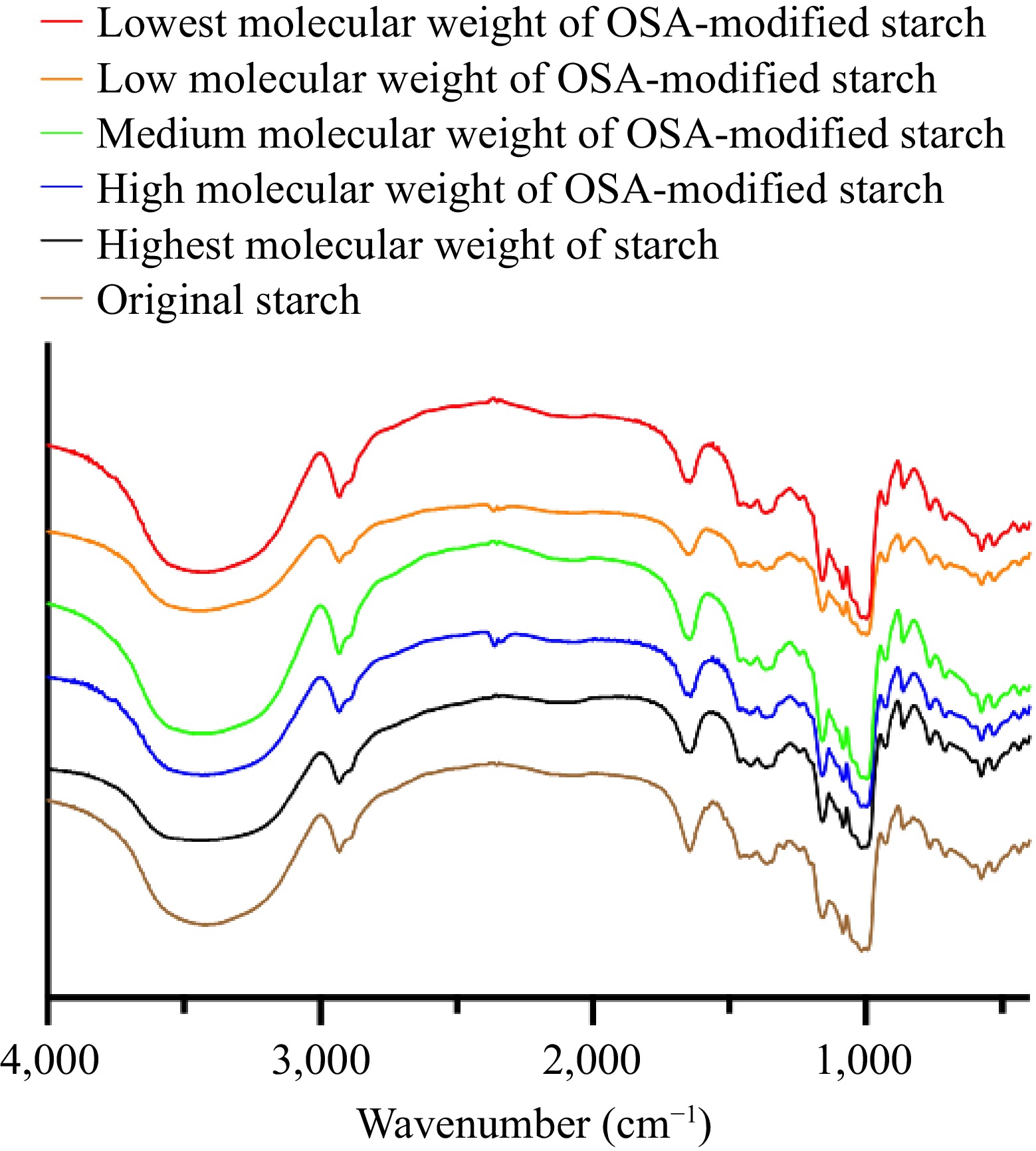
      Figure 1. 

      FT-IR spectra of different molecular weights of OSA-modified starches.

    • The single-factor method was employed to investigate potential factors influencing the yield during the extraction process, with results depicted in Fig. 2. During optimization, the extraction method and solvent type were refined based on emulsifying attributes and polarity alignment with the flavonoid molecule[26,27]. In combination with OSA-modified starch solution, ultrasound-assisted extraction methods provided superior results, establishing them as the optimal extraction method. The concentration of extraction solution and molecular weight of OSA-modified starch were considered in subsequent optimization, shown in Fig. 2c. As initial concentrations shifted, extraction efficiency varied with molecular weight, generally first declining and then increasing. Notably, medium molecular weight OSA-modified starch demonstrated peak performance at an initial concentration of 20 mg/mL . Further optimization, using the single-factor method, pinpointed the ideal solid-to-liquid ratio at 15 mg/mL and extraction duration at 40 min.

      Figure 2. 

      Optimization results of extraction factors (extraction time, extraction method, ratio of solid to liquid, different molecular weights of OSA-modified starches).

      The notable difference in flavonoid extraction rates using OSA-modified starch may stem from the viscosity and degree of esterification of the OSA-modified starch, as well as the self-assembly state of OSA-modified starch molecules in water induced by these properties. Current research findings suggest that a lower viscosity does not unequivocally result in a higher extraction rate, even though a reduced molecular weight might yield a higher degree of esterification. This indicates that both viscosity and degree of esterification are crucial determinants of the extraction rate, but their effects are not linear. Previous studies have shown that OSA-modified starch can self-assemble in water, forming internal cavities of varying dimensions and can establish supramolecular complexes with naringin. Concurrently, mid-molecular weight OSA-modified starch has been demonstrated to enhance the solubility of naringin in water more proficiently. Furthermore, in this study, naringin was utilized as a reference standard to calibrate the total flavonoid content[15]. Considering these aspects collectively, the optimal size of the supramolecular internal cavity of the OSA-modified starch during extraction, along with the solubilizing effect of OSA-modified starch on naringin, might be the primary factors driving the observed variations in extraction rates.

      In summary, the optimal extraction process encompasses the following steps:

      Firstly, disperse 800 mg of medium molecular weight OSA-modified starch (M-OSAS) in 40 mL of deionized water. After 24 h of sealed storage, heat and stir the mixture in a water bath at 350 r/min and 75 °C for 10 min. Subsequently, cool the gelatinized M-OSAS to room temperature.

      Secondly, introduce 600 mg of 'Quzhiqiao' powder to the 40 mL dextrinized M-OSAS solution. After thorough mixing and sealing with cling film, subject it to ultrasonication (1,000 W, 40 min). Post-ultrasonication centrifugation produces the extraction solution.

      thirdly, repeat the extraction solution process three times as per the yield test method detailed in Subsection "Evaluation of extraction results". The resultant yield from this optimized process is 63.72% ± 5.12%.

    • Building on the optimal extraction process detailed in Subsection "Optimization results of the extraction process", flavonoids from "Quzhiqiao" powder were extracted using OSA-modified starches of varying molecular weights. The resultant extract solutions underwent in vitro antioxidant analysis, with findings summarized in Table 3. The DPPH radical scavenging activity of these extracts correlated positively with the molecular weight of OSA-modified starch, suggesting an increase in components soluble in organic systems as the molecular weight rises[2829]. Notably, extracts from both M- and L-OSAS showcased strong ABTS radical scavenging activity, indicating a richer presence of water- and alcohol-soluble components[3031]. The M-OSAS extract, in particular, displayed superior Fe3+ reduction capacity. Significant variations between results emphasize the substantial impact of OSAS molecular weight on Fe3+ reduction within its extract. These findings underscore the influence of OSAS molecular weight on its extraction composition, suggesting it could be a pivotal factor in its efficacy in extracting total flavonoids from "Quzhiqiao".

      Table 3.  In vitro antioxidant results of extraction solution.*

      SampleDPPH radical scavenging activity (%)Equivalent to Trolox (μM)**Equivalent to FeSO4
      (mM)
      Hst-OSAS11.41 ± 0.77a885.72 ± 3.21a1.460 ± 0.048a
      H-OSAS23.70 ± 3.43b888.94 ± 8.32a1.515 ± 0.026b
      M-OSAS33.63 ± 0.45c896.20 ± 9.86b1.615 ± 0.067c
      L-OSAS54.84 ± 4.94d895.06 ± 8.63b1.497 ± 0.034d
      Lst-OSAS55.47 ± 3.85d889.43 ± 7.92a1.382 ± 0.018e
      * Each experiment was repeated three times and expressed as mean ± SD. Different letters in the same column represent a significant difference between the two groups of data (p < 0.05). ** Trolox, a water-soluble analog of vitamin E, is used as a control antioxidant standard.
    • This study examined the extraction capabilities of OSA-modified starch, specifically targeting the flavonoid component in "Quzhiqiao". Optimal conditions were defined, revealing that a gelatinized solution of OSA-modified starch with a molecular weight of 9.25 × 10⁴ Da was the most effective for flavonoid extraction from "Quzhiqiao" under ultrasound assistance (1,000 W, 40 min). It was also determined that a starch solution concentration of 20 mg/mL and a solid-to-liquid ratio of 15 mg/mL were optimal. Following these parameters, the flavonoid yield from "Quzhiqiao" using OSA-modified starch reached 63.72% ± 5.12%. Furthermore, OSA-modified starch extracts demonstrated significantly different antioxidant properties in vitro. These differences greatly affect the experimental data in different models of antioxidant experiments in vitro. As a result, molecular weight might play a crucial role in determining extraction efficiency and extracted ingredients.

    • The authors confirm contribution to the paper as follows: conceptualization, data curation, methodology, formal analysis, software, visualization, roles/Writing - original draft: Wang L, Zhou X; writing - review & editing: Wang L, Zhou X, Lu S, Cui Q; funding acquisition, project administration: Lu S, Cui Q; supervision: Liu J, Quek SY; resources, validation: Lu S, Cui Q, Liu J, Quek SY. All authors reviewed the results and approved the final version of the manuscript.

    • Due to administrative requirements, the original data of the experiments during the research period of the project are not available to the public, but available from the corresponding author or the first author upon request.

      • This work was supported by the National Natural Science Foundation of China (grant number 31571892), National Natural Science Fund of China (grant number 82204552), Natural Science Foundation of Zhejiang Province (grant number LQ22H280007), Research Project of Zhejiang Chinese Medical University (grant number 2022JKZKTS10).

      • The authors declare that they have no conflict of interest.

      • # These authors contributed equally: Lu Wang, Xuyi Zhou

      • Copyright: © 2023 by the author(s). Published by Maximum Academic Press, Fayetteville, GA. This article is an open access article distributed under Creative Commons Attribution License (CC BY 4.0), visit https://creativecommons.org/licenses/by/4.0/.
    Figure (2)  Table (3) References (31)
  • About this article
    Cite this article
    Wang L, Zhou X, Lu S, Quek SY, Liu J, et al. 2023. Utilizing OSA-modified starch with various molecular weights for flavonoid extraction from 'Quzhiqiao' (immature fruit of Citrus paradisi 'Changshan Huyou'). Beverage Plant Research 3:30 doi: 10.48130/BPR-2023-0030
    Wang L, Zhou X, Lu S, Quek SY, Liu J, et al. 2023. Utilizing OSA-modified starch with various molecular weights for flavonoid extraction from "Quzhiqiao" (immature fruit of Citrus paradisi 'Changshan Huyou'). Beverage Plant Research 3:30 doi: 10.48130/BPR-2023-0030

Catalog

  • About this article

/

DownLoad:  Full-Size Img  PowerPoint
Return
Return