ARTICLE   Open Access    

Genome-wide discovery of CBL genes in Nitraria tangutorum Bobr. and functional analysis of NtCBL1-1 under drought and salt stress

More Information
  • Calcineurin B-like (CBL) proteins are a class of important Ca2+ receptors that play key roles in plant stress response. CBLs have been shown to participate in responses to abiotic stresses such as drought, salt, and cold in many plant species, including Arabidopsis and rice. However, little is known about their potential functions in the desert halophyte Nitraria tangutorum. Here, we have identified 11 CBL genes distributed across six chromosomes of N. tangutorum and categorized them into four groups through phylogenetic analysis. Synteny analysis showed that they have strong collinear relationships and have undergone purifying selection during their evolution. NtCBL promoter regions contain a variety of cis-acting elements related to hormone and environmental stress responses. Real-time quantitative PCR showed that the expression of NtCBLs differed significantly among various tissues and was upregulated by salt and drought stress. We chose NtCBL1-1 for an in-depth functional characterization and observed that transgenic Arabidopsis plants expressing NtCBL1-1 exhibited increased tolerance to both drought and salt stress. Compared to wild-type Arabidopsis, transgenic lines showed higher germination rates, slower chlorophyll degradation, more soluble proteins, and reduced accumulation of the oxidative stress marker malondialdehyde. These findings indicate that NtCBL1-1 plays a significant role in responding to drought and salt stress, laying the foundation for further investigations into the functional mechanisms of NtCBL genes in N. tangutorum.
  • First visualized by Robert Hooke in 1665, cells had long been regarded as individual units of a whole organism. Whether the cell represents an autonomous entity was a question that had been a subject of debate in 19th Century. The observation of intercellular bridges and plasmodesmata supports the idea that the cellular structure forms the protoplasmic continuity, highlighting the importance of reciprocal interaction of cells within a multicellular organism. As a pioneering cell biologist, Wilson wrote in 1923, "it is the 'organism as a whole' and a 'property of the system as such' "[1], almost all plant cells are connected by the intercellular channel called plasmodesmata (PD)[2].

    Primary PD is a straight channel-like structure, as small as 30-50 nm in diameter, connecting two neighboring plant cells[3,4]. A major component of this channel is an endoplasmic reticulum (ER) derived central membranous strands called desmotubles, which form presumably through trapping ER strands in the cell plate during cytokinesis[5,6]. In between the desmotubule and flanking plasma membrane is the cytosolic space called cytoplasmic sleeve[7,8]. Components including cytoskeletons, a GPI-anchor protein and PD localizing proteins (PDLP) have been suggested to participate in the organization and function of plasmodesmata[9, 10].

    More recently, sphingolipids were found to affect the pore size of plasmodesmata[11]. Interestingly, analysis of Physcomitrium patens plasmodesmata proteome suggested the enrichment of cell-wall located proteins including EXORDIUM-family members and xyloglucan transglycosylases in plasmodesmata[12]. In particular, this study identified callose-degrading glycolyl hydrolase family 17 (GHL17) proteins as an abundant PD protein family[12], suggesting the potentially conserved plasmodesmata regulation by callose (will be further discussed later in this review) over the evolution.

    Smaller molecules, ions and metabolic substance can all pass through PD by diffusion. Other micro-molecules including proteins and RNAs are thought to transverse PD via active transport[1115]. Mobile molecules can move across PD via either the cytoplasmic sleeve, or through the desmotubule (in lumen or lateral diffusion in the desmotubule membrane), or via diffusion in the flanking plasma membrane[16,17]. In support of these hypotheses, it was found that the interference of the membrane structure affected PD permeability[17]. In old tissues, plant cells further produce secondary PD that is normally branched and complex in shape. Localized cell wall modification could be involved in secondary PD formation, and the complexity of this type of PD is correlated with reduced PD permeability[18,19]. Nevertheless,the detailed mechanism and the exact roles of secondary PD during development are still far from clear. Interestingly, multiple types of PD were found at grafted wounds, suggesting that different PD types could have distinct functions[20]. In this review, we focus on our current understanding of cell-to-cell signaling across plasmodesmata.

    The observation of cell-to-cell movement of large molecules initially arose from the micro-injection of fluorescent dye in plant tissues[2124]. The first endogenous protein exhibiting the intercellular mobility is KNOTTED1 (KN1), a homeodomain protein essential for maintenance of the shoot apical meristem (SAM) in maize[25,26]. Recently, the ribosomal RNA-processing protein 44A (AtRRP44A) was shown to mediate the cell-to-cell trafficking of KN1[27]. Since then, a large number of transcription factors were identified in plants that can move between tissues and cells to provide positional instruction during plant development[21]. These mobile regulators can traffic across just a few cell layers to function locally or over a long distance to affect global developmental change.

    One of the central questions in organogenesis is how to spatiotemporally maintain stem cells and specify cell fates. In SAM, WUSCHEL (WUS) is expressed in the organizing center of shoot apical meristem, but the protein moves to the layer1 and 2 (L1 & 2) of shoot apical meristem where WUS triggers CLAVATA 3 (CLV3) expression, which in turn inhibits WUS transcription in L1 and L2 layer[28,29]. With this WUS-CLV3 feedback loop, plants can maintain the stem cell population in proper size in SAM. With the similar strategy, plants maintain the root stem cell niche via WOX5-CLE40 loop, in which WOX5 traffics from quiescent center (QC) to columella stem cell (CSC) to repress the cell differentiation[30]. In Arabidopsis, SHOOT MERISTEMLESS (STM) and ARABIDOPSIS KNOTTED-LIKE (KNAT1)/BREVIPEDICELLUS (BP) are two homologs of the KN1 gene, previously described to be mobile in maize SAM. When driven by an L1 specific promoter, STM and KNAT1 were observed to move from the L1 layer into the inner cell layers of the SAM[31,32]. In addition, KNAT1 was able to pass the interface between cortex and epidermis in Arabidopsis when mis-expressed by a mesophyll specific promoter[33].

    In embryogenesis, TARGET OF MONOPTEROS 7 (TMO7), encoding a bHLH transcription factor, is essential for hypophysis, the founder cell for forming root apex during post-embryonic growth. TMO7 is transcribed in embryonic cells while the TMO7-GFP fusion can be detected in the neighboring hypophysis, indicating a non-cell-autonomy of this regulator[34,35]. In post-embryonic growth, intercellular movement of transcriptional factors regulates a variety of developmental aspects ranging from root radial patterning to root hair and trichome initiation. These mobile regulators including SHORT-ROOT (SHR), CAPRICE (CPC), TRANSPARENT TESTA GLABRA 1 (TTG1), GLABRA 3 (GL3), ENHANCER OF TRY AND CPC 3 (ETC3)/ TRIPTYCHON (TRY), UBIQUITIN-SPECIFIC PROTEASE (UBP1) have been well reviewed previously[15,21]. A previous screen estimated that around 15% of transcriptional factors in roots can move between cells[36]. In contrast, we only have limited understanding of the functionality of these mobile proteins.

    Recently, more mobile transcriptional factors have been identified (summarized in Table 1). Two closely related AT-hook family members, AT-HOOK MOTIF NUCLEAR LOCALIZED PROTEIN 3 (AHL3) and AHL4, were shown to interact in vivo and regulate the boundaries between the procambium and xylem[37]. Interestingly, their interaction seemed to be required for their intercellular trafficking. A SHR target, SCL23 displays a bidirectional radial spread and long-range movement into meristem in Arabidopsis roots. Through direct interaction, SCL23 controls movement of SHR and participate in endodermal specification in the root meristem[38].

    Table 1.  Summary of the mobile transcription factors identified in plants.
    Mobile TFsFunctionMoves from:toReference
    HY5Root growth and N uptakeShoot-to-rootChen et al. (2016)[41]
    DWARF14Regulate the development of AMsThrough phloem into axillary meristems (AMs)Kameoka et al. (2016)[139]
    BdMUTEBdMUTE is required for subsidiary cell formationGMCs to neighboring cell filesRaissig et al. (2017)[97]
    SPCHStomatal cell fateCell-to-cell diffusion in the leaf epidermis of chorusGuseman et al. (2010)[96]
    AN3Leaf developmentFrom the mesophyll to the epidermis in leavesKawade et al. (2013)[140]
    WUSMeristem maintenanceFrom the organizing centre to L1, L2 layersYadav et al. (2011)[28]
    KN1/STMMeristem maintenanceBroadly in the SAMKim et al. (2003)[31], 2005[32]
    PLT2Longitudinal root zonationLongitudinally from the root meristem forming a gradientMahonen et al. (2014)[141]; Galinha et al. (2007)[142]
    SHRRoot radial patterning and RAM maintenanceWithin Stele; Stele into endodermis, QC, CEI and CEDKoizumi et al. (2011)[44], Nakajima et al. (2001)[78]
    AHL3/AHL4Xylem specificationFrom procambium cells to the xylemZhou et al. (2013)[37]
    WOX5Stem cell maintenanceQC to CSCPi et al. (2015)[30]
    TMO7Recruitment of the hypophysisEmbryo into the upper cell of suspensorSchlereth (2010)[34]; Lu et al. (2018)[35]
    Cyp1Root growthFrom leaves to root in tomatoSpiegelman et al. (2015)[143]
    UBP1Transition from cell division to elongationStele and LRC to cells into transition/elongation zoneTsukagoshi et al. (2010)[144]
    SCL23Endodermal cell fateBidirectional radial spread and movement into meristemLong et al. (2015)[38]
    TTG1Trichome patterningAtrichoblasts into trichome initials
    CPCTrichome patterning, root hair initiationTrichome initials into Atrichoblasts; non-root hair cell into root hair cellWester et al. (2009)[90]
    GL3/EGL3Root hair initiationRoot hair cell into non-root hair cellKang et al. (2013)[91]
     | Show Table
    DownLoad: CSV

    Besides the local regulation, transcriptional factors were also found to traffic long-distance between organs to direct global developmental transition in plants in Fig 1. An early example is the detection of FLOWERING LOCUS T (FT) trafficking from leaves where it is synthesized in response to day length, to the SAM to trigger flowering[39,40]. Recently, a light-activated transcriptional factor, ELONGATED HYPOCOTYL 5 (HY5) was shown to move via phloem from shoot-to-root. This translocation of HY5 was proposed to mediate light-activated root growth and N uptake from the soil to balance photosynthetic carbon fixation in the leaf[41].

    Figure 1.  Mobile proteins and RNAs in plant development and stress response. The mobile regulators participate widely in the development of different organs (as illustrated). They can travel short-range to regulate local tissue patterning or long-distance to transduce systemic signaling. Gray arrow: phloem-based long-distance movement. WUS and STM regulate SAM maintenance; SPCH, BdMUTE, AN3, TTG1, GL3 and CPC are involved in epidermal patterning. In roots, PLT2, SHR, AtDof4.1, AHL3/AHL4, WOX5, TMO7, UBP1 and SCL23 govern a variety of processes including cell division, radial patterning, stem cell maintenance and developmental transition. Long-distance signaling regulators such as FT and HY5 can traffic from leaves to SAM to promote flowering, and from shoot to root to regulate root growth and nitrate uptake respectively. Environmental stresses can induce PD closure. Small RNAs including miR399d, 827 and 2111 move from aerial parts to roots in response to phosphate starvation.

    Considering the size of transcriptional factors, PD seems to be the most possible way for the intercellular translocation. With an iclas3m system (described in detail in a later part of this review) that blocked the PD between stele and endodermis, SHR intercellular transport was terminated[3]. Another piece of evidence supporting PD transport of transcriptional factors is the blocked movement of TMO7 from meristematic cells into the root cap in the cals3-2d, a mutant in which PD is restricted by over-accumulated callose[35]. To get access to PD, transcriptional factors could exploit intracellular apparatus including microtubules and endomembrane delivery system[42,43]. Besides, an unknown function protein named SHR INTERACTING EMBRYONIC LETHAL (SIEL) was shown to interact with a number of mobile transcriptional factors and the mutation of this gene seemed to reduce SHR intercellular movement[44]. As SIEL partially localized to endosomes, it was proposed that this protein could function as a 'shuttle' to facilitate delivery of mobile transcriptional factors. In addition, some facilitating proteins have also been identified. After passing through PD, a few mobile proteins including APS KINASE 1 (KN1), SHOOT MERISTEMLESS (STM) and TRANSPARENT TESTA GLABRA 1 (TTG1) were discovered to associate with a group of type II chaperonin complexes consisting of CHAPERONIN CONTAINING T-COMPLEX POLYPEPTIDE-1 SUBUNIT 7 and 8 (CCT7 & CCT8), which facilitate the movement possibly by promoting the protein refolding after the PD cross-over[27].

    Although no specific domain has been identified that accounts for intercellular mobility, the cell-to-cell transport of transcriptional factors seemed to be protein sequence-dependent. Homeodomain (HD) and the helical domains have been shown to be necessary and sufficient for PD-mediated transport of KN1. Unlike this, three conserved domains (HD, WUS-box, and EAR-like domain) in WUS are not required for its movement. Instead, WUS mobility seems to be controlled by a non-conserved sequence between the HD domain and WUS-box[29]. Despite triple GFP Tag impaired TMO7 movement, protein size did not seem to be the primary determinant of intercellular transport. Instead, TMO7 was found to move in a sequence-dependent manner, and both nuclear residence and protein modification are important for TMO7 mobility[35]. In two other mobile transcriptional factors, CPC and SHR, the mobility relied on multiple regions within the proteins. In addition, the mobility of these two proteins seemed to be associated with the subcellular distribution in both the cytoplasm and the nucleus.

    In addition to transcriptional factors, small RNAs also participate in transcriptional regulation of diverse developmental and physiological events in plants. Small RNAs are 21−24 nt long and can be generally divided into siRNAs and miRNAs[45]. Small RNAs function either through degrading target genes by near-perfect complementarity, or via transcriptional silencing by histone modification and DNA methylation[4650]. Small RNAs were often regarded as the long-distance signals as the initial efforts dissecting their mobility exploited the grafting system in which mutants defective in small RNAs biogenesis were included. Facilitated by high-throughput sequencing techniques, researchers identified a large number of mobile siRNAs that can traffic from shoot to root presumably via phloem. Besides siRNA, a large number of miRNAs were discovered to traffic in phloem exudates over long distance. Low-phosphate induced miR399s exhibited a shoot-to-root movement to repress downstream targets including PHO2 in the root[51]. Similarly,miR399d, miR827 and miR2111 were all found in grafting experiments to relocate from aerial parts to roots in response to phosphate starvation[52]. During rhizobial infection, miR2111 functioned as long-distance signals to post-transcriptionally regulate symbiosis suppressor TOO MUCH LOVE in roots[53]. miR395 can also translocate from wild-type scions to rootstocks of the miRNA processing mutant hen1-1 to target the APS gene[54]. In addition, both miR156 and miR172 have been confirmed as potentially phloem-mobile miRNAs that regulate tuber formation[5557].

    In grafting system, only small RNAs transporting from shoot-to-root via phloem could be analyzed. Other approaches that allow for the comparison between the expression areas and in situ RNA distribution patterns may help the identification of small RNAs acting locally as non-cell autonomous signals. To establish adaxial–abaxial leaf polarity, a member of Trans-acting small interfering RNA (ta-siRNA) family forms a gradient across the leaves by intercellular diffusion. This diffusion-driven pattern of ta-siRNA shapes the expression pattern of AUXIN RESPONSE FACTOR3 (ARF3), an abaxial determinant gene. Another small RNA, miR390 was proved to regulate the leaf polarity by the cell-to-cell movement from vasculature and pith region below the shoot apical meristem to the vegetative apex[54]. In addition, miRNA165/166 were discovered to move from the endodermis into the stele to regulate the xylem cell fate[58]. Moreover, miR394 was shown to regulate stem cell maintenance in SAM by the PD-mediated movement from L1 to inner cell layers to repress LEAF CURLING RESPONSIVENESS (LCR) expression[59].

    In addition to siRNA and miRNA, mRNAs have also been found to travel beyond the cells in which they are expressed in Fig 1. In addition to the early example of mobile mRNAs of KN1, potato sucrose transporter SUC1 mRNA was also confirmed to be mobile. In grafting experiments, a number of mRNAs were found to travel, such as FT, FVE and AGL24 in Arabidopsis[60], Aux/IAA in melon and Arabidopsis[61], PP16 and NACP in pumpkin[62,63], BEL5 and POTH1 in potato, SLR/IAA14 in apple[64], PFP-T6 and PS in tomato[65] (summarized in Table 2). Recently, Luo et al. developed a fluorescence-based mRNA labeling system to identify mobile mRNAs targeted to PD[66]. Their analyses revealed that only mobile rather than not non-mobile mRNAs were selectively targeted to PD, providing further evidence for PD mediated transport of mRNAs. Interestingly, using a Nicotiana benthamiana/tomato heterograft system, Xia et al. found some mRNAs have bidirectional mobility between shoots and roots. In addition, forced expression of non-mobile mRNAs in the companion cells did not confer the mobility[6771]. Thus, the movement of mRNA is likely an actively regulated process. Moreover, a large number of graft-transmissible mRNAs have been identified by high throughput sequencing in a variety of species including Arabidopsis, tobacco, grape, cucumber and tomato[6772].

    Table 2.  List of mobile RNAs with functions in organ development.
    Mobile factorFunctionMoves from: toReference
    mRNA
    KN1SAM maintenanceinjected cell to neighbouring cellsLucas et al. (1995)[26]
    SUC1Sucrose transportcompanion cells to sieve elementsKuhn et al. (1999)[145]
    FT1Flowering inductionLeaf to SAMLu et al. (2012)[60]
    Aux/IAA18Root developmentLeaf to rootNotaguchi et al. (2012)[61]
    PP16RNA transportPhloem to shoot apexXoconostle-Cazares et al. (1999)[62]
    NACPMeristem maintenancePhloem to shoot apexRuiz-Medrano et al. (1999)[146]
    StBEL5Tuber formationLeaf to rootBanerjee et al. (2009)[147]
    POTH1Leaf developmentLeaf to rootMahajan et al. (2012)[148]
    SLR/IAA14Lateral root formationShoot to rootKanehira et al. (2010)[64]
    PFP-T6Leaf developmentLeaf to leaf primordiaKim et al. (2001)[65]
    PSPathogen resistanceShoot to root and vice versaZhang et al. (2018)[149]
    GAILeaf developmenthost to parasiteRoney et al. (2007) [150]; David-Schwartz et al. (2008)[151]
    ATCFloral initiationLeaf to flower apicesHuang et al. (2012)[152]
    FVEfloral regulatorsRoot to SAMYang and Yu (2010)[153]
    AGL24floral regulatorsRoot to SAMYang and Yu (2010)[153]
    siRNA
    ta-siRNAEstablishment of leaf polaritythe adaxial to the abaxial side of the leafChitwood et al. (2009)[154]
    hc-siRNADNA methylationShoot to rootBaldrich et al. (2016)[155]
    miRNA
    miR165/166Xylem specificationendodermis into the steleCarlsbecker et al. (2010)[58]
    miR390Leaf polarityvasculature and pith region below the SAM to SAMChitwood et al. (2009)[154]
    miR394Meristem maintenanceL1 to inner layers in the shoot meristemKnauer et al. (2013)[59]
    miR395Sulfate homeostasisgraft unionsBuhtz et al. (2010)[54]
    miR399dPhosphate homeostasisshoot to root and vice versaPant et al. (2008)[156]; Lin et al. (2008)[51]
    miR172regulate tuber formationLeaf to rootMartin et al. (2009)[55]
    miR2111Phosphate homeostasis;
    Rhizobial infection;
    shoot to root and vice versaHuen et al. (2017)[52];
    Tsikou et al. (2018)[53]
    miR827Phosphate homeostasisshoot to root and vice versaHuen et al. (2017)[52]
     | Show Table
    DownLoad: CSV

    A plant organ is usually composed of morphologically and functionally different cell types in different positions. Small molecules can move between cells and across plasmodesmata, which mediates crucial intercellular communication for the growth and development of plant tissues and organs. For example, a plant root is composed of concentrically arranged cell layers with epidermis, cortex, endodermis, and stele locating from outside to inside[73]. This anatomic arrangement highlights the regulation of tissue patterning instructed by positional information, often through the exchange of signaling molecules between cells. A number of developmental processes including root radial patterning, root hair initiation and trichome formation, have emerged as the model system for studying tissue patterning in plants.

    In root, the formation of the endodermal cell layer starts from the endodermal and cortex initial cells in root stem cell niche, where two transcriptional factors, SHR and SCARECROW (SCR) promote the expression of CYCD6;1 to allow the switch of cell division pattern from anticlinal to periclinal[7477]. This results in the formation of two distinct layers of cells within the ground tissue, and the role of SHR in specifying the endodermal layer was proposed based on the fact that the endodermal layer was completely absent in shr-2 mutant. Intriguingly, SHR expression is restricted in stele, but the SHR protein is actively transported through PD from stele toward the outside to play non-cell-autonomous roles[78,79]. In the enodermis, SHR directly activates SCR which, in turn, physically binds to SHR to trap this mobile transcription factor in the nucleus of the endodermis, preventing further movement[77]. This mechanism was discovered to be conserved in rice and thus was proposed to be an evolutionarily conserved mechanism defining a single endodermal cell layer in almost all land plants[74]. However, a study on rice SHR homologs suggested that SHR alone is insufficient to determine endodermal cell fate[80]. Consistent with this argument, mis-expression of SHR indicated that SHR ability to confer endodermal identity partially relied on cell lineage and was coordinated by uncharacterized positional information, presumably derived from stele.

    Specific expression of marker genes, as often used previously to determine endodermal cell fate, is sometimes misleading. A prominent feature of the endodermis is the formation of the Casparian Strip (CS), an apoplastic barrier between vascular tissues and outer ground tissues[81]. The presence of functional CS is therefore a better trait for precise evaluation of endodermal identity. Two recent studies revealed that SHR does serves as a master regulator activating a hierarchical downstream network for CS formation[82,83]. The combination of SHR mediated cascade and another independent peptide signal derived from stele forms the minimum set of regulators that program endodermal identity, exemplified by the formation of functional CS[83]. Since both SHR and the peptide are specifically expressed in vascular tissues, CS formation represents the elaborate developmental control by stele-to-endodermis movement of mobile regulators. Besides CS, SHR and its downstream target SCR can activate the expression of miRNA165/166 in the endodermis which in turn moves back to vasculature to repress a class III homeodomain-leucine zipper transcription factors for proper xylem formation[58]. Thus the reciprocal communication between ground tissue and vasculature in root spatially defines the radial patterning in root. In Cardamine, a recent study indicated that a differential spatial distribution of miR165/166 is responsible for forming the extra cortex layer[84]. In addition to roots, miR165/166 also function in other organs including leaf primordial and ovule. By restricting PHB expression in incipient inner integument, miR165/166 promotes the correct ovule patterning[85]. Interestingly, a callose synthase mutant in maize, named tie-dyed2 (tdy-2), affects the development of vasculature, suggesting the mechanism of vascular development directed by intercellular communication (possibly via miR165/166) is likely conserved in crops[86,87]. In addition to roots, plasmodesmata also plays a key role in regulating leaf development, particularly the formation of leaf veins[88].

    Trichomes and root hairs, originating from the epidermis in leaves and roots respectively play important roles in protecting plants from bio/abiotic stresses, and promoting nutrient absorption[89,90]. In Arabidopsis, the initiation of trichomes and root hairs is precisely patterned in epidermis, indicating an essential role of cell-to-cell communication in these processes.

    In trichome initiation, both positive regulator TRANSPARENT TESTA GLABRA (TTG1) and negative regulator ENHANCER OF TRY AND CPC 3 (ETC3) and CAPRICE (CPC) move between cells. In incipient trichome cells, TTG1 protein accumulates through a trapping/depletion mechanism mediated by GLABRA3 (GL3)[91]. On the other hand, the repressor of ETC3 and CPC move into the neighboring non-trichome cells (also regulated by GL3), forming inactivated MYB/bHLH/WD40 to inhibit the development towards trichomes[92]. Recently, PdBG4 has been implicated in regulating PD permeability in Arabidopsis trichome development[93]. In root hairs, CPC serves as a positive regulator and it is trapped in the hair-position root epidermis by interacting with EGL3 and GL3 after the movement[94]. The trn1 mutant is defective in the position-dependent pattern of root hairs and cause the ectopic expression of WER, GL2 and EGL3, suggesting that TRN1 also participates in the position-dependent cell fate determination[95,96].

    Stomata on epidermis are responsible for water and gas exchange between the plants and the environments. The mature stomata structure is produced through successive cell division and differentiation process, with both processes subject to highly spatiotemporal regulation[97]. In a GLUCAN SYNTHASE-LIKE 8 (GSL8) mutant in which normal callose deposition is disrupted, SPCH-GFP diffused to neighboring cells from meristemoids, resulting in excessive proliferation of stomatal-lineage cells. This observation suggests that proper gating of critical regulators, likely through callose regulation, regulates the correct patterning of stomata complex[98]. MUTE, another key transcriptional factor required to terminate asymmetric division and promote the transition of meristemoids to GMCs, was shown in Brachypodium to move from GMCs to neighboring cells to induce the subsidiary cells (SCs) formation[99].

    Plants respond to stresses often by accumulation of callose, which is negatively correlated with PD permeability in Fig 2. A variety of abiotic stresses have been associated to callose induction, such as cold stress[100,101], wounding[102,103], heat stress[104,105], and heavy metals[106109]. Although detailed mechanism is not entirely clear, callose synthases were found to participate in the callose regulation. In Arabidopsis, there are 12 callose synthase (CalS) family members. When exposed to excess iron, the cals5 and cals12 mutants showed an attenuated callose deposition in phloem, compared to wild type and other cals mutants. This result suggests that cals5 and cals12 may play specific roles in iron stress response in Arabidopsis[110]. In tomato, cold stress has long been known to cause catfacing fruits or malformed fruits by breeders and gardeners. A recent study proved this phenomenon was caused by the restriction of SlWUS intercellular movement via plasmodesmata in floral meristem[101]. The cold induced callose accumulation blocked the plasmodesmata, resulting in the excessive activation of CLV3 and TAG1, and disrupted WUS-CLV3/WUS-TAG1 negative feedback loops[101].

    Figure 2.  Regulation of PD permeability by callose. (a) Schematic illustration of regulation of the PD aperture by callose deposition in flanking regions of PD. Induced callose accumulation closes PD permeability and blocks the intercellular movement of transcription factors and small RNAs. (b) The design of ‘icals3m’ system that can inducibly (via estradiol induction cassette) promote callose deposition in specific cell types (via cell-type specific promoters)[128],[138].

    It has been reported that PD regulation serves as an innate defense strategy[111]. Pathogens trigger both pathogen-associated molecular pattern (PAMP) and PAMP-triggered immunity (PTI) systems, which have been reported to induce callose deposition[112]. Upon SMV virus invasion, callose was accumulated in soybean phloem which prevents the virus from traveling long distances[113]. Salicylic acid (SA) is a plant immune signal produced upon pathogen infection, which has also been shown to trigger PD closure and affect symplastic communication. Elevation of SA level seemed to be necessary for the PD response during bacterial infection, and the expression of bacterial derived salicylate hydroxylase (NahG) gene in plants resulted in higher susceptivity to bacteria[113]. Biotic stresses including pathogen infection are known to modulate ROS level and callose abundance in infected regions, which is presumably responsible for the altered PD permeability[114,115].

    Virus can also regulate the mesenchymal plasmodesmata in tobacco[109] and it was recently reported that ROS-mediated PD closure is controlled by multiple pathways, either in SA- or PDLP5-dependent manners. Change of callose level in biotic stresses is also modulated by callose synthase members[112,113]. SA-dependent PD regulation requires the function of callose synthase1 (CalS1). However, the CalS8 seemed to be more involved in basal and ROS-dependent PD regulation[103]. Callose synthase members have also been widely reported in recent years. CsCalS4 function was identified in pollen development in cucumber, and CsCalS1/8 homologous genes were induced by cucumber fungus and functioned as the key factors in response to biological stress[114]. GhCalS5 and ZmCals were found to promote callose synthesis in cotton and maize in responsive to stresses[116,117].

    In addition, PD-localized proteins also emerged as the regulator of PD aperture during biotic stresses. It was shown that the PD closure triggered by chitin was dependent on the activity of PD-localized receptor-like protein LYM2[111]. Besides, bacterial flagellin could rapidly activate the expression of CML41, a PD-localized Ca2+-binding protein, which is necessary for the induction of callose at PD.

    Callose is the linear polysaccharide that is composed of β-1,3-glucan. It is a component of cell wall and is frequently found to deposit at PD, where it is believed to control the PD permeability during plant development and stress response. It was found the precise developmental transition often relies on the regulation of symplastic continuity. In birch, bud dormancy entry and release are associated with the shift between callose production and turnover. Callose accumulation at PD in the shoot apical and rib meristems can seal off the symplastic communication and promote the bud dormancy[116121]. A period of chilling, however, triggers gibberellin biosynthesis, resulting in increased expression of 1,3-β-glucanases and degradation of callose. Accumulating evidence suggests that callose regulation is actually implicated in a wide range of developmental processes, including seed germination, embryogenesis, cell division, flowering and reproduction[122124]. In tomato, a short period of cold stress is sufficient to induce callose accumulation in floral meristem and blocked intercellular movement of SlWUS, resulting in malformed fruits[101]. In olives, callose deposition, as part of cell wall modification, regulates fruit abscission[114].

    Through a genetic screen for defective vascular development, Vaten et al. (Helariutta group) identified three semi-dominant alleles of CALLOSE SYNTHASE 3 (cals3d) that caused an increase in callose deposition at PD and abnormal plant growth[3,19]. In the root, cals3d mutants all showed aberrant radial patterning and misspecification of the phloem and the xylem. Consistent with these phenotypes, cals3d roots exhibited decreased PD-mediated symplastic movement of free GFP, SHR and miRNA165/66[3,125]. It thus seemed that the identified dominant mutations can substantially enhance the ability of CALS3 to promote callose deposition at PD. By combining these mutations in a vector containing LexA-VP16-ER (XVE)-based estradiol inducible cassette, the Helariutta group designed an elegant tool named as the 'icals3m system'. Driven by specific promoters, this system can potentially be used to temporally manipulate callose at PD and symplastic communication in particular cell types[3,126].

    The initial attempts using this system in vascular tissues and lateral root development proved to be successful[3,125,127]. With specific induction of icals3m system in xylem pole pericycle, Benitez-Alfonso et al. detected a significantly increased number of initiated primordial [126]. Together with the observation of a transient symplastic isolation of the primordium prior to emergence, they confirmed the essential role of callose based symplastic connectivity between pericycle cells, founder cells, and the neighboring tissue during lateral root patterning[122]. More recently, icals3m system was used to dissect the roles of symplastic communication in root apical stem[122]. Driven by an endodermis-specific EN7 promoter, icals3m induced symplastic blockage led to severe root patterning defects, shown by disrupted cell division direction, misspecification of cell fate as well as impaired cell polarity. In root tip, different cell types including endodermis all derived from the root stem cell niche, where QC was believed to repress the differentiation of surrounding stem cells based on an early classic laser ablation experiment carried out in the 1990's[127]. However, icals3m system provides an alternative non-invasive approach to examine the role of QC. With the expression under WOX5 promoter, icals3m system was clearly shown to induce callose specifically in QC[128]. The visible callose signal based on aniline blue staining was detected as quickly as 6 h after the estradiol induction[129]. This icals3m system was further used to study the interaction between root cap and the root meristem[124,128130]. When the symplastic communication between root cap and root meristem was disrupted, developmental defects were observed in both parts: In meristem, stem cell maintenance was affected while in root cap the starch granules, the marker commonly used as an indicator of columella differentiation, disppeared[125]. An earlier study showed that starch granules in columella cells relied on auxin concentration[131]. In this study, short-term disruption of symplastic communication was sufficient to cause defects in stem cells, while it took longer for auxin distribution in root meristem to occur[125]. In fact, plasmodesmata itself can act as the channel for auxin flow[131,132]. Furthermore, icals3m system also was employed in the study of phloem unloading[132]. A phloem pole pericycle specific promoter CalS8 and a companion cell and metaphloem sieve element specific promoter psAPL were both used to drive icals3m to block the connection between different phloem cell types[133]. A direct developmental defects arose from the blocked plasmodesmata in phloem was the reduced growth of axillary buds[50].

    To summarize, callose regulation is a central mechanism to control symlastic communication during plant development. Spatiotemporal expression of icals3m system can be an effective tool to deepen our understanding of the developmental regulation mediated by symplastic signals. The power of this system can be even higher with the combination with other techniques including cell type specific OMICs. The application of this system in vegetable studies would greatly enhance our ability to dissect various aspects of development and physiology in vegetable species ranging from fruit development to stress resistance.

    Intercellular signaling across plasmodesmata plays crucial roles in a wide range of processes in plants. The currently identified signaling molecules across plasmodesmata are mainly transcription factors and RNAs. However, accumulating evidence suggests that many other signaling pathways including calcium signaling, redox signaling, phosphorylation signaling, and hormone signaling can also function in non-cell-autonamous manner[134136]. As these pathways are often complex and interplay with each other, it is still difficult to unravel such non-cell-autonamous functions. With the advance in high-resolution imaging techniques, such as super-resolution microscopy, researchers will be able to visualize in vivo the action and mobility of the molecular players involved in intercellular signaling[137].

    In addition to visualizing the intercellular mobility of molecules, it is crucial to precisely evaluate the phenotype with a specific intercellular signaling disrupted. Developing cell type specific approaches is the key step and thus identification of promoters with restricted expression in certain cell types is important. Furthermore, abolishing gene function in a specific cell type is a valuable tool for studying intercellular signaling. Previously, cell-specific RNAi was employed but the intercellular mobility of small RNAs prevents the precise evaluation of gene function. Recent rapid development of CRISPR-Cas9 technique has emerged as a powerful tool for this purpose. The combination of cell-specific expression of Cas9 with reporters that allows for visualizing the gene editing in different cells could greatly enhance our ability to precisely evaluate the function of mobile regulators.

    Lastly, to gain a more comprehensive understanding of the plasmodesmata mediated intercellular signaling, it is important to integrate multiple approaches, such as high-resolution imaging, single-cell technique, multi-omics, and computational modeling. Although the cell-to-cell signaling often occurs locally, the impact could be systemic in plants. The complete assessment of plasmodesmata-mediated intercellular signalling, as well as derived tissue- or cell-type-specific techniques, will not only benefit the study of plant development, but also provide the opportunity for future biotechnological renovation of plants.

    This work was supported by carbon-nitrogen high efficiency grants from Fujian Agriculture and Forestry University (118992201A).

  • The authors declare that they have no conflict of interest.

  • Supplemental Table S1 Primers for Quantitative RT-PCR in N.tangutorum.
    Supplemental Table S2 Primers for isolation of NtCBL1-1 fragment and construction of overexpression vector.
    Supplemental Table S3 CBL genes used to construct phylogenetic trees from 12 species except N.tangutorum.
    Supplemental Table S4 KaKs analysis in N.tangutorum.
    Supplemental Table S5 Identity between CBLs gene in Nitraria tangutorum.
    Supplemental Fig. S1 Cis-regulatory elements analysis of NtCBLs. (A) A heat map showing the number of cis-acting elements related to stress response in NtCBLs. (B) The distribution of cis-acting elements in the promoter region (ATG upstream) of NtCBLs, where different boxes represent different cis-acting elements.
    Supplemental Fig. S2 Positive identification of partial NtCBL1-1 transgenic T3 generation; different lanes represent different transgenic lines.
    Supplemental Fig. S3 Relative expression levels in partial transgenic T3 generations, with wild-type Arabidopsis as a reference.
    Supplemental Fig. S4 Phenotypic changes in overexpression lines 1 and 3 following exposure to 200 mM NaCl for 0−7 days.
    Supplemental Fig. S5 Phenotypic changes in overexpression lines 1 and 3 following exposure to 300 mM mannitol for 0−7 days.
    Supplemental file S1 Genome annotations.
  • [1]

    Li X, Ma Z, Song Y, Shen W, Yue Q, et al. 2023. Insights into the molecular mechanisms underlying responses of apple trees to abiotic stresses. Horticulture Research 10:uhad144

    doi: 10.1093/hr/uhad144

    CrossRef   Google Scholar

    [2]

    Jia X, Zhu Y, Hu Y, Zhang R, Cheng L, et al. 2019. Integrated physiologic, proteomic, and metabolomic analyses of Malus halliana adaptation to saline – alkali stress. Horticulture Research 6:91

    doi: 10.1038/s41438-019-0172-0

    CrossRef   Google Scholar

    [3]

    Kudla J, Xu Q, Harter K, Gruissem W, Luan S. 1999. Genes for calcineurin B-like proteins in Arabidopsis are differentially regulated by stress signals. Proceedings of the National Academy of Sciences of the United States of America 96:4718−23

    doi: 10.1073/pnas.96.8.4718

    CrossRef   Google Scholar

    [4]

    Kolukisaoglu Ü, Weinl S, Blazevic D, Batistic O, Kudla J. 2004. Calcium sensors and their interacting protein kinases: genomics of the Arabidopsis and rice CBL-CIPK signaling networks. Plant Physiology 134:43−58

    doi: 10.1104/pp.103.033068

    CrossRef   Google Scholar

    [5]

    Zhang H, Yang B, Liu W, Li H, Wang L, et al. 2014. Identification and characterization of CBL and CIPK gene families in canola (Brassica napus L.). BMC Plant Biology 14:8

    doi: 10.1186/1471-2229-14-8

    CrossRef   Google Scholar

    [6]

    Xi Y, Liu J, Dong C, Cheng ZMM. 2017. The CBL and CIPK gene family in grapevine (Vitis vinifera): genome-wide analysis and expression profiles in response to various abiotic stresses. Frontiers in Plant Science 8:978

    doi: 10.3389/fpls.2017.00978

    CrossRef   Google Scholar

    [7]

    Tang R, Wang C, Li K, Luan S. 2020. The CBL–CIPK calcium signaling network: unified paradigm from 20 years of discoveries. Trends in Plant Science 25:604−17

    doi: 10.1016/j.tplants.2020.01.009

    CrossRef   Google Scholar

    [8]

    Thoday-Kennedy EL, Jacobs AK, Roy SJ. 2015. The role of the CBL–CIPK calcium signalling network in regulating ion transport in response to abiotic stress. Plant Growth Regulation 76:3−12

    doi: 10.1007/s10725-015-0034-1

    CrossRef   Google Scholar

    [9]

    Shi H, Quintero FJ, Pardo JM, Zhu J. 2002. The putative plasma membrane Na+/H+ antiporter SOS1 controls long-distance Na+ transport in plants. The Plant Cell 14:465−77

    doi: 10.1105/tpc.010371

    CrossRef   Google Scholar

    [10]

    Cheong YH, Pandey GK, Grant JJ, Batistic O, Li L, et al. 2007. Two calcineurin B-like calcium sensors, interacting with protein kinase CIPK23, regulate leaf transpiration and root potassium uptake in Arabidopsis. The Plant Journal 52:223−39

    doi: 10.1111/j.1365-313X.2007.03236.x

    CrossRef   Google Scholar

    [11]

    Lee SC, Lan WZ, Kim BG, Li L, Cheong YH, et al. 2007. A protein phosphorylation/dephosphorylation network regulates a plant potassium channel. Proceedings of the National Academy of Sciences of the United States of America 104:15959−64

    doi: 10.1073/pnas.0707912104

    CrossRef   Google Scholar

    [12]

    Pandey GK, Grant JJ, Cheong YH, Kim BG, Li LG, et al. 2008. Calcineurin-B-like protein CBL9 interacts with target kinase CIPK3 in the regulation of ABA response in seed germination. Molecular Plant 1:238−48

    doi: 10.1093/mp/ssn003

    CrossRef   Google Scholar

    [13]

    Ma X, Li Y, Gai W, Li C, Gong Z. 2021. The CaCIPK3 gene positively regulates drought tolerance in pepper. Horticulture Research 8:216

    doi: 10.1038/s41438-021-00651-7

    CrossRef   Google Scholar

    [14]

    Yang Y, Yang F, Li X, Shi R, Lu J. 2013. Signal regulation of proline metabolism in callus of the halophyte Nitraria tangutorum Bobr. grown under salinity stress. Plant Cell, Tissue and Organ Culture (PCTOC) 112:33−42

    doi: 10.1007/s11240-012-0209-7

    CrossRef   Google Scholar

    [15]

    Katoh K, Standley DM. 2013. MAFFT multiple sequence alignment software version 7: improvements in performance and usability. Molecular Biology and Evolution 30:772−80

    doi: 10.1093/molbev/mst010

    CrossRef   Google Scholar

    [16]

    Nguyen LT, Schmidt HA, Von Haeseler A, Minh BQ. 2015. IQ-TREE: a fast and effective stochastic algorithm for estimating maximum-likelihood phylogenies. Molecular Biology and Evolution 32:268−74

    doi: 10.1093/molbev/msu300

    CrossRef   Google Scholar

    [17]

    Letunic I, Bork P. 2021. Interactive Tree Of Life (iTOL) v5: an online tool for phylogenetic tree display and annotation. Nucleic Acids Research 49:W293−W296

    doi: 10.1093/nar/gkab301

    CrossRef   Google Scholar

    [18]

    Tang H, Bowers JE, Wang X, Ming R, Alam M, et al. 2008. Synteny and collinearity in plant genomes. Science 320:486−88

    doi: 10.1126/science.1153917

    CrossRef   Google Scholar

    [19]

    Wang D, Zhang Y, Zhang Z, Zhu J, Yu J. 2010. KaKs_Calculator 2.0: a toolkit incorporating gamma-series methods and sliding window strategies. Genomics, Proteomics & Bioinformatics 8:77−80

    doi: 10.1016/S1672-0229(10)60008-3

    CrossRef   Google Scholar

    [20]

    Chen C, Chen H, Zhang Y, Thomas HR, Frank MH, et al. 2020. TBtools: an integrative toolkit developed for interactive analyses of big biological data. Molecular Plant 13:1194−202

    doi: 10.1016/j.molp.2020.06.009

    CrossRef   Google Scholar

    [21]

    Lescot M, Déhais P, Thijs G, Marchal K, Moreau Y, et al. 2002. PlantCARE, a database of plant cis-acting regulatory elements and a portal to tools for in silico analysis of promoter sequences. Nucleic Acids Research 30:325−27

    doi: 10.1093/nar/30.1.325

    CrossRef   Google Scholar

    [22]

    Livak KJ, Schmittgen TD. 2001. Analysis of relative gene expression data using real-time quantitative PCR and the 2−ΔΔCᴛ method. Methods 25:402−08

    doi: 10.1006/meth.2001.1262

    CrossRef   Google Scholar

    [23]

    Clough SJ, Bent AF. 1998. Floral dip: a simplified method for Agrobacterium-mediated transformation of Arabidopsis thaliana. The Plant Journal 16:735−43

    doi: 10.1046/j.1365-313x.1998.00343.x

    CrossRef   Google Scholar

    [24]

    Lichtenthaler HK, Wellburn AR. 1983. Determinations of total carotenoids and chlorophylls a and b of leaf extracts in different solvents. Biochemical Society Transactions 11:591−92

    doi: 10.1042/bst0110591

    CrossRef   Google Scholar

    [25]

    Hernandez-Garcia CM, Finer JJ. 2014. Identification and validation of promoters and cis-acting regulatory elements. Plant Science 217:109−19

    doi: 10.1016/j.plantsci.2013.12.007

    CrossRef   Google Scholar

    [26]

    Li H, Wang X, Li Q, Xu P, Liu Z, et al. 2022. GmCIPK21, a CBL-interacting protein kinase confers salt tolerance in soybean (Glycine max. L). Plant Physiology and Biochemistry 184:47−55

    doi: 10.1016/j.plaphy.2022.05.027

    CrossRef   Google Scholar

    [27]

    Chen L, Ren F, Zhou L, Wang Q, Zhong H, et al. 2012. The Brassica napus calcineurin B-Like 1/CBL-interacting protein kinase 6 (CBL1/CIPK6) component is involved in the plant response to abiotic stress and ABA signalling. Journal of Experimental Botany 63:6211−22

    doi: 10.1093/jxb/ers273

    CrossRef   Google Scholar

    [28]

    Aslam M, Fakher B, Jakada BH, Zhao L, Cao S, et al. 2019. Genome-wide identification and expression profiling of CBL-CIPK gene family in pineapple (Ananas comosus) and the role of AcCBL1 in abiotic and biotic stress response. Biomolecules 9:293

    doi: 10.3390/biom9070293

    CrossRef   Google Scholar

    [29]

    Cho JH, Choi MN, Yoon KH, Kim KN. 2018. Ectopic expression of SjCBL1, calcineurin B-like 1 gene from Sedirea japonica, rescues the salt and osmotic stress hypersensitivity in Arabidopsis cbl1 mutant. Frontiers in Plant Science 9:1188

    doi: 10.3389/fpls.2018.01188

    CrossRef   Google Scholar

    [30]

    Bybordi A, Tabatabaei J. 2009. Effect of salinity stress on germination and seedling properties in canola cultivars (Brassica napus L.). Notulae Botanicae Horti Agrobotanici Cluj-Napoca 37:71−76

    Google Scholar

    [31]

    Hopmans JW, Qureshi AS, Kisekka I, Munns R, Grattan SR, et al. 2021. Critical knowledge gaps and research priorities in global soil salinity. Advances in Agronomy 169:1−191

    doi: 10.1016/bs.agron.2021.03.001

    CrossRef   Google Scholar

    [32]

    Cheong YH, Sung SJ, Kim BG, Pandey GK, Cho JS, et al. 2010. Constitutive overexpression of the calcium sensor CBL5 confers osmotic or drought stress tolerance in Arabidopsis. Molecules and Cells 29:159−65

    doi: 10.1007/s10059-010-0025-z

    CrossRef   Google Scholar

    [33]

    Cheong YH, Kim KN, Pandey GK, Gupta R, Grant JJ, et al. 2003. CBL1, a calcium sensor that differentially regulates salt, drought, and cold responses in Arabidopsis. The Plant Cell 15:1833−45

    doi: 10.1105/tpc.012393

    CrossRef   Google Scholar

    [34]

    Gao Y, Zhang G. 2019. A calcium sensor calcineurin B-like 9 negatively regulates cold tolerance via calcium signaling in Arabidopsis thaliana. Plant Signaling & Behavior 14:e1573099

    doi: 10.1080/15592324.2019.1573099

    CrossRef   Google Scholar

    [35]

    Zhang F, Li L, Jiao Z, Chen Y, Liu H, et al. 2016. Characterization of the calcineurin B-Like (CBL) gene family in maize and functional analysis of ZmCBL9 under abscisic acid and abiotic stress treatments. Plant Science 253:118−29

    doi: 10.1016/j.plantsci.2016.09.011

    CrossRef   Google Scholar

    [36]

    Feng X, Wang Y, Zhang N, Gao S, Wu J, et al. 2021. Comparative phylogenetic analysis of CBL reveals the gene family evolution and functional divergence in Saccharum spontaneum. BMC Plant Biology 21:395

    doi: 10.1186/s12870-021-03175-3

    CrossRef   Google Scholar

    [37]

    Sun T, Wang Y, Wang M, Li T, Zhou Y, et al. 2015. Identification and comprehensive analyses of the CBL and CIPK gene families in wheat (Triticum aestivum L.). BMC Plant Biology 15:269

    doi: 10.1186/s12870-015-0657-4

    CrossRef   Google Scholar

    [38]

    Eng WH, Ho WS. 2019. Polyploidization using colchicine in horticultural plants: a review. Scientia Horticulturae 246:604−17

    doi: 10.1016/j.scienta.2018.11.010

    CrossRef   Google Scholar

    [39]

    Zhao W, Liu H, Zhang L, Hu Z, Liu J, et al. 2019. Genome-wide identification and characterization of FBA gene family in polyploid crop Brassica napus. International Journal of Molecular Sciences 20:5749

    doi: 10.3390/ijms20225749

    CrossRef   Google Scholar

    [40]

    Nardeli SM, Artico S, Aoyagi GM, de Moura SM, da Franca Silva T, et al. 2018. Genome-wide analysis of the MADS-box gene family in polyploid cotton (Gossypium hirsutum) and in its diploid parental species (Gossypium arboreum and Gossypium raimondii). Plant Physiology and Biochemistry 127:169−84

    doi: 10.1016/j.plaphy.2018.03.019

    CrossRef   Google Scholar

    [41]

    Van de Peer Y, Mizrachi E, Marchal K. 2017. The evolutionary significance of polyploidy. Nature Reviews Genetics 18:411−24

    doi: 10.1038/nrg.2017.26

    CrossRef   Google Scholar

    [42]

    Zhang X, Ren X, Qi X, Yang Z, Feng X, et al. 2022. Evolution of the CBL and CIPK gene families in Medicago: genome-wide characterization, pervasive duplication, and expression pattern under salt and drought stress. BMC Plant Biology 22:512

    doi: 10.1186/s12870-022-03884-3

    CrossRef   Google Scholar

    [43]

    Yamaguchi-Shinozaki K, Shinozaki K. 2005. Organization of cis-acting regulatory elements in osmotic- and cold-stress-responsive promoters. Trends in Plant Science 10:88−94

    doi: 10.1016/j.tplants.2004.12.012

    CrossRef   Google Scholar

    [44]

    Daszkowska-Golec A. 2016. The role of abscisic acid in drought stress: how ABA helps plants to cope with drought stress. In Drought Stress Tolerance in Plants, eds Hossain M, Wani S, Bhattacharjee S, Burritt D, Tran LS. Vol 2. Springer, Cham. pp. 123−51. https://doi.org/10.1007/978-3-319-32423-4_5

    [45]

    Ma X, Gai W, Qiao Y, Ali M, Wei A, et al. 2019. Identification of CBL and CIPK gene families and functional characterization of CaCIPK1 under Phytophthora capsici in pepper (Capsicum annuum L.). BMC Genomics 20:775

    doi: 10.1186/s12864-019-6125-z

    CrossRef   Google Scholar

    [46]

    Zhu X, Wang B, Wang X, Wei X. 2022. Identification of the CIPK-CBL family gene and functional characterization of CqCIPK14 gene under drought stress in quinoa. BMC Genomics 23:447

    doi: 10.1186/s12864-022-08683-6

    CrossRef   Google Scholar

    [47]

    Jiang M, Zhao C, Zhao M, Li Y, Wen G. 2020. Phylogeny and evolution of calcineurin B-like (CBL) gene family in grass and functional analyses of rice CBLs. Journal of Plant Biology 63:117−30

    doi: 10.1007/s12374-020-09240-y

    CrossRef   Google Scholar

    [48]

    Lu T, Zhang G, Sun L, Wang J, Hao F. 2017. Genome-wide identification of CBL family and expression analysis of CBLs in response to potassium deficiency in cotton. PeerJ 5:e3653

    doi: 10.7717/peerj.3653

    CrossRef   Google Scholar

    [49]

    Kang HK, Nam KH. 2016. Reverse function of ROS-induced CBL10 during salt and drought stress responses. Plant Science 243:49−55

    doi: 10.1016/j.plantsci.2015.11.006

    CrossRef   Google Scholar

    [50]

    Gao C, Lu S, Zhou R, Wang Z, Li Y, et al. 2022. The OsCBL8-OsCIPK17 module regulates seedling growth and confers resistance to heat and drought in rice. International Journal of Molecular Sciences 23:12451

    doi: 10.3390/ijms232012451

    CrossRef   Google Scholar

    [51]

    Huang S, Chen M, Zhao Y, Wen X, Guo Z, et al. 2020. CBL4-CIPK5 pathway confers salt but not drought and chilling tolerance by regulating ion homeostasis. Environmental and Experimental Botany 179:104230

    doi: 10.1016/j.envexpbot.2020.104230

    CrossRef   Google Scholar

    [52]

    Mafakheri A, Siosemardeh AF, Bahramnejad B, Struik PC, Sohrabi Y. 2010. Effect of drought stress on yield, proline and chlorophyll contents in three chickpea cultivars. Australian Journal of Crop Science 4:580−85

    Google Scholar

    [53]

    Ahmad P, Abdel Latef AA, Hashem A, Abd Allah EF, Gucel S, et al. 2016. Nitric oxide mitigates salt stress by regulating levels of osmolytes and antioxidant enzymes in chickpea. Frontiers in Plant Science 7:347

    doi: 10.3389/fpls.2016.00347

    CrossRef   Google Scholar

    [54]

    Del Rio D, Stewart AJ, Pellegrini N. 2005. A review of recent studies on malondialdehyde as toxic molecule and biological marker of oxidative stress. Metabolism & Cardiovascular Diseases 15:316−28

    doi: 10.1016/j.numecd.2005.05.003

    CrossRef   Google Scholar

    [55]

    Alavilli H, Awasthi JP, Rout GR, Sahoo L, Lee BH, et al. 2016. Overexpression of a barley aquaporin gene, HvPIP2;5 confers salt and osmotic stress tolerance in yeast and plants. Frontiers in Plant Science 7:1566

    doi: 10.3389/fpls.2016.01566

    CrossRef   Google Scholar

    [56]

    Rubio MC, González EM, Minchin FR, Webb KJ, Arrese-Igor C, et al. 2002. Effects of water stress on antioxidant enzymes of leaves and nodules of transgenic alfalfa overexpressing superoxide dismutases. Physiologia Plantarum 115:531−40

    doi: 10.1034/j.1399-3054.2002.1150407.x

    CrossRef   Google Scholar

  • Cite this article

    Zhu L, Wu J, Li M, Fang H, Zhang J, et al. 2023. Genome-wide discovery of CBL genes in Nitraria tangutorum Bobr. and functional analysis of NtCBL1-1 under drought and salt stress. Forestry Research 3:28 doi: 10.48130/FR-2023-0028
    Zhu L, Wu J, Li M, Fang H, Zhang J, et al. 2023. Genome-wide discovery of CBL genes in Nitraria tangutorum Bobr. and functional analysis of NtCBL1-1 under drought and salt stress. Forestry Research 3:28 doi: 10.48130/FR-2023-0028

Figures(9)  /  Tables(1)

Article Metrics

Article views(3761) PDF downloads(324)

ARTICLE   Open Access    

Genome-wide discovery of CBL genes in Nitraria tangutorum Bobr. and functional analysis of NtCBL1-1 under drought and salt stress

Forestry Research  3 Article number: 28  (2023)  |  Cite this article

Abstract: Calcineurin B-like (CBL) proteins are a class of important Ca2+ receptors that play key roles in plant stress response. CBLs have been shown to participate in responses to abiotic stresses such as drought, salt, and cold in many plant species, including Arabidopsis and rice. However, little is known about their potential functions in the desert halophyte Nitraria tangutorum. Here, we have identified 11 CBL genes distributed across six chromosomes of N. tangutorum and categorized them into four groups through phylogenetic analysis. Synteny analysis showed that they have strong collinear relationships and have undergone purifying selection during their evolution. NtCBL promoter regions contain a variety of cis-acting elements related to hormone and environmental stress responses. Real-time quantitative PCR showed that the expression of NtCBLs differed significantly among various tissues and was upregulated by salt and drought stress. We chose NtCBL1-1 for an in-depth functional characterization and observed that transgenic Arabidopsis plants expressing NtCBL1-1 exhibited increased tolerance to both drought and salt stress. Compared to wild-type Arabidopsis, transgenic lines showed higher germination rates, slower chlorophyll degradation, more soluble proteins, and reduced accumulation of the oxidative stress marker malondialdehyde. These findings indicate that NtCBL1-1 plays a significant role in responding to drought and salt stress, laying the foundation for further investigations into the functional mechanisms of NtCBL genes in N. tangutorum.

    • Plants encounter various abiotic stresses throughout their life. Salt, drought, cold, and other abiotic stresses adversely affect plant growth and development, sometimes even leading to plant death[1]. Over the course of evolution, plants have gradually developed various mechanisms to mitigate the damage caused by environmental stress[2]. These pathways encompass the calcium ion response mechanism, where external stimuli induce alterations in the concentration of free calcium ions within the cytoplasm. This shift is subsequently transduced into downstream signals, instigating a sequence of responses that empower plants to adapt to or resist changes in their external environment. Calcineurin B-like proteins (CBLs) are a family of Ca2+ receptors. The CBL protein was first identified in the model plant Arabidopsis and was named for its high homology to animal neuronal calcium sensors (NCS) and yeast calcineurin B (CNB)[3].

      As Ca2+ receptors, CBLs have a typical helix-loop-helix elongation factor hand (EF-hand) domain that binds calcium ions. Various numbers of CBL genes have been identified in plants, including 10 in Arabidopsis[4], six in Brassica napus[5], and eight in grape[6]. Plant CBLs typically interact with CBL-interacting protein kinases (CIPKs) to form CBL–CIPK complexes, which transmit the Ca2+ signal[7]. The CBL–CIPK signaling network mediates plant responses to abiotic stresses such as high salt, drought, ABA, and low temperature, and it has an important role in the maintenance of normal plant physiological activities[8]. For example, AtCBL4–AtCIPK24 phosphorylates the Na+/H+ antiporter AtNHX7 on the cell membrane under high-salt stress[9], promoting efflux of excess Na+ to maintain an appropriate balance of Na+ inside and outside the cell. Both CBL1 and CBL9 target CIPK23, which plays a role in the regulation of potassium uptake and stomatal movement[10,11]. Overexpression of AtCBL9 and AtCIPK3 increased the tolerance of transgenic plants to exogenous ABA[12]. CaCIPK3 and CaCBL2 interact to improve drought tolerance in pepper[13].

      Nitraria tangutorum Bobr. (N. tangutorum) is a deciduous shrub from the Nitrariaceae that is endemic to China and widely distributed in arid, semi-arid, and saline desert areas of northwest China[14]. N. tangutorum shows strong resistance to salt and alkali drought and has a highly developed root network, enabling it to stabilize sand and act as an effective windbreak. It therefore has an important role in protecting and maintaining the balance of the ecological environment, particularly in desert, semi-desert, and salinized areas of China.

      Currently, research on N. tangutorum predominantly centers on physiological and ecological aspects, with relatively few studies addressing gene function analysis, especially for stress-resistant gene families. This has hindered the progress of molecular biology research on N. tangutorum. The CBL gene family has been extensively demonstrated to play a role in responding to abiotic stresses in other plant species. Nevertheless, their distribution and functionality in the halophyte N. tangutorum have yet to be documented.

      In this study, CBL family members in N. tangutorum were identified, and their basic physicochemical properties, phylogeny, and stress responses were characterized. Subsequently, a representative CBL gene, NtCBL1-1, was cloned, and the effects of its overexpression in Arabidopsis were analyzed. These findings offer insights into the evolution and biological functions of the CBL gene family in N. tangutorum, thus establishing a theoretical basis for further investigations into the mechanisms of abiotic stress resistance in this desert halophyte. Also, it provides valuable insights for improving the stress tolerance of other agricultural and forestry plants.

    • N. tangutorum was collected in Dengkou County, Inner Mongolia in 2020. The fruit was subsequently extracted, dried, and stored in sandy soil at 4 °C for a vernalization period of 3 months. After vernalization, the seeds were germinated in a seedling tray, and the resulting seedlings were transplanted into nutrient-rich soil. They were then cultivated in a greenhouse with a temperature of 23 °C, under a light-dark cycle of 16 h of light and 8 h of darkness.

      Stress experiments were conducted using 2-month-old seedlings with multiple replicating seedlings employed for each treatment. One group was exposed to salt stress at 500 mM NaCl, while another group was exposed to drought stress at 20% PEG 6000. During stress treatment, the corresponding salt or PEG solution was carefully added to the seedling culture medium several times until the new solution began to slowly permeate. Subsequently, seedlings were immersed in an equal concentration response solution to maintain consistent stress levels. For each stress condition (0, 1, 4, 8, or 24 h), whole seedlings were exposed, and three seedlings were sampled from each treatment at each time point. Following sampling, the seedlings were rapidly frozen in liquid nitrogen and stored at −80 °C for RNA extraction.

    • Arabidopsis AtCBL protein sequences and rice (Oryza sativa) OsCBL protein sequences were downloaded from TAIR (www.arabidopsis.org/) and Rice Genome Annotation Project (http://rice.plantbiology.msu.edu/) databases. CBL sequences from rice and Arabidopsis were used as BLASTP search queries to identify potential CBL genes in the unpublished genome sequence of N. tangutorum. The candidate sequences were submitted to the SMART database for further confirmation (http://smart.embl-heidelberg.de/). The identified CBL genes were named according to their homologous to AtCBL genes. Cell-PLoc2.0 (www.csbio.sjtu.edu.cn/bioinf/Cell-PLoc-2/) was used to predict their subcellular localization, and ExPASy (https://web.expasy.org/) was used to calculate their isoelectric points (PIs) and molecular weights (MWs).

    • MAFFT was used with default parameters to construct a multiple alignment of CBL nucleotide sequences[15]. IQ-TREE[16] was used to build a phylogenetic tree (iqtree -s -m MFP -b 1000 -nt auto), which was then visualized with iTOL[17]. Inter-chromosomal relationships among N. tangutorum CBLs were determined using TBtools, and inter-species synteny analysis was performed with the MCscan pipeline of JCVI[18]. Ka/Ks ratios between NtCBL gene pairs were calculated with KaKs_Calculator 2.0[19].

    • Protein motifs in the CBLs were analyzed using MEME website tools (http://meme-suite.org/tools/meme); the motif length ranged from 10 to 50 amino acid residues, the maximum number of motifs identified was 50, and other parameters were set to default values. Gene structures were analyzed using TBtools[20]. The promoter sequence was derived from the genome annotations file (Supplemental File S1), covering a 3,000 bp region upstream of the CBL gene's initiation codon. This extracted promoter sequence was then submitted to the PlantCARE platform[21] for the prediction of cis-acting elements. The results of this prediction were subsequently visualized using Adobe Illustrator.

    • Plant total RNA was extracted using the Easte Super Total RNA Extraction Kit (Promega, Shanghai, China). Subsequently, cDNA was synthesized using the HiScript III 1st Strand cDNA Synthesis Kit (Vazyme, Nanjing, China). Quantitative real-time PCR primers were designed using the NCBI Primer-BLAST tools (https://www.ncbi.nlm.nih.gov/tools/primer-blast/), and the primer details can be found in Supplemental Table S1. The quantitative PCR was performed using AceQ qPCR SYBR Green Master Mix (Vazyme, Nanjing, China). For each stress condition and time point, three biological replicates were analyzed. The relative expression levels were determined using the 2−ΔΔCᴛ method as described by Livak & Schmi[22].

    • Based on the gene family identification results, specific primers were designed using the reference CDS sequence of NtCBL1-1 (Supplemental Table S2) for the cloning process. Following successful cloning, the NtCBL1-1 fragment was incorporated into the pBI121 overexpression vector utilizing XmaI and SacI restriction sites. The resultant overexpression vector was then introduced into wild-type Arabidopsis (Col-0) plants using the Agrobacterium tumefaciens infection transgenic method, as previously reported by Clough & Bent[23]. Positive transgenic plants were selected and subsequently propagated to generate homozygous strains of the T3 generation, which were employed for stress resistance experiments.

    • To measure germination rate, seeds of wild-type and T3-generation transgenic Arabidopsis were sterilized and placed onto ½ Murashige and Skoog (MS) medium containing 0 or 150 mM NaCl or 300 mM mannitol. Fifty to 70 seeds of each transgenic line were arrayed on each petri dish, and there were three biological replicate plates. On day 7, the plates were photographed and the germination percentage calculated.

      For phenotypic and physiological assessments, transgenic Arabidopsis plants were transferred from petri dishes to nutrient-rich soil after 10 d of initial growth and then cultivated for an additional 10 d. Plants with roughly uniform growth were selected and irrigated with 200 mM NaCl or 300 mM mannitol to simulate salt or drought stress. The amount of solution added to each pot was the same. Phenotypic changes were observed continuously; leaves were collected, snap-frozen in liquid nitrogen, and stored in a −80 °C freezer for physiological and biochemical measurements. Soluble protein and MDA were detected using commercial kits (Jiancheng Bioengineering, Nanjing, China). Chlorophyll content was determined by the method of Lichtenthaler & Wellburn[24]. In brief, the main veins were removed from fresh Arabidopsis leaves, and samples of leaf material (0.1 g) were weighed and placed into 10 mL centrifuge tubes. Extraction solution (1:1 absolute ethanol:acetone) was added to each centrifuge tube, and extraction was performed under dark conditions for 24 h, during which time the chlorophyll was completely dissolved by shaking 3–5 times. Each assay was performed with three biological replicates of each treatment and time point.

    • By performing blastp analysis using candidate sequences from Arabidopsis and rice CBL proteins, combined with redundancy elimination and domain analysis, we successfully pinpointed 11 members of the CBL gene family in the entire genome of N. tangutorum. These genes were designated NtCBL1-1 to NtCBL10-2, primarily based on their homology with AtCBLs.

      Subsequently, basic information on these identified CBL genes was compiled. It was observed that these genes were unevenly distributed across six chromosomes. The amino acid lengths ranged from 213 to 321, with isoelectric points (pI) between 4.66 and 5.22. Their molecular weight (Mw) sizes ranged from 24.65 kDa to 36.84 kDa (Table 1). Furthermore, predictions of subcellular localization indicated a likelihood of expression on the plasma membrane for all identified members.

      Table 1.  Physicochemical properties of CBLs of N.tangutorum

      Gene IDOriginal IDLocusLength (aa)MW (kDa)PISubcellular localization prediction
      NtCBL1-1NITAA04G1075Chr4A21324.484.66Plasma membrane
      NtCBL1-2NITAB04G1197Chr4B21324.484.66Plasma membrane
      NtCBL3-1NITAB02G1010Chr2B25229.065.05Plasma membrane
      NtCBL3-2NITAA02G0832Chr2A22626.104.82Plasma membrane
      NtCBL4-1NITAA02G2027Chr2A21324.655.25Plasma membrane
      NtCBL4-2NITAA02G2024Chr2A21324.725.17Plasma membrane
      NtCBL4-3NITAB02G2334Chr2B21324.695.24Plasma membrane
      NtCBL8-1NITAB04G1638Chr4B32136.845.22Plasma membrane
      NtCBL8-2NITAA04G1516Chr4A22626.195.01Plasma membrane
      NtCBL10-1NITAB05G0933Chr5B26730.714.88Plasma membrane
      NtCBL10-2NITAA05G0858Chr5A27531.594.94Plasma membrane
    • To explore the phylogenetic relationships among CBLs, we constructed a maximum likelihood phylogenetic tree using 120 CBL sequences from 13 species (Supplemental Table S3), including N. tangutorum. Our analysis revealed that these CBL genes fall mainly into five distinct categories, with most genes clustered together based on their respective gene names. Clade 5 stands out with only three CBL genes found in Selaginella moellendorffii, while the other four subgroups (1 to 4) contain 30, 19, 43, and 25 genes, respectively. NtCBL genes were identified in Clades 1, 2, 3, and 4, with Clade 3 being particularly abundant, housing up to five NtCBL genes (Fig. 1).
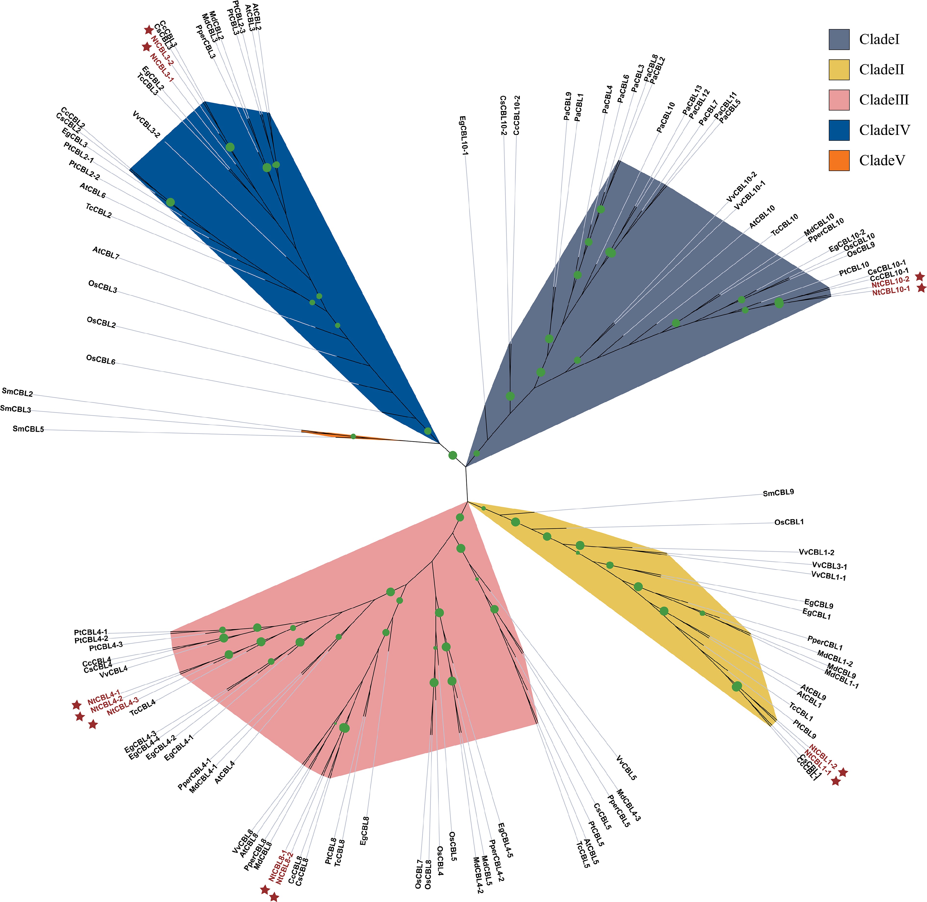
      Figure 1. 

      The phylogenetic tree shows the relationships between CBL protein sequences from N. tangutorum and 12 other species. Phylogenetic analysis was performed using IQ-TREE software with maximum likelihood (ML) method and subjected to 1,000 bootstrap replicates. The modules are color coded to represent the five subclades of CBLs. NtCBLs are indicated by dark red stars. Green dots of different sizes indicate Bootstraps confidence levels above 80.

      To investigate the structural relationships among the 11 NtCBLs, we performed a genomic collinearity analysis using JCVI. Seven collinear CBL pairs were identified in the N. tangutorum genome: NtCBL1-1 and NtCBL1-2, NtCBL3-1 and NtCBL3-2, NtCBL8-1 and NtCBL8-2, NtCBL10-1 and NtCBL10-2, NtCBL4-1 and NtCBL4-2, NtCBL4-1 and NtCBL4-3, and NtCBL4-2 and NtCBL4-3 (Fig. 2a). Their chromosomal distribution and sequence similarity suggest that most NtCBL gene pairs were generated through whole genome duplication/polyploidization, while some NtCBLs, such as NtCBL4-1 and NtCBL4-2, appear to have undergone tandem duplication.
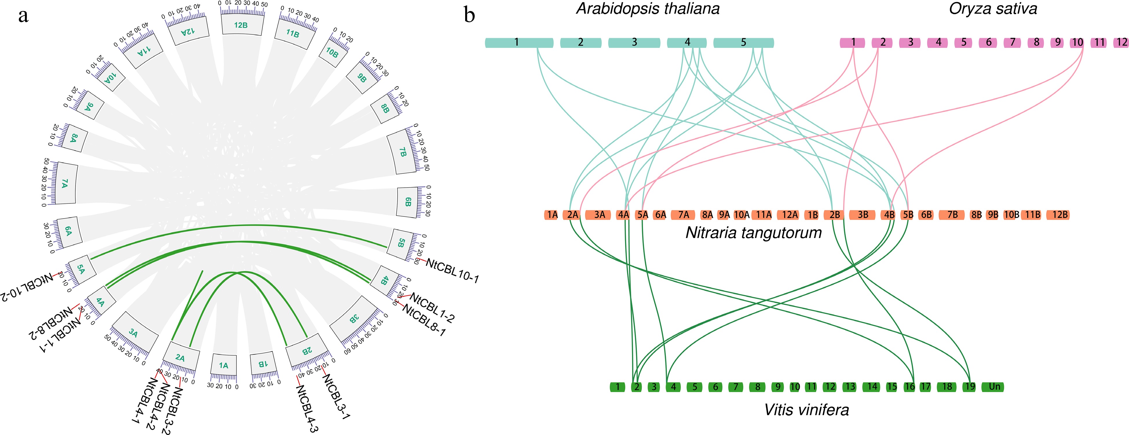
      Figure 2. 

      Genome-wide synteny analysis of CBL gene family among N. tangutorum and three other species. (a) inter-chromosomal relationships of NtCBLs (the links on the green curve indicate synteny relationships between genes). (b) Synteny analyses between the CBLs of N. tangutorum, Arabidopsis, Vitis vinifera, and Oryza sativa, the links between species indicate homologous relationships between genes.

      We conducted Ka/Ks calculations for the gene pairs, uncovering that, aside from NtCBL4-1 and NtCBL4-2, which displayed a Ks value of zero, the Ka/Ks values for the remaining gene pairs were notably below 1 (Supplemental Table S4). This observation suggests that these gene pairs underwent evolutionary purifying selection, emphasizing a constrained evolutionary process. Collinearity between genes from different species often indicates functional similarities. In light of this, we performed a collinear analysis of CBL genes across Arabidopsis, rice, grape, and N. tangutorum. The outcome indicated the existence of 12, 6, and 10 collinear relationships between NtCBLs and Arabidopsis, rice, and grape, respectively (Fig. 2b). This suggests that NtCBLs may share a close evolutionary relationship with Arabidopsis and grape, while showing a relatively distant relationship with rice.

    • To obtain a more comprehensive understanding of the gene structure of CBL genes in N. tangutorum, we conducted an analysis of their gene structures. We observed that all CBL genes exhibited intron structures, with gene lengths spanning from 3,667 to 6,098 base pairs and coding sequence (CDS) lengths ranging from 8 to 11 exons. Notably, a lack of a 5' UTR region was observed in NtCBL8-1, while NtCBL10-2 lacked a 3' UTR region. Conversely, typical 5' UTR and 3' UTR structures were present in the remaining genes (Fig. 3).
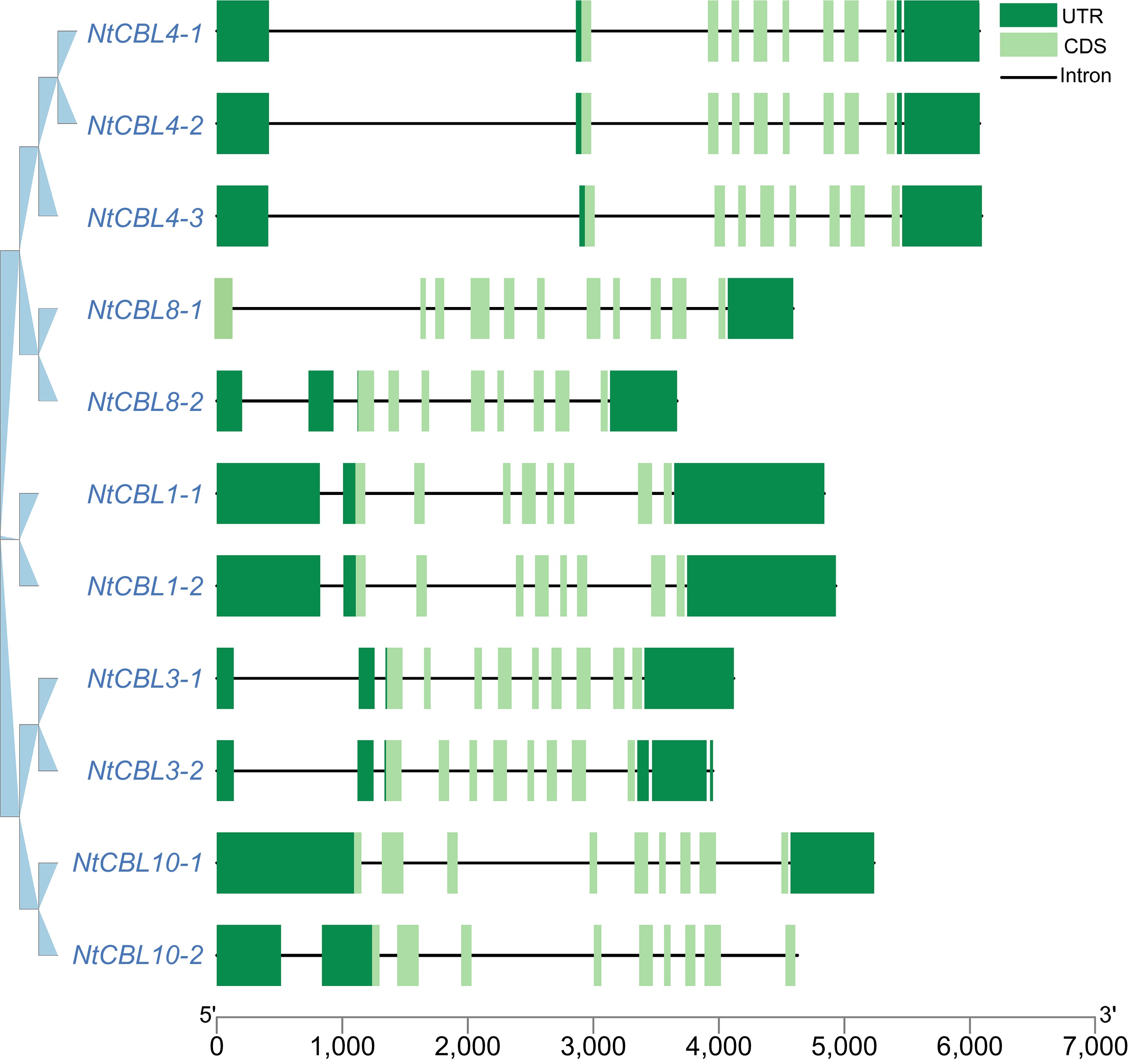
      Figure 3. 

      Gene structure of the CBL gene family in N. tangutorum. The dark green boxes represent the UTR (Untranslated Region), the light green boxes represent the CDS (gene coding region), and the black lines represent the intron region.

      Simultaneously, the pattern distribution within NtCBL proteins was investigated. As shown in Fig. 4, eight major motifs were identified among NtCBLs. Notably, motifs 1, 2, and 6 exhibited the highest degree of conservation of all CBLs. However, it is worth noting that motif 3 was absent in NtCBL4-1. In addition, pattern alterations were observed in four gene pairs that underwent fragment duplication, such as NtCBL8-1 and NtCBL8-2, as well as NtCBL10-1 and NtCBL10-2. Collectively, these findings suggest that functional differentiation may have occurred in NtCBLs during evolution.
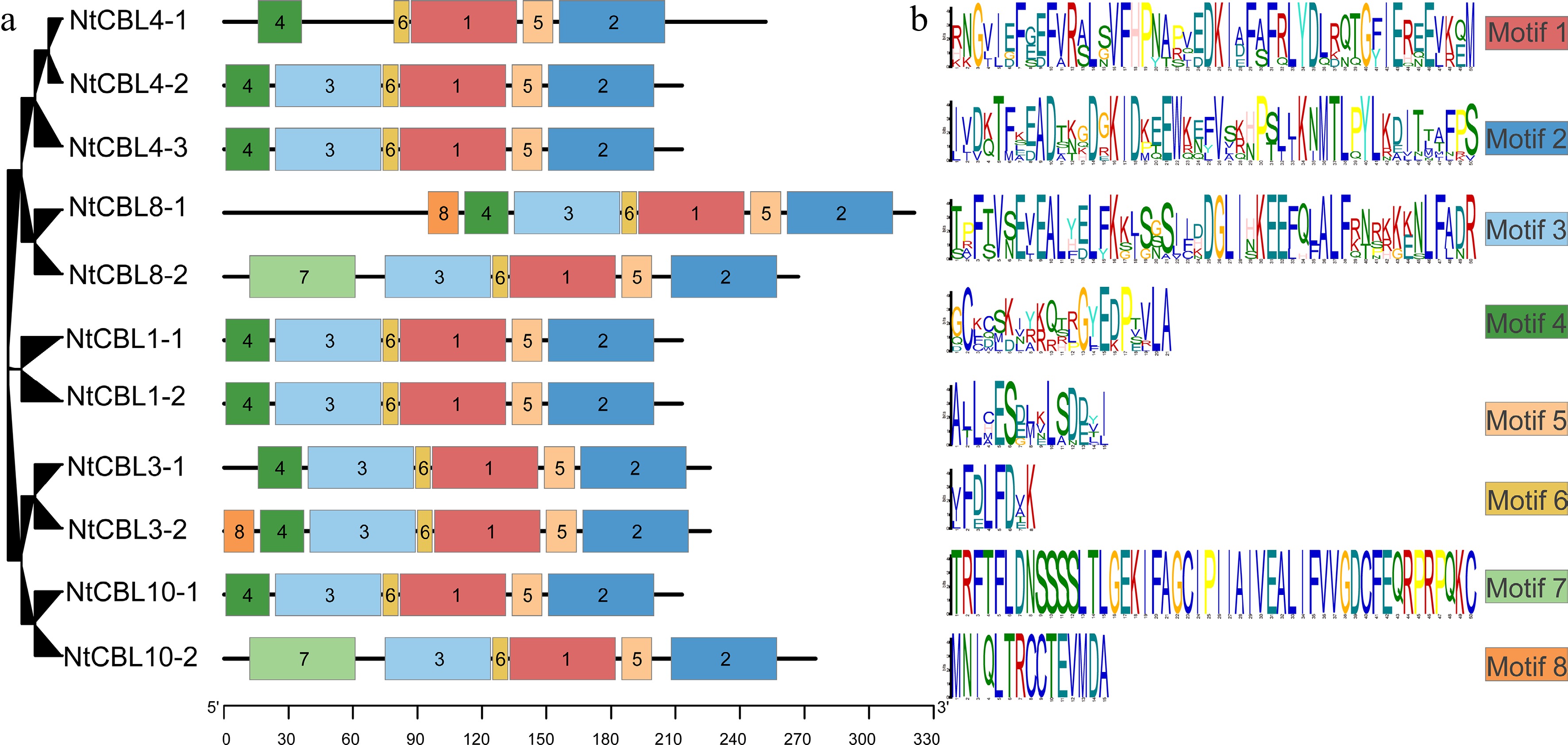
      Figure 4. 

      Conservative motifs distribution in N. tangutorum. (a) Distribution of different motifs on NtCBL genes. (b) Specific sequence information of different motifs, with larger letters indicating higher conservation.

    • Promoter cis-acting elements constitute a critical region for transcription initiation, as highlighted by Hernandez-Garcia & Finer[25]. Analysis of these elements holds significant value in unraveling the potential functions of genes. To investigate plausible biological roles of the N. tangutorum CBL gene family. We examined the sequence located 3 kb upstream of the NtCBL gene initiation codon for cis-acting element analysis.

      This analysis unveiled the existence of a multitude of cis-regulatory elements intricately associated with hormone and stress responses. Specifically, cis-acting elements associated with abscisic acid responsiveness, auxin-responsive elements, methyl jasmonate (MeJA) responsiveness, gibberellin responsiveness, and salicylic acid responsiveness were identified within hormone signaling pathways. In addition, a range of abiotic stress-related elements were identified, including anaerobic induction, defense and stress responsiveness, drought inducibility, and low-temperature responsiveness (Supplemental Fig. S1). Collectively, these findings strongly suggest that CBL genes may indeed participate in biological functions related to these hormone and stress response pathways.

    • Relevant studies have shown that CBLs play a role in drought and salt stress processes in plants[26]. Meanwhile, previous analysis revealed cis-acting elements associated with drought or salt stress response in the NtCBL promoter region. All these findings point to the potential involvement of NtCBLs in drought or salt stress responses in N. tangutorum.

      Therefore, we have devised a set of experiments using qRT-PCR to investigate the expression patterns of NtCBL genes in different tissues and under abiotic salt stress conditions. First, we conducted an analysis of sequence similarity among NtCBLs (Supplemental Table S5). Due to the tetraploid nature of N. tangutorum, the genes within the NtCBL gene family exhibit extremely high similarity (nearly exceeding 95%). This high similarity made it nearly impossible to design specific primers. Therefore, NtCBL1-1 and NtCBL1-2 were combined as NtCBL1 for qRT-PCR, and the same approach was applied to the other genes.

      The expression patterns of the NtCBL gene family in the roots, stems, and leaves of N. tangutorum were initially examined through a comparison of expression results across different tissues. It was observed that the relative expression of all NtCBLs was highest in the stem. NtCBLs could be roughly divided into two groups based on their expression patterns. In the first group, which included NtCBL-1 and NtCBL-10, gene expression was ranked stem > leaf > root. In the second group, which included NtCBL-3, NtCBL-4, and NtCBL-8, gene expression was ranked stem > root > leaf. These different expression patterns of NtCBLs suggest that they may have distinct, tissue-specific functions (Fig. 5).
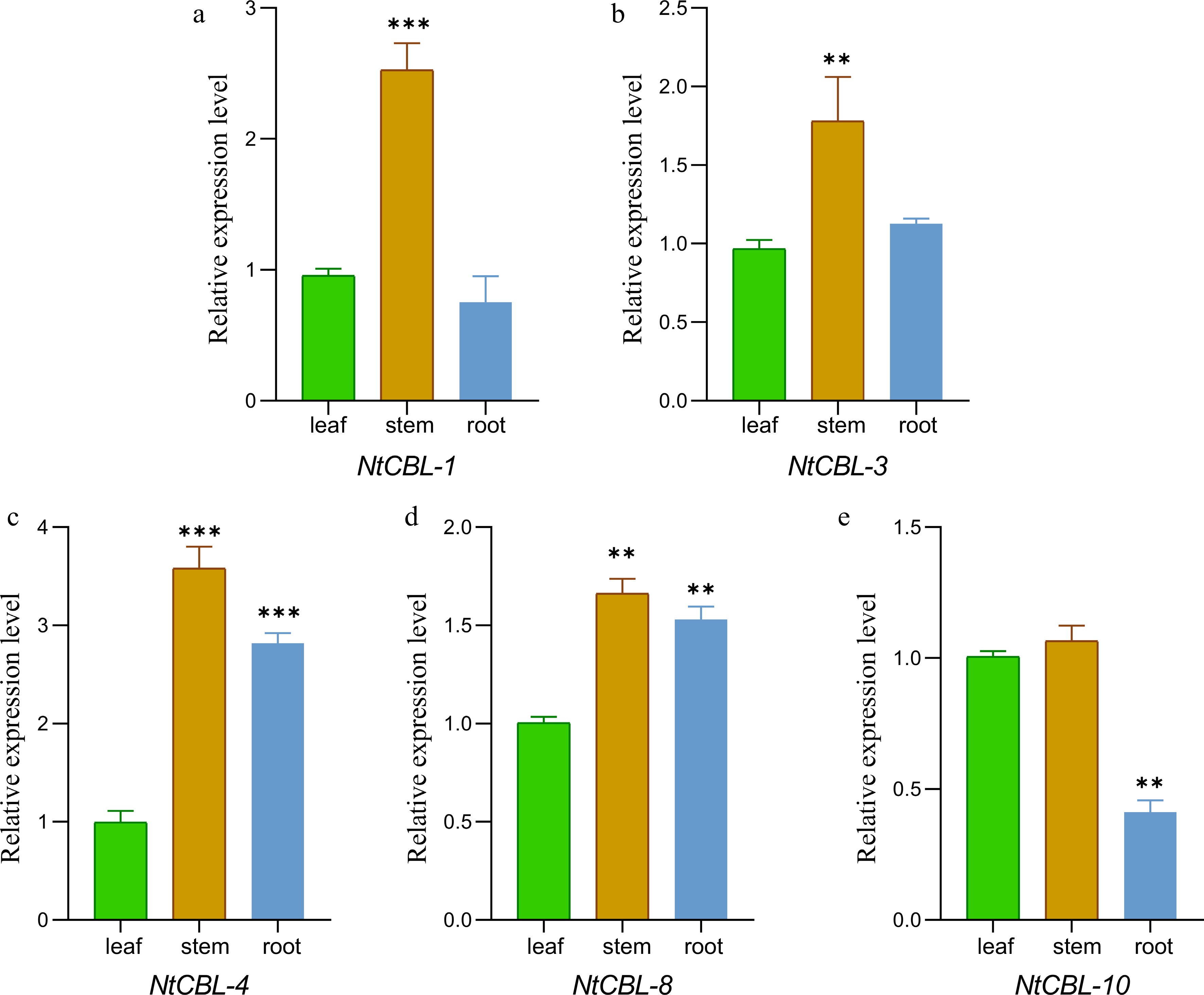
      Figure 5. 

      The gene expression characteristics of N. tangutorum CBL genes in root, stem, and leaf tissues were analyzed by fluorescence quantitative PCR. **p < 0.01, ***p < 0.001 (ANOVA followed by Tukey’s HSD).

      Subsequently, we examined the expression patterns of NtCBL genes under salt and drought stress. The majority of NtCBLs demonstrated distinct responses to these stresses, although the timing and magnitude of the response differed among individual genes (Fig. 6). Under drought stress, the expression of NtCBL1 and NtCBL3 peaked at 8 h, whereas NtCBL4 and NtCBL10 reached their highest expression levels at 1 h, and NtCBL8 showed no significant change throughout the experiment. All NtCBL genes were significantly upregulated under salt stress, with their expression peaking at 1 h of salt stress and subsequently declining. These findings unequivocally illustrate that the expression of NtCBL genes is highly influenced by drought and salt stress, implying that NtCBL proteins likely contribute to N. tangutorum's response to these challenging environmental conditions.
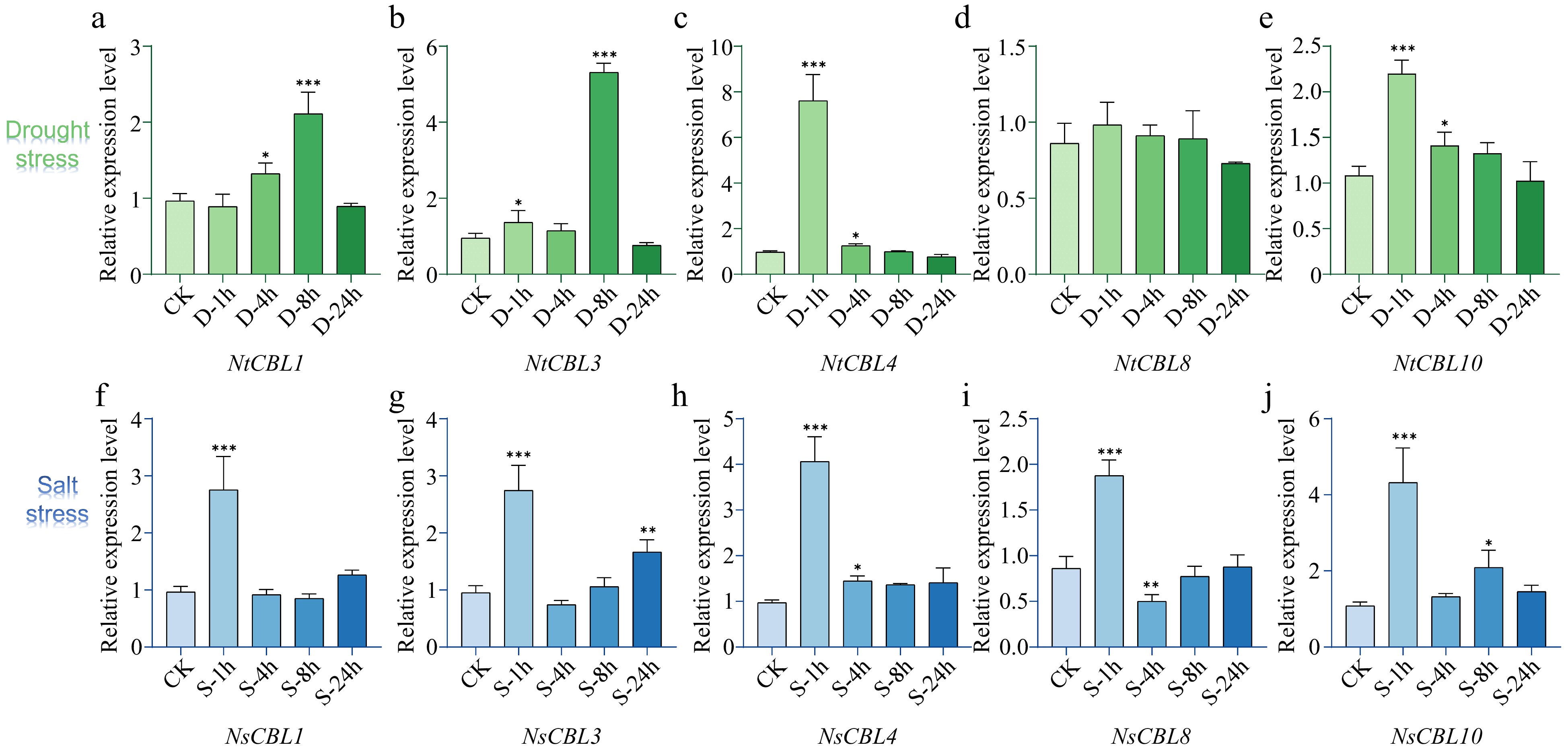
      Figure 6. 

      Expression patterns of NtCBL genes during drought (upper panels) and salt stress (lower panels) in N. tangutorum. 'ck' stands for untreated control, while '1h', '4h', '8h', and '24h' respectively represent different time points of salt or stress treatment, which are 1, 4, 8, and 24 h. **p < 0.01, ***p < 0.001 (ANOVA followed by Tukey’s HSD).

    • With reference to research in other plant species[2729] and response to drought and salt stress in N.tangutorum, we chose NtCBL1 as the representative gene for NtCBL and examined its potential role in enhancing stress resistance through overexpression in Arabidopsis. We designed specific primers only for NtCBL1 (Supplemental Table S5), and confirmed that the cloned gene was NtCBL1-1 by first-generation sequencing comparison, then constructed the 35S:NtCBL1-1 overexpression vector and transformed it into wild-type Arabidopsis via Agrobacterium-mediated transformation to obtain positive transgenic plants. We obtained multiple transgenic positive lines (Supplemental Fig. S2) and randomly selected eight of these lines for analysis of their relative expression levels. We observed variations in the expression levels among these lines (Supplemental Fig. S3). Based on their expression levels, we specifically chose lines 1, 4, and 6 and renamed them as lines 1, 2, and 3, respectively, for subsequent functional validation of NtCBL1-1.

      Previous studies have shown that stress can significantly affect seed germination rates[30]. To determine whether heterologous overexpression of NtCBL1-1 affected Arabidopsis germination, we sowed seeds of wild-type Arabidopsis and three independent transgenic lines on ½ MS medium containing 0 or 150 mM NaCl or 300 mM mannitol, and we observed their germination rate after 7 d. Under normal growing conditions, wild-type and transgenic lines germinated rapidly, and their germination rates were similar (Fig. 7). Under 150 mM salt stress, the germination rate of wild-type Arabidopsis was reduced, and the three overexpression lines showed higher germination than wild Arabidopsis. Under drought stress, both wild-type and transgenic lines showed reduced germination compared to control conditions, but the germination rate of transgenic lines was significantly higher than that of wild-type lines. Thus, overexpression of NtCBL1-1 in Arabidopsis ameliorated—at least in part—the inhibition of germination caused by salt and drought stress.
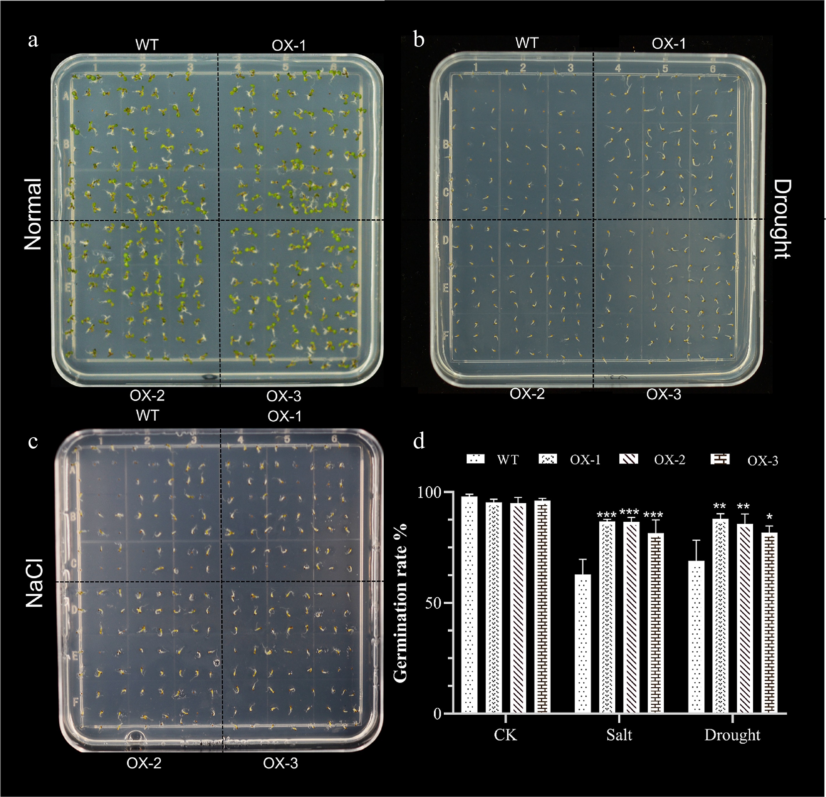
      Figure 7. 

      Heterologous expression of NtCBL1-1 increases Arabidopsis germination rates under salt and drought stress. (a)−(c) Phenotypic charts of germination rates under normal growth conditions, 150 mM NaCl treatment, and 300 mM mannitol treatment, with about 70 seeds per dish and three replicates for each experiment. (d) Germination rate statistics of Arabidopsis overexpressing NtCBL1-1 under normal growth conditions, 150 mM NaCl treatment, and 300 mM mannitol treatment. *p < 0.05, **p < 0.01, ***p < 0.001 (ANOVA followed by Tukey’s HSD).

    • To assess whether the heterologous expression of NtCBL1-1 impacts the salt and drought tolerance of Arabidopsis, we subjected soil-grown seedlings to irrigation with 200 mM NaCl or 300 mM mannitol for a duration of 7 d. On the first day of salt stress, leaves of wild-type Arabidopsis began to show slight wilting, but transgenic plants overexpressing NtCBL1-1 showed no visible changes (Fig. 8a, Supplemental Fig. S4). On day 3, leaf yellowing became visible on wild-type Arabidopsis, and the transgenic plants began to wilt. On day 5, leaves of wild-type Arabidopsis were severely wilted and showed large areas of yellowing, whereas leaves of the transgenic plants had begun to turn yellow. On day 7, wild-type plants were completely withered, but only some leaves of the transgenic plants were withered and yellow.
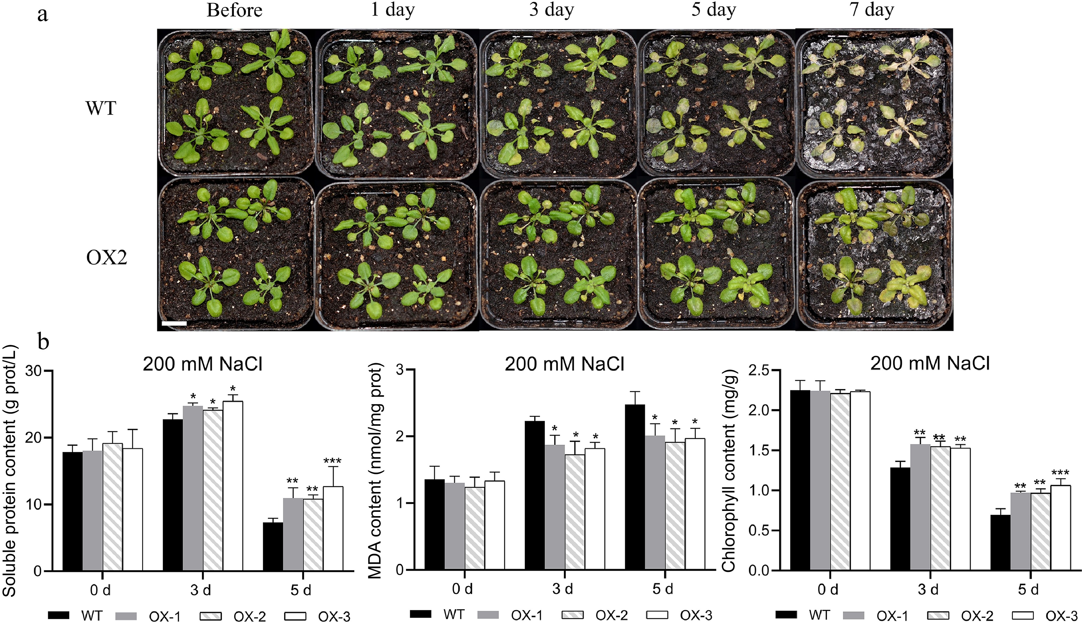
      Figure 8. 

      Heterologous overexpression of NtCBL1-1 increased salt tolerance in Arabidopsis. (a) Wild-type Arabidopsis phenotypes and overexpression lines exposed to 200 mM NaCl for 0–7 d. (b) Chlorophyll content, soluble protein content, and MDA content after 0–5 d of exposure to 200 mM NaCl. Statistical significance denoted as *p < 0.05, **p < 0.01, ***p < 0.001 (ANOVA followed by Tukey’s HSD).

      Since the wild-type plants had nearly died by the 7th day of the salt stress treatment, we conducted physiological measurements exclusively on plants harvested on days 0, 3, and 5. Before salt stress treatment, there were no significant differences in chlorophyll content between wild-type and transgenic lines. Chlorophyll content decreased as the duration of salt stress increased in both wild-type and transgenic Arabidopsis, but chlorophyll content was significantly higher in transgenic lines than in the wild type. This difference was most striking on day 5 (Fig. 8b). Soluble protein content showed a trend similar to that of chlorophyll content; it was lower in the wild type than in the transgenic lines under salt stress. Although all genotypes showed accumulation of MDA during the stress treatment, MDA content was significantly lower in the transgenic lines.

      The transgenic lines also showed less severe stress symptoms than the wild type in response to simulated drought (Fig. 9a, Supplemental Fig. S5). In contrast to the salt stress results, there were no significant differences in MDA or soluble protein content on day 3 of drought stress. However, on day 5, the MDA content was significantly higher in the wild type than in the transgenic lines, and the soluble protein content was significantly lower (Fig. 9b). Thus, overexpression of NtCBL1-1 also improved drought stress tolerance of transgenic Arabidopsis.
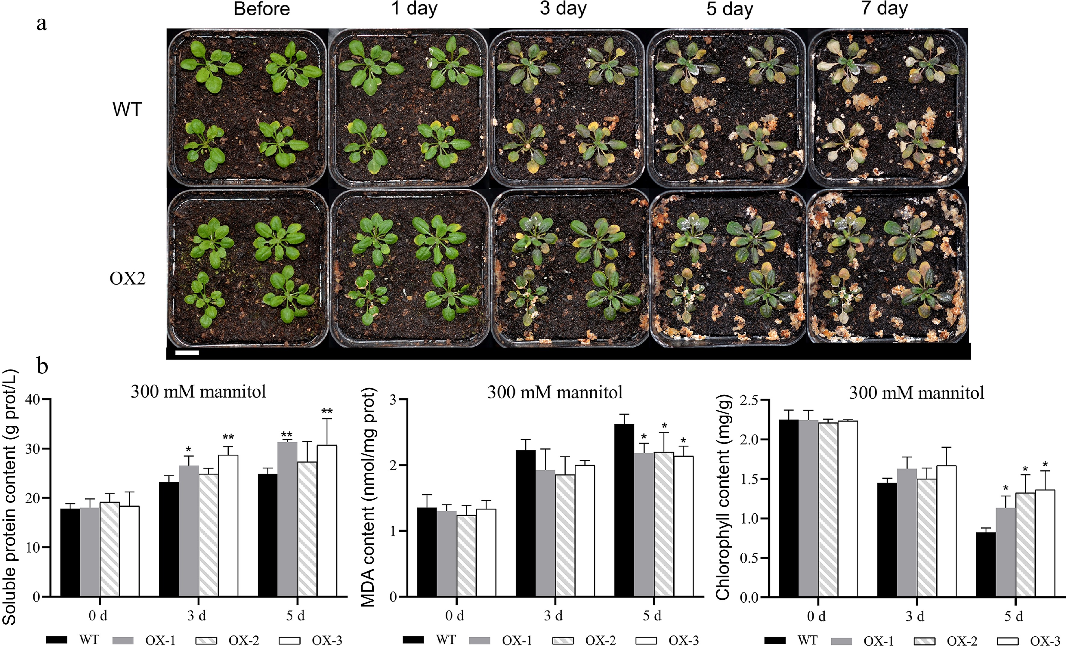
      Figure 9. 

      Heterologous overexpression of NtCBL1-1 in Arabidopsis increased drought stress tolerance. (a) Phenotypic comparison between wild-type Arabidopsis and overexpression lines under 300 mM mannitol exposure for 0–7 d. (b) Chlorophyll content, soluble protein content, and MDA content of wild-type Arabidopsis and overexpression lines exposed to 300 mM mannitol for 0–5 d. Statistical significance denoted as *p < 0.05, **p < 0.01 (ANOVA followed by Tukey’s HSD).

    • Deterioration of the soil environment significantly limits plant growth and development, and soil salinization is becoming an increasingly serious threat to forestry and agricultural production. As of 2021, about one billion hectares of land worldwide have been affected by soil salinization, accounting for about 7% of the Earth's total land area[31]. Excessive salinization impairs the normal growth of most plants, but halophytes can typically complete their entire life cycle under such conditions. Studying the adaptive mechanisms that enable halophytes to thrive under saline-alkali conditions is therefore important for the utilization and restoration of saline-alkali land.

      As important Ca2+ sensors, CBLs have key roles in plant perception and response to a variety of abiotic stresses, including drought[32], salinity[33], and cold[34], as well as the stress hormone ABA[35]. CBLs have been studied in a number plants, and 10, 9, and 7 CBL genes have been identified and characterized in Arabidopsis[4], Saccharum spontaneum[36], and Triticum aestivum[37], respectively. However, the number of CBL genes and their functions in the halophyte N. tangutorum have not previously been reported. In this study, we identified 11 CBL genes in the N. tangutorum genome and classified them into four clades on the basis of their phylogenetic relationships. Each clade showed general conservation of gene structure and protein domain composition, although there were some differences (Fig. 4) that may reflect functional differentiation of the NtCBLs.

      Plant polyploidization refers to the increase in the number of chromosomes within plant cells, often manifested as the duplication of entire sets of chromosomes[38]. Polyploidization leads to the presence of multiple identical or highly similar gene copies within the genome. These duplicated copies of genes undergo mutations during evolution and accumulate new features, driving the expansion of gene families. For instance, in the polyploid crop Brassica napus, the number of FBA gene family members is higher than in diploid plants such as Arabidopsis and rice[39]. Additionally, the MADS-box gene count in tetraploid Gossypium hirsutum was significantly higher than diploid Gossypium hirsutum[40].

      Plant polyploidization also leads to gene redundancy and enhanced stability[41]. In N. tangutorum, all CBL genes have homologs in their corresponding subgenomes. That is, except for the NtCBL4 gene, all NtCBL genes exhibit two highly similar gene duplications on corresponding chromosomes in their respective subgenomes. For example, NtCBL1-1 and NtCBL1-2 occur on CHR4A and CHR4B, while NtCBL10-1 and NtCBL10-2 are located on CHR5A and CHR5B, respectively. This duplication probably results from the tetraploidization process in N. tangutorum. In general, polyploidization offers significant benefits to plants, especially in adapting to environmental changes. This may be one reason N. tangutorum adapts to extreme saline-alkaline and drought conditions. Through polyploidization, plants may produce more copies of genes, and these copies could mutate, leading to new traits or functions. This diversity helps plants better adapt to various pressures or environmental conditions and helps them maintain a competitive advantage during their evolutionary process.

      Apart from chromosomal duplication, gene self-replication and differentiation also play significant roles in the expansion and evolution of gene families. Systematic phylogenetic analysis in this study reveals the presence of multiple copies of the CBL4 gene in several non-polyploid plants. For instance, there are three copies in Populus euphratica, five copies in E. grandis, two copies in C. sinensis, and three copies in M. domestica (Fig. 1). Multiple copies of CBL4 have also been observed in other plants, such as two copies in canola[5], and three copies in M. sativa [42]. Generally, gene redundancy is critical to maintaining essential biological functions in certain plants and may play a pivotal role in their adaptability and survival. Expansion of the CBL4 subfamily may be closely related to this phenomenon. In N. tangutorum, despite being a polyploid plant, the CBL4 gene had undergone replication prior to polyploidization, resulting in the formation of NtCBL4-1 and NtCBL4-2, suggesting a potentially significant role for CBL4 in the life processes of N. tangutorum.

      The cis-acting elements in the promoter region can regulate gene expression, thereby revealing internal information about gene function[43]. Analysis of the NtCBL promoter reveals various cis-acting elements associated with hormone regulation and defense responses. For instance, within the NtCBL gene promoter region, there are abscisic acid (ABA) responsive elements known as ABREs (Fig. 5). In the promoter regions of CBL genes in other species such as pepper, quinoa, and Vitis vinifera, we have found ABRE elements, indicating the relative conservation of ABRE elements in the CBL gene promoter regions. When plants face stress like drought, their water levels decrease, leading to the production of ABA. The ABA signaling pathway is activated, triggering a series of physiological and biochemical responses to help plants adapt to drought stress[44]. The varying responses of CBL genes to drought in pepper[45], quinoa[46], Vitis vinifera [6], and N. tangutorum might be associated with ABRE elements.

      Genes typically display patterns of tissue-specific expression. In our study, we observed predominant expression of CBL genes in stem and root, similar to the expression pattern of CBL in rice. However, in rice, CBL4 exhibits major expression in leaves[47], while in N. tangutorum, NtCBL4 exhibits the least expression in leaves. In cotton research, the expression of GhCBL4-5 was found to be higher in stem and root compared to leaves[48], which is somewhat similar to the expression pattern of NtCBL4 in this study. Despite differences in the expression patterns of CBL genes across species and tissues, they exhibit notable responses to stressors such as drought, salinity, and other adverse conditions. This high degree of conservancy in their response to environmental stressors might indicate the pivotal role CBL genes play in physiological regulation and stress responses in plants. Wide adaptability suggests the universality and significance of CBL genes and their regulated signaling pathways across different species.

      Currently, multiple genes within the CBL family have been cloned and their function under non-biological stressors has been validated[4951]. Among these genes, CBL1 is one of the most extensively studied representatives within the CBL gene family. CBL1 has been studied in many plant species, including Arabidopsis[33] and Brassica napus[27], but its function in N. tangutorum remains to be fully characterized. Here, we selected NtCBL1-1 as a representative of the NtCBL gene family and performed a preliminary functional characterization by overexpression in Arabidopsis. Although the sequences of NtCBL1-1 and NtCBL1-2 were highly similar, we were able to design specific primers for cloning NtCBL1-1, and we confirmed the identity of the cloned gene by Sanger sequencing. We then used this gene to construct an overexpression vector for Arabidopsis transformation.

      Previous reports on CBL1 in other species, as well as our own qRT-PCR results, led us to speculate that NtCBL1-1 might participate in responses to salt and drought stress. Although both stresses impaired seed germination, seeds of the NtCBL1-1 overexpression lines had a higher germination rate than those of the wild type under stressed conditions (Fig. 8). Likewise, the transgenic plants showed less severe symptoms in response to salt stress and simulated drought. Leaves of the wild type showed severe wilting and yellowing under both stresses, whereas those of transgenic plants were only slightly yellow, and this difference became more obvious with increasing stress duration.

      Studies have shown that plant stress can lead to chlorophyll degradation[52], and plants with greater stress resistance tend to have higher soluble protein content[53]. MDA is a lipid peroxidation product whose content can serve as a measure of oxidative damage[54].

      Overexpression of stress-resistant genes may affect plant physiological characteristics, such as soluble protein content, malondialdehyde (MDA) levels, and chlorophyll content. These physiological traits are often associated with a plant's ability to withstand stress and its mechanisms for coping with environmental pressures. Over-expression of stress-resistant genes typically triggers the activation of signaling pathways, leading to a series of biochemical responses in plants. This results in the production of more stress-resistant proteins and metabolites, including soluble proteins and antioxidant enzymes. This process helps alleviate oxidative stress, enhances the plant's resistance to free radicals and oxidative damage, thereby reducing the accumulation of oxidative byproducts such as soluble proteins and MDA. Chlorophyll, a key molecule in photosynthesis, plays an important role in protecting plants from environmental stressors. Overexpression of stress-resistant genes can reduce the damage to chlorophyll caused by stress factors, helping maintain photosynthetic efficiency and normal plant growth.

      For example, researchers have found that overexpression of the stress-resistant gene HvPIP2;5 in transgenic Arabidopsis leads to retention of more chlorophyll and a reduction in MDA accumulation[55]. In another study, overexpression of the superoxide dismutase gene reduced the accumulation of soluble proteins in alfalfa leaf tissues[56].

      These findings suggest that by overexpressing stress-resistant genes, plants can better cope with environmental stress, reduce oxidative damage, and reduce the accumulation of oxidative byproducts, thus maintaining their normal growth and survival.

      In this study, transgenic Arabidopsis overexpressing NtCBL1-1 contained more chlorophyll and soluble protein and less MDA than the wild type under salt stress (Fig. 8), indicating that they had higher salt tolerance. Responses to simulated drought stress were similar: Arabidopsis overexpressing NtCBL1-1 showed greater drought tolerance than the wild type (Fig. 9). Thus, overexpression of CBL1-1 from N. tangutorum improved the abiotic stress tolerance of transgenic Arabidopsis. The calcium receptor encoded by NtCBL1-1 may therefore form part of the signal transduction pathways that underlie the high salt tolerance of this dryland halophyte. Overexpression of the NtCBL1-1 may have activated multiple physiological pathways in Arabidopsis, aiding the plant in better coping with environmental stress, alleviating oxidative stress, protecting chlorophyll, and maintaining water balance. These physiological changes enhance the stress resistance of the transgenic Arabidopsis.

      This study revealed that the overexpression of the NtCBL1-1 in Arabidopsis led to enhanced phenotypes, germination rates, and chlorophyll content under drought stress compared to wild-type Arabidopsis. However, this is just the tip of the iceberg, as we still need a more comprehensive understanding of the underlying regulatory networks. In parallel, we are actively engaged in the development of a transgenic system specifically tailored for N. tangutorum. We eagerly anticipate conducting in-depth mechanistic inquiries through self-overexpression analysis. Moreover, the translocation of CBL into N. tangutorum holds the promise of yielding more robust salt-tolerant halophytes. This prospective outcome could offer valuable insights for future biotechnology-driven advancements in forestry breeding and ecological restoration.

    • In this study, we identified CBL family members in N. tangutorum and characterized their basic physicochemical properties, phylogeny, and responses to stress. The results indicate that there are a total of 11 NtCBL genes distributed across six chromosomes in N. tangutorum. Expression analysis reveals that these genes are highly responsive to both salt and drought stress. Functional studies of NtCBL1-1 demonstrate that transgenic Arabidopsis plants overexpressing NtCBL1-1 exhibit enhanced tolerance to both drought and salt stress. Under drought and salt stress conditions, compared to wild-type Arabidopsis, transgenic plants show increased germination rates, slower chlorophyll degradation, higher accumulation of soluble proteins, and reduced levels of the oxidative stress marker malondialdehyde. These findings underscore the significant role of NtCBL1-1 in responding to drought and salt stress and provide insight into the evolution and biological functions of the CBL gene family in N. tangutorum, providing a theoretical foundation for further study of abiotic stress resistance mechanisms in this desert halophyte.

    • The authors confirm contribution to the paper as follows: study conception and design: Chen J, Cheng T; data collection: Wu J, Li M, Zhang J; analysis and interpretation of results: Zhu L, Fang H, Chen Y; draft manuscript preparation: Zhu L, Li M. All authors reviewed the results and approved the final version of the manuscript.

    • All data generated or analyzed during this study are included in this article and its supplementary table files. The Gene sequence, CDS sequence, and GFF annotation information of all NtCBL gene families are included in the supplementary files.

      • This work was supported by the Natural Science Foundation of China (No. 31770715) and Priority Academic Program Development of Jiangsu Higher Education Institutions (PAPD).

      • The authors declare that they have no conflict of interest.

      • Supplemental Table S1 Primers for Quantitative RT-PCR in N.tangutorum.
      • Supplemental Table S2 Primers for isolation of NtCBL1-1 fragment and construction of overexpression vector.
      • Supplemental Table S3 CBL genes used to construct phylogenetic trees from 12 species except N.tangutorum.
      • Supplemental Table S4 KaKs analysis in N.tangutorum.
      • Supplemental Table S5 Identity between CBLs gene in Nitraria tangutorum.
      • Supplemental Fig. S1 Cis-regulatory elements analysis of NtCBLs. (A) A heat map showing the number of cis-acting elements related to stress response in NtCBLs. (B) The distribution of cis-acting elements in the promoter region (ATG upstream) of NtCBLs, where different boxes represent different cis-acting elements.
      • Supplemental Fig. S2 Positive identification of partial NtCBL1-1 transgenic T3 generation; different lanes represent different transgenic lines.
      • Supplemental Fig. S3 Relative expression levels in partial transgenic T3 generations, with wild-type Arabidopsis as a reference.
      • Supplemental Fig. S4 Phenotypic changes in overexpression lines 1 and 3 following exposure to 200 mM NaCl for 0−7 days.
      • Supplemental Fig. S5 Phenotypic changes in overexpression lines 1 and 3 following exposure to 300 mM mannitol for 0−7 days.
      • Supplemental file S1 Genome annotations.
      • Copyright: © 2023 by the author(s). Published by Maximum Academic Press, Fayetteville, GA. This article is an open access article distributed under Creative Commons Attribution License (CC BY 4.0), visit https://creativecommons.org/licenses/by/4.0/.
    Figure (9)  Table (1) References (56)
  • About this article
    Cite this article
    Zhu L, Wu J, Li M, Fang H, Zhang J, et al. 2023. Genome-wide discovery of CBL genes in Nitraria tangutorum Bobr. and functional analysis of NtCBL1-1 under drought and salt stress. Forestry Research 3:28 doi: 10.48130/FR-2023-0028
    Zhu L, Wu J, Li M, Fang H, Zhang J, et al. 2023. Genome-wide discovery of CBL genes in Nitraria tangutorum Bobr. and functional analysis of NtCBL1-1 under drought and salt stress. Forestry Research 3:28 doi: 10.48130/FR-2023-0028

Catalog

  • About this article

/

DownLoad:  Full-Size Img  PowerPoint
Return
Return