ARTICLE   Open Access    

Effect of drought stress on dieback disease development under Lasiodiplodia theobromae infection in cocoa clone 'MCC 02'

More Information
  • Cocoa is spread over tropical countries, being extensively cultivated by mostly smallholders and processed by industries as the raw material of chocolate. Pathogens and drought are one of the biotic and abiotic factors limiting the productivity of cocoa. The main objective of this study was to evaluate the pathogenicity of Lasiodiplodia theobromae in drought-stressed and well-watered cocoa clone MCC 02. MCC 02, a popular cocoa clone in Sulawesi (Indonesia), was evaluated in the greenhouse for infection of L. theobromae under water-stressed and well-watered conditions. Dieback, leaf chlorotic and necrotic, scion survival, and vascular streaking were determined. The results indicated that the treated cocoa seedlings exposed to water stress corresponding to 25% field capacity during both inoculation of the pathogen simultaneously with the initiation of water stress or seven days after water stress commenced were more susceptible to L. theobromae than well-watered ones. However, this effect was mainly relevant when the pathogen was inoculated through a wound on the stem. Moreover, the severity of the disease on inoculation of L. theobromae simultaneously with the initiation of water stress was higher than that of the disease on inoculation seven days after water stress commenced but not significantly different. This study demonstrates the potential threat of drought stress to cocoa plants under the infestation of L. theobromae and emphasizes the significant effect of water stress in interaction with L. theobromae that should be considered in plant management, especially under the climate change scenario in Sulawesi, in which the drought will increase and last longer.
  • Salvia rosmarinus L. (old name Rosmarinus officinalis), common name Rosemary thrives well in dry regions, hills and low mountains, calcareous, shale, clay, and rocky substrates[1]. Salvia rosmarinus used since ancient times in traditional medicine is justified by its antiseptic, antimicrobial, anti-inflammatory, antioxidant, and antitumorigenic activity[1,2]. The main objective of the study is to evaluate the antimicrobial activity of different extracts of Salvia rosmarinus in vitro, and its compounds related to in silico targeting of enzymes involved in cervical cancer. Since the start of the 20th century, some studies have shown that microbial infections can cause cervical cancers worldwide, infections are linked to about 15% to 20% of cancers[3]. More recently, infections with certain viruses like Human papillomaviruses (HPV) and Human immunodeficiency virus (HIV), bacteria like Chlamydia trachomatis, and parasites like schistosomiasis have been recognized as risk factors for cancer in humans[3]. Then again, cancer cells are a group of diseases characterized by uncontrolled growth and spread of abnormal cells. Many things are known to increase the risk of cancer, including dietary factors, certain infections, lack of physical activity, obesity, and environmental pollutants[4]. Some studies have found that unbalanced common flora Lactobacillus bacteria around the reproductive organ of females increases the growth of yeast species (like Candida albicans) and some studies have found that women whose blood tests showed past or current Chlamydia trachomatis infection may be at greater risk of cervical cancer. It could therefore be that human papillomavirus (HPV) promotes cervical cancer growth[3]. Salvia rosmarinus is traditionally a healer chosen as a muscle relaxant and treatment for cutaneous allergy, tumors, increases digestion, and the ability to treat depressive behavior; mothers wash their bodies to remove bacterial and fungal infections, promote hair growth, and fight bad smells[5] .

    The study of plant-based chemicals, known as phytochemicals, in medicinal plants is gaining popularity due to their numerous pharmacological effects[6] against drug resistance pathogens and cancers. The causes of drug resistance to bacteria, fungi, and cancer are diverse, complex, and only partially understood. The factors may act together to initiate or promote infections and carcinogenesis in the human body is the leading cause of death[7]. Antimicrobial medicines are the cornerstone of modern medicine. The emergence and spread of drug-resistant pathogens like bacteria and fungi threaten our ability to treat common infections and to perform life-saving procedures including cancer chemotherapy and cesarean sections, hip replacements, organ transplantation, and other surgeries[7]. On the other hand, information about the current magnitude of the burden of bacterial and fungal drug resistance, trends in different parts of the world, and the leading pathogen–drug combinations contributing to the microbial burden is crucial. If left unchecked, the spread of drug resistance could make many microbial pathogens much more lethal in the future than they are today. In addition to these, cancers can affect almost any part of the body and have many anatomies and molecular subtypes that each require specific management strategies to avoid or inhibit them. There are more than 200 different types of cancer that have been detected. The world's most common cancers affecting men are lung, prostate, colorectal, stomach, and liver cancers[8]. While breast, cervix, colorectal, lung, and stomach cancers are the most commonly diagnosed among women[8]. Although some cancers said to be preventable they seem to still be one of the causes of death to humans, for example cervical cancer. The need to fill the gap to overcome the problem of searching for antimicrobials and anticancers from one source of Salvia rosmarinus is of importance.

    Cervical cancer is a common cancer in women and a prominent cause of death[9]. In Ethiopia, cervical cancer is a big deal for women aged 15 to 44, coming in as the second most common cancer[9]. Globally, it's the fourth most common prevalent disease for women[10]. Aberrant methylation of tumor-suppressor genes' promoters can shut down their important functions and play a big role in causing cervical tumors[10]. There are various cervical cancer repressor genes (proteins turn off or reduce gene expression from the affected gene), such as CCNA1, CHF, HIT, PAX1, PTEN, SFRP4, and TSC1. The genes play a crucial role in causing cervical cancer by regulating transcription and expression through promoter hypermethylation, leading to precursor lesions during cervical development and malignant transformation[11]. The process of DNA methylation is primarily carried out by a group of enzymes known as DNA methyltransferases (DNMT1). It has been reported that DNMT1 (PDB ID: 4WXX), a protein responsible for DNA methylation can contribute to the development of cervical cancer. DNMT1 inhibits the transcription of tumor suppressor genes, facilitating tumorigenesis, which finally develops into cervical cancer. Tumor suppressor gene transcription is inhibited by DNMT1, which helps cancer grow and eventually leads to cervical cancer. Repressive genes' hypermethylation may be decreased, their expression can be increased, and the phenotype of malignant tumors can be reversed by inhibiting the DNMT1 enzyme.

    On the other hand, infection by the human papilloma virus (HPV) phenotype 16, enzyme 6 (PDB ID: 4XR8) has been correlated with a greatly increased risk of cervical cancer worldwide[12]. Based on variations in the nucleotide sequences of the virus genome, over 100 distinct varieties of the human papilloma virus (HPV) have been identified (e.g. type 1, 2 etc.). Genital warts can result from certain types 6 and 11 of sexually transmitted HPVs. Other HPV strains, still, that can infect the genitalia, do not show any symptoms of infection[8]. Persistent infection with a subset of approximately 13 so-called 'high-risk' sexually transmitted HPVs, including such as types 16, 18, 31, 33, 35, 39, 45, 51, 52, 56, 58, 59, and 68 different from the ones that cause warts may lead to the development of cervical intraepithelial neoplasia (CIN), vulvar intraepithelial neoplasia (VIN), penile intraepithelial neoplasia (PIN), and/or anal intraepithelial neoplasia (AIN). These are precancerous lesions and can progress to invasive cancer. Almost all occurrences of cervical cancer have HPV infection as a required component[13]. Superfluous infection by HPV type 16 E6 (PDB ID: 4XR8) has been correlated with a greatly increased genital risk of precursor cervical cancer worldwide[11]. Scholars more defined in major biochemical and biological activities of HPV type 16 E6 (PDB ID: 4XR8) in high-risk HPV oncogenes and how they may work together in the development of cervical disease and cancer[13].

    One potential approach to treat cervical cancer is to inhibit the activity of the DNMT1 and HPV type 16 E6 enzymes specifically[1316]. Over 50% of clinical drug forms worldwide originate from plant compounds[17]. In the past, developing new drugs was a lengthy and costly process. However, with the emergence of bioinformatics, the use of computer-based tools and methods have become increasingly important in drug discovery. One such method is molecular docking and ADMET profiling which involves using the structure of a drug to screen for potential candidates. This approach is known as structure-based drug design and can save both time and resources during the research process[15]. Structural-based drug designing addresses ligand binding sites with a known protein structure[15]. Using free binding energies, a computational method known as docking examines a large number of molecules and suggests structural theories for impeding the target molecule[17]. Nowadays, due to increasing antibiotic resistance like bacteria, fungi, and cancer cells, natural products remain an important source for discovering antimicrobial compounds and novel drugs for anti-cancers like cervical cancers. Therefore, the purpose of this research is to assess the antimicrobial activity of extracts, molecular docking, ADMET profiling in anticancer properties of compounds isolated from Salvia rosmarinus, on a targeting DNMT1 and HPV type 16 E6 in human cervical cancer. In the present study, various solvent crude extracts obtained from Salvia rosmarinus were used for antimicrobial activity and the isolated compounds 1 and 2 were submitted for in silico study to target the DNMT1 and HPV type 16 E6 enzymes to inhibit the growth of human cervical cancer cells.

    Healthy Salvia rosmarinus leaves were collected in Bacho district, Southwest Showa, Oromia, Ethiopia, during the dry season of November 2022. The plant materials were authenticated by Melaku Wondafrish, Natural Science Department, Addis Ababa University and deposited with a voucher number 3/2-2/MD003-80/8060/15 in Addis Ababa University's National Herbarium.

    The most common organic solvent used in extractions of medicinal plants is 2.5 L of petroleum ether, chloroform/methanol (1:1), and methanol. The test culture medium for microbes was used and performed in sterile Petri dishes (100 mm diameter) containing sterile Muller–Hinton Agar medium (25 mL, pH 7) and Sabouraud Dextrose Agar (SDA) for bacteria and fungi, respectively. A sterile Whatman filter paper (No. 1) disc of 6 mm diameter was used to determine which antibiotics an infective organism is sensitive to prescribed by a minimum zone of inhibition (MZI). Ciprofloxacin antibiotic reference (manufactured by Wellona Pharma Ciprofloxacin tablet made in India) and Ketoconazole 2% (made in Bangladesh) were used as a positive controls for antibacterial and antifungal, respectively and Dimethyl sulfoxide (DMSO) 98.9% was used as a negative control for antimicrobial tests. In the present study, the height of the column was 650 mm and the width was 80 mm. Several studies by previous researchers showed the acceptable efficiency of column chromatography (up to 43.0% w/w recovery) in the fractionation and separation of phenolic compounds from plant samples[18]. In column chromatography, the ideal stationary phase used silica gel 60 (0.200 mm) particles. The 1H-NMR spectrums of the compounds were analyzed using a 600 MHz NMR machine and 150 MHz for 13C NMR. The compounds were dissolved in MeOD for compound 1 and in DMSO for compound 2 for NMR analysis. On the other hand, UV spectroscopy (made in China) used 570 nm ultraviolet light to determine the absorbency of flavonoids (mg·g−1) phytochemicals.

    The samples (extracts) were analyzed to detect the presence of certain chemical compounds such as alkaloids (tested using Wagner's reagents), saponins (tested using the froth test), steroids (tested with Liebermann Burchard's tests), terpenoids (tested with Lidaebermann Burchard's tests), quinones, and flavonoids (tested using Shinoda tests)[19].

    The leaves of Salvia rosmarinus (500 g) were successively extracted using maceration using petroleum ether, chloroform/methanol (1:1), and methanol, every one 2.5 L for 72 h to afford 3.6, 6, and 53 g crude extracts, respectively. The methanol/chloroform (1:1) extract (6 g) was loaded to silica gel (150 g) column chromatography using the increasing polarity of petroleum ether, methanol/chloroform (1:1) solvent system to afford 80 fractions (100 mL each). The fraction obtained from chloroform/methanol 1:1 (3:2) after repeated column chromatography yielded compound 1 (18 mg). Fractions 56-65, eluted with chloroform/methanol (1:1) were combined and purified with column chromatography to give compound 2 (10 mg).

    The microorganisms were obtained from the Ethiopia Biodiversity Institution (EBI). Two gram-positive bacteria namely Staphylococcus aureus serotype (ATCC 25923) and Streptococcus epidermidis (ATCC14990); and three gram-negative bacteria, namely Escherichia coli (ATCC 25922), Pseudomonas aeruginosa (ATCC 5702), and Klebsiella pneumonia (ATCC e13883) were inoculated overnight at 37 °C in Muller–Hinton Agar/MHA culture medium and two fungus strains of Candida albicans (ATCC 16404) and Aspergillus niger (ATCC 11414) were inoculated overnight at 27−30 °C in Sabouraud Dextrose Agar/SDA culture medium[20].

    The antibacterial and antifungal activities of different crude extracts obtained from Salvia rosmarinus plant leaves were evaluated by the disk diffusion method (in accordance with the 13th edition of the CLSI M02 document on hardydiagnostics.com/disk-diffusion). Briefly, the test was performed in sterile Petri dishes (100 mm diameter) containing solid and sterile Muller–Hinton Agar medium (25 mL, pH 7) and Sabouraud Dextrose Agar (SDA) for bacteria and fungi, respectively. The extracts were placed on the surface of the media that had previously been injected with a sterile microbial suspension (one microbe per petri dish) after being adsorbed on sterile paper discs (5 μL per Whatman disc of 6 mm diameter). To prevent test samples from eventually evaporating, all Petri dishes were sealed with sterile laboratory films. They were then incubated at 37 °C for 24 h, and the zone diameter of the inhibition was measured and represented in millimeters. Ciprofloxacin antibiotic reference (manufactured by Wellona Pharma Ciprofloxacin tablet, India) was used as a positive control and DMSO was used as a negative control for antibacterial activity test while Ketoconazole 2% (Bangladesh) was used as a positive control and 10 μL of 0.2% agar as a negative control for antifungal activity tests[20]. The term 'inhibitory concentration' refers to the minimum sample concentration required to kill 99.9% of the microorganisms present[21]. Three repetitions of the crude extract sample were used to precisely measure the inhibitory halo diameter (in mm), which was then expressed as mean ± standard deviation to assess the anti-microbial activity.

    Cervical cancer-causing protein was identified through relevant literature. The protein molecule structure of DNA (cytosine-5)-methyltransferase 1 (DNMT1) (PDB ID: 4WXX)[21] and HPV type 16 E6 (PDB ID: 4XR8)[21] - a protein known to cause cervical cancer - were downloaded from the Protein Data Bank[22]. The stability of the protein molecule was assessed using Rampage[23].

    Phytochemical constituents of Salvia rosmarinus plant leaves were used to select a source of secondary metabolites (ligands). Ligand molecules were obtained through plant extraction, and isolation, and realized with PubChem (https://pubchem.ncbi.nlm.nih.gov/). The ligands were downloaded in Silver diamine fluoride format (SDF) and then converted to PDB format using an online SMILES translator (https://cactus.nci.nih.gov/translate/). The downloaded files were in PDB format, which was utilized for running various tools and software[24].

    The Biovia Discovery Studio Visualizer software was used to analyze the protein molecule. The protein molecule was converted into PDB format and its hierarchy was analyzed by selecting ligands and water molecules. Both the protein molecule and the water molecules lost their attached ligands during the analysis. Finally, the protein's crystal structure was saved in a PDB file[25].

    PyRx software was utilized to screen secondary metabolites and identify those ligands with the lowest binding energy to the protein target. The ligands with the lowest binding energy were further screened for their drug-likeliness property through analysis. It is worth noting that PyRx runs on PDBQT format. To begin using PyRx, it needs to load a protein molecule. This molecule should be converted from PDB to the protein data bank, partial charge (Q), and Atom Type (PDBQT) format. Once the protein molecule is loaded, it can import ligands from a specific folder in Silver diamine fluoride format. The ligand energy was minimized and changed to PDBQT format. The protein was docked with the ligand and screened based on minimum binding energy (https://cactus.nci.nih.gov/translate/).

    The optimal ligand was selected for final docking using AutoDock Vina and Biovia by modifying the reference of Discovery Studio Client 2021 (https://cactus.nci.nih.gov/translate/).

    The protein target from the Protein Data Bank (PDB) was loaded onto the graphical interface of AutoDock Vina. To prepare the protein for docking, water molecules were removed, hydrogen polar atoms were added, and Kollman charges were assigned to the protein molecule. Ultimately, PDBQT format was used to store the protein. After being imported in PDB format, the Ligand molecule was transformed to PDBQT format. Next, a grid box was chosen to represent the docked region. The command prompt was used to run AutoDock Vina and the outcomes were examined (https://cactus.nci.nih.gov/translate/).

    Docking the ligand with the protein target DNMT1(PDB ID: 4WXX)[22] and HPV type 16 E6 (PDB ID: 4XR8)[21] enzymes were performed using Biovia Discovery Studio Client 2021 by loading the protein target first followed by the ligand in PDB format. The charges were attached to the protein molecule, and the energy was minimized for the ligands. Both the protein and ligand molecules were prepared for docking. Once the docking process was complete, the results were analyzed based on several parameters, including absolute energy, clean energy, conf number, mol number, relative energy, and pose number. The interaction between the protein and ligand was analyzed using structure visualization tools, such as Biovia Discovery Studio Visualizer and PyMol (https://cactus.nci.nih.gov/translate/).

    The process of visualizing the structure was carried out using the PyMol tool. PyMol is a freely available software. Firstly, the protein molecule in PDBQT form was loaded on the PyMol graphical screen. Then, the output PDBQT file was added. The docked structure was visualized and the 'molecule' option was changed to 'molecular surface' under the 'shown as' menu (https://cactus.nci.nih.gov/translate/).

    Drug likeliness properties of the screened ligands were evaluated using the SwissADME online server. SMILE notations were obtained from PubChem and submitted to the SwissADME web server for analysis. The drugs were subjected to Lipinski's rule of five[20] for analysis. Lipinski's rules of five were selected for final docking through AutoDock Vina and Biovia Discovery Studio Client 2021. Ligands 1 and 2 were analyzed using Lipinski's rule of five for docking with AutoDock Vina and Biovia Discovery Studio Client 2021.

    The antimicrobial analysis data generated by triplicate measurements reported as mean ± standard deviation, and a bar graph also generated by GraphPad Prism version 8.0.1 (244) for Windows were used to perform the analysis. GraphPad Prism was used and combined with scientific graphing, comprehensive bar graph fitting (nonlinear regression), understandable statistics, and data organization. Prism allows the performance and modification of basic statistical tests commonly used and determined through the statistical applications in microbiology labs (https://graphpad-prism.software.informer.com/8.0/).

    Phytochemical screening of the different extracts for the presence (+) and absence (−) of alkaloids, steroids, glycosides, coumarins, terpenoids, flavonoids, carbohydrates, tannins, and saponins were done. The present study showed that alkaloids, terpenoids, flavonoids, and tannins tests in S. rosmarinus leaves of petroleum ether, chloroform/methanol (1:1), and methanol extracts were high whereas glycoside, coumarins, and carbohydrates had a moderate presence. The extract of S. rosmarinus leaves contain commonly bioactive constituents such as alkaloids, steroids, terpenoids, flavonoids, tannins, and saponins. These bioactive chemicals have active medicinal properties. Phytochemical compounds found in S. rosmarinus leaves have the potential to treat cancer cells and pathogens. The study also found that these flavonoids are related to natural phenolic compounds with anticancer and antimicrobial properties in the human diet (Table 1).

    Table 1.  Phytochemical screening tests result of petroleum ether, chloroform/methanol (1:1) and methanol extracts of Salvia rosmarinus leaves.
    Botanical name Phytochemicals Phytochemical screening tests Different extracts
    Petroleum ether Chloroform/methanol (1:1) Mehanol
    Salvia rosmarinus Alkaloids Wagner's test ++ ++ ++
    Steroids Libermann Burchard test ++ + ++
    Glycoside Keller-Killiani test +
    Coumarins Appirade test + +
    Terpenoids Libermann Burchard test ++ ++ ++
    Flavonoids Shinoda test ++ ++ ++
    Carbohydrate Fehling's test ++ ++
    Tannins Lead acetate test ++ ++ ++
    Saponins Foam test + + +
    + indicates moderate presence, ++ indicates highly present, − indicates absence.
     | Show Table
    DownLoad: CSV

    Two compounds were isolated and characterized using NMR spectroscopic methods (Fig. 1 & Supplementary Fig. S1ac). Compound 1 (10 mg) was isolated as yellow crystals from the methanol/chloroform (1:1) leaf extract of Salvia rosmarinus. The TLC profile showed a spot at Rf 0.42 with methanol/chloroform (3:2) as a mobile phase. The 1H-NMR spectrum (600 MHz, MeOD, Table 2, Supplementary Fig. S1a) of compound 1 showed the presence of one olefinic proton signal at δ 5.3 (t, J = 3.7 Hz, 1H), two deshielded protons at δ 4.7 (m, 1H), and 4.1 (m, 1H) associated with the C-30 exocyclic methylene group, and one O-bearing methine proton at δH 3.2 (m, 1H), and six methyl protons at δ 1.14 (s, 3H), 1.03 (d, J = 6.3 Hz, 3H), 1.00 (s, 3H), 0.98 (s, 3H), 0.87 (s, 3H), and 0.80 (s, 3H). A proton signal at δ 2.22 (d, J = 13.5 Hz, 1H) was attributed to methine proton for H-18. Other proton signals integrate for 20 protons were observed in the range δ 2.2 to 1.2. The proton decoupled 13C-NMR and DEPT-135 spectra (151 MHz, MeOD, Supplementary Fig. S1b & c) of compound 1 revealed the presence of 30 well-resolved carbon signals, suggesting a triterpene skeleton. The analysis of the 13C NMR spectrum displayed signals corresponding to six methyl, nine methylene, seven methine, and eight quaternary carbons. Among them, the signal observed at δ 125.5 (C-12) belongs to olefinic carbons. The methylene carbon showed signals at δC 39.9, 28.5, 18.1, 36.7, 23.9, 30.4, 26.5, 32.9, and 38.6. The quaternary carbons showed a signal at δC 39.4, 41.9, 38.4, 138.2, 41.8, and 47.8. The signals of exocyclic methylene carbon signals appeared at δ 153.1 and 103.9. The spectrum also showed sp3 oxygenated methine carbon at δ 78.3 and carboxyl carbon at δ 180.2. The spectrum revealed signals due to methyl groups at δC 27.4, 16.3, 15.0, 20.2, 22.7, and 16.4. The remaining carbon signals for aliphatic methines were shown at δC 55.3, 55.2, 53.0, and 37.1. The NMR spectral data of compound 1 is in good agreement with data reported for micromeric acid, previously reported from the same species by Abdel-Monem et al.[26]. (Fig. 1, Table 2).

    Figure 1.  Structure of isolated compounds from the leaves of Salvia rosmarinus.
    Table 2.  Comparison of the 13C-NMR spectral data of compound 1 and micromeric acid (MeOD, δ in ppm).
    Position NMR data of compound 1 Abdel-Monem
    et al.[26]
    1H-NMR 13C-NMR 13C-NMR
    1 38.60 39.9
    2 27.8 28.5
    3 3.2 (m, 1H) 78.3 80.3
    4 39.4 39.9
    5 55.3 56.7
    6 18.1 18.3
    7 36.7 34.2
    8 41.9 40.7
    9 53 48.8
    10 38.4 38.2
    11 23.9 24.6
    12 5.3 (t, J = 3.7 Hz, 1H) 125.5 127.7
    13 138.2 138
    14 41.8 43.3
    15 30.4 29.1
    16 26.5 25.6
    17 47.8 48
    18 δ 2.22 (d, J = 13.5 Hz, 1H) 55.2 56.1
    19 37.1 38.7
    20 153.1 152.8
    21 32.9 33.5
    22 39.0 40.1
    23 27.4 29.4
    24 16.3 16.9
    25 15.0 16.6
    26 20.2 18.3
    27 22.7 24.6
    28 180.2 177.8
    29 16.4 17.3
    30 4.7 (m, 1H), and 4.1 (m, 1H) 103.9 106.5
     | Show Table
    DownLoad: CSV

    Compound 2 (18 mg) was obtained as a white amorphous isolated from 40% methanol/chloroform (1:1) in petroleum ether fraction with an Rf value of 0.49. The 1H NMR (600 MHz, DMSO, Supplementary Fig. S2a) spectral-data showed two doublets at 7.79 (d, J = 8.7 Hz, 2H), and 6.90 (d, J = 8.7 Hz, 2H) which are evident for the presence of 1,4-disubstituted aromatic group. The oxygenated methylene and terminal methyl protons were shown at δ 4.25 (q, J = 7.1 Hz, 2H) and 1.29 (t, J = 7.1 Hz, 3H), respectively. The13C-NMR spectrum, with the aid of DEPT-135 (151 MHz, DMSO, Table 3, Supplementary Fig. S2b & c) spectra of compound 2 confirmed the presence of well-resolved seven carbon peaks corresponding to nine carbons including threee quaternary carbons, one oxygenated methylene carbon, one terminal methyl carbon, and two symmetrical aromatic methine carbons. The presence of quaternary carbon signals was shown at δ 120.9 (C-1), 148.2 (C-4), and ester carbonyl at δ 166.0 (C-7). The symmetry aromatic carbons signal was observed at δ 131.4 (C-2, 6), and 116.8 (C-3, 5). The oxygenated methylene and terminal methyl carbons appeared at δC 60.4 (C-8) and 14.7 (C-9), respectively. The spectral results provided above were in good agreement with those for benzocaine in the study by Alotaibi et al.[27]. Accordingly, compound 2 was elucidated to be benzocaine (4-Aminobenzoic acid-ethyl ester) (Table 3, Fig. 1, Supplementary Fig. S2ac), this compound has never been reported before from the leaves of Salvia rosmarinus.

    Table 3.  Comparison of the 1H-NMR, and 13C-NMR spectral data of compound 2 and benzocaine (DMSO, δ in ppm).
    Position NMR data of compound 2 Alotaibi et al.[27]
    1H-NMR 13C-NMR 1H-NMR 13C-NMR
    1 120.9 119
    2 7.79 (d, J = 8.7 Hz, 2H) 131.4 7.86 (d, J = 7.6 Hz) 132
    3 6.90 (d, J = 8.7 Hz, 2H) 116.8 6.83 (d, J = 7.6 Hz) 114
    4 148.2 151
    5 6.90 (d, J = 8.7 Hz, 2H) 116.8 6.83 (d, J = 7.6 Hz) 114
    6 7.79 (d, J = 8.7 Hz, 2H) 131.4 7.86 (d, J = 7.6 Hz) 132
    7 166.0 169
    8 4.3 (q, J = 7.1 Hz, 2H) 60.4 4.3 (q, J = 7.0 Hz) 61
    9 1.3 (t, J = 7.1 Hz, 3H) 14.7 1.36 (t, J = 7.0 Hz) 15
     | Show Table
    DownLoad: CSV

    The extracts and isolated compounds from Salvia rosmarinus were evaluated in vitro against microbes from gram-positive bacteria (S. aureus and S. epidermidis), gram-negative bacteria (E. coli, P. aeruginosa, and K. pneumoniae) and fungi (C. albicans and A. Niger) (Table 4). The petroleum ether extracts exhibited significant activity against all the present study-tested microbes at 100 μg·mL−1, resulting in an inhibition zone ranging from 7 to 21 mm. Chloroform/methanol (1:1) and methanol extracts demonstrated significant activity against all the present study-tested microbes at 100 μg·mL−1 exhibiting inhibition zones from 6 to 14 mm and 6 to 13 mm, respectively (Table 4). The chloroform/methanol (1:1) extracts were significantly active against bacteria of E. coli and K. pneumonia, and A. Niger fungi at 100 μg·mL−1. On the other hand, chloroform/methanol (1:1) extracts were significantly inactive against the S. rosmarinus and P. aeruginosa of bacteria and C. albicans of fungi, and again chloroform/methanol (1:1) extracts overall significantly active produced an inhibition zone of 12 to 14 mm (Table 4). Methanol extracts exhibited significant activity against S. aureus, E. coli bacteria, and A. Niger fungi at 100 μg·mL−1. The inhibition zone was recorded to be 11 to 13 mm. However, methanol extracts exhibited significant inactivity against K. pneumoniae (Table 4). The overall result of our studies shows that Salvia rosmarinus was extracted and evaluated in vitro, exhibiting significant antibacterial and antifungal activity, with inhibition zones recorded between 6 to 21 mm for bacteria and 5 to 21 mm for fungi. In our study, the positive control for ciprofloxacin exhibited antibacterial activity measured at 21.33 ± 1.15 mm, 15.00 ± 0.00 mm, and 14.20 ± 0.50 mm for petroleum ether, chloroform/methanol (1:1), and methanol extracts, respectively. Similarly, the positive control for ketoconazole demonstrated antifungal activity of 22.00 ± 1.00 mm, 13.67 ± 0.58 mm, and 15.00 ± 0.58 mm for petroleum ether, chloroform/methanol (1:1), and methanol extracts, respectively. Additionally, our findings indicated that the mean values of flavonoids (mg/g) tested were 92.2%, 90.4%, and 94.0% for petroleum ether, chloroform/methanol (1:1), and methanol extracts, respectively. This suggests that the groups of phenolic compounds evaluated play a significant role in antimicrobial activities, particularly against antibiotic-resistant strains.

    Table 4.  Comparison of mean zone of inhibition (MZI) leaf extracts of Salvia rosmarinus.
    Type of specimen, and standard antibiotics for
    each sample
    Concentration (μg·mL−1) of extract
    in 99.8% DMSO
    Average values of the zone of inhibition (mm)
    Gram-positive (+) bacteria Gram-negative (−) bacteria Fungai
    S. aurous S. epidermidis E. coli P. aeruginosa K. pneumoniae C. albicans A. niger
    Petroleum ether extracts
    S. rosmarinus 50 18.50 ± 0.50 15.33 ± 0.58 0.00 ± 0.00 0.00 ± 0.00 10.00 ± 0.00 15.93 ± 0.12 4.47 ± 0.50
    75 19.87 ± 0.06 17.00 ± 0.00 9.33 ± 0.29 10.53 ± 0.50 10.93 ± 0.12 18.87 ± 0.23 5.47 ± 0.50
    100 21.37 ± 0.78 17.50 ± 0.50 11.47 ± 0.50 13.17 ± 0.29 12.43 ± 0.51 20.83 ± 0.76 6.70 ± 0.10
    Standard antibiotics Cipro. 21.33 ± 1.15 18.33 ± 0.58 9.33 ± 0.58 12.30 ± 0.52 15.00 ± 0.00
    Ketocon. 22.00 ± 1.00 10.67 ± 0.58
    Chloroform/methanol (1:1) extracts
    50 5.47 ± 0.42 0.00 ± 0.00 10.33 ± 0.00 0.00 ± 0.00 9.70 ± 0.00 0.00 ± 0.12 8.47 ± 0.50
    S. rosmarinus
    75 5.93 ± 0.06 0.00 ± 0.00 11.33 ± 0.29 0.00 ± 0.50 12.50 ± 0.12 0.00 ± 0.23 10.67 ± 0.50
    100 6.47 ± 0.06 0.00 ± 0.00 14.17 ± 0.50 7.33 ± 0.29 14.17 ± 0.51 0.00 ± 0.76 12.67 ± 0.10
    Standard antibiotics Cipro. 15.00 ± 0.00 11.00 ± 1.00 11.33 ± 0.58 10.00 ± 0.52 12.67 ± 0.00
    Ketocon. 7.00 ± 1.00 13.67 ± 0.58
    Methanol extracts
    50 9.17 ± 0.29 5.50 ± 0.50 0.00 ± 0.00 7.50 ± 0.00 0.00 ± 0.00 6.57 ± 0.12 0.00 ± 0.50
    S. rosmarinus
    75 9.90 ± 0.10 6.93 ± 0.12 9.33 ± 0.29 8.50 ± 0.50 0.00 ± 0.00 8.70 ± 0.23 0.00 ± 0.50
    100 11.63 ± 0.55 7.97 ± 0.06 11.47 ± 0.50 9.90 ± 0.10 0.00 ± 0.00 10.83 ± 0.76 13.13 ± 0.10
    Standard antibiotics Cipro. 13.00 ± 0.00 11.50 ± 0.50 14.20 ± 0.58 13.33 ± 0.29 10.00 ± 0.00
    Ketocon. 12.00 ± 1.00 15.00 ± 0.58
    Mean values of flavonoids (mg·g−1) by 570 nm
    S. rosmarinus
    Petroleum ether extracts Chloroform/methanol (1:1) extracts Methanol extracts
    50 0.736 0.797 0.862
    75 0.902 0.881 0.890
    100 0.922 0.904 0.940
    Samples: Antibiotics: Cipro., Ciprofloxacin; Ketocon., ketoconazole (Nizoral); DMSO 99.8%, Dimethyl sulfoxide.
     | Show Table
    DownLoad: CSV

    Determining the three solvent extracts in S. rosmarinus plants resulted in relatively high comparable with positive (+) control. Especially, the S. rosmarinus petroleum ether leaf extracts against drug resistance human pathogenic bacteria S. aureus, S. epidermidis, E. coli, P. aeruginosa, and K. pneumoniae were minimum zone of inhibition (MZI) recorded that 21.37 ± 0.78, 17.50 ± 0.50, 11.47 ± 0.50, 13.17 ± 0.29, and 12.43 ± 0.51 mm, respectively and against human pathogenic fungi C. albicans and A. niger were minimum zone of inhibition (MZI) recorded that 20.83 ± 0.76 and 6.70 ± 0.10 mm, respectively which was used from bacteria against S. aureus MZI recorded that 21.37 ± 0.78 mm higher than the positive control (21.33 ± 1.15 mm). The S. rosmarinus of chloroform/methanol (1:1) extracts were found to be against E. coli (14.17 ± 0.50 mm) and K. pneumoniae (14.17 ± 0.51 mm) higher than the positive control 11.33 ± 0.58 and 12.67 ± 0.00 mm, respectively. The methanol extracts of leaves in the present study plants were found to have overall MZI recorded less than the positive control. The Salvia rosmarinus crude extracts showed better antifungal activities than the gram-negative (−) bacteria (Table 4, Fig 2, Supplementary Fig. S3). Therefore, the three extracts, using various solvents of different polarity indexes, have been attributed to specific biological activities. For example, the antimicrobial activities of Salvia rosmarinus extracts may be due to the presence of alkaloids, terpenoids, flavonoids, tannins, and saponins in natural products (Table 1).

    Figure 2.  Microbes' resistance with drugs relative to standard antibiotics in extracts of Salvia rosmarinus. The figures represent understudy of three extracts derived from Salvia rosmarinus. (a) Petroleum ether, (b) chloroform/methanol (1:1), and (c) methanol extracts tested in Salvia rosmarinus.

    Compounds 1 and 2 were isolated from chloroform/methanol (1:1) extract of Salvia rosmarinus (Fig. 1, Tables 2 & 3). The plant extract exhibited highest antibacterial results recorded a mean inhibition with diameters of 21 and 14 mm at a concentration of 100 mg·mL−1 against S. aureus and E. coli/K. pneumoniae, respectively. After testing, overall it was found that the highly active petroleum ether extract of Salvia rosmarinus was able to inhibit the growth of S. aureus and C. albicans, with inhibition zones of 21 and 20 mm, respectively. The petroleum ether extracts showed good efficacy against all tested microbes, particularly gram-positive bacteria and fungi (Table 4). This is noteworthy because gram-negative bacteria generally exhibit greater resistance to antimicrobial agents. Petroleum ether and chloroform/methanol (1:1) extracts of the leaves were used at a concentration of 100 mg·mL−1, resulting in impressive inhibition zone diameters of 11 and 14 mm for E. coli, 13 and 7 mm for P. aeruginosa, and 12 and 14 mm for K. pneumoniae, respectively.

    The present study found that at a concentration of 50 μg·mL−1, petroleum ether, chloroform/methanol (1:1), and MeOH extracts did not display any significant inhibition zone effects against the tested microbes. This implies that the samples have a dose-dependent inhibitory effect on the pathogens. The leaves of Salvia rosmarinus have been found to possess remarkable antimicrobial properties against gram-negative bacteria in different extracts such as E. coli, P. aeruginosa, and K. pneumoniae with 14.17 ± 0.50 in chloroform/methanol (1:1), 13.17 ± 0.29 in petroleum ether and 14.17 ± 0.51 in chloroform/methanol (1:1), respectively. However, in the present study, Salvia rosmarinus was found to possess remarkable high zones of inhibition with diameters of 21.37 ± 0.78 and 17.50 ± 0.50 mm antimicrobial properties against S. aureus, and S. epidermidis of gram-positive bacteria, respectively (Supplementary Fig. S3). The results are summarized in Fig. 2ac.

    The crystal structure of human DNMT1 (351-1600), classification transferase, resolution: 2.62 Å, PDB ID: 4WXX. Active site dimensions were set as grid size of center X = −12.800500 Å, center Y = 34.654981 Å, center Z = −24.870231 Å (XYZ axis) and radius 59.081291. A study was conducted to investigate the binding interaction of the isolated compounds 1 and 2 of the leaves of Salvia rosmarinus with the binding sites of the DNMT1 enzyme in human cervical cancer (PDB ID: 4WXX), using molecular docking analysis.

    The study also compared the results with those of standard anti-cancer agents Jaceosidin (Table 5 & Fig. 3). The compounds isolated had a final fixing energy extending from −5.3 to −8.4 kcal·mol−1, as shown in Table 4. It was compared to jaceosidin (–7.8 kcal·mol−1). The results of the molecular docking analysis showed that, compound 1 (−8.4 kcal·mol−1) showed the highest binding energy values compared with the standard drugs jaceosidin (–7.8 kcal·mol−1). Compound 2 has shown lower docking affinity (–5.3 kcal·mol−1) but good matching amino acid residue interactions compared to jaceosidin. After analyzing the results, it was found that the isolated compounds had similar residual interactions and docking scores with jaceosidin.

    Table 5.  Molecular docking results of ligand compounds 1 and 2 against DNMT1 enzyme (PDB ID: 4WXX).
    Ligands Binding affinity

    ( kcal·mol−1)
    H-bond Residual interactions
    Hydrophobic/electrostatic Van der Waals
    1 −8.4 ARG778 (2.85249), ARG778 (2.97417), VAL894 (2.42832) Lys-889, Pro-879, Tyr-865, His-795, Cys-893, Gly-760, Val-759, Phe-892, Phe-890, Pro-884, Lys-749
    2 −5.3 ARG596 (2.73996), ALA597 (1.84126), ILE422 (2.99493), THR424 (2.1965), ILE422 (2.93653) Electrostatic Pi-Cation-ARG595 (3.56619), Hydrophobic Alkyl-ARG595 (4.15839), Hydrophobic Pi-Alkyl-ARG595 (5.14967) Asp-423, Glu-428, Gly-425, Ile-427, Trp-464, Phe-556, Gln-560, Gln-594, Glu-559, Gln-598, Ser-563
    Jaceosidin −7.8 ASP571 (2.93566), GLN573 (2.02126), GLU562 (2.42376), GLN573 (3.49555), GLU562 (3.46629) Hydrophobic Alkyl-PRO574 (4.59409), Hydrophobic Alkyl-ARG690 (5.09748), Hydrophobic Pi-Alkyl-PHE576 (5.1314), Hydrophobic Pi-Alkyl-PRO574 (4.97072), Hydrophobic Pi-Alkyl-ARG690 (5.07356) Glu-698, Cys-691, Ala-695, Pro-692, Val-658, Glu-566, Asp-565
     | Show Table
    DownLoad: CSV
    Figure 3.  The 2D and 3D binding interactions of compounds against DNMT1 enzyme (PDB ID: 4WXX). The 2D and 3D binding interactions of compound 1 and 2 represent against DNMT1 enzyme, and jaceosidin (standard) against DNMT1 enzyme.

    Hence, compound 1 might have potential anti-cancer agents. However, anti-cancer in vitro analysis has not yet been performed. Promising in silico results indicate that further research could be beneficial. The 2D and 3D binding interactions of compounds 1 and 2 against human cervical cancer of DNMT1 enzyme (PDB ID: 4WXX) are presented in Fig. 3. The binding interactions between the DNMT1 enzyme (PDB ID: 4WXX), and compound 1 (Fig. 3) and compound 2 (Fig. 3) were displayed in 3D. Compounds and amino acids are connected by hydrogen bonds (green dash lines) and hydrophobic interactions (non-green lines).

    Crystal structure of the HPV16 E6/E6AP/p53 ternary complex at 2.25 Å resolution, classification viral protein, PDB ID: 4XR8. Active site dimensions were set as grid size of center X = −43.202782 Å, center Y = −39.085513 Å, center Z = −29.194115 Å (XYZ axis), R-value observed 0.196, and Radius 65.584122. A study was conducted to investigate the binding interaction of the isolated compounds 1 and 2 of the leaves of Salvia rosmarinus with the binding sites of the enzyme of human papilloma virus (HPV) type 16 E6 (PDB ID: 4XR8), using molecular docking analysis software. The study also compared the results with those of standard anti-cancer agents jaceosidin (Table 6 & Fig. 4). The compounds isolated had a bottom most fixing energy extending from −6.3 to −10.1 kcal·mol−1, as shown in Table 6. It was compared to jaceosidin (–8.8 kcal·mol−1). The results of the molecular docking analysis showed that, compound 1 (−10.1 kcal·mol−1) showed the highest binding energy values compared with the standard drugs jaceosidin (–8.8 kcal·mol−1). Compound 2 has shown lower docking affinity (–6.3 kcal·mol−1) but good matching amino acid residue interactions compared to jaceosidin. After analyzing the results, it was found that the isolated compounds had similar residual interactions and docking scores with jaceosidin.

    Table 6.  Molecular docking results of ligand compounds 1 and 2 against HPV type 16 E6 (PDB ID: 4XR8).
    Ligands Binding affinity
    (kcal·mol−1)
    H-bond Residual interactions
    Hydrophobic/electrostatic Van der Waals
    1 −10.1 ASN101 (2.25622), ASP228 (2.88341) Asp-148, Lys-176, Lys-180, Asp-178, Ile-179, Tyr-177, Ile-334, Glu-382, Gln-336, Pro-335, Gln-73, Arg-383, Tyr-100
    2 −6.5 TRP63 (1.90011), ARG67 (2.16075), ARG67 (2.8181) Hydrophobic Pi-Sigma-TRP341 (3.76182), Hydrophobic Pi-Pi Stacked-TYR156 (4.36581), Hydrophobic Pi-Pi T-shaped-TRP63 (5.16561), Hydrophobic Pi-Pi T-shaped-TRP63 (5.44632), Hydrophobic Alkyl-PRO155 (4.34691), Hydrophobic Pi-Alkyl-TRP341 (4.11391), Hydrophobic Pi-Alkyl-ALA64 (4.61525) Glu-154, Arg-345, Asp-66, Met-331, Glu-112, Lys-16, Trp-231
    Jaceosidin −8.8 ARG146 (2.06941), GLY70 (3.49991), GLN73 (3.38801) Electrostatic Pi-Cation-ARG67 (3.93442), Hydrophobic Pi-Alkyl-PRO49 (5.40012) Tyr-342, Tyr-79, Ser-338, Arg-129, Pro-335, Leu-76, Tyr-81, Ser-74, Tyr-71, Ser-80, Glu-46
     | Show Table
    DownLoad: CSV
    Figure 4.  The 2D and 3D binding interactions of compounds against HPV type 16 E6 (PDB ID: 4XR8). The 2D and 3D binding interactions of compound 1 and 2 represent against HPV type 16 E6 enzyme, and jaceosidin (standard) against HPV type 16 E6 enzyme.

    Hence, compounds 1 and 2 might have potential anti-cancer agents of HPV as good inhibitors. However, anti-cancer in vitro analysis has not been performed yet on HPV that causes cervical cancer agents. Promising in silico results indicate that further research could be beneficial. The 2D and 3D binding interactions of compounds 1 and 2 against human papilloma virus (HPV) type 16 E6 enzyme (PDB ID: 4XR8) are presented in Fig. 4. The binding interactions between the HPV type 16 E6 enzyme (PDB ID: 4XR8) and compound 1 (Fig. 4) and compound 2 (Fig. 4) were displayed in 3D. Compounds and amino acids are connected by hydrogen bonds (magenta lines) and hydrophobic interactions (non-green lines).

    In silico bioactivities of a drug, including drug-likeness and toxicity, predict its oral activity based on the document of Lipinski's Rule[25] was stated and the results of the current study showed that the compounds displayed conform to Lipinski's rule of five (Table 7). Therefore, both compounds 1 and 2 should undergo further investigation as potential anti-cancer agents. Table 8 shows the acute toxicity predictions, such as LD50 values and toxicity class classification (ranging from 1 for toxic, to 6 for non-toxic), for each ligand, revealing that none of them were acutely toxic. Furthermore, they were found to be similar to standard drugs. Isolated compound 1 has shown toxicity class classification 4 (harmful if swallowed), while 2 showed even better toxicity prediction giving results of endpoints such as hepatotoxicity, mutagenicity, cytotoxicity, and irritant (Table 8). All the isolated compounds were predicted to be non-hepatotoxic, non-irritant, and non-cytotoxic. However, compound 1 has shown carcinogenicity and immunotoxicity (Table 9). Hence, based on ADMET prediction analysis, none of the compounds have shown acute toxicity, so they might be proven as good drug candidates.

    Table 7.  Drug-likeness predictions of compounds computed by Swiss ADME.
    Ligands Formula Mol. Wt. (g·mol−1) NRB NHA NHD TPSA (A°2) Log P (iLOGP) Log S (ESOL) Lipinski's rule of five
    1 C30H46O3 454.68 1 3 2 57.53 3.56 −6.21 1
    2 C 9H11NO2 165.19 3 2 1 52.32 1.89 −2.21 0
    Jaceosidin C17H14O7 330.3 3 7 3 105 1.7 1 0
    NHD, number of hydrogen donors; NHA, number of hydrogen acceptors; NRB, number of rotatable bonds; TPSA, total polar surface area; and log P, octanol-water partition coefficients; Log S, turbid metric of solubility.
     | Show Table
    DownLoad: CSV
    Table 8.  Pre ADMET predictions of compounds, computed by Swiss ADME.
    Ligands Formula Skin permeation value
    (logKp - cm·s−1)
    GI
    absorption
    Inhibitor interaction
    BBB permeability Pgp substrate CYP1A2 inhibitor CYP2C19 inhibitor CYP2C9 inhibitor CYP2D6 inhibitor
    1 C30H46O3 −4.44 Low No No No No No No
    2 C 9H11NO2 −5.99 High Yes No No No No No
    Jaceosidin C17H14O7 −6.13 High No No Yes No Yes Yes
    GI, gastrointestinal; BBB, blood brain barrier; Pgp, P-glycoprotein; and CYP, cytochrome-P.
     | Show Table
    DownLoad: CSV
    Table 9.  Toxicity prediction of compounds, computed by ProTox-II and OSIRIS property explorer.
    Ligands Formula LD50
    (mg·kg−1)
    Toxicity
    class
    Organ toxicity
    Hepatotoxicity Carcinogenicity Immunotoxicity Mutagenicity Cytotoxicity Irritant
    1 C30H46O3 2,000 4 Inactive Active Active Inactive Inactive Inactive
    2 C 9H11NO2 NA NA Inactive Inactive Inactive Inactive Inactive Inactive
    Jaceosidin C17H14O7 69 3 Inactive Inactive Inactive Inactive Inactive Inactive
    NA, not available.
     | Show Table
    DownLoad: CSV

    Rosemary is an evergreen perennial plant that belongs to the family Lamiaceae, previously known as Rosmarinus officinalis. Recently, the genus Rosmarinus was combined with the genus Salvia in a phylogenetic study and became known as Salvia rosmarinus[28,29] and it has been used since ancient times for various medicinal, culinary, and ornamental purposes. In the field of food science, rosemary is well known as its essential oil is used as a food preservative, thanks to its antimicrobial and antioxidant properties, rosemary has many other food applications such as cooking, medicinal, and pharmacology uses[30]. According to the study, certain phytochemical compounds found in Salvia rosmarinus leaves have the potential to halt the growth of cancer cells, and pathogens or even kill them[31]. In literature, alkaloids are found mostly in fungi and are known for their strong antimicrobial properties, which make them valuable in traditional medicine[32,33]. However, in the present study, S. rosmarinus species have been shown to possess alkaloids. Most alkaloids have a bitter taste and are used to protect against antimalarial, antiasthma, anticancer, antiarrhythmic, analgesic, and antibacterial[33] also some alkaloids containing nitrogen such as vincristine, are used to treat cancer.

    Steroids occur naturally in the human body. They are hormones that help regulate our body's reaction to infection or injury, the speed of metabolism, and more. On the other hand, steroids are reported to have various biological activities such as chronic obstructive pulmonary disease (COPD), multiple sclerosis, and imitate male sex hormones[34]. It is a natural steroid compound occurring both in plants and animals[35]. Thus, were found in the present study. Terpenoids are derived from mevalonic acid (MVA) which is composed of a plurality of isoprene (C5) structural units. Terpenoids, like mono-terpenes and sesquiterpenes, are widely found in nature and more than 50,000 have been found in plants that reduce tumors and cancers. Many volatile terpenoids, such as menthol and perillyl alcohol, are used as raw materials for spices, flavorings, and cosmetics[36]. In the present study, high levels of these compounds were found in Salvia rosmarinus leaves.

    Flavonoids are a class of phenolic compounds commonly found in fruits and vegetables and are considered excellent antioxidants[37]. Similarly, the results of this study revealed that S. rosmarinus contain flavonoids. According to the literature, these flavonoids, terpenoids, and steroid activities include anti-diabetic, anti-inflammatory, anti-cancer, anti-bacterial, hepatic-protective, and antioxidant effects[36]. Tannins are commonly found in most terrestrial plants[38] and have the potential to treat cancer, and HIV/AIDS as well as to treat inflamed or ulcerated tissues. Similarly, in the present study, tannins were highly found in the presented plant. On the other hand, due to a sudden rise in the number of contagious diseases and the development of antimicrobial resistance against current drugs, drug development studies are vital to discovering novel medicinal compounds[30] and add to these cancer is a complex multi-gene disease[39] as in various cervical cancer repressor genes[11] that by proteins turn off or reduce gene expression from the affected gene to cause cervical cancer by regulating transcription and expression through promoter hypermethylation (DNMT1), leading to precursor lesions during cervical development and malignant transformation.

    In a previous study[40], a good antibacterial result was recorded at a median concentration (65 μg·mL−1). Methanol extract showed a maximum and minimum zone antibacterial result against negative bacteria E. coli 14 + 0.71 and most of the petroleum ether tests show null zone of inhibition. However, in the present study at a concentration of 100 μg·mL−1, the methanol extract demonstrated both maximum and minimum antibacterial zones against E. coli 11.47 ± 0.50. Conversely, the test conducted with petroleum ether exhibited a good zone of inhibition by increasing concentration. Further research may be necessary to determine the optimal concentration for this extract to maximize its efficacy. The results obtained in gram-negative bacteria such as E. coli, P. aeruginosa, and K. pneumoniae are consistent with previous research findings[41]. However, in the present study, Salvia rosmarinus has been found to possess high zones of inhibition with diameters of 21.37 ± 0.78 and 17.50 ± 0.50 mm antimicrobial properties against S. aureus, and S.epidermidis of gram-positive bacteria, respectively (Table 4 & Fig. 2, Supplementary Fig. S3). According to a previous study[42], the ethanolic leaf extract of Salvia rosmarinus did exhibit activity against C. albicans strains. In the present study, the antifungal activity of petroleum ether extracts from Salvia rosmarinus were evaluated against two human pathogenic fungi, namely C. albicans and A. niger. The findings showed that at a concentration of 100 μg·mL−1, the extracts were able to inhibit the growth of C. albicans 20.83 ± 0.76 resulting in a minimum zone of inhibition.

    Antimicrobial agents can be divided into groups based on the mechanism of antimicrobial activity. The main groups are: agents that inhibit cell wall synthesis, depolarize the cell membrane, inhibit protein synthesis, inhibit nucleic acid synthesis, and inhibit metabolic pathways in bacteria. On the other hand, antimicrobial resistance mechanisms fall into four main categories: limiting the uptake of a drug; modifying a drug target; inactivating a drug; and active drug efflux. Because of differences in structure, etc., there is a variation in the types of mechanisms used by gram-negative bacteria vs gram-positive bacteria. Gram-negative bacteria make use of all four main mechanisms, whereas gram-positive bacteria less commonly use limiting the uptake of a drug[43]. The present findings showed similar activity in chloroform/methanol (1:1) and methanol extracts of leaves of Salvia rosmarinus than gram-negative bacteria like P. aeruginosa and Klebsiella pneumoniae. However, Staphylococcus epidermidis of gram-positive bacteria under chloroform/methanol (1:1) extracts have similarly shown antimicrobial résistance. This occurred due to intrinsic resistance that may make use of limiting uptake, drug inactivation, and drug efflux that need further study. The structure of the cell wall thickness and thinners of gram-negative and gram-positive bacteria cells, respectively when exposed to an antimicrobial agent, there happen two main scenarios may occur regarding resistance and persistence. In the first scenario, resistant cells survive after non-resistant ones are killed. When these resistant cells regrow, the culture consists entirely of resistant bacteria. In the second scenario, dormant persistent cells survive. While the non-persistent cells are killed, the persistent cells remain. When regrown, any active cells from this group will still be susceptible to the antimicrobial agent.

    Ferreira et al.[44] explained that with molecular docking, the interaction energy of small molecular weight compounds with macromolecules such as target protein (enzymes), and hydrophobic interactions and hydrogen bonds at the atomic level can be calculated as energy. Several studies have been conducted showing natural products such as epigallocatechin-3-gallate-3-gallate (EGCG), curcumin, and genistein can be used as an inhibitor of DNMT1[4547] . In the literature micromenic (1) is used for antimicrobial activities and for antibiotic-resistance like methicillin-resistant Staphylococcus aureus (MRSA)[48], and benzocaine (2) is used to relieve pain and itching caused by conditions such as sunburn or other minor burns, insect bites or stings, poison ivy, poison oak, poison sumac, minor cuts, or scratches[49]. However, in the present study, Salvia rosmarinus was used as a source of secondary metabolites (ligands) by using chloroform/methanol (1:1) extract of the plant leaves yielded to isolate micromeric (1) and benzocaine (2) in design structure as a candidate for drugs as inhibitors of the DNMT1 enzyme by inhibiting the activity of DNMT1 that prevent the formation of cervical cancer cells.

    Cervical cancer is one of the most dangerous and deadly cancers in women caused by Human papillomaviruses (HPV). Some sexually transmitted HPVs (type 6 owner of E6) may cause genital warts. There are several options for the treatment of early-stage cervical cancer such as surgery, nonspecific chemotherapy, radiation therapy, laser therapy, hormonal therapy, targeted therapy, and immunotherapy, but there is no effective cure for an ongoing HPV infection. In the present study, Salvia rosmarinus leaves extracted and isolated compounds 1 and 2 are one of the therapeutic drugs design structure as a candidate drug for inhibiting HPV type 16 E6 enzyme. Similarly, numerous researchers have conducted studies on the impact of plant metabolites on the treatment of cervical cancer. Their research has demonstrated that several compounds such as jaceosidin, resveratrol, berberin, gingerol, and silymarin may be active in treating the growth of cells[47].

    Small-molecule drugs are still most commonly used in the treatment of cancer[50]. Molecular docking in in silico looks for novel small-molecule (ligands) interacting with genes or DNA or protein structure agents which are still in demand, newly designed compounds are required to have a specific even multi-targeted mechanism of action to anticancer and good selectivity over normal cells. In addition to these, in the literature, anti-cancer drugs are not easily classified into different groups[51]. Thus, drugs have been grouped according to their chemical structure, presumed mechanism of action, and cytotoxic activity related to cell cycle arrest, transcription regulation, modulating autophagy, inhibition of signaling pathways, suppression of metabolic enzymes, and membrane disruption[52]. Another problem for grouping anticancers often encountered is the resistance that may emerge after a brief period of a positive reaction to the therapy or may even occur in drug-naïve patients[50]. In recent years, many studies have investigated the molecular mechanism of compounds affecting cancer cells and results suggest that compounds exert their anticancer effects by providing free electron charge inhibiting some of the signaling pathways that are effective in the progression of cancer cells[53] and numerous studies have shown that plant-based compounds such as phenolic acids and sesquiterpene act as anticancer agents by affecting a wide range of molecular mechanisms related to cancer[53]. The present investigations may similarly support molecular mechanisms provided for the suppression of metabolic enzymes of cervical cancer.

    The main aim of the study was to evaluate the antimicrobial activity of different extracts of Salvia rosmarinus in vitro, and its compounds related to in silico targeting of enzymes involved in cervical cancer. The phytochemical screening tests indicated the presence of phytochemicals such as alkaloids, terpenoids, flavonoids, and tannins in its extracts. The plant also exhibited high antimicrobial activity, with varying efficacy in inhibiting pathogens in a dose-dependent manner (50−100 μg·mL−1). However, this extract exhibited a comparatively high inhibition zone in gram-positive and gram-negative bacteria had lower inhibition zones against E. coli, P. aeruginosa, and K. pneumoniae, respectively, and stronger antifungal activity 20.83 ± 0.76 mm inhibition zone against C. albicans fungi. Molecular docking is a promising approach to developing effective drugs through a structure-based drug design process. Based on the docking results, the in silico study predicts the best interaction between the ligand molecule and the protein target DNMT1 and HPV type 16 E6. Compound 1 (–8.3 kcal·mol−1) and 2 (–5.3 kcal·mol−1) interacted with DNMT1 (PDB ID: 4WXX) and the same compound 1 (–10.1 kcal·mol−1) and 2 (–6.5 kcal·mol−1) interacted with HPV type 16 E6 (PDB ID: 4XR8). Compounds 1 and 2 may have potential as a medicine for treating agents of cancer by inhibiting enzymes DNMT1 and HPV type 16 E6 sites, as well as for antimicrobial activities. None of the compounds exhibited acute toxicity in ADMET prediction analysis, indicating their potential as drug candidates. Further studies are required using the in silico approach to generate a potential drug through a structure-based drug-designing approach.

  • The authors confirm contribution to the paper as follows: all authors designed and comprehended the research work; plant materials collection, experiments performing, data evaluation and manuscript draft: Dejene M; research supervision and manuscript revision: Dekebo A, Jemal K; NMR results generation: Tufa LT; NMR data analysis: Dekebo A, Tegegn G; molecular docking analysis: Aliye M. All authors reviewed the results and approved the final version of the manuscript.

  • All data generated or analyzed during this study are included in this published article.

  • This work was partially supported by Adama Science and Technology University under Grant (ASTU/SP-R/171/2022). We are grateful for the fellowship support from Adama Science and Technology University (ASTU), the identification of plants by Mr. Melaku Wendafrash, and pathogenic strain support from the Ethiopian Biodiversity Institute (EBI). We also thank the technical assistants of the Applied Biology and Chemistry departments of Haramaya University (HU) for their help.

  • The authors declare that they have no conflict of interest.

  • [1]

    BPS-statistics Indonesia. 2023. Indonesian Cocoa Statistics 2022. Vol. 7. 73 pp. www.bps.go.id/en/publication/2023/11/30/ef4419ba62e6ec7d4490218e/statistik-kakao-indonesia-2022.html

    [2]

    Ploetz R. 2016. The impact of diseases on cacao production: a global overview. In Cacao Diseases, eds. Bailey B, Meinhardt L. Cham: Springer. pp. 33−59. doi: 10.1007/978-3-319-24789-2_2

    [3]

    Marelli JP, Guest DI, Bailey BA, Evans HC, Brown JK, et al. 2019. Chocolate under threat from old and new cocao diseases. Phytopathology 109(8):1331−43

    doi: 10.1094/PHYTO-12-18-0477-RVW

    CrossRef   Google Scholar

    [4]

    Delgado-Ospina J, Molina-Hernández JB, Chaves-López C, Romanazzi G, Paparella A. 2021. The role of fungi in the cocoa production chain and the challenge of climate change. Journal of Fungi 7(3):202

    doi: 10.3390/jof7030202

    CrossRef   Google Scholar

    [5]

    Mullen JM, Gilliam CH, Hagan AK, Morgan-Jones G. 1991. Canker of dogwood caused by Lasiodiplodia theobromae, a disease influenced by drought stress or cultivar selection. Plant Disease 75(9):886−89

    doi: 10.1094/PD-75-0886

    CrossRef   Google Scholar

    [6]

    Smith H, Wingfield MJ, Coutinho TA, Crous PW. 1996. Sphaeropsis sapinea and Botryosphaeria dothidea endophytic in Pinus spp. and Eucalyptus spp. in South Africa. South African Journal of Botany 62(2):86−88

    doi: 10.1016/s0254-6299(15)30596-2

    CrossRef   Google Scholar

    [7]

    Burgess T, Wingfield BD, Wingfield MJ. 2001. Comparison of genotypic diversity in native and introduced populations of Sphaeropsis sapinea isolated from Pinus radiata. Mycological Research 105(11):1331−39

    doi: 10.1017/S0953756201005056

    CrossRef   Google Scholar

    [8]

    Flowers J, Hartman J, Vaillancourt L. 2003. Detection of latent Sphaeropsis sapinea infections in Austrian pine tissues using nested-polymerase chain reaction. Phytopathology 93(12):1471−77

    doi: 10.1094/PHYTO.2003.93.12.1471

    CrossRef   Google Scholar

    [9]

    Khanzada MA, Lodhi AM, Shahzad S. 2004. Mango dieback and gummosis in Sindh Pakistan caused by Lasiodiplodia theobromae. Plant Health Progress 5(1):13−18

    doi: 10.1094/php-2004-0302-01-dg

    CrossRef   Google Scholar

    [10]

    Ko WH, Wang IT, Ann PJ. 2004. Lasiodiplodia theobromae as a causal agent of kumquat dieback in Taiwan. Plant Disease 88(12):1383

    doi: 10.1094/PDIS.2004.88.12.1383A

    CrossRef   Google Scholar

    [11]

    Ismail AM, Cirvilleri G, Polizzi G, Crous PW, Groenewald JZ, et al. 2012. Lasiodiplodia species associated with dieback disease of mango (Mangifera indica) in Egypt. Australasian Plant Pathology 41:649−60

    doi: 10.1007/s13313-012-0163-1

    CrossRef   Google Scholar

    [12]

    Adu-Acheampong R, Archer S, Leather S. 2012. Resistance to dieback disease caused by Fusarium and Lasiodiplodia species in cacao (Theobroma cacao L.) Genotypes. Experimental Agriculture 48(1):85−98

    doi: 10.1017/s0014479711000883

    CrossRef   Google Scholar

    [13]

    Borges RCF, Santos MDM, Macedo MA, Martins I, Nascimento AG, et al. 2015. A trunk canker disease of Tectona grandis induced by Lasiodiplodia theobromae in Brazil. New Disease Reports 31(1):26

    doi: 10.5197/j.2044-0588.2015.031.026

    CrossRef   Google Scholar

    [14]

    Nam MH, Park MS, Kim HS, Kim Ti, Lee EM, et al. 2016. First report of dieback caused by Lasiodiplodia theobromae in strawberry plants in Korea. Mycobiology 44(4):319−24

    doi: 10.5941/MYCO.2016.44.4.319

    CrossRef   Google Scholar

    [15]

    Li HL, Jayawardena RS, Xu W, Hu M, Li XH, et al. 2019. Lasiodiplodia theobromae and L. pseudotheobromae causing leaf necrosis on Camellia sinensis in Fujian Province, China. Canadian Journal of Plant Pathology 41(2):277−84

    doi: 10.1080/07060661.2019.1569559

    CrossRef   Google Scholar

    [16]

    Serrato-Diaz LM, Mariño YA, Guadalupe I, Bayman P, Goenaga R. 2020. First report of Lasiodiplodia pseudotheobromae and Colletotrichum siamense causing cacao pod rot, and first report of C. tropicale causing cacao pod rot in Puerto Rico. Plant Disease 104(2):592

    doi: 10.1094/pdis-06-19-1333-pdn

    CrossRef   Google Scholar

    [17]

    Chen J, Zhu Z, Fu Y, Cheng J, Xie J, Lin Y. 2021. Identification of Lasiodiplodia pseudotheobromae Causing Fruit Rot of Citrus in China. Plants 10(2):202

    doi: 10.3390/plants10020202

    CrossRef   Google Scholar

    [18]

    Puig AS, Keith LM, Matsumoto TK, Gutierrez OA, Marelli JP. 2021. Virulence tests of Neofusicoccum parvum, Lasiodiplodia theobromae, and Phytophthora palmivora on Theobroma cocao. European Journal of Plant Pathology 159:851−62

    doi: 10.1007/s10658-021-02210-1

    CrossRef   Google Scholar

    [19]

    Mbenoun M, Momo Zeutsa EH, Samuels G, Nsouga Amougou F, Nyasse S. 2008. Dieback due to Lasiodiplodia theobromae, a new constraint to cocoa production in Cameroon. Plant Pathology 57(2):381

    doi: 10.1111/j.1365-3059.2007.01755.x

    CrossRef   Google Scholar

    [20]

    Kannan C, Karthik M, Priya K. 2010. Lasiodiplodia theobromae causes a damaging dieback of cocoa in India. Plant Pathology 59(2):410

    doi: 10.1111/j.1365-3059.2009.02192.x

    CrossRef   Google Scholar

    [21]

    Alvindia DG, Gallema FLM. 2017. Lasiodiplodia theobromae causes vascular streak dieback (VSD)–like symptoms of cacao in Davao Region, Philippines. Australasian Plant Disease Notes 12:54

    doi: 10.1007/s13314-017-0279-9

    CrossRef   Google Scholar

    [22]

    Asman A, Rosmana A, Bailey BA, Shahin AS, Stream MD. 2020. Lasiodiplodia theobromae: an emerging threat to cocoa causes dieback and canker disease in Sulawesi. Increasing the Resilience of Cacao to Major Pest and Disease Threats in the 21 st Century, Proc. of an Asia-Pacific Regional Cocoa IPM symposium, Australian Centre for International Agricultural Research, 2019. Canberra, Australia: ACIAR. pp. 1−87

    [23]

    Huda-Shakirah AR, Mohamed Nor NMI, Zakaria L, Leong YH, Mohd MH. 2022. Lasiodiplodia theobromae as a causal pathogen of leaf blight, stem canker, and pod rot of Theobroma cacao in Malaysia. Scientific Reports 12:8966

    doi: 10.1038/s41598-022-13057-9

    CrossRef   Google Scholar

    [24]

    Ali SS, Asman A, Shao J, Balidion JF, Strem MD, et al. 2020. Genome and transcriptome analysis of the latent pathogen Lasiodiplodia theobromae, an emerging threat to the cocao industry. Genome 63(1):37−52

    doi: 10.1139/gen-2019-0112

    CrossRef   Google Scholar

    [25]

    Puig AS. 2023. Fungal Pathogens of Cacao in Puerto Rico. Plants 12(22):3855

    doi: 10.3390/plants12223855

    CrossRef   Google Scholar

    [26]

    Atkinson NJ, Urwin PE. 2012. The interaction of plant biotic and abiotic stresses: from genes to the field. Journal of Experimental Botany 63(10):3523−43

    doi: 10.1093/jxb/ers100

    CrossRef   Google Scholar

    [27]

    Paolinelli-Alfonso M, Villalobos-Escobedo JM, Rolshausen P, Herrera-Estrella A, Galindo-Sánchez C, et al. 2016. Global transcriptional analysis suggests Lasiodiplodia theobromae pathogenicity factors involved in modulation of grapevine defensive response. BMC Genomics 17:615

    doi: 10.1186/s12864-016-2952-3

    CrossRef   Google Scholar

    [28]

    Yan JY, Zhao WS, Chen Z, Xing QK, Zhang W, et al. 2018. Comparative genome and transcriptome analyses reveal adaptations to opportunistic infections in woody plant degrading pathogens of Botryosphaeriaceae. DNA Research 25(1):87−102

    doi: 10.1093/dnares/dsx040

    CrossRef   Google Scholar

    [29]

    Songy A, Fernandez O, Clément C, Larignon P, Fontaine F. 2019. Grapevine trunk diseases under thermal and water stresses. Planta 249:1655−79

    doi: 10.1007/s00425-019-03111-8

    CrossRef   Google Scholar

    [30]

    Tardieu F. 2013. Plant response to environmental conditions: assessing potential production, water demand, and negative effects of water deficit. Frontiers in Physiology 4:17

    doi: 10.3389/fphys.2013.00017

    CrossRef   Google Scholar

    [31]

    Basu S, Ramegowda V, Kumar A, Pereira A. 2016. Plant adaptation to drought stress. F1000Research 5:1554

    doi: 10.12688/f1000research.7678.1

    CrossRef   Google Scholar

    [32]

    Qaderi MM, Martel AB, Dixon SL. 2019. Environmental factors influence plant vascular system and water regulation. Plants 8(3):65

    doi: 10.3390/plants8030065

    CrossRef   Google Scholar

    [33]

    Läderach P, Martinez-Valle A, Schroth G, Castro N. 2013. Predicting the future climatic suitability for cocoa farming of the world's leading producer countries, Ghana and Côte d'Ivoire. Climatic Change 119:841−54

    doi: 10.1007/s10584-013-0774-8

    CrossRef   Google Scholar

    [34]

    Schroth G, Läderach P, Martinez-Valle AI, Bunn C, Jassogne L. 2016. Vulnerability to climate change of cocoa in West Africa: Patterns, opportunities and limits to adaptation. Science of The Total Environment 556:231−41

    doi: 10.1016/j.scitotenv.2016.03.024

    CrossRef   Google Scholar

    [35]

    Medina V, Laliberté B. 2017. A review of research on the effects of drought and temperature stress and increased CO2 on Theobroma cacao L., and the role of genetic diversity to address climate change. Costa Rica: Costa Rica Bioversity International. 51 pp.

    [36]

    Farrell AD, Rhiney K, Eitzinger A, Umaharan P. 2018. Climate adaptation in a minor crop species: is the cocoa breeding network prepared for climate change? Agroecology and Sustainable Food Systems 42:812−33

    doi: 10.1080/21683565.2018.1448924

    CrossRef   Google Scholar

    [37]

    Gateau-Rey L, Tanner EVJ, Rapidel B, Marelli JP, Royaert S. 2018. Climate change could threaten cocoa production: effects of 2015-16 El Niño-related drought on cocoa agroforests in Bahia, Brazil. PLoS One 13:e0200454

    doi: 10.1371/journal.pone.0200454

    CrossRef   Google Scholar

    [38]

    Lahive F, Hadley P, Daymond AJ. 2019. The physiological responses of cacao to the environment and the implications for climate change resilience. a review. Agronomy for Sustainable Development 39:5

    doi: 10.1007/s13593-018-0552-0

    CrossRef   Google Scholar

    [39]

    Hebbar KB, Apshara E, Chandran KP, Prasad PVV. 2020. Effect of elevated CO2, high temperature, and water deficit on growth, photosynthesis, and whole plant water use efficiency of cocoa (Theobroma cacao L.). International Journal of Biometeorology 64:47−57

    doi: 10.1007/s00484-019-01792-0

    CrossRef   Google Scholar

    [40]

    Ceccarelli V, Fremout T, Zavaleta D, Lastra S, Imán Correa S, et al. 2021. Climate change impact on cultivated and wild cacao in Peru and the search of climate change-tolerant genotypes. Diversity and Distributions 27(8):1462−76

    doi: 10.1111/ddi.13294

    CrossRef   Google Scholar

    [41]

    Bostock RM, Pye MF, Roubtsova TV. 2014. Predisposition in plant disease: exploiting the nexus in abiotic and biotic stress perception and response. Annual Review of Phytopathology 52:517−49

    doi: 10.1146/annurev-phyto-081211-172902

    CrossRef   Google Scholar

    [42]

    Correia B, Pintó-Marijuan M, Neves L, Brossa R, Dias MC, et al. 2014. Water stress and recovery in the performance of two Eucalyptus globulus clones: physiological and biochemical profiles. Physiologia Plantarum 150(4):580−92

    doi: 10.1111/ppl.12110

    CrossRef   Google Scholar

    [43]

    Barradas C, Pinto G, Correia B, Castro BB, Phillips AJL, et al. 2018. Drought x disease interaction in Eucalyptus globulus under Neofusicoccum eucalyptorum infection. Plant Pathology 67(1):87−96

    doi: 10.1111/ppa.12703

    CrossRef   Google Scholar

    [44]

    van Niekerk JM, Strever AE, du Toit PG, Halleen F, Fourie PH. 2011. Influence of water stress on Botryosphaeriaceae disease expression in grapevines. Phytopathologia Mediterranea 50:S151−S165

    Google Scholar

    [45]

    Gallé A, Haldimann P, Feller U. 2007. Photosynthetic performance and water relations in young pubescent oak (Quercus pubescens) trees during drought stress and recovery. New Phytologist 174(4):799−810

    doi: 10.1111/j.1469-8137.2007.02047.x

    CrossRef   Google Scholar

    [46]

    Volaire F, Thomas H, Lelièvre F. 1998. Survival and recovery of perennial forage grasses under prolonged Mediterranean drought: I. Growth, death, water relations and solute content in herbage and stubble. New Phytologist 140(3):439−49

    doi: 10.1111/j.1469-8137.1998.00288.x

    CrossRef   Google Scholar

    [47]

    Allen CD, Macalady AK, Chenchouni H, Bachelet D, McDowell N, et al. 2010. A global overview of drought and heat-induced tree mortality reveals emerging climate change risks for forests. Forest Ecology and Management 259(4):660−84

    doi: 10.1016/j.foreco.2009.09.001

    CrossRef   Google Scholar

    [48]

    Susilo WA. 2013. Peran petani dalam pengembangan Klon-Klon Lokal di Sulawesi. In Warta Pusat Penelitian Kopi dan Kakao Indonesia. Vol. 25. Jember, Indonesia: Pusat Penelitian Kopi dan Kakao Indonesia (Indonesian Coffee and Cocoa Research Institute (ICCRI)). pp. 1−6. https://warta.iccri.net/wp-content/uploads/2023/06/1.-Agung-Wahyu-Susilo-Peran-Petani-dalam-Pengembangan-Klon-Klon-Lokal-di-Sulawesi.pdf

    [49]

    Kementerian Pertanian. 2017. Keputusan Menteri Pertanian No. 25/Kpts/KB.020/5/2017 Tentang Pedoman Produksi, Sertifikasi, Peredaran dan Pengawasan Benih Tanaman Kakao. Decisions and Regulations of the Minister of Agriculture. Indonesia: Directorate General of Plantations. https://ditjenbun.pertanian.go.id/regulasi/peraturan-menteri-pertanian/

    [50]

    Asman A, Rosmana A, Amin N, Bailey BA, Ali SS. 2019. Survival capacity of cacao top grafted with scions infected by vascular streak dieback pathogen: potential source of the disease long-distance spread. Archives of Phytopathology and Plant Protection 52(13−14):1095−103

    doi: 10.1080/03235408.2019.1688447

    CrossRef   Google Scholar

    [51]

    White TJ, Bruns T, Lee S, Taylor J. 1990. Amplification and direct sequencing of fungal ribosomal RNA genes for phylogenetics. In PCR Protocols: A Guide to Methods and Applications, eds. Innis MA, Gelfand DH, Sninsky JJ, White TJ. New York: Academic Press Inc. pp. 315–22. doi: 10.1016/b978-0-12-372180-8.50042-1

    [52]

    Alves A, Crous PW, Correia A, Phillips A. 2008. Morphological and molecular data reveal cryptic speciation in Lasiodiplodia theobromae. Fungal Diversity 28:1−13

    Google Scholar

    [53]

    Mayee CD, Datar VV. 1986. Phytopathometry. Tech. Bull. 1. Univ. Press. Marathwada Agriculture University, Parbhani (M. S.).

    [54]

    Shaner G, Finney RE. 1977. The effect of nitrogen fertilization on the expression of slow-mildewing resistance in Knox wheat. Phytopathology 67:1051−56

    doi: 10.1094/phyto-67-1051

    CrossRef   Google Scholar

    [55]

    Madden LV, Hughes G, Van Den Bosch F. 2007. The study of plant disease epidemics. St. Paul, USA: APS Press. doi: 10.1094/9780890545058

    [56]

    Desprez-Loustau ML, Marçais B, Nageleisen LM, Piou D, Vannini A. 2006. Interactive effects of drought and pathogens in forest trees. Annals of Forest Science 63:597−612

    doi: 10.1051/forest:2006040

    CrossRef   Google Scholar

    [57]

    Sturrock RN, Frankel SJ, Brown AV, Hennon PE, Kliejunas JT, et al. 2011. Climate change and forest diseases. Plant Pathology 60(1):133−49

    doi: 10.1111/j.1365-3059.2010.02406.x

    CrossRef   Google Scholar

    [58]

    Agustí-Brisach C, Moldero D, Raya MdC, Lorite IJ, Orgaz F, et al. 2020. Water stress enhances the progression of branch dieback and almond decline under field conditions. Plants 9(9):1213

    doi: 10.3390/plants9091213

    CrossRef   Google Scholar

    [59]

    Oliva J, Stenlid J, Martínez-Vilalta J. 2014. The effect of fungal pathogens on the water and carbon economy of trees: Implications for drought-induced mortality. New Phytologist 203(4):1028−35

    doi: 10.1111/nph.12857

    CrossRef   Google Scholar

    [60]

    Hossain M, Veneklaas EJ, Hardy GEStJ, Poot P. 2019. Tree host pathogen interactions as influenced by drought timing: linking physiological performance, biochemical defence and disease severity. Tree Physiology 39(1):6−18

    doi: 10.1093/treephys/tpy113

    CrossRef   Google Scholar

    [61]

    Caldeira MC. 2019. The timing of drought coupled with pathogens may boost tree mortality. Tree Physiology 39(1):1−5

    doi: 10.1093/treephys/tpy141

    CrossRef   Google Scholar

    [62]

    Batista E, Lopes A, Alves A. 2021. What do we know about Botryosphaeriaceae? an overview of a worldwide cured dataset. Forests 12(3):313

    doi: 10.3390/f12030313

    CrossRef   Google Scholar

    [63]

    Fischer M, Kassemeyer HH. 2012. Water regime and its possible impact on expression of Esca symptoms in Vitis vinifera: growth characters and symptoms in the greenhouse after artificial infection with Phaeomoniella chlamydospora. Vitis - Journal of Grapevine Research 51(3):129−135

    Google Scholar

    [64]

    Lima MRM, Machado AF, Gubler WD. 2017. Metabolomic study of Chardonnay grapevines double stressed with esca-associated fungi and drought. Phytopathology 107(6):669−80

    doi: 10.1094/PHYTO-11-16-0410-R

    CrossRef   Google Scholar

    [65]

    Sherwood P, Villari C, Capretti P, Bonello P. 2015. Mechanisms of induced susceptibility to Diplodia tip blight in drought-stressed Austrian pine. Tree Physiology 35(5):549−62

    doi: 10.1093/treephys/tpv026

    CrossRef   Google Scholar

    [66]

    Yildiz A, Benlioglu K, Benlioglu HS. 2014. First report of strawberry dieback caused by Lasiodiplodia theobromae. Plant Disease 98(11):1579

    doi: 10.1094/PDIS-11-13-1192-PDN

    CrossRef   Google Scholar

    [67]

    Bautista-Cruz MA, Almaguer-Vargas G, Leyva-Mir SG, Colinas-León MT, Correia KC, et al. 2019. Phylogeny, distribution and pathogenicity of Lasiodiplodia species associated with cankers and dieback symptoms of Persian lime in Mexico. Plant Disease 103(6):1156−65

    doi: 10.1094/PDIS-06-18-1036-RE

    CrossRef   Google Scholar

    [68]

    Moreira-Morrillo AA, Cedeño-Moreira ÁV, Canchignia-Martínez F, Garcés-Fiallos FR. 2021. Lasiodiplodia theobromae (Pat.) Griffon & Maubl [(syn. ) Botryodiplodia theobromae Pat] in the cocoa crop: symptoms, biological cycle, and strategies management. Scientia Agropecuaria 12(4):653−62

    doi: 10.17268/sci.agropecu.2021.068

    CrossRef   Google Scholar

    [69]

    Rosmana A, Sjam S, Dewi VS, Asman A, Fhiqrah M. 2022. Root and collar rot disease: a new threat to young cacao (Theobroma cacao L.) plants in Sulawesi, Indonesia. Australasian Plant Pathology 51:475−82

    doi: 10.1007/s13313-022-00878-5

    CrossRef   Google Scholar

    [70]

    Çiftçi O, Özer G, Türkölmez Ş, Derviş S. 2023. Lasiodiplodia theobromae and Neoscytalidium dimidiatum associated with grafted walnut (Juglans regia L.) decline in Turkey. Journal of Plant Diseases and Protection 130:1117−28

    doi: 10.1007/s41348-023-00745-5

    CrossRef   Google Scholar

    [71]

    Apel K, Hirt H. 2004. Reactive oxygen species: metabolism, oxidative stress, and signal transduction. Annual Review of Plant Biology 55:373−99

    doi: 10.1146/annurev.arplant.55.031903.141701

    CrossRef   Google Scholar

    [72]

    Rohrs-Richey JK, Mulder CPH, Winton LM, Stanosz G. 2011. Physiological performance of an Alaskan shrub (Alnus fruticosa) in response to disease (Valsa melanodiscus) and water stress. New Phytologist 189(1):295−307

    doi: 10.1111/j.1469-8137.2010.03472.x

    CrossRef   Google Scholar

    [73]

    Olatinwo R, Guo Q, Fei S, Otrosina W, Klepzig KD, et al. 2013. Climate induced changes in vulnerability to biological threats in the Southern United States. In Climate Change Adaptation and Mitigation Management Options: A Guide for Natural Resource Managers in Southern Forest Ecosystems, eds. Vose JM, Klepzig KD. Boca Raton: CRC Press. pp. 127−72. doi: 10.1201/b15613-10

    [74]

    Crist CR, Schoeneweiss DF. 1975. The influence of controlled stresses on susceptibility of European white birch stems to attack by Botryosphaeria dothidea. Phytopathology 65(4):369−73

    Google Scholar

    [75]

    Úrbez-Torres JR, Leavitt GM, Voegel TM, Gubler WD. 2006. Identification and distribution of Botryosphaeria spp. associated with grapevine cankers in California. Plant Disease 90(12):1490−503

    doi: 10.1094/PD-90-1490

    CrossRef   Google Scholar

    [76]

    Pitt WM, Huang R, Steel CC, Savocchia S. 2013. Pathogenicity and epidemiology of Botryosphaeriaceae species isolated from grapevines in Australia. Australasian Plant Pathology 42:573−82

    doi: 10.1007/s13313-013-0221-3

    CrossRef   Google Scholar

    [77]

    Qiu Y, Steel CC, Ash GJ, Savocchia S. 2016. Effects of temperature and water stress on the virulence of Botryosphaeriaceae spp. causing dieback of grapevines and their predicted distribution using CLIMEX in Australia. Acta Horticulturae 1115:171−82

    doi: 10.17660/actahortic.2016.1115.26

    CrossRef   Google Scholar

  • Cite this article

    Asman A, Iwanami T, Rosmana A. 2024. Effect of drought stress on dieback disease development under Lasiodiplodia theobromae infection in cocoa clone 'MCC 02'. Beverage Plant Research 4: e034 doi: 10.48130/bpr-0024-0023
    Asman A, Iwanami T, Rosmana A. 2024. Effect of drought stress on dieback disease development under Lasiodiplodia theobromae infection in cocoa clone 'MCC 02'. Beverage Plant Research 4: e034 doi: 10.48130/bpr-0024-0023

Figures(6)  /  Tables(4)

Article Metrics

Article views(2292) PDF downloads(604)

Other Articles By Authors

ARTICLE   Open Access    

Effect of drought stress on dieback disease development under Lasiodiplodia theobromae infection in cocoa clone 'MCC 02'

Beverage Plant Research  4 Article number: e034  (2024)  |  Cite this article

Abstract: Cocoa is spread over tropical countries, being extensively cultivated by mostly smallholders and processed by industries as the raw material of chocolate. Pathogens and drought are one of the biotic and abiotic factors limiting the productivity of cocoa. The main objective of this study was to evaluate the pathogenicity of Lasiodiplodia theobromae in drought-stressed and well-watered cocoa clone MCC 02. MCC 02, a popular cocoa clone in Sulawesi (Indonesia), was evaluated in the greenhouse for infection of L. theobromae under water-stressed and well-watered conditions. Dieback, leaf chlorotic and necrotic, scion survival, and vascular streaking were determined. The results indicated that the treated cocoa seedlings exposed to water stress corresponding to 25% field capacity during both inoculation of the pathogen simultaneously with the initiation of water stress or seven days after water stress commenced were more susceptible to L. theobromae than well-watered ones. However, this effect was mainly relevant when the pathogen was inoculated through a wound on the stem. Moreover, the severity of the disease on inoculation of L. theobromae simultaneously with the initiation of water stress was higher than that of the disease on inoculation seven days after water stress commenced but not significantly different. This study demonstrates the potential threat of drought stress to cocoa plants under the infestation of L. theobromae and emphasizes the significant effect of water stress in interaction with L. theobromae that should be considered in plant management, especially under the climate change scenario in Sulawesi, in which the drought will increase and last longer.

    • Cocoa (Theobroma cacao L.), the raw material of chocolate, is one of the important estate commodities for many people in several producing countries in Africa, Central America, South America, and Asia, including Indonesia. In Indonesia, the majority of cocoa growers have small-sized cocoa farms, and cocoa trees are cultivated by mostly smallholder farmers. However, area cultivation gradually decreased in the last ten years, 1,421,009 Ha as of 2022, which decreased from 1,740,612 Ha as of 2013[1]. Sulawesi Island is the largest producer of cocoa beans in Indonesia, with nearly 60% production per year. Like other perennial crops, many cacao trees are damaged by old and new fungal diseases, leading to significant yield losses[24].

      Lasiodiplodia theobromae, a member of the family Botryosphaeriaceae, is a diverse fungus and often resides in plant systems without producing disease symptoms[5]. In the plant tissue, the fungus can colonize the plant tissue as a latent pathogen following endophytic infections[68]. On the other hand, L. theobromae causes disease in many plants[918]. On cocoa, L. theobromae is a unique pathogen and is considered an important pathogen because the pathogen causes several diseases, including diebacks, stem cankers, leaf blight, and pod rot[12,1923]. Also, the pathogen has been isolated from numerous tissues and conditions, including tissues showing symptoms of vascular streak dieback (VSD) disease[21,24]. In Indonesia, the pathogen is considered a newly emerging threat to cocoa production in Sulawesi[22]. Currently, the disease associated with the pathogen appears to be alarming. Monitoring of the Lasiodiplodia cocoa dieback disease was conducted on a cocoa farm in East Luwu, South Sulawesi, in August 2019, and the incidence of the disease was as high as 30%. In 2022 and 2023, the disease occurrences were observed in other cocoa areas in South Sulawesi, including Pinrang, Soppeng, Enrekang and Luwu Regency. Also, the disease was detected in two cocoa regencies in Southeast Sulawesi. L. theobromae is known to kill tissue on the vital organs, stems, pods, and even trunks of cocoa, causing substantial yield losses and tree mortality may occur[1820,23]. Similar aggressivity of L. theobromae to Phytophthora palmivora on stems of cocoa was reported[25].

      The impact of the disease caused by L. theobromae appears to be increasing, perhaps in association with a pressure of abiotic and biotic stresses[26]. Abiotic factors like temperature and drought are known to influence the interactions between plant hosts and L. theobromae[2729]. Drought incidence may result from poor irrigation, high or low temperature, or unbalanced soil application of mineral salts and fertilizers[3032]. In addition, the incidence of drought in cacao cultivation areas is related to global warming. So, climate change issues on cocoa and the tropics, in general, are becoming an increasing concern[3340].

      Drought stress could make plants more susceptible to infection by pathogens through predisposition changes in plants[5,4143]. The family Botryosphaeriaceae members are recognized opportunistic pathogens that cause severe diseases in drought-stressed plants[43,44]. Drought stress may influence the interactions between L. theobromae and their plant hosts[5], including the interaction between L. theobromae and cocoa. In a previous study, water stress was applied to 6-month-old cocoa seedlings inoculated by L. theobromae presumably to increase the susceptibility of cocoa, but no comparison was made between watering treatment and water stress treatment[19]. Little is known about the interaction between the disease caused by Lasiodiplodia and cocoa under drought stress conditions, particularly in Sulawesi, where cocoa dieback disease caused by Lasiodiplodia has occurred. Sulawesi, the largest plantation area of cocoa in Indonesia has experienced a prolonged drought and is estimated to face a long drought session. Considering the increasing frequency of drought conditions forecasted in Indonesia[4547] and the potential economic damage of L. theobromae as an emerging threat to cocoa sustainability in Sulawesi, this study aimed to evaluate the effect of drought stress on cocoa clone MCC 02 interaction with L. theobromae.

    • Masamba Cocoa Clone (MCC) 02 cocoa clone, also known as '45', was selected by local farmer selections. The clone originated from the North Luwu Regency of South Sulawesi Province, Indonesia, and it was invented in 2006 by two local farmers M. Nasir and H. Andi Mulyadi. In 1987, a farmer M. Nasir identified one superior cocoa tree on his farm, and then H. Andi Mulyadi propagated the cocoa tree through clonal propagation using a side grafting technique. MCC 02 is considered a high-yield clone (> 3 tons ha−1 per year), resistant to VSD and pod rot diseases, and cocoa pod borer (CPB)[48]. Also, the clone has been planted widely by farmers in Sulawesi and Indonesia. In addition, the clone has been certified and recommended by the Indonesian government to be planted[49]. Currently, MCC 02 is a favorite clone and comparatively tolerant to cocoa dieback caused by L. theobromae. The clone has been distributed to almost all provinces in Indonesia, and its population is higher than other cocoa clones.

    • Seeds of the clonal trees of MCC 02 were harvested for rootstock production. After the seeds were selected, the seeds were washed, removed from their placenta, soaked overnight, and treated with 1% Dithane M-45 fungicide (a.i. mancozeb 80%). Then, the seeds were placed in the germination sack. The germinated seeds were planted in poly-ethylene (PE) bags (15 cm × 22 cm) containing soil. Seedlings were placed in a nursery shade house with ultraviolet (UV) plastic as a roof and maintained with good irrigation. The temperature inside the nursery ranged from 27 to 33 °C during the daytime, and relative humidity ranged from 76% to 90%.

      Five-month-old seedling rootstocks were selected for grafting. Grafting was performed in a nursery shade house. The nursery shade house is surrounded by cocoa, durian, and rambutan trees and located in the Village of Tarengge, Wotu District, East Luwu Regency, in South Sulawesi (2°33'28.3" S, 120°47'53.8" E). Meanwhile, healthy scions of MCC 02 that measured a length of ± 9 cm and a diameter of 5−9 mm and contained green-brownish to brownish buds were taken from plagiotropic branches. Also, the healthy scions were taken from mature and productive trees on the same farm. The grafting process was conducted based on the procedure of Asman et al.[50].

    • The isolate of L. theobromae (CAS0321) used in this study was isolated from cocoa that was associated with dieback symptoms in South Sulawesi, Indonesia. Among four Lasiodiplodia that were obtained, the L. theobromae isolate CAS0321 was more virulent based on bioassay and pathogenicity tests in a previous study. The fungal inoculum was maintained as pure cultures in-vitro on Potato Dextrose Agar (PDA; Merck) medium at 25−28 °C in the dark.

      The identity of L. theobromae isolate (CAS0321) and three other Lasiodiplodia isolates were confirmed by morphological identification and performed sequencing of the internal transcribed spacer (ITS) and the elongation factor 1-alpha (EF1α) regions after PCR amplification. PCR amplification of the ITS region and elongation factor 1-alpha (EF1α) of the template DNA of Lasiodiplodia was performed using the primers pairs ITS1-ITS4[51] and primer pairs EF1-688F and EF1-1251R described by Alves et al.[52], respectively. For polymerase chain reaction (PCR) amplification of ITS and EF1α were performed using (2×) MyTaq HS Red Mix (Bioline, BIO-25048) with the following conditions: Initial denaturation at 95 °C for 3 min (ITS) / 2 min (EF1α), then 35 cycles (ITS) / 35 cycles (EF1α) of denaturation at 95 °C for 30 s (ITS) / 30 s (EF1α), annealing at 55 °C for 15 s (ITS) / 30 s (EF1α), extension at 72 °C for 30 s (ITS) / 45 s (EF1α), and final extension at 72 °C for 4 min (ITS)/5 min (EF1α).

      The PCR products were electrophoresed in a 1% TBE agarose gel. The size of the amplified PCR products was determined using a 100 bp DNA ladder. DNA sequencing through Bi-directional sequencing was conducted by 1st Base, Apical Scientific Company, Selangor, Malaysia.

    • The stem inoculation was performed on each scion of the clone MCC 02 grafted on MCC 02 seedling rootstock when the grafted scion was four months old. For inoculation, the surface of the stem bark was disinfected with 70% ethanol and left to dry. A 9-mm square cut was made into the wood between two nodes. An 8-mm diameter mycelial PDA round plug was removed from the edge of actively growing cultures and placed onto the stem wounds, with the mycelium facing the cambium. The inoculated wound was wrapped with Parafilm M (Bemis) to prevent desiccation and contamination. The Parafilm was removed at the end of the experiment. Control plants were inoculated with sterile PDA agar plugs.

      The air temperature was recorded daily in the greenhouse and varied between 29.8 to 33.9 °C, and relative humidity ranged from 88.8% to 99% during the daytime. After inoculation, the temperature varied from 30.2−35 °C, and relative humidity ranged from 69% to 99% from 10:00 am to 4:00 pm (daytime).

    • Water content was determined by a gravimetric method of three representative samples of poly-bags with soil. Each poly-bag was weighed to obtain the poly-bag wet weight after flooding the soil and then drained overnight through gravitational drainage. Then, the soil was allowed to dry in an oven at 100 °C for 24 h and then weighed to obtain the poly-bag's dry weight. Soil water content was determined using the following formula;

      Soilwatercontent(%)=(WWDW)DW×100

      where, WW, wet soil weight; DW, dry soil weight

      During the experiment, the sets composed of poly-bag, plant and soil were weighed daily using digital scales to monitor the required weight. Correction in weight difference on subsequent days was conducted by adding water to maintain until reaching the desired field capacity (FC). The period of water stress was 30 d.

    • Graft-propagated cocoa plants were maintained under greenhouse conditions before drought imposition. The greenhouse was located in the area of the Faculty of Agriculture, Hasanuddin University, city of Makassar, South Sulawesi (5°07'53.3" S, 119°29'05.8" E). There are two levels of water treatments, namely, water-stressed (WS), corresponding to 25% field capacity (FC), and well-watered (WW), corresponding to 80% FC. 25% FC was selected for water stress treatment because the level was considered moderate water stress, while below 25% FC was considered too dry and severe, which may impair the plants and thus influence the study. Also, there are two kinds of inoculum treatments (fungus and control). Then, there are two different experiments (timings of inoculation), namely:

      1. Inoculation of L. theobromae was applied to plants simultaneously with the initiation of water stress. The experiment was designed with the following treatments:

      a. Water-stressed (WS) - 25% FC + L. theobromae

      b. Well-watered (WW) - 80% FC + L. theobromae

      c. Water-stressed (WS) - 25% FC + Control

      d. Well-watered (WW) - 80% FC + Control

      2. Inoculation of L. theobromae was applied to plants seven days after water stress commenced. The experiment was designed with the following treatments:

      a. Water-stressed (WS) - 25% FC + L. theobromae

      b. Well-watered (WW) - 80% FC + L. theobromae

      c. Water-stressed (WS) - 25% FC + Control

      d. Well-watered (WW) - 80% FC + Control

      The trial was arranged as a completely randomized design, and the experiment combinations were repeated once. Each treatment was repeated with four replications per treatment combination. Also, each treatment consisted of six plants per replication, providing a total of 192 plants for all treatment combinations.

    • The disease development was evaluated by its severity weekly. In addition, the scion was cross-sectioned at the end of the experiment (2 months after inoculation). The dark brown to black vascular streaking area was measured with a digital calliper. The disease severity was measured every week by developing severity scores:

      The severity of the disease is determined by scoring symptoms in individual seedlings as follows: 0 (nil) = No visible symptoms; 1 (low) = percentage of chlorotic/necrotic: below 50%, branch remain alive; 2 (moderate) = percentage of chlorotic/necrotic: above 50%, branch remain alive; 3 (moderately-high) = percentage of branch dieback: below 50%; 4 (very high) = percentage of branch dieback: above 50%−80%; 5 (very high) = percentage of branch dieback: 80%−100%.

      Mean disease severity was calculated using the formula[53]:

      I=(n×v)Z×N×100

      where n represents the number of infected plants on each score; v is a score on each infestation category; Z is the highest score; and N represents the total number of plants observed.

      The disease progression was evaluated by the area under the disease progress curve (AUDPC) that was generally calculated from the initial scoring to the last as the total area under the graph of disease severity against time. Specifically, in this study, the AUDPC value was determined according to the dieback severity estimates corresponding to the disease ratings[54,55]:

      AUDPC=n1i=1yi+yi+12×(ti+1ti)

      where, yi is an assessment of a disease percentage at the i-th observation, ti is time at the i-th observation, and n is the total number of observations.

    • Lasiodiplodia was reisolated from four plants per treatment at the end of the experiment. Lasiodiplodia from symptomatic inoculated stems were re-identified using morphological colony characteristics. Re-isolation was conducted from treated and untreated plants onto a PDA medium. Seedling stems were surface sterilized with NaOCl solution (5%) for 3 min and rinsed in sterilized water three times. Approximately 3–5 mm diameter pieces of stems were placed on a PDA medium supplemented with chloramphenicol and incubated at room temperature in the dark.

    • Analysis of differences in dieback severity, scion survival, and vascular streaking progression on the two types of experiment was conducted by one-way analysis of variance (ANOVA). Factorial or two-way ANOVA was used to determine the effects of every single factor (water treatment and inoculum type) and their interaction. When significant differences were detected, means were separated by Tukey's test at the level of significance (p < 0.05) or 5% probability level. The normality data was checked by the skewness and kurtosis test.

    • The plants inoculated with L. theobromae under two distinct watering regime treatments as summarized in Figs 13 and Tables 13, respectively, showed a marked difference in disease severity between well-watered and water-stressed treatments. During the whole period of the evaluation (seven to 56 d after inoculation), the mean disease incidence under the water stress treatment was significantly different. The external symptoms, such as dieback, chlorotic, and necrotic, were visible 7 d after inoculation (Fig. 5ag) and then increased gradually. After that, no other symptoms appeared until the end of the observation in both experiments (Fig. 6ag). Meanwhile, the plants treated with a PDA agar plug (healthy control) in both watering regime treatments remain symptomless regardless of water stress (Fig. 5h & i).

      In the experiment where the plants were inoculated with L. theobromae simultaneously to water stress imposition (Fig. 1), inoculation treatment with water stress induced the highest incidence of dieback (40.8%), followed by inoculation treatment with well-watered (15.8%) by the time of the first observation (7 d after inoculation). These two treatments are significantly different. Also, the inoculation treatment under water-stressed condition leads to significantly higher disease incidents than that in the control (no inoculation), either under water-stressed condition (0%) or under well-watered condition (0%). However, there was no significant difference between the inoculation treatment and the control (no inoculation) under well-watered condition. Meanwhile, at the second evaluation (14 d after inoculation), the mean percentage of dieback increased (42.5%) on the plot with inoculation treatment accompanied by water stress, and there was no more increase in dieback incidence until the end of the evaluation. In contrast, the dieback incidence on the plot with inoculation treatment with well-watering increased at the fourth evaluation (28 d after inoculation) (19.2%). Then, the dieback severity was constant until the end of the evaluation. For the controls, the plants remained healthy until the end of the experiment. Similarly, the AUDPC values indicate that inoculation treatment under water stress caused the highest degree of dieback. Moreover, the effect of the water stress imposition (factor 1) was not significant on dieback severity from the beginning of the evaluation (1 week after inoculation) until the end of the evaluation. Meanwhile, the effect of the inoculum type (factor 2) was significant in any evaluation event. On the other hand, the interaction (water stress imposition x inoculum type) effect was not significant, in either the percentage of dieback severity or AUDPC disease severity (Table 1).
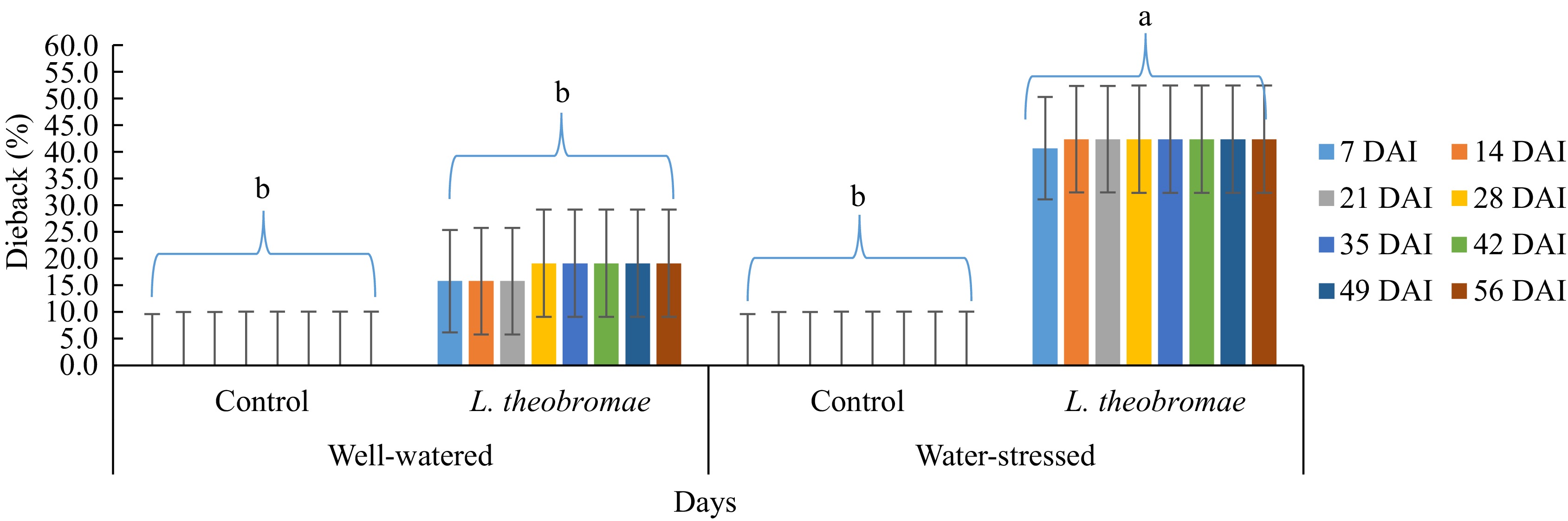
      Figure 1. 

      Dieback severity in cocoa MCC 02 clone inoculated with L. theobromae simultaneous to water-stress imposition and with PDA as a control inoculum for 56 d. Bars with the same letter indicate no significant difference according to the Tukey's test at p ≤ 0.05. DAI: Days after inoculation.

      Table 1.  Two-way ANOVA of dieback severity in two different watering regimes and two different inoculum types. Cocoa MCC 02 clone inoculated with L. theobromae simultaneous to water stress imposition and with PDA as a control inoculum for 56 d and evaluated 1, 2, 3, 4, and 5 weeks after inoculation in South Sulawesi (from January 2023 to March 2023). In all treatments, dieback symptoms were first observed one week after inoculation.

      Watering regime/
      inoculum type
      Dieback severity
      1 week after inoculation
      Dieback severity
      2 weeks after inoculation
      Dieback severity
      3 weeks after inoculation
      Dieback severity
      4 weeks after inoculation
      Dieback severity
      5 weeks after inoculation
      Mean AUDPC disease severity
      for 8 weeks
      Averages for each watering regime
      Well-watered 7.92% 7.92% 7.92% 9.58% 9.58% 62.92%
      Water-stressed 20.42% 20.42% 20.42% 21.25% 21.25% 148.33%
      Tukey's test 15.08% (NS) 15.08% (NS) 15.08% (NS) 13.73% (NS) 13.73% (NS) 98.85% (NS)
      Averages for each inoculation time
      Control 0.00 0.00 0.00 0.00 0.00 0.00
      L. theobromae 28.33% 28.33% 28.33% 30.83% 30.83% 211.25%
      Tukey's test 15.08% (**) 15.08% (**) 15.08% (**) 13.73% (**) 13.73% (**) 98.85% (**)
      Analysis of variance (p-value)
      Watering regime (W) NS NS NS NS NS NS
      Inoculum type (I) ** ** ** ** ** **
      W × I NS NS NS NS NS NS
      ** and NS indicate statistical significance at p < 0.01, 0.05, and not significant by Tukey's test analysis (p < 0.05), respectively. Mean disease severity as described on Materials and methods section.

      In the experiment where the plants were inoculated with L. theobromae seven days after water stress imposition (Fig. 2), at the beginning of the evaluation, the highest severity of dieback was recorded at the inoculation treatment under water stress (35.0%). The value was significantly higher than those in the plot of the inoculation treatment with well-watering (5.0%) and both controls (water-stressed or well-watered) (0%). Similar to the experiment where the plants were inoculated simultaneously to water stress imposition, the mean percentage of dieback on the plot with inoculation treatment with water stress increased (38.3%) in the second observation. In addition, the severity increased on the plot with inoculation treatment with well-watering as well (6.7%). However, they were significantly different. The dieback severity under both water-stressed and well-watered conditions remained steady after the second evaluation until the end of the experiment. In addition, the plants in the control groups, both well-watered and water-stressed, remained symptomless (0%) until the end of the evaluation. Likewise, the AUDPC value of the plot with inoculation treatment with water stress was significantly high. Furthermore, there is an effect of the water stress imposition (factor 1) on dieback severity where the influence was highly significant from the beginning of the evaluation (1 week after inoculation) until the end of the evaluation. Similarly, the inoculum type (factor 2) and interaction (water-stress imposition × inoculation time) effects were highly significant in any evaluation event, either a percentage of dieback severity or AUDPC disease severity (Table 2).
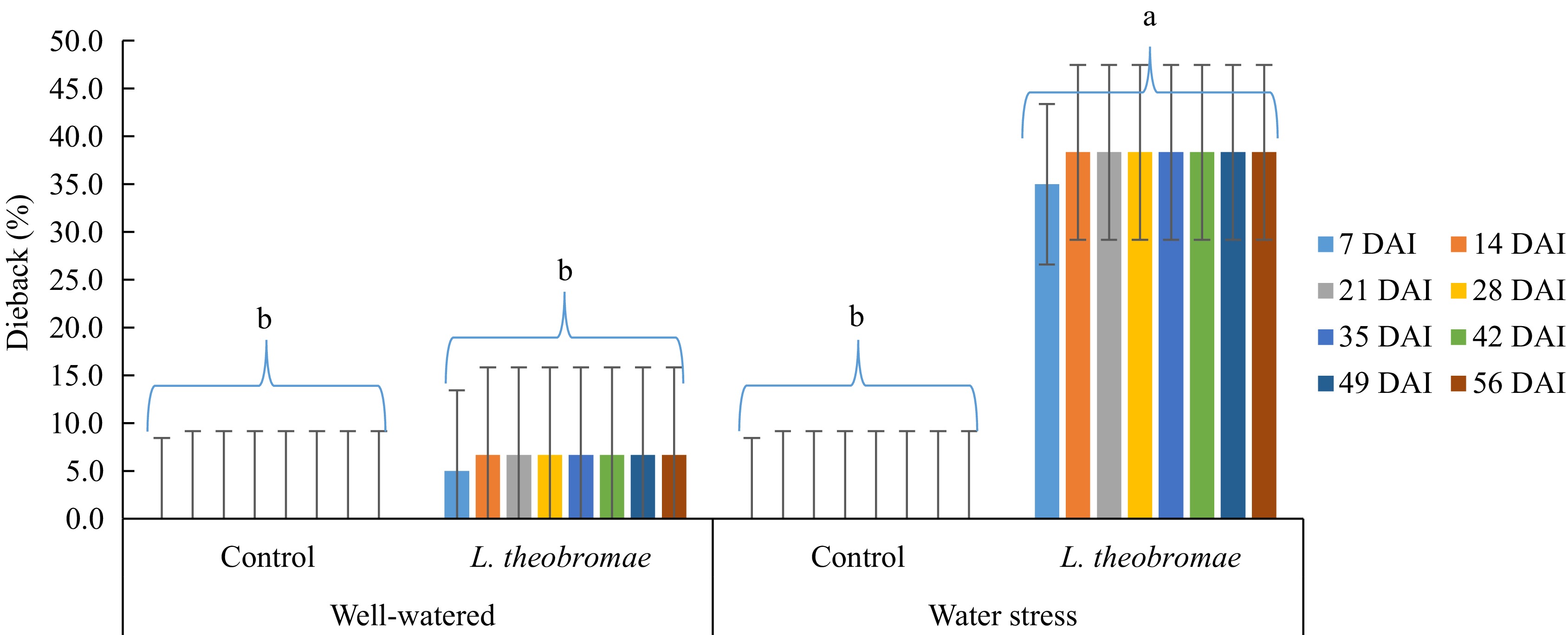
      Figure 2. 

      Dieback severity in MCC 02 cocoa clone inoculated with L. theobromae 7 d after the initiation of water-stress imposition and with PDA as a control inoculum for 56 d. Bars with the same letter indicate no significant difference according to the Tukey's test at p ≤ 0.05. DAI: Days after inoculation.

      Table 2.  Two-way ANOVA of dieback severity in two different watering regimes and two different inoculum types. Cocoa MCC 02 clone inoculated with L. theobromae 7 d after the initiation of water-stress imposition and with PDA as a control inoculum for 56 d and evaluated 1, 2, 3, 4, and 5 weeks after inoculation in South Sulawesi (from January 2023 to March 2023). In all treatments, dieback symptoms were first observed one week after inoculation.

      Watering regime/
      inoculum type
      Dieback severity
      1 week after inoculation
      Dieback severity
      2 weeks after inoculation
      Dieback severity
      3 weeks after inoculation
      Dieback severity
      4 weeks after inoculation
      Dieback severity
      5 weeks after inoculation
      Mean AUDPC disease severity for 8 weeks
      Averages for each watering regime
      Well-watered 2.50% 3.33% 3.33% 3.33% 3.33% 22.92%
      Water-stressed 17.50% 19.17% 19.17% 19.17% 19.17% 133.33%
      Tukey's test 8.29% (**) 7.52% (**) 7.52% (**) 7.52% (**) 7.52% (**) 52.62% (**)
      Averages for each inoculation time
      Control 0.00 0.00 0.00 0.00 0.00 0.00
      L. theobromae 20.00% 22.50% 22.50% 22.50% 22.50% 156.25%
      Tukey's test 8.29% (**) 7.52% (**) 7.52% (**) 7.52% (**) 7.52% (**) 52.62% (**)
      Analysis of variance (p-value)
      Watering regime (W) ** ** ** ** ** **
      Inoculum type (I) ** ** ** ** ** **
      W × I ** ** ** ** ** **
      ** indicate statistical significance at p < 0.01, 0.05 by Tukey's test analysis (p < 0.05). Mean disease severity as described on materials and methods.

      When AUDPC values of dieback severity were compared in two different watering regimes and two different times of inoculation of L. theobromae, the AUDPC values with the simultaneous and delayed inoculation treatments under water-stressed condition were significantly higher than those with the simultaneous and delayed inoculation treatments under well-watered condition as well as those in the controls (Fig. 3). In addition, the AUDPC value with the delayed inoculation treatment under well-watered condition was not significantly different from the control (Fig. 3). The combination of water stress imposition and inoculation time showed a variety of dieback severity. There is an effect of the water stress imposition (factor 1) on dieback severity where the influence was highly significant from the beginning of the evaluation (1 week after inoculation) until the end of the evaluation. Meanwhile, inoculation time (factor 2) and interaction (water-stress imposition × inoculation time) effects were not significant in any evaluation event, either percentage of dieback severity or AUDPC disease severity (Table 3).
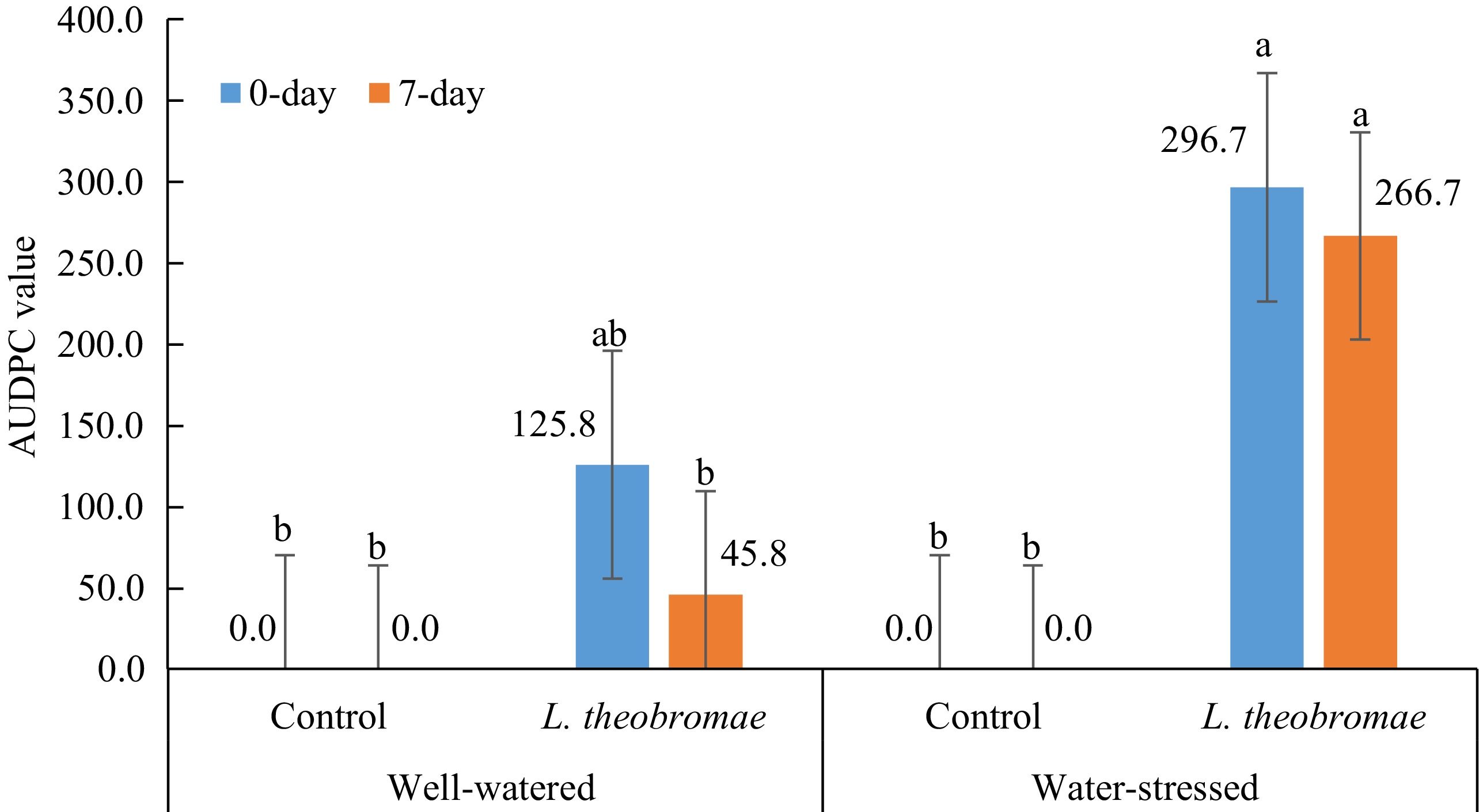
      Figure 3. 

      Mean AUDPC values of dieback severity in two different watering regimes and two different times of inoculation of L. theobromae evaluated 1, 2, 3, 4, and 5 weeks after inoculation in South Sulawesi (from January 2023 to March 2023). In all treatments, dieback symptoms were first observed one week after inoculation. Bars with the same letter do not differ significantly according to the Tukey's test analysis (p < 0.05).

      Table 3.  Two-way ANOVA of dieback severity in four different watering regimes and two different times of inoculation of L. theobromae evaluated 1, 2, 3, 4, and 5 weeks after inoculation in South Sulawesi (from January 2023 to March 2023). In all treatments, dieback symptoms were first observed one week after inoculation.

      Watering regime/
      inoculation time
      Dieback severity
      1 week after inoculation
      Dieback severity
      2 weeks after inoculation
      Dieback severity
      3 weeks after inoculation
      Dieback severity
      4 weeks after inoculation
      Dieback severity
      5 weeks after inoculation
      Mean AUDPC disease severity for 8 weeks
      Averages for each watering regime
      Well-watered LT 5.21%b 5.63%b 5.63%b 6.46%b 6.46%b 42.92%b
      Water-stressed LT 18.96%a 20.21%a 20.21%a 20.21%a 20.21%a 140.83%a
      Well-watered CO 0.00b 0.00b 0.00b 0.00b 0.00b 0.00b
      Water-stressed CO 0.00b 0.00b 0.00b 0.00b 0.00b 0.00b
      Tukey's test 13.11% (**) 12.56% (**) 12.56% (**) 11.77% (**) 11.77% (**) 83.99% (**)
      Averages for each inoculation time
      0-day 28.33% 29.17% 29.17% 30.83% 30.83% 211.25%
      7-day 20.00% 22.50% 22.50% 22.50% 22.50% 156.25%
      Tukey's test NS NS NS NS NS NS
      Analysis of variance (p-value)
      Watering regime (W) ** ** ** ** ** **
      Inoculation time (I) NS NS NS NS NS NS
      W × I NS NS NS NS NS NS
      Numbers in the same column followed by the same letter are not significantly different by Tukey's test analysis (p < 0.05). ** and NS indicate statistical significance at p < 0.01, 0.05, and not significant, respectively. Mean disease severity as described in the Materials and methods. LT: L. theobromae; CO: Control, a PDA plug.

      Inoculation of L. theobromae with water-stress imposition consistently resulted in higher dieback severity, regardless of the inoculation time, than inoculation of L. theobromae under well-watered condition (Figs 13 & Tables 13). In addition, black conidiomata were apparent after the branch or stem was colonized thoroughly by L. theobromae (Fig. 6h).

    • The treatments of water stress showed significantly different rates of survival of scions after inoculation of L. theobromae and without inoculation (controls). The lowest survival rates of scions were observed by the water-stressed treatments, with simultaneous inoculation and inoculation on the seventh day after water-stress imposition at 50% and 66.7%, respectively (Fig. 4). Meanwhile, the largest survival rates of scions after inoculation were perceived by well-watering treatments with simultaneous inoculation and inoculation on the seventh day after initiation of well-watering, 95.8% and 91.7%, respectively (Fig. 4). Controls on both water stress and well-watered treatments remained healthy (100% survival rate) until the end of the experiment.
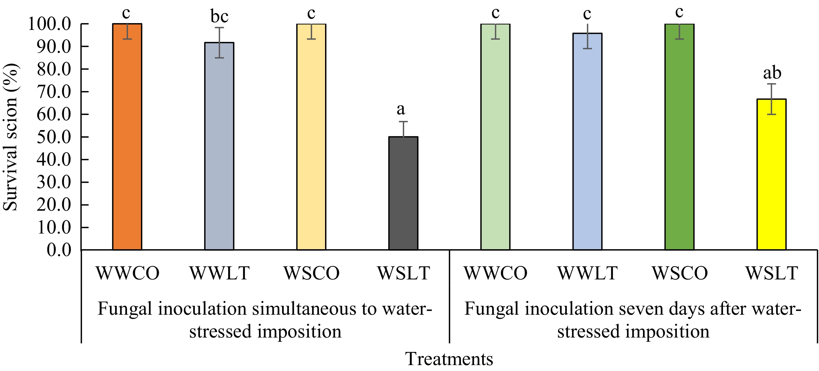
      Figure 4. 

      Survival rates of scions inoculated by L. theobromae in two different experiments: Fungal inoculation simultaneous to water stress imposition (Experiment 1) and 7 d after the initiation of water stress imposition (Experiment 2), with PDA as a control inoculum for 64 d. Differences in letters above the bar on each treatment indicate statistically significant differences by Tukey's test analysis (p < 0.05). WWCO: Well-watered treated with a PDA plug (control); WWLT: Well-watered with L. theobromae inoculation; WSCO: Water-stressed treated with a PDA plug (control); WSLT: Water-stressed with L. theobromae inoculation.

      On the surviving scions, vascular streaking was visible in the vertical sections of all the fungal-inoculated scions (Table 4, Fig. 6io). The vascular streaking length showed no significant difference in all fungal inoculated treatments, regardless of water stress and timing of inoculation. However, vascular streaking spread faster on the scions with water-stress treatment than on those with well-watering. Also, the percentage of colonization of the pathogen on the scions with the water-stress treatments was higher than that on the scions that were well-watered. In addition, the pathogen moved upward and downward from the inoculation site. Controls on both water stress and well-watering remained healthy in the vascular until the end of the experiment (Fig. 6pr).

      Table 4.  Vascular streaking length (mm) in the stem of MCC 02 cocoa clone inoculated with Lasiodiplodia theobromae simultaneous to water-stress imposition (Experiment 1) and 7 d after the initiation of water-stressed imposition (Experiment 2) with PDA as a control inoculum for 64 d.

      No. Experiment Treatment Vascular streaking (mm) Vascular streaking length compared
      to scion length
      Distance of from inoculation site to edge of lesion (mm)
      Upward Downward
      1 L. theobromae inoculation simultaneous to water-stressed imposition Well-watered Control 0.0b 0.0c 0.0b 0.0b
      L. theobromae 71.3a 72.6%ab 34.5a 36.8a
      Water-stressed Control 0.0b 0.0c 0.0b 0.0b
      L. theobromae 80.2a 83.7%a 40.0a 40.2a
      2 L. theobromae inoculation seven days after water-stressed imposition Well-watered Control 0.0b 0.0c 0.0b 0.0b
      L. theobromae 71.2a 65.1%b 38.6a 32.7a
      Water-stressed Control 0.0b 0.0c 0.0b 0.0b
      L. theobromae 82.6a 87.3%a 42.4a 40.2a
      Tukey's test at α = 0.05 16.84 (**) 15.13% (**) 8.67 (**) 10.41 (**)
      Columns with the same letter do not differ significantly according to Tukey's test at α = 0.05.
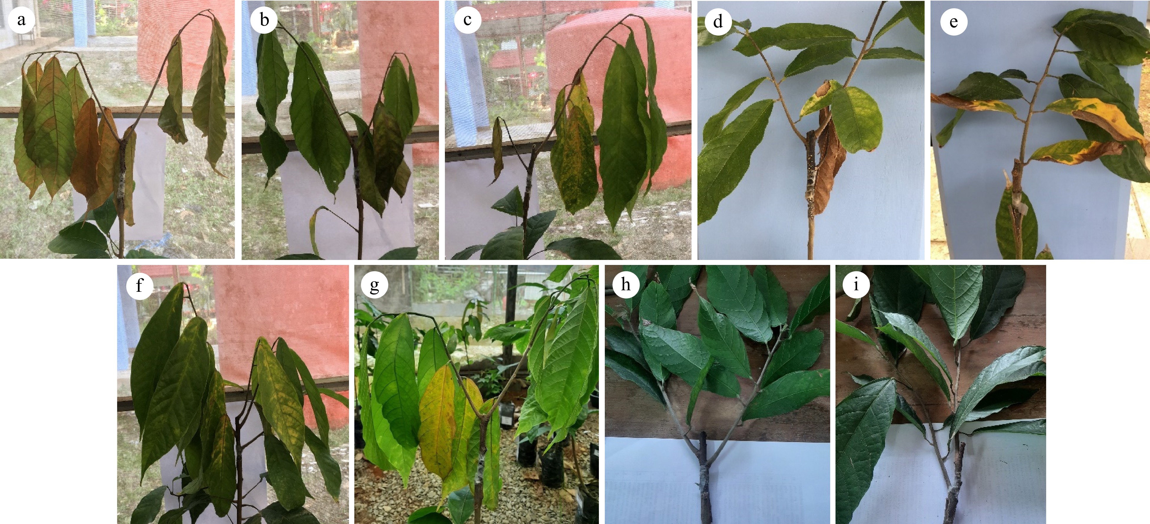
      Figure 5. 

      Various initial symptoms on leaves of cocoa scions inoculated by L. theobromae (simultaneously or 7 d after water imposition) under well-watered and water-stressed conditions. (a), (b) dieback; (c)−(e) chlorotic and mixed of chlorotic and necrotic; (f), (g) chlorotic; (h), (i) control (a PDA plug).
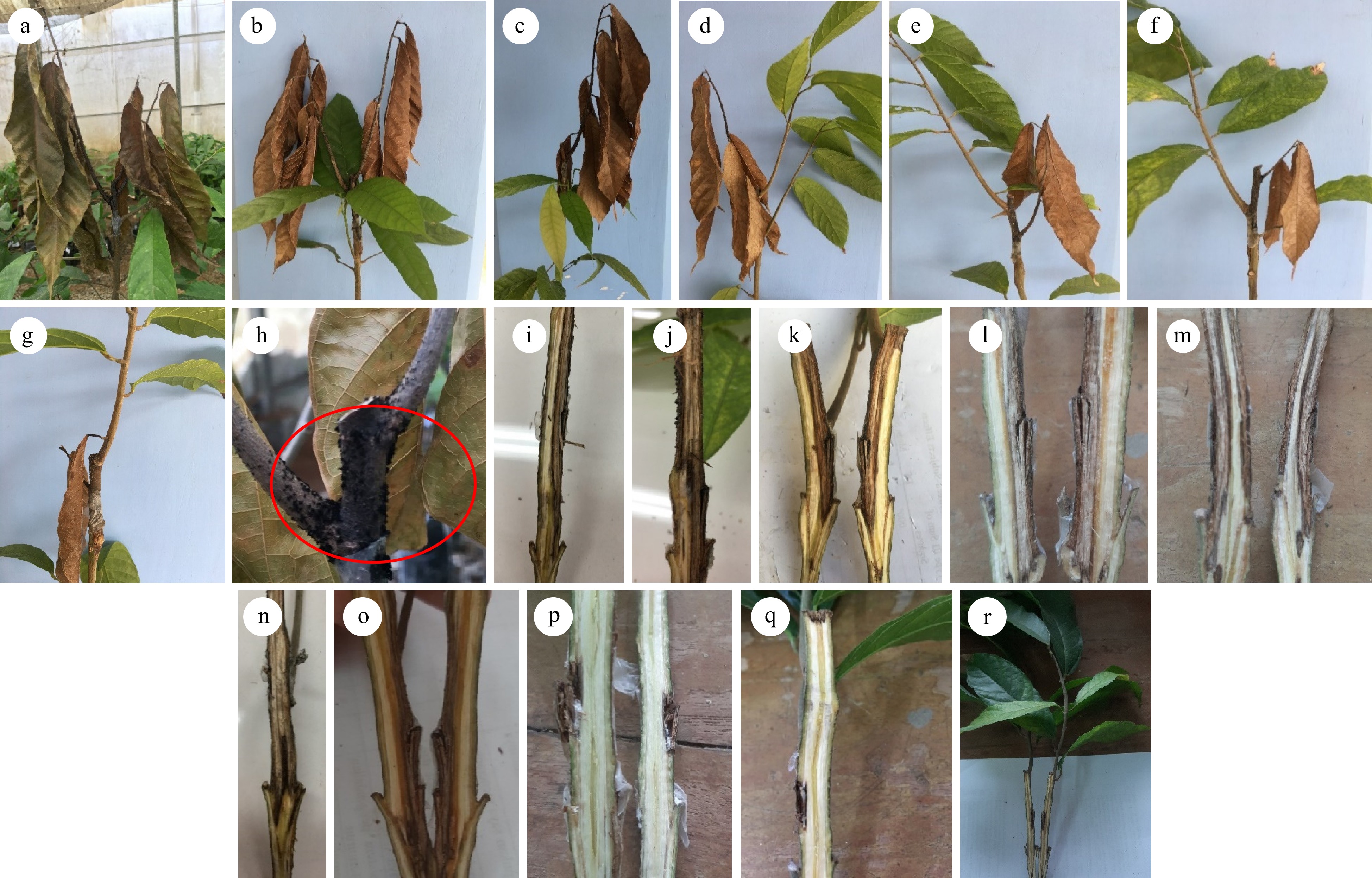
      Figure 6. 

      Various advanced symptoms on cocoa scions inoculated with L. theobromae simultaneously with water imposition and 7 d after the initiation of water imposition. (a)−(c) dieback on all branches, three, two, and one branch(es), respectively; (d)−(g) dieback on one side of the branch; (g)browning leaf at lower leaf; (h) presence of black conidiomata (red circle) on the fungal inoculated stem; (i)−(o) vertical section of fungal inoculated scion stems showed vascular streaking; (p)−(r) vertical section of control showed symptomless/no vascular streaking.

    • The current study explored the influence and the interaction of drought stress and inoculation of L. theobromae to dieback disease on T. cocoa clone MCC 02. For these purposes, we determined such responses as dieback, leaf chlorotic and necrotic, and vascular streaking of cocoa to L. theobromae infection under drought treatments through artificial inoculation on potted plants prepared from a typical cocoa clone in Sulawesi. The results indicated that water deprivation promoted disease development, regardless of the timing of inoculation during drought stress. Generally, drought situations influence plants by inducing damage to water relations and making plants more predisposed to pathogen onset and other biotic attacks. Also, the severity of the disease probably elevates with drought stress[5658]. In addition, drought stress may exacerbate the development of the disease in the trees[43,5862].

      Although drought stress is the most abiotic factor studied on tree susceptibility to pathogens[43,44,63,64], the role of drought on cocoa susceptibility to L. theobromae in Sulawesi had remained poorly understood. To the authors' knowledge, this research is the first attempt to evaluate the interaction of drought stress and disease caused by L. theobromae in Indonesia. The present results showed the apparent destructive effect of the water stress under infection of the pathogen. Similarly, it has been known that water stress increases the susceptibility of perennial plants to L. theobromae and other Botryosphaeriaceae species[43,44,65].

      Dieback and vascular streaking symptoms were clearly expressed on each inoculated plant. The results found here corroborate the pathogenicity of L. theobromae to cocoa clone MCC 02 and are in accordance with the previous study that indicates L. theobromae is one of the most aggressive and destructive phytopathogenic fungi responsible for causing such broad disease symptoms as dieback, canker, chlorotic, root and collar root and leaf blight in many plants[915,18,23,6670]. In addition, the increase in disease severity in a plant exposed to water stress may predisposed by host physiology status where during water stress, accumulation and production of the certain compounds that induce disease defense were altered, including amino acids and reactive oxygen species (ROS)[65,71]. Also, the production of biochemical defense may decrease because of water stress[56,72].

      Relatively few studies have directly addressed the mechanism of interaction and how L. theobromae became more severe under water stress on cocoa when most pathogens strive in opposite conditions. However, drought could make trees more susceptible to pathogens because drought decreases the availability of plant resources for defenses against plant pathogens. How drought affects pathogen survival is not clear. However, some fungal pathogens are very adaptable, and the design of fungal reproductive systems is diverse to manage fluctuating environmental conditions[73]. Moreover, a framework estimates that necrotrophs fungal pathogens, which mostly relies on nutrients from dead tree cells, accelerate drought-induced tree mortality by colonizing sapwood and damaging plant transport systems[59].

      Stress duration plays an important role in host-pathogen interaction. A study conducted on stems of European white birch revealed that Botryosphaeria dothidea causes canker after exposure to water stress for a minimum of 3 d at the acceptable level[74]. Also, host-pathogen interaction was influenced by the timing of the different stresses. A study on dogwood revealed that water imposition before the inoculation of L. theobromae resulted in a greater effect on canker development than post-inoculation water stress[5].

      L. theobromae can grow in wide different environmental conditions and a wide range of temperatures[7577]. The average temperature during the experiment was relatively in the high range (29.8–33.9 °C). However, with such a temperature range, L. theobromae can grow optimally and colonize the plant aggressively[7577]. In addition, such a temperature range seemed to increase the virulence of Botryosphaeriaceae spp.[27,77]. However, at this point, more studies are needed to evaluate the effect of temperature on the development of disease severity in cocoa.

      Although this research is only tested on potted plants in a greenhouse using artificial inoculation, only performed on a single clone and cannot represent directly mature trees that are established in the field, the results of this study showed obvious pictures of the importance of drought × L. theobromae interaction-inducing disease.

      The interaction of drought and L. theobromae reported here suggested that water limitation before and after fungal inoculation makes plants more susceptible to fungal pathogens and weakens plant defense against pathogen infection, indicating that water deprivation supports the effectivity of pathogen infection (predisposition). Also, this study emphasized that the influence of water deficit on plant physiological status was more prominent compared to the fungal infection.

      More integrated studies are needed to follow up on the findings here and to tackle the severity of dieback diseases in cocoa under drought conditions in which more frequent and longer are predicted, the evaluation of different cocoa clones will be necessary to gain more understanding of the effect of drought stress on dieback disease development under L. theobromae infection, and evaluation of other abiotic and biotic stress factors on dieback disease development under L. theobromae infection will help comprehend the dynamics of the disease in the field.

    • This study has shown that drought stress increases the severity of the dieback disease caused by L. theobromae on cocoa clone MCC 02. It was also observed that drought stress × L. theobromae interaction increased the length of vascular streaking in the stems of cocoa. In addition, we observed external disease symptoms and vascular streaking at the interaction between well-watered × L. theobromae. This research extends our knowledge of the impact of drought on plant-pathogen interaction in cocoa and water stress should be avoided in cocoa plantations due to its detrimental impact on severity of dieback diseases.

    • The authors confirm contribution to the paper as follows: study conception and design: Asman A, Rosmana A, Iwanami T; material preparation and data collection: Asman A, Rosmana A; analysis and interpretation of results: Asman A, Iwanami T, Rosmana A; draft manuscript preparation: Asman A; review and editing: Iwanami T, Asman A, Rosmana A. All authors reviewed the results and approved the final version of the manuscript.

    • All data generated or analyzed during this study are included in this published article.

    • The first author would like to thank Mr. Anwar (Ayye) for his assistance in providing cocoa seedlings, and Nurmala Rasyda, S.P., Mr Kamaruddin, Ardan, Ahmad, S.P., M.Si, Husnul Chatimah, S.P., Inayah Maghfirah Ramadhani, S.P., Fadia Ersya Matterru, S.P., Dian Anugrah, S.P., and Asri for the technical assistance.

      • The authors declare that they have no conflict of interest.

      • Copyright: © 2024 by the author(s). Published by Maximum Academic Press, Fayetteville, GA. This article is an open access article distributed under Creative Commons Attribution License (CC BY 4.0), visit https://creativecommons.org/licenses/by/4.0/.
    Figure (6)  Table (4) References (77)
  • About this article
    Cite this article
    Asman A, Iwanami T, Rosmana A. 2024. Effect of drought stress on dieback disease development under Lasiodiplodia theobromae infection in cocoa clone 'MCC 02'. Beverage Plant Research 4: e034 doi: 10.48130/bpr-0024-0023
    Asman A, Iwanami T, Rosmana A. 2024. Effect of drought stress on dieback disease development under Lasiodiplodia theobromae infection in cocoa clone 'MCC 02'. Beverage Plant Research 4: e034 doi: 10.48130/bpr-0024-0023

Catalog

  • About this article

/

DownLoad:  Full-Size Img  PowerPoint
Return
Return