ARTICLE   Open Access    

Gastrointestinal digestion fate of Tremella fuciformis polysaccharide and its effect on intestinal flora: an in vitro digestion and fecal fermentation study

More Information
  • In this work, the gastrointestinal digestive outcome of Tremella fuciformis polysaccharide (TFP) was examined using in vitro simulated experiments, together with its effect on the intestinal microbiota. TFP did not significantly alter during the stage of oral digestion, according to an in vitro digestion investigation. Nevertheless, glycosidic connections of TFP were broken throughout the intestinal and stomach digesting phases, which resulted in the dissociation of macromolecular aggregates, a marked rise in decreasing sugar content (CR), as well as a drop in molecular weight (Mw). Additionally, microbial community analysis following fecal fermentation in vitro indicated that TFP might control the alpha and beta diversity of gut microbiota and change the genus- and phylum-level community composition. It increased the abundance of beneficial bacteria including Megasphaera, Phascolarctobacterium, and Bacteroides, and suppressed the growth of harmful bacteria like Escherichia-shigella and Fusobacterium, thus contributing to maintaining gut homeostasis. These results suggested that TFP could have a positive impact on health through enhancing the gut microbiota environment, giving a theoretical basis for its use as a prebiotic.
  • The intestinal mucosal barrier is composed of epithelial cells and intercellular connections, which can effectively regulate the transportation of large molecules in the intestinal lumen, and prevent microorganisms, harmful solutes, toxins, and intraluminal antigens from entering bodies[1]. Mucosal barrier factors such as trefoil factor (TFF) family, diamine oxidase (DAO), and transform growth factor-ɑ (TGF-α) have protective and restorative effects on intestinal mucosal integrity, which are synthesized and secreted by intestinal mucosa[24]. The damage to intestinal barrier function can cause the invasion of antigens and bacteria in the lumen, and eventually lead to intestinal diseases, including diarrhea, inflammatory bowel disease (IBD), and Crohn's disease[57].

    The Notch signaling pathway plays a crucial role in a series of cellular processes, including proliferation, differentiation, development, migration, and apoptosis. Research suggests that the Notch signaling pathway is involved in intestinal development, and it can be connected to intestinal cell lineage specification[8]. Furthermore, the Notch signaling pathway can regulate intestinal stem cells, CD4+T cells, innate lymphoid cells, macrophages, and intestinal microbiota, and intervene in the intestinal mucosal barriers in cases of ulcerative colitis[9]. The activated Notch signaling pathway can suppress the goblet cells differentiation and mucus secretion, which would result in the disruption of the intestinal mucosal barrier[10]. In addition, the absence of the Notch signaling pathway would cause the dysfunction of tight junctions and adherens junctions of intestinal mucosa, which could lead to increased permeability of epithelial cells and exposure of luminal contents to the immune system and inflammation[11]. Phytochemicals, such as cucurbitacin, honokiol and quercetin have been reported to have therapeutic effects on intestinal diseases by targeting the Notch signaling pathway[1214].

    Plant-based diets rich in phytochemicals, such as phenolics, anthocyanins, and vitamins, have been related to the prevention of human diseases[15]. Anthocyanins belong to a subclass of flavonoids, and are mainly in the form of different anthocyanin combined with glucose, galactose, and arabinose[16]. The physiological action of anthocyanins, such as antioxidant activity, anti-inflammation, and anti-obesity effects have been widely reported[17,18]. M3G is found naturally in plants and is reported to be the most common anthocyanin in different blueberry varieties[19,20]. Previous researchers reported that M3G from blueberry suppressed the growth and metastasis potential of hepatocellular carcinoma cells, modulated gut microbial dysbiosis, and protected TNF-α induced inflammatory response injury in vascular endothelial cells[2123]. Anthocyanins with diverse molecular structures and from different dietary sources are bioavailable at diet-relevant dosage rates, and anthocyanins from berry fruit are absorbed and excreted by both humans and rats[24]. Studies have shown that anthocyanins have a protective effect on intestinal barrier damage by regulating gut microbiota, tight junction (TJ) protein expression, and secretion of MUC2[2527]. In addition to physiological indicators related to the intestinal barrier, the regulation of the Notch signaling pathway was involved in this study. Therefore, it is speculated that M3G could improve the colonic mucosal barrier function via the Notch signaling pathway.

    To prove this hypothesis, the effects of M3G on the colonic mucosal barrier function were investigated in DSS-induced colitis mice. The pathological morphology of the colon tissue, markers of intestinal physical barrier function and immune barrier function and the Notch signaling pathway were evaluated to reveal the mechanism of M3G in improving colonic mucosal barrier function. The results are expected to lay the foundation for the utilization of anthocyanins as a promising natural product for improving intestinal diseases.

    M3G (CAS: 30113-37-2) used in this study was purchased from Xinyi Science and Technology Instrument Business Department, Baoji, Shanxi, China (HPLC ≥ 98%).

    Five-week-old male C57BL/6J mice (Wanlei Bio Co., Ltd., Shenyang, Liaoning, China) weighing 16−18 g were housed and given AIN-93M diet (Wanlei Bio Co., Ltd., Shenyang, Liaoning, China) feeding. The animal experiment was carried out according to the guidelines of the Standards for Laboratory Animals of China (GB 14922-94, GB 14923-94, and GB/T 14925-94) and the Ethics Committee of Shenyang Agricultural University (IACUC Issue No.: 2023022401). All animal housing and experiments were conducted in strict accordance with the institutional guidelines for the care and use of laboratory animals.

    After acclimating to the breeding environment for one week, the mice were randomly divided into two groups: control group (CG group, n = 6) and model group (n = 12). On days 1−7, the mice of the model group were given 2.5% DSS (CAS: 9011-18-1) dissolved in drinking water, while the mice of the CG group were given the same volume of drinking water as the model group. On days 8−14, mice of the model group were randomly divided into two equal groups (n = 6): DSS group and M3G group. The mice of the CG and DSS groups were administered intragastrically via drinking water, and the mice of the M3G group were administered intragastrically by M3G (5 mg/kg body weight (BW)/d) dissolved in drinking water, and the liquid volume was controlled to be the same. For all groups of mice, the body weight, food consumption, stool consistency, and bloody stool were measured daily during the experiment. On day 15, mice were sacrificed after a 12 h fast, and the colon tissue samples of the mice were collected. The length of the colon tissue was measured and recorded.

    Colon tissue was embedded with paraffin and then cut into sections. After being dewaxed from paraffin, the tissue was placed in water, and stained with hematoxylin and eosin solution. The stained tissue was dehydrated and sealed for observation. A microscope (BX53, Olympus Co., Ltd., Tokyo, Japan) was used to observe the stained tissue and photographed using 100× magnification. The damage in epithelial cells of colon tissue was evaluated.

    The colon tissue was dewaxed and placed in water after being embedded with paraffin, and then stained with schiff and hematoxylin solution. The stained tissue was dehydrated and sealed for observation. A microscope was used to observe the stained tissue and photographed using 100× magnification. The mucosal thickness and the population of goblet cells of the colon tissue were measured and recorded.

    The expression of MUC2 in colon tissue was detected by RT-PCR. Total RNA was extracted from colon tissue, and the concentration was determined using an ultraviolet spectrophotometer (Thermo Fisher Scientific, Waltham, MA, USA). The single-stranded cDNA of the extracted RNA (0.1 μL) was synthesized using a Transcriptor First Strand cDNA Synthesis Kit (Roche Co., Ltd., Basel, Kanton Basel, Switzerland). ExicylerTM 96 (Bioneer Corporation, Daejeon, Korea) was used to analyze the fluorescence quantitative cDNA. The reaction conditions were as follows: 94 °C for 5 min, 94 °C for 10 s, 60 °C for 20 s, 72 °C for 30 s, then followed by 40 cycles of 72 °C for 2 min 30 s, 40 °C for 1 min 30 s, and then melting from 60 to 94 °C, and incubating at 25 °C for 1−2 min. The primer sequences are shown in Table 1.

    Table 1.  The primer sequences used in the RT-PCR analysis.
    Gene Prime Sequence (5'-3') Size (bp)
    MUC2 Forward TGTGCCTGGCTCTAATA 17
    Reverse AGGTGGGTTCTTCTTCA 17
    β-actin Forward CTGTGCCCATCTACGAGGGCTAT 23
    Reverse TTTGATGTCACGCACGATTTCC 22
     | Show Table
    DownLoad: CSV

    The whole proteins from the colon tissue (200 mg) were extracted using a Whole Cell Lysis Assay kit (BioTeke Co., Ltd., Beijing, China) according to the manufacturer’s protocol. Briefly, colon tissue was cut into pieces, and then mixed with phenylmethylsulfonyl fluorid (PMSF), followed by adding the protein extraction reagents A and B to prepare the tissue homogenate. Protein concentration was then determined using a Bradford Kit (BioTeke Co., Ltd., Beijing, China) according to the manufacturer’s protocol. Proteins were separated by sodium dodecyl sulfate-polyacrylamide gel electrophoresis (SDS-PAGE). The membranes were blocked with 5% non-fat dry milk in TBST buffer for 1 h, followed by incubating overnight at 4 °C with the appropriate monoclonal primary antibody (detailed information in Table 2). Then membranes were washed to remove non-bound antibodies, and then incubated with the secondary antibody (goat anti-rabbit immunoglobin G-horseradish peroxidase (IgG-HRP), 1:5000; Wanlei Bio Co., Ltd., Shenyang, Liaoning, China) at 37 °C for 45 min. The enhanced chemiluminescence (ECL) western blotting detection reagent (Wanlei Bio Co., Ltd., Shenyang, Liaoning, China) was used to detect the protein bands, and then the protein bands were visualized by a Gel Imaging System (Beijing Liuyi Biotechnology Co., Ltd., Beijing, China).

    Table 2.  Details of the primary antibodies used in the experiment.
    Primary antibody Dilution ratio Manufacturer
    Claudin-1 1:500 Wanlei Bio Co., Ltd.
    Occludin 1:500 Wanlei Bio Co., Ltd.
    ZO-1 1:500 Wanlei Bio Co., Ltd.
    iFABP 1:1000 ABclonal Technology Co., Ltd.
    DLL1 1:500 Wanlei Bio Co., Ltd.
    DLL4 1:1000 ABclonal Technology Co., Ltd.
    Notch1 1:500 Wanlei Bio Co., Ltd.
    NICD 1:1000 Affinity Biosciences Co., Ltd.
    Hes1 1:1000 ABclonal Technology Co., Ltd.
    TFF3 1:1000 Affinity Biosciences Co., Ltd.
    β-actin 1:1000 Wanlei Bio Co., Ltd.
     | Show Table
    DownLoad: CSV

    The level of SIgA in colon tissue was detected by SIgA ELISA kit (Model number: EM1362, Wuhan Fine Biotech Co., Ltd., Wuhan, Hubei, China). The experiment was carried out according to the ELISA kit instructions.

    CD4+T (CD3+CD4+) cells and CD8+T (CD3+CD8+) cells in colonic lamina propria monocytes (LPMC) were detected by FCM. The colonic epithelial cells were isolated by adding digestive solution to the colon tissue. After digestion, screening, re-suspension precipitation, and centrifugation, the precipitate was collected as colonic LPMC. Labeled antibodies were added to the flow tube and incubated according to the instructions. After washing with phosphate buffered saline (PBS), the cells were re-suspended in PBS solution. The CD4+T (CD3+CD4+) cells and CD8+T (CD3+CD8+) cells were detected by flow cytometric (Agilent Technologies, Inc., Santa Clara, CA, USA).

    Data are expressed as mean ± standard deviation (SD) based on six replicates. Differences between two groups were assessed using Student's t tests. The results were considered statistically significant at p < 0.05. Data were analyzed using Graph Pad Prism 8.0 (Graph Pad Software, San Diego, CA, USA) and SPSS 17.0 (IBM Corporation, Armonk, NY, USA).

    The body weight of mice was monitored daily during the experimental period. As shown in Fig. 1a, after being fed with DSS for 7 d, the body weight of the mice in the DSS group was reduced significantly (p < 0.01) compared with that of the CG group. While the body weight of the mice in the M3G group was increased, and exhibited significantly higher (p < 0.01) body weight gain compared with the DSS group. The DAI score of the mice in the DSS group was significantly higher (p < 0.01) than that of the CG group, but M3G supplementation significantly lowered (p < 0.01) the DAI score (Fig. 1b), which demonstrates that the DSS-induced mice colitis model was established successfully and M3G inhibited colon tissue damage in DSS-fed mice. Compared with the CG group, the food intake of the mice fed with DSS was significantly reduced (p < 0.01), and M3G supplementation significantly increased (p < 0.05) the food intake (Fig. 1c).

    Figure 1.  Effect of M3G on body weight gain, DAI score, and food intake in mice. (a) Body weight gain. (b) DAI score (Fecal consistency: 0 = normal, 1 = semi-formed, 2 = soft stool, 3 = diarrhea or watery stool; bloody stool: 0 = occult blood negative, 1 = occult blood positive, 2 = visible bloody stool, 3 = massive hemorrhage; weight loss: 0 = 0%, 1 = 1%−5 %, 2 = 6%−10% ; 3 = 11%−15% reduction). (c) Food intake. Results are expressed as the mean ± SD (n = 6). * p < 0.05 and ** p < 0.01 indicate significant differences between two groups.

    Representative HE and PAS-stained sections of the colon are shown in Fig. 2a & b. The morphology of colon tissue in the CG group did not exhibit any damage, while damage in epithelial cells and goblet cells, inflammatory cells infiltration, and separation of the muscle layer and mucosal muscle layer were observed in the DSS group. The above damage to the tissue was changed for the better by M3G supplementation, with only small levels of damage in epithelial cells and goblet cells, inflammatory cell infiltration, and separation of the muscle layer and mucosal muscle layer compared to that in the CG group.

    Figure 2.  Effect of M3G on the pathological morphology of colon tissue. (a) HE staining of colon tissue. Original magnifications: 100×. (b) PAS staining of colon tissue. Original magnifications: 100×. (c) The damage score of colon tissue. (1) Epithelial cell damage: 0 = normal morphology; 1 = regional destruction of the epithelial surface; 2 = diffuse epithelial destruction and/or mucosal ulcers involving submucosa; 3 = severe epithelial damage. (2) Inflammatory cell infiltration: 0 = no infiltration or less than 5 cells; 1 = mild infiltration of the inherent layer; 2 = moderate infiltration of the muscular mucosa; 3 = high infiltration of the muscular mucosa; 4 = severe infiltration involving submucosa. (3) Separation of muscle layer and mucosal muscle layer: 0 = normal; 1 = moderate; 2 = severe. (4) Goblet cell depletion: 0 = no depletion; 1 = presence of disordered goblet cells; 2 = 1 to 3 regions without goblet cells; 3 = more than 3 regions without goblet cells; 4 = complete depletion of goblet cells. (d) The colon length. (e) The mucosal thickness of colon tissue. (f) Number of goblet cells in colon tissue. Results are expressed as the mean ± SD (n = 6). * p < 0.05 and ** p < 0.01 indicate significant differences between two groups.

    HE score was calculated to evaluate the degree of colon tissue damage. The HE score in the DSS group was significantly higher (p < 0.01) than that in the CG group. Supplementation of M3G significantly reduced (p < 0.01) the score, which decreased by almost 50% of that in the DSS group (Fig. 2c). The colon length in the DSS group was significantly lower (p < 0.01) than that in the CG group, which was significantly increased (p < 0.01) by M3G supplementation (Fig. 2d). The mucosal thickness and the number of goblet cells of the colon tissue were determined by PAS staining. Compared with the CG group, the mucosal thickness and the number of goblet cells of the colon tissue were significantly decreased (p < 0.01) in the DSS group, but those were significantly increased (p < 0.01) in the M3G group (Fig. 2e & f). These results suggest that M3G supplementation can decrease the pathological damage in the colon tissue induced by DSS.

    The mRNA expression level of MUC2 in the colonic mucosa tissue was determined. Compared with the CG group, DSS significantly decreased (p < 0.01) the mRNA level of MUC2, but this effect was significantly suppressed (p < 0.01) by M3G supplementation (Fig. 3a). The protein levels of claudin-1, occludin, ZO-1, iFABP, and TFF3 in the colon tissue were subsequently assessed. The protein expression levels of claudin-1, occludin, and ZO-1 in the DSS group were significantly lower than those in the CG group (p < 0.01). M3G supplementation significantly inhibited (p < 0.01) the reduction in the expression of these proteins (Fig. 3bd). Regarding the protein expression level of iFABP, it was significantly higher (p < 0.01) in the DSS group than that in the CG group. M3G supplementation significantly decreased (p < 0.01) iFABP expression level compared with the DSS group (Fig. 3e). The protein expression level of TFF3 was significantly higher (p < 0.01) in the DSS group than that in the CG group. However, M3G supplementation significantly intensified (p < 0.01) the increase of TFF3 expression compared with the DSS group (Fig. 3f). These results indicate that M3G can regulate colonic epithelial barrier function.

    Figure 3.  Effects of M3G on the colonic epithelial barrier disruption. (a) The mRNA expression level of MUC2. (b) The protein expression level of claudin-1. (c) The protein expression level of occludin. (d) The protein expression level of ZO-1. (e) The protein expression level of iFABP. (f) The protein expression level of TFF3. Results are expressed as the mean ± SD (n = 6). * p < 0.05 and ** p < 0.01 indicate significant differences between two groups.

    The content of CD4+T (CD3+CD4+) and CD8+T (CD3+CD8+) cells in colonic LPMC of mice in each group were detected by FCM as shown in Fig. 4 and Supplemental Fig. S1. Compared with the CG group, the percentages of CD4+T (CD3+CD4+) and CD8+T (CD3+CD8+) cells in the DSS group were significantly increased (p < 0.01), and those were significantly decreased (p < 0.05) after M3G supplementation (Fig. 4a & b). SIgA was measured as an indicator of immune barrier function in the colon tissue. The level of SIgA in the DSS group was significantly lower (p < 0.01) than that in the CG group. Supplementation of M3G significantly raised (p < 0.01) the level of SIgA (Fig. 4c). These results indicate that M3G can improve the colonic immune dysfunction induced by DSS.

    Figure 4.  Effect of M3G on the colonic immune barrier function. (a) The percentage of CD4+T (CD3+CD4+) cells in the colon tissue. (b) The percentage of CD8+T (CD3+CD8+) cells in the colon tissue. (c) The level of SIgA in the colonic mucosal tissue. Results are expressed as the mean ± SD (n = 6). * p < 0.05 and ** p < 0.01 indicate significant differences between two groups.

    Subsequently, the protein expression levels of Notch1, NICD, DLL4, DLL1, and Hes1 in the colon tissue were measured (Fig. 5). DSS significantly up-regulated (p < 0.01) these protein expression levels compared with the CG group. M3G supplementation significantly (p < 0.01) down-regulated the protein expression levels of Notch1, NICD, DLL4, DLL1, and Hes1 in comparison with the DSS group. The results indicate that M3G can inhibit the over-activation of the Notch signaling pathway.

    Figure 5.  Effects of M3G on the protein expression levels of Notch1, NICD, DLL4, DLL1, and Hes1 in the colon tissue. (a) The protein expression level of Notch1. (b) The protein expression level of NICD. (c) The protein expression level of DLL4. (d) The protein expression level of DLL1. (e) The protein expression level of Hes1. Results are expressed as the mean ± SD (n = 6). * p < 0.05 and ** p < 0.01 indicate significant differences between two groups.

    To explain the relationships between them, the Spearman r correlations between biomarkers and the Notch signaling pathway were analyzed, and represented using a heatmap. As shown in Fig. 6, colon length, food intake, ZO-1, number of goblet, MUC2, SIgA, claudin-1, occludin, body weight gain, and mucosal thickness were significantly (p < 0.01 or p < 0.05) positively correlated with Notch1, NICD, DLL4, DLL1, and Hes1. Conversely, HE score, iFABP, DAI score, CD4+T, and CD8+T were significantly (p < 0.01) negatively correlated with Notch1, NICD, DLL4, DLL1 and Hes1. In particular, TFF3 was only significantly (p < 0.05) positively correlated with DLL1, and Hes1. The results indicate that M3G may ameliorate the colonic mucosal barrier dysfunction via modulation of the Notch signaling pathway.

    Figure 6.  Heatmap of the Spearman r correlations between biomarkers and the Notch signaling pathway. * p < 0.05 and ** p < 0.01 indicate significant differences between two groups.

    Within the last five years, special attention is being paid to the therapeutic effects of anthocyanins. The recent studies could give evidence to prove the therapeutic potential of anthocyanins from different sources against various diseases via in vitro, in vivo, and epidemiological experiments[28]. The anthocyanin supplementation has been demonstrated to have positive effects on intestinal health[29]. The intestinal barrier is one of the crucial factors which can affect intestinal health and normal intestinal barrier function not only maintains intestinal health but also protects overall health by protecting the body from intestine injury, pathogen infection, and disease occurrence[30]. The disruption of intestinal barrier integrity is regarded as an important factor leading to IBD, obesity, and metabolic disorders[3133]. In this study, the effects of M3G on regulating the colonic physical barrier function and colonic immune barrier function in DSS-induced colitis mice were explored.

    M3G supplementation reduced the DAI score and the HE score of colon tissue, and restored colon length, mucosal thickness, and the number of goblet cells in the colon tissue, indicating that M3G can alleviate DSS-induced colon tissue damage and colitis symptoms in mice. The DAI score was developed as a simplified clinical colitis activity index to assess the severity of colitis[34]. Zhao et al. have found that black rice anthocyanin-rich extract can significantly decrease the DAI and HE scores of colon tissue in DSS-induced colitis mice[35]. Intestinal goblet cells are mainly differentiated from multipotential stem cells. Intestinal stem cells were located at the base of the crypt and distributed in intestinal mucosal epithelial cells, composing around 50% of colon epithelial cells[36]. The mucus layer is formed by goblet cell secretion, it separates the intestinal epithelium from the intestinal lumen, thereby preventing the invasion of pathogenic microorganisms and the translocation of intestinal microbiota[37]. The results suggested that M3G might repair the colonic mucosa by increasing the number of goblet cells.

    MUC2 is the most abundant mucin secreted by goblet cells of the colon, goblet cells, and MUC2 play important roles in maintaining and protecting the intestinal mucosal barrier[38]. M3G supplementation restored the level of MUC2 by nearly double that of the DSS group. Claudin-1, occludin, and ZO-1 are TJ proteins, which is an important component of the intestinal physical barrier[39]. M3G supplementation mitigated the down-regulation of claudin-1, occludin, and ZO-1 in DSS-induced colitis mice. Wang et al. have found that Lonicera caerulea polyphenols can increase the expression levels of occludin in HFD rats[40]. Chen et al. have found that purple-red rice anthocyanins alleviated intestinal barrier dysfunction in cyclophosphamide-induced mice by up-regulating the expression of tight junction proteins[41]. iFABP can serve as a biomarker of small bowel damage in coeliac disease and Crohn's disease, and supplementation of M3G down-regulated the expression level of iFABP in colon tissue[42]. It has been reported that TFF3 alleviated the intestinal barrier function by reducing the expression of TLR4 in rats with nonalcoholic steatohepatitis, and supplementation of M3G increased the expression level of TFF3 in this research[43]. Although the effect of M3G on epithelial TJ proteins and other related proteins were limited, the beneficial effect of M3G on the colonic mucosal barrier function was supported by histological evaluation.

    The intestinal immune barrier is composed of intestinal mucosal lymphoid tissue and intestinal plasma cell secreted antibodies. Inflammatory damage in acute colitis influences the gut microbiota, epithelial barrier, and immune function in subsequent colitis[44]. Fructose can influence colon barrier function by regulating some main physical, immune, and biological factors in rats[45]. SIgA, CD4+T cells, and CD8+T cells play important roles in the functioning of the human immune system. The results indicated that M3G supplementation can maintain the colonic immune barrier function by modulating the level of SIgA, and the percentages of CD4+T cells and CD8+T cells of colon tissue.

    Notch signaling pathways are important for the maintenance of intestinal epithelial barrier integrity, and its abnormal activation is related to IBD and colon cancer[46]. Among the four Notch receptors in mammals, the most scattered in the intestine is Notch1[47]. NICD is the active form of the Notch receptor, NICD enters the nucleus, binds to the recruitment co-activator transcription complex, and then combines with the Hes gene to regulate the fate of cells[48,49]. DLL1 and DLL4 can serve as ligands for Notch signaling receptors[50]. Lin et al. found that qingbai decoction had beneficial effects on the mucus layer and mechanical barrier of DSS-induced colitis by inhibiting Notch signaling[51]. Supplementation with M3G significantly down-regulated Notch1, NICD, DLL4, DLL1, and Hes1 expression levels in the colon tissue, and there is a significant correlation between biomarkers and Notch signaling pathway-related proteins, suggesting that M3G might sustain the colonic barrier function via inhibiting the Notch signaling pathway.

    The present results showed that M3G exerted an improvement effect on the colonic mucosal barrier (physical barrier and immune barrier) function in DSS-induced colitis mice. The positive impact of M3G on the colonic mucosal barrier function may be ascribed to the modulation of permeability, stability, and integrity of the colonic mucosa by down-regulating the Notch signaling pathway. The results provide theoretical support for anthocyanins as the raw materials of functional products related to intestinal health. However, this study also has limitations, such as lacking intensive research on the effect mechanisms through cell experiments. Therefore, intensive research and clinical tests are the future direction.

    The authors confirm contribution to the paper as follows: study conception and design: Zhang C, Jiao X; data collection: Zhang L; analysis and interpretation of results: Zhang C, Zhang L, Zhang B, Zhang Y; draft manuscript preparation: Zhang C, Wang Y, Tan H, Li L, Jiao X. All authors reviewed the results and approved the final version of the manuscript.

    The datasets generated during and/or analyzed during the current study are available from the corresponding author on reasonable request.

    We would like to thank the Liaoning Provincial Department of Education Project - General Project (LJKMZ20221060).

  • The authors declare that they have no conflict of interest.

  • Supplemental File 1 Changes in molecular weight of TFP during in vitro digestion. (a) TFP. (b) TFP-S. (c) TFP-G. (d) TFP-I.
  • [1]

    Nie Y, Luo F, Lin Q. 2018. Dietary nutrition and gut microflora: A promising target for treating diseases. Trends in Food Science & Technology 75:72−80

    doi: 10.1016/j.jpgs.2018.03.002

    CrossRef   Google Scholar

    [2]

    He X, Fang J, Guo Q, Wang M, Li Y, et al. 2020. Advances in antiviral polysaccharides derived from edible and medicinal plants and mushrooms. Carbohydrate Polymers 229:115548

    doi: 10.1016/j.carbpol.2019.115548

    CrossRef   Google Scholar

    [3]

    Yang D, Liu Y, Zhang L. 2019. Tremella polysaccharide: The molecular mechanisms of its drug action. Progress in Molecular Biology and Translational Science 163:383−421

    doi: 10.1016/bs.pmbts.2019.03.002

    CrossRef   Google Scholar

    [4]

    Xu J, Zou Y, Guo L, Lin J, Jiang Z, et al. 2023. Rheological and microstructural properties of polysaccharide obtained from the gelatinous Tremella fuciformis fungus. International Journal of Biological Macromolecules 228:153−64

    doi: 10.1016/j.ijbiomac.2022.12.214

    CrossRef   Google Scholar

    [5]

    Ge X, Huang W, Xu X, Lei P, Sun D, et al. 2020. Production, structure, and bioactivity of polysaccharide isolated from Tremella fuciformis XY. International Journal of Biological Macromolecules 148:173−81

    doi: 10.1016/j.ijbiomac.2020.01.021

    CrossRef   Google Scholar

    [6]

    Li X, Su Q, Pan Y. 2023. Overcharged lipid metabolism in mechanisms of antitumor by Tremella fuciformis-derived polysaccharide. International Journal of Oncology 62:11

    doi: 10.3892/ijo.2022.5459

    CrossRef   Google Scholar

    [7]

    Tu J, Brennan M, Hui X, Wang R, Peressini D, et al. 2022. Utilisation of dried shiitake, black ear and silver ear mushrooms into sorghum biscuits manipulates the predictive glycaemic response in relation to variations in biscuit physical characteristics. International Journal of Food Science & Technology 57:2715−28

    doi: 10.1111/ijfs.15500

    CrossRef   Google Scholar

    [8]

    Ruan Y, Li H, Pu L, Shen T, Jin Z. 2018. Tremella fuciformis Polysaccharides Attenuate Oxidative Stress and Inflammation in Macrophages through miR-155. Analytical Cellular Pathology 2018:5762371

    doi: 10.1155/2018/5762371

    CrossRef   Google Scholar

    [9]

    Zhou Y, Chen X, Yi R, Li G, Sun P, et al. 2018. Immunomodulatory Effect of Tremella Polysaccharides against Cyclophosphamide-Induced Immunosuppression in Mice. Molecules 23:239

    doi: 10.3390/molecules23020239

    CrossRef   Google Scholar

    [10]

    Xu Y, Xie L, Zhang Z, Zhang W, Tang J, et al. 2021. Tremella fuciformis Polysaccharides Inhibited Colonic Inflammation in Dextran Sulfate Sodium-Treated Mice via Foxp3+ T Cells, Gut Microbiota, and Bacterial Metabolites. Frontiers in Immunology 12:648162

    doi: 10.3389/fimmu.2021.648162

    CrossRef   Google Scholar

    [11]

    Yui T, Ogawa K, Kakuta M, Misaki A. 1995. Chain conformation of a glucurono-xylo-mannan isolated from fruit body of Tremella fuciformis Berk. Journal of Carbohydrate Chemistry 14:255−63

    doi: 10.1080/07328309508002068

    CrossRef   Google Scholar

    [12]

    Zhang Y, Hu M, Zhu K, Wu G, Tan L. 2018. Functional properties and utilization of Artocarpus heterophyllus Lam seed starch from new species in China. International Journal of Biological Macromolecules 107:1395−405

    doi: 10.1016/j.ijbiomac.2017.10.001

    CrossRef   Google Scholar

    [13]

    Wang C, Li W, Chen Z, Gao X, Yuan G, et al. 2018. Effects of simulated gastrointestinal digestion in vitro on the chemical properties, antioxidant activity, α-amylase and α-glucosidase inhibitory activity of polysaccharides from Inonotus obliquus. Food Research International 103:280−88

    doi: 10.1016/j.foodres.2017.10.058

    CrossRef   Google Scholar

    [14]

    Zhou W, Yan Y, Mi J, Zhang H, Lu L, et al. 2018. Simulated digestion and fermentation in vitro by human gut microbiota of polysaccharides from bee collected pollen of Chinese wolfberry. Journal of Agricultural and Food Chemistry 66:898−907

    doi: 10.1021/acs.jafc.7b05546

    CrossRef   Google Scholar

    [15]

    Guerra A, Etienne-Mesmin L, Livrelli V, Denis S, Blanquet-Diot S, et al. 2012. Relevance and challenges in modeling human gastric and small intestinal digestion. Trends in Biotechnology 30:591−600

    doi: 10.1016/j.tibtech.2012.08.001

    CrossRef   Google Scholar

    [16]

    Wu DT, An LY, Liu W, Hu YC, Wang SP, et al. 2022. In vitro fecal fermentation properties of polysaccharides from Tremella fuciformis and related modulation effects on gut microbiota. Food Research International 156:111185

    doi: 10.1016/j.foodres.2022.111185

    CrossRef   Google Scholar

    [17]

    Yang M, Yang Y, He Q, Zhu P, Liu M, et al. 2021. Intestinal Microbiota — A Promising Target for Antiviral Therapy? Frontiers in Immunology 12:676232

    doi: 10.3389/fimmu.2021.676232

    CrossRef   Google Scholar

    [18]

    Li H, Liu S, Liu Y, Li W, Niu A, et al. 2022. Effects of in vitro digestion and fermentation of Nostoc commune Vauch. polysaccharides on properties and gut microbiota. Carbohydrate Polymers 281:119055

    doi: 10.1016/j.carbpol.2021.119055

    CrossRef   Google Scholar

    [19]

    Ayimbila F, Siriwong S, Nakphaichit M, Keawsompong S. 2022. In vitro gastrointestinal digestion of Lentinus squarrosulus powder and impact on human fecal microbiota. Scientific Reports 12:2655

    doi: 10.1038/s41598-022-06648-z

    CrossRef   Google Scholar

    [20]

    Wu DT, Deng Y, Zhao J, Li SP. 2017. Molecular characterization of branched polysaccharides from Tremella fuciformis by asymmetrical flow field-flow fractionation and size exclusion chromatography. Journal of Separation Science 40:4272−80

    doi: 10.1002/jssc.201700615

    CrossRef   Google Scholar

    [21]

    Brodkorb A, Egger L, Alminger M, Alvito P, Assunção R, et al. 2019. INFOGEST static in vitro simulation of gastrointestinal food digestion. Nature Protocols 14:991−1014

    doi: 10.1038/s41596-018-0119-1

    CrossRef   Google Scholar

    [22]

    Han X, Zhou Q, Gao Z, Lin X, Zhou K, et al. 2022. In vitro digestion and fecal fermentation behaviors of polysaccharides from Ziziphus Jujuba cv. Pozao and its interaction with human gut microbiota. Food Research International 162:112022

    doi: 10.1016/j.foodres.2022.112022

    CrossRef   Google Scholar

    [23]

    Wu DT, Fu Y, Guo H, Yuan Q, Nie XR, et al. 2021. In vitro simulated digestion and fecal fermentation of polysaccharides from loquat leaves: Dynamic changes in physicochemical properties and impacts on human gut microbiota. International Journal of Biological Macromolecules 168:733−42

    doi: 10.1016/j.ijbiomac.2020.11.130

    CrossRef   Google Scholar

    [24]

    Wu DT, Yuan Q, Guo H, Fu Y, Li F, et al. 2021. Dynamic changes of structural characteristics of snow chrysanthemum polysaccharides during in vitro digestion and fecal fermentation and related impacts on gut microbiota. Food Research International 141:109888

    doi: 10.1016/j.foodres.2020.109888

    CrossRef   Google Scholar

    [25]

    Liu D, Tang W, Yin JY, Nie SP, Xie MY. 2021. Monosaccharide composition analysis of polysaccharides from natural sources: Hydrolysis condition and detection method development. Food Hydrocolloids 116:106641

    doi: 10.1016/j.foodhyd.2021.106641

    CrossRef   Google Scholar

    [26]

    Qiu J, Zhang H, Wang Z. 2019. Ultrasonic degradation of Polysaccharides from Auricularia auricula and the antioxidant activity of their degradation products. LWT 113:108266

    doi: 10.1016/j.lwt.2019.108266

    CrossRef   Google Scholar

    [27]

    Kazemi M, Khodaiyan F, Hosseini SS. 2019. Eggplant peel as a high potential source of high methylated pectin: Ultrasonic extraction optimization and characterization. LWT 105:182−89

    doi: 10.1016/j.lwt.2019.01.060

    CrossRef   Google Scholar

    [28]

    Wang B, Huang B, Yang B, Ye L, Zeng J, et al. 2023. Structural elucidation of a novel polysaccharide from Ophiopogonis Radix and its self-assembly mechanism in aqueous solution. Food Chemistry 402:134165

    doi: 10.1016/j.foodchem.2022.134165

    CrossRef   Google Scholar

    [29]

    Nie XR, Li HY, Du G, Lin S, Hu R, et al. 2019. Structural characteristics, rheological properties, and biological activities of polysaccharides from different cultivars of okra (Abelmoschus esculentus) collected in China. International Journal of Biological Macromolecules 139:459−67

    doi: 10.1016/j.ijbiomac.2019.08.016

    CrossRef   Google Scholar

    [30]

    Feng H, Jin H, Gao Y, Yan S, Zhang Y, et al. 2020. Effects of freeze-thaw cycles on the structure and emulsifying properties of peanut protein isolates. Food Chemistry 330:127215

    doi: 10.1016/j.foodchem.2020.127215

    CrossRef   Google Scholar

    [31]

    Bai Y, Zhou Y, Zhang R, Chen Y, Wang F, et al. 2023. Gut microbial fermentation promotes the intestinal anti-inflammatory activity of Chinese yam polysaccharides. Food Chemistry 402:134003

    doi: 10.1016/j.foodchem.2022.134003

    CrossRef   Google Scholar

    [32]

    Zhu X, Hao R, Lv X, Zhou X, Li D, et al. 2024. Nuciferine ameliorates high-fat diet-induced disorders of glucose and lipid metabolism in obese mice based on the gut–liver axis. Food Frontiers 5:188−201

    doi: 10.1002/fft2.292

    CrossRef   Google Scholar

    [33]

    Liu Y, Duan X, Duan S, Li C, Hu B, et al. 2020. Effects of in vitro digestion and fecal fermentation on the stability and metabolic behavior of polysaccharides from Craterellus cornucopioides. Food & Function 11:6899−910

    doi: 10.1039/d0fo01430c

    CrossRef   Google Scholar

    [34]

    Yuan Y, Li C, Zheng Q, Wu J, Zhu K, et al. 2019. Effect of simulated gastrointestinal digestion in vitro on the antioxidant activity, molecular weight and microstructure of polysaccharides from a tropical sea cucumber (Holothuria leucospilota). Food Hydrocolloids 89:735−41

    doi: 10.1016/j.foodhyd.2018.11.040

    CrossRef   Google Scholar

    [35]

    Chen G, Xie M, Wan P, Chen D, Ye H, et al. 2018. Digestion under saliva, simulated gastric and small intestinal conditions and fermentation in vitro by human intestinal microbiota of polysaccharides from Fuzhuan brick tea. Food Chemistry 244:331−39

    doi: 10.1016/j.foodchem.2017.10.074

    CrossRef   Google Scholar

    [36]

    Yan JK, Chen TT, Wang L, Wang ZW, Li C, et al. 2022. In vitro simulated digestion affecting physicochemical characteristics and bioactivities of polysaccharides from barley (Hordeum vulgare L.) grasses at different growth stages. International Journal of Biological Macromolecules 219:876−85

    doi: 10.1016/j.ijbiomac.2022.08.043

    CrossRef   Google Scholar

    [37]

    Wu DT, Nie XR, Gan RY, Guo H, Fu Y, et al. 2021. In vitro digestion and fecal fermentation behaviors of a pectic polysaccharide from okra (Abelmoschus esculentus) and its impacts on human gut microbiota. Food Hydrocolloids 114:106577

    doi: 10.1016/j.foodhyd.2020.106577

    CrossRef   Google Scholar

    [38]

    Xu X, Chen A, Ge X, Li S, Zhang T, et al. 2020. Chain conformation and physicochemical properties of polysaccharide (glucuronoxylomannan) from fruit bodies of Tremella fuciformis. Carbohydrate Polymers 245:116354

    doi: 10.1016/j.carbpol.2020.116354

    CrossRef   Google Scholar

    [39]

    Jiao X, Li F, Zhao J, Wei Y, Zhang L, et al. 2023. Structural diversity and physicochemical properties of polysaccharides isolated from pumpkin (Cucurbita moschata) by different methods. Food Research International 163:112157

    doi: 10.1016/j.foodres.2022.112157

    CrossRef   Google Scholar

    [40]

    Jiang Y, Xu Y, Li F, Li D, Huang Q. 2020. Pectin extracted from persimmon peel: A physicochemical characterization and emulsifying properties evaluation. Food Hydrocolloids 101:105561

    doi: 10.1016/j.foodhyd.2019.105561

    CrossRef   Google Scholar

    [41]

    Wang D, Wang D, Yan T, Jiang W, Han X, et al. 2019. Nanostructures assembly and the property of polysaccharide extracted fromTremella Fuciformis fruiting body. International Journal of Biological Macromolecules 137:751−60

    doi: 10.1016/j.ijbiomac.2019.06.198

    CrossRef   Google Scholar

    [42]

    Qin C, Yang G, Zhu C, Wei M. 2022. Characterization of edible film fabricated with HG-type hawthorn pectin gained using different extraction methods. Carbohydrate Polymers 285:119270

    doi: 10.1016/j.carbpol.2022.119270

    CrossRef   Google Scholar

    [43]

    Norcino LB, Mendes JF, Natarelli CVL, Manrich A, Oliveira JE, et al. 2020. Pectin films loaded with copaiba oil nanoemulsions for potential use as bio-based active packaging. Food Hydrocolloids 106:105862

    doi: 10.1016/j.foodhyd.2020.105862

    CrossRef   Google Scholar

    [44]

    Ai J, Yang Z, Liu J, Schols HA, Battino M, et al. 2022. Structural characterization and in vitro fermentation characteristics of enzymatically extracted black mulberry polysaccharides. Journal of Agricultural and Food Chemistry 70:3654−65

    doi: 10.1021/acs.jafc.1c07810

    CrossRef   Google Scholar

    [45]

    Liu C, Du P, Cheng Y, Guo Y, Hu B, et al. 2021. Study on fecal fermentation characteristics of aloe polysaccharides in vitro and their predictive modeling. Carbohydrate Polymers 256:117571

    doi: 10.1016/j.carbpol.2020.117571

    CrossRef   Google Scholar

    [46]

    Li X, Guo R, Wu X, Liu X, Ai L, et al. 2020. Dynamic digestion of tamarind seed polysaccharide: Indigestibility in gastrointestinal simulations and gut microbiota changes in vitro. Carbohydrate Polymers 239:116194

    doi: 10.1016/j.carbpol.2020.116194

    CrossRef   Google Scholar

    [47]

    Zhang W, Hu B, Liu C, Hua H, Guo Y, et al. 2022. Comprehensive analysis of Sparassis crispa polysaccharide characteristics during the in vitro digestion and fermentation model. Food Research International 154:111005

    doi: 10.1016/j.foodres.2022.111005

    CrossRef   Google Scholar

    [48]

    Xu J, Wang R, Zhang H, Wu J, Zhu L, Zhan X. 2021. In vitro assessment of prebiotic properties of oligosaccharides derived from four microbial polysaccharides. LWT 147:111544

    doi: 10.1016/j.lwt.2021.111544

    CrossRef   Google Scholar

    [49]

    Song X, Cui W, Meng F, Xia Q, Li X, et al. 2022. Glucopyranose from Pleurotus geesteranus prevent alcoholic liver diseases by regulating Nrf2/HO-1-TLR4/NF-κ B signalling pathways and gut microbiota. Food & Function 13:2441−55

    doi: 10.1039/d1fo03486c

    CrossRef   Google Scholar

    [50]

    Yu C, Ahmadi S, Shen S, Wu D, Xiao H, et al. 2022. Structure and fermentation characteristics of five polysaccharides sequentially extracted from sugar beet pulp by different methods. Food Hydrocolloids 126:107462

    doi: 10.1016/j.foodhyd.2021.107462

    CrossRef   Google Scholar

    [51]

    Shen W, Shen M, Zhao X, Zhu H, Yang Y, et al. 2017. Anti-obesity effect of capsaicin in mice fed with high-fat diet is associated with an increase in population of the gut bacterium Akkermansia muciniphila. Frontiers in Microbiology 8:272

    doi: 10.3389/fmicb.2017.00272

    CrossRef   Google Scholar

    [52]

    Zhang X, Aweya JJ, Huang ZX, Kang ZY, Bai ZH, et al. 2020. In vitro fermentation of Gracilaria lemaneiformis sulfated polysaccharides and its agaro-oligosaccharides by human fecal inocula and its impact on microbiota. Carbohydrate Polymers 234:115894

    doi: 10.1016/j.carbpol.2020.115894

    CrossRef   Google Scholar

    [53]

    Hao Z, Wang X, Yang H, Tu T, Zhang J, et al. 2021. PUL-Mediated Plant Cell Wall Polysaccharide Utilization in the Gut Bacteroidetes. International Journal of Molecular Sciences 22:3077

    doi: 10.3390/ijms22063077

    CrossRef   Google Scholar

    [54]

    Rivera-Piza A, Lee SJ. 2020. Effects of dietary fibers and prebiotics in adiposity regulation via modulation of gut microbiota. Applied Biological Chemistry 63:2

    doi: 10.1186/s13765-019-0482-9

    CrossRef   Google Scholar

    [55]

    Gómez B, Gullón B, Remoroza C, Schols HA, Parajó JC, et al. 2014. Purification, Characterization, and Prebiotic Properties of Pectic Oligosaccharides from Orange Peel Wastes. Journal of Agricultural and Food Chemistry 62:9769−82

    doi: 10.1021/jf503475b

    CrossRef   Google Scholar

  • Cite this article

    Zhu X, Su J, Zhang L, Si F, Li D, et al. 2024. Gastrointestinal digestion fate of Tremella fuciformis polysaccharide and its effect on intestinal flora: an in vitro digestion and fecal fermentation study. Food Innovation and Advances 3(2): 202−211 doi: 10.48130/fia-0024-0018
    Zhu X, Su J, Zhang L, Si F, Li D, et al. 2024. Gastrointestinal digestion fate of Tremella fuciformis polysaccharide and its effect on intestinal flora: an in vitro digestion and fecal fermentation study. Food Innovation and Advances 3(2): 202−211 doi: 10.48130/fia-0024-0018

Figures(6)  /  Tables(2)

Article Metrics

Article views(2736) PDF downloads(530)

ARTICLE   Open Access    

Gastrointestinal digestion fate of Tremella fuciformis polysaccharide and its effect on intestinal flora: an in vitro digestion and fecal fermentation study

Food Innovation and Advances  3 2024, 3(2): 202−211  |  Cite this article

Abstract: In this work, the gastrointestinal digestive outcome of Tremella fuciformis polysaccharide (TFP) was examined using in vitro simulated experiments, together with its effect on the intestinal microbiota. TFP did not significantly alter during the stage of oral digestion, according to an in vitro digestion investigation. Nevertheless, glycosidic connections of TFP were broken throughout the intestinal and stomach digesting phases, which resulted in the dissociation of macromolecular aggregates, a marked rise in decreasing sugar content (CR), as well as a drop in molecular weight (Mw). Additionally, microbial community analysis following fecal fermentation in vitro indicated that TFP might control the alpha and beta diversity of gut microbiota and change the genus- and phylum-level community composition. It increased the abundance of beneficial bacteria including Megasphaera, Phascolarctobacterium, and Bacteroides, and suppressed the growth of harmful bacteria like Escherichia-shigella and Fusobacterium, thus contributing to maintaining gut homeostasis. These results suggested that TFP could have a positive impact on health through enhancing the gut microbiota environment, giving a theoretical basis for its use as a prebiotic.

    • Prebiotic digestion and fermentation have drawn more attention in the past several years owing to the beneficial effects of prebiotics on host health[1]. Bioactive polysaccharides extracted from medicinal and edible plants and mushrooms exhibited prebiotic characteristics[2]. As a type of medicinal and edible fungus, Tremella fuciformis is rich in polysaccharides, proteins, dietary fiber, and other bioactive components[3]. In Tremella fuciformis, Tremella fuciformis polysaccharide (TFP) is the major bioactive substance[4] and exhibits various physiological activities such as antioxidant[5], anti-tumor[6], blood sugar control[7], anti-inflammatory[8], and immune-enhancing effects[9]. TFP has been confirmed by Xu et al. to prevent mice from colitis caused by dextran sulfate sodium (DSS), showing a reduction in colonic peroxidase and serum diamine oxidase activity, as well as alleviation of colonic tissue damage[10]. In addition, according to Yui et al., the major chain of TFP was identified as α-D-mannose, with β-D-xylobiose, β-D-gluconic acid, and β-D-xylose attached to the C-2 position of main chain[11]. Due to its excellent physiological activities and structure, the creation of medicinal goods and functional foods have made extensive use of TFP.

      It is common knowledge that the bioactivity of polysaccharides is largely associated with their digestion, absorption, and functional properties in the digestive system[12]. Absorption of polysaccharides is a crucial physiological step in the course of digestion and fermentation[13], involving the coordinated actions of various organs in the human digestive system, for instance the small intestine, stomach, and mouth. Eventually, the nutrients can be applied for subsequent fermentation via gut microbiota[14]. Due to technical difficulties and ethical limitations, conducting human experiments to determine the effects of polysaccharides are challenging[15]. Therefore, in vitro models that mimic the human gastrointestinal tract, including the stomach, intestines, and colon, are particularly important for assessing the fermentative and digestive properties of polysaccharides. A study by Wu et al. stated that the TFP was continuously degraded in the process of fermentation, and the total sugar, uronic acid content, molecular weight, and apparent viscosity of TFP decreases significantly with increasing fermentation time[16]. Studies on TFP features related to fermentation and digestion are few. Consequently, it is critical to examine the digestion mechanism of TFP and explore its actual effects in the human body, providing a basis for understanding the bioactivity mechanisms of TFP.

      Gut microbiota not only participate in physiological processes such as digestion, absorption, and metabolism of nutrients but also play important roles in immune regulation, biological defense, and maintaining intestinal homeostasis[17]. Intestinal inflammation and other diseases have been intimately linked to an imbalance in the gut microbiota, making the use of natural edible polysaccharides for intervention and regulation of gut diseases, obesity, and type II diabetes a current research focus. Since the human body lacks enzymes that are activated by carbohydrates, the majority of non-starch polysaccharides can only be fermented and utilized by the microbial community in the intestines to maintain microbial balance and diversity[18]. Edible fungal polysaccharides (primarily β-glucans) can reach the distal colon and be degraded by carbohydrate-active enzymes encoded by the gut microbiota, thus raising the number of beneficial bacteria (e.g., Phascolarctobacterium and Bacteroides) to modulate the composition of the intestinal microbiota[19]. As a result, knowledge of the interaction between the gut microbiota and TFP is essential for designing and manufacturing TFP-based functional health foods.

      In previous studies, in colitis-affected mice, TFP has been shown to affect the equilibrium of gut microbiota and protect the intestinal barrier. Nevertheless, TFP is a biopolymer that is difficult to absorb and digest, and its exact bioactivity mechanism is yet unknown. In this study, using an in vitro digestion model, the properties of TFP digestion during in vitro digestion were examined, followed by evaluating the interaction between poorly digestible TFP and gut microbiota using an in vitro fecal fermentation model. These findings provide a basis for clarifying the underlying digestive and fermentation mechanisms of TFP and give a theoretical basis for the mechanism of its bioactivity.

    • Tremella fuciformis and Inulin (> 98% purity) were provided by Gutian County, Fujian Province, China and Shanghai Yuanye Bio-Technology Co., Ltd (Shanghai, China), respectively. The other chemicals were all analytically graded.

    • Based on previous studies, the hot water extraction of polysaccharides from Tremella fuciformis were performed with slight modifications[20]. Details are supplied in the Supplementary File 1.

    • In accordance with previous methods, with slight changes, in vitro digestion of TFP was performed[2130]. As shown in Fig. 1a, TFP-I, TFP-G, and TFP-S are the names of TFP samples that were digested under various in vitro digesting circumstances, such as saliva-gastrointestinal, saliva-gastric, and saliva digestion, separately. Details were supplied in Supplementary File 1.
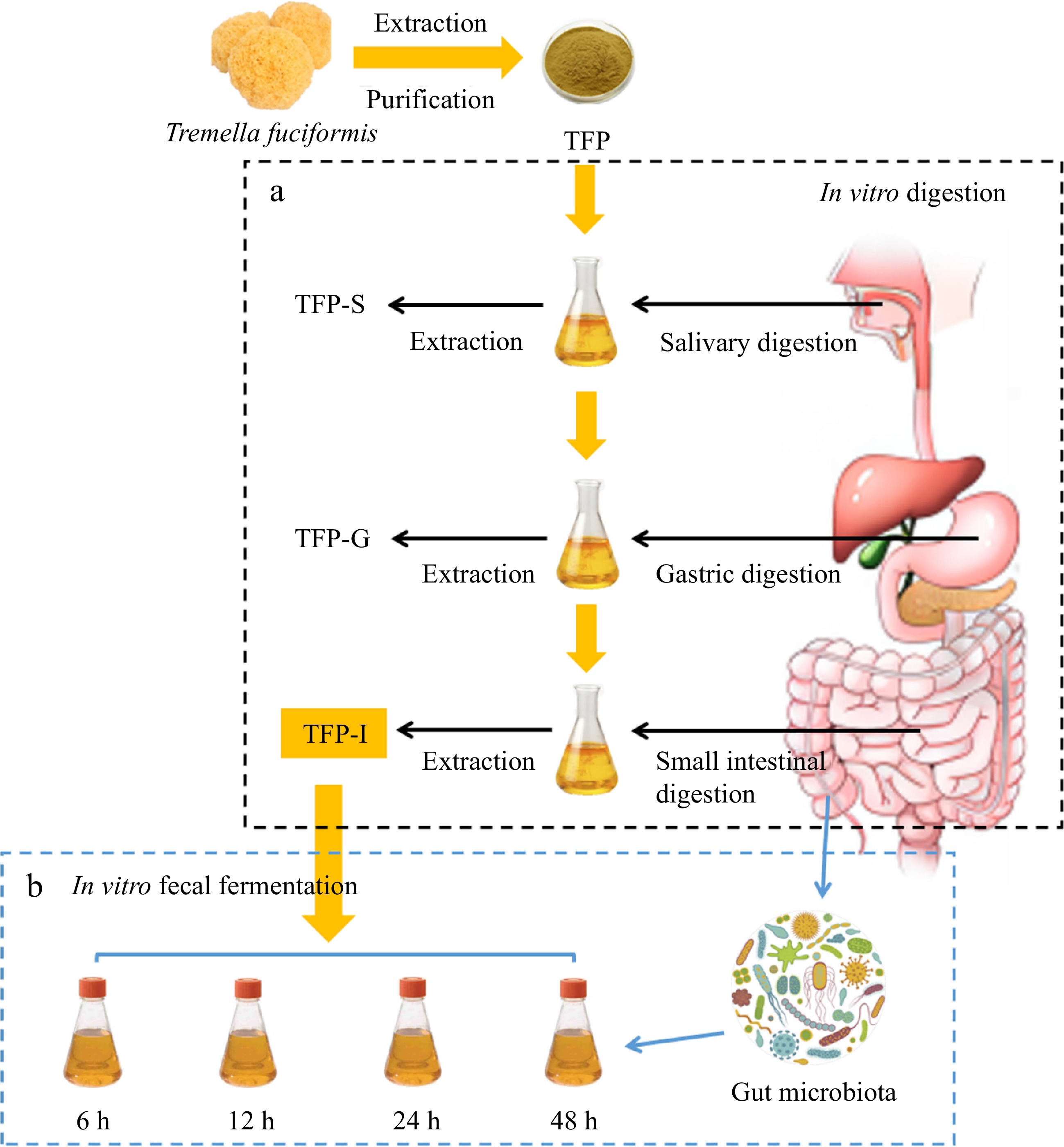
      Figure 1. 

      Flow diagram of (a) in vitro digestion and (b) fecal fermentation method of TFP.

    • The collection of fresh fecal samples, preparation of fermentation media, and methods for in vitro fermentation can be referenced from previous studies with slight modifications[31]. As shown in Fig. 1b, using TFP-I as the carbon source, it was added to the culture medium and subjected to in vitro fermentation using the collected gut microbiota from healthy individuals, named as TFP group. The group that had no carbon supply was designated as the blank control or BLANK group. To serve as a positive control, inulin (a recognized soluble polysaccharide serving as a prebiotic) was used as a substitute for TFP, named as FOS group. Details are supplied in the Supplementary File 1.

    • Measurement of CR, pH, carbohydrate content, and gas production during fermentation in vitro were conducted in accordance with previous studies[23]. Details are supplied in the Supplementary File 1.

    • The fermentation broth was subjected to low-temperature high-speed centrifugation to collect the bacterial pellet in cryovials, which were then stored at −80 °C after liquid nitrogen flash freezing. The gut microbiota investigation was carried out with the methodology outlined in our earlier research[32]. Details are supplied in the Supplementary File 1.

    • The data in the results were presented as 'mean ± SD'. Significant differences were indicated by letters (a−e) at p < 0.05.

    • Table 1 reveals that the total polysaccharide content in TFP were 89.62 ± 0.82%, indicating that polysaccharide is the main component of TFP and its content decreased significantly after gastrointestinal digestion[18]. Notably, the Mw of TFP decreased significantly in the digestion stage, and the content of CR increased significantly (Table 2), indicating that the glucoside bond was destroyed, leading to the decrease of Mw of TFP[33,34].

      Table 1.  Data summarization of TFP, TFP-S, TFP-G and TFP-I.

      TFP TFP-S TFP-G TFP-I
      Total polysaccharides (%) 89.62 ± 0.82a 88.90 ± 0.55ab 87.28 ± 0.78b 86.07 ± 1.03c
      Total uronic acids (%) 15.35 ± 0.80a 15.83 ± 0.36a 14.74 ± 0.11b 14.15 ± 0.33c
      Total proteins (%) 2.53 ± 0.05a 1.90 ± 0.01b 0.79 ± 0.01c 0.60 ± 0.02d
      Molecular weight
      Mw × 104 (Da) 2.0361 ± 0.0375a 1.9686 ± 0.0412a 1.7864 ± 0.0109b 1.6620 ± 0.0156c
      Mw/Mn 1.33172 1.2779 1.20094 1.36855
      Constituent monosaccharides and molar ratios
      Man 1.00 1.00 1.00 1.00
      GlcA 0.07 0.08 0.08 0.07
      Glc 0.86 0.73 0.75 0.58
      Xyl 0.42 0.44 0.40 0.41
      Fuc 0.19 0.19 0.20 0.20

      Table 2.  Variations in CR of TFP during in vitro digestion.

      Processes Time (h) CR (mg·mL−1)
      Origin 0.115 ± 0.001a
      Saliva digestion stage 0.25 0.113 ± 0.003a
      0.5 0.114 ± 0.002a
      1 0.116 ± 0.001a
      Gastric juice digestion stage 0.5 0.312 ± 0.032e
      1 0.364 ± 0.002d
      2 0.408 ± 0.010c
      4 0.583 ± 0.023b
      6 0.729 ± 0.016a
      Small intestinal juice digestion stage 0.5 0.809 ± 0.011c
      1 0.836 ± 0.024bc
      2 0.880 ± 0.039b
      4 0.931 ± 0.022ab
      6 0.950 ± 0.005a
    • Table 2 indicates that the content of CR did not vary considerably in the course of salivary digestion[35], but increased significantly after gastric digestion. This is due to the lower pH conditions in the stomach causing the glucoside bond to break, leading to an increase in the reducing end[36,37].

    • In Fig. 2a, the monosaccharide composition of TFP includes Fuc, Glc, Man, Gal, Xyl, GlcA, GalA, and Ara. Among them, the major monosaccharides in TFP are Glc and Man. Previous studies have shown that the main chain of TFP consists of (1→3)-α-D-mannopyranosyl residues and the side chains consist of Fucp, β-GlcAp, and β-Xylp residues[38]. The molar ratio of Glc declined after simulated digestion, which could be attributed to the lower pH causing degradation of the polysaccharide side chains[18,37], suggesting that in vitro digestion could affect the monosaccharide composition of polysaccharides.
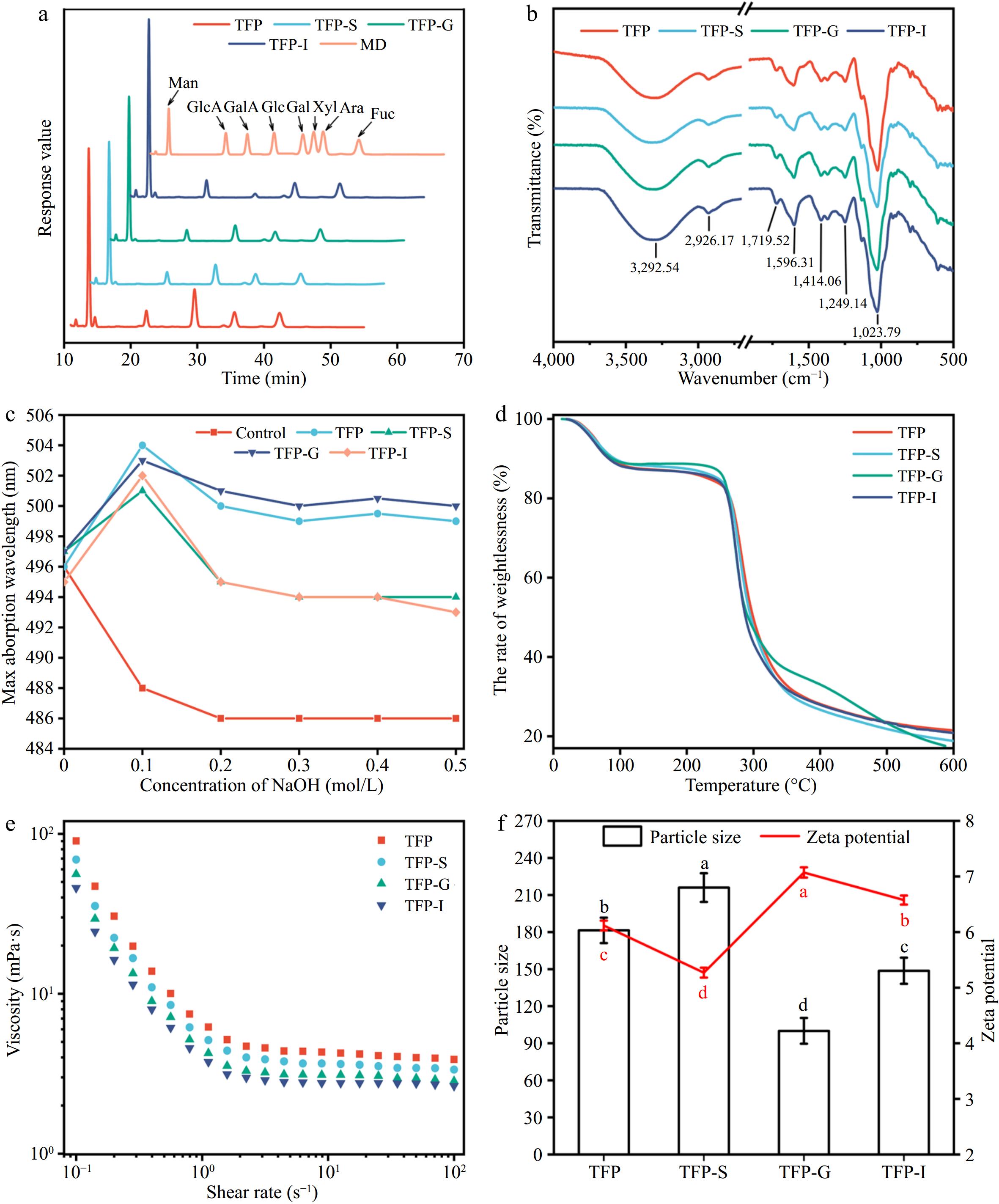
      Figure 2. 

      Variations in structural characterizations of TFP during in vitro digestion. (a) Monosaccharide composition. (b) FT-IR. (c) Congo red staining. (d) Thermogravimetric curve. (e) Rheological properties. (f) Particle size and zeta potential.

    • Figure 2b demonstrated that FT-IR spectra of TFP following the simulated digestion were comparable, suggesting that the RG-I backbone and other structural features of TFP were unaffected by the in vitro simulated digestion process[37]. In particular, the existence of carbohydrates was confirmed by the characteristic peak at 3,600−3,200 cm−1, which correlated with the O-H stretching vibration[39], and the range of 1,400−1,200 cm−1, which correlated with the C-H bending vibration[40,41]. The presence of uronic acids was indicated by the asymmetric stretching vibration of free carboxyl groups, which fell within the range of 1,590−1,644 cm−1[40]. Additionally, at 1,555 cm−1, there was no absorption peak, indicating a very low protein content in the polysaccharide samples. The peak at around 917 cm−1 presented the characteristic vibration of the non-symmetric ring stretching of pyranose[5].

    • TFP was described as a polysaccharide with a triple helix shape in Fig. 2c, and this structure held unchanged even after in vitro digestion was simulated. The TG curves in Fig. 2d did not reveal any discernible variations between various phases of TFP digestion. At temperatures between 25 and 600 °C, polysaccharides exhibited three stages of thermal degradation[42]. Notably, in the second stage (101−337 °C), there was a sharp decrease in weight, mainly because the anhydrous organic components gradually decompose under high-temperature heating. The rhamnogalacturonan chain was degraded, leading to carbonization and oxidation, causing the volatilization and loss of a large number of volatile small molecules. In the third stage (337−600 °C), aromatic carbon residues undergo combustion[43].

    • The apparent viscosities of TFP exhibited a typical Newtonian plateau at high shear rates, as presented in Fig. 2e[29]. Figure 2f exhibits that TFP-G had the lowest particle size, suggesting that TFP dissociates more readily in the acidic environment of the stomach. The highest charge was observed for TFP-G, indicating that the small intestine was able to absorb and utilize TFP-G-digested samples more easily.

    • Enzymes encoded by gut microbiota could break down carbohydrates into fermentable sugars, and the growth metabolism of gut microbiota could influence the content of total carbohydrates in the fermentation medium. As shown in Fig. 3a, the carbohydrate content of all substrates decreased most rapidly during the first 6 h, indicating that the fecal microbiota was in the logarithmic growth phase with the highest carbohydrate consumption[44]. With increasing fermentation time, the total sugar content in BLANK, TFP, and FOS groups all showed a decreasing trend, suggesting varying degrees of carbohydrate utilization and the presence of unfermentable components. Studies have found that the consumption of aloe polysaccharides after 48 h of fermentation was approximately 56%[45], and the total sugar consumption of loquat polysaccharides after fermentation was as high as 85%[46], In this experiment, after 48 h of fermentation, the FOS group consumed approximately 69.90% and the TFP group consumed approximately 66.08%. Therefore, TFP was a good fermentation substrate that could be effectively utilized by microorganisms.

      Figure 3. 

      Variations in CR, pH, residual carbohydrates and gas production during in vitro fermentation of TFP. (a) Total carbohydrates. (b) Reducing sugars. (c) pH value. (d) The amount of gas produced.

      As can be seen from Fig. 3b, throughout the entire process of fecal fermentation, the fermentation broth contained very few reducing sugars, ranging from 0.09 ± 0.03 mg/mL to 0.12 ± 0.04 mg/mL, suggesting that the gut microbiota can fully use the reducing sugars generated by TFP-I, with a dynamic balance between enzymatic hydrolysis rate and utilization rate[47].

      The pH level is a crucial signal throughout the fermentation process. Figure 3c exhibits that the pH values of the FOS and TFP groups were consistently lower than those of the BLANK group, owing to acidic substances like short-chain fatty acids (SCFAs) were produced throughout the fermentation process through the fermentation of polysaccharides. The development of pathogenic bacteria may be inhibited by the reduction in intestinal pH. Therefore, TFP and inulin could lower the colonic pH and maintain gut health.

      The gut microbiota tends to produce gases like CH4, H2 and CO2 while fermenting carbohydrates, which could cause adverse symptoms and were the main reason for the limitation of prebiotic application[48]. In Fig. 3d, the gas production of FOS, TFP, and BLANK groups gradually raised during fermentation. After fermentation for 48 h, the gas volume produced by TFP fermentation (0.53 mL) was significantly lower than that of inulin (1.08 mL), indicating that TFP was a more advantageous prebiotic biomass than inulin in terms of gas production.

    • Gut microbiota are crucial for the body's ability to absorb and store energy, perform a number of metabolic processes, and control the immune system, which are crucial for human health and disease. Previous studies have found that through altering gut microbiota, TFP reduced colitis caused by DSS in mice. Thus, it was essential to understand the connection between gut bacteria and TFP, as modulating gut microbiota could contribute to disease prevention and promote health.

      Figure 4ac indicated that most bacterial diversity in the samples was covered by the sequencing depth, indicating that the volume of sequencing data was appropriate. The findings in Fig. 4d & e revealed that there were remarkable differences in the gut microbiota composition between the BLANK, FOS, and TFP groups. Figure 4f demonstrated that between the three groups, there were more inter-group differences than intra-group differences, with intra-group differences being very minor. Figure 4g presented a certain distance between the samples in BLANK, FOS and TFP groups, indicating the specificity of bacterial distribution[49]. In summary, the inter-group differences of each experimental group were remarkably different from one another, and these differences outweighed the intragroup differences, suggesting that carbohydrates from different sources had different effects on the microbial community.
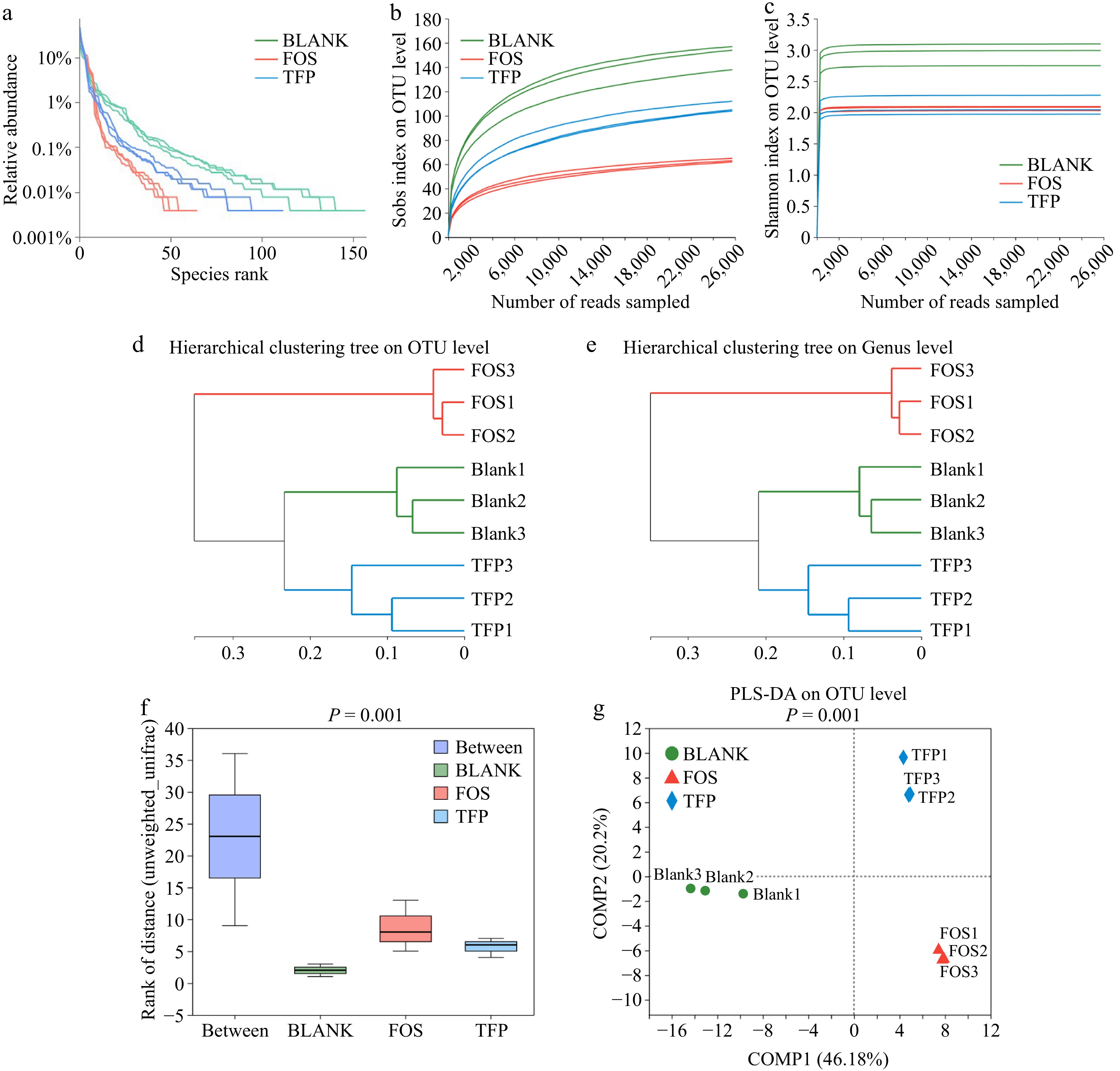
      Figure 4. 

      Correlation curve of species diversity and between-group similarity analysis of gut microflora in vitro fermentation for 48 h. (a) Rank-Abundance curve. (b), (c) Rarefaction curve. (d), (e) Hierarchical clustering tree based on OUT and Genus levels. (f) ANOSIM/Adonis analysis. (g) PLS-DA analysis.

      The samples in the TFP and FOS groups had much lower Sobs, Shannon, ACE, and Chao indices than the BLANK group, as presented in Figs. 5ad, indicating that supplying the gut microbiota with inulin and TFP as carbon sources can result in various degrees of decline in microbial abundance and diversity[43]. The FOS group samples had the lowest microbial diversity and richness, which was similar to the findings of Yu et al.[50]. These findings displayed that the microbial community composition might be changed by both FOS and TFP interventions. In addition, PCA analysis, PCoA analysis, and NMDS analysis (Fig. 5eg) demonstrated that the samples from the TFP, FOS, and BLANK groups exhibited a certain distance, suggesting that the gut microbiota compositions of the three groups varied. The results indicated that TFP, together with gut microbiota could change the microbial community composition, and the impact on gut microbiota varies when using carbohydrates from different sources for in vitro fermentation.
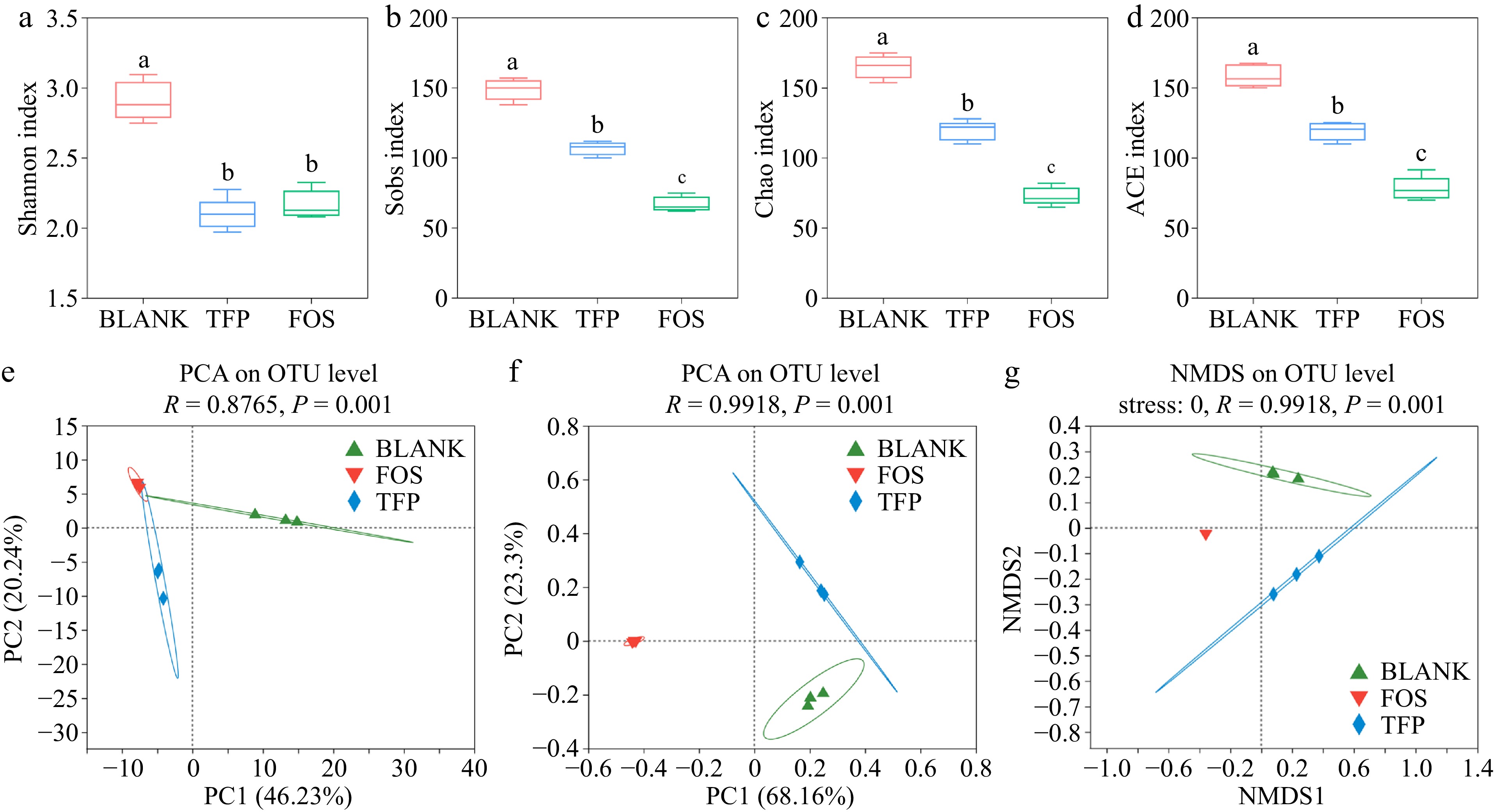
      Figure 5. 

      Analysis of α and β diversity after 48 h of fermentation in vitro in the gut microbial community. (a)-(d) α diversity indices. (e) PCA analysis. (f) PCoA analysis. (g) NMDS analysis.

      Figure 6 displayed the changes in gut microbiota after 48 h of in vitro fermentation. As illustrated in Fig. 6a, c & e, the BLANK group at the genus level consisted mainly of Phascolarctobacterium, Bacteroides, Escherichia-Shigella, Klebsiella and Fusobacterium. In comparison with the BLANK group, Bacteroide proportion significantly increased in the TFP group. Bacteroides were one of the most significant genera of intestinal microbiota and could digest dietary fiber polysaccharides and host glycans. In addition, Bacteroides acted as a key player in the fight against obesity, immune disorders, and the alleviation of intestinal inflammation[51]. The proportion of Megasphaera and Phascolarctobacterium was elevated in the TFP group versus the BLANK group, which was in line with previous findings[37]. At the same time, Escherichia-Shigella and Fusobacterium were reduced in the TFP group, which suggested that TFP could facilitate the beneficial bacteria development and suppress the harmful bacteria development. In the FOS group, the relative abundance of Escherichia-Shigella significantly increased, as Escherichia-Shigella lacked carbohydrate-active enzymes and cannot utilize polysaccharides, whereas inulin, as a low-molecular-weight carbon source, facilitated its growth[52]. Furthermore, the relative abundance of Bifidobacterium, which could degrade and apply inulin to enhance the generation of fermentation end products might be greatly raised by inulin[37].
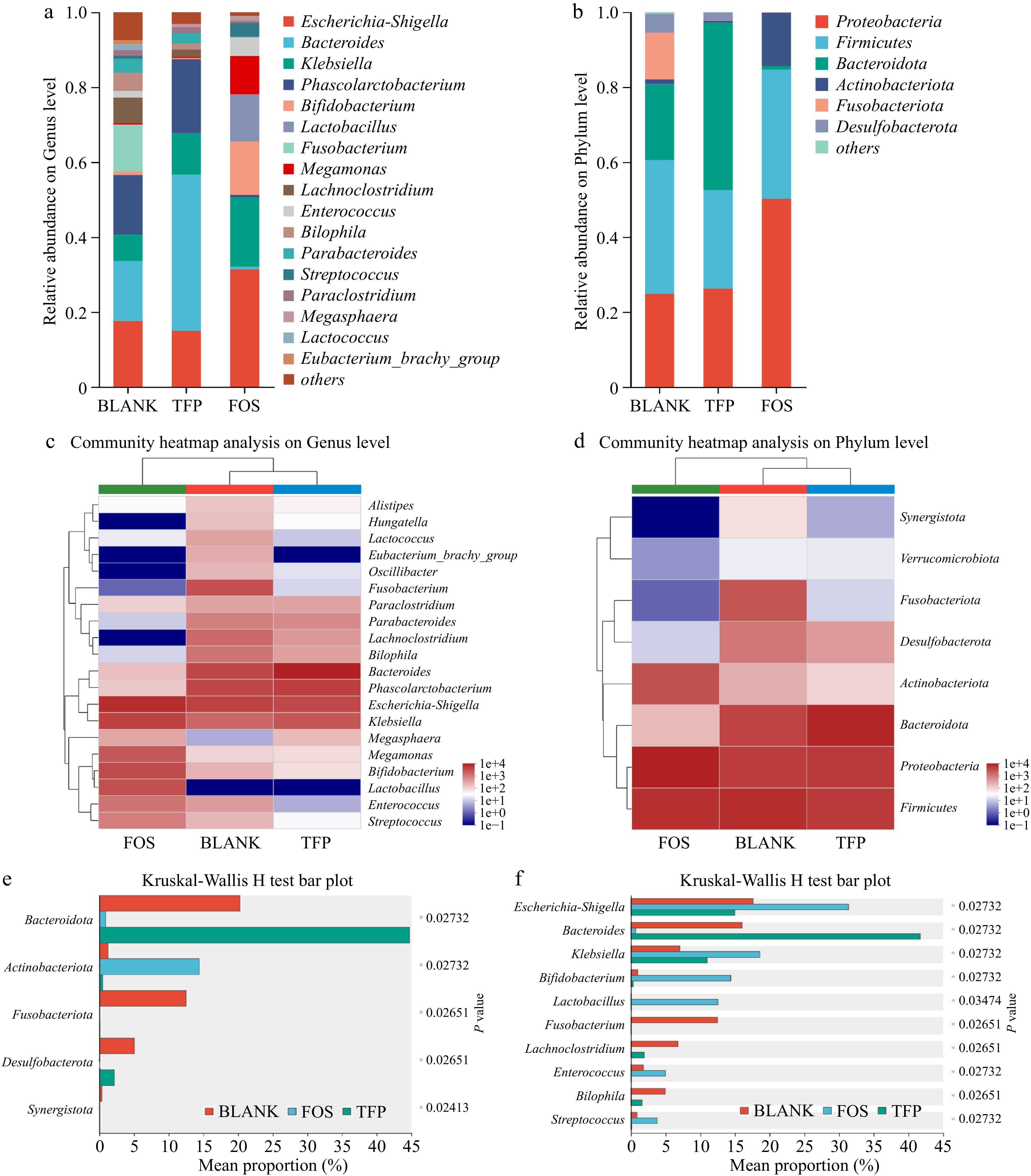
      Figure 6. 

      Analysis of gut microbial community composition during 48 h of in vitro fermentation. (a), (b) Relative abundance. (c), (d) Community heatmap analysis. (e), (f) Kruskal-Wallis H test bar plot.

      Figure 6b, d & f indicated that the major bacteria in the BLANK group at the phylum level were Proteobacteria, Firmicutes, Fusobacteriota, and Bacteroidetes. In comparison to the BLANK group, the TFP group had a much higher proportion of Bacteroidetes, but a markedly lower proportion of Firmicutes. Bacteroidetes was one of the main intestinal bacteria that were responsible for degrading polysaccharides[53]. When degrading substrates, it released polysaccharide hydrolases and glycoside hydrolases for the degradation of the Gal side chain structures along with the RG-I backbone of polysaccharides[37]. In addition, a rise in the ratio of Bacteroidetes to Firmicutes may reduce the risk of insulin resistance and obesity[54]. Therefore, TFP may play a role in anti-obesity and reducing insulin resistance by regulating the ratio of Bacteroidetes to Firmicutes. Fusobacteriota was generally considered to be associated with some opportunistic pathogens. The low proportion of Fusobacteriota in both TFP and FOS groups indicated that the addition of TFP and inulin could inhibit certain opportunistic pathogens. Proteobacteria and Actinobacteriota were the most varied bacterial phyla, which are often present in the fecal microbiota of healthy individuals[52]. The relative abundance of Actinobacteriota in the FOS group was substantially higher than the BLANK group. Actinobacteriota were Gram-positive bacteria that could convert carbohydrates into non-toxic acidic substances and were believed to promote intestinal health[55]. In summary, inulin and TFP have the potential to alter the gut microbiota composition, especially by encouraging the growth of beneficial bacteria. However, there were differences in their effects on gut microbiota. Compared to the FOS group, the addition of TFP had less interference with the normal community structure and was more conducive to maintaining gut homeostasis in the short term.

    • In conclusion, the present research demonstrated that TFP partially degraded under circumstances resembling salivary gastrointestinal digestion, leading to a notable rise in CR content and a fall in Mw. Furthermore, indigestible TFP-I may be extensively applied by the human gut microbiota during in vitro fecal fermentation. TFP demonstrated the ability to modulate both α and β diversity in the intestinal microbiota and induce changes in the community composition at the phylum and genus levels. This included a decrease in the growth of harmful bacteria for instance Escherichia-Shigella and Fusobacterium and a rise in the abundance of beneficial bacteria like Megasphaera, Phascolarctobacterium, and Bacteroides. These findings indicated that TFP had the potential to be a functional food that enhanced the intestinal microbiota environment, thereby promoting health and preventing disease, e.g., prebiotic.

    • The authors confirm contribution to the paper as follows: conceptualization, methodology, software, investigation, formal analysis, visualization: Zhu X; writing - original draft: Zhu X; writing - review & editing: Su J, Zhang L, Si F, Li D, Jiang Y, Zhang C; supervision: Jiang Y, Zhang C; resources: Zhang C. All authors reviewed the results and approved the final version of the manuscript.

    • This published article and associated supplementary information files contain all of the data generated or analyzed during this work.

      • This work was financially supported by the National Natural Science Foundation of China (31901644) and the University Innovation Team of Shandong Province (2022KJ243).

      • The authors declare that they have no conflict of interest.

      • Supplemental File 1 Changes in molecular weight of TFP during in vitro digestion. (a) TFP. (b) TFP-S. (c) TFP-G. (d) TFP-I.
      • Copyright: © 2024 by the author(s). Published by Maximum Academic Press on behalf of China Agricultural University, Zhejiang University and Shenyang Agricultural University. This article is an open access article distributed under Creative Commons Attribution License (CC BY 4.0), visit https://creativecommons.org/licenses/by/4.0/.
    Figure (6)  Table (2) References (55)
  • About this article
    Cite this article
    Zhu X, Su J, Zhang L, Si F, Li D, et al. 2024. Gastrointestinal digestion fate of Tremella fuciformis polysaccharide and its effect on intestinal flora: an in vitro digestion and fecal fermentation study. Food Innovation and Advances 3(2): 202−211 doi: 10.48130/fia-0024-0018
    Zhu X, Su J, Zhang L, Si F, Li D, et al. 2024. Gastrointestinal digestion fate of Tremella fuciformis polysaccharide and its effect on intestinal flora: an in vitro digestion and fecal fermentation study. Food Innovation and Advances 3(2): 202−211 doi: 10.48130/fia-0024-0018

Catalog

  • About this article

/

DownLoad:  Full-Size Img  PowerPoint
Return
Return