Loading [MathJax]/jax/output/SVG/jax.js
ARTICLE   Open Access    

Identification and fungicide sensitivity of the blue mold pathogen in postharvest-stored elephant garlic bulbs in Thailand

More Information
  • Received: 05 November 2024
    Revised: 25 November 2024
    Accepted: 26 November 2024
    Published online: 20 December 2024
    Studies in Fungi  9 Article number: e015 (2024)  |  Cite this article
  • Blue mold disease is one of the most important postharvest diseases affecting garlic bulbs. In 2023, this disease was found on bulbs of elephant garlic [Allium ampeloprasum var. ampeloprasum (Borrer) Syme] in Chiang Mai Province, Thailand, during the postharvest storage period. Three fungal isolates were obtained and identified as Penicillium allii based on morphological characteristics and phylogenetic analysis of combined sequences of the internal transcribed spacer (ITS) of ribosomal DNA, β-tubulin (BenA), calmodulin (CaM), and RNA polymerase II second largest subunit (rpb2) genes. In the pathogenicity test, garlic bulbs inoculated with the isolated fungi exhibited symptoms similar to those observed during the postharvest storage period. In the fungicide screening test, carbendazim, difenoconazole + azoxystrobin, and difenoconazole effectively completely inhibited this fungus at both half and recommended dosages, while the fungus showed insensitivity to captan and mancozeb. Additionally, double-recommended dosages of carbendazim, copper oxychloride, difenoconazole combined with azoxystrobin, and difenoconazole alone exhibited complete inhibition of the fungus. To the best of our knowledge, this is the first report of postharvest blue mold disease on elephant garlic bulbs caused by P. allii in Thailand. Furthermore, the results of the fungicide sensitivity screening could help in developing effective management strategies for controlling postharvest blue mold disease on elephant garlic bulbs caused by P. allii.
  • Absidia was introduced by van Tieghem[1], with A. reflexa as the type species. Preliminarily, Absidia was accommodated in Absidiaceae as the type genus[2]. However, with DNA sequence techniques used in species identification, Absidia always formed a well-supported monophyletic clade with genera in Cunninghamellaceae (viz., Chlamydoabsidia, Cunninghamella, Gongronella, Hesseltinella, and Halteromyces)[37]. Hence, Absidia was hitherto classified in Cunninghamellaceae[8] whereas Absidiaceae was treated as a synonym of the highly polyphyletic family Mucoraceae by Kirk et al.[7]. Although, Hoffmann[9] highly recommended to resurrect the monophyletic Absidiaceae in accordance with Voigt et al.[10].

    Since the genus Absidia was introduced[1], many different allied genera were subsequently established to accommodate absidia-like species based solely on morphological characteristics[1116]. Considering the morphological distinctiveness, it may be challenging among the genera Absidia, Gongronella, Lichtheimia, Mycocladus, Phycomyces, Proabsidia, Protoabsidia, Pseudoabsidia, Rhizopus, and Tieghemella, because these genera are highly similar in morphology[17]. Nevertheless, Hesseltine & Ellis[17] accepted only Absidia and Gongronella as distinct genera. However, a morpho-molecular approach coupled with physiology significantly segregated these genera into different families. Among them, only Absidia (synonyms: Proabsidia, Pseudoabsidia, and Tieghemella), and Gongronella were classified in Cunninghamellaceae whereas Lichtheimia (synonym: Protoabsidia) was classified in Lichtheimiaceae, Mycocladus in Mycocladaceae, Phycomyces in Phycomycetaceae, and Rhizopus in Rhizopodaceae[18].

    A comprehensive study based on morphology, phylogeny, and physiology provided reliable species delineation of Absidia[19]. Absidia species were segregated into three groups viz. mesophilic species (optimal growth temperature 25–34 °C), mycoparasitic species on mucoralean fungi (optimal growth temperature 14–25 °C) and thermotolerant species (optimal growth temperature 37–45 °C)[19]. Consequently, the new genus Lentamyces was established to accommodate the mycoparasitic species[20], whereas the thermotolerant species were transferred to the genus Lichtheimia[21]. To date, only the mesophilic species (optimal growth temperature 25–34 °C) were accepted as Absidia sensu stricto[9,20,21]. Currently, species identification and classification for the genus Absidia are mainly based on morphological characteristics coupled with phylogeny and physiology (e.g., growth temperature)[9,19,20,2226]. Based on evolutionary relationships, Zhao et al.[25] updated the classification framework of the phylum Mucoromycota and calibrated the taxonomic ranks with estimated divergence times using molecular dating. Among these, the estimated divergence time of the phylum Mucoromycota was earlier than 617 Mya, the classes and orders earlier than 547 Mya, the families earlier than 199 Mya, and the genera earlier than 12 Mya. According to Zhao et al.[25], the divergence time of Absidia was estimated to be 327 Mya for the stem age and 135 Mya for the crown age, based on a time-scaled Bayesian maximum clade credibility phylogenomic tree inferred from ITS and LSU rDNA sequences[25].

    Absidia is ubiquitous and distributed in a wide range of ecological niches worldwide, including air, animal dung, food, insect remains, leaf litter, and soil[24]. Notably, soil accounts for more than 50% of Absidia species[24]. China has been recognized as a region of high diversity for Absidia species, contributing ~20% to the origin of taxonomic types in this genus[24,27]. This genus is typically characterized by sporangiophores with columellae bearing one to several projections, rhizoids forming between the sporangiophores but never opposite them, and the development of pyriform, deliquescent-walled and apophysate sporangia[1,9,19,20,24,25,2831]. Some species within Absidia such as A. biappendiculata, A. chinensis, A. cinerea, and A. jiangxiensis, produce zygospores within zygosporangia, with their opposite suspensory cells possessing appendages[9,19,25].

    The secondary metabolites of Absidia have been relatively well-studied, particularly for their industrial implications[25,3236]. For instance, A. coerulea is capable of bio-transforming saponins, and regioselectivity to 20(S)-protopanaxatriol. Also, this species has been used in bio-transformation of the (−)-α-bisabolol, and in the hydroxylation of dehydroepiandrosterone (DHEA) to 7α-hydroxy-5-androstene-17-one (7α-OH-DHEA) and 7β-hydroxy-5-androstene-17-one (7β-OH-DHEA). These processes provide efficient pathways to enhance the yield of saponins, (−)-α-Bisabolol and 7β-OH-DHEA for the cosmetic and pharmaceutical industries[3437]. Furthermore, A. coerulea and A. glauca have been used in the bio-transforming of flavones (chrysin, apigenin, luteolin, and diosmetin), flavanones (pinocembrin, naringenin, eriodictyol, and hesperetin) and 3-Oxo-Oleanolic acid into hydroxylated metabolites. These two species are also efficient producers of chitosan, which is widely used in food processing and antimicrobial products, as well as in the bio-transforming of steroid products[33,34]. Absidia cylindrospora has demonstrated the ability to act as a biosorption for cadmium, copper, and lead under controlled experimental conditions[34,38]. Additionally, A. cylindrospora and A. fusca have been used in bioremediation processes due to their capacity to degrade polycyclic aromatic compounds, such as hydrocarbons[34,39]. Moreover, during the bio-transformation of 20(R)-panaxadiol by A. corymbifera, six oxidized and hydroxylated products reported were identified as novel compounds[32]. Other Absidia species have also been explored for enzyme engineering and metabolomics in industrial contexts. For instance, α-galactosidase from A. reflexa has been used to produce rubusoside derivatives, A. repens has been utilized to yield chitosan and laccase from A. spinosa has been applied for the biotransformation of cresol red[25,40]. Given these findings, Absidia is a promising resource for applications in food processing, antimicrobial production, and biotransformation. Besides, new secondary metabolites are frequently discovered during microbial transformation involving Absidia species.

    During a survey to collect microfungi associated with avocado in Yunnan Province, China, two isolates were obtained, one from the soil in an avocado plantation and another as a root endophyte of an avocado tree. Based on morphological, phylogenetic, and physiological analyses, these isolates were determined to be conspecific and represent a novel species of Absidia.

    The two fungal strains were isolated from soil samples from an avocado plantation and fresh healthy roots of avocado trees, both of which were collected from Menglian County, Yunnan Province, China in September, 2023. Soil samples were collected from 10 cm below ground level, near the shallow roots of avocado trees using a sterilized auger boring tool (model XDB, China, total length 110 cm, drill outer diameter 57 mm, inner diameter 51 mm, single sampling length 20 cm). A portion of the soil (1 g) was mixed with 9 mL of sterile distilled H2O. Serial dilutions of the mixtures were prepared from 10−1 to 10−4. For each dilution, a 100 μL aliquot was plated onto potato dextrose agar (PDA; Qingdao Daily Water Biotechnology Co. Ltd. Shandong, China) with antibiotics (streptomycin sulfate, 100 mg/L, and ampicillin, 100 mg/L). The plates were incubated at 25–30 °C. The PDA plate was examined daily with an Olympus stereo-microscope series MoDELSZ2-ILST. The presence of colonies was transferred to new PDA plates. The isolation and induction of root endophytes from the root tissue of avocado tree were carried out following the guidelines as described in Senanayake et al.[41] and Mattoo & Nonzom[42]. Root samples were cleaned with tap water, and each root was cut into several small pieces (5 mm long). The root pieces were surface sterilized using 2% sodium hypochlorite (NaOCl) for 1 min, followed by three repeated washings with sterilized water. Furthermore, surface sterilization was carried out via washing with 75% ethanol for 30 s and three series of washing with sterile distilled water, and finally it was allowed to dry under a laminar flow cabinet. Then, the edges of the root pieces were trimmed, transferred onto plates, and incubated at 25–30 °C for 2–5 d. When individual hyphal tips grew out from root pieces, the mycelia were picked using sterilized needles, placed onto new PDA plates, and incubated at 25–30 °C under 12 h alternating light and dark conditions for one week to one month. After one month, the sporulation was observed. Dry culture materials were deposited in the herbarium of the Cryptogams Kunming Institute of Botany Academia Sinica (KUN-HKAS). The living cultures were conserved at the Kunming Institute of Botany Culture Collection (KUNCC). Index Fungorum number is provided for the newly described taxon.

    Micromorphological features (sporangia, sporangiophores, sporangiospores, and columellae, etc.) were examined and captured by a Nikon compound microscope (model ECLIPSE Ni-U, Japan) connected with a Nikon DS-Ri2 camera using DIC microscopy. Measurements of these morphological features were made in Tarosoft® Image Frame Work software version 0.9.7. Photographic plates were edited and combined in Adobe Photoshop CS6 software (Adobe Systems Inc., USA). The determination of maximum growth temperature followed the method of Zhao et al.[24]. Four fungal blocks (5 mm × 5 mm) were selected from a one-week-old colony growing on PDA and placed separately onto four new PDA plates, representing four replicates. These replicates were incubated at 30 °C for 2 d. Subsequently, the temperature was gradually increased in 2 °C increments (32, 34, and 36 °C), with incubation at each temperature for 2 d, until the colonies ceased to grow. When colony growth stopped (at 36 °C), the maximum growth temperature was confirmed by observing the mycelium growth at a reduced temperature (35 °C) at which the colonies also failed to grow.

    The genomic DNA was extracted from fresh mycelia grown on PDA medium for two weeks using the Biospin Fungus Genomic DNA Extraction Kit (BioFlux®, Hangzhou, China) following the manufacturer's instructions. Partial DNA sequences of the internal transcribed spacers (ITS1-5.8S-ITS2) and the 28S large subunit rDNA (LSU) were amplified by primers ITS5 and ITS4[43], and LR0R and LR5[44], respectively. The component of the PCR reaction mixture was performed in a 25 μl total volume containing 2 μl of DNA template, 1 μl of each forward and reverse primers (10 μM), 12.5 μl of 2× Power Taq PCR Master Mix (mixture of EasyTaqTM DNA Polymerase, dNTPs, and optimized buffer, Beijing Bio Teke Corporation (Bio Teke), China) and 8.5 μl of ddH2O (double-distilled water). The thermal cycling of PCR amplification for ITS and LSU followed the method of Xu et al.[45] but the annealing temperature was adjusted to 52 °C. PCR products were sent to TsingKe Biological Technology (Beijing) Co., Ltd, China for purification and sequencing. Newly generated consensus sequences were deposited in GenBank under accession numbers provided in Table 1.

    Table 1.  Taxa names, strain numbers, and GenBank accession numbers of taxa used for the present phylogenetic analyses.
    Species names Strain numbers GenBank accession numbers
    ITS LSU
    Absidia abundans XY09265 ON074697 ON074681
    Absidia abundans CGMCC 3.16255 NR_182590 ON074683
    Absidia abundans XY09274 ON074696 ON074682
    Absidia aguabelensis URM 8213 NR_189383 NG_241934
    Absidia alpina CGMCC 3.16104 OL678133 /
    Absidia ampullacea CGMCC 3.16054 NR_191130 NG_242502
    Absidia anomala CBS 125.68 NR_103626 NG_058562
    Absidia biappendiculata CBS 187.64 NR_190243 MZ350147
    Absidia bonitoensis URM 7889 MN977786 MN977805
    Absidia brunnea CGMCC 3.16055 NR_191131 NG_242503
    Absidia caatinguensis URM 7156 NR_154704 NG_058582
    Absidia caerulea CBS 101.36 MH855718 MH867230
    Absidia caerulea XY00608 OL620081 /
    Absidia californica CBS 126.68 NR_077169 NG_056998
    Absidia californica FSU4748 AY944873 EU736301
    Absidia cheongyangensis CNUFC CY2203 PP844904 PP852788
    Absidia chinensis CGMCC 3.16057 MZ354141 MZ350135
    Absidia chinensis CGMCC 3.16056 NR_191132 NG_242504
    Absidia cinerea CGMCC 3.16062 NR_191134 NG_242506
    Absidia coerulea FSU767 AY944870 /
    Absidia coerulea XY00729 OL620082 /
    Absidia cornuta URM 6100 NR_172976 MN625255
    Absidia crystalloides CGMCC3.27496 PP377803 PP373736
    Absidia crystalloides SAUCC693201 PP377804 PP373737
    Absidia cuneospora CBS 101.59 MH857828 NG_058559
    Absidia cylindrospora CBS 153.63 / NG_058563
    Absidia cylindrospora XY00313 ON123744 ON123765
    Absidia cylindrospora var. cylindrospora CBS 100.08 JN205822 JN206588
    Absidia digitula CGMCC 3.16058 MZ354142 MZ350136
    Absidia edaphica MFLU 20-0416 NR_172305 NG_075367
    Absidia fluvii CNUFC CY2240 PP844891 PP852703
    Absidia frigida CGMCC 3.16201 NR_182565 NG_149032
    Absidia fusca CBS 102.35 NR_103625 NG_058552
    Absidia gemella CGMCC 3.16202 OM108488 NG_149033
    Absidia glauca CBS 129233 MH865253 MH876693
    Absidia glauca CBS 101.08 NR_111658 NG_058550
    Absidia glauca FSU660 AY944879 EU736302
    Absidia globospora CGMCC 3.16031 NR_189829 NG_241930
    Absidia globospora CGMCC 3.16035 MW671538 MW671545
    Absidia globospora CGMCC 3.16036 MW671539 MW671546
    Absidia healeyae UoMAU1 MT436028 MT436027
    Absidia heterospora SHTH021 JN942683 JN982936
    Absidia jiangxiensis CGMCC 3.16105 OL678134 /
    Absidia jiangxiensis [as jindoensis] XY000810 ON123748 ON123770
    Absidia jindoensis CNUFC-PTI1-1 MF926622 MF926616
    Absidia koreana EML-IFS45-1 KR030062 KR030056
    Absidia kunryangriensis CNUFC CY2230 PP844905 PP956882
    Absidia lobata CGMCC 3.16256 ON074690 ON074679
    Absidia longissima CGMCC 3.16203 NR_182566 OM030225
    Absidia macrospora FSU4746 AY944882 EU736303
    Absidia medulla CGMCC 3.16034 NR_189832 NG_241932
    Absidia menglianensis KUNCC 24-18541 PQ594927 PQ594929
    Absidia menglianensis KUNCC 24-18542 PQ594928 PQ594930
    Absidia microsporangia KUNF-22-121A OQ868364 OQ868366
    Absidia microsporangia KUNF-22-126A OQ868365 OQ868367
    Absidia microsporangia KUNF-22-316 PP264375 PP264469
    Absidia montepascoalis URM 8218 MW473494 MW561560
    Absidia multispora URM 8210 MN953780 MN953782
    Absidia nigra CGMCC 3.16060 MZ354144 MZ350138
    Absidia nigra CGMCC 3.16059 MZ354143 MZ350137
    Absidia nigra CBS 127.68 NR_173068 NG_058560
    Absidia oblongispora CGMCC 3.16061 NR_191133 NG_242505
    Absidia ovalispora CGMCC 3.16019 NR_176748 MW264131
    Absidia pacifica CGMCC3.27497 PP377802 PP373735
    Absidia pacifica SAUCC413601 PP377801 PP373734
    Absidia panacisoli SYPF 7183 NR_159563 NG_063948
    Absidia paracylindrospora CNUFC L2207 PP844907 PP956883
    Absidia pararepens CCF 6352 MT193669 NG_242483
    Absidia pateriformis CGMCC3.27495 PP377805 PP373738
    Absidia pateriformis SAUCC634702 PP377806 PP373739
    Absidia pernambucoensis URM 7219 MN635568 MN635569
    Absidia pseudocylindrospora CBS 100.62 NR_145276 NG_058561
    Absidia psychrophilia FSU4745 AY944874 EU736306
    Absidia psychrophilia CBS 128.68 NR_154667 NG_058544
    Absidia purpurea CGMCC 3.16106 NR_191153 /
    Absidia radiata CGMCC 3.16257 ON074698 ON074684
    Absidia radiata XY09330 ON074699 ON074685
    Absidia repens CBS 115583 NR_103624 NG_058551
    Absidia saloaensis URM 8209 MN953781 MN953783
    Absidia sichuanensis CGMCC 3.16258 NR_182589 ON074688
    Absidia soli MFLU 20-0414 NR_172306 NG_075368
    Absidia sp. [as pseudocylindrospora] EML-FSDY6-2 KU923817 KU923814
    Absidia spinosa FSU551 AY944887 EU736307
    Absidia stercoraria EML-DG8-1 KU168828 KT921998
    Absidia sympodialis CGMCC 3.16063 MZ354147 MZ350141
    Absidia sympodialis CGMCC 3.16064 NR_191135 NG_242507
    Absidia tarda URM 8412 PP844911 PP956884
    Absidia terrestris FMR 14989 LT795003 LT795005
    Absidia thailandica MFLUCC 23-0073 OR606547 OR606546
    Absidia turgida CGMCC 3.16032 NR_189830 NG_241931
    Absidia varians CGMCC 3.16065 NR_191136 NG_242508
    Absidia variiprojecta URM 8620 PP844913 PP956885
    Absidia variispora URM 8720 PP844915 PP956886
    Absidia virescens CGMCC 3.16066 MZ354150 MZ350144
    Absidia virescens CGMCC 3.16067 NR_191137 NG_242509
    Absidia xinjiangensis CGMCC 3.16107 OL678136 /
    Absidia yunnanensis XY09528 ON074701 ON074686
    Absidia yunnanensis CGMCC 3.16259 NR_182591 NG_149054
    Absidia zonata CGMCC 3.16033 NR_189831 NG_242490
    Absidia zygospora MFLUCC 23-0061 OR104965 OR104992
    Cunninghamella blakesleeana CBS 782.68 JN205869 MH870950
    Cunninghamella elegans CBS 167.53 MH857146 HM849700
    The ex-type strains are in bold and newly sequences in the present study are indicated in blue. Abbreviations: CBS: Westerdijk Fungal Biodiversity Institute, Utrecht, Netherlands; CCF: Culture Collection of Fungi, Charles University in Prague; CGMCC: China General Microbiological Culture Collection Center, Beijing, China; CNUFC: Chonnam National University Fungarium, Gwangju, South Korea; FMR: Facultad de Medicina, Reus, Tarragona, Spain; KUNCC: Kunming Institute of Botany Culture Collection, Kunming, China; MFLU: Herbarium of Mae Fah Luang University, Chiang Rai, Thailand; MFLUCC: Mae Fah Luang University Culture Collection, Chiang Rai, Thailand; NRRL: USDA-Agricultural Research Service Culture Collection, US; SAUCC: Shandong Agricultural University Culture Collection (SAUCC), China; URM: University Recife Mycology Culture Collection of the Universidade Federal de Pernambuco, Brazil.
     | Show Table
    DownLoad: CSV

    The nucleotide BLAST search (https://blast.ncbi.nlm.nih.gov/Blast.cgi, accessed on 10 August 2024) was applied to discover taxa closely related to the new isolates (KUNCC 24-18541 and KUNCC 24-18542). Similarity indices from the BLAST search indicated that KUNCC 24-18541 and KUNCC 24-18542 are conspecific and belong to Absidia (Cunninghamellaceae). Therefore, to reveal accurate phylogenetic placements of the new isolates, a concatenated ITS-LSU phylogeny of Absidia were analyzed based on maximum-likelihood and Bayesian inference criteria. Sequence datasets of representative taxa in Absidia and Cunninghamella (Table 1) were retrieved from recent publications[2226,2931], downloaded from GenBank (www.ncbi.nlm.nih.gov/nucleotide; accessed on 29 November 2024). Preliminarily individual DNA sequence matrixes were aligned via the online platform, MAFFT v. 7.511[46]. The ambiguous sites of the aligned sequence datasets were further trimmed and improved where necessary using BioEdit v. 6.0.7[47]. The complementation of the missing nucleotide bases at the start and end of the consensus sequence was trimmed. Individual gene datasets were prior analyzed by maximum likelihood to compare the congruence of tree topologies and further concatenated ITS-LSU sequence matrix was analyzed.

    Maximum-likelihood (ML) analysis was performed via the online portal CIPRES Science Gateway v. 3.3[48], with RAxML-HPC v.8 on XSEDE (8.2.12) tool, using default settings but following the adjustments: the GAMMA nucleotide substitution model and 1000 rapid bootstrap replicates. The evolutionary model of nucleotide substitution for Bayesian inference (BI) analysis was selected independently for each locus using MrModeltest 2.3[49]. GTR + I + G was the best fit for ITS and LSU loci under the Akaike Information Criterion (AIC). BI analysis was performed via MrBayes v. 3.2.6[50]. Markov chain Monte Carlo sampling (MCMC) was used to determine posterior probabilities (PP)[51,52]. Six simultaneous Markov chains were run for 1,000,000 generations and trees were sampled every 100th generation. The 0.15 'temperature' value was set in the MCMC heated chain. All sampled topologies beneath the asymptote (20%) were discarded as part of a burn-in procedure and the remaining 8,000 trees were used for calculating posterior probabilities (PP) in the 50% majority rule consensus tree (when the standard deviation of split frequency is lower than 0.01).

    The tree topologies generated in this study were visualized on FigTree v. 1.4.0[53]. The phylogram was edited and laid out using Microsoft Office PowerPoint 2016 (Microsoft Inc., Redmond, WA, USA) and converted to a tiff file in Adobe Photoshop CS6 software (Adobe Systems Inc., San Jose, CA, USA). The final alignment and phylogram were submitted to TreeBASE (submission ID: 31814, www.treebase.org, accessed on 10 December 2024).

    The concatenated ITS-LSU sequence matrix comprised 102 strains of representative species in Absidia, with Cunninghamella blakesleeana (CBS 782.68) and C. elegans (CBS 167.53) as the outgroup taxa. The dataset consisted of 2,175 total characters, including gaps (ITS: 1–1,175 bp, LSU: 1,176–2,175 bp). The best-scoring ML tree was selected to represent the phylogenetic relationships of two new isolates with other representative taxa in Absidia (Fig. 1), with the final ML optimization likelihood value of –31,904.047878 (ln). All free model parameters were estimated by the RAxML GAMMA model of rate heterogeneity, with 1,431 distinct alignment patterns. Estimated base frequencies were as follows: A = 0.266550, C = 0.195987, G = 0.247413, T = 0.290050, with substitution rates AC = 1.040788, AG = 2.710746, AT = 1.633236, CG = 0.778485, CT = 5.127044, and GT = 1.000000. The gamma distribution shape parameter alpha = 0.336716 and the tree-length = 11.191212. The final average standard deviation of split frequencies at the end of the total MCMC generations was calculated as 0.009041 in BI analysis.

    Figure 1.  Maximum likelihood phylogenetic tree generated by RAxML based on a concatenated ITS and LSU sequence data. The tree is rooted to Cunninghamella blakesleeana (CBS 782.68) and C. elegans (CBS 167.53). Maximum likelihood bootstrap values (MLBS) greater than 70% and Bayesian posterior probabilities (BYPP) greater than 0.95 are noted above the nodes as MLBS/BYPP. Ex-type strains are in bold and newly generated sequences are in blue.

    Tree topologies generated based on ML and BI analyses were similar in the present study and the ML phylogenetic backbone tree is shown in Fig. 1. In the present study, phylogenetic affinities of most Absidia species were well-resolved with significant support in ML and BI analyses (up to 70% MLBS and 0.95 BYPP). However, A. aguabelensis, A. alpina, A. caatinguensis, A. cheongyangensis, A. healeyae, A. heterospora, A. longissimi, A. medulla, A. koreana, A. oblongispora, A. pernambucoensis, A. tarda, and A. variispora are not phylogenetically well-resolved in the present study (remarkable as black circle on the tree). This lack of resolution may be attributed to limited taxon sampling and ambiguous sites in the ITS alignments. Absidia caatinguensis formed a well-resolved branch with significant support in the ML analysis (82% MLBS) but lacked significant support in BI analysis. This species formed an independent branch basal to A. microsporangia, A. radiata, A. ampullacea, A. yunnanensis, and A. alpina, respectively. Absidia medulla and A. variispora formed well-supported branches in the BI analysis (0.96 BYPP and 0.98 BYPP, respectively), though both species showed weak support in the ML analysis. Absidia medulla formed an independent branch positioned as a sister to A. nigra. Conversely, A. variispora was basal to a subclade comprising A. tarda, A. cheongyangensis, A. crystalloides, A. oblongispora, A. heterospora, A. paracylindrospora, A. cylindrospora var. cylindrospora, and A. terrestris. Absidia tarda was identified as a sister to A. cheongyangensis, while A. oblongispora was sister to A. heterospora. These four species formed a weakly supported monophyletic subclade with A. crystalloides. Absidia aguabelensis formed a separated, weakly supported branch with A. koreana and clustered with A. longissimi, A. zonata, and A. kunryangriensis, supported by significant values (97% MLBS and 1.00 BYPP). Absidia healeyae was closely related to A. gemella and A. xinjiangensis, forming a basal branch clustered with these two species. Absidia pernambucoensis clustered as basal to a group comprising Absidia sp. [as pseudocylindrospora] strain EML-FSDY6-2, A. stercoraria, A. cinerea, A. biappendiculata, A. pseudocylindrospora, A. cornuta, and A. spinosa. The two new isolates (strains KUNCC 24-18541 and KUNCC 24-18542) formed a robust subclade (100% MLBS and 1.00 BYPP), and is sister to A. brunnea (82% MLBS and 0.99 BYPP), and A. soli (100% MLBS and 1.00 BYPP) within Absidia. Hence, Absidia menglianensis (strains KUNCC 24-18541 and KUNCC 24-18542) is introduced as a novel species.

    Absidia menglianensis H.B. Jiang, C.Y. Luo & Phookamsak, sp. nov.

    Index Fungorum number: IF 902073; Fig. 2.

    Etymology: The specific epithet 'menglianensis' refers to the location, Menglian, Yunnan Province of China, where the new species was collected.

    Holotype: KUN-HKAS 144524.

    Figure 2.  Absidia menglianensis (KUN-HKAS 144524, holotype). (a), (b) Culture from above and reverse; (c) Sporangiospores; (d) Sporangium; (e), (f) Sporangia with columellae (arrow); (g) Rhizoids; (h) Swelling on sporangiophores; (i), (j) Sporangiophores. Scale bars: (c), (f) = 10 μm, (d) = 15 μm, (e) = 7 μm, (g) = 20 μm, (h), (j) = 50 μm, (i) = 30 μm.

    Endophytic on roots of avocado trees (Persea americana) and living in the soil of an avocado plantation. Hyphae hyaline at first, becoming brown when mature, 5–14.5 μm wide. Stolons branched, hyaline or light brown, smooth, with few septa near the base of sporangiophores, 3–8.5 μm wide. Rhizoids brown to dark brown, rarely branched. Sporangiophores 110–185 × 2.5–4.5 μm (¯x = 150 × 3.6 μm, n = 20), arising from stolons, or substrate hyphae, monopodial, erect, mostly unbranched, 2–4 in whorls, initially hyaline, brown to dark brown at maturity, with a septum 16–21.5 μm below apophyses, sometimes a swelling flask-shaped structure beneath sporangia. Sporangia 13–25 × 10–22 μm (¯x = 19 × 16 μm, n = 20), brown, globose to pyriform, multi-spored, smooth, deliquescent-walled. Apophyses distinct, hyaline or subhyaline, 3–5.5 μm high, gradually widened upwards, 3–6.5 μm wide at the base, and 8–13.5 μm wide at the top. Columellae 10–15 × 8–13 μm (¯x = 12 × 10.5 μm, n = 20), hyaline, hemispherical to pyriform, smooth, bearing a collarette and a 2–3 μm long projection at the apex. Sporangiospores 3–3.6 × 2–2.5 μm (¯x = 3.3 × 2.3 μm, n = 20), hyaline, subglobose to ovoid, smooth-walled. Chlamydospores absent.

    Maximum growth temperature: 36 °C.

    Culture characteristics: Colony grows fast on PDA, reaching 30 mm in 3 d at room temperature (25–30 °C) under normal light conditions, colonies cottony to floccose, irregular, raised, pale brown to brown in the center, white at the margin. Mycelium superficial or immersed in agar medium, with brown to dark brown, branched, septate, smooth hyphae. The fungus can grow at room temperature, 32, 34, and 35 °C and cannot be grown at 36 °C (Fig. 3).

    Figure 3.  Mycelial growth of Absidia menglianensis in PDA at different temperatures (32, 34, 35, 36 °C) after 3 d. (a)–(d) Colonies at 32 °C; (e)–(h) Colonies at 34 °C; (i)–(l) Colonies at 35 °C; (m)–(p) Colonies at 36 °C.

    Material examined: China, Yunnan Province, Pu'er Prefecture, Menglian Dai, Lahu, and Va Autonomous County, Mangxin Town, isolated from soil in the avocado plantation (22°10′34″ N, 99°33′42″ E, altitude 1,078.6 m), 15 September 2023, H.B. Jiang, dried herbarium culture, MLAS011 (KUN-HKAS 144524, holotype), ex-holotype living culture, KUNCC 24-18542.

    Other materials: China, Yunnan Province, Pu'er Prefecture, Menglian Dai, Lahu, and Va Autonomous County, Mangxin Town, associated with the root of avocado trees (22°10′34″ N, 99°33′42″ E, altitude 1,078.6 m), 15 September 2023, H.B. Jiang, root 2-17 (KUN-HKAS 144523); living culture, KUNCC 24-18541.

    Notes: Based on NCBI nucleotide blast search of ITS region, Absidia menglianensis (strains KUNCC 24-18541 and KUNCC 24-18542) showed the closest similarity with A. brunnea strain CGMCC 3.16055 with 98.17% similarity (Identities = 536/546 bp, with two gaps), and A. soli strain MFLU 20-0413 (96.45% similarity, Identities = 380/394 bp, with five gaps) and the type strain of A. soli (MFLUCC 20-0089) with 95.50% similarity (Identities = 467/489 bp, with six gaps). Absidia menglianensis has a close relationship with A. brunnea and A. soli based on morphology and phylogeny. These three species are morphological resemblance in forming monopodial, erect sporangiophores with a septum below apophyses, brown, globose sporangia at the maturity, sometimes with a swelling beneath sporangia, funnel-shaped apophyses, hyaline columellae bearing collarette and apical projection, and hyaline sporangiospores (this study)[25,54]. However, A. menglianensis can be distinguished from A. brunnea and A. soli in size of sporangia, range of the septum below the apophyses, sporangiospore shape, and maximum growth temperature. Absidia brunnea, A. menglianensis, and A. soli presented sister relationships in the concatenated ITS-LSU phylogeny and can be distinct with high statistically supported values (Fig. 1). Compared to the size of sporangia, A. brunnea (17.5–38 × 19–34.5 μm)[25] and A. soli (16–51 × 15–45.5 μm)[54] have larger sporangia than A. menglianensis (13–25 × 10–22 μm). The septum below the apophysis presents different measurement ranges among A. brunnea (11–17 μm)[25], A. menglianensis (16–21.5 μm), and A. soli (21.5–37.5 μm)[54]. Besides, A. menglianensis and A. soli formed only cylindrical to ovoid sporangiospores[54], while A. brunnea formed ovoid sporangiospores with vacuoles[25]. Moreover, from physiology, the maximum growth temperature of A. menglianensis (36 °C on PDA) differs from A. brunnea (35 °C on MEA)[25] and A. soli (37 °C on YMA and PDA)[54]. Although they are slight differences in maximum growth temperature (1–2 °C), the limitation of maximum growth temperature range is a significant feature in delineating species in this fungal group. Furthermore, the nucleotide pairwise comparison of the ITS region demonstrated that A. menglianensis (strains KUNCC 24-18541 and KUNCC 24-18542) differs from A. brunnea (CGMCC 3.16055, ex-type strain) in 9/523 bp (1.72%) and differs from A. soli (MFLUCC 20-0089, ex-type strain) in 22/489 bp (4.5%). Hence, the species is justified as a new species.

    Avocado (Persea americana) is one of the important economic crops in the Yunnan Province of China. However, there is relatively little knowledge about fungi associated with this plant as well as soil fungi in its plantation. Therefore, the present study attempted to explore the fungal diversity on avocado as well as soil fungi in the land-use for avocado plantations. In the present study, the new species Absidia menglianensis was found as a root endophyte as well as a soil fungus in an avocado plantation, which confirmed that Absidia is ubiquitous in a wide range of hosts and habitats in various ecological niches. In accordance with soil fungi that are usually recognized as saprobes in nature; however, many soil fungi are also known as soil borne pathogens, taking part in regulating soil health and managing interactions between soil and economic crops[5456]. Interestingly, A. menglianensis can act either as a root endophyte or a soil fungus. This demonstrates the possibility of horizontal propagation of A. menglianensis. Unfortunately, a neighboring avocado plantation is facing root rot disease, where the causal agent was identified as Phytophthora sp. Whether A. menglianensis as a basal fungus joins the pathogenic process of root rot disease on avocado or as an endophyte helps the resistance to Phytophthora sp., the correlation between the two basal fungi and how they influence the pathogenic process of root rot disease in avocado plantations requires further study.

    Studies on fungi associated with avocado plants are underrepresented in China, with only 15 fungal species reported from avocados in this region[27]. In the present study, a new species Absidia menglianensis is introduced, thereby enriching the number of fungi associated with the avocado. This is also the first report of an Absidia species associated with the avocado in China, with implications for expanding this knowledge globally. This novel discovery contributes significantly to fungal research on economic plants, such as the avocado, by providing insights into the diversity and polyphasic taxonomy of fungi associated with this host plant. Furthermore, it offers potential strategies for crop management aimed at reducing avocado diseases caused by fungi. It is likely that more fungal species will be discovered in avocado plantations in China as further sampling and investigations are conducted.

    The present study enriches the diversity of Absidia species that improves an accurate species number of the genus within the country and globally. Currently, about 33 Absidia species have been reported in China including the novel species introduced herein[25,57]. Consequently, China could be considered as a hotspot for species richness of Absidia, which currently accounts for more than 1/3 of the species number of total Absidia globally[24,25,2931]. In a large-scale geography of China, species of Absidia were mostly studied in western China, particularly Yunnan Province, which has been reported to contain 14 species (including A. menglianensis)[57].

    Through the phylogenetic analyses in the present study, some strains of Absidia species that are available in GenBank, were misidentified. For instance, the unpublished strain XY000810 was identified as A. jindoensis, of which sequence data are currently only known on ITS and LSU in the GenBank database without other relevant information such as collection site, host information, morphological illustration, etc. However, the nucleotide blast search of the ITS region indicated that the strain is identical to A. jiangxiensis (CGMCC 3.16105), occurring with the phylogenetic results that strain XY000810 shared the same branch length with A. jiangxiensis (CGMCC 3.16105). Therefore, the strain XY000810 was identified as A. jiangxiensis herein temporarily based on the present phylogeny. Furthermore, A. pseudocylindrospora strain EML-FSDY6-2 formed a robust subclade (100% MLBS and 1.00 BYPP; Fig. 1) with A. stercoraria (EML-DG8-1) and was distant from type strain of A. pseudocylindrospora (CBS 100.62) (Fig. 1), of which the results are similar to those of Zhao et al.[25]. The nucleotide blast search of the ITS region indicated A. pseudocylindrospora strain EML-FSDY6-2 is closest in similarity to A. stercoraria (EML-DG8-1) with 98.97% similarity. Besides, a comparison between A. pseudocylindrospora strain EML-FSDY6-2 and ex-type strain CBS 100.62 demonstrated their morphological resemblances. However, they are different in size of columellae (9.5–19.5 μm vs 9–26 μm[58]). Hence, the A. pseudocylindrospora strain EML-FSDY6-2 was treated as Absidia sp. in the present study and the conspecific status of Absidia sp. strain EML-FSDY6-2 and A. stercoraria (EML-DG8-1) require further clarification and study. These findings should be taken into account for further resolving taxonomic ambiguities occurring in the genus Absidia, providing a better taxonomic resolution in the genus and extending to the higher taxonomic rank.

    In the current state of species number in Absidia, there is confusion on the taxonomic classification and a variable of species number in different fungal repositories. Index Fungorum[57] lists 88 species of Absidia, nine varieties, and one form under A. ramosa var. ramosa, whereas Species Fungorum[59] accepted only 78 species of Absidia without any varieties or forms. In contrast, there are 83 species of Absidia and seven varieties accommodated in MycoBank[60]. Therefore, the accurate species number of Absidia requires further refinement and a solution to this problem needs to be found. Otherwise, there will be continuous confusion on the extant species in this fungal group for subsequent research. Among these, 73 Absidia species were clarified based on their molecular phylogeny.

  • The authors confirm contribution to the paper as follows: conceptualization, data curation, and formal analysis: Luo C, Chen F, Jiang H, Phookamsak R; funding acquisition: Xu J, Jiang H; investigation, methodology, and writing − original draft: Luo C, Chen F, Jiang H; project administration: Jiang H, Phookamsak R; supervision: Xu J, Jiang H, Phookamsak R; writing − review and editing: Jiang H, Sun F, Phookamsak R. All authors reviewed the results and approved the final version of the manuscript.

  • The data generated and analyzed during this study are available in this article. DNA sequence data are available in the GenBank database, and the accession numbers are provided in Table 1. Specimens and living cultures of new species have been deposited in a herbarium. The final alignment and phylogram were deposited in TreeBASE (submission ID: 31814). The introduced novel species, Absidia menglianensis, has been registered in Index Fungorum (Index Fungorum number: IF 902073).

  • We acknowledge the Biology Experimental Center, Germplasm Bank of Wild Species, Kunming Institute of Botany, Chinese Academy of Sciences for providing the facilities of their molecular laboratory. Hongbo Jiang appreciates the Postdoctoral Directional Training Foundation of Yunnan Province (Grant No. E33O38E261) under Yunnan Provincial Department of Human Resources and Social Security, Yunnan, China and the 1st batch of national (Chinese) postdoctoral overseas attraction program in 2023 from Ministry of Education of China (MOE). The authors thank Prof. Shaun Pennycook for his help in the nomenclature of the species. Rungtiwa Phookamsak sincerely acknowledges Introducing Talents Start-up Fund of Kunming Institute of Botany, Chinese Academy of Sciences, Yunnan Revitalization Talent Support Program 'Young Talent' Project (Grant No. YNWR-QNBJ-2020-120), Yunnan Revitalization Talent Support Program 'High-end Foreign Expert' Project, and Independent research of Department of Economic Plants and Biotechnology, Yunnan Key Laboratory for Wild Plant Resources, Kunming Institute of Botany, Chinese Academy of Sciences (Grant No. Y537731261), Jianchu Xu thanks Yunnan Department of Sciences and Technology of China (Grant No. 202302AE090023, 202303AP140001).

  • The authors declare that they have no conflict of interest.

  • [1]

    Bongiorno PB, Fratellone PM, LoGiudice P. 2008. Potential health benefits of garlic (Allium sativum): A narrative review. Journal of Complementary and Integrative Medicine 5:1−24

    doi: 10.2202/1553-3840.1084

    CrossRef   Google Scholar

    [2]

    Najda A, Błaszczyk L, Winiarczyk K, Dyduch J, Tchórzewska D. 2016. Comparative studies of nutritional and health-enhancing properties in the "garlic-like" plant Allium ampeloprasum var. ampeloprasum (GHG-L) and A. sativum. Scientia Horticulturae 201:247−55

    doi: 10.1016/j.scienta.2016.01.044

    CrossRef   Google Scholar

    [3]

    Sahidur MR, Islam S, Jahurul MHA. 2023. Garlic (Allium sativum) as a natural antidote or a protective agent against diseases and toxicities: A critical review. Food Chemistry Advances 3:e100353

    doi: 10.1016/j.focha.2023.100353

    CrossRef   Google Scholar

    [4]

    Kim S, Kim DB, Jin W, Park J, Yoon W, et al. 2018. Comparative studies of bioactive organosulphur compounds and antioxidant activities in garlic (Allium sativum L.), elephant garlic (Allium ampeloprasum L.) and onion (Allium cepa L.). Natural Product Research 32:1193−97

    doi: 10.1080/14786419.2017.1323211

    CrossRef   Google Scholar

    [5]

    Painter R, Wszelaki A, Washburn D, Bruce N. 2024. Garlic and Elephant Garlic. Center for Crop Diversification, University of Kentucky College of Agriculture, Food and Environment, USA. https://ccd.uky.edu/resources/crops/vegetables/garlic

    [6]

    FAOSTAT. 2022. Crop and livestock products. http://fao.org/faostat/en/#data/QCL (Accessed 25 March 2024

    [7]

    Mishra RK, Jaiswal RK, Kumar D, Saabale PR, Singh A. 2014. Management of major diseases and insect pests of onion and garlic: A comprehensive review. Journal of Plant Breeding and Crop Science 6:160−170

    doi: 10.5897/JPBCS2014.0467

    CrossRef   Google Scholar

    [8]

    Mondani L, Chiusa G, Battilani P. 2021. Fungi associated with garlic during the cropping season, with focus on Fusarium proliferatum and F. oxysporum. Plant Health Progress 22:37−46

    doi: 10.1094/php-06-20-0054-rs

    CrossRef   Google Scholar

    [9]

    Anum H, Tong Y, Cheng R. 2024. Different preharvest diseases in garlic and their eco-friendly management strategies. Plants 13:e267

    doi: 10.3390/plants13020267

    CrossRef   Google Scholar

    [10]

    Schwartz HF, Mohan SK. 2008. Compendium of Onion and Garlic Diseases and Pests. 2nd Edition. Saint Paul, Minnesota, USA: American Phytopathological Society. 136 pp. doi: 10.1094/9780890545003

    [11]

    Overy DP, Frisvad JC, Steinmeier U, Thrane U. 2005. Clarification of the agents causing blue mold storage rot upon various flower and vegetable bulbs: implications for mycotoxin contamination. Postharvest Biology and Technology 35:217−21

    doi: 10.1016/j.postharvbio.2004.08.001

    CrossRef   Google Scholar

    [12]

    Dugan FM, Hellier BC, Lupien SL. 2007. Pathogenic fungi in garlic seed cloves from the United States and China, and effcacy of fungicides against pathogens in garlic germplasm in Washington State. Journal of Phytopathology 155:437−45

    doi: 10.1111/j.1439-0434.2007.01255.x

    CrossRef   Google Scholar

    [13]

    Dugan FM, Lupien SL, Vahling-Armstrong CM, Chastagner GA, Schroeder BK. 2017. Host ranges of Penicillium species causing blue mold of bulb crops in Washington State and Idaho. Crop Protection 96:265−72

    doi: 10.1016/j.cropro.2017.03.002

    CrossRef   Google Scholar

    [14]

    Dugan FM, Strausbaugh CA. 2019. Catalog of Penicillium spp. causing blue mold of bulbs, roots, and tubers. Mycotaxon 134:197−213

    doi: 10.5248/134.197

    CrossRef   Google Scholar

    [15]

    Salinas MC, Cavagnaro PF. 2020. In vivo and in vitro screening for resistance against Penicillium allii in garlic accessions. European Journal of Plant Pathology 156:173−187

    doi: 10.1007/s10658-019-01875-z

    CrossRef   Google Scholar

    [16]

    Office of Agricultural Economics. 2024. Garlic percent product 67. www.frac.info (Accessed 4 November 2024

    [17]

    Choi YW, Hyde KD, Ho WH. 1999. Single spore isolation of fungi. Fungal Diversity 3:29−38

    Google Scholar

    [18]

    White TJ, Bruns T, Lee S, Taylor J. 1990. Amplification and direct sequencing of fungal ribosomal RNA genes for phylogenetics. In PCR Protocols: A Guide to Methods and Applications, eds. Innis MA, Gelfand DH, Sninsky JJ, White TJ. New York: Academic Press. pp. 315−22. doi: 10.1016/b978-0-12-372180-8.50042-1

    [19]

    Glass NL, Donaldson GC. 1995. Development of primer sets designed for use with the PCR to amplify conserved genes from filamentous ascomycetes. Applied and Environmental Microbiology 61:1323−30

    doi: 10.1128/aem.61.4.1323-1330.1995

    CrossRef   Google Scholar

    [20]

    Peterson SW, Vega FE, Posada F, Nagai C. 2005. Penicillium coffeae, a new endophytic species isolated from a coffee plant and its phylogenetic relationship to P. fellutanum, P. thiersii and P. brocae based on parsimony analysis of multilocus DNA sequences. Mycologia 97:659−66

    doi: 10.1080/15572536.2006.11832796

    CrossRef   Google Scholar

    [21]

    Liu YJ, Whelen S, Hall BD. 1999. Phylogenetic relationships among ascomycetes: evidence from an RNA polymerse II subunit. Molecular Biology and Evolution 16:1799−808

    doi: 10.1093/oxfordjournals.molbev.a026092

    CrossRef   Google Scholar

    [22]

    Edgar RC. 2004. MUSCLE: multiple sequence alignment with high accuracy and high throughput. Nucleic Acids Research 32:1792−97

    doi: 10.1093/nar/gkh340

    CrossRef   Google Scholar

    [23]

    Hall T. 1999. BioEdit: a user-friendly biological sequence alignment editor and analysis program for Windows 95/98/NT. Nucleic Acids Symposium Series 41:95−98

    Google Scholar

    [24]

    Felsenstein J. 1985. Confidence limits on phylogenies: an approach using the bootstrap. Evolution 39:783−91

    doi: 10.2307/2408678

    CrossRef   Google Scholar

    [25]

    Stamatakis A. 2014. RAxML version 8: a tool for phylogenetic analysis and post-analysis of large phylogenies. Bioinformatics 30:1312−13

    doi: 10.1093/bioinformatics/btu033

    CrossRef   Google Scholar

    [26]

    Ronquist F, Teslenko M, Van Der Mark P, Ayres DL, Darling A, et al. 2012. MrBayes 3.2: efficient Bayesian phylogenetic inference and model choice across a large model space. Systematic Biology 61:539−42

    doi: 10.1093/sysbio/sys029

    CrossRef   Google Scholar

    [27]

    Darriba D, Taboada GL, Doallo R, Posada D. 2012. jModelTest 2: more models, new heuristics and parallel computing. Nature Methods 9:772

    doi: 10.1038/nmeth.2109

    CrossRef   Google Scholar

    [28]

    Rambaut A. 2014. FigTree. Version 1.4. 2. University of Edinburgh, Edinburgh, UK. http://tree.bio.ed.ac.uk/software/Figtree/

    [29]

    Khuna S, Kumla J, Srinuanpan S, Lumyong S, Suwannarach N. 2023. Multifarious characterization and efficacy of three phosphate-solubilizing Aspergillus species as biostimulants in improving root induction of cassava and sugarcane stem cuttings. Plants 12:3630

    doi: 10.3390/plants12203630

    CrossRef   Google Scholar

    [30]

    Suwannarach N, Khuna S, Thitla T, Senwanna C, Nuangmek W, et al. 2024. Morpho-phylogenetic identification and characterization of new causal agents of Fusarium species for postharvest fruit rot disease of muskmelon in northern Thailand and their sensitivity to fungicides. Frontiers in Plant Science 15:e1459759

    doi: 10.3389/fpls.2024.1459759

    CrossRef   Google Scholar

    [31]

    Pandey AK, Hubballi M, Sharma HK, Ramesh R, Roy S, et al. 2024. Molecular delineation and genetic diversity of Fusarium species complex causing tea dieback in India and their sensitivity to fungicides. Crop Protection 181:e106707

    doi: 10.1016/j.cropro.2024.106707

    CrossRef   Google Scholar

    [32]

    Frisvad JC, Samson RA. 2004. Polyphasic taxonomy of Penicillium subgenus Penicillium: a guide to identification of food and air-borne terverticillate Penicillia and their mycotoxins. Studies in Mycology 49:1−174

    Google Scholar

    [33]

    Houbraken J, Samson RA. 2011. Phylogeny of Penicillium and the segregation of Trichocomaceae into three families. Studies in Mycology 70:1−51

    doi: 10.3114/sim.2011.70.01

    CrossRef   Google Scholar

    [34]

    Houbraken J, Kocsubé S, Visagie CM, Yilmaz N, Wang XC, et al. 2020. Classification of Aspergillus, Penicillium, Talaromyces and related genera (Eurotiales): an overview of families, genera, subgenera, sections, series and species. Studies in Mycology 95:5−169

    doi: 10.1016/j.simyco.2020.05.002

    CrossRef   Google Scholar

    [35]

    Visagie CM, Houbraken J, Frisvad JC, Hong SB, Klaassen CHW, et al. 2014. Identification and nomenclature of the genus Penicillium. Studies in Mycology 78:343−371

    doi: 10.1016/j.simyco.2014.09.001

    CrossRef   Google Scholar

    [36]

    Cavagnaro PF, Camargo A, Piccolo, RJ, Lampasona SG, Burba JL, et al. 2005. Resistance to Penicillium hirsutum Dierckx in garlic accessions. European Journal of Plant Pathology 112:195−199

    doi: 10.1007/s10658-005-1750-6

    CrossRef   Google Scholar

    [37]

    Visagie CM, Yilmaz N. 2023. Along the footpath of Penicillium discovery: six new species from the Woodville big tree Forest Trail. Mycologia 115:87−106

    doi: 10.1080/00275514.2022.2135915

    CrossRef   Google Scholar

    [38]

    Bashir U, Javed S, Anwar W, Nawaz K, Hafeez R. 2017. First report of Penicillium polonicum causing blue mold on stored garlic (Allium sativum) in Pakistan. Plant Disease 101:1037−38

    doi: 10.1094/pdis-07-16-1069-pdn

    CrossRef   Google Scholar

    [39]

    Valdez JG, Makuch MA, Ordovini AF, Masuelli RW, Overy DP, et al. 2006. First report of Penicillium allii as a field pathogen of garlic (Allium sativum). Plant Pathology 55:e583

    doi: 10.1111/j.1365-3059.2006.01411.x

    CrossRef   Google Scholar

    [40]

    Valdez JG, Makuch MA, Ordovini AF, Frisvad JC, Overy DP, et al. 2009. Identification, pathogenicity and distribution of Penicillium spp. isolated from garlic in two regions in Argentina. Plant Pathology 58:352−61

    doi: 10.1111/j.1365-3059.2008.01960.x

    CrossRef   Google Scholar

    [41]

    Kim YK, Hong SJ, Jee JJ, Park JH, Han EJ, et al. 2010. Biological control of garlic blue mold using Pantoea agglomerans S59-4. The Korean Journal of Pesticide Science 14:148−56

    Google Scholar

    [42]

    Stošić S, Ristić D, Trkulja N, Živković S. 2024. Penicillium species associated with postharvest blue mold rots of garlic in Serbia. Plant Disease 101:e6

    doi: 10.1094/PDIS-04-24-0890-RE

    CrossRef   Google Scholar

    [43]

    Tini F, Beccari G, Terzaroli N, Berna E, Covarelli L, et al. 2024. Phytosanitary problems in elephant garlic (Allium ampeloprasum var. holmense) in the "Val di Chiana" area (Central Italy), and evaluation of potential control strategies. Phytopathologia Mediterranea 63:53−72

    doi: 10.36253/phyto-14911

    CrossRef   Google Scholar

    [44]

    Sharma K, Raj H, Sharma A. 2019. In vitro evaluation of safer fungicides in management of Penicillium digitaum causing green mould of Kinnow. Journal of Pharmacognosy and Phytochemistry 8:1291−94

    Google Scholar

    [45]

    Ghuffar S, Irshad G, Naz F, Khan MA. 2021. Studies of Penicillium species associated with blue mold disease of grapes and management through plant essential oils as non-hazardous botanical fungicides. Green Processing and Synthesis 10:21−36

    doi: 10.1515/gps-2021-0007

    CrossRef   Google Scholar

    [46]

    Zhang Y, Zhang B, Luo C, Fu Y, Zhu F. 2021. Fungicidal actions and resistance mechanisms of prochloraz to Penicillium digitatum. Plant Disease 105:408−15

    doi: 10.1094/PDIS-05-20-1128-RE

    CrossRef   Google Scholar

    [47]

    Jurick WM, Macarisin O, Gaskins VL, Janisiewicz WJ, Peter KA, et al. 2019. Baseline sensitivity of Penicillium spp. to difenoconazole. Plant Disease 103:331−37

    doi: 10.1094/PDIS-05-18-0860-RE

    CrossRef   Google Scholar

    [48]

    Khadiri M, Boubaker H, Farhaoui A, Ezrari S, Radi M, et al. 2024. In vitro assessment of Penicillium expansum sensitivity to difenoconazole. Microorganisms 12:e2169

    doi: 10.3390/microorganisms12112169

    CrossRef   Google Scholar

    [49]

    Gálvez L, Palmero D. 2022. Fusarium dry rot of garlic bulbs caused by Fusarium proliferatum: a review. Horticulturae 8:628

    doi: 10.3390/horticulturae8070628

    CrossRef   Google Scholar

    [50]

    FRAC. 2020. Fungal control agents sorted by cross resistance pattern and mode of action. www.frac.info (Accessed 3 November 2024

    [51]

    Yin Y, Miao J, Shao W, Liu X, Zhao Y, et al. 2023. Fungicide resistance: progress in understanding mechanism, monitoring, and management. Phytopathology 113:707−18

    doi: 10.1094/PHYTO-10-22-0370-KD

    CrossRef   Google Scholar

    [52]

    Deising HB, Reimann S, Pascholati SF. 2008. Mechanisms and significance of fungicide resistance. Brazilian Journal of Microbiology 39:286−95

    doi: 10.1590/S1517-83822008000200017

    CrossRef   Google Scholar

    [53]

    Corkley I, Fraaije B, Hawkins N. 2022. Fungicide resistance management: maximizing the effective life of plant protection products. Plant Pathology 71:150−69

    doi: 10.1111/ppa.13467

    CrossRef   Google Scholar

    [54]

    Davies CR, Wohlgemuth F, Young T, Violet J, Dickinson M, et al. 2021. Evolving challenges and strategies for fungal control in the food supply chain. Fungal Biology Reviews 36:15−26

    doi: 10.1016/j.fbr.2021.01.003

    CrossRef   Google Scholar

  • Cite this article

    Suwannarach N, Khuna S, Chaiwong K, Senwanna C, Nuangmek W, et al. 2024. Identification and fungicide sensitivity of the blue mold pathogen in postharvest-stored elephant garlic bulbs in Thailand. Studies in Fungi 9: e015 doi: 10.48130/sif-0024-0015
    Suwannarach N, Khuna S, Chaiwong K, Senwanna C, Nuangmek W, et al. 2024. Identification and fungicide sensitivity of the blue mold pathogen in postharvest-stored elephant garlic bulbs in Thailand. Studies in Fungi 9: e015 doi: 10.48130/sif-0024-0015

Figures(3)  /  Tables(2)

Article Metrics

Article views(2122) PDF downloads(455)

ARTICLE   Open Access    

Identification and fungicide sensitivity of the blue mold pathogen in postharvest-stored elephant garlic bulbs in Thailand

Studies in Fungi  9 Article number: e015  (2024)  |  Cite this article

Abstract: Blue mold disease is one of the most important postharvest diseases affecting garlic bulbs. In 2023, this disease was found on bulbs of elephant garlic [Allium ampeloprasum var. ampeloprasum (Borrer) Syme] in Chiang Mai Province, Thailand, during the postharvest storage period. Three fungal isolates were obtained and identified as Penicillium allii based on morphological characteristics and phylogenetic analysis of combined sequences of the internal transcribed spacer (ITS) of ribosomal DNA, β-tubulin (BenA), calmodulin (CaM), and RNA polymerase II second largest subunit (rpb2) genes. In the pathogenicity test, garlic bulbs inoculated with the isolated fungi exhibited symptoms similar to those observed during the postharvest storage period. In the fungicide screening test, carbendazim, difenoconazole + azoxystrobin, and difenoconazole effectively completely inhibited this fungus at both half and recommended dosages, while the fungus showed insensitivity to captan and mancozeb. Additionally, double-recommended dosages of carbendazim, copper oxychloride, difenoconazole combined with azoxystrobin, and difenoconazole alone exhibited complete inhibition of the fungus. To the best of our knowledge, this is the first report of postharvest blue mold disease on elephant garlic bulbs caused by P. allii in Thailand. Furthermore, the results of the fungicide sensitivity screening could help in developing effective management strategies for controlling postharvest blue mold disease on elephant garlic bulbs caused by P. allii.

    • Garlic (Allium spp.), especially the bulb, is commonly consumed and valued for both culinary and medicinal purposes due to its nutritional richness and numerous beneficial bioactive compounds essential for human health[13]. Elephant garlic [Allium ampeloprasum var. ampeloprasum (Borrer) Syme], hardneck garlic [A. sativum var. ophioscorodon (Link) Döll], and softneck garlic (A. sativum var. sativum L.) are popular varieties that have been cultivated worldwide[4,5]. In 2022, global garlic production reached 2.91 million tons, valued at 3.43 billion USD. China was the largest producer, contributing 2.13 million tons (73% of world production), followed by India with 0.3 million tons, Bangladesh with 0.05 million tons, and Egypt with 0.03 million tons[6]. Myanmar is the top garlic producer in Southeast Asia followed by Thailand and Indonesia[6]. At every stage of growth, harvesting, and post-harvest storage, garlic is susceptible to various diseases caused by bacteria, fungi, and viruses[79]. Diseases can significantly damage garlic bulb production and quality[9,10]. Blue mold disease, caused by Penicillium species, is a common issue affecting garlic bulbs during both the cultivation process and postharvest storage[9,1114]. This disease can lead to significant customer dissatisfaction and economic losses in garlic production worldwide[9,11,14,15].

      In Thailand, the northern part is the main region for garlic cultivation[16]. Nowadays, elephant garlic is a significant vegetable crop extensively cultivated in Thailand. Thus, the area of plantations used for growing garlic has significantly increased in Thailand. However, the incidence and severity of diseases have also increased when plants are grown in sub-optimal areas and unsuitable storage conditions. In 2023, blue mold disease caused by fungi was observed on elephant garlic bulbs during the storage period in Chiang Mai Province in Thailand, with a degree of incidence within the range of 20% to 30%. Importantly, there had been no prior reports of blue mold disease on elephant garlic bulbs in Thailand. Therefore, the objective of this study was to isolate the causal fungal agents of this disease. The isolated fungi were identified using both morphological and molecular data. Pathogenicity tests were conducted, and Koch's postulates were applied to assess the effects of the isolated fungi on asymptomatic elephant garlic bulbs. Moreover, the sensitivity of the isolated fungi to several commercial fungicides was investigated using solid culture techniques.

    • Blue mold disease was observed on elephant garlic bulbs (A. ampeloprasum var. ampeloprasum) throughout the postharvest storage at 25 to 30 °C and 65% to 75% relative humidity over a period of 7 to 14 d in Chiang Mai Province, northern Thailand in 2023 (March to April). Garlic bulbs exhibiting typical symptoms were collected from postharvest storage stores and shipped to the laboratory within 24 h. After being transferred to the laboratory, symptomatic bulbs were examined using a stereo microscope (Nikon H55OS, Tokyo, Japan) and stored in a plastic container with moist filter paper to promote fungal sporulation.

    • Samples of bulb disease were processed to isolate the fungal causal agents. The single conidial isolation method described by Choi et al.[17] was used to isolate the causal fungi from the lesions. This process was conducted on 1.0% water agar containing 0.5 mg/L streptomycin. The individual germinated conidia were observed after incubation at 25 °C for 24–48 h and then transferred directly onto potato dextrose agar (PDA; CONDA, Madrid, Spain) supplemented with 0.5 mg/L streptomycin under a stereo microscope. Pure cultures were deposited in the Culture Collection of Sustainable Development of Biological Resources (SDBR) Laboratory, Faculty of Science, Chiang Mai University, Thailand. The characteristics of the fungal colonies, including colony morphology, pigmentation, and odor, were examined on PDA, Czapek yeast extract agar (CYA), and malt extract agar (MEA; Difco, France) after incubation in the dark for 7 d at 25 °C. Micromorphological characteristics were assessed using a light microscope (Nikon Eclipse Ni-U, Tokyo, Japan). The Tarosoft® Image Frame Work software was used to measure at least 50 samples for each anatomical structure (such as conidiophores, phialides, and conidia).

    • Genomic DNA was extracted from the fungal cultures of each isolate that grew on PDA at 25 °C for 5 d, using a Fungal DNA Extraction Kit (FAVORGEN, Ping-Tung, Taiwan) according to the manufacturer's protocol. Amplification of the internal transcribed spacer (ITS) of ribosomal DNA, β-tubulin (BenA), calmodulin (CaM), and RNA polymerase II second largest subunit (rpb2) genes using ITS5/ITS4[18], Bt2a/Bt2b[19], CF1/CF4[20], and RPB2-5F/RPB2-7CR[21], respectively. The PCR for these four genes was conducted in separate PCR reactions and consisted of an initial denaturation at 95 °C for 3 min, followed by 35 cycles of denaturation at 95 °C for 30 s, annealing at 52 °C for 30 s (ITS and BenA); 51 °C for 1 min (CaM) and 52 °C for 1 min (rpb2), extension at 72 °C for 1 min, and final extension at 72 °C for 10 min on a peqSTAR thermal cycler (PEQLAB Ltd., Fareham, UK). PCR products were checked on 1% agarose gel electrophoresis and purified using a PCR clean-up Gel Extraction NucleoSpin® Gel and a PCR Clean-up Kit (Macherey-Nagel, Düren, Germany), according to the manufacturer's instructions. The purified PCR products were directly sequenced. Sequencing reactions were performed, and the above-mentioned PCR primers were employed to automatically determine the sequences in the Genetic Analyzer at the 1st Base Company (Kembangan, Malaysia).

    • The analysis of the ITS, BenA, CaM, and rpb2 sequences was conducted with the use of similarity searches employing the BLAST program available at NCBI (http://blast.ddbj.nig.ac.jp/top-e.html, accessed on 10 July 2024). The sequences from this study and those obtained from previous studies, together with sequences downloaded from the nucleotide GenBank database are listed in Table 1. Multiple sequence alignment was performed with MUSCLE[22] and improved where necessary using BioEdit v. 6.0.7[23]. Finally, the combination datasets of ITS, BenA, CaM, and rpb2 sequences were performed.

      Table 1.  Details of sequences in Penicillium section Fasciculata used in molecular phylogenetic analysis.

      Penicillium species Strain/isolate GenBank accession number
      ITS BenA CaM rpb2
      P. albocoremium CBS 472.84T AJ004819 AY674326 KUJ896819 KU904344
      P. allii CBS 131.89T AY674331 KU896820 KU904345
      P. allii SDBR-CMU499 PP998350 PQ032853 PQ032856 PQ032859
      P. allii SDBR-CMU500 PP998351 PQ032854 PQ032857 PQ032860
      P. allii SDBR-CMU501 PP998352 PQ032855 PQ032858 PQ032861
      P. aurantiogriseum CBS 324.89 AF033476 AY674296 KU896822 JN406573
      P. camemberti MUCL 29790T AB479314 FJ930956 KU896825 JN121484
      P. cavernicola CBS 100540T MH862709 KJ834439 KU896827 KU904348
      P. caseifulvum CBS 101134T MH862722 AY674372 KU896826 KU904347
      P. commune CBS 311.48T AY213672 AY674366 KU896829 KU904350
      P. concentricum CBS 477.75T KC411763 AY674413 DQ911131 KT900575
      P. coprobium CBS 561.90T DQ339559 AY674425 KU896830 KT900576
      P. discolor CBS 474.84T OW986149 AY674348 KU896834 KU904351
      P. echinulatum CBS 317.48T MH856364 AY674341 DQ911133 KU904352
      P. freii CBS 476.84T MH861769 KU896813 KU896836 KU904353
      P. gladioli CBS 332.48T AF033480 AY674287 KU896837 JN406567
      P. glandicola CBS 498.75T AB479308 AY674415 KU896838 KU904354
      P. griseofulvum CBS 185.27T AF033468 AY674432 JX996966 JN121449
      P. hirsutum CBS 135.41T AY373918 AF003243 KU896840 JN406629
      P. hordei CBS 701.68T MN431391 AY674347 KU896841 KU904355
      P. italicum CBS 339.48T KJ834509 AY674398 DQ911135
      P. melanoconidium CV1331 JX091410 JX091545 JX141587 KU904358
      P. neoechinulatum CBS 101135T JN942722 AF003237 KU896844 JN985406
      P. nordicum DTO 098-F7 KJ834513 KJ834476 KU896845 KU904359
      P. palitans CBS 107.11T KJ834514 KJ834480 KU896847 KU904360
      P. polonicum CBS 222.28T AF033475 AY674305 KU896848 JN406609
      P. solitum CBS 424.89T AY373932 AY674354 KU896851 KU904363
      P. thymicola CBS 111225T KJ834518 AY674321 FJ530990 KU904364
      P. tricolor CBS 635.93T MH862450 AY674313 KU896852 JN985422
      P. ulaiense CBS 210.92T KC411695 AY674408 KUB96854 KU904365
      P. verrucosum CBS 603.74T AY373938 AY674323 DQ911138 JN121539
      P. vulpinum CBS 126.23T AF506012 KJ834501 KU896857 KU904367
      Ex-type species are indicated by the superscript letters as 'T'. '−' indicates the absence of sequencing information in GenBank. The fungal isolates and sequences obtained in this study are in bold.

      For phylogenetic analyses, Penicillium italicum (CBS 339.48) and P. ulaiense (CBS 210.92) were selected as the outgroup. The maximum likelihood (ML) analysis was carried out using RAxML-HPC2 version 8.2.12 on the GTRCAT model with 25 categories and 1000 bootstrap (BS) replications[24,25] via the CIPRES web portal. Bayesian inference (BI) analysis was performed with MrBayes v. 3.2.6 software for Windows[26]. The best substitution model for BI analysis was estimated using the jModelTest 2.1.10[27] by employing the Akaike information criterion (AIC). Bayesian posterior probability (PP) was determined by Markov Chain Monte Carlo Sampling (MCMC). Four simultaneous Markov chains were run for a million generations with random initial trees, wherein every 100 generations were sampled. The first 25% of generated trees representing the burn-in phase of the analysis were eliminated, while the remaining trees were used for calculating PP in the majority-rule consensus tree. The phylogenetic trees were visualized using FigTree v1.4.0[28].

    • Conidia from fungal isolates cultivated for two weeks on PDA were used in this experiment. Healthy commercial elephant garlic bulbs were washed thoroughly, and then their surfaces were sterilized by soaking them for 5 min in a sterile sodium hypochlorite solution with a concentration of 1.5% (v/v). Following that, sterile distilled water was used to wash them three times. The bulbs were allowed to air-dry at room temperature (25 ± 2 °C) for 10 min after surface disinfection. Following the air-drying process, a quantity of 10 μL of a conidial suspension (1 × 10⁶ conidia/mL) in sterile water from each fungal isolate was dropped onto each bulb. Consequently, sterile distilled water was used as an inoculant for the control. The inoculated bulbs were placed in individual 4 L sterile plastic boxes maintained at 80% relative humidity. These containers were kept in a growth chamber at a temperature of 25 °C under a 12-h light cycle for one week. A total of ten replicates were used for each treatment, which was repeated twice under the same conditions. The disease symptoms were observed. Confirmation of Koch's postulates was achieved by re-isolating the fungi through the isolation method from any lesions that occurred on the inoculated bulbs.

    • Seven commercially available fungicides, including benalaxyl-M (4%) + mancozeb (65%) (Fantic M WG®, Thailand), captan (Captan 50®, Thailand), carbendazim (Dazine®, Thailand), copper oxychloride (Copina 85 WP®, Thailand), difenoconazole (12.5%) + azoxystrobin (20%) (Ortiva®, Thailand), difenoconazole (Score®, Thailand), and mancozeb (Newthane M-80®, Thailand), were examined in this study according to the approach indicated through previous studies[29,30]. The fungicides used in this study were available commercially in Thailand and were approved for usage. The in vitro applications of benalaxyl-M + mancozeb, captan, carbendazim, copper oxychloride, difenoconazole + azoxystrobin, difenoconazole, and mancozeb were recommended at dosages of 1,380, 750, 750, 1,700, 243.75, 187.5, and 1,200 ppm, respectively, according to the labels for each fungicide. The final concentration was obtained by preparing each fungicide and adding it to an autoclaved PDA. Each fungicide was used in three different dosages: half-recommended, recommended, and double-recommended. The test media were inoculated with mycelial plugs (5 mm in diameter) that had been cultivated on PDA in the dark at 25 °C for one week. The control did not add any fungicide. The plates were maintained in darkness at a temperature of 25 °C. Following one week of incubation, the mycelial growth of each isolate was evaluated on individual plates and a comparison was made between the growth in PDA supplemented with fungicides and the growth observed in the control. The calculation of the percentage growth inhibition for each isolate was performed using the formula provided by Pandey et al.[31]. Each isolate was classified as sensitive (> 50% inhibition), insensitive (< 50% inhibition), or totally inhibited (100% inhibition) based on their growth inhibition rates[30,31]. Five replicates were conducted for each fungicide and fungal isolate, and the experiments were independently repeated twice under the same biological conditions.

    • The Shapiro-Wilk test in SPSS software version 26 was used to examine data from the two repeated fungicide sensitivity experiments at a significant level of p < 0.05 to perform the normality test. The results indicated non-significant findings, so the data from these repeated experiments were assessed for the assumptions of one-way analysis of variance (ANOVA). Duncan's Multiple Range Test (DMRT) was then used to identify significant differences at p ≤ 0.05.

    • Initial symptoms, water-soaked areas on the outer surface of scales were observed. Later, white mycelium and blue powdery mold develop on the surface of the lesions (Fig. 1a). These lesions appear as brown, tan, or grey colored areas when the bulbs are cut. In advanced stages, infected bulbs disintegrated into a watery rot.
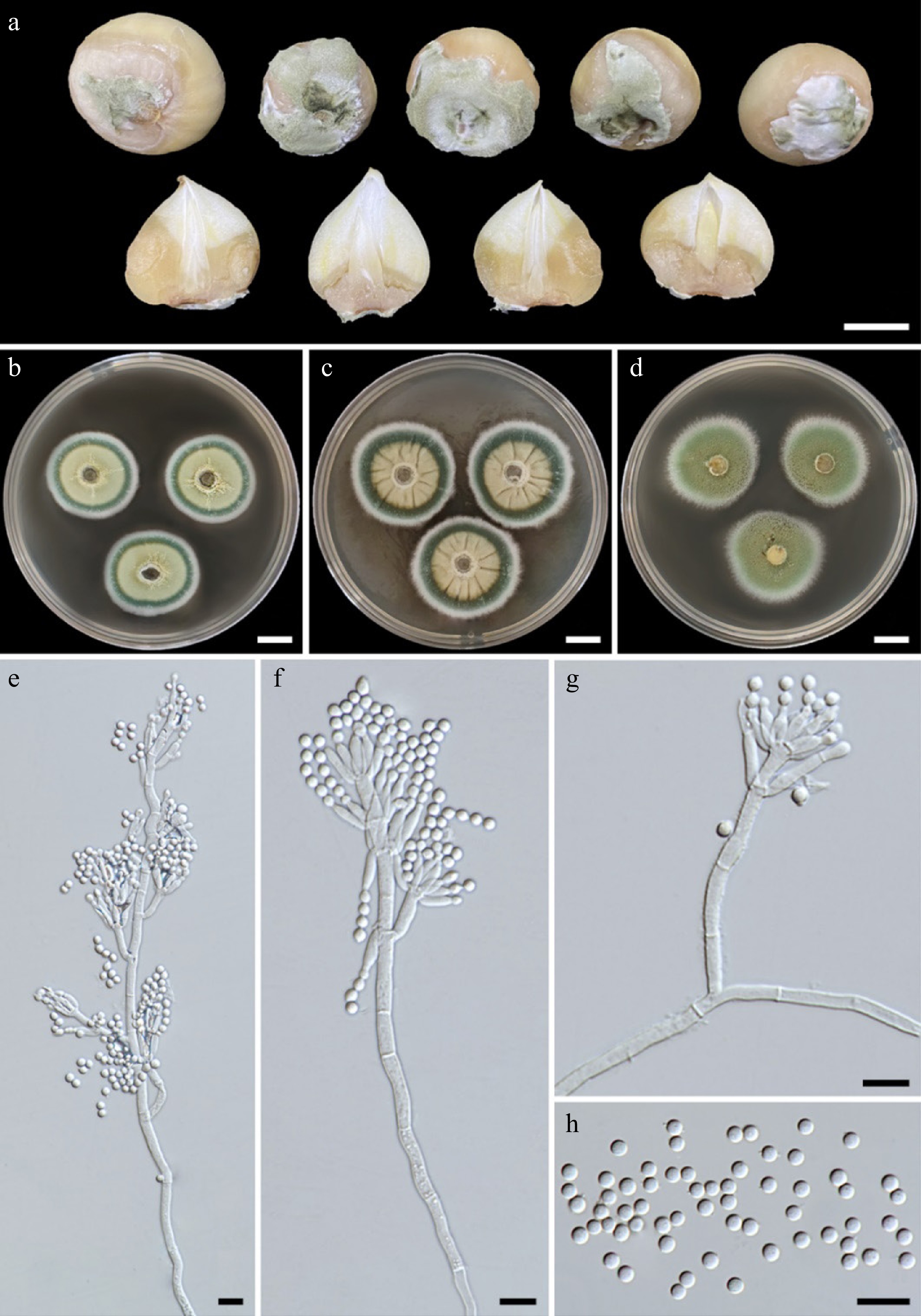
      Figure 1. 

      (a) Natural symptoms of blue mold disease on bulbs of elephant garlic by Penicillium allii . Colonies of Penicillium allii SDBR-CMU499 after incubation at 25 °C for one week. (b) PDA. (c) CYA. (d) MEA. (e)–(g) Conidiophores. (h) Conidia. Scale bars: (a)–(d) = 10 mm, (e)–(h) = 10 μm.

    • Three fungal isolates (CMU499, CMU500, and CMU501) with similar morphology were obtained and deposited at the SDBR-CMU under accession numbers SDBR-CMU499, SDBR-CMU500, and SDBR-CMU501, respectively. Colonies PDA, CYA, and MEA were 29–32, 32–37, and 33–37 mm in diameter, respectively after incubation for one week at 25 °C (Fig. 1bd). Colonies on PDA and MEA were white, flat with entire edges, conidium turquoise, white in the center, dull green at the margins; reverse pale yellow for PDA and yellow-brown for MEA. Colonies on CYA were white, flat with entire edges, conidium dull green; reverse white. All fungal isolates could produce conidiophores, and phialides, and sporulate in all of the agar media. Conidiophores terverticillate (Fig. 1eg). Stipes rough-walled, 13.2–181.2 × 2.3–3.9 μm. Rami one or two, rough-walled and appressed or divergent, 8.4–24.7 × 2.5–4.6 μm. Metulae divergent, in verticils of 2–4, 8–19.1 × 2.3–4.6 μm. Phialides ampulliform, in verticils of 3 to 9, 6–17.9 × 1.7–6.9 μm. Conidia globose, 2.6–4.3 μm in diameter, smooth-walled, dull green (Fig. 1h). Based on these morphological characteristics, all fungal isolates were initially identified as belonging to Penicillium[3235]. Fungal identification was then further confirmed using multi-gene phylogenetic analyses.

    • The ITS, BenA, CaM, and rpb2 sequences obtained from three fungal isolates in this study have been deposited in GenBank (Table 1). According to the BLAST results, all fungal isolates were identified as members of the Penicillium section Fasciculata. The combined ITS, BenA, CaM, and rpb2 sequences dataset consists of 32 taxa, and the aligned dataset includes 2,399 characters comprising gaps (ITS: 1–553, BenA: 554–927, CaM: 928–1,442, and rpb2: 1,443–2,399). The best-scoring RAxML tree was established with a final ML optimization likelihood value of –9,279.455311. Accordingly, the matrix contained 612 distinct alignment patterns with 5.04% undetermined characters or gaps. The estimated base frequencies were found to be: A = 0.235164, C = 0.269054, G = 0.262373, and T = 0.233408; substitution rates AC = 1.298254, AG = 4.466196, AT = 1.340638, CG = 0.787838, CT = 9.362594, and GT = 1.00000. The values of the gamma distribution shape parameter alpha and the Tree-Length were 0.580385 and 0.524602, respectively. Additionally, BI analysis yielded a final average standard deviation of 0.002257 for the split frequencies at the end of all MCMC generations. Regarding topology, the phylogenetic trees generated from both ML and BI analyses were similar. Consequently, the phylogenetic tree obtained from the ML analysis was selected and is displayed in Fig. 2. The results indicated that three fungal isolates SDBR-CMU499, SDBR-CMU500, and SDBR-CMU501 clustered with P. allii CBS 131.89 (ex-type strain) with strong statistical (100% BS and 1.0 PP) supports. Therefore, all fungal isolates obtained in this were identified as P. allii based on morphological and molecular data.
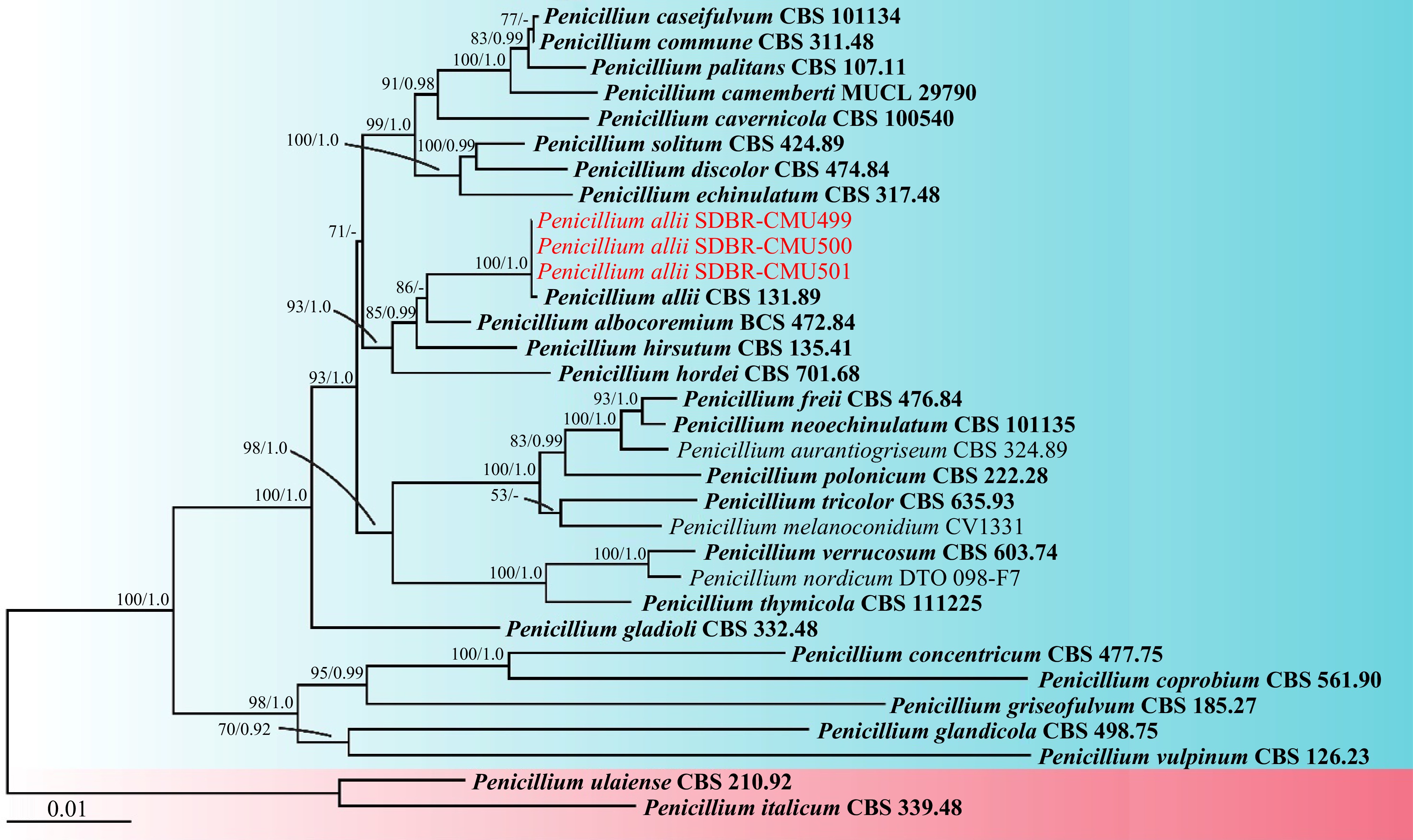
      Figure 2. 

      Phylogram derived from maximum likelihood analysis of the combined ITS, BenA, CaM, and rpb2 sequences of 30 taxa in the Penicillium section Fasciculata and two taxa in the Penicillium section Penicillium. Penicillium italicum CBS 339.48 and P. ulaiense CBS 210.92 were used as outgroups. Bootstrap values ≥ 50% (left) and Bayesian posterior probabilities ≥ 0.90 (right) are displayed above nodes. The scale bar represents the expected number of nucleotide substitutions per site. The sequences of fungal species obtained in this study are in red. The ex-type strain are in bold.

    • The initial symptoms appeared on bulbs of elephant garlic 3 d after being inoculated. After 7 d, all inoculated bulbs displayed powdery mold at their centers, surrounded by orange-brown water-soaked lesions (Fig. 3bd). Whereas, control bulbs were asymptomatic (Fig. 3a). Penicillium allii was consistently reisolated from the inoculated bulbs on PDA to complete Koch's postulates.
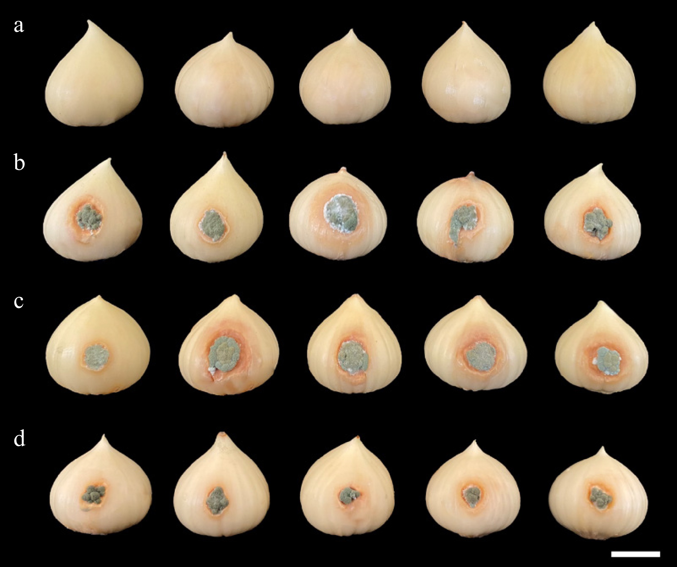
      Figure 3. 

      Pathogenicity test using Penicillium allii SDBR-CMU499, SDBR-CMU500, and SDBR-CMU501 on bulbs of elephant garlic after one week inoculation at 25 °C. (a) Control bulbs treated with sterile distilled water instead of inoculum. Blue mold disease on bulbs of elephant garlic after inoculation of isolate (b) SDBR-CMU499, (c) SDBR-CMU500, and (d) SDBR-CMU501. Scale bar: 10 mm.

    • Seven commercially available fungicides in Thailand were tested in this study. After one week, the mycelial growths in response to the fungicides at three different dosages, including half-recommended (1/2RD), recommended (RD), and double-recommended (2RD) were calculated and presented in Table 2. The results revealed that the growth inhibition values varied across different fungicides, dosages, and fungal isolates. Data on the percentage of mycelial inhibition for each fungal isolate, related to the fungicides, passed the normality test (Shapiro-Wilk test, p < 0.001), thereby assuming normal distributions. Therefore, ANOVA followed by DMRT (p ≤ 0.05) was used to identify significant differences. The findings indicated that all fungal isolates were completely inhibited by carbendazim, difenoconazole + azoxystrobin, and difenoconazole at all tested dosages (Table 2). In the tests for captan and mancozeb, all isolates demonstrated sensitivity to 2RD. Therefore, based on the recommended dosages, carbendazim, difenoconazole + azoxystrobin, and difenoconazole could be effectively applied to control this pathogen.

      Table 2.  Percentage of mycelial inhibition and reactions of three isolates of Penicillium allii against fungicides.

      Fungicides Dosages Inhibition of mycelial growth (%)* Reaction
      SDBR-CMU499 SDBR-CMU500 SDBR-CMU501
      Benalaxyl-M + mancozeb 1/2RD 30.08 ± 1.41 c 29.27 ± 2.25 c 30.08 ± 2.53 c Insensitive
      RD 55.28 ± 1.41 b 56.10 ± 3.62 b 60.16 ± 1.67 b Sensitive
      2RD 82.11 ± 2.82 a 83.74 ± 1.41 a 83.74 ± 3.45 a Sensitive
      Captan 1/2RD 2.44 ± 3.50 c 1.63 ± 2.41 c 2.44 ± 1.25 c Insensitive
      RD 4.88 ± 2.25 b 4.88 ± 2.23 b 5.69 ± 2.30 b Insensitive
      2RD 72.36 ± 1.60 a 73.93 ± 3.45 a 73.98 ± 2.82 a Sensitive
      Carbendazim 1/2RD 100 ± 0 a 100 ± 0 a 100 ± 0 a Inhibition
      RD 100 ± 0 a 100 ± 0 a 100 ± 0 a Inhibition
      2RD 100 ± 0 a 100 ± 0 a 100 ± 0 a Inhibition
      Copper oxychloride 1/2RD 61.79 ± 1.60 c 58.54 ± 1.05 c 56.91 ± 1.41 f Sensitive
      RD 68.29 ± 1.20 b 68.29 ± 2.05 b 68.29 ± 2.54 d Sensitive
      2RD 100 ± 0 a 100 ± 0 a 100 ± 0 a Inhibition
      Difenoconazole + azoxystrobin 1/2RD 100 ± 0 a 100 ± 0 a 100 ± 0 a Inhibition
      RD 100 ± 0 a 100 ± 0 a 100 ± 0 a Inhibition
      2RD 100 ± 0 a 100 ± 0 a 100 ± 0 a Inhibition
      Difenoconazole 1/2RD 100 ± 0 a 100 ± 0 a 100 ± 0 a Inhibition
      RD 100 ± 0 a 100 ± 0 a 100 ± 0 a Inhibition
      2RD 100 ± 0 a 100 ± 0 a 100 ± 0 a Inhibition
      Mancozeb 1/2RD 19.51 ± 2.44 c 22.76 ± 3.73 c 21.14 ± 1.41 c Insensitive
      RD 47.15 ± 1.45 b 47.15 ± 2.82 b 43.90 ± 2.44 b Insensitive
      2RD 58.54 ± 2.44 a 54.47 ± 1.42 a 55.28 ± 1.45 a Sensitive
      * Results are means of five replicates ± standard deviation with the independently repeated twice. Data with different letters within the same column for each fungal isolate and fungicide indicate a significant difference at p ≤ 0.05 according to Duncan's multiple range test. 1/2RD, RD, and 2RD indicate half of the recommended dosage, recommended dosage, and double the recommended dosage, respectively.
    • Penicillium species are widely recognized as one of the most significant genera, known to cause major diseases in numerous economically valuable crops cultivated worldwide, including garlic[9,10,12,13,36]. Traditionally, Penicillium species have been identified using both macromorphological and micromorphological characteristics. However, morphological traits alone are insufficient to differentiate closely related Penicillium species due to the extensive range of morphological variations. Therefore, molecular techniques are crucial for accurately identifying Penicillium at the species level. Several previous studies have utilized a combination of ribosomal DNA (ITS) and protein-coding genes (BenA, CaM, rpb1, and rpb2) as powerful tools to identify Penicillium species since species-level identification remained unresolved when used solely on the ribosomal DNA gene[32,34,35,37]. In this study, three isolates of P. allii were obtained from the rot lesions of blue mold disease on elephant garlic bulbs in northern Thailand. The identification of this fungal species followed methods similar to those used for identifying Penicillium, which involve combining phylogenetic analysis of multiple genes with their morphological characteristics.

      In this study, Koch's postulates were fulfilled by conducting pathogenicity tests on all isolates of P. allii. The findings demonstrate that postharvest blue mold disease on elephant garlic bulbs in northern Thailand, caused by P. allii identified in this study resembles the disease caused by previously identified Penicillium pathogens, particularly P. hirsutum, which affects garlic bulbs worldwide[1113,36]. Penicillium polonicum has been reported as a causal agent of blue mold on stored garlic bulbs in Pakistan[38]. Penicillium allii was known to cause postharvest blue mold disease on garlic bulbs in Argentina[15,39,40]. In the USA, P. albocoremium, P. expansum, P. glabrum, P. paraherquei, and P. radicicola can cause blue mold on garlic bulbs[13]. In Korea, blue mold disease on garlic bulbs caused by P. hirsutum has been reported[41]. Five Penicillium species, namely P. allii, P. glabrum, P. italicum, P. polonicum, and P. psychrotrophicum were identified and confirmed as postharvest pathogens causing blue mold rot of garlic in Serbia[42]. Recently, P. allii was the most virulent pathogen causing blue mold disease of elephant garlic bulbs in Italy, accounting for 95% of cases, followed by P. citrinum (4%) and P. brevicompactum (1%)[43]. Before this study, there were no reports of blue mold disease on elephant garlic bulbs in Thailand. Thus, this represents the first report of postharvest blue mold disease on elephant garlic bulbs caused by P. allii in Thailand.

      To manage and control fungal-caused plant diseases, a variety of fungicides have been used. Several studies have documented the effectiveness of fungicides in affecting sensitive, resistant strains of plant pathogenic fungi, particularly those in the Penicillium species, on their in vitro mycelial growth[4446]. In this study, the sensitivity and inhibition of P. allii to fungicides varied among different fungicides and dosages. These findings are consistent with previous studies, which reported that the sensitivity and inhibition of Penicillium species to fungicides varies based on the type and dosage of the fungicide, as well as fungal species[4648]. Before this study, prochloraz had been used against P. allii to control diseases related to sprouting germination in Europe[49]. In this study, carbendazim, difenoconazole + azoxystrobin, and difenoconazole at both half and recommended dosages exhibited complete inhibition of P. allii. The information on the in vitro inhibition, sensitivity, and resistance of fungicides against P. allii, which causes postharvest blue mold disease on elephant garlic bulbs, would be beneficial for in vivo applications and for managing this disease in Thailand and globally. However, environmental factors and the fungicide's metabolism in the plant can cause the results of in vitro fungicide testing to differ from in vivo responses. Therefore, further studies are required to conduct in vivo fungicide sensitivity and disease inhibition assays based on the in vitro findings. Additionally, several previous studies have established that fungicide-resistant strains are a result of both excessive and prolonged fungicide treatment[5052]. Utilizing biological control agents, rotating crops, adhering to fungicide treatment guidelines, and maintaining cleanliness in fields, equipment, and storage spaces are all essential components of a comprehensive strategy to reduce fungicide resistance in fungi[9,50,53,54].

    • Garlic blue mold disease, caused by Penicillium species, leads to significant economic losses during postharvest storage worldwide. In the present study, P. allii was isolated from infected bulbs of elephant garlic in northern Thailand. The identification of this fungi involved the analysis of their morphological characteristics and conducting multi-gene phylogenetic analyses. The assessment of pathogenicity for P. allii showed similar symptoms throughout the artificial inoculation process, as observed during the postharvest storage period. Therefore, this study represents the first report of elephant garlic blue mold disease caused by P. allii in Thailand. In the fungicide screening test, carbendazim, difenoconazole + azoxystrobin, and difenoconazole were found to effectively control this pathogen at both half and full recommended dosages. Thus, half of the recommended dosages can be used in managing this disease, serving as a guideline for prevention and helping to reduce pathogen resistance to fungicides. The findings of this study will enhance our understanding of postharvest blue mold disease in elephant garlic bulbs and provide insights for developing effective management strategies and prevention methods to minimize significant economic losses. Further research on the epidemiology of this disease would be required for effective monitoring, prevention, and control.

      • The authors sincerely appreciate the financial support provided by Chiang Mai University, Thailand and the University of Phayao Innovation Fund [Fundamental Fund 2024 (227/2567)], Thailand.

      • The authors confirm contribution to the paper as follows: conceptualization: Suwannarach N, Khuna S; formal analysis: Suwannarach N, Khuna S, Chaiwong K, Senwanna C, Kumla J; investigation, methodology: Suwannarach N, Khuna S, Chaiwong K; resources: Suwannarach N, Khuna S, Chaiwong K; software: Khuna S, Chaiwong K, Senwanna C, Kumla J; validation: Suwannarach N, Khuna S, Senwanna C, Nuangmek W; data curation: Khuna S, Senwanna C, Nuangmek W, Kumla J; visualization: Khuna S, Chaiwong K; writing–original draft: Suwannarach N, Khuna S, Nuangmek W, Kumla J; writing–review & editing: Suwannarach N, Khuna S, Chaiwong K, Senwanna C, Nuangmek W, Kumla J; supervision, project administration: Suwannarach N; funding acquisition: Suwannarach N, Nuangmek W. All authors have read and agreed to the published version of the manuscript.

      • The DNA sequences generated in this study have been submitted to GenBank and can be accessed through the accession numbers provided in the paper.

      • The authors declare that they have no conflict of interest. Nakarin Suwannarach and Jaturong Kumla are the Editorial Board members of Studies in Fungi who are blinded from reviewing or making decisions on the manuscript. The article was subject to the journal's standard procedures, with peer review handled independently of these Editorial Board members and the research groups.

      • Copyright: © 2024 by the author(s). Published by Maximum Academic Press, Fayetteville, GA. This article is an open access article distributed under Creative Commons Attribution License (CC BY 4.0), visit https://creativecommons.org/licenses/by/4.0/.
    Figure (3)  Table (2) References (54)
  • About this article
    Cite this article
    Suwannarach N, Khuna S, Chaiwong K, Senwanna C, Nuangmek W, et al. 2024. Identification and fungicide sensitivity of the blue mold pathogen in postharvest-stored elephant garlic bulbs in Thailand. Studies in Fungi 9: e015 doi: 10.48130/sif-0024-0015
    Suwannarach N, Khuna S, Chaiwong K, Senwanna C, Nuangmek W, et al. 2024. Identification and fungicide sensitivity of the blue mold pathogen in postharvest-stored elephant garlic bulbs in Thailand. Studies in Fungi 9: e015 doi: 10.48130/sif-0024-0015

Catalog

  • About this article

/

DownLoad:  Full-Size Img  PowerPoint
Return
Return