ARTICLE   Open Access    

Identification of Ralstonia solanacearum resistant solanum plants as potential rootstock to manage bacterial wilt disease in tomato production

More Information
  • Using morphological and molecular markers, this study screened tomato (Solanum lycopersicum) and garden egg (Solanum melongena) accessions for resistance to bacterial wilt disease. The solanum plants were inoculated with Ralstonia solanacearum and evaluated for disease incidence and severity in a field trial set up in a Randomised Complete Block Design with four replications. Molecular markers conferring resistance to R. solanacearum Phylotype I and II were used to identify durable and partial resistance. Results showed significant variation in disease incidence and severity among accessions, with tomato accessions exhibiting higher susceptibility. Accession CRI-01 had the highest disease incidence (54.0%), while accession GD had the lowest (13.0%). Accession CRI-04 showed moderate resistance with a disease severity index of 0.37, while accession GC had the highest disease severity index (0.90). Accession L_020 demonstrated moderate resistance in the field (43.0% disease incidence) and possessed durable resistant genes, making it a promising rootstock for managing bacterial wilt disease in tomato production. This research contributes to the development of integrated pest management strategies for sustainable tomato production.
  • Plants are continuously subjected to unpredictable environmental conditions and encounter a multitude of stressors throughout their growth and development, posing a significant challenge to global crop production and food security[1]. Heat and drought are undoubtedly the two most important stresses that have a huge impact on crops. Both elicit a wide array of biochemical, molecular, and physiological alterations and responses, impacting diverse cellular processes and ultimately influencing crop yield and quality[2].

    A primary physiological consequence of both stresses is the diminished photosynthetic capacity, partially resulting from the degradation of chlorophyll due to leaf senescence under stress conditions. Chlorophyll accumulation was diminished in numerous plants subjected to drought or heat stress conditions[3,4]. Various environmental stresses prompt excessive generation of reactive oxygen species (ROS), initiating oxidative damage that compromises lipids, and proteins, and poses a serious threat to cellular functions[2]. To mitigate oxidative stress and minimize damage, plants have developed various protective mechanisms to neutralize ROS. Several antioxidant enzymes, such as SOD, POD, and CAT, are integral to cellular antioxidative defense mechanisms. Additionally, antioxidants such as anthocyanins and proline serve as crucial ROS scavengers[5,6]. The elevation in temperature typically induces the transient synthesis of heat shock proteins (Hsps), which function as molecular chaperones in protecting proteins from denaturation and aggregation, with their activity primarily regulated at the transcriptional level by heat shock factors (Hsfs)[7]. The significance of Hsps and Hsfs in all organisms, including plants, has been assessed in various stress conditions that could disrupt cellular homeostasis and result in protein dysfunction[7]. Drought stress can also trigger the transcription of a suite of marker genes, including RD29A, RD29B, NCED3, AREB1, Rab18, etc., which assist plants in mitigating cellular damage during dehydration and bolstering their resilience to stress[810].

    Previous research efforts focusing on the regulatory control of stress-related genes have largely centered around protein-coding genes. In recent years, non-protein-coding transcripts have emerged as important regulatory factors in gene expression. Among them, long non-coding RNAs (lncRNAs) lncRNAs have been identified as implicated in various abiotic stresses[11,12]. LncRNAs are a class of non-coding RNAs (ncRNAs) exceeding 200 nucleotides in length. They possess minimal or no protein-coding potential[13]. In plants, lncRNAs are specifically transcribed by RNA polymerases Pol IV, Pol V, Pol II, and Pol III[14,15]. LncRNAs exhibit low abundance and display strong tissue and cellular expression specificity relative to mRNAs. Moreover, sequence conservation of lncRNAs is was very poor across different plant species[13,16,17]. The widespread adoption of high-throughput RNA sequencing technology has revealed lncRNAs as potential regulators of plant development and environmental responses. In cucumber, RNA-seq analysis has predicted 2,085 lncRNAs to be heat-responsive, with some potentially acting as competitive endogenous RNAs (ceRNAs) to execute their functions[18]. In radish, a strand-specific RNA-seq (ssRNA-seq) technique identified 169 lncRNAs that were differentially expressed following heat treatment[19]. In Arabidopsis, asHSFB2a, the natural antisense transcript of HSFB2a was massively induced upon heat stress and exhibited a counteracted expression trend relative to HSFB2a. Overexpression of asHSFB2a entirely suppressed the expression of HSFB2a and impacted the plant's response to heat stress[20]. For drought stress resistance, 244 lncRNAs were predicted in tomatoes to be drought responsive probably by interacting with miRNAs and mRNAs[21]. Under drought stress and rehydration, 477 and 706 lncRNAs were differentially expressed in drought-tolerant Brassica napus Q2 compared to drought-sensitive B. napus, respectively[22]. In foxtail millet and maize, 19 and 644 lncRNAs, respectively, were identified as drought-responsive[23,24]. Despite the identification of numerous lncRNAs by high-throughput sequencing, which suggests their potential involvement in various abiotic stress processes, only a minority have been experimentally validated for function.

    In our previous study, we characterized 1,229 differentially expressed (DE) lncRNAs in Chinese cabbage as heat-responsive, and subsequent bioinformatics analysis reduced this number to 81, which are more likely associated with heat resistance[25]. lnc000283 and lnc012465 were selected from among them for further functional investigation. The findings indicated that both lnc000283 and lnc012465 could be promptly induced by heat shock (HS). Overexpression of either lnc000283 or lnc012465 in Arabidopsis plants enhanced their capacity to tolerate heat stress. Additionally, both lnc000283 and lnc012465 conferred drought tolerance to transgenic Arabidopsis.

    The lncRNA sequences examined in this study were from Chiifu-401-42 Chinese cabbage and all Arabidopsis plants were of the Col-0 background. Transgenic plants expressing lnc000283 and lnc012465 were generated using the Agrobacterium tumefaciens-mediated floral dip method[26]. Single-copy and homozygous T3 plants were identified through genetic segregation on an agar medium supplemented with kanamycin. The T3 generation plants, or their homozygous progeny, were utilized in the experiments.

    For phenotypic assessment, Arabidopsis seeds were initially sown on filter paper moistened with ddH2O and placed in a 4 °C freezer for 2 d. Subsequently, they were evenly planted in nutrient-rich soil and transferred to a growth chamber operating a 16-h day/8-h night cycle, with day/night temperatures of 22 °C/18 °C and a light intensity of 250 μmol·m−2·s−1. After 10 d of growth, Arabidopsis plants with uniform growth were transferred to 50-hole plates. Arabidopsis plants grown in Petri dishes were firstly seed-sterilized and then sown on 1/2 MS medium supplemented with 10 g·L−1 sucrose. The seeds were then placed in a 4 °C refrigerator for 2 d in the dark before transferring them to a light incubator. The day/night duration was set to 16 h/8 h, the day/night temperature to 21 °C/18 °C, and the light intensity to 100 μmol·m−2·s−1.

    For heat treatment, 3-week-old seedlings were subjected to 38 °C for 4 d within a light incubator, subsequently transferred to their original growth conditions under the same light/dark cycles. For drought treatment, 3-week-old Arabidopsis seedlings were deprived of water for 10 d, followed by rehydration to facilitate a 2-d recovery period. Plants were photographed and surveyed both before and after treatment.

    The lncRNA sequences (lnc000283 and lnc012465) were chemically synthesized based on RNA-seq data, with restriction sites for BamH1 and Kpn1 engineered upstream and downstream. The resultant lncRNA constructs were subcloned into the pCambia2301 binary vector, incorporating a cauliflower mosaic virus (CaMV) 35S promoter. The recombinant vectors were transformed into Escherichia coli TOP10 competent cells (Clontech), incubated at 37 °C overnight, after which single clones were selected for PCR verification, and the confirmed positive colonies were submitted for sequencing. Following verification, the correct plasmids were introduced into A. tumefaciens strain GV3101 using the freeze-thaw method and subsequently transformed into Arabidopsis wild-type (Col) plants.

    To quantify the chlorophyll content, the aerial portions of wild-type and transgenic Arabidopsis plants, grown in Petri dishes were weighed, minced, and then subjected to boiling in 95% ethanol until fully decolorized. Aliquots of 200 μL from the extract were transferred to a 96-well plate and the absorbance at 663 nm and 645 nm was measured via spectrophotometry by a microplate reader (Multiskan GO, Thermo Scientific, Waltham, MA, USA). Three biological replicates were analyzed for WT and each transgenic line. Chlorophyll content was determined according to the formula of the Arnon method[27]: Chlorophyll a = (12.72A663 − 2.59A645) v/w, Chlorophyll b = (22.88A645 − 4.67A663) v/w, Total chlorophyll = (20.29A645 + 8.05A663) v/w.

    The quantification of anthocyanin was performed as follows: aerial parts of wild-type and transgenic Arabidopsis plants, cultivated in Petri dishes, were weighed and ground to powder in liquid nitrogen. Subsequently, the samples were incubated in 600 μL of acidified methanol (containing 1% HCl) at 70 °C for 1 h. Following this, 1 mL of chloroform was added, and the mixture was vigorously shaken to remove chlorophyll. The mixture was then centrifuged at 12,000 rpm for 5 min, after which the absorbance of the aqueous phase was determined at 535 nm using a spectrophotometer (Shimadzu, Kyoto, Japan). Three biological replicates were analyzed for WT and each transgenic line. The relative anthocyanin content was calculated according to anthocyanin concentration and extraction solution volume. One anthocyanin unit is defined as an absorption unit at a wavelength of 535 nm in 1 mL of extract solution. In the end, the quantity was normalized to the fresh weight of each sample.

    Three-week-old transgenic and WT A. thaliana plants, subjected to normal conditions or varying durations of heat or drought stress, were utilized for subsequent physiological assessments. All assays were performed in accordance with the method described by Chen & Zhang[28]. In brief, 0.1 g of fresh leaf tissue was homogenized in 500 μL of 100 mM PBS (pH 7.8) while chilled on ice. The homogenate was then centrifuged at 4 °C, and the resultant supernatant was employed for further analysis. For the determination of MDA content, 100 μL of the supernatant was combined with 500 μL of a 0.25% thiobarbituric acid (TBA) solution (which was prepared by dissolving 0.125 g of TBA in 5 mL of 1 mol·L−1 NaOH before being added to 45 mL of 10% TCA) and boiled for 15 min. Following a 5 min cooling period on ice, the absorbance was measured at 532 nm and 600 nm. The activity of POD was determined as follows: initially, 28 μL of 0.2% guaiacol and 19 μL of 30% H2O2 were sequentially added to 50 mL of 10mM PBS (pH 7.0), after thorough heating and mixing, 1 mL was transferred into a cuvette, then 50 μL of the supernatant was added to the cuvette and the absorbance at 470 nm was monitored every 15 s for 1 min. To determine the proline content, a reaction solution was prepared by mixing 3% sulfosalicylic acid, acetic acid, and 2.5% acidic ninhydrin in a ratio of 1:1:2, then 50 μL of the supernatant was added to 1 mL of the reaction solution, which was then subjected to a boiling water bath for 15 min (the solution turned red after the boiling water bath). Following cooling on ice, the absorbance at 520 nm was recorded. For the quantification of proline, an L-proline standard curve was prepared by dissolving 0, 5, 10, 15, 20, 25, and 30 μg of L-proline in 0.5 mL of ddH2O, followed by the addition of 1 mL of the reaction solution and measuring the absorbance at 520 nm. The proline content in the samples was then determined based on the L-proline standard curve.

    Total RNA was isolated from the aerial parts of Arabidopsis using the TaKaRa MiniBEST Plant RNA Extraction Kit, followed by purification and reverse transcription using the PrimeScript RT reagent Kit with gDNA Eraser (Takara). The cDNA product was diluted 10 times and real-time PCR was conducted in triplicate for each biological replicate using SYBR PCR Master Mix (Applied Biosystems) on the ABI 7500 system under the following conditions: 98 °C for 3 min, followed by 40 cycles of 98 °C for 2 s and 60 °C for 30 s. The relative expression levels of each gene were normalized against the transcript abundance of the endogenous control UBC30 (At5g56150) and calculated using the 2−ΔCᴛ method. The specific primers employed for qRT-PCR are detailed in Supplemental Table S1.

    In our prior investigation, dozens of lncRNAs associated with the heat stress response in Chinese cabbage were identified through informatics analysis. Two lncRNAs (lnc000283 and lnc012465) were chosen for genetic transformation in Arabidopsis to elucidate their functions comprehensively. Transcriptome data analysis indicated that the expression of lnc000283 and lnc012465 in Chinese cabbage were both induced by HS. To verify the accuracy, the expression patterns of lnc000283 and lnc012465 were confirmed through quantitative real-time PCR (qRT-PCR), and the results from qRT-PCR were consistent with those obtained from RNA-seq (Fig. 1a). The corresponding homologous genes in Arabidopsis were identified as CNT2088434 and CNT2088742, exhibiting sequence similarities of 88% and 87%, respectively (Supplemental Fig. S1). Subcellular localization predictions using the lnclocator database (www.csbio.sjtu.edu.cn/bioinf/lncLocator) suggested that both lncRNAs are localized within the nucleus (Supplemental Table S2). Bioinformatics analysis was conducted using the CPC tool (http://cpc.cbi.pku.edu.cn/) indicated that lnc000283 and lnc012465 are noncoding sequences, with coding probabilities of 0.0466805 and 0.0432148, respectively comparable to the well-characterized lncRNAs COLDAIR and Xist, but significantly lower than those of the protein-coding genes UBC10 and ACT2 (Fig. 1b).

    Figure 1.  Characteristics of lnc000283 and lnc012465. (a) Expression level of lnc000283 and lnc012465 in Chinese cabbage leaves treated at 38 °C at different time points, as determined by qRT-PCR and RNA-seq. CK is a representative plant before heating, and T1, T4, T8, and T12 denote plants that were subjected to 38 °C for 1, 4, 8, and 12 h, respectively. The expression levels were normalized to the expression level of Actin. (b) Analysis of coding potential for lnc000283 and lnc012465. The coding potential scores were calculated using the CPC program. UBC10 (At5g53300) and ACT2 (At3g18780) are positive controls that encode proteins. COLDAIR (HG975388) and Xist (L04961) serve as negative controls, exhibiting minimal protein-coding potential.

    To elucidate the role of lnc000283 and lnc012465 in response to abiotic stress, overexpression vectors were constructed for these lncRNAs, driven by the CaMV 35S promoter, and they were introduced into Arabidopsis thaliana (Col-0 ecotype). Through PCR identification and generational antibiotic screening, two homozygous positive lines for lnc012465 and lnc000283 were obtained. The relative expression levels of these lncRNAs were assessed using qRT-PCR (Fig. 2a). When plants were grown in 1/2 MS medium, with the consumption of nutrients, and reduction of water, the leaves of WT began to turn yellow, but the lnc000283 and lnc012465 overexpression lines developed a deep purple color of leaf veins (Fig. 2b). Examination of chlorophyll and anthocyanin contents in the plants revealed that both overexpression lines had higher levels of chlorophyll and anthocyanin compared to the WT, suggesting that the transgenic plants might possess enhanced resistance to nutritional or water stress (Fig. 2c, d).

    Figure 2.  Arabidopsis plants overexpressing lnc000283 and lnc012465 had higher anthocyanins and chlorophyll content. (a) The relative expression level of lnc000283 and lnc012465 in WT and different transgenic lines. UBC10 (At5g53300) was used as an internal control. Each value is mean ± sd (n = 3). (b) The phenotype of WT and Arabidopsis overexpressing lnc000283 or lnc012465 grown on 1/2 MS medium 50 d after sowing. The (c) anthocyanin and (d) chlorophyll content of WT and transgenic Arabidopsis overexpressing lnc000283 or lnc012465. The asterisks above the bars indicate statistical significance using Student's t-test (*, p < 0.05; **, p < 0.01).

    Given that lnc000283 and lnc012465 were highly induced by heat, the thermotolerance of the overexpressing (OE) plants were compared to that of the wild type. Arabidopsis plants were initially exposed to a an HS treatment at 38 °C for 4 d, followed by recovery at room temperature. The death caused by HS was processive. Post-severe HS challenge for 4 d, OE plants initially appeared similar to WT, but upon recovery, their leaves started to fold or curl, followed by a transition to yellow, white, and eventually drying out (Fig. 3a). OE lnc000283 and OE lnc012465 plants exhibited enhanced thermotolerance compared to WT, with lnc012465 showing particularly strong tolerance (Fig. 3a; Supplemental Fig. S2a). After 5 d of recovery, leaf coloration indicated that transgenic plants maintained a significantly higher percentage of green leaves and a lower percentage of bleached leaves compared to WT (Fig. 3b; Supplemental Fig. S2b). Under non-heat-stress conditions, WT and OE plants possessed comparable water content. However, following heat stress, the fresh-to-dry weight ratio of OE lnc000283 and lnc012465 lines was significantly greater than that of WT (Fig. 3c; Supplemental Fig. S2c). Abiotic stresses frequently trigger the production of excessive reactive oxygen species (ROS), which are believed to cause lipid peroxidation of membrane lipids, leading to damage to macromolecules. Leaf MDA content is commonly used as an indicator of lipid peroxidation under stress conditions; therefore, the MDA content in both transgenic and WT plants was assessed. Figure 3d shows that the MDA content in WT plants progressively increased after heat treatment, whereas in the two lines overexpressing lnc012465, the MDA content increased only slightly and remained significantly lower than that in WT at all time points. In plants overexpressing lnc000283, the MDA content did not significantly differ from that of WT before heat stress. However, after 4 d of heat treatment, the MDA content was significantly lower compared to WT (Supplemental Fig. S2d). The results suggested that the expression of both lnc012465 and lnc000283 can mitigate injury caused by membrane lipid peroxidation under heat-stress conditions. Peroxidase (POD) is a crucial antioxidant enzyme involved in ROS scavenging. Figure 3e and Supplemental Fig. S2e demonstrate that POD activity increased in both transgenic and WT plants after heat treatment. However, the increase in WT plants was modest, whereas OE lnc000283 and OE lnc012465 plants exhibited consistently higher POD activity. As anticipated, proline levels were induced in response to stress in all studied plants (Fig. 3f; Supplemental Fig. S2f). However, under normal conditions and 2 d post-heat stress treatment, the proline content in OE lnc000283 and OE lnc012465 plants did not exhibit significant changes compared to WT (Fig. 3f; Supplemental Fig. S2f). Moreover, after 4 d of heat stress, the proline content in OE lnc012465 lines was significantly lower than in WT, and the OE lnc000283 transgenic line 12-6 also showed a marked decrease in proline content compared to WT (Fig. 3f; Supplemental Fig. S2f). The results indicated that the thermotolerance of plants overexpressing either lnc000283 or lnc012465 was independent of proline accumulation.

    Figure 3.  Overexpressing lnc012465 lines are more tolerant to heat stress. (a) Phenotypes of WT and OE lnc012465 plants were assessed before and after exposure to heat stress. The heat treatment was applied to 25-day-old Arabidopsis plants. (b) The percentage of leaves with different colors in Arabidopsis after heat treatment and recovery for 5 d. (c) The fresh-to-dry weight ratio of Arabidopsis leaves was measured before and after 38 °C heat treatment. (d)−(f) depict the MDA content, POD activity, and proline content in Arabidopsis leaves at varying durations of heat stress. The asterisks above the bars indicate statistical significance using Student's t-test (*, p < 0.05; **, p < 0.01; ***, p < 0.001).

    To elucidate the molecular mechanisms by which lncRNAs enhance thermotolerance in Arabidopsis, the expression of the Hsf gene HsfA7a and three Hsps (Hsp25.3, Hsa32, and Hsp18.1-CI) in OE lnc000283, OE lnc012465, and WT Arabidopsis plants were investigated at various time points following heat treatment. As shown in Fig. 4 and Supplemental Fig. S3, both Hsf and Hsps exhibited a rapid response to heat stress with strong induction. Notably, the transcripts of HsfA7a and Hsp25.3 were significantly upregulated at 1 h after heat exposure, then experienced a sharp decrease. Hsa32 and Hsp18.1-CI were highly induced at 1 h and, unlike the other proteins, sustained high expression levels at 3 h (Fig. 4; Supplemental Fig. S3). At 1 h post-heat treatment, the transcript levels of Hsa32 and HsfA7a in OE lnc000283 did not significantly differ from those in WT. However, by 3 h, Hsa32 expression was roughly 50% of the WT level, while HsfA7a expression was approximately double that of WT (Supplemental Fig. S3). The overexpression of lnc000283 did not significantly affect the transcript level of Hsp25.3 at any of the tested time points. Notably, Hsp18.1-CI expression in both lines overexpressing lnc000283 was significantly induced at all three detection points post-heat treatment, reaching approximately 4-9-fold higher levels than in the WT (Supplemental Fig. S3). In Arabidopsis plants with elevated expression of lnc012465, the expression patterns of all Hsp and Hsf genes were similar to those in plants overexpressing lnc000283, with the notable exception of Hsa32. Unlike the WT, Hsa32 did not show a trend of down-regulation at 3 h post-heat treatment (Fig. 4). The findings suggest that the substantial induction of Hsp18.1-CI may play a role in enhancing the thermotolerance of Arabidopsis plants overexpressing lnc000283 and lnc012465.

    Figure 4.  The expression of HSF and HSP genes in lnc012465 overexpressing lines before and after different heat treatment times. Gene expression levels were quantified using RT-qPCR and normalized to UBC10 (At5g53300). Each value represents the mean ± standard deviation (n = 3). The asterisks above the bars indicate statistical significance using Student's t-test (*, p < 0.05; **, p < 0.01; ***, p < 0.001).

    Prior research has implicated a significant proportion of genes in conferring resistance to various abiotic stresses. To elucidate the functions of lnc000283 and lnc012465 more thoroughly, WT and transgenic plants were subjected to drought stress by depriving them of water for 9 d. It was noted that the majority of leaves in WT plants withered and dried, whereas the OE lnc000283 and OE lnc012465 plants exhibited reduced withering, with only a minority displaying dryness (Fig. 5a; Supplemental Fig. S4a). Eight days post-rewatering, a negligible fraction of WT seedlings exhibited recovery, whereas the overwhelming majority of transgenic plants regained vigorous growth (Fig. 5a; Supplemental Fig. S4a). The transgenic plants demonstrated a significantly higher survival rate compared to the WT plants. Following 9 d of water deficit treatment, less than 40% of the WT plants survived, whereas the OE 012465 lines 8-7 and 9-1 exhibited survival rates of 100% and 95%, respectively, and the OE 000283 lines 11-10 and 12-6 had survival rates of 87% each. (Fig. 5b; Supplemental Fig. S4b). Water loss serves as a critical metric for assessing plant drought tolerance, hence the fresh-to-dry weight ratio of excised leaves was assessed via desiccation analysis. Following 4 d of drought treatment, the fresh-to-dry weight ratio for WT plants was reduced to 43%, whereas for OE lnc000283 lines 11-10 and 12-6, it was reduced to 73% and 75%, respectively. For OE 012465 lines 8-7 and 9-1, the ratios were reduced to 67% and 62%, respectively (Fig. 5c; Supplemental Fig. S4c). The findings indicated that lnc000283 and lnc012465 endow the transgenic plants with drought tolerance.

    Figure 5.  Overexpressing lnc012465 lines are more tolerant to drought stress. (a) Phenotype of WT and OE lnc012465 plants before and after subjecting to drought stress. Drought treatment was carried out on 20-day-old Arabidopsis plants. (b) The percentage of leaves with different colors in Arabidopsis after heat treatment and recovery for 5 d. (c) The fresh weight to dry weight ratio of Arabidopsis leaves before and after undergoing 38 °C heat treatment. (d)−(f) MDA content, POD activity, and proline content in Arabidopsis leaves under different times of heat stress. The asterisks above the bars indicate statistical significance using Student's t-test (*, p < 0.05; **, p < 0.01; ***, p < 0.001)

    MDA content in leaves is a standard biomarker for assessing the extent of drought stress-induced damage. Prior to drought stress exposure, MDA levels in WT and transgenic plants were comparable. However, following 7 and 9 d of water deficit, the MDA content in the transgenic plants was markedly reduced compared to the WT, suggesting a less severe degree of membrane lipid peroxidation in the transgenic plants (Fig. 5d; Supplemental Fig. S4d). Oxidative stress frequently coincides with drought stress, hence the activity of POD was assessed to evaluate the ROS scavenging ability. The findings indicated that as the duration of drought treatment increased, POD activity progressively rose. Before drought exposure, the POD activity in lines 11-10 and 12-6 of OE 000283 was 2.4-fold and 2.2-fold higher than that of the WT, respectively (refer to Supplemental Fig. S4e). Following drought treatment, the POD activity in the transgenic lines remained significantly elevated compared to the wild type, although the enhancement was less pronounced than before the treatment (Supplemental Fig. S4e). In the OE 012465 plants, the POD activity in lines 8-7 and 9-1 significantly surpassed that of the wild type, with the discrepancy being more pronounced during drought stress (Fig. 5e). The proline content in WT and OE 000283 plants exhibited no significant differences before and after 7 d of treatment. However, after 9 d of drought, the proline content in OE 000283 plants was significantly lower compared to that in the WT (Supplemental Fig. S4f). OE 000465 plants showed no significant difference from the wild type before and after drought treatment (Fig. 5f). The findings were consistent with those under heat stress, indicating that the enhanced stress resistance due to the overexpression of lnc000283 and lnc012465 in Arabidopsis is not reliant on proline accumulation.

    Following drought stress treatment, the expression levels of drought-related genes such as RD29A, RD29B, NCED3, AREB1, and Rab18 were significantly elevated in plants overexpressing lnc000283 and lnc012465 compared to WT plants. These findings suggest that lnc000283 and lnc012465 modulate Arabidopsis drought tolerance by regulating the expression of genes associated with the drought stress response (Fig. 6; Supplemental Fig. S5).

    Figure 6.  The expression of drought-responsive genes in lnc012465 overexpressing lines before and after different drought treatment time. Gene expression levels were determined by qRT-PCR normalized against UBC10 (At5g53300). Each value is mean ± sd (n = 3). The asterisks above the bars indicate statistical significance using Student's t-test (*, p < 0.05; **, p < 0.01; ***, p < 0.001).

    The integrity of global food security is under threat due to the confluence of rapid population expansion and profound climatic shifts[29]. Amidst the shifting climatic landscape, heat and drought stress have emerged as primary limitations to crop yield and global food security. Understanding how plants detect stress cues and acclimate to challenging conditions is a pivotal biological inquiry. Moreover, enhancing plant resilience to stress is essential for maintaining agricultural productivity and fostering environmental sustainability[2]. Concurrently, the advancement of next-generation sequencing (NGS) technology has led to the identification of a substantial number of lncRNAs that participate in diverse stress responses, with functional analyses having been conducted on several of these molecules.[30] For instance, in the case of potatoes, the lncRNA StFLORE has been identified to modulate water loss through its interaction with the homologous gene StCDF1[31]. LncRNA TCONS_00021861 can activate the IAA biosynthetic pathway, thereby endowing rice with resistance to drought stress[32]. In wheat, the expression of TalnRNA27 and TalnRNA5 was upregulated in response to heat stress[33]. Our prior investigation identified a total of 81 lncRNAs in Chinese cabbage that engage in intricate interactions with their respective mRNA targets across various phases of heat treatment[25]. Two lncRNAs, lnc000283 and lnc012465, were chosen for subsequent functional analysis. Findings confirmed that these lncRNAs endow transgenic Arabidopsis plants with enhanced tolerance to both heat and drought, thereby offering novel resources for enhancing stress resistance through genetic engineering.

    Abiotic stresses frequently trigger the synthesis of anthocyanins, serving as natural antioxidants that mitigate oxidative damage by neutralizing surplus reactive oxygen species (ROS), thereby protecting plants from growth inhibition and cell death, allowing plants to adapt to abiotic stresses[34,35]. For instance, during chilling stress, the accumulation of anthocyanins within leaves can mitigate oxidative damage, thereby enhancing the photosynthetic rate[36]. Consequently, the level of abiotic stress tolerance can be inferred from the concentration of anthocyanins. The reduction of photosynthetic ability is one of the key physiological phenomena of stresses, which is partly due to the degradation of chlorophyll caused by leaf senescence during stress. The reduced accumulation of chlorophyll in the plants was seen in many plants when exposed to drought or heat stress conditions. The current investigation revealed that lncRNA-overexpressing plants cultivated in Petri dishes exhibited increased accumulation of both chlorophyll and anthocyanins in advanced growth phases, indicating that these transgenic plants, overexpressing lnc000283 and lnc012465, demonstrated enhanced stress tolerance and superior growth performance relative to WT (Fig. 2c, d).

    Upon exposure to heat stress, there is a marked induction of transcription for numerous genes that encode molecular chaperones in plants, with the vast majority of these genes contributing to the prevention of protein denaturation-related damage and the augmentation of thermotolerance[3739]. The present investigation identified multiple heat-inducible genes in plants overexpressing lnc000283 and lnc012465, as well as in WT (Fig. 4; Supplemental Fig. S3). The findings indicated that of the four HSP or HSF genes examined, Hsp18.1-CI exhibited a significantly greater abundance in both OE lnc000283 and OE lnc012465 plants compared to the WT following heat treatment for several days. Hsp18.1-CI, formerly referred to as Hsp18.2 has been the subject of investigation since 1989.[40] Following the fusion of the 5' region of Hsp18.2 in frame with the uidA gene of Escherichia coli, the activity of GUS, serving as the driver gene was observed to increase upon exposure to HS[40]. The Arabidopsis hsfA2 mutant exhibited diminished thermotolerance after heat acclimation, with the transcript levels of Hsp18.1-CI being substantially reduced compared to those in wild-type plants following a 4-h recovery period[41]. The findings revealed that the upregulation of Hsp18.1-CI protein is a critical mechanism by which plants achieve enhanced protection against heat stress in adverse environmental conditions, thereby bolstering their thermotolerance.

    Plants cultivated in natural settings are often subjected to concurrent multiple abiotic stresses, which can exacerbate threats to their routine physiological functions, growth, and developmental processes[42,43]. Elucidating the molecular mechanisms underlying plant responses to abiotic stress is crucial for the development of new crop varieties with enhanced tolerance to multiple abiotic stresses. Previous research has indicated that the overexpression of certain protein-coding genes can endow plants with resistance to a variety of abiotic stresses. For instance, tomatoes with robust expression of ShCML44 demonstrated significantly enhanced tolerance to drought, cold, and salinity stresses[44]. Overexpression of PeCBF4a in poplar plants confers enhanced tolerance to a range of abiotic stresses[45]. With respect to lncRNAs, transgenic Arabidopsis plants that overexpress lncRNA-DRIR displayed marked increased tolerance to salt and drought stresses compared to the wild-type[46]. In the present study, both overexpression lines of lnc000283 and lnc012465 exhibited resistance to heat and drought stresses, thereby contributing to the enhancement of plant resilience against multiple stresses (Figs 3, 5; Supplemental Figs S2, S4).

    The number of genes implicated in plant drought resistance is regulated by both ABA-dependent and ABA-independent pathways[47,48]. It is well established that the expression of RD29A exhibits a high level of responsiveness to drought stress, operating through both ABA-dependent and ABA-independent mechanisms[49]. RD29B, AREB1, and RAB18 are governed by an ABA-dependent regulatory pathway[10,49,50]. NCED3 is involved in ABA biosynthesis[51]. In the present study, the transcript levels of RD29A, RD29B, NCED3, AREB1, and RAB18 were significantly elevated in OE lnc000283 and OE lnc012465 plants compared to those in the WT plants (Fig. 6; Supplemental Fig. S5). The findings indicated that the drought tolerance imparted by OE lnc000283 and OE lnc012465 plants is contingent upon an ABA-dependent mechanism.

    Prior research has indicated that certain long non-coding RNAs (lncRNAs) can assume analogous roles across diverse biological contexts. For example, the lncRNA bra-miR156HG has been shown to modulate leaf morphology and flowering time in both B. campestris and Arabidopsis[52]. Heterogeneous expression of MSL-lncRNAs in Arabidopsis has been associated with the promotion of maleness, and similarly, it is implicated in the sexual lability observed in female poplars[53]. In the present study, lnc000283 and lnc012465 were induced by heat in Chinese cabbage, and their heterologous expression was found to confer heat tolerance in Arabidopsis. Additionally, sequences homologous to lnc000283 and lnc012465 were identified in Arabidopsis (Supplemental Fig. S1). The data suggest that these sequences may share a comparable function to that of heat-inducible sequences, potentially accounting for the conservation of lnc000283 and lnc012465'os functionality across various species.

    In conclusion, the functions of two heat-inducible lncRNAs, lnc000283 and lnc012465 have been elucidated. Transgenic Arabidopsis lines overexpressing these lncRNAs accumulated higher levels of anthocyanins and chlorophyll at a later stage of growth compared to the WT when grown on Petri dishes. Furthermore, under heat and drought stress conditions, these OE plants exhibited enhanced stress tolerance, with several genes related to the stress resistance pathway being significantly upregulated. Collectively, these findings offer novel insights for the development of new varieties with tolerance to multiple stresses.

    The authors confirm contribution to the paper as follows: study conception and supervision: Li N, Song X; experiment performing: Wang Y, Sun S; manuscript preparation and revision: Wang Y, Feng X, Li N. All authors reviewed the results and approved the final version of the manuscript.

    All data generated or analyzed during this study are included in this published article and its supplementary information files.

    This work was supported by the National Natural Science Foundation of China (32172583), the Natural Science Foundation of Hebei (C2021209019), the Natural Science Foundation for Distinguished Young Scholars of Hebei (C2022209010), and the Basic Research Program of Tangshan (22130231H).

  • The authors declare that they have no conflict of interest.

  • [1]

    Ghorai AK, Dutta S, Roy Barman A. 2022. Genetic diversity of Ralstonia solanacearum causing vascular bacterial wilt under different agro-climatic regions of West Bengal, India. PLoS One 17:e0274780

    doi: 10.1371/journal.pone.0274780

    CrossRef   Google Scholar

    [2]

    Cen Y, Huang Y, Hu S, Zhang L, Zhang J. 2022. Early detection of bacterial wilt in tomato with portable hyperspectral spectrometer. Remote Sensing 14(12):2882

    doi: 10.3390/rs14122882

    CrossRef   Google Scholar

    [3]

    Ramesh R, D'Souza M, Asolkar T, Achari G, Gupta MJ. 2022. Rootstocks for the management of bacterial wilt in eggplant (Solanum melongena L.) and tomato (Solanum lycopersicum L.) in the coastal regions of India. Advances in Agriculture 2022:8594080

    doi: 10.1155/2022/8594080

    CrossRef   Google Scholar

    [4]

    Yin J, Zhang Z, Zhu C, Wang T, Wang R, et al. 2022. Heritability of tomato rhizobacteria resistant to Ralstonia solanacearum. Microbiome 10:227

    doi: 10.1186/s40168-022-01413-w

    CrossRef   Google Scholar

    [5]

    Subedi N, Gilbertson RL, Osei MK, Cornelius E, Miller SA. 2014. First report of bacterial wilt caused by Ralstonia solanacearum in Ghana, West Africa. Plant Disease 98(6):840

    doi: 10.1094/PDIS-09-13-0963-PDN

    CrossRef   Google Scholar

    [6]

    Dossoumou ME, Sikirou R, Adandonon A, Gonroudobou J, Baba-Moussa L. 2021. Tomato hybrid and local varieties screened for resistance to bacterial wilt caused by Ralstonia solanacearum under screen house and field conditions. American Journal of Plant Sciences 12(8):1222−35

    doi: 10.4236/ajps.2021.128085

    CrossRef   Google Scholar

    [7]

    Barik S, Reddy AC, Ponnam N, Kumari M, Acharya GC, et al. 2020. Breeding for bacterial wilt resistance in eggplant (Solanum melongena L. ): progress and prospects. Crop Protection 137:105270

    doi: 10.1016/j.cropro.2020.105270

    CrossRef   Google Scholar

    [8]

    Chen K, Ali Khan RA, Cao W, Ling M. 2020. Sustainable and ecofriendly approach of managing soil born bacterium Ralstonia solanacearum (smith) using dried powder of Conyza canadensis. Pathogens 9(5):327

    doi: 10.3390/pathogens9050327

    CrossRef   Google Scholar

    [9]

    Namisy A, Chen JR, Prohens J, Metwally E, Elmahrouk M, et al. 2019. Screening cultivated eggplant and wild relatives for resistance to bacterial wilt (Ralstonia solanacearum). Agriculture 9:157

    doi: 10.3390/agriculture9070157

    CrossRef   Google Scholar

    [10]

    Boschi F, Schvartzman C, Murchio S, Ferreira V, Siri MI, et al. 2017. Enhanced bacterial wilt resistance in potato through expression of Arabidopsis EFR and introgression of quantitative resistance from Solanum commersonii. Frontiers in Plant Science 8:1642

    doi: 10.3389/fpls.2017.01642

    CrossRef   Google Scholar

    [11]

    Yuliar, Nion YA, Toyota K. 2015. Recent trends in control methods for bacterial wilt diseases caused by Ralstonia solanacearum. Microbes and Environments 30(1):1−11

    doi: 10.1264/jsme2.ME14144

    CrossRef   Google Scholar

    [12]

    Manickam R, Chen JR, Sotelo-Cardona P, Kenyon L, Srinivasan R. 2021. Evaluation of different bacterial wilt resistant eggplant rootstocks for grafting tomato. Plants 10:75

    doi: 10.3390/plants10010075

    CrossRef   Google Scholar

    [13]

    Suchoff DH, Louws FJ, Gunter CC. 2019. Yield and disease resistance for three bacterial wilt-resistant tomato rootstocks. HortTechnology 29(3):330−37

    doi: 10.21273/horttech04318-19

    CrossRef   Google Scholar

    [14]

    Grieneisen ML, Aegerter BJ, Scott Stoddard C, Zhang M. 2018. Yield and fruit quality of grafted tomatoes, and their potential for soil fumigant use reduction. A meta-analysis. Agronomy for Sustainable Development 38:29

    doi: 10.1007/s13593-018-0507-5

    CrossRef   Google Scholar

    [15]

    Davoudpour Y, Schmidt M, Calabrese F, Richnow HH, Musat N. 2020. High resolution microscopy to evaluate the efficiency of surface sterilization of Zea Mays seeds. PLoS One 15:e0242247

    doi: 10.1371/journal.pone.0242247

    CrossRef   Google Scholar

    [16]

    Newton B-Mensah I, Osei K, Prempeh RNA. 2021. Screening tomato genotypes for bacterial wilt disease (Ralstonia solanacearum) resistance in Ghana. European Journal of Agriculture and Food Sciences 3(5):1−8

    doi: 10.24018/ejfood.2021.3.5.277

    CrossRef   Google Scholar

    [17]

    Sharma D, Singh Y. 2019. Characterization of Ralstonia solanacearum isolates using biochemical, cultural, molecular methods and pathogenicity tests. Journal of Pharmacognosy and Phytochemistry 8(4):2884−89

    Google Scholar

    [18]

    Aslam MN, Mukhtar T, Hussain MA, Raheel M. 2017. Assessment of resistance to bacterial wilt incited by Ralstonia solanacearum in tomato germplasm. Journal of Plant Diseases and Protection 124:585−90

    doi: 10.1007/s41348-017-0100-1

    CrossRef   Google Scholar

    [19]

    Doyle JJ. 1990. Isolation of plant DNA from fresh tissue. Focus 12:13−15

    Google Scholar

    [20]

    Fegan M, Prior P. 2005. How complex is the Ralstonia solanacearum species complex? In Bacterial Wilt Disease and the Ralstonia solanacearum Species Complex, eds Allen C, Prior P, Hayward AC. Minnesota: APS Press. pp. 449−61

    [21]

    Bamazi B, Banito A, Ayisah KD, Sikirou R, Paret ML, et al. 2022. Distribution and incidence of tomato bacterial wilt caused by Ralstonia solanacearum in the central region of Togo. Plant Health Progress 23:235−40

    doi: 10.1094/php-09-21-0117-s

    CrossRef   Google Scholar

    [22]

    Ayisah KD, Simiti K, Aziadekey MK. 2019. Prevalence and occurrence of various wilt pathogens associated with tomato (Solanum lycopersicum L.) in Togo. Asian Journal of Agricultural and Horticultural Research 4(4):1−9

    doi: 10.9734/ajahr/2019/v4i430032

    CrossRef   Google Scholar

    [23]

    Sikirou R, Dossoumou ME, Honfoga J, Afari-Sefa V, Srinivasan R, et al. 2021. Screening of Amaranthus sp. varieties for resistance to bacterial wilt caused by Ralstonia solanacearum. Horticulturae 7(11):465

    doi: 10.3390/horticulturae7110465

    CrossRef   Google Scholar

    [24]

    Stella K, Maina M, Jesca M. 2020. Identification of Ralstonia solanacearum resistant rootstocks for tomato grafting. Journal of Animal & Plant Sciences 43(3):7452−57

    doi: 10.35759/janmplsci.v43-3.1

    CrossRef   Google Scholar

    [25]

    Wang JF, Ho FI, Truong HTH, Huang SM, Balatero CH, et al. 2013. Identification of major QTLs associated with stable resistance of tomato cultivar 'Hawaii 7996' to Ralstonia solanacearum. Euphytica 190:241−52

    doi: 10.1007/s10681-012-0830-x

    CrossRef   Google Scholar

    [26]

    Carmeille A, Caranta C, Dintinger J, Prior P, Luisetti J, et al. 2006. Identification of QTLs for Ralstonia solanacearum race 3-phylotype II resistance in tomato. Theoretical and Applied Genetics 113:110−21

    doi: 10.1007/s00122-006-0277-3

    CrossRef   Google Scholar

    [27]

    Lavale SA, Debnath P, Mathew D, Abdelmotelb KF. 2022. Two decades of omics in bacterial wilt resistance in Solanaceae, what we learned? Plant Stress 5:100099

    doi: 10.1016/j.stress.2022.100099

    CrossRef   Google Scholar

    [28]

    Olasanmi B, Kyallo M, Yao N. 2021. Marker-assisted selection complements phenotypic screening at seedling stage to identify cassava mosaic disease-resistant genotypes in African cassava populations. Scientific Reports 11:2850

    doi: 10.1038/s41598-021-82360-8

    CrossRef   Google Scholar

    [29]

    Wang JF, Lin CH. 2005. Integrated management of tomato bacterial wilt. Shanhua, Tainan: AVRDC - The World Vegetable Center. 12 pp. https://avrdc.org/wpfb-file/bact_wilt_tomato-pdf/

  • Cite this article

    Adomako J, Osei MK, Prempeh RNA, Osei-Bonsu I, Gyau J, et al. 2024. Identification of Ralstonia solanacearum resistant solanum plants as potential rootstock to manage bacterial wilt disease in tomato production. Technology in Horticulture 4: e020 doi: 10.48130/tihort-0024-0017
    Adomako J, Osei MK, Prempeh RNA, Osei-Bonsu I, Gyau J, et al. 2024. Identification of Ralstonia solanacearum resistant solanum plants as potential rootstock to manage bacterial wilt disease in tomato production. Technology in Horticulture 4: e020 doi: 10.48130/tihort-0024-0017

Figures(3)  /  Tables(6)

Article Metrics

Article views(1391) PDF downloads(432)

ARTICLE   Open Access    

Identification of Ralstonia solanacearum resistant solanum plants as potential rootstock to manage bacterial wilt disease in tomato production

Technology in Horticulture  4 Article number: e020  (2024)  |  Cite this article

Abstract: Using morphological and molecular markers, this study screened tomato (Solanum lycopersicum) and garden egg (Solanum melongena) accessions for resistance to bacterial wilt disease. The solanum plants were inoculated with Ralstonia solanacearum and evaluated for disease incidence and severity in a field trial set up in a Randomised Complete Block Design with four replications. Molecular markers conferring resistance to R. solanacearum Phylotype I and II were used to identify durable and partial resistance. Results showed significant variation in disease incidence and severity among accessions, with tomato accessions exhibiting higher susceptibility. Accession CRI-01 had the highest disease incidence (54.0%), while accession GD had the lowest (13.0%). Accession CRI-04 showed moderate resistance with a disease severity index of 0.37, while accession GC had the highest disease severity index (0.90). Accession L_020 demonstrated moderate resistance in the field (43.0% disease incidence) and possessed durable resistant genes, making it a promising rootstock for managing bacterial wilt disease in tomato production. This research contributes to the development of integrated pest management strategies for sustainable tomato production.

    • Tomato (Solanum lycopersicum) is affected by several soil-borne diseases, however, bacterial wilt disease caused by Ralstonia solanacearum is of great concern to farmers due to the pathogen's high genetic diversity, wide host range, and adaptation to several environmental conditions[1]. The destructive nature of the disease in vegetable production and more especially on the economy of tomatoes has been reported by multiple studies. Previous studies[24] have reported a high yield loss of about 90% in tomatoes under severe bacterial wilt disease outbreaks. Since the wilt disease on tomatoes was reported in Ghana[5], no resistant cultivar has been identified, confirming that most commercial tomato varieties are susceptible to the disease[6].

      High pathogen aggressiveness, numerous host ranges, and highly favorable environmental conditions make controlling of the disease difficult. Notwithstanding, several cultural practices including field sanitation, soil amendments, and field and crop rotations have been employed to reduce the impact of the disease. In addition to the limited number of effective chemical management strategies, the use of synthetic chemicals is highly discouraged due to numerous harmful side effects on the environment, animal and human life, and increased development of chemical resistance[7,8]. The habitation of the pathogen in the xylem of its host and the ability to reside deep in the soil makes the use of chemicals ineffective[9].

      The use of resistant varieties to manage diseases is both environmentally friendly and relatively cost-effective for farmers[10]. A major hindrance to the use of resistant varieties in tomato production has been the negative correlation between crop yield and bacterial wilt disease resistance[11]. To overcome this, the grafting of desirable susceptible varieties onto resistant rootstocks has recently been explored and popularized as an effective control mechanism against R. solanacearum-induced wilting in several crops. Earlier studies[12,13] have reported on the use of resistant solanum rootstocks to reduce the incidence and severity of bacterial wilt disease in susceptible tomatoes culminating in the high demand for bacterial wilt disease suppressive rootstocks in several countries including the USA. In addition to disease management, utilization of rootstocks has been found to improve plant establishment in disease-endemic fields, increase the tolerance level of the scion to environmental stress, and ultimately increase the yield of the desired crop[12,14]. Given the above benefits, multiple efforts have been geared toward screening and identifying R. solanacearum-tolerant rootstocks. For example, Ramesh et al. & Namisy et al.[3,9] screened and identified varieties of Solanum torvum rootstocks resistant to bacterial wilt disease. Again, in several countries, commercial rootstocks have been developed and marketed to farmers to control the disease. In Ghana, however, no commercially available rootstock has been developed and little information exists on the use of rootstocks to manage bacterial wilt disease in tomatoes. Equally, farmers do not have access to any management intervention against the wilt disease, hence leaving infected plants unmanaged. Developing an accessible technology that is eco-friendly, easy to adapt, and less costly to smallholder farmers will enormously increase tomato production and productivity in the country. Identifying and promoting the use of resistant rootstocks could be a valuable tool for tomato growers in Ghana. To achieve this, there is the need to continually screen available germplasm or accession to identify promising lines or rootstocks. Therefore, in this study, it was hypothesized that evaluating some solanum plants may lead to the identification of R. solanacearum-resistant accession(s) for use as rootstock. To test this hypothesis, 13 solanum accessions were evaluated to select resistant rootstocks to provide alternate control and mitigate the negative impact of bacterial wilt disease in Ghana.

    • Thirteen accessions comprising seven tomatoes (Solanum lycopersicum), and six garden eggs (Solanum melongena) breeding lines and cultivars were obtained from different sources (Table 1). Seeds of the various accessions were surface sterilized in 70% ethanol and serially rinsed in sterile distilled water as described by Davoudpour et al.[15] before sowing in separate trays filled with commercial cocopeat.

      Table 1.  List of tomato and garden egg accessions evaluated in the study.

      No. Code Solanum spp. Biological status Source Country/Region of origin
      1 BL 729 Tomato Breeding line Worldveg Taiwan
      2 BL 9884 Tomato Breeding line Worldveg Taiwan
      3 L_020 Tomato Open pollinated TGRC, UC Davis USA
      4 GC Tomato Open-pollinated TGRC, UC Davis USA
      5 BL 1534 Tomato Breeding line Worldveg Taiwan
      6 GD Tomato Open pollinated TGRC, UC Davis USA
      7 GG Tomato Open pollinated TGRC, UC Davis USA
      8 CRI-06 Garden eggs Breeding line CSIR-CRI Ghana
      9 CRI-04 Garden eggs Breeding line CSIR-CRI Ghana
      10 CRI-03 Garden eggs Breeding line CSIR-CRI Ghana
      11 CRI-02 Garden eggs Breeding line CSIR-CRI Ghana
      12 CRI-01 Garden eggs Breeding line CSIR-CRI Ghana
      13 Black Beauty Garden eggs Open pollinated CSIR-CRI Ghana
    • Pure cultures of the bacterial pathogen were obtained from the Plant Pathology Laboratory of the CSIR-Crops Research Institute (CSIR-CRI), Kumasi, Ghana. The pathogen was initially isolated from garden eggs and tomatoes and properly identified and reported by Newton et al.[16] as R. solanacearum. The obtained pathogen was multiplied on a susceptible tomato variety, Pectomech[16]. To diagnose the presence of the bacterial pathogen, the streaming test was carried out on inoculated plants showing symptoms of wilt disease. The bacterial pathogen was further re-isolated from the infected tomato by dipping a sterilized inoculation loop in the ooze and streaking on Nutrient Agar media[17]. The streaked plates were incubated for 48 h at a temperature of 28 ± 2 °C. Colonies of the bacteria growing on the plates were harvested into sterilized distilled water in glass vials and stored at room temperature until used as inoculum. The inoculum concentration was adjusted to 108 cfu/mL before use.

    • Three-week-old seedlings of each accession were transplanted at a spacing of 0.5 m × 0.5 m, onto raised beds with plot sizes of 10 m × 1 m. Before transplanting, the roots of the seedlings were gently wounded by cutting the tips of the tertiary roots using sterilized scissors. The scissors were sterilized after each use. The wounded roots were dipped separately into R. solanacearum inoculum suspension for 30 min before transplanting. The experiment was laid out in a completely randomized block design with four replications where each replication consisted of 20 plants. The experimental field had initially been cropped to maize and had no history of bacterial wilt disease incidence. All agronomic practices such as the application of fertilizer at the rate of 60-40-40 kg/ha, N, P2O5, and K2O respectively, and weed control were carried out when needed.

    • The inoculated plants were monitored daily after inoculation for the appearance of wilt symptoms. Following the expression of wilt symptoms, disease incidence, and severity were recorded every 5 d over 30 d. Disease incidence was determined as the proportion of plants showing wilt symptoms in relation to the number of stands per accession. Plants showing symptoms of bacterial wilt disease were further assessed and scored for disease severity on a 0–5, rating scale (0- no wilted leaves, 5-dead plants) as shown in Fig. 1[9].
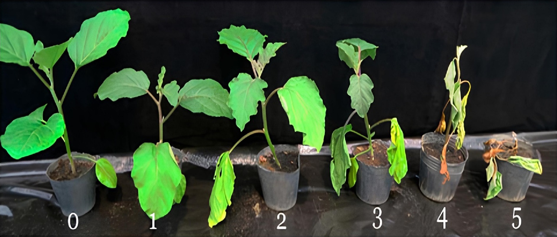
      Figure 1. 

      Bacterial wilt disease rating scale (0 = no symptoms, 1 = only one leaf partially wilted, 2 = two or three leaves wilted, 3 = all leaves except two or three wilted, 4 = all leaves wilted, 5 = dead plant).

      The disease severity index (DI) was calculated following the formula: DI = (N1 × 1 + N2 × 2 + N3 × 3 + N4 × 4 + N5 × 5)/ (Nt/5)[9], where N1 to N5 = the number of plants with disease rating scale values from 0 to 5, and Nt = the total number of plants observed. Based on the disease index, each Solanum line was categorized as resistant or susceptible as shown in Table 2[18].

      Table 2.  Scale based on disease index for the classification of solanum germplasm.[2]

      DI score (0−1) Reaction
      0.00−0.20 Highly resistant
      0.21−0.30 Resistant
      0.31−0.40 Moderately resistant
      0.41−0.50 Moderately susceptible
      0.51−0.60 Susceptible
      0.61−0.90 Highly susceptible
      0.91−1.00 Extremely susceptible
    • Leaves of approximately 0.2 g were collected into sampling bags, transferred into pre-frozen mortars, and homogenized. Subsequently, samples were transferred into 2 mL Eppendorf tubes for DNA isolation.

    • Genomic DNA was isolated using CTAB (Cetyltrimethylammonium bromide)[19]. DNA was quantified using a Nanodrop 2000 C Spectrophotometer (Thermoscientific, USA) and quality was checked on a 0.8% agarose gel. Nine bacterial wilt trait-linked markers were used for the study as presented in Table 3[20]. These primers are linked to QTLs (Quantitative Trait Loci) Bwr 12 and 6 which confer resistance to bacterial wilt disease PCR was performed using SeeAmp (Hangzhou Bioer Technology Co. Ltd, China) thermal cycler. The PCR amplification reaction of 10 μL contained 10X DreamTaq PCR buffer, 10 mM dNTPs, 10 μM of forward and reverse primer, 2.5 U/μL DNA polymerase, 50 ng DNA template, and nuclease-free water. For the PCR, three controls were used to prevent the scoring of false bands. These comprised a known positive control, a known negative control, and a no template control (NTC). All samples including the positive controls were duplicated to ensure the reliability and reproducibility of results. Amplified products were separated on 1.5% agarose gel in TBE buffer, stained with ethidium bromide, and an image was captured using AlphaImager HP (Proteinsimple, USA). Scoring of bands was conducted using AlphaImager HP Software Version. 3.4.

      Table 3.  Sequences and expected product size of primers used for the study.

      Trait Primer R-gene Forward primer (5'-3') Reverse primer (3'-5') Annealing temperature (°C) Product size (bp) Ref.
      Resistant genotype Susceptible genotype
      Bacteria wilt resistance SLM12–2 Bwr-12 ATCTCATTCAACGCACACCA AACGGTGGAAACTATTG
      AAAGG
      55 209 No reference band [12]
      SLM12–10 Bwr-12 ACCGCCCTAGCCATAAAGAC TGCGTCGAAAATAGTTGCAT 242
      SLM6–124 Bwr-6 CATGGGTTAGCAGATGATT
      CAA
      GCTAGGTTATTGGGCCAGAA 292
      SLM6–118 Bwr-6 TCCCAAAGTGCAATAGG
      ACA
      CACATAACATGGAGTTCGACAGA 183
      SLM6–119 Bwr-6 GCCTGCCCTACAACAAC
      ATT
      CGACATCAAACCTATGAC
      TGGA
      255
      SLM6–136 Bwr-6 CCAGGCCACATAGAACTC
      AAG
      ACAGGTCTCCATACGGCATC 290
      SLM6–17 Bwr-6 TCCTTCAAATCTCCCA
      TCAA
      ACGAGCAATTGCAAGG
      AAAA
      186
      SLM6–94 Bwr-6 CTAAATTTAAATGGACAA
      GTAATAGCC
      CACGATAGGTTGGTATTTTCTGG 276
    • The band size for resistant genotypes was scored as present (+) whilst that of susceptible genotypes was scored as absent (−).

    • One-way ANOVA at a probability level of 5% (p < 0.05) was performed for the wilting incidence and disease severity index using Statistix version 8.0. Before analysis, data on percent wilting was arcsine transformed to improve normality. Differences between the means were compared and separated using Fisher's Least Significant Difference (LSD) test.

    • The various accessions screened showed symptoms of bacterial wilt disease with varied levels of reaction to the R. solanacearum infection at the various assessment periods. Thirty days after inoculation, significant differences (p < 0.05) in percent wilting incidence were observed among the garden egg accessions. Among the garden egg accessions, wilting incidence ranged from 54.0% to 71.0% in accessions CRI-01 and CRI-03 respectively (Fig. 2). A similar trend was observed among the tomato accessions evaluated as significant differences (p < 0.05) in disease incidence were observed among them. Among the tomato lines, accessions L_020, and GD, GC recorded the lowest and highest disease incidence of 56% and 90% respectively (Fig. 2) at the end of the assessment period. Generally, the tomato accessions recorded higher disease incidence compared to garden eggs.
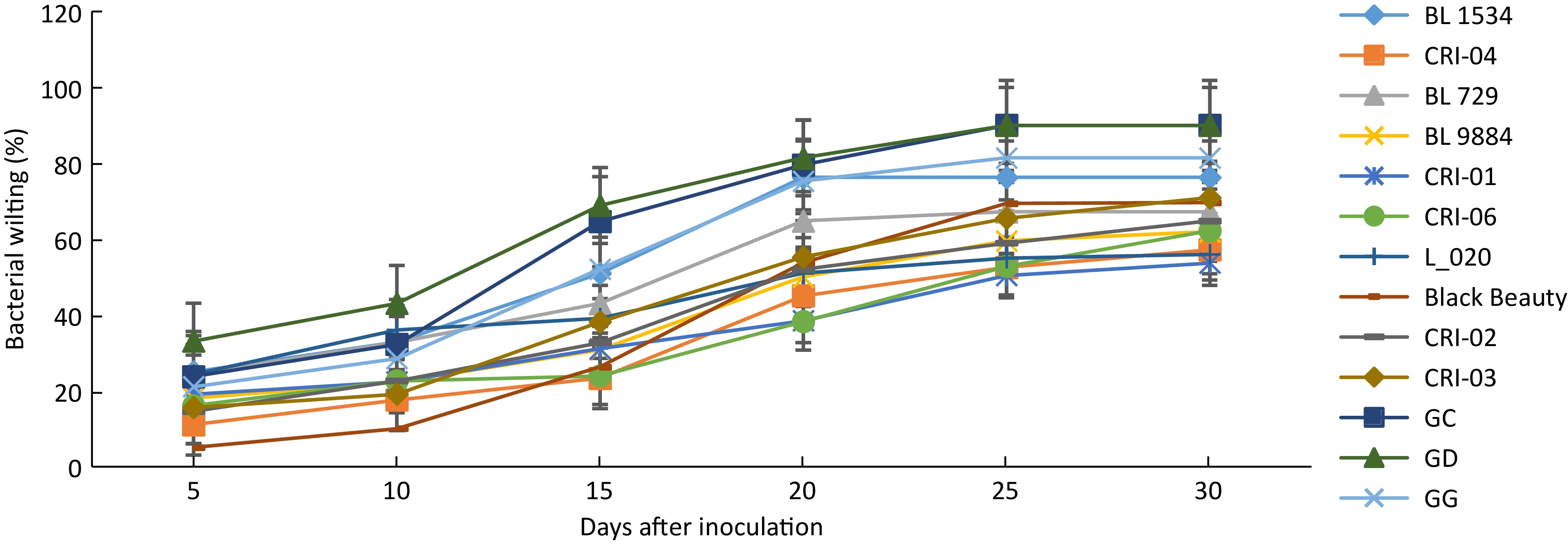
      Figure 2. 

      Wilting incidence among 13 solanum accessions. Error bars represent the standard error of the treatment means.

      Disease severity among the garden egg accessions ranged from 0.45 to 0.61 with no significant differences among them (Table 4). This was, however, not the case with the tomato accessions as significant differences (p < 0.05) in disease severity index were observed among the various genotypes. The lowest disease severity (0.37) was recorded for accession (L_020) compared to 0.90 recorded for accession GC (Table 5). Based on the disease severity index, none of the accessions evaluated was found to be highly resistant to the bacterial pathogen. Two accessions (CRI-04 and L_020), however, were moderately resistant with accessions BL1534, GD, GG, and GC recording disease severities above 0.6 and therefore classified as highly susceptible to the pathogen.

      Table 4.  Mean bacterial wilt disease severity among garden egg accessions evaluated.

      Accession Mean disease
      severity index (0–1)
      Host reaction
      CRI-06 0.45 Moderately susceptible
      CRI-03 0.56 Susceptible
      CRI-04 0.37 Moderately resistant
      CRI-02 0.47 Moderately susceptible
      CRI-01 0.52 Susceptible
      Black Beauty 0.61 Susceptible
      p < 0.05 NS

      Table 5.  Bacterial wilt disease severity and host reaction status among tomato accessions evaluated.

      Accession Disease severity index (0–1) Host reaction
      BL 729 0.53ab Susceptible
      BL 9884 0.44b Moderately susceptible
      GG 0.71ab Highly susceptible
      L_020 0.39b Moderately resistant
      GC 0.90a Highly susceptible
      BL 1534 0.70ab Highly susceptible
      GD 0.70ab Highly susceptible
      Means followed by different letters are significantly different.
    • All the samples produced visible bands following the PCR amplification (Fig. 3), however only samples with the expected band size were scored as positive. Three of the tomato genotypes (L_020, GG, and GC) representing 23%, showed expected bands for all two primers of Bwr-12 whilst none of the garden egg genotypes showed expected bands for Bwr-12 (Table 6). Genotypes that showed expected bands for primers linked to Bwr-6 ranged from two to eight. Four tomato genotypes (BL1534, BL729, GG, and BL9884) and four garden egg genotypes (Black Beauty, CRI-01, CRI-02, and CRI-03) scored the maximum number of alleles for primer SLM 6-118 whilst only two tomato genotypes (BL729 and GD) scored the minimum number of alleles for SLM 6-17. Two of the garden egg genotypes (CRI-01 and CRI-06) showed alleles for only one of the Bwr-6 genes (SLM 6-118 and 6-110 respectively). The genotypes screened for all the nine primers showed alleles ranging from one to seven. Across all the nine primers used, only one tomato genotype (GG) had alleles for seven of the primers whilst none of the garden eggs had alleles across all nine primers. Only one tomato genotype (BL729) had alleles for six of the primers linked to Bwr-6 whilst the garden egg genotype (Black Beauty) had four alleles (Table 6). Genotypes with both Bwr-6 and Bwr-12 exhibit stable resistance against Phylotype I and II strains of the bacteria; hence this study identified some genotypes that had partial resistance (only Bwr-6) and others with durable resistance (combination of both genes). In effect, genotypes that showed amplification for at least one primer of both QTLs (Bwr-6 and Bwr-12) were classified as durable resistance, hence a total of three genotypes were identified. Genotypes with only one marker (either Bwr-6 or Bwr-12) were classified as partial resistance, hence a total of 10 genotypes were identified.
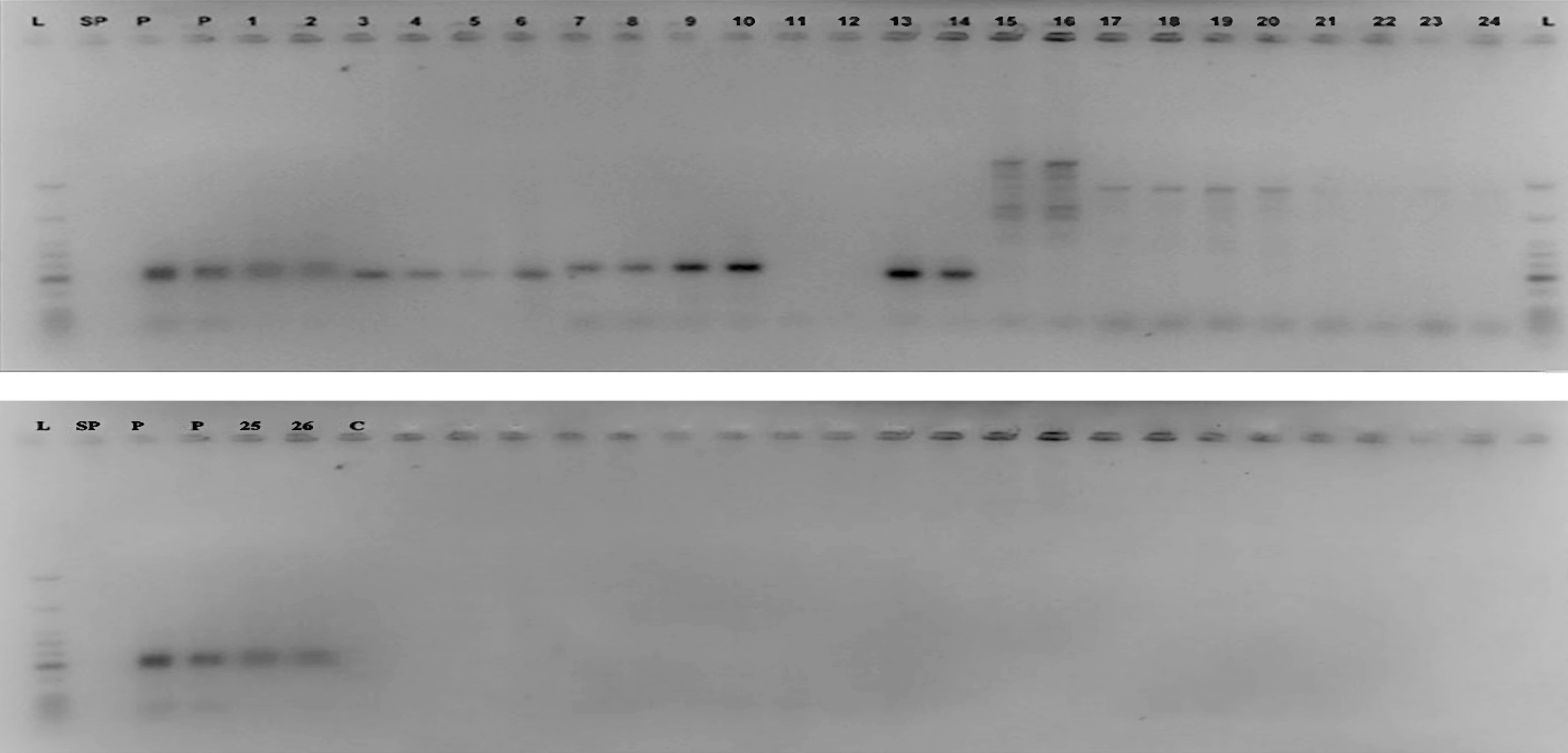
      Figure 3. 

      Agarose gel image of the marker SLM 12-2 for the detection of the Bwr-12 gene L = Molecular weight ladder; SP = Space; P = Positive control; Well 1 & 2 = L_020; 3 & 4 = BL1534; 5 & 6 = BL729; 7 & 8 = GG; 9 & 10 = GC; 11 & 12 = GD; 13 & 14 = BL9884; 15 & 16 = Black Beauty; 17 & 18 = CRI 01; 19 & 20 = CRI 02; 21 & 22 = CRI 03; 23 & 24 = CRI 04; 25 & 26 = CRI 06; C = Negative control.

      Table 6.  Scores for bacteria wilt resistant gene(s) in tomato and garden egg genotypes.

      Genotypes SSR markers Disease reaction
      Bwr-12 Bwr-6
      SLM 12-2 SLM 12-10 SLM 6-136 SLM 6-119 SLM 6-94 SLM 6-118 SLM 6-110 SLM 6-124 SLM 6-17
      L_020 +/+ +/+ +/+ +/+ +/+ −/− −/− −/− −/− Durable resistance
      BL1534 −/− −/− +/+ −/− −/− +/+ −/− −/− −/− Partial resistance
      BL729 −/− −/− −/− +/+ +/+ +/+ +/+ +/+ +/+ Partial resistance
      GG +/+ +/+ +/+ +/+ −/− +/+ +/+ +/+ −/− Durable resistance
      GC +/+ +/+ +/+ −/− +/+ −/− +/+ +/+ −/− Durable resistance
      GD −/− −/− −/− −/− −/− −/− −/− −/− +/+ Partial resistance
      BL9884 −/− −/− +/+ +/+ −/− +/+ −/− +/+ −/− Partial resistance
      Black Beauty −/− −/− −/− +/+ +/+ +/+ +/+ +/+ −/− Partial resistance
      CRI 01 −/− −/− −/− −/− −/− +/+ −/− −/− −/− Partial resistance
      CRI 02 −/− −/− +/+ −/− −/− +/+ −/− −/− −/− Partial resistance
      CRI 03 −/− −/− +/+ −/− −/− +/+ −/− −/− −/− Partial resistance
      CRI 04 −/− −/− −/− −/− −/− −/− −/− −/− −/− Partial resistance
      CRI 06 −/− −/− −/− −/− −/− −/− +/+ −/− −/− Partial resistance
    • Bacterial wilt disease caused by R. solanacearum is a major constraint to global tomato production[24]. The negative effect of the disease is further aggravated by the limited number of management options available to farmers to effectively control the disease[6]. Grafting of susceptible cultivars with desired traits unto a bacterial wilt-resistant rootstock has been identified and promoted as a sustainable means to manage the disease. Both phenotypic and molecular tools have been employed and used successfully to select suitable rootstocks for grafting. The current study screened six tomato and seven garden egg accessions using artificial inoculation procedures and molecular markers to identify and/or select bacterial wilt-resistant rootstocks. The present results showed that all the test materials artificially inoculated with R. solanacearum showed symptoms of wilting. Previous studies[16,21,22] reported that host plants of Solanum spp. present symptoms of wilting following infection and establishment of the pathogen in them. Although all the accessions showed symptoms of infection, they varied in the disease parameters like the incidence and severity of wilting assessed. This assertion is supported by the fact that 57.0% and 16.0% of the tomato and garden eggs lines respectively recorded a wilting incidence of more than 70%. The significant variations in disease expression among and between the accessions as recorded in this study corroborate previous studies[6,23] that reported similar trends in solanaceous cultivars. Based on the phenotypic parameters (wilting incidence and severity scores) measured for this study, none of the test materials screened could be described as resistant to the bacterial wilt disease, although, accessions, L_020 (tomato) and CRI-04 (garden eggs) were found to be moderately resistant. The identification of moderately resistant accessions in the present work supports the findings of Namisy et al. & Stella et al.[9,24] who identified moderately resistant tomato and garden egg lines from field trials for use as rootstocks to manage bacterial wilt disease. The large number of R. solanacearum-susceptible accessions recorded in this study confirms the wide host range of the pathogen causing bacteria wilt disease and the limitation in the identification and selection of resistant rootstocks to manage the disease. Although artificial inoculation procedures have successfully been used to select bacterial wilt-resistant rootstocks, complementing it with marker-assisted selection is considered a standard approach for resistance screening. For this study, in addition to the field evaluation, molecular markers were used to enable efficient identification of genotypes with resistant genes for bacterial wilt diseases as well as multiple resistant gene combinations, which would not have been possible with symptom expression alone. These multiple resistant genotypes could be used in areas where the diseases occur sequentially or simultaneously. Two strains of R. solanacearum that cause bacterial wilt have been reported as being prevalent in Ghana[5]. Bwr-6 and Bwr-12 QTLs have been identified to confer resistance to the disease with Bwr-6 conferring resistance against phylotypes I and II; Bwr-12 conferring resistance against only phylotype I[25]. It has also been reported that genotypes with both Bwr-6 and Bwr-12 show stable resistance against Phylotypes I and II[25], hence any genotype with a combination of both QTLs expresses more durable resistance than genotypes with only one of the genes. Markers used in this study were able to identify genotypes with either Bwr-6 or Bwr-12 or a combination of both. Since genotypes with both Bwr-6 and Bwr-12 show stable resistance, this study identified three tomato genotypes (L_020, GG and GC) that had a combination of both genes and thus, agree with the findings of Carmeille et al.[26] who detected both QTLs in some tomato lines. Furthermore, four of the genotypes had either of the QTLs, indicating partial resistance. All the garden egg genotypes had one or more of the alleles for Bwr-6, indicating partial resistance. In a similar study,[27] it was found that stable QTLs for bacterial wilt resistance in garden eggs were located on chromosomes 3 and 6. The QTLs on chromosome 6 overlap with the BW-resistant QTL (Bwr-6) in tomatoes. This explains the current results where there was no amplification for Bwr-12 QTL in the garden egg genotypes but there was amplification in the Bwr-6 QTL. Contrary to the morphological studies, two accessions (GC and GG) classified as resistant were identified as highly susceptible to the pathogen under field conditions. The difference in morphological and molecular categorization as obtained in this study is consistent with Olasanmi et al.[28] who reported inconsistencies in cassava mosaic disease resistance levels in cassava genotypes based on field and molecular marker data. Accessions classified as resistant based on molecular markers but identified as susceptible under phenotypic evaluation according to Wang & Lin[29] may be attributed to the presence and interaction of other strains of the R. solanacearum on the field, inoculum density, soil moisture, temperature, and presence of root-knot nematodes which can affect the stability of bacterial wilt resistance in crop genotypes.

    • The results presented in this study show variations in the reaction of different accessions to bacterial wilt disease using both phenotypic and molecular markers. Using only phenotypic scores two accessions were classified as moderately resistant while three accessions were selected as resistant based on the molecular markers used. However, only accession L_020 phenotypically identified as moderately resistant was further confirmed as resistant based on the molecular data and therefore, selected as a promising genotype to be exploited as a potential rootstock to manage bacterial wilt disease. Due to the inconsistent phenotypic and molecular data obtained in this study, accessions (CRI-04, GG, and GC) cannot be selected as potential rootstocks to manage bacterial wilt disease in tomato production. These accessions can be screened further with additional molecular markers and under different fields to confirm the results obtained.

    • The authors confirm contribution to the paper as follows: performing the research: Adomako J, Prempeh RNA; data analysis and technical help: Osei MK, Gyau J, draft of the manuscript: Cho MC, Adomako J, Prempeh RNA, Osei MK; experiments design, study supervision and manuscript revision: Boakye-Mensah IN, Osei-Bonsu I, Ofori P. All authors reviewed the results and approved the final version of the manuscript.

    • The datasets generated during and/or analyzed during the current study are available from the corresponding author on reasonable request.

      • This study was supported by a grant from KAFACI, Rural Development Administration of Korea (Grant No. KAH2000106).

      • The authors declare that they have no conflict of interest.

      • Copyright: © 2024 by the author(s). Published by Maximum Academic Press, Fayetteville, GA. This article is an open access article distributed under Creative Commons Attribution License (CC BY 4.0), visit https://creativecommons.org/licenses/by/4.0/.
    Figure (3)  Table (6) References (29)
  • About this article
    Cite this article
    Adomako J, Osei MK, Prempeh RNA, Osei-Bonsu I, Gyau J, et al. 2024. Identification of Ralstonia solanacearum resistant solanum plants as potential rootstock to manage bacterial wilt disease in tomato production. Technology in Horticulture 4: e020 doi: 10.48130/tihort-0024-0017
    Adomako J, Osei MK, Prempeh RNA, Osei-Bonsu I, Gyau J, et al. 2024. Identification of Ralstonia solanacearum resistant solanum plants as potential rootstock to manage bacterial wilt disease in tomato production. Technology in Horticulture 4: e020 doi: 10.48130/tihort-0024-0017

Catalog

  • About this article

/

DownLoad:  Full-Size Img  PowerPoint
Return
Return