REVIEW   Open Access    

Review of the toxic effects and health functions of arecoline on multiple organ systems

More Information
  • Arecoline, the principal active alkaloid in the areca nut, is known for its ability to induce euphoric sensations. Since ancient times, arecoline has garnered attention for its therapeutic potential in addressing psychiatric disorders and alleviating gastrointestinal ailments. However, in 2020, the International Agency for Research on Cancer has classified arecoline as 'probably carcinogenic to humans' (Group 2B carcinogen), supported by compelling mechanistic evidence. The mechanism of action of arecoline has been extensively studied, but the results of these studies are scattered and lack systematic integration and generalization. In this paper, we have systematically summarized the mechanism of arecoline within the oral cavity, central nervous system, cardiovascular system, and digestion system, in terms of both health functions and toxic effects. In addition, we found some concentration-effect relationship between arecoline in the central nervous system and digestive system, i.e., low doses are beneficial and high doses are harmful. By summarizing the mechanisms of arecoline, this review is poised to provide in-depth and valuable insights into the clinical practice and targeted therapy of arecoline in the future.
  • The Lonicera Linn. genus is a constituent member of the Caprifoliaceae family[1]. It is the largest genus in this family and comprises at least 200 species with a notable presence in North Africa, North America, Asia, and Europe[1]. Members of the Lonicera genus possess a wide range of economic benefits from their use as ornamental plants to food and as plants credited with numerous health benefits. Conspicuous among the numerous members of this genus with known medicinal uses are L. japonica, L. macranthoides, L. hypoglauca, L. confusa, and L. fulvotomentosa[2]. Though these species feature prominently in the Chinese Pharmacopoeia, other species such as L. acuminata, L. buchananii, and L. similis are recognized as medicinal resources in certain parts of China[1]. Among the aforementioned species, L. japonica takes precedence over the rest due to its high medicinal and nutritional value[3,4]. For instance, the microRNA MIR2911, an isolate from L. japonica, has been reported to inhibit the replication of viruses[57]. Also, the water extract of L. japonica has been used to produce various beverages and health products[8]. The Lonicera genus therefore possesses huge prospects in the pharmaceutical, food, and cosmetic industries as an invaluable raw material[9].

    The main active constituents of the Lonicera genus include organic acids, flavonoids, iridoids, and triterpene saponins. Chlorogenic acids, iridoids, and flavones are mainly credited with the anti-inflammatory, antiviral, anticancer, and antioxidant effects of the various Lonicera species[1013]. Their hepatoprotective, immune modulatory, anti-tumor and anti-Alzheimer’s effects are for the most part ascribed to the triterpene saponins[1416]. As stated in the Chinese Pharmacopoeia and backed by the findings of diverse research groups, the plants of the Lonicera genus are known to possess high amounts of organic acids (specifically chlorogenic acid) and pentacyclic triterpenoid saponins[2,1719]. The flower and flower bud have traditionally served as the main medicinal parts of the Lonicera genus even though there is ample evidence that the leaves possess the same chemical composition[20]. A perusal of the current scientific literature reveals the fact that little attention has been devoted to exploring the biosynthesis of the chemical constituents of the Lonicera genus with the view to finding alternative means of obtaining higher yields. It is therefore imperative that priority is given to the exploration of the biosynthesis of these bioactive compounds as a possible means of resource protection. There is also the need for further research on ways to fully tap the medicinal benefits of other plant parts in the Lonicera genus.

    Here, we provide a comprehensive review of relevant scientific literature covering the structure, pharmacology, multi-omics analyses, phylogenetic analysis, biosynthesis, and metabolic engineering of the main bioactive constituents of the Lonicera genus. Finally, we proffer suggestions on the prospects of fully exploiting and utilizing plants of the Lonicera genus as useful medicinal plant resources.

    A total of at least 400 secondary metabolites have been reported for the Lonicera genus. These metabolites are categorized into four main groups (Fig. 1a), including not less than 50 organic acids, 80 flavonoids, 80 iridoids, and 80 triterpene saponins[2123]. Organic acids are mainly derivatives of p-hydroxycinnamic acid and quinic acid. Among the organic acids, chlorogenic acids are reported to be the main bioactive compounds in L. japonica[2426]. The organic acids are most abundant in the leaves, while the least amounts are found in the stem of L. japonica. The flowers of the plant are known to contain moderately high amounts of organic acids[27]. The basic core structure of the flavonoids is 2-phenylchromogen. Luteolin and its glycoside which are characteristic flavonoids of the Lonicera genus are most abundant in L. japonica[28]. On the whole, the flavonoid contents in L. japonica are also highest in the leaves, available in moderate amounts in the flowers, and in lowest amounts in the stem[21]. The core structures of the iridoids are iridoid alcohols, the chemical properties of which are similar to hemiacetal. The iridoids often exist in the form of iridoid glycosides in plants. Secoiridoids glycosides are predominant in the Lonicera genus[25]. In L. japonica, the contents of the iridoids are most abundant in the flowers, moderate in leaves, and lowest in the stem[21]. The characteristic saponins of the Lonicera genus are mainly pentacyclic triterpenoids, including the hederin-, oleanane-, lupane-, ursulane- and fernane-types, etc[22]. The hederin-type saponins are reported in the highest amounts in L. macranthoides[17] (Fig. 1b).

    Figure 1.  Core structures of main secondary metabolites and their distribution in five species of Lonicera. (a) 1 and 2, the main core structures of organic acids; 3, the main core structures of flavonoids; 4, the main core structures of iridoids; 5, the main core structures of triterpene saponins. (b) Comparison of dry weight of four kinds of substances in five species of Lonicera[17,28].

    The similarities between chlorogenic acid (CGA) and flavonoids can be traced back to their biosynthesis since p-coumaroyl CoA serves as the common precursor for these compounds[29]. p-coumaroyl CoA is obtained through sequential catalysis of phenylalanine and its biosynthetic intermediates by phenylalanine-ammonia-lyase (PAL), cinnamate 4-hydroxylase (C4H) and 4-coumarate CoA ligase (4CL)[3033].

    CGA is a phenolic acid composed of caffeic acid and quinic acid and is the most important bioactive compound among the organic acids. Its biosynthesis has been relatively well-established; three main biosynthetic routes have been propounded (Fig. 2a). One route relates to the catalysis of caffeoyl-CoA and quinic acid by hydroxycinnamoyl-CoA quinate transferase (HQT)/hydroxycinnamoyl CoA shikimate/quinate hydroxycinnamoyl transferase (HCT) to produce CGA[3437]. The HQT-mediated pathway has been deemed the major route for CGA synthesis in in different plant species[38,39]. The second biosynthetic route stems from the biosynthesis of p-coumaroyl quinate through the catalytic effect of HCT/HQT and subsequent hydroxylation of p-coumaroyl quinate under the catalysis of p-coumarate 3'-hydroxylase (C3’H)[34,36,37]. For the third route, caffeoyl glucoside serves as the intermediate to form CGA, a process that is catalyzed by hydroxycinnamyl D-glucose: quinic acid hydroxycinnamyl transferase (HCGQT)[40,41].

    Figure 2.  Biosynthetic pathways of main bioactive constituents of Lonicera. (a) Biosynthetic pathways of chlorogenic acid. (b) Biosynthetic pathways of luteoloside. (c) Biosynthetic pathways of secologanin. (d) Biosynthetic pathways of hederin-type triterpene saponins. PAL, phenylalanine ammonia-lyase; C4H, cinnamate 4-hydroxylase; 4CL, 4-hydroxycinnamoyl CoA ligase; HCT, hydroxycinnamoyl CoA shikimate/quinate hydroxycinnamoyl transferase; C3’H, p-coumaroyl 3-hydroxylase; HQT, hydroxycinnamoyl-CoA quinate transferase; UGCT, UDP glucose: cinnamate glucosyl transferase; CGH, p-coumaroyl-D-glucose hydroxylase; HCGQT, hydroxycinnamoyl D-glucose: quinate hydroxycinnamoyl transferase; CHS, Chalcone synthase; CHI, Chalcone isomerase; FNS, Flavone synthase; F3H, flavonoid 30-monooxygenase/flavonoid 30-hydroxylase; UF7GT, flavone 7-O-β-glucosyltransferase; GPS, Geranyl pyrophosphatase; GES, geraniol synthase; G8O, geraniol 10-hydroxylase/8-oxidase; 8HO, 8-hydroxygeraniol oxidoreductase; IS, iridoid synthase; IO, iridoid oxidase; 7DLGT, 7-deoxyloganetic acid glucosyltransferase; 7DLH, 7-deoxyloganic acid hydroxylase; LAMT, loganic acid O-methyltransferase; SLS, secologanin synthase; FPS, farnesyl pyrophosphate synthase; SS, squalene synthase; SE, squalene epoxidase; β-AS, β-amyrin synthase; OAS, oleanolic acid synthetase.

    The key enzymes in the biosynthesis of p-coumaroyl CoA, and invariably CGA, thus, PAL, C4H, and 4CL have been established in diverse studies such as enzyme gene overexpression/knockdown[42], enzyme activity analysis[33] and transcriptomics[18]. However, the centrality of HQT in the biosynthesis of CGA remains disputable. While some studies have reported a strong correlation between HQT expression level with CGA content and distribution[18,35,39,43,44], others found no such link[45], bringing into question the role of HQT as a key enzyme in CGA biosynthesis.

    Few studies have been conducted on the regulation of CGA biosynthesis in the Lonicera genus. It was found that overexpression of the transcription factor, LmMYB15 in Nicotiana benthamiana can promote CGA accumulation by directly activating 4CL or indirectly binding to MYB3 and MYB4 promoters[46]. LjbZIP8 can specifically bind to PAL2 and act as a transcriptional repressor to reduce PAL2 expression levels and CGA content[47]. Under NaCl stress, increased PAL expression promoted the accumulation of phenolic substances in leaves without oxidative damage, a condition that was conducive to the accumulation of the bioactive compounds in leaves[48].

    Luteolin and its derivative luteolin 7-O- glucoside (luteoloside) are representative flavonoids of the Lonicera genus. Similar to CGA, luteolin is biosynthesized from p-coumaroyl CoA but via a different route. The transition from p-coumaroyl CoA to luteolin is underpinned by sequential catalysis by chalcone synthetase (CHS), chalcone isomerase (CHI), flavonoid synthetase (FNS), and flavonoid 3'-monooxygenase/flavonoid 3'-hydroxylase (F3'H)[45,49,50] (Fig. 2b). Luteoloside is synthesized from luteolin by UDP glucose-flavonoid 7-O-β-glucosyltransferase (UF7GT)[51]. Similar to CGA biosynthesis, the key enzymes of luteolin synthesis include PAL, C4H, and 4CL in addition to FNS[33,45,52]. The content of luteoloside was found to be highly abundant in senescing leaves relative to other tissues such as stem, flowers, and even young leaves[52]. Through transcriptomic analysis, luteoloside biosynthesis-related differentially expressed unigenes (DEGs) such as PAL2, C4H2, flavone 7-O-β-glucosyltransferase (UFGT), 4CL, C4H, chalcone synthase 2 and flavonoid 3'-monooxygenase (F3'H) genes were found to be upregulated in the senescing leaves. Biosynthesis-related transcription factors such as MYB, bHLH, and WD40 were also differentially expressed during leaf senescence[52], while bHLH, ERF, MYB, bZIP, and NAC were differentially expressed during flower growth[53]. Further analysis of the transcription factors revealed that MYB12, MYB44, MYB75, MYB114, MYC12, bHLH113, and TTG1 are crucial in luteoloside biosynthesis[52,53].

    The biosynthesis of terpenoids mainly involves three stages; formation of intermediates, formation of basic structural skeleton, and modification of basic skeleton[54]. The intermediates of terpenoids are mainly formed through the mevalonate (MVA) and methylerythritol phosphate (MEP) pathways, and eventually converted to the universal isoprenoid precursors, isopentenyl pyrophosphate (IPP) and its isomer dimethylallyl pyrophosphate (DMAPP) through a series of enzyme-catalyzed reactions. Under the catalysis of geranyl pyrophosphatase (GPS), IPP is then converted to geranyl pyrophosphate (GPP). Different terpenoids are subsequently derived from GPP as the intermediate product. For instance, in the formation of secoiridoid, GPP first removes the phosphoric acid group to obtain geraniol, second through a series of reactions such as oxidation and cyclization, the skeleton of iridoid, namely iridodial, can be obtained. Finally, through a series of reactions, the basic carbon skeleton of the secoiridoid, namely secologanin, is obtained[5561] (Fig. 2c). In the formation of triterpene saponins, the key step lies in the formation of the precursor, 2,3-oxidosqualene, a reaction that is catalyzed by squalene epoxidase (SE). There are many pentacyclic triterpenes in the Lonicera genus, the most important type being the hederin-type saponins with hederagenin as aglycones. Hederin-type saponins are produced after the synthesis of oleanolic acid from β-amyrin and catalyzed by β-starch synthetase (β-AS) and Oleanolic acid synthase (OAS)[62,63]. The skeletal modification of the triterpenoid saponins is mainly achieved via the activities of the CYP450 enzymes and UDP-glycosyltransferase (UGT). Hence, the corresponding aglycones are first obtained via oxidation by the CYP450 enzymes (e.g., CYP72A), and further subjected to glycosylation by the appropriate UGT enzyme[6365] (Fig. 2d). Skeletal formations of the iridoids and triterpene saponins in general have been well researched, but the same cannot be said about the enzymes involved in biosynthesis of these groups of compounds in the Lonicera genus. To fully utilize the iridoids and triterpene saponins in the Lonicera genus, it is necessary to further explore their biosyntheses with the view to enhancing and optimizing the process.

    Given the importance of the bioactive compounds in the Lonicera genus, continual isolation of these compounds using the traditional methods are not only tedious and time-consuming, but also unsustainable. With the development and application of microbial metabolic engineering, different strategies have been introduced to produce these bioactive compounds by heterologous synthesis (Table 1).

    Table 1.  Biosynthesis of Lonicera-specialized metabolites using metabolic engineering.
    Engineering bacteriaOperational methodsProductsYieldRefs
    S. cerevisiaeEliminate the tyrosine-induced feedback inhibition, delete genes involved in competing pathways and overexpress rate-limiting enzymesCaffeic acid569.0 mg/L[69]
    S. cerevisiaeEmploye a heterologous tyrosine ammonia lyase and a 4HPA3H complex composed of HpaB and HpaC derived from different speciesCaffeic acid289.4 mg/L[73]
    S. cerevisiaeSupply and recycle of three cofactors: FADH2, S-adenosyl-L-methion, NADPHCaffeic acid
    Ferulic acid
    Caffeic acid: 5.5 g/L;
    Ferulic acid: 3.8 g/L
    [117]
    E. coliKnocking out competing pathwaysCaffeic acid7,922 mg/L[118]
    E. coliArtificial microbial community, a polyculture of three recombinant Escherichia coli strainsChlorogenic acid250 μM[68]
    Cell-free biosynthesisExtract and purify spy-cyclized enzymes (CFBS-mixture)Chlorogenic acid711.26 mg/L[70]
    S. cerevisiaeThree metabolic engineering modules were systematically optimized: shikimate pathway and carbon distribution, branch pathways, CGA pathway genesChlorogenic acidFlask fermentation: 234.8 mg/L;
    Fed-batch fermentation:
    806.8 mg/L
    [119]
    E. coliUsing modular coculture engineering: construction of the defective strain improves the production and utilization of precursor substancesChlorogenic acid131.31 mg/L[122]
    E. coliIntroduce heterologous UDP-glucose biosynthetic genesLuteolin34 mg/L[120]
    Y. lipolyticaOverexpression of the key genes involved in the mevalonate pathway, the gene encoding cytochrome P450 (CYP716A12) to that encoding NADPH-P450 reductaseOleanolic acid129.9 mg/L[85]
    S. cerevisiaeImprove the pairing efficiency between Cytochrome P450 monooxygenase and reductase and the expression level of key genesOleanolic acid606.9 mg/L[121]
    S. cerevisiaeHeterologous expression and optimization of CrAS, CrAO, and AtCPR1, and regulation of ERG1 and NADPH regeneration systemOleanolic acid433.9 mg/L[123]
     | Show Table
    DownLoad: CSV

    Due to the demand for CGA in the food, pharmaceutical, chemical, and cosmetic industries, the traditional means of obtaining the same requires a relatively longer period for plant maturation to obtain low yields of the desired product. This therefore brings into question the sustainability and efficiency of this approach. The alternative and sustainable approach has been to produce CGA using synthetic biology and metabolic engineering.

    Current research has sought to utilize Escherichia coli (and its mutant strain) and Saccharomyces cerevisiae to synthetically generate CGA and other flavonoids[6673]. For instance, Cha et al. employed two strains of E. coli to produce a relatively good yield of CGA (78 mg/L). Their approach was based on the ability of one strain to generate caffeic acid from glucose and the other strain to use the caffeic acid produced and quinic acid as starting materials to synthesize CGA[66]. Using a bioengineered mutant of E. coli (aroD mutant), Kim et al. increased the yield of CGA to as high as 450 mg/L[67]. Others have sought to increase the yield of CGA by employing a polyculture of three E. coli strains that act as specific modules for the de novo biosynthesis of caffeic acid, quinic acid and CGA. This strategy eliminates the competition posed by the precursor of CGA (i.e., caffeic acid and quinic acid) and generally results in improved production of CGA[68]. Saccharomyces cerevisiae is a chassis widely used for the production of natural substances from plants with an intimal structure that can be used for the expression of cytochrome P450 enzymes that cannot be expressed in E. coli. Researchers have used yeast to increase the production of organic acids[69]. A de novo biosynthetic pathway for the construction of CGA in yeast has been reported new cell-free biosynthetic system based on a mixture of chassis cell extracts and purified Spy cyclized enzymes were adopted by Niu et al. to a produce the highest yield of CGA reported so far up to 711.26 ± 15.63 mg/L[70].

    There are many studies on the metabolic engineering for the synthesis of flavonoids, but few on luteolin and its glycosides. Strains of E. coli have been engineered with specific uridine diphosphate (UDP)-dependent glycosyltransferase (UGT) to synthesize three novel flavonoid glycosides. These glycosides were quercetin 3-O-(N-acetyl) quinovosamine (158.3 mg/L), luteolin 7-O-(N-acetyl) glucosaminuronic acid (172.5 mg/L) and quercetin 3-O-(N-acetyl)-xylosamine (160.8 mg/L)[71]. Since most of the flavonoid glycosides synthesized in E. coli are glucosylated, Kim et al. in their bid to synthesize luteolin-7-O-glucuronide, deleted the araA gene that encodes UDP-4-deoxy-4-formamido-L-arabinose formyltransferase/UDP-glucuronic acid C-4'' decarboxylase in E.coli and were able to obtain a yield of 300 mg/L of the desired product[72].

    Terpenoidal saponins are mostly derived from slow-growing plants and usually possess multiple chiral centers[74]. Traditional isolation and even chemical synthesis of the terpenoidal saponins are both tedious and uneconomical for large-scale production. Therefore, it is necessary to find other ways to synthesize these compounds known to have diverse pharmacological functions.

    Heterologous synthesis has become an important way to improve the target products. With the development of synthetic biology, heterologous synthesis of triterpene saponins involves chassis of both plant and microbial origin. In this regard, Nicotiana benthamiana is a model plant species for the reconstruction of the biosynthetic pathways of different bioactive compounds including monoterpenes, hemiterpenes, and diterpenes[59,7577]. Aside from Nicotiana benthamiana, other plants have also been used as heterologous hosts[78]. Heterologous synthesis using microbial hosts mainly involves Saccharomyces cerevisiae and Escherichia coli[7981], and other microorganisms[82,83]. Comparatively, plants as biosynthetic hosts have the advantages of an established photosynthetic system, abundant supply of relevant enzymes, and presence of cell compartments, etc. They are however not as fast growing as the microorganisms, and it is also difficult to extract and separate the desired synthesized compounds from them as hosts.

    Although heterologous synthesis has many advantages, the premise of successful construction of synthetic pathway in host is to elucidate the unique structure of the compound and the key enzyme reaction mechanism in the biosynthetic pathway. There is little research on metabolic engineering of the hederin-type pentacyclic triterpene saponins in Lonicera, but there are studies on the heterologous synthesis of its aglycone precursor, oleanolic acid[84,85]. There is a dearth of scientific literature on key enzymes in the biosynthesis of pentacyclic triterpenoid saponins in the Lonicera genus.

    Scientific evidence by diverse research groups has linked members of the Lonicera genus to a wide range of pharmacological effects (Fig. 3). These pharmacological effects are elicited by different chemical constituents, much of the underlying mechanisms of which have been elucidated by the omics techniques. Here, we summarize the pharmacological effects and pharmacodynamics of the Lonicera genus in the last 6 years.

    Figure 3.  Schematic summary of four main pharmacological effects (anti-inflammatory, antimicrobial, anti-oxidative and hepatoprotective effects) of the Lonicera genus and the underlying mechanisms of actions.

    Bioactive compounds of plants in the Lonicera genus have demonstrated varying degrees of anti-inflammatory actions. In a recent study, Lv et al. showed that lonicerin inhibits the activation of NOD-like receptor thermal protein domain associated protein 3 (NLRP3) through regulating EZH2/AtG5-mediated autophagy in bone marrow-derived macrophages of C57BL/6 mice[86]. The polysaccharide extract of L. japonica reduces atopic dermatitis in mice by promoting Nrf2 activation and NLRP3 degradation through p62[87]. Several products of Lonicera have been reported to have ameliorative effects on DSS-induced colitis. Among them, flavonoids of L. rupicola can improve the ulcerative colitis of C57BL/6 mice by inhibiting PI3K/AKT, and pomace of L. japonica can improve the ulcerative colitis of C57BL/6 mice by improving the intestinal barrier and intestinal flora[88,89]. The flavonoids can also ameliorate ulcerative colitis induced by local enema of 2,4,6-trinitrobenzene sulfonic acid (TNBS) in Wistar rats by inhibiting NF-κB pathway[90]. Ethanol extract from L. Japonica has demonstrated the potential to inhibit the expressions of inflammatory cytokines in serum and macrophages of LPS-induced ICR mice[91]. The water extract of L. japonica and luteolin were found to exhibit their anti-inflammatory effects via the inhibition of the JAK/STAT1/3-dependent NF-κB pathway and induction of HO-1 expression in RAW263.7 cells induced by pseudorabies virus (PRV)[92].

    Existing scientific evidence indicates that the extracts of plants in the Lonicera genus exhibit strong inhibition against different pathogenic microorganisms. Phenolic compounds from L. japonica demonstrated a particularly significant inhibitory effect against Staphylococcus aureus and Escherichia coli, in vitro, making these compounds potential food preservatives[93]. Influenza A virus is a serious threat to human health. Recent research has found the ethanol extract of L. japonica to possess a strong inhibitory effect against H1N1 influenza virus-infected MDCK cells and ICR mice[94]. The incidence of the COVID-19 pandemic called to action various scientists in a bid to find safe and efficacious treatment[95]. Traditional Chinese medicines became an attractive alternative in this search. The water extract of the flower bud of L. japonica which has traditionally served as a good antipyretic and antitussive agent attracted the attention of researchers. Scientific evidences have confirmed that the water extract of L. japonica can induce let-7a expression in human rhabdomyosarcoma cells or neuronal cells and blood of lactating mice, inhibiting the entry and replication of the virus in vitro and in vivo[96]. In addition, the water extract of L. japonica also inhibits the fusion of human lung cancer cells Calu-3 expressing ACE2 receptor and BGK-21 cells transfected with SARS-CoV-2 spike protein, and up-regulates the expression of miR-148b and miR-146a[97].

    Oxidative stress has been implicated in the pathophysiology of many diseases, hence, amelioration of the same could be a good therapeutic approach[98,99]. In keeping with this therapeutic strategy, various compounds from the Lonicera genus have demonstrated the ability to relieve oxidative stress due to their pronounced antioxidant effects. For instance, the polyphenolic extract of L. caerulea berry was found to activate the expression of AMPK-PGC1α-NRF1-TFAM proteins in the skeletal muscle mitochondria, improve the activity of SOD, CAT and GSH-Px enzymes in blood and skeletal muscle, relieve exercise fatigue in mice by reducing oxidative stress in skeletal muscle, and enhance mitochondrial biosynthesis and cell proliferation[100]. The diverse health benefits of the anthocyanins from L. japonica have been mainly credited to their antioxidant and anti-inflammatory effects. The anthocyanin and cyanidin-3-o-glucoside have been reported to possess the potential to prolong life and delay senescence of Drosophila through the activation of the KEAP1/NRF2 signaling pathway[101].

    The liver is an essential organ that contributes to food digestion and detoxification of the body. These functions expose the liver to diverse toxins and metabolites. The Lonicera genus is rich in phytochemicals that confer protection on the liver against various toxins. The phenolic compound, 4, 5-di-O-Caffeoylquinic acid methyl ester was shown to be able to improve H2O2-induced liver oxidative damage in HepG2 cells by targeting the Keap1/Nrf2 pathway[102]. Hepatic fibrosis is a complex dynamic process, with the propensity to progress to liver cancer in severe cases. The L. japonicae flos water extract solution increased the cell viability of FL83B cells treated with thioacetamide (TAA), decreased the levels of serum alanine aminotransferase (ALT) and alkaline phosphatase (ALP), inhibited the transformation growth factor β1 (TGF-β1) and liver collagen deposition[103]. Sweroside, a secoiridoid glucoside isolate of L. japonica is known to protect the C57BL/6 mice liver from hepatic fibrosis by up-regulating miR-29a and inhibiting COL1 and TIMP1[104].

    Aside from the aforementioned, other pharmacological effects have been ascribed to the Lonicera genus. The ethanolic extract of L. caerulea has been reported to inhibit the proliferation of SMMC-7721 and H22 hepatoma cells, while its anthocyanins induced the apoptosis of tumor cells via the release of cytochrome C and activation of caspase[105]. AMPK/PPARα axes play an important role in lipid metabolism. A chlorogenic acid-rich extract of L. Japonica was found to significantly decrease the early onset of high-fat diet-induced diabetes in Sprague-Dawley rats via the CTRPs-AdipoRs-AMPK/PPARα axes[106]. In a high-fat diet-induced non-alcoholic fatty liver disease in C57BL/6 mice, treatment with L. caerulea polyphenol extract decreased serum inflammatory factors and endotoxin levels and the Firmicutes/Bacteroidetes ratio, an indication of its modulatory effect on the gut microbiota[107]. The iridoid-anthocyanin extract from L. caerulea berry contributed to alleviating the symptoms of intestinal infection with spirochaeta in mice[108].

    The traditional classification of the Lonicera genus based on the morphology of member plants is further categorized into two subgenera, Chamaecerasus and Periclymenum. The Chamaecerasus includes four categories, Coeloxylosteum, Isika, Isoxylosteum and Nintooa. The Periclymenum includes two categories, Subsect. Lonicera and Subsect. Phenianthi (Supplemental Table S1).

    High-throughput chloroplast genome sequencing of L. japonica found its length to be 155078 bp, which is similar to the structure of the typical angiosperm chloroplast genome. It contains a pair of inverted repeat regions (IRa and IRb, 23774 bp), a large single copy region (LSC, 88858 bp) and a small single copy area (SSC, 18672 bp)[109,110]. However, compared with chloroplast genomes of other plants, the chloroplast genome of L. japonica has a unique rearrangement between trnI-CAU and trnN-GUU[110]. Based on the phylogenetic analysis of the plastid genomes of seven plants in the Lonicera genus, 16 diverging hot spots were identified as potential molecular markers for the development of the Lonicera plants[111]. The phylogeny of Lonicera is rarely researched at the molecular level and the pattern of repetitive variation and adaptive evolution of the genome sequence is still unknown. Chloroplast genome sequences are highly conserved, but insertions and deletions, inversions, substitutions, genome rearrangements, and translocations also occur and have become powerful tools for studying plant phylogeny[112,113].

    We present here the phylogenetic tree of the Lonicera genus based on the published complete chloroplast genome sequences downloaded from the National Center for Biotechnology Information (NCBI) database using the Maximum likelihood method (Fig. 4). Based on our chloroplast phylogenies, we propose to merge L. harae into Sect. Isika and L. insularis into Chamaecerasus, but whether L. insularis belongs to Sect. Isika or Sect. Coeloxylosteum is uncertain. Based on protein-coding regions (CDS) of the chloroplast genome or complete chloroplast genomes, Liu et al. and Chen et al. supported the classification of the two subgenera in Lonicera[111,114]. Sun et al. and Srivastav et al. demonstrated a classification between the two subgenera with more species by using sequences of nuclear loci generated, chloroplast genome, and restriction site-associated DNA sequencing (RADSeq)[115,116]. However, our phylogenetic analysis and that of Sun et al. show relations within the subgenus Chamaecerasus are tanglesome in some respects[116]. Plant traits are affected by the environment to varying degrees. Since evidence of plant speciation is implicit in its genome sequence, comparative analysis at the molecular level provides a relatively accurate depiction of inherent changes that might have occurred over time. These findings suggest the need for more species of the Lonicera genus to be sequenced to provide a more accurate theoretical basis for the evolution of the Lonicera plants and a more effective revision in the classification of the Lonicera genus.

    Figure 4.  Phylogenetic tree of 42 species of the Lonicera genus based on complete chloroplast genome sequence data. The phylogenetic tree was constructed by the maximum likelihood method. Coeloxylosteum, Isika, Isoxylosteum, and Nintooa belong to Chamaecerasus and Subsect. Lonicera belongs to the Periclymenum. Chamaecerasus and Periclymenum are the two subgenera of Lonicera. 'Not retrieved' indicates that the species failed to retrieve a subordinate taxon in the Lonicera.

    The Lonicera genus is rich in diverse bioactive compounds with immeasurable prospects in many fields. Members of this genus have been used for thousands of years in traditional Chinese medicine for heat-clearing and detoxification. These plants generally have a good taste and form part of the ingredients of various fruit juices. In cosmetics, they are known to possess anti-aging and moisturizing functions. Plants of the Lonicera genus are also known for their good ecological adaptability and can be used to improve soil and ecological environment. Based on the value of the Lonicera genus, besides researching their use through molecular biological means, their efficient utilization can also be promoted in the following ways: (1) The stems and leaves of the plants could be developed for consumption and use since the chemical profiles of these parts do not differ significantly from the flowers. This way, the wastage of this scarce resource could be minimized or avoided. (2) Most of the Lonicera plants are vines or shrubs and their natural regeneration speed is slow, so the introduction and domestication of species could be strengthened to avoid overexploitation of wild resources.

    At present, only the research on the biosynthesis and efficacy of chlorogenic acid is quite comprehensive and has been used widely in various fields. There is limited research on various aspects of other bioactive compounds and should therefore be given priority in future research goals. Currently, the multi-omics analytical approach has gradually evolved as a reliable and helpful analytical platform. Hence, multi-omics research on the Lonicera genus could lead to discoveries in drug discovery and human health.

    The authors confirm contribution to the paper as follows: study conception and design, draft manuscript preparation: Yin X, Chen X, Li W, Tran LSP, Lu X; manuscript revision: Yin X, Chen X, Li W, Tran LSP, Lu X, Chen X, Yin X, Alolga RN; data/literature collection: Chen X, Yin X; figure preparation: Chen X, Yin X; figure revision: Alolga RN, Yin X, Chen X, Li W, Tran LSP, Lu X. All authors reviewed the results and approved the final version of the manuscript.

    All data generated or analyzed during this study are included in this published article and its supplementary information file.

    This work was partially supported by the National Natural Science Foundation of China (NSFC, Nos 82173918 and 82373983).

  • The authors declare that they have no conflict of interest. Xiaojian Yin is the Editorial Board member of Medicinal Plant Biology who was blinded from reviewing or making decisions on the manuscript. The article was subject to the journal's standard procedures, with peer-review handled independently of this Editorial Board member and the research groups.

  • [1]

    Peng W, Liu YJ, Wu N, Sun T, He XY, et al. 2015. Areca catechu L.(Arecaceae):a review of its traditional uses, botany, phytochemistry,pharmacology and toxicology. Journal of ethnopharmacology164:340−56

    [2]

    Volgin AD, Bashirzade A, Amstislavskaya TG, Yakovlev OA, Demin KA, et al. 2019. DARK Classics in Chemical Neuroscience: Arecoline. ACS Chemical Neuroscience 10:2176−85

    doi: 10.1021/acschemneuro.8b00711

    CrossRef   Google Scholar

    [3]

    Mehrtash H, Duncan K, Parascandola M, David A, Gritz ER, et al. 2017. Defining a global research and policy agenda for betel quid and areca nut. The Lancet Oncology 18:E767−E775

    doi: 10.1016/S1470-2045(17)30460-6

    CrossRef   Google Scholar

    [4]

    Pasupuleti RR, Lee CH, Osborne PG, Wu MT, Ponnusamy VK. 2022. Rapid green analytical methodology for simultaneous biomonitoring of five toxic areca nut alkaloids using UHPLC-MS/MS for predicting health hazardous risks. Journal of Hazardous Materials 422:126923

    doi: 10.1016/j.jhazmat.2021.126923

    CrossRef   Google Scholar

    [5]

    Oliveira NG, Ramos DL, Dinis-Oliveira RJ. 2021. Genetic toxicology and toxicokinetics of arecoline and related areca nut compounds: an updated review. Archives of Toxicology 95:375−93

    doi: 10.1007/s00204-020-02926-9

    CrossRef   Google Scholar

    [6]

    Huizinga JD, Chen JH, Zhu YF, Pawelka A, McGinn RJ, et al. 2014. The origin of segmentation motor activity in the intestine. Nature communications 5:3326

    doi: 10.1038/ncomms4326

    CrossRef   Google Scholar

    [7]

    Chen Q, Jiang Z, Zhang J, Cao L, Chen Z. 2021. Arecoline hydrobromide enhances jejunum smooth muscle contractility via voltage-dependent potassium channels in W/Wv mice. Physiological Research 70:437−46

    doi: 10.33549/physiolres.934557

    CrossRef   Google Scholar

    [8]

    Xu Z, Adilijiang A, Wang W, You P, Lin D, et al. 2019. Arecoline attenuates memory impairment and demyelination in a cuprizone-induced mouse model of schizophrenia. Neuroreport 30:134−38

    doi: 10.1097/WNR.0000000000001172

    CrossRef   Google Scholar

    [9]

    Tariot PN, Cohen RM, Welkowitz JA, Sunderland T, Newhouse PA, et al. 1988. Multiple-dose arecoline infusions in Alzheimer's disease. Archives of General Psychiatry 45:901−5

    doi: 10.1001/archpsyc.1988.01800340023003

    CrossRef   Google Scholar

    [10]

    Marques MM, Beland FA, Lachenmeier DW, Phillips DH, Chung FL, et al. 2021. Carcinogenicity of acrolein, crotonaldehyde, and arecoline. The Lancet Oncology 22:19−20

    doi: 10.1016/S1470-2045(20)30727-0

    CrossRef   Google Scholar

    [11]

    Guha N, Warnakulasuriya S, Vlaanderen J, Straif K. 2014. Betel quid chewing and the risk of oral and oropharyngeal cancers: a meta-analysis with implications for cancer control. International Journal of Cancer 135:1433−43

    doi: 10.1002/ijc.28643

    CrossRef   Google Scholar

    [12]

    Lee CH, Ko YC, Huang HL, Chao YY, Tsai CC, et al. 2003. The precancer risk of betel quid chewing, tobacco use and alcohol consumption in oral leukoplakia and oral submucous fibrosis in southern Taiwan. British Journal of Cancer 88:366−72

    doi: 10.1038/sj.bjc.6600727

    CrossRef   Google Scholar

    [13]

    Shih YH, Chiu KC, Wang TH, Lan WC, Tsai BH, et al. 2021. Effects of melatonin to arecoline-induced reactive oxygen species production and DNA damage in oral squamous cell carcinoma. Journal of the Formosan Medical Association 120:668−78

    doi: 10.1016/j.jfma.2020.07.037

    CrossRef   Google Scholar

    [14]

    Chuerduangphui J, Ekalaksananan T, Chaiyarit P, Patarapadungkit N, Chotiyano A, et al. 2018. Effects of arecoline on proliferation of oral squamous cell carcinoma cells by dysregulating c-Myc and miR-22, directly targeting oncostatin M. PLoS One 13:e0192009

    doi: 10.1371/journal.pone.0192009

    CrossRef   Google Scholar

    [15]

    Hsu HJ, Chang KL, Yang YH, Shieh TY. 2001. The effects of arecoline on the release of cytokines using cultured peripheral blood mononuclear cells from patients with oral mucous diseases. The Kaohsiung Journal of Medical Sciences 17:175−82

    Google Scholar

    [16]

    Shih YT, Chen PS, Wu CH, Tseng YT, Wu YC, et al. 2010. Arecoline, a major alkaloid of the areca nut, causes neurotoxicity through enhancement of oxidative stress and suppression of the antioxidant protective system. Free Radical Biology & Medicine 49:1471−79

    doi: 10.1016/j.freeradbiomed.2010.07.017

    CrossRef   Google Scholar

    [17]

    Lin WY, Tsai BCK, Day CH, Chiu PL, Chen RJ, et al. 2021. Arecoline induces heart injure via Fas/Fas ligand apoptotic pathway in heart of Sprague-Dawley rat. Environmental toxicology 36:1567−75

    doi: 10.1002/tox.23153

    CrossRef   Google Scholar

    [18]

    Wang TN, Huang MS, Lin MC, Duh TH, Lee CH, et al. 2014. Betel chewing and arecoline affects eotaxin-1, asthma and lung function. PLoS One 9:e91889

    doi: 10.1371/journal.pone.0091889

    CrossRef   Google Scholar

    [19]

    Zhou J, Sun Q, Yang Z, Zhang J. 2014. The hepatotoxicity and testicular toxicity induced by arecoline in mice and protective effects of vitamins C and E. The Korean Journal of Physiology & Pharmacology 18:143−48

    doi: 10.4196/kjpp.2014.18.2.143

    CrossRef   Google Scholar

    [20]

    Cox S, Vickers ER, Ghu S, Zoellner H. 2010. Salivary arecoline levels during areca nut chewing in human volunteers. Journal of Oral Pathology & Medicine 39:465−9

    doi: 10.1111/j.1600-0714.2009.00881.x

    CrossRef   Google Scholar

    [21]

    Venkatesh D, Puranik RS, Vanaki SS, Puranik SR. 2018. Study of salivary arecoline in areca nut chewers. Journal of Oral and Maxillofacial Pathology 22:446

    doi: 10.4103/jomfp.JOMFP_143_18

    CrossRef   Google Scholar

    [22]

    Rajalalitha P, Vali S. 2005. Molecular pathogenesis of oral submucous fibrosis − a collagen metabolic disorder. Journal of Oral Pathology & Medicine 34:321−28

    doi: 10.1111/j.1600-0714.2005.00325.x

    CrossRef   Google Scholar

    [23]

    Jeng JH, Wang YJ, Chiang BL, Lee PH, Chan CP, et al. 2003. Roles of keratinocyte inflammation in oral cancer: regulating the prostaglandin E2, interleukin-6 and TNF-α production of oral epithelial cells by areca nut extract and arecoline. Carcinogenesis 24:1301−15

    doi: 10.1093/carcin/bgg083

    CrossRef   Google Scholar

    [24]

    Kale AD, Mane DR, Shukla D. 2013. Expression of transforming growth factor β and its correlation with lipodystrophy in oral submucous fibrosis: an immunohistochemical study. Medicina oral, patologia oral y cirugia bucal 18:e12−e18

    doi: 10.4317/medoral.18226

    CrossRef   Google Scholar

    [25]

    Vindevoghel L, Kon A, Lechleider RJ, Uitto J, Roberts AB, et al. 1998. Smad-dependent transcriptional activation of human type VII collagen gene (COL7A1) promoter by transforming growth factor-β. The Journal of Biological Chemistry 273:13053−57

    doi: 10.1074/jbc.273.21.13053

    CrossRef   Google Scholar

    [26]

    Prockop DJ, Kivirikko KI. 1995. Collagens: molecular biology, diseases, and potentials for therapy. Annual Review of Biochemistry 64:403−34

    doi: 10.1146/annurev.bi.64.070195.002155

    CrossRef   Google Scholar

    [27]

    Feres-Filho EJ, Choi YJ, Han X, Takala TE, Trackman PC. 1995. Pre- and post-translational regulation of lysyl oxidase by transforming growth factor-β1 in osteoblastic MC3T3-E1 cells. The Journal of Biological Chemistry 270:30797−803

    doi: 10.1074/jbc.270.51.30797

    CrossRef   Google Scholar

    [28]

    Deng YT, Chen HM, Cheng SJ, Chiang CP, Kuo MYP. 2009. Arecoline-stimulated connective tissue growth factor production in human buccal mucosal fibroblasts: Modulation by curcumin. Oral Oncology 45:e99−e105

    doi: 10.1016/j.oraloncology.2009.04.004

    CrossRef   Google Scholar

    [29]

    Li X, Ling TY, Gao YJ, Tang DS, Li WH. 2009. Arecoline and oral keratinocytes may affect the collagen metabolism of fibroblasts. Journal of Oral Pathology & Medicine 38:422−26

    doi: 10.1111/j.1600-0714.2009.00758.x

    CrossRef   Google Scholar

    [30]

    Chang YC, Tsai CH, Lai YL, Yu CC, Chi WY, et al. 2014. Arecoline-induced myofibroblast transdifferentiation from human buccal mucosal fibroblasts is mediated by ZEB1. Journal of cellular and molecular medicine 18:698−708

    doi: 10.1111/jcmm.12219

    CrossRef   Google Scholar

    [31]

    Lee YH, Yang LC, Hu FW, Peng CY, Yu CH, et al. 2016. Elevation of Twist expression by arecoline contributes to the pathogenesis of oral submucous fibrosis. Journal of the Formosan Medical Association 115:311−17

    doi: 10.1016/j.jfma.2015.05.009

    CrossRef   Google Scholar

    [32]

    Warnakulasuriya S, Kujan O, Aguirre-Urizar JM, Bagan JV, González-Moles MÁ, et al. 2021. Oral potentially malignant disorders: A consensus report from an international seminar on nomenclature and classification, convened by the WHO Collaborating Centre for Oral Cancer. Oral Diseases 27:1862−80

    doi: 10.1111/odi.13704

    CrossRef   Google Scholar

    [33]

    Sung H, Ferlay J, Siegel RL, Laversanne M, Soerjomataram I, et al. 2021. Global Cancer Statistics 2020: GLOBOCAN Estimates of Incidence and Mortality Worldwide for 36 Cancers in 185 Countries. CA: A Cancer Journal for Clinicians 71:209−49

    doi: 10.3322/caac.21660

    CrossRef   Google Scholar

    [34]

    Chen YK, Huang HC, Lin LM, Lin CC. 1999. Primary oral squamous cell carcinoma: an analysis of 703 cases in southern Taiwan. Oral Oncology 35:173−79

    doi: 10.1016/S1368-8375(98)00101-8

    CrossRef   Google Scholar

    [35]

    Chang NW, Pei RJ, Tseng HC, Yeh KT, Chan HC, et al. 2010. Co-treating with arecoline and 4-nitroquinoline 1-oxide to establish a mouse model mimicking oral tumorigenesis. Chemico-Biological Interactions 183:231−37

    doi: 10.1016/j.cbi.2009.10.005

    CrossRef   Google Scholar

    [36]

    Tu HF, Chen MY, Lai JCY, Chen YL, Wong YW, et al. 2019. Arecoline-regulated ataxia telangiectasia mutated expression level in oral cancer progression. Head & Neck 41:2525−37

    doi: 10.1002/hed.25718

    CrossRef   Google Scholar

    [37]

    Helton ES, Chen X. 2007. p53 modulation of the DNA damage response. Journal of Cellular Biochemistry 100:883−96

    doi: 10.1002/jcb.21091

    CrossRef   Google Scholar

    [38]

    Tsai YS, Lee KW, Huang JL, Liu YS, Juo SHH, et al. 2008. Arecoline, a major alkaloid of areca nut, inhibits p53, represses DNA repair, and triggers DNA damage response in human epithelial cells. Toxicology 249:230−37

    doi: 10.1016/j.tox.2008.05.007

    CrossRef   Google Scholar

    [39]

    Lee PH, Chang MC, Chang WH, Wang TM, Wang YJ, et al. 2006. Prolonged exposure to arecoline arrested human KB epithelial cell growth: regulatory mechanisms of cell cycle and apoptosis. Toxicology 220:81−89

    doi: 10.1016/j.tox.2005.07.026

    CrossRef   Google Scholar

    [40]

    Li J, Chen S, Liao Y, Wang H, Zhou D, et al. 2022. Arecoline Is Associated With Inhibition of Cuproptosis and Proliferation of Cancer-Associated Fibroblasts in Oral Squamous Cell Carcinoma: A Potential Mechanism for Tumor Metastasis. Frontiers in oncology 12:925743

    doi: 10.3389/fonc.2022.925743

    CrossRef   Google Scholar

    [41]

    Lu TX, Rothenberg ME. 2018. MicroRNA. The Journal of Allergy and Clinical Immunology 141:1202−7

    doi: 10.1016/j.jaci.2017.08.034

    CrossRef   Google Scholar

    [42]

    Zhang Y, Wang X, Han S, Wang Y, Liu R, et al. 2021. Suppression of miR-886-3p mediated by arecoline (ARE) contributes to the progression of oral squamous cell carcinoma. Journal of Investigative Medicine 69:377−81

    doi: 10.1136/jim-2020-001405

    CrossRef   Google Scholar

    [43]

    Shiah SG, Hsiao JR, Chang WM, Chen YW, Jin YT, et al. 2014. Downregulated miR329 and miR410 promote the proliferation and invasion of oral squamous cell carcinoma by targeting Wnt-7b. Cancer Research 74:7560−72

    doi: 10.1158/0008-5472.CAN-14-0978

    CrossRef   Google Scholar

    [44]

    Shiah SG, Hsiao JR, Chang HJ, Hsu YM, Wu GH, et al. 2020. MiR-30a and miR-379 modulate retinoic acid pathway by targeting DNA methyltransferase 3B in oral cancer. Journal of Biomedical Science 27:46

    doi: 10.1186/s12929-020-00644-z

    CrossRef   Google Scholar

    [45]

    Hsieh YP, Chen KC, Chen MY, Huang LY, Su AY, et al. 2022. Epigenetic deregulation of protein tyrosine kinase 6 promotes carcinogenesis of oral squamous cell carcinoma. International Journal of Molecular Sciences 23:4495

    doi: 10.3390/ijms23094495

    CrossRef   Google Scholar

    [46]

    Lee CH, Chang JSM, Syu SH, Wong TS, Chan JYW, et al. 2015. IL-1β promotes malignant transformation and tumor aggressiveness in oral cancer. Journal of Cellular Physiology 230:875−84

    doi: 10.1002/jcp.24816

    CrossRef   Google Scholar

    [47]

    Li X, Chen W, Gao Y, Song J, Gu Y, et al. 2022. Fat mass and obesity-associated protein regulates arecoline-exposed oral cancer immune response through programmed cell death-ligand 1. Cancer Science 113:2962−73

    doi: 10.1111/cas.15332

    CrossRef   Google Scholar

    [48]

    Zhou B, Zhu W, Ren C. 2019. First steps to regulate advertising of areca nut in China. The Lancet Oncology 20:615−16

    doi: 10.1016/S1470-2045(19)30231-1

    CrossRef   Google Scholar

    [49]

    Siregar P, Audira G, Feng LY, Lee JH, Santoso F, et al. 2021. Pharmaceutical assessment suggests locomotion hyperactivity in zebrafish triggered by arecoline might be associated with multiple muscarinic acetylcholine receptors activation. Toxins 13:259

    doi: 10.3390/toxins13040259

    CrossRef   Google Scholar

    [50]

    Sun YP, Liu Q, Luo J, Guo P, Chen F, et al. 2010. Systemic administration of arecoline reduces ethanol-induced sleeping through activation of central muscarinic receptor in mice. Alcoholism, Clinical and Experimental Research 34:150−7

    doi: 10.1111/j.1530-0277.2009.01076.x

    CrossRef   Google Scholar

    [51]

    Xiao BM, Xiao NQ, Peng MJ, Gong LM, Li C, et al. 2013. Research on effects of arecoline on refreshing and acute toxicity test. China Modern Medicine 20:14−16

    doi: 10.3969/j.issn.1674-4721.2013.20.005

    CrossRef   Google Scholar

    [52]

    Ono M, Minamoto Y, Shibata S, Watanabe S. 1995. Attenuating effect of arecoline and physostigmine on an impairment of mealtime-associated activity rhythm in old rats. Physiology & behavior 57:189−91

    doi: 10.1016/0031-9384(94)00248-4

    CrossRef   Google Scholar

    [53]

    Ghelardini C, Galeotti N, Lelli C, Bartolini A. 2001. M1 receptor activation is a requirement for arecoline analgesia. Il Farmaco 56:383−85

    doi: 10.1016/S0014-827X(01)01091-6

    CrossRef   Google Scholar

    [54]

    Chandra JN, Malviya M, Sadashiva CT, Subhash MN, Rangappa KS. 2008. Effect of novel arecoline thiazolidinones as muscarinic receptor 1 agonist in Alzheimer's dementia models. Neurochemistry international 52:376−83

    doi: 10.1016/j.neuint.2007.07.006

    CrossRef   Google Scholar

    [55]

    Serikuly N, Alpyshov ET, Wang D, Wang J, Yang L, et al. 2021. Effects of acute and chronic arecoline in adult zebrafish: Anxiolytic-like activity, elevated brain monoamines and the potential role of microglia. Progress in Neuro-Psychopharmacology & Biological Psychiatry 104:109977

    doi: 10.1016/j.pnpbp.2020.109977

    CrossRef   Google Scholar

    [56]

    Molinengo L, Cassone MC, Orsetti M. 1986. Action of arecoline on the levels of acetylcholine, norepinephrine and dopamine in the mouse central nervous system. Pharmacology Biochemistry and Behavior 24:1801−3

    doi: 10.1016/0091-3057(86)90525-3

    CrossRef   Google Scholar

    [57]

    Aviello G, Knaus UG. 2018. NADPH oxidases and ROS signaling in the gastrointestinal tract. Mucosal Immunology 11:1011−23

    doi: 10.1038/s41385-018-0021-8

    CrossRef   Google Scholar

    [58]

    Jiang JM, Wang L, Gu HF, Wu K, Xiao F, et al. 2016. Arecoline Induces Neurotoxicity to PC12 Cells: Involvement in ER Stress and Disturbance of Endogenous H2S Generation. Neurochemical research 41:2140−48

    doi: 10.1007/s11064-016-1929-6

    CrossRef   Google Scholar

    [59]

    Ko AMS, Lee CH, Ko AMJ, Ko YC. 2020. Betel quid dependence mechanism and potential cessation therapy. Progress in Neuro-Psychopharmacology & Biological Psychiatry 103:109982

    doi: 10.1016/j.pnpbp.2020.109982

    CrossRef   Google Scholar

    [60]

    Chen PH, Tu HP, Wang SJ, Ko AMS, Lee CP, et al. 2012. Monoamine oxidase A variants are associated with heavy betel quid use. Addiction Biology 17:786−97

    doi: 10.1111/j.1369-1600.2011.00331.x

    CrossRef   Google Scholar

    [61]

    Papke RL, Horenstein NA, Stokes C. 2015. Nicotinic Activity of Arecoline, the Psychoactive Element of "Betel Nuts", Suggests a Basis for Habitual Use and Anti-Inflammatory Activity. PLoS One 10:e0140907

    doi: 10.1371/journal.pone.0140907

    CrossRef   Google Scholar

    [62]

    So EC, Huang YM, Hsing CH, Liao YK, Wu SN. 2015. Arecoline inhibits intermediate-conductance calcium-activated potassium channels in human glioblastoma cell lines. European journal of pharmacology 758:177−87

    doi: 10.1016/j.ejphar.2015.03.065

    CrossRef   Google Scholar

    [63]

    Demissie S, Rogers CF, Hiramoto NS, Ghanta VK, Hiramoto RN. 1995. Arecoline a muscarinic cholinergic agent conditions central pathways that modulate natural killer cell activity. Journal of neuroimmunology 59:57−63

    doi: 10.1016/0165-5728(95)00025-W

    CrossRef   Google Scholar

    [64]

    Molinengo L, Orsetti M, Pastorello B, Scordo I, Ghi P. 1995. The action of arecoline on retrieval and memory storage evaluated in the staircase maze. Neurobiology of Learning and Memory 63:167−73

    doi: 10.1006/nlme.1995.1017

    CrossRef   Google Scholar

    [65]

    Soncrant TT, Raffaele KC, Asthana S, Berardi A, Pearse Morris P, et al. 1993. Memory improvement without toxicity during chronic, low dose intravenous arecoline in Alzheimer's disease. Psychopharmacology (Berl) 112:421−27

    doi: 10.1007/BF02244889

    CrossRef   Google Scholar

    [66]

    Raffaele KC, Asthana S, Berardi A, Haxby JV, Morris PP, et al. 1996. Differential response to the cholinergic agonist arecoline among different cognitive modalities in Alzheimer's disease. Neuropsychopharmacology 15:163−70

    doi: 10.1016/0893-133X(95)00179-H

    CrossRef   Google Scholar

    [67]

    Shakya B, Siddique YH. 2018. Exploring the neurotoxicity and changes in life cycle parameters of Drosophila melanogaster exposed to arecoline. The Journal of Basic Applied Zoology 79:1

    doi: 10.1186/s41936-018-0014-x

    CrossRef   Google Scholar

    [68]

    Yan W, Zhang T, Li S, Wang Y, Zhu L, et al. 2023. oxidative stress and endoplasmic reticulum stress contributes to arecoline and its secondary metabolites-induced dyskinesia in zebrafish embryo. International Journal of Molecular Sciences 24:6327

    doi: 10.3390/ijms24076327

    CrossRef   Google Scholar

    [69]

    García-Algar O, Vall O, Alameda F, Puig C, Pellegrini M, et al. 2005. Prenatal exposure to arecoline (areca nut alkaloid) and birth outcomes. Archives of disease in childhood − Fetal and neonatal edition 90:F276−F277

    doi: 10.1136/adc.2004.061325

    CrossRef   Google Scholar

    [70]

    The Writing Committee of the Report on Cardiovascular Health and Diseases in China. 2022. Report on cardiovascular health and diseases in China 2021: an updated summary. Biomedical and Environmental Sciences 35:573−603

    doi: 10.3967/bes2022.079

    CrossRef   Google Scholar

    [71]

    Choudhury MD, Chetia P, Choudhury KD, Talukdar AD, Datta-Choudhari M. 2012. Atherogenic effect of Arecoline: A computational study. Bioinformation 8:229−32

    doi: 10.6026/97320630008229

    CrossRef   Google Scholar

    [72]

    Hung DZ, Deng JF. 1998. Acute myocardial infarction temporally related to betel nut chewing. Veterinary and Human Toxicology 40:25−28

    Google Scholar

    [73]

    Chen YC, Lee HC, Lee HH, Su HM, Lin TH, Hsu PC. 2016. Areca nut chewing complicated with non-obstructive and obstructive ST elevation myocardial infarction. Acta Cardiologica Sinica 32:103−7

    doi: 10.6515/acs20141225a

    CrossRef   Google Scholar

    [74]

    Tseng SK, Chang MC, Hsu M-L, Su CY, Chi LY, et al. 2014. Arecoline inhibits endothelial cell growth and migration and the attachment to mononuclear cells. Journal of Dental Sciences 9:258−64

    doi: 10.1016/j.jds.2012.12.015

    CrossRef   Google Scholar

    [75]

    Goto H, Tanaka N, Tanigawa K, Shimada Y, Itoh T, et al. 1997. Endothelium-dependent vasodilator effect of extract prepared from the seeds of Areca catechu on isolated rat aorta. Phytotherapy Research 11:457−59

    doi: 10.1002/(SICI)1099-1573(199709)11:6<457::AID-PTR123>3.0.CO;2-J

    CrossRef   Google Scholar

    [76]

    Beil ME, Goodman FR, Shlevin HH, Smith EF III. 1986. Evaluation of the cardiovascular effects of arecoline in the anesthetized dog. Drug Development Research 9:203−12

    doi: 10.1002/ddr.430090304

    CrossRef   Google Scholar

    [77]

    Krstić MK. 1973. The origin of the hypertensive effect of arecoline in rats. Pharmacology 9:16−26

    doi: 10.1159/000136362

    CrossRef   Google Scholar

    [78]

    Ku CW, Day CH, Ou HC, Ho TJ, Chen RJ, et al. 2021. The molecular mechanisms underlying arecoline-induced cardiac fibrosis in rats. Open Life Sciences 16:1182−92

    doi: 10.1515/biol-2021-0116

    CrossRef   Google Scholar

    [79]

    Ho TJ, Chi-Kang Tsai B, Kuo CH, Luk HN, Day CH, et al. 2022. Arecoline induces cardiotoxicity by upregulating and activating cardiac hypertrophy-related pathways in Sprague-Dawley rats. Chemico-Biological Interactions 354:109810

    doi: 10.1016/j.cbi.2022.109810

    CrossRef   Google Scholar

    [80]

    Xie DP, Chen LB, Liu CY, Zhang CL, Liu KJ, et al. 2004. Arecoline excites the colonic smooth muscle motility via M3 receptor in rabbits. The Chinese Journal of Physiology 47:89−94

    Google Scholar

    [81]

    Li CB, Yang X, Tang WB, Liu CY, Xie DP. 2010. Arecoline excites the contraction of distal colonic smooth muscle strips in rats via the M3 receptor−extracellular Ca2+ influx−Ca2+ store release pathway. Canadian Journal of Physiology and Pharmacology 88:439−47

    doi: 10.1139/Y10-024

    CrossRef   Google Scholar

    [82]

    Gemmell MA. 1958. Arecoline hydrobromide as a taentafuge in dogs, with special reference to its use in controlling hydatid disease. Australian Veterinary Journal 34:207−12

    doi: 10.1111/j.1751-0813.1958.tb05879.x

    CrossRef   Google Scholar

    [83]

    Nikitin V, Yakovlev N, Kochetov V. 1963. The efficacy of arecoline against cestodes in dogs. Veterinariya 40:53−54

    Google Scholar

    [84]

    Zhu L, Li D, Yang X. 2023. Gut metabolomics and 16S rRNA sequencing analysis of the effects of arecoline on non-alcoholic fatty liver disease in rats. Frontiers in Pharmacology 14:1132026

    doi: 10.3389/fphar.2023.1132026

    CrossRef   Google Scholar

    [85]

    Xu M, Su S, Jiang S, Li W, Zhang Z, et al. 2023. Short-term arecoline exposure affected the systemic health state of mice, in which gut microbes played an important role. Ecotoxicology and Environmental Safety 259:115055

    doi: 10.1016/j.ecoenv.2023.115055

    CrossRef   Google Scholar

    [86]

    Zhao H, Ding T, Chen Y, Yang W, Rao J, et al. 2023. Arecoline aggravates acute ulcerative colitis in mice by affecting intestinal microbiota and serum metabolites. Frontiers in Immunology 14:1197922

    doi: 10.3389/fimmu.2023.1197922

    CrossRef   Google Scholar

    [87]

    Dasgupta R, Saha I, Pal S, Bhattacharyya A, Sa G, et al. 2006. Immunosuppression, hepatotoxicity and depression of antioxidant status by arecoline in albino mice. Toxicology 227:94−104

    doi: 10.1016/j.tox.2006.07.016

    CrossRef   Google Scholar

    [88]

    Liu ST, Young GC, Lee YC, Chang YF. 2011. A preliminary report on the toxicity of arecoline on early pregnancy in mice. Food and Chemical Toxicology 49:144−48

    doi: 10.1016/j.fct.2010.10.009

    CrossRef   Google Scholar

    [89]

    Taylor RFH, Al-Jarad N, John LME, Barnes NC, Conroy DM. 1992. Betel-nut chewing and asthma. Lancet 339:1134−36

    doi: 10.1016/0140-6736(92)90732-I

    CrossRef   Google Scholar

    [90]

    Xiao RM, Wang JJ, Chen JY, Sun LJ, Chen Y. 2014. Effects of arecoline on hepatic cytochrome P450 activity and oxidative stress. The Journal of Toxicological Sciences 39:609−14

    doi: 10.2131/jts.39.609

    CrossRef   Google Scholar

    [91]

    Cheng HL, Su SJ, Huang LW, Hsieh BS, Hu YC, et al. 2010. Arecoline induces HA22T/VGH hepatoma cells to undergo anoikis - involvement of STAT3 and RhoA activation. Molecular cancer 9:126

    doi: 10.1186/1476-4598-9-126

    CrossRef   Google Scholar

    [92]

    Chan YH, Liu TC, Liao CK, Cheng YF, Tsai CH, et al. 2019. Consumption of betel quid contributes to sensorineural hearing impairment through arecoline-induced oxidative stress. Scientific Reports 9:14554

    doi: 10.1038/s41598-019-49815-5

    CrossRef   Google Scholar

    [93]

    Chang BE, Liao MH, Kuo MYP, Chen CH. 2004. Developmental toxicity of arecoline, the major alkaloid in betel nuts, in zebrafish embryos. Birth Defects Research Part A, Clinical and molecular teratology 70:28−36

    doi: 10.1002/bdra.10136

    CrossRef   Google Scholar

    [94]

    Li WD, Zang CJ, Yin S, Shen W, Sun QY, Zhao M. 2020. Metformin protects against mouse oocyte apoptosis defects induced by arecoline. Cell Proliferation 53:e12809

    doi: 10.1111/cpr.12809

    CrossRef   Google Scholar

    [95]

    Wang SW, Hwang GS, Chen TJ, Wang PS. 2008. Effects of arecoline on testosterone release in rats. American journal of physiology − Endocrinology and metabolism 295:E497−E504

    doi: 10.1152/ajpendo.00045.2008

    CrossRef   Google Scholar

    [96]

    Selvan RS, Venkateswaran KS, Rao AR. 1989. Influence of arecoline on immune system: I. Short term effects on general parameters and on the adrenal and lymphoid organs. Immunopharmacology and immunotoxicology 11:347−77

    doi: 10.3109/08923978909005375

    CrossRef   Google Scholar

    [97]

    Dasgupta R, Chatterji U, Nag TC, Chaudhuri-Sengupta S, Nag D, et al. 2010. Ultrastructural and hormonal modulations of the thyroid gland following arecoline treatment in albino mice. Molecular and Cellular Endocrinology 319:1−7

    doi: 10.1016/j.mce.2010.01.005

    CrossRef   Google Scholar

    [98]

    Wen XM, Zhang YL, Liu XM, Guo SX, Wang H. 2006. Immune responses in mice to arecoline mediated by lymphocyte muscarinic acetylcholine receptor. Cell Biology International 30:1048−53

    doi: 10.1016/j.cellbi.2006.09.015

    CrossRef   Google Scholar

    [99]

    Hsu HF, Tsou TC, Chao HR, Shy CG, Kuo YT, et al. 2010. Effects of arecoline on adipogenesis, lipolysis, and glucose uptake of adipocytes − A possible role of betel-quid chewing in metabolic syndrome. Toxicology and Applied Pharmacology 245:370−77

    doi: 10.1016/j.taap.2010.04.008

    CrossRef   Google Scholar

    [100]

    Tian ZH, Weng JT, Shih LJ, Siao AC, Chan TY, et al. 2018. Arecoline inhibits the growth of 3T3-L1 preadipocytes via AMP-activated protein kinase and reactive oxygen species pathways. PLoS One 13:e0200508

    doi: 10.1371/journal.pone.0200508

    CrossRef   Google Scholar

  • Cite this article

    Liu H, Zheng H, Zhang J, Chen F, Hu X, et al. 2024. Review of the toxic effects and health functions of arecoline on multiple organ systems. Food Innovation and Advances 3(1): 31−41 doi: 10.48130/fia-0024-0005
    Liu H, Zheng H, Zhang J, Chen F, Hu X, et al. 2024. Review of the toxic effects and health functions of arecoline on multiple organ systems. Food Innovation and Advances 3(1): 31−41 doi: 10.48130/fia-0024-0005

Figures(3)  /  Tables(2)

Article Metrics

Article views(5276) PDF downloads(396)

REVIEW   Open Access    

Review of the toxic effects and health functions of arecoline on multiple organ systems

Food Innovation and Advances  3 2024, 3(1): 31−41  |  Cite this article

Abstract: Arecoline, the principal active alkaloid in the areca nut, is known for its ability to induce euphoric sensations. Since ancient times, arecoline has garnered attention for its therapeutic potential in addressing psychiatric disorders and alleviating gastrointestinal ailments. However, in 2020, the International Agency for Research on Cancer has classified arecoline as 'probably carcinogenic to humans' (Group 2B carcinogen), supported by compelling mechanistic evidence. The mechanism of action of arecoline has been extensively studied, but the results of these studies are scattered and lack systematic integration and generalization. In this paper, we have systematically summarized the mechanism of arecoline within the oral cavity, central nervous system, cardiovascular system, and digestion system, in terms of both health functions and toxic effects. In addition, we found some concentration-effect relationship between arecoline in the central nervous system and digestive system, i.e., low doses are beneficial and high doses are harmful. By summarizing the mechanisms of arecoline, this review is poised to provide in-depth and valuable insights into the clinical practice and targeted therapy of arecoline in the future.

    • Areca nut, derived from the seeds of the Areca catechu L. palm, stands as a traditional commodity deeply rooted in the cultures of Asia, East Africa, and the Western Pacific[1]. Chewing areca nut is an ancient custom followed by the people living in these areas to obtain relaxation, better concentration, and euphoria, and statistically, adult chewing rates range from 2.3% in China to 47.8% in Indonesia[2,3]. The constituents within the areca nut encompass diverse compounds, including polysaccharides, flavonoids, fatty acids, and alkaloids[4]. Among these components, alkaloids stand out as the primary active constituents, and arecoline constitutes a significant proportion of 0.3%−0.6%[1,5]. Approximately 600 million individuals worldwide consume areca nut, making arecoline the most commonly used substance by humans after alcohol, caffeine, and nicotine[2].

      Historically, areca nut has served as a medicinal plant with ancient roots. Areca nut occupies an essential position in traditional Chinese medicine classics such as the Compendium of Materia Medica and is often used to treat gastrointestinal disorders such as dysentery, bloating, and constipation[1]. Modern studies have shown that arecoline, the main active ingredient in areca nut, stimulates intestinal smooth muscle contraction and promotes intestinal peristalsis by stimulating muscarinic acetylcholine receptor (mAchR) and voltage-gated potassium channels, thus improving intestinal health[6,7]. In addition, as a psychoactive substance, arecoline can alleviate spatial working memory deficits in neurodivergent mice and cognitive deficits in Alzheimer's patients under specific conditions, demonstrating therapeutic potential for neurological disorders[8,9].

      However, in 2020, the International Agency for Research on Cancer classified arecoline as 'probably carcinogenic to humans' (Group 2B carcinogen) based on compelling mechanistic evidence[10]. Approximately half of oral cancers reported are attributed to areca nut chewing in the Indian subcontinent and Taiwan[11]. When chewing areca nut, the oral cells are rubbed by areca nut fibers and infiltrated by arecoline, prone to inflammatory reactions and collagen disorders, forming oral mucosal fibrosis, a type of oral precancerous lesion[12]. In addition, increased oxidative stress, epigenetic dysregulation, and immune dysfunction due to arecoline may also be an important cause of oral cancer[1315]. Arecoline can affect virtually every organ in the body, including but not limited to neurotoxicity[16], cardiotoxicity[17], causing asthma[18], and decreasing embryonic viability[19]. Given the widespread use of arecoline, it is particularly urgent to clarify the pharmacologic and toxicologic effects and mechanisms of arecoline on various organs.

      In this review, we briefly discuss the multifaceted actions of arecoline on various organs, considering pharmacological and toxicological perspectives, and offering a nuanced understanding of how arecoline affects different physiological systems. We delve into the health functions of arecoline on vital systems, including its influence on neurotransmitter modulation, smooth muscle contraction, and the notable antiparasitic properties of arecoline. We also underpin the toxic effects of arecoline on critical organ systems, encompassing factors like fibrosis, oxidative stress, immune dysfunction, and epigenetic alterations. Given the extensive discussions surrounding arecoline, we aim to advance the understanding of its intricate pharmacological and toxicological profiles, ultimately paving the way for developing therapeutic strategies.

    • Arecoline has been observed to accumulate in the oral cavity after entry, posing a significant risk to oral health. Salivary concentrations of arecoline in volunteers chewing 0.5 grams of areca nut ranged from 5.66 to 97.39 µg/mL[20]. Even after a brief chewing period, 100 ng/mL residual concentrations are common[21]. In vitro, arecoline can stimulate cultured cells at concentrations as low as 0.1 µg/mL and is cytotoxic at 10 µg/mL[20]. Epidemiological studies have established a correlation between regular consumption of areca nut and potentially malignant oral diseases, such as oral submucous fibrosis (OSF) and oral squamous cell carcinoma (OSCC) (Fig. 1).
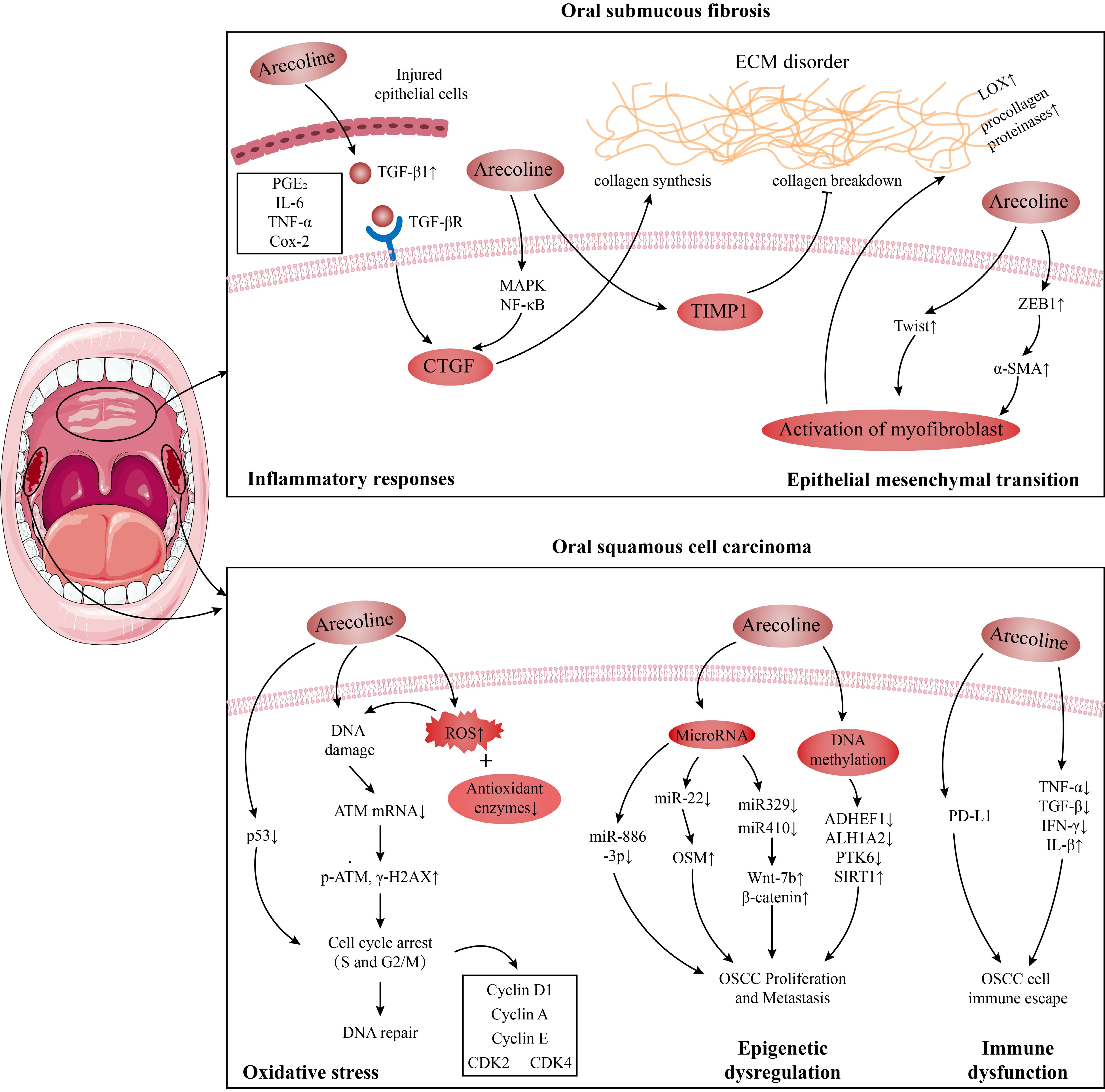
      Figure 1. 

      Possible mechanisms of oral submucous fibrosis (OSF) and oral oral squamous cell carcinoma (OSCC) induced by arecoline.

    • OSF is a kind of oral potentially malignant disorder, mainly caused by areca nut chewing[22]. The physical friction of arecoline coarse fiber and the chemical irritation of arecoline can cause damage and inflammation to oral tissue, and long-term chewing habits can lead to abnormal and persistent tissue inflammation, which is a critical factor in developing cancer and tissue fibrosis[23]. Various inflammatory mediators play pivotal roles in these pathogenic processes. Arecoline is implicated in stimulating the cellular expression of pro-inflammatory and pro-fibrotic cytokines, including prostaglandin E2 (PGE2), interleukin-6 (IL-6), tumor necrosis factor α (TNF-α), transforming growth factor-β (TGF-β) and cyclooxygenase 2 (Cox-2)[23,24]. Among those inflammatory factors, arecoline enhances collagen synthesis[25], increases procollagenase levels[26], and upregulates lysyl oxidase activity (a key enzyme in collagen fiber processing)[27] by inducing TGF-β signaling[24]. Arecoline also induces the expression of connective tissue growth factor (CTGF), a downstream target of TGF-β, by activating mitogen-activated protein kinase (MAPK) and nuclear factor kappa-light-chain-enhancer of activated B cells (NF-κB)[28]. Additionally, in oral keratinocytes-fibroblasts, tissue inhibitor of metalloproteinases 1 (TMP1), an inhibitor of enzymes involved in extracellular matrix (ECM) degradation, exhibited increased production following arecoline pre-treatment[29].

      Another possible OSF route is epithelial-mesenchymal transition (EMT). Zinc finger E-box binding homeobox 1, a transcription factor that instigates EMT, experiences increased expression under the influence of arecoline. This upregulation drives the expression of α-smooth muscle actin (α-SMA) via activation of the α-SMA promoter, thus prompting the differentiated metastasis of myofibroblasts in buccal mucosal fibroblasts, subsequently contributing to ECM accumulation and participating in OSF pathogenesis[30]. Moreover, Twist is another EMT transcription factor that plays a role in arecoline-associated OSF by regulating collagen contraction and wound healing capacity in OSF[31]. Overall, stimulation of the immune system, TMP1, and EMT, leading to disturbances in collagen homeostasis, are possible mechanisms by which arecoline causes OSF, as detailed in Fig. 1.

    • OSF is a precancerous lesion that may precede the OSCC diagnosis[32]. According to the Global Cancer Observatory, in 2020, there were 377,713 cases (2.0%) of lip and oral cancer and 177,757 deaths (1.8%)[33]. OSCC accounts for more than 90% of oral cancer cases, with a five-year survival rate of 40%−50%[34]. Approximately half of oral cancers reported are attributed to areca nut chewing in the Indian subcontinent and Taiwan[11]. In mouse models, a standard method for simulating oral tumors is a combined treatment with arecoline and 4-nitroquinoline-1-oxide[35]. These findings support that OSCC has a significant association with arecoline. Existing studies demonstrate that arecoline may cause OSCC by inducing increased oxidative stress, epigenetic dysregulation, and immune dysfunction.

      Arecoline induced the production of reactive oxygen species (ROS) and reduced expression of antioxidant enzymes, leading to chromosomal damage and gene mutations[13], one of the pathogenic mechanisms of OSCC. Typically, the body's ROS is in equilibrium with the antioxidant system, but sometimes, this equilibrium can be disturbed, such as after arecoline ingestion. When 50−200 μg/mL arecoline was used to treat gingival epithelial Smulow-Glickman cells and oral epithelial cell lines OEC-M1 and SAS, it induced ROS production and inhibited catalase expression, caused DNA double-strand breaks and activated the DNA repair response, which was characterized by down-regulation of ATM mRNA expression and an increase in p-ATM and γ-H2AX expression in the cells[13,36]. The tumor suppressor gene p53 regulates almost all DNA repair pathways[37]. Tsai et al. found that arecoline can inhibit the p53-regulated p21WAF1 promoter and p53 protein expression in KB and HEp-2 cells, inducing cell cycle arrest in S and G2/M phases[38]. Apart from these changes, arecoline also increases the phosphorylation levels of Wee1 kinase and Cdc2Tyr15 associated with S and G2/M phase arrest in KB epithelial cells[39], as well as regulating the expression of cell cycle protein D1, cell cycle protein A, cell cycle protein E, CDK4 and CDK2 in HaCaT keratinocytes[40].

      Arecoline also induces epigenetic changes in oral cells, including alterations in non-coding RNA and DNA methylation, which can lead to the development of OSCC. MicroRNA is an endogenous non-coding single-stranded RNA molecule[41]. In the ORL-48(T) squamous cell carcinoma cell line, 0.025 μg/mL arecoline decreased miR-22 expression in cells, leading to reduced inhibition of oncostatin M, an IL-6-family inflammatory cytokine, which then promotes OSCC proliferation[14]. Similarly, in OSCC cell lines, arecoline could also participate in OSCC proliferation and metastasis by inhibiting miR-886-3p[42]. In OSCC patients and mice, arecoline promotes OSCC proliferation by reducing the expression of miR-329 and miR-410 genes, inducing the expression of Wnt-7b and β-catenin proteins[43]. Abnormal DNA methylation is another common epigenetic change in OSCC. Under arecoline exposure, miR-30a and miR-379 levels were reduced in OSCC cells, targeting increased DNMT3B expression, which mediated the downregulation and methylation of ADHEF1 and ALH1A2 involved in retinoid metabolism to promote the progression of OSCC[44]. In addition, arecoline exposure resulted in hypomethylation of PTK6 with increased PTK6 expression, which increased the proliferation rate, migration, and invasion of OSCC cells, as demonstrated in mice[45].

      As the body's defense barrier, the immune system is important in recognizing and rejecting tumors. As early as 2001, it was noted that areca nut/arecoline reduced IL-2, TNF-α, TGF-β, and interferon-gamma (IFN-γ) levels in mononuclear cells of normal subjects and patients with squamous cell carcinoma[15]. In OSCC cell lines, arecoline increased the production of the pro-inflammatory factor IL-β partly through inflammasome, and IL-β induced angiogenesis and EMT, thereby promoting OSCC invasiveness[46]. Likewise, arecoline stimulated the production of PGE2, inhibited the expression of CD69 on CD4+ and CD8+ T cells in cellular KB oral cancer cells[23]. Furthermore, arecoline also increased the expression of obesity-associated protein, which regulates the expression of programmed cell death-ligand 1 via m6A modification and myc, as a means to increase the resistance of OSCC cells to CD8 T cells, thereby conferring the ability to immune escape from OSCC cells[47]. Based on the results of the current studies, long-term use of arecoline may increase the risk of OSCC. Therefore, reducing or avoiding exposure to arecoline is one of the most critical steps to prevent the risk of OSCC. In conclusion, both in vivo and in vitro studies support that arecoline impairs oral health.

    • There is a proverb in Hunan Province, China—areca nut and smoke; mana is boundless; areca nut and wine, get everything you want; areca nut, smoke, and wine, live to 99[48]. As a fat-soluble tertiary amine, arecoline crosses the blood-brain barrier well to enter and modulate the CNS, delivering a wide range of bodily effects, including euphoria, cognitive modulation, and addiction (Table 1)[2].

      Table 1.  Effect of arecoline on the CNS.

      EffectAnimal/cellSpecific effectPathway/mediatorsDoseRef.
      Beneficial effectsXenopus laevis oocytesAnti-inflammatory activityAs a silent agonist of α7 nAChR, targeting and regulating intracellular signaling against inflammation and pain/[61]
      Glioblastoma cell lines (U373 and U87MG)Interfere with the aggressiveness of malignant gliomasInhibition of intermediate conductance Ca2+-activated K+ channels10 and 30 μM[62]
      ZebrafishCluster disruption and increased social interactionIncreased norepinephrine, serotonin, and DOPAC levels decreased 5-hydroxyindoleacetic acid/serotonin level, and homovanillic acid/dopamine ratios10 mg/L[55]
      ZebrafishMotor hyperactivityBinds with multiple mAChRs (M1−M4) to induce hyperactivity0.001, 0.01, 0.1,
      and 1 ppm
      [49]
      Male Swiss albino miceAntinociceptionBy activation of central muscarinic receptors0.3−1 mg/kg ip[53]
      RatAttenuated a time perception impairment induced by daily scheduled feedingBy modulating central cholinergic10 mg/kg/d[52]
      RatAnti-phenobarbital sodium-induced sleep timeNot mentioned0.5 mg[51]
      Male ICR miceShortened the duration of
      ethanol-induced sleep
      Acts as a muscarinic agonist to relieve ethanol-induced central depression and intoxication0.125−1.0 mg/kg, s.c.[50]
      CPZ miceAttenuating memory impairment and demyelinationActs as a muscarinic receptor 1 cholinergic agonist to improve cognition and promote myelination processes in the frontal cortex2.5 or 5 mg/kg/d[8]
      Female BALB/c miceIncreased the activity of preactivated NK cellsBy stimulating the secretion of corticotropin-releasing hormone and adrenocorticotropic hormone1.5 mg/kg[63]
      Male albino ratsImproved retrieval and memory storage in the stair mazeNot mentioned0.5 mg/kg[64]
      Human (Alzheimer)Low-dose arecoline improved cognitive performance, highest-dose impaired psychomotor activationBy modulating central cholinergic1, 2, or 4 mg/h infusions 2 h[9]
      Human (Alzheimer)Improved memoryAs a cholinergic agonist, maintaining patients’ cholinergic steady-state0.042−1.7 mg/h Infusion for 11−16 d[65]
      Human (Alzheimer)Improved cognitionAs a muscarinic receptor agonist, regulating patients’ cholinergic system0.5, 1, 2, 4, 8, 16, 22,
      28, 34, and 40 mg/d
      [66]
      NeurotoxicityPrimary cortical neuronInduction of neuronal cell deathBy attenuating antioxidant defense and enhancing oxidative stress50−200 μM[16]
      PC12 CellsApoptosisBy inducing endoplasmic reticulum stress, attenuating H2S levels, CBS and 3-MST protein expression0.5−2 mM[58]
      Drosophila melanogasterNeurotoxic agent and affected the life cycle parametersBy reducing acetylcholinesterase and MAO, increasing caspase-3, caspase-9 activity, and oxidative stress20, 40 and 80 μM[67]
      ZebrafishDyskinesiaBy increasing ROS, endoplasmic reticulum stress, apoptotic p53 signaling pathway.10 μM[68]
      Male albino ratsDecreased correct responses and accelerated spontaneous decay of memoryNot mentioned3.5 and 8 mg/kg[64]
      Addiction-relatedXenopus laevis oocytesHabitual useBy activating addiction-related nAChR activity, receptors containing α4, β2, α6 and β3 subunits/[61]
      Xenopus laevis oocytesAddictionBy activating α4 nAChR100 μM[62]
      ZebrafishWithdrawal syndrome-like responsesNot mentioned1 mg/L[55]
      Pregnant womenExceptional adverse birth outcomeNot mentionedNot mentioned[69]
    • The effects of arecoline on the CNS are complex; at some doses, arecoline can cause excitability and enhance cognitive performance. In zebrafish (Danio rerio) larvae, arecoline increases locomotor activity even at concentrations as low as 0.001 ppm[49]. In mice, arecoline shortens ethanol-induced sleep time (0.125 to 1.0 mg/kg)[50]. Arecoline also increases anti-1.5 × 10⁻⁴ phenobarbital sodium-induced sleep time by up to 38 min (0.5 mg)[51]. These phenotypes suggest that arecoline has significant excitatory effects. In addition, spatial memory impairment and brain demyelination were well alleviated in schizophrenic mice treated with 5 mg/kg/d arecoline[8]. Daily arecoline injections of 10 mg/kg attenuated the impairment of mealtime-associated activity on the fasting day in old rats[52]. In Chinese medicine, herbs with areca nut as the main ingredient can manage palpitations, insomnia, and mental irregularities[1]. In clinical practice, a low dose of arecoline can improve cognitive impairment, emotional capacity, and psychomotor activity in Alzheimer's patients[9]. It was found that arecoline is an agonist of mAChRs, which may promote body excitability and antinociception effects and improve learning and memory by activating the M1 muscarinic receptor subtype[53,54]. Additionally, arecoline exposure increased dopamine levels in the brains of mice and zebrafish, which may also be a reason for arecoline's ability to promote excitation in the organism[55,56]. These findings indicate that arecoline enhances cognitive performance and induces organic excitability, possibly by modulating neurotransmitter homeostasis in the brain.

    • Surprisingly, as the concentration increases, arecoline begins to disrupt the oxidative and antioxidant balance in the body, inducing neurotoxicity and apoptosis. NADPH oxidase (NOX) is a key enzyme for redox signaling and a significant source of ROS cluster in vivo[57]. Cellular experiments indicated that arecoline at concentrations of 50−200 μM can increase ROS by upregulating NOX2 levels and decrease glutathione (GSH) and superoxide dismutase (SOD) levels, causing redox imbalance in neurons. In this state, the expression of pro-apoptotic proteins (cytochrome c, Bax, caspase-9, and caspase-3) was increased, and the manifestation of anti-apoptotic protein Bcl-2 was diminished, which finally induced neuronal cell death[16]. Jiang et al. suggested that arecoline can induce neurotoxicity by causing endoplasmic reticulum stress in neuronal cells and interfering with endogenous H2S synthesis[58]. Moreover, zebrafish showed elevated expression of the protooncogenes c-fos and c-jun in the brain after 10 mg/L arecoline treatment, which was associated with cancer transformation and progression[55].

    • After long-term consumption of areca nut, users may experience dependence such as tolerance, loss of control, craving, and salience, with surveys indicating that a high percentage of current users are dependent, accounting for 40% to 80% of the total[59]. Once discontinued, users may experience withdrawal symptoms, including mood swings, anxiety, irritability, and insomnia[2]. Dependency mechanisms are usually associated with the brain's dopamine system. As early as the 1980s, researchers showed that arecoline increased dopamine levels in the mouse cortex[56], and this was validated by the results that zebrafish exposed to acute arecoline increased brain levels of norepinephrine, serotonin, and the dopamine metabolite 3,4-Dihydroxyphenylacetic acid (DOPAC)[55]. Chen et al. emphasized that this increased dopamine may be partially derived from Monoamine oxidase A (MAO-A) inhibition, and MAO-A activity was indeed inhibited in neuroblastoma SH-SY5Y cells and rats after arecoline treatment, and individuals carrying the at-risk MAO-A allele were more likely to exhibit a dependent response in the population survey[60]. Furthermore, the α4β2 nicotinic acetylcholine receptor (nAChR) in the brain is a crucial regulator of the limbic dopamine system in the midbrain, and arecoline may mediate the rewarding effects behind habitual arecoline use through activation of the α4β2 nAChR[61].

      The studies above have shown that the effects of arecoline on the CNS are complex. At lower doses, arecoline stimulates acetylcholine receptors, improving cognition and euphoria. However, higher doses of arecoline can induce neurotoxicity, apoptosis, and cancer transformation in the CNS. Prolonged intake and abuse of arecoline can lead to addiction, tolerance, and dependence through the release of dopamine in the brain. The positive cognitive enhancement effects of arecoline can be utilized to provide a new therapeutic idea for related diseases. However, it is important to be aware of the potential neurological problems that may arise from long-term use and abuse of arecoline to safeguard our neurological health.

    • The China Cardiovascular Health and Disease Report 2021 projects that cardiovascular disease now affects 330 million people and is currently the leading cause of death for the population, accounting for more than 40%[70]. Areca nut chewing may increase the risk of cardiovascular diseases[71], for instance, following areca nut chewing, one patient with coronary artery disease experienced an acute myocardial infarction[72], while two other patients experienced ST-elevation myocardial infarction[73]. Tseng et al. pointed out that when arecoline concentration was higher than 0.2 mM, it caused contraction, rounding, and shedding of EAHY cells, weakened the wound closure activity of EAHY cells, and promoted the adhesion of monocytes to EAHY cells, which is an early step in atherosclerosis[74]. Furthermore, arecoline causes atherosclerosis by interfering with low-density lipoprotein (LDL) receptors to inhibit endocytosis of LDL cholesterol and interfering with high-density lipoprotein (HDL) receptors to prevent hepatic uptake of HDL cholesterol[71].

      In addition to atherosclerosis, Goto et al. found that a subfraction isolated from areca nut (concentration above 3 × 10⁻⁷ μg/mL) had vasodilatory effects and relaxed rat aorta with intact endothelium-containing[75]. In anesthetized dogs, only 10 ng/kg of arecoline was able to reduce arterial blood pressure. When the dose was increased to 30 or 100 μg/kg, the experimental animals even showed a sustained interruption of cardiac activity[76]. Mice also showed a decrease in blood pressure after arecoline injection[77], suggesting arecoline may have similar, evolutionarily conserved cardiovascular system effects in animals such as humans, canines, and mice.

    • Studies have established an association between arecoline and cardiovascular disease, but the underlying mechanisms regarding arecoline-induced cardiotoxicity are poorly understood. Similar to the induction of oral cell fibrosis, arecoline induces cardiomyocyte fibrosis by disrupting the balance of the extracellular matrix by affecting immunity (TGF-β) and the enzymes that synthesize and degrade the extracellular matrix. Specifically, when mice were fed arecoline above 5 mg/kg/d, significant fibrosis occurred in the heart; on the one hand, arecoline was able to increase the expression of TGF-β1 and its downstream molecule p-Smad2/3, CTGF and its transcription factor SP1 were in turn regulated by the p-Smad2/3 pathway to increase their expression, thus participating in cardiac fibrosis; on the other hand, arecoline increased plasminogen activator and plasminogen activator expression, which in turn is involved in cardiac fibrosis by inducing the expression of matrix metalloproteinases-9[78]. In a study by Lin et al., arecoline at 5 mg/kg/d and above-induced protein expression of Fas-ligand receptors (Fas) ligand, Fas and Fas-associated protein with death domain in Sprague-Dawley rat cardiomyocytes, followed by activation of caspase 8 and caspase 3, causing apoptosis[17]. When the concentration was increased to 50 mg/kg/d, the authors found that the expression levels of apoptotic factors (tBid, Bak, cytochrome c) were increased and survival biomarkers (Bcl2, BclxL) were decreased, and the mitochondrial apoptotic pathway was activated in rat cardiomyocytes[17]. In addition, recent experiments have shown that arecoline-treated mice develop signs of cardiac hypertrophy, which are associated with expression of the MEK5/ERK5 and JAK2/STAT3 signaling pathways, the MAPK signaling cascade, as well as calcium-regulated neurophosphatase and NFATc3[79].

      Together, these findings support that arecoline may influence the course of cardiovascular diseases in a dose-response relationship (Fig. 2). However, given the complexity of the cardiovascular system and the large number of causal factors of such diseases, there is no exact mechanism to elucidate the relationship between arecoline and cardiovascular system diseases.

      Figure 2. 

      Possible mechanisms of cardiovascular diseases and cardiotoxicity.

    • When peristalsis occurs in the gastrointestinal tract, it moves and mixes the food to facilitate the absorption of nutrients and water, which is essential for life[6]. Modern research has shown that arecoline, the main active ingredient in areca nut, stimulates the contraction of the gastrointestinal tract muscles in several ways. In the jejunum, arecoline hydrobromide causes smooth muscle contraction by inhibiting voltage-gated potassium channels and inducing smooth muscle cell depolarization[7]. Similarly, in mice and rabbits, arecoline induced colonic smooth muscle motility in a dose-dependent manner, stimulated by the muscarinic (M3) receptor − extracellular Ca2+ influx − Ca2+ store release pathway[80,81]. In addition, as a known antiparasitic with low toxicity, arecoline relieved symptoms of gastrointestinal disorders such as vomiting, diarrhea, and intestinal obstruction caused by the parasites through paralyzing the parasites[82,83].

    • Human health is shaped and influenced by the body's commensal microbiota, especially the gut microbiota. For example, one of the mechanisms involved in the pathogenesis of non-alcoholic fatty liver disease (NAFLD) is the alteration of gut microbiota. Zhu et al. found that the abundance of intestinal flora was increased after 0.5−5 mg/kg arecoline treatment in mice, in which Butyricicoccus, Christensenella, and Coriobacteriaceae reversed NAFLD in mice by modulating the Cox-2/PGE2 pathway as well as by increasing the protective effect of intestinal epithelial cells[84]. Additionally, alterations in gut microbiota may induce changes in immunity and exacerbate intestinal disorders. After exposure to 6 and 30 mg/kg arecoline, inflammation and dysbiosis occurred in the intestines of mice, and the results of correlation analyses indicated that arecoline may accelerate the secretion of lipopolysaccharides by promoting an increase in the abundance of the Muribaculaceae bacterium DSM 103720, which in turn encourages the development of inflammation[85]. On a similar note, treatment with 5 mg/kg arecoline decreased the proportion of probiotics in the mouse gut and increased Odoribacter, Bacteroides, and pro-inflammatory factors (IL-6, IL-1β, and TNF-α) as a means of exacerbating ulcerative enteritis in mice[86].

      In conclusion, although arecoline can treat dyspepsia, constipation, and anti-parasite, as well as improve the intestinal homeostasis of the NAFLD model through the intestinal flora, inappropriate use of arecoline can disturb the homeostasis of intestinal flora, leading to different degrees of intestinal inflammation and ecological dysregulation. Therefore, despite the medicinal value of arecoline, the mechanism of action on the gastrointestinal tract needs to be further explored in the future, aiming to protect the health of the human gastrointestinal tract while enabling the development of new therapeutic agents and targets for gastrointestinal diseases.

    • The effects of arecoline are systemic, in addition to the impact on organs mentioned above, arecoline also has effects causing bronchoconstriction, hepatotoxicity, reproductive toxicity (Fig. 3). Wang et al. first linked arecoline, Eotaxin-1, and asthma, and their results showed that concentrations of arecoline were negatively correlated with some indexes of respiratory function in asthmatic patients[18]. Arecoline also exhibits toxic effects on hepatocytes and germ cells. After arecoline treatment, hepatocytes in mice showed damage to nuclei and mitochondria, accumulation of sizeable intracellular lipid droplets, decreased expression of antioxidant substances, and an increase in hepatotoxicity markers in a dose-dependent manner[87]. In addition, the testicular weight, sperm count, and viability of male mice were significantly reduced after arecoline treatment[19]; pregnancy rate in female mice was significantly reduced and embryo growth and implantation were affected after 200 μg of arecoline treatment[88]. Interestingly, the hepatotoxicity of arecoline and reproductive toxicity to males were attenuated when supplemented with vitamins C and E, which may be related to the antioxidant function of vitamins C and E[19].

      Figure 3. 

      Effects and mechanisms of arecoline on different organs and systems.

      The immune system is an important barrier against external threats and is distributed throughout the body's tissues. For example, arecoline, when present in the host, disrupts this barrier and can lead to various diseases. In the oral cavity and cardiomyocytes, arecoline causes ECM dysregulation by promoting cytokine TGF-β expression, ultimately resulting in fibrosis[24,78]. Similarly, arecoline promotes invasion of OSCC and immune evasion by increasing pro-inflammatory factors and resistance to CD8 T cells[46,47]. In the digestive system, however, arecoline indirectly acts on immunity by altering the gut flora, causing or aggravating gut inflammation[86]. For ease of understanding, we list the effects of arecoline on each of the systems mentioned in this section and their possible mechanisms in Table 2.

      Table 2.  Other toxicological and pharmacological effects of arecoline.

      EffectAnimal/cellSpecific effectPathway/mediatorsDoseRef.
      Respiratory systemHuman and dermal and gingival fibroblastCausing lung function impairmentIn pro-inflammatory conditions (IL-4 and
      TNF-α), arecoline can induce eotaxin-1 release and alter the disease process in asthma
      25 and 100 μg/mL[18]
      HumanAsthmaPossibly related to arecoline-induced bronchoconstriction/[89]
      HepatotoxicityHuman liver microsome and Male Wistar ratsHepatotoxicityBy increasing the hepatic CYP2E1 and CYP2B activity, induced oxidative damage, liver cirrhosis, and hepatocellular carcinoma4, 20, and 100 mg/kg/d[90]
      HA22T/VGH hepatoma cellsInducing anoikisBy inhibiting STAT3 and SHP2 phosphorylation, decreasing the levels of anti-apoptotic factors, as well as by promoting the activity of pro-apoptotic factors0−100 μg/mL[91]
      Human and C57BL/6 mice’s organ of Corti and spiral ganglionsSensorineural hearing impairmentReducing cochlear explant cell activity, inducing cell death and ROS production by causing disruption of hair cells in the organ
      of Corti
      0.2, 0.8, 2, and 10 mM[92]
      MiceFatty degeneration and inflammatory infiltrationBy increasing serum alkaline phosphatase, glutamate oxaloacetate transaminase, glutamate-pyruvate transaminase, and decreasing levels of reduced glutathione, glutathione-S-transferase, SOD, and catalase10 mg/kg body weight[19]
      MiceDecreasing nuclear size; the rough endoplasmic reticulum with profusely inflated cisternae and abundance of lipid dropletsBy Upregulating SGOT and SGPT (hepatotoxicity marker enzymes) in serum5, 10, and 20 mg/kg body weight[87]
      ReproductionZebrafish embryosReducing survival of embryos with growth retardation and lower heart rateGeneral cytotoxic effects mainly due to intracellular thiol depletion0.01%−0.04% (wt/vol)[93]
      OocyteApoptosisBy disrupting actin filament dynamics, spindle assembly, and kinetochore-microtubule attachment stability, mitochondrial distribution, and increasing oxidative stress levels180 μg/mL[94]
      ICR mice and blastocystsReduction of early embryos and inhibition of blastocyst growth and expansionBy inducing DNA damage, cell cycle arrest, or apoptosis0−8.47 × 10⁻² M[88]
      Male ratsStimulation of testosterone secretionBy activating L-type calcium channels, increasing 17β-hydroxysteroid dehydrogenase activity and StAR expression, thereby stimulating testosterone production1 μg/kg[95]
      Immunity and endocrineSwiss albino miceLymphocyte depletion of the thymic cortex and the B and T lymphocyte areas in the spleen and MLN, Elevated corticosterone, SGOT, and SGPT levels, and decreased white and red blood cell countsNot mentioned20 mg/kg[96]
      Adult male miceThe orientation of nuclei was irregular, follicle degeneration, a decrease in the T3, T4, number, and size of thyroid follicles, and an increase in the TSH levelMAChRs mediate the effect of arecoline on thyroid10 mg/kg[97]
      BALB/c miceReducing the spleen index, hemolysin, IL-2 production, and splenocyte proliferation induced by concanavalin A or lipopolysaccharideMediated via mAChRs2 mg/kg[98]
      FatMouse 3T3-L1 cells and humanAdipocyte dysfunctionInhibiting adipogenic differentiation, inducing adenylate cyclase-dependent lipolysis, and interfering with insulin-induced glucose uptake≥ 300 µM[99]
      3T3-L1 cellsRegulating the growth of preadipocytesInhibiting the CDK family and the CKI pathway by inactivating AMPK activity as
      well as the intracellular ROS pathway
      0−1,000 μM[100]
    • This paper provides an up-to-date review of the pharmacologic and toxicologic mechanisms of arecoline. We focus on the oral cavity, central nervous system, cardiovascular system, and digestive system and establish a framework for the pharmacological and toxicological mechanisms of systemic systems after arecoline interventions, hoping to provide some directions for refining the understanding of the mechanism of action of arecoline and its future development. We have found arecoline to have potential therapeutic effects by promoting excitation and improving learning and memory through modulation of nAChRs and mAChRs, as well as causing smooth muscle contractions, promoting intestinal peristalsis, treating indigestion, and being antiparasitic in a way that paralyzes parasites. However, arecoline has been shown to cause varying degrees of damage to various systems throughout the body, including causing OSF, OSCC, neurotoxicity, addiction, cardiotoxicity, hepatotoxicity, and reproductive toxicity. Furthermore, in the effects of arecoline on the CNS and digestive system, we found a dose-effect relationship between arecoline, pharmacology, and toxicology, i.e., low doses are beneficial while high doses are harmful. Therefore, to rationally utilize the pharmacological properties of arecoline, further studies are needed to understand the pharmacological and toxicological mechanisms of arecoline fully and to clarify the dosage-effect relationship and the long-term effects to ensure that the protection of human health and safety accompanies the development of the drug.

      • This work was supported by the National Natural Science Foundation of China Project (32200387) and the Emergency Project for Risk Assessment of Areca Nut (Key Project of the Department of Agriculture and Rural Affairs of Hainan Province & Wanning Municipal People's Government).

      • The authors confirm contribution to the paper as follows: writing - original draft: Liu H; visualization: Liu H; writing - review & editing: Zheng H, Wang X; supervision: Zheng H, Wang X; resources: Hu X, Chen F, Zheng H, Zhang J; project administration: Hu X, Chen F, Zheng H, Zhang J, Wang X; funding acquisition: Hu X, Chen F, Zheng H, Zhang J, Wang X. All authors reviewed the results and approved the final version of the manuscript.

      • All data generated or analyzed during this study are included in this published article.

      • The authors declare that they have no conflict of interest.

      • Copyright: © 2024 by the author(s). Published by Maximum Academic Press on behalf of China Agricultural University, Zhejiang University and Shenyang Agricultural University. This article is an open access article distributed under Creative Commons Attribution License (CC BY 4.0), visit https://creativecommons.org/licenses/by/4.0/.
    Figure (3)  Table (2) References (100)
  • About this article
    Cite this article
    Liu H, Zheng H, Zhang J, Chen F, Hu X, et al. 2024. Review of the toxic effects and health functions of arecoline on multiple organ systems. Food Innovation and Advances 3(1): 31−41 doi: 10.48130/fia-0024-0005
    Liu H, Zheng H, Zhang J, Chen F, Hu X, et al. 2024. Review of the toxic effects and health functions of arecoline on multiple organ systems. Food Innovation and Advances 3(1): 31−41 doi: 10.48130/fia-0024-0005

Catalog

  • About this article

/

DownLoad:  Full-Size Img  PowerPoint
Return
Return