ARTICLE   Open Access    

Distinct roles of the IRE1α arm and PERK arm of unfolded protein response in arachidonic acid-induced ferroptosis in hepatocytes

More Information
  • Ferroptosis is a distinct form of cell death that is driven by iron-dependent phospholipid peroxidation. Polyunsaturated fatty acids (PUFAs), particularly arachidonic acid (AA) and adrenal acid (AdA), are most prone to lipid peroxidation, which induces ferroptosis and affects the function of cell membranes. In this study, we discovered that AA induces ferritinophagy in hepatocytes, a selective form of autophagy that degrades ferritin, triggering unstable iron overload. Mechanistically, AA enhances cellular uptake of bound iron by up-regulating transferrin receptor 1 (TfR1). Additionally, AA induces endoplasmic reticulum stress (ER stress) and simultaneously activates two of its branches, pancreatic ER kinase (PERK) and inositol-requiring enzyme 1 (IRE1). Notably, PERK and IRE1 appear to play distinct roles in inducing ferritinophagy. Inhibition of PERK reduced the AA-induced increase of Fe2+ by alleviating ferritinophagy, while inhibition of IRE1 further exacerbated ferroptosis by activating ferritinophagy. Furthermore, there seems to be an interaction between the signaling pathways of ER stress, and inhibition of IRE1 exacerbates AA-induced ferritinophagy by further activating the PERK signaling pathway, thereby exacerbating the extent of cell death. Collectively, our findings suggest that iron overload is involved in AA-induced hepatocyte ferroptosis and that this process is regulated by ER stress-mediated ferritinophagy. This study suggests potential therapeutic strategies for treating liver diseases related to lipid metabolism disorders by intervening in the ferroptosis process.
  • The Fabaceae family, the third largest family in angiosperms, contains about 24,480 species (WFO, https://wfoplantlist.org), and has been a historically important source of food crops[14]. Peas (formerly Pisum sativa L. renamed to Lathyrus oleraceusPisum spp. will be used hereafter due to historical references to varietal names and subspecies that may not have been fully synonymized), are a member of the Fabaceae family and is among one of the oldest domesticated food crops with ongoing importance in feeding humans and stock. Peas originated in Western Asia and the Mediterranean basin where early finds from Egypt have been dated to ~4500 BCE and further east in Afghanistan from ~2000 BCE[5], and have since been extensively cultivated worldwide[6,7]. Given that peas are rich in protein, dietary fiber, vitamins, and minerals, have become an important part of people's diets globally[810].

    Domesticated peas are the result of long-term human selection and cultivation, and in comparison to wild peas, domesticated peas have undergone significant changes in morphology, growth habits, and yield[1113]. From the long period of domestication starting in and around Mesopotamia many diverse lineages of peas have been cultivated and translocated to other parts of the world[14,15]. The subspecies, Pisum sativum subsp. sativum is the lineage from which most cultivars have been selected and is known for possessing large, round, or oval-shaped seeds[16,17]. In contrast, the subspecies, P. sativum subsp. elatius, is a cultivated pea which more closely resembles wild peas and is mainly found in grasslands and desert areas in Europe, Western Asia, and North Africa. Pisum fulvum is native to the Mediterranean basin and the Balkan Peninsula[15,18], and is resistant to pea rust caused by the fungal pathogen Uromyces pisi. Due to its resistance to pea rust, P. fulvum has been cross-bred with cultivated peas in the development of disease-resistant strains[19]. These examples demonstrate the diverse history of the domesticated pea and why further study of the pea pan-plastome could be employed for crop improvement. While studies based on the nuclear genome have been used to explore the domestication history of pea, these approaches do not account for certain factors, such as maternal inheritance. Maternal lineages, which are inherited through plastomes, play a critical role in understanding the full domestication process. A pan-plastome-based approach will no doubt allow us to investigate the maternal genetic contributions and explore evolutionary patterns that nuclear genome studies may overlook. Besides, pan-plastome analysis enables researchers to systematically compare plastome diversity across wild and cultivated species, identifying specific regions of the plastome that contribute to desirable traits. These plastid traits can then be transferred to cultivated crops through introgression breeding or genetic engineering, leading to varieties with improved resistance to disease, environmental stress, and enhanced agricultural performance.

    Plastids are organelles present in plant cells and are the sites in which several vital biological processes take place, such as photosynthesis in chloroplasts[2024]. Because the origin of plastids is the result of an ancient endosymbiotic event, extant plastids retain a genome (albeit much reduced) from the free-living ancestor[25]. With the advancement of high-throughput DNA sequencing technology, over 13,000 plastid genomes or plastomes have been published in public databases by the autumn of 2023[24]. Large-scale comparison of plastomic data at multiple taxonomic levels has shown that plastomic data can provide valuable insights into evolution, interspecies relationships, and population genetic structure. The plastome, in most cases, displays a conserved quadripartite circular genomic architecture with two inverted repeat (IR) regions and two single copy (SC) regions, referred to as the large single-copy (LSC) and small single-copy (SSC) regions. However, some species have lost one copy of the inverted repeated regions, such as those in Erodium (Geraniaceae family)[26,27] and Medicago (Fabaceae)[28,29]. Compared to previous plastomic studies based on a limited number of plastomes, the construction of pan-plastomes attempts to describe all nucleotide variants present in a lineage through intensive sampling and comparisons. Such datasets can provide detailed insights into the maternal history of a species and help to better understand applied aspects such as domestication history or asymmetries in maternal inheritance, which can help guide future breeding programs. Such pan-plastomes have recently been constructed for several agriculturally important species. A recent study focuses on the genus Gossypium[20], using plastome data at the population level to construct a robust map of plastome variation. It explored plastome diversity and population structure relationships within the genus while uncovering genetic variations and potential molecular marker loci in the plastome. Besides, 65 samples were combined to build the pan-plastome of Hemerocallis citrina[30] , and 322 samples for the Prunus mume pan-plastome[31]. Before these recent efforts, similar pan-plastomes were also completed for Beta vulgaris[32], and Nelumbo nucifera[33]. However, despite the agricultural importance of peas, no such pan-plastome has been completed.

    In this study, 103 complete pea plastomes were assembled and combined another 42 published plastomes to construct the pan-plastome. Using these data, the following analyses were conducted to better understand the evolution and domestication history of pea: (1) genome structural comparisons, (2) codon usage bias, (3) simple sequence repeat patterns, (4) phylogenetic analysis, and (5) nucleotide variation of plastomes in peas.

    One hundred and three complete pea plastomes were de novo assembled from public whole-genome sequencing data[34]. For data quality control, FastQC v0.11.5 (www.bioinformatics.babraham.ac.uk/projects/fastqc/) was utilized to assess the quality of the reads and ensure that the data was suitable for assembly. The clean reads were then mapped to a published pea plastome (MW308610) plastome from the GenBank database (www.ncbi.nlm.nih.gov/genbank) as the reference using BWA v0.7.17[35] and SAMtools v1.9[36] to isolate plastome-specific reads from the resequencing data. Finally, these plastome-specific reads were assembled de novo using SPAdes v3.15.2[37]. The genome annotation was conducted by Geseq online program (https://chlorobox.mpimp-golm.mpg.de/geseq.html). Finally, the OGDRAW v1.3.1[38] program was utilized to visualize the circular plastome maps with default settings. To better resolve the pan-plastome for peas, 42 complete published pea plastomes were also downloaded from NCBI and combined them with the de novo data (Supplementary Table S1).

    To investigate the codon usage in the pan-plastome of pea, we utilized CodonW v.1.4.2 (http://codonw.sourceforge.net) to calculate the Relative Synonymous Codon Usage (RSCU) value of the protein-coding genes (PCGs) longer than 300 bp, excluding stop codons. The RSCU is a calculated metric used to evaluate the relative frequency of usage among synonymous codons encoding the same amino acid. An RSCU value above 1 suggests that the codon is utilized more frequently than the average for a synonymous codon. Conversely, a value below 1 indicates a lower-than-average usage frequency. Besides, the Effective Number of Codons (ENC) and the G + C content at the third position of synonymous codons (GC3s) were also calculated in CodonW v.1.4.2. The ENC value and GC3s value were utilized for generating the ENC-GC3s plot, with the expected ENC values (standard curve), are calculated according to formula: ENC = 2 + GC3s + 29 / [GC3s2 + (1 – GC3s)2][39].

    The MISA program[40] was utilized to detect simple sequence repeats (SSRs), setting the minimum threshold for repeat units at 10 for mono-motifs, 6 for di-motifs, and 5 for tri-, tetra-, penta-, and hexa-motif microsatellites, respectively.

    The 145 complete pea plastomes were aligned using MAFFT v 7.487[41]. Single nucleotide variants (SNVs)-sites were used to derive an SNV only dataset from the entire-plastome alignment[42]. A total of 959 SNVs were analyzed using IQ-TREE v2.1[43] with a TVMe + ASC + R2 substitution model, determined by ModelTest-NG[44] based on BIC, and clade support was assessed with 1,000 bootstrap replicates. Vavilovia formosa (MK604478) was chosen as an outgroup. The principal coordinates analysis (PCA) was conducted in TASSEL 5.0[45].

    DnaSP v6[46] was utilized to identify different haplotypes among the plastomes, with gaps and missing data excluded. Haplotype networks were constructed in Popart v1.7[47] using the median-joining algorithm. Haplotype diversity (Hd) for each group was calculated by DnaSP v6[46], and the evolutionary distances based on the Tamura-Nei distance model were computed based on the population differentiation index (FST) between different groups with the plastomic SNVs.

    In this study, the pan-plastome structure of peas was elucidated (Fig. 1). The length of these plastomes ranged from 120,826 to 122,547 bp. And the overall GC content varied from 34.74% to 34.87%. In contrast to typical plastomes characterized by a tetrad structure, the plastomes of peas contained a single IR copy. The average GC content among all pea plastomes was 34.8%, with the highest amount being 34.84% and the lowest 34.74%, with minimal variation among the pea plastomes.

    Figure 1.  Pea pan-plastome annotation map. Indicated by arrows, genes listed inside and outside the circle are transcribed clockwise and counterclockwise, respectively. Genes are color-coded by their functional classification. The GC content is displayed as black bars in the second inner circle. SNVs, InDels, block substitutions and mixed variants are represented with purple, green, red, and black lines, respectively. Single nucleotide variants (SNVs), block substitutions (BS, two or more consecutive nucleotide variants), nucleotide insertions or deletions (InDels), and mixed sites (which comprise two or more of the preceding three variants at a particular site) are the four categories into which variants are divided.

    A total of 110 unique genes were annotated (Supplementary Table S2), of which 76 genes were PCGs, 30 were transfer RNA (tRNA) genes and four were ribosomal RNA (rRNA) genes. Genes containing a single intron, include nine protein-coding genes (rpl16, rpl2, ndhB, ndhA, petB, petD, rpoC1, clpP, atpF) and six tRNA genes (trnK-UUU, trnV-UAC, trnL-UAA, trnA-UGC, trnI-GAU, trnG-UCC). Additionally, two protein-coding genes ycf3 and rps12 were found to contain two introns.

    The codon usage frequency in pea plastome genes is shown in Fig. 2a. The analysis of codon usage in the pea plastome indicated significant biases for specific codons across various amino acids. Here a nearly average usage in some amino acids was observed, such as Alanine (Ala) and Valine (Val). For most amino acids, the usage of different synonymous codons was not evenly distributed. Regarding stop codons, a nearly even usage was found, with 37.0% for TAA, 32.2% for TAG and 30.8% for TGA.

    Figure 2.  (a) The overall codon usage frequency in 51 CDSs (length > 300 bp) from the pea pan-plastome. (b) The heatmap of RSCU values in 51 CDSs (length > 300 bp) from the pea pan-plastome. The x-axis represents different codons and the y-axis represents different CDSs. The tree at the top was constructed based on a Neighbor-Joining algorithm.

    The RSCU heatmap (Fig. 2b) showed different RSCU values for all codons in plastomic CDSs. In general, a usage bias for A/T in the third position of codons was found among CDSs in the pea pan-plastome. The RSCU values among these CDSs ranged from 0 to 4.8. The highest RSCU value (4.8) was found with the CGT codon in the cemA gene, where six synonymous codons exist for Arg but only CGT (4.8) and AGG (1.2) were used in this gene. This explained in large part the extreme RSCU value for CGT, resulting in an extreme codon usage bias in this amino acid.

    In the ENC-GC3s plot (Fig. 3), 31 PCGs were shown below the standard curve, while 20 PCGs were above. Besides, around 12 PCGs were near the curve, which meant these PCGs were under the average natural selection and mutation pressure. This plot displayed that the codon usage preferences in pea pan-plastomes were mostly influenced by natural selection. Five genes were shown an extreme influence with natural selection for its extreme ΔENC (ENCexpected – ENC) higher than 5, regarding as petB (ΔENC = 5.18), psbA (ΔENC = 8.96), rpl16 (ΔENC = 5.62), rps14 (ΔENC = 14.29), rps18 (ΔENC = 6.46) (Supplementary Table S3).

    Figure 3.  The ENC-GC3s plot for pea pan-plastome, with GC3s as the x-axis and ENC as the y-axis. The expected ENC values (standard curve) are calculated according to formula: ENC = 2 + GC3s + 29 / [GC3s2 + (1 − GC3s)2].

    For SSR detection (Fig. 4), mononucleotide, dinucleotide, and trinucleotide repeats were identified in the pea pan-plastome including A/T, AT/TA, and AAT/ATT. The majority of these SSRs were mononucleotides (A/T), accounting for over 90% of all identified repeats. Additionally, we observed that A/T and AT/TA repeats were present in all pea accessions, whereas only about half of the plastomes contained AAT/ATT repeats. It was also found that the number of A/T repeats exhibited the greatest diversity, while the number of AAT/ATT repeats showed convergence in all plastomes that possessed this repeat.

    Figure 4.  Simple sequence repeats (SSRs) in the pea pan-plastome. The x-axis represents different samples of pea and the y-axis represents the number of repeats in this sample. (a) The number of A/T repeats in the peapan-plastome. (b) The number of AT/TA and AAT/ATT repeats of pea pan-plastomes.

    To better understand the phylogenetic relationships and evolutionary history of peas, a phylogenetic tree was reconstructed using maximum likelihood for 145 pea accessions utilizing the whole plastome sequences (Fig. 5a). The 145 pea accessions were grouped into seven clades with high confidence. These groups were named the 'PF group', 'PSeI-a group', 'PSeI-b group', 'PA group', 'PSeII group', 'PSeIII group', and the 'PS group'. The naming convention for these groups relates to the majority species names for accessions in each group, where P. fulvum makes up the 'PF group', P. sativum subsp. elatius in the 'PSeI-a group', 'PSeI-b group', 'PSeII group', and 'PSeIII group', P. abyssinicum in the 'PA group', and P. sativum in the 'PS group'. From this phylogenetic tree, it was observed that the 'PSeI-a group' and the 'PSeI-b group' had a close phylogenetic relationship and nearly all accessions in these two groups (except DCG0709 accession was P. sativum) were identified as P. sativum subsp. elatius. In addition to the P. sativum subsp. elatius found in PSeI, seven accessions from the PS group were identified as P. sativum subsp. elatius.

    The PCA results (Fig. 5b) also confirmed that domesticated varieties P. abyssinicum were closer to cultivated varieties PSeI and PSeII, while PSeIII was more closely clustered with cultivated varieties of P. sativum. A previous study has indicated that P. sativum subsp. sativum and P. abyssinicum were independently domesticated from different P. sativum subsp. elatius populations[34].

    The complete plastome sequences were utilized for haplotype analysis using TCS and median-joining network methods (Fig. 5c). A total of 76 haplotypes were identified in the analysis. The TCS network resolved a similar pattern as the other analyses in that six genetic clusters were resolved with genetic clusters PS and PSeIII being very closely related. The genetic cluster containing P. fulvum exhibits greater genetic distance from other genetic clusters. The genetic clusters containing P. abyssinicum (PA) and P. sativum (PS) had lower levels of intracluster differentiation. In the TCS network, Hap30 and Hap31 formed distinct clusters from other haplotypes, such as Hap27, which may account for the genetic difference between the 'PSeI-a group' and 'PSeI-b group'. The network analysis results were consistent with the findings of the phylogenetic tree and principal component analyses results in this study.

    Figure 5.  (a) An ML tree resolved from 145 pea plastomes. (b) PCA analysis showing the first two components. (c) Haplotype network of pea plastomes. The size of each circle is proportional to the number of accessions with the same haplotype. (d) Genetic diversity and differentiation of six clades of peas. Pairwise FST between the corresponding genetic clusters is represented by the numbers above the lines joining two bubbles.

    Among the six genetic clusters, the highest haplotype diversity (Hd) was observed in PSeIII (Hd = 0.99, π = 0.22 × 10⁻³), followed by PSeII (Hd = 0.96, π = 0.43 × 10⁻³), PSeI (Hd = 0.96, π = 0.94 × 10⁻³), PF (Hd = 0.94, π = 0.6 × 10⁻⁴), PS (Hd = 0.88, π = 0.3 × 10⁻⁴), and PA (Hd = 0.70, π = 0.2 × 10⁻⁴). Genetic differentiation was evaluated between each genetic cluster by calculating FST values. As shown in Fig. 5d, except for the relatively lower population differentiation between PS and PSeIII (FST = 0.54), and between PSeI and PSeII (FST = 0.59), the FST values between the remaining clades ranged from 0.7 to 0.9. The highest population differentiation was observed between PF and PA (FST = 0.98). The FST values between PSeI and different genetic clusters were relatively low, including PSeI and PF (FST = 0.80), PSeI and PS (FST = 0.77), PSeI and PSeIII (FST = 0.72), PSeI and PSeII (FST = 0.59), and PSeI and PA (FST = 0.72).

    To further determine the nucleotide variations in the pea pan-plastome, 145 plastomes were aligned and nucleotide differences analyzed across the dataset. A total of 1,579 variations were identified from the dataset (Table 1), including 965 SNVs, 24 Block Substitutions, 426 InDels, and 160 mixed variations of these three types. Among the SNVs, transitions were more frequent than transversions, with 710 transitions and 247 transversions. In transitions, T to G and A to C had 148 and 139 occurrences, respectively, while in transversions, G to A and C to T had 91 and 77 occurrences, respectively.

    Table 1.  Nucleotide variation in the pan-plastome of peas.
    Variant Total SNV Substitution InDel Mix
    (InDel, SNV)
    Mix
    (InDel, SUB)
    Total 1,576 965 24 426 156 4
    CDS 734 445 6 176 103 4
    Intron 147 110 8 29 0 0
    tRNA 20 15 1 4 0 0
    rRNA 11 3 0 6 2 0
    IGS 663 392 9 211 51 0
     | Show Table
    DownLoad: CSV

    When analyzing variants by their position to a gene (Fig. 6), there were 731 variations in CDSs, accounting for 46.3% of the total variations, including 443 SNVs (60.6%), six block substitutions (0.83%), 175 InDels (23.94%), and four mixed variations (14.64%). There were 104 variants in introns, accounting for 6.59% of the total variations, including 78 SNVs (75%), seven block substitutions (6.73%), and 19 InDels (18.27%). IGS (Intergenic spacers) contained 660 variations, accounting for 41.8% of the total variations, including 394 SNVs (59.7%), nine block substitutions (1.36%), 207 InDels (31.36%), and 50 mixed variations (7.58%). The tRNA regions contained 63 variants, accounting for 3.99% of the total variations, including 47 SNVs (74.6%) and 14 InDels (22.2%). The highest number of variants were detected in the IGS regions, while the lowest were found in introns. Among CDSs, accD (183) had the highest number of variations. In introns, rpL16 (18) and ndhA (16) had the most variants. In the IGS regions, ndhD-trnI-CAU (73), and trnL-UAA-trnT-UGU (44) possessed the greatest number of variants.

    Figure 6.  Variant locations within the pea pan-plastome categorized by genic position (Introns, CDS, and IGS).

    Finally, examples of some genes with typical variants were provided to better illustrate the sequence differences between clades (Fig. 7). For example, the present analysis revealed that the ycf1 gene exhibited a high number of variant loci, which included unique single nucleotide variants (SNVs) specific to the P. abyssinicum clade. Additionally, a unique InDel variant belonging to P. abyssinicum was identified. Similar unique SNVs and InDels were also found in other genes, such as matK and rpoC2, distinguishing the P. fulvum clade from others. These unique SNVs and InDels could serve as DNA barcodes to distinguish different maternal lineages of peas.

    Figure 7.  Examples of variant sites.

    The present research combined 145 pea plastomes to construct a pan-plastome of peas. Compared to single plastomic studies, pan-plastome analyses across a species or genus provide a higher-resolution understanding of phylogenetic relationships and domestication history. Most plastomes in plants possess a quadripartite circular structure with two inverted repeat (IR) regions and two single copy regions (LSC and SSC)[2024]. However, the complete loss of one of the IR regions in the pea plastome was observed which is well-known among the inverted repeat-lacking clade (IRLC) species in Fabaceae. The loss of IRs has been documented in detail from other genera such as Erodium (Geraniaceae family)[26,27] and Medicago (Fabaceae family)[28,29]. This phenomenon although not commonly observed, constitutes a significant event in the evolutionary trajectories of certain plant lineages[26]. Such large-scale changes in plastome architecture are likely driven in part by a combination of selective pressures and genetic drift[48]. In the pea pan-plastome, it was also found that, compared to some plants with IR regions, the length of the plastomes was much shorter, and the overall GC content was lower. This phenomenon was due to the loss of one IR with high GC content.

    Repetitive sequences are an important part of the evolution of plastomes and can be used to reconstruct genealogical relationships. Mononucleotide SSRs are consistently abundant in plastomes, with many studies identifying them as the most common type of SSR[4952]. Among these, while C/G-type SSRs may dominate in certain species[53,54], A/T types are more frequently observed in land plants. The present research was consistent with these previous conclusions, showing an A/T proportion exceeding 90% (Fig. 4). Due to their high rates of mutation, SSRs are widely used to study phylogenetic relationships and genetic variation[55,56]. Additionally, like other plants, pea plastome genes have a high frequency of A/Ts in the third codon position. This preference is related to the higher AT content common among most plant plastomes and Fabaceae plastomes in particular with their single IRs[57,58]. The AT-rich regions are often associated with easier unwinding of DNA during transcription and potentially more efficient and accurate translation processes[59]. The preference for A/T in third codon positions may also be influenced by tRNA availability, as the abundance of specific tRNAs that recognize these codons can enhance the efficiency of protein synthesis[60,61]. However, not all organisms exhibit this preference for A/T-ending codons. For instance, many bacteria have GC-rich genomes and thus show a preference for G/C-ending codons[6264]. This variation in codon usage bias reflects the differences in genomic composition and the evolutionary pressures unique to different lineages.

    This study also comprehensively examined the variant loci of the pea pan-plastome. Among these variant sites, some could potentially serve as DNA barcode sites for specific lineages of peas, such as ycf1, rpoC2, and matK. Both ycf1 and matK have been widely used as DNA barcodes in many species[6568], as they are hypervariable. Researchers now have a much deeper understanding of the crucial role plastomes have played in plant evolution[6971]. By generating a comprehensive map of variant sites, future researchers can now more effectively trace differences in plastotypes to physiological and metabolic traits for use in breeding elite cultivars.

    The development of a pan-plastome for peas provides new insights into the maternal domestication history of this important food crop. Based on the phylogenetic analysis in this study, we observed a clear differentiation between wild and cultivated peas, with P. fulvum being the earliest diverging lineage, and was consistent with former research[34]. The ML tree (Fig. 5a) indicated that cultivated peas had undergone at least two independent domestications, namely from the PA and PS groups, which is consistent with former research[34]. However, as the present study added several accessions over the previous study and plastomic data was utilized, several differences were also found[34], such as the resolution of the two groups, referred to as PSeI-a group and PSeI-b group which branched between the PA group and PF group. Previous research based on nuclear data[34] only and with fewer accessions showed that the PA group and PF group were closely related in phylogeny, with no PSeI group appearing between them. One possible explanation is that the PSeI-a and PSeI-b lineages represents the capture and retention of a plastome from a now-extinct lineage while backcrossing to modern cultivars has obscured this signal in the nuclear genomic datasets. However, procedural explanations such as incorrectly identified accessions might have also resulted in such patterns. In either case, the presence of these plastomes in the cultivated pea gene pool should be explored for possible associations with traits such as disease resistance and hybrid incompatibility. This finding underscores the complexity of the domestication process and highlights the role of hybridization and selection in shaping the genetic landscape of cultivated peas. As such, future studies integrating data from the nuclear genome, mitogenome, and plastome will undoubtedly provide deeper insights into the phylogeny and domestication of peas. This pan-plastome research, encompassing a variety of cultivated taxa, will also support the development of elite varieties in the future.

    This study newly assembled 103 complete pea plastomes. These plastomes were combined with 42 published pea plastomes to construct the first pan-plastome of peas. The length of pea plastomes ranged from 120,826 to 122,547 bp, with the GC content varying from 34.74% to 34.87%. The codon usage pattern in the pea pan-plastome displayed a strong bias for A/T in the third codon position. Besides, the codon usage of petB, psbA, rpl16, rps14, and rps18 were shown extremely influenced by natural selection. Three types of SSRs were detected in the pea pan-plastome, including A/T, AT/TA, and AAT/ATT. From phylogenetic analysis, seven well-supported clades were resolved from the pea pan-plastome. The genes ycf1, rpoC2, and matK were found to be suitable for DNA barcoding due to their hypervariability. The pea pan-plastome provides a valuable supportive resource in future breeding and selection research considering the central role chloroplasts play in plant metabolism as well as the association of plastotype to important agronomic traits such as disease resistance and interspecific compatibility.

  • The authors confirm contribution to the paper as follows: study conception and design: Wang J; data collection: Kan J; analysis and interpretation of results: Kan J, Wang J; draft manuscript preparation: Kan J, Wang J, Nie L; project organization and supervision: Tiwari R, Wang M, Tembrock L. All authors reviewed the results and approved the final version of the manuscript.

  • The annotation files of newly assembled pea plastomes were uploaded to the Figshare website (https://figshare.com/, doi: 10.6084/m9.figshare.26390824).

  • This study was funded by the Guangdong Pearl River Talent Program (Grant No. 2021QN02N792) and the Shenzhen Fundamental Research Program (Grant No. JCYJ20220818103212025). This work was also funded by the Science Technology and Innovation Commission of Shenzhen Municipality (Grant No. RCYX20200714114538196) and the Innovation Program of Chinese Academy of Agricultural Sciences. We are also particularly grateful for the services of the High-Performance Computing Cluster in the Agricultural Genomics Institute at Shenzhen, Chinese Academy of Agricultural Sciences.

  • The authors declare that they have no conflict of interest.

  • [1]

    Piomelli D, Volterra A, Dale N, Siegelbaum SA, Kandel ER, et al. 1987. Lipoxygenase metabolites of arachidonic acid as second messengers for presynaptic inhibition of Aplysia sensory cells. Nature 328:38−43

    doi: 10.1038/328038a0

    CrossRef   Google Scholar

    [2]

    Fernández Peralbo MA, Priego-Capote F, Galache-Osuna JG, Luque de Castro MD. 2013. Targeted analysis of omega-6-derived eicosanoids in human serum by SPE-LC-MS/MS for evaluation of coronary artery disease. Electrophoresis 34:2901−9

    doi: 10.1002/elps.201200603

    CrossRef   Google Scholar

    [3]

    Francés DE, Motiño O, Agrá N, González-Rodríguez Á, González-Rodríguez Á, et al. 2015. Hepatic cyclooxygenase-2 expression protects against diet-induced steatosis, obesity, and insulin resistance. Diabetes 64:1522−31

    doi: 10.2337/db14-0979

    CrossRef   Google Scholar

    [4]

    Martínez-Clemente, M, Ferré N, Titos E, Horrillo R, González-Périz A, et al. 2010. Disruption of the 12/15-lipoxygenase gene (Alox15) protects hyperlipidemic mice from nonalcoholic fatty liver disease. Hepatology 52:1980−91

    doi: 10.1002/hep.23928

    CrossRef   Google Scholar

    [5]

    Yang WS, Sriramaratnam R, Welsch ME, Shimada K, Skouta R, et al. 2014. Regulation of ferroptotic cancer cell death by GPX4. Cell 156:317−31

    doi: 10.1016/j.cell.2013.12.010

    CrossRef   Google Scholar

    [6]

    Tang D, Chen X, Kang R, Kroemer G. 2021. Ferroptosis: Molecular mechanisms and health implications. Cell Research 31:107−25

    doi: 10.1038/s41422-020-00441-1

    CrossRef   Google Scholar

    [7]

    Dixon SJ, Lemberg KM, Lamprecht MR, Skouta R, Zaitsev EM, et al. 2012. Ferroptosis: An iron-dependent form of nonapoptotic cell death. Cell 149:1060−72

    doi: 10.1016/j.cell.2012.03.042

    CrossRef   Google Scholar

    [8]

    Jiang X, Stockwell BR, Conrad M. 2021. Ferroptosis: Mechanisms, biology and role in disease. Nature Reviews Molecular Cell Biology 22:266−82

    doi: 10.1038/s41580-020-00324-8

    CrossRef   Google Scholar

    [9]

    Zhou B, Liu J, Kang R, Klionsky DJ, Kroemer G, et al. 2020. Ferroptosis is a type of autophagy-dependent cell death. Seminars in Cancer Biology 66:89−100

    doi: 10.1016/j.semcancer.2019.03.002

    CrossRef   Google Scholar

    [10]

    Qin X, Zhang J, Wang B, Xu G, Yang X, et al. 2021. Ferritinophagy is involved in the zinc oxide nanoparticles-induced ferroptosis of vascular endothelial cells. Autophagy 17:4266−85

    doi: 10.1080/15548627.2021.1911016

    CrossRef   Google Scholar

    [11]

    Kagan VE, Mao G, Qu F, Angeli JPF, Doll S, et al. 2017. Oxidized arachidonic and adrenic PEs navigate cells to ferroptosis. Nature Chemical Biology 13:81−90

    doi: 10.1038/nchembio.2238

    CrossRef   Google Scholar

    [12]

    Hou W, Xie Y, Song X, Sun X, Lotze MT, et al. 2016. Autophagy promotes ferroptosis by degradation of ferritin. Autophagy 12:1425−28

    doi: 10.1080/15548627.2016.1187366

    CrossRef   Google Scholar

    [13]

    Wu Z, Geng Y, Lu X, Shi Y, Wu G, et al. 2019. Chaperone-mediated autophagy is involved in the execution of ferroptosis. Proceedings of the National Academy of Sciences of the United States of America 116:2996−3005

    doi: 10.1073/pnas.1819728116

    CrossRef   Google Scholar

    [14]

    Bai Y, Meng L, Han L, Jia Y, Zhao Y, et al. 2019. Lipid storage and lipophagy regulates ferroptosis. Biochemical and Biophysical Research Communications 508:997−1003

    doi: 10.1016/j.bbrc.2018.12.039

    CrossRef   Google Scholar

    [15]

    Dixon SJ, Patel DN, Welsch M, Skouta R, Lee ED, et al. 2014. Pharmacological inhibition of cystine-glutamate exchange induces endoplasmic reticulum stress and ferroptosis. eLife 3:e02523

    doi: 10.7554/eLife.02523

    CrossRef   Google Scholar

    [16]

    Chen Y, Mi Y, Zhang X, Ma Q, Song Y, et al. 2019. Dihydroartemisinin-induced unfolded protein response feedback attenuates ferroptosis via PERK/ATF4/HSPA5 pathway in glioma cells. Journal of Experimental and Clinical Cancer Research 38:402

    doi: 10.1186/s13046-019-1413-7

    CrossRef   Google Scholar

    [17]

    Chen D, Fan Z, Rauh M, Buchfelder M, Eyupoglu IY, et al. 2017. ATF4 promotes angiogenesis and neuronal cell death and confers ferroptosis in a xCT-dependent manner. Oncogene 36:5593−608

    doi: 10.1038/onc.2017.146

    CrossRef   Google Scholar

    [18]

    Yan Q, Zhang W, Lin M, Teymournejad O, Budachetri K, et al. 2021. Iron robbery by intracellular pathogen via bacterial effector-induced ferritinophagy. PNAS 118:e2026598118

    doi: 10.1073/pnas.2026598118

    CrossRef   Google Scholar

    [19]

    Feng H, Schorpp K, Jin J, Yozwiak CE, Hoffstrom BG, et al. 2020. Transferrin receptor is a specific ferroptosis marker. Cell Reports 30:3411−3423.E7

    doi: 10.1016/j.celrep.2020.02.049

    CrossRef   Google Scholar

    [20]

    Bochkov VN, Oskolkova OV, Birukov KG, Levonen AL, Binder CJ, et al. 2010. Generation and biological activities of oxidized phospholipids. Antioxidants & Redox Signaling 12:1009−59

    doi: 10.1089/ars.2009.2597

    CrossRef   Google Scholar

    [21]

    Dixon SJ, Stockwell BR. 2014. The role of iron and reactive oxygen species in cell death. Nature Chemical Biology 10:9−17

    doi: 10.1038/nchembio.1416

    CrossRef   Google Scholar

    [22]

    Weber RA, Yen FS, Nicholson SPV, Alwaseem H, Bayraktar EC, et al. 2020. Maintaining iron homeostasis is the key role of lysosomal acidity for cell proliferation. Molecular Cell 77:645−655.E7

    doi: 10.1016/j.molcel.2020.01.003

    CrossRef   Google Scholar

    [23]

    Puri P, Baillie RA, Wiest MM, Mirshahi F, Choudhury J, et al. 2007. A lipidomic analysis of nonalcoholic fatty liver disease. Hepatology 46:1081−90

    doi: 10.1002/hep.21763

    CrossRef   Google Scholar

    [24]

    Wang D, Wei Y, Pagliassotti MJ. 2006. Saturated fatty acids promote endoplasmic reticulum stress and liver injury in rats with hepatic steatosis. Endocrinology 147:943−51

    doi: 10.1210/en.2005-0570

    CrossRef   Google Scholar

    [25]

    Chang TK, Lawrence DA, Lu M, Tan J, Harnoss JM, et al. 2018. Coordination between two branches of the unfolded protein response determines apoptotic cell fate. Molecular Cell 71:629−636.E5

    doi: 10.1016/j.molcel.2018.06.038

    CrossRef   Google Scholar

    [26]

    Simopoulos AP. 2008. The importance of the omega-6/omega-3 fatty acid ratio in cardiovascular disease and other chronic diseases. Experimental Biology and Medicine 233:674−88

    doi: 10.3181/0711-MR-311

    CrossRef   Google Scholar

    [27]

    Calder PC. 2011. Fatty acids and inflammation: The cutting edge between food and pharma. European Journal of Pharmacology 668:S50−S58

    doi: 10.1016/j.ejphar.2011.05.085

    CrossRef   Google Scholar

    [28]

    Ricciotti E, Fitzgerald GA. 2011. Prostaglandins and inflammation. Arteriosclerosis, Thrombosis, and Vascular Biology 31:986−1000

    doi: 10.1161/ATVBAHA.110.207449

    CrossRef   Google Scholar

    [29]

    Poulsen RC, Gotlinger KH, Serhan CN, Kruger MC. 2008. Identification of inflammatory and proresolving lipid mediators in bone marrow and their lipidomic profiles with ovariectomy and omega-3 intake. American Journal of Hematology 83:437−45

    doi: 10.1002/ajh.21170

    CrossRef   Google Scholar

    [30]

    Park E, Chung SW. 2019. ROS-mediated autophagy increases intracellular iron levels and ferroptosis by ferritin and transferrin receptor regulation. Cell Death & Disease 10:822

    doi: 10.1038/s41419-019-2064-5

    CrossRef   Google Scholar

    [31]

    Ren B, Liu H, Gao H, Liu S, Zhang Z, et al. 2017. Celastrol induces apoptosis in hepatocellular carcinoma cells via targeting ER-stress/UPR. Oncotarget 8:93039−50

    doi: 10.18632/oncotarget.21750

    CrossRef   Google Scholar

    [32]

    Ye P, Mimura J, Okada T, Sato H, Liu T, et al. 2014. Nrf2- and ATF4-dependent upregulation of xCT modulates the sensitivity of t24 bladder carcinoma cells to proteasome inhibition. Molecular and Cellular Biology 34:3421−34

    doi: 10.1128/MCB.00221-14

    CrossRef   Google Scholar

    [33]

    Xu M, Tao J, Yang Y, Tan S, Liu H, et al. 2020. Ferroptosis involves in intestinal epithelial cell death in ulcerative colitis. Cell Death & Disease 11:86

    doi: 10.1038/s41419-020-2299-1

    CrossRef   Google Scholar

    [34]

    Kwon MY, Park E, Lee SJ, Chung SW. 2015. Heme oxygenase-1 accelerates erastin-induced ferroptotic cell death. Oncotarget 6:24393−403

    doi: 10.18632/oncotarget.5162

    CrossRef   Google Scholar

    [35]

    Park EJ, Park YJ, Lee S, Lee K, Yoon C. 2019. Whole cigarette smoke condensates induce ferroptosis in human bronchial epithelial cells. Toxicology Letters 303:55−66

    doi: 10.1016/j.toxlet.2018.12.007

    CrossRef   Google Scholar

    [36]

    Lee YS, Lee DH, Choudry HA, Bartlett DL, Lee YJ. 2018. Ferroptosis-induced endoplasmic reticulum stress: Cross-talk between ferroptosis and apoptosis. Molecular Cancer Research 16:1073−76

    doi: 10.1158/1541-7786.MCR-18-0055

    CrossRef   Google Scholar

    [37]

    Lin CH, Tseng HF, Hsieh PC, Chiu V, Lin T, et al. 2021. Nephroprotective role of chrysophanol in hypoxia/reoxygenation-induced renal cell damage via apoptosis, er stress, and ferroptosis. Biomedicines 9:1283

    doi: 10.3390/biomedicines9091283

    CrossRef   Google Scholar

  • Cite this article

    Zhang H, Han K, Yin S, Fan L, Hu H, et al. 2023. Distinct roles of the IRE1α arm and PERK arm of unfolded protein response in arachidonic acid-induced ferroptosis in hepatocytes. Food Innovation and Advances 2(3):184−192 doi: 10.48130/FIA-2023-0020
    Zhang H, Han K, Yin S, Fan L, Hu H, et al. 2023. Distinct roles of the IRE1α arm and PERK arm of unfolded protein response in arachidonic acid-induced ferroptosis in hepatocytes. Food Innovation and Advances 2(3):184−192 doi: 10.48130/FIA-2023-0020

Figures(7)

Article Metrics

Article views(3957) PDF downloads(532)

ARTICLE   Open Access    

Distinct roles of the IRE1α arm and PERK arm of unfolded protein response in arachidonic acid-induced ferroptosis in hepatocytes

Food Innovation and Advances  2 2023, 2(3): 184−192  |  Cite this article

Abstract: Ferroptosis is a distinct form of cell death that is driven by iron-dependent phospholipid peroxidation. Polyunsaturated fatty acids (PUFAs), particularly arachidonic acid (AA) and adrenal acid (AdA), are most prone to lipid peroxidation, which induces ferroptosis and affects the function of cell membranes. In this study, we discovered that AA induces ferritinophagy in hepatocytes, a selective form of autophagy that degrades ferritin, triggering unstable iron overload. Mechanistically, AA enhances cellular uptake of bound iron by up-regulating transferrin receptor 1 (TfR1). Additionally, AA induces endoplasmic reticulum stress (ER stress) and simultaneously activates two of its branches, pancreatic ER kinase (PERK) and inositol-requiring enzyme 1 (IRE1). Notably, PERK and IRE1 appear to play distinct roles in inducing ferritinophagy. Inhibition of PERK reduced the AA-induced increase of Fe2+ by alleviating ferritinophagy, while inhibition of IRE1 further exacerbated ferroptosis by activating ferritinophagy. Furthermore, there seems to be an interaction between the signaling pathways of ER stress, and inhibition of IRE1 exacerbates AA-induced ferritinophagy by further activating the PERK signaling pathway, thereby exacerbating the extent of cell death. Collectively, our findings suggest that iron overload is involved in AA-induced hepatocyte ferroptosis and that this process is regulated by ER stress-mediated ferritinophagy. This study suggests potential therapeutic strategies for treating liver diseases related to lipid metabolism disorders by intervening in the ferroptosis process.

    • Arachidonic acid (AA), an ω-6 polyunsaturated fatty acid (PUFA), is a ubiquitous endogenous active substance found especially in brain and nervous tissue[1]. AA and its metabolites have complex and diverse biological roles and are involved in regulating numerous physiological processes in the body, as well as the development of major diseases[2]. Abnormal AA metabolism is implicated in many metabolic diseases, including fatty liver, insulin resistance, and hyperlipidemia, that disrupt hepatic glucose and lipid metabolism homeostasis[3,4].

      Ferroptosis is a newly recognized mode of cell death driven by iron-dependent accumulation of lethal lipid peroxides[57]. Ferroptosis is characterized by peroxidation of phospholipids, impaired glutathione peroxidase 4 (GPX4) activity, and accumulation of redox-active iron, which are closely linked to the development of various diseases[6,8]. Fe2+ plays a significant role in regulating oxidative stress and metabolic processes and can bind to ferritin to form a complex for storage[9]. Increased intracellular Fe2+ levels promote ferroptosis by promoting iron uptake or impairing iron storage[6,10]. Lipid metabolomics indicate that AA is particularly susceptible to lipid peroxidation and induces ferroptosis, but the underlying mechanism of iron metabolism dysfunction remains unclear[11]. Several types of autophagy have been reported to influence ferroptosis by modulating iron accumulation, lipid peroxidation, and antioxidant protein degradation[1214].

      Ferritinophagy, in particular, mediated by nuclear receptor coactivator 4 (NCOA4), degrades ferritin and releases Fe2+, thereby increasing intracellular iron levels and promoting ferroptosis[9]. Moreover, endoplasmic reticulum stress (ER stress) plays a crucial role in ferroptosis regulation, but its function in ferroptosis seems to be context-dependent[1517]. There are reports showing that in some disease conditions, the ER stress promotes the occurence of ferroptosis. In addition, autophagy activation is also closely related to ER stress through the ATF4-CHOP signaling pathway[18].

      In this study, we explored the role and association of ER stress, ferritinophagy, and ferroptosis in AA-induced hepatocyte death. Our study reveals that AA-induced ferroptosis in hepatocytes is linked to iron overload due to impaired iron metabolism. AA upregulates the expression of transferrin receptor 1 (TfR1) on the cell membrane, leading to greater cellular iron uptake. Additionally, AA induces ER stress and activates ferritinophagy, causing the degradation of ferritin and releasing Fe2+. Our study provides new insights into the mechanism by which ER stress-ferrintinophagy regulates ferroptosis and participates in AA-induced liver injury, and serves as the basis for searching for potential strategies for lipid metabolism disorder-related diseases based on ferroptosis process intervention.

    • Arachidonic acid (AA) (purity ≥ 99%), chloroquine (CQ), IRE1α inhibitor 4µ8C, PERK inhibitor GSK2606414 and ferroptosis inhibitor liproxstatin-1 (Lip-1) were purchased from MedChemExpress (Monmouth Junction, NJ, USA). Cycloheximide (CHX) was obtained from Sigma-Aldrich (St Louis, MO, USA). Primary antibodies specific for phospho-PERK (#3192), phospho-eIF2α (#3398), Bip (#3183), phospho-JNK (#4668), FTH (#4393) and β-actin (#4970) were purchased from Cell Signaling Technology (Beverly, MA, USA). Phospho-IRE1α (#ab48187) was purchased from Abcam (Cambridge, MA, USA). NOCA4 (#abs134557) was purchased from Absin (Shanghai, China). Transferrin receptor (#ab84036) was purchased from Proteintech (Rosemont, IL, USA). ATF4 (#60035-1-Ig) was purchased from Proteintech (Rosemont, IL, USA). And antibodies for p62 (#PM045), LC3 (#PM036), ATG5 (#M153-3) and the second-antibody specific for Rabbit and Mouse were purchased from MBL International Corporation (Woburn, MA, USA).

    • AML-12 cells were grown in DMEM/F12 medium (Hyclone, Logan, UT, USA) supplemented with 10% fetal bovine serum and 1% ITS without antibiotics. L02 cells were grown in RPMI 1640 medium (Hyclone, Logan, UT, USA) supplemented with 10% fetal bovine serum without antibiotics. All cells were cultured in a humidified incubator with 5% CO2 at 37 °C. Cells were grown in 6-well plates for 24 h to 40%−50% confluence. The medium was then changed and cells were incubated in fresh medium with different agents.

    • Cell lysates were prepared in ice-cold RIPA lysis buffer containing protease inhibitors and phosphatase inhibitors (Sigma-Aldrich). After the samples were centrifuged, their protein concentrations were determined using a BCA protein quantitative assay kit (Solarbio Life Science, Beijing, China). Equal amounts of protein samples were separated by SDS-PAGE and transferred to nitrocellulose membranes (0.2 μm, Merck Millipore, USA). Membranes were blocked with 5% skim milk PBS for 1 h at room temperature, and then performed with the primary antibody at 4 °C overnight with gentle shaking. Signals were visualized by enhanced chemiluminescence (Fisher/Pierce, Rockford, IL, USA; 32106) and recorded on X-ray film (Eastman Kodak Company, Rochester, NY, USA; XBT-1). Bands were quantified with Image J software, normalized to β-actin.

    • To assess cell viability, after drug treatment, the medium was removed and cells were fixed in 1% glutaraldehyde solution in PBS for 15 min, and then stained with 0.02% aqueous crystal violet solution for 30 min. After washing with PBS, stained cells were solubilized with 75% ethanol. The absorbance at 570 nm with the reference filter 405 nm was evaluated using a microplate reader (Thermo).

    • The intracellular Fe2+ and lipid peroxides were assessed using FerroOrange (#F374, DojinDo, Japan) and Liperfluo (#L248, DojinDo, Japan), respectively. In brief, the cells were treated with drugs for a certain period of time and washed twice with PBS to remove residues. Then cells were treated with 1 μM FerroOrange or 1 μM Liperfluo with PBS for 30 min at 37 °C. After incubation, FerroOrange or Liperfluo was removed by washing three times with PBS, followed by measurement with a fluorescence microplate reader, respectively.

    • siRNAs targeting PERK (sc-36213) and IRE1α (sc-40705) were purchased from Santa Cruz Biotechnologies. The cells were transfected with specific or non-targeting siRNA for 24 h using INTERFER in siRNA transfection reagent (polyplus-transfection, Inc. 409-10) according to the manufacturer's instructions, and then were used for subsequent experiments.

    • Statistical analysis was performed using GraphPad Prism. Data are presented as mean ± standard deviation. Statistical analysis was assessed by means of one-way analysis of variance (ANOVA) with appropriate post hoc comparisons. p < 0.05 (*) was considered statistically significant.

    • In this study, we evaluated the effect of exogenous AA on cell viability in L02 and AML12 cell lines. A dose-dependent reduction was observed in cell viability upon treatment with increasing concentrations of AA (Fig. 1a & b). To further investigate the role of ferroptosis in AA-induced cell death, we used the ferroptosis inhibitor liproxstatin-1 (lip-1) and detected that pretreatment with lip-1 significantly alleviated AA-induced cytotoxicity and increased cell viability (Fig. 1c & d, ** p < 0.01). Additionally, lip-1 pretreatment was found to reduce the elevation of iron content and lipid peroxides caused by AA (Fig. 1eh). Taken together, these findings support the involvement of ferroptosis in AA-induced hepatocyte death.

      Figure 1. 

      AA induces ferroptosis in hepatocytes at different levels. (a) L02 and (b) AML12 were treated with indicated AA for 24 h. The relative viability of cells was determined by crystal violet staining. Cell viability obtained after (c) L02 and (d) AML12 cells were pretreated with 1 μM Lip-1 for 6 h and then treated with the indicated AA for 24 h. The data are presented as mean ± SD. ** p < 0.01 versus the control group. # p < 0.05 versus the AA treatment group. Lip-1 blocked AA-induced upregulation of Fe2+, quantified in (e) L02 and (f) AML12 cells using the FerroOrange kit. Lip-1 blocked AA-induced upregulation of lipid peroxide levels, quantified in (g) L02 and (h) AML12 cells using the Liperfluo kit. Values are presented as mean ± SD. * p < 0.05, ** p < 0.01 and *** p < 0.001 versus the control group. # p < 0.05, ## p < 0.01 and ### p < 0.01 versus the AA treatment group.

    • Recent research suggests that TfR1 could be a specific marker for ferroptosis[19]. To determine the source of iron during AA-induced ferroptosis, we first examined TfR1 expression in cells. As shown in Fig. 2, compared to untreated cells, AA treatment significantly increased TfR1 expression in L02 and AML12 cells, indicating that AA upregulates TfR1 to facilitate iron uptake. GPX4 is the primary enzyme that protects cell membranes from peroxidative damage, and its inactivation can induce ferroptosis[7,20,21]. Therefore, GPX4 expression changes in cells was also examined. Consistently, AA treatment caused a significant decrease in GPX4 expression (Fig. 2, * p < 0.05, ** p < 0.01). These results suggest that AA increases intracellular iron levels by upregulating TfR1 expression and alters cellular redox homeostasis, leading to decreased GPX4 expression.

      Figure 2. 

      AA up-regulates TfR1 expression to transport more iron into cells, while also down-regulating GPX4. Western blotting analysis of TfR1 and GPX4 were performed 24 h after AA treatment in (a) L02 and (b) AML12 cells. The relative intensity of TfR1 was analyzed after AA treatment. Values are presented as means ± SD. * p < 0.05, ** p < 0.01, *** p < 0.001 versus the control group.

    • To investigate the mechanism of Fe2+ release, we next detected the expression of FTH. It was significantly increased in L02 and AML12 cells treated with AA, along with NCOA4, potentially due to the upregulation of TfR1 and the regulation of NCOA4 transcriptional level by AA (Fig. 3a & b). To further explain whether the degradation of ferritin lead to the increase of Fe2+ in the labile iron pool of cells, we examined the changes in autophagy-related markers and found that AA treatment upregulated the expression of ATG5 and increased the conversion of LC3 I to II (Fig. 3a & b). Furthermore, the autophagy flux inhibitor CQ was observed to reverse the decrease in cell viability caused by AA (Fig. 3c & d), indicating that AA activated autophagy. In addition, the levels of p62, LC3 II, NCOA4, and FTH were increased to a greater extent in AA-treated cells in the presence of CQ (Fig. 3e & f). Although CQ has been reported to disrupt transferrin flux[22], it caused a similar, or even greater degree of FTH accumulation in AA-treated L02 and AML12 cells, suggesting AA activates ferrintinophagy, leading to the degradation of FTH in both cell lines. To further demonstrate our findings, we examined the effect of AA on FTH expression in the presence of the protein synthesis inhibitor cycloheximide (CHX). AA treatment reduced FTH in the presence of CHX, supporting its role in activating ferritinophagy. These results suggest that AA-induced ferroptosis involves NCOA4-mediated ferritinophagy by accelerating FTH degradation.
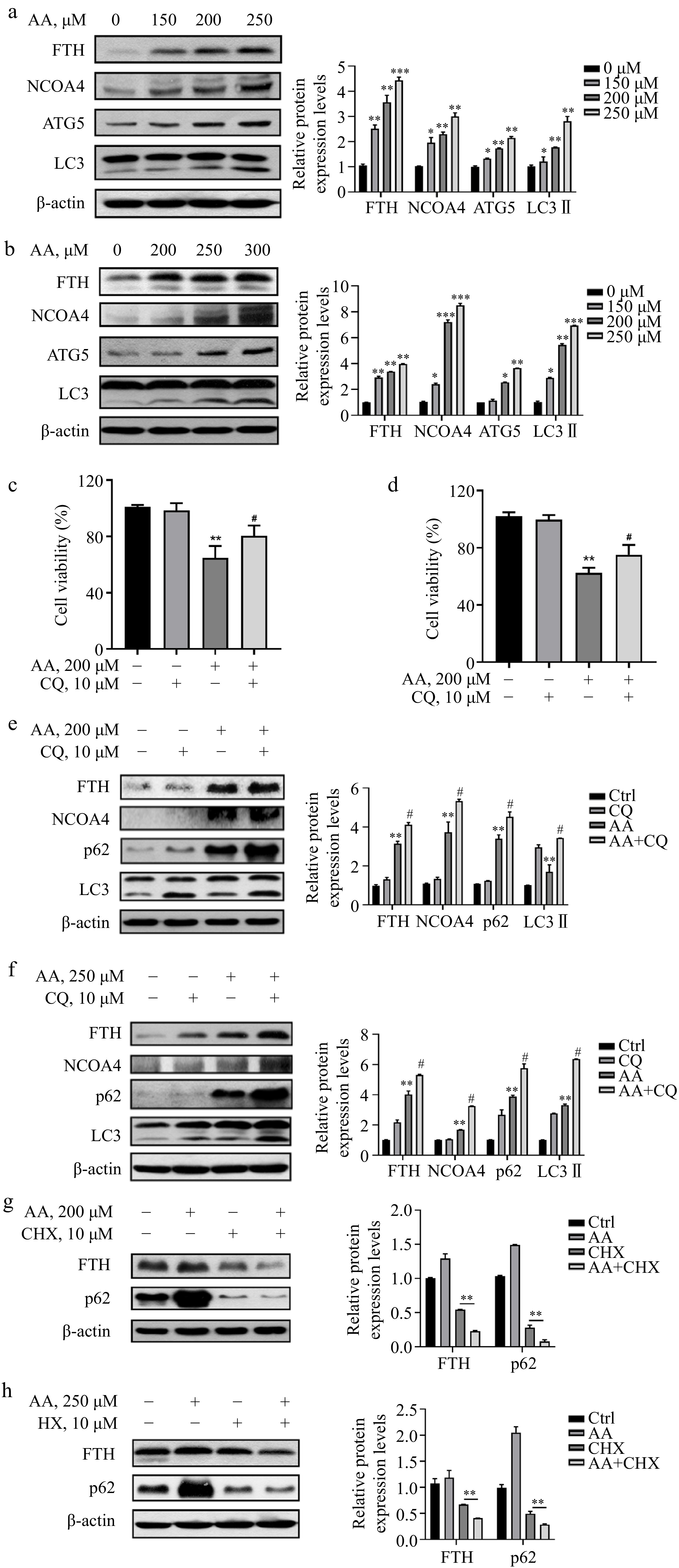
      Figure 3. 

      AA increases free iron levels in labile iron pools by activating ferritinophagy. AA activated the expression of ferritinophagy-related proteins. The cells were treated with AA for 24 h, and the expression of FTH, NCOA4, ATG5 and LC3 in (a) L02 and (b) AML12 cells were detected by Western blotting. Inhibition of autophagy alleviated AA-induced ferroptosis. Cells were treated with AA and/or the autophagy flux inhibitor CQ, and the cell viability of (c) L02 and (d) AML12 was measured. Expression of ferritinophagy-related proteins. The cells were treated with AA and/or CQ for 24 h, and the lysates of (e) L02 and (f) AML12 cells were subjected to Western blot analysis to analyze the changes of FTH, NCOA4, p62 and LC3 protein expression. Expression of FTH and p62 after inhibition of protein synthesis. After the cells were treated with CHX, they were treated with AA for a certain period of time. Changes in FTH and p62 expression in (g) L02 and (h) AML12 cells were detected by Western blot to further demonstrate the activation of ferritinophagy. β-actin served as a loading control. The intensity of each protein expression band was quantified by densitometry normalized to β-actin. * p < 0.05, ** p < 0.01 and *** p < 0.001 versus the control group. # p < 0.05 and ## p < 0.01 versus the AA treatment group.

    • Studies have demonstrated that excessive consumption of PUFAs in the diet can cause endoplasmic reticulum (ER) stress. In liver diseases such as non-alcoholic steatohepatitis (NASH), the proportion of ω-6 PUFAs is notably increased. Therefore, the regulation of ER stress is deemed an important target for treating NASH[23,24]. Based on this, we postulated that ER stress might be implicated in AA-induced ferroptosis. Consistent with our hypothesis, the result showed that AA prompted ER stress in a dose-dependent manner in hepatocytes L02 and AML12, involving two branches of PERK and IRE1. It was evidenced by the elevated protein expression levels of Bip, p-PERK, p-eIF2α, ATF4, p-IRE1 and p-JNK (Fig. 4a & b). In conclusion, these findings indicate that PERK- and IRE1-mediated ER stress plays a role in AA-induced hepatocyte ferroptosis.
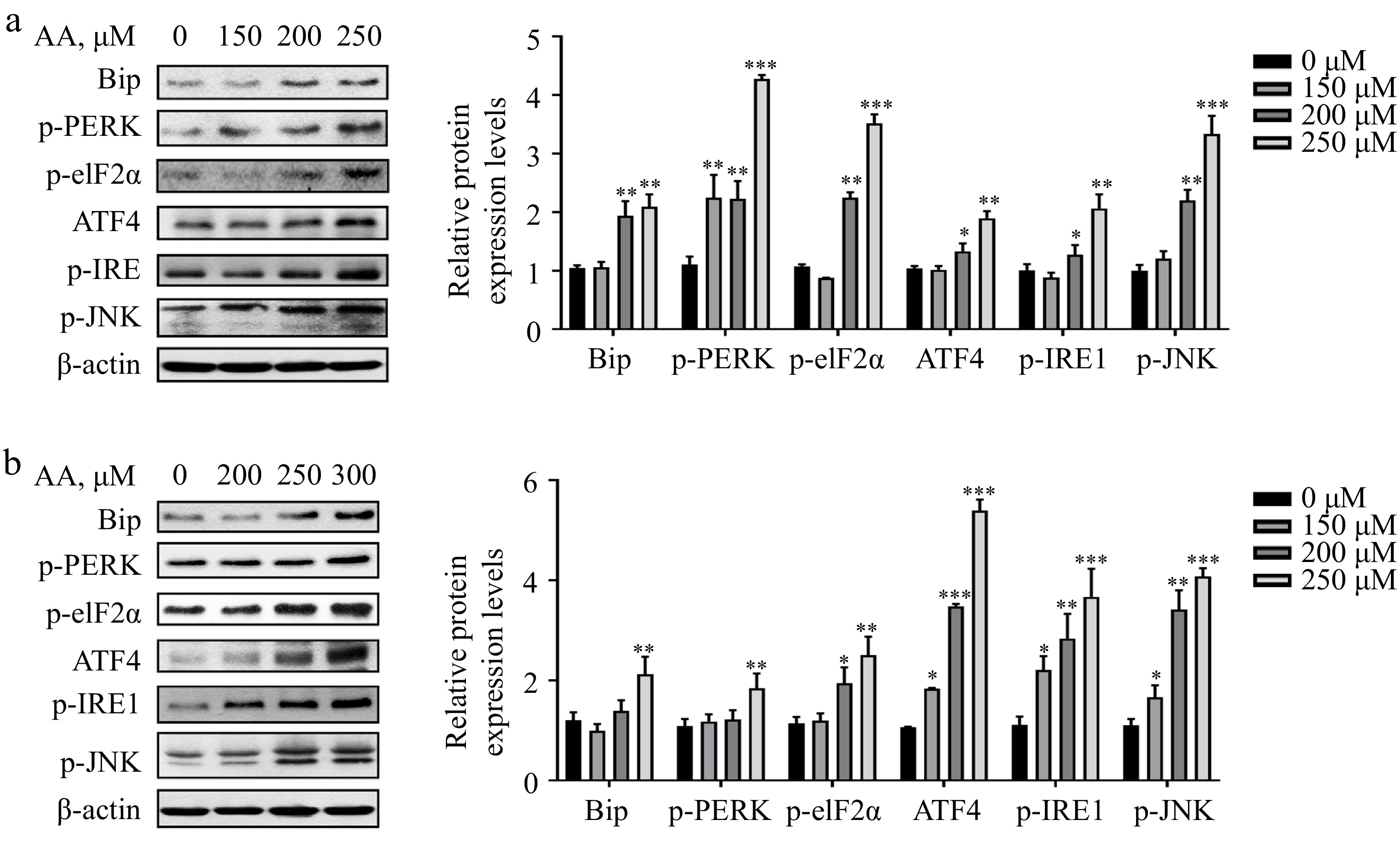
      Figure 4. 

      Activated ER stress is involved in AA-induced ferroptosis. Western blotting analysis of Bip, p-PERK, p-eIF2α, ATF4, p-IRE1α and p-JNK were performed in (a) L02 and (b) AML12 cells. Bar chart shows relative quantitative levels of each protein. β-actin was used as a loading control. Data are presented as mean ± SD. * p < 0.05, ** p < 0.01 and *** p < 0.001 versus the control group.

    • We proceeded to investigate the role of ER stress in AA-induced ferroptosis. Firstly, to assess the contribution of PERK signaling in AA-induced ferroptosis, we pre-treated cells with GSK2606414 (an inhibitor of PERK) prior to AA exposure. Figure 5a & b demonstrate that 1 μM GSK pre-treatment reversed AA-induced reduction in cell viability in L02 and AML12 cells. In addition, RNA interference is utilized to evaluate cell viability changes after PERK knockdown in L02 cells, revealing that PERK knockdown similarly reversed AA-induced reduction of cell viability (Fig. 5c). Next, we assessed changes in ferritinophagy markers in cells after inhibiting the PERK signaling pathway. Both GSK pre-treatment or PERK-inhibited led to increased accumulation of FTH and NCOA4, and decreased LC3I to LC3II conversion in AA-treated cells, indicating weakened ferritinophagy (Fig. 5d & e). In conclusion, our findings suggest that ER stress is upstream of ferritinophagy, and inhibiting PERK signaling can alleviate ferritinophagy by reducing ER stress, ultimately alleviating AA-induced cellular ferroptosis.

      Figure 5. 

      AA triggers lethal ferritinophagy by activating the PERK signaling pathway. Cell viability of (a) AML12 and (b) L02 cells pretreated with 1 μM GSK for 2 h and then stimulated with AA for 24 h. (c) Effect of knockdown of PERK on AA-induced ferroptosis in L02 cells. Western blotting analysis of p-eIF2α, ATF4, FTH, NCOA4, p62 and LC3 were performed in (d) AML12 and (e) L02 cells treated with GSK or PERK-si and AA. The data are presented as mean ± SD. ** p < 0.01 and *** p < 0.001 versus the control group. # p < 0.05 and ## p < 0.01 versus the AA treatment group.

    • To investigate the specific role of IRE1 in AA-induced ferroptosis, we utilized the IRE1 inhibitor 4μ8C to observe changes in cell viability after IRE1 inhibition. Surprisingly, inhibiting IRE1 not only failed to alleviate cellular ferroptosis, but it further decreased cell viability (Fig. 6ac). Previous studies have demonstrated that PERK and IRE1 can alleviate ER stress by coordinating the unfolded protein response (UPR)[25]. However, in unresolved ER stress, the PERK signaling pathway is continuously activated while the IRE1 signaling pathway is paradoxically attenuated, ultimately promoting cell death[25]. Therefore, the two branches of ER stress appear to play distinct roles in AA-induced ferroptosis. Specifically, IRE1 is activated as a protective signal of cells, and its inhibition exacerbates ferroptosis. Building on previous findings, we wondered whether IRE1 inhibition could exacerbate ferritinophagy by further activating the PERK signaling pathway. As such, the changes in the expression of related proteins were examined. As depicted in Fig. 6d & e, inhibition of IRE1 did activate the PERK signaling pathway, as evidenced by the down-regulation of p-IRE1 and up-regulation of p-eIF2α. Furthermore, the decreased accumulation of FTH, NCOA4, and p62, as well as the increased conversion of LC3I to LC3II, indicated further activation of ferritinophagy. These findings support the critical role played by IRE1 in ER stress-induced ferroptosis, where it functions as a protective signal in cells. Additionally, inhibiting IRE1 could exacerbate AA-induced ferritinophagy by further activating the PERK signaling pathway.

      Figure 6. 

      Inhibition of IRE1 instead activates the PERK signaling pathway, which in turn exacerbates AA-induced ferritinophagy. Cell viability of (a) AML12 and (b) L02 cells pretreated with 25 μM 4μ8C for 2 h and then stimulated with AA for 24 h. (c) Effect of knockdown of IRE1 on AA-induced ferroptosis in L02 cells. Western blotting analysis of p-eIF2α, ATF4, FTH, NCOA4, p62 and LC3 were performed in (d) AML12 and (e) L02 cells treated with GSK or PERK-si and AA. The data are presented as mean ± SD. ** p < 0.01 and *** p < 0.001 versus the control group. # p < 0.05 and ## p < 0.01 versus the GSK group or PERK-si group.

    • ω-6 and ω-3 PUFAs are essential fatty acids that play important roles in energy storage and production, as well as being crucial components of biological membranes and signaling molecules[26,27]. Many lipids derived from ω-6 PUFAs have pro-inflammatory functions, while those derived from ω-3 PUFAs possess anti-inflammatory properties[28,29]. AA, as a typical ω-6 PUFA, has a wide range of biological roles but its metabolic abnormalities are implicated in various liver diseases[3,4]. In this study, we conducted an in-depth investigation into the mechanism of iron metabolism disturbance in AA-induced ferroptosis.

      Recent studies suggest that ferroptosis may be an autophagy-dependent cell death. Ferroptosis inducers such as erastin and RSL3 lead to the accumulation of autophagosomes, while autophagy-deficient cells exhibit impaired ferroptosis under various stimulations[9]. Multiple forms of selective autophagy, including NCOA4-mediated ferritinophagy, RAB7A-dependent lipophagy, and heat shock protein 90-dependent chaperone autophagy, have been shown to promote ferroptosis by increasing iron load and impairing antioxidant systems[1214]. Especially for ferrintinophagy, high levels of labile iron ensure rapid accumulation of intracellular ROS, which is critical for ferroptosis[30]. Our investigation into the role of autophagy in AA-induced ferroptosis revealed that inhibition of autophagy by CQ could limit AA-induced ferroptosis in hepatocytes, indicating that AA activates autophagy in hepatocytes. Furthermore, AA increases cellular iron uptake by upregulating TfR1 expression, which results in an increase in intracellular FTH expression. At the same time, AA also accelerates the degradation of ferritin, leading to an increase in Fe2+ levels in cells. These results explain the hyperactivation of autophagy during AA-induced toxicity. However, further investigation is needed to determine if other forms of autophagy are involved in AA-induced ferroptosis.

      In recent years, researchers have found that ER stress may play an important role in ferroptosis[1517], but the relationship between ER stress and ferroptosis and its specific mechanism remain unclear. ER stress can affect different processes in various cells. Pathological conditions such as energy or nutrient deprivation and altered redox state can disrupt the homeostasis of the ER, leading to protein misfolding and activation of a series of signaling pathways, which is called the unfolded protein response (UPR)[31]. The UPR plays a key role in the maintenance of cellular homeostasis and is the key to the normal physiological functions of cells[31]. It has been reported that the expression level of ATF4 is increased in erastin-induced ferroptosis, and ATF4 can enhance the expression of SLC7A11 through a feedback loop, thereby limiting ferroptosis[32]. On the other hand, the production of ROS during ferroptosis is related to the PERK/eIF2α signaling pathway[15]. Additionally, ER stress promotes ferroptosis in ulcerative colitis and exposure to cigarette smoke, a mechanism involving heme oxygenase-1[3335]. It has also been reported that the activation of ER stress participates in the synergistic effect of ferroptosis and apoptosis through the CHOP-PUMA pathway[36,37]. In addition, under pathological conditions, activated ER stress not only does not inhibit ferroptosis, but may aggravate the occurrence of ferroptosis, indicating that under persistent or severe disease conditions, ER stress can not only initiate apoptosis and autophagy signaling pathways but also cause ferroptosis. Our study shows that AA activates ER stress, including both PERK and IRE1 signaling pathways. Moreover, AA-activated ER stress can affect cellular ferroptosis by regulating ferritinophagy. Inhibition of PERK alleviated AA-induced ferroptosis in hepatocytes by attenuating ferritinophagy, whereas inhibition of IRE1 aggravated AA-induced ferritinophagy by further activating PERK signaling.

      The abnormal metabolism of AA is related to the occurrence and development of many metabolic diseases in the human body, which will destroy the homeostasis of lipid metabolism and further lead to inflammation or cancer. It remains to be determined to what extent other cell types produce the AA-induced ferroptosis we observed in hepatocytes by activating ER stress and ferrinrinophagy. Therefore, future studies should focus on evaluating the efficacy of inhibiting ER stress alone, or in combination with ferroptosis inhibitors, and even related validation in animal models to induce the best and beneficial results.

    • In conclusion, our study confirmed that AA induces hepatocyte ferroptosis in a dose-dependent manner, which is closely related to the activation of ER stress and ferritinophagy. Specifically, exogenous AA induces ferroptosis by upregulating the expression of TfR1 on the cell membrane and accelerating the degradation of ferritin, resulting in iron overload. The investigation into the effect of different branches of ER stress on ferritinophagy provides new insights into AA-induced ferroptosis (Fig. 7).

      Figure 7. 

      Potential signaling pathways for AA-induced ferroptosis in hepatocytes. The polyunsaturated fatty acid AA upregulates the expression of TfR1 on the cell membrane surface and activates ferritinophagy, thereby increasing the level of Fe2+ in the cellular labile iron pool. In addition, AA-induced ER stress is located upstream of ferritinophagy, and its two branches, PERK and IRE1 signaling pathways, play different roles in regulating ferritinophagy.

      • This study was supported by a grant from the National Key Research and Development Program of China (2022YFF1100205).

      • The authors declare that they have no conflict of interest.

      • Copyright: © 2023 by the author(s). Published by Maximum Academic Press on behalf of China Agricultural University, Zhejiang University and Shenyang Agricultural University. This article is an open access article distributed under Creative Commons Attribution License (CC BY 4.0), visit https://creativecommons.org/licenses/by/4.0/.
    Figure (7)  References (37)
  • About this article
    Cite this article
    Zhang H, Han K, Yin S, Fan L, Hu H, et al. 2023. Distinct roles of the IRE1α arm and PERK arm of unfolded protein response in arachidonic acid-induced ferroptosis in hepatocytes. Food Innovation and Advances 2(3):184−192 doi: 10.48130/FIA-2023-0020
    Zhang H, Han K, Yin S, Fan L, Hu H, et al. 2023. Distinct roles of the IRE1α arm and PERK arm of unfolded protein response in arachidonic acid-induced ferroptosis in hepatocytes. Food Innovation and Advances 2(3):184−192 doi: 10.48130/FIA-2023-0020

Catalog

  • About this article

/

DownLoad:  Full-Size Img  PowerPoint
Return
Return