REVIEW   Open Access    

Cork taint of wines: the formation, analysis, and control of 2,4,6- trichloroanisole

More Information
  • Cork taint has devastating effects on the aroma and quality of the wine, which can cause an annual loss of may be up to more than one billion dollars. There are many causes of cork taint, but 2,4,6-trichloroanisole (2,4,6-TCA) is a major contributor, giving the wine a wet-moldy smell. This study provided a comprehensive overview of the occurrence, detection, and control/remediation of 2,4,6-TCA. The occurrence and formation mechanisms of 2,4,6-TCA mainly include microbial O-methylation of chlorophenols and chlorination of anisole. The source of 2,4,6-TCA in wine is the cork or other woodworks, but it is also possible to contaminate wine from the environment. Due to the extremely low odor threshold concentration of 2,4,6-TCA, the effective sample pre-enrichment for instrument identification and quantification is more important. The control/remediation strategies of 2,4,6-TCA mainly include eliminating 2,4,6-TCA in cork and removing 2,4,6-TCA from wine by adsorption. Finally, the challenges and possible future research directions in this research field were discussed and proposed.
  • The Fabaceae family, the third largest family in angiosperms, contains about 24,480 species (WFO, https://wfoplantlist.org), and has been a historically important source of food crops[14]. Peas (formerly Pisum sativa L. renamed to Lathyrus oleraceusPisum spp. will be used hereafter due to historical references to varietal names and subspecies that may not have been fully synonymized), are a member of the Fabaceae family and is among one of the oldest domesticated food crops with ongoing importance in feeding humans and stock. Peas originated in Western Asia and the Mediterranean basin where early finds from Egypt have been dated to ~4500 BCE and further east in Afghanistan from ~2000 BCE[5], and have since been extensively cultivated worldwide[6,7]. Given that peas are rich in protein, dietary fiber, vitamins, and minerals, have become an important part of people's diets globally[810].

    Domesticated peas are the result of long-term human selection and cultivation, and in comparison to wild peas, domesticated peas have undergone significant changes in morphology, growth habits, and yield[1113]. From the long period of domestication starting in and around Mesopotamia many diverse lineages of peas have been cultivated and translocated to other parts of the world[14,15]. The subspecies, Pisum sativum subsp. sativum is the lineage from which most cultivars have been selected and is known for possessing large, round, or oval-shaped seeds[16,17]. In contrast, the subspecies, P. sativum subsp. elatius, is a cultivated pea which more closely resembles wild peas and is mainly found in grasslands and desert areas in Europe, Western Asia, and North Africa. Pisum fulvum is native to the Mediterranean basin and the Balkan Peninsula[15,18], and is resistant to pea rust caused by the fungal pathogen Uromyces pisi. Due to its resistance to pea rust, P. fulvum has been cross-bred with cultivated peas in the development of disease-resistant strains[19]. These examples demonstrate the diverse history of the domesticated pea and why further study of the pea pan-plastome could be employed for crop improvement. While studies based on the nuclear genome have been used to explore the domestication history of pea, these approaches do not account for certain factors, such as maternal inheritance. Maternal lineages, which are inherited through plastomes, play a critical role in understanding the full domestication process. A pan-plastome-based approach will no doubt allow us to investigate the maternal genetic contributions and explore evolutionary patterns that nuclear genome studies may overlook. Besides, pan-plastome analysis enables researchers to systematically compare plastome diversity across wild and cultivated species, identifying specific regions of the plastome that contribute to desirable traits. These plastid traits can then be transferred to cultivated crops through introgression breeding or genetic engineering, leading to varieties with improved resistance to disease, environmental stress, and enhanced agricultural performance.

    Plastids are organelles present in plant cells and are the sites in which several vital biological processes take place, such as photosynthesis in chloroplasts[2024]. Because the origin of plastids is the result of an ancient endosymbiotic event, extant plastids retain a genome (albeit much reduced) from the free-living ancestor[25]. With the advancement of high-throughput DNA sequencing technology, over 13,000 plastid genomes or plastomes have been published in public databases by the autumn of 2023[24]. Large-scale comparison of plastomic data at multiple taxonomic levels has shown that plastomic data can provide valuable insights into evolution, interspecies relationships, and population genetic structure. The plastome, in most cases, displays a conserved quadripartite circular genomic architecture with two inverted repeat (IR) regions and two single copy (SC) regions, referred to as the large single-copy (LSC) and small single-copy (SSC) regions. However, some species have lost one copy of the inverted repeated regions, such as those in Erodium (Geraniaceae family)[26,27] and Medicago (Fabaceae)[28,29]. Compared to previous plastomic studies based on a limited number of plastomes, the construction of pan-plastomes attempts to describe all nucleotide variants present in a lineage through intensive sampling and comparisons. Such datasets can provide detailed insights into the maternal history of a species and help to better understand applied aspects such as domestication history or asymmetries in maternal inheritance, which can help guide future breeding programs. Such pan-plastomes have recently been constructed for several agriculturally important species. A recent study focuses on the genus Gossypium[20], using plastome data at the population level to construct a robust map of plastome variation. It explored plastome diversity and population structure relationships within the genus while uncovering genetic variations and potential molecular marker loci in the plastome. Besides, 65 samples were combined to build the pan-plastome of Hemerocallis citrina[30] , and 322 samples for the Prunus mume pan-plastome[31]. Before these recent efforts, similar pan-plastomes were also completed for Beta vulgaris[32], and Nelumbo nucifera[33]. However, despite the agricultural importance of peas, no such pan-plastome has been completed.

    In this study, 103 complete pea plastomes were assembled and combined another 42 published plastomes to construct the pan-plastome. Using these data, the following analyses were conducted to better understand the evolution and domestication history of pea: (1) genome structural comparisons, (2) codon usage bias, (3) simple sequence repeat patterns, (4) phylogenetic analysis, and (5) nucleotide variation of plastomes in peas.

    One hundred and three complete pea plastomes were de novo assembled from public whole-genome sequencing data[34]. For data quality control, FastQC v0.11.5 (www.bioinformatics.babraham.ac.uk/projects/fastqc/) was utilized to assess the quality of the reads and ensure that the data was suitable for assembly. The clean reads were then mapped to a published pea plastome (MW308610) plastome from the GenBank database (www.ncbi.nlm.nih.gov/genbank) as the reference using BWA v0.7.17[35] and SAMtools v1.9[36] to isolate plastome-specific reads from the resequencing data. Finally, these plastome-specific reads were assembled de novo using SPAdes v3.15.2[37]. The genome annotation was conducted by Geseq online program (https://chlorobox.mpimp-golm.mpg.de/geseq.html). Finally, the OGDRAW v1.3.1[38] program was utilized to visualize the circular plastome maps with default settings. To better resolve the pan-plastome for peas, 42 complete published pea plastomes were also downloaded from NCBI and combined them with the de novo data (Supplementary Table S1).

    To investigate the codon usage in the pan-plastome of pea, we utilized CodonW v.1.4.2 (http://codonw.sourceforge.net) to calculate the Relative Synonymous Codon Usage (RSCU) value of the protein-coding genes (PCGs) longer than 300 bp, excluding stop codons. The RSCU is a calculated metric used to evaluate the relative frequency of usage among synonymous codons encoding the same amino acid. An RSCU value above 1 suggests that the codon is utilized more frequently than the average for a synonymous codon. Conversely, a value below 1 indicates a lower-than-average usage frequency. Besides, the Effective Number of Codons (ENC) and the G + C content at the third position of synonymous codons (GC3s) were also calculated in CodonW v.1.4.2. The ENC value and GC3s value were utilized for generating the ENC-GC3s plot, with the expected ENC values (standard curve), are calculated according to formula: ENC = 2 + GC3s + 29 / [GC3s2 + (1 – GC3s)2][39].

    The MISA program[40] was utilized to detect simple sequence repeats (SSRs), setting the minimum threshold for repeat units at 10 for mono-motifs, 6 for di-motifs, and 5 for tri-, tetra-, penta-, and hexa-motif microsatellites, respectively.

    The 145 complete pea plastomes were aligned using MAFFT v 7.487[41]. Single nucleotide variants (SNVs)-sites were used to derive an SNV only dataset from the entire-plastome alignment[42]. A total of 959 SNVs were analyzed using IQ-TREE v2.1[43] with a TVMe + ASC + R2 substitution model, determined by ModelTest-NG[44] based on BIC, and clade support was assessed with 1,000 bootstrap replicates. Vavilovia formosa (MK604478) was chosen as an outgroup. The principal coordinates analysis (PCA) was conducted in TASSEL 5.0[45].

    DnaSP v6[46] was utilized to identify different haplotypes among the plastomes, with gaps and missing data excluded. Haplotype networks were constructed in Popart v1.7[47] using the median-joining algorithm. Haplotype diversity (Hd) for each group was calculated by DnaSP v6[46], and the evolutionary distances based on the Tamura-Nei distance model were computed based on the population differentiation index (FST) between different groups with the plastomic SNVs.

    In this study, the pan-plastome structure of peas was elucidated (Fig. 1). The length of these plastomes ranged from 120,826 to 122,547 bp. And the overall GC content varied from 34.74% to 34.87%. In contrast to typical plastomes characterized by a tetrad structure, the plastomes of peas contained a single IR copy. The average GC content among all pea plastomes was 34.8%, with the highest amount being 34.84% and the lowest 34.74%, with minimal variation among the pea plastomes.

    Figure 1.  Pea pan-plastome annotation map. Indicated by arrows, genes listed inside and outside the circle are transcribed clockwise and counterclockwise, respectively. Genes are color-coded by their functional classification. The GC content is displayed as black bars in the second inner circle. SNVs, InDels, block substitutions and mixed variants are represented with purple, green, red, and black lines, respectively. Single nucleotide variants (SNVs), block substitutions (BS, two or more consecutive nucleotide variants), nucleotide insertions or deletions (InDels), and mixed sites (which comprise two or more of the preceding three variants at a particular site) are the four categories into which variants are divided.

    A total of 110 unique genes were annotated (Supplementary Table S2), of which 76 genes were PCGs, 30 were transfer RNA (tRNA) genes and four were ribosomal RNA (rRNA) genes. Genes containing a single intron, include nine protein-coding genes (rpl16, rpl2, ndhB, ndhA, petB, petD, rpoC1, clpP, atpF) and six tRNA genes (trnK-UUU, trnV-UAC, trnL-UAA, trnA-UGC, trnI-GAU, trnG-UCC). Additionally, two protein-coding genes ycf3 and rps12 were found to contain two introns.

    The codon usage frequency in pea plastome genes is shown in Fig. 2a. The analysis of codon usage in the pea plastome indicated significant biases for specific codons across various amino acids. Here a nearly average usage in some amino acids was observed, such as Alanine (Ala) and Valine (Val). For most amino acids, the usage of different synonymous codons was not evenly distributed. Regarding stop codons, a nearly even usage was found, with 37.0% for TAA, 32.2% for TAG and 30.8% for TGA.

    Figure 2.  (a) The overall codon usage frequency in 51 CDSs (length > 300 bp) from the pea pan-plastome. (b) The heatmap of RSCU values in 51 CDSs (length > 300 bp) from the pea pan-plastome. The x-axis represents different codons and the y-axis represents different CDSs. The tree at the top was constructed based on a Neighbor-Joining algorithm.

    The RSCU heatmap (Fig. 2b) showed different RSCU values for all codons in plastomic CDSs. In general, a usage bias for A/T in the third position of codons was found among CDSs in the pea pan-plastome. The RSCU values among these CDSs ranged from 0 to 4.8. The highest RSCU value (4.8) was found with the CGT codon in the cemA gene, where six synonymous codons exist for Arg but only CGT (4.8) and AGG (1.2) were used in this gene. This explained in large part the extreme RSCU value for CGT, resulting in an extreme codon usage bias in this amino acid.

    In the ENC-GC3s plot (Fig. 3), 31 PCGs were shown below the standard curve, while 20 PCGs were above. Besides, around 12 PCGs were near the curve, which meant these PCGs were under the average natural selection and mutation pressure. This plot displayed that the codon usage preferences in pea pan-plastomes were mostly influenced by natural selection. Five genes were shown an extreme influence with natural selection for its extreme ΔENC (ENCexpected – ENC) higher than 5, regarding as petB (ΔENC = 5.18), psbA (ΔENC = 8.96), rpl16 (ΔENC = 5.62), rps14 (ΔENC = 14.29), rps18 (ΔENC = 6.46) (Supplementary Table S3).

    Figure 3.  The ENC-GC3s plot for pea pan-plastome, with GC3s as the x-axis and ENC as the y-axis. The expected ENC values (standard curve) are calculated according to formula: ENC = 2 + GC3s + 29 / [GC3s2 + (1 − GC3s)2].

    For SSR detection (Fig. 4), mononucleotide, dinucleotide, and trinucleotide repeats were identified in the pea pan-plastome including A/T, AT/TA, and AAT/ATT. The majority of these SSRs were mononucleotides (A/T), accounting for over 90% of all identified repeats. Additionally, we observed that A/T and AT/TA repeats were present in all pea accessions, whereas only about half of the plastomes contained AAT/ATT repeats. It was also found that the number of A/T repeats exhibited the greatest diversity, while the number of AAT/ATT repeats showed convergence in all plastomes that possessed this repeat.

    Figure 4.  Simple sequence repeats (SSRs) in the pea pan-plastome. The x-axis represents different samples of pea and the y-axis represents the number of repeats in this sample. (a) The number of A/T repeats in the peapan-plastome. (b) The number of AT/TA and AAT/ATT repeats of pea pan-plastomes.

    To better understand the phylogenetic relationships and evolutionary history of peas, a phylogenetic tree was reconstructed using maximum likelihood for 145 pea accessions utilizing the whole plastome sequences (Fig. 5a). The 145 pea accessions were grouped into seven clades with high confidence. These groups were named the 'PF group', 'PSeI-a group', 'PSeI-b group', 'PA group', 'PSeII group', 'PSeIII group', and the 'PS group'. The naming convention for these groups relates to the majority species names for accessions in each group, where P. fulvum makes up the 'PF group', P. sativum subsp. elatius in the 'PSeI-a group', 'PSeI-b group', 'PSeII group', and 'PSeIII group', P. abyssinicum in the 'PA group', and P. sativum in the 'PS group'. From this phylogenetic tree, it was observed that the 'PSeI-a group' and the 'PSeI-b group' had a close phylogenetic relationship and nearly all accessions in these two groups (except DCG0709 accession was P. sativum) were identified as P. sativum subsp. elatius. In addition to the P. sativum subsp. elatius found in PSeI, seven accessions from the PS group were identified as P. sativum subsp. elatius.

    The PCA results (Fig. 5b) also confirmed that domesticated varieties P. abyssinicum were closer to cultivated varieties PSeI and PSeII, while PSeIII was more closely clustered with cultivated varieties of P. sativum. A previous study has indicated that P. sativum subsp. sativum and P. abyssinicum were independently domesticated from different P. sativum subsp. elatius populations[34].

    The complete plastome sequences were utilized for haplotype analysis using TCS and median-joining network methods (Fig. 5c). A total of 76 haplotypes were identified in the analysis. The TCS network resolved a similar pattern as the other analyses in that six genetic clusters were resolved with genetic clusters PS and PSeIII being very closely related. The genetic cluster containing P. fulvum exhibits greater genetic distance from other genetic clusters. The genetic clusters containing P. abyssinicum (PA) and P. sativum (PS) had lower levels of intracluster differentiation. In the TCS network, Hap30 and Hap31 formed distinct clusters from other haplotypes, such as Hap27, which may account for the genetic difference between the 'PSeI-a group' and 'PSeI-b group'. The network analysis results were consistent with the findings of the phylogenetic tree and principal component analyses results in this study.

    Figure 5.  (a) An ML tree resolved from 145 pea plastomes. (b) PCA analysis showing the first two components. (c) Haplotype network of pea plastomes. The size of each circle is proportional to the number of accessions with the same haplotype. (d) Genetic diversity and differentiation of six clades of peas. Pairwise FST between the corresponding genetic clusters is represented by the numbers above the lines joining two bubbles.

    Among the six genetic clusters, the highest haplotype diversity (Hd) was observed in PSeIII (Hd = 0.99, π = 0.22 × 10⁻³), followed by PSeII (Hd = 0.96, π = 0.43 × 10⁻³), PSeI (Hd = 0.96, π = 0.94 × 10⁻³), PF (Hd = 0.94, π = 0.6 × 10⁻⁴), PS (Hd = 0.88, π = 0.3 × 10⁻⁴), and PA (Hd = 0.70, π = 0.2 × 10⁻⁴). Genetic differentiation was evaluated between each genetic cluster by calculating FST values. As shown in Fig. 5d, except for the relatively lower population differentiation between PS and PSeIII (FST = 0.54), and between PSeI and PSeII (FST = 0.59), the FST values between the remaining clades ranged from 0.7 to 0.9. The highest population differentiation was observed between PF and PA (FST = 0.98). The FST values between PSeI and different genetic clusters were relatively low, including PSeI and PF (FST = 0.80), PSeI and PS (FST = 0.77), PSeI and PSeIII (FST = 0.72), PSeI and PSeII (FST = 0.59), and PSeI and PA (FST = 0.72).

    To further determine the nucleotide variations in the pea pan-plastome, 145 plastomes were aligned and nucleotide differences analyzed across the dataset. A total of 1,579 variations were identified from the dataset (Table 1), including 965 SNVs, 24 Block Substitutions, 426 InDels, and 160 mixed variations of these three types. Among the SNVs, transitions were more frequent than transversions, with 710 transitions and 247 transversions. In transitions, T to G and A to C had 148 and 139 occurrences, respectively, while in transversions, G to A and C to T had 91 and 77 occurrences, respectively.

    Table 1.  Nucleotide variation in the pan-plastome of peas.
    Variant Total SNV Substitution InDel Mix
    (InDel, SNV)
    Mix
    (InDel, SUB)
    Total 1,576 965 24 426 156 4
    CDS 734 445 6 176 103 4
    Intron 147 110 8 29 0 0
    tRNA 20 15 1 4 0 0
    rRNA 11 3 0 6 2 0
    IGS 663 392 9 211 51 0
     | Show Table
    DownLoad: CSV

    When analyzing variants by their position to a gene (Fig. 6), there were 731 variations in CDSs, accounting for 46.3% of the total variations, including 443 SNVs (60.6%), six block substitutions (0.83%), 175 InDels (23.94%), and four mixed variations (14.64%). There were 104 variants in introns, accounting for 6.59% of the total variations, including 78 SNVs (75%), seven block substitutions (6.73%), and 19 InDels (18.27%). IGS (Intergenic spacers) contained 660 variations, accounting for 41.8% of the total variations, including 394 SNVs (59.7%), nine block substitutions (1.36%), 207 InDels (31.36%), and 50 mixed variations (7.58%). The tRNA regions contained 63 variants, accounting for 3.99% of the total variations, including 47 SNVs (74.6%) and 14 InDels (22.2%). The highest number of variants were detected in the IGS regions, while the lowest were found in introns. Among CDSs, accD (183) had the highest number of variations. In introns, rpL16 (18) and ndhA (16) had the most variants. In the IGS regions, ndhD-trnI-CAU (73), and trnL-UAA-trnT-UGU (44) possessed the greatest number of variants.

    Figure 6.  Variant locations within the pea pan-plastome categorized by genic position (Introns, CDS, and IGS).

    Finally, examples of some genes with typical variants were provided to better illustrate the sequence differences between clades (Fig. 7). For example, the present analysis revealed that the ycf1 gene exhibited a high number of variant loci, which included unique single nucleotide variants (SNVs) specific to the P. abyssinicum clade. Additionally, a unique InDel variant belonging to P. abyssinicum was identified. Similar unique SNVs and InDels were also found in other genes, such as matK and rpoC2, distinguishing the P. fulvum clade from others. These unique SNVs and InDels could serve as DNA barcodes to distinguish different maternal lineages of peas.

    Figure 7.  Examples of variant sites.

    The present research combined 145 pea plastomes to construct a pan-plastome of peas. Compared to single plastomic studies, pan-plastome analyses across a species or genus provide a higher-resolution understanding of phylogenetic relationships and domestication history. Most plastomes in plants possess a quadripartite circular structure with two inverted repeat (IR) regions and two single copy regions (LSC and SSC)[2024]. However, the complete loss of one of the IR regions in the pea plastome was observed which is well-known among the inverted repeat-lacking clade (IRLC) species in Fabaceae. The loss of IRs has been documented in detail from other genera such as Erodium (Geraniaceae family)[26,27] and Medicago (Fabaceae family)[28,29]. This phenomenon although not commonly observed, constitutes a significant event in the evolutionary trajectories of certain plant lineages[26]. Such large-scale changes in plastome architecture are likely driven in part by a combination of selective pressures and genetic drift[48]. In the pea pan-plastome, it was also found that, compared to some plants with IR regions, the length of the plastomes was much shorter, and the overall GC content was lower. This phenomenon was due to the loss of one IR with high GC content.

    Repetitive sequences are an important part of the evolution of plastomes and can be used to reconstruct genealogical relationships. Mononucleotide SSRs are consistently abundant in plastomes, with many studies identifying them as the most common type of SSR[4952]. Among these, while C/G-type SSRs may dominate in certain species[53,54], A/T types are more frequently observed in land plants. The present research was consistent with these previous conclusions, showing an A/T proportion exceeding 90% (Fig. 4). Due to their high rates of mutation, SSRs are widely used to study phylogenetic relationships and genetic variation[55,56]. Additionally, like other plants, pea plastome genes have a high frequency of A/Ts in the third codon position. This preference is related to the higher AT content common among most plant plastomes and Fabaceae plastomes in particular with their single IRs[57,58]. The AT-rich regions are often associated with easier unwinding of DNA during transcription and potentially more efficient and accurate translation processes[59]. The preference for A/T in third codon positions may also be influenced by tRNA availability, as the abundance of specific tRNAs that recognize these codons can enhance the efficiency of protein synthesis[60,61]. However, not all organisms exhibit this preference for A/T-ending codons. For instance, many bacteria have GC-rich genomes and thus show a preference for G/C-ending codons[6264]. This variation in codon usage bias reflects the differences in genomic composition and the evolutionary pressures unique to different lineages.

    This study also comprehensively examined the variant loci of the pea pan-plastome. Among these variant sites, some could potentially serve as DNA barcode sites for specific lineages of peas, such as ycf1, rpoC2, and matK. Both ycf1 and matK have been widely used as DNA barcodes in many species[6568], as they are hypervariable. Researchers now have a much deeper understanding of the crucial role plastomes have played in plant evolution[6971]. By generating a comprehensive map of variant sites, future researchers can now more effectively trace differences in plastotypes to physiological and metabolic traits for use in breeding elite cultivars.

    The development of a pan-plastome for peas provides new insights into the maternal domestication history of this important food crop. Based on the phylogenetic analysis in this study, we observed a clear differentiation between wild and cultivated peas, with P. fulvum being the earliest diverging lineage, and was consistent with former research[34]. The ML tree (Fig. 5a) indicated that cultivated peas had undergone at least two independent domestications, namely from the PA and PS groups, which is consistent with former research[34]. However, as the present study added several accessions over the previous study and plastomic data was utilized, several differences were also found[34], such as the resolution of the two groups, referred to as PSeI-a group and PSeI-b group which branched between the PA group and PF group. Previous research based on nuclear data[34] only and with fewer accessions showed that the PA group and PF group were closely related in phylogeny, with no PSeI group appearing between them. One possible explanation is that the PSeI-a and PSeI-b lineages represents the capture and retention of a plastome from a now-extinct lineage while backcrossing to modern cultivars has obscured this signal in the nuclear genomic datasets. However, procedural explanations such as incorrectly identified accessions might have also resulted in such patterns. In either case, the presence of these plastomes in the cultivated pea gene pool should be explored for possible associations with traits such as disease resistance and hybrid incompatibility. This finding underscores the complexity of the domestication process and highlights the role of hybridization and selection in shaping the genetic landscape of cultivated peas. As such, future studies integrating data from the nuclear genome, mitogenome, and plastome will undoubtedly provide deeper insights into the phylogeny and domestication of peas. This pan-plastome research, encompassing a variety of cultivated taxa, will also support the development of elite varieties in the future.

    This study newly assembled 103 complete pea plastomes. These plastomes were combined with 42 published pea plastomes to construct the first pan-plastome of peas. The length of pea plastomes ranged from 120,826 to 122,547 bp, with the GC content varying from 34.74% to 34.87%. The codon usage pattern in the pea pan-plastome displayed a strong bias for A/T in the third codon position. Besides, the codon usage of petB, psbA, rpl16, rps14, and rps18 were shown extremely influenced by natural selection. Three types of SSRs were detected in the pea pan-plastome, including A/T, AT/TA, and AAT/ATT. From phylogenetic analysis, seven well-supported clades were resolved from the pea pan-plastome. The genes ycf1, rpoC2, and matK were found to be suitable for DNA barcoding due to their hypervariability. The pea pan-plastome provides a valuable supportive resource in future breeding and selection research considering the central role chloroplasts play in plant metabolism as well as the association of plastotype to important agronomic traits such as disease resistance and interspecific compatibility.

  • The authors confirm contribution to the paper as follows: study conception and design: Wang J; data collection: Kan J; analysis and interpretation of results: Kan J, Wang J; draft manuscript preparation: Kan J, Wang J, Nie L; project organization and supervision: Tiwari R, Wang M, Tembrock L. All authors reviewed the results and approved the final version of the manuscript.

  • The annotation files of newly assembled pea plastomes were uploaded to the Figshare website (https://figshare.com/, doi: 10.6084/m9.figshare.26390824).

  • This study was funded by the Guangdong Pearl River Talent Program (Grant No. 2021QN02N792) and the Shenzhen Fundamental Research Program (Grant No. JCYJ20220818103212025). This work was also funded by the Science Technology and Innovation Commission of Shenzhen Municipality (Grant No. RCYX20200714114538196) and the Innovation Program of Chinese Academy of Agricultural Sciences. We are also particularly grateful for the services of the High-Performance Computing Cluster in the Agricultural Genomics Institute at Shenzhen, Chinese Academy of Agricultural Sciences.

  • The authors declare that they have no conflict of interest.

  • [1]

    Peña-Neira A, Fernández de Simón B, García-Vallejo MC, Hernández T, Cadahía E, et al. 2000. Presence of cork-taint responsible compounds in wines and their cork stoppers. European Food Research and Technology 211:257−61

    doi: 10.1007/s002170000193

    CrossRef   Google Scholar

    [2]

    Keng A, Botezatu A. 2023. Uncorking haloanisoles in wine. Molecules 28:2532

    doi: 10.3390/molecules28062532

    CrossRef   Google Scholar

    [3]

    Jové P, Pareras A, De Nadal R, Verdum M. 2021. Development and optimization of a quantitative analysis of main odorants causing off flavours in cork stoppers using headspace solid-phase microextraction gas chromatography tandem mass spectrometry. Journal of Mass Spectrometry 56:e4728

    doi: 10.1002/jms.4728

    CrossRef   Google Scholar

    [4]

    Dietrich AM, Burlingame GA. 2020. A review: The challenge, consensus, and confusion of describing odors and tastes in drinking water. Science of The Total Environment 713:135061

    doi: 10.1016/j.scitotenv.2019.135061

    CrossRef   Google Scholar

    [5]

    Judet-Correia D, Bensoussan M, Charpentier C, Dantigny P. 2013. Influence of temperature, copper and CO2 on spore counts and geosmin production by Penicillium expansum. Australian Journal of Grape and Wine Research 19:81−86

    doi: 10.1111/ajgw.12004

    CrossRef   Google Scholar

    [6]

    Romano A, Navarini L, Lonzarich V, Bogialli S, Pastore P, et al. 2022. 2,4,6-Trichloroanisole off-flavor screening in green Coffea arabica by a novel vocus NO+ CI-MS method: A study on green coffee from different geographical origins. Journal of Agricultural and Food Chemistry 70:11412

    doi: 10.1021/acs.jafc.2c03899

    CrossRef   Google Scholar

    [7]

    Tempere S, Schaaper MH, Cuzange E, de Revel G, Sicard G. 2017. Masking of several olfactory notes by infra-threshold concentrations of 2,4,6-trichloroanisole. Chemosensory Perception 10:69−80

    doi: 10.1007/s12078-017-9227-5

    CrossRef   Google Scholar

    [8]

    Takeuchi H, Kato H, Kurahashi T. 2013. 2,4,6-Trichloroanisole is a potent suppressor of olfactory signal transduction. Proceeding of the National Academy of Sciences of the United States of America 110:16235−40

    doi: 10.1073/pnas.1300764110

    CrossRef   Google Scholar

    [9]

    Abhijith GR, Ostfeld A. 2021. Modeling the formation and propagation of 2, 4, 6-trichloroanisole, a dominant taste and odor compound, in Water Distribution Systems. Water 13:638

    doi: 10.3390/w13050638

    CrossRef   Google Scholar

    [10]

    Zhang K, Cao C, Zhou X, Zheng F, Sun Y, et al. 2018. Pilot investigation on formation of 2,4,6-trichloroanisole via microbial O-methylation of 2,4,6-trichlorophenol in drinking water distribution system: An insight into microbial mechanism. Water Research 131:11−21

    doi: 10.1016/j.watres.2017.12.013

    CrossRef   Google Scholar

    [11]

    Ge F, Zhu L, Chen H. 2006. Effects of pH on the chlorination process of phenols in drinking water. Journal of Hazardous Materials 133:99−105

    doi: 10.1016/j.jhazmat.2005.09.062

    CrossRef   Google Scholar

    [12]

    Zhang K, Zhou X, Zhang T, Mao M, Li L, et al. 2016. Kinetics and mechanisms of formation of earthy and musty odor compounds: Chloroanisoles during water chlorination. Chemosphere 163:366−72

    doi: 10.1016/j.chemosphere.2016.08.051

    CrossRef   Google Scholar

    [13]

    Zhang K, Luo Z, Zhang T, Mao M, Fu J. 2016. Study on formation of 2,4,6-trichloroanisole by microbial O-methylation of 2,4,6-trichlorophenol in lake water. Environmental Pollution 219:228−34

    doi: 10.1016/j.envpol.2016.10.042

    CrossRef   Google Scholar

    [14]

    Ghochlavi N, Aghapour AA, Khorsandi H. 2022. Biodegradation of 2,4,6 trichlorophenol by sequencing batch reactors (SBR) equipped with a rotating biological bed and operated in an anaerobic-aerobic condition. Frontiers in Environmental Science 10:2053

    doi: 10.3389/fenvs.2022.1015790

    CrossRef   Google Scholar

    [15]

    Zheng W, Su R, Lin X, Liu J. 2022. Nanochannel array modified three-dimensional graphene electrode for sensitive electrochemical detection of 2,4,6-trichlorophenol and prochloraz. Frontiers in Chemistry 10:954802

    doi: 10.3389/fchem.2022.954802

    CrossRef   Google Scholar

    [16]

    Li H, Li J, Hou C, Du S, Ren Y, et al. 2010. A sub-nanomole level electrochemical method for determination of prochloraz and its metabolites based on medical stone doped disposable electrode. Talanta 83:591−95

    doi: 10.1016/j.talanta.2010.10.002

    CrossRef   Google Scholar

    [17]

    Huff J. 2012. Long-term toxicology and carcinogenicity of 2,4,6-trichlorophenol. Chemosphere 89:521−25

    doi: 10.1016/j.chemosphere.2012.05.015

    CrossRef   Google Scholar

    [18]

    Tian F, Qiao C, Wang C, Pang T, Guo L, et al. 2022. Dissipation behavior of prochloraz and its metabolites in grape under open-field, storage and the wine-making process. Journal of Food Composition and Analysis 114:104846

    doi: 10.1016/j.jfca.2022.104846

    CrossRef   Google Scholar

    [19]

    Prak S, Gunata Z, Guiraud JP, Schorr-Galindo S. 2007. Fungal strains isolated from cork stoppers and the formation of 2,4,6-trichloroanisole involved in the cork taint of wine. Food Microbiology 24:271−80

    doi: 10.1016/j.fm.2006.05.002

    CrossRef   Google Scholar

    [20]

    Maggi L, Mazzoleni V, Fumi MD, Salinas MR. 2008. Transformation ability of fungi isolated from cork and grape to produce 2,4,6-trichloroanisole from 2,4,6-trichlorophenol. Food Additives & Contaminants: Part A 25:265−69

    doi: 10.1080/02652030701522991

    CrossRef   Google Scholar

    [21]

    Alvarez-Rodríguez ML, López-Ocaña L, López-Coronado JM, Rodríguez E, Martínez MJ, et al. 2002. Cork taint of wines: role of the filamentous fungi isolated from cork in the formation of 2,4,6-trichloroanisole by O-methylation of 2,4,6-trichlorophenol. Applied and Environmental Microbiology 68:5860−69

    doi: 10.1128/AEM.68.12.5860-5869.2002

    CrossRef   Google Scholar

    [22]

    Álvarez-Rodríguez ML, Belloch C, Villa M, Uruburu F, Larriba G, et al. 2003. Degradation of vanillic acid and production of guaiacol by microorganisms isolated from cork samples. FEMS Microbiology Letters 220:49−55

    doi: 10.1016/S0378-1097(03)00053-3

    CrossRef   Google Scholar

    [23]

    Endo M, Matsui C, Maeta N, Uehara Y, Matsuda R, et al. 2021. Growth characteristics of Aspergillus oryzae in the presence of 2,4,6-trichlorophenol. The Journal of General and Applied Microbiology 67:256−59

    doi: 10.2323/jgam.2021.06.001

    CrossRef   Google Scholar

    [24]

    Nyström A, Grimvall A, Krantz-Rüilcker C, Sävenhed R, Åkerstrand K. 1992. Drinking water off-flavour caused by 2,4,6-trichloroanisole. Water Science and Technology 25:241−49

    doi: 10.2166/wst.1992.0058

    CrossRef   Google Scholar

    [25]

    Zhou X, Zhang K, Zhang T, Yang Y, Ye M, et al. 2019. Formation of odorant haloanisoles and variation of microorganisms during microbial O-methylation in annular reactors equipped with different coupon materials. Science of the Total Environment 679:1−11

    doi: 10.1016/j.scitotenv.2019.04.329

    CrossRef   Google Scholar

    [26]

    Zhou X, Zhang K, Zhang T, Cen C, Pan R. 2021. Biotransformation of halophenols into earthy-musty haloanisoles: Investigation of dominant bacterial contributors in drinking water distribution systems. Journal of Hazardous Materials 403:123693

    doi: 10.1016/j.jhazmat.2020.123693

    CrossRef   Google Scholar

    [27]

    Bai X, Zhang T, Qu Z, Li H, Yang Z. 2017. Contribution of filamentous fungi to the musty odorant 2,4,6-trichloroanisole in water supply reservoirs and associated drinking water treatment plants. Chemosphere 182:223−30

    doi: 10.1016/j.chemosphere.2017.04.138

    CrossRef   Google Scholar

    [28]

    Zheng T, Ma YL, Li WS, Deng JX, Li H, et al. 2023. Trichoderma species from rhizosphere of Oxalis corymbose release volatile organic compounds inhibiting the seed germination and growth of Echinochloa colona. Arabian Journal of Chemistry 16:105274

    doi: 10.1016/j.arabjc.2023.105274

    CrossRef   Google Scholar

    [29]

    Hitfield FB, Ly Nguyen TH, Tindale CR. 1991. Effect of relative humidity and incubation time on the O-methylation of chlorophenols in fibreboard by Paecilomyces variotii. Journal of the Science of Food and Agriculture 55:19−26

    doi: 10.1002/jsfa.2740550104

    CrossRef   Google Scholar

    [30]

    Gee JM, Peel JL. 1974. Metabolism of 2,3,4,6 tetrachlorophenol by microorganisms from broiler house litter. Journal of General Microbiology 85:237−43

    doi: 10.1099/00221287-85-2-237

    CrossRef   Google Scholar

    [31]

    Coque JJR, Alvarez-Rodríguez ML, Larriba G. 2003. Characterization of an inducible chlorophenol O-methyltransferase from Trichoderma longibrachiatum involved in the formation of chloroanisoles and determination of its role in cork taint of wines. Applied and Environmental Microbiolog 69:5089−95

    doi: 10.1128/AEM.69.9.5089-5095.2003

    CrossRef   Google Scholar

    [32]

    Prat C, Ruiz-Rueda O, Trias R, Anticó E, Capone D, et al. 2009. Molecular fingerprinting by PCR-denaturing gradient gel electrophoresis reveals differences in the levels of microbial diversity for musty-earthy tainted corks. Applied and Environmental Microbiology 75:1922−1931

    doi: 10.1128/AEM.02758-08

    CrossRef   Google Scholar

    [33]

    Tang Y, Wu Z, Zhang Y, Wang C, Ma X, et al. 2023. Cultivation-dependent and cultivation-independent investigation of O-methylated pollutant-producing bacteria in three drinking water treatment plants. Water Research 231:119618

    doi: 10.1016/j.watres.2023.119618

    CrossRef   Google Scholar

    [34]

    Allard AS, Remberger M, Neilson AH. 1987. Bacterial O-methylation of halogen-substituted phenols. Applied and Environmental Microbiology 53:839−45

    doi: 10.1128/aem.53.4.839-845.1987

    CrossRef   Google Scholar

    [35]

    Azevedo J, Brandao E, Soares S, Oliveira J, Lopes P, et al. 2020. Polyphenolic characterization of nebbiolo red wines and their interaction with salivary proteins. Foods 9:1867

    doi: 10.3390/foods9121867

    CrossRef   Google Scholar

    [36]

    Azevedo J, Jesus M, Brandao E, Soares S, Oliveira J, et al. 2022. Interaction between salivary proteins and cork phenolic compounds able to migrate to wine model solutions. Food Chemistry 367:130607

    doi: 10.1016/j.foodchem.2021.130607

    CrossRef   Google Scholar

    [37]

    Azevedo J, Lopes P, Mateus N, de Freitas V. 2022. Cork, a natural choice to wine? Foods 11:2638

    doi: 10.3390/foods11172638

    CrossRef   Google Scholar

    [38]

    Monteiro S, Bundaleski N, Malheiro A, Cabral M, Teodoro OMND. 2022. Cross contamination of 2, 4, 6-trichloroanisole in cork stoppers. Journal of Agricultural and Food Chemistry 70:6747−54

    doi: 10.1021/acs.jafc.2c02493

    CrossRef   Google Scholar

    [39]

    Rebezov M, Saeed K, Khaliq A, Rahman SJU, Sameed N, et al. 2022. Application of electrolyzed water in the food industry: A review. Applied Sciences-Basel 12:6639

    doi: 10.3390/app12136639

    CrossRef   Google Scholar

    [40]

    Zhang WL, Cao JK, Jiang WB. 2021. Application of electrolyzed water in postharvest fruits and vegetables storage: A review. Trends in Food Science & Technology 114:599−607

    doi: 10.1016/j.jpgs.2021.06.005

    CrossRef   Google Scholar

    [41]

    Cravero F, Englezos V, Rantsiou K, Torchio F, Giacosa S, et al. 2018. Control of Brettanomyces bruxellensis on wine grapes by post-harvest treatments with electrolyzed water, ozonated water and gaseous ozone. Innovative Food Science & Emerging Technologies 47:309−16

    doi: 10.1016/j.ifset.2018.03.017

    CrossRef   Google Scholar

    [42]

    Giacosa S, Gabrielli M, Torchio F, Río Segade S, Grobas AMM, et al. 2019. Relationships among electrolyzed water postharvest treatments on winegrapes and chloroanisoles occurrence in wine. Food Research International 120:235−43

    doi: 10.1016/j.foodres.2019.02.034

    CrossRef   Google Scholar

    [43]

    Gabrielli M, Englezos V, Rolle L, Río Segade S, Giacosa S, et al. 2020. Chloroanisoles occurrence in wine from grapes subjected to electrolyzed water treatments in the vineyard. Food Research International 137:109704

    doi: 10.1016/j.foodres.2020.109704

    CrossRef   Google Scholar

    [44]

    Tarasov A, Jung RE. 2023. Is Airborne 2,4,6-Trichloroanisole (TCA) a threat for bottled wine? Australian Journal of Grape and Wine Research 2023:6637804

    doi: 10.1155/2023/6637804

    CrossRef   Google Scholar

    [45]

    Romano A, Capozzi V, Khomenko I, Biasioli F. 2023. Advances in the Application of Direct Injection Mass Spectrometry Techniques to the Analysis of Grape, Wine and Other Alcoholic Beverages. Molecules 28:7642

    doi: 10.3390/molecules28227642

    CrossRef   Google Scholar

    [46]

    Zhang J, Zhang A, Fan C, Li X, Cui Z, et al. 2022. Determination of multihalo- phenols and anisoles in wine by gas chromatography tandem mass spectrometry through online derivatization and head space solid phase Microextraction. Food Analytical Methods 15:3435−43

    doi: 10.1007/s12161-022-02371-7

    CrossRef   Google Scholar

    [47]

    Ruiz-Delgado A, Arrebola-Liébanas FJ, Romero-González R, López-Ruiz R, Garrido Frenich A. 2016. Headspace solid-phase microextraction coupled to gas chromatography-tandem mass spectrometry for the determination of haloanisoles in sparkling (cava and cider) and non-sparkling (wine) alcoholic beverages. Food Additives and Contaminants: Part A 33:1535−44

    doi: 10.1080/19440049.2016.1229870

    CrossRef   Google Scholar

    [48]

    Özhan D, Anli RE, Vural N, Bayram M. 2009. Determination of Chloroanisoles and Chlorophenols in Cork and Wine by using HS-SPME and GC-ECD Detection. Journal of the Institute of Brewing 115:71−77

    doi: 10.1002/j.2050-0416.2009.tb00346.x

    CrossRef   Google Scholar

    [49]

    Karpas Z, Guamán AV, Calvo D, Pardo A, Marco S. 2012. The potential of ion mobility spectrometry (IMS) for detection of 2,4,6-trichloroanisole (2,4,6-TCA) in wine. Talanta 93:200−5

    doi: 10.1016/j.talanta.2012.02.012

    CrossRef   Google Scholar

    [50]

    Lichvanová Z, Ilbeigi V, Sabo M, Tabrizchi M, Matejčík S. 2014. Using corona discharge-ion mobility spectrometry for detection of 2,4,6-Trichloroanisole. Talanta 127:239−43

    doi: 10.1016/j.talanta.2014.04.021

    CrossRef   Google Scholar

    [51]

    Márquez-Sillero I, Aguilera-Herrador E, Cárdenas S, Valcárcel M. 2011. Determination of 2, 4, 6-tricholoroanisole in water and wine samples by ionic liquid-based single-drop microextraction and ion mobility spectrometry. Analytica Chimica Acta 702:199−204

    doi: 10.1016/j.aca.2011.06.046

    CrossRef   Google Scholar

    [52]

    Márquez-Sillero I, Cárdenas S, Valcárcel M. 2012. Headspace–multicapillary column–ion mobility spectrometry for the direct analysis of 2,4,6-trichloroanisole in wine and cork samples. Journal of Chromatography A 1265:149−54

    doi: 10.1016/j.chroma.2012.09.087

    CrossRef   Google Scholar

    [53]

    Jové P, Vives-Mestres M, De Nadal R, Verdum M. 2021. Development, optimization and validation of a sustainable and quantifiable methodology for the determination of 2,4,6-trichloroanisole, 2,3,4,6-tetrachloroanisole, 2,4,6-tribromoanisole, pentachloroanisole, 2-methylisoborneole and geosmin in air. Processes 9:1571

    doi: 10.3390/pr9091571

    CrossRef   Google Scholar

    [54]

    Meléndez F, Arroyo P, Gómez-Suárez J, Santos JP, Yuste FJ, et al. 2022. Detection of TCA in cork stoppers using an electronic nose. IEEE International Symposium on Olfaction and Electronic Nose (ISOEN), Aveiro, Portugal, 2022. USA: IEEE. https://doi.org/10.1109/ISOEN54820.2022.9789615

    [55]

    Santos JP, Sayago I, Sanjurjo JL, Perez-Coello MS, Díaz-Maroto MC. 2022. Rapid and non-destructive analysis of corky off-flavors in natural cork stoppers by a wireless and portable electronic nose. Sensors 22:4687

    doi: 10.3390/s22134687

    CrossRef   Google Scholar

    [56]

    Meléndez F, Arroyo P, Gómez-Suárez J, Palomeque-Mangut S, Suárez JI, et al. 2022. Portable electronic nose based on digital and analog chemical sensors for 2,4,6-trichloroanisole discrimination. Sensors 22:3453

    doi: 10.3390/s22093453

    CrossRef   Google Scholar

    [57]

    Peres AM, Freitas P, Dias LG, Sousa MEBC, Castro LM, et al. 2013. Cyclic voltammetry: A tool to quantify 2,4,6-trichloroanisole in aqueous samples from cork planks boiling industrial process. Talanta 117:438−44

    doi: 10.1016/j.talanta.2013.09.039

    CrossRef   Google Scholar

    [58]

    Sanvicens N, Sánchez-Baeza F, Marco MP. 2003. Immunochemical determination of 2,4,6-trichloroanisole as the responsible agent for the musty odor in foods. 1. Molecular modeling studies for antibody production. Journal of Agricultural and Food Chemistry 51:3924−31

    doi: 10.1021/jf034003h

    CrossRef   Google Scholar

    [59]

    Moore E, Pravda M, and Guilbault GG. 2003. Development of a biosensor for the quantitative detection of 2, 4, 6-trichloroanisole using screen printed electrodes. Analytica Chimica Acta 484:15−24

    doi: 10.1016/S0003-2670(03)00311-8

    CrossRef   Google Scholar

    [60]

    Lausterer R, Sanvicens N, Marco MP, Hock B. 2003. Enzyme immunoassay for 2,4,6-trichloroanisole based on monoclonal antibodies. Analytical Letters 36:713−29

    doi: 10.1081/AL-120018795

    CrossRef   Google Scholar

    [61]

    Apostolou T, Pascual N, Marco MP, Moschos A, Petropoulos A, et al. 2014. Extraction-less, rapid assay for the direct detection of 2,4,6-trichloroanisole (TCA) in cork samples. Talanta 125:336−40

    doi: 10.1016/j.talanta.2014.03.023

    CrossRef   Google Scholar

    [62]

    Cappellin L, Lopez-Hilfiker FD, Pospisilova V, Ciotti L, Pastore P, et al. 2020. Thermal desorption-vocus enables online nondestructive quantification of 2,4,6-trichloroanisole in cork stoppers below the perception threshold. Analytical Chemistry 92:9823−29

    Google Scholar

    [63]

    Damiano C, Intrieri D, Sonzini P, Rizzato S, Di Natale C, et al. 2022. Nickel (0) complexes as promising chemosensors for detecting the "cork taint" in wine. European Journal of Inorganic Chemistry 2022:e202101013

    doi: 10.1002/ejic.202101013

    CrossRef   Google Scholar

    [64]

    Lizarraga E, Irigoyen A, Belsue V, González-Peñas E. 2004. Determination of chloroanisole compounds in red wine by headspace solid-phase microextraction and gas chromatography-mass spectrometry. Journal of Chromatography A 1052:145−49

    doi: 10.1016/j.chroma.2004.08.046

    CrossRef   Google Scholar

    [65]

    Zalacain A, Alonso GL, Lorenzo C, Iñiguez M, Salinas MR. 2004. Stir bar sorptive extraction for the analysis of wine cork taint. Journal of Chromatography A 1033:173−178

    doi: 10.1016/j.chroma.2003.12.059

    CrossRef   Google Scholar

    [66]

    Campillo N, Viñas P, Cacho JI, Peñalver R, Hernández-Córdoba M. 2010. Evaluation of dispersive liquid-liquid microextraction for the simultaneous determination of chlorophenols and haloanisoles in wines and cork stoppers using gas chromatography-mass spectrometry. Journal of Chromatography A 1217:7323−30

    doi: 10.1016/j.chroma.2010.09.058

    CrossRef   Google Scholar

    [67]

    Bai X, Zhang T, Li H, Yang Z. 2016. Simultaneous dispersive liquid–liquid microextraction based on a low-density solvent and derivatization followed by gas chromatography for the simultaneous determination of chloroanisoles and the precursor 2,4,6-trichlorophenol in water samples. Journal of Separation Science 39:2146−55

    doi: 10.1002/jssc.201600098

    CrossRef   Google Scholar

    [68]

    Taylor MK, Young TM, Butzke CE, and Ebeler SE. 2000. Supercritical fluid extraction of 2, 4, 6-trichloroanisole from cork stoppers. Journal of Agricultural and Food Chemistry 48:2208−11

    doi: 10.1021/jf991045q

    CrossRef   Google Scholar

    [69]

    Ezquerro O, Garrido-López A, Tena MT. 2006. Determination of 2,4,6-trichloroanisole and guaiacol in cork stoppers by pressurised fluid extraction and gas chromatography-mass spectrometry. Journal of Chromatography A 1102:18−24

    doi: 10.1016/j.chroma.2005.10.023

    CrossRef   Google Scholar

    [70]

    Callejón RM, Ubeda C, Ríos-Reina R, Morales, ML, Troncoso AM. 2016. Recent developments in the analysis of musty odour compounds in water and wine: A review. Journal of Chromatography A 1428:72−85

    doi: 10.1016/j.chroma.2015.09.008

    CrossRef   Google Scholar

    [71]

    Dong ZY, Lin YL, Zhang TY, Hu CY, Pan Y, et al. 2021. The formation, analysis, and control of chlor(am)ination-derived odor problems: A review. Water Research 203:117549

    doi: 10.1016/j.watres.2021.117549

    CrossRef   Google Scholar

    [72]

    Marsol-Vall A, Ainsa S, Lopez R, Ferreira V. 2022. Development and validation of a method for the analysis of halophenols and haloanisoles in cork bark macerates by stir bar sorptive extraction heart-cutting two-dimensional gas chromatography negative chemical ionization mass spectrometry. Journal of Chromatography A 1673:463186

    doi: 10.1016/j.chroma.2022.463186

    CrossRef   Google Scholar

    [73]

    Pizarro C, Sáenz-González C, Pérez-del-Notario N, González-Sáiz JM. 2011. Development of a dispersive liquid–liquid microextraction method for the simultaneous determination of the main compounds causing cork taint and Brett character in wines using gas chromatography–tandem mass spectrometry. Journal of Chromatography A 1218,:1576−84

    doi: 10.1016/j.chroma.2011.01.055

    CrossRef   Google Scholar

    [74]

    Goto S, Urase T, and Nakakura K. 2023. Novel and simple method for quantification of 2, 4, 6-trichlorophenol with microbial conversion to 2,4,6-trichloroanisole. Microorganisms 11:2133

    doi: 10.3390/microorganisms11092133

    CrossRef   Google Scholar

    [75]

    Wang C, Zou P, Zhang T, Li H, Yang Z. 2017. Simultaneous determination of haloanisoles and halophenols in water using in situ acylation combined with solid-phase microextraction with gas chromatography and mass spectrometry. Journal of Separation Science 40:514−23

    doi: 10.1002/jssc.201600863

    CrossRef   Google Scholar

    [76]

    Stranig S, Leitner E, Leis D, Siegmund B. 2024. Mouldy and musty off-flavour in garlic is caused by the presence of 2,4,6-trichloroanisole. Journal of Food Composition and Analysis 126:105936

    doi: 10.1016/j.jfca.2023.105936

    CrossRef   Google Scholar

    [77]

    Zhang N, Xu B, Qi F, Kumirska J. 2016. The occurrence of haloanisoles as an emerging odorant in municipal tap water of typical cities in China. Water Research 98:242−49

    doi: 10.1016/j.watres.2016.04.023

    CrossRef   Google Scholar

    [78]

    Vakinti M, Mela SM, Fernández E, Psillakis E. 2019. Room temperature and sensitive determination of haloanisoles in wine using vacuum-assisted headspace solid-phase microextraction. Journal of Chromatography A 1602:142−49

    doi: 10.1016/j.chroma.2019.03.047

    CrossRef   Google Scholar

    [79]

    González-Centeno MR, Tempère S, Teissedre PL, Chira K. 2021. Use of alimentary film for selective sorption of haloanisoles from contaminated red wine. Food Chemistry 350:128364

    doi: 10.1016/j.foodchem.2020.128364

    CrossRef   Google Scholar

    [80]

    Alañón ME, Alarcón M, Díaz-Maroto IJ, Pérez-Coello MS, Díaz-Maroto MC. 2021. Corky off-flavor compounds in cork planks at different storage times before processing. Influence on the quality of the final stoppers. Journal of the Science of Food Agriculture 101:4735−42

    doi: 10.1002/jsfa.11119

    CrossRef   Google Scholar

    [81]

    Cacho JI, Nicolás J, Viñas P, Campillo N, Hernández-Córdoba M. 2016. Control of halophenol and haloanisole concentration in wine cellar environments, wines, corks and wood staves using gas chromatography with mass spectrometry. Australian Journal of Grape and Wine Research 22:391−98

    doi: 10.1111/ajgw.12231

    CrossRef   Google Scholar

    [82]

    Cacho JI, Nicolás J, Viñas P, Campillo N, Hernández-Córdoba M. 2016. Direct sample introduction-gas chromatography-mass spectrometry for the determination of haloanisole compounds in cork stoppers. Journal of Chromatography A 1475:74−79

    doi: 10.1016/j.chroma.2016.11.002

    CrossRef   Google Scholar

    [83]

    Valdés O, Marican A, Avila-Salas F, Castro RI, Mirabal Y, et al. 2019. Simple approach for cleaning up 2,4,6-trichloroanisole from alcoholic-beverage-reconstituted solutions using polymeric materials. Australian Journal of Grape and Wine Research 25:327−37

    doi: 10.1111/ajgw.12396

    CrossRef   Google Scholar

    [84]

    Cosme F, Gomes S, Vilela A, Filipe-Ribeiro L, Nunes FM. 2022. Air-depleted and solvent-impregnated cork powder as a new natural and sustainable fining agent for removal of 2, 4, 6-trichloroanisole (TCA) from red wines. Molecules 27:4614

    doi: 10.3390/molecules27144614

    CrossRef   Google Scholar

    [85]

    Garde-Cerdán T, Zalacain A, Lorenzo C, Alonso JL, Salinas MR. 2008. Molecularly imprinted polymer-assisted simple clean-up of 2,4,6-trichloroanisole and ethylphenols from aged red wines. American Journal of Enology and Viticulture 59:396−400

    doi: 10.5344/ajev.2008.59.4.396

    CrossRef   Google Scholar

    [86]

    Philipp C, Sari S, Brandes W, Nauer S, Patzl-Fischerleitner E, et al. 2022. Reduction in off-flavors in wine using special filter layers with integrated zeolites and the effect on the volatile profile of Austrian wines. Applied Sciences 12:4343

    doi: 10.3390/app12094343

    CrossRef   Google Scholar

    [87]

    Guedes P, Mateus EP, Fernandes JP, Ribeiro AB. 2019. Electro-technologies for the removal of 2, 4, 6-trichloroanisole from naturally contaminated cork discs: Reactor design and proof of concept. Chemical Engineering Journal 361:80−88

    doi: 10.1016/j.cej.2018.12.040

    CrossRef   Google Scholar

    [88]

    Recio E, Álvarez-Rodríguez ML, Rumbero A, Garzón E, Coque JJR. 2011. Destruction of chloroanisoles by using a hydrogen peroxide activated method and its application to remove chloroanisoles from cork stoppers. Journal of Agricultural and Food Chemistry 59:12589−97

    doi: 10.1021/jf2035753

    CrossRef   Google Scholar

    [89]

    Vlachos P, Stathatos E, Lyberatos G, Lianos P. 2008. Gas-phase photocatalytic degradation of 2,4,6-trichloroanisole in the presence of a nanocrystalline Titania film. Applications to the treatment of cork stoppers. Catalysis Communications 9:1987−90

    doi: 10.1016/j.catcom.2008.03.031

    CrossRef   Google Scholar

    [90]

    Sainz-García A, González-Marcos A, Múgica-Vidal R, Muro-Fraguas I, Gallarta-González F, et al. 2023. Wine corks decontamination using plasma activated water. Current Research in Food Science 7:100639

    doi: 10.1016/j.crfs.2023.100639

    CrossRef   Google Scholar

    [91]

    Fang L, Hallam D, Bermúdez R. 2016. Experimental studies on removal of airborne haloanisoles by non-thermal plasma air purifiers. Energy and Buildings 130:238−43

    doi: 10.1016/j.enbuild.2016.08.035

    CrossRef   Google Scholar

  • Cite this article

    Zhou H, Xie Y, Wu T, Wang X, Gao J, et al. 2024. Cork taint of wines: the formation, analysis, and control of 2,4,6- trichloroanisole. Food Innovation and Advances 3(2): 111−125 doi: 10.48130/fia-0024-0011
    Zhou H, Xie Y, Wu T, Wang X, Gao J, et al. 2024. Cork taint of wines: the formation, analysis, and control of 2,4,6- trichloroanisole. Food Innovation and Advances 3(2): 111−125 doi: 10.48130/fia-0024-0011

Figures(6)  /  Tables(2)

Article Metrics

Article views(3197) PDF downloads(994)

REVIEW   Open Access    

Cork taint of wines: the formation, analysis, and control of 2,4,6- trichloroanisole

Food Innovation and Advances  3 2024, 3(2): 111−125  |  Cite this article

Abstract: Cork taint has devastating effects on the aroma and quality of the wine, which can cause an annual loss of may be up to more than one billion dollars. There are many causes of cork taint, but 2,4,6-trichloroanisole (2,4,6-TCA) is a major contributor, giving the wine a wet-moldy smell. This study provided a comprehensive overview of the occurrence, detection, and control/remediation of 2,4,6-TCA. The occurrence and formation mechanisms of 2,4,6-TCA mainly include microbial O-methylation of chlorophenols and chlorination of anisole. The source of 2,4,6-TCA in wine is the cork or other woodworks, but it is also possible to contaminate wine from the environment. Due to the extremely low odor threshold concentration of 2,4,6-TCA, the effective sample pre-enrichment for instrument identification and quantification is more important. The control/remediation strategies of 2,4,6-TCA mainly include eliminating 2,4,6-TCA in cork and removing 2,4,6-TCA from wine by adsorption. Finally, the challenges and possible future research directions in this research field were discussed and proposed.

    • According to the OIV database, global wine production is stable at around 26 billion liters, while wine consumption is around 23−25 billion liters. But 2%−5% of the world's wine was 'corked', which may cause more than one billion dollars in loss per year[1]. In the past, this problem was considered to be with corks, so it is known as 'cork taint'. We know that there are multiple causes of cork taint, and many more haloanisoles that contribute to cork taint, including 2,4,6-trichloroanisole (2,4,6-TCA), 2,3,4,6-tetrachloroanisole (2,3,4,6-TeCA), pentachloroanisole (PCA), and 2,4,6-tribromoanisiole (2,4,6-TBA)[2]. But 2,4,6-TCA is the biggest contributor to cork taint. 2,4,6-TCA is a common problem in the wine industry, producing a damaging odor commonly described by the senses as 'wet newspaper', 'damp basement', 'earthy', 'musty', and 'moldy'.

      This paper will review the occurrence and formation mechanisms of 2,4,6-TCA, the detection methods of 2,4,6-TCA, and the control/remediation strategies of 2,4,6-TCA, which hope to provide a reference for the research of 2,4,6-TCA in wine.

    • Cork taint is an unavoidable problem in the wine industry. Heavy cork taint can give off a very destructive odor in wine. In a lesser level, however, it can simply blunt aromas and flavors, making a wine seem muted and uninteresting. Some studies have shown cork taint is a contaminant in wine caused by musty aroma compounds, such as multihalo-anisoles (like 2,4,6-TCA, 2,3,4,6-tetrachloroanisole (TeCA), 2,4,6-tribromoanisole (TBA), pentachloroanisole (PCA), etc), and geosmine (GSM), 2-methylisoborneol (2-MIB)[1,3]. And the most common culprit is 2,4,6-TCA. GSM and 2-MIB are more common in drinking water taste and odor problems, which are always complained about by customers. Studies show that GSM and 2-MIB are mainly produced by heterotrophic bacteria, cyanobacteria, fungi, and bryophytes[4,5]. And they usually cause earthy-musty-moldy odors. Earthy or musty sensory defects found in wine made from rotten grapes are often associated with GSM. Some studies revealed that this may be due to the presence of Penicillium expansum and other species[5].

      However, among musty aroma compounds, 2,4,6-TCA is the key substance. The reason why 2,4,6-TCA is an extremely destructive odor is that, according to relevant studies, the threshold of perception of 2,4,6-TCA in water and wine is 0.03−2 ng/L and 4 ng/L respectively[6]. Secondly, 2,4,6-TCA has a certain masking effect on the perception of other aroma substances, which may be because 2,4,6-TCA enters the lipid bilayer and destroys the membrane order in the lipid microenvironment[7]. Furthermore, the activity of cyclic nucleotide-gated (CNG) channels to cilia is inhibited, which inhibits the perception of other aromas[8].

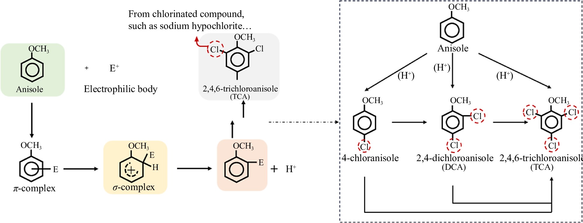
    • According to the available studies, there are two main pathways for the formation of 2,4,6-TCA[9,10]. One way is the chlorination of anisole, a natural organic compound. The other is the generation of 2,4,6-TCA from the precursor substance 2,4,6-trichlorophenol (2,4,6-TCP) by microbial O-methylation. This is a typical electrophilic substitution reaction about the generation of 2,4,6-TCA from the substitution reaction of chlorine with anisole. The reaction has three main steps (Fig. 1): (1) the electrophilic body (E) attacks the benzene ring to form the π-complex and retains the benzene ring structure; (2) the electrophilic body in the π-complex attaches to a carbon atom on the benzene ring and becomes the σ-complex; and (3) a hydrogen atom bound to the benzene ring detaches and produces H+[11]. Thus, when chlorinated reagents were used, under the right conditions chlorine atoms will replace the hydrogen atoms in the benzene ring of anisole to form 2,4,6-TCA. Zhang et al. has shown that the pH determines whether the reaction occurs or not. Only under acidic and weakly acidic conditions, the substitution reaction of anisole with chlorine took place[12]. The pH of wine is usually at 3.0−4.0, and the grapes themselves are also acidic, so possessing the prerequisites for the anisole-chlorination reaction.

      Figure 1. 

      Pathway of electrophilic substitution of anisole produces 2,4,6-TCA.

    • The microbial pathway for 2,4,6-TCA formation is the microbial transfer of the donor methyl group to the hydroxyl group of 2,4,6-TCP using chlorophenol O-methyltransferases (CPOMTs), which is similar to the bimolecular nucleophilic substitution reaction (SN2), in which a nucleophilic reagent attacks the substrate, provides an electron pair to the new bond, and replaces the leaving group[9,13] (Fig. 2). 2,4,6-TCP, a precursor substance in the microbial pathway formed by 2,4,6-TCA, is recognized as one of the major environmental pollutants by the US Environmental Protection Agency (USEPA). Because 2,4,6-TCP is commonly used as pesticides, herbicides, fungicides, insecticides, and disinfectants, but it is chemically stable so that it is hard to degrade, so we can often detect it in surface water, soil, and atmosphere[14,15]. The International Agency for Research on Cancer (IARC) has classified 2,4,6-TCP as a B2 carcinogen, because studies have shown that 2,4,6-TCP has significant pathological effects and potential carcinogenicity[16,17]. It has been reported that 2,4,6-TCP can affect the human nervous system and respiratory system causing diseases, such as cough and chronic bronchitis[18]. Therefore, the conversion of 2,4,6-TCP to 2,4,6-TCA by microbial action is a common biological mechanism of toxicity reduction.

      Figure 2. 

      Pathway of 2,4, 6-trichlorophenol biosynthesis catalyzed by O-methyltransferase.

      Regarding CPOMTs in the microbial pathway, in terms of their methyl donors, they can be classified as S-adenosyl methionine (SAM)-dependent and non-SAM-dependent. SAM-dependent means that it only can use S-adenosyl methionine as a methyl donor, while non-SAM-dependent means that it can use a wide range of methyl donors, such as methanol, methylamine and methionine. Grapes and wine are rich in chemicals so that they can provide a rich source of methyl for the synthesis of 2,4,6-TCA.

      As for the microorganisms in the microbial synthesis pathway of 2,4,6-TCA, they were mainly isolated from cork and water, because the problem of 2,4,6-TCA is mainly focused on wine cork and water. The microorganisms involved in the microbial pathway of 2,4,6-TCA formation, include bacteria, fungi, cyanobacteria and algae[10,13,19,20]. Currently the cork is still considered to be the main reason causing cork taint. Thus, studies focus on analyzing the information of fungal flora in cork and found that it is mainly composed of Penicillium spp., Aspergillus spp., Chrysonilia sitophila, Mucor racemosus, Paecilomyces spp., Trichoderma spp., Cladosporium spp., Fusarium spp., Acremonium spp., Monilia spp., Rhizoctonia spp., Mortierella spp., and Verticillium spp.[19,21,22]. However some studies revealed that Penicillium spp. (such as Penicillium chrysogenum, Penicillium glabrum), Aspergillus spp. (such as Aspergillus niger, Aspergillus oryzae), Chrysonilia sitophila, Fusarium spp., Mucor racemosus, Paecilomyces spp., and Trichoderma spp. are the main fungi, which can produce 2,4,6-TCA[19,21]. Among them, the transformation efficiency of Fusarium spp. and Trichoderma spp. strains was higher[21]. For example, one researcher isolated a SAM-dependent CPOMT from Trichoderma longibrachiatum, which can catalyze the O-methylation of several chlorophenols, including 2,4,6-TCP[23]. And study showed that its conversion efficiency was up to 37.56%. Recent research suggested that taste and odor in drinking water are mainly caused by 2,4,6-TCA. It was found that 2,4,6-TCA in water is mainly produced due to O-methylation by microorganisms. The microbial species in water are more abundant. The fungi that were found to be able to convert 2,4,6-TCP to 2,4,6-TCA were mainly dominated by Phialophora spp., Acremonium spp., and Penicillium spp.[24,25]. In addition, some bacteria, such as Gram-positive Rhodococcus spp. and Gram-negative Acinetobacter spp., can convert 2,4,6-TCP to 2,4,6-TCA[26]. Moreover, it was also found that two common cyanobacteria and algae, such as Chlorella vulgaris and Anabaena flos-aquae, can convert 2,4,6-TCP to 2,4,6-TCA[13]. From available literature, we know that the 2,4,6-TCA production capacity was significantly different between the different strains. Table 1 summarizes the current strains isolated from cork and water with the ability to convert 2,4,6-TCP to 2,4,6-TCA.

      Table 1.  Microorganisms associated with 2,4,6-TCA production.

      GenusSpeciesIsolated fromThe rate/ability of converting
      TCP to TCA
      Detection methodsRef.
      FungiAcremoniumStrictumSettled water3.3%−14.24%SPME-GC-MS[27]
      FungiAspergillusRaw one-piece cork stoppersIn MEA medium, 44.5%−54.9%
      On the cork, 19.9%−21.5%
      GC-ECD[20]
      FungiAspergillusNigerCork stoppersOn solid cork medium, 0.16%
      On liquid medium, 0.65%.
      HS-SPME-GC-MS[19]
      FungiAspergillusOryzaeTap water; StoppersOn solid cork medium, 0.21%;
      On liquid medium, 1.17%
      SPME-GC-MS;
      HS-SPME-GC-MS
      [19,27,28]
      FungiAspergillusVersicolorSettled water40.5%SPME-GC-MS[27]
      FungiBjerkanderaAdustaFinished water2.0 × 10−5%−0.18%SPME-GC-MS[27]
      FungiBotrytisCinereaGrapesIn MEA medium, 34.1%;
      On wood plugs, 28.4%
      GC-ECD[20]
      FungiChrysoniliaSitophilaRaw one-piece cork stoppersIn MEA medium, 64.6%;
      On wood plugs, 4.3%
      GC-ECD[20]
      FungiCladosporiumCladosporioidesFinished water7.0 × 10−2%SPME-GC-MS[27]
      FungiCladosporiumOxysporumCork stoppers14.31%HPLC[21]
      FungiCyclotellaHebeianaLakeInitially 0.2 mg/L 2,4,6-TCP; eventually 4.08 ng/L 2,4,6-TCA can be producedSPME-GC-MS[13]
      FungiFusariumAsiaticumFinished water0.28%SPME-GC-MS[27]
      FungiFusicollaMatuoiFinished water2.6%SPME-GC-MS[27]
      FungiFusariumOxysporumCork stoppers28.65%HPLC[21]
      FungiLaccariaAmethystinaRaw water;
      Settled water;
      Post filtration water; Finished water
      2.9 × 10⁻²%SPME-GC-MS[27]
      FungiMortierellaAlpinaCork stoppers0.11%HPLC[21]
      FungiMucorPlumbeusCork stoppers0.03%HPLC[21]
      FungiMucorRacemosusCork stoppersOn solid cork medium, 5.21%;
      On liquid medium, 5.21%
      HS- SPME-GC-MS[19]
      FungiPaecilomycesVariotiiFibreboard cartons2%−65%HPLC; GC-MS[29]
      FungiPaecilomycesViridisCork stoppers7.88%HPLC[21]
      FungiPaecilomycesCork stoppersOn solid cork medium, 3.65%;
      On liquid medium, 4.45%
      HS- SPME-GC-MS[19]
      FungiPenicilliumRaw one-piece cork stoppersIn MEA medium, 23.2-37%;
      On cork, 1.3%−53.1%
      GC-ECD[20]
      FungiPenicilliumChrysogenumCork stoppers3.29%−7.87%HS- SPME-GC-MS[19]
      FungiPenicilliumCitreonigrumCork stoppers13.28%HPLC[21]
      FungiPenicilliumDecumbensCork stoppers0.11%HPLC[21]
      FungiPenicilliumGlabrumCork stoppers2.18%−20.43%HS- SPME-GC-MS[19]
      FungiPenicilliumPurpurogenumCork stoppers11.02%HPLC[21]
      FungiPhialemoniopsisOcularisPost filtration water0.13%SPME-GC-MS[27]
      FungiPseudomonasLakeSPME-GC-MS[13]
      FungiRhizopusOryzaeBroiler house litter< 1%Gas-Liquid Chromatography[30]
      FungiScopulariopsisBrevicaulisBroiler house litter60%Gas-Liquid Chromatography[30]
      FungiSistotremaBrinkmanniiPost filtration water; Finished water2.3%SPME-GC-MS[27]
      FungiTalaromycesPinophilusFinished water2.7%SPME-GC-MS[27]
      FungiTrichodermaRaw one-piece cork stoppers; LakeIn MEA medium, 64.4%;
      On cork, 13%
      GC-ECD; SPME-GC-MS[13,20]
      FungiTrichodermaLongibrachiatumCork37.56%[31]
      FungiTrichodermaVirideCork stoppers3.37%−4.86%HS- SPME-GC-MS[19]
      FungiVerticilliumPsalliotaeCork stoppers6.9%HPLC[21]
      BacteriaAcinetobacterWater2.4 × 10−10 ug*h (cell/mL)GC-MS[32]
      BacteriaBacillusAustralimarisWaterOMPPC (1.31 × 10⁻⁹ ng/CFU)SPME-GC-ECD[33]
      BacteriaBrachybacteriumBrachybacteriumCorkHS-SPME-GC-MS[32]
      BacteriaBrachybacteriumParaconglomeratumCorkHS-SPME-GC-MS[32]
      BacteriaBradyrhizobiumFrederickiiWaterproduce 2,4,6-TCA
      (1.7 × 10⁻⁹ ng/CFU)
      SPME-GC-ECD[33]
      BacteriaBrevundimonasWaterSPME-GC-ECD[33]
      BacteriaCaulobacterWaterSPME-GC-ECD[33]
      BacteriaChromobacteriumWaterSPME-GC-ECD[33]
      BacteriaErythrobacterWaterSPME-GC-ECD[33]
      BacteriaEscherichiaColiLakeInitial 0.2 mg/L 2,4,6-TCP;
      generated 4.6 ng/L 2,4,6-TCA
      SPME-GC-MS[13]
      BacteriaFlavobacteriumCorkHS-SPME-GC-MS[32]
      BacteriaMicrobacteriumOxydansCorkHS-SPME-GC-MS[32]
      BacteriaPaenibacillusWaterSPME-GC-ECD[33]
      BacteriaPelomonasWaterSPME-GC-ECD[33]
      BacteriaRalstoniaMannitolilyticaWaterSPME-GC-ECD[33]
      BacteriaRhodoccoccusAcinetobacterLakeSPME-GC-MS[13]
      BacteriaRhodococcusWater5.5 × 10−8 ug*h (cell/mL)GC-MS[34]
      BacteriaXanthobacterWaterSPME-GC-ECD[33]
      CyanobacteriaChlorellaVulgarisLakeInitial 0.2 mg/L 2,4,6-TCP;
      generated 30.5 ng/L 2,4,6-TCA
      SPME-GC-MS[13]
      AlgaeAnabaenaFlos-aquaeLakeInitially 0.2 mg/L 2,4,6-TCP;
      generated 10.2 ng/L 2,4,6-TCA
      SPME-GC-MS[13]
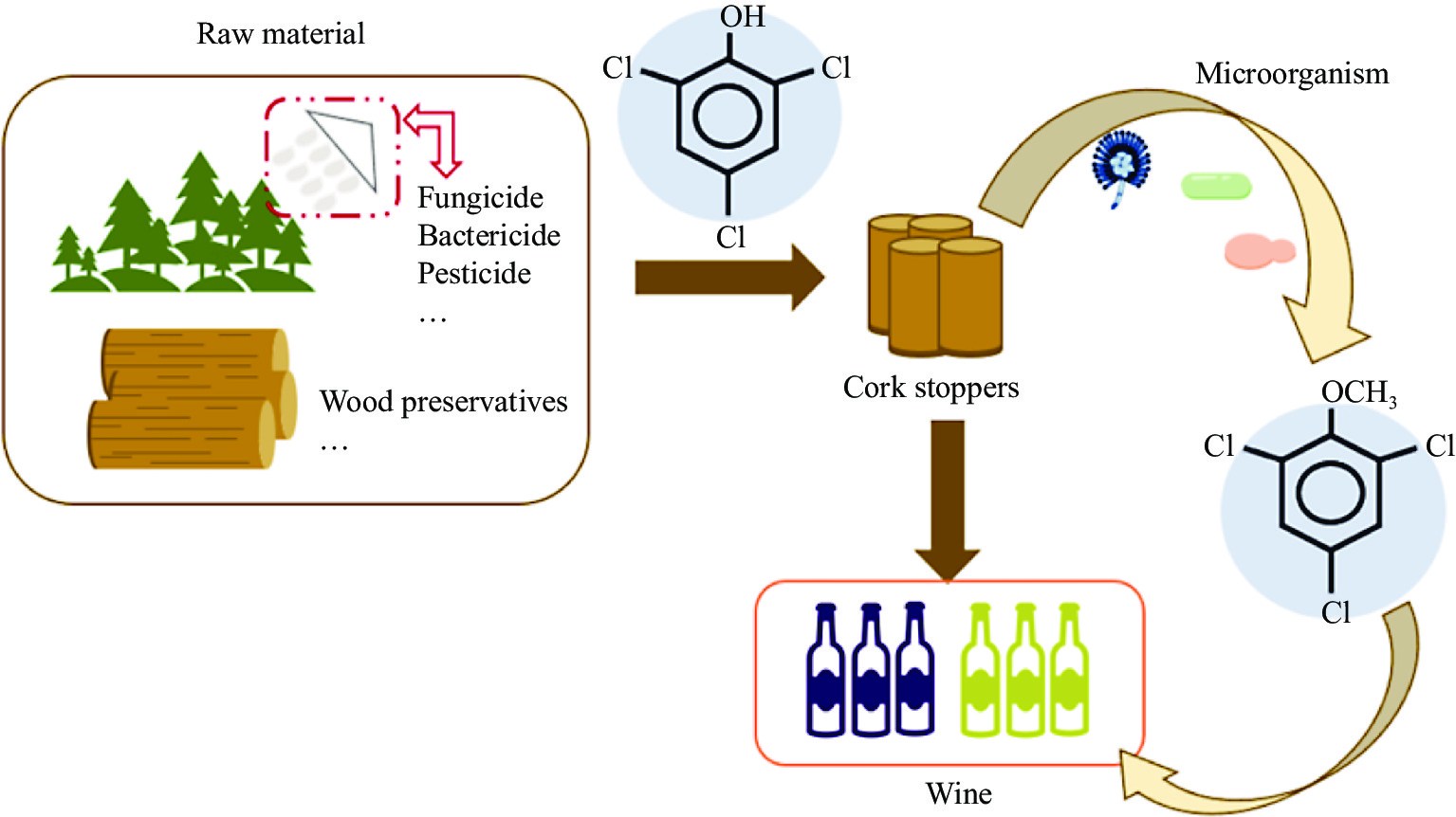
    • In the early 1980s, the source of cork taint was identified as microbial contamination of cork, and the residual chlorophenol compounds from pesticides during oak growth and wood preservatives, which in turn produce 2,4,6-TCA. When the cork comes into contact with the wine, 2,4,6-TCA can be transferred from the cork to the wine (Fig. 3). As we all know, cork is mainly made from the bark of cork oak, due to its good elasticity, it can play a good role in sealing the mouth of the bottle. Moreover, there are tiny spaces between the cork cells, which cannot completely isolate the air, so facilitating the slow development and maturation of the wine in the bottle again. Besides, when the cork is in direct contact with the wine, some components in the cork can be transferred to the wine, such as phenolic compounds, tannins, and ketones[35,36]. According to the statistical report of the International Organization of Vine and Wine (OIV), 70% of the world's total wine production of bottled wine is sealed with cork[37]. Therefore, cork is currently considered the main culprit of 2,4,6-TCA taint in wine. Monteiro et al. showed that cross-contamination of cork can occur through both the liquid and gas phases, i.e., a cork contaminated with 2,4,6-TCA is partially contaminated with a clean cork immersed in either pure water or an alcohol solution. And the contaminated cork with clean storage for some time, the clean cork will likewise be partially contaminated[38]. Therefore, methods that can quickly and non-destructively detect whether a cork is contaminated with 2,4,6-TCA are much needed. Moreover, 2,4,6-TCA contamination can be transmitted through the gas phase, suggesting that 2,4,6-TCA can contaminate wine through the air from other woods, such as oak barrels, cellar beams, and wood chips.

      Figure 3. 

      2,4,6-TCA originates from cork stoppers.

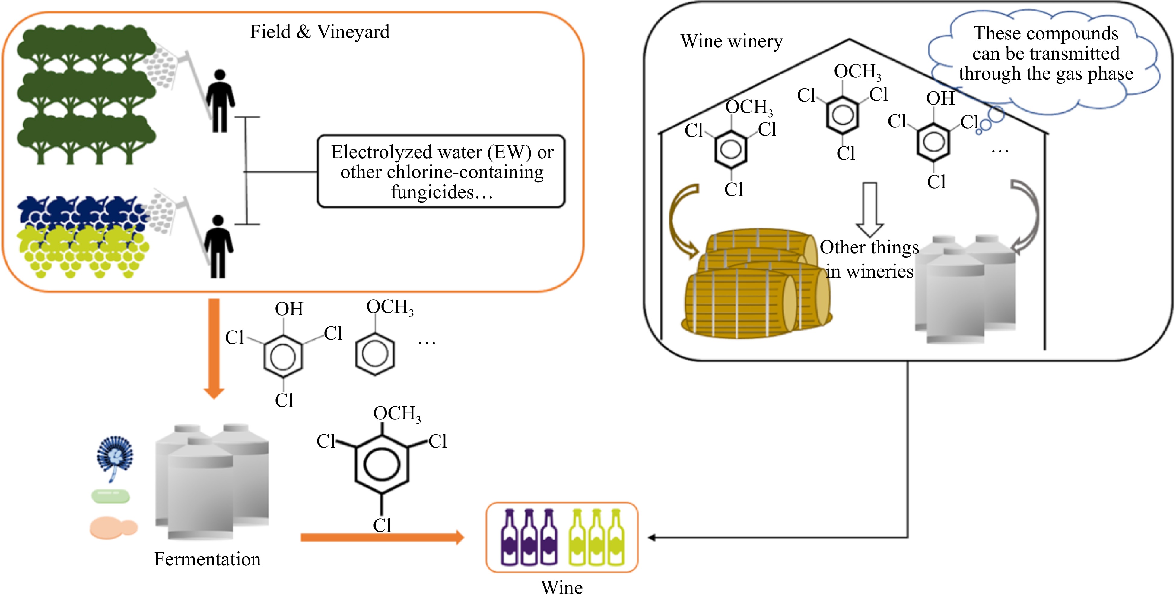
    • The odor problem of drinking water is also often complained about by consumers, studies have found that the substances causing these odors are mainly GSM, 2-MIB, and 2,4,6-TCA. The formation of 2,4,6-TCA in water is mainly through microbial O-methylation, because the chlorination of anisole occurs only under acidic conditions, while the pH of drinking water usually does not present acidity. Therefore, even if 2,4,6-TCA was effectively removed from the source water, 2,4,6-TCA would still be generated in drinking water[9,10,12]. In addition to SAM, other methyl donors, including methanol and methylamines are present in water in the form of natural organic matter[9]. In the drinking water distribution and delivery system, microorganisms tend to grow in the pipes, so it is easier to generate 2,4,6-TCA. Water is usually used in the winemaking process, so it may also be a source of 2,4,6-TCA in wine (Fig. 4).

      Figure 4. 

      2,4,6-TCA originates from water.

    • Recently, electrolyzed water (EW) has attracted a lot of attention as a new high-performance technology for potential applications in the food industry. EW has a certain disinfection effect on microorganisms and is extremely promising as a bactericidal agent with a wide range of disinfection effects and eco-friendliness[39,40]. Some studies have shown the bactericidal potential of EW against wine spoilage yeasts, e.g., Brettanomyces spp.[41]. However, some studies have found that both pre-harvest and post-harvest applications of EW increase the concentration of 2,4,6-TCA in wine[42,43]. Previous research explained that EW application leads to chlorine residues on the grape surface, which in turn produce 2,4,6-TCA in response to microbial action. This suggests that the use of chlorinated fungicides during grape growing is also a source of 2,4,6-TCA in wine. Therefore, we need to use fungicides properly.

      Monteiro et al. suggested that 2,4,6-TCA contamination can be transmitted through the gas phase[38]. Once 2,4,6-TCA forms in wooden materials inside the cellar or winery, it can migrate into the air and contaminate winery equipment and oenological materials. Finally, it can lead to cork taint (Fig. 5). Some researchers simulated air contamination (initial d5-TCA concentration was 50 ng/L of air) and stored the sealed wine for 6 to 24 months[44]. They found that these wines were at risk of contamination. Another study also reported that sparkling wine sealed with crown caps was contaminated by airborne tetrachloroanisole after 14 months of storage.

      Figure 5. 

      2,4,6-TCA originates from others.

    • The detection of 2,4,6-TCA is challenging due to its concentration in wine, which is usually at the ng/L level, and the complexity of matrices such as wine and cork. To address this challenge, researchers have developed chromatography-based or bioanalytical techniques for the detection of 2,4,6-TCA in various matrices such as cork, wine, and water. Usually, gas chromatography-mass spectrometry (GC-MS) and gas chromatography-electronic capture detector (GC-ECD) are used to detect 2,4,6-TCA concentration.

      GC-MS is a common detection tool in the field of wine research and can be used for qualitative analysis as well as quantitative analysis. GC-MS can achieve high separation efficiency for the identification and quantification of aroma substances, such as haloanisoles, esters, and terpenes[45]. Tarasov et al. determined 2,4,6-TCA content in wine by GC-MS combined with solid phase microextraction (SPME), and its limits of detection (LODs) can achieve 0.4 ng/L[44]. Wines contain a variety of substances, so the detected sample matrix is complex. Thus, the background noise of the detection using GC-MS is large. Some researchers use GC-MS/MS, which is highly selective and sensitive to compounds compared to GC-MS, and it can better detect multiple substances with similar structures simultaneously. Zhang et al. used GC-MS/MS coupled with headspace solid phase microextraction (HS-SPME) to determine nine multihalo-anisoles (such as 2,3,4,5-TeCA, 2,3,4,6- TeCA, PeCA, TBA, 2,4,6-TCA) and multihalo-phenols (such as PeCP, TBP, TCP, TeCP) in wine, and its LODs can achieve within 3.0 ng/L[46]. Ruiz-Delgado et al. also determined cork contaminants in wine by GC-MS/MS combined with HS-SPME, and the LODs for 2,4,6-TCA, 2,3,4,6-TeCA, 2,4,6-TBA, and PCA in wine were less than 0.3 ng/L[47].

      ECD is an ion detector which is highly sensitive to compounds containing electronegative elements. Chloroanisole and its precursor chlorophenols contain multiple chlorine atoms, which are electronegative, so the ECD was chosen to detect them with good selectivity and high sensitivity. Özhan et al. assayed the levels of 2,4-dichloroanisole (DCA), 2,4,6-TCA, 2,3,4,6-TeCA, PCA, 2,4,6-TCP, 2,3,4,6-TeCP, PCP in red wine from different wineries in Turkey using HS-SPME and GC-ECD detection, and the LODs were less than 1.0 ng/L[48]. In addition, compared to GC-MS/GC-MS/MS, GC-ECD has a low purchase price and maintenance cost. However, ECD is mainly suitable for halogenated cork-taint compounds.

      Meanwhile, a large number of studies related to the separation and identification of odor-active compounds in food by gas chromatography-olfactometry (GC-O) have been carried out. Some studies screened and identified the odor-active compounds in ice wines by GC-O combined with comprehensive two-dimensional GC and time-of-flight mass spectrometry (GC×GC-TOFMS), and it can identify more than 200 volatile compounds. Although there is no study about using GC-O combined with MS for detecting 2,4,6-TCA, it is a good detect method, because it not only evaluation of the odor compounds but also identification with MS information.

      Ion Mobility Spectrometry (IMS) is an analytical technique for characterizing molecules by gas phase mobility, which has the advantage of rapid detection, high sensitivity, and the ability to avoid interference from other compounds present in the matrix and is often used for the detection of various explosives, drugs and narcotics[49,50]. In addition, ion mobility spectrometers are relatively inexpensive and can provide spectra in the millisecond range. These advantages make IMS suitable for the detection of volatile or semi-volatile compounds in different matrices. But its selectivity is limited, extraction and preconcentration of 2,4,6-TCA from wine samples is necessary. Because there are interfering substances in wine samples, mainly ethanol, which can overlap with the signal of 2,4,6-TCA. Thus, Márquez-Sillero et al. firstly used solid-phase extraction to remove ethanol. They then combined the use of ionic-based single drop microextraction (ILSDME) and IMS for the determination of 2,4,6-TCA in water and wine samples. This method LOD is 0.2 ng/L[51]. The next year, they developed a new method based on IMS that the interference of ethanol was negligible. They analyzed 2,4,6-TCA in wine and cork samples by headspace-multicapillary column-ion mobility spectrometry (HS-MCC-IMS), and the detection limit of wine is 0.012 ng/L, the detection limit of cork is 0.28 ng/L[52]. It greatly improves the sensitivity of the detection method.

      As mentioned above, 2,4,6-TCA can also cross-contaminate through the gas phase, which means 2,4,6-TCA may also be present in the ambient air of the winery. Thus, it is important to detect 2,4,6-TCA in the environment early in the winemaking process to prevent cork taint of wine. Therefore, a method based on thermal desorption coupled to GC-MS (TD-GCMS) was proposed for the determination of low concentrations of the target compounds in the air, using a porous polymer resin based on 2,6-diphenylene oxide as an adsorbent instead of bentonite, which was used in the past to capture target compounds in the air[53].

    • However, GC-MS, GC-MS/MS, or GC-ECD needs a previous step of sample preparation, which usually is destructive and time-consuming, and sometimes requires using organic solvents. Among the available techniques, the electronic nose stands out. The electronic nose (Enose), also known as an odor scanner is a novel instrument developed in the 1990s for rapid food testing. It is an instrument consisting of a set of chemical gas sensors with partial specificity and an appropriate pattern recognition system, capable of recognizing simple or complex odors[54]. Santos et al. investigated the feasibility of a small wireless portable nose (WiNOSE 6), composed of non-specific cross-sensitivity sensors, capable of measuring up to eight microsensors to detect typical and atypical odor compounds in natural cork[55]. Corks were introduced in a 50 mL vial with two holes at the top, one for atmospheric air and the other connected to a nasal cannula. Each measurement cycle consisted of a 9-min desorption phase and a 1-min adsorption phase. And results showed close to 100% identification of defects such as MDMP, TCA, and 1-octene-3-one. Melendez et al. also present a prototype of a novel Enose that uses an array of digital and analog metal oxide gas sensors with a total of 31 signals capable of detecting 2,4,6-TCA and classifying cork samples with low 2,4,6-TCA concentrations (15.1 ng/L)[56]. The Enose can provide a non-destructive, faster, and cheaper method of analysis compared to GC methods. However, the sensitivity of the Enose could be improved, as people have a sensory threshold of 2-10 ng/L for 2,4,6-TCA in wine.

    • Since GC methods often require pretreatment of the sample to be measured, and the instruments are also more expensive and sophisticated, often requiring specialized personnel to operate. Peres et al. quantified 2,4,6-TCA in cork plates using cyclic voltammetry (CV), a commonly used electrochemical research method to study the nature, mechanism, and kinetic parameters of electrode reactions, and also for quantitative determination of reactant concentrations[57]. It works by applying a pulsed voltage in the form of an isosceles triangle to the working electrode and controlling the electrode potential at different rates with one or more repeated scans of the triangular waveform over time to obtain a current-potential polarization curve. Sanvicens et al. used a portable Potentiostat-Galvanostat device (PG580, Uniscan) together with a silver working electrode (M295Ag, Radiometer), a platinum counter electrode (M241Pt, Radiometer) and an Ag/AgCl double-junction reference electrode (M90-02, Orion) for measuring the current of sample collected from the cork plank boiling process[57]. In addition, CV devices are portable, fast, and low-cost, and do not require specialized technicians, so making them promising for field industrial applications.

    • To improve the sample quantity or test cycle, speed and reduce cost, some emerging rapid assays have been proposed, such as enzyme-linked immunosorbent assays (ELISAs)[58] and immunoamperometric assays[59]. ELISAs are an immunoassay method that combines the high specificity of antigen-antibody reaction with the high efficiency of enzyme catalysis, mainly based on the ability of antigens or antibodies to adsorb onto the surface of the solid-phase carrier and maintain its immunological activity, and then use the specific binding of antigens and antibodies for the qualitative and quantitative detection of immunological reactions. Lausterer et al. prepared antibodies specific for TCA by fused cells and used the Rami kit for ELISA detection of signal amplification up to 10 ng/L[60]. They synthesized haptens B (5-(2,4,6-trichlorophenoxy)pentanoic acid) and C (3-(3,5-dichloro-4-methoxyphenyl)propanoic acid) by chemical methods and conjugated with bovine serum albumin and keyhole limpet hemocyanine by the active ester method, respectively. Then an immune response is induced by injecting these synthesized compounds into immunized animals, such as mice. Subsequently, lymphocytes were collected from immune animals and fused with myeloma cells to form hybridoma cells. Then hybridoma cells with 2,4,6-TCA selectivity were screened by immunization and fusion. Finally, two different cell lines (Rami and Hbab) were selected from the selected hybridoma cells, cloned, and stored at low temperatures. However, ELISAs methods usually require preparative steps such as extraction and concentration, which increase the analysis time. Therefore, electrochemical immunosensing technique was developed for 2,4,6-TCA detection. This technique can avoid sample interferences. Apostolou et al. based the team on a previous bioelectric recognition assay (BERA) biosensor system, which is based on the determination of the electrical response of cultured membrane-engineered fibroblasts suspended in an alginate gel matrix[61]. High-throughput screening of TCA in cork was achieved by osmotically inserting a specific TCA antibody (pAb78) into the cork. This new method can detect very low concentrations of 2,4,6-TCA (down to 0.2 ng/L) in just 5 min. This new biosensor offers several practical advantages, including a significant reduction in total assay time and the ability to perform high-throughput screening directly in the field and production facilities without the need for any support infrastructure. However, this method does not provide reliable quantitative results and only detects a small fraction of the concentration in the sample due to the extremely low solubility of 2,4,6-TCA in water[61].

    • Recently, Romano et al. tested a new method for the determination of 2,4,6-TCA in cork based on chemical ionization time-of-flight mass spectrometry (CI-TOF) using a 'Vocus' ion source and an ion-molecule reactor (IMR), which allowed a rapid and highly sensitive detection of 2,4,6-TCA in coffee beans within 3 s[6]. And they suggested that the method is also feasible for other food products. Cappellin et al. simulated a real industrial scenario and determined the 2,4,6-TCA content of 10,100 natural cork batches in three different batches in just 8 hours and 25 min, which is equal to 3 s per cork[62]. This method far exceeds existing analytical methods in terms of speed and has approximately the same detection limits as other assays. Therefore, this new non-destructive, rapid, and sensitive detection technique has the potential to be a breakthrough for the cork and wine industry. Based on the article, it can be hypothesized that the technique can detect other pollutants. However it is not possible to determine the applicability of the technique for the simultaneous detection of multiple contaminants.

      Damiano et al. developed a method based on Ni(0) complexes to detect 2,4,6-TCA in cork indirectly by UV-Vis spectroscopy, since aryl chlorides can effectively participate in the oxidative addition reaction with phosphorylated Ni(0)(BINAP) (η2-PhCN), forming very active Ni(II) complex, and the complex-forming complex Ni(II) has a characteristic UV absorption band at 444 nm, so the 2,4,6-TCA concentration in cork can be quantified indirectly by UV-visible absorption spectroscopy[63]. Compared to biosensors, the advantages of such chemical sensors include fast response and portability, allowing for rapid testing in the field without need for complex laboratory equipment. In addition, these sensors typically have a low cost and are suitable for small producers. However, limitations of these sensors include sensitivity to environmental conditions such as pH, incubation time, and interfering substances from cork. In addition, the selectivity and sensitivity of these sensors may be limited and require further optimization and validation. All in all, although the Ni(0) complex is sensitive to oxygen in this method, it provides an idea for the future development of an inexpensive chemical sensor suitable for 2,4,6-TCA quantification.

    • The key to the analysis of 2,4,6-TCA in wine is sample preparation with pre-enrichment or extraction in advance. The common pretreatment methods reported in domestic and international research includes head space solid microextraction (HS-SPME)[64], stir bar sorptive extraction (SBSE)[65], dispersive liquid-liquid microextraction (DLLME)[66,67], supercritical fluid extraction (SFE)[68], accelerated solvent extraction (ASE), and pressurized fluid extraction (PFE)[69]. These different extraction techniques combined with GC-MS and other detection techniques have been successfully used to identify 2,4,6-TCA in wine, cork, or water[46].

    • SPME is the most commonly reported technique for sample extraction or pre-enrichment. Reported methods based on SPME combined with different instruments for the detection of typical odorants are summarized in Table 2. The SPME method uses a fibrous membrane coated with an extraction phase (liquid polymer or solid adsorbent) to extract different types of analytes (volatile or non-volatile substances) from various media (liquid or gas phase)[70]. SPME methods are easy to perform and can greatly reduce environmental contamination by the use of organic solvents, and can make the LOD of the method as low as ng/L. SPME methods can be carried out either by direct immersion in liquid samples (DI-SPME) or the more commonly used headspace method (HS-SPME). Jové et al. used HS-SPME and GC–MS/MS to detect 2,4,6-TCA, 2,3,4,6-TeCA, 2,4,6-TBA, and PCA in cork stoppers. Results showed that the divinylbenzene/carboxenpolydimethylsiloxane/polydimethylsiloxane (DVB/CAR/PDMS) fibers could detect haloanisoles with the LODs at 0.01–0.50 ng/L[3]. However, we need to further develop and optimize the procedure for different situations such as different solvent types to improve the extraction efficiency and accuracy of the results.

      Table 2.  Analysis methodology regarding 2,4,6-TCA.

      Microextraction methodologiesDetection methodologiesLODLOQAnalysis time per sampleRef.
      InstrumentSample typeAnalytesFiber typeExtraction conditionInstrumentColumn typeGC conditionMS conditionInternal standards
      HS-SPMEWined5-TCAPDMS,
      100 μm
      Incubation
      temperature: 55 °C
      Incubation time: 3 min
      Sample extraction time: 11 min
      Sample desorb time:
      4 min
      QP-2010 Plus GC-MS (Shimadzu, Kyoto, Japan)RTX-5MSGas flow 1.61 mL/min

      Oven program:
      90 °C for 0 min,
      10 °C/min to 205 °C, and then 30 °C/min to 280 °C
      SIMd5-TBA0.4 ng/L1 ng/L32 min[44]
      HS-SPMEWineTCA, TCP, 2,3,4,6-TeCA, TBA, 2,3,4,6-TeCP, 2,3,4,5-TeCA, PeCA, TBP, PeCPDVB/CAR/
      PDMS,
      50/30 µm
      Incubation
      temperature: 60 °C
      Incubation time: 5 min
      Sample extraction time: 45 min
      Sample desorb time:
      5 min
      Agilent 7890A
      GC-7000B triple quadrupole MS
      HP-5Flow rate: 1.18 mL/min

      Oven program:
      50 °C for 1 min,
      10 °C/min to 200 °C, and then 40 °C/min to 280 °C hold for 3 min
      MS/
      MS-MRM
      TCA-d5Haloanisoles:
      3 ng/L

      Halophenols:
      10 ng/L
      Haloanisoles:
      10 ng/L

      Halophenols: 30−100 ng/L
      76 min[46]
      HS-SPMEWineTCA, TeCA, TBA, PCAPDMS,
      100 μm
      Incubation
      temperature: 40 °C
      Incubation time: 5 min
      Sample extraction time: 30 min
      Sample desorb time:
      15 min
      ScionGC system (Bruker Corporation, Freemont, CA, USA) × Scion QqQ-MS/MS instrument (Bruker)VF-5msFlow rate: 1 mL/min

      Oven program:
      90 °C for 5 min,
      30 °C/min to 280 °C hold for 7 min
      SRM4-iodoanisoleTCA: 0.1 ng/L

      TeCA: 0.2 ng/L

      TBA: 0.3 ng/L

      PCA: 0.1 ng/L
      TCA: 0.4 ng/L

      TeCA: 0.6 ng/L

      TBA: 0.9 ng/L

      PCA: 0.3 ng/L
      68.33 min[47]
      HS-SPMECiderTCA, TeCA, TBA, PCAPDMS,
      100 μm
      Incubation
      temperature: 40 °C
      Incubation time: 5 min
      Sample extraction time: 30 min
      Sample desorb time:
      15 min
      ScionGC system (Bruker Corporation, Freemont, CA, USA) × Scion QqQ-MS/MS instrument (Bruker)VF-5msFlow rate: 1 mL/min

      Oven program:
      90 °C for 5 min,
      30 °C/min to 280 °C hold for 7 min
      SRM4-iodoanisoleTCA: 0.2 ng/L

      TeCA: 0.2 ng/L

      TBA: 0.3 ng/L

      PCA: 0.1 ng/L
      TCA: 0.5 ng/L

      TeCA: 0.7 ng/L
      TBA: 1.1 ng/L

      PCA: 0.5 ng/L
      68.33 min[47]
      HS-SPMECavaTCA, TeCA, TBA, PCAPDMS,
      100 μm
      Incubation
      temperature: 40 °C
      Incubation time: 5 min
      Sample extraction time: 30 min
      Sample desorb time:
      15 min
      ScionGC system (Bruker Corporation, Freemont, CA, USA) × Scion QqQ-MS/MS instrument (Bruker)VF-5msFlow rate: 1 mL/min

      Oven program:
      90 °C for 5 min,
      30 °C/min to 280 °C hold for 7 min
      SRM4-iodoanisoleTCA: 0.1 ng/L
      TeCA: 0.2 ng/L

      TBA: 0.4 ng/L

      PCA: 0.2 ng/L
      TCA: 0.4 ng/L
      TeCA: 0.6 ng/L

      TBA: 1.3 ng/L

      PCA: 0.7 ng/L
      68.33 min[47]
      HS-SPMEWaterTCA, TCPPDMS,
      100 μm
      GC-2010/parvum 2, Shimadzu, Kyoto, Japan5MS/SilFlow rate: 42 cm/s

      Oven program:
      40 °C for 3 min,
      10 °C/min to 80 °C, and then 15 °C/min to
      250 °C hold for 3 min
      SIM> 21.3 min[74]
      HS-SPMEWater2-CP, 2-BP, 2,4-DCP, 2,4,6-TCP, 2,4-DBP, 2,4,6-TBP, 2,4,6-TCA, 2,4,6-TBAPDMS/DVB,
      65 μm
      Incubation
      temperature: 60 °C
      Incubation time: 10 min
      Sample extraction time: 30 min
      Sample desorb time:
      3 min
      Agilent 7890 GC × Agilent 5975 MSHP-5MSFlow rate: 1 mL/min

      Oven program:
      40 °C for 3 min,
      15 °C/min to 235 °C hold for 1 min
      SIM4-iodoanisoleHaloanisoles: 0.23−0.29 ng/L
      Halophenols: 0.24−0.91 ng/L
      Haloanisoles: 0.97−0.77 ng/L
      Halophenols: 0.80−3.30 ng/L
      60 min[75]
      HS-SPMEGarlicTCA, TBACWR/PDMS, 120 µmIncubation
      temperature: 80 °C
      Sample extraction
      time: 20 min
      Shimadzu GC-2010 × Shimadzu TQ8050Rxi-5 msFlow rate: 35 cm/s

      Oven program:
      70 °C for 1 min,
      10 °C/min to 300 °C hold for 3 min.
      MS/MS-MRMTCA-d5TCA:
      0.02 μg/kg

      TBA:
      0.03 μg/kg
      47 min[76]
      SPMEWater2,4,6-TCA, 2,3,6-TCA, 2,3,4-TCA, 2,4,6-TBAPDMS/DVB/
      CAB,
      50/30 µm
      Incubation temperature: 70 °C
      Incubation time: 10 min
      Sample extraction time: 30 min
      Sample desorb time:
      10 min
      GC-MSHP-17MSOven program:
      45 °C for 4 min,
      10 °C/min to 240 °C hold for 1 min,
      30 °C/min to 280 °C
      hold for 4 min
      SIM2,4,6-TCA:
      0.098 ng/L
      2,3,6-TCA:
      0.127 ng/L
      2,3,4-TCA:
      0.109 ng/L
      2,4,6-TBA:
      0.086 ng/L
      79.8 min[77]
      Vacuum-assisted HSSPMEWineTCA, TeCA, PCA, TBAPDMS/DVB,
      65 μm
      Incubation
      temperature: 25 °C
      Incubation time: 10 min
      Sample extraction time: 30 min
      Sample desorb time:
      15 min
      Shimadzu GC-17 A, GC-ECDDB-5MSFlow rate: 1 mL/min

      Oven program:
      90 °C for 5 min,
      20 °C/min to 280 °C hold for 5 min
      TCA 0.16 ng/L

      TeCA 0.18 ng/L

      PCA 0.19 ng/L

      TBA 0.13 ng/L
      74.5 min[78]
      LLEWineTCA, TeCA, PCA, TBA, TCP, TeCP, PCP, TBPAgilent HP 5980 GC × ECD (Agilent Technologies, USA)CP-Sil 5CBOven program:
      40 °C for 0 min,
      3 °C/min to 160 °C, and then 5 °C/min to
      220 °C hold for 10 min
      >
      38 min
      [79]
      Pressurized liquid extractionCorkMDMP, IPMP, IBMP, TCA, TCP, TeCA, TeCP, TBA, TBP, PCAAgilent 6890 N GC × Agilent 5973 N MSDB-5Flow rate: 1 mL/min

      Oven program:
      40 °C for 10 min,
      2 °C/min to 155 °C, and then 20 °C/min to
      260 °C hold for 9 min
      SIM2,3,6-trichloroanisole0.10 ng/g> 81.75 min[80]
      DLLMEWine2-CA, 4-CA, 2-BA, 2,6-DCA, 2-CP, 4-BA, 4-CP, 2-BP, 2,4-DCA, 4-BP, 2,6-DCP, 2,4,6-TCA, 3M4CP, 2,4-DCP, 2,4,6-TCP, 2,4-DBA, 2,3,4,6-TeCA, 2,4-DBP, 2,4,6-TBA, 2,3,4,6-TeCP, 2,3,4,5-TeCA, PCA, 2,4,6-TBP, PCPAgilent 6890 N GC × Agilent 5973 MSHP-5MSFlow rate: 1 mL/min

      Oven program:
      40 °C for 5 min,
      5 °C/min to 105 °C hold for 3.5 min,
      5 °C/min to 120 °C hold for 3 min,
      10 °C/min to 145 °C,
      and then
      5 °C/min to 185 °C,
      10 °C/min to 200 °C
      hold for 0.5 min
      SIM0.006–0.05 ng/mL40 min[81]
      CorkTCA, TeCA, TBA, PCAAgilent 6890N GC × Agilent 5973 MSHP-5MSFlow rate: 1 mL/min

      Oven program:
      80 °C for 0.6 min,
      25 °C/min to 180 °C hold for 0.6 min
      25 °C/min to 210 °C
      hold for 0.8 min,
      50 °C/min to 300 °C
      hold for 1.4 min
      SIM5-Bromo-2-chloroanisoleTCA: 1.6 ng/g

      TeCA: 2.6 ng/g

      TBA: 1.7 ng/g

      PCA: 2.5 ng/g
      TCA: 5.4 ng/g

      TeCA: 8.8 ng/g

      TBA: 5.7 ng/g

      PCA: 8.5 ng/g
      > 9.6 min[82]
    • SBSE is a method of extracting a target substance by stirring and contacting a sample solution using a stir bar with a specific fiber coating. After adsorption of the target substances in the sample onto the fiber coating, the fibers are fed into GC-MS for analytical determination. SBSE can extract a large amount of solution, and the extracted target substances can be immobilized by the adsorbent material in the stir bar for a sufficient period, which makes it convenient for on-site sampling and transportation[71]. Moreover, SBSE is very effective for trace components because the extraction phase is relatively large (about 5 μL for 10 mm) compared to that of SPME (about 65 μL for 100 μm)[72]. SBSE is also selective and can selectively adsorb target compounds, thereby reducing the effect of interfering substances. Marsol-Vall et al. used SBSE and heart-cutting two-dimensional gas chromatography to detect halophenols and haloanisoles in cork bark macerates. Results showed that the method gave LODs and LOQs ranging from 0.03 to 0.24 ng/L[72].

    • Liquid-liquid extraction is one of the most classical pretreatment methods and has been widely used for the analysis of various matrix parameters. DLLME has been developed since 2006, which has the characteristics of simple operation, high enrichment factor, and low consumption of organic solvents. In DLLME, an organic solvent (extraction solvent) is dispersed into an aqueous sample with the help of a co-solvent, the dispersant. The dispersion permits the formation of a large contact surface between the sample and the extractant, thus facilitating the extraction of analytes from the organic phase. Pizarro et al. used a method based on DLLME combined with GC-MS/MS technique to analyze compounds responsible for cork-taint off-flavors in wine[73]. Results showed that the method gave LODs and LOQs ranging from 5 to 41 ng/L. Despite the many advantages of DLLME methods, such as economy, simplicity, and rapidity, there are still some disadvantages and application limitations. The key to DLLME methods lies in the selection of the most suitable extraction and dispersion solvents. These two solvents greatly affect the sensitivity of the method. Secondly, since the DLLME method uses organic solvents for extraction, it may result in solvent residues in the extract. This may interfere with subsequent analytical results, especially in analyses with high sensitivity requirements.

    • The removal methods of 2,4,6-TCA generated in cork are one of the research hotspots in the field of wine safety and quality. However, few studies have been conducted on the methods for the elimination of 2,4,6-TCA in wine. With the development of analysis technology and control methods, research on the removal of wine odor substances has gradually developed. According to the source of 2,4,6-TCA, the control methods mainly include two ways. One is the prevention of the intrusion or formation of 2,4,6-TCA, and the other is the removal of 2,4,6-TCA. Some recommendations for reducing the risk of 2,4,6-TCA contamination in wine are shown in Fig. 6.
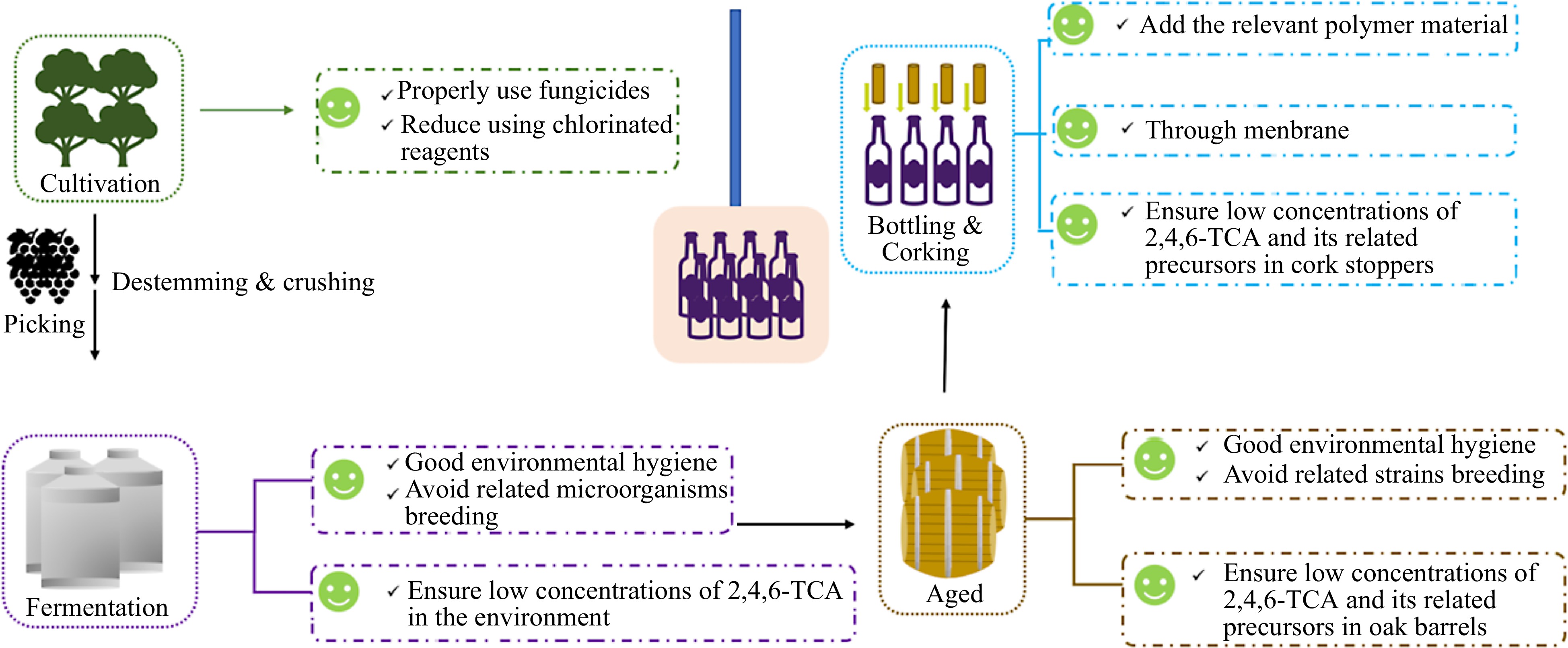
      Figure 6. 

      Possible ways in which 2,4,6-TCA can contaminate wine, and recommendations for reducing the risk of 2,4,6-TCA contamination.

    • Consumers often realize that cork taint has occurred in wine. Because cork taint is still mainly caused by the cork. Therefore, to reduce the loss of business, most research still focuses on removing the relevant compounds from cork-tainted wine. Most remediation methods for cork-tainted wines focus on using a variety of materials, including polymer material and membrane filtration techniques.

    • Because of the unpleasant organoleptic effects of 2,4,6-TCA on wine and the growing body of research showing that 2,4,6-TCA does not originate only in cork, it is necessary to find an effective method of eliminating or minimizing 2,4,6-TCA with minimal impact on wine quality. In the past, it was common practice for small wineries to remediate TCA by blending slightly contaminated wines with uncontaminated wines to reduce the TCA concentration to sensory thresholds, but this method tended to contaminate large amounts of wine. As a result, researchers have also tried different methods to remove TCA from wine, with studies suggesting that aqueous suspensions of activated carbon from coconuts could eliminate 'corkiness' and synthetic aliphatic polymers (UHMWPE) could be used to effectively reduce 2,4,6-TCA concentrations in wine[83,84]. Molecularly imprinted polymers (MPPs) are synthetic materials with synthetic recognition sites that specifically bind to target molecules and have been shown to be effective in removing 2,4,6-TCA from wine[85]. The use of cork residue and cork powder as bio-sorbent is effective in the removal of pesticides and other pollutants from wastewater. Cosme et al. improved the adsorption performance of cork waste material and the addition of 0.25 g/L significantly reduced 2,4,6-TCA by 91%[84]. Valdés et al. tested sodium alginate, polyaniline emeraldine base (PANI-EB), polyaniline emeraldine salt (PANI-ES) and three generations of different cross-linked derivatives (G3, G4 and G5) of polyamides on 2,4,6-TCA and showed that their adsorption capacities on 2,4,6-TCA were all greater than 75% and did not affect the concentration of phenolics in wine, which has potential applications[83].

      However, the addition of these new substances to the wine, whether new substances will be introduced, and whether these substances will cause quality safety issues remains to be explored. Therefore, the application of this method in practice needs to be further explored.

    • The above-mentioned substances are often with low selectivity and may affect other compounds in the wine, thus affecting the quality of the wine. Therefore, researchers considered membrane filtration techniques with some selectivity. The depth filter sheet FIBRAFIX® TX-R, invented by the company Filtrox group (Zwingen, Switzerland), proved to be effective in removing 2,4,6-TCA and 2,4,6-TBA from wine[86]. However, the loss of esters and monoterpenes in the filtered wine and the high cost of the special filter sheet make its practical application a matter of consideration. González-Centeno et al. used alimentary film to adsorb 2,4,6-TCA from red wines and found that the removal rate was 81%−83%[79]. And it did not affect the total phenol and tannin content of the wine, as well as on the content of some volatile compounds, but may have a significant absorption effect on some esters, without affecting the fruitiness of the wine. Thus, it has a potential application prospect.

    • Although there are two ways of 2,4,6-TCA formation, the main one is through O-methylation by microorganisms. Microorganisms are present in abundance and diversity, both in the vineyard, during fermentation, and in the environment in which fermentation and bottle storage take place. In particular, the microorganisms in the vineyard are often the key to the characterization of the wine. Therefore, it is impossible to prevent 2,4,6-TCA from forming in grapes and wine. 2,4,6-TCA can contaminate wine from cork stoppers or cellar environment. Thus, we can prevent 2,4,6-TCA from contaminating wine from these two sources.

    • Cork stoppers are highly effective as wine sealers, allowing the wine to develop and age over time. However, cork taint was first discovered in cork stoppers, and now, cork stoppers are still considered to be the main source of 2,4,6-TCA in wine. The cork industry has tried to prevent, control, or even eradicate 2,4,6-TCA, but it is a tricky action. Thus, developing a rapid, operational and non-destructive method for the detection of 2,4,6-TCA in cork stoppers is pressing. By testing each cork stopper before use, contaminating wine can be radically avoided.

      Secondly, developing an effective and economical technology to eliminate 2,4,6-TCA in cork is also important for prevention of the intrusion or formation of 2,4,6-TCA in wine, and reducing cost allowance. Electrochemical (EC) technology can control the chemical properties of water by electrolysis, thus creating favorable conditions for the reduction and oxidation of the removed targets. Since this method is renewable, and environmentally friendly without the use of chemical reagents, we can regulate the reaction rate by controlling the current intensity. Therefore, this method is gaining attention and has been studied for the removal of different contaminants from several matrices alone or in combination with other techniques. Guedes et al. applied the EC technique to the removal of 2,4,6-TCA from cork discs and found that the application of low-level direct current was able to remove 2,4,6-TCA from cork discs[87]. However, since cork discs are insulated, immersion of cork discs in a water bath is required for more efficient removal. And then their results showed that it can reduce 41% of the 2,4,6-TCA (2−5 ng/L) contaminated cork discs to 0.49 ng/L under optimal conditions and reducing 85% of the contaminated cork discs to 1.5 ng/L.

      The activation of hydrogen peroxide is capable of generating a large number of oxidative radicals, like hydroxyl groups and/or single oxygen, that can react and destroy phenolic compounds. Because haloanisoles and halophenols are very similar in chemical structure. Therefore, Recio et al. investigated the catalytic degradation of 2,4,6-TCA in cork by molybdate ions under alkaline conditions with hydrogen peroxide as the oxidizing agent and found that it could reduce the 2,4,6-TCA content in cork by 86%[88]. In this regard, previous studies have also proposed the employment of heterogeneous photocatalysis to destroy 2,4,6-TCA during the storage of cork stoppers. Vlachos et al. used titanium dioxide as a photocatalyst to effectively remove 2,4,6-TCA from cork stoppers under a low-intensity near-UV radiation source[89].

      The unique chemical reaction and energy transfer between gaseous plasma and water occur in the absence of any other chemicals, yet produces a product with remarkable instantaneous broad-spectrum biology activity known as plasma active water (PAW). Research showed it can inactivate plant-related pathogenic organisms and deactivation of bacteria and viruses, due to the presence of active ingredients such as ROS and RNS. Sainz-Garcia et al. used PAW generated during 5 min of plasma activation time in which contaminated corks were individually immersed for 3 h. Results show that 75.2% of 2,4,6-TCA was removed[90]. In addition, the reacting substance that plays a major role in the decomposition of 2,4,6-TCA, as well as other chloroanisole and chlorophenol molecules, was identified as OH·. The mechanism of OH· degradation of 2,4,6-TCA: firstly, demethylation is produced by a hydroxylation reaction, followed by an attack of the Cl atom by OH·.

    • As we know wines may contain contaminant precursors before bottling and during storage due to contamination of the cell environment. To prevent contamination of wine during storage in the cellar, it is important to strictly control the concentration of chloroanisoles and their precursors in the cellar air. Fang et al. evaluated a non-thermal plasma air purification technology on removing two airborne haloanisole compounds, such as 2,4,6-TCA and 2,4,6-TBA. Laboratory test results showed that the non-thermal plasma air purification technology is effective in removing 2,4,6-TCA and 2,4,6-TBA and its single pass efficiency was higher than 82%. The field study showed effective reduction of airborne 2,4,6-TCA and 2,4,6-TBA in a wine cellar after 5-d operation of non-thermal plasma air purifiers[91]. The air purifiers tested in this study used close-coupled field technology (CCFT), which is generated by a controlled low-level non-thermal plasma with the addition of an electromagnetic field and a destructive cloud of supercharged electrons. When a compound is subjected to a closed-coupled field, the supercharged electrons may act on covalent or electrically charged bonds, separating them and causing molecular rupture.

    • Many studies suggested that using chlorine-containing reagent can increase the risk of 2,4,6-TCA taint[42,43,71]. And TCA was originally found in corks that had been bleached with chlorine bleach[2]. Furthermore, to reduce 2,4,6-TCA taint and economic losses, strict prevention and control should be carried out. Firstly, the use of fungicides, insecticides, herbicides and other organic pesticides containing chlorophenols is strictly prohibited or minimized during the grape ripening period to reduce the contamination of 2,4,6-TCA at the source. Secondly, the use of chlorine-containing fungicides is strictly prohibited or minimized during the brewing process. Last but not least, keeping hygiene clean in winery and cellar can avoid related microorganisms breeding.

    • 2,4,6-TCA resulting in cork taint is a devastating problem for the wine industry. 2,4,6-TCA is mainly generated by microbial O-methylation of chlorophenols. Using contaminated cork stoppers, environmental 2,4,6-TCA, and chlorinated reagents in the vineyard and winemaking contribute to TCA taint in wine. The sensory threshold for 2,4,6-TCA is extremely low, even 2,4,6-TCA at low concentrations in wine, it can impair wine quality. Accurately identifying and quantifying 2,4,6-TCA in wine, as well as cost-effective removing and controlling 2,4,6-TCA in wine, are extremely important to minimize wine industry losses. To have deeper perceptions of TCA taint, several important topics related to TCA taint are suggested to be further studied in future work.

      First, the O-methylation of the 2,4,6-TCP precursor is the dominant pathway for the biosynthesis of 2,4,6-TCA, which is catalyzed by CPOMTs. There are few studies that have identified the characteristics of CPOMTs in water research. There is still a lack of research focusing on this problem on wine research. In future studies, it is believed that some advanced methods, such as metagenomics, macro-transcriptomics, and macro-proteomics, will be promising tools to reveal more comprehensive mechanism of O-methylation of chlorophenol precursor. Furthermore, the contributions of other multihalo-anisoles, such as 2,3,4,6-tetrachloroanisole, pentachloroanisole and 2,4,6-tribromoanisole to cork taint can also be further studies processes.

      Second, microorganisms are in flux in the vineyard and the winemaking process. The community structure in the vineyard is different in different seasons or the wine at different stages of vinification. Therefore, it is meaningful to systematically screen for strains capable of producing cork-taint-related odors. Using data-driven analysis to evaluate the formation potential related to TCA can be useful to prevent the corresponding strains from colonizing vineyards and wineries, which can also solve TCA contamination of wine at the source (vineyard and winemaking process).

      Most of the research regarding the removal of TCA focused on adsorption. In general, these materials often reach adsorption saturation, which greatly increases the cost of the winery. Investigating the mechanism of TCA adsorption and solving the current adsorption saturation problem of these materials so that they can be recycled is a future concern. A few studies have mentioned that the yeast cells can reduce TCA concentration in wine, it is worth exploring in depth to reduce haloanisole and halophenol through looking for more economical and green alternative materials without affecting the original quality of the wine and the improvement of control strategies.

    • The authors confirm contribution to the paper as follows: study conception and design: Zhan J, You Y, Huang W, Zhou H; data collection: Zhou H, Xie Y, Wu T, Wang X, Gao J, Tian B; analysis and interpretation of results: Zhou H; draft manuscript preparation: Zhan J, You Y, Zhou H. All authors reviewed the results and approved the final version of the manuscript.

    • All data generated or analyzed during this study are included in this published article.

      • This work was supported by the Funded by Bureau of Culture and Tourism of Fangshan District, Beijing (The research on improving the flavour quality of Fangshan Wine).

      • The authors declare that they have no conflict of interest.

      • Copyright: © 2024 by the author(s). Published by Maximum Academic Press on behalf of China Agricultural University, Zhejiang University and Shenyang Agricultural University. This article is an open access article distributed under Creative Commons Attribution License (CC BY 4.0), visit https://creativecommons.org/licenses/by/4.0/.
    Figure (6)  Table (2) References (91)
  • About this article
    Cite this article
    Zhou H, Xie Y, Wu T, Wang X, Gao J, et al. 2024. Cork taint of wines: the formation, analysis, and control of 2,4,6- trichloroanisole. Food Innovation and Advances 3(2): 111−125 doi: 10.48130/fia-0024-0011
    Zhou H, Xie Y, Wu T, Wang X, Gao J, et al. 2024. Cork taint of wines: the formation, analysis, and control of 2,4,6- trichloroanisole. Food Innovation and Advances 3(2): 111−125 doi: 10.48130/fia-0024-0011

Catalog

  • About this article

/

DownLoad:  Full-Size Img  PowerPoint
Return
Return