ARTICLE   Open Access    

Identification of two antagonistic fungi and antifungal activity analysis against anthracnose in tea plant (Camellia sinensis)

  • # Authors contributed equally: Lu Liu, Haonan Guan

More Information
  • Anthracnose, a prevalent disease affecting tea leaves, poses a significant threat to tea yield and quality. Current control measures predominantly rely on chemical pesticides, raising concerns over environmental contamination and pesticide residues. In exploring biological alternatives, 13 epiphytic fungi were isolated from healthy tea leaves, with nine demonstrating non-pathogenic characteristics. Through a plate confrontation test, strains exhibiting high antagonistic activity against anthracnose were identified, notably En10 and En12, which effectively suppressed the growth of tea plant anthracnose pathogens. Morphological and molecular analyses classified En10 as Aspergillus niger and En12 as Talaromyces. The application of spore suspensions of varying concentrations of En10 and En12 onto tea leaves revealed a significant reduction in anthracnose incidence. Notably, En12 exhibited the capacity to enhance the antioxidant potential of tea leaves. These findings underscore the potential of utilizing antagonistic fungi En10 and En12 for anthracnose control in tea plant, offering valuable insights and technical foundations for biological management strategies against this disease.
  • Scale insects (Hemiptera: Coccomorpha) with hypogeic habits are considered of high phytosanitary relevance for coffee crops (Rubiaceae: Coffea spp.) in Colombia[1]. A total of 65 species of scale insects associated with coffee roots have been recorded in Colombia[24]. The most species-rich family is the Pseudococcidae with 28 species distributed in nine genera: Dysmicoccus Ferris, 1950 (13 spp.), followed by Pseudococcus Westwood, 1840 (four spp.), Phenacoccus Cockerell, 1893 (three spp.), Planococcus Ferris, 1950, and Spilococcus Ferris, 1950 (two spp. each), and Chorizococcus McKenzie, 1960, Distichlicoccus Ferris, 1950, Ferrisia Fullaway, 1923, and Paraputo Laing, 1929 (one sp. each). For the family Rhizoecidae, 19 species have been recorded in six genera, namely, Rhizoecus Kunckel d'Herculais, 1878 (13 spp.), Pseudorhizoecus Green, 1933 (two spp.), and Capitisetella Hambleton, 1977, Coccidella Hambleton, 1946, Geococcus Green, 1902, and Ripersiella Tinsley, 1899 (one sp. each). Other minor families include Coccidae and Ortheziidae (five spp. in each family), Xenococcidae (three spp.), and Putoidae and Diaspididae (two spp. in each family) and Margarodidae (one sp.). For this study all previous records were re-analysed with the purpose of providing an accurate list of species

    The taxonomic identification of scale insects by a morphological approach is particularly difficult, mainly for two reasons. First, they are small insects (usually < 5 mm) that require the preparation of slide-mounted specimens. Second, the taxonomic keys needed for morphological identifications are primarily designed for adult female specimens[5]. Differing from other insect orders (e.g., Coleoptera, Diptera and Hymenoptera), female scale insects lack well-defined tagmata, as well as sclerites, sutures, or discernible areas. Characters of taxonomic value in scale insects include cuticular processes, such as pores, ducts and, setae[5]. Recognizing these cuticular structures on such small bodies poses a difficult task for non-expert entomologists. To facilitate accessible identification, this manuscript offers an illustrated taxonomic key to scale insect species associated with coffee roots in Colombia and is aimed at users with basic knowledge of scale insect morphology.

    A careful revision of the specimens studied by Caballero et al.[2], preserved in the Scale Insect Collection at the Entomological Museum 'Universidad Nacional Agronomía Bogota' UNAB (Bogotá, Colombia), was carried out to exclude species that are doubtfully recorded from coffee roots in Colombia. This re-assessment allowed the compilation of an accurate list of species that could be included in the taxonomic key. Additional species and information from Caballero[3] and Caballero et al.[4] also were used for construction of the key. List of species recorded for Colombia and c ollection data of specimens analized are in Supplemmental Table S1 and S2 respectivately.

    The illustrated taxonomic key (Table 1) is based on the external morphology of the adult female with a dichotomous structure. Each couplet after the first one is numbered followed by the number of the preceding couplet in parenthesis, e.g. 12(7) means that couplet 12 is derived from couplet 7; the numbers at the end of the couplet indicate the next couplet in order to arrive at the species name that best matches the character states selected by the user. It is illustrated in most of the steps using microphotographs. Acquisition and analysis of images were done with a Lumenera 1-5C camera and the software Image Pro Insight 8.0. Designs were performed with Affinity Photo V 2.1 and Affinity Designer V 2.1 software. The taxonomic keys were structured with some adaptations of published taxonomic keys[615]. The general morphological terminology follows Kondo & Watson[5] with specific terminology for Coccidae[6,16], Margarodidae[17], Ortheziidae[7], Diaspididae[18], Pseudococcidae, Putoidae[8,9], and Rhizoecidae[10,11]. The abdominal segmentation is given as SabdI for abdominal segment 1 to SabdVIII for abdominal segment 8. All microphotographs are of adult female scale insects or their taxonomically important morphological structures.

    Table 1.  Illustrated taxonomic key.
    No.DetailsRef.
    1Abdominal spiracles present (Fig. 1a)2
    Abdominal spiracles absent (Fig. 1b)7
    Fig. 1 Abdominal spiracles (sp) on margin (a) present on Eurhizococcus colombianus, (b) absent on Distichlicoccus takumasai.
    2(1)Anal aperture without pores and setae (Fig. 2a); legs shorter than half of the transversal diameter of body (Fig. 2b); eyespots and mouthparts absentEurhizococcus colombianus

    Jakubski, 1965
    Anal aperture forming a well-developed anal ring with pores and setae (Fig. 2c); legs longer than transversal diameter of body; eyespots and mouthparts present (Fig. 2d)
    3
    Fig. 2 Eurhizococcus colombianus: (a) Anal aperture without pores and setae in the border, (b) section of mid body showing the length of hind leg (lel) and transversal body line (btl). Insignorthezia insignis: (c) Anal aperture with pores (po) and setae (st), (d) section of head with protruding eyespot (es) and labium (lb).
    3(2)Antennae each with eight segments (Fig. 3a)4
    Antennae each with fewer than five segments (Fig. 3b)5
    Fig. 3 (a) Eight-segmented antenna. (b) Four-segmented antenna.
    4(3)Transversal bands of spines absent in ventral region surrounded by an ovisac band (Fig. 4a); dorsal interantennal area without sclerosis (Fig. 4b)Insignorthezia insignis (Browne, 1887)
    Transversal bands of spine plates present in ventral region surrounded by an ovisac band (Fig. 4c); longitudinal sclerosis on dorsum in interantennal area (Fig. 4d)Praelongorthezia praelonga (Douglas, 1891)
    Fig. 4 Insignorthezia insignis: (a) Abdomen without transversal clusters of wax plates, (b) Dorsal interantennal area without sclerosis. Praelongorthezia praelonga: (c) Abdomen with transversal clusters of wax plates marked by dash lines, (d) dorsal interantennal area with a longitudinal sclerotic plate (ep).
    5(3)Antennae each with three segments (Fig. 5a)Newsteadia andreae Caballero, 2021
    Antennae each with four segments (Fig. 5b)6
    Fig. 5 (a) Three-segmented antenna of Newsteadia andreae. Note the presence of pseudosegmentation which gives the appearance of additional segments in the last antennal segment. (b) Four-segmented antenna of Mixorthezia minima.
    6(5)Dorsal area anterior to anal ring with simple pores on protuberances (Fig. 6a); ventral areas surrounding each coxa with a row of wax plate spines (Fig. 6b)Mixorthezia minima Koczné Benedicty & Kozár, 2004
    Dorsal area anterior to anal ring without simple pores or protuberances (Fig. 6c); ventral areas posterior to each coxa without wax plate spines (Fig. 6d)Mixorthezia neotropicalis (Silvestri, 1924)
    Fig. 6 Mixorthezia minima: (a) Dorsum of area anterior to anal ring with close-up of simple pores on protuberances (dash box); (b) ventral area posterior to each coxa with a row of wax plate spines (dash box). Mixorthezia neotropicalis: (c) Close-up of dorsum of area anterior to anal ring lacking simple pores on protuberances (dash box); (d) ventral area posterior to each coxa without associated wax plate spines.
    7(1)Anal plates present (Fig. 7a)8
    Anal plates absent (Fig. 7b)12
    Fig. 7 (a) Anal apparatus of Saissetia coffeae with anal plates (ap) covering the anal aperture (aa). (b) Anal apparatus of Pseudococcus sp. with anal aperture lacking anal plates.
    8(7)Antennae and legs with length similar to or shorter than spiracles (Fig. 8a)9
    Antennae and legs with length at least twice as long as spiracles (Fig. 8b)11
    Fig. 8 (a) Antenna (an) and foreleg (lg) (green lines), and anterior spiracle (sp) (yellow line) of Toumeyella coffeae showing their relative length. Note the similar size of the limbs and spiracle. (b) Antenna (an) and leg (lg) (green lines), and anterior spiracle (sp) (yellow line) of Coccus viridis showing their relative length. Note the relatively smaller size of the spiracle.
    9(8)Ventral tubular macroducts present (Fig. 9)Toumeyella coffeae
    Kondo, 2013
    Ventral tubular macroducts absent10
    Fig. 9 Ventral tubular macroducts (dash box) and close-up of macroducts (photo on right side).
    10(9)Orbicular pores (Fig. 10a) and cribriform platelets present (Fig. 10b); dorsal setae absent; opercular pores absentCryptostigma urichi (Cockerell, 1894)
    Orbicular pores and cribriform platelets absent; dorsal setae present (Fig. 10c); numerous opercular pores present throughout mid areas of dorsum (Fig. 10d)Akermes colombiensis Kondo & Williams, 2004
    Fig. 10 Cryptostigma urichi: (a) Orbicular pore and (b) close-up of a cribriform platelet. Akermes colombiensis: (c) Close-up of a dorsal body setae (dash box) and (d) close-up of opercular pores (arrows).
    11(8)Band of ventral tubular ducts in lateral and submarginal regions absent, ventral tubular ducts of one type; anal plates without discal setae (Fig. 11a); dorsal body setae capitate or clavate (Fig. 11b); perivulvar pores with seven or eight loculi, rarely with 10 loculi (Fig. 11c)Coccus viridis
    (Green, 1889)
    Band of ventral tubular ducts in lateral and submarginal regions present, submarginal region with two types of tubular ducts (Fig. 11d); anal plates with discal setae (Fig. 11e); dorsal body setae spine-like, apically pointed (Fig. 11f); perivulvar pores mostly with 10 loculi (Fig. 11g)Saissetia coffeae
    (Walker, 1852)
    Fig. 11 Coccus viridis: (a) Anal plates without discal setae; (b) dorsal body setae capitate (top) or clavate (below); (c) multilocular disc pores mostly with eight loculi. Saissetia coffeae: (d) Ventral submarginal region with two types of tubular ducts; (e) each anal plate with a discal seta; (f) dorsal body setae acute; (g) multilocular disc pores with mostly 10 loculi.
    12(7)Cerarii present on body margin, at least a pair on each anal lobe (Fig. 12a)13
    Cerarii absent on body margin (Fig. 12b)38
    Fig. 12 Abdominal body margin of (a) Pseudococcus sp. with three cerarii (dash box) and (b) Rhizoecus sp. (dash box) without cerarii.
    13(12)Enlarged oral collar tubular ducts composed of a sclerotized area surrounding the border and a set of flagellated setae (Ferrisia-type oral collar tubular ducts) (Fig. 13a)Ferrisia uzinuri
    Kaydan & Gullan, 2012
    Oral collar tubular ducts simple, not as above (Fig. 13b) or absent14
    Fig. 13 (a) Ferrisia-type oral collar tubular ducts with aperture of tubular duct (ad) surrounded by a sclerotized area (sa) and associated flagellate setae (fs). (b) Oral collar tubular ducts simple (arrows).
    14(12)Antenna with nine segments (Fig. 14a)15
    Antenna with eight segments (Fig. 14b) or fewer (Fig. 14c)19
    Fig. 14 Antenna with (a) nine segments, (b) eight segments and (c) seven segments.
    15(14)Cerarii with more than five conical setae (Fig. 15a); hind trochanter with six sensilla, three on each surface (Fig. 15b)16
    Cerarii with two lanceolate setae (Fig. 15c); hind trochanter with four sensilla, two on each surface (Fig. 15d)17
    Fig. 15 Puto barberi: (a) upper and lateral view of a cerarius, (b) close-up of the surface of trochanter with three sensilla (arrows). Phenacoccus sisalanus: (c) cerarius, (d) trochanter with two sensilla (arrows) on single surface.
    16(15)Cerarii with tubular ducts (Fig. 16a)Puto antioquensis
    (Murillo, 1931)
    Cerarii without tubular ducts (Fig. 16b)Puto barberi
    (Cockerell, 1895)
    Fig. 16 (a) Cerarius associated with tubular ducts (arrows). (b) Cerarius without tubular ducts.
    17(15)Oral collar tubular ducts absentPhenacoccus sisalanus Granara de Willink, 2007
    Oral collar tubular ducts present, at least on venter (Fig. 17)18
    Fig. 17 Ventral surface with oral collar tubular ducts (dash circles).
    18(17)Oral collar tubular ducts restricted to venterPhenacoccus solani
    Ferris, 1918
    Oral collar tubular ducts present on dorsum and venterPhenacoccus parvus Morrison, 1924
    19(14)Oral rim tubular ducts present (Fig. 18)20
    Oral rim tubular ducts absent26
    Fig. 18 Oral rim tubular ducts in upper view (dash circles) and close-up of lateral view.
    20(19)Oral rim tubular ducts present on venter onlyPseudococcus landoi (Balachowsky, 1959)
    Oral rim tubular ducts present on both dorsum and venter21
    21(20)Cerarii restricted to anal lobes (Fig. 19a)Chorizococcus caribaeus Williams & Granara de Willink, 1992
    Cerarii present, at least on the last five abdominal segments (Fig. 19b)22
    Fig. 19 Location of cerarii (dash boxes) on abdominal margin with close-up of cerarius (a) restricted to anal lobes (dash boxes) and (b) cerarii present on the last five abdominal segments.
    22(21)Circulus absent (Fig. 20a)23
    Circulus present (Fig. 20b)24
    Fig. 20 Ventral mid area of abdominal segments III and IV (dash box) of (a) Distichlicoccus takumasai without circulus and (b) Pseudococcus jackbeardsleyi with circulus.
    23(22)Multilocular disc pores present on venter of SabdIV and posterior segments (Fig. 21a); hind coxa with translucent pores and hind femur without translucent pores (Fig. 21b)Spilococcus pressus
    Ferris, 1950
    Multilocular disc pores absent, if some present, not more than three around vulvar opening (i.e. venter of SabdVII or SabdVIII); hind coxa without translucent pores (Fig. 21c) and hind femur with translucent pores (Fig. 21d)Distichlicoccus takumasai Caballero, 2021
    Fig. 21 Spilococcus pressus: (a) Ventral section of abdomen with multilocular disc pores (arrows); (b) hind leg with close-up of coxa with translucent pores (arrows). Distichlicoccus takumasai: (c) Hind coxa without translucent pores; (d) hind femur with translucent pores (arrows).
    24(22)Eyes without discoidal pores nor sclerotized surrounding area (Fig. 22a); circulus with transversal diameter 40 to
    60 µm (Fig. 22b)
    Pseudococcus luciae Caballero, 2021
    Eyes with discoidal pores and sclerotized surrounding area (Fig. 22c); circulus diameter 100 to 200 µm (Fig. 22d)26
    25(24)Oral rim tubular ducts on dorsal abdominal segments numbering three to eight; area between posterior ostiole and cerarius of SabdVII without oral rim tubular ducts (Fig. 23a)Pseudococcus elisae Borchsenius, 1947
    Oral rim tubular ducts on dorsal abdominal segments numbering 14 to 27; area between posterior ostiole and cerarius of SabdVII with an oral rim tubular duct (Fig. 23b)Pseudococcus jackbeardsleyi Gimpel & Miller, 1996
    Fig. 22 Pseudococcus luciae: (a) Eyespot without surrounding sclerotized area nor associated pores; (b) circulus ca. 58 µm wide. Pseudococcus jackbeardsleyi: (a) Eyespot with sclerotized area (sa) and associated pores (po); (d) circulus ca. 154 µm wide.
    Fig. 23 (a) Dorsal margin of abdominal segments VI to VIII, between cerarius of anal lobe (C1), cerarius of SabdVII (C2) and posterior ostiole (os) without oral rim tubular ducts. (b) Dorsal margin of abdominal segments VI to VIII, between cerarius of anal lobe (C1), cerarius of SabdVII (C2) and posterior ostiole (os) with an oral rim tubular duct and/or cerarius adjacent to SabdVII.
    26(19)Oral collar tubular ducts (Fig. 24) on both dorsum and venter27
    Oral collar tubular ducts restricted to venter28
    Fig. 24 Oral collar tubular duct in lateral view.
    27(26)Hind coxa with translucent pores (Fig. 25a); anal lobe with sclerotized bar, not on a sclerotized area (Fig. 25b); multilocular disc pores present posterior to fore coxaPlanococcus citri-minor complex
    Hind coxa without translucent pores (Fig. 25c); anal lobe without sclerotized bar, on a sclerotized area (Fig. 25d); multilocular disc pores absent posterior to fore coxaDysmicoccus quercicolus (Ferris, 1918)
    28(27)Oral collar tubular ducts absent on venter of both head and thorax.29
    Oral collar tubular ducts present on either head or thorax, but not on both areas (Fig. 26)30
    Fig. 25 Planococcus citri-minor complex: (a) Hind coxa with translucent pores (dash box) and (b) anal lobe with a sclerotization forming a bar (ab). Dysmicoccus quercicolus: (c) Hind coxa without translucent pores and (d) anal lobe with irregular broad sclerotized area (sa).
    Fig. 26 Marginal area of Dysmicoccus grassii, lateral to posterior spiracle (ps), with close-up of oral collar tubular ducts (oc) (left side).
    29(28)Translucent pores present on hind coxa, trochanter, femur and tibia (Fig. 27a); marginal clusters of oral collar tubular ducts on venter of SabdVI and SabdVIIDysmicoccus caribensis Granara de Willink, 2009
    Translucent pores restricted to hind femur and tibia (Fig. 27b); marginal clusters of oral collar tubular ducts present on venter of SabdIV to SabdVIIParaputo nasai
    Caballero, 2021
    Fig. 27 (a) Hind leg of Dysmicoccus caribensis with translucent pores on coxa (cx), trochanter (tr) and femur (fm), and tibia (tb). (b) Hind leg of Paraputo nasai with translucent pores restricted to femur (fm) and tibia (tb).
    30(28)Hind coxa with translucent pores (Fig. 28a)Dysmicoccus sylvarum
    Williams & Granara de Willink, 1992
    Hind coxa without translucent pores (Fig. 28b)31
    Fig. 28 (a) Translucent pores on hind coxa. (b) Translucent pores absent on hind coxa.
    31(30)Hind trochanter with translucent pores (Fig. 29a)Dysmicoccus varius
    Granara de Willink, 2009
    Hind trochanter without translucent pores (Fig. 29b)32
    Fig. 29 Translucent pores (a) on hind trochanter, (b) absent from hind trochanter.
    32(31)Oral collar tubular ducts present on margin of thorax (Fig. 30)33
    Oral collar tubular ducts absent from margin of thorax34
    Fig. 30 Prothorax margin of Dysmicoccus grassii with close-up of oral collar tubular ducts.
    33(32)Multilocular disc pores absent on SabdV; dorsal area immediately anterior to anal ring with tuft of flagellate setae; longest flagellate seta as long as diameter of anal ring (Fig. 31a), and discoidal pores larger than trilocular pores (Fig. 31b)Dysmicoccus radicis
    (Green, 1933)
    Multilocular disc pores present on SabdV; dorsal area immediately anterior to anal ring without a tuft of flagellate setae; flagellate setae much shorter than diameter of anal ring (Fig. 31c) and discoidal pores smaller than trilocular pores (Fig. 31d)Dysmicoccus grassii (Leonardi, 1913)
    34(32)Oral collar tubular ducts absent in interantennal area35
    Oral collar tubular ducts present in interantennal area (Fig. 32)36
    35(34)Translucent pores on hind leg restricted to tibia (Fig. 33a)Dysmicoccus perotensis
    Granara de Willink, 2009
    Translucent pores on hind leg present on tibia and femur (Fig. 33b)Dysmicoccus joannesiae-neobrevipes complex
    Fig. 31 Dysmicoccus radicis: (a) Area anterior to anal ring with a cluster of flagellate setae (fs) and anal ring (ar) showing the diameter of the different pores (dash box); (b) discoidal pores (dp) and trilocular pores (tp). Dysmicoccus grassii: (c) Area anterior to anal ring with scattered short flagellate setae (fs) contrasted with anal ring (ar) diameter (dash box); (d) discoidal pores (dp) and trilocular pores (tp) with similar diameter.
    Fig. 32 Interantennal area (dash box) of Dysmicoccus brevipes with close-up of oral collar tubular ducts.
    Fig. 33 (a) Hind leg of Dysmicoccus perotensis with close-up of femur and tibia with translucent pores on tibia only (arrows). (b) Hind leg of Dysmicoccus joannesiae-neobrevipes complex with close-up of femur and tibia with translucent pores (arrows).
    36(34)Hind coxa with translucent pores (see Fig. 28a)Dysmicoccus mackenziei
    Beardsleyi, 1965
    Hind coxa without translucent pores (see Fig. 28b)37
    37(36)Dorsal SabdVIII setae forming a tuft-like group, each seta conspicuously longer than remaining dorsal abdominal setae (Fig. 34a) and setal length similar to anal ring diameter (60–80 µm long)Dysmicoccus brevipes (Cockerell, 1893)
    Dorsal SabdVIII setae evenly distributed, each setae as long as remaining dorsal abdominal setae (Fig. 34b) and length less than half diameter of anal ringDysmicoccus texensis-neobrevipes complex
    38(12)Tritubular ducts absent39
    Tritubular ducts present (Fig. 35a-b)46
    Fig. 34 (a) Abdomen of Dysmicoccus brevipes with dorsal setae on SabdVIII (lfs) longer than setae on anterior segments (sfs). (b) Abdomen of Dysmicoccus texensis-neobrevipes complex with dorsal setae (ufs) along the abdominal segments of uniform length and scattered distribution.
    Fig. 35 (a) Tritubular duct in upper (left) and lateral view (right) with the border of the cuticular ring attached to tubules. (b) Tritubular duct with the border of the cuticular ring widely separated from tubules (arrows).
    39(38)Anal lobes strongly protruded, bulbiform (Fig. 36a) jutting out from margin for a distance equivalent to diameter of anal ring40
    Anal lobes shallow, if protruded, their length never more than half of diameter of anal ring (Fig. 36b)42
    Fig. 36 (a) Abdomen of Neochavesia caldasiae with anal lobes (al) protruding beyond the anal aperture (aa). (b) Abdomen of Ripersiella sp. with anal lobes (al) at the same level as the anal aperture (aa).
    40(39)Anal aperture located at the same level as the base of anal lobes (Fig. 37a); antennae located on ventral margin of headNeochavesia caldasiae (Balachowsky, 1957)
    Anal aperture located anterior to bases of anal lobes (Fig. 37b); antennae located on dorsum of head41
    Fig. 37 (a) Abdomen of Neochavesia caldasiae with anal aperture (aa) positioned between the anal lobes (al), at the same level as the bases of anal lobes (dash line). (b) Abdomen of Neochavesia eversi with anal aperture (aa) situated anterior to the bases of the anal lobes (al) (dash line).
    41(40)Antennae each with five segments, situated on a membranous base (Fig. 38a); length of hind claw less than length of hind tarsus (Fig. 38b)Neochavesia trinidadensis (Beardsley, 1970)
    Antennae each with four segments, situated on a sclerotized base (Fig. 38c); hind claw longer than hind tarsus (Fig. 38d)Neochavesia eversi (Beardsley, 1970)
    Fig. 38 (a) Antenna with four segments and a membranous base (mb). (b) Hind tarsus (green line) longer than the hind claw (red line). (c) Antenna with four segments and a sclerotized base (sb). (d) Hind tarsus (green line) shorter than hind claw (red line).
    42(39)Body setae capitate, at least on one surface (Fig. 39a)43
    Body setae never capitate (Fig. 39b)44
    Fig. 39 (a) Capitate setae. (b) Flagellate setae.
    43(42)Anal aperture without associated cells (Fig. 40a); three-segmented antennae (Fig. 40b); ventral setae in median
    and submedian regions capitate
    Capitisitella migrans
    (Green, 1933)
    Anal aperture surrounded by cells (Fig. 40c); six-segmented antennae (Fig. 40d); ventral setae in medial and submedial regions flagellateWilliamsrhizoecus coffeae
    Caballero & Ramos, 2018
    44(42)Three-segmented antennae (Fig. 41a); circulus present (Fig. 41b)Pseudorhizoecus bari
    Caballero & Ramos, 2018
    Five-segmented antennae (Fig. 41c); circulus absent45
    Fig. 40 Capitisitella migrans: (a) Anal aperture of surrounded only by setae; (b) antenna composed of three segments. Williamsrhizoecus coffeae: (c) Anal aperture of surrounded by setae and cells (flesh); (d) antenna composed of six segments.
    Fig. 41 Pseudorhizoecus bari: (a) Antenna composed of three segments and (b) circulus. (c) Antenna of Pseudorhizoecus proximus composed of five segments.
    45(44)Multilocular disc pores absent; anal aperture ornamented with small protuberances and two to five short setae, each seta never longer than 1/3 diameter of anal aperture, without cells (Fig. 42a)Pseudorhizoecus proximus
    Green, 1933
    Multilocular disc pores present (Fig. 42b); anal aperture not ornamented with small protruberances, ring with well-developed cells and six long setae, each seta as long as diameter of anal ring (Fig. 42c)Ripersiella andensis (Hambleton,
    1946)
    Fig. 42 (a) Anal aperture of Pseudorhizoecus proximus surrounded by protuberances (pr) and a few short setae (st). Ripersiella andensis: (b) Ventral section of abdomen with multilocular disc pores (mp); (c) anal aperture with a ring of cells and six long setae (se).
    46(38)Anal lobes strongly protruded, conical, each one with a stout spine at apex (Fig. 43a)Geococcus coffeae
    Green, 1933
    Anal lobes flat or barely protruded, without spines at apex (Fig. 43b)47
    47(46)Venter of abdomen with clusters of trilocular pores in medial region (Fig. 44a)Coccidella ecuadorina Konczné Benedicty & Foldi, 2004
    Venter of abdomen with trilocular pores evenly dispersed, never forming clusters in medial region (Fig. 44b)48
    Fig. 43 (a) Abdomen of Geococcus coffeae with protruding anal lobe (al) with a stout spine at the apex (sp). (b) Abdomen of Rhizoecus sp. with anal lobe (al) flat, with numerous flagellate setae (fs) at the apex.
    Fig. 44 (a) Ventral surface of Coccidella ecuadorina with clusters of trilocular pores (tc) (dash box) on medial region of abdomen. (b) Ventral surface of Rhizoecus sp. with trilocular pores (tr) scattered on venter of abdomen.
    48(47)Antennae with six well-developed segments (Fig. 45a)51
    Antennae with five well-developed segments (Fig. 45b), apical segment sometimes partially divided (Fig. 45c)49
    Fig. 45 (a) Six-segmented antenna. (b) Five-segmented antenna. (c) Five-segmented antenna with partially divided apical segment (pd). Note: antennal segments numbered in Roman numerals.
    49(48)Antennae length more than 140 µm (Fig. 46a); tritubular ducts of similar diameter to trilocular pores (± 2 µm variation) (Fig. 46b); tritubular ducts with space between ductules and edge as wide as the ductules (Fig. 46c); slender ductule, width/length ratio 1:6Rhizoecus coffeae
    Laing, 1925
    Antennae length less than 130 µm (Fig. 46d); tritubular ducts of diameter nearly twice diameter of trilocular pores (Fig. 46e); tritubular ducts with reduced space or without space between ductules and edge (Fig. 46f); stout ductule, width/length ratio 1:350
    50(49)Tubular ducts present (Fig. 47a); each anal lobe with around 28 dorsal setae of similar length, greater than 30 µm (Fig. 47b, al); and dorsal marginal clusters of setae on SabdVII 20–30 µm long (Fig. 47b, SabdVII)Rhizoecus setosus (Hambleton, 1946)
    Tubular ducts absent; each anal lobe with around 14 dorsal setae, with length less than 15 µm (Fig. 47c, al); dorsal marginal clusters of setae on SabdVII with length less 15 µm (Fig. 47c, SabdVII)Rhizoecus compotor
    Williams & Granara de Willink, 1992
    Fig. 46 (a) Antenna ca. 207 µm long. (b) Tritubular ducts (td) and trilocular pores (tp) with similar diameter. (c) Close-up of a tritubular duct indicating the space between the cuticular ring (mg) and the ductule (dt). (d) Antenna ca. 105 µm long. (e) Each tritubular duct (td) twice the diameter of a trilocular pore (tp). (f) Close-up of tritubular duct without a space between the cuticular ring (mg) and the ductule (dt).
    Fig. 47 Rhizoecus setosus: (a) Tubular ducts (td); (b) anal lobe (al) and abdominal segment (SabdVII) with marginal clusters of setae longer than 30 µm. (c) Abdomen of Rhizoecus compotor with marginal cluster of setae shorter than 20 µm on anal lobe (al) and abdominal segment (SabdVII).
    51(48)Fore tibia with at least one of two internal preapical setae spine-like (Fig. 48a-b)52
    Fore tibia with both internal preapical setae flagellate (Fig. 48c)56
    Fig. 48 Fore legs with preapical setae on tibia (ft): (a) one flagellate (fs) and one spine seta (ss), (b) with a pair of spine setae (ss), (c) with a pair of flagellate setae (fs).
    52(51)Fore tibia with one internal preapical spine-like setae and other seta flagellate (Fig. 48a); anal ring composed of spine-like setae (Fig. 49a); circulus absentRhizoecus spinipes (Hambleton, 1946)
    Fore tibia with both internal preapical setae spine-like (Fig. 48b); anal ring composed of flagellate-like setae (Fig. 49b); at least, one circulus present (Fig. 49c)53
    Fig. 49 (a) Anal ring (ar) of Rhizoecus spinipes with spine-like setae (ss). (b) Anal ring (ar) of Rhizoecus arabicus with flagellate setae (fs). (c) Circulus of Rhizoecus cacticans.
    53(52)Claw digitules setose and short, length less than half length of claw (Fig. 50a)54
    Claw digitules capitate and long, as long as claw (Fig. 50b)55
    Fig. 50 Claw with claw digitule: (a) setose (sd), (b) flagellate (fd).
    54(53)Anal ring with external row composed of 35 cells or more (Fig. 51a, ext); anal ring with external and internal rows separated by a space as wide as a cell of the external row (Fig. 51a, spc); anal ring cells without spicules (Fig. 51a, sp)Rhizoecus variabilis Hambleton, 1978
    Anal ring with external row composed of less than 30 cells (Fig. 51b, ext); anal ring with external and internal rows separated by a narrow space, as wide as half (or less) a cell of the external row (Fig. 51b, spc); anal ring cells with spicules (Fig. 51b, sp)Rhizoecus arabicus Hambleton, 1976
    Fig. 51 (a) Anal ring of Rhizoecus variabilis with external row (ext) of anal ring consisting of over 35 cells; external row separated from the internal row (int) by a similar width as the diameter of a cell (spc). (b) Anal ring of Rhizoecus arabicus with external row (ext) of anal ring with less than 30 cells; external row separated from the internal row (int) by a width less than half the diameter of a cell (spc); cells of the external row with spicules (sp).
    55(53)More than 80 tritubular ducts; circulus with basal diameter at least five times greater than apical diameter (Fig. 52a); stick-like genital chamber, parallel borders and all of similar width and structure, length across about two abdominal segments (169–175 µm long) (Fig. 52b)Rhizoecus atlanticus (Hambleton, 1946)
    Less than 50 tritubular ducts; circulus with basal diameter less than three times the apical diameter (Fig. 52c); genital chamber with basal third two times wider than anterior two-thirds, length across one abdominal segment (43–52 µm long) (Fig. 52d)Rhizoecus cacticans (Hambleton, 1946)
    Fig. 52 Rhizoecus atlanticus: (a) Circulus with diameter at base five times the apical diameter, (b) genital chamber tubular shape, length ca. 150 µm long. Rhizoecus cacticans: (c) Circulus with diameter at base about two times the apical diameter, (d) genital chamber with proximal section basiform and distal section tubular, with arms, length ca. 45 µm long.
    56(51)Multilocular disc pores absent on dorsumRhizoecus mayanus (Hambleton, 1946)
    Multilocular disc pores present on dorsum57
    57(56)Marginal prothoracic setae length greater than 50 µm (Fig. 53a); marginal SabdVII setae length greater than 45 µm (Fig 53b)Rhizoecus colombiensis Ramos-Portilla & Caballero, 2016
    Marginal prothoracic setae length less than 25 µm (Fig. 53c); marginal SabdVII setae length less than 30 µm (Fig. 53d)58
    Fig. 53 Rhizoecus colombiensis: (a) Body margin with a long seta (pts) (> 40 µm), longer than remaining setae in prothorax; (b) margin of abdominal segment VII (SabdVII) (st). with a long seta (pts) (> 40 µm), longer than remaining setae in abdomen. Rhizoecus americanus: (c) Margin of prothorax (pts) with setae of uniform length, shorter than 30 µm; (d) margin of abdominal segment VII (SabdVII) with setae (st) shorter than 30 µm.
    58(57)Tritubular ducts of two sizesRhizoecus caladii
    Green, 1933
    Tritubular ducts of three sizesRhizoecus americanus (Hambleton, 1946)
     | Show Table
    DownLoad: CSV

    The following illustrated taxonomic key (Table 1) is a tool for the identification of adult female scale insects (Hemiptera : Sternorrhyncha : Coccomorpha) associated with coffee roots in Colombia, which includes 59 species from seven families (see Supplemental Table S1).

    The taxonomic key includes 59 species associated with coffee roots. Hemiberlesia sp., Odonaspis sp., Rhizoecus stangei McKenzie, 1962, Spilococcus mamillariae (Bouche, 1844), Planococcus citri (Risso, 1813) and Planococcus minor (Maskell, 1897) were excluded from the key. In the case of the two armoured scale insects, the specimens were found in Berlese funnel samples associated with coffee roots[2], however, there is no evidence of these species feeding on the roots and there are no previous records of association of Hemiberlesia nor Odonaspis species with coffee roots.

    Previous records of single specimens of R. stangei and S. mamillariae by Caballero et al.[2] were determined as misidentifications of Rhizoecus caladii Green, 1933 and Spilococcus pressus Ferris, 1950, respectively. Spilococcus mamillariae is considered as an oligophagous species, but mainly associated with Cactaceae plants and feeding on the aerial parts of plants[19,20]. There are no records of S. mamillariae being found on any plant species of the family Rubiaceae, and hence we have removed this species from the list of species associated with coffee roots. The species R. stangei, which has been recorded only from Mexico and lacks information on its host plant[21] has apparently not been found since its original description[8].

    Planococcus citri and Pl. minor were listed also by Caballero et al.[2] as literature records. the morphological identification of P. citri and P. minor needs to be complemented with molecular and geographical analysis to be more accurate[22]. Therefore, the present key considers only identification to the Planococcus citri-minor complex.

    Furthermore, many specimens of Dysmicoccus collected from coffee roots in Colombia have morphological character states that overlap with Dysmicoccus neobrevipes Beardsley, 1959, Dysmicoccus joannesiae (Costa Lima, 1939) and Dysmicoccus texensis (Tinsley, 1900). The first case is a mix of character states of D. texensis and D. neobrevipes. The number of setae in the abdominal cerarii and the size of oral collar tubular ducts are the most important characters used to differentiate the adult females of Dysmicoccus species[8,23]. Adult females of D. texensis have a consistent pattern of only two setae in all thoracic and abdominal cerarii, along with a uniform size of oral collar tubular ducts (OC). On the other hand, D. neobrevipes varies in the number of setae in the cerarii, ranging from two to seven, accompanied by two distinct sizes of OC. These character states are generally constant among specimens found on the aerial parts of plants. However, among the specimens examined here, while the anal lobe cerarii consistently have two setae on the specimens of D. texensis found on the roots, the remaining cerarii display a variable number of setae, notably ranging from two to five, particularly within the abdominal cerarii. Furthermore, the OC of these specimens all are the same size. Regarding the differences in number of setae in the cerarii, Granara de Willink[23] underlined the need of more comprehensive studies to definitively separate these species.

    The second case involves D. joannesiae and D. neobrevipes. These species exhibit similarities in the number of setae on each cerarius (ranging from two to seven setae per cerarius) and differences in the number of clusters of OC along the abdominal margin; D. joannesiae has more than 25 clusters of OC and D. neobrevipes has fewer than 10 clusters of OC[8]. Granara de Willink also separated these two species by the presence of OC on the thorax and head[23] (present in D. neobrevipes and absent in D. joannesiae). Within the specimens of putative D. neobrevipes studied here, a few had clusters of OC numbering 15 to 20 along the abdominal margin and OC on the thorax and head. The primary challenge with addressing this dilemma lies in the fact that D. joannesiae has only been reported on Joannesia princeps Vell., 1798 (Euphorbiaceae) in Brazil and on Annona muricata (Annonaceae) intercepted in London from Saint Lucia[8,24]. Moreover, there has been no additional morphological variations recorded in the new records of D. joannesiae since its initial description in 1932 by Costa Lima. Therefore, the character states defining D. joannesiae are based on six type specimens. Based on these arguments, the following taxonomic key considers two species complex groups, namely the Dysmicoccus texensis-neobrevipes complex and the D. joannesiae-neobrevipes complex.

    Following article 31.1.2 of the International Commission of Nomenclature (ICZN), herein we make a change in nomenclature for Distichlicoccus takumasae Caballero, 2021. The ending -ae for takumasae is incorrect because the species was dedicated to Dr. Takumasa Kondo (a male coccidologist), and thus the correct ending is -i, hence the species epithet is herein amended to 'takumasai'. The corrected name is Distichlicoccus takumasai Caballero, 2021.

    After reviewing the species of scale insects associated with coffee roots in Colombia, we have compiled a list of 59 species (Supplemental Table S1). Although this study did not focus on the effect of habit (aerial vs underground) or host plant on the morphology of scale insects, we detected significant morphological variation within facultative hypogeal species. Until further studies allow an understanding of the overlap of character states between D. texensisD. neobrevipes and D. joannesiaeD. neobrevipes, we suggest considering these species as a morphological complex for hypogeal specimens. Further ecomorphological studies should be conducted to determine whether the morphology of a species may differ when feeding on the aerial parts compared when feeding on the underground parts of a host and to try to elucidate what factors trigger those changes, especially in species associated with coffee plants. As for the species complex, further collecting, morphological, and molecular studies should help elucidate these taxonomic problems.

    During the literature review performed for this study, we realized that most of the records of species are limited to mentioning the host but not the plant part on which collections were made, however, it is suspected that most species are normally collected from the aerial parts of the plant host. Although this taxonomic key is limited to root-associated species recorded in Colombia, this key could be useful for identifying scale insects associated with coffee in other tropical regions, extending also to species collected from the aerial parts of the hosts.

    The authors confirm contributions to the paper as follows: study conception and design: Caballero A, Kondo T; data collection: Caballero A, Kondo T; analysis and interpretation of results: Caballero A, Kondo T; draft manuscript preparation: Caballero A, Kondo T. Both authors reviewed the results and approved the final version of the manuscript.

    The data (microscopy slides of specimens) that support the findings of this study are available in the Scale insect repository of the entomological museum Universidad Nacional Agronomia Bogota – UNAB, Facultad de Ciencias Agrarias, Colombia. All data generated or analyzed during this study are included in this published article and its supplementary information files.

    The authors thank Dr. Andrea Ramos-Portilla for clarifying some aspects of the morphological variations of Rhizoecus species and Dr. Penny Gullan (Australian National University, Canberra, Australia) for reviewing an earlier version of the manuscript. Many thanks to Erika Valentina Vergara (AGROSAVIA) and Dr. Francisco Serna (Universidad Nacional de Colombia) for their help to access the Museum UNAB. Special thanks to Dr Giuseppina Pellizzari (University of Padova, Italy) for advice on scientific nomenclature. This study was financed by Colciencias (Programa Nacional de Ciencias Básicas [National Program on Basic Sciences]), code 110165843233, contract FP44842-004-2015), by the entomological museum UNAB (Facultad Ciencias Agrarias, Universidad Nacional de Colombia, sede Bogotá) and by Federación Nacional de Cafeteros.

  • The authors declare that they have no conflict of interest.

  • [1]

    Chen Z, Chen X. 1990. The diagnosis of tea diseases and their control. Shanghai, China: Shanghai Science and Technical Publishers.

    [2]

    Ponmurugan P, Manjukarunambika K, Gnanamangai BM. 2016. Impact of various foliar diseases on the biochemical, volatile and quality constituents of green and black teas. Australasian Plant Pathology 45:175−85

    doi: 10.1007/s13313-016-0402-y

    CrossRef   Google Scholar

    [3]

    Liu X, Jiang J, Zhan JB, Zhou KY, Chen Z. 2011. Main diseases and insect pests of tea in Meitan county. Guizhou Agricultural Sciences 39(9):77−80(In Chinese)

    doi: 10.3969/j.issn.1001-3601.2011.09.020

    CrossRef   Google Scholar

    [4]

    Rao J. 2021. Research progress in the control of tea anthracnose. Agricultural technology service 38(8):39−42+46(in Chinese)

    Google Scholar

    [5]

    Bora P, Chandra Bora L, Bhuyan RP, Hashem A, Fathi Abd-Allah E. 2022. Bioagent consortia assisted suppression in grey blight disease with enhanced leaf nutrients and biochemical properties of tea (Camellia sinensis). Biological Control 170:104907

    doi: 10.1016/j.biocontrol.2022.104907

    CrossRef   Google Scholar

    [6]

    Lu S, Zhao X, Luo L, Zhang X, Cheng Y, et al. 2021. Screening, Identification and Application of Trichoderma Strain Antagonizing Tea Grey Blight. Guizhou Agricultural Sciences 49(3):44−49(In Chinese)

    doi: 10.3969/j.issn.1001-3601.2021.03.007

    CrossRef   Google Scholar

    [7]

    Zhu Y, Luo X, Liang H, et al. 2022. Identification of a Tea Rhizosphere Bacterium and its Biocontrol of Tea Anthracnose Disease. Journal of Tea Science 42(1):87−100(In Chinese)

    doi: 10.3969/j.issn.1000-369X.2022.01.009

    CrossRef   Google Scholar

    [8]

    Yang X, Tan L, Zhang Y, Chen Z, Liu C, et al. 2023. Identification of pathogen from tea leaves with gray blight disease and screening of biocontrol strain. Journal of Hunan Agricultural University (Natural Sciences) 49(2):195−200(In Chinese)

    doi: 10.13331/j.cnki.jhau.2023.02.011

    CrossRef   Google Scholar

    [9]

    Le Cocq K, Gurr SJ, Hirsch PR, Mauchline TH. 2017. Exploitation of endophytes for sustainable agricultural intensification. Molecular Plant Pathology 18:469−73

    doi: 10.1111/mpp.12483

    CrossRef   Google Scholar

    [10]

    Wani ZA, Ashraf N, Mohiuddin T, Riyaz-Ul-Hassan S. 2015. Plant-endophyte symbiosis, an ecological perspective. Applied Microbiology and Biotechnology 99:2955−65

    doi: 10.1007/s00253-015-6487-3

    CrossRef   Google Scholar

    [11]

    Busby PE, Peay KG, Newcombe G. 2016. Common foliar fungi of Populus trichocarpa modify Melampsora rust disease severity. New Phytologist 209:1681−92

    doi: 10.1111/nph.13742

    CrossRef   Google Scholar

    [12]

    Kottb M, Gigolashvili T, Großkinsky DK, Piechulla B. 2015. Trichoderma volatiles effecting Arabidopsis: from inhibition to protection against phytopathogenic fungi. Frontiers in Microbiology 6:995

    doi: 10.3389/fmicb.2015.00995

    CrossRef   Google Scholar

    [13]

    Shoresh M, Harman GE, Mastouri F. 2010. Induced systemic resistance and plant responses to fungal biocontrol agents. Annual Review of Phytopathology 48:21−43

    doi: 10.1146/annurev-phyto-073009-114450

    CrossRef   Google Scholar

    [14]

    Stein E, Molitor A, Kogel KH, Waller F. 2008. Systemic resistance in Arabidopsis conferred by the mycorrhizal fungus piriformospora indica requires jasmonic acid signaling and the cytoplasmic function of NPR1. Plant and Cell Physiology 49:1747−51

    doi: 10.1093/pcp/pcn147

    CrossRef   Google Scholar

    [15]

    Ryals JA, Neuenschwander UH, Willits MG, Molina A, Steiner HY, et al. 1996. Systemic acquired resistance. The Plant Cell 8:1809

    doi: 10.2307/3870231

    CrossRef   Google Scholar

    [16]

    De Silva NI, Brooks S, Lumyong S, Hyde KD. 2019. Use of endophytes as biocontrol agents. Fungal Biology Reviews 33(2):133−48

    doi: 10.1016/j.fbr.2018.10.001

    CrossRef   Google Scholar

    [17]

    Grabka R, d'Entremont TW, Adams SJ, Walker AK, Tanney JB, et al. 2022. Fungal endophytes and their role in agricultural plant protection against pests and pathogens. Plants 11(3):384

    doi: 10.3390/plants11030384

    CrossRef   Google Scholar

    [18]

    Akinsanmi OA, Nisa S, Jeff-Ego OS, Shivas RG, Drenth A. 2017. Dry flower disease of Macadamia in Australia caused by Neopestalotiopsis macadamiae sp. nov. and Pestalotiopsis macadamiae sp. nov. Plant Disease 101:45−53

    doi: 10.1094/PDIS-05-16-0630-RE

    CrossRef   Google Scholar

    [19]

    Bai Q, Zhai L, Chen X, Hong N, Xu W, et al. 2015. Biological and molecular characterization of five Phomopsis species associated with pear shoot canker in China. Plant Disease 99:1704−12

    doi: 10.1094/PDIS-03-15-0259-RE

    CrossRef   Google Scholar

    [20]

    Chen Y, Qiao W, Zeng L, Shen D, Liu Z, et al. 2017. Characterization, pathogenicity, and phylogenetic analyses of Colletotrichum species associated with brown blight disease on Camellia sinensis in China. Plant Disease 101:1022−28

    doi: 10.1094/PDIS-12-16-1824-RE

    CrossRef   Google Scholar

    [21]

    Wang W, Liang X, Zhang R, Gleason ML, Sun G. 2017. Liquid shake culture overcomes solid plate culture in inducing conidial production of Colletotrichum isolates. Australasian Plant Pathology 46:285−87

    doi: 10.1007/s13313-017-0490-3

    CrossRef   Google Scholar

    [22]

    Gao JF. 2000. Experiment technique of plant physiology. Beijing, China: World Books Press

    [23]

    Zheng S, Zhou Z, Chen X, Cai L, Jiang S, et al. 2023. Screening, identification and culture condition optimization of antagonistic endophytic bacteria against Gloeosporium theae-sinensis Miyake. Journal of Tea Science 43(2):205−15(In Chinese)

    doi: 10.13305/j.cnki.jts.2023.02.006

    CrossRef   Google Scholar

    [24]

    Zheng S, Gao P, Zhang X, Chen X. 2023. Screening and culture condition optimization of antagonistic soil bacteria against Gloeosporium theae-sinensis Miyake. Subtropical Agriculture Research 2023,19(3):194−201(In Chinese)

    doi: 10.13321/j.cnki.subtrop.agric.res.2023.03.007

    CrossRef   Google Scholar

    [25]

    Dai Y, Wu N, Gao Z, Wang G, Gong A, et al. 2021. Screening and Identification of the Endophytic Bacteria Bacillus velezensis Against Tea Anthracnose. Journal of Xinyang Normal University (Natural Science Edition) 34(2):201−7 (in Chinese)

    Google Scholar

    [26]

    Zhang Y, Tan L, Ren Z, Yang Y, Yang X, et al. 2023. Screening, Identification and Determination of Antagonistic Actinomycetes Strain against Tea Anthracnose. Chinese Journal of Biological Control 39(3):646−56(In Chinese)

    doi: 10.16409/j.cnki.2095-039x.2023.02.031

    CrossRef   Google Scholar

    [27]

    Li C, Zhou J, Du G, Chen J, Takahashi S, et al. 2020. Developing Aspergillus niger as a cell factory for food enzyme production. Biotechnology Advances 44:107630

    doi: 10.1016/j.biotechadv.2020.107630

    CrossRef   Google Scholar

    [28]

    Xu Y, Liu Y, Pang D, Ma Y, Tang H, et al. 2023. Catalytic Characteristics of Aspergillus niger Protease in Pu'er Tea. Guizhou Agricultural Sciences 51(11):43−48(in Chinese)

    doi: 10.3969/j.issn.1001-3601.2023.11.006

    CrossRef   Google Scholar

    [29]

    Li WJ, Li H, Ni H, Li LJ. 2019. Effect of Aspergillus niger extracellular enzymes on the tea polyphenols of oolong tea. Food Research and Development 40(22):11−19

    doi: 10.12161/j.issn.1005-6521.2019.22.003

    CrossRef   Google Scholar

    [30]

    Belancic A, Scarpa J, Peirano A, Díaz R, Steiner J, et al. 1995. Penicillium purpurogenum produces several xylanases: purification and properties of two of the enzymes. Journal of Biotechnology 41:71−79

    doi: 10.1016/0168-1656(95)00057-w

    CrossRef   Google Scholar

    [31]

    Steiner J, Socha C, Eyzaguirre J. 1994. Culture conditions for enhanced cellulase production by a native strain of Penicillium purpurogenum. World Journal of Microbiology and Biotechnology 10:280−84

    doi: 10.1007/BF00414863

    CrossRef   Google Scholar

    [32]

    Maeda RN, Barcelos CA, Santa Anna LMM, Pereira N Jr. 2013. Cellulase production by Penicillium funiculosum and its application in the hydrolysis of sugar cane bagasse for second generation ethanol production by fed batch operation. Journal of Biotechnology 163:38−44

    doi: 10.1016/j.jbiotec.2012.10.014

    CrossRef   Google Scholar

    [33]

    Goyari S, Devi SH, Bengyella L, Khan M, Sharma CK, et al. 2015. Unveiling the optimal parameters for cellulolytic characteristics of Talaromyces verruculosus SGMNPf3 and its secretory enzymes. Journal of Applied Microbiology 119:88−98

    doi: 10.1111/jam.12816

    CrossRef   Google Scholar

    [34]

    Prabhukarthikeyan SR, Keerthana U, Raguchander T. 2018. Antibiotic-producing Pseudomonas fluorescens mediates rhizome rot disease resistance and promotes plant growth in turmeric plants. Microbiological Research 210:65−73

    doi: 10.1016/j.micres.2018.03.009

    CrossRef   Google Scholar

    [35]

    Khan AL, Waqas M, Khan AR, Hussain J, Kang SM, et al. 2013. Fungal endophyte Penicillium janthinellum LK5 improves growth of ABA-deficient tomato under salinity. World Journal of Microbiology and Biotechnology 29:2133−44

    doi: 10.1007/s11274-013-1378-1

    CrossRef   Google Scholar

  • Cite this article

    Liu L, Guan H, Jiao M, Ma Z, Bao Y, et al. 2024. Identification of two antagonistic fungi and antifungal activity analysis against anthracnose in tea plant (Camellia sinensis). Beverage Plant Research 4: e032 doi: 10.48130/bpr-0024-0020
    Liu L, Guan H, Jiao M, Ma Z, Bao Y, et al. 2024. Identification of two antagonistic fungi and antifungal activity analysis against anthracnose in tea plant (Camellia sinensis). Beverage Plant Research 4: e032 doi: 10.48130/bpr-0024-0020

Figures(5)  /  Tables(1)

Article Metrics

Article views(2438) PDF downloads(558)

ARTICLE   Open Access    

Identification of two antagonistic fungi and antifungal activity analysis against anthracnose in tea plant (Camellia sinensis)

Beverage Plant Research  4 Article number: e032  (2024)  |  Cite this article

Abstract: Anthracnose, a prevalent disease affecting tea leaves, poses a significant threat to tea yield and quality. Current control measures predominantly rely on chemical pesticides, raising concerns over environmental contamination and pesticide residues. In exploring biological alternatives, 13 epiphytic fungi were isolated from healthy tea leaves, with nine demonstrating non-pathogenic characteristics. Through a plate confrontation test, strains exhibiting high antagonistic activity against anthracnose were identified, notably En10 and En12, which effectively suppressed the growth of tea plant anthracnose pathogens. Morphological and molecular analyses classified En10 as Aspergillus niger and En12 as Talaromyces. The application of spore suspensions of varying concentrations of En10 and En12 onto tea leaves revealed a significant reduction in anthracnose incidence. Notably, En12 exhibited the capacity to enhance the antioxidant potential of tea leaves. These findings underscore the potential of utilizing antagonistic fungi En10 and En12 for anthracnose control in tea plant, offering valuable insights and technical foundations for biological management strategies against this disease.

    • Tea, a significant cash crop in China, faces a growing array of diseases due to the expanding cultivation areas. Being a monocultural perennial crop, tea plant thrive in warm, moist regions with consistent rainfall, creating an environment conducive to pathogen proliferation. Currently, over 400 types of pathogens have been identified in tea plant, predominantly fungi and bacteria, with viruses and algae being less prevalent[1]. Given that the leaf is the primary yield of the tea plant, the impact of leaf diseases holds greater importance. It has been observed that diseased leaves exhibit a significant reduction in polyphenols, catechins, and amino acids in comparison to healthy leaves[2]. Tea leaf diseases, including tea plant anthracnose, blister blight, grey blight, and red leaf spot, are prevalent in tea plantations[3]. Among these, anthracnose, the most widespread disease in tea gardens, is attributed to infection by the Deuteromycotina colletotrichum. After infection, leaves exhibit dark green water stains initially, followed by the gradual emergence of brown or reddish-brown spots. Subsequently, the affected areas transition to a grayish-white color. The diseased leaves become fragile and can easily break. Severe outbreaks can lead to extensive damage, directly impacting the quality and yield of spring tea the following year[4].

      Tea disease control methods encompass agricultural, chemical, and biological approaches. Agricultural control involves strategic fertilization, precise pruning, and systematic harvesting in tea gardens to manage diseases. Chemical agents remain the primary means of disease control due to their cost-effectiveness, broad applicability, and high efficacy. However, the excessive use of pesticides can lead to the development of resistance in diseases and pests, causing environmental pollution and leaving behind pesticide residues that pose a threat to human health. As living standards improve, there is a growing focus on the quality and safety of tea, the conservation of biodiversity in tea gardens, and the mitigation of environmental pollution. The use of biological control methods has become increasingly crucial for the sustainable growth of the tea industry, drawing attention due to its non-resistance and safety benefits. Notably, the investigation of pathogen antagonism has garnered escalating interest[5]. Lu et al.[6] identified 35 Trichoderma isolates from the rhizosphere soil of a healthy tea garden, with seven exhibiting antagonistic activity against the tea grey blight pathogen. Zhu et al.[7] isolated Bacillus amylolytica JT68 from the tea plant rhizosphere, which demonstrated a 50.73% inhibitory rate of volatile organic compounds against tea plant anthracnose. Moreover, JT68 exhibited inhibitory rates ranging from 70.0% to 93.2% against various plant pathogens including Magnaporthe oryzae, Colletotrichum higginsianum, and Verticillium dahliae. Currently, JT68 is utilized in the production of tea leaf fertilizer. Yang et al.[8] through the organization separation of tea grey blight disease in the separate identification of microorganisms, in addition to screening to pathogen Pseudopestalotiopsis theae, also to screen the inhibitory action to the strains of pathogenic bacteria of kc-6, identified as Bacillus amyloliquefaciens. In addition to inhibiting the growth of pathogenic bacteria, kc-6 also had a better inhibitory effect on Phomopsis vexans, Phytophthora melonis and Fusarium graminearum.

      In nature, plants are actually in a symbiotic state with microorganisms. Endophytes refer to the microorganisms residing in different plant organs and tissues, which do not induce disease in the plant temporarily[9]. Endophytes encompass epiphytic fungi, actinomycetes, and bacteria. They are widely distributed and have developed a mutually beneficial symbiotic relationship through long-term co-evolution with plants[10]. Endophytes play a crucial role in protecting plants against pathogenic fungi, bacteria, viruses, and pests, enhancing the host's stress resistance under adverse conditions. For instance, non-pathogenic foliar fungi in Populus trichocarpa have been shown to influence the severity of Melampsora rust disease[11]. Endophytes also can produce secondary metabolites that inhibit pathogen effects on plants, such as Trichoderma activating host defense pathways to mitigate disease impact[12]. Additionally, endophytes can also induce systemic acquired resistance (SAR) or systemic resistance (ISR) against pathogen invasion[1315]. Some of them secrete low concentrations of metabolites to antagonize pathogens[16].They colonizing almost all plants and conferring higher resistance to colonized hosts compared to non-colonized plants[17].

      Current research on tea anthracnose primarily focuses on isolating and identifying pathogens, with limited studies on biological control methods and few antagonists utilized for managing this disease in tea plant. This study isolated and identified epiphytic fungi present in healthy tea leaves, screening for those with potent inhibitory effects. The findings offer a potential approach for biologically controlling tea plant anthracnose and serve as a valuable reference for developing biocontrol agents against this disease in tea plant.

    • One-year-old Camellia sinensis 'Longjing 43' cutting seedlings were acquired from the Tea Sci-tech Demonstration Base of Northwest A&F University in Xixiang, Shaanxi Province, China. The second and third leaves, which were healthy and mature on the new branches of the tea plant with the same leaf size, were selected for the experiment. The potato dextrose agar (PDA) medium was purchased from Beijing Solarbio Science & Technology Co., Ltd. (Beijing, China).

    • In the field, tea leaves with brown dead, irregular water stains on the edges, and other typical symptoms of anthracnose were collected. Fresh diseased leaves of tea plant with typical diseased spots were selected, and four pieces of leaf tissue (4 mm2) at the junction were cut with sterilized scissors (at the junction of the healthy and diseased regions). The cut diseased leaf tissue was disinfected with 0.1% H2O2 surface for 2 min, then transferred to 75% ethanol for 20 s, and rinsed with sterilized water three consecutive times for 1 min each time. The leaves were placed on sterile paper and excess water was removed from the leaf tissue. The sterilized leaves were transferred to PDA medium in a super-clean workbench, with four pieces in each dish. After labeling, the leaves were placed in a 28 °C incubator for dark culture. After the fungal growth, the mycelium was selected for purification, and then the plant pathogenic fungal were obtained.

      DNA was extracted using the CTAB method (200 mM Tris-HCl, pH 8.0, 50 mM EDTA, 20 mM NaCl, and 1% SDS)[18,19] and internal transcribed spacer (ITS) region was amplified by ITS1/4 primers (F: 5'-TCCGTAGGTGAACCTGCGG-3', R: 5'-TCCTCCGCTTATTGATATGC-3'). The procedures used for the amplification of ITS sequences were as follows: predenaturation at 95 °C for 3 min, denaturation at 95 °C for 30 s, annealing at 52 °C for 45 s, extension at 72 °C for 90 s, extension at 72 °C for 10 min after 35 cycles. The size of the amplified fragment was detected by 1.5 μL PCR product in 1.5% agarose gel electrophoresis, which was purified and sequenced in a sangon (Shanghai, China). The sequences were blast in NCBI GenBank and were identified as Colletotrichum camelliae with 99% similarity.

      The fully unfolded 'Longjing 43' second and third leaves were selected with 75% ethanol for surface disinfection, scrubbed with sterile water and air dried naturally. The sterilized head was used to damage the leaves, avoiding the vein position. A hole punch with a diameter of 6 mm was used to punch holes at the edge of fresh mycelia. The mycelia pieces were inoculated on the damaged part of the leaves, and fresh PDA medium was used as the control. Each strain had at least six duplicates. The inoculated leaves were wrapped in plastic wrap and cultured in an artificial climate chamber (25 ± 2 °C; humidity above 70%). To verify whether the results satisfied Koch's rule, the infected leaves were isolated and re-identified to determine whether they were pre-inoculation strains[20].

    • Fungus with obvious antagonistic effects against pathogens on PDA medium were selected in the process of successive transfer culture. Single hyphal tips were transferred to a new PDA and cultured for 5 d at 25 °C in the dark. The size and morphology of fungal spores, hyphae, and other reproduction organs were observed with a microscope (Olympus BX51, Olympus Corporation, Monolith, Tokyo, Japan).

    • To identify the isolate potential antagonistic fungi, the DNA was extracted by CTAB method[18,19]. The specific primers of ITS1/4 was used to amplify the sequences. The ITS sequence was amplified according to the above PCR procedure, and the products were detected by electrophoresis and sent to the biological engineer for sequencing. All sequences from this study were submitted to GenBank. BLASTn algorithm was used to compare with the GenBank sequence database. According to the results of BLASTn analysis, the ITS sequences of the top ten species with the highest similarity were included in the phylogenetic analysis as references. Use MEGA5.0's default settings for its multiple sequences and manually optimize to achieve maximum sequence similarity. A phylogenetic tree was constructed based on the neighbor-joining method. Bootstrap analysis was carried out using the p-distance model. A Bootstrap value of 1,000 repetitions was used to assess clade stability and phylogenetic tree reliability.

    • All fungal strains were transferred from stored cultures onto PDA plates and cultured at 25 °C in the dark. A mycelial plug (5 mm in diameter) of Colletotrichum camelliae cut from the margin of the medium area was placed on the center of a new PDA plate (9 cm in diameter). In the tested group, the potential antagonistic fungi were inoculated at a distance of 4 cm from the pathogenic fungi with two 5 mm mycelial plugs on its left and right side, while the corresponding site was replaced by two 5 mm PDA plugs in the control group. Each combination had at least three biological replicates. All plates were cultured at 25 °C in the dark and observed daily. When the pathogenic fungi stopped growing or the colony spread over the whole plant in the control group, the antagonistic type and the area of the pathogen colony were recorded and measured. If the tested fungi inhibited the growth of the pathogen and grew faster than the pathogen or the parasitized pathogen, the fungi was considered to have an antagonistic effect against the pathogen in vitro.

    • The fungi with antagonistic effects in vitro were cultured in PDA medium in the dark at 25 °C. The mycelium disk with a diameter of 5 mm was penetrated from the edge of the actively growing colony. The disc was inoculated to potato glucose broth (PDB) and shake cultured (140 rpm) at 25 °C for 3 to 4 d, and spores were collected by centrifugation[21]. Then, the spore suspension was prepared to 104/ml, 105/ml, and 106/ml, respectively. Spore suspensions of different concentrations were sprayed on both sides of healthy tea leaves and incubated at 25 °C and 70% relative humidity for 48 h in the artificial climate chamber. The blank control (CK) was treated similarly, but sterile water was used instead of spore suspension. Each leaf was punctured with a sterile needle to create a symmetrical wound. Then, a 5 mm mycelial plug of pathogen was inoculated on the wound site at the back of the leaf. All leaves were cultured at 25 °C and 70% relative humidity for 48 h in the artificial climate chamber. The four time points of 1, 3, 5, and 7 d were observed, and the area of lesions was measured using ImageJ software.

    • The UV-1800 spectrophotometer (Shimadzu, Japan) was used to determine the activity of superoxide dismutase (SOD), peroxidase (POD) and catalase (CAT) according to a previously described protocol[22]. In brief, the samples for the assessment of antioxidant enzymes, SOD, POD, and CAT were treated with nitrogen blue tetrazole, guaiacol, and hydrogen peroxide methods, respectively.

    • Statistical analyses were performed using Excel and GraphPad Prism 9. The means and standard deviations of the data were calculated and statistically analyzed with analysis of t-test.

    • To identify epiphytic fungi that exhibit antagonistic effects against tea plant anthracnose pathogens, epiphytic fungi from healthy tea plant leaves were initially isolated and purified. These fungi were then tested for pathogenicity and assessed for their antagonistic properties. The findings revealed that a total of 13 epiphytic fungi were purified, which belonged to various genera, including Cercospora, Cladosporium tenuissimum, Fusarium oxysporum, Nemania bipapillata, Phoma sp., Phytophthora, Trichoderma, Aspergillus niger, Macrocybe gigantea, and Talaromyces (Table 1). Among them, nine species were not pathogenic to tea leaves. We conducted plate antagonism experiments on these nine epiphytic fungi against the pathogen Colletotrichum camelliae, respectively. As a result, it was found that four of them exhibited antagonistic properties against pathogens (Fig. 1). Furthermore, En10 and En11 exhibit antagonistic behavior towards pathogens through types of substrate competition and mycoparasitism, whereas En12 and En13 exhibit antibiosis type (Table 1, Fig. 1). Therefore, the En10 and En12 strains were selected for further analysis due to their unique antagonistic characteristics. Through examination of colony and spore morphology (Fig. 2a & b) and phylogenetic identification (Fig. 2c), it was determined that En10 and En12 were affiliated with Aspergillus niger and Talaromyces, respectively. These results suggest that these two endophytic fungi may have potential as biocontrol agents against tea plant anthracnose pathogen.

      Table 1.  Information of endophytes isolated from tea plant.

      No. Cultivar Taxonomy Pathogenicity Antagonistic type
      En01 'Shaancha1' Cercospora No
      En02 'Shaancha1' Cercospora Yes
      En03 'Shaancha1' Cercospora Yes
      En04 'Shaancha1' Cladosporium tenuissimum No
      En05 'Shaancha1' Fusarium oxysporum Yes
      En06 'Shaancha1' Nemania bipapillata Yes
      En07 'Shaancha1' Phoma sp. No
      En08 'Shaancha1' Phytophthora No
      En09 'Shaancha1' Trichoderma No
      En10 'Longjing 43' Aspergillus niger No AC
      En11 'Longjing 43' Macrocybe gigantea No AC
      En12 'Longjing 43' Talaromyces No B
      En13 'Longjing 43' Talaromyces No B
      In column of 'Antagonistic Type', A, B and C represent competition for substrate, antibiosis and mycoparasitism, respectively.
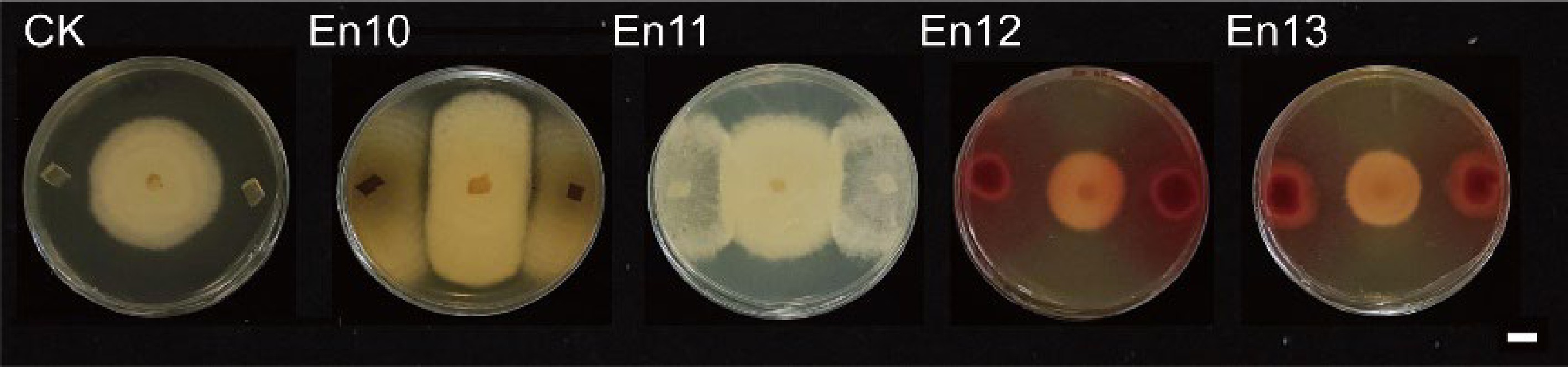
      Figure 1. 

      Plate antagonism experiments on endophytic fungi against the pathogen Colletotrichum camelliae. Scale bar = 1 cm. CK: Two 5 mm PDA plugs were used as controls.
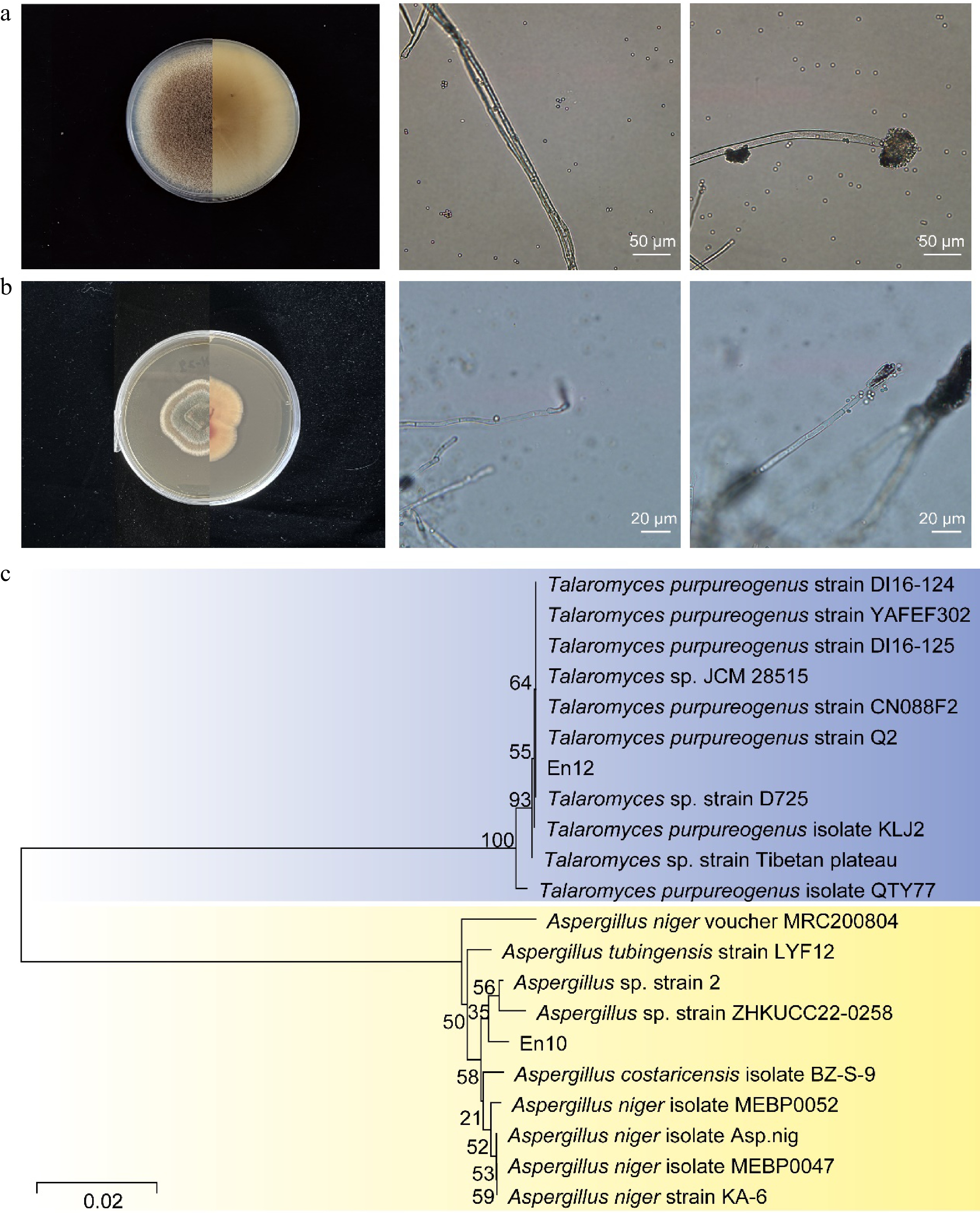
      Figure 2. 

      Morphological and molecular identification of En10 and En12. (a) Front and reverse view of En10 colony on PDA after 9 d, displaying hyphae and conidia. (b) Front and reverse view of En12 colony on PDA after 9 d, showing hyphae and conidia. (c) Phylogenetic analysis based on sequences of the internal transcribed spacer between En10 and En12.

    • To further verify the inhibition of antagonistic fungi against the anthracnose pathogen, a dual culture test was executed. The results demonstrated that the two antagonistic fungi had a significant inhibitory effect on the colony growth of the anthracnose pathogen. After a culture period of 9 d, En10 and En12 exhibited inhibitory rates of 59% and 44% on the growth of the anthracnose pathogen, respectively (Fig 3). These results further confirm the antagonistic effects of En10 and En12 against the tea plant anthracnose pathogen in vitro.
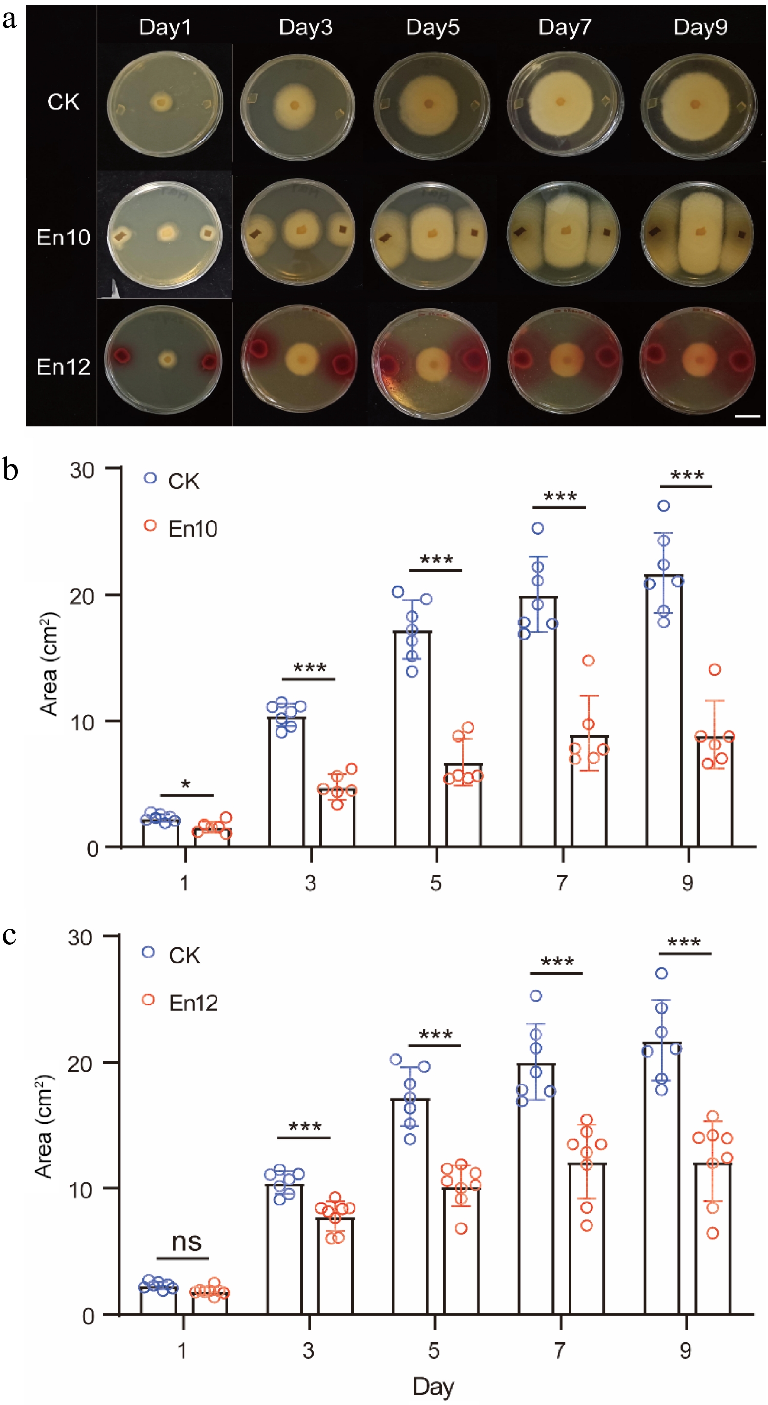
      Figure 3. 

      Inhibition of colony growth of anthracnose pathogen by antagonistic fungi En10 and En12. (a) Dual culture test on anta gonistic fungi against the anthracnose pathogen. CK: Two 5 mm PDA plugs were used as controls. Scale bar = 1 cm. (b), (c) Area change of pathogen under the condition of antagonistic culture. The data represent the average ± SD of biological repeats, ns represents no significant difference and * stands for significant difference analysis.

    • To further elucidate the efficacy of antagonistic fungi in controlling anthracnose in tea leaves, we conducted resistance experiments on tea plant. Tea leaves were sprayed with spore suspensions of antagonistic fungi at three distinct concentrations, including 104/ml, 105/ml and 106/ml. However, it was observed that spraying high and medium concentrations of spore suspension followed by inoculation pathogen resulted in a significant number of tea leaf deaths. For instance, after spraying 106/ml concentration of En10 and En12, the mortality rate of tea leaves reached 14.29% and 66.70% on the third day, respectively, while the control group had a mortality rate of 0%. On the fifth day, the mortality rate of tea leaves reached 42.86% and 83.30%, respectively, while the control group had a mortality rate of 42.86% (data not shown). Therefore, the concentration of 104/ml spore suspension was chosen for treatment. The results of the study demonstrated a noteworthy reduction in the incidence rate of tea leaves that were treated with antagonistic fungi, as opposed to the control group. This suggests that the application of antagonistic fungi on tea leaf has the potential to inhibit tea plant anthracnose (Fig. 4a & b).
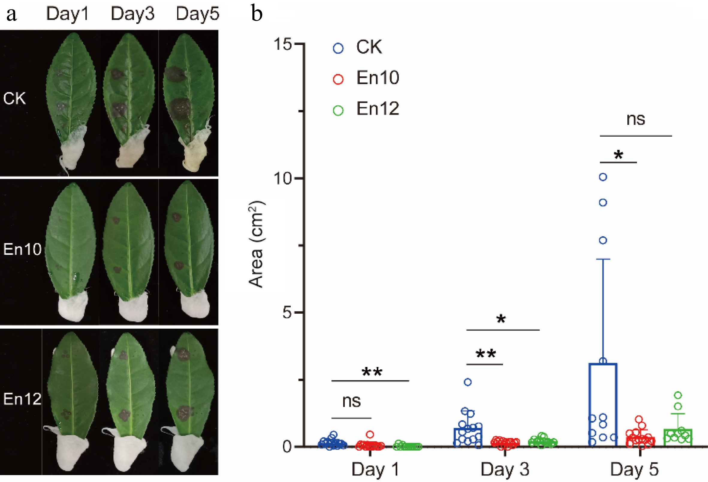
      Figure 4. 

      Determination of the control effect of antagonistic fungi against the anthracnose on tea leaves. (a) and (b) indicate the images and the diseased area of tea leaves treated by blank solvent, fungi spores of En10 and En12 after 1, 3, and 5 d, respectively. The data represent the average ± SD of biological repeats. * means significant difference (p ≤ 0.05); ** represents significant difference (p ≤ 0.01); ns indicates no significant difference.

    • The activities of antioxidant enzymes in tea leaves treated with En10 and En12 were measured. The results indicated a significant upregulation of SOD and POD enzyme activities following En12 treatment compared to the control (Fig. 5a & b). Particularly noteworthy was the 5.47-fold increase in SOD activity, while CAT enzyme activity remained unchanged (Fig. 5c). In contrast, although En10 exhibited notable resistance to the anthracnose pathogen, treatment of tea leaves with it did not result in significant alterations in the activity of associated antioxidant enzymes (Fig. 5). It is suggested that En12 may enhance the antioxidant response of tea leaves, increasing their resistance to anthracnose.
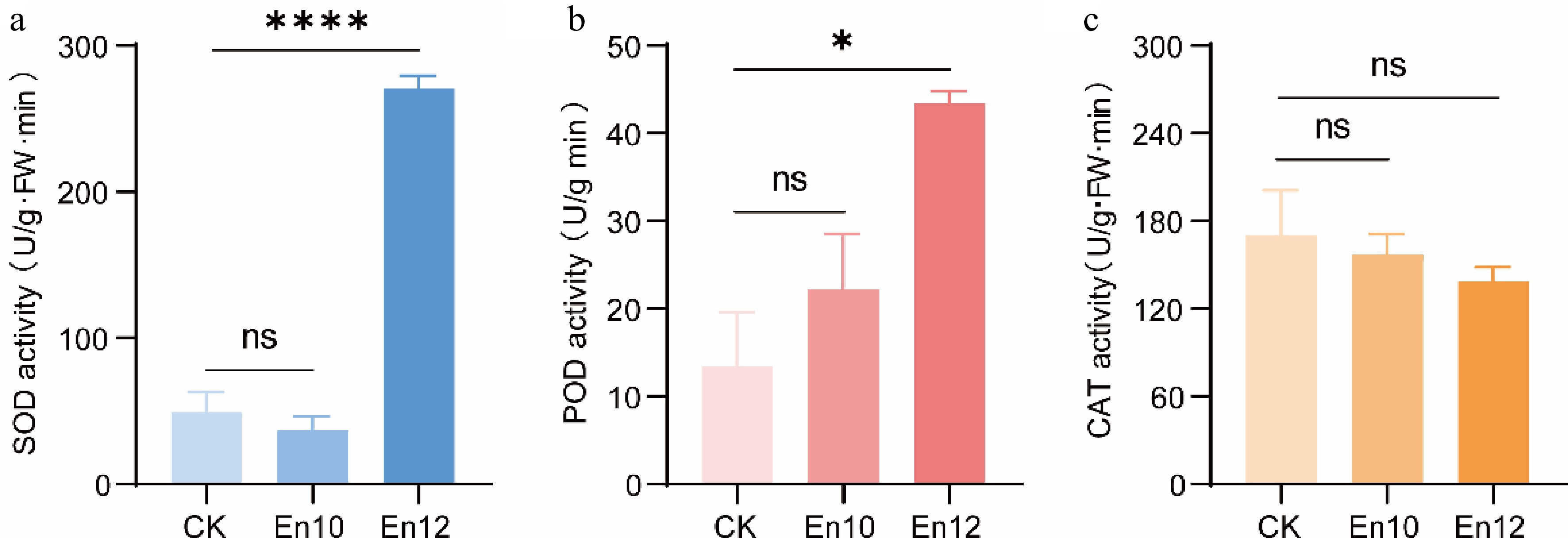
      Figure 5. 

      Detection of enzyme activity in tea leaves after spraying with antagonistic fungi. (a)−(c) indicates the SOD activity, POD activity and CAT activity, respectively. CK: sterile water treated. En10 (En12): antagonistic fungi spores treated. The data represent the average ± SD of three biological repeats. * stands for significant difference analysis and ns indicates no significant difference.

    • Anthracnose, a prevalent disease affecting tea plant worldwide, hinders plant growth and diminishes tea quality[2]. Current control methods rely heavily on chemical agents. However, increasing consumer demand for healthier products has highlighted concerns over pesticide residues in tea and environmental contamination in tea gardens. The emerging field of endophytes in plant defense against pathogens presents promising alternatives for combating fungal, bacterial, viral, and pest-related threats in tea cultivation[17]. The potential application of endophytic antagonism in managing tea plant diseases is promising due to the abundance of epiphytic fungi in tea plant. Therefore, researchers have isolated and identified endophytes from the tea plant. So far, endophytes known to exhibit antagonistic activity against anthracnose include Bacillus amylolyticus[7], Bacillus subtilis[23], Bacillus atrophaeus[24], Bacillus velezensis[25] and Streptomyces luteosporeus[26]. In this study, epiphytic fungi were isolated from the leaves of healthy tea leaves, resulting in the purification of 13 epiphytic fungi representing Cercospora, Cladosporium tenuissimum, Fusarium oxysporum, Nemania bipapillata, Phoma sp., Phytophthora, Trichoderma, Aspergillus niger, Macrocybe gigantea, and Talaromyces (Table 1). Notably, nine of these species were found to be non-pathogenic to tea leaves. Experimental findings demonstrated significant antagonistic effects against anthracnose pathogens by two specific epiphytic fungi, En10 and En12 (Fig. 1). Subsequent identification revealed that En10 and En12 were classified as Aspergillus niger and Talaromyces, respectively (Fig. 2). This represents the first documented instance of their antagonistic activity against tea plant anthracnose.

      Aspergillus niger is a common species of Aspergillus fungi, which is widely found in food, plant products, and soil species[27]. It has a strong ability to produce enzymes, and can produce a variety of high-activity extracellular enzymes, such as cellulase, pectinase, amylase, acid protease, glucose oxidase, and so on. Therefore, it is widely used in the fermentation industry. Some studies have shown that adding Aspergillus niger during tea fermentation can promote the decomposition of tea protein and affect the quality of tea[28,29]. In this study, it was found that Aspergillus niger En10 had a significant inhibi tory effect on the growth of anthracnose pathogen colonies (Figs 1 & 3). After spraying 104/ml spore suspension of En10 on healthy tea leaves (Fig. 4), the incidence of anthracnose inoculation was significantly reduced. This suggests that Aspergillus niger En10 has potential as a biocontrol agent against tea anthracnose.

      Talaromyces are important decomposers in nature and some Talaromyces fungi can produce important enzymes. For example, Talaromyces verruculosus, Talaromyces pinophilus and Talaromyces purpureogenus can produce large amounts of cellulase and xylanase[3033]. Some metabolites of Talaromyces can also enhance the absorption of minerals by plants, thereby improving the disease resistance and stress resistance of plants. A strain of Talaromyces En12 was identified, which could significantly improve the resistance of tea leaves to anthracnose after spraying a spore suspension (Fig. 4). Furthermore, the antagonistic activity of En12 may be attributed to its enhancement of the antioxidant capability of tea leaves, such as the activities of SOD and POD (Fig. 5). This finding highlights the potential of using epiphytic fungi, such as Aspergillus niger and Talaromyces, to be developed as low-cost biocontrol agents for effectively controlling tea plant anthracnose.

      Studies have shown that endophytes can improve plant resistance to pathogens through different mechanisms, such as producing secondary metabolites to inhibit the growth of pathogens and inducing plant defense mechanisms. Endophytes can also induce the production of polyphenol oxidase, peroxidase, phenylalanine ammoniase, and superoxide dismutase in plants to improve the disease resistance of host plants. Prabhukarthikeyan et al. found that after treatment by Pseudomonas fluorescens, the enzyme activities of POD, PPO, PAL, SOD and CAT in roots and stalks of turmeric have been significantly improved, and they have effectively prevented and controlled the root rot disease of turmeric[34]. Khan et al. isolated from the roots of tomato a penicillium strain LK5[35]. Its secondary metabolites can increase the activity of POD, CAT, PPO, and GSH, to improve the defense capability of the plant itself. In our investigation, significant alterations in SOD and POD enzyme activities in tea leaves were observed following treatment with En12 (Fig. 5). These changes potentially contribute to enhancing the tea plant's resistance against anthracnose pathogen incursion. Nonetheless, our primary focus centered on the screening and characterization of epiphytic fungi with efficacy against tea anthracnose. Further research is warranted to elucidate the mechanisms of epiphytic fungi action, isolate and identify antibacterial constituents, and explore the interactions between epiphytic fungi and their host tea plant. Overall, our findings demonstrate the potential of using epiphytic fungi as a cost-effective and environmentally friendly approach for controlling tea plant anthracnose.

    • Thirteen strains of epiphytic fungi were isolated and purified from healthy tea leaves, with nine found to be non-pathogenic. Through plate confrontation tests, two strains, En10 identified as Aspergillus niger, and En12 identified as Talaromyces, were observed to significantly inhibit the growth of pathogens. Dual culture experiments and disease resistance trials on tea leaves demonstrated that En10 and En12 effectively suppressed the invasion of anthracnose disease. Additionally, En12 displayed potential to improve the antioxidant response of tea leaves, thereby increasing resistance to anthracnose. In all, this study identified two epiphytic fungi, belonging to Aspergillus niger and Talaromyces, that have the potential to be used as biological control agents for the management of tea plant anthracnose.

    • The authors confirm contribution to the paper as follows: study conception and design: Wang W, Yu Y, Bao L; tea samples collection: Liu L, Jiao M, Ma Z, Ma Y, Zhou J; experiment conduction: Guan H, Liu L, Xie X, Bao Y; data analysis, visualization: Guan H, Liu L, Wang W; original draft preparation: Wang W, Liu L. All authors reviewed the results and approved the final version of the manuscript.

    • All data supporting the conclusions of this study may be found in the publication, which are available online. Any additional relevant information can be obtained from the corresponding author upon request.

      • This work was supported by the National Natural Science Foundation of China (32202551), China Agriculture Research System of MOF and MARA (CARS-19) and the National key research and development program of China (2022YFD1602003). Some texts in this paper were polished by Stork's Writing Assistant (www.storkapp.me/writeassistant).

      • The authors declare that they have no conflict of interest.

      • # Authors contributed equally: Lu Liu, Haonan Guan

      • Copyright: © 2024 by the author(s). Published by Maximum Academic Press, Fayetteville, GA. This article is an open access article distributed under Creative Commons Attribution License (CC BY 4.0), visit https://creativecommons.org/licenses/by/4.0/.
    Figure (5)  Table (1) References (35)
  • About this article
    Cite this article
    Liu L, Guan H, Jiao M, Ma Z, Bao Y, et al. 2024. Identification of two antagonistic fungi and antifungal activity analysis against anthracnose in tea plant (Camellia sinensis). Beverage Plant Research 4: e032 doi: 10.48130/bpr-0024-0020
    Liu L, Guan H, Jiao M, Ma Z, Bao Y, et al. 2024. Identification of two antagonistic fungi and antifungal activity analysis against anthracnose in tea plant (Camellia sinensis). Beverage Plant Research 4: e032 doi: 10.48130/bpr-0024-0020

Catalog

  • About this article

/

DownLoad:  Full-Size Img  PowerPoint
Return
Return