ARTICLE   Open Access    

Exploration of different pork varieties affecting its lipid oxidation stability

  • # Authors contributed equally: Xinyuan Huang, Hui Liu

More Information
  • The lipid oxidation degree of different pork varieties was different, which may be due to varying levels of antioxidant stability. The purpose of this research was to explore the endogenous antioxidant factors and disparities in oxidative stability between various pork varieties. According to the single-factor experimental design, six pigs from each of the Duroc × Landrace × Yorkshire (DLY), Ningxiang pig (NX), and Rongchang pigs (RC) and were selected, and the longissimus dorsi muscle was used as the experimental sample to determine the fat and fatty acid content, lipid oxidation characterization value, antioxidant capacity and endogenous antioxidant factors of the four varieties pork. The results show that the RC exhibited the highest total fat and fatty acid content, whereas the DLY displayed the lowest levels (p < 0.05). The Thiobarbital acid reactants (TBRAS) of DLY were significantly higher than the other three varieties, and there was no significant difference in peroxide value (POV), conjugated diene (CD), and lipid peroxide (LPO), among the four pork varieties. NX exhibited the highest level of comprehensive antioxidant activity, while Duroc × Landrace × Yorkshire (DLY) displayed the lowest level of comprehensive antioxidant activity (p < 0.05). The results suggested that the antioxidant stability of the four varieties of pork was different. Compared with DLY, RC, and DW, NX showed the best antioxidant stability and the lowest lipid oxidation degree. The activities of superoxide dismutase (SOD), glutathione peroxidase (GSH-Px), vitamin E, and total phenol content (TPC) were significantly correlated with antioxidant capacity.
  • Rice (Oryza sativa L.) is a world staple crop that feeds over half of the world's population[1,2]. In recent years, high-temperature events are becoming more frequent and intensive as a result of global warming, which can severely affect rice grain yield and quality[3,4]. During flowering and grain filling stages, high-temperature stress can result in a significant reduction in seed setting rate and influence amylose content, starch fine structure, functional properties and chalkiness degree of rice[57]. Transcriptome and proteome analysis in rice endosperm have also been used to demonstrate the differences in high-temperature environments at gene and protein expression levels[810]. In addition, as an important post-translational modification, protein phosphorylation has proven to be involved in the regulation of starch metabolism in response to high-temperature stress[11]. However, little is known about whether protein ubiquitination regulates seed development under high-temperature stress.

    Ubiquitination is another form of post-translational modification that plays key roles in diverse cellular processes[12]. Several reports have described the functions of ubiquitination in rice defense responses based on ubiquitome analysis. Liu et al.[13] investigated relationships between ubiquitination and salt-stress responses in rice seedlings using a gel-based shotgun proteomic analysis and revealed the potential important role of protein ubiquitination in salt tolerance in rice. Xie et al.[14] identified 861 peptides with ubiquitinated lysines in 464 proteins in rice leaf cells by combining highly sensitive immune affinity purification and high resolution liquid chromatography-tandem mass spectrometry (LC-MS/MS). These ubiquitinated proteins regulated a wide range of processes, including response to stress stimuli. A later study revealed the relationships between ubiquitination status and activation of rice defense responses, and generated an in-depth quantitative proteomic catalog of ubiquitination in rice seedlings after chitin and flg22 treatments, providing useful information for understanding how the ubiquitination system regulates the defense responses upon pathogen attack[15]. Although many studies have shown that ubiquitination plays improtant roles in the heat response of plant[16,17], there has been little systematic discussion on the ubiquitome of rice endosperm in the context of global climate change.

    In this study, we examine the high-temperature induced ubiquitination change in two rice varieties with different starch qualities, through a label-free quantitative ubiquitome analysis. This study provides a comprehensive view of the function of ubiquitination in high-temperature response of rice developing seed, which will shed new light on the improvement of rice grain quality under heat stress.

    Two indica rice varieties with different starch quality, 9311 and Guangluai4 (GLA4), were used as materials. 9311 is a heat-sensitive variety, which displays low amylose content with good starch quality; while GLA4 is known to be the parental variety of HT54, an indica breeding line with heat tolerance, and thus GLA4 is possibly heat tolerant, which shows high amylose content with poor starch quality[18,19]. Rice growth conditions, sample treatment and collection were conducted as previously described[11].

    Husk, pericarp and embryo were detached from immature rice grains on ice[20]. Rice endosperm was then ground with liquid nitrogen, and the cell powder was sonicated 10 rounds of 10 s sonication and 15 s off-sonication on ice in lysis buffer (6 M Guanidine hydrochloride, pH 7.8−8.0, 0.1% Protease Inhibitor Cocktail) using a high intensity ultrasonic processor. Following lysis, the suspension was centrifuged at 14,000 g for 40 min at 4 °C to remove the debris. The supernatant was collected, and the protein concentration was estimated using BCA assay (Pierce BCA Protein assay kit, Thermo Fisher Scientific, Waltham, MA, USA) before further analysis.

    The protein mixture was reduced by DTT with the final concentration of 10 mM at 37 °C for 1 h, alkylated by iodoacetamide with a final concentration of 50 mM at room temperature in the dark for 0.5 h, and digested by trypsin (1:50) at 37 °C for 16 h. Then the sample was diluted by adding trifluoroacetic acid (TFA) to the final concentration of 0.1%. The enzymatic peptides were desalted on a Sep-Pak C18 cartridge (Waters, Milford, MA, USA), concentrated by lyophilization and reconstituted in precooled IAP buffer (50 mM MOPS-NaOH PH 7.2, 10 mM Na2HPO4, 50 mM NaCl) for further analysis.

    The peptides solution was incubated with prewashed K-ε-GG antibody beads (PTMScan Ubiquitin Remnant Motif (K-ε-GG) Kit), and gently shaken at 4 °C for 1.5 h. The suspension was centrifuged at 2,000 g for 30 s, and the supernatant was removed. The Anti-K-ε-GG antibody beads were washed with IAP Buffer three times and with ddH2O three times. The peptides were eluted from the beads with 0.15% trifluoroacetic acid (TFA). Finally, the eluted fractions were combined and desalted with C18 Stage Tips.

    LC-MS/MS analysis were performed using the methods of Pang et al.[11]. Raw mass spectrometric data were analyzed with MaxQuant software (version 1.3.0.5) and were compared with the indica rice protein sequence database (Oryza sativa subsp. indica-ASM465v1). Parameters were set according to Pang et al.[11]. All measurements were obtained from three separate biological replicates.

    Quantification of the modified peptides was performed using the label-free quantification (LFQ) algorithm[11]. Differentially ubiquitinated sites (proteins) in response to high-temperature were identified by Student's t-test (p < 0.05, log2(fold-change) > 1) with at least two valid values in any condition or the ubiquitination sites that exhibited valid values in one condition (at least two of three replicates) and none in the other.

    Subcellular localization was performed using CELLO database (http://cello.life.nctu.edu.tw). Gene Ontology (GO) annotation proteome was derived from the AgriGO (http://bioinfo.cau.edu.cn/agriGO/). The differential metabolic profiles were visualized with MapMan software (version 3.6.0RC1). Kyoto Encyclopedia of Genes and Genomes (KEGG) pathway annotation was performed by using KEGG Automatic Annotation Server (KAAS) software. A p-value of < 0.05 was used as the threshold of significant enrichment. SWISS-MODEL was used to generate the tertiary structure of GBSSI (SWISS-MODEL, http://swissmodel.expasy.org/). The figures were annotated with Adobe Illustrator (Adobe Systems, San Jose, CA, USA).

    To elucidate how high-temperature stress influences rice developing endosperm at the ubiquitination level, a label-free analysis was performed to quantify ubiquitome from two indica rice varieties under normal (9311-C and GLA4-C) and high-temperature conditions (9311-H and GLA4-H). The distribution of mass error of all the identified peptides was near zero and most of them (71.5%) were between -1 and 1 ppm, suggesting that the mass accuracy of the MS data fits the requirement (Fig. 1a). Meanwhile, the length of most peptides distributed between 8 and 42, ensuring that sample preparation reached standard conditions (Fig. 1b).

    Figure 1.  Characteristics of the ubiquitinated proteome of rice endosperm and QC validation of MS data. (a) Mass error distribution of all identified ubiquitinated peptides. (b) Peptide length distribution. (c) Frequency distribution of ubiquitinated proteins according to the number of ubiquitination sites identified.

    In all endosperm samples, a total of 437 ubiquitinated peptides were identified from 246 ubiquitinated proteins, covering 488 quantifiable ubiquitinated sites (Supplemental Table S1). Among the ubiquitinated proteins, 60.6% had only one ubiquitinated lysine site, and 18.7%, 8.1%, 5.3%, or 7.3% had two, three, four, or five and more ubiquitinated sites, respectively. In addition, four proteins (1.6%, BGIOSGA004052, BGIOSGA006533, BGIOSGA006780, BGIOSGA022241) were ubiquitinated at 10 or more lysine sites (Fig. 1c, Supplemental Table S1). The proteins BGIOSGA008317 had the most ubiquitination sites with the number of 16. It was noted that besides ubiquitin, two related ubiquitin-like proteins NEDD8 and ISG15 also contain C-terminal di-Gly motifs generated by trypsin cleavage, and the modifications of these three proteins cannot be distinguished by MS[21]. Here, the di-Gly-modified proteome therefore represents a composite of proteins modified by these three proteins. However, the sites from NEDD8 or ISG15 modifications were limited because they mediate only a few reactions in cells[21].

    To better understand the lysine ubiquitome changes in rice endosperm induced by high-temperature, we performed a Gene Ontology (GO) functional annotation analysis on all identified ubiquitinated proteins (Fig. 2a). In the biological process GO category, 'metabolic process' and 'cellular process' were mainly enriched, accounting for 75.1% and 74.1% of ubiquitinated proteins, respectively. In addition, 34.6% proteins were associated with 'response to stimulu', emphasizing the regulatory role of ubiquitination modification in response to high-temperature stress. From the cellular component perspective, ubiquitinated proteins were mainly associated with 'cellular anatomical entity' (99.4%), 'intracellular' (84.4%) and 'protein-containing complex' (29.9%). The molecular function category result suggested that these proteins were largely involved in 'binding' (62.7%), 'catalytic activity' (43.4%) and 'structural molecule activity' (16.5%). Furthermore, subcellular location annotation information indicated that 34.7%−39.4% proteins were located in the cytoplasm, and other were mostly located in the nucleus (23.5%−27.7%), plasma membrane (9.4%−11.4%), and chloroplast (9.6%−12.8%) (Fig. 2b). It is noteworthy that the ubiquitinated proteins located in the cytoplasm were decreased in high-temperature environments in both varieties.

    Figure 2.  Analysis of ubiquitinated proteins and motifs. (a) Gene ontology (GO) functional characterization of ubiquitinated proteins. (b) Subcellular localization of ubiquitinated proteins. From the inside out, the ring represents 9311-C, 9311-H, GLA4-C and GLA4-H, respectively. (c) Motif enrichment analysis of ubiquitinated proteins.

    The following two significantly enriched motifs from all of the identified ubiquitinated sites were identified using MoDL analysis: [A/S]xKub and Kubxx[E/Q/R/V]x[E/G/L/P/Q/R/Y], which covered 84 and 100 sequences, respectively (Fig. 2c). Further analysis showed that the conserved alanine (A) and glutamic acid (E) were included in upstream and downstream of the ubiquitinated lysine sites in rice endosperm. A similar phenomenon also occurred in rice leaf[14], wheat leaf[22], and petunia[23], indicating that alanine (A) and glutamic acid (E) were likely to be the specific amino acids in conserved ubiquitination motifs in plants. Additionally, serine (S) was enriched at the position -2 (upstream) of the ubiquitinated lysine, while various amino acids such as arginine (R), glutamic acid (E), glutamine (Q), valine (V) were found at positions +3 and +5 (downstream).

    To detect possible changes in rice endosperm ubiquitome attributable to high-temperature stress, we then performed LFQ analysis on all quantifiable ubiquitination sites within our dataset. As shown in Fig. 3a, more ubiquitinated proteins, peptides and sites were detected in the treatment groups (9311-H and GLA4-H), suggesting that exposure to high-temperature stress may increase the ubiquitination events in rice endosperm. Only 282 common ubiquitinated sites in 158 proteins were quantifiable for all sample groups due to reversible ubiquitination induced by high-temperature (Fig. 3b). Principal component analysis (PCA) showed that three repeats of each sample clustered together, and four groups were clearly separated (Fig. 3c). Furthermore, the differentially expression profiles of ubiquitination sites (proteins) in 9311 and GLA4 under high-temperature stress were depicted to further understand the possible changes (Fig. 3d). Where LFQ values were missing, the data were filtered to identify those ubiquitination sites with a consistent presence/absence expression pattern. These analyses yielded 89 ubiquitination sites that were only present in 9311-H and six that were only present in 9311-C (Fig. 3d, Supplemental Table S2). Similarly, 51 differentially expressed ubiquitination sites were present in GLA4-H and 13 ubiquitination sites only occurred in GLA4-C (Fig. 3d & Supplemental Table S3). Beyond that, a total of 113 and 50 significantly changed ubiquitination sites (p < 0.05, log2(fold-change) >1) were screened out in 9311 and GLA4, respectively (Fig. 3d, Supplemental Tables S4 & S5). For subsequent comparative analysis, the ubiquitination expression profiles with consistent presence/absence and ubiquitination sites with significant differences in statistical testing were combined and named as 9311-Up, 9311-Down, GLA4-Up, and GLA4-Down, respectively (Fig. 3d). The number of significantly up-regulated ubiquitination sites was far greater than down-regulated ubiquitination sites in both 9311 and GLA4 varieties. These findings indicated that high temperature not only induced the occurrence of ubiquitination sites, but also significantly upregulated the intensity of ubiquitination. Beyond that, the magnitude of the up-regulation in 9311 was higher than that in GLA4 (Fig. 3b & d), indicating that the ubiquitination modification of heat-sensitive varieties was more active than heat-resistant varieties in response to high-temperature stress.

    Figure 3.  A temperature regulated rice endosperm ubiquitome. (a) The number of ubiquitinated proteins, peptides and sites detected in four group samples. (b) Venn diagram of ubiquitination sites (proteins) detected in four group samples. (c) PCA based on ubiquitination intensity across all four sample groups with three biological repetitions. (d) Differentially expression profiles of ubiquitination sites (proteins) in 9311 and GLA4 under high-temperature stress. The expression profiles of selected ubiquitination sites (p < 0.05, log2(fold-change) >1) were normalized using the Z-score and presented in a heatmap.

    To further investigate the ubiquitination regulatory pattern under high temperature stress in two varieties, four groups with significantly regulated sites were analyzed. There were 37 ubiquitination sites showed the same regulatory trend in 9311 and GLA4, accounting for 17.8% and 32.5% of the total differentially expressed sites in 9311 and GLA4, respectively. Among them, 36 ubiquitination sites were upregulated and one site was downregulated (Fig. 4a). In addition, 159 upregulated ubiquitination sites and three downregulated sites were only present in 9311, while 53 upregulated sites and 15 downregulated sites were only present in GLA4. Moreover, nine ubiquitination sites showed opposite regulatory trends in 9311 and GLA4. A similar regulatory trend of ubiquitination proteins is shown in Fig. 4b. It is noted that some proteins had both upregulated and downregulated ubiquitination sites (Supplemental Tables S6 & S7), indicating that significant differences in ubiquitination were, to some extent, independent of protein abundance.

    Figure 4.  Comparison of differentially ubiquitinated sites and proteins in 9311 and GLA4 under high-temperature stress.

    To understand the function of ubiquitination in response to the high-temperature stress of rice endosperm, we conducted GO enrichment-based clustering analysis of the differentially ubiquitinated proteins in 9311 and GLA4 at high temperature, respectively (Fig. 5). In the biological process category of 9311, proteins were relatively enriched in the carbohydrate metabolic process, polysaccharide metabolic process, starch biosynthetic process, cellular macromolecule localization, protein localization, intracellular transport, and phosphorylation (Fig. 5). For the molecular function analysis, we found that the proteins related to kinase activity, nucleotidyltransferase activity, phosphotransferase activity, and nutrient reservoir activity were enriched (Fig. 5). The two principal cellular components were intrinsic component of membrane and integral component of membrane (Fig. 5). There was no significantly enriched GO term in the GLA4 group due to the dataset containing relatively few proteins, and thus, further enrichment analysis was conducted on the proteins that were common to both varieties. The results showed that proteins were over-represented in carbon metabolism, including starch biosynthesis and metabolism, glucan biosynthesis and metabolism, and polysaccharide biosynthesis and metabolism (Fig. 5), indicating the importance of carbohydrate synthesis and metabolism in the ubiquitination regulatory network.

    Figure 5.  Enrichment analysis of differentially expressed ubiquitinated proteins based on Gene Ontology (GO) terms.

    To identify pathways which were differentially ubiquitinated under high-temperature stress, the KEGG pathway-based clustering analysis was conducted. The results showed that the differentially ubiquitinated proteins in both 9311 and GLA4 were mostly abundant in the pathways of carbohydrate metabolism, starch and sucrose metabolism, folding, sorting and degradation, translation, ribosome, and protein processing in endoplasmic reticulum (Fig. 6a). In the 9311 group, the pathways of carbohydrate metabolism, starch and sucrose metabolism, glycosyltransferases, glycolysis, and energy metabolism were enriched in the differentially ubiquitinated proteins (Fig. 6b); while there was no significantly enriched KEGG pathway in the GLA4 group. We further found the proteins that were common to both varieties were only significantly enriched in the starch and sucrose metabolism pathways (p = 0.04). The ubiquitination proteins involved in the starch and sucrose metabolism mainly include: sucrose hydrolysis (SUS, FK, UGPase), and starch synthesis (AGPase, GBSSI, BEI, BEIIb, PUL, PHO1), which are discussed below.

    Figure 6.  KEGG classification and enrichment analysis of differentially ubiquitinated proteins. (a) Number of differentially ubiquitinated proteins based on KEGG classification in 9311 and GLA4. (b) KEGG enrichment analysis of differentially ubiquitinated proteins in 9311.

    Although many reports have described specific examples of ubiquitination in rice defense responses[13,15,16], our knowledge on global changes in the developing endosperm ubiquitome under high-temperature stress is still lacking. In this study, a label-free quantitative proteomic analysis of ubiquitination was applied to examine the high-temperature induced ubiquitination change of two indica rice varieties (9311 and GLA4) with distinct starch quality. We identified many new lysine modification sites on proteins involved in various pathways, highlighting the complexity of the ubiquitination-mediated regulatory system in high-temperature stress responses in rice.

    Heat shock proteins accumulate under various stresses and play important roles in plant defenses against abiotic stresses[24,25]. Research has shown that a number of heat shock proteins were prominent in the rice ubiquitome network, of which OsHSP71.1, and OsHSP82A showed increased ubiquitination levels under chitin and flg22 treatment[15]. Here, seven lysine residues on five heat shock proteins possessed ubiquitination modification in rice endosperm. Three sites (BGIOSGA011420-K78, BGIOSGA026764-K99, BGIOSGA029594-K106) showed significant up-regulation in 9311 under high-temperature stress, while in GLA4, the ubiquitination level of BGIOSGA011420-K78 was down-regulated. This differential ubiquitination of heat-tolerant and heat-sensitive varieties provided a basis for studying the regulation of post-translational modification of heat shock proteins under high-temperature stress, despite the regulatory role of those heat shock proteins being still unclear.

    Transcription factors (TFs) play an essential role in the regulation of gene expression. A total of three transcription factors were identified in the ubiquitination dataset, of which two were NAC family members. As one of the largest plant-specific TF families, NAC is involved in the responses to abiotic and biotic stresses[26]. The ubiquitination modification of K173 in BGIOSGA018048 was specifically expressed in the 9311-H group, which may affect the stress resistance level in high-temperature environments. In addition, two sites K148 and K149 of ERF TF family member BGIOSGA024035 was downregulated in GLA4 under high-temperature stress. This differential ubiquitination was likely to affect the expression of related genes regulated by this transcription factor.

    The results of GO and KEGG enrichment analysis of differential ubiquitination proteins indicated that the sucrose and starch metabolic pathway was largely affected by ubiquitination regulation under high-temperature stress (Figs 5 & 6). The ubiquitination sites involved in sucrose and starch metabolism are listed in Table 1. To assess how high-temperature stress affects the crucial pathway, the significantly differential ubiquitination sites in 9311 and GLA4 were displayed in the heatmap of specific proteins (Fig. 7).

    Table 1.  Ubiquitination sites related to sucrose and starch metabolism in rice endosperm.
    Gene nameAnnotationProtein entryModification site(s)
    SUS1Sucrose synthase 1BGIOSGA010570K172, K177
    SUS2Sucrose synthase 2BGIOSGA021739K160, K165, K176, K804
    SUS3Sucrose synthase 3BGIOSGA026140K172, K177, K541, K544, K588
    FKFructokinaseBGIOSGA027875K143
    UGPaseUDP-glucose pyrophosphorylaseBGIOSGA031231K27, K150, K303, K306
    AGPS1ADP-glucose pyrophosphorylase small subunit 1BGIOSGA030039K94, K464, K484
    AGPS2ADP-glucose pyrophosphorylase small subunit 2BGIOSGA027135K106, K132, K385, K403, K406, K476, K496
    AGPL2ADP-glucose pyrophosphorylase large subunit 2BGIOSGA004052K41, K78, K134, K191, K227, K254, K316, K338, K394, K396, K463, K508, K513
    AGPL3ADP-glucose pyrophosphorylase large subunit 3BGIOSGA017490K509
    GBSSIGranule bound starch synthase IBGIOSGA022241K130, K173, K177, K181, K192, K258, K371, K381, K385, K399, K462, K517, K530, K549, K571, K575
    BEIStarch branching enzyme IBGIOSGA020506K103, K108, K122
    BEIIbStarch branching enzyme IIbBGIOSGA006344K134
    PULStarch debranching enzyme:PullulanaseBGIOSGA015875K230, K330, K431, K736, K884
    PHO1Plastidial phosphorylaseBGIOSGA009780K277, K445, K941
     | Show Table
    DownLoad: CSV
    Figure 7.  Sucrose and starch pathway at the ubiquitination levels in rice endosperm under high-temperature stress.

    In cereal endosperm, sucrose is the substrate for the biosynthesis of starch. The formation of glucose 1-phosphate (G1P, used in starch synthesis, see below) from sucrose requires a series of enzymes[27]. Here, we found that sucrose synthase 1 (SUS1), SUS2, SUS3, fructokinase (FK), and UDP-glucose pyrophosphorylase (UGPase) were ubiquitinated among all sample groups (Table 1, Fig. 7). In the ubiquitome of seedling and leaf in japonica rice, ubiquitination sites have been found in SUS1, SUS2, UGPase, and FK, which were related to sucrose hydrolysis[14,15]. SUS catalyzed the process of cleaving sucrose into UDP-glucose (UDPG) and fructose. Two ubiquitination sites, K172 and K177, were identified in SUS1 in rice endosperm, which were also found in rice leaves[14]. A total of four ubiquitination sites were identified in SUS2, two of which were also reported in rice seedling and leaf, indicating the conservation of the lysine residues in different rice tissues. It was noted that all four ubiquitination sites in SUS2 were upregulated in high-temperature environments, although the regulated sites of 9311 and GLA4 were different. In 9311, the ubiquitination levels of K160, K174, and K804 were increased, while GLA4 was only upregulated in K176. The ubiquitination sites K541, K544, and K588 in SUS3 were screened from developing rice seeds for the first time. In addition, SUS3 had two completely overlapping sites K172 and K177 with SUS1, and it was difficult to determine which enzymes the two sites belonged to. The ubiquitination levels of SUS3-K541 and SUS3-K544 in 9311 significantly increased in high-temperature environments, while there was no significant difference in the ubiquitination level of SUS3 in GLA4. Overall, the ubiquitination sites of SUS in rice endosperm were located in the functional domain except for SUS2-K804, reflecting the importance of ubiquitination regulation in SUS.

    UGPase catalyses the conversion of glucose 1-phosphate and UTP into UDPG[28]. Research has shown that the mutation of UGPase gene lead to chalky endosperm[29]. As shown in Table 1, four ubiquitination sites K27, K150, K303, and K306 were identified in rice endosperm, which were completely inconsistent with the seven ubiquitination sites in rice seedlings and two in leaves[14,15], reflecting the tissue specificity. We speculated that the UGPase with different modification sites may play different regulatory roles in metabolic pathways in different tissues. Under high-temperature stress, the ubiquitination level of UGPase-K27 was 8.1-fold up-regulated. Liao et al.[10] demonstrated that the expression of UDPase was down-regulated in both heat-tolerant and heat-sensitive rice lines under high temperature conditions, which could reasonably explain the significant up-regulation of UGPase-K27 ubiquitination level. The ubiquitination site K143 of FK was also reported in seedling tissues[15].

    The AGPase reaction represents the first committed step of starch biosynthesis[27]. A total of 22 lysine ubiquitination sites were identified in four AGPase subunits (AGPL2, AGPL3, AGPS1, AGPS2). AGPL2 had 13 ubiquitination sites, of which six were located in NTP_transferase domain, including K254, K338, K191, K134, K227, and K316. High-temperature stress resulted in an increase in the ubiquitination level of K254 in both 9311 and GLA4, and significant upregulation of K508 and K513 in 9311, as well as K191, K227, and K316 in GLA4. In contrast, AGPL2-K394 were significantly downregulated in GLA4. AGPL3 contained one ubiquitination site K509, and the modification level of AGPL3-K509 was up-regulated in high-temperature environments in 9311. AGPS1 had one specific ubiquitination site K464 and another two sites K94 and K484 that completely overlapped with AGPS2-K106 and AGPS2-K496, respectively. The modification levels of K464 and K484 significantly increased in high-temperature environments in 9311, and K94 was significantly up-regulated in both varieties. There were seven ubiquitination sites in AGPS2 in rice endosperm, which were different with the sites found in rice leaves[14]. In addition to the two sites that overlapped with AGPS1, AGPS2 had another two ubiquitination sites (K406 and K132) that upregulated in high-temperature environments.

    Amylose content is one of the key determinants that strongly influence rice grain quality[30]. The biosynthesis of amylose requires the catalytic effect of granule-bound starch synthase I (GBSSI)[30,31]. Here, a total of 16 ubiquitination sites were identified in GBSSI (Table 1, Fig. 8a). Among these ubiquitination sites, six lysine residues (K130, K173, K177, K181, K192, K258) were located in glycosyltransferase 5 (GT5) domain, and three sites (K399, K462, K517) were located in GT1 domain (Fig. 6), indicating the important role of ubiquitination regulation of GBSSI. Under high-temperature stress, the ubiquitination levels of six sites (K130, K177, K399, K381, K385, K549) increased in two indica rice varieties, while one sites (K258) showed downregulation in 9311 (Fig. 8a). Numerous studies had described that the amylose content was reduced under high-temperature stress in rice[5,7], which might be due to the degradation of GBSSI proteins caused by the increased significantly up-regulated ubiquitination sites. These ubiquitination sites identified in rice GBSSI with significant differences under high-temperature stress were expected to become a new breakthrough point for the improvement of starch quality.

    Figure 8.  Structure of GBSSI. (a) Domain structure of GBSSI and ubiquitination sites with significant differences in response to high-temperature stress. (b) 3D model of GBSSI and the relationship between ubiquitination sites K462 and ADP, SO4 (salt bridge or hydrogen bond).

    To further determine the regulatory role of the ubiquitination sites in GBSSI, SWISS-MODEL was used to predict 3D structural model. As shown in Fig. 8b, GBSSI had three SO4 (sulfate ions) and one ADP ligand. These ligands interact with GBSSI through hydrogen bonds and salt bridges. Three sites, K447, R458, and K462, were associated with SO4 through salt bridges, while G100, N265, Q412, K462, and Q493 interact with the hydrogen bonds of ADP in GBSSI[32,33]. Based on this finding, it can be reasonably inferred that the K462 site with ubiquitination modification located in the GT1 domain played an important role in the interaction between GBSSI, SO4, and ADP. An in-depth investigation was necessary to gain a more comprehensive understanding of the regulatory function of ubiquitination modification at GBSSI-K462, although there was no significant difference in the ubiquitination level under high-temperature stress.

    Amylopectin, the major component of starch, is synthesized by the coordinated action of multiple enzymes including soluble starch synthase (SSs), starch branching enzyme (BEs), starch debranching enzyme (DBEs), and phosphorylases (PHOs or Phos) with ADPG as a substrate. In this study, ubiquitination sites were detected in BEs, DBEs, and Phos.

    BEs, covering two isoforms, BEI and BEII, are responsible for catalyzing the formation of α-1,6-glucosidic linkages of amylopectin[34]. There were three ubiquitination sites (K103, K108, and K122) identified in BEI (Fig. 9a). K122 was the first amino acid in the carbohydrate-binding module 48 (CBM48) domain. Sequence alignment analyses of BEs from eight plants revealed K122 was conserved among all plants' BEI (Fig. 9a), suggesting a high probability of the functional effects of ubiquitination modification at this site. In high-temperature environments, ubiquitination levels of K108 and K122 were significantly up-regulated in 9311, while no significantly regulated ubiquitination sites of BEI were observed in GLA4. Only one ubiquitination site, K134, was found in BEIIb (Fig. 9a). The ubiquitination levels showed a slightly upward trend with no significant differences in high-temperature environments in both varieties. These changes could be one of the reasons for increased gelatinization temperature and relative crystallinity of rice starch in response to high-temperature[5].

    Figure 9.  Domain structure of (a) BEs, (b) PUL and (c) Pho1 as well as their ubiquitination sites with significant differences in response to heat stress. Residues in red indicate the ubiquitination site. Non-ubiquitinated residues are shown in dark grey.

    DBEs consists of isoamylase (ISA) and pullulanase (PUL) with catalytic function for hydrolyzing α-1,6-glucosic linkages[35]. In the present study, we found that only PUL was ubiquitinated in rice endosperm (Fig. 9b). Among five ubiquitination sites (K230, K330, K432, K736, and K884) identified in PUL, K230 was located in the PULN2 domain, while K330 was in the CBM48 domain. Under high-temperature stress, K330 showed completely opposite regulatory trends in two cultivars. In addition, the ubiquitination level of K884, located in the DUF3372 domain, was significantly up-regulated in 9311. Previous study has reported that the expression of PUL was significantly up-regulated in 9311 under high-temperature stress, while GLA4 showed down-regulation in PUL abundance[11]. Consequently, there might be two possible functions of these ubiquitination sites. One possibility is that ubiquitination sites were unrelated to protein degradation; instead, they regulated the biosynthesis of amylopectin by affecting other functions of the protein. Secondly, ubiquitination sites were associated with protein degradation, and the levels of ubiquitination modification were based on protein abundance, resulting in a completely consistent regulation of ubiquitination modification and protein abundance under high-temperature stress.

    PHOs, including two types, Pho1/PHO1 and Pho2/PHO2, are responsible for the transfer of glucosyl units from Glc-1-P to the non-reducing end of a-1,4-linked glucan chains[36]. Pho1 is a temperature-dependent enzyme and considered crucial not only during the maturation of amylopectin but also in the initiation process of starch synthesis[37,38]. The three ubiquitination sites (K277, K445, K941) identified in Pho1 were located in two phosphorylase domains. We found that two sites, Pho1-K277 and Pho1-K445, were only ubiquitinated in high-temperature environments in 9311 and GLA4, respectively. Pang et al.[11] has demonstrated that the protein abundance of Pho1 decreased under high-temperature stress, especially in GLA4. Satoh et al.[38] reported that the functional activity of Pho1 was weakened under conditions of high temperature and its function might be complement by one or more other factors. Hence, these ubiquitination modifications that specifically occurred in high-temperature environments might be related to the degradation of Pho1 proteins.

    As a factory for protein synthesis in cells, the ribosome is an extremely crucial structure in the cell[39]. It has been proven that multiple ribosomal subunits were abundantly ubiquitinated in Arabidopsis and wheat[22]. In the present study, 57 ubiquitination sites involving 33 ribosome subunits were identified in 40S and 60S ribosome complexes in rice. Under high-temperature stress, the ubiquitination levels of some sites were significantly upregulated or downregulated, implying that ubiquitination of ribosomal proteins is likely to be an important regulatory mechanism in high-temperature response in rice endosperm. The results of GO and KEGG enrichment analysis indicated that the ribosome system was one of the most active systems for ubiquitination regulation under high-temperature stress. We speculated that the ubiquitin-proteasome system might be involved in the removal of subunits or entire ribosomes that were improperly folded in high-temperature environments. As shown in Fig. 10, the S10e, L18Ae, S27Ae, L9e, S3e, S28e, S20e, and S2e subunits were significantly up-regulated in 9311, while L13e subunits showed a completely opposite regulatory trend at the ubiquitination sites K81 and K88. In GLA4, the ubiquitination levels of S10e, S27Ae, L10Ae, L9e, S3e, S2e, and L4e showed a significant increase, while the ubiquitination level of L17e was significantly down-regulated under high-temperature stress. A total of seven ubiquitination sites involving S10e, S27Ae, L9e, S3e, and S2e subunits were jointly up-regulated in both two varieties. These sites might be related to the degradation of improperly folded ribosome subunits under high-temperature stress, while other ubiquitination sites with variety specificity might be associated with ribosomal function.

    Figure 10.  Ribosome system at the ubiquitination levels in rice endosperm under high-temperature stress. Grey shadings represent ubiquitinated proteins with no significant differences under heat stress. Red and blue shadings indicate up-regulated and down-regulated ubiquitinated proteins, respectively. Orange shading displays a combination of up- and down-regulated ubiquitinated sites in the same ubiquitinated protein.

    In conclusion, this study provides the first comprehensive view of the ubiquitome in rice developing endosperm, and demonstrated that ubiquitination has diverse functions in the high-temperature response of rice endosperm by modulating various cellular processes, especially the sucrose and starch metabolism. Comparative analysis of the temperature-induced ubiquitination status revealed some similarities and more interesting differences between 9311 and GLA4. These differences might be the reason for the different qualities formation of the two indica rice varieties, which could provide potential genetic resources for the improvement of the heat resistance in rice. Considering the diversity of ubiquitination modification, it is worthwhile to further validate and explore the function and regulatory mechanism of the key targets and key pathways. The findings provide valuable insights into the role of ubiquitination in response to high-temperature stress and lay a foundation for further functional analysis of lysine ubiquitination in rice.

    The authors confirm contribution to the paper as follows: study conception and design: Bao J, Pang Y; data collection: Pang Y; analysis and interpretation of results: Pang Y; draft manuscript preparation: Ying Y; Revised manuscript preparation: Ying Y, Pang Y, Bao J. All authors reviewed the results and approved the final version of the manuscript.

    The datasets generated during and/or analyzed during the current study are available from the corresponding author on reasonable request.

    This work was financially supported by the AgroST Project (NK2022050102) and Zhejiang Provincial Natural Science Foundation (LZ21C130003).

  • The authors declare that they have no conflict of interest.

  • [1]

    Alfaia CM, Lopes PA, Madeira MS, Pestana JM, Coelho D, et al. 2019. Current feeding strategies to improve pork intramuscular fat content and its nutritional quality. In Advances in Food and Nutrition Research, ed. Toldrá F. vol. 89. UK: Academic Press. pp. 53−94. https://doi.org/10.1016/bs.afnr.2019.03.006

    [2]

    Zhang Z, Liao Q, Sun Y, Pan T, Liu S, et al. 2021. Lipidomic and transcriptomic analysis of the longissimus muscle of Luchuan and Duroc pigs. Frontiers in Nutrition 8:667622

    doi: 10.3389/fnut.2021.667622

    CrossRef   Google Scholar

    [3]

    Zhang R, Yang M, Hou X, Hou R, Wang L, et al. 2022. Characterization and difference of lipids and metabolites from Jianhe White Xiang and Large White pork by high-performance liquid chromatography-tandem mass spectrometry. Food Research International 162:111946

    doi: 10.1016/j.foodres.2022.111946

    CrossRef   Google Scholar

    [4]

    Johnson DR, Decker EA. 2015. The role of oxygen in lipid oxidation reactions: a review. Annual Review of Food Science and Technology 6:171−90

    doi: 10.1146/annurev-food-022814-015532

    CrossRef   Google Scholar

    [5]

    Soladoye OP, Juárez ML, Aalhus JL, Shand P, Estévez M. 2015. Protein oxidation in processed meat: mechanisms and potential implications on human health. Comprehensive Reviews in Food Science and Food Safety 14:106−22

    doi: 10.1111/1541-4337.12127

    CrossRef   Google Scholar

    [6]

    Zhang W, Xiao S, Ahn DU. 2013. Protein Oxidation: Basic Principles and Implications for Meat Quality. Critical Reviews in Food Science and Nutrition 53:1191−201

    doi: 10.1080/10408398.2011.577540

    CrossRef   Google Scholar

    [7]

    Wen W, Luo X, Xia B, Guan J, Nie Y, et al. 2015. Post-mortem oxidative stability of three yak (Bos grunniens) muscles as influenced by animal age. Meat Science 105:121−25

    doi: 10.1016/j.meatsci.2015.03.014

    CrossRef   Google Scholar

    [8]

    Kopec W, Wiliczkiewicz A, Jamroz D, Biazik E, Pudlo A, et al. 2016. Antioxidant status of turkey breast meat and blood after feeding a diet enriched with histidine. Poultry Science 95:53−61

    doi: 10.3382/ps/pev311

    CrossRef   Google Scholar

    [9]

    Sacchetti G, Di Mattia C, Pittia P, Martino G. 2008. Application of a radical scavenging activity test to measure the total antioxidant activity of poultry meat. Meat Science 80:1081−85

    doi: 10.1016/j.meatsci.2008.04.030

    CrossRef   Google Scholar

    [10]

    Serpen A, Gökmen V, Fogliano V. 2012. Total antioxidant capacities of raw and cooked meats. Meat Science 90:60−26

    doi: 10.1016/j.meatsci.2011.05.027

    CrossRef   Google Scholar

    [11]

    Mielnik MB, Rzeszutek A, Triumf EC, Egelandsdal B. 2011. Antioxidant and other quality properties of reindeer muscle from two different Norwegian regions. Meat Science 89:526−32

    doi: 10.1016/j.meatsci.2011.05.021

    CrossRef   Google Scholar

    [12]

    Triumf EC, Purchas RW, Mielnik M, Maehre HK, Elvevoll E, et al. 2012. Composition and some quality characteristics of the longissimus muscle of reindeer in Norway compared to farmed New Zealand red deer. Meat Science 90:122−29

    doi: 10.1016/j.meatsci.2011.06.011

    CrossRef   Google Scholar

    [13]

    Quaresma MAG, Trigo-Rodrigues I, Alves SP, Martins SIV, Barreto AS, et al. 2012. Nutritional evaluation of the lipid fraction of Iberian red deer (Cervus elaphus hispanicus) tenderloin - ScienceDirect. Meat Science 92:519−24

    doi: 10.1016/j.meatsci.2012.05.021

    CrossRef   Google Scholar

    [14]

    Brewer MS. 2011. Natural antioxidants: sources, compounds, mechanisms of action, and potential applications. Comprehensive Reviews in Food Science and Food Safety 10(4):221−47

    doi: 10.1111/j.1541-4337.2011.00156.x

    CrossRef   Google Scholar

    [15]

    Decker EA, Xu ZM. 1998. Minimizing Rancidity in Muscle Foods. Food Technology 52:54−59

    Google Scholar

    [16]

    Shahidi F, Zhong Y. 2010. Novel antioxidants in food quality preservation and health promotion. European Journal of Lipid Science & Technology 112:930−40

    doi: 10.1002/ejlt.201000044

    CrossRef   Google Scholar

    [17]

    Pradhan AA, Rhee KS, Hernández P. 2000. Stability of catalase and its potential role in lipid oxidation in meat. Meat Science 54:385−90

    doi: 10.1016/S0309-1740(99)00114-X

    CrossRef   Google Scholar

    [18]

    Descalzo AM, Sancho AM. 2008. A review of natural antioxidants and their effects on oxidative status odor and quality of fresh beef produced in Argentina. Meat Science 79:423−36

    doi: 10.1016/j.meatsci.2007.12.006

    CrossRef   Google Scholar

    [19]

    Kim HW, Choi YS, Choi JH, Kim HY, Hwang KE, et al. 2013. Antioxidant effects of soy sauce on color stability and lipid oxidation of raw beef patties during cold storage. Meat Science 95:641−46

    doi: 10.1016/j.meatsci.2013.06.006

    CrossRef   Google Scholar

    [20]

    Wootton-Beard PC, Moran A, Ryan L. 2011. Stability of the total antioxidant capacity and total polyphenol content of 23 commercially available vegetable juices before and after in vitro digestion measured by FRAP, DPPH, ABTS and Folin–Ciocalteu methods. Food Research International 44:217−24

    doi: 10.1016/j.foodres.2010.10.033

    CrossRef   Google Scholar

    [21]

    Huang Y, Zhou L, Zhang J, Liu X, Zhang Y, et al. 2020. A large-scale comparison of meat quality and intramuscular fatty acid composition among three Chinese indigenous pig breeds. Meat Science 168:108182

    doi: 10.1016/j.meatsci.2020.108182

    CrossRef   Google Scholar

    [22]

    Rey AI, Lopez-Bote CJ, Kerry JP, Lynch PB, Buckley DJ, et al. 2004. Modification of lipid composition and oxidation in porcine muscle and muscle microsomes as affected by dietary supplementation of n-3 with either n-9 or n-6 fatty acids and α-tocopheryl acetate. Animal Feed Science & Technology 113:223−38

    doi: 10.1016/j.anifeedsci.2003.08.007

    CrossRef   Google Scholar

    [23]

    Cameron ND, Enser M, Nute GR, Whittington FM, Penman JC, et al. 2000. Genotype with nutrition interaction on fatty acid composition of intramuscular fat and the relationship with flavour of pig meat. Meat Science 55(2):187−95

    doi: 10.1016/S0309-1740(99)00142-4

    CrossRef   Google Scholar

    [24]

    Du M, Ahn DU, Sell JL. 2000. Effects of dietary conjugated linoleic acid and linoleic: linolenic acid ratio on polyunsaturated fatty acid status in laying hens. Poultry Science 79:1749−56

    doi: 10.1093/ps/79.12.1749

    CrossRef   Google Scholar

    [25]

    Dou L, Jin Y, Li H, Liu C, Yang Z, et al. 2023. Effect of feeding system on muscle fiber composition, antioxidant capacity, and nutritional and organoleptic traits of goat meat. Animals 13:172

    doi: 10.3390/ani13010172

    CrossRef   Google Scholar

    [26]

    Ma L, Liu G. 2017. Simultaneous analysis of malondialdehyde, 4-Hydroxy-2-hexenal, and 4-Hydroxy-2-nonenal in vegetable oil by reversed-phase high-performance liquid chromatography. Journal of Agricultural and Food Chemistry 65(51):11320−28

    doi: 10.1021/acs.jafc.7b04566

    CrossRef   Google Scholar

    [27]

    Shestivska V, Antonowicz SS, Dryahina K, Kubišta J, Smith D, et al. 2015. Direct detection and quantification of malondialdehyde vapour in humid air using selected ion flow tube mass spectrometry supported by gas chromatography/mass spectrometry. Rapid Communications in Mass Spectrometry 29(11):1069−79

    doi: 10.1002/rcm.7198

    CrossRef   Google Scholar

    [28]

    Fashi A, Salarian AA, Zamani A. 2018. Solvent-stir bar microextraction system using pure tris-(2-ethylhexyl) phosphate as supported liquid membrane: A new and efficient design for the extraction of malondialdehyde from biological fluids. Talanta 182:299−305

    doi: 10.1016/j.talanta.2018.02.002

    CrossRef   Google Scholar

    [29]

    Chen Y, Lei YY, Li BJ. 2019. A study on the changes in physicochemical indicators of Sichuan bacon under different packaging and storage conditions. Meat Industry 15:29−31+35

    doi: 10.3969/j.issn.1008-5467.2019.04.009

    CrossRef   Google Scholar

    [30]

    Chen W, Zhu HL, Shi Y, Zhao MM, Wang H, et al. 2012. Comparative analysis on antioxidative ability of muscle between Laiwu pig and large white. Asian-Australasian Journal of Animal Sciences 25(8):1190−96

    doi: 10.5713/ajas.2012.12019

    CrossRef   Google Scholar

    [31]

    Hu H, Wu C, Ding Y, Zhang X, Yang M, et al. 2019. Comparative analysis of meat sensory quality, antioxidant status, growth hormone and orexin between Anqingliubai and Yorkshire pigs. Journal of Applied Animal Research 47:357−61

    doi: 10.1080/09712119.2019.1643729

    CrossRef   Google Scholar

    [32]

    Leung HW, Vang MJ, Mavis RD. 1981. The cooperative interaction between vitamin E and vitamin C in suppression of peroxidation of membrane phospholipids. Biochimica et Biophysica Acta (BBA) - Lipids and Lipid Metabolism 664:266−72

    doi: 10.1016/0005-2760(81)90049-7

    CrossRef   Google Scholar

    [33]

    Gülçin İ, Elmastaş M, Naserian AA, Farhangfar H, Emami A,. 2018. The effect of grapeseed oil on performance rumen fermentation, antioxidant status and subcutaneous adipose fatty acid profile in lambs. Journal of Animal Physiology and Animal Nutrition 102:157−65

    doi: 10.1111/jpn.12673

    CrossRef   Google Scholar

    [34]

    Gülçin İ, Elmastaş M, Aboul-Enein HY. 2012. Antioxidant activity of clove oil – A powerful antioxidant source. Arabian Journal of Chemistry 5:489−99

    doi: 10.1016/j.arabjc.2010.09.016

    CrossRef   Google Scholar

    [35]

    Echegaray N, Pateiro M, Munekata PES, Lorenzo JM, Chabani Z, et al. 2021. Measurement of Antioxidant Capacity of Meat and Meat Products: Methods and Applications. Molecules 26:3880

    doi: 10.3390/molecules26133880

    CrossRef   Google Scholar

    [36]

    Chen W, Zeng QF, Xu HP, Fang GF, Wang SD, et al. 2018. Comparison and relationship between meat colour and antioxidant capacity of different pig breeds. Animal Production Science 58(11):2152−57

    doi: 10.1071/AN16184

    CrossRef   Google Scholar

    [37]

    Halliwell B, Adhikary A, Dingfelder M, Dizdaroglu M. 2021. Hydroxyl radical is a significant player in oxidative DNA damage in vivo. Chemical Society Reviews 50:8355−60

    doi: 10.1039/D1CS00044F

    CrossRef   Google Scholar

    [38]

    Sies H. 2017. Hydrogen peroxide as a central redox signaling molecule in physiological oxidative stress: Oxidative eustress. Redox Biology 11:613−19

    doi: 10.1016/j.redox.2016.12.035

    CrossRef   Google Scholar

    [39]

    Muhlisin, Utamaa DT, Lee JH, Choi JH, Lee SK. 2016. Antioxidant enzyme activity, iron content and lipid oxidation of raw and cooked Meat of Korean native chickens and other poultry. Asian-Australasian Journal of Animal Sciences 29:695−701

    doi: 10.5713/ajas.15.0256

    CrossRef   Google Scholar

    [40]

    Pastsart U, De Boever M, Claeys E, De Smet S. 2013. Effect of muscle and post-mortem rate of pH and temperature fall on antioxidant enzyme activities in beef. Meat Science 93:681−86

    doi: 10.1016/j.meatsci.2012.11.008

    CrossRef   Google Scholar

    [41]

    Leopoldini M, Russo N, Toscano M. 2011. The molecular basis of working mechanism of natural polyphenolic antioxidants. Food Chemistry 125:288−306

    doi: 10.1016/j.foodchem.2010.08.012

    CrossRef   Google Scholar

    [42]

    Yusuf AL, Adeyemi KD, Roselina K, Alimon AR, Goh YM, et al. 2018. Dietary supplementation of different parts of Andrographis paniculata affects the fatty acids, lipid oxidation, microbiota, and quality attributes of longissimus muscle in goats. Food Research International 111:699−707

    doi: 10.1016/j.foodres.2018.06.015

    CrossRef   Google Scholar

    [43]

    Qwele K, Hugo A, Oyedemi SO, Moyo B, Masika PJ, et al. 2013. Chemical composition fatty acid content and antioxidant potential of meat from goats supplemented with Moringa (Moringa oleifera) leaves, sunflower cake and grass hay. Meat Science 93:455−62

    doi: 10.1016/j.meatsci.2012.11.009

    CrossRef   Google Scholar

    [44]

    Niki E. 1987. Interaction of ascorbate and α-tocopherol. Annals of the New York Academy of Sciences 498:186−99

    doi: 10.1111/j.1749-6632.1987.tb23761.x

    CrossRef   Google Scholar

    [45]

    Burdeos GC, Nakagawa K, Kimura F, Miyazawa T. 2012. Tocotrienol attenuates triglyceride accumulation in HepG2 cells and F344 rats. Lipids 47:471−81

    doi: 10.1007/s11745-012-3659-0

    CrossRef   Google Scholar

  • Cite this article

    Huang X, Liu H, Tang X, Zhang Y, Zhao Y. 2024. Exploration of different pork varieties affecting its lipid oxidation stability. Food Materials Research 4: e017 doi: 10.48130/fmr-0024-0008
    Huang X, Liu H, Tang X, Zhang Y, Zhao Y. 2024. Exploration of different pork varieties affecting its lipid oxidation stability. Food Materials Research 4: e017 doi: 10.48130/fmr-0024-0008

Figures(5)  /  Tables(2)

Article Metrics

Article views(2538) PDF downloads(330)

ARTICLE   Open Access    

Exploration of different pork varieties affecting its lipid oxidation stability

Food Materials Research  4 Article number: e017  (2024)  |  Cite this article

Abstract: The lipid oxidation degree of different pork varieties was different, which may be due to varying levels of antioxidant stability. The purpose of this research was to explore the endogenous antioxidant factors and disparities in oxidative stability between various pork varieties. According to the single-factor experimental design, six pigs from each of the Duroc × Landrace × Yorkshire (DLY), Ningxiang pig (NX), and Rongchang pigs (RC) and were selected, and the longissimus dorsi muscle was used as the experimental sample to determine the fat and fatty acid content, lipid oxidation characterization value, antioxidant capacity and endogenous antioxidant factors of the four varieties pork. The results show that the RC exhibited the highest total fat and fatty acid content, whereas the DLY displayed the lowest levels (p < 0.05). The Thiobarbital acid reactants (TBRAS) of DLY were significantly higher than the other three varieties, and there was no significant difference in peroxide value (POV), conjugated diene (CD), and lipid peroxide (LPO), among the four pork varieties. NX exhibited the highest level of comprehensive antioxidant activity, while Duroc × Landrace × Yorkshire (DLY) displayed the lowest level of comprehensive antioxidant activity (p < 0.05). The results suggested that the antioxidant stability of the four varieties of pork was different. Compared with DLY, RC, and DW, NX showed the best antioxidant stability and the lowest lipid oxidation degree. The activities of superoxide dismutase (SOD), glutathione peroxidase (GSH-Px), vitamin E, and total phenol content (TPC) were significantly correlated with antioxidant capacity.

    • Pork is the most abundant and consumed meat in the world[1], providing many essential nutrients for the human body, such as protein, amino acids, fat, minerals, and vitamins. However, with the improvement of people's living standards, people's requirements for the quality and flavor of pork are also increasing. Among the main commercial pig breeds, DLY is one of the major commercial pig breeds in our country, which is known for its fast growth rate and high lean meat rate, but its IMF (intramuscular fat) content is low and its meat quality is inferior[2]. Local breeds of pigs are known for their high IMF content and unique flavor. IMF is closely related to visual marbling and is one of the most critical quality attributes of pork[3]. Compared with DLY, local pigs in China have attracted attention due to their excellent meat quality. However, high-fat content is also more prone to lipid oxidation, which is one of the main limiting factors affecting the quality and consumer acceptability of meat and meat products, as it affects the nutritional value and sensory properties of meat, reducing the consumer’s purchasing desire[4,5]. So oxidative stability is crucial for the quality of fresh and processed meat[6]. Oxidative stability is usually related to endogenous antioxidant capacity, which can protect the matrix from reactive oxygen species and free radicals. It is usually determined by gene expression related to hypoxia response and energy metabolism[7].

      Earlier scholars had studied the content of antioxidant compounds and oxidative stability from domestic[810] and wild animals[11,12]. The nonenzymatic antioxidant compounds include primarily vitamin E homologues (tocochromanols: tocopherols and tocotrienols)[13], vitamin C (L–ascorbic acid), carotenoids (carotenes and xanthophylls), and phenolic and polyphenolic compounds[14]. The enzymatic antioxidant systems mostly include superoxide dismutase, catalase, and glutathione peroxidase[15], which can have an effective impact on many oxidation levels as primary and secondary antioxidants (as scavengers, quenchers, inactivators, chelators, inhibitors, and reductants)[16].

      Previously, most studies about the impact of lipid oxidation endogenous antioxidant factors in meat or meat products have focused on the changes in lipid antioxidant factors during storage or processing[17]. However, the differences in endogenous antioxidant factors and oxidative stability of pork in different varieties have been rarely discussed.

      Ningxiang Pigs (NX) from the Institute of Subtropical Agricultural Ecology, Chinese Academy of Sciences, Changsha, Hunan Province, Rongchang Pigs (RC) from the Pig Breeding Institute of Chongqing Academy of Animal Husbandry Sciences, and Duroc × Wujin Pigs (DW) from the Laopu Family Breeding Base in Xuanwei, Yunnan Province (China), and Duroc × Landrace × Yorkshire (DLY) from Beijing in the same batch with the same feeding conditions, gender, and age were slaughtered at the slaughterhouse, and the outer ridges of the left half of the carcass gathered within 4 h and kept at −20 °C. The aim is to explore the variations in endogenous antioxidant factors and oxidative stability of pork across different varieties, with the intention of establishing a theoretical framework for enhanced regulation of lipid oxidation in pork while upholding its exceptional quality.

    • A total of 24, 6 month-old pigs, that meet the requirements to be put out, including six Duroc × Landrace × Yorkshire (DLY), six Ningxiang pigs (NX), six Rongchang pigs (RC), and six Duroc × Wujin pigs (DW) and live weight of 95−130 kg were selected. Following the period of growth, the pigs were slaughtered at a nearby slaughterhouse. The handling of all pigs adhered to the Regulations on Administration of Hog Slaughter and Good manufacturing practice for pig slaughter (GB/T 19479-2004) in China. The left longissimus muscle of each pig was removed from its carcasses and then subjected to vacuum packing, samples were then stored in the refrigerator until completely frozen and transported from the slaughterhouse cold chain to the laboratory for storage at −20 °C.

    • The determination of IMF content is based on the Soxhlet extraction method in GB 5009.6-2016 'National Food Safety Standards - Determination of Fat in Foods'. Samples of pork dorsal muscles from different places are taken and dried in a 105 °C oven for 8 h to remove moisture. They are ground into powder using a mortar and placed in a receiving cup. After adding petroleum ether, they are extracted using a Soxhlet extractor. After the extraction is completed, the receiving cup is dried in a 105 °C oven for 1 h to remove petroleum ether, then placed in a dryer for 30 min to cool until constant temperature and weight, and then weighed. The fat content was calculated based on the difference before and after the extraction of the receiving cup.

    • The determination of fatty acid content in pork refers to the external standard method in GB 5009.168-2016 'National Food Safety Standard - Determination of Fatty Acids in Foods'. The pork mince is freeze-dried into meat powder, and 0.500 g (± 0.005) of the freeze-dried meat powder is sequentially added to 5.0 mL of toluene and 6 mL of 10% acetyl chloride methanol solution. The extraction is carried out in a constant temperature water bath at 80 °C for 2 h. After extraction, the reaction solution is transferred to a 50 mL centrifuge tube and washed with 6% sodium carbonate solution. After centrifugation, the supernatant was taken for gas chromatography determination, and mixed fatty acid methyl esters were used as external standards for quantification.

    • To compare the degree of lipid oxidation in four pork varieties, Thiobarbital acid reactants (TBRAS), peroxide value (POV), conjugated diene (CD) and Lipid Peroxide (LPO) were selected as the lipid oxidation characterization indexes.

    • Referring to the method of Descalzo & Sancho[18], a 0.5 g cutting of an appropriate size of meat sample ws taken, 3 mL of thiobarbituric acid (TBA) solution and 15 mL of trichloroacetic acid (TCA) solution were added. The samples were then placed in a water bath at 90 °C for 40 min. When the system cools down, 5 mL of chloroform was added and centrifuged at 3,000 g for 5 min. Measure the absorbance value at 532 nm, calculate TBARS according to Eqn (1), and the result is expressed as the milligrams of malondialdehyde (MDA) per kilogram of meat sample.

      TBRAS=A532×9.48 (1)

      In the formula, TBRAS − content of thiobarbituric acid reactants, mg MDA/kg; A532 − absorbance value of solution; 9.48 − constant coefficient.

    • The POV in the sample is based on the method in GB 5009.227-2016 National Food Safety Standards - Determination of peroxide value in food, with slight modifications. The pork minclets were taken, mixed with petroleum ether, and then soaked for 14 h. The filtrate was filtered through a funnel filled with anhydrous sodium sulfate, and the petroleum ether was dried by a rotary evaporator under reduced pressure. The remaining residue after evaporation was taken for determination.

    • Referring to the method of Kim et al.[19], an appropriate size of meat sample was cut, 0.5 g was then taken and 5 mL of deionized water added at 10,000 r/min. Samples were then homogenized for 20 s, 0.5 mL of the homogenate was taken, and 5 mL of a mixture of n-hexane and isopropanol (V/V = 3:1) added for 2 min for extraction, then centrifuged at 2,000 r/min for 10 min. The absorbance of the supernatant was then measured at 233 nm. The molar extinction coefficient of CD at this wavelength is 25.5 × 10³ L/(mol·cm), expressed as the number of micromoles of CD per milligram of meat sample.

    • Sample preparation: Accurately weigh the pork samples from various sources and add 0.9% physiological saline in the ratio of weight (g) : volume (mL) = 1:9. Ice bath homogenization was performed at 10,000 r/min for 30 s/time, and repeated twice to produce 10% tissue homogenization. Centrifuge at 4 °C and 2,500 r/min for 10 min, and collect the supernatant to determine the content of lipid peroxides.

      Determination principle: Under the condition of reacting at 45 °C for 60 min, one molecule of LPO reacts with two molecules of chromogenic reagents to produce a stable chromophore with a maximum absorption peak at 586 nm. The content of LPO in the test sample can be obtained by comparing standard curves or calculating through formulas. The measurement steps and calculation of lipid peroxide content in pork tissue are based on the instructions for the Lipid Peroxide Determination Kit (Nanjing Jiancheng Biotechnology Co., Ltd., Nanjing, China, No. A106-1).

    • In this study, the antioxidant capacity of four varieties of pork was comprehensively evaluated by total antioxidant capacity (T-AOC), DPPH free radical scavenging power (DPPH) and Hydroxyl Radical Inhibition (OH•), the total antioxidant capacity was discussed from two perspectives: Ferric Reducing Ability of Plasma (FRAP) and ABTS radical scavenging ability (ABTS).

      The Sample pretreatment and determination method of T-AOC (FRAP), T-AOC (ABTS) DPPH free radical scavenging power and Hydroxyl Radical Inhibition (OH•) are based on the instructions for the T-AOC (FRAP) assay kit (Nanjing Jiancheng Biotechnology Co., Ltd., Nanjing, China, No. A015-3-1), the total antioxidant capacity (ABTS) assay kit (Nanjing Jiancheng Biotechnology Co., Ltd., Nanjing, China, No. A015-2-1), the instructions of the DPPH free radical scavenging assay kit (Nanjing Jiancheng Biotechnology Co., Ltd., Nanjing, China, No. A153-1-1) and the hydroxyl radical inhibition assay kit (Nanjing Jiancheng Biotechnology Co., Ltd., Nanjing, China, No. A018-1-1) respectively.

    • The endogenous antioxidant factor was mainly determined by antioxidant oxidase(GSH-Px, SOD, CAT), total phenol content (TPC), vitamin C, and vitamin E content are four common substances with antioxidant activity in pork.

    • Principle and method for measuring glutathione peroxidase (GSH-Px), superoxide dismutase (SOD) and catalase (CAT) enzyme activity is based on the instructions for the GSH-Px enzyme assay kit (Nanjing Jiancheng Biotechnology Co., Ltd., Nanjing, China, No. A005-1-1); the SOD enzyme assay kit (Nanjing Jiancheng Biotechnology Co., Ltd., Nanjing, China, No. A001-3-2) and the CAT enzyme assay kit (Nanjing Jiancheng Biotechnology Co. Ltd. Nanjing, China, No. A007-1-1) respectively.

    • Referring to the method of Wootton-Beard et al.[20] with slight modifications, measure the total phenol content, the pork sample was ground and weighed 2.000 g (± 0.005). Eighteen ml of 0.9% physiological saline was added, and ice bath homogenization was performed at 10,000 r/min for 30 s/time. The homogenization was repeated twice to produce 10% tissue homogenization. At 4 °C, 3,000 r/min, centrifugation was performed for 10 min, and the supernatant was taken at 500 μL, then mixed with an equal volume of deionized water. Twenty percent sodium carbonate solution (W/V) was then added to Folin Ciocalteau reagent to 1 mol/L and mixed thoroughly. Under dark conditions, the mixture is kept in a 25 °C water bath for 1 h. The sample is then centrifuged at 3,000 g and 4 °C for 10 min to remove excess sodium carbonate solution. The absorbance value of the supernatant was measured at 760 nm (as a blank control), and a known concentration of gallic acid solution was used as the standard curve. The total phenolic concentration of the extract was expressed as milligrams of gallic acid (GAE) per gram of muscle.

    • Sample pre-treatment and determination principle and method are based on the instructions for the Vitamin C Content Determination Kit (Nanjing Jiancheng Biotechnology Co., Ltd., Nanjing, China, NO.A009-1-1) and the Vitamin E Content Determination Kit (Nanjing Jiancheng Biotechnology Co., Ltd., Nanjing, China, A008-1-1).

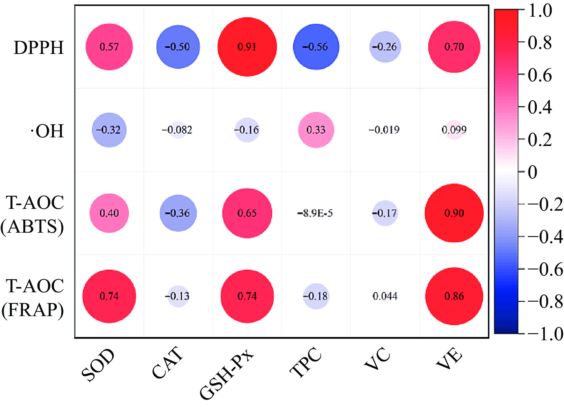
    • All experimental data were organized in Excel 2019 and statistical analysis was performed using SPSS 26.0 software (IBM, Chicago, IL, USA), Additionally, the data obtained were subjected to a one-way analysis of variance (ANOVA), and Duncan’s multiple comparison was used to determine the difference, the significance levels are defined at p < 0.05. Principal component analysis (PCA) through the correlation coefficient matrix method was applied for multivariate analysis using the 'OriginPro 2021'. Correlations between total antioxidant capacity, DPPH, OH•-, and antioxidant factors were determined by correlation analyses using Pearson’s linear correlation coefficient procedure.

    • The IMF content measurement results are shown in Table 1, the IMF content of RC was the highest, while the DLY was the lowest. The IMF content of NX and DW was between RC and DLY, as shown in Table 1, and there was a significant difference (p < 0.05). This result is consistent with previous research on the IMF content of local breeds and commercial pigs. Zhang et al.[2] found that the IMF content of local pigs in China is higher than that of commercial pigs. Zhang et al.[3] compared the differences in lipids and metabolites between Jianhe White Xiang pig and Large White pig, and the results showed that the IMF content of Jianhe White Xiang pig was 7.88%, significantly higher than that of Large White pig by 3.04%. The high-fat content also causes local pigs to be more prone to fat oxidation.

      Table 1.  Differences of fat and fatty acid content in different varieties of pork.

      ItemDLYNXRCDW
      FAT (g/100 g)1.42 ± 0.32c3.17 ± 0.56b3.72 ± 0.30a3.15 ± 0.27b
      SFA (g/100 g)3.30 ± 0.34C4.99 ± 0.46b6.95 ± 0.50a5.53 ± 0.35b
      MUFA (g/100 g)3.69 ± 0.39a5.47 ± 0.51b6.30 ± 0.59b6.92 ± 0.53b
      PUFA (g/100 g)1.45 ± 0.09b1.76 ± 0.09a1.91 ± 0.08a1.48 ± 0.05b
      FAs (g/100 g)8.44 ± 0.80b12.23 ± 1.03a15.15 ± 1.11a13.94 ± 0.91b
      DLY (Duroc × Landrace × Yorkshire pig); NX (Yorkshire × Ningxiang pig); RC (Rongchang pig); DW (Duroc × Wujin pig). The values are presented by means ± standard deviations. a, b mean values with various superscripts in a row were significantly different (p < 0.05). n = 6.

      The main step of lipid oxidation is the decomposition of the fatty acid into lower molecular weight compounds, and compared with saturated fatty acids, unsaturated fatty acids are more easily oxidized. Studies have shown that monounsaturated fatty acids (MUFA) are the main fatty acids (FAs) in Chinese native pig meat[21]. The results of determining the content of saturated fatty acids (SFA) in pigs from four different varieties showed in Table 1, that RC had the highest content of saturated fatty acids (SFA) and polyunsaturated fatty acids (PUFA); DW has the highest MUFA content (p < 0.05). Due to the significantly lower total fat content of DLY compared to the other three types of pigs, the content of each fatty acid in DLY is lower than that of the other three types. Rey et al.[22] also confirmed a significant increase in PUFAs content in pork with edible flaxseed oil. Fatty acid composition is one of the main contributors to pork flavor and is influenced by breed and genotype[23]. Highly saturated fatty acids (SFAs) are considered to have positive effects on stabilizing fat oxidation[24]. Local breed pigs have high fat and fatty acid content, which may lead to high oxidation degree and increased oxidation products.

    • Thiobarbital acid reactants (TBRAS) are lipid oxidation indicators that affect consumer acceptance[25], calculated from a standard curve of malondialdehyde (MDA), and expressed as mg of MDA per kg sample. Due to the presence of unsaturated bonds, PUFAs are usually more prone to oxidation than SFAs, resulting in the production of different metabolites, which may have adverse effects on human health, shelf life, and meat quality[1]. MDA is the main secondary lipid oxidation product[26]. Compared with other secondary metabolites of lipid peroxidation, MDA is stable and abundant and is considered one of the most important markers of lipid peroxidation status[27,28]. Peroxide value (POV) is a parameter that reflects the degree of fat oxidation, and is a quantitative indicator of the product of double bonds in unsaturated fatty acids combined with oxygen in the air, indicating the degree of fat oxidation. A high POV value indicates a significant accumulation of intermediate products from fat oxidation, but these intermediate products will quickly undergo further oxidation reactions to generate small molecule substances as they accumulate[29]. Conjugated Dienes (CD) is the amount of conjugated diene contained in the oil was expressed as an index of lipid hydroperoxides in the extracted lipid fraction of the pork[19]. Lipid peroxidation (LPO) is the oxidation of polyunsaturated fatty acids (PUFAs) in biological systems and causes changes in the physical structure and characteristics of the cell membrane.

      The determination results of lipid oxidation degree of four local pork varieties are shown in Table 2. There was no significant difference in POV, CD, and LPO, among the four local pork varieties. The content of TBRAS in DLY was significantly higher than that of the other three varieties (p < 0.05), indicating that the degree of fat oxidation of DLY was higher than that of the other three pork varieties and there was no significant difference in the content of TBRAS among the other three varieties. Previous studies have found that pig breed is one important factor of the MDA content of porcine Longissimus dorsi muscle during storage. The MDA contents of Large White pigs were significantly higher than Laiwu pigs (p < 0.01) when the muscles stored at 4 and −20 °C[30]. Hu el.[31] also confirmed this, and their results indicate that compared with the Yorkshire pigs, the Anqingliubai pig exhibited lower MDA concentration (p < 0.05). Previous studies have shown that the degree of unsaturated fatty acids is the main factor determining the amount of MDA formation. The results of lipid oxidation showed that although the local breeds of pigs have high fat and fatty acid content. However, the amount of oxidized products and the degree of oxidation are not much different from the DLY with lower fat content. This may be related to the differences in the antioxidant capacity of different varieties of pork due to different endogenous antioxidant factors, such as antioxidant enzyme activity, polyphenol content, and vitamin E content. It has been also observed that vitamin E and vitamin C delay the formation of MDA, during the oxidation of rat liver microsomes and phospholipid liposomes catalyzed by ferrous ions[32].

      Table 2.  Differences of lipid oxidation indices in different varieties of pork

      ItemDLYNXRCDW
      TBRAS (mg MDA/kg)0.63 ± 0.03a0.56 ± 0.01b0.59 ± 0.05b0.57 ± 0.02b
      POV (meq/kg)0.16 ± 0.02a0.14 ± 0.01a0.14 ± 0.04a0.17 ± 0.02a
      CD (mol/g)1.52 ± 0.01a1.43 ± 0.08a1.50 ± 0.01a1.51 ± 0.01a
      LPO (μmol/g prot)0.14 ± 0.01a0.14 ± 0.01a0.17 ± 0.02a0.16 ± 0.02a
      The values are presented by means ± standard deviations. The lowercase letterings a and b mean values with various superscripts in a row were significant differences (p < 0.05). n = 6.
    • T-AOC is a comprehensive indicator of overall antioxidant capacity[33]. This study comprehensively evaluated its total antioxidant capacity (T-AOC) through two methods: iron ion reduction ability (FARP) and ABTS radical scavenging ability (ABTS). The FRAP test is a technique that measures the ability of antioxidants to reduce ferric ions (Fe3+) to ferrous ions (Fe2+)[34]. The foundation of the ABTS test is based on the measurement of the ability of antioxidants to reduce the previously generated cationic radical[35]. The measurement results as shown in Fig. 1, and NX had the highest FARP and ABTS radical scavenging ability (p < 0.05), while DLY had the lowest FARP, and there was no significant difference in ABTS radical scavenging ability among RC, DW, and DLY varieties( p > 0.05), as shown in Fig. 1b. This difference may be due to the different types and contents of antioxidant-active substances in different varieties of pork. There are research findings that state using up to 4% grapeseed oil in lamb diets may be a good method to improve meat antioxidant activity and fatty acid composition, without affecting animal production performance[33], which may be due to the fact that grape seeds contain a large number of substances with antioxidant activity, such as antioxidant enzymes, polyphenols and vitamin E, etc. Moreover, the pig breed is one important factor on the T-AOC of porcine Longissimus dorsi muscle. Hu et al.[31] confirmed the Anqingliubai pigs exhibited higher T-AOC compared with the Yorkshire pigs (p < 0.05). Chen et al.[36] also confirmed that meat had more total antioxidant capacity in Chinese native and breed pigs than DLY (p < 0.05).
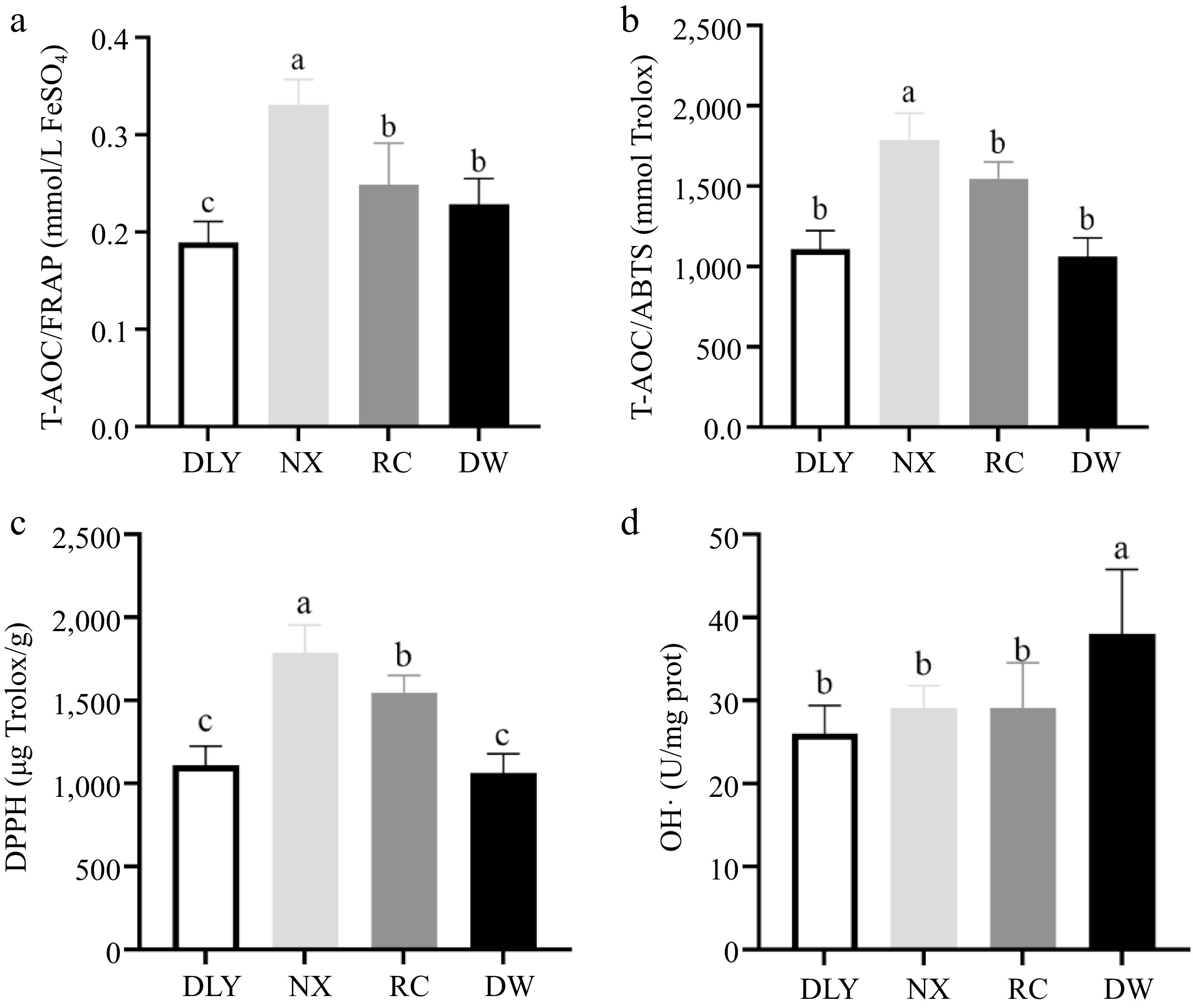
      Figure 1. 

      Comparison of antioxidant capacity characterization value of four varieties of pork. (a) Iron ion reduction ability (FARP); (b) ABTS radical scavenging ability (ABTS); (c) Comparison of DPPH among four varieties of pork; (d) Comparison of OH•- among four varieties of pork. Different letters indicate significant differences between pork samples of different breeds (p < 0.05). n = 6.

    • DPPH can reflect the ability of the muscle antioxidant system to scavenge free radicals, this ability may come from vitamin C, polyphenols and some antioxidant activity of reducing substances[18]. The DPPH test is based on the measurement of the reducing capacity of antioxidants against the free radical DPPH. This measurement is generally carried out through the determination of the decrease in the absorbance, which is currently a technique judged a standard for the in vitro determination of antioxidants that is extensively employed for the evaluation of free radical scavenging potentials of distinct compounds[35]. The results of DPPH free radical scavenging ability measurement of four local pork samples are shown in Fig. 1c, the NX had the highest DPPH free radical scavenging ability, significantly higher than RC, while DW and DLY had the lowest DPPH free radical scavenging ability, significantly lower than RC (p < 0.05).

    • Hydroxyl radical plays a prominent role in the oxidation of protein, lipid, and nucleic acid in vivo because the oxidization ability of hydroxyl radicals is the highest among reactive oxygen species (ROS). Moreover, lipid peroxyl radicals (LOO•) produced by the oxidation of the lipid bilayer of the cell membrane by hydroxyl radical triggers a lipid peroxidation chain reaction and damages cell membranes. The inhibitory ability of hydroxyl radical (OH•) can represent the antioxidant ability[37]. The measurement results of the inhibitory ability of hydroxyl radical (OH•) of four local pork varieties showed that DW had the highest hydroxyl radical inhibition ability, significantly higher than NX, RC, and DLY (p < 0.05). There was no difference in hydroxyl radical inhibition ability among the other three varieties (p > 0.05), as shown in Fig 1d. This showed that DW had stronger ability to inhibit hydroxyl radical oxidation of membrane lipid bilayer, generate lipid peroxidation free radical and trigger lipid peroxidation chain reaction process than the other three varieties.

      The above results indicated that there were differences in T-AOC/DPPH and inhibitory ability of hydroxyl radical (OH•) of different pork varieties, which might be attributed to the differences in endogenous antioxidant factors and the types and contents of antioxidant active substances contained in different pork varieties, such as antioxidant enzymes, polyphenols, vitamin C and vitamin E.

    • Antioxidant enzymes are considered as important protectors in muscles to prevent lipid oxidation. The endogenous antioxidant enzymes in muscles mainly include superoxide dismutase (SOD), catalase (CAT), and glutathione peroxidase (GSH-Px). SOD is the first line of defense of the antioxidant system[7]. CAT is a peroxidase that catalyzes the decomposition of hydrogen peroxide into water and oxygen[38]. GSH-Px inhibits further oxidative damage[39]. The results of endogenous antioxidant enzyme activity measurements of four breeds of pork are shown in Fig. 2, that among the four types of local pork, the SOD enzyme, and GSH-Px enzyme activities of NX were the highest; DW have the highest CAT enzyme activity; The activity of SOD and CAT enzymes in DLY is the lowest; The activity of GSH-Px enzyme in DW pigs was the lowest, but there was no significant difference compared to DLY (p < 0.05). Differences in the activity of the three antioxidant enzymes may lead to differences in the antioxidant capacity of the four varieties pork. Studies have shown that catalase (CAT), superoxide dismutase (SOD), and glutathione peroxidase (GPx) can effectively delay lipid oxidation in beef[40]. Chen et al.[30] found in their research that the Laiwu pig as a Chinese indigenous pig breed showed strong SOD, CAT and GSH-Px activity, and it suggested that Laiwu pig can more effectively inhibit lipid oxidation and have a long shelf-life after slaughter compared to the Large White pig.

      Figure 2. 

      Comparison of antioxidant enzyme activities among four pork varieties.

    • Polyphenols have been shown to have various antioxidant activities, including scavenging free radicals, inhibiting enzymes involved in free radical production, and chelating metal ions[41]. The Folin Ciocalteu trial can be considered a method to determine the antioxidant capacity by electron transfer, and high content of phenolic compounds in foods have been associated with high antioxidant capacity. From the results shown in Fig. 3, the total phenol content of RC is the lowest, but there is no significant difference from the total phenol content of NX and DW, and the total phenol content of DLY is the highest (p < 0.05) but there was no significant difference with NX and DW content. Yusuf et al.[42] found that there was no difference in the percentage of fat, water, protein, and ash components in goat muscle in the feed supplemented with different parts of Andrographis paniculata, but there were differences in polyphenol content, which lead to differences in antioxidant capacity. This is consistent with the observation of Qwele et al.[43], where eating spicy wood leaves increased the total phenol content in goat meat, and also improved antioxidant capacity. These studies indicated that exogenous TPC could affect the antioxidant capacity of meat, but the endogenous TPC in fresh meat had a relatively limited effect on the antioxidant capacity due to their low content. To our knowledge, only limited information about the phenolic content in pig meat have been previously reported.

      Figure 3. 

      Comparison of total phenol content in four varieties of pork.

    • Vitamins C and E function as water-soluble and lipid-soluble chain-breaking antioxidants, respectively, and protect lipids, proteins, and membranes from oxidative damage. Vitamin C scavenges oxygen radicals in the aqueous phase, whereas vitamin E scavenges oxygen radicals within the membranes. This interaction between vitamin C and vitamin E radicals can take place not only in homogeneous solutions but also in liposomal membrane systems[44]. It has been also observed that vitamins E and C delay the formation of MDA, during the oxidation of rat liver microsomes and phospholipid liposomes catalyzed by ferrous ion[32].

      The results of vitamin C determination for four types of local pork were shown in Fig. 4a, the vitamin C content of DW was the highest, significantly higher than RC and DLY, but there was no significant difference compared to NX content. The NX had the highest vitamin E content, significantly higher than DLY, while RC and DW had the lowest vitamin E content, significantly lower than DLY as shown in Fig. 4b (p < 0.05). The difference in vitamin C and vitamin E content in the four varieties of pork may affect the difference in their antioxidant capacity. Descalzo & Sancho[18] found that vitamin E in animal tissues affects the stability of lipids during the storage of meat. It is capable of quenching free radicals and thus protects phospholipids and cholesterol against oxidation, and a higher intake of natural antioxidants can result in transferring these molecules to animal tissues with a consequent increase of total antioxidant capacity[18]. The inhibitory effect of vitamin E on lipid peroxidation is documented based on in vivo experiments, Burdeos et al.[45] reported that the daily intake of tocotorienol-rich rice bran oil (10 mg of rice bran oil containing 3.14 mg α-tocotrie-nol, 5.04 mg γ-tocotrienol, 0.04 mg δ-tocotrienol, 0.19 mg α-TocH, 0.21 mg γ-TocH, and 0.20 mg δ-TocH by oral gavage using 200 μL corn oil as a vehicle) for 3 weeks decreased levels of plasma phospholipid hydroperoxide (PLOOH) in rats. This indicates that vitamin E can effectively enhance antioxidant capacity.

      Figure 4. 

      Comparison of vitamin content in four varieties of pork. (a) Vitamin C, (b) Vitamin E.

    • To evaluate the relationship between antioxidant performance and endogenous antioxidant factors of four pig breeds, antioxidant capacity was used as the Y variable and endogenous antioxidant factors as the X variable for Pearson correlation analysis. The results are shown in Fig. 5, and show that the T-AOC and DPPH free radical scavenging ability of the four pig breeds were positively correlated with SOD and GSH-Px activity, vitamin E content, and DPPH had the strongest correlation with GSH-Px activity, with a correlation coefficient of 0.91, The correlation between T-AOC and vitamin E content is strongest, with T-AOC (ABTS) and T-AOC (FARP) being 0.9 and 0.86, respectively. OH• inhibitory capacity is positively correlated with TPC content, with a correlation coefficient of 0.33. It can be seen that the antioxidant factors that have significant effects on the antioxidant performance of the four varieties of pork are SOD, GSH-Px activity, vitamin E, and TPC content.

      Figure 5. 

      Correlation analysis between antioxidant capacity and antioxidant factors.

      Antioxidant capacity analyses show NX had the highest FARP, ABTS radical scavenging ability, and DPPH, DW had the highest Hydroxyl Radical Inhibition Ability. Antioxidant factors analyses NX had the highest vitamin E content, the highest SOD activity and the highest GSH-PX activity, which may be the main factors to enhance the antioxidant capacity of NX. DW had the highest OH•, although its TPC content was not the highest, there was no significant difference between it and DLY with the highest content. DLY had the lowest FARP among the four varieties, and ABTS and DPPH are significantly lower than NX, meanwhile, SOD and GSH-Px enzyme activity are the lowest, it’s vitamin E content is also significantly lower than NX. Which is consistent with the results of correlation analysis.

    • The oxidative stability of different varieties of pork has significant differences. Compared with DLY, the local pork varieties have higher fat and fatty acid content, but there is no significant difference in their content of lipid oxidation products compared to DLY, which maybe due to their strong antioxidant properties. Owing to the difference in endogenous antioxidant factors, NX pigs have the best antioxidant stability and lowest content of lipid oxidation products compared with DLY, RC, and DW, and the antioxidant factors that have a significant impact on the antioxidant performance of the four breeds pig meat are SOD, GSH-Px activity, vitamin E, and TPC content. Through this experiment, we can learn more about the differences in antioxidant stability of different varieties of pork, and provide a theoretical basis for regulating lipid oxidation of different varieties of pork, to retain the superior quality of local varieties of pork and provide basic data for the development of pork products.

    • The authors confirm contribution to the paper as follows: conceptualization, resources, visualization: Huang X, Tang X; methodology: Huang X, Tang X, Zhao Y, Zhang Y; software: Huang X, Zhao Y, Liu H; validation: Tang X, Zhang Y, Zhao Y; formal analysis, investigation, data curation: Huang X; original draft preparation, review and editing: Tang X, Huang X, Liu H; Supervision: Liu H, Zhang Y; project administration: Tang X, Huang X; funding acquisition: Tang X. All authors reviewed the results and approved the final version of the manuscript.

    • The datasets generated during the current study are available from the corresponding author on reasonable request.

      • This research was funded by the National Natural Science Foundation of China, Grant No. 32172152; the National Key R&D Program of China, Grant No. 2022YFD1601902, the China Agriculture Research System, Grant No. CARS-35 and the Agricultural Science and Technology Innovation Program of CAAS.

      • The authors declare that they have no conflict of interest.

      • # Authors contributed equally: Xinyuan Huang, Hui Liu

      • Copyright: © 2024 by the author(s). Published by Maximum Academic Press on behalf of Nanjing Agricultural University. This article is an open access article distributed under Creative Commons Attribution License (CC BY 4.0), visit https://creativecommons.org/licenses/by/4.0/.
    Figure (5)  Table (2) References (45)
  • About this article
    Cite this article
    Huang X, Liu H, Tang X, Zhang Y, Zhao Y. 2024. Exploration of different pork varieties affecting its lipid oxidation stability. Food Materials Research 4: e017 doi: 10.48130/fmr-0024-0008
    Huang X, Liu H, Tang X, Zhang Y, Zhao Y. 2024. Exploration of different pork varieties affecting its lipid oxidation stability. Food Materials Research 4: e017 doi: 10.48130/fmr-0024-0008

Catalog

  • About this article

/

DownLoad:  Full-Size Img  PowerPoint
Return
Return