REVIEW   Open Access    

Gut microbiota-associated metabolites in metabolic diseases and their impact from food processing

More Information
  • Gut microbiota-associated metabolites can be synthesized endogenously or derived from dietary nutrients and host compounds. Among them, alkaloids, terpenes, and flavones originating from edible and medicinal foods have attracted remarkable interest recently and play crucial roles in metabolic diseases. The efficacy of these metabolites is susceptible to dietary intervention, especially after food processing. Therefore, this review comprehensively summarizes the different sources of common gut microbial metabolites, including microbial self-synthesis, biodegradation of exogenous substances (mainly dietary nutrients), and participation in host metabolism. In addition, the latest studies on novel metabolites such as alkaloids, terpenoids, and flavonoids are discussed, and their action mechanisms on metabolic diseases are elaborated. How food processing impacts dietary nutrients and their metabolites is carefully examined, as well as their effects on disease modification. These insights could contribute to a deeper understanding of the mechanisms by which diet efficacy helps prevent metabolic diseases, particularly through gut microbial metabolites.
  • Crops require a variety of nutrients for growth and nitrogen is particularly important. Nitrogen is the primary factor limiting plant growth and yield formation, and it also plays a significant role in improving product quality[14]. Nitrogen accounts for 1%−3% of the dry weight of plants and is a component of many compounds. For example, it is an important part of proteins, a component of nucleic acids, the skeleton of cell membranes, and a constituent of chlorophyll[5,6]. When the plant is deficient in nitrogen, the synthesis process of nitrogen-containing substances such as proteins decrease significantly, cell division and elongation are restricted, and chlorophyll content decreases, and this leads to short and thin plants, small leaves, and pale color[2,7,8]. If nitrogen in the plant is in excess, a large number of carbohydrates will be used for the synthesis of proteins, chlorophyll, and other substances, so that cells are large and thin-walled, and easy to be attacked by pests and diseases. At the same time, the mechanical tissues in the stem are not well developed and are prone to collapse[3,8,9]. Therefore, the development of new crop varieties with both high yields and improved nitrogen use efficiency (NUE) is an urgently needed goal for more sustainable agriculture with minimal nitrogen demand.

    Plants obtain inorganic nitrogen from the soil, mainly in the form of NH4+ and nitrate (NO3)[1013]. Nitrate uptake by plants occurs primarily in aerobic environments[3]. Transmembrane proteins are required for nitrate uptake from the external environment as well as for transport and translocation between cells, tissues, and organs. NITRATE TRANSPORTER PROTEIN 1 (NRT1)/PEPTIDE TRANSPORTER (PTR) family (NPF), NRT2, CHLORIDE CHANNEL (CLC) family, and SLOW ACTIVATING ANION CHANNEL are four protein families involved in nitrate transport[14]. One of the most studied of these is NRT1.1, which has multiple functions[14]. NRT1.1 is a major nitrate sensor, regulating many aspects of nitrate physiology and developmental responses, including regulating the expression levels of nitrate-related genes, modulating root architecture, and alleviating seed dormancy[1518].

    There is mounting evidence that plant growth and development are influenced by interactions across numerous phytohormone signaling pathways, including abscisic acid, gibberellins, growth hormones, and cytokinins[3,19,20]. To increase the effectiveness of plant nitrogen fertilizer application, it may be possible to tweak the signaling mediators or vary the content of certain phytohormones. Since the 1930s, research on the interplay between growth factors and N metabolism has also been conducted[3]. The Indole acetic acid (IAA) level of plant shoots is shown to decrease in early studies due to N shortage, although roots exhibit the reverse tendency[3,21]. In particular, low NO3 levels caused IAA buildup in the roots of Arabidopsis, Glycine max, Triticum aestivum, and Zea mays, indicating that IAA is crucial for conveying the effectiveness of exogenous nitrogen to the root growth response[20,22,23].

    Studies have shown that two families are required to control the expression of auxin-responsive genes: one is the Auxin Response Factor (ARF) and the other is the Aux/IAA repressor family[2426]. As the transcription factor, the ARF protein regulates the expression of auxin response genes by specifically binding to the TGTCNN auxin response element (AuxRE) in promoters of primary or early auxin response genes[27]. Among them, rice OsARF18, as a class of transcriptional repressor, has been involved in the field of nitrogen utilization and yield[23,28]. In rice (Oryza sativa), mutations in rice salt tolerant 1 (rst1), encoding the OsARF18 gene, lead to the loss of its transcriptional repressor activity and up-regulation of OsAS1 expression, which accelerates the assimilation of NH4+ to Asn and thus increases N utilization[28]. In addition, dao mutant plants deterred the conversion of IAA to OxIAA, thus high levels of IAA strongly activates OsARF18, which subsequently represses the expression of OsARF2 and OsSUT1 by directly binding to the AuxRE and SuRE promoter motifs, resulting in the inhibition of carbohydrate partitioning[23]. As a result, rice carrying the dao has low yields.

    Apples (Malus domestica) are used as a commercially important crop because of their high ecological adaptability, high nutritional value, and annual availability of fruit[29]. To ensure high apple yields, growers promote rapid early fruit yield growth by applying nitrogen. However, the over-application of nitrogen fertilizer to apples during cultivation also produces common diseases and the over-application of nitrogen fertilizer is not only a waste of resources but also harmful to the environment[29]. Therefore, it is of great significance to explore efficient nitrogen-regulated genes to understand the uptake and regulation of nitrogen fertilizer in apples, and to provide reasonable guidance for nitrogen application during apple production[30]. In this study, MdARF18 is identified which is a key transcription factor involved in nitrate uptake and transport in apples and MdARF18 reduces NO3 uptake and assimilation. Further analysis suggests that MdRF18 may inhibit the transcriptional level of MdNRT1.1 promoter by directly binding to its TGTCTT target, thus affecting normal plant growth.

    The protein sequence of apple MdARF18 (MD07G1152100) was obtained from The Apple Genome (https://iris.angers.inra.fr/gddh13/). Mutant of arf18 (GABI_699B09) sequence numbers were obtained from the official TAIR website (www.arabidopsis.org). The protein sequences of ARF18 from different species were obtained from the protein sequence of apple MdARF18 on the NCBI website. Using these data, a phylogenetic tree with reasonably close associations was constructed[31].

    Protein structural domain prediction of ARF18 was performed on the SMART website (https://smart.embl.de/). Motif analysis of ARF18 was performed by MEME (https://meme-suite.org/meme/tools/meme). Clustal was used to do multiple sequence comparisons. The first step was accessing the EBI web server through the Clustal Omega channel. The visualization of the results was altered using Jalview, which may be downloaded from www.jalview.org/download.[32]

    The apple 'Orin' callus was transplanted on MS medium containing 1.5 mg·L−1 6-benzylaminopurine (6-BA) and 0.5 mg·L−1 2,4 dichlorophenoxyacetic acid (2,4-D) at 25 °C, in the dark, at 21-d intervals. 'Royal Gala' apple cultivars were cultured in vermiculite and transplanted at 25 °C every 30 d. The Arabidopsis plants used were of the Columbia (Col-0) wild-type variety. Sowing and germinating Arabidopsis seeds on MS nutrient medium, and Arabidopsis seeds were incubated and grown at 25 °C (light/dark cycle of 16 h/8 h)[33].

    The nutrient solution in the base contained 1.0 mM CaCl2, 1.0 mM KH2PO4, 1.0 mM MgSO4, 0.1 mM FeSO4·7H2O 0.1 mM Na2EDTA·2H2O, 50 μM MnSO4·H2O, 50 μM H3BO3, 0.05 μM CuSO4·5H2O, 0.5 μM Na2MoO4·2H2O, 15 μM ZnSO4·7H2O, 2.5 μM KI, and 0.05 μM CoCl·6H2O, and 0.05 μM CoCl·6H2O, and 0.05 μM CoCl· 6H2O. 2H2O, 15 μM ZnSO4·7H2O, 2.5 μM KI and 0.05 μM CoCl·6H2O, and 0.05 μM CoCl·6H2O, supplemented with 0.5 mM, 2 mM, and 10 mM KNO3 as the sole nitrogen source, and added with the relevant concentrations of KCl to maintain the same K concentration[33,34].

    For auxin treatment, 12 uniformly growing apple tissue-cultured seedlings (Malus domestica 'Royal Gala') were selected from each of the control and treatment groups, apple seedlings were incubated in a nutrient solution containing 1.5 mg·L−1 6-BA, 0.2 mg·L−1 naphthalene acetic acid, and IAA (10 μM) for 50 d, and then the physiological data were determined. Apple seedlings were incubated and grown at 25 °C (light/dark cycle of 16 h/8 h).

    For nitrate treatment, Arabidopsis seedlings were transferred into an MS medium (containing different concentrations of KNO3) as soon as they germinated to test root development. Seven-day-old Arabidopsis were transplanted into vermiculite and then treated with a nutrient solution containing different concentrations of KNO3 (0.5, 2, 10 mM) and watered at 10-d intervals. Apple calli were treated with medium containing 1.5 mg·L−1 6-BA, 0.5 mg·L−1 2,4-D, and varying doses of KNO3 (0.5, 2, and 10 mM) for 25 d, and samples were examined for relevant physiological data. Apple calli were subjected to the same treatment for 1 d for GUS staining[35].

    To obtain MdARF18 overexpression materials, the open reading frame (ORF) of MdARF18 was introduced into the pRI-101 vector. To obtain pMdNRT1.1 material, the 2 kb segment located before the transcription start site of MdNRT1.1 was inserted into the pCAMBIA1300 vector. The Agrobacterium tumefaciens LBA4404 strain was cultivated in lysozyme broth (LB) medium supplemented with 50 mg·L−1 kanamycin and 50 mg·L−1 rifampicin. The MdARF18 overexpression vector and the ProMdNRT1.1::GUS vector were introduced into Arabidopsis and apple callus using the flower dip transformation procedure. The third-generation homozygous transgenic Arabidopsis (T3) and transgenic calli were obtained[36]. Information on the relevant primers designed is shown in Supplemental Table S1.

    Plant DNA and RNA were obtained using the Genomic DNA Kit and the Omni Plant RNA Kit (tDNase I) (Tiangen, Beijing, China)[37].

    cDNA was synthesized for qPCR by using the PrimeScript First Strand cDNA Synthesis Kit (Takara, Dalian, China). The cDNA for qPCR was synthesized by using the PrimeScript First Strand cDNA Synthesis Kit (Takara, Dalian, China). Quantitative real-time fluorescence analysis was performed by using the UltraSYBR Mixture (Low Rox) kit (ComWin Biotech Co. Ltd., Beijing, China). qRT-PCR experiments were performed using the 2−ΔΔCᴛ method for data analysis. The data were analyzed by the 2−ΔΔCᴛ method[31].

    GUS staining buffer contained 1 mM 5-bromo-4-chloro-3-indolyl-β-glutamic acid, 0.01 mM EDTA, 0.5 mM hydrogen ferrocyanide, 100 mM sodium phosphate (pH 7.0), and 0.1% (v/v) Triton X-100 was maintained at 37 °C in the dark. The pMdNRT1.1::GUS construct was transiently introduced into apple calli. To confirm whether MdNRT1.1 is activated or inhibited by MdARF18, we co-transformed 35S::MdARF18 into pMdNRT1.1::GUS is calling. The activity of transgenic calli was assessed using GUS labeling and activity assays[33,38].

    The specimens were crushed into fine particles, combined with 1 mL of ddH2O, and thereafter subjected to a temperature of 100 °C for 30 min. The supernatant was collected in a flow cell after centrifugation at 12,000 revolutions per minute for 10 min. The AutoAnalyzer 3 continuous flow analyzer was utilized to measure nitrate concentrations. (SEAL analytical, Mequon, WI, USA). Nitrate reductase (NR) activity was characterized by the corresponding kits (Solarbio Life Science, Beijing, China) using a spectrophotometric method[31].

    Y1H assays were performed as previously described by Liu et al.[39]. The coding sequence of MdARF18 was integrated into the pGADT7 expression vector, whereas the promoter region of MdNRT1.1 was included in the pHIS2 reporter vector. Subsequently, the constitutive vectors were co-transformed into the yeast monohybrid strain Y187. The individual transformants were assessed on a medium lacking tryptophan, leucine, and histidine (SDT/-L/-H). Subsequently, the positive yeast cells were identified using polymerase chain reaction (PCR). The yeast strain cells were diluted at dilution factors of 10, 100, 1,000, and 10,000. Ten μL of various doses were added to selective medium (SD-T/-L/-H) containing 120 mM 3-aminotriazole (3-AT) and incubated at 28 °C for 2−3 d[37].

    Dual-luciferase assays were performed as described previously[40]. Full-length MdARF18 was cloned into pGreenII 62-SK to produce MdARF18-62-SK. The promoter fragment of MdNRT1.1 was cloned into pGreenII 0800-LUC to produce pMdNRT1.1-LUC. Different combinations were transformed into Agrobacterium tumefaciens LBA4404 and the Agrobacterium solution was injected onto the underside of the leaves of tobacco (Nicotiana benthamiana) leaves abaxially. The Dual Luciferase Reporter Kit (Promega, www.promega.com) was used to detect fluorescence activity.

    Total protein was extracted from wild-type and transgenic apple calli with or without 100 μM MG132 treatment. The purified MdARF18-HIS fusion protein was incubated with total protein[41]. Samples were collected at the indicated times (0, 1, 3, 5, and 7 h).

    Protein gel blots were analyzed using GST antibody. ACTIN antibody was used as an internal reference. All antibodies used in this study were provided by Abmart (www.ab-mart.com).

    Unless otherwise noted, every experiment was carried out independently in triplicate. A one-way analysis of variance (ANOVA) was used to establish the statistical significance of all data, and Duncan's test was used to compare results at the p < 0.05 level[31].

    To investigate whether auxin affects the effective uptake of nitrate in apple, we first externally applied IAA under normal N (5 mM NO3) environment, and this result showed that the growth of Gala apple seedlings in the IAA-treated group were better than the control, and their fresh weights were heavier than the control group (Fig. 1a, d). The N-related physiological indexes of apple seedlings also showed that the nitrate content and NR activity of the root part of the IAA-treated group were significantly higher than the control group, while the nitrate content and NR activity of the shoot part were lower than the control group (Fig. 1b, c). These results demonstrate that auxin could promote the uptake of nitrate and thus promotes growth of plants.

    Figure 1.  Auxin enhances nitrate uptake of Gala seedlings. (a) Phenotypes of apple (Malus domestica 'Royal Gala') seedlings grown nutritionally for 50 d under IAA (10 μM) treatment. (b) Nitrate content of shoot and root apple (Malus domestica 'Royal Gala') seedlings treated with IAA. (c) NR activity in shoot and root of IAA treatment apple (Malus domestica 'Royal Gala') seedlings. (d) Seedling fresh weight under IAA treatment. Bars represent the mean ± SD (n = 3). Different letters above the bars indicate significant differences using the LSD test (p < 0.05).

    To test whether auxin affects the expression of genes related to nitrogen uptake and metabolism. For the root, the expression levels of MdNRT1.1, MdNRT2.1, MdNIA1, MdNIA2, and MdNIR were higher than control group (Supplemental Fig. S1a, f, hj), while the expression levels of MdNRT1.2, MdNRT1.6 and MdNRT2.5 were lower than control group significantly (Supplemental Fig. S1b, d, g). For the shoot, the expression of MdNRT1.1, MdNRT1.5, MdNRT1.6, MdNRT1.7, MdNRT2.1, MdNRT2.5, MdNIA1, MdNIA2, and MdNIR genes were significantly down-regulated (Supplemental Fig. S1a, cj). This result infers that the application of auxin could mediate nitrate uptake in plants by affecting the expression levels of relevant nitrate uptake and assimilation genes.

    Since the auxin signaling pathway requires the regulation of the auxin response factors (ARFs)[25,27], it was investigated whether members of ARF genes were nitrate responsive. Firstly, qPCR quantitative analysis showed that the five subfamily genes of MdARFs (MdARF9, MdARF2, MdARF12, MdARF3, and MdARF18) were expressed at different levels in various organs of the plant (Supplemental Fig. S2). Afterward, the expression levels of five ARF genes were analyzed under different concentrations of nitrate treatment (Fig. 2), and it was concluded that these genes represented by each subfamily responded in different degrees, but the expression level of MdARF18 was up-regulated regardless of low or high nitrogen (Fig. 2i, j), and the expression level of MdARF18 showed a trend of stable up-regulation under IAA treatment (Supplemental Fig. S3). The result demonstrates that MdARFs could affect the uptake of external nitrate by plants and MdARF18 may play an important role in the regulation of nitrate uptake.

    Figure 2.  Relative expression analysis of MdARFs subfamilies in response to different concentrations of nitrate. Expression analysis of representative genes from five subfamilies of MdARF transcription factors. Bars represent the mean ± SD (n = 3). Different letters above the bars indicate significant differences using the LSD test (p < 0.05).

    MdARF18 (MD07G1152100) was predicted through The Apple Genome website (https://iris.angers.inra.fr/gddh13/) and it had high fitness with AtARF18 (AT3G61830). The homologs of ARF18 from 15 species were then identified in NCBI (www.ncbi.nlm.nih.gov) and then constructed an evolutionary tree (Supplemental Fig. S4). The data indicates that MdARF18 was most closely genetically related to MbARF18 (Malus baccata), indicating that they diverged recently in evolution (Supplemental Fig. S4). Conserved structural domain analyses indicated that all 15 ARF18 proteins had highly similar conserved structural domains (Supplemental Fig. S5). In addition, multiple sequence alignment analysis showed that all 15 ARF18 genes have B3-type DNA-binding domains (Supplemental Fig. S6), which is in accordance with the previous reports on ARF18 protein structure[26].

    To explore whether MdARF18 could affect the development of the plant's root system. Firstly, MdARF18 was heterologously expressed into Arabidopsis, and an arf18 mutant (GABI_699B09) Arabidopsis was also obtained (Supplemental Fig. S7). Seven-day-old MdARF18 transgenic Arabidopsis and arf18 mutants were treated in a medium with different nitrate concentrations for 10 d (Fig. 3a, b). After observing results, it was found that under the environment of high nitrate concentration, the primary root of MdARF18 was shorter than arf18 and wild type (Fig. 3c), and the primary root length of arf18 is the longest (Fig. 3c), while there was no significant difference in the lateral root (Fig. 3d). For low nitrate concentration, there was no significant difference in the length of the primary root, and the number of lateral roots of MdARF18 was slightly more than wild type and arf18 mutant. These results suggest that MdARF18 affects root development in plants. However, in general, low nitrate concentrations could promote the transport of IAA by NRT1.1 and thus inhibit lateral root production[3], so it might be hypothesized that MdARF18 would have some effect on MdNRT1.1 thus leading to the disruption of lateral root development.

    Figure 3.  MdARF18 inhibits root development. (a) MdARF18 inhibits root length at 10 mM nitrate concentration. (b) MdARF18 promotes lateral root growth at 0.5 mM nitrate concentration. (c) Primary root length statistics. (d) Lateral root number statistics. Bars represent the mean ± SD (n = 3). Different letters above the bars indicate significant differences using the LSD test (p < 0.05).

    To investigate whether MdARF18 affects the growth of individual plants under different concentrations of nitrate, 7-day-old overexpression MdARF18, and arf18 mutants were planted in the soil and incubated for 20 d. It was found that arf18 had the best growth of shoot, while MdARF18 had the weakest shoot growth at any nitrate concentration (Fig. 4a). MdARF18 had the lightest fresh weight and the arf18 mutant had the heaviest fresh weight (Fig. 4b). N-related physiological indexes revealed that the nitrate content and NR activity of arf18 were significantly higher than wild type, whereas MdARF18 materials were lower than wild type (Fig. 4c, d). More detail, MdARF18 had the lightest fresh weight under low and normal nitrate, while the arf18 mutant had the heaviest fresh weight, and the fresh weight of arf18 under high nitrate concentration did not differ much from the wild type (Fig. 4b). Nitrogen-related physiological indexes showed that the nitrate content of arf18 was significantly higher than wild type, while MdARF18 was lower than wild type. The NR activity of arf18 under high nitrate did not differ much from the wild type, but the NR activity of MdARF18 was the lowest in any treatment (Fig. 4c, d). These results indicate that MdARF18 significantly inhibits plant growth by inhibiting plants to absorb nitrate, and is particularly pronounced at high nitrate concentrations.

    Figure 4.  Ectopic expression of MdARF18 inhibits Arabidopsis growth. (a) Status of Arabidopsis growth after one month of incubation at different nitrate concentrations. (b) Fresh weight of Arabidopsis. (c) Nitrate content of Arabidopsis. (d) NR activity in Arabidopsis. Bars represent the mean ± SD (n = 3). Different letters above the bars indicate significant differences using the LSD test (p < 0.05).

    In addition, to further validate this conclusion, MdARF18 overexpression calli were obtained and treated with different concentrations of nitrate (Supplemental Fig. S8). The results show that the growth of overexpressed MdARF18 was weaker than wild type in both treatments (Supplemental Fig. S9a). The fresh weight of MdARF18 was significantly lighter than wild type (Supplemental Fig. S9b), and its nitrate and NR activity were lower than wild type (Supplemental Fig. S9c, d), which was consistent with the above results (Fig. 4). This result further confirms that MdARF18 could inhibit the development of individual plants by inhibiting the uptake of nitrate.

    Nitrate acts as a signaling molecule that takes up nitrate by activating the NRT family as well as NIAs and NIR[3,34]. To further investigate the pathway by which MdARF18 inhibits plant growth and reduces nitrate content, qRT-PCR was performed on the above plant materials treated with different concentrations of nitrate (Fig. 5). The result shows that the expression levels of AtNRT1.1, AtNIA1, AtNIA2, and AtNIR were all down-regulated in overexpression of MdARF18, and up-regulated in the arf18 mutant (Fig. 5a, hj). There was no significant change in AtNRT1.2 at normal nitrate levels, but AtNRT1.2 expression levels were down-regulated in MdARF18 and up-regulated in arf18 at both high and low nitrate levels (Fig. 5b). This trend in the expression levels of these genes might be consistent with the fact that MdARF18 inhibits the expression of nitrogen-related genes and restricts plant growth. The trend in the expression levels of these genes is consistent with MdARF18 restricting plant growth by inhibiting the expression of nitrogen-related genes. However, AtNRT1.5, AtNRT1.6, AtNRT1.7, AtNRT2.1, and AtNRT2.5 did not show suppressed expression levels in MdARF18 (Fig. 5cg). These results suggest that MdARF18 inhibits nitrate uptake and plant growth by repressing some of the genes for nitrate uptake or assimilation.

    Figure 5.  qPCR-RT analysis of N-related genes. Expression analysis of N-related genes in MdARF18 transgenic Arabidopsis at different nitrate concentrations. Bars represent the mean ± SD (n = 3). Different letters above the bars indicate significant differences using the LSD test (p < 0.05).

    In addition, to test whether different concentrations of nitrate affect the protein stability of MdARF18. However, it was found that there was no significant difference in the protein stability of MdARF18 at different concentrations of nitrate (Supplemental Fig. S10). This result suggests that nitrate does not affect the degradation of MdARF18 protein.

    To further verify whether MdARF18 can directly bind N-related genes, firstly we found that the MdNRT1.1 promoter contains binding sites to ARF factors (Fig. 6a). The yeast one-hybrid research demonstrated an interaction between MdARF18 and the MdNRT1.1 promoter, as shown in Fig. 6b. Yeast cells that were simultaneously transformed with MdNRT1.1-P-pHIS and pGADT7 were unable to grow in selected SD medium. However, cells that were transformed with MdNRT1.1-P-pHIS and MdARF18-pGADT7 grew successfully in the selective medium. The result therefore hypothesizes that MdARF18 could bind specifically to MdNRT1.1 promoter to regulate nitrate uptake in plants.

    Figure 6.  MdARF18 binds directly to the promoter of MdNRT1.1. (a) Schematic representation of MdNRT1.1 promoter. (b) Y1H assay of MdARF18 bound to the MdNRT1.1 promoter in vitro. 10−1, 10−2, 10−3, and 10−4 indicate that the yeast concentration was diluted 10, 100, 1,000, and 10,000 times, respectively. 3-AT stands for 3-Amino-1,2,4-triazole. (c) Dual luciferase assays demonstrate the binding of MdARF18 with MdNRT1.1 promoter. The horizontal bar on the left side of the right indicates the captured signal intensity. Empty LUC and 35S vectors were used as controls. Representative images of three independent experiments are shown here.

    To identify the inhibition or activation of MdNRT1.1 by MdARF18, we analyzed their connections by Dual luciferase assays (Fig. 6c), and also analyzed the fluorescence intensity (Supplemental Fig. S11). It was concluded that the fluorescence signals of cells carrying 35Spro and MdNRT1.1pro::LUC were stronger, but the mixture of 35Spro::MdARF18 and MdNRT1.1pro::LUC injected with fluorescence signal intensity was significantly weakened. Next, we transiently transformed the 35S::MdARF18 into pMdNRT1.1::GUS transgenic calli (Fig. 7). GUS results first showed that the color depth of pMdNRT1.1::GUS and 35S::MdARF18 were significantly lighter than pMdnNRT1.1::GUS alone (Fig. 7a). GUS enzyme activity, as well as GUS expression, also indicated that the calli containing pMdnNRT1.1::GUS alone had a stronger GUS activity (Fig. 7b, c). In addition, the GUS activity of calli containing both pMdNRT1.1:GUS and 35S::MdARF18 were further attenuated under both high and low nitrate concentrations (Fig. 7a). These results suggest that MdARF18 represses MdNRT1.1 expression by directly binding to the MdNRT1.1 promoter region.

    Figure 7.  MdARF18 inhibits the expression of MdNRT1.1. (a) GUS staining experiment of pMdNRT1.1::GUS transgenic calli and transgenic calli containing both pMdNRT1.1::GUS and 35S::MdARF18 with different nitrate treatments. (b) GUS activity assays in MdARF18 overexpressing calli with different nitrate treatments. (c) GUS expression level in MdARF18 overexpressing calli with different nitrate treatments. Bars represent the mean ± SD (n = 3). Different numbers of asterisk above the bars indicate significant differences using the LSD test (*p < 0.05 and **p< 0.01).

    Plants replenish their nutrients by absorbing nitrates from the soil[42,43]. Previous studies have shown that some of the plant hormones such as IAA, GA, and ABA interact with nitrate[25,4445]. The effect of nitrate on the content and transport of IAA has been reported in previous studies, e.g., nitrate supply reduced IAA content in Arabidopsis, wheat, and maize roots and inhibited the transport of IAA from shoot to root[20,21]. In this study, it was found that auxin treatment promoted individual fresh weight gain and growth (Fig. 1a, b). Nitrate content and NR activity were also significantly higher in their root parts (Fig. 1c, d) and also affected the transcript expression levels of related nitrate uptake and assimilation genes (Supplemental Fig. S1). Possibly because IAA can affect plant growth by influencing the uptake of external nitrates by the plant.

    ARFs are key transcription factors to regulate auxin signaling[4649]. We identified five representative genes of the apple MdARFs subfamily and they all had different expression patterns (Supplemental Fig. S2). The transcript levels of each gene were found to be affected to different degrees under different concentrations of nitrate, but the expression level of MdARF18 was up-regulated under both low and high nitrate conditions (Fig. 2). The transcript level of MdARF18 was also activated under IAA treatment (Supplemental Fig. S3), so MdARF18 began to be used in the study of the mechanism of nitrate uptake in plants. In this study, an Arabidopsis AtARF18 homolog was successfully cloned and named MdARF18 (Supplemental Figs S4, S5). It contains a B3-type DNA-binding structural domain consistent with previous studies of ARFs (Supplemental Fig. S6), and arf18 mutants were also obtained and their transcript levels were examined (Supplemental Fig. S7).

    Plants rely on rapid modification of the root system to efficiently access effective nitrogen resources in the soil for growth and survival. The plasticity of root development is an effective strategy for accessing nitrate, and appropriate concentrations of IAA can promote the development of lateral roots[7,44]. The present study found that the length of the primary root was shortened and the number of lateral roots did increase in IAA-treated Gl3 apple seedlings (Supplemental Fig. S12). Generally, an environment with low concentrations of nitrate promotes the transport of IAA by AtNRT1.1, which inhibits the growth of lateral roots[14]. However, in the research of MdARF18 transgenic Arabidopsis, it was found that the lateral roots of MdARF18-OX increased under low concentrations of nitrate, but there was no significant change in the mutant arf18 (Fig. 3). Therefore, it was hypothesized that MdARF18 might repress the expression of the MdNRT1.1 gene or other related genes that can regulate root plasticity, thereby affecting nitrate uptake in plants.

    In rice, several researchers have demonstrated that OsARF18 significantly regulates nitrogen utilization. Loss of function of the Rice Salt Tolerant 1 (RST1) gene (encoding OsARF18) removes its ability to transcriptionally repress OsAS1, accelerating the assimilation of NH4+ to Asn and thereby increasing nitrogen utilization[28]. During soil incubation of MdARF18-OX Arabidopsis, it was found that leaving aside the effect of differences in nitrate concentration, the arf18 mutant grew significantly better than MdARF18-OX and had higher levels of nitrate and NR activity in arf18 than in MdARF18-OX. This demonstrates that MdARF18 may act as a repressor of nitrate uptake and assimilation, thereby inhibiting normal plant development (Fig. 4). Interestingly, an adequate nitrogen environment promotes plant growth, but MdARF18-OX Arabidopsis growth and all physiological indexes were poorer under high nitrate concentration than MdARF18-OX at other concentrations. We hypothesize that MdARF18 may be activated more intensively at high nitrate concentrations, or that MdARF18 suppresses the expression levels of genes for nitrate uptake or assimilation (genes that may play a stronger role at high nitrate concentrations), thereby inhibiting plant growth. In addition, we obtained MdARF18 transgenic calli (Supplemental Fig. S8) and subjected them to high and low concentrations of nitrate, and also found that MdARF18 inhibited the growth of individuals at both concentrations (Supplemental Fig. S9). This further confirms that MdARF18 inhibits nitrate uptake in individuals.

    ARF family transcription factors play a key role in transmitting auxin signals to alter plant growth and development, e.g. osarf1 and osarf24 mutants have reduced levels of OsNRT1.1B, OsNRT2.3a and OsNIA2 transcripts[22]. Therefore, further studies are needed to determine whether MdARF18 activates nitrate uptake through different molecular mechanisms. The result revealed that the transcript levels of AtNRT1.1, AtNIA1, AtNIA2, and AtNIR in MdARF18-OX were consistent with the developmental pattern of impaired plant growth (Fig. 5). Unfortunately, we attempted to explore whether variability in nitrate concentration affects MdARF18 to differ at the protein level, but the two did not appear to differ significantly (Supplemental Fig. S10).

    ARF transcription factors act as trans-activators/repressors of N metabolism-related genes by directly binding to TGTCNN/NNGACA-containing fragments in the promoter regions of downstream target genes[27,50]. The NRT family plays important roles in nitrate uptake, transport, and storage, and NRT1.1 is an important dual-affinity nitrate transporter protein[7,5052], and nitrogen utilization is very important for apple growth[53,54]. We identified binding sites in the promoters of these N-related genes that are compatible with ARF factors, and MdARF18 was found to bind to MdNRT1.1 promoter by yeast one-hybrid technique (Fig. 6a, b). It was also verified by Dual luciferase assays that MdARF18 could act as a transcriptional repressor that inhibited the expression of the downstream gene MdNRT1.1 (Fig. 6c), which inhibited the uptake of nitrate in plants. In addition, the GUS assay was synchronized to verify that transiently expressed pMdNRT1.1::GUS calli with 35S::MdARF18 showed a lighter staining depth and a significant decrease in GUS transcript level and enzyme activity (Fig. 7). This phenomenon was particularly pronounced at high concentrations of nitrate. These results suggest that MdARF18 may directly bind to the MdNRT1.1 promoter and inhibit its expression, thereby suppressing NO3 metabolism and decreasing the efficiency of nitrate uptake more significantly under high nitrate concentrations.

    In conclusion, in this study, we found that MdARF18 responds to nitrate and could directly bind to the TGTCTT site of the MdNRT1.1 promoter to repress its expression. Our findings provide new insights into the molecular mechanisms by which MdARF18 regulates nitrate transport in apple.

    The authors confirm contribution to the paper as follows: study conception and design: Liu GD; data collection: Liu GD, Rui L, Liu RX; analysis and interpretation of results: Liu GD, Li HL, An XH; draft manuscript preparation: Liu GD; supervision: Zhang S, Zhang ZL; funding acquisition: You CX, Wang XF; All authors reviewed the results and approved the final version of the manuscript.

    Data sharing not applicable to this article as no datasets were generated or analyzed during the current study.

    This work was supported by the National Natural Science Foundation of China (32272683), the Shandong Province Key R&D Program of China (2022TZXD008-02), the China Agriculture Research System of MOF and MARA (CARS-27), the National Key Research and Development Program of China (2022YFD1201700), and the National Natural Science Foundation of China (NSFC) (32172538).

  • The authors declare that they have no conflict of interest.

  • [1]

    Liu Z, Yan C, Lin X, Ai C, Dong X, et al. 2022. Responses of the gut microbiota and metabolite profiles to sulfated polysaccharides from sea cucumber in humanized microbiota mice. Food & Function 13(7):4171−83

    doi: 10.1039/D1FO04443E

    CrossRef   Google Scholar

    [2]

    Chen J, Xiao Y, Li D, Zhang S, Wu Y, et al. 2023. New insights into the mechanisms of high-fat diet mediated gut microbiota in chronic diseases. iMeta 2(1):e69

    doi: 10.1002/imt2.69

    CrossRef   Google Scholar

    [3]

    Liu L, Wang H, Chen X, Zhang Y, Zhang H, et al. 2023. Gut microbiota and its metabolites in depression: From pathogenesis to treatment. EBioMedicine 90:104527

    doi: 10.1016/j.ebiom.2023.104527

    CrossRef   Google Scholar

    [4]

    Bui TPN, De Vos WM. 2021. Next-generation therapeutic bacteria for treatment of obesity, diabetes, and other endocrine diseases. Best Practice & Research Clinical Endocrinology Metabolism 35(3):101504

    doi: 10.1016/j.beem.2021.101504

    CrossRef   Google Scholar

    [5]

    Krautkramer KA, Fan J, Bäckhed F. 2021. Gut microbial metabolites as multi-kingdom intermediates. Nature Reviews Microbiology 19(2):77−94

    doi: 10.1038/s41579-020-0438-4

    CrossRef   Google Scholar

    [6]

    Schirmer M, Garner A, Vlamakis H, Xavier R J. 2019. Microbial genes and pathways in inflammatory bowel disease. Nature Reviews Microbiology 17(8):497−511

    doi: 10.1038/s41579-019-0213-6

    CrossRef   Google Scholar

    [7]

    Shaffer M, Thurimella K, Quinn K, Doenges K, Zhang X, et al. 2019. AMON: annotation of metabolite origins via networks to integrate microbiome and metabolome data. BMC Bioinformatics 20(1):614

    doi: 10.1186/s12859-019-3176-8

    CrossRef   Google Scholar

    [8]

    Shepherd ES, DeLoache WC, Pruss KM, Whitaker WR, Sonnenburg JL. 2018. An exclusive metabolic niche enables strain engraftment in the gut microbiota. Nature 557(7705):434−38

    doi: 10.1038/s41586-018-0092-4

    CrossRef   Google Scholar

    [9]

    Agus A, Clément K, Sokol H. 2021. Gut microbiota-derived metabolites as central regulators in metabolic disorders. Gut 70(6):1174−82

    doi: 10.1136/gutjnl-2020-323071

    CrossRef   Google Scholar

    [10]

    Rodríguez-Daza MC, Pulido-Mateos EC, Lupien-Meilleur J, Guyonnet D, Desjardins Y, et al. 2021. Polyphenol-mediated gut microbiota modulation: toward prebiotics and further. Frontiers in Nutrition 8:689456

    doi: 10.3389/fnut.2021.689456

    CrossRef   Google Scholar

    [11]

    Wang J, Feng W, Tang F, Ao H, Peng C. 2019. Gut microbial transformation, a potential improving factor in the therapeutic activities of four groups of natural compounds isolated from herbal medicines. Fitoterapia 138:104293

    doi: 10.1016/j.fitote.2019.104293

    CrossRef   Google Scholar

    [12]

    Ansari MHR, Saher S, Parveen R, Khan W, Khan IA, et al. 2022. Role of gut microbiota metabolism and biotransformation on dietary natural products to human health implications with special reference to biochemoinformatics approach. Journal of Traditional and Complementary Medicine 13(2):150−60

    doi: 10.1016/j.jtcme.2022.03.005

    CrossRef   Google Scholar

    [13]

    Dai H, Han J, Wang T, Yin WB, Chen Y, et al. 2023. Recent advances in gut microbiota-associated natural products: structures, bioactivities, and mechanisms. Natural Product Reports 40(6):1078−93

    doi: 10.1039/D2NP00075J

    CrossRef   Google Scholar

    [14]

    Sá AGA, Moreno YMF, Carciofi BAM. 2020. Food processing for the improvement of plant proteins digestibility. Critical Reviews in Food Science and Nutrition 60(20):3367−86

    doi: 10.1080/10408398.2019.1688249

    CrossRef   Google Scholar

    [15]

    Lavelle A, Sokol H. 2020. Gut microbiota-derived metabolites as key actors in inflammatory bowel disease. Nature Reviews Gastroenterology & Hepatology 17(4):223−37

    Google Scholar

    [16]

    Tarracchini C, Lugli GA, Mancabelli L, Van Sinderen D, Turroni F, et al. 2024. Exploring the vitamin biosynthesis landscape of the human gut microbiota. mSystems 9(10):e0092924

    doi: 10.1128/msystems.00929-24

    CrossRef   Google Scholar

    [17]

    Zabolotneva AA, Kolesnikova IM, Vasiliev IY, Grigoryeva TV, Roumiantsev SA, et al. 2024. The obesogenic gut microbiota as a crucial factor defining the depletion of predicted enzyme abundance for vitamin B12 synthesis in the mouse intestine. Biomedicines 12(6):1280

    doi: 10.3390/biomedicines12061280

    CrossRef   Google Scholar

    [18]

    Qi X, Zhang Y, Zhang Y, Luo F, Song K, et al. 2023. Vitamin B12 produced by Cetobacterium somerae improves host resistance against pathogen infection through strengthening the interactions within gut microbiota. Microbiome 11(1):135

    doi: 10.1186/s40168-023-01574-2

    CrossRef   Google Scholar

    [19]

    Ribeiro M, Maciel C, Cruz P, Darmancier H, Nogueira T, et al. 2023. Exploiting potential probiotic lactic acid bacteria isolated from Chlorella vulgaris photobioreactors as promising vitamin B12 producers. Foods 12(17):3277

    doi: 10.3390/foods12173277

    CrossRef   Google Scholar

    [20]

    Uebanso T, Yoshimoto A, Aizawa S, Nakamura M, Masuda R, et al. 2020. Glycolate is a novel marker of vitamin B2 deficiency involved in gut microbe metabolism in mice. Nutrients 12(3):736

    doi: 10.3390/nu12030736

    CrossRef   Google Scholar

    [21]

    Rodionov DA, Arzamasov AA, Khoroshkin MS, Iablokov SN, Leyn SA, et al. 2019. Micronutrient requirements and sharing capabilities of the human gut microbiome. Frontiers in Microbiology 10:1316

    doi: 10.3389/fmicb.2019.01316

    CrossRef   Google Scholar

    [22]

    Atasoglu C, Valdés C, Walker ND, Newbold CJ, Wallace RJ. 1998. De novo synthesis of amino acids by the ruminal bacteria Prevotella bryantii B14, Selenomonas ruminantium HD4, and Streptococcus bovis ES1. Applied and Environmental Microbiology 64(8):2836−43

    doi: 10.1128/AEM.64.8.2836-2843.1998

    CrossRef   Google Scholar

    [23]

    Brown EM, Ke X, Hitchcock D, Jeanfavre S, Avila-Pacheco J, et al. 2019. Bacteroides-derived sphingolipids are critical for maintaining intestinal homeostasis and symbiosis. Cell Host & Microbe 25(5):668−80

    doi: 10.1016/j.chom.2019.04.002

    CrossRef   Google Scholar

    [24]

    Liang D, Zhang L, Chen H, Zhang H, Hu H, et al. 2021. Potato resistant starch inhibits diet-induced obesity by modifying the composition of intestinal microbiota and their metabolites in obese mice. International Journal of Biological Macromolecules 180:458−69

    doi: 10.1016/j.ijbiomac.2021.02.209

    CrossRef   Google Scholar

    [25]

    Mao G, Li S, Orfila C, Shen X, Zhou S, et al. 2019. Depolymerized RG-I-enriched pectin from citrus segment membranes modulates gut microbiota, increases SCFA production, and promotes the growth of Bifidobacterium spp., Lactobacillus spp. and Faecalibaculum spp. Food & Function 10(12):7828−43

    doi: 10.1039/c9fo01534e

    CrossRef   Google Scholar

    [26]

    Guo J, Zhang M, Wang H, Li N, Lu Z, et al. 2022. Gut microbiota and short chain fatty acids partially mediate the beneficial effects of inulin on metabolic disorders in obese ob/ob mice. Journal of Food Biochemistry 46(5):e14063

    doi: 10.1111/jfbc.14063

    CrossRef   Google Scholar

    [27]

    Meenu M, Xu B. 2019. A critical review on anti-diabetic and anti-obesity effects of dietary resistant starch. Critical Reviews in Food Science and Nutrition 59(18):3019−31

    doi: 10.1080/10408398.2018.1481360

    CrossRef   Google Scholar

    [28]

    Brandsma E, Kloosterhuis NJ, Koster M, Dekker DC, Gijbels MJJ, et al. 2019. A proinflammatory gut microbiota increases systemic inflammation and accelerates atherosclerosis. Circulation Research 124(1):94−100

    doi: 10.1161/CIRCRESAHA.118.313234

    CrossRef   Google Scholar

    [29]

    Wu J, Ma N, Johnston LJ, Ma X. 2020. Dietary nutrients mediate intestinal host defense peptide expression. Advances in Ntrition 11(1):92−102

    doi: 10.1093/advances/nmz057

    CrossRef   Google Scholar

    [30]

    Ma N, He T, Johnston LJ, Ma X. 2020. Host-microbiome interactions: the aryl hydrocarbon receptor as a critical node in tryptophan metabolites to brain signaling. Gut Microbes 11(5):1203−19

    doi: 10.1080/19490976.2020.1758008

    CrossRef   Google Scholar

    [31]

    Araújo JR, Tazi A, Burlen-Defranoux O, Vichier-Guerre S, Nigro G, et al. 2020. Fermentation products of commensal bacteria alter enterocyte lipid metabolism. Cell Host & Microbe 27(3):358−375.e7

    doi: 10.1016/j.chom.2020.01.028

    CrossRef   Google Scholar

    [32]

    Wu J, Zhao Y, Wang X, Kong L, Johnston LJ, et al. 2022. Dietary nutrients shape gut microbes and intestinal mucosa via epigenetic modifications. Critical Reviews in Food Science and Nutrition 62(3):783−97

    doi: 10.1080/10408398.2020.1828813

    CrossRef   Google Scholar

    [33]

    Dong F, Hao F, Murray IA, Smith PB, Koo I, et al. 2020. Intestinal microbiota-derived tryptophan metabolites are predictive of Ah receptor activity. Gut Microbes 12(1):e1788899

    doi: 10.1080/19490976.2020.1788899

    CrossRef   Google Scholar

    [34]

    Du L, Qi R, Wang J, Liu Z, Wu Z. 2021. Indole-3-propionic acid, a functional metabolite of Clostridium sporogenes, promotes muscle tissue development and reduces muscle cell inflammation. International Journal of Molecular Sciences 22(22):12435

    doi: 10.3390/ijms222212435

    CrossRef   Google Scholar

    [35]

    Cui W, Guo M, Liu D, Xiao P, Yang C, et al. 2024. Gut microbial metabolite facilitates colorectal cancer development via ferroptosis inhibition. Nature Cell Biology 26(1):124−37

    doi: 10.1038/s41556-023-01314-6

    CrossRef   Google Scholar

    [36]

    Fang Z, Pan T, Li L, Wang H, Zhu J, et al. 2022. Bifidobacterium longum mediated tryptophan metabolism to improve atopic dermatitis via the gut-skin axis. Gut Microbes 14(1):2044723

    doi: 10.1080/19490976.2022.2044723

    CrossRef   Google Scholar

    [37]

    Kitada Y, Muramatsu K, Toju H, Kibe R, Benno Y, et al. 2018. Bioactive polyamine production by a novel hybrid system comprising multiple indigenous gut bacterial strategies. Science Advances 4(6):eaat0062

    doi: 10.1126/sciadv.aat0062

    CrossRef   Google Scholar

    [38]

    Anwar S, Bhandari U, Panda BP, Dubey K, Khan W, et al. 2018. Trigonelline inhibits intestinal microbial metabolism of choline and its associated cardiovascular risk. Journal of Pharmaceutical and Biomedical Analysis 159:100−12

    doi: 10.1016/j.jpba.2018.06.027

    CrossRef   Google Scholar

    [39]

    Dehghan P, Farhangi MA, Nikniaz L, Nikniaz Z, Asghari-Jafarabadi M. 2020. Gut microbiota-derived metabolite trimethylamine N-oxide (TMAO) potentially increases the risk of obesity in adults: An exploratory systematic review and dose-response meta-analysis. Obesity Reviews 21(5):e12993

    doi: 10.1111/obr.12993

    CrossRef   Google Scholar

    [40]

    Li R, Zheng M, Zheng M, Cai R, Cui X, et al. 2022. Metagenomic analysis reveals the linkages between bacteria and the functional enzymes responsible for potential ammonia and biogenic amine production in alfalfa silage. Journal of Applied Microbiology 132(4):2594−604

    doi: 10.1111/jam.15411

    CrossRef   Google Scholar

    [41]

    Sugiyama Y, Nara M, Sakanaka M, Gotoh A, Kitakata A, et al. 2017. Comprehensive analysis of polyamine transport and biosynthesis in the dominant human gut bacteria: Potential presence of novel polyamine metabolism and transport genes. The International Journal of Biochemistry & Cell Biology 93:52−61

    doi: 10.1016/j.biocel.2017.10.01

    CrossRef   Google Scholar

    [42]

    Sakanaka M, Sugiyama Y, Kitakata A, Katayama T, Kurihara S. 2016. Carboxyspermidine decarboxylase of the prominent intestinal microbiota species Bacteroides thetaiotaomicron is required for spermidine biosynthesis and contributes to normal growth. Amino Acids 48(10):2443−51

    doi: 10.1007/s00726-016-2233-0

    CrossRef   Google Scholar

    [43]

    Li D, Feng Y, Tian M, Ji J, Hu X, et al. 2021. Gut microbiota-derived inosine from dietary barley leaf supplementation attenuates colitis through PPARγ signaling activation. Microbiome 9(1):83

    doi: 10.1186/s40168-021-01028-7

    CrossRef   Google Scholar

    [44]

    Huang Y, Wang YF, Ruan XZ, Lau CW, Wang L, et al. 2024. The role of KLF2 in regulating hepatic lipogenesis and blood cholesterol homeostasis via the SCAP/SREBP pathway. Journal of Lipid Research 65(1):100472

    doi: 10.1016/j.jlr.2023.100472

    CrossRef   Google Scholar

    [45]

    Li Y, Dong P, Dai L, Wang S. 2023. Untargeted and targeted metabolomics reveal the active peptide of eupolyphaga sinensis walker against hyperlipidemia by modulating imbalance in amino acid metabolism. Molecules 28(20):7049

    doi: 10.3390/molecules28207049

    CrossRef   Google Scholar

    [46]

    Kenny DJ, Plichta DR, Shungin D, Koppel N, Hall AB, et al. 2020. Cholesterol metabolism by uncultured human gut bacteria influences host cholesterol level. Cell Host & Microbe 28(2):245−257.e6

    doi: 10.1016/j.chom.2020.05.013

    CrossRef   Google Scholar

    [47]

    Yao L, D'Agostino GD, Park J, Hang S, Adhikari AA, et al. 2022. A biosynthetic pathway for the selective sulfonation of steroidal metabolites by human gut bacteria. Nature Microbiology 7(9):1404−18

    doi: 10.1038/s41564-022-01176-y

    CrossRef   Google Scholar

    [48]

    Deng C, Pan J, Zhu H, Chen ZY. 2023. Effect of gut microbiota on blood cholesterol: A review on mechanisms. Foods 12(23):4308

    doi: 10.3390/foods12234308

    CrossRef   Google Scholar

    [49]

    Guzior DV, Quinn RA. 2021. Review: microbial transformations of human bile acids. Microbiome 9(1):140

    doi: 10.1186/s40168-021-01101-1

    CrossRef   Google Scholar

    [50]

    Pruss KM, Chen H, Liu Y, Van Treuren W, Higginbottom SK, et al. 2023. Host-microbe co-metabolism via MCAD generates circulating metabolites including hippuric acid. Nature Communications 14(1):512

    doi: 10.1038/s41467-023-36138-3

    CrossRef   Google Scholar

    [51]

    Brial F, Chilloux J, Nielsen T, Vieira-Silva S, Falony G, et al. 2021. Human and preclinical studies of the host-gut microbiome co-metabolite hippurate as a marker and mediator of metabolic health. Gut 70(11):2105−14

    doi: 10.1136/gutjnl-2020-323314

    CrossRef   Google Scholar

    [52]

    Zhang L, Ma XG. 2024. A comprehensive review on biotransformation, interaction, and health of gut microbiota and bioactive components. Combinatorial Chemistry & High Throughput Screening 27(11):1551−65

    doi: 10.2174/0113862073257733231011072004

    CrossRef   Google Scholar

    [53]

    Cheng H, Liu J, Tan Y, Feng W, Peng C. 2022. Interactions between gut microbiota and berberine, a necessary procedure to understand the mechanisms of berberine. Journal of Pharmaceutical Analysis 12(4):541−55

    doi: 10.1016/j.jpha.2021.10.003

    CrossRef   Google Scholar

    [54]

    Li C, Ai G, Wang Y, Lu Q, Luo C, et al. 2020. Oxyberberine, a novel gut microbiota-mediated metabolite of berberine, possesses superior anti-colitis effect: Impact on intestinal epithelial barrier, gut microbiota profile and TLR4-MyD88-NF-κB pathway. Pharmacological Research 152:104603

    doi: 10.1016/j.phrs.2019.104603

    CrossRef   Google Scholar

    [55]

    Kamble SH, Sharma A, King TI, Berthold EC, León F, et al. 2020. Exploration of cytochrome P450 inhibition mediated drug-drug interaction potential of kratom alkaloids. Toxicology Letters 319:148−54

    doi: 10.1016/j.toxlet.2019.11.005

    CrossRef   Google Scholar

    [56]

    Zhang ZW, Cong L, Peng R, Han P, Ma SR, et al. 2021. Transformation of berberine to its demethylated metabolites by the CYP51 enzyme in the gut microbiota. Journal of Pharmaceutical Analysis 11(5):628−37

    doi: 10.1016/j.jpha.2020.10.001

    CrossRef   Google Scholar

    [57]

    Su Y, Huang P, Wu Z, Dai W, Zhang Y, et al. 2024. Effect of dietary supplementation with sanguinarine on meat quality and lipid metabolism of broilers. Poultry Science 103(8):103925

    doi: 10.1016/j.psj.2024.103925

    CrossRef   Google Scholar

    [58]

    Yue SJ, Liu J, Wang WX, Wang AT, Yang XY, et al. 2019. Berberine treatment-emergent mild diarrhea associated with gut microbiota dysbiosis. Biomedicine & Pharmacotherapy 116:109002

    doi: 10.1016/j.biopha.2019.109002

    CrossRef   Google Scholar

    [59]

    Wu H, Chen Q, Liu J, Chen X, Luo H, et al. 2021. Microbiome analysis reveals gut microbiota alteration in mice with the effect of matrine. Microbial Pathogenesis 156:104926

    doi: 10.1016/j.micpath.2021.104926

    CrossRef   Google Scholar

    [60]

    Wang Y, Zhang Z, Du M, Ji X, Liu X, et al. 2024. Berberine alleviates ETEC-induced intestinal inflammation and oxidative stress damage by optimizing intestinal microbial composition in a weaned piglet model. Frontiers in Immunology 15:1460127

    doi: 10.3389/fimmu.2024.1460127

    CrossRef   Google Scholar

    [61]

    Liu Q, Liu S, Cao H, Ji W, Li C, et al. 2021. Ramulus mori (Sangzhi) alkaloids (SZ-A) ameliorate glucose metabolism accompanied by the modulation of gut microbiota and ileal inflammatory damage in type 2 diabetic KKAy mice. Frontiers in Pharmacology 12:642400

    doi: 10.3389/fphar.2021.642400

    CrossRef   Google Scholar

    [62]

    Shu X, Li M, Cao Y, Li C, Zhou W, et al. 2021. Berberine alleviates non-alcoholic steatohepatitis through modulating gut microbiota mediated intestinal FXR activation. Frontiers in Pharmacology 12:750826

    doi: 10.3389/fphar.2021.750826

    CrossRef   Google Scholar

    [63]

    Fusco W, Lorenzo MB, Cintoni M, Porcari S, Rinninella E, et al. 2023. Short-chain fatty-acid-producing bacteria: Key components of the human gut microbiota. Nutrients 15(9):2211

    doi: 10.3390/nu15092211

    CrossRef   Google Scholar

    [64]

    Wolf PG, Devendran S, Doden HL, Ly LK, Moore T, et al. 2021. Berberine alters gut microbial function through modulation of bile acids. BMC Microbiology 21(1):24

    doi: 10.1186/s12866-020-02020-1

    CrossRef   Google Scholar

    [65]

    Mitchelson KAJ, O'Connell F, O'Sullivan J, Roche HM. 2024. Obesity, dietary fats, and gastrointestinal cancer risk-potential mechanisms relating to lipid metabolism and inflammation. Metabolites 14(1):42

    doi: 10.3390/metabo14010042

    CrossRef   Google Scholar

    [66]

    Xiang D, Yang J, Liu L, Yu H, Gong X, et al. 2023. The regulation of tissue-specific farnesoid X receptor on genes and diseases involved in bile acid homeostasis. Biomedicine & Pharmacotherapy 168:115606

    doi: 10.1016/j.biopha.2023.115606

    CrossRef   Google Scholar

    [67]

    Liu Z, Liu J, Tang R, Zhang Z, Tian S. 2024. Procyanidin B1 and coumaric acid from highland barley alleviated high-fat-diet-induced hyperlipidemia by regulating PPARα-mediated hepatic lipid metabolism and gut microbiota in diabetic C57BL/6J mice. Foods 13(12):1843

    doi: 10.3390/foods13121843

    CrossRef   Google Scholar

    [68]

    Ning Y, Xu F, Xin R, Yao F. 2020. Palmatine regulates bile acid cycle metabolism and maintains intestinal flora balance to maintain stable intestinal barrier. Life Sciences 262:118405

    doi: 10.1016/j.lfs.2020.118405

    CrossRef   Google Scholar

    [69]

    Li Z, Jiang JD, Kong WJ. 2014. Berberine up-regulates hepatic low-density lipoprotein receptor through Ras-independent but AMP-activated protein kinase-dependent Raf-1 activation. Biological & Pharmaceutical Bulletin 37(11):1766−75

    doi: 10.1248/bpb.b14-00412

    CrossRef   Google Scholar

    [70]

    Zhang C, Deng J, Liu D, Tuo X, Xiao L, et al. 2018. Nuciferine ameliorates hepatic steatosis in high-fat diet/streptozocin-induced diabetic mice through a PPARα/PPARγ coactivator-1α pathway. British Journal of Pharmacology 175(22):4218−28

    doi: 10.1111/bph.14482

    CrossRef   Google Scholar

    [71]

    Dou Y, Huang R, Li Q, Liu Y, Li Y, et al. 2021. Oxyberberine, an absorbed metabolite of berberine, possess superior hypoglycemic effect via regulating the PI3K/Akt and Nrf2 signaling pathways. Biomedicine & Pharmacotherapy 137:111312

    doi: 10.1016/j.biopha.2021.111312

    CrossRef   Google Scholar

    [72]

    Wu YS, Li ZM, Chen YT, Dai SJ, Zhou XJ, et al. 2020. Berberine improves inflammatory responses of diabetes mellitus in Zucker diabetic fatty rats and insulin-resistant HepG2 cells through the PPM1B pathway. Journal of Immunology Research 2020:2141508

    doi: 10.1155/2020/2141508

    CrossRef   Google Scholar

    [73]

    Guo B, Yang B, Pang X, Chen T, Chen F, et al. 2019. Fucoxanthin modulates cecal and fecal microbiota differently based on diet. Food & Function 10(9):5644−55

    doi: 10.1039/c9fo01018a

    CrossRef   Google Scholar

    [74]

    Sell LB, Ramelow CC, Kohl HM, Hoffman K, Bains JK, et al. 2022. Farnesol induces protection against murine CNS inflammatory demyelination and modifies gut microbiome. Clinical Immunology 235:108766

    doi: 10.1016/j.clim.2021.108766

    CrossRef   Google Scholar

    [75]

    Han C, Wu X, Zou N, Zhang Y, Yuan J, et al. 2021. Cichorium pumilum Jacq extract inhibits LPS-induced inflammation via MAPK signaling pathway and protects rats from hepatic fibrosis caused by abnormalities in the gut-liver axis. Frontiers in Pharmacology 12:683613

    doi: 10.3389/fphar.2021.683613

    CrossRef   Google Scholar

    [76]

    Ricci C, Rizzello F, Valerii MC, Spisni E, Gionchetti P, et al. 2022. Geraniol treatment for irritable bowel syndrome: A double-blind randomized clinical trial. Nutrients 14(19):4208

    doi: 10.3390/nu14194208

    CrossRef   Google Scholar

    [77]

    Quan LH, Zhang C, Dong M, Jiang J, Xu H, et al. 2020. Myristoleic acid produced by enterococci reduces obesity through brown adipose tissue activation. Gut 69(7):1239−47

    doi: 10.1136/gutjnl-2019-319114

    CrossRef   Google Scholar

    [78]

    Zhang Y, Peng Y, Zhao L, Zhou G, Li X. 2021. Regulating the gut microbiota and SCFAs in the faeces of T2DM rats should be one of antidiabetic mechanisms of mogrosides in the fruits of Siraitia grosvenorii. Journal of Ethnopharmacology 274:114033

    doi: 10.1016/j.jep.2021.114033

    CrossRef   Google Scholar

    [79]

    Xu Z, Huang J, Wen M, Zhang X, Lyu D, et al. 2024. Gentiopicroside ameliorates glucose and lipid metabolism in T2DM via targeting FGFR1. Phytomedicine 132:155780

    doi: 10.1016/j.phymed.2024.155780

    CrossRef   Google Scholar

    [80]

    Fu J, Yu H, Guo Q, Wang Y, Xu H, et al. 2023. Metabolic transformation of gentiopicrin, a liver protective active ingredient, based on intestinal bacteria. Molecules 28(22):7575

    doi: 10.3390/molecules28227575

    CrossRef   Google Scholar

    [81]

    Li YX, Lv L, Li SL, Qian HH. 2024. Gentianine alleviates dextran sulfate sodium-induced ulcerative colitis via inhibition of TLR4/NLRP3-mediated pyroptosis. International immunopharmacology 126:111214

    doi: 10.1016/j.intimp.2023.111214

    CrossRef   Google Scholar

    [82]

    Zhang F, He F, Li L, Guo L, Zhang B, et al. 2020. Bioavailability based on the gut microbiota: A new perspective. Microbiology and Molecular Biology Reviews 84(2):e00072-19

    doi: 10.1128/MMBR.00072-19

    CrossRef   Google Scholar

    [83]

    Yang H, Li Y, Huo P, Li XO, Kong D, et al. 2015. Protective effect of Jolkinolide B on LPS-induced mouse acute lung injury. International Immunopharmacology 26(1):119−24

    doi: 10.1016/j.intimp.2015.03.021

    CrossRef   Google Scholar

    [84]

    Liu S, Zhang S, Lv X, Lu J, Ren C, et al. 2019. Limonin ameliorates ulcerative colitis by regulating STAT3/miR-214 signaling pathway. International Immunopharmacology 75:105768

    doi: 10.1016/j.intimp.2019.105768

    CrossRef   Google Scholar

    [85]

    Ben Ammar R, Zahra HA, Abu Zahra AM, Alfwuaires M, Abdulaziz Alamer S, et al. 2023. Protective effect of fucoxanthin on zearalenone-induced hepatic damage through Nrf2 mediated by PI3K/AKT signaling. Marine Drugs 21(7):391

    doi: 10.3390/md21070391

    CrossRef   Google Scholar

    [86]

    Suwanmanee G, Tantrawatpan C, Kheolamai P, Paraoan L, Manochantr S. 2023. Fucoxanthin diminishes oxidative stress damage in human placenta-derived mesenchymal stem cells through the PI3K/Akt/Nrf-2 pathway. Scientific Reports 13(1):22974

    doi: 10.1038/s41598-023-49751-5

    CrossRef   Google Scholar

    [87]

    Yang S, Li J, Yan L, Wu Y, Zhang L, et al. 2024. Molecular mechanisms of fucoxanthin in alleviating lipid deposition in metabolic associated fatty liver disease. Journal of Agricultural and Food Chemistry 72(18):10391−405

    doi: 10.1021/acs.jafc.4c00590

    CrossRef   Google Scholar

    [88]

    Chang YH, Chen YL, Huang WC, Liou CJ. 2018. Fucoxanthin attenuates fatty acid-induced lipid accumulation in FL83B hepatocytes through regulated Sirt1/AMPK signaling pathway. Biochemical and Biophysical Research Communications 495(1):197−203

    doi: 10.1016/j.bbrc.2017.11.022

    CrossRef   Google Scholar

    [89]

    Ou HC, Chou WC, Chu PM, Hsieh PL, Hung CH, et al. 2019. Fucoxanthin protects against oxLDL-induced endothelial damage via activating the AMPK-Akt-CREB-PGC1α pathway. Molecular Nutrition & Food Research 63(10):e1801353

    doi: 10.1002/mnfr.201801353

    CrossRef   Google Scholar

    [90]

    Ding LL, Matsumura M, Obitsu T, Sugino T. 2021. Phytol supplementation alters plasma concentrations of formate, amino acids, and lipid metabolites in sheep. Animal 15(3):100174

    doi: 10.1016/j.animal.2021.100174

    CrossRef   Google Scholar

    [91]

    Nakanishi T, Kagamizono K, Yokoyama S, Suzuki R, Sakakibara H, et al. 2020. Effects of dietary phytol on tissue accumulation of phytanic acid and pristanic acid and on the tissue lipid profiles in mice. Animal Science Journal 91(1):e13424

    doi: 10.1111/asj.13424

    CrossRef   Google Scholar

    [92]

    Torequl Islam M, Shimul Bhuia M, Paulo Martins de Lima J, Paulo Araujo Maia F, Beatriz Herminia Ducati A, et al. 2023. Phytanic acid, an inconclusive phytol metabolite: A review. Current Research in Toxicology 5:100120

    doi: 10.1016/j.crtox.2023.100120

    CrossRef   Google Scholar

    [93]

    Kasahara N, Imi Y, Amano R, Shinohara M, Okada K, et al. 2023. A gut microbial metabolite of linoleic acid ameliorates liver fibrosis by inhibiting TGF-β signaling in hepatic stellate cells. Scientific Reports 13(1):18983

    doi: 10.1038/s41598-023-46404-5

    CrossRef   Google Scholar

    [94]

    Zhang Z, Diao P, Zhang X, Nakajima T, Kimura T, et al. 2022. Clinically relevant dose of pemafibrate, a novel selective peroxisome proliferator-activated receptor α modulator (SPPARMα), lowers serum triglyceride levels by targeting hepatic PPARα in mice. Biomedicines 10(7):1667

    doi: 10.3390/biomedicines10071667

    CrossRef   Google Scholar

    [95]

    Zhu SS, Liu JW, Yan YM, Liu Y, Mao Z, et al. 2020. Terpenoids from Resina commiphora regulating lipid metabolism via activating PPARα and CPT1 expression. Organic Letters 22(9):3428−32

    doi: 10.1021/acs.orglett.0c00898

    CrossRef   Google Scholar

    [96]

    Kuroyanagi K, Kang MS, Goto T, Hirai S, Ohyama K, et al. 2008. Citrus auraptene acts as an agonist for PPARs and enhances adiponectin production and MCP-1 reduction in 3T3-L1 adipocytes. Biochemical and Biophysical Research Communications 366(1):219−25

    doi: 10.1016/j.bbrc.2007.11.119

    CrossRef   Google Scholar

    [97]

    Alseekh S, Perez de Souza L, Benina M, Fernie AR. 2020. The style and substance of plant flavonoid decoration; towards defining both structure and function. Phytochemistry 174:112347

    doi: 10.1016/j.phytochem.2020.112347

    CrossRef   Google Scholar

    [98]

    Dias MC, Pinto DCGA, Silva AMS. 2021. Plant flavonoids: Chemical characteristics and biological activity. Molecules 26(17):5377

    doi: 10.3390/molecules26175377

    CrossRef   Google Scholar

    [99]

    Wang M, Yu F, Zhang Y, Chang W, Zhou M. 2022. The effects and mechanisms of flavonoids on cancer prevention and therapy: Focus on gut microbiota. International Journal of Biological Sciences 18(4):1451−75

    doi: 10.7150/ijbs.68170

    CrossRef   Google Scholar

    [100]

    Santangelo R, Silvestrini A, Mancuso C. 2019. Ginsenosides, catechins, quercetin and gut microbiota: Current evidence of challenging interactions. Food and Chemical Toxicology 123:42−49

    doi: 10.1016/j.fct.2018.10.042

    CrossRef   Google Scholar

    [101]

    Di Pede G, Bresciani L, Calani L, Petrangolini G, Riva A, et al. 2020. The human microbial metabolism of quercetin in different formulations: An in vitro evaluation. Foods 9(8):1121

    doi: 10.3390/foods9081121

    CrossRef   Google Scholar

    [102]

    Tan J, Li Y, Hou DX, Wu S. 2019. The effects and mechanisms of cyanidin-3-glucoside and its phenolic metabolites in maintaining intestinal integrity. Antioxidants 8(10):479

    doi: 10.3390/antiox8100479

    CrossRef   Google Scholar

    [103]

    Li J, Liu W, Jiao W, Lian Y, Mi S, Chitrakar B, et al. 2024. Effect of ferulic acid and p-coumaric acid on lowering uric acid through network pharmacology and in vitro studies. Food Safety and Health 2(1):133−44

    doi: 10.1002/fsh3.12027

    CrossRef   Google Scholar

    [104]

    Tan S, Rupasinghe TW, Tull DL, Boughton B, Oliver C, et al. 2014. Degradation of curcuminoids by in vitro pure culture fermentation. Journal of Agricultural and Food Chemistry 62(45):11005−15

    doi: 10.1021/jf5031168

    CrossRef   Google Scholar

    [105]

    Ahn HJ, You HJ, Park MS, Li Z, Choe D, et al. 2020. Microbial biocatalysis of quercetin-3-glucoside and isorhamnetin-3-glucoside in Salicornia herbacea and their contribution to improved anti-inflammatory activity. RSC Advances 10(9):5339−50

    doi: 10.1039/C9RA08059G

    CrossRef   Google Scholar

    [106]

    Su T, Huang C, Yang C, Jiang T, Su J, et al. 2020. Apigenin inhibits STAT3/CD36 signaling axis and reduces visceral obesity. Pharmacological Research 152:104586

    doi: 10.1016/j.phrs.2019.104586

    CrossRef   Google Scholar

    [107]

    Li H, Zhang M, Wang Y, Gong K, Yan T, et al. 2022. Daidzein alleviates doxorubicin-induced heart failure via the SIRT3/FOXO3a signaling pathway. Food & Function 13(18):9576−88

    doi: 10.1039/d2fo00772j

    CrossRef   Google Scholar

    [108]

    Luo T, Miranda-Garcia O, Sasaki G, Wang J, Shay NF. 2018. Genistein and daidzein decrease food intake and body weight gain in mice, and alter LXR signaling in vivo and in vitro. Food & Function 9(12):6257−67

    doi: 10.1039/c8fo01718b

    CrossRef   Google Scholar

    [109]

    Ronis MJ, Chen Y, Badeaux J, Badger TM. 2009. Dietary soy protein isolate attenuates metabolic syndrome in rats via effects on PPAR, LXR, and SREBP signaling. The Journal of Nutrition 139(8):1431−38

    doi: 10.3945/jn.109.107029

    CrossRef   Google Scholar

    [110]

    Zhang H, Chi M, Chen L, Sun X, Wan L, et al. 2021. Daidzein alleviates cisplatin-induced muscle atrophy by regulating Glut4/AMPK/FoxO pathway. Phytotherapy Research 35(8):4363−76

    doi: 10.1002/ptr.7132

    CrossRef   Google Scholar

    [111]

    Han S, You L, Hu Y, Wei S, Liu T, et al. 2023. Ginsenoside F2 enhances glucose metabolism by modulating insulin signal transduction in human hepatocarcinoma cells. Journal of Ginseng Research 47(3):420−28

    doi: 10.1016/j.jgr.2022.10.003

    CrossRef   Google Scholar

    [112]

    Mokashi P, Khanna A, Pandita N. 2017. Flavonoids from Enicostema littorale blume enhances glucose uptake of cells in insulin resistant human liver cancer (HepG2) cell line via IRS-1/PI3K/Akt pathway. Biomedicine & Pharmacotherapy 90:268−77

    doi: 10.1016/j.biopha.2017.03.047

    CrossRef   Google Scholar

    [113]

    Zhou Y, Wang S, Wan T, Huang Y, Pang N, et al. 2020. Cyanidin-3-O-β-glucoside inactivates NLRP3 inflammasome and alleviates alcoholic steatohepatitis via SirT1/NF-κB signaling pathway. Free Radical Biology & Medicine 160:334−41

    doi: 10.1016/j.freeradbiomed.2020.08.006

    CrossRef   Google Scholar

    [114]

    Yue B, Ren J, Yu Z, Luo X, Ren Y, et al. 2020. Pinocembrin alleviates ulcerative colitis in mice via regulating gut microbiota, suppressing TLR4/MD2/NF-κB pathway and promoting intestinal barrier. Bioscience Reports 40(7):BSR20200986

    doi: 10.1042/BSR20200986

    CrossRef   Google Scholar

    [115]

    Ju S, Ge Y, Li P, Tian X, Wang H, et al. 2018. Dietary quercetin ameliorates experimental colitis in mouse by remodeling the function of colonic macrophages via a heme oxygenase-1-dependent pathway. Cell Cycle 17(1):53−63

    doi: 10.1080/15384101.2017.1387701

    CrossRef   Google Scholar

    [116]

    Hussain T, Yuan D, Tan B, Murtaza G, Rahu N, et al. 2020. Eucommia ulmoides flavones (EUF) abrogated enterocyte damage induced by LPS involved in NF-κB signaling pathway. Toxicology in Vitro 62:104674

    doi: 10.1016/j.tiv.2019.104674

    CrossRef   Google Scholar

    [117]

    Wu S, Hu R, Tan J, He Z, Liu M, et al. 2019. Cyanidin 3-glucoside and its metabolites protect against nonalcoholic fatty liver disease: Crosstalk between serum lipids, inflammatory cytokines and MAPK/ERK pathway. Stroke 50:AWP534

    doi: 10.1161/str.50.suppl_1.wp534

    CrossRef   Google Scholar

    [118]

    Jeong HJ, Nam SY, Kim HY, Jin MH, Kim MH, et al. 2018. Anti-allergic inflammatory effect of vanillic acid through regulating thymic stromal lymphopoietin secretion from activated mast cells. Natural Product Research 32(24):2945−49

    doi: 10.1080/14786419.2017.1389938

    CrossRef   Google Scholar

    [119]

    Forde CG, Decker EA. 2022. The importance of food processing and eating behavior in promoting healthy and sustainable diets. Annual Review of Nutrition 42:377−99

    doi: 10.1146/annurev-nutr-062220-030123

    CrossRef   Google Scholar

    [120]

    Meléndez-Martínez AJ, Esquivel P, Rodriguez-Amaya DB. 2023. Comprehensive review on carotenoid composition: Transformations during processing and storage of foods. Food Research International 169:112773

    doi: 10.1016/j.foodres.2023.112773

    CrossRef   Google Scholar

    [121]

    Castro-Barquero S, Ruiz-León AM, Sierra-Pérez M, Estruch R, Casas R. 2020. Dietary strategies for metabolic syndrome: A comprehensive review. Nutrients 12(10):2983

    doi: 10.3390/nu12102983

    CrossRef   Google Scholar

    [122]

    Bhat ZF, Morton JD, Bekhit AEDA, Kumar S, Bhat HF. 2021. Thermal processing implications on the digestibility of meat, fish and seafood proteins. Comprehensive Reviews in Food Science and Food Safety 20(5):4511−48

    doi: 10.1111/1541-4337.12802

    CrossRef   Google Scholar

    [123]

    Chupeerach C, Aursalung A, Watcharachaisoponsiri T, Whanmek K, Thiyajai P, et al. 2021. The effect of steaming and fermentation on nutritive values, antioxidant activities, and inhibitory properties of tea leaves. Foods 10(1):117

    doi: 10.3390/foods10010117

    CrossRef   Google Scholar

    [124]

    Zheng B, Zhong S, Tang Y, Chen L. 2020. Understanding the nutritional functions of thermally-processed whole grain highland barley in vitro and in vivo. Food Chemistry 310:125979

    doi: 10.1016/j.foodchem.2019.125979

    CrossRef   Google Scholar

    [125]

    Jain T, Grover K, Kaur G. 2016. Effect of processing on nutrients and fatty acid composition of garden cress (Lepidium sativum) seeds. Food Chemistry 213:806−12

    doi: 10.1016/j.foodchem.2016.07.034

    CrossRef   Google Scholar

    [126]

    Xu A, Zhao Y, Shi Y, Zuo X, Yang Y, et al. 2022. Effects of oxidation-based tea processing on the characteristics of the derived polysaccharide conjugates and their regulation of intestinal homeostasis in DSS-induced colitis mice. International Journal of Biological Macromolecules 214:402−13

    doi: 10.1016/j.ijbiomac.2022.06.115

    CrossRef   Google Scholar

    [127]

    Li T, Rui Z, Mao L, Chang Y, Shao J, et al. 2023. Eight weeks of Bifidobacterium lactis BL-99 supplementation improves lipid metabolism and sports performance through short-chain fatty acids in cross-country skiers: A preliminary study. Nutrients 15(21):4554

    doi: 10.3390/nu15214554

    CrossRef   Google Scholar

    [128]

    Ji J, Zhang S, Yuan M, Zhang M, Tang L, et al. 2022. Fermented Rosa roxburghii tratt juice alleviates high-fat diet-induced hyperlipidemia in rats by modulating gut microbiota and metabolites. Frontiers in Pharmacology 13:883629

    doi: 10.3389/fphar.2022.883629

    CrossRef   Google Scholar

    [129]

    Zhang Y, Dong L, Zhang J, Shi J, Wang Y, et al. 2021. Adverse effects of thermal food processing on the structural, nutritional, and biological properties of proteins. Annual Review of Food Science and Technology 12:259−86

    doi: 10.1146/annurev-food-062320-012215

    CrossRef   Google Scholar

    [130]

    Yang S, Mi L, Wang K, Wang X, Wu J, et al. 2023. Comparative metabolomics analysis in the clean label ingredient of NFC spine grape juice processed by mild heating vs high pressure processing. Food Innovation and Advances 2(2):95−105

    doi: 10.48130/FIA-2023-0011

    CrossRef   Google Scholar

    [131]

    Wang X, Dong L, Ma C, Wang Z, Hu X, et al. 2023. Impact of high-hydrostatic pressure and thermal processing on the antioxidant profiles and capacity of tomato juice during storage. Food Innovation and Advances 2(2):124−34

    doi: 10.48130/FIA-2023-0016

    CrossRef   Google Scholar

    [132]

    Li M, Chen X, Deng J, Ouyang D, Wang D, et al. 2020. Effect of thermal processing on free and bound phenolic compounds and antioxidant activities of hawthorn. Food Chemistry 332:127429

    doi: 10.1016/j.foodchem.2020.127429

    CrossRef   Google Scholar

    [133]

    Chen Y, Wang J, Zou L, Cao H, Ni X, et al. 2023. Dietary proanthocyanidins on gastrointestinal health and the interactions with gut microbiota. Critical Reviews in Food Science and Nutrition 63(23):6285−308

    doi: 10.1080/10408398.2022.2030296

    CrossRef   Google Scholar

    [134]

    Ângela Aragão M M, Pires L, Santos-Buelga C, Barros L, Calhelha RC. 2024. Revitalising riboflavin: Unveiling its timeless significance in human physiology and health. Foods 13(14):2255

    doi: 10.3390/foods13142255

    CrossRef   Google Scholar

    [135]

    Nosratabadi S, Ashtary-Larky D, Hosseini F, Namkhah Z, Mohammadi S, et al. 2023. The effects of vitamin C supplementation on glycemic control in patients with type 2 diabetes: A systematic review and meta-analysis. Diabetes & Metabolic Syndrome 17(8):102824

    doi: 10.1016/j.dsx.2023.102824

    CrossRef   Google Scholar

    [136]

    Ranjitha Gracy TK, Sharanyakanth PS, Radhakrishnan M. 2020. Non-thermal technologies: Solution for hazardous pesticides reduction in fruits and vegetables. Critical Reviews in Food Science and Nutrition 62(7):1782−99

    doi: 10.1080/10408398.2020.1847029

    CrossRef   Google Scholar

    [137]

    Barbhuiya RI, Singha P, Singh SK. 2021. A comprehensive review on impact of non-thermal processing on the structural changes of food components. Food Research International 149:110647

    doi: 10.1016/j.foodres.2021.110647

    CrossRef   Google Scholar

    [138]

    Stübler AS, Lesmes U, Juadjur A, Heinz V, Rauh C, et al. 2020. Impact of pilot-scale processing (thermal, PEF, HPP) on the stability and bioaccessibility of polyphenols and proteins in mixed protein- and polyphenol-rich juice systems. Innovative Food Science & Emerging Technologies 64:102426

    doi: 10.1016/j.ifset.2020.102426

    CrossRef   Google Scholar

    [139]

    Yildiz S, Pokhrel PR, Unluturk S, Barbosa-Cánovas GV. 2021. Shelf life extension of strawberry juice by equivalent ultrasound, high pressure, and pulsed electric fields processes. Food Research International 140:110040

    doi: 10.1016/j.foodres.2020.110040

    CrossRef   Google Scholar

    [140]

    Noguera NH, Lima DC, Filho EGA, Fonteles TV, Rodrigues S. 2021. Influence of different non-thermal processing on guava, orange, and tangerine juices and the food matrix effects. Food and Bioprocess Technology 14:1662−72

    doi: 10.1007/s11947-021-02663-6

    CrossRef   Google Scholar

    [141]

    Mehta D, Sharma N, Bansal V, Sangwan RS, Yadav SK. 2022. Impact of ultrasonication, ultraviolet and atmospheric cold plasma processing on quality parameters of tomato-based beverage in comparison with thermal processing. Innovative Food Science & Emerging Technologies 52:343−49

    doi: 10.1016/j.ifset.2019.01.015

    CrossRef   Google Scholar

    [142]

    Armas Díaz Y, Ferreiro Cotorruelo MS, Battino MA. 2023. The role of dietary polyphenols in the control of chronic noncommunicable diseases. Food Safety and Health 1(1):13−21

    doi: 10.1002/fsh3.12013

    CrossRef   Google Scholar

    [143]

    Tan C, Wang M, Kong Y, Wan M, Deng H, et al. 2022. Anti-inflammatory and intestinal microbiota modulation properties of high hydrostatic pressure treated cyanidin-3-glucoside and blueberry pectin complexes on dextran sodium sulfate-induced ulcerative colitis mice. Food & Function 13(8):4384−98

    doi: 10.1039/d1fo03376j

    CrossRef   Google Scholar

    [144]

    Rodríguez Ó, Gomes WF, Rodrigues S, Fernandes FAN. 2017. Effect of indirect cold plasma treatment on cashew apple juice (Anacardium occidentale L.). LWT 84:457−63

    doi: 10.1016/j.lwt.2017.06.010

    CrossRef   Google Scholar

    [145]

    Zhang B, Tan C, Zou F, Sun Y, Shang N, et al. 2022. Impacts of cold plasma technology on sensory, nutritional and safety quality of food: A review. Foods 11(18):2818

    doi: 10.3390/foods11182818

    CrossRef   Google Scholar

    [146]

    Yan J, Xue Q, Chen W, Wang K, Peng D, et al. 2022. Probiotic-fermented rice buckwheat alleviates high-fat diet-induced hyperlipidemia in mice by suppressing lipid accumulation and modulating gut microbiota. Food Research International 155:111125

    doi: 10.1016/j.foodres.2022.111125

    CrossRef   Google Scholar

    [147]

    Chai Z, Yan Y, Zan S, Meng X, Zhang F. 2022. Probiotic-fermented blueberry pomace alleviates obesity and hyperlipidemia in high-fat diet C57BL/6J mice. Food Research International 157:111396

    doi: 10.1016/j.foodres.2022.111396

    CrossRef   Google Scholar

    [148]

    Yan Y, Zhang F, Chai Z, Liu M, Battino M, et al. 2019. Mixed fermentation of blueberry pomace with L. rhamnosus GG and L. plantarum-1: Enhance the active ingredient, antioxidant activity and health-promoting benefits. Food and Chemical Toxicology 131:110541

    doi: 10.1016/j.fct.2019.05.049

    CrossRef   Google Scholar

    [149]

    Zhu Z, Huang A, Chen M, Wang J, Li Z, et al. 2023. Impacts of selenium enrichment on nutritive value and obesity prevention of Cordyceps militaris: A nutritional, secondary metabolite, and network pharmacological analysis. Food Chemistry: X 19:100788

    doi: 10.1016/j.fochx.2023.100788

    CrossRef   Google Scholar

  • Cite this article

    Huang A, Wu Q, Thanuphol P, da Cruz LL, Xie Z, et al. 2024. Gut microbiota-associated metabolites in metabolic diseases and their impact from food processing. Food Innovation and Advances 3(4): 438−448 doi: 10.48130/fia-0024-0038
    Huang A, Wu Q, Thanuphol P, da Cruz LL, Xie Z, et al. 2024. Gut microbiota-associated metabolites in metabolic diseases and their impact from food processing. Food Innovation and Advances 3(4): 438−448 doi: 10.48130/fia-0024-0038

Figures(3)

Article Metrics

Article views(1149) PDF downloads(310)

REVIEW   Open Access    

Gut microbiota-associated metabolites in metabolic diseases and their impact from food processing

Food Innovation and Advances  3 2024, 3(4): 438−448  |  Cite this article

Abstract: Gut microbiota-associated metabolites can be synthesized endogenously or derived from dietary nutrients and host compounds. Among them, alkaloids, terpenes, and flavones originating from edible and medicinal foods have attracted remarkable interest recently and play crucial roles in metabolic diseases. The efficacy of these metabolites is susceptible to dietary intervention, especially after food processing. Therefore, this review comprehensively summarizes the different sources of common gut microbial metabolites, including microbial self-synthesis, biodegradation of exogenous substances (mainly dietary nutrients), and participation in host metabolism. In addition, the latest studies on novel metabolites such as alkaloids, terpenoids, and flavonoids are discussed, and their action mechanisms on metabolic diseases are elaborated. How food processing impacts dietary nutrients and their metabolites is carefully examined, as well as their effects on disease modification. These insights could contribute to a deeper understanding of the mechanisms by which diet efficacy helps prevent metabolic diseases, particularly through gut microbial metabolites.

    • The microbial community in the gut significantly impacts various physiological processes, including food metabolism, vitamin synthesis, gut mucosal barrier integrity, immunomodulation, and pathogen defense[1]. Disruptions in the gut microbiota are associated with various metabolic diseases, such as obesity, diabetes, anxiety, and depression[2,3]. Microbial metabolites are synthesized de novo by bacteria or formed through bioconversion of foreign substances (mainly dietary nutrients) or host-derived compounds[4]. Emerging evidence highlights the involvement of gut microbiota in host physiology and metabolic pathways, largely through the production of diverse metabolites[5]. Consequently, these gut microbiota-associated metabolites are pivotal in unraveling the mechanisms that govern host health.

      A significant challenge in studying the health effects of microbes is differentiating metabolites produced by the microbiota and those generated in human tissues or directly derived from dietary sources. Metagenomic analysis, which catalogs the genes present in the microbial environment, aids in providing mechanistic insights into the metabolic changes linked to disease[6]. Metabolomic analysis explores which compounds may mediate the relationship between microbial activity and host disease, offering the possibility of interpreting various metabolite sources[7]. The gut microbiota can convert certain compounds into active metabolites with improved bioavailability and therapeutic properties than their original forms[810]. Representative compounds transformed by the gut microbiota include alkaloids, flavonoids, and terpenoids[11,12]. These naturally occurring active ingredients are widely present in various foods and, even at low levels, can significantly contribute to anti-inflammatory, antioxidant, and lipid-lowering effects, which can help improve metabolic diseases[13]. Further clarification of these novel metabolite types and their action mechanisms is crucial for understanding how the gut microbiota mediates dietary bioactivity.

      Dietary nutrients, frequently altered during food processing, are a primary factor influencing gut microbial-related metabolites. Throughout processing, dietary nutrients undergo various changes, affecting microbial metabolism in different ways[14]. This review summarizes three distinct sources of microbial metabolites and describes the regulatory roles of representative microbial metabolites from each category in host disease states. Moreover, it details the roles of three novel bacterial metabolites—alkaloids, terpenoids, and flavonoids—in specific aspects of human health, as well as their interactions with the gut microbiota. The aim is to clarify the mechanisms involved and enhance understanding of how specific bacterial species and metabolites influence human health. Additionally, how food processing influences the nutritional makeup of the diet and its potential effects on disease management are highlighted, which is essential for assessing the relationship between diet and health.

    • Metabolites involved in gut microbiota are crucial mediators of their efficacies[15]. These metabolites can broadly be classified into three categories: (i) those synthesized de novo by gut microbes; (ii) those produced by gut microbes directly from dietary components; and (iii) those produced by the host and subsequently modified by gut microbes[13]. Some of these metabolites may have multiple sources, and here representative examples of these metabolites in most cases are summarized and discussed (Fig. 1).
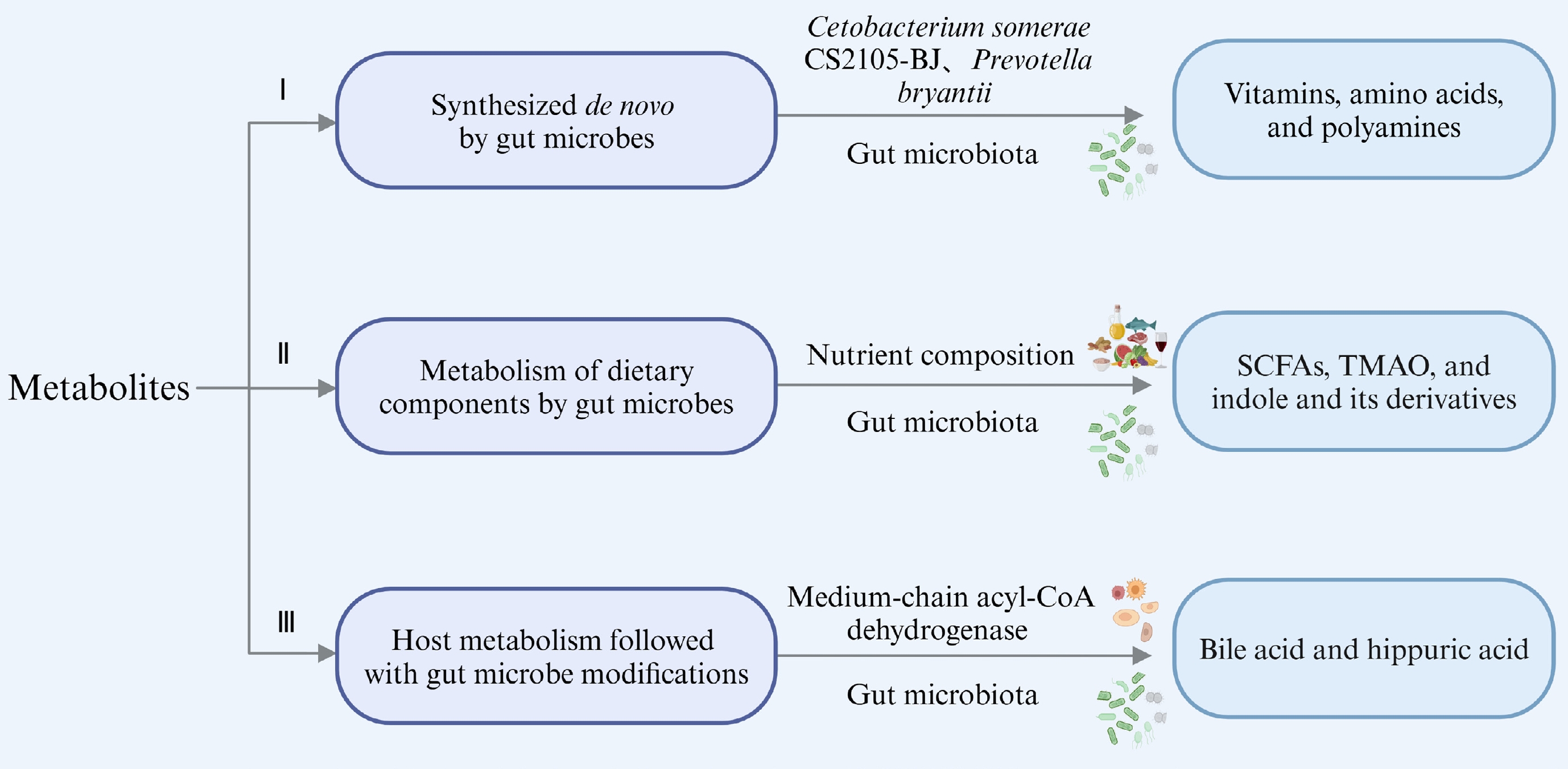
      Figure 1. 

      Types of microbial metabolites derived from various sources (created with BioRender.com).

    • Research indicates that the gut microbiota can synthesize vitamin K and the majority of water-soluble B vitamins like biotin, riboflavin, thiamine, folates, pantothenic acid, and nicotinic acid[16]. For instance, cobalamin (vitamin B12) is exclusively produced by microbes, specifically anaerobic bacteria[17]. Genomic analysis combined with in vivo and in vitro metabolic experiments confirmed that Cetobacterium somerae CS2105-BJ is capable of de novo vitamin B12 synthesis. It contains essential genes (hemL, cbiT/cobD, and cobC) that play a role in various stages of B12 biosynthesis, which are responsible for the synthesis of uroporphyrinogen III, adenosylmethylcobalamin, and lower ligand, respectively[18,19]. In situations where dietary vitamin B2 was depleted in a mouse model, the gut microbiota provided short-term compensation[20]. Certain vitamins, like B vitamins, which serve as precursors to essential metabolic cofactors, can be synthesized by specific gut bacteria known as prototrophic bacteria. However, they must also be obtained from other bacterial species (auxotrophic bacteria) and from the host’s diet[21]. This highlights that microbial synthesis of specific metabolites may depend on environmental conditions and the availability of exogenous sources, necessitating multi-bacterial cooperation. Additionally, amino acids and fatty acids can be synthesized de novo by gut microbes. Research has demonstrated ruminal bacteria such as Selenomonas ruminantium, Streptococcus bovis, and Prevotella bryantii engage in amino acid synthesis under physiological conditions, particularly enriching glutamic acid and aspartate in the presence of peptides[22]. Bacteroidetes are known symbionts capable of sphingolipid production. For example, Bacteroides thetaiotaomicron can initiate biological sphingolipid synthesis via serine palmitoyl transferase[23].

    • Microbial dietary metabolites include short-chain fatty acids (SCFAs), trimethylamine oxide (TMAO), and indole and its derivatives. SCFAs, primarily derived from dietary fiber fermentation, are a major focus. For example, resistant starch type 3 from potatoes promotes the production of propionic and acetic acids by Bifidobacterium, Ruminococcus, Bacteroides, and Coprococcus[24]. Pectin, rich in rhamnogalacturonan-I, significantly promotes the proliferation of Bifidobacterium, Faecalibaculum, and Lactobacilli in C57BL/6J mice, and it is metabolized to SCFAs[25]. Dietary inulin supports SCFA-producing Ruminococcaceae and Lachnospiraceae, increasing fecal SCFA concentrations[26]. Specifically, Bifidobacterium produce lactate and acetate, Bacteroides generate propionate and acetate, and Butyrivibrio and Fusobacterium produce butyrate as their primary metabolites[27]. Other bacteria that produce SCFAs include Anaerostipes, Anaerotruncus, Bacteroides, Coprococcus, Clostridium, Dialister, Eubacterium, Faecalibacterium, Lactobacillus paracasei, Odoribacter, Parabacteroides, and Ruminococcus[2832].

      Tryptophan metabolites, particularly indoles, such as 3-indolepropionic acid, indoleacetic acid, indole acrylate, indoleacrylic acid, indole-3-carbaldehyde, and tryptamine, form another extensively studied group of dietary metabolites[33]. For instance, 3-indolepropionic acid is biosynthesized from tryptophan by Clostridium sporogenes[34]. Commensal Peptostreptococcus metabolizes tryptophan into indoleacrylic acid[35], while Bifidobacterium longum CCFM1029 produces indole-3-carbaldehyde[36]. Putrescine, derived from arginine through a hybrid pathway involving Escherichia coli and Enterococcus faecalis, is another notable metabolite. Bifidobacterium spp. that produce acidic compounds accelerate putrescine production[37].

      Choline metabolites are classic nitrogenous dietary compounds. The synthesis of TMAO increases following the consumption of a diet high in phosphatidylcholine and L-carnitine[38]. Gut microbiota, like Citrobacter freundii, are capable of transforming into trimethylamine (TMA), which is then oxidized to TMAO[9]. Research has indicated that trigonelline can reduce TMAO levels by inhibiting the activity of C. freundii and flavin-containing monooxygenase 3 in mice. In vitro experiments also showed that the addition of fenuelline to choline-rich medium could significantly inhibit the production of C. freundii and reduce TMA and MAO production[9,39]. In another study involving obese hypertensive individuals, Lactobacillus plantarum was found to decarboxylate ornithine to produce putrescine[40]. Spermidine production is facilitated by some gut microbiota, such as E. coli and Bacteroides, utilizing carboxyspermidine decarboxylase and spermidine synthase[41,42]. Additionally, inosine, as a purine nucleotide metabolite, may be produced by Lactobacillus during the fermentation of barley leaves[43]. In general, there are a wide variety of microbial metabolites from dietary sources, and the efficacy of many substances, including SCFAs and indole derivatives, have been extensively studied with their relevant mechanism identified. Whether new dietary metabolites with good efficacy beyond these substances can be identified needs further exploration.

    • The liver serves as the primary organ to regulate cholesterol homeostasis, controlling both cholesterol intake and de novo synthesis. Endogenous cholesterol synthesis is mainly regulated by the SCAP-SREBP-HMGCR pathway[44,45]. In addition, a microbial enzyme called ismA has been identified previously that can convert cholesterol into coprostanol, a lipid that is excreted from the body rather than being absorbed[46]. Yao et al.[47]identified a gene encoding the sulfotransferase (BtSULT, BT0416), which is widely found in Bacteroides and mediates the sulfation of cholesterol.

      Bile acids (BAs), which are synthesized in the liver from cholesterol under the action of cholesterol- 7α-hydroxylase (CYP7A1), and are conjugated with taurine or glycine, undergo modifications by gut microbiota to produce secondary BA metabolites[43,48,49]. Pruss et al.[50] used stable isotope tracers along with bacterial and host genes to uncover a common metabolic pathway of hippuric acid production by host microorganisms. They confirmed that Clostridium sporogenes reduced phenylalanine into phenylpropionic acid[50-51]. The third type of metabolite, which is metabolized by the host and subsequently modified by microorganisms, has not been detailedly explained in most studies. This complexity likely arises from the intricate interactions between the host and gut microbiota, which makes it challenging to distinctly delineate their respective roles in metabolite modification.

    • Metabolic diseases, such as diabetes, atherosclerosis, hypertension, and obesity, are often linked with dysregulation of gut microbial metabolites[9]. Natural products-derived metabolites, including alkaloids, terpenoids, and flavonoids, undergo substantial modification by gut microbiota, affecting their pharmacological effects and roles in metabolic diseases[52]. These compounds typically exhibit low absorption rates in intestinum tenue and are further broken down by microbiota in the colon into microbial metabolites, which have been confirmed to play a significant role in regulating metabolic diseases[52]. The following section will specifically explore the interaction between these diet-derived metabolites and gut microbiota, as well as their functions in metabolic diseases (Fig. 2).
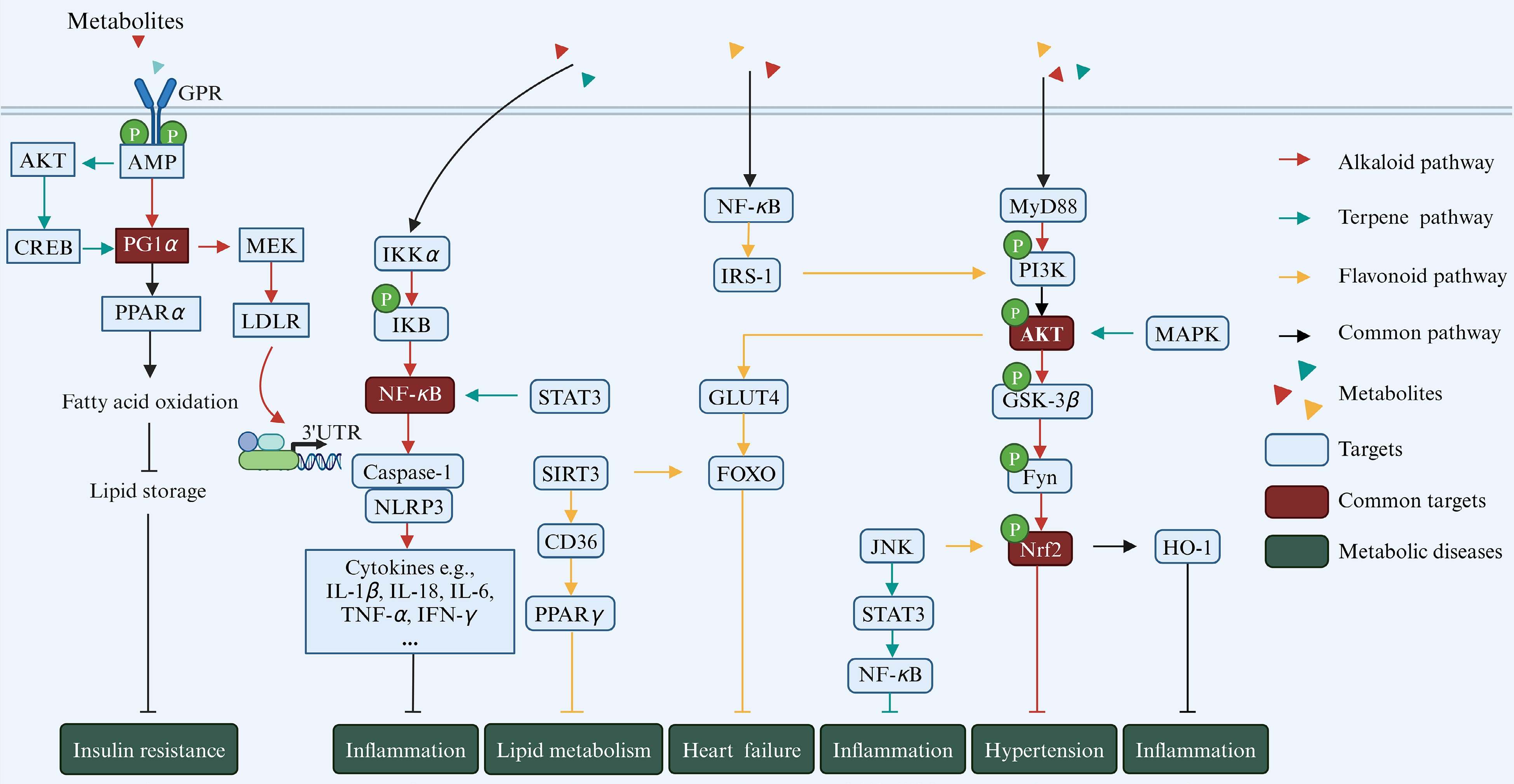
      Figure 2. 

      Natural product-derived gut microbial metabolites in metabolic diseases. Different colors refer to different classes of metabolites, with red representing alkaloids, green representing terpenoids, and yellow representing flavonoids. The blue boxes indicate key sites on the pathways, and the brown boxes indicate key sites shared by the three metabolites (created with BioRender.com).

    • Gut microbiota are essential for the absorption of alkaloids, such as Staphylococcus aureus, Enterococcus faecalis, and Enterococcus faecium, and they possess nitrate reductase enzymes that metabolize alkaloids[53,54]. Previous studies have indicated that the cytochrome CYP450 family, including CYP2D6, CYP1A2, CYP51, and CYP7A1, plays a significant role in the metabolic transformation of alkaloids[5557], including O-demethylation and hydroxylation[55 ]. Alkaloids can influence the body’s ecological physiology by regulating gut microbiota and their metabolic products. Supplementation with alkaloids increases the abundance of Bacteroides, Parabacteroides[58], Ruminococcus[59], Blautia[60], Faecalibaculum, Allobaculum[61], Clostridiales, and Lactobacillus[62], which are shown to produce SCFAs, TMAO, and BAs[61,63,64].

      The primary causes of obesity are abnormalities in energy balance and weight regulation, often accompanied by enlarged fat cells and changes in insulin function, which can further lead to the development of diabetes. Peroxisome proliferator activated receptor alpha (PPARα) and peroxlsome proliferator-activated receptor-gamma coactivator-1 alpha (PGC1α) are associated with the proliferation of adipose tissue and adipocyte hypertrophy following dietary intake, while targets such as liver X receptor (LXR), farnesoid X receptor (FXR), and CYP7A1 play crucial roles in regulating lipid, glucose, and energy metabolism, making them key targets for the treatment of metabolic diseases such as obesity and diabetes[6567]. Alkaloids can effectively act on the above targets and play a key role in disease improvement. For instance, palmatine treatment can regulate BA metabolism, reduce cholesterol accumulation, and alleviate obesity by enhancing the expression of PPARα and CYP7A1 while suppressing the expression of FXR[68]. Berberine can inhibit hepatic lipogenesis by up-regulating the expression of intestinal FXR and fibroblast growth factor 15, as well as activating the AMP-activated protein kinase (AMPK)-dependent Raf-1 pathway, thereby affecting the expression of the low-density lipoprotein receptor (LDLR)[62,69]. Notably, LDLR is a transmembrane glycoprotein primarily responsible for clearing lipoproteins, and an effective therapeutic target for treating hypercholesterolemia and related cardiovascular diseases[69]. In diabetic mice, supplementing with ruthenine improved the disease by activating the PPARα/PGC1α signaling pathway and upregulating β-oxidation-related genes in the liver[70]. In addition, studies have shown that Streptococcus faecalis can convert berberine in Rhizoma coptidis into oxyberberine, a potent hypoglycemic metabolite[54]. This metabolite contributes to anti-diabetes by upregulating the mRNA expression of the pancreatic nuclear factor erythroid 2-related factor 2 (Nrf2) signaling pathway and activating the phosphatidylinositol 3-kinase (PI3K)/protein kinase B (Akt) signaling pathway[71].

      Alkaloids exhibit anti-inflammatory properties by inhibiting pathways such as TRIF-dependent NF-κB signaling. For example, oxyberberine modulates the toll-like receptor 4 (TLR4)-myeloid differentiation factor 88 (MyD88)-NF-κB pathway[54] and the TLR4/PI3K/NF-κB pathways, reducing inflammatory responses[71]. For another, berberine can exert its effects by inhibiting the phosphorylation of insulin receptor substrate-1 (IRS-1) and increasing the phosphorylation of AKT in adipose tissue and cultured adipocytes[72]. These studies highlight the significant role of alkaloids in human health by interacting with gut microbiota and their metabolic products, thereby contributing to the maintenance of homeostasis. Despite extensive research on the structure and physiological activity of alkaloids metabolites, investigations into their in vivo roles and specific mechanisms in regulating human health are still limited.

    • Terpenoids have demonstrated efficacy in ameliorating metabolic disorders by regulating gut microbiota. Studies indicate that terpenoids such as fucoxanthin affect the Firmicutes/Bacteroidetes ratio and influence the abundance of key species, which are pivotal in anti-obesity activities[73]. Additionally, terpenoids have been shown to enhance populations of Lactobacillus, Bifidobacterium, Clostridium sensu stricto 1, Turicibacter, uncultured Erysipelotrichaceae, Faecalibaculum, and Ruminococcus, while reducing Lachnospiraceae NK4A136, uncultured Lachnospiraceae, and taxa such as Prevotellaceae UCG-003 and Oscillospira[7476]. In another context, the administration of ginsenoside extract enriched gut E. faecalis significantly[77]. Zhang et al.[78] confirmed that a guava extract abundant in 1-3 glucose-based residues of triterpenoid glycosides has significantly regulated Elusimicrobium, Lachnospiraceae UCG-004, acetic acid, butyric acid, and 1β-hydroxycholic acid in T2DM rats. Gentiopicroside has been shown to directly bind FGFR1, promoting FGF21 signaling, and subsequently activating PI3K/AKT and AMPK pathways to regulate glucose and lipid metabolism[79]. Reports indicate that gentiopicroside can be metabolized by intestinal bacteria into potential pharmacologically active metabolites such as erythrocentaurin, gentianine, and gentianal[80]. Consumption of erythrocentaurin can inhibit α-amylase activity, improving postprandial hyperglycemia, while supplementation with gentiopicroside may reduce inflammation by inhibiting TLR4/NLRP3-mediated pyroptosis[80,81].

      The anti-inflammatory mechanisms of terpenoids involve several key factors. Glycyrrhetic acid is a triterpenoid saponin extracted from licorice. It can be hydrolyzed by β-D glucuronidase of Eubacterium sp. strain GLH and then converted to Glycyrrhetic acid 3-O-mono-D-glucuronide, enhancing anti-inflammatory activity[82]. Han et al.[75]illustrated that lactucin curtails p38, extracellular signal-regulated kinase (ERK), and AKT phosphorylation within lipopolysaccharides (LPS)-stimulated RAW264.7 cells, consequently diminishing the mRNA and protein levels of inducible nitric oxide synthase and cyclooxygenase-2, and attenuating interleukin-6 (IL-6) production. Jolkinolide B mitigates LPS-induced degradation of NF-κB inhibitor alpha (IκBα) and phosphorylation of p65 and MAPK, subsequently reducing histological alterations and inflammatory markers[83]. Limonin, a triterpenoid extracted from citrus, mitigates intestinal inflammation by downregulating p-STAT3/miR-214 levels, demonstrating anti-inflammatory and apoptotic effects[84]. Fucoxanthin, derived from marine algae, activates Nrf2/Antioxidant response element (ARE) through PI3K/Akt pathways, enhancing antioxidant synthesis[8587]. Fucoxanthin also activates AMPK, reducing fatty acid synthesis via the sirtuin 1 (SIRT1)/AMPK pathway[88]and improving mitochondrial function while reducing oxidative stress in endothelial cells through the AMPK-Akt-cAMP response element-binding protein (CREB)-PGC1α pathway[89].

      Phytol, a plastid terpenoid synthesized via the methylerythritol phosphate pathway in plastids, is released when chlorophyll molecules are partially digested in animals[90]. It is absorbed in the small intestine and converted to phytanic acid in the liver[91]. Phytanic acid activates PPARγ and the retinoid-X-receptor (RXR), regulating lipid metabolism in various cell types[92]. The activation of PPARγ in adipose tissues improves insulin resistance, while PPARα activation in the liver lowers circulating lipid levels[93,94]. Terpenoids derived from Resina commiphora modulate lipid metabolism by augmenting PPARα and carnitine palmitoyl transferase 1 (CPT1) expression[95]. Auraptene, a novel PPARα and PPARγ ligand found in citrus fruits exerts anti-atherosclerotic and anti-diabetic effects by balancing anti-inflammatory/pro-inflammatory cytokine levels in adipocytes[96]. These functions of dietary terpenoids are important for the control of metabolic diseases through dietary intervention.

    • Flavonoids can be classified into several categories: flavones (including apigenin and luteolin), flavonols (including quercetin and curcumin), flavanones (including naringenin and hesperidin), flavanols (including catechin and epicatechin), isoflavones (including daidzein and genistein), and anthocyanins (including pelargonidin and cyanidin). They are primarily derived from fruits, vegetables, legumes, onions, seeds, plant roots and stems, tea, wine, olive oil, tomato sauce, and various dietary supplements[97,98]. After supplementing the diet with flavonoid-rich foods, such as soybean, bilberry, citrus fruit, and green tea, the enzymes produced by gut bacteria undergo various reactions including deglycosylation, demethylation, and oxidation, which generate biologically active metabolites with health benefits such as genistein, quercetin, anthocyanin, and catechin[99]. Santangelo et al.[100] summarized multiple studies showing that quercetin inhibits some intestinal bacteria such as Streptococcus, Lactobacillus, Bifidobacterium, and Bacteroides, and was converted into small molecules like phenylacetic acid, phenylpropanoic acid, dihydroxyphenylacetic acid, hydroxybenzoic acid, dihydroxybenzoic acid, and propionic acid, absorbed and utilized by the body[101]. The catabolism of cyanidin-3-O-glucoside in the gastrointestinal tract yields bioactive metabolites like protocatechuic acid, phloroglucinaldehyde, vanillic acid, and ferulic acid, supporting the integrity of the mucosal barrier and improving the health of the microbiota[102,103]. Escherichia fergusonii and E. coli metabolize curcumin into dihydrocurcumin, tetrahydrocurcumin, and ferulic acid[104]. In particular, Bifidobacterium animalis subsp. lactis AD011 efficiently converted 85% of quercetin 3-glucoside and isorhamnetin 3-glucoside into quercetin and isorhamnetin, respectively, within just 2 h without degrading the flavonoid backbone[105].

      Flavonoids exert significant effects on metabolic diseases through interactions with key targets such as PPAR, AMPK, Nrf2, and NF-κB. For instance, they can bind to non-phosphorylated signal transducer and activator of transcription 3 (STAT3) in visceral adipose tissue, reducing its phosphorylation and transcriptional activity. This action leads to a decreased expression of the STAT3 target gene CD36, which typically influences adipogenesis and anti-visceral obesity by suppressing PPAR-γ expression[106]. Daidzein influences cardiac energy metabolism by modulating Sirtuin 3 (SIRT3), thereby improving lipid, glucose and ketone body metabolism disorders, and mitigating mitochondrial dysfunction, which is crucial for meeting cardiac ATP demands both in vivo and in vitro[107]. Daidzein influences the LXR signaling pathway, activating RXR, PPARα, and AMPKα, which enhances energy metabolism[108,109]. In skeletal muscle, daidzein inhibits the Glut4/AMPK/Forkhead box O (FoxO) pathway and reduces the expression of atrogin1 and muscle ring finger protein1, thereby inhibiting the protein degradation of the skeletal muscle[110].

      Insulin induces GLUT-4 transport from the vesicle to the plasma membrane, thereby promoting glucose uptake in cells, through the PI3K/Akt pathway. Disruption of these pathways may lead to insulin resistance, which can subsequently result in diabetes[111]. Fraction D, rich in flavonoids isolated from Enicostema littorale, enhances cell glucose uptake by upregulating IRS-1/PI3K/Akt pathway components[112]. Both cyanidin-3-O-β-glucoside and pinocembrin inhibit inflammatory pathways like NF-κB and TLR4/MD2, providing therapeutic benefits for liver and intestinal inflammation[113,114]. Cyanidin-3-O-β-glucoside treatment stimulates SIRT1 activity, inhibits NF-κB acetylation, and subsequently inhibits the activation of inflammasomes and the release of pro-inflammatory cytokines in liver cell lines[113]. Quercetin in diet improves experimental colitis by enhancing the anti-inflammatory and antibacterial capacities of macrophages through the Nrf2/Heme Oxygenase 1 (HO-1) pathway[115]. Additionally, the inclusion of Eucommia ulmoides flavones leads to increased levels of phosphorylated Akt, IκBα, and IKKα/β, while simultaneously reducing the expression of Bax and Caspase-3 proteins in LPS-treated cells[116]. Other flavonoid metabolites, including protocatechuic acid, phloroglucinol, and vanillic acid, can downregulate the MAPK pathway by inhibiting the phosphorylation of ERK, c-Jun N-terminal kinase (JNK), and p38[117,118]. Overall, flavonoid metabolites play active roles in modulating various pathways, including NF-κB, MAPK, ERK, JNK, and p38, contributing to their anti-inflammatory and metabolic regulatory effects in different physiological conditions.

    • Food processing, including thermal processing and non-thermal processing, plays a crucial role in our daily diet[119]. These processes can alter the nutritional composition of food by introducing new compounds or converting existing ones[120] (Fig. 3). Considering that utilizing dietary interventions to improve metabolic diseases has become an effective means[121], its efficacy is highly correlated with gut microbiota metabolites as described previously. Therefore, comprehending the effects of food processing technology is crucial for assessing the nutritional value of processed foods and selecting appropriate processed foods based on differential health needs.
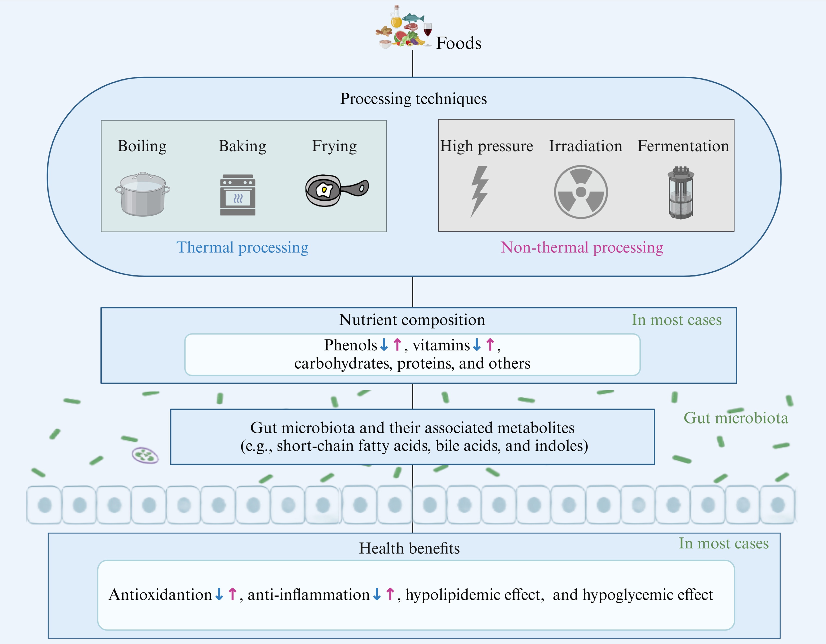
      Figure 3. 

      Impact of food processing on dietary nutrients and their health benefits via gut microbiota and related metabolites. The blue arrows represent the impact of thermal processing, while the purple arrows represent the impact of non-thermal processing (created with BioRender.com).

    • Heat treatments, such as boiling, baking, stewing, steaming, frying, and roasting, are widely used in both domestic and industrial food processing[122]. Previous research has indicated that the carbohydrate levels in grains, vegetables, and fruits typically rise following heat treatment[123, 124]. For example, heat-treated highland barley has been found to have a high dietary fiber content[124]. Similarly, it is found that boiling increases the content of fiber by 8.32% in garden cress seeds[125]. The increase in dietary fiber after thermal processing yields some beneficial outcomes, such as boosting the prevalence of beneficial gut bacteria including Bifidobacteria and Bacteroides, while suppressing harmful bacteria like Helicobacter and Enterococcus[126]. It also enhances the levels of SCFAs. These typical gut bacteria and metabolites are strongly associated with lipid metabolism and can effectively improve metabolic diseases[127,128]. The variability of these nutrients highlights that different processing methods may impact the efficacy of food intervention in varying ways, possibly due to changes in active components and their related metabolites[129,130].

      Nevertheless, it is crucial to recognize that most nutrients, including proteins, phenols, vitamins, and minerals, are lost after thermal processing[123]. During heat treatment, protein structures can undergo modifications due to direct oxidation or complex reactions with other components of the food, leading to changes in processing characteristics and a reduction in nutritional value, with potential negative health implications[131]. For example, heat treatment of hawthorn has been shown to cause the degradation of soluble phenols and proanthocyanidins[132], which are vital in the gastrointestinal tract due to their antioxidant, anti-inflammatory, and antibacterial properties[133]. These compounds also promote the growth of probiotics like Bifidobacterium and Lactobacillus, offering potential health benefits. Additionally, steaming fresh tea significantly reduces the levels of vitamins (B2, B3, and C) and minerals[123]. Vitamin B2 has been reported to prevent cancer, hyperglycemia, hypertension, diabetes, oxidative stress, and other health conditions, while vitamin C is known for its anti-inflammatory, antioxidant, and hypoglycemic effects[134,135]. The loss of these nutrients significantly reduces the health benefits of processed foods. Taken together, while heat treatment can increase dietary fiber, it can also have varying degrees of negative effects on the original nutrients and health benefits of the diet.

    • Non-thermal processing technologies, such as cold plasma treatment, irradiation, high-pressure processing, ultrasound, pulsed light technology, pulsed electric field treatment, and microbial fermentation, are widely used in food processing to circumvent the negative effects of heat[136]. These methods are designed to eliminate the need for high temperatures, thereby preserving the nutritional integrity of food[137]. For example, high-pressure electric field treatment has been demonstrated to elevate the anthocyanin content in strawberries, thereby improving its bioavailability[138,139]. In addition, ultrasound and ozone treatments have been found to increase the levels of phenols and vitamin C, and the antioxidant capacity of guava juice[140]. Mehta et al. found that cold plasma treatment for 10 min retained up to 95% of vitamin C [141]. These polyphenols and vitamins are crucial for human health[142], highlighting the potential of non-thermal processing to optimize the nutritional retention and even enhance the nutritional value of food products. As demonstrated by Tan et al.[143], high hydrostatic pressure treatment at 400 MPa for 15 min altered the monosaccharide composition of Cyanidin 3-glucoside and blueberry pectin complexes, decreasing mannose, fructose, glucose, xylose, and arabinose while increasing galactose, which improved colitis in mice through reducing intestinal oxidative stress, enhancing anti-inflammatory factors, and inhibiting the NF-κB signaling pathway mediated by the gut microbiota. Similar benefits have been observed with plasma treatment of orange juice and apple juice[144]. In terms of proteins, cold plasma treatment can oxidize hairtail muscle proteins, creating a more intensive protein network, which improves the texture and taste of the hairtail[145].

      Microbial fermentation in food processing can lead to the formation of specific nutritional components due to the involvement of microorganisms. For instance, probiotic fermentation significantly increases γ-aminobutyric acid, rutin, total polyphenols, and total flavonoids in rice buckwheat[146]. Fermented rice buckwheat has been shown to reverse high-fat diet-induced intestinal dysbiosis, suppress hepatic cholesterol synthesis and lipogenesis, and stimulate lipolysis by rebalancing the Firmicutes/Bacteroidetes ratio, enhancing SCFA-producing bacteria like Bacteroidetes, Lactobacillus, and Blautia, and increasing total SCFA content[147]. Furthermore, blueberry residue fermented by Lactobacillus rhamnosus GG and Lactobacillus plantarum-1 significantly suppressed inflammation-related cytokines (tumor necrosis factor-α, IL-1β, IL-6), upregulated PPAR-α, and downregulated sterol regulatory element-binding protein-1 and fatty acid synthase, thereby enhancing lipid metabolism[147]. Additionally, Yan et al.[148]observed a rise in lactic acid and a decrease in citric acid during the fermentation process. Recently, new non-thermal technologies, such as selenium processing, have emerged. For example, our previous study found that selenium processing markedly increased total carbohydrate and soluble dietary fiber content while decreasing protein and insoluble dietary fiber content in Cordyceps militaris[149]. It also enhanced the biosynthesis of secondary metabolites, such as terpenoids and alkaloids. The application of these new technologies not only preserves food quality and enhances flavor but also allows for adjusting the nutritional composition of food to meet enhanced functional requirements.

    • Gut microbial-associated metabolites are systematically categorized into three groups: de novo synthesis, microbial dietary metabolism, and host metabolic contributions. The beneficial role and potential mechanism of novel gut microbial metabolites derived from natural products in chronic diseases are becoming clear. Furthermore, gut microbiota and their associated metabolites provide valuable insights into how food processing influences nutritional value and health benefits. However, the intricate diversity and complexity of these metabolites make the precise molecular mechanisms underlying their effects still poorly comprehended. To address this, future studies should aim to: (i) accurately elucidate the entire production process of metabolites; (ii) clarify the spatial and temporal efficacy targets of metabolites; and (iii) comprehensively understand the regulatory patterns of different types of metabolites in the body. Achieving these goals in future research will pave the way for deeper exploration of gut microbial metabolites and their health functions, providing more insightful comprehension and innovation in this field.

      • This work was supported by the Guangdong Basic and Applied Basic Research Foundation (2023A1515010744), National Natural Science Foundation of China (32202014), GDAS' Project of Science and Technology Development (2022GDASZH-2022010101), and State Key Laboratory of Applied Microbiology Southern China (SKLAM002-2022).

      • The authors confirm their contribution to the paper as follows: writing - original draft: Huang A; visualization: Huang A, Chen M; writing - review & editing: Ding Y, Zhu Z, Thanuphol P, da Cruz LL, Xie Z; supervision: Ding Y, Zhang F, Zhu Z; resources: Wu Q, Ding Y, Zhu Z; project administration: Ding Y, Zhu Z; funding acquisition: Wu Q, Ding Y, Zhu Z. All authors reviewed the results and approved the final version of the manuscript.

      • All data generated or analyzed during this study are included in this published article.

      • The authors declare that they have no conflict of interest.

      • Copyright: © 2024 by the author(s). Published by Maximum Academic Press on behalf of China Agricultural University, Zhejiang University and Shenyang Agricultural University. This article is an open access article distributed under Creative Commons Attribution License (CC BY 4.0), visit https://creativecommons.org/licenses/by/4.0/.
    Figure (3)  References (149)
  • About this article
    Cite this article
    Huang A, Wu Q, Thanuphol P, da Cruz LL, Xie Z, et al. 2024. Gut microbiota-associated metabolites in metabolic diseases and their impact from food processing. Food Innovation and Advances 3(4): 438−448 doi: 10.48130/fia-0024-0038
    Huang A, Wu Q, Thanuphol P, da Cruz LL, Xie Z, et al. 2024. Gut microbiota-associated metabolites in metabolic diseases and their impact from food processing. Food Innovation and Advances 3(4): 438−448 doi: 10.48130/fia-0024-0038

Catalog

  • About this article

/

DownLoad:  Full-Size Img  PowerPoint
Return
Return