ARTICLE   Open Access    

Quantitative analysis of curcumin compounds in ginger by ultra-high-performance liquid chromatography coupled with tandem mass spectrometry

More Information
  • Curcumin compounds are important bioactive compounds in ginger, yet their analysis is limited by their low concentrations. In the current research, a highly sensitive and reliable approach for simultaneous quantitative detection of three curcumin compounds in ginger samples was established using ultra-high-performance liquid chromatography coupled with tandem mass spectrometry (UHPLC-MS/MS). The extraction solvent, volume of extraction solvent, sonication time, and oscillation time were optimized by a single factor experiment. The method validation results showed that the regression coefficients were higher than 0.9990, and the linearity was satisfactory. Matrix effects were negligible with the values of 94.6%–98.8%. The recovery at three spiking levels was between 81.7% and 100.0%, and the precision was less than 5.4%. The approach could be used to determine the curcumin components in ginger samples since the results demonstrate that it is easy to use, practicable, repeatable, and accurate.
  • Atractylodes macrocephala Koidz. (common names 'Baizhu' in Chinese and 'Byakujutsu' in Japanese) is a diploid (2n = 2x = 24) and out-crossing perennial herb in the Compositae family, and has a long history of cultivation in temperate and subtropical areas of East Asia as it is widely used in traditional herbal remedies with multiple pharmacological activities[13]. The 'Pharmacopoeia of the People's Republic of China' states that 'Baizhu' is the dry rhizome of A. macrocephala Koidz. (Atractylodis Macrocephalae Rhizoma, AMR). However, in Japanese traditional medicine 'Baizhu' can be referred to both: A. japonica or A. macrocephala[4].

    A. macrocephala is naturally endemic to China and cultivated in more than 200 towns in China, belonging to Zhejiang, Hunan, Jiangxi, Anhui, Fujian, Sichuan, Hubei, Hebei, Henan, Jiangsu, Guizhou, Shanxi, and Shaanxi Provinces[3]. A. macrocephala grows to a height of 20–60 cm (Fig. 1). The leaves are green, papery, hairless, and generally foliole with 3–5 laminae with cylindric glabrous stems and branches. The flowers grow and aggregate into a capitulum at the apex of the stem. The corollas are purplish-red, and the florets are 1.7 cm long. The achenes, densely covered with white, straight hairs, are obconic and measure 7.5 mm long. The rhizomes used for medicinal purposes are irregular masses or irregularly curving cylinders about 3–13 cm long and 1.5–7 cm in diameter with an outwardly pale greyish yellow to pale yellowish color or a sparse greyish brown color. The periderm-covered rhizomes are externally greyish brown, often with nodose protuberances and coarse wrinkles. The cross-sections are white with fine dots of light yellowish-brown to brown secretion. Rhizomes are collected from plants that are > 2 years old during the spring. The fibrils are removed, dried, and used for medicinal purposes[5, 6].

    Figure 1.  Plant morphology of A. macrocephala.

    The medicinal properties of AMRs are used for spleen deficiency, phlegm drinking, dizziness, palpitation, edema, spontaneous sweating, benefit Qi, and fetal restlessness[7]. The AMR contains various functional components, among which high polysaccharide content, with a yield close to 30%[8]. Therefore, the polysaccharides of A. macrocephala Koidz. rhizome (AMRP) are essential in assessing the quality control and bioactivity of A. macrocephala. Volatile oil accounts for about 1.4% of AMR, with atractylon and atractylodin as the main components[9]. Atractylon can be converted to atractylenolide I (AT-I), atractylenolide II (AT-II), and atractylenolide III (AT-III) under ambient conditions. AT-III can be dehydrated to AT-II under heating conditions[10, 11]. AMRs, including esters, sesqui-, and triterpenes, have a wide range of biological activities, such as improving immune activity, intestinal digestion, neuroprotective activity, immune anti-inflammatory, and anti-tumor.

    In recent years, research on the pharmacological aspects of AMR has continued to increase. Still, the discovery of the main active components in AMR is in its infancy. The PAO-ZHI processing of AMR is a critical step for AMR to exert its functional effects, but also, in this case, further work is required. Studies on the biosynthesis of bioactive compounds and different types of transcriptomes advanced current knowledge of A. macrocephala, but, as mentioned, required more systematic work. Ulteriorly, an outlook on the future research directions of A. macrocephala was provided based on the advanced technologies currently applied in A. macrocephala (Fig. 2).

    Figure 2.  Current progress of A. macrocephala.

    A. macrocephala is distributed among mountainous regions more than 800 m above sea level along the middle and lower reaches of the Yangtze River (China)[5]. Due to over-exploitation and habitat destruction, natural populations are rare, threatened, and extinct in many locations[1,12]. In contrast to its native range, A. macrocephala is widely cultivated throughout China, in a total area of 2,000–2,500 ha, with a yield of 7,000 t of rhizomes annually[13]. A. macrocephala is mainly produced in Zhejiang, Anhui, and Hebei (China)[14]. Since ancient times, Zhejiang has been the famous producing area and was later introduced to Jiangxi, Hunan, Hebei, and other places[15]. Wild A. macrocephala is currently present in at least 14 provinces in China. It is mainly distributed over three mountain ranges, including the Tianmu and Dapan mountains in Zhejiang Province and the Mufu mountains along the border of Hunan and Jiangxi Provinces. A. macrocephala grows in a forest, or grassy areas on mountain or hill slopes and valleys at an altitude of 600–2,800 m. A. macrocephala grows rapidly at a temperature of 22–28 °C, and favors conditions with total precipitation of 300–400 mm evenly distributed among the growing season[16]. Chen et al. first used alternating trilinear decomposition (ATLD) to characterize the three-dimensional fluorescence spectrum of A. macrocephala[17]. Then they combined the three-dimensional fluorescence spectrum with partial least squares discriminant analysis (PLS-DA) and k-nearest neighbor method (kNN) to trace the origin of Atractylodes samples. The results showed that the classification models established by PLS-DA and kNN could effectively distinguish the samples from three major Atractylodes producing areas (Anhui, Hunan, and Zhejiang), and the classification accuracy rate (CCR) of Zhejiang atractylodes was up to 80%, and 90%, respectively[17]. Zhang et al. compared the characteristics, volatile oil content, and chemical components of attested materials from six producing areas of Zhejiang, Anhui, Hubei, Hunan, Hebei, and Henan. Differences in the shape, size, and surface characteristics were reported, with the content of volatile oil ranging from 0.58% to 1.22%, from high to low, Hunan (1.22%) > Zhejiang (1.20%) > Anhui (1.02%) > Hubei (0.94%) > Henan (0.86%) > Hebei (0.58%)[18]. This study showed that the volatile oil content of A. macrocephala in Hunan, Anhui, and Hubei is not much different from that of Zhejiang, which is around 1%. A. macrocephala is a local herb in Zhejiang, with standardized cultivation techniques, with production used to reach 80%–90% of the country. However, in recent years, the rapid development of Zhejiang's real estate economy has reduced the area planted with Zhejiang A. macrocephala, resulting in a sudden decrease in production. Therefore, neighboring regions, such as Anhui and Hunan, vigorously cultivate A. macrocephala, and the yield and quality of A. macrocephala can be comparable to those of Zhejiang. The results were consistent with the data reports[18]. Guo et al. analyzed the differentially expressed genes of Atractylodes transcripts from different regions by the Illumina HiSeq sequencing platform. It was found that 2,333, 1,846, and 1,239 DEGs were screened from Hubei and Hebei, Anhui and Hubei, and Anhui and Hebei Atrexia, respectively, among which 1,424, 1,091, and 731 DEGs were annotated in the GO database. There were 432, 321, and 208 DEGs annotated in the KEGG database. These DEGs were mainly related to metabolic processes and metabolic pathways of secondary metabolites. The highest expression levels of these genes were found in Hubei, indicating higher terpenoid production in Hubei[19]. Other compounds were differentially accumulated in Atractylodes. Chlorogenic acid from Hebei was 0.22%, significantly higher than that from Zhejiang and Anhui[20]. Moreover, the content of neochlorogenic acid and chlorogenic acid decreased after processing, with the highest effect reported in Zhejiang, with the average transfer rate of neochlorogenic acid and chlorogenic acid reaching 55.68% and 55.05%[20]. All these changes would bring great help in distinguishing the origins of A. macrocephala.

    Medicinal AMR can be divided into raw AMR and cooked AMR. The processing method is PAO-ZHI; the most traditional method is wheat bran frying. The literature compared two different treatment methods, crude A. macrocephala (CA) and bran-processed A. macrocephala, and found that the pharmacological effects of AMR changed after frying with wheat bran, mainly in the anti-tumor, antiviral and anti-inflammatory effects[21]. The anti-inflammatory effect was enhanced, while the anti-tumor and antiviral effects were somewhat weakened, which may be related to the composition changes of the compounds after frying. The study of the content of AT-I, II, and III, and atractyloside A, in rat serum provided helpful information on the mechanism of wheat bran processing[22]. In addition to frying wheat bran, Sun et al. used sulfur fumigation to treat AMR[23]. They found that the concentration of different compounds changed, producing up to 15 kinds of terpenoids. Changes in pharmacological effects were related to treatment and the type of illumination[24,25]. Also, artificial light can improve the various biological functions. A. macrocephala grew better under microwave electrodeless light, with a chlorophyll content of 57.07 ± 0.65 soil and plant analyzer develotrnent (SPAD)[24]. The antioxidant activity of AMR extract treated with light-emitting diode (LED)-red light was the highest (95.3 ± 1.1%) compared with other treatments[24]. The total phenol and flavonoid contents of AMR extract treated with LED-green light were the highest at 24.93 ± 0.3 mg gallic acid equivalents (GAE)/g and 11.2 ± 0.3 mg quercetin equivalents (QE)/g compared with other treatments[24, 25]. Polysaccharides from Chrysanthemun indicum L.[26] and Sclerotium rolfsiisacc[27] can improve AMR's biomass and bioactive substances by stimulating plant defense and thus affect their efficacy. In summary, there are compositional differences between A. macrocephala from different origins. Besides, different treatments, including processing mode, light irradiation, and immune induction factors, which can affect AMR's biological activity, provide some reference for the cultivation and processing of A. macrocephala (Fig. 3).

    Figure 3.  Origin, distribution and processing of A. macrocephala.

    The AMR has been reported to be rich in polysaccharides, sesquiterpenoids (atractylenolides), volatile compounds, and polyacetylenes[3]. These compounds have contributed to various biological activities in AMR, including immunomodulatory effects, improving gastrointestinal function, anti-tumor activity, neuroprotective activity, and anti-inflammatory.

    AMRP has received increasing attention as the main active component in AMR because of its rich and diverse biological activities. In the last five years, nine AMRP have been isolated from AMR. RAMP2 had been isolated from AMR, with a molecular weight of 4.354 × 10³ Da. It was composed of mannose, galacturonic acid, glucose, galactose, and arabinose, with the main linkages of →3-β-glcp-(1→, →3,6-β-glcp-(1→, →6-β-glcp-(1→, T-β-glcp-(1→, →4-α-galpA-(1→, →4-α-galpA-6-OMe-(1→, →5-α-araf-(1→, →4,6-β-manp-(1→ and →4-β-galp-(1→[28]. Three water-soluble polysaccharides AMAP-1, AMAP-2, and AMAP-3 were isolated with a molecular weight of 13.8 × 10⁴ Da, 16.2 × 10⁴ Da, and 8.5 × 10⁴ Da, respectively. Three polysaccharides were deduced to be natural pectin-type polysaccharides, where the homogalacturonan (HG) region consists of α-(1→4)-linked GalpA residues and the ramified region consists of alternating α-(1→4)-linked GalpA residues and α-(1→2)-linked Rhap residues. Besides, three polysaccharides were composed of different ratios of HG and rhamnogalacturonan type I (RG-I) regions[29]. Furthermore, RAMPtp has been extracted from AMR with a molecular weight of 1.867 × 10³ Da. It consists of glucose, mannose, rhamnose, arabinose, and galactose with 60.67%, 14.99%, 10.61%, 8.83%, and 4.90%, connected by 1,3-linked β-D Galp and 1,6-linked β-D Galp residues[30]. Additionally, PAMK was characterized by a molecular weight of 4.1 kDa, consisting of galactose, arabinose, and glucose in a molar ratio of 1:1.5:5, with an alpha structure and containing 96.47% polysaccharide and small amounts of protein, nucleic acid, and uric acid[31]. Another PAMK extracted from AMR had a molecular weight of 2.816 × 10³ Da and consisted of glucose and mannose in molar ratios of 0.582 to 0.418[32]. Guo et al. isolated PAMK with a molecular weight of 4.748 × 10³ g/mol from AMR, consisting of glucose, galactose, arabinose, fructose, and mannose in proportions of 67.01%, 12.32%, 9.89%, 1.18%, and 0.91%, respectively[33]. In addition, AMP1-1 is a neutral polysaccharide fragment with a molecular weight of 1.433 kDa isolated from AMR. It consists of glucose and fructose, and the structure was identified as inulin-type fructose α-D-Glcp-1→(2-β-D-Fruf-1)7[34]. These reports indicated that, in general, polysaccharides are extracted by water decoction, ultrasonic-assisted extraction, enzyme hydrolysis method, and microwave-assisted extraction. The separation and purification are column chromatography, stepwise ethanol precipitation, and ultrafiltration. Their physicochemical properties and structural characterization are generally achieved by determining the molecular weight, determining the monosaccharide composition, analyzing the secondary structure, and glycosidic bond configuration of polysaccharides with Fourier transform infrared (FT-IR) and nuclear magnetic resonance (NMR). The advanced structures of polysaccharides can be identified by high-performance size exclusion chromatography-multiangle laser light scattering (HPSEC-MALLS), transmission electron microscopy (TEM), and scanning electron microscopy (SEM) techniques (Table 1). AMRP has various physiological functions, including immunomodulatory effects, improving gastrointestinal function, and anti-tumor activity. The related biological activities, animal models, monitoring indicators, and results are summarized in Table 1.

    Table 1.  Components and bioactivity of polysaccharides from Atractylodes macrocephala Koidz. Rhizome.
    Pharmacological activitiesDetailed functionPolysaccharides informationModelDoseTest indexResultsRef.
    Immunomodulatory effectsRestore immune
    function
    /Chicken models
    (HS-induced)
    200 mg/kgOxidative index;
    Activities of mitochondrial complexes and ATPases;
    Ultrastructure in chicken spleens;
    Expression levels of cytokines, Mitochondrial dynamics- and apoptosis-related genes
    Alleviated
    the expression of
    IL-1 ↑,TNF-α ↑, IL-2 ↓, IFN- γ ↓; mitochondrial dynamics- and anti-apoptosis-related genes ↓; pro-apoptosis-related genes ↑;
    the activities of mitochondrial complexes and ATPases ↓ caused by HS
    [35]
    Regulate the immune function/Chicken models
    (HS-induced)
    200 mg/kgiNOS–NO activities;
    ER stress-related genes;
    Apoptosis-related genes;
    Apoptosis levels
    Alleviated NO content ↑; activity of iNOS ↑ in the chicken spleen; GRP78, GRP94, ATF4, ATF6, IRE ↑; caspase3 ↑; Bcl-2 ↓ caused by HS[36]
    Relieve immunosuppressionCommercial AMR powder (purity 70%)Geese models
    (CTX-induced)
    400 mg/kgSpleen development;
    Percentages of leukocytes in peripheral blood
    Alleviated the spleen damage;
    T and B cell proliferation ↓; imbalance of leukocytes; disturbances of humoral; cellular immunity caused by CTX
    [37]
    Active the lymphocytesCommercial AMR powder (purity 95%)Geese models
    (CTX-induced)
    400 mg/kgThymus morphology;
    The level of serum GMC-SF, IL-1b, IL-3, IL-5;
    mRNA expression of CD25, novel_mir2, CTLA4 and CD28 signal pathway
    Maintain normal cell morphology of thymus;
    Alleviated GMC-SF ↓, IL-1b ↓, IL-5↓, IL-6↓, TGF-b↓; IL-4 ↑, IL-10 ↑; novel_mir2 ↓, CD25↓, CD28↓ in thymus and lymphocytes caused by CTX
    [38]
    Alleviate immunosuppressionCommercial AMR powder (purity 70%)Geese models
    (CTX-induced)
    400 mg/kgThymus development;
    T cell proliferation rate;
    The level of CD28, CD96, MHC-II;
    IL-2 levels in serum;
    differentially expressed miRNAs
    Alleviated thymus damage;
    T lymphocyte proliferation rate ↓; T cell activation ↓; IL-2 levels ↓ caused by CTX;
    Promoted novel_mir2 ↑; CTLA4 ↓; TCR-NFAT signaling pathway
    [39]
    Alleviates T cell activation declineCommercial AMR powder (purity 95%)BALB/c female mice (CTX-induced)200 mg/kgSpleen index;
    Morphology, death, cytokine concentration of splenocytes;
    Th1/Th2 ratio, activating factors of lymphocytes;
    T cell activating factors;
    mRNA expression level in CD28 signal pathway
    Improved the spleen index;
    Alleviated abnormal splenocytes morphology and death; Balance Th1/Th2 ratio; IL-2 ↑, IL-6 ↑, TNF-α ↑, IFN-γ ↑; mRNA levels of CD28, PLCγ-1, IP3R, NFAT, AP-1 ↑
    [40]
    Immunoregulation and ImmunopotentiationCommercial AMR powder (purity 80%)BMDCs (LPS-induced);
    Female BALB/c mice (ovalbumin as a model antigen)
    /Surface molecule expression of BMDCs;
    Cytokines secreted by dendritic cell supernatants;
    OVA-specific antibodies in serum;
    Cytokines in serum;
    Lymphocyte immunophenotype
    Expression of CD80 and CD86 ↑; IL-1β ↑, IL-12 ↑, TNF-α↑ and IFN-γ ↑; OVA-specific antibodies in serum ↑; Secretion of cytokines ↑; Proliferation rate of spleen lymphocytes ↑; Activation of CD3+CD4+ and CD3+CD8+ lymphocytes[46]
    Increase immune-response capacity of the spleen in miceCommercial AMR powder (purity 70%)BALB/c female mice100, 200, 400 mg/kgSpleen index;
    Concentrations of cytokines;
    mRNA and protein expression levels in TLR4 signaling
    In the medium-PAMK group:
    IL-2, IL-4, IFN-c, TNF-a ↑; mRNA and protein expression of TLR4, MyD88, TRAF6, TRAF3, NF-κB in the spleen ↑
    [41]
    Immunological activityCommercial AMR powder (purity 80%)Murine splenic lymphocytes (LPS or PHA-induced)13, 26, 52, 104, 208 μg/mLT lymphocyte surface markersLymphocyte proliferation ↑;
    Ratio of CD4+/CD8+ T cells ↑
    [47]
    Immunomodulatory activityTotal carbohydrates content 95.66 %Mouse splenocytes
    (Con A or LPS-induced)
    25, 50, 100 μg/mLSplenocyte proliferation;
    NK cytotoxicity;
    Productions of NO and cytokines;
    Transcription factor activity;
    Signal pathways and receptor
    Promoted splenocyte proliferation; Cells enter S and G2/M phases; Ratios of T/B cells ↑; NK cytotoxicity ↑; Transcriptional activities of NFAT ↑; NF-κB, AP-1 ↑; NO, IgG, IL-1α, IL-1β, IL-2, IL-3, IL-4, IL-6, IL-10, IL-12p40, IL-12p70, IL-13, IFN-γ, TNF-α, G-CSF, GM-CSF, KC, MIP-1α, MIP-1β, RANTES, Eotaxin ↑[42]
    Promote the proliferation of thymic epithelial cellsContents of fucrhaara, galactose, glucose, fructose,
    and xylitol: 0.98%, 0.40%, 88.67%, 4.47%, and 5.47%
    MTEC1 cells50 μg/mLCell viability and proliferation;
    lncRNAs, miRNAs, and mRNAs expression profiles in MTEC1 cells
    The differential genes were 225 lncRNAs, 29 miRNAs, and 800 mRNAs; Genes enriched in cell cycle, cell division, NF-κB signaling, apoptotic process, and MAPK signaling pathway[44]
    Immunomodulatory activityMW: 4.354 × 10³ Da;
    Composed of mannose, galacturonic acid, glucose, galactose and arabinose;
    The main linkages are →3-β-glcp-(1→, →3,6-β-glcp-(1→, →6-β-glcp-(1→, T-β-glcp-(1→,
    →4-α-galpA-(1→, →4-α-galpA-6-OMe-(1→, →5-α-araf-(1→, →4,6-β-manp-(1→ and →4-β-galp-(1→
    CD4+ T cell50, 100, 200 μg/mLMolecular weight;
    Monosaccharide composition;
    Secondary structure;
    Surface topography;
    Effect on Treg cells
    Treg cells percentage ↑; mRNA expressions of Foxp3, IL-10 and IL-2 ↑; STAT5 phosphorylation levels ↑; IL-2/STAT5 pathway[28]
    Immunostimulatory activityMW of AMAP-1, AMAP-2, and AMAP-3 were 13.8×10⁴ Da, 16.2×10⁴ Da and 8.5×10⁴ Da;
    HG region consists of α-(1→4)-linked GalpA residues
    RAW264.7 cells (LPS-induced)80, 40, 200 μg/mLMolecular weight;
    Total carbohydrate;
    Uronic acid contents;
    Secondary structure;
    Monosaccharide composition;
    Immunostimulatory activity
    RG-I-rich AMAP-1 and AMAP-2 improved the release of NO[29]
    Immunomodulatory effectMW: 1.867×10³ Da;
    Contents of glucose, mannose, rhamnose,
    arabinose and galactose: 60.67%, 14.99%, 10.61%, 8.83% and 4.90%
    SMLN lymphocytes25
    μg/ml
    Molecular weight;
    Monosaccharide composition;
    Ultrastructure;
    Intracellular Ca2+concentration;
    Target genes;
    Cell cycle distribution
    [Ca2+]i ↑; More cells in S and G2/M phases; IFN-γ ↑, IL-17A ↑; mRNA expressions of IL-4 ↓[30]
    Macrophage activationTotal carbohydrates content 95.66 %RAW264.7 macrophages (LPS-induced)25, 50, 100 μg/mLPinocytic activity;
    Phagocytic uptake;
    Phenotypic characterization;
    Cytokine production;
    Bioinformatics analysis;
    Transcription factor inhibition
    IL-6, IL-10 and TNF-α ↑; CCL2 and CCL5 ↑; Pinocytic and phagocytic activity ↑; CD40, CD80, CD86, MHC-I, MHC-II ↑; NF-κB and Jak-STAT pathway[43]
    Immunomodulatory effectTotal carbohydrates content 95.66 %SMLN lymphocytes25, 50, 100 μg/mLCytokine production;
    CD4+ and CD8+ lymphocytes;
    Target genes;
    Bioinformatics analysis;
    T and B lymphocyte proliferation;
    Receptor binding and blocking
    IFN-γ, IL-1α, IL-21, IFN-α, CCL4, CXCL9, CXCL10 ↑; CD4+ and CD8+subpopulations proportions ↑;
    c-JUN, NFAT4, STAT1, STAT3 ↑;
    67 differentially expressed miRNAs (55 ↑ and
    12 ↓), associated with immune system pathways; Affect T and B lymphocytes
    [45]
    Improving gastrointestinal functionRelieve enteritis and improve intestinal
    flora disorder
    Commercial AMR powder (purity 70%);
    Contents of fucrhaara, galactose, glucose, xylitol, and fructose: 0.98%, 0.40%, 88.67%, 4.47%, and 5.47%
    Goslings (LPS-induced)400 mg/kgSerum CRP, IL-1β, IL-6, and TNF-α levels;
    Positive rate of IgA;
    TLR4, occludin, ZO-1, cytokines, and immunoglobulin mRNA expression;
    Intestinal flora of gosling excrement
    Relieved IL-1β, IL-6, TNF-α levels in serum ↑; the number of IgA-secreting cells ↑; TLR4 ↑; tight junction occludin and ZO-1 ↓; IL-1β mRNA expression in the small intestine ↑; Romboutsia ↓ caused by LPS[48]
    Ameliorate ulcerative colitisMW: 2.391 × 10⁴ Da;
    Composed of mannose, glucuronic acid, glucose and arabinose in a molar ratio of 12.05:6.02:72.29:9.64
    Male C57BL/6J mice (DDS-induced)10, 20, 40 mg/kg bwHistopathological evaluation;
    Inflammatory mediator;
    Composition of gut microbiota;
    Feces and plasma for global metabolites profiling
    Butyricicoccus, Lactobacillus ↑;
    Actinobacteria, Akkermansia, Anaeroplasma, Bifidobacterium, Erysipelatoclostridium, Faecalibaculum, Parasutterella,
    Parvibacter, Tenericutes, Verrucomicrobia ↓;
    Changed 23 metabolites in fecal content; 21 metabolites in plasma content
    [49]
    Attenuate ulcerative colitis/Male SD rats (TNBS-induced);
    Co-culture BMSCs and IEC-6 cells
    540 mg/kg
    (for rats);
    400 μg/mL (for cell)
    Histopathological analysis;
    Cell migration;
    Levels of cytokines
    Potentiated BMSCs’ effect on preventing colitis and homing the injured tissue, regulated cytokines;
    BMSCs and AMP promoted the migration of IEC
    [52]
    Against intestinal mucosal injuryMW: 3.714 × 10³ Da;
    Composed of glucose, arabinose, galactose, galacturonic acid, rhamnose
    and mannose with molar ratios of 59.09:23.22:9.32:4.70:2.07:1.59
    Male C57BL/6 mice (DDS-induced)100 mg/kgIntestinal morphology;
    IL-6, TNF-α and IL-1β in serum;
    mRNA expression;
    Intestinal microbiota
    Alleviated body weight ↓; colon length ↓; colonic damage caused by DSS;
    Over-expression of TNF-α, IL-1β, IL-6 ↓; Infiltration of neutrophils in colon ↓; Mucin 2 ↑;
    Tight junction protein Claudin-1 ↑;
    Harmful bacteria content ↓;
    Beneficial bacteria content ↑
    [50]
    Against intestinal injuryTotal carbohydrates 95.66 %IECs (DDS-induced)5, 25, 50 μg/mLCell proliferation and apoptosis;
    Expression levels of intercellular TJ proteins;
    lncRNA screening
    Proliferation and survival of IECs ↑;
    Novel lncRNA ITSN1-OT1 ↑;
    Blocked the nuclear import of phosphorylated STAT2
    [51]
    Anti-tumor activityInduce apoptosis in transplanted H22 cells in miceMW: 4.1× 10³ Da;
    Neutral heteropolysaccharide composed of galactose, arabinose, and glucose with α-configuration (molar ratio, 1:1.5:5)
    Female Kunming mice100 and 200 mg/kg (for rats)Secondary structure;
    Molecular weight;
    Molecular weight;
    Thymus index and Spleen index;
    Lymphocyte Subpopulation in peripheral blood;
    Cell cycle distribution
    In tumor-bearing mice CD3+, CD4+, CD8+ ↓;
    B cells ↑
    [31]
    Regulate the innate immunity of colorectal cancer cellsCommercial AMR powder (purity 70%)C57BL/6J mice (MC38 cells xenograft model)500 mg/kgExpression of pro-inflammatory cytokines and secretionIL-6, IFN-λ, TNF-α, NO ↑ through MyD88/TLR4-dependent signaling pathway;
    Survival duration of mice with tumors ↑;
    Prevent tumorigenesis in mice
    [54]
    Induce apoptosis of Eca-109 cellsMW: 2.1× 10³ Da;
    Neutral hetero polysaccharide composed
    of arabinose and glucose (molar ratio, 1:4.57) with pyranose rings and α-type and β-type glycosidic linkages
    Eca-109 cells0.25, 0.5, 1, 1.5, 2.00 mg/mLCell morphology;
    Cell cycle arrest;
    Induction of apoptosis
    Accelerate the apoptosis of Eca109 cells[53]
    '/' denotes no useful information found in the study.
     | Show Table
    DownLoad: CSV

    To study the immunomodulatory activity of AMRP, the biological models generally adopted are chicken, goose, mouse, and human cell lines. Experiments based on the chicken model have generally applied 200 mg/kg doses. It was reported that AMRP protected the chicken spleen against heat stress (HS) by alleviating the chicken spleen immune dysfunction caused by HS, reducing oxidative stress, enhancing mitochondrial function, and inhibiting cell apoptosis[35]. Selenium and AMRP could improve the abnormal oxidation and apoptosis levels and endoplasmic reticulum damage caused by HS, and could act synergistically in the chicken spleen to regulate biomarker levels[36]. It indicated that AMRP and the combination of selenium and AMRP could be applied as chicken feed supplementation to alleviate the damage of HS and improve chicken immunity.

    The general application dose in the goose model is also 200 mg/kg, and the main injury inducer is cyclophosphamide (CTX). AMRP alleviated CTX-induced immune damage in geese and provided stable humoral immune protection[37]. Little is known about the role of AMRP in enhancing immunity in geese through the miRNA pathway. It was reported that AMRP alleviated CTX-induced decrease in T lymphocyte activation levels through the novel _mir2/CTLA4/CD28/AP-1 signaling pathway[38]. It was also reported that AMRP might be achieved by upregulating the TCR-NFAT pathway through novel_mir2 targeting of CTLA4, thereby attenuating the immune damage induced by CTX[39]. This indicated that AMRP could also be used as goose feed supplementation to improve the goose's autoimmunity.

    The typical injury inducer for mouse models is CTX, and the effects on mouse spleen tissue are mainly observed. BALB/c female mice were CTX-induced damage. However, AMRP increased cytokine levels and attenuated the CTX-induced decrease in lymphocyte activation levels through the CD28/IP3R/PLCγ-1/AP-1/NFAT signaling pathway[40]. It has also been shown that AMRP may enhance the immune response in the mouse spleen through the TLR4-MyD88-NF-κB signaling pathway[41].

    Various cellular models have been used to study the immune activity of AMRP, and most of these studies have explored the immune activity with mouse splenocytes and lymphocytes. Besides, the commonly used damage-inducing agents are LPS, phytohemagglutinin (PHA), and concanavalin A (Con A).

    In one study, the immunoreactivity of AMRP was studied in cultured mouse splenocytes. LPS and Con A served as controls. Specific inhibitors against mitogen-activated protein kinases (MAPKs) and NF-κB significantly inhibited AMRP-induced IL-6 production. The results suggested that AMRP-induced splenocyte activation may be achieved through TLR4-independent MAPKs and NF-κB signaling pathways[42]. Besides, AMRP isolated from AMR acting on LPS-induced RAW264.7 macrophages revealed that NF-κB and Jak-STAT signaling pathways play a crucial role in regulating immune response and immune function[43]. RAMP2 increased the phosphorylation level of STAT5 in Treg cells, indicating that RAMP2 could increase the number of Treg cells through the IL-2/STAT5 signaling pathway[28]. Furthermore, the relationship between structure and immune activity was investigated. Polysaccharides rich in RG-I structure and high molecular weight improved NO release from RAW264.7 cells. Conversly, polysaccharides rich in HG structure and low molecular weight did not have this ability, indicating that the immunoreactivity of the polysaccharide may be related to the side chain of RG-I region[29]. Moreover, the effect of AMRP on the expression profile of lncRNAs, miRNAs, and mRNAs in MTEC1 cells has also been investigated. The differentially expressed genes include lncRNAs, Neat1, and Limd1. The involved signaling pathways include cell cycle, mitosis, apoptotic process, and MAPK[44].

    Xu et al. found that AMRP affects supramammary lymph node (SMLN) lymphocytes prepared from healthy Holstein cows. Sixty-seven differentially expressed miRNAs were identified based on microRNA sequencing and were associated with immune system pathways such as PI3K-Akt, MAPKs, Jak-STAT, and calcium signaling pathways. AMRP exerted immunostimulatory effects on T and B lymphocytes by binding to T cell receptor (TCR) and membrane Ig alone, thereby mobilizing immune regulatory mechanisms within the bovine mammary gland[45].

    AMRP can also be made into nanostructured lipid carriers (NLC). Nanoparticles as drug carriers can improve the action of drugs in vivo. NLC, as a nanoparticle, has the advantages of low toxicity and good targeting[46]. The optimization of the AMRP-NLC preparation process has been reported. The optimum technologic parameters were: the mass ratio of stearic acid to caprylic/capric triglyceride was 2:1. The mass ratio of poloxamer 188 to soy lecithin was 2:1. The sonication time was 12 min. The final encapsulation rate could reach 76.85%[47]. Furthermore, AMRP-NLC interfered with the maturation and differentiation of bone marrow-derived dendritic cells (BMDCs). Besides, AMRP-NLC, as an adjuvant of ovalbumin (OVA), could affect ova-immunized mice with enhanced immune effects[46].

    AMRP also has the effect of alleviating intestinal damage. They are summarized in Table 1. The common damage-inducing agents are lipopolysaccharide (LPS), dextran sulfate sodium (DDS), and trinitrobenzene sulfonic acid (TNBS). A model of LPS-induced enteritis in goslings was constructed to observe the effect of AMRP on alleviating small intestinal damage. Gosling excrement was analyzed by 16S rDNA sequencing to illuminate the impact of AMRP on the intestinal flora. Results indicated that AMRP could maintain the relative stability of cytokine levels and immunoglobulin content and improve intestinal flora disorder[48]. Feng et al. used DDS-induced ulcerative colitis (UC) in mice and explored the alleviating effects of AMRP on UC with 16S rDNA sequencing technology and plasma metabolomics. The results showed that AMRP restored the DDS-induced disruption of intestinal flora composition, regulated the production of metabolites such as short-chain fatty acids and cadaveric amines, and regulated the metabolism of amino acids and bile acids by the host and intestinal flora[49]. A similar study has reported that AMRP has a protective effect on the damage of the intestinal mucosal barrier in mice caused by DSS. It was found that AMRP increased the expression of Mucin 2 and the tight junction protein Claudin-1. In addition, AMRP decreased the proportion of harmful bacteria and increased the potentially beneficial bacteria content in the intestine[50]. The protective effect of AMRP on DSS-induced damage to intestinal epithelial cells (IECs) has also been investigated. The results showed that AMRP promoted the proliferation and survival of IECs.

    In addition, AMRP induced a novel lncRNA ITSN1-OT1, which blocked the nuclear import of phosphorylated STAT2 and inhibited the DSS-induced reduced expression and structural disruption of tight junction proteins[51]. AMRP can also act in combination with cells to protect the intestinal tract. The ulcerative colitis model in Male Sprague-Dawley (SD) rats was established using TNBS, and BMSCs were isolated. IEC-6 and BMSCs were co-cultured and treated by AMRP. The results showed that AMRP enhanced the prevention of TNBS-induced colitis in BMSCs, promoted the migration of IEC, and affected the expression of various cytokines[52]. These reports indicated that the 16S rDNA sequencing technique could become a standard method to examine the improvement of gastrointestinal function by AMRP.

    AMRP has anti-tumor activity and other biological activities. AMRP can induce apoptosis in Hepatoma-22 (H22) and Eca-109 cells and modulate the innate immunity of MC38 cells. For instance, the anti-tumor effects of AMRP were investigated by constructing a tumor-bearing mouse model of H22 tumor cells. AMRP blocked the S-phase of H22 tumor cells and induced an immune response, inhibiting cell proliferation[31]. In addition, AMRP can inhibit cell proliferation through the mitochondrial pathway and by blocking the S-phase of Eca-109 tumor cells[53]. AMRP affects MC38 tumor cells, and the anti-tumor effect of AMRP was investigated with Toll-like receptor 4 (TLR4) KO C57BL/6 mice and the construction of the MC38 tumor cell xenograft model. AMRP significantly inhibited the development of MC38 cells in mice and prolonged the survival of tumor-bearing mice. AMRP activity was diminished in TLR4 KO mice. Combined with the immunoblotting assay results, it was shown that TLR4 regulated the MyD88-dependent signaling pathway, which has a critical effect on the anti-tumor effect of AMRP[54].

    AMR contains a large number of bioactive compounds. Among them, small molecule compounds include esters, sesquiterpenes, and other compounds. These small molecule compounds have significant pharmacological activities, including anti-tumor, neuroprotective, immunomodulatory, and anti-inflammatory. In the last five years, small molecule compounds have been increasingly identified (Fig. 4), with atractylenolides as the main component of AMR extracts[11]. Atractylenolides are a small group of sesquiterpenoids. Atractylenolides include AT-I, AT-II, and AT-III, lactones isolated from AMR.

    Figure 4.  Structure of small molecule compounds with bioactivities from AMR. Atractylenolide I (1); Atractylenolide II (2); Atractylenolide III (3); 3β-acetoxyl atractylenolide I (4); 4R,5R,8S,9S-diepoxylatractylenolide II (5); 8S,9S-epoxyla-tractylenolide II (6); Atractylmacrols A (7); Atractylmacrols B (8); Atractylmacrols C (9); Atractylmacrols D (10); Atractylmacrols E (11); 2-[(2E)-3,7-dimethyl-2,6-octadienyl]-6-methyl-2,5-cyclohexadiene-1,4-dione (12); 8-epiasterolid (13); (3S,4E,6E,12E)-1-acetoxy-tetradeca-4,6,12-triene-8,10-diyne-3,14-diol (14); (4E,6E,12E)-tetradeca-4,6,12-triene-8,10-diyne-13,14-triol (15); 1-acetoxy-tetradeca-6E,12E-diene-8, 10-diyne-3-ol (16); 1,3-diacetoxy-tetradeca-6E, 12E-diene-8,10-diyne (17); Biatractylenolide II (18); Biepiasterolid (19); Biatractylolide (20).

    The anti-tumor activity was mainly manifested by AT-I and AT-II, especially AT-I (Table 2). Anti-tumor activity has been studied primarily in vivo and in vitro. However, there is a lack of research on the anti-tumor activity of atractylenolide in human clinical trials. The concentration of atractylenolide applied on cell lines was < 400 μM, or < 200 mg/kg on tumor-bearing mice.

    Table 2.  Anti-tumor activity of atractylenolides.
    TypesSubstancesModelIndexDoseSignal pathwayResultsRef.
    Human colorectal cancerAT-IIIHCT-116 cell;
    HCT-116 tumor xenografts bearing in nude mice
    Cell viability;
    Cell apoptotic;
    mRNAs and protein
    expressions of Bax, Bcl-2, caspase-9 and caspase-3
    25, 50, 100, 200 μM (for cell);
    50, 100,
    200 mg/kg (for rats)
    Bax/Bcl-2 signaling pathwayPromoting the expression of proapoptotic related gene/proteins; Inhibiting the expression of antiapoptotic related gene/protein; Bax↑; Caspase-3↓; p53↓; Bcl-2↓[55]
    Human gastric carcinomaAT-IIHGC-27 and AGS cell
    Cell viability;
    Morphological changes;
    Flow cytometry;
    Wound healing;
    Cell proliferation, apoptosis, and motility
    50, 100, 200, 400 μMAkt/ERK signaling pathwayCell proliferation, motility↓; Cell apoptosis↑; Bax↑;
    Bcl-2↓; p-Akt↓; p-ERK↓
    [56]
    Mammary
    tumorigenesis
    AT-IIMCF 10A cell;
    Female SD rats (NMU-induced)
    Nrf2 expression and nuclear accumulation;
    Cytoprotective effects;
    Tumor progression;
    mRNA and protein levels of Nrf2;
    Downstream detoxifying enzymes
    20, 50, 100 μM (for cell);
    100 and 200 mg/kg (for rats)
    JNK/ERK-Nrf2-ARE signaling pathway;
    Nrf2-ARE signaling pathway
    Nrf2 expressing↑; Nuclear translocation↑; Downstream detoxifying enzymes↓; 17β-Estradiol↓; Induced malignant transformation[57]
    Human colon adenocarcinomaAT-IHT-29 cellCell viability;
    TUNEL and Annexin V-FITC/PI double stain;
    Detection of initiator and
    executioner caspases level
    10, 20, 40, 80, 100 μMMitochondria-dependent pathwayPro-survival Bcl-2↓; Bax↑; Bak↑; Bad↑; Bim↑; Bid↑; Puma↑[58]
    Sensitize triple-negative
    TNBC cells to paclitaxel
    AT-IMDA-MB-231 cell;
    HS578T cell;
    Balb/c mice (MDA-MB-231 cells-implanted)
    Cell viability
    Transwell migration
    CTGF expression
    25, 50, 100 μM (for cell);
    50 mg/kg (for rats)
    /Expression and secretion of CTGF↓; CAF markers↓; Blocking CTGF expression and fibroblast activation[59]
    Human ovarian cancerAT-IA2780 cellCell cycle;
    Cell apoptosis;
    Cyclin B1 and CDK1 level
    12.5, 25, 50, 100 and 200 μMPI3K/Akt/mTOR
    signaling pathway
    Cyclin B1, CDK1↓; Bax↑;
    Caspase-9↓; Cleaved caspase-3↓; Cytochrome c↑; AIF↑; Bcl-2↓; Phosphorylation level of PI3K, Akt, mTOR↓
    [60]
    Impaired metastatic properties transfer of CSCsAT-ILoVo-CSCs; HT29-CSCsCell migration
    and invasion;
    miR-200c expression;
    Cell apoptosis
    200 μMPI3K/Akt/mTOR signaling pathwaySuppressing miR-200c activity; Disrupting EV uptake by non-CSCs[61]
    Colorectal cancerAT-IHCT116 cell;
    SW480 cell;
    male BALB/c nude mice (HCT116-implanted)
    Cell viability;
    Cell apoptosis;
    Glucose uptake;
    Lactate Production;
    STAT3 expression;
    Immunohistological analysis
    25, 50, 100, 150, 200 μM (for cell);
    50 mg/kg (for rats)
    JAK2/STAT3 signalingCaspase-3↑; PARP-1↓;
    Bax↑; Bcl-2↓; Rate-limiting glycolytic
    enzyme HK2↓; STAT3 phosphorylation↓
    [62]
    Human lung cancerAT-INSCLC cells (A549 and H1299);
    female nude mice (A549-Luc cells- implanted)
    Cell viability;
    Cell cycle;
    Phosphorylation and protein expression of
    ERK1/2, Stat3,
    PDK1, transcription factor SP1;
    mRNA levels of PDK1 gene
    12.5, 25, 50, 100, 150 μM (for cell);
    25 and 75 mg/kg (for rats)
    /ERK1/2↑; Stat3↓; SP1↓;
    PDK1↓
    [63]
    '/' denotes no useful information found in the study.
     | Show Table
    DownLoad: CSV

    AT-III affects human colorectal cancer. AT-II affects human gastric carcinoma and mammary tumorigenesis. AT-I affects human colon adenocarcinoma, human ovarian cancer, metastatic properties transfer of Cancer stem cells (CSCs), colorectal cancer, and human lung cancer, and enhances the sensitivity of triple-negative breast cancer cells to paclitaxel. Current techniques have made it possible to study the effects of atractylenolide on tumors at the signaling pathway level (Table 2). For instance, AT-III significantly inhibited the growth of HCT-116 cells and induced apoptosis by regulating the Bax/Bcl-2 apoptotic signaling pathway. In the HCT116 xenograft mice model, AT-III could inhibit tumor growth and regulate the expression of related proteins or genes. It indicated that AT-III could potentially treat human colorectal cancer[55]. AT-II significantly inhibited the proliferation and motility of HGC-27 and AGS cells and induced apoptosis by regulating the Akt/ERK signaling pathway. It suggested that AT-II can potentially treat gastric cancer[56]. However, in this study, the anti-tumor effects of AT-II in vivo were not examined. AT-II regulated intracellular-related enzyme expression in MCF 10A cells through the JNK/ERK-Nrf2-ARE signaling pathway. AT-II reduced inflammation and oxidative stress in rat mammary tissue through the Nrf2-ARE signaling pathway. AT-II inhibited tumor growth in the N-Nitroso-N-methyl urea (NMU)-induced mammary tumor mice model, indicating that AT-II can potentially prevent breast cancer[57]. AT-I induced apoptosis in HT-29 cells by activating anti-survival Bcl-2 family proteins and participating in a mitochondria-dependent pathway[58]. It indicated that AT-I is a potential drug effective against HT-29 cells. However, the study was only conducted in vitro; additional in vivo experimental data are needed. AT-I can enhance the sensitivity of triple-negative breast cancer (TNBC) cells to paclitaxel. MDA-MB-231 and HS578T cell co-culture systems were constructed, respectively. AT-I was found to impede TNBC cell migration. It also enhanced the sensitivity of TNBC cells to paclitaxel by inhibiting the conversion of fibroblasts into cancer-associated fibroblasts (CAFs) by breast cancer cells. In the MDA-MB-231 xenograft mice model, AT-I was found to enhance the effect of paclitaxel on tumors and inhibit the metastasis of tumors to the lung and liver[59]. AT-I inhibited the growth of A2780 cells through PI3K/Akt/mTOR signaling pathway, promoting apoptosis and blocking the cell cycle at G2/M phase change, suggesting a potential therapeutic agent for ovarian cancer[60]. However, related studies require in vivo validation trials. CSCs are an important factor in tumorigenesis. CSCs isolated from colorectal cancer (CRC) cells can metastasize to non-CSCs via miR-200c encapsulated in extracellular vesicles (EVs).

    In contrast, AT-I could inhibit the activity and transfer of miR-200c. Meanwhile, interfere with the uptake of EVs by non-CSCs. This finding contributes to developing new microRNA-based natural compounds against cancer[61]. AT-I has the function of treating colorectal cancer. HCT116 and SW480 cells were selected for in vitro experiments, and AT-I was found to regulate STAT3 phosphorylation negatively. The HCT116 xenograft mice model was constructed, and AT-I was found to inhibit the growth of HCT116. AT-I induced apoptosis in CRC cells, inhibited glycolysis, and blocked the JAK2/STAT3 signaling pathway, thus exerting anti-tumor activity[62]. The in vitro experiments were performed with A549 and H1299 cell lines. The in vivo experiments were performed to construct the A549-Luc xenograft mice model. The results showed that AT-I inhibited lung cancer cell growth by activating ERK1/2. AT-I inhibited SP1 protein expression and phosphorylation of Stat3, decreasing PDK1 gene expression. The study showed that AT-I could inhibit lung cancer cell growth and targeting PDK1 is a new direction for lung cancer treatment[63]. The research on the anti-tumor of atractylenolide is relatively complete, and there are various signaling pathways related to its anti-tumor activity. Based on the above information, the anti-tumor mechanism of atractylenolide in the past five years was schemed (Fig. 5).

    Figure 5.  Schematic diagram for the anti-tumor mechanism of atractylenolides.

    In recent years, few studies have been conducted on the neuroprotective activity of esters or sesquiterpenoids from AMR. The neuroprotective effects of AT-III have been studied systematically. Biatractylolide has also been considered to have a better neuroprotective effect. New compounds continue to be identified, and their potential neuroprotective effects should be further explored. The related biological activities, animal models, monitoring indicators, and results are summarized in Table 3. Neuroprotective effects include the prevention and treatment of various diseases, such as Parkinson's, Alzheimer's, anti-depressant anxiety, cerebral ischemic injury, neuroinflammation, and hippocampal neuronal damage. In vivo and in vitro will shed light on the potential effect of sesquiterpenoids from AMR and other medicinal plants.

    Table 3.  Neuroprotective effects of esters and sesquiterpenoids.
    ActivitiesSubstancesModelIndexDoseSignal pathwayResultsRef.
    Establish a PD modelAT-II; AT-I;
    Biepiasterolid;
    Isoatractylenolide I;
    AT-III; 3β-acetoxyl atractylenolide I;
    (4E,6E,12E)- tetradeca-4,6,12-triene-8,10-diyne-13,14-triol;
    (3S,4E,6E,12E)-1-acetoxy-tetradeca-4,6,12-triene-8,10-diyne-3,14-diol
    SH-SY5Y cell (MPP+-induced)Cell viability10, 1, 0.1 μM/All compounds have inhibitory activity on MPP+-
    induced SH-SY5Y cell
    [64]
    /4R,5R,8S,9S-diepoxylatractylenolide II;
    8S,9S-epoxyla-tractylenolide II
    BV-2 microglia cells (LPS-induced)Cell viability;
    NO synthase
    inhibitor;
    IL-6 levels
    6.25, 12.5, 25, 50, 100 μMNF-κB signaling
    pathway
    NO inhibition with IC50 values
    of 15.8, and 17.8 μМ, respectively;
    IL-6 ↓
    [65]
    Protecting Alzheimer’s diseaseBiatractylolidePC12 cell (Aβ25-35-induced);
    Healthy male Wistar rats (Aβ25-35-induced)
    Cell viability;
    Morris water maze model;
    TNF-α, IL-6, and IL-1β
    20, 40, 80 μM (for cells);
    0.1, 0.3, 0.9 mg/kg (for rats)
    NF-κB signaling
    pathway
    Reduce apoptosis; Prevent cognitive decline; Reduce the activation of NF-κB signal pathway[66]
    /BiatractylolidePC12 and SH-SY5Y cell (glutamate-induced)Cell viability;
    Cell apoptosis;
    LDA;
    Protein expression
    10, 15, 20 μMPI3K-Akt-GSK3β-Dependent
    Pathways
    GSK3β protein expression ↓;
    p-Akt protein expression ↑
    [67]
    Parkinson's DiseaseAT-IBV-2 cells (LPS-induced);
    Male C57BL6/J mice (MPTP-intoxicated)
    mRNA and protein levels;
    Immunocytochemistry; Immunohistochemistry;
    25, 50, 100 μM (for cells);
    3, 10, 30 mg/kg/mL (for rats)
    /NF-κB ↓; HO-1 ↑; MnSOD ↑; TH-immunoreactive neurons ↑; Microglial activation ↓[68]
    Anti depressant like effectAT-IMale ICR mice (CUMS induced depressive like behaviors)Hippocampal neurotransmitter levels;
    Hippocampal pro inflammatory cytokine levels;
    NLRP3 inflammasome in the hippocampi
    5, 10, 20 mg/kg/Serotonin ↓;
    Norepinephrine ↓; NLRP3 inflammasome ↓; (IL)-1β ↓
    [69]
    Alzheimer's diseaseBiatractylenolide II/AChE inhibitory activities;
    Molecular docking
    //Biatractylenolide II can interact with PAS and CAS of AChE[70]
    Cerebral ischemic injury and
    neuroinflammation
    AT-IIIMale C57BL/6J mice (MCAO- induced);
    Primary microglia (OGDR
    stimulation)
    Brain infarct size;
    Cerebral blood flow;
    Brain edema;
    Neurological deficits;
    Protein expressions of proinflammatory;
    Anti-inflammatory
    cytokines
    0.01, 0.1, 1, 10, 100 μM (for cells);
    0.1–10 mg/kg
    (for rats)
    JAK2/STAT3/Drp1-dependent mitochondrial fissionBrain infarct size ↓;
    Restored CBF;
    ameliorated brain edema; Improved neurological deficits;
    IL-1β ↓; TNF-α ↓; IL-6 ↓;
    Drp1 phosphorylation ↓
    [71]
    Reduces depressive- and anxiogenic-like behaviorsAT-IIIMale SD rats (LPS-induced and CUMS rat model)Forced swimming test;
    Open field test;
    Sucrose preference test;
    Novelty-suppressed feeding test;
    Proinflammatory cytokines levels
    3, 10, 30 mg/kg/30 mg/kg AT-III produced an anxiolytic-like effect; Prevented depressive- and anxiety-like behaviors; Proinflammatory cytokines levels ↓[72]
    Alleviates
    injury in rat
    hippocampal neurons
    AT-IIIMale SD rats (isoflurane-induced)Apoptosis and autophagy in the hippocampal neurons;
    Inflammatory factors;
    Levels of p-PI3K,
    p-Akt, p-mTOR
    1.2, 2.4, 4.8 mg/kgPI3K/Akt/mTOR signaling pathwayTNF-α ↓; IL-1β ↓; IL-6 ↓; p-PI3K ↑; p-Akt ↑; p-mTOR ↑[73]
    ''/' denotes no useful information found in the study.
     | Show Table
    DownLoad: CSV

    Zhang et al. identified eight compounds from AMR, two newly identified, including 3β-acetoxyl atractylenolide I and (3S,4E,6E,12E)-1-acetoxy-tetradecane-4,6,12-triene-8,10-diyne-3,14-diol. 1-Methyl-4-phenylpyridinium (MPP+) could be used to construct a model of Parkinson's disease. A model of MPP+-induced damage in SH-SY5Y cells was constructed. All eight compounds showed inhibitory effects on MPP+-induced damage[64]. Si et al. newly identified eight additional sesquiterpenoids from AMR. A model of LPS-induced BV-2 cell injury was constructed. 4R, 5R, 8S, 9S-diepoxylatractylenolide II and 8S, 9S-epoxylatractylenolide II had significant anti-neuroinflammatory effects. Besides, the anti-inflammatory effect of 4R, 5R, 8S, 9S-diepoxylatractylenolide II might be related to the NF-κB signaling pathway[65]. Biatractylolide has a preventive effect against Alzheimer's disease. In vitro experiments were conducted by constructing an Aβ25-35-induced PC12 cell injury model. In vivo experiments were conducted by constructing an Aβ25-35-induced mice injury model to examine rats' spatial learning and memory abilities. Biatractylolide reduced hippocampal apoptosis, alleviated Aβ25-35-induced neurological injury, and reduced the activation of the NF-κB signaling pathway. Thus, it can potentially treat Aβ-related lesions in the central nervous system[66]. It has also been shown that biatractylolide has neuroprotective effects via the PI3K-Akt-GSK3β-dependent pathway to alleviate glutamate-induced damage in PC12 and SH-SY5Y cells[67]. The attenuating inflammatory effects of AT-I were examined by constructing in vivo and in vitro Parkinson's disease models. Furthermore, AT-I alleviated LPS-induced BV-2 cell injury by reducing the nuclear translocation of NF-κB. AT-I restored 1-methyl-4-phenyl-1,2,3,6-tetrahydropyridine (MPTP)-induced behavioral impairment in C57BL6/J mice, protecting dopaminergic neurons[68]. AT-I also has anti-depressant effects. Chronic unpredictable mild stress (CUMS) induced depressive behavior in institute of cancer research (ICR) mice, and AT-I achieved anti-depressant function by inhibiting the activation of NLRP3 inflammatory vesicles, thereby reducing IL-1β content levels[69]. Biatractylenolide II is a newly identified sesquiterpene compound with the potential for treating Alzheimer's disease. The AChE inhibitory activity of biatractylenolide II was measured, and molecular simulations were also performed. It was found to interact with the peripheral anion site and active catalytic site of AChE[70]. AT-III has a broader neuroprotective function. The middle cerebral artery (MCAO) mouse model and oxygen-glucose deprivation-reoxygenation (OGDR) microglia model were constructed. AT-III was found to ameliorate brain edema and neurological deficits in MCAO mice. In addition, AT-III suppressed neuroinflammation and reduced ischemia-related complications through JAK2/ STAT3-dependent mitochondrial fission in microglia[71]. In order to investigate the anti-depressant and anti-anxiolytic effects of AT-III, the LPS-induced depression model and CUMS model were constructed. Combined with the sucrose preference test (SPT), novelty-suppressed feeding test (NSFT), and forced swimming test (FST) to demonstrate that AT-III has anti-depressant and anti-anxiolytic functions by inhibiting hippocampal neuronal inflammation[72]. In addition, AT-III also has the effect of attenuating hippocampal neuronal injury in rats. An isoflurane-induced SD rats injury model was constructed. AT-III alleviated apoptosis, autophagy, and inflammation in hippocampal neurons suggesting that AT-III can play a role in anesthesia-induced neurological injury[73]. However, AT-III attenuates anesthetic-induced neurotoxicity is not known.

    Immunomodulatory and anti-inflammatory activities are studied in vivo and in vitro. The construction of an inflammatory cell model in vitro generally uses RAW 264.7 macrophages. Different cells, such as BV2 microglia, MG6 cells, and IEC-6 cells, can also be used. Active compounds' immune and anti-inflammatory activity is generally examined using LPS-induced cell and mouse models. For enteritis, injury induction is performed using TNBS and DSS. Several studies have shown that AT-III has immunomodulatory and anti-inflammatory activities. Other sesquiterpene compounds also exhibit certain activities. The related biological activities, animal models, monitoring indicators, and results are summarized in Table 4. For example, five new sesquiterpene compounds, atractylmacrols A-E, were isolated from AMR. The anti-inflammatory effect of the compounds was examined with LPS-induced RAW264.7 macrophage damage, and atractylmacrols A-E were found to inhibit NO production[74]. Three compounds, 2-[(2E)-3,7-dimethyl-2,6-octadienyl]-6-methyl-2, 5-cyclohexadiene-1, 4-dione (1); 1-acetoxy-tetradeca-6E,12E-diene-8, 10-diyne-3-ol (2); 1,3-diacetoxy-tetradeca-6E, 12E-diene-8,10-diyne (3) were isolated from AMR. All three compounds could inhibit the transcriptional activity and nuclear translocation of NF-κB. The most active compound was compound 1, which reduced pro-inflammatory cytokines and inhibited MAPK phosphorylation[75]. Twenty-two compounds were identified from AMR. LPS-induced RAW 264.7 macrophages and BV2 cell injury models were constructed, respectively. Among them, three compounds, AT-I, AT-II, and 8-epiasterolid showed significant damage protection in both cell models and inhibited LPS-induced cell injury by inactivating the NF-κB signaling pathway[76]. To construct a TNBS-induced mouse colitis model, AT-III regulated oxidative stress through FPR1 and Nrf2 signaling pathways, alleviated the upregulation of FPR1 and Nrf2 proteins, and reduced the abundance of Lactobacilli in injured mice[77]. AT-III also has anti-inflammatory effects in peripheral organs. A model of LPS-injured MG6 cells was constructed. AT-III alleviated LPS injury by significantly reducing the mRNA expression of TLR4 and inhibiting the p38 MAPK and JNK pathways[78]. It indicated that AT-III has the potential as a therapeutic agent for encephalitis. The neuroprotective and anti-inflammatory effects of AT-III were investigated in a model of LPS-induced BV2 cell injury and a spinal cord injury (SCI) mouse model. AT-III alleviated the injury in SCI rats, promoted the conversion of M1 to M2, and attenuated the activation of microglia/macrophages, probably through NF-κB, JNK MAPK, p38 MAPK, and Akt signaling pathways[79]. AT-III has a protective effect against UC. DSS-induced mouse model and LPS-induced IEC-6 cell injury model were constructed. AT-III alleviated DSS and LPS-induced mitochondrial dysfunction by activating the AMPK/SIRT1/PGC-1α signaling pathway[80].

    Table 4.  Immunomodulatory and anti-inflammatory activities of esters and sesquiterpenoids.
    ActivitiesSubstanceModelIndexDoseSignal pathwayResultRef.
    Against LPS-induced NO productionAtractylmacrols A-ERAW264.7 macrophages (LPS-induced)Isolation;
    Structural identification;
    Inhibition activity of
    NO production
    25 μM/Have effects on LPS-induced NO production[74]
    Anti-inflammatory2-[(2E)-3,7-dimethyl-2,6-octadienyl]-6-methyl-2,5-cyclohexadiene-1,
    4-dione;
    1-acetoxy-tetradeca-6E,12E-diene-8, 10-diyne-3-ol;
    1,3-diacetoxy-tetradeca-6E, 12E-diene-8,
    10-diyne
    RAW 264.7
    macrophages (LPS-induced)
    Level of NO and PGE2;
    Level of iNOS, COX-2;
    Levels of pro-inflammatory cytokines;
    Phosphorylation of MAPK(p38, JNK, and ERK1/2)
    2 and 10 μMNF-κB signaling pathwayIL-1β ↓; IL-6 ↓; TNF-α ↓;
    p38 ↓; JNK ↓; ERK1/2 ↓
    [75]
    Anti-inflammatoryAT-I; AT-II;
    8-epiasterolid
    RAW264.7 macrophages;
    BV2 microglial cells (LPS-
    induced)
    Structure identification;
    NO, PGE2 production;
    Protein expression of iNOS, COX-2, and cytokines
    40 and 80 μMNF-κB signaling pathway.NO ↓; PGE2 ↓; iNOS ↓;
    COX-2 ↓; IL-1β ↓; IL-6 ↓; TNF-α ↓
    [76]
    Intestinal inflammationAT-IIIMale C57BL/6 mice (TNBS-induced)Levels of myeloperoxidase;
    Inflammatory factors;
    Levels of the prooxidant markers, reactive oxygen species, and malondialdehyde;
    Antioxidant-related enzymes;
    Intestinal flora
    5, 10, 20 mg/kgFPR1 and Nrf2 pathwaysDisease activity index score ↓; Myeloperoxidase ↓; Inflammatory factors interleukin-1β ↓; Tumor necrosis factor-α ↓; Antioxidant enzymes catalase ↓; Superoxide dismutase ↓; Glutathione peroxidase ↓; FPR1 and Nrf2 ↑; Lactobacilli ↓[77]
    Anti-inflammatoryAT-IIIMG6 cells (LPS-
    induced)
    mRNA and protein levels of TLR4,
    TNF-α, IL-1β, IL-6, iNOS, COX-2;
    Phosphorylation of p38 MAPK and JNK
    100 μMp38 MAPK and JNK signaling pathwaysTNF-α ↓; IL-1β ↓; IL-6 ↓;
    iNOS ↓; COX-2 ↓
    [78]
    Ameliorates spinal cord injuryAT-IIIBV2 microglial (LPS-
    induced);
    Female SD rats (Infinite Horizon impactor)
    Spinal cord lesion area;
    Myelin integrity;
    Surviving neurons;
    Locomotor function;
    Microglia/macrophages;
    Inflammatory factors
    1, 10, 100 μM (for cell);
    5 mg/kg (for rats)
    NF-κB,
    JNK MAPK, p38 MAPK, and Akt pathways
    Active microglia/macrophages;
    Inflammatory mediators ↓
    [79]
    Ulcerative colitisAT-IIIIEC-6 (LPS-induced);
    C57BL/6J male mice (DSS-induced)
    MDA,GSH content;
    SOD activity;
    Intestinal permeability;
    Mitochondrial membrane potential;
    Complex I and complex IV activity
    40 and 80 μM (for cell);
    5 and 10 mg/kg (for rats)
    AMPK/
    SIRT1/PGC-1α signaling pathway
    Disease activity index ↓;
    p-AMPK ↑; SIRT1 ↑;
    PGC-1α ↑;
    Acetylated PGC-1α ↑
    [80]
    '/' denotes no useful information found in the study.
     | Show Table
    DownLoad: CSV

    The biosynthetic pathways for bioactive compounds of A. macrocephala are shown in Fig. 6. The biosynthetic pathways of all terpenes include the mevalonate (MVA) pathway in the cytosol and the 2-C-methyl-D-erythritol 4-phosphate (MEP) pathway in the plastid[81]. The cytosolic MVA pathway is started with the primary metabolite acetyl-CoA and supplies isopentenyl (IPP), and dimethylallyl diphosphate (DMAPP) catalyzed by six enzymatic steps, including acetoacetyl-CoA thiolase (AACT), hydroxymethylglutaryl-CoA synthase (HMGS), hydroxymethylglutaryl-CoA reductase (HMGR), mevalonate kinase (MVK), phosphomevalonate kinase (PMK) and mevalonate 5-phosphate decarboxylase (MVD)[82]. IPP and DMAPP can be reversibly isomerized by isopentenyl diphosphate isomerase (IDI)[83]. In the MEP pathway, D-glyceraldehyde-3-phosphate (GAP) and pyruvate are transformed into IPP and DMAPP over seven enzymatic steps, including 1-deoxy-d-xylulose 5-phosphate synthase (DXS), 1-deoxy-d-xylulose 5-phosphate reductoisomerase (DXR), 2C-methyl-d-erythritol 4-phosphate cytidyltransferase (MECT), 4-(cytidine 5′-diphospho)-2C-methyl-d-erythritol kinase (CMK), 2C-methyl-d-erythritol-2,4-cyclodiphosphate synthase (MECP), 4-hydroxy-3-methylbut-2-enyl diphosphate synthase (HDS) and 4-hydroxy-3-methylbut-2-enyl diphosphate reductase (HDR) were involved in the whole process[84]. The common precursor of sesquiterpenes is farnesyl diphosphate (FPP) synthesized from IPP and DMAPP under the catalysis of farnesyl diphosphate synthase (FPPS)[85]. Various sesquiterpene synthases, such as β-farnesene synthase (β-FS), germacrene A synthase (GAS), β-caryophyllene synthase (QHS), convert the universal precursor FPP into more than 300 different sesquiterpene skeletons in different species[8689]. Unfortunately, in A. macrocephala, only the functions of AmFPPS in the sesquiterpenoid biosynthetic pathway have been validated in vitro[90]. Identifying sesquiterpene biosynthesis in A. macrocephala is difficult due to the lack of: isotope-labeled biosynthetic pathways, constructed genetic transformation system, and high-quality genome.

    Figure 6.  Biosynthetic pathways for bioactive compounds of A. macrocephala.

    With the gradual application of transcriptome sequencing technology in the study of some non-model plants, the study of A. macrocephala has entered the stage of advanced genetics and genomics. Yang et al. determined the sesquiterpene content in the volatile oil of AMR by gas chromatography and mass spectrometry (GC-MS) in A. macrocephala. Mixed samples of leaves, stems, rhizomes, and flowers of A. macrocephala were sequenced by Illumina high throughput sequencing technology[91]. Similarly, compounds' relative content in five A. macrocephala tissue was quantitatively detected by ultra-performance liquid chromatography-tandem mass spectrometry. Sesquiterpenoids accumulations in rhizomes and roots were reported[90]. Seventy-three terpenoid skeleton synthetases and 14 transcription factors highly expressed in rhizomes were identified by transcriptome analysis. At the same time, the function of AmFPPS related to the terpenoid synthesis pathway in A. macrocephala was verified in vitro[90]. In addition to the study of the different tissue parts of A. macrocephala, the different origin of A. macrocephala is also worthy of attention. The AMR from different producing areas was sequenced by transcriptome. Seasonal effects in A. macrocephala were also studied. Interestingly, compared with one-year growth AMR, the decrease of terpenes and polyketone metabolites in three-year growth AMR was correlated with the decreased expression of terpene synthesis genes[92]. Infestation of Sclerotium rolfsii sacc (S. rolfsii) is one of the main threats encountered in producing A. macrocephala[93]. To explore the expression changes of A. macrocephala-related genes after chrysanthemum indicum polysaccharide (CIP) induction, especially those related to defense, the samples before and after treatment were sequenced. The expression levels of defense-related genes, such as polyphenol oxidase (PPO) and phenylalanine ammonia-lyase (PAL) genes, were upregulated in A. macrocephala after CIP treatment[94].

    Traditional Chinese Medicine (TCM), specifically herbal medicine, possesses intricate chemical compositions due to both primary and secondary metabolites that exhibit a broad spectrum of properties, such as acidity-base, polarity, molecular mass, and content. The diverse nature of these components poses significant challenges when conducting quality investigations of TCM[95]. Recent advancements in analytical technologies have contributed significantly to the profiling and characterizing of various natural compounds present in TCM and its compound formulae. Novel separation and identification techniques have gained prominence in this regard. The aerial part of A. macrocephala (APA) has been studied for its anti-inflammatory and antioxidant properties. The active constituents have been analyzed using high-performance liquid chromatography-electrospray ionization-tandem mass spectrometry (HPLC-ESI-MS/MS). The results indicated that APA extracts and all sub-fractions contain a rich source of phenolics and flavonoids. The APA extracts and sub-fractions (particularly ACE 10-containing constituents) exhibited significant anti-inflammatory and antioxidant activity[96]. In another study, a four-dimensional separation approach was employed using offline two-dimensional liquid chromatography ion mobility time-of-flight mass spectrometry (2D-LC/IM-TOF-MS) in combination with database-driven computational peak annotation. A total of 251 components were identified or tentatively characterized from A. macrocephala, including 115 sesquiterpenoids, 90 polyacetylenes, 11 flavonoids, nine benzoquinones, 12 coumarins, and 14 other compounds. This methodology significantly improved in identifying minor plant components compared to conventional LC/MS approaches[97]. Activity-guided separation was employed to identify antioxidant response element (ARE)-inducing constituents from the rhizomes of dried A. macrocephala. The combination of centrifugal partition chromatography (CPC) and an ARE luciferase reporter assay performed the separation. The study's results indicate that CPC is a potent tool for bioactivity-guided purification from natural products[98]. In addition, 1H NMR-based metabolic profiling and genetic assessment help identify members of the Atractylodes genus[99]. Moreover, there were many volatile chemical compositions in A. macrocephala. The fatty acyl composition of seeds from A. macrocephala was determined by GC-MS of fatty acid methyl esters and 3-pyridylcarbinol esters[100]. Fifteen compounds were identified in the essential oil extracted from the wild rhizome of Qimen A. macrocephala. The major components identified through gas chromatography-mass spectrometry (GC-MS) analysis were atractylone (39.22%) and β-eudesmol (27.70%). Moreover, gas purge microsolvent extraction (GP-MSE) combined with GC-MS can effectively characterize three species belonging to the Atractylodes family (A. macrocephala, A. japonica, and A. lancea)[101].

    So far, the research on A. macrocephala has focused on pharmacological aspects, with less scientific attention to biogeography, PAO-ZHI processing, biosynthesis pathways for bioactive compounds, and technology application. The different origins lead to specific differences in appearance, volatile oil content, volatile oil composition, and relative percentage content of A. macrocephala. However, A. macrocephala resources lack a systematic monitoring system regarding origin traceability and quality control, and there is no standardized process for origin differentiation. Besides, the PAO-ZHI processing of A. macrocephala is designed to reduce toxicity and increase effectiveness. The active components will have different changes before and after processing. But current research has not been able to decipher the mechanism by which the processing produces its effects. Adaptation of in vivo and in vitro can facilitate understanding the biological activity. The choice of the models and doses is particularly important. The recent studies that identified AMR bioactivities provided new evidence but are somewhat scattered. For example, in different studies, the same biological activity corresponds to different signaling pathways, but the relationship between the signaling pathways has not been determined. Therefore, a more systematic study of the various activities of AMR is one of the directions for future pharmacological activity research of A. macrocephala. In addition, whether there are synergistic effects among the active components in AMR also deserves further study, but they are also more exhaustive. As for the biosynthesis of bioactive compounds in A. macrocephala, the lack of isotopic markers, mature genetic transformation systems, and high-quality genomic prediction of biosynthetic pathways challenge the progress in sesquiterpene characterization. In recent years, the transcriptomes of different types of A. macrocephala have provided a theoretical basis and research foundation for further exploration of functional genes and molecular regulatory mechanisms but still lack systematicity. Ulteriorly, applying new technologies will gradually unlock the mystery of A. macrocephala.

    This work was supported by the Key Scientific and Technological Grant of Zhejiang for Breeding New Agricultural Varieties (2021C02074), National Young Qihuang Scholars Training Program, National 'Ten-thousand Talents Program' for Leading Talents of Science and Technology Innovation in China, National Natural Science Foundation of China (81522049), Zhejiang Provincial Program for the Cultivation of High level Innovative Health Talents, Zhejiang Provincial Ten Thousands Program for Leading Talents of Science and Technology Innovation (2018R52050), Research Projects of Zhejiang Chinese Medical University (2021JKZDZC06, 2022JKZKTS18). We appreciate the great help/technical support/experimental support from the Public Platform of Pharmaceutical/Medical Research Center, Academy of Chinese Medical Science, Zhejiang Chinese Medical University.

  • The authors declare that they have no conflict of interest.

  • [1]

    Garza-Cadena C, Ortega-Rivera DM, Machorro-García G, Gonzalez-Zermeño EM, Homma-Dueñas D, et al. 2023. A comprehensive review on ginger (Zingiber officinale) as a potential source of nutraceuticals for food formulations: Towards the polishing of gingerol and other present biomolecules. Food Chemistry 413:135629

    doi: 10.1016/j.foodchem.2023.135629

    CrossRef   Google Scholar

    [2]

    FAOSTAT. 2022. Production quantities of Ginger, raw by country. www.fao.org/faostat/en/#data/QCL/visualize

    [3]

    Liu Y, Liu J, Zhang Y. 2019. Research progress on chemical constituents of Zingiber officinale Roscoe. BioMed Research International 2019:5370823

    doi: 10.1155/2019/5370823

    CrossRef   Google Scholar

    [4]

    Iara Gomes de Oliveira L, Karoline Almeida da Costa W, de Candido de Oliveira F, França Bezerril F, Priscila Alves Maciel Eireli L, et al. 2024. Ginger beer derived from back-slopping: volatile compounds, microbial communities on activation and fermentation, metabolites and sensory characteristics. Food Chemistry 435:137640

    doi: 10.1016/j.foodchem.2023.137640

    CrossRef   Google Scholar

    [5]

    Pu Y, Chen L, He X, Ma Y, Cao J, et al. 2023. Potential beneficial effects of functional components of edible plants on COVID-19: Based on their anti-inflammatory and inhibitory effect on SARS-CoV-2. Food Innovation and Advances 2(1):44−59

    doi: 10.48130/FIA-2023-0006

    CrossRef   Google Scholar

    [6]

    Kiyama R. 2020. Nutritional implications of ginger: chemistry, biological activities and signaling pathways. The Journal of Nutritional Biochemistry 86:108486

    doi: 10.1016/j.jnutbio.2020.108486

    CrossRef   Google Scholar

    [7]

    Jiang T, Liao W, Charcosset C. 2020. Recent advances in encapsulation of curcumin in nanoemulsions: A review of encapsulation technologies, bioaccessibility and applications. Food Research International 132:109035

    doi: 10.1016/j.foodres.2020.109035

    CrossRef   Google Scholar

    [8]

    Araiza-Calahorra A, Akhtar M, Sarkar A. 2018. Recent advances in emulsion-based delivery approaches for curcumin: From encapsulation to bioaccessibility. Trends in Food Science & Technology 71:155−69

    doi: 10.1016/j.jpgs.2017.11.009

    CrossRef   Google Scholar

    [9]

    Wakte PS, Sachin BS, Patil AA, Mohato DM, Band TH, et al. 2011. Optimization of microwave, ultra-sonic and supercritical carbon dioxide assisted extraction techniques for curcumin from Curcuma longa. Separation and Purification Technology 79:50−55

    doi: 10.1016/j.seppur.2011.03.010

    CrossRef   Google Scholar

    [10]

    Li Z, Lin Q, McClements DJ, Fu Y, Xie H, et al. 2021. Curcumin-loaded core-shell biopolymer nanoparticles produced by the pH-driven method: Physicochemical and release properties. Food Chemistry 355:129686

    doi: 10.1016/j.foodchem.2021.129686

    CrossRef   Google Scholar

    [11]

    Wan L, Chen Q, Huang M, Liu F, Pan S. 2019. Physiochemical, rheological and emulsifying properties of low methoxyl pectin prepared by high hydrostatic pressure-assisted enzymatic, conventional enzymatic, and alkaline de-esterification: A comparison study. Food Hydrocolloids 93:146−55

    doi: 10.1016/j.foodhyd.2019.02.022

    CrossRef   Google Scholar

    [12]

    Jiang T, Ghosh R, Charcosset C. 2021. Extraction, purification and applications of curcumin from plant materials—A comprehensive review. Trends in Food Science & Technology 112:419−30

    doi: 10.1016/j.jpgs.2021.04.015

    CrossRef   Google Scholar

    [13]

    Ding N, Zhou Y, Dou P, Chang SKC, Feng R, et al. 2024. Colorful and nutritious abundance: potential of natural pigment application in aquatic products. Food Innovation and Advances 3(3):232−43

    doi: 10.48130/fia-0024-0023

    CrossRef   Google Scholar

    [14]

    Esatbeyoglu T, Huebbe P, Ernst IMA, Chin D, Wagner AE, et al. 2012. Curcumin—from molecule to biological function. Angewandte Chemie Internatinal Edition 51:5308−32

    doi: 10.1002/anie.201107724

    CrossRef   Google Scholar

    [15]

    Doldolova K, Bener M, Lalikoğlu M, Aşçı YS, Arat R, et al. 2021. Optimization and modeling of microwave-assisted extraction of curcumin and antioxidant compounds from turmeric by using natural deep eutectic solvents. Food Chemistry 353:129337

    doi: 10.1016/j.foodchem.2021.129337

    CrossRef   Google Scholar

    [16]

    Kou X, Ke Y, Wang X, Rahman MRT, Xie Y, et al. 2018. Simultaneous extraction of hydrophobic and hydrophilic bioactive compounds from ginger (Zingiber officinale Roscoe). Food Chemistry 257:223−29

    doi: 10.1016/j.foodchem.2018.02.125

    CrossRef   Google Scholar

    [17]

    Osorio-Tobón JF, Carvalho PIN, Rostagno MA, Meireles MAA. 2016. Process integration for turmeric products extraction using supercritical fluids and pressurized liquids: Economic evaluation. Food and Bioproducts Processing 98:227−35

    doi: 10.1016/j.fbp.2016.02.001

    CrossRef   Google Scholar

    [18]

    Shirsath SR, Sable SS, Gaikwad SG, Sonawane SH, Saini DR, et al. 2017. Intensification of extraction of curcumin from Curcuma amada using ultrasound assisted approach: Effect of different operating parameters. Ultrasonics Sonochemistry 38:437−45

    doi: 10.1016/j.ultsonch.2017.03.040

    CrossRef   Google Scholar

    [19]

    Yan S, Li Y, Liu J, Si D, Zhang X. 2023. Guideline for extraction, qualitative, quantitative, and stability analysis of anthocyanins. eFood 4:e59

    doi: 10.1002/efd2.59

    CrossRef   Google Scholar

    [20]

    Bahrani S, Ghaedi M, Khoshnood Mansoorkhani MJ, Ostovan A. 2017. A highly selective nanocomposite based on MIP for curcumin trace levels quantification in food samples and human plasma following optimization by central composite design. Journal of Chromatography B 1040:129−35

    doi: 10.1016/j.jchromb.2016.12.011

    CrossRef   Google Scholar

    [21]

    Setyaningsih D, Santoso YA, Hartini YS, Murti YB, Hinrichs WLJ, et al. 2021. Isocratic high-performance liquid chromatography (HPLC) for simultaneous quantification of curcumin and piperine in a microparticle formulation containing Curcuma longa and Piper nigrum. Heliyon 7:e06541

    doi: 10.1016/j.heliyon.2021.e06541

    CrossRef   Google Scholar

    [22]

    Tinello F, Lante A. 2019. Valorisation of ginger and turmeric peels as source of natural antioxidants. Plant Foods for Human Nutrition 74:443−45

    doi: 10.1007/s11130-019-00748-4

    CrossRef   Google Scholar

    [23]

    Tang W, Zhu SC, Tan XJ, Cao J, Ye LH. 2023. Chemometrics and antioxidant activity assisted nontargeted metabolomics for the identification of ginger species. Journal of Pharmaceutical and Biomedical Analysis 234:115546

    doi: 10.1016/j.jpba.2023.115546

    CrossRef   Google Scholar

    [24]

    Eysseric E, Barry K, Beaudry F, Houde M, Gagnon C, et al. 2017. Application of spectral accuracy to improve the identification of organic compounds in environmental analysis. Analytical Chemistry 89:9805−13

    doi: 10.1021/acs.analchem.7b01761

    CrossRef   Google Scholar

    [25]

    Wu C, Wang L, Li H, Yu S. 2019. Determination of 4(5)-methylimidazole in foods and beverages by modified QuEChERS extraction and liquid chromatography-tandem mass spectrometry analysis. Food Chemistry 280:278−85

    doi: 10.1016/j.foodchem.2018.12.065

    CrossRef   Google Scholar

    [26]

    Jin C, Kong W, Luo Y, Wang J, Wang H, et al. 2010. Development and validation of UPLC method for quality control of Curcuma longa Linn.: Fast simultaneous quantitation of three curcuminoids. Journal of Pharmaceutical Biomedical Analysis 53:43−49

    doi: 10.1016/j.jpba.2010.03.021

    CrossRef   Google Scholar

    [27]

    Ložnjak Švarc P, Barnkob LL, Jakobsen J. 2021. Quantification of vitamin D3 and 25-hydroxyvitamin D3 in food – The impact of eluent additives and labelled internal standards on matrix effects in LC-MS/MS analysis. Food Chemistry 357:129588

    doi: 10.1016/j.foodchem.2021.129588

    CrossRef   Google Scholar

    [28]

    Weng R, Lou S, Pang X, Song Y, Su X, et al. 2020. Multi-residue analysis of 126 pesticides in chicken muscle by ultra-high-performance liquid chromatography coupled to quadrupole time-of-flight mass spectrometry. Food Chemistry 309:125503

    doi: 10.1016/j.foodchem.2019.125503

    CrossRef   Google Scholar

    [29]

    Caminhas LD, Freitas Pereira de Souza G, Rath S. 2024. Assessment of tropane alkaloid levels in Brazilian buckwheat flour products: A novel LC-UHPLC-MS/MS approach using solid-liquid extraction at low temperature. Food Chemistry 438:138010

    doi: 10.1016/j.foodchem.2023.138010

    CrossRef   Google Scholar

    [30]

    Jia Z, Wang Y, Wang L, Zheng Y, Jin P. 2022. Amino acid metabolomic analysis involved in flavor quality and cold tolerance in peach fruit treated with exogenous glycine betaine. Food Research International 157:111204

    doi: 10.1016/j.foodres.2022.111204

    CrossRef   Google Scholar

    [31]

    Patil SS, Deshannavar UB, Gadekar-Shinde SN, Gadagi AH, Kadapure SA. 2023. Optimization studies on batch extraction of phenolic compounds from Azadirachta indica using genetic algorithm and machine learning techniques. Heliyon 9:e21991

    doi: 10.1016/j.heliyon.2023.e21991

    CrossRef   Google Scholar

    [32]

    Xi J, Yan L. 2017. Optimization of pressure-enhanced solid-liquid extraction of flavonoids from Flos Sophorae and evaluation of their antioxidant activity. Separation and Purification Technology 175:170−76

    doi: 10.1016/j.seppur.2016.10.013

    CrossRef   Google Scholar

    [33]

    Akter J, Hossain MA, Takara K, Islam MZ, Hou DX. 2019. Antioxidant activity of different species and varieties of turmeric (Curcuma spp): Isolation of active compounds. Comparative Biochemistry and Physiology, part C 215:9−17

    doi: 10.1016/j.cbpc.2018.09.002

    CrossRef   Google Scholar

    [34]

    Cheng M, He J, wang H, Li C, Wu G, et al. 2023. Comparison of microwave, ultrasound and ultrasound-microwave assisted solvent extraction methods on phenolic profile and antioxidant activity of extracts from jackfruit (Artocarpus heterophyllus Lam.) pulp. LWT 173:114395

    doi: 10.1016/j.lwt.2022.114395

    CrossRef   Google Scholar

    [35]

    Singh S, Singh R, Banerjee S, Negi AS, Shanker K. 2012. Determination of anti-tubercular agent in mango ginger (Curcuma amada Roxb.) by reverse phase HPLC-PDA-MS. Food Chemistry 131:375−79

    doi: 10.1016/j.foodchem.2011.08.054

    CrossRef   Google Scholar

    [36]

    Lee JH, Choung MG. 2011. Determination of curcuminoid colouring principles in commercial foods by HPLC. Food Chemistry 124:1217−22

    doi: 10.1016/j.foodchem.2010.07.049

    CrossRef   Google Scholar

    [37]

    Jorge-Montalvo P, Vílchez-Perales C, Visitación-Figueroa L. 2023. Evaluation of antioxidant capacity, structure, and surface morphology of ginger (Zingiber officinale) using different extraction methods. Heliyon 9:e16516

    doi: 10.1016/j.heliyon.2023.e16516

    CrossRef   Google Scholar

    [38]

    Cui Z, Yao L, Ye J, Wang Z, Hu Y. 2021. Solubility measurement and thermodynamic modelling of curcumin in twelve pure solvents and three binary solvents at different temperature(T=278.15-323.15K). Journal of Molecular Liquids 338:116795

    doi: 10.1016/j.molliq.2021.116795

    CrossRef   Google Scholar

    [39]

    Pinelo M, Rubilar M, Jerez M, Sineiro J, Núñez MJ. 2005. Effect of solvent, temperature, and solvent-to-solid ratio on the total phenolic content and antiradical activity of extracts from different components of grape pomace. Journal of Agricultural and Food Chemistry 53:2111−17

    doi: 10.1021/jf0488110

    CrossRef   Google Scholar

    [40]

    Katsampa P, Valsamedou E, Grigorakis S, Makris DP. 2015. A green ultrasound-assisted extraction process for the recovery of antioxidant polyphenols and pigments from onion solid wastes using Box–Behnken experimental design and kinetics. Industrial Crops and Products 77:535−43

    doi: 10.1016/j.indcrop.2015.09.039

    CrossRef   Google Scholar

    [41]

    Liu D, Zeng XA, Sun DW, Han Z. 2013. Disruption and protein release by ultrasonication of yeast cells. Innovative Food Science & Emerging Technologies 18:132−37

    doi: 10.1016/j.ifset.2013.02.006

    CrossRef   Google Scholar

    [42]

    Ravindran A, Ramaswamy HS. 2023. Effect of sonication - cooking on the immunoreactivity of soy slurry from germinated soybeans. Food Innovation and Advances 2(2):60−68

    doi: 10.48130/FIA-2023-0008

    CrossRef   Google Scholar

    [43]

    Bi W, Tian M, Row KH. 2011. Ultrasonication-assisted extraction and preconcentration of medicinal products from herb by ionic liquids. Talanta 85:701−6

    doi: 10.1016/j.talanta.2011.04.054

    CrossRef   Google Scholar

    [44]

    Suslick KS, Eddingsaas NC, Flannigan DJ, Hopkins SD, Xu H. 2011. Extreme conditions during multibubble cavitation: Sonoluminescence as a spectroscopic probe. Ultrasonics Sonochemistry 18:842−46

    doi: 10.1016/j.ultsonch.2010.12.012

    CrossRef   Google Scholar

    [45]

    Zhu CQ, Chen JB, Zhao CN, Liu XJ, Chen YY, et al. 2023. Advances in extraction and purification of citrus flavonoids. Food Frontiers 4:750−81

    doi: 10.1002/fft2.236

    CrossRef   Google Scholar

    [46]

    Qian S, Lu M, Zhou X, Sun S, Han Z, et al. 2024. Improvement in caffeic acid and ferulic acid extraction by oscillation-assisted mild hydrothermal pretreatment from sorghum straws. Bioresource Technology 396:130442

    doi: 10.1016/j.biortech.2024.130442

    CrossRef   Google Scholar

    [47]

    Gan CY, Latiff AA. 2011. Optimisation of the solvent extraction of bioactive compounds from Parkia speciosa pod using response surface methodology. Food Chemistry 124:1277−83

    doi: 10.1016/j.foodchem.2010.07.074

    CrossRef   Google Scholar

    [48]

    Wang Y, You J, Yu Y, Qu C, Zhang H, et al. 2008. Analysis of ginsenosides in Panax ginseng in high pressure microwave-assisted extraction. Food Chemistry 110:161−67

    doi: 10.1016/j.foodchem.2008.01.028

    CrossRef   Google Scholar

    [49]

    Liu Y, Jiang H. 2023. Qualitative and quantitative analysis of curcumin in dried ginger by the resonance rayleigh scattering technique and absorption spectroscopy. Journal of Food Composition and Analysis 115:104923

    doi: 10.1016/j.jfca.2022.104923

    CrossRef   Google Scholar

  • Cite this article

    Jiang Y, Liu X, Zhao Y, Zhang J, Qiu J, et al. 2024. Quantitative analysis of curcumin compounds in ginger by ultra-high-performance liquid chromatography coupled with tandem mass spectrometry. Food Innovation and Advances 3(4): 353−359 doi: 10.48130/fia-0024-0033
    Jiang Y, Liu X, Zhao Y, Zhang J, Qiu J, et al. 2024. Quantitative analysis of curcumin compounds in ginger by ultra-high-performance liquid chromatography coupled with tandem mass spectrometry. Food Innovation and Advances 3(4): 353−359 doi: 10.48130/fia-0024-0033

Figures(2)  /  Tables(4)

Article Metrics

Article views(1675) PDF downloads(532)

ARTICLE   Open Access    

Quantitative analysis of curcumin compounds in ginger by ultra-high-performance liquid chromatography coupled with tandem mass spectrometry

Food Innovation and Advances  3 2024, 3(4): 353−359  |  Cite this article

Abstract: Curcumin compounds are important bioactive compounds in ginger, yet their analysis is limited by their low concentrations. In the current research, a highly sensitive and reliable approach for simultaneous quantitative detection of three curcumin compounds in ginger samples was established using ultra-high-performance liquid chromatography coupled with tandem mass spectrometry (UHPLC-MS/MS). The extraction solvent, volume of extraction solvent, sonication time, and oscillation time were optimized by a single factor experiment. The method validation results showed that the regression coefficients were higher than 0.9990, and the linearity was satisfactory. Matrix effects were negligible with the values of 94.6%–98.8%. The recovery at three spiking levels was between 81.7% and 100.0%, and the precision was less than 5.4%. The approach could be used to determine the curcumin components in ginger samples since the results demonstrate that it is easy to use, practicable, repeatable, and accurate.

    • Ginger (Zingiber Officinale Roscoe) is a native plant of tropical Asia and Southeast Asia which is now cultivated worldwide[1]. In 2022, the total area of ginger cultivation worldwide was about 450,000 hectares, producing more than 4.87 million tons and China is the second-largest producing country[2].

      Ginger is a fresh rhizome of perennial herbaceous plants in the ginger family and genus, which is a widely used medicinal and edible homologous plant. It contains a variety of chemical components, including gingerols, shogaols, curcumin compounds, terpenes, polysaccharides, amino acids, etc[3,4]. Due to these physiologically active ingredients, ginger has anti-inflammatory, anticancer, antioxidant, antiemetic, antibacterial, and other functions[5,6].

      Curcumin compounds are a group of natural phenolic compounds existing in the rhizomes of some plants in the Zingiberaceae family[7], which gives food a unique flavor and yellow color. Curcumin has the largest content among them, along with demethoxycurcumin and bisdemethoxycurcumin[8,9]. Curcumin compounds possess an assortment of biological functions, such as anti-inflammatory, anti-cancer[10,11], and antioxidant[12] properties. In food, curcumin compounds are often used as food additives considering that they are common natural pigments and condiments[13]. In medicine, curcumin compounds have the characteristics of chemical prevention and treatment, and have high medicinal value[14].

      At present, there are two main extraction methods for curcumin compounds, including traditional extraction methods and modern extraction methods[1518]. Complicated specialized apparatus and equipment are usually needed for modern extraction methods like ultrasound-assisted extraction, pressured liquid extraction, and supercritical fluid extraction, although they may have better extraction effects. In comparison, traditional extraction methods like solvent extraction is more mature in technology[19]. The operation is relatively simple but requires more reagents. The main technique for the detection of curcumin components in ginger is high-performance liquid chromatography (HPLC)[2022]. However, the detection was challenged by the low concentration and the concentration differences between curcumin compounds. High sensitivity and specificity in quantitative aspects could be obtained using multiple reaction monitoring (MRM) of tandem mass spectrometry (MS/MS) method, on the other hand, ultra-high-performance liquid chromatography (UHPLC) separates targeted substances effectively and reduce the disturbance of the complex matrix[23].

      In the current study, an efficient and reliable analysis approach for curcumin, demethoxycurcumin and bisdemethoxycurcumin was established. Ginger specimens were obtained by the solid-liquid extraction method and then detected using UHPLC-MS/MS. The development of this detection approach can create a foundation for the mechanism study of curcumin compounds in ginger and benefit the deep utilization of curcumin components.

    • A Milli-Q Academic system (Millipore, Burlington, Massachusetts, USA) was employed to prepare ultra-pure water. Thermo Fisher Scientific Co., Ltd. (Beijing, China) provided LC/MS grade formic acid and ammonium formate, while Thermo Fisher Scientific Co., Ltd. (Waltham, Massachusetts, USA) provided LC/MS grade acetonitrile, methanol, ethanol, acetone, and ethyl acetate. Dimethyl sulfoxide (DMSO) (> 99.9%) was purchased from Bailing Wei Technology Co., Ltd. (Beijing, China). Curcumin (≥ 99.0%), demethoxycurcumin (≥ 99.8%), and bisdemethoxycurcumin (≥ 98.1%) standards were acquired from Alta Scientific Co., Ltd. (Tianjin, China).

    • Considering the different polarities and sensitivity to light of the three curcumin compounds, methanol was utilized as a solvent to make a standard stock solution of curcumin and DMSO was used for demethoxycurcumin and bisdemethoxycurcumin then dissolved into a brown glass vial. Stock solution (50 mg∙mL−1) of each of the curcumin compounds was prepared. Then the mixed standard solution of 50 μg∙mL−1 was prepared using an aqueous solution containing 0.05% formic acid and acetonitrile containing 0.05% formic acid (1:1, v/v) and kept at −20 °C.

    • Ginger samples used for method optimization and validation were purchased from local markets of Beijing, China. Before analysis, approximately 500 g of each ginger sample was washed, sliced, and dried in a freeze-drying machine for a duration of 120 h. The dried ginger samples were then ground using a grinder, passed through a 60-mesh sieve, and stored at −20 °C until analysis.

      Ginger powders of 1.000 g (± 0.001 g) were precisely weighed and transported into a centrifuge tube with 30 mL methanol. The samples were then sonicated for 10 min at 20 kHz, oscillated at 250 rpm for 4 h, and centrifuged for 5 min at 10,000 rpm under 4 °C. The solution of supernatant was then filtered through a 0.22 μm microfilter into a glass vial for detection. Light was avoided during curcumin compounds extraction.

    • Using a Thermo Scientific Ultimate 3000 system with a Waters X Select Peptide HSS T3 column (2.1 mm × 100 mm, 2.5 μm), the chromatographic separation was accomplished. A 0.05% formic acid aqueous solution served as mobile phase A, whereas a 0.05% formic acid-acetonitrile solution served as mobile phase B. The flow rate was set as 0.45 mL∙min−1, and the elution gradient was set as follows: 0−1.6 min, 50% B; 1.6−2.0 min, 50%−100% B; 2.0−5.0 min, 100% B; 5.1 min, 50% B and 5.1−8.0 min, 50% B. A temperature of 35 °C was maintained for the column, and 5 μL of injection volume was used.

      Mass spectrometry analysis was conducted with Thermo Scientific TSQ Endura. Triple Quadrupole Mass Spectrometer coupled with an electrospray ion source under positive ion mode. Quantification was performed employing the MRM mode. The operation parameters were as follows: ion spray temperature, 320 °C; ion transfer tube temperature, 325 °C; ion spray voltage, 3,600 V; sheath gas, 30 psi; auxiliary gas, 10 psi; exhaust gas, 0 psi. The data were analyzed with Thermo Fisher XCalibur software for quantitative analysis.

    • Method validation for the three curcumin compounds was carried out with sensitivity, linearity, accuracy, and precision. Limits of quantification (LOQ) and limits of detection (LOD) were used to assess the sensitivity. The LOD was calculated at the signal-to-noise ratio (S/N) of 3 and the LOQ was obtained at the S/N of 10. Using a solvent (methanol) standard calibration curve with concentration levels ranging from LOQ, linearity was evaluated. The slope approach was used to assess the matrix effect. The calculation formula of the slope method was 'Matrix effect = A/B × 100%', where A and B stand for the slopes of the matrix-matched calibration curve and the solvent standard calibration curve, respectively[24,25]. The accuracy of the approach was assessed by the spiking recovery with the spiking content which equals approximately 50%, 100%, and 200% of the background compound content in ginger samples. Relative standard deviations (RSD) were used to assess intra- and inter-day accuracy by injecting six replicates of the standard solution (200 μg∙L−1) within a day and over three consecutive days, respectively.

    • All sample results were collected by analysis of data processed through the Xcalibur program (Thermo Fisher Scientific, USA). Microsoft Office Excel 2019, Origin 2018, and SPSS Statistics 27.0 were used for data statistical analysis.

    • Triple quadrupole mass spectrometry (QQQ-MS) employing positive electrospray ionization mode was utilized to obtain the mass spectra of the three curcumin compounds. The protonated molecular ions [M + H]+ of m/z 369.2, 339.2, and 309.1 were the predominant precursor ions in their respective mass spectra (Table 1). Then, at an ideal collision energy, the ions underwent collision-induced dissociation.

      Table 1.  MS parameters of the three curcumin compounds.

      Compound RT (min) Precursor ion (m/z) Product ion (m/z) Fragmentor (V) CE (V)
      Curcumin 2.75 369.2 285.1*/177.1 139 16/18
      Demethoxycurcumin 2.45 339.2 255.1*/147.1 148 15/21
      Bisdemethoxycurcumin 2.18 309.1 225.1*/147.1 128 13/21
      * represents the quantitative ion.

      To develop an approach for the detection of curcumin compounds in ginger, two chromatographic columns were selected, namely Waters XBridge BEH C18 (2.1 mm × 150 mm, 3.5 μm) and Waters Xselect Peptide HSS T3 column (2.1 mm × 100 mm, 2.5 μm). The results showed that the HSS T3 column had better peak shape and higher resolution. This may be because HSS T3 is a fully porous silica filler with low bonding phase density, triple-bonded C18, and proprietary capping, which can operate in 100% aqueous mobile phase and retain polar and non-polar compounds, while BEH C18 is a fully porous hybrid filler with high bonding phase density and cannot retain polar compounds like curcumin compounds[26]. Based on the resolution of the peak shape and the response value, the Waters Xselect Peptide HSS T3 column was selected for the present study.

      To attain the suitable separation efficiency for the targeted compounds, the chromatographic parameters were optimized. The water/methanol system and water/acetonitrile system were optimized as mobile phases. It was found that the three curcumin compounds could be separated in both mobile phase systems but showed a higher response in the water/acetonitrile system. It is known that adding acid or salt to the mobile phase can improve the shape of the chromatographic peaks and meanwhile, increase the analysis sensitivity. Generally, formic acid can stabilize the ion suppression of the eluents, while ammonium formate can improve the abundance of the protonated adduct and its product ion prominently[27]. Therefore, in this study, four different water/acetonitrile systems were tested, including water/acetonitrile, 0.05% (v/v) formic acid water/acetonitrile, 5 mmol∙L−1 ammonium formate water/acetonitrile, 0.05% (v/v) formic acid, and 5 mmmol∙L−1 ammonium formate water/acetonitrile. The results demonstrated that the separation of curcumin compounds was significantly improved after adding formic acid to the mobile phase, meanwhile, the analysis sensitivity was increased and the peak shape was also improved. This is because liquid chromatography conditions can affect the ionization efficiency of the target compound and ionization inhibitors can assist in chromatographic separation[28]. For this reason, the mobile phases for this research were chosen to be 0.05% (v/v) formic acid water and 0.05% (v/v) formic acid acetonitrile.

      The three curcumin components were determined simultaneously for a total running time of 8 min. The absence of a co-elution peak within the retention time of curcumin, demethoxycurcumin, and bisdemethoxycurcumin indicated that this method has excellent selectivity. The extracted ion chromatogram of the three curcumin compounds acquired by UHPLC-MS/MS is shown in Fig. 1.
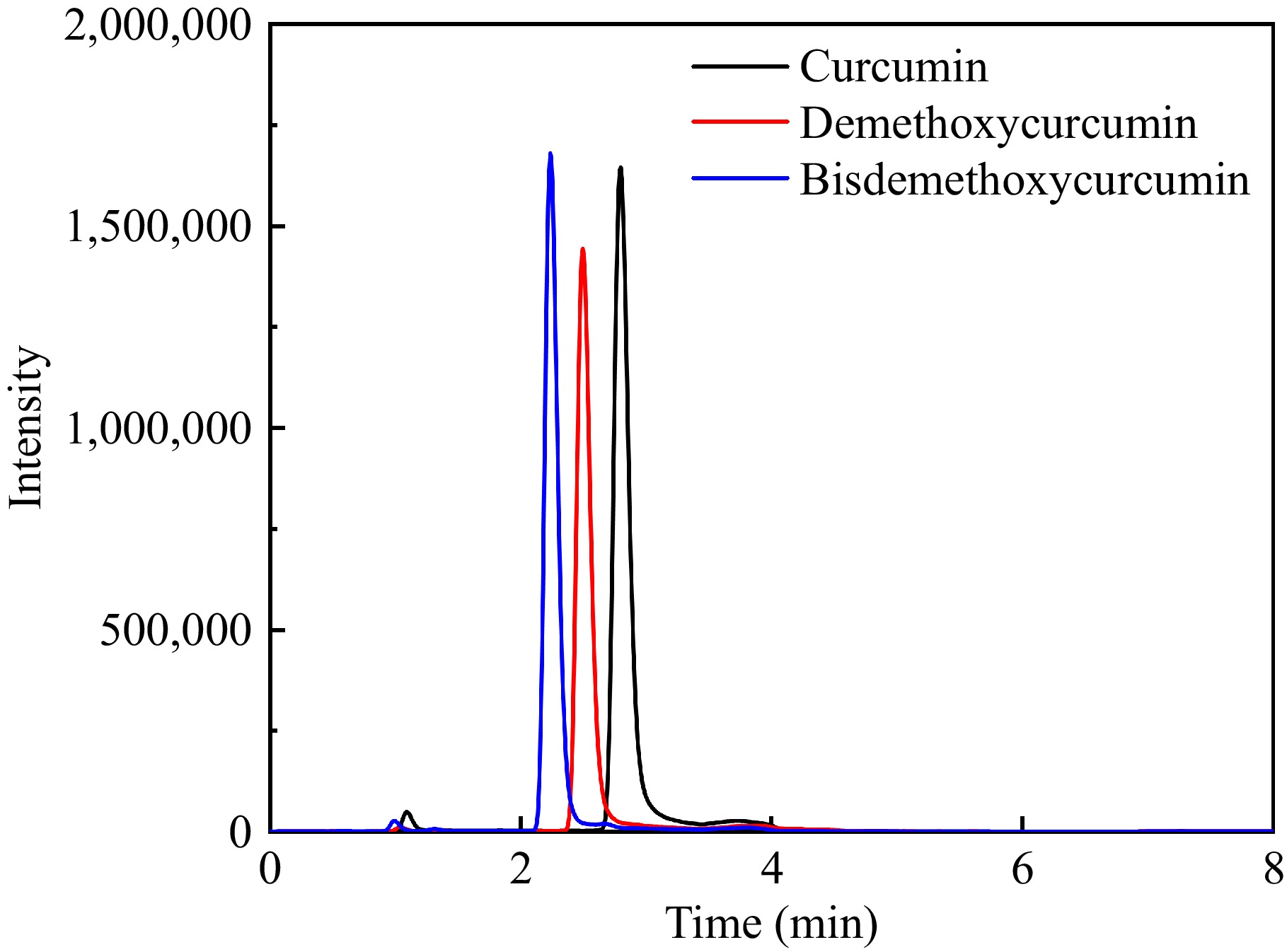
      Figure 1. 

      Extracted ion chromatogram of the three curcumin compounds.

    • Bioactive ingredients have traditionally been extracted from food, and Chinese herbal medicine using solid-liquid extraction procedures.

      Solid-liquid extraction techniques have traditionally been used for the extraction of bioactive ingredients from agricultural products, food and Chinese herbal medicine, including phenolic compounds, amino acids, flavonoids, terpenes, tannins, and alkaloids[2932]. Considering the intricate nature of the ginger and the chemical and physical characteristics of the three curcumin compounds, the extraction solvent was optimized in the current study. Based on the literature, five different extraction solvents have been employed to extract curcumin compounds, including methanol, acetonitrile, ethanol, ethyl acetate, and acetone[12,3335]. Therefore, these five solvents were optimized as the extraction solvent and the results of the extraction efficiency of curcumin, demethoxycurcumin, and bisdemethoxycurcumin are shown in Fig. 2a. Higher concentrations of the three curcumin compounds were obtained in the methanol extracts, and ethyl acetate exhibited the worst extraction efficiency. The results were consistent with those of Lee & Choung[36], who studied the extraction efficiency of curcumin compounds in 54 items of 16 commercial foods, and evaluated the extraction efficiency of solvents as follows: methanol > acetonitrile > ethyl acetate. Jorge-Montalvo et al. also found that solvents with relatively medium polarity (such as methanol with a value of 0.76) exhibited better extraction results of the antioxidant compounds like curcumin compounds in ginger[37]. Indeed, the solubility of compounds in solvents are consistent with the 'like dissolves like' principle. The extraction results were associated not only with solvent polarity but also with other parameters like molecular size, conformation, intermolecular hydrogen bonds, and solvent solute effects[38]. In addition to the extraction efficiency, the samples extracted with methanol had better chromatographic peak shape, which was narrower, sharper, and more symmetrical. For this reason, methanol was selected as the extraction solution in this study.
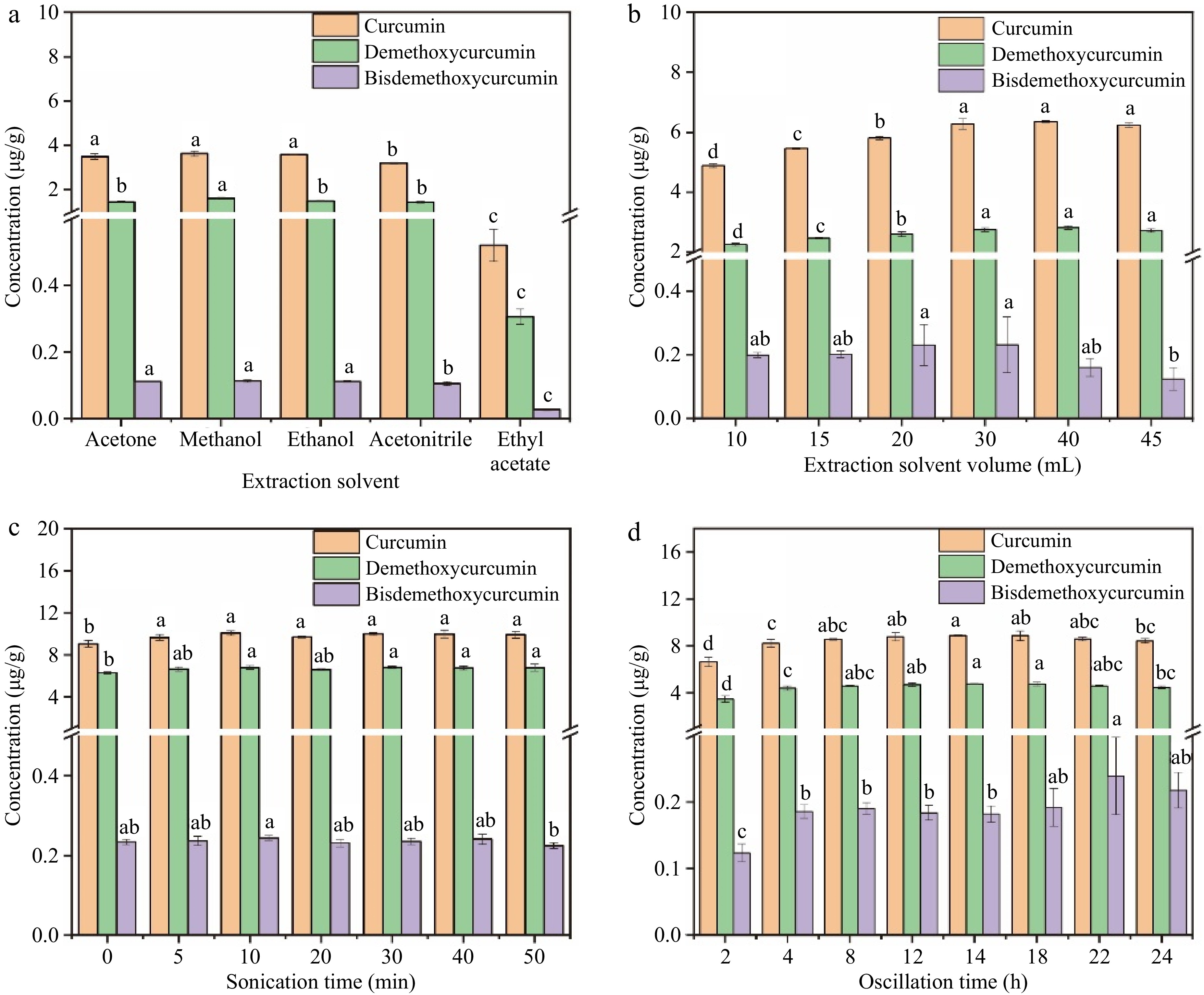
      Figure 2. 

      Effects of extraction conditions on the extraction efficiency of the three curcumin compounds. (a) Extraction solvent, (b) extraction solvent volume, (c) sonication time, (d) oscillation time.

    • In the solid-liquid extraction process, the extraction solvent volume is essential to ensuring both an adequate extraction yield and a low cost. In the current study, different extraction solvent volumes of 10, 15, 20, 30, 40, and 45 mL were compared. As illustrated in Fig. 2b, the volume of methanol has significant impacts on the extraction results of curcumin compounds. With the increase of solvent volume from 10 mL to 30 mL, the concentration of the three curcumin compounds showed an upward trend. This was caused by the mass transfer principle, which meant that the driving force during the mass transfer process was the consistency gradient between the solid and the liquid. This gradient increased with increasing solvent-to-solid ratios, leading to improved extraction efficiency[39]. However, when the solvent amount was higher than 30 mL, there would not be enough liquid phase in relation to the dispersed phase to accomplish sufficient transfer that various equilibria may occur, leading to non-negligible resistance to mass transfer[40]. At the same time, with the increase in methanol volume, some unwanted lipids and other matrix components were also extracted. This explains why the extraction efficiency of the curcumin compounds did not elevate when the volume of methanol increased from 30 to 50 mL, and even showed a downward trend. Therefore, the optimal extraction solvent volume selected in this experiment was 30 mL.

    • Ultrasonication is an effective method for cell disruption in laboratory conditions[41]. To ensure the full extraction of curcumin compounds, the sonication time of the ginger powder and extraction solvent mixture was optimized. As shown in Fig. 2c, the extraction yield rose with the ultrasonic treatment time rising from 0 to 10 min (p < 0.05) and maintained at a stable level with the sonication time continuing to go up to 50 min. As is known, ultrasound has beneficial impacts in food analysis, which utilizes the cavitation effect, referring to a series of dynamic processes in which sound waves propagate in a liquid, causing periodic alternating changes in sound pressure, forming local compressed and expanded phases in the liquid[42]. Due to the nonlinear vibration of bubbles caused by cavitation and the explosion pressure generated when they burst, many physical or chemical effects could be generated along with cavitation, which induced a positive effect for extraction. However, ultrasonic cavitation could also generate extreme physical conditions such as high temperature, which was observed in the extraction with the increase of the sonication time and could cause the thermal degradation of the curcumin compounds[43,44]. The time cost is a significant reference for extraction and purification methods[45]. Therefore, considering the extraction efficiency and time cost comprehensively, a sonication time of 10 min was selected.

    • Oscillation is another factor that affected the extraction efficiency of curcumin compounds. It is a common method for extracting targeted compounds, which mainly uses the mass transfer theory to produce the transfer of constituents in the system[46]. The oscillation time of 2–24 h was optimized in the current study and the results are illustrated in Fig. 2d. The extraction yield showed an upward trend with the increase of oscillation time from 2 to 8 h, and a significant increase were seen from 2 to 4 h (p < 0.05). After oscillating for more than 4 h, there was no significant differences in the extraction yield of curcumin compounds. Extraction time usually has positive effects on the yield of extracted compounds, and prolonging extraction time is beneficial for the extraction. Indeed, in the process of extraction, solid samples were wetted and dissolved by the solvents, and the release of components from the samples to the solvents takes a certain amount of time[47]. Previous studies have shown that the extraction yield increased with the prolongation of extraction time, but after a certain period of time, this increased rate decreases with the extension of time[48]. This may be the saturation of the targeted compounds in the extraction solvents, or even the degradation of some bioactive substances due to thermal exposure during the long-term extraction process[15]. Hence, 4 h was chosen as the optimal oscillation time.

    • The LODs, LOQs, calibration linearity, spiking recovery, matrix effect, and precision were all examined to evaluate the performance of the current approach. As shown in Table 2, the LODs of curcumin, demethoxycurcumin, and bisdemethoxycurcumin were 0.003–0.006 mg∙kg−1, and the LOQs were 0.015 mg∙kg−1 (calculated by dry weight), indicating satisfactory detection sensitivity. The three curcumin compounds showed good linearities with coefficients of determination (R2) exceeding 0.9990. The matrix effects were between 94.6% and 98.8%, implying that there were negligible matrix interferences in ginger extracts and the solvent standard calibration curve can be used for method quantification. The method accuracy was evaluated using recovery at three spiking concentrations, which were approximately 50%, 100%, and 200% of the background compound content in ginger samples. The spiking levels were 4, 8, and 16 mg∙kg−1 for curcumin, 2.7, 5.4, and 10.8 mg∙kg−1 for demethoxycurcumin, and 0.15, 0.3, and 0.6 mg∙kg−1 for bisdemethoxycurcumin. The recovery of three curcumin compounds was 81.7%–100.0%, and the intra-day and inter-day precision expressed in RSD was lower than 5.4%. In conclusion, the approach of establishment is sensitive, accurate, and precise, indicating that the approach is reliable as well as appropriate for the quantification of curcumin components in ginger samples.

      Table 2.  Method validation results of the established methods.

      Compound LOD (mg∙kg−1) LOQ (mg∙kg−1) Linear range (mg∙kg−1) R2 Matrix
      effect (%)
      Recovery (%) ± SD Intra-day RSD (%) Inter-day RSD (%)
      Low Mid High
      Curcumin 0.006 0.015 0.015-30 0.9999 94.6 97.1 ± 10.7 90.4 ± 19.6 96.8 ± 5.6 5.3 1.8
      Demethoxycurcumin 0.006 0.015 0.015-30 0.9990 98.8 100 ± 4.6 94.5 ± 6.1 95.2 ± 4.1 3.5 2.6
      Bisdemethoxycurcumin 0.003 0.015 0.015-30 0.9995 96.1 85.0 ± 9.9 81.7 ± 10.5 83.8 ± 1.6 5.2 5.4
      SD means standard deviation (n = 6); RSD means relative standard deviations (n = 6).

      Next, a comparison was made between the UHPLC-MS/MS method established in this study and the reported methods for the quantification of curcumin compounds in ginger samples. As shown in Table 3, the method validation assessment was entirely implemented only in this study and the current method exhibited the highest sensitivity with satisfactory linearity, matrix effect, accuracy, and precision.

      Table 3.  Comparison of the present study for the determination curcumin compound by different methods in ginger*.

      Compound name Analysis methodb LOD LOQ R2 Matrix
      effect (%)
      Recovery
      (%)
      RSD (%) Ref.
      Curcumin HPLC-UV 0.028 ng∙mL−1 0.093 ng∙mL−1 0.9983 / > 98 0.94 and 1.07 (intra-day)
      4.44 (inter-day)
      [20]
      Curcumin RRS 0.018 mg∙L−1 0.047 mg∙kg−1 1.000 / 97.7–103  2.0–2.6 (intra-day)
      2.0–2.6 (inter-day)
      [49]
      DWO-VIS 0.013 mg∙L−1 0.33 mg∙kg−1 0.9999 96.3–104 2.3–2.6 (inter-day)
      2.2–2.8 (intra-day)
      Curcumin UHPLC-IM-Q-TOF/MS 1.53 ng∙mL−1 5.09 ng∙mL−1 0.9910 / 93 1.17 (intra-day),
      2.73 (inter-day)
      [23]
      Demethoxycurcumin 0.77 ng∙mL−1 2.57 ng∙mL−1 0.9922 106 1.91 (intra-day),
      3.72 (inter-day)
      Bisdemethoxycurcumin 1.09 ng∙mL−1 3.63 ng∙mL−1 0.9906 85 2.85 (intra-day),
      3.2 (inter-day)
      Curcumin UHPLC-MS/MS 0.006 mg∙kg−1 0.015 mg∙kg−1 0.9999 94.6 98.7–104.2 5.3 (intra-day),
      1.8 (inter-day)
      Current
      study
      Demethoxycurcumin 0.006 mg∙kg−1 0.015 mg∙kg−1 0.9990 98.8 100.7–102.9 3.5 (intra-day),
      2.6 (inter-day)
      Bisdemethoxycurcumin 0.003 mg∙kg−1 0.015 mg∙kg−1 0.9995 96.1 80.0–83.3 5.2 (intra-day),
      5.4 (inter-day)
      '/', not provided. b. HPLC-UV, high-performance liquid chromatography; RRS, resonance Rayleigh scattering; DWO-VIS, dual wavelength visible absorption spectroscopy; UHPLC-IM-Q-TOF/MS, ultra-high-performance liquid chromatograph ion mobility quadrupole time-of-flight mass spectrometry; HPLC-MS/MS, ultra-high-performance liquid chromatography coupled with tandem mass spectrometry.
    • To prove the applicability of the current method of solid-liquid extraction and UHPLC-MS/MS determination for the quantification of curcumin compounds in ginger, six ginger samples were randomly selected for quantitative analysis. The results are exhibited in Table 4. As shown in Table 4, the concentration of curcumin in all ginger samples was the highest, which was 0.575–10.143 mg∙kg−1, and the content of demethoxycurcumin was lower, with the value of 0.368–7.196 mg∙kg−1. The concentration of bisdemethoxycurcumin was the lowest of the three curcumin compounds, ranging from 0.019 to 0.281 mg∙kg−1. In addition, sample A had the lowest concentration of three curcumin compounds, while sample E had the highest concentration.

      Table 4.  Concentration of the three curcumin compounds in ginger samples.

      Compound Concentration (mg∙kg−1)*
      Sample A Sample B Sample C Sample D Sample E Sample F
      Curcumin 0.575 ± 0.042 0.719 ± 0.008 5.228 ± 0.043 7.304 ± 0.095 10.143 ± 0.095 4.16 ± 0.073
      Demethoxycurcumin 0.368 ± 0.029 0.511 ± 0.009 3.148 ± 0.042 4.589 ± 0.049 7.196 ± 0.081 3.05 ± 0.037
      Bisdemethoxycurcumin 0.019 ± 0.001 0.019 ± 0.001 0.218 ± 0.009 0.276 ± 0.004 0.281 ± 0.005 0.191 ± 0.005
      * The data were expressed as 'mean values ± standard deviation'.
    • This study effectively established a quantitative approach using UHPLC-MS/MS for the simultaneous detection of curcumin, demethoxycurcumin, and bisdemethoxycurcumin in ginger samples. The method provides good sensitivity, negligible matrix effect, and good accuracy and precision, which indicates that the developed method was reliable and suitable for the quantitative detection of three curcumin compounds in ginger samples.

      • This work was supported by the Youth Innovation of CAAS (Y2023QC19), the China Agriculture Research System of MOF and MARA, the Young Elite Scientists Sponsorship Program by CAST (2023QNRC001), and the Agricultural Science and Technology Innovation Program of CAAS (CAAS-ASTIP-IQSTAP-2024).

      • The authors declare that they have no known conflict financial interests or personal relationships that could have appeared to influence the work reported in this paper.

      • Copyright: © 2024 by the author(s). Published by Maximum Academic Press on behalf of China Agricultural University, Zhejiang University and Shenyang Agricultural University. This article is an open access article distributed under Creative Commons Attribution License (CC BY 4.0), visit https://creativecommons.org/licenses/by/4.0/.
    Figure (2)  Table (4) References (49)
  • About this article
    Cite this article
    Jiang Y, Liu X, Zhao Y, Zhang J, Qiu J, et al. 2024. Quantitative analysis of curcumin compounds in ginger by ultra-high-performance liquid chromatography coupled with tandem mass spectrometry. Food Innovation and Advances 3(4): 353−359 doi: 10.48130/fia-0024-0033
    Jiang Y, Liu X, Zhao Y, Zhang J, Qiu J, et al. 2024. Quantitative analysis of curcumin compounds in ginger by ultra-high-performance liquid chromatography coupled with tandem mass spectrometry. Food Innovation and Advances 3(4): 353−359 doi: 10.48130/fia-0024-0033

Catalog

  • About this article

/

DownLoad:  Full-Size Img  PowerPoint
Return
Return