ARTICLE   Open Access    

Enhanced extraction of phenolic compounds from red cabbage utilizing microwave-assisted method: a Box-Behnken approach for optimization

More Information
  • The main objective of this study was to determine the optimum conditions for microwave-assisted extraction (MAE) of red cabbage phenolic compounds using response surface methodology (RSM). A Box-Behnken experimental design was used to investigate the effects of extraction time (5, 10, 15 min), solid/solvent ratio (1/20, 1/30, 1/40, w/w), and microwave power (200, 400, 600 W) on total phenolic content (TPC) and total antioxidant capacity (TAC). TPC and TAC were determined using the Folin-Ciocalteu, DPPH, and CUPRAC assays. The optimal conditions for MAE were found to be 9.166 min extraction time, 1/20 solid/solvent ratio, and 501.768 W microwave power for the ethanol-water solvent, and 5 min extraction time, 1/20 solid/solvent ratio, and 384.097 W microwave power for the water-only solvent. Scanning Electron Microscopy (SEM) analysis revealed that MAE is more efficient in cell wall breakdown than conventional methods. These findings imply that MAE is a superior method for extracting phenolic compounds from red cabbage, with higher yields and reduced processing times compared to conventional extraction (CE). This work provides valuable insights for optimizing phenolic extraction processes, highlighting the advantages of MAE in producing high-quality phenolic extracts for potential functional food applications.
  • Atractylodes macrocephala Koidz. (common names 'Baizhu' in Chinese and 'Byakujutsu' in Japanese) is a diploid (2n = 2x = 24) and out-crossing perennial herb in the Compositae family, and has a long history of cultivation in temperate and subtropical areas of East Asia as it is widely used in traditional herbal remedies with multiple pharmacological activities[13]. The 'Pharmacopoeia of the People's Republic of China' states that 'Baizhu' is the dry rhizome of A. macrocephala Koidz. (Atractylodis Macrocephalae Rhizoma, AMR). However, in Japanese traditional medicine 'Baizhu' can be referred to both: A. japonica or A. macrocephala[4].

    A. macrocephala is naturally endemic to China and cultivated in more than 200 towns in China, belonging to Zhejiang, Hunan, Jiangxi, Anhui, Fujian, Sichuan, Hubei, Hebei, Henan, Jiangsu, Guizhou, Shanxi, and Shaanxi Provinces[3]. A. macrocephala grows to a height of 20–60 cm (Fig. 1). The leaves are green, papery, hairless, and generally foliole with 3–5 laminae with cylindric glabrous stems and branches. The flowers grow and aggregate into a capitulum at the apex of the stem. The corollas are purplish-red, and the florets are 1.7 cm long. The achenes, densely covered with white, straight hairs, are obconic and measure 7.5 mm long. The rhizomes used for medicinal purposes are irregular masses or irregularly curving cylinders about 3–13 cm long and 1.5–7 cm in diameter with an outwardly pale greyish yellow to pale yellowish color or a sparse greyish brown color. The periderm-covered rhizomes are externally greyish brown, often with nodose protuberances and coarse wrinkles. The cross-sections are white with fine dots of light yellowish-brown to brown secretion. Rhizomes are collected from plants that are > 2 years old during the spring. The fibrils are removed, dried, and used for medicinal purposes[5, 6].

    Figure 1.  Plant morphology of A. macrocephala.

    The medicinal properties of AMRs are used for spleen deficiency, phlegm drinking, dizziness, palpitation, edema, spontaneous sweating, benefit Qi, and fetal restlessness[7]. The AMR contains various functional components, among which high polysaccharide content, with a yield close to 30%[8]. Therefore, the polysaccharides of A. macrocephala Koidz. rhizome (AMRP) are essential in assessing the quality control and bioactivity of A. macrocephala. Volatile oil accounts for about 1.4% of AMR, with atractylon and atractylodin as the main components[9]. Atractylon can be converted to atractylenolide I (AT-I), atractylenolide II (AT-II), and atractylenolide III (AT-III) under ambient conditions. AT-III can be dehydrated to AT-II under heating conditions[10, 11]. AMRs, including esters, sesqui-, and triterpenes, have a wide range of biological activities, such as improving immune activity, intestinal digestion, neuroprotective activity, immune anti-inflammatory, and anti-tumor.

    In recent years, research on the pharmacological aspects of AMR has continued to increase. Still, the discovery of the main active components in AMR is in its infancy. The PAO-ZHI processing of AMR is a critical step for AMR to exert its functional effects, but also, in this case, further work is required. Studies on the biosynthesis of bioactive compounds and different types of transcriptomes advanced current knowledge of A. macrocephala, but, as mentioned, required more systematic work. Ulteriorly, an outlook on the future research directions of A. macrocephala was provided based on the advanced technologies currently applied in A. macrocephala (Fig. 2).

    Figure 2.  Current progress of A. macrocephala.

    A. macrocephala is distributed among mountainous regions more than 800 m above sea level along the middle and lower reaches of the Yangtze River (China)[5]. Due to over-exploitation and habitat destruction, natural populations are rare, threatened, and extinct in many locations[1,12]. In contrast to its native range, A. macrocephala is widely cultivated throughout China, in a total area of 2,000–2,500 ha, with a yield of 7,000 t of rhizomes annually[13]. A. macrocephala is mainly produced in Zhejiang, Anhui, and Hebei (China)[14]. Since ancient times, Zhejiang has been the famous producing area and was later introduced to Jiangxi, Hunan, Hebei, and other places[15]. Wild A. macrocephala is currently present in at least 14 provinces in China. It is mainly distributed over three mountain ranges, including the Tianmu and Dapan mountains in Zhejiang Province and the Mufu mountains along the border of Hunan and Jiangxi Provinces. A. macrocephala grows in a forest, or grassy areas on mountain or hill slopes and valleys at an altitude of 600–2,800 m. A. macrocephala grows rapidly at a temperature of 22–28 °C, and favors conditions with total precipitation of 300–400 mm evenly distributed among the growing season[16]. Chen et al. first used alternating trilinear decomposition (ATLD) to characterize the three-dimensional fluorescence spectrum of A. macrocephala[17]. Then they combined the three-dimensional fluorescence spectrum with partial least squares discriminant analysis (PLS-DA) and k-nearest neighbor method (kNN) to trace the origin of Atractylodes samples. The results showed that the classification models established by PLS-DA and kNN could effectively distinguish the samples from three major Atractylodes producing areas (Anhui, Hunan, and Zhejiang), and the classification accuracy rate (CCR) of Zhejiang atractylodes was up to 80%, and 90%, respectively[17]. Zhang et al. compared the characteristics, volatile oil content, and chemical components of attested materials from six producing areas of Zhejiang, Anhui, Hubei, Hunan, Hebei, and Henan. Differences in the shape, size, and surface characteristics were reported, with the content of volatile oil ranging from 0.58% to 1.22%, from high to low, Hunan (1.22%) > Zhejiang (1.20%) > Anhui (1.02%) > Hubei (0.94%) > Henan (0.86%) > Hebei (0.58%)[18]. This study showed that the volatile oil content of A. macrocephala in Hunan, Anhui, and Hubei is not much different from that of Zhejiang, which is around 1%. A. macrocephala is a local herb in Zhejiang, with standardized cultivation techniques, with production used to reach 80%–90% of the country. However, in recent years, the rapid development of Zhejiang's real estate economy has reduced the area planted with Zhejiang A. macrocephala, resulting in a sudden decrease in production. Therefore, neighboring regions, such as Anhui and Hunan, vigorously cultivate A. macrocephala, and the yield and quality of A. macrocephala can be comparable to those of Zhejiang. The results were consistent with the data reports[18]. Guo et al. analyzed the differentially expressed genes of Atractylodes transcripts from different regions by the Illumina HiSeq sequencing platform. It was found that 2,333, 1,846, and 1,239 DEGs were screened from Hubei and Hebei, Anhui and Hubei, and Anhui and Hebei Atrexia, respectively, among which 1,424, 1,091, and 731 DEGs were annotated in the GO database. There were 432, 321, and 208 DEGs annotated in the KEGG database. These DEGs were mainly related to metabolic processes and metabolic pathways of secondary metabolites. The highest expression levels of these genes were found in Hubei, indicating higher terpenoid production in Hubei[19]. Other compounds were differentially accumulated in Atractylodes. Chlorogenic acid from Hebei was 0.22%, significantly higher than that from Zhejiang and Anhui[20]. Moreover, the content of neochlorogenic acid and chlorogenic acid decreased after processing, with the highest effect reported in Zhejiang, with the average transfer rate of neochlorogenic acid and chlorogenic acid reaching 55.68% and 55.05%[20]. All these changes would bring great help in distinguishing the origins of A. macrocephala.

    Medicinal AMR can be divided into raw AMR and cooked AMR. The processing method is PAO-ZHI; the most traditional method is wheat bran frying. The literature compared two different treatment methods, crude A. macrocephala (CA) and bran-processed A. macrocephala, and found that the pharmacological effects of AMR changed after frying with wheat bran, mainly in the anti-tumor, antiviral and anti-inflammatory effects[21]. The anti-inflammatory effect was enhanced, while the anti-tumor and antiviral effects were somewhat weakened, which may be related to the composition changes of the compounds after frying. The study of the content of AT-I, II, and III, and atractyloside A, in rat serum provided helpful information on the mechanism of wheat bran processing[22]. In addition to frying wheat bran, Sun et al. used sulfur fumigation to treat AMR[23]. They found that the concentration of different compounds changed, producing up to 15 kinds of terpenoids. Changes in pharmacological effects were related to treatment and the type of illumination[24,25]. Also, artificial light can improve the various biological functions. A. macrocephala grew better under microwave electrodeless light, with a chlorophyll content of 57.07 ± 0.65 soil and plant analyzer develotrnent (SPAD)[24]. The antioxidant activity of AMR extract treated with light-emitting diode (LED)-red light was the highest (95.3 ± 1.1%) compared with other treatments[24]. The total phenol and flavonoid contents of AMR extract treated with LED-green light were the highest at 24.93 ± 0.3 mg gallic acid equivalents (GAE)/g and 11.2 ± 0.3 mg quercetin equivalents (QE)/g compared with other treatments[24, 25]. Polysaccharides from Chrysanthemun indicum L.[26] and Sclerotium rolfsiisacc[27] can improve AMR's biomass and bioactive substances by stimulating plant defense and thus affect their efficacy. In summary, there are compositional differences between A. macrocephala from different origins. Besides, different treatments, including processing mode, light irradiation, and immune induction factors, which can affect AMR's biological activity, provide some reference for the cultivation and processing of A. macrocephala (Fig. 3).

    Figure 3.  Origin, distribution and processing of A. macrocephala.

    The AMR has been reported to be rich in polysaccharides, sesquiterpenoids (atractylenolides), volatile compounds, and polyacetylenes[3]. These compounds have contributed to various biological activities in AMR, including immunomodulatory effects, improving gastrointestinal function, anti-tumor activity, neuroprotective activity, and anti-inflammatory.

    AMRP has received increasing attention as the main active component in AMR because of its rich and diverse biological activities. In the last five years, nine AMRP have been isolated from AMR. RAMP2 had been isolated from AMR, with a molecular weight of 4.354 × 10³ Da. It was composed of mannose, galacturonic acid, glucose, galactose, and arabinose, with the main linkages of →3-β-glcp-(1→, →3,6-β-glcp-(1→, →6-β-glcp-(1→, T-β-glcp-(1→, →4-α-galpA-(1→, →4-α-galpA-6-OMe-(1→, →5-α-araf-(1→, →4,6-β-manp-(1→ and →4-β-galp-(1→[28]. Three water-soluble polysaccharides AMAP-1, AMAP-2, and AMAP-3 were isolated with a molecular weight of 13.8 × 10⁴ Da, 16.2 × 10⁴ Da, and 8.5 × 10⁴ Da, respectively. Three polysaccharides were deduced to be natural pectin-type polysaccharides, where the homogalacturonan (HG) region consists of α-(1→4)-linked GalpA residues and the ramified region consists of alternating α-(1→4)-linked GalpA residues and α-(1→2)-linked Rhap residues. Besides, three polysaccharides were composed of different ratios of HG and rhamnogalacturonan type I (RG-I) regions[29]. Furthermore, RAMPtp has been extracted from AMR with a molecular weight of 1.867 × 10³ Da. It consists of glucose, mannose, rhamnose, arabinose, and galactose with 60.67%, 14.99%, 10.61%, 8.83%, and 4.90%, connected by 1,3-linked β-D Galp and 1,6-linked β-D Galp residues[30]. Additionally, PAMK was characterized by a molecular weight of 4.1 kDa, consisting of galactose, arabinose, and glucose in a molar ratio of 1:1.5:5, with an alpha structure and containing 96.47% polysaccharide and small amounts of protein, nucleic acid, and uric acid[31]. Another PAMK extracted from AMR had a molecular weight of 2.816 × 10³ Da and consisted of glucose and mannose in molar ratios of 0.582 to 0.418[32]. Guo et al. isolated PAMK with a molecular weight of 4.748 × 10³ g/mol from AMR, consisting of glucose, galactose, arabinose, fructose, and mannose in proportions of 67.01%, 12.32%, 9.89%, 1.18%, and 0.91%, respectively[33]. In addition, AMP1-1 is a neutral polysaccharide fragment with a molecular weight of 1.433 kDa isolated from AMR. It consists of glucose and fructose, and the structure was identified as inulin-type fructose α-D-Glcp-1→(2-β-D-Fruf-1)7[34]. These reports indicated that, in general, polysaccharides are extracted by water decoction, ultrasonic-assisted extraction, enzyme hydrolysis method, and microwave-assisted extraction. The separation and purification are column chromatography, stepwise ethanol precipitation, and ultrafiltration. Their physicochemical properties and structural characterization are generally achieved by determining the molecular weight, determining the monosaccharide composition, analyzing the secondary structure, and glycosidic bond configuration of polysaccharides with Fourier transform infrared (FT-IR) and nuclear magnetic resonance (NMR). The advanced structures of polysaccharides can be identified by high-performance size exclusion chromatography-multiangle laser light scattering (HPSEC-MALLS), transmission electron microscopy (TEM), and scanning electron microscopy (SEM) techniques (Table 1). AMRP has various physiological functions, including immunomodulatory effects, improving gastrointestinal function, and anti-tumor activity. The related biological activities, animal models, monitoring indicators, and results are summarized in Table 1.

    Table 1.  Components and bioactivity of polysaccharides from Atractylodes macrocephala Koidz. Rhizome.
    Pharmacological activitiesDetailed functionPolysaccharides informationModelDoseTest indexResultsRef.
    Immunomodulatory effectsRestore immune
    function
    /Chicken models
    (HS-induced)
    200 mg/kgOxidative index;
    Activities of mitochondrial complexes and ATPases;
    Ultrastructure in chicken spleens;
    Expression levels of cytokines, Mitochondrial dynamics- and apoptosis-related genes
    Alleviated
    the expression of
    IL-1 ↑,TNF-α ↑, IL-2 ↓, IFN- γ ↓; mitochondrial dynamics- and anti-apoptosis-related genes ↓; pro-apoptosis-related genes ↑;
    the activities of mitochondrial complexes and ATPases ↓ caused by HS
    [35]
    Regulate the immune function/Chicken models
    (HS-induced)
    200 mg/kgiNOS–NO activities;
    ER stress-related genes;
    Apoptosis-related genes;
    Apoptosis levels
    Alleviated NO content ↑; activity of iNOS ↑ in the chicken spleen; GRP78, GRP94, ATF4, ATF6, IRE ↑; caspase3 ↑; Bcl-2 ↓ caused by HS[36]
    Relieve immunosuppressionCommercial AMR powder (purity 70%)Geese models
    (CTX-induced)
    400 mg/kgSpleen development;
    Percentages of leukocytes in peripheral blood
    Alleviated the spleen damage;
    T and B cell proliferation ↓; imbalance of leukocytes; disturbances of humoral; cellular immunity caused by CTX
    [37]
    Active the lymphocytesCommercial AMR powder (purity 95%)Geese models
    (CTX-induced)
    400 mg/kgThymus morphology;
    The level of serum GMC-SF, IL-1b, IL-3, IL-5;
    mRNA expression of CD25, novel_mir2, CTLA4 and CD28 signal pathway
    Maintain normal cell morphology of thymus;
    Alleviated GMC-SF ↓, IL-1b ↓, IL-5↓, IL-6↓, TGF-b↓; IL-4 ↑, IL-10 ↑; novel_mir2 ↓, CD25↓, CD28↓ in thymus and lymphocytes caused by CTX
    [38]
    Alleviate immunosuppressionCommercial AMR powder (purity 70%)Geese models
    (CTX-induced)
    400 mg/kgThymus development;
    T cell proliferation rate;
    The level of CD28, CD96, MHC-II;
    IL-2 levels in serum;
    differentially expressed miRNAs
    Alleviated thymus damage;
    T lymphocyte proliferation rate ↓; T cell activation ↓; IL-2 levels ↓ caused by CTX;
    Promoted novel_mir2 ↑; CTLA4 ↓; TCR-NFAT signaling pathway
    [39]
    Alleviates T cell activation declineCommercial AMR powder (purity 95%)BALB/c female mice (CTX-induced)200 mg/kgSpleen index;
    Morphology, death, cytokine concentration of splenocytes;
    Th1/Th2 ratio, activating factors of lymphocytes;
    T cell activating factors;
    mRNA expression level in CD28 signal pathway
    Improved the spleen index;
    Alleviated abnormal splenocytes morphology and death; Balance Th1/Th2 ratio; IL-2 ↑, IL-6 ↑, TNF-α ↑, IFN-γ ↑; mRNA levels of CD28, PLCγ-1, IP3R, NFAT, AP-1 ↑
    [40]
    Immunoregulation and ImmunopotentiationCommercial AMR powder (purity 80%)BMDCs (LPS-induced);
    Female BALB/c mice (ovalbumin as a model antigen)
    /Surface molecule expression of BMDCs;
    Cytokines secreted by dendritic cell supernatants;
    OVA-specific antibodies in serum;
    Cytokines in serum;
    Lymphocyte immunophenotype
    Expression of CD80 and CD86 ↑; IL-1β ↑, IL-12 ↑, TNF-α↑ and IFN-γ ↑; OVA-specific antibodies in serum ↑; Secretion of cytokines ↑; Proliferation rate of spleen lymphocytes ↑; Activation of CD3+CD4+ and CD3+CD8+ lymphocytes[46]
    Increase immune-response capacity of the spleen in miceCommercial AMR powder (purity 70%)BALB/c female mice100, 200, 400 mg/kgSpleen index;
    Concentrations of cytokines;
    mRNA and protein expression levels in TLR4 signaling
    In the medium-PAMK group:
    IL-2, IL-4, IFN-c, TNF-a ↑; mRNA and protein expression of TLR4, MyD88, TRAF6, TRAF3, NF-κB in the spleen ↑
    [41]
    Immunological activityCommercial AMR powder (purity 80%)Murine splenic lymphocytes (LPS or PHA-induced)13, 26, 52, 104, 208 μg/mLT lymphocyte surface markersLymphocyte proliferation ↑;
    Ratio of CD4+/CD8+ T cells ↑
    [47]
    Immunomodulatory activityTotal carbohydrates content 95.66 %Mouse splenocytes
    (Con A or LPS-induced)
    25, 50, 100 μg/mLSplenocyte proliferation;
    NK cytotoxicity;
    Productions of NO and cytokines;
    Transcription factor activity;
    Signal pathways and receptor
    Promoted splenocyte proliferation; Cells enter S and G2/M phases; Ratios of T/B cells ↑; NK cytotoxicity ↑; Transcriptional activities of NFAT ↑; NF-κB, AP-1 ↑; NO, IgG, IL-1α, IL-1β, IL-2, IL-3, IL-4, IL-6, IL-10, IL-12p40, IL-12p70, IL-13, IFN-γ, TNF-α, G-CSF, GM-CSF, KC, MIP-1α, MIP-1β, RANTES, Eotaxin ↑[42]
    Promote the proliferation of thymic epithelial cellsContents of fucrhaara, galactose, glucose, fructose,
    and xylitol: 0.98%, 0.40%, 88.67%, 4.47%, and 5.47%
    MTEC1 cells50 μg/mLCell viability and proliferation;
    lncRNAs, miRNAs, and mRNAs expression profiles in MTEC1 cells
    The differential genes were 225 lncRNAs, 29 miRNAs, and 800 mRNAs; Genes enriched in cell cycle, cell division, NF-κB signaling, apoptotic process, and MAPK signaling pathway[44]
    Immunomodulatory activityMW: 4.354 × 10³ Da;
    Composed of mannose, galacturonic acid, glucose, galactose and arabinose;
    The main linkages are →3-β-glcp-(1→, →3,6-β-glcp-(1→, →6-β-glcp-(1→, T-β-glcp-(1→,
    →4-α-galpA-(1→, →4-α-galpA-6-OMe-(1→, →5-α-araf-(1→, →4,6-β-manp-(1→ and →4-β-galp-(1→
    CD4+ T cell50, 100, 200 μg/mLMolecular weight;
    Monosaccharide composition;
    Secondary structure;
    Surface topography;
    Effect on Treg cells
    Treg cells percentage ↑; mRNA expressions of Foxp3, IL-10 and IL-2 ↑; STAT5 phosphorylation levels ↑; IL-2/STAT5 pathway[28]
    Immunostimulatory activityMW of AMAP-1, AMAP-2, and AMAP-3 were 13.8×10⁴ Da, 16.2×10⁴ Da and 8.5×10⁴ Da;
    HG region consists of α-(1→4)-linked GalpA residues
    RAW264.7 cells (LPS-induced)80, 40, 200 μg/mLMolecular weight;
    Total carbohydrate;
    Uronic acid contents;
    Secondary structure;
    Monosaccharide composition;
    Immunostimulatory activity
    RG-I-rich AMAP-1 and AMAP-2 improved the release of NO[29]
    Immunomodulatory effectMW: 1.867×10³ Da;
    Contents of glucose, mannose, rhamnose,
    arabinose and galactose: 60.67%, 14.99%, 10.61%, 8.83% and 4.90%
    SMLN lymphocytes25
    μg/ml
    Molecular weight;
    Monosaccharide composition;
    Ultrastructure;
    Intracellular Ca2+concentration;
    Target genes;
    Cell cycle distribution
    [Ca2+]i ↑; More cells in S and G2/M phases; IFN-γ ↑, IL-17A ↑; mRNA expressions of IL-4 ↓[30]
    Macrophage activationTotal carbohydrates content 95.66 %RAW264.7 macrophages (LPS-induced)25, 50, 100 μg/mLPinocytic activity;
    Phagocytic uptake;
    Phenotypic characterization;
    Cytokine production;
    Bioinformatics analysis;
    Transcription factor inhibition
    IL-6, IL-10 and TNF-α ↑; CCL2 and CCL5 ↑; Pinocytic and phagocytic activity ↑; CD40, CD80, CD86, MHC-I, MHC-II ↑; NF-κB and Jak-STAT pathway[43]
    Immunomodulatory effectTotal carbohydrates content 95.66 %SMLN lymphocytes25, 50, 100 μg/mLCytokine production;
    CD4+ and CD8+ lymphocytes;
    Target genes;
    Bioinformatics analysis;
    T and B lymphocyte proliferation;
    Receptor binding and blocking
    IFN-γ, IL-1α, IL-21, IFN-α, CCL4, CXCL9, CXCL10 ↑; CD4+ and CD8+subpopulations proportions ↑;
    c-JUN, NFAT4, STAT1, STAT3 ↑;
    67 differentially expressed miRNAs (55 ↑ and
    12 ↓), associated with immune system pathways; Affect T and B lymphocytes
    [45]
    Improving gastrointestinal functionRelieve enteritis and improve intestinal
    flora disorder
    Commercial AMR powder (purity 70%);
    Contents of fucrhaara, galactose, glucose, xylitol, and fructose: 0.98%, 0.40%, 88.67%, 4.47%, and 5.47%
    Goslings (LPS-induced)400 mg/kgSerum CRP, IL-1β, IL-6, and TNF-α levels;
    Positive rate of IgA;
    TLR4, occludin, ZO-1, cytokines, and immunoglobulin mRNA expression;
    Intestinal flora of gosling excrement
    Relieved IL-1β, IL-6, TNF-α levels in serum ↑; the number of IgA-secreting cells ↑; TLR4 ↑; tight junction occludin and ZO-1 ↓; IL-1β mRNA expression in the small intestine ↑; Romboutsia ↓ caused by LPS[48]
    Ameliorate ulcerative colitisMW: 2.391 × 10⁴ Da;
    Composed of mannose, glucuronic acid, glucose and arabinose in a molar ratio of 12.05:6.02:72.29:9.64
    Male C57BL/6J mice (DDS-induced)10, 20, 40 mg/kg bwHistopathological evaluation;
    Inflammatory mediator;
    Composition of gut microbiota;
    Feces and plasma for global metabolites profiling
    Butyricicoccus, Lactobacillus ↑;
    Actinobacteria, Akkermansia, Anaeroplasma, Bifidobacterium, Erysipelatoclostridium, Faecalibaculum, Parasutterella,
    Parvibacter, Tenericutes, Verrucomicrobia ↓;
    Changed 23 metabolites in fecal content; 21 metabolites in plasma content
    [49]
    Attenuate ulcerative colitis/Male SD rats (TNBS-induced);
    Co-culture BMSCs and IEC-6 cells
    540 mg/kg
    (for rats);
    400 μg/mL (for cell)
    Histopathological analysis;
    Cell migration;
    Levels of cytokines
    Potentiated BMSCs’ effect on preventing colitis and homing the injured tissue, regulated cytokines;
    BMSCs and AMP promoted the migration of IEC
    [52]
    Against intestinal mucosal injuryMW: 3.714 × 10³ Da;
    Composed of glucose, arabinose, galactose, galacturonic acid, rhamnose
    and mannose with molar ratios of 59.09:23.22:9.32:4.70:2.07:1.59
    Male C57BL/6 mice (DDS-induced)100 mg/kgIntestinal morphology;
    IL-6, TNF-α and IL-1β in serum;
    mRNA expression;
    Intestinal microbiota
    Alleviated body weight ↓; colon length ↓; colonic damage caused by DSS;
    Over-expression of TNF-α, IL-1β, IL-6 ↓; Infiltration of neutrophils in colon ↓; Mucin 2 ↑;
    Tight junction protein Claudin-1 ↑;
    Harmful bacteria content ↓;
    Beneficial bacteria content ↑
    [50]
    Against intestinal injuryTotal carbohydrates 95.66 %IECs (DDS-induced)5, 25, 50 μg/mLCell proliferation and apoptosis;
    Expression levels of intercellular TJ proteins;
    lncRNA screening
    Proliferation and survival of IECs ↑;
    Novel lncRNA ITSN1-OT1 ↑;
    Blocked the nuclear import of phosphorylated STAT2
    [51]
    Anti-tumor activityInduce apoptosis in transplanted H22 cells in miceMW: 4.1× 10³ Da;
    Neutral heteropolysaccharide composed of galactose, arabinose, and glucose with α-configuration (molar ratio, 1:1.5:5)
    Female Kunming mice100 and 200 mg/kg (for rats)Secondary structure;
    Molecular weight;
    Molecular weight;
    Thymus index and Spleen index;
    Lymphocyte Subpopulation in peripheral blood;
    Cell cycle distribution
    In tumor-bearing mice CD3+, CD4+, CD8+ ↓;
    B cells ↑
    [31]
    Regulate the innate immunity of colorectal cancer cellsCommercial AMR powder (purity 70%)C57BL/6J mice (MC38 cells xenograft model)500 mg/kgExpression of pro-inflammatory cytokines and secretionIL-6, IFN-λ, TNF-α, NO ↑ through MyD88/TLR4-dependent signaling pathway;
    Survival duration of mice with tumors ↑;
    Prevent tumorigenesis in mice
    [54]
    Induce apoptosis of Eca-109 cellsMW: 2.1× 10³ Da;
    Neutral hetero polysaccharide composed
    of arabinose and glucose (molar ratio, 1:4.57) with pyranose rings and α-type and β-type glycosidic linkages
    Eca-109 cells0.25, 0.5, 1, 1.5, 2.00 mg/mLCell morphology;
    Cell cycle arrest;
    Induction of apoptosis
    Accelerate the apoptosis of Eca109 cells[53]
    '/' denotes no useful information found in the study.
     | Show Table
    DownLoad: CSV

    To study the immunomodulatory activity of AMRP, the biological models generally adopted are chicken, goose, mouse, and human cell lines. Experiments based on the chicken model have generally applied 200 mg/kg doses. It was reported that AMRP protected the chicken spleen against heat stress (HS) by alleviating the chicken spleen immune dysfunction caused by HS, reducing oxidative stress, enhancing mitochondrial function, and inhibiting cell apoptosis[35]. Selenium and AMRP could improve the abnormal oxidation and apoptosis levels and endoplasmic reticulum damage caused by HS, and could act synergistically in the chicken spleen to regulate biomarker levels[36]. It indicated that AMRP and the combination of selenium and AMRP could be applied as chicken feed supplementation to alleviate the damage of HS and improve chicken immunity.

    The general application dose in the goose model is also 200 mg/kg, and the main injury inducer is cyclophosphamide (CTX). AMRP alleviated CTX-induced immune damage in geese and provided stable humoral immune protection[37]. Little is known about the role of AMRP in enhancing immunity in geese through the miRNA pathway. It was reported that AMRP alleviated CTX-induced decrease in T lymphocyte activation levels through the novel _mir2/CTLA4/CD28/AP-1 signaling pathway[38]. It was also reported that AMRP might be achieved by upregulating the TCR-NFAT pathway through novel_mir2 targeting of CTLA4, thereby attenuating the immune damage induced by CTX[39]. This indicated that AMRP could also be used as goose feed supplementation to improve the goose's autoimmunity.

    The typical injury inducer for mouse models is CTX, and the effects on mouse spleen tissue are mainly observed. BALB/c female mice were CTX-induced damage. However, AMRP increased cytokine levels and attenuated the CTX-induced decrease in lymphocyte activation levels through the CD28/IP3R/PLCγ-1/AP-1/NFAT signaling pathway[40]. It has also been shown that AMRP may enhance the immune response in the mouse spleen through the TLR4-MyD88-NF-κB signaling pathway[41].

    Various cellular models have been used to study the immune activity of AMRP, and most of these studies have explored the immune activity with mouse splenocytes and lymphocytes. Besides, the commonly used damage-inducing agents are LPS, phytohemagglutinin (PHA), and concanavalin A (Con A).

    In one study, the immunoreactivity of AMRP was studied in cultured mouse splenocytes. LPS and Con A served as controls. Specific inhibitors against mitogen-activated protein kinases (MAPKs) and NF-κB significantly inhibited AMRP-induced IL-6 production. The results suggested that AMRP-induced splenocyte activation may be achieved through TLR4-independent MAPKs and NF-κB signaling pathways[42]. Besides, AMRP isolated from AMR acting on LPS-induced RAW264.7 macrophages revealed that NF-κB and Jak-STAT signaling pathways play a crucial role in regulating immune response and immune function[43]. RAMP2 increased the phosphorylation level of STAT5 in Treg cells, indicating that RAMP2 could increase the number of Treg cells through the IL-2/STAT5 signaling pathway[28]. Furthermore, the relationship between structure and immune activity was investigated. Polysaccharides rich in RG-I structure and high molecular weight improved NO release from RAW264.7 cells. Conversly, polysaccharides rich in HG structure and low molecular weight did not have this ability, indicating that the immunoreactivity of the polysaccharide may be related to the side chain of RG-I region[29]. Moreover, the effect of AMRP on the expression profile of lncRNAs, miRNAs, and mRNAs in MTEC1 cells has also been investigated. The differentially expressed genes include lncRNAs, Neat1, and Limd1. The involved signaling pathways include cell cycle, mitosis, apoptotic process, and MAPK[44].

    Xu et al. found that AMRP affects supramammary lymph node (SMLN) lymphocytes prepared from healthy Holstein cows. Sixty-seven differentially expressed miRNAs were identified based on microRNA sequencing and were associated with immune system pathways such as PI3K-Akt, MAPKs, Jak-STAT, and calcium signaling pathways. AMRP exerted immunostimulatory effects on T and B lymphocytes by binding to T cell receptor (TCR) and membrane Ig alone, thereby mobilizing immune regulatory mechanisms within the bovine mammary gland[45].

    AMRP can also be made into nanostructured lipid carriers (NLC). Nanoparticles as drug carriers can improve the action of drugs in vivo. NLC, as a nanoparticle, has the advantages of low toxicity and good targeting[46]. The optimization of the AMRP-NLC preparation process has been reported. The optimum technologic parameters were: the mass ratio of stearic acid to caprylic/capric triglyceride was 2:1. The mass ratio of poloxamer 188 to soy lecithin was 2:1. The sonication time was 12 min. The final encapsulation rate could reach 76.85%[47]. Furthermore, AMRP-NLC interfered with the maturation and differentiation of bone marrow-derived dendritic cells (BMDCs). Besides, AMRP-NLC, as an adjuvant of ovalbumin (OVA), could affect ova-immunized mice with enhanced immune effects[46].

    AMRP also has the effect of alleviating intestinal damage. They are summarized in Table 1. The common damage-inducing agents are lipopolysaccharide (LPS), dextran sulfate sodium (DDS), and trinitrobenzene sulfonic acid (TNBS). A model of LPS-induced enteritis in goslings was constructed to observe the effect of AMRP on alleviating small intestinal damage. Gosling excrement was analyzed by 16S rDNA sequencing to illuminate the impact of AMRP on the intestinal flora. Results indicated that AMRP could maintain the relative stability of cytokine levels and immunoglobulin content and improve intestinal flora disorder[48]. Feng et al. used DDS-induced ulcerative colitis (UC) in mice and explored the alleviating effects of AMRP on UC with 16S rDNA sequencing technology and plasma metabolomics. The results showed that AMRP restored the DDS-induced disruption of intestinal flora composition, regulated the production of metabolites such as short-chain fatty acids and cadaveric amines, and regulated the metabolism of amino acids and bile acids by the host and intestinal flora[49]. A similar study has reported that AMRP has a protective effect on the damage of the intestinal mucosal barrier in mice caused by DSS. It was found that AMRP increased the expression of Mucin 2 and the tight junction protein Claudin-1. In addition, AMRP decreased the proportion of harmful bacteria and increased the potentially beneficial bacteria content in the intestine[50]. The protective effect of AMRP on DSS-induced damage to intestinal epithelial cells (IECs) has also been investigated. The results showed that AMRP promoted the proliferation and survival of IECs.

    In addition, AMRP induced a novel lncRNA ITSN1-OT1, which blocked the nuclear import of phosphorylated STAT2 and inhibited the DSS-induced reduced expression and structural disruption of tight junction proteins[51]. AMRP can also act in combination with cells to protect the intestinal tract. The ulcerative colitis model in Male Sprague-Dawley (SD) rats was established using TNBS, and BMSCs were isolated. IEC-6 and BMSCs were co-cultured and treated by AMRP. The results showed that AMRP enhanced the prevention of TNBS-induced colitis in BMSCs, promoted the migration of IEC, and affected the expression of various cytokines[52]. These reports indicated that the 16S rDNA sequencing technique could become a standard method to examine the improvement of gastrointestinal function by AMRP.

    AMRP has anti-tumor activity and other biological activities. AMRP can induce apoptosis in Hepatoma-22 (H22) and Eca-109 cells and modulate the innate immunity of MC38 cells. For instance, the anti-tumor effects of AMRP were investigated by constructing a tumor-bearing mouse model of H22 tumor cells. AMRP blocked the S-phase of H22 tumor cells and induced an immune response, inhibiting cell proliferation[31]. In addition, AMRP can inhibit cell proliferation through the mitochondrial pathway and by blocking the S-phase of Eca-109 tumor cells[53]. AMRP affects MC38 tumor cells, and the anti-tumor effect of AMRP was investigated with Toll-like receptor 4 (TLR4) KO C57BL/6 mice and the construction of the MC38 tumor cell xenograft model. AMRP significantly inhibited the development of MC38 cells in mice and prolonged the survival of tumor-bearing mice. AMRP activity was diminished in TLR4 KO mice. Combined with the immunoblotting assay results, it was shown that TLR4 regulated the MyD88-dependent signaling pathway, which has a critical effect on the anti-tumor effect of AMRP[54].

    AMR contains a large number of bioactive compounds. Among them, small molecule compounds include esters, sesquiterpenes, and other compounds. These small molecule compounds have significant pharmacological activities, including anti-tumor, neuroprotective, immunomodulatory, and anti-inflammatory. In the last five years, small molecule compounds have been increasingly identified (Fig. 4), with atractylenolides as the main component of AMR extracts[11]. Atractylenolides are a small group of sesquiterpenoids. Atractylenolides include AT-I, AT-II, and AT-III, lactones isolated from AMR.

    Figure 4.  Structure of small molecule compounds with bioactivities from AMR. Atractylenolide I (1); Atractylenolide II (2); Atractylenolide III (3); 3β-acetoxyl atractylenolide I (4); 4R,5R,8S,9S-diepoxylatractylenolide II (5); 8S,9S-epoxyla-tractylenolide II (6); Atractylmacrols A (7); Atractylmacrols B (8); Atractylmacrols C (9); Atractylmacrols D (10); Atractylmacrols E (11); 2-[(2E)-3,7-dimethyl-2,6-octadienyl]-6-methyl-2,5-cyclohexadiene-1,4-dione (12); 8-epiasterolid (13); (3S,4E,6E,12E)-1-acetoxy-tetradeca-4,6,12-triene-8,10-diyne-3,14-diol (14); (4E,6E,12E)-tetradeca-4,6,12-triene-8,10-diyne-13,14-triol (15); 1-acetoxy-tetradeca-6E,12E-diene-8, 10-diyne-3-ol (16); 1,3-diacetoxy-tetradeca-6E, 12E-diene-8,10-diyne (17); Biatractylenolide II (18); Biepiasterolid (19); Biatractylolide (20).

    The anti-tumor activity was mainly manifested by AT-I and AT-II, especially AT-I (Table 2). Anti-tumor activity has been studied primarily in vivo and in vitro. However, there is a lack of research on the anti-tumor activity of atractylenolide in human clinical trials. The concentration of atractylenolide applied on cell lines was < 400 μM, or < 200 mg/kg on tumor-bearing mice.

    Table 2.  Anti-tumor activity of atractylenolides.
    TypesSubstancesModelIndexDoseSignal pathwayResultsRef.
    Human colorectal cancerAT-IIIHCT-116 cell;
    HCT-116 tumor xenografts bearing in nude mice
    Cell viability;
    Cell apoptotic;
    mRNAs and protein
    expressions of Bax, Bcl-2, caspase-9 and caspase-3
    25, 50, 100, 200 μM (for cell);
    50, 100,
    200 mg/kg (for rats)
    Bax/Bcl-2 signaling pathwayPromoting the expression of proapoptotic related gene/proteins; Inhibiting the expression of antiapoptotic related gene/protein; Bax↑; Caspase-3↓; p53↓; Bcl-2↓[55]
    Human gastric carcinomaAT-IIHGC-27 and AGS cell
    Cell viability;
    Morphological changes;
    Flow cytometry;
    Wound healing;
    Cell proliferation, apoptosis, and motility
    50, 100, 200, 400 μMAkt/ERK signaling pathwayCell proliferation, motility↓; Cell apoptosis↑; Bax↑;
    Bcl-2↓; p-Akt↓; p-ERK↓
    [56]
    Mammary
    tumorigenesis
    AT-IIMCF 10A cell;
    Female SD rats (NMU-induced)
    Nrf2 expression and nuclear accumulation;
    Cytoprotective effects;
    Tumor progression;
    mRNA and protein levels of Nrf2;
    Downstream detoxifying enzymes
    20, 50, 100 μM (for cell);
    100 and 200 mg/kg (for rats)
    JNK/ERK-Nrf2-ARE signaling pathway;
    Nrf2-ARE signaling pathway
    Nrf2 expressing↑; Nuclear translocation↑; Downstream detoxifying enzymes↓; 17β-Estradiol↓; Induced malignant transformation[57]
    Human colon adenocarcinomaAT-IHT-29 cellCell viability;
    TUNEL and Annexin V-FITC/PI double stain;
    Detection of initiator and
    executioner caspases level
    10, 20, 40, 80, 100 μMMitochondria-dependent pathwayPro-survival Bcl-2↓; Bax↑; Bak↑; Bad↑; Bim↑; Bid↑; Puma↑[58]
    Sensitize triple-negative
    TNBC cells to paclitaxel
    AT-IMDA-MB-231 cell;
    HS578T cell;
    Balb/c mice (MDA-MB-231 cells-implanted)
    Cell viability
    Transwell migration
    CTGF expression
    25, 50, 100 μM (for cell);
    50 mg/kg (for rats)
    /Expression and secretion of CTGF↓; CAF markers↓; Blocking CTGF expression and fibroblast activation[59]
    Human ovarian cancerAT-IA2780 cellCell cycle;
    Cell apoptosis;
    Cyclin B1 and CDK1 level
    12.5, 25, 50, 100 and 200 μMPI3K/Akt/mTOR
    signaling pathway
    Cyclin B1, CDK1↓; Bax↑;
    Caspase-9↓; Cleaved caspase-3↓; Cytochrome c↑; AIF↑; Bcl-2↓; Phosphorylation level of PI3K, Akt, mTOR↓
    [60]
    Impaired metastatic properties transfer of CSCsAT-ILoVo-CSCs; HT29-CSCsCell migration
    and invasion;
    miR-200c expression;
    Cell apoptosis
    200 μMPI3K/Akt/mTOR signaling pathwaySuppressing miR-200c activity; Disrupting EV uptake by non-CSCs[61]
    Colorectal cancerAT-IHCT116 cell;
    SW480 cell;
    male BALB/c nude mice (HCT116-implanted)
    Cell viability;
    Cell apoptosis;
    Glucose uptake;
    Lactate Production;
    STAT3 expression;
    Immunohistological analysis
    25, 50, 100, 150, 200 μM (for cell);
    50 mg/kg (for rats)
    JAK2/STAT3 signalingCaspase-3↑; PARP-1↓;
    Bax↑; Bcl-2↓; Rate-limiting glycolytic
    enzyme HK2↓; STAT3 phosphorylation↓
    [62]
    Human lung cancerAT-INSCLC cells (A549 and H1299);
    female nude mice (A549-Luc cells- implanted)
    Cell viability;
    Cell cycle;
    Phosphorylation and protein expression of
    ERK1/2, Stat3,
    PDK1, transcription factor SP1;
    mRNA levels of PDK1 gene
    12.5, 25, 50, 100, 150 μM (for cell);
    25 and 75 mg/kg (for rats)
    /ERK1/2↑; Stat3↓; SP1↓;
    PDK1↓
    [63]
    '/' denotes no useful information found in the study.
     | Show Table
    DownLoad: CSV

    AT-III affects human colorectal cancer. AT-II affects human gastric carcinoma and mammary tumorigenesis. AT-I affects human colon adenocarcinoma, human ovarian cancer, metastatic properties transfer of Cancer stem cells (CSCs), colorectal cancer, and human lung cancer, and enhances the sensitivity of triple-negative breast cancer cells to paclitaxel. Current techniques have made it possible to study the effects of atractylenolide on tumors at the signaling pathway level (Table 2). For instance, AT-III significantly inhibited the growth of HCT-116 cells and induced apoptosis by regulating the Bax/Bcl-2 apoptotic signaling pathway. In the HCT116 xenograft mice model, AT-III could inhibit tumor growth and regulate the expression of related proteins or genes. It indicated that AT-III could potentially treat human colorectal cancer[55]. AT-II significantly inhibited the proliferation and motility of HGC-27 and AGS cells and induced apoptosis by regulating the Akt/ERK signaling pathway. It suggested that AT-II can potentially treat gastric cancer[56]. However, in this study, the anti-tumor effects of AT-II in vivo were not examined. AT-II regulated intracellular-related enzyme expression in MCF 10A cells through the JNK/ERK-Nrf2-ARE signaling pathway. AT-II reduced inflammation and oxidative stress in rat mammary tissue through the Nrf2-ARE signaling pathway. AT-II inhibited tumor growth in the N-Nitroso-N-methyl urea (NMU)-induced mammary tumor mice model, indicating that AT-II can potentially prevent breast cancer[57]. AT-I induced apoptosis in HT-29 cells by activating anti-survival Bcl-2 family proteins and participating in a mitochondria-dependent pathway[58]. It indicated that AT-I is a potential drug effective against HT-29 cells. However, the study was only conducted in vitro; additional in vivo experimental data are needed. AT-I can enhance the sensitivity of triple-negative breast cancer (TNBC) cells to paclitaxel. MDA-MB-231 and HS578T cell co-culture systems were constructed, respectively. AT-I was found to impede TNBC cell migration. It also enhanced the sensitivity of TNBC cells to paclitaxel by inhibiting the conversion of fibroblasts into cancer-associated fibroblasts (CAFs) by breast cancer cells. In the MDA-MB-231 xenograft mice model, AT-I was found to enhance the effect of paclitaxel on tumors and inhibit the metastasis of tumors to the lung and liver[59]. AT-I inhibited the growth of A2780 cells through PI3K/Akt/mTOR signaling pathway, promoting apoptosis and blocking the cell cycle at G2/M phase change, suggesting a potential therapeutic agent for ovarian cancer[60]. However, related studies require in vivo validation trials. CSCs are an important factor in tumorigenesis. CSCs isolated from colorectal cancer (CRC) cells can metastasize to non-CSCs via miR-200c encapsulated in extracellular vesicles (EVs).

    In contrast, AT-I could inhibit the activity and transfer of miR-200c. Meanwhile, interfere with the uptake of EVs by non-CSCs. This finding contributes to developing new microRNA-based natural compounds against cancer[61]. AT-I has the function of treating colorectal cancer. HCT116 and SW480 cells were selected for in vitro experiments, and AT-I was found to regulate STAT3 phosphorylation negatively. The HCT116 xenograft mice model was constructed, and AT-I was found to inhibit the growth of HCT116. AT-I induced apoptosis in CRC cells, inhibited glycolysis, and blocked the JAK2/STAT3 signaling pathway, thus exerting anti-tumor activity[62]. The in vitro experiments were performed with A549 and H1299 cell lines. The in vivo experiments were performed to construct the A549-Luc xenograft mice model. The results showed that AT-I inhibited lung cancer cell growth by activating ERK1/2. AT-I inhibited SP1 protein expression and phosphorylation of Stat3, decreasing PDK1 gene expression. The study showed that AT-I could inhibit lung cancer cell growth and targeting PDK1 is a new direction for lung cancer treatment[63]. The research on the anti-tumor of atractylenolide is relatively complete, and there are various signaling pathways related to its anti-tumor activity. Based on the above information, the anti-tumor mechanism of atractylenolide in the past five years was schemed (Fig. 5).

    Figure 5.  Schematic diagram for the anti-tumor mechanism of atractylenolides.

    In recent years, few studies have been conducted on the neuroprotective activity of esters or sesquiterpenoids from AMR. The neuroprotective effects of AT-III have been studied systematically. Biatractylolide has also been considered to have a better neuroprotective effect. New compounds continue to be identified, and their potential neuroprotective effects should be further explored. The related biological activities, animal models, monitoring indicators, and results are summarized in Table 3. Neuroprotective effects include the prevention and treatment of various diseases, such as Parkinson's, Alzheimer's, anti-depressant anxiety, cerebral ischemic injury, neuroinflammation, and hippocampal neuronal damage. In vivo and in vitro will shed light on the potential effect of sesquiterpenoids from AMR and other medicinal plants.

    Table 3.  Neuroprotective effects of esters and sesquiterpenoids.
    ActivitiesSubstancesModelIndexDoseSignal pathwayResultsRef.
    Establish a PD modelAT-II; AT-I;
    Biepiasterolid;
    Isoatractylenolide I;
    AT-III; 3β-acetoxyl atractylenolide I;
    (4E,6E,12E)- tetradeca-4,6,12-triene-8,10-diyne-13,14-triol;
    (3S,4E,6E,12E)-1-acetoxy-tetradeca-4,6,12-triene-8,10-diyne-3,14-diol
    SH-SY5Y cell (MPP+-induced)Cell viability10, 1, 0.1 μM/All compounds have inhibitory activity on MPP+-
    induced SH-SY5Y cell
    [64]
    /4R,5R,8S,9S-diepoxylatractylenolide II;
    8S,9S-epoxyla-tractylenolide II
    BV-2 microglia cells (LPS-induced)Cell viability;
    NO synthase
    inhibitor;
    IL-6 levels
    6.25, 12.5, 25, 50, 100 μMNF-κB signaling
    pathway
    NO inhibition with IC50 values
    of 15.8, and 17.8 μМ, respectively;
    IL-6 ↓
    [65]
    Protecting Alzheimer’s diseaseBiatractylolidePC12 cell (Aβ25-35-induced);
    Healthy male Wistar rats (Aβ25-35-induced)
    Cell viability;
    Morris water maze model;
    TNF-α, IL-6, and IL-1β
    20, 40, 80 μM (for cells);
    0.1, 0.3, 0.9 mg/kg (for rats)
    NF-κB signaling
    pathway
    Reduce apoptosis; Prevent cognitive decline; Reduce the activation of NF-κB signal pathway[66]
    /BiatractylolidePC12 and SH-SY5Y cell (glutamate-induced)Cell viability;
    Cell apoptosis;
    LDA;
    Protein expression
    10, 15, 20 μMPI3K-Akt-GSK3β-Dependent
    Pathways
    GSK3β protein expression ↓;
    p-Akt protein expression ↑
    [67]
    Parkinson's DiseaseAT-IBV-2 cells (LPS-induced);
    Male C57BL6/J mice (MPTP-intoxicated)
    mRNA and protein levels;
    Immunocytochemistry; Immunohistochemistry;
    25, 50, 100 μM (for cells);
    3, 10, 30 mg/kg/mL (for rats)
    /NF-κB ↓; HO-1 ↑; MnSOD ↑; TH-immunoreactive neurons ↑; Microglial activation ↓[68]
    Anti depressant like effectAT-IMale ICR mice (CUMS induced depressive like behaviors)Hippocampal neurotransmitter levels;
    Hippocampal pro inflammatory cytokine levels;
    NLRP3 inflammasome in the hippocampi
    5, 10, 20 mg/kg/Serotonin ↓;
    Norepinephrine ↓; NLRP3 inflammasome ↓; (IL)-1β ↓
    [69]
    Alzheimer's diseaseBiatractylenolide II/AChE inhibitory activities;
    Molecular docking
    //Biatractylenolide II can interact with PAS and CAS of AChE[70]
    Cerebral ischemic injury and
    neuroinflammation
    AT-IIIMale C57BL/6J mice (MCAO- induced);
    Primary microglia (OGDR
    stimulation)
    Brain infarct size;
    Cerebral blood flow;
    Brain edema;
    Neurological deficits;
    Protein expressions of proinflammatory;
    Anti-inflammatory
    cytokines
    0.01, 0.1, 1, 10, 100 μM (for cells);
    0.1–10 mg/kg
    (for rats)
    JAK2/STAT3/Drp1-dependent mitochondrial fissionBrain infarct size ↓;
    Restored CBF;
    ameliorated brain edema; Improved neurological deficits;
    IL-1β ↓; TNF-α ↓; IL-6 ↓;
    Drp1 phosphorylation ↓
    [71]
    Reduces depressive- and anxiogenic-like behaviorsAT-IIIMale SD rats (LPS-induced and CUMS rat model)Forced swimming test;
    Open field test;
    Sucrose preference test;
    Novelty-suppressed feeding test;
    Proinflammatory cytokines levels
    3, 10, 30 mg/kg/30 mg/kg AT-III produced an anxiolytic-like effect; Prevented depressive- and anxiety-like behaviors; Proinflammatory cytokines levels ↓[72]
    Alleviates
    injury in rat
    hippocampal neurons
    AT-IIIMale SD rats (isoflurane-induced)Apoptosis and autophagy in the hippocampal neurons;
    Inflammatory factors;
    Levels of p-PI3K,
    p-Akt, p-mTOR
    1.2, 2.4, 4.8 mg/kgPI3K/Akt/mTOR signaling pathwayTNF-α ↓; IL-1β ↓; IL-6 ↓; p-PI3K ↑; p-Akt ↑; p-mTOR ↑[73]
    ''/' denotes no useful information found in the study.
     | Show Table
    DownLoad: CSV

    Zhang et al. identified eight compounds from AMR, two newly identified, including 3β-acetoxyl atractylenolide I and (3S,4E,6E,12E)-1-acetoxy-tetradecane-4,6,12-triene-8,10-diyne-3,14-diol. 1-Methyl-4-phenylpyridinium (MPP+) could be used to construct a model of Parkinson's disease. A model of MPP+-induced damage in SH-SY5Y cells was constructed. All eight compounds showed inhibitory effects on MPP+-induced damage[64]. Si et al. newly identified eight additional sesquiterpenoids from AMR. A model of LPS-induced BV-2 cell injury was constructed. 4R, 5R, 8S, 9S-diepoxylatractylenolide II and 8S, 9S-epoxylatractylenolide II had significant anti-neuroinflammatory effects. Besides, the anti-inflammatory effect of 4R, 5R, 8S, 9S-diepoxylatractylenolide II might be related to the NF-κB signaling pathway[65]. Biatractylolide has a preventive effect against Alzheimer's disease. In vitro experiments were conducted by constructing an Aβ25-35-induced PC12 cell injury model. In vivo experiments were conducted by constructing an Aβ25-35-induced mice injury model to examine rats' spatial learning and memory abilities. Biatractylolide reduced hippocampal apoptosis, alleviated Aβ25-35-induced neurological injury, and reduced the activation of the NF-κB signaling pathway. Thus, it can potentially treat Aβ-related lesions in the central nervous system[66]. It has also been shown that biatractylolide has neuroprotective effects via the PI3K-Akt-GSK3β-dependent pathway to alleviate glutamate-induced damage in PC12 and SH-SY5Y cells[67]. The attenuating inflammatory effects of AT-I were examined by constructing in vivo and in vitro Parkinson's disease models. Furthermore, AT-I alleviated LPS-induced BV-2 cell injury by reducing the nuclear translocation of NF-κB. AT-I restored 1-methyl-4-phenyl-1,2,3,6-tetrahydropyridine (MPTP)-induced behavioral impairment in C57BL6/J mice, protecting dopaminergic neurons[68]. AT-I also has anti-depressant effects. Chronic unpredictable mild stress (CUMS) induced depressive behavior in institute of cancer research (ICR) mice, and AT-I achieved anti-depressant function by inhibiting the activation of NLRP3 inflammatory vesicles, thereby reducing IL-1β content levels[69]. Biatractylenolide II is a newly identified sesquiterpene compound with the potential for treating Alzheimer's disease. The AChE inhibitory activity of biatractylenolide II was measured, and molecular simulations were also performed. It was found to interact with the peripheral anion site and active catalytic site of AChE[70]. AT-III has a broader neuroprotective function. The middle cerebral artery (MCAO) mouse model and oxygen-glucose deprivation-reoxygenation (OGDR) microglia model were constructed. AT-III was found to ameliorate brain edema and neurological deficits in MCAO mice. In addition, AT-III suppressed neuroinflammation and reduced ischemia-related complications through JAK2/ STAT3-dependent mitochondrial fission in microglia[71]. In order to investigate the anti-depressant and anti-anxiolytic effects of AT-III, the LPS-induced depression model and CUMS model were constructed. Combined with the sucrose preference test (SPT), novelty-suppressed feeding test (NSFT), and forced swimming test (FST) to demonstrate that AT-III has anti-depressant and anti-anxiolytic functions by inhibiting hippocampal neuronal inflammation[72]. In addition, AT-III also has the effect of attenuating hippocampal neuronal injury in rats. An isoflurane-induced SD rats injury model was constructed. AT-III alleviated apoptosis, autophagy, and inflammation in hippocampal neurons suggesting that AT-III can play a role in anesthesia-induced neurological injury[73]. However, AT-III attenuates anesthetic-induced neurotoxicity is not known.

    Immunomodulatory and anti-inflammatory activities are studied in vivo and in vitro. The construction of an inflammatory cell model in vitro generally uses RAW 264.7 macrophages. Different cells, such as BV2 microglia, MG6 cells, and IEC-6 cells, can also be used. Active compounds' immune and anti-inflammatory activity is generally examined using LPS-induced cell and mouse models. For enteritis, injury induction is performed using TNBS and DSS. Several studies have shown that AT-III has immunomodulatory and anti-inflammatory activities. Other sesquiterpene compounds also exhibit certain activities. The related biological activities, animal models, monitoring indicators, and results are summarized in Table 4. For example, five new sesquiterpene compounds, atractylmacrols A-E, were isolated from AMR. The anti-inflammatory effect of the compounds was examined with LPS-induced RAW264.7 macrophage damage, and atractylmacrols A-E were found to inhibit NO production[74]. Three compounds, 2-[(2E)-3,7-dimethyl-2,6-octadienyl]-6-methyl-2, 5-cyclohexadiene-1, 4-dione (1); 1-acetoxy-tetradeca-6E,12E-diene-8, 10-diyne-3-ol (2); 1,3-diacetoxy-tetradeca-6E, 12E-diene-8,10-diyne (3) were isolated from AMR. All three compounds could inhibit the transcriptional activity and nuclear translocation of NF-κB. The most active compound was compound 1, which reduced pro-inflammatory cytokines and inhibited MAPK phosphorylation[75]. Twenty-two compounds were identified from AMR. LPS-induced RAW 264.7 macrophages and BV2 cell injury models were constructed, respectively. Among them, three compounds, AT-I, AT-II, and 8-epiasterolid showed significant damage protection in both cell models and inhibited LPS-induced cell injury by inactivating the NF-κB signaling pathway[76]. To construct a TNBS-induced mouse colitis model, AT-III regulated oxidative stress through FPR1 and Nrf2 signaling pathways, alleviated the upregulation of FPR1 and Nrf2 proteins, and reduced the abundance of Lactobacilli in injured mice[77]. AT-III also has anti-inflammatory effects in peripheral organs. A model of LPS-injured MG6 cells was constructed. AT-III alleviated LPS injury by significantly reducing the mRNA expression of TLR4 and inhibiting the p38 MAPK and JNK pathways[78]. It indicated that AT-III has the potential as a therapeutic agent for encephalitis. The neuroprotective and anti-inflammatory effects of AT-III were investigated in a model of LPS-induced BV2 cell injury and a spinal cord injury (SCI) mouse model. AT-III alleviated the injury in SCI rats, promoted the conversion of M1 to M2, and attenuated the activation of microglia/macrophages, probably through NF-κB, JNK MAPK, p38 MAPK, and Akt signaling pathways[79]. AT-III has a protective effect against UC. DSS-induced mouse model and LPS-induced IEC-6 cell injury model were constructed. AT-III alleviated DSS and LPS-induced mitochondrial dysfunction by activating the AMPK/SIRT1/PGC-1α signaling pathway[80].

    Table 4.  Immunomodulatory and anti-inflammatory activities of esters and sesquiterpenoids.
    ActivitiesSubstanceModelIndexDoseSignal pathwayResultRef.
    Against LPS-induced NO productionAtractylmacrols A-ERAW264.7 macrophages (LPS-induced)Isolation;
    Structural identification;
    Inhibition activity of
    NO production
    25 μM/Have effects on LPS-induced NO production[74]
    Anti-inflammatory2-[(2E)-3,7-dimethyl-2,6-octadienyl]-6-methyl-2,5-cyclohexadiene-1,
    4-dione;
    1-acetoxy-tetradeca-6E,12E-diene-8, 10-diyne-3-ol;
    1,3-diacetoxy-tetradeca-6E, 12E-diene-8,
    10-diyne
    RAW 264.7
    macrophages (LPS-induced)
    Level of NO and PGE2;
    Level of iNOS, COX-2;
    Levels of pro-inflammatory cytokines;
    Phosphorylation of MAPK(p38, JNK, and ERK1/2)
    2 and 10 μMNF-κB signaling pathwayIL-1β ↓; IL-6 ↓; TNF-α ↓;
    p38 ↓; JNK ↓; ERK1/2 ↓
    [75]
    Anti-inflammatoryAT-I; AT-II;
    8-epiasterolid
    RAW264.7 macrophages;
    BV2 microglial cells (LPS-
    induced)
    Structure identification;
    NO, PGE2 production;
    Protein expression of iNOS, COX-2, and cytokines
    40 and 80 μMNF-κB signaling pathway.NO ↓; PGE2 ↓; iNOS ↓;
    COX-2 ↓; IL-1β ↓; IL-6 ↓; TNF-α ↓
    [76]
    Intestinal inflammationAT-IIIMale C57BL/6 mice (TNBS-induced)Levels of myeloperoxidase;
    Inflammatory factors;
    Levels of the prooxidant markers, reactive oxygen species, and malondialdehyde;
    Antioxidant-related enzymes;
    Intestinal flora
    5, 10, 20 mg/kgFPR1 and Nrf2 pathwaysDisease activity index score ↓; Myeloperoxidase ↓; Inflammatory factors interleukin-1β ↓; Tumor necrosis factor-α ↓; Antioxidant enzymes catalase ↓; Superoxide dismutase ↓; Glutathione peroxidase ↓; FPR1 and Nrf2 ↑; Lactobacilli ↓[77]
    Anti-inflammatoryAT-IIIMG6 cells (LPS-
    induced)
    mRNA and protein levels of TLR4,
    TNF-α, IL-1β, IL-6, iNOS, COX-2;
    Phosphorylation of p38 MAPK and JNK
    100 μMp38 MAPK and JNK signaling pathwaysTNF-α ↓; IL-1β ↓; IL-6 ↓;
    iNOS ↓; COX-2 ↓
    [78]
    Ameliorates spinal cord injuryAT-IIIBV2 microglial (LPS-
    induced);
    Female SD rats (Infinite Horizon impactor)
    Spinal cord lesion area;
    Myelin integrity;
    Surviving neurons;
    Locomotor function;
    Microglia/macrophages;
    Inflammatory factors
    1, 10, 100 μM (for cell);
    5 mg/kg (for rats)
    NF-κB,
    JNK MAPK, p38 MAPK, and Akt pathways
    Active microglia/macrophages;
    Inflammatory mediators ↓
    [79]
    Ulcerative colitisAT-IIIIEC-6 (LPS-induced);
    C57BL/6J male mice (DSS-induced)
    MDA,GSH content;
    SOD activity;
    Intestinal permeability;
    Mitochondrial membrane potential;
    Complex I and complex IV activity
    40 and 80 μM (for cell);
    5 and 10 mg/kg (for rats)
    AMPK/
    SIRT1/PGC-1α signaling pathway
    Disease activity index ↓;
    p-AMPK ↑; SIRT1 ↑;
    PGC-1α ↑;
    Acetylated PGC-1α ↑
    [80]
    '/' denotes no useful information found in the study.
     | Show Table
    DownLoad: CSV

    The biosynthetic pathways for bioactive compounds of A. macrocephala are shown in Fig. 6. The biosynthetic pathways of all terpenes include the mevalonate (MVA) pathway in the cytosol and the 2-C-methyl-D-erythritol 4-phosphate (MEP) pathway in the plastid[81]. The cytosolic MVA pathway is started with the primary metabolite acetyl-CoA and supplies isopentenyl (IPP), and dimethylallyl diphosphate (DMAPP) catalyzed by six enzymatic steps, including acetoacetyl-CoA thiolase (AACT), hydroxymethylglutaryl-CoA synthase (HMGS), hydroxymethylglutaryl-CoA reductase (HMGR), mevalonate kinase (MVK), phosphomevalonate kinase (PMK) and mevalonate 5-phosphate decarboxylase (MVD)[82]. IPP and DMAPP can be reversibly isomerized by isopentenyl diphosphate isomerase (IDI)[83]. In the MEP pathway, D-glyceraldehyde-3-phosphate (GAP) and pyruvate are transformed into IPP and DMAPP over seven enzymatic steps, including 1-deoxy-d-xylulose 5-phosphate synthase (DXS), 1-deoxy-d-xylulose 5-phosphate reductoisomerase (DXR), 2C-methyl-d-erythritol 4-phosphate cytidyltransferase (MECT), 4-(cytidine 5′-diphospho)-2C-methyl-d-erythritol kinase (CMK), 2C-methyl-d-erythritol-2,4-cyclodiphosphate synthase (MECP), 4-hydroxy-3-methylbut-2-enyl diphosphate synthase (HDS) and 4-hydroxy-3-methylbut-2-enyl diphosphate reductase (HDR) were involved in the whole process[84]. The common precursor of sesquiterpenes is farnesyl diphosphate (FPP) synthesized from IPP and DMAPP under the catalysis of farnesyl diphosphate synthase (FPPS)[85]. Various sesquiterpene synthases, such as β-farnesene synthase (β-FS), germacrene A synthase (GAS), β-caryophyllene synthase (QHS), convert the universal precursor FPP into more than 300 different sesquiterpene skeletons in different species[8689]. Unfortunately, in A. macrocephala, only the functions of AmFPPS in the sesquiterpenoid biosynthetic pathway have been validated in vitro[90]. Identifying sesquiterpene biosynthesis in A. macrocephala is difficult due to the lack of: isotope-labeled biosynthetic pathways, constructed genetic transformation system, and high-quality genome.

    Figure 6.  Biosynthetic pathways for bioactive compounds of A. macrocephala.

    With the gradual application of transcriptome sequencing technology in the study of some non-model plants, the study of A. macrocephala has entered the stage of advanced genetics and genomics. Yang et al. determined the sesquiterpene content in the volatile oil of AMR by gas chromatography and mass spectrometry (GC-MS) in A. macrocephala. Mixed samples of leaves, stems, rhizomes, and flowers of A. macrocephala were sequenced by Illumina high throughput sequencing technology[91]. Similarly, compounds' relative content in five A. macrocephala tissue was quantitatively detected by ultra-performance liquid chromatography-tandem mass spectrometry. Sesquiterpenoids accumulations in rhizomes and roots were reported[90]. Seventy-three terpenoid skeleton synthetases and 14 transcription factors highly expressed in rhizomes were identified by transcriptome analysis. At the same time, the function of AmFPPS related to the terpenoid synthesis pathway in A. macrocephala was verified in vitro[90]. In addition to the study of the different tissue parts of A. macrocephala, the different origin of A. macrocephala is also worthy of attention. The AMR from different producing areas was sequenced by transcriptome. Seasonal effects in A. macrocephala were also studied. Interestingly, compared with one-year growth AMR, the decrease of terpenes and polyketone metabolites in three-year growth AMR was correlated with the decreased expression of terpene synthesis genes[92]. Infestation of Sclerotium rolfsii sacc (S. rolfsii) is one of the main threats encountered in producing A. macrocephala[93]. To explore the expression changes of A. macrocephala-related genes after chrysanthemum indicum polysaccharide (CIP) induction, especially those related to defense, the samples before and after treatment were sequenced. The expression levels of defense-related genes, such as polyphenol oxidase (PPO) and phenylalanine ammonia-lyase (PAL) genes, were upregulated in A. macrocephala after CIP treatment[94].

    Traditional Chinese Medicine (TCM), specifically herbal medicine, possesses intricate chemical compositions due to both primary and secondary metabolites that exhibit a broad spectrum of properties, such as acidity-base, polarity, molecular mass, and content. The diverse nature of these components poses significant challenges when conducting quality investigations of TCM[95]. Recent advancements in analytical technologies have contributed significantly to the profiling and characterizing of various natural compounds present in TCM and its compound formulae. Novel separation and identification techniques have gained prominence in this regard. The aerial part of A. macrocephala (APA) has been studied for its anti-inflammatory and antioxidant properties. The active constituents have been analyzed using high-performance liquid chromatography-electrospray ionization-tandem mass spectrometry (HPLC-ESI-MS/MS). The results indicated that APA extracts and all sub-fractions contain a rich source of phenolics and flavonoids. The APA extracts and sub-fractions (particularly ACE 10-containing constituents) exhibited significant anti-inflammatory and antioxidant activity[96]. In another study, a four-dimensional separation approach was employed using offline two-dimensional liquid chromatography ion mobility time-of-flight mass spectrometry (2D-LC/IM-TOF-MS) in combination with database-driven computational peak annotation. A total of 251 components were identified or tentatively characterized from A. macrocephala, including 115 sesquiterpenoids, 90 polyacetylenes, 11 flavonoids, nine benzoquinones, 12 coumarins, and 14 other compounds. This methodology significantly improved in identifying minor plant components compared to conventional LC/MS approaches[97]. Activity-guided separation was employed to identify antioxidant response element (ARE)-inducing constituents from the rhizomes of dried A. macrocephala. The combination of centrifugal partition chromatography (CPC) and an ARE luciferase reporter assay performed the separation. The study's results indicate that CPC is a potent tool for bioactivity-guided purification from natural products[98]. In addition, 1H NMR-based metabolic profiling and genetic assessment help identify members of the Atractylodes genus[99]. Moreover, there were many volatile chemical compositions in A. macrocephala. The fatty acyl composition of seeds from A. macrocephala was determined by GC-MS of fatty acid methyl esters and 3-pyridylcarbinol esters[100]. Fifteen compounds were identified in the essential oil extracted from the wild rhizome of Qimen A. macrocephala. The major components identified through gas chromatography-mass spectrometry (GC-MS) analysis were atractylone (39.22%) and β-eudesmol (27.70%). Moreover, gas purge microsolvent extraction (GP-MSE) combined with GC-MS can effectively characterize three species belonging to the Atractylodes family (A. macrocephala, A. japonica, and A. lancea)[101].

    So far, the research on A. macrocephala has focused on pharmacological aspects, with less scientific attention to biogeography, PAO-ZHI processing, biosynthesis pathways for bioactive compounds, and technology application. The different origins lead to specific differences in appearance, volatile oil content, volatile oil composition, and relative percentage content of A. macrocephala. However, A. macrocephala resources lack a systematic monitoring system regarding origin traceability and quality control, and there is no standardized process for origin differentiation. Besides, the PAO-ZHI processing of A. macrocephala is designed to reduce toxicity and increase effectiveness. The active components will have different changes before and after processing. But current research has not been able to decipher the mechanism by which the processing produces its effects. Adaptation of in vivo and in vitro can facilitate understanding the biological activity. The choice of the models and doses is particularly important. The recent studies that identified AMR bioactivities provided new evidence but are somewhat scattered. For example, in different studies, the same biological activity corresponds to different signaling pathways, but the relationship between the signaling pathways has not been determined. Therefore, a more systematic study of the various activities of AMR is one of the directions for future pharmacological activity research of A. macrocephala. In addition, whether there are synergistic effects among the active components in AMR also deserves further study, but they are also more exhaustive. As for the biosynthesis of bioactive compounds in A. macrocephala, the lack of isotopic markers, mature genetic transformation systems, and high-quality genomic prediction of biosynthetic pathways challenge the progress in sesquiterpene characterization. In recent years, the transcriptomes of different types of A. macrocephala have provided a theoretical basis and research foundation for further exploration of functional genes and molecular regulatory mechanisms but still lack systematicity. Ulteriorly, applying new technologies will gradually unlock the mystery of A. macrocephala.

    This work was supported by the Key Scientific and Technological Grant of Zhejiang for Breeding New Agricultural Varieties (2021C02074), National Young Qihuang Scholars Training Program, National 'Ten-thousand Talents Program' for Leading Talents of Science and Technology Innovation in China, National Natural Science Foundation of China (81522049), Zhejiang Provincial Program for the Cultivation of High level Innovative Health Talents, Zhejiang Provincial Ten Thousands Program for Leading Talents of Science and Technology Innovation (2018R52050), Research Projects of Zhejiang Chinese Medical University (2021JKZDZC06, 2022JKZKTS18). We appreciate the great help/technical support/experimental support from the Public Platform of Pharmaceutical/Medical Research Center, Academy of Chinese Medical Science, Zhejiang Chinese Medical University.

  • The authors declare that they have no conflict of interest.

  • Supplementary Table S1 Experimental design by Box-Behnken and the results* of the experiments (Solvent: water).
    Supplementary Table S2 Experimental design by Box-Behnken and the results* of the experiments (Solvent: ethanol-water, 50:50, v:v).
  • [1]

    Pal CBT, Jadeja GC. 2019. Microwave-assisted deep eutectic solvent extraction of phenolic antioxidants from onion (Allium cepa L.) peel: a Box–Behnken design approach for optimization. Journal of Food Science and Technology 56(9):4211−23

    doi: 10.1007/s13197-019-03891-7

    CrossRef   Google Scholar

    [2]

    Essa MM, Bishir M, Bhat A, Chidambaram SB, Al-Balushi B, et al. 2023. Functional foods and their impact on health. Journal of Food Science and Technology 60(3):820−34

    doi: 10.1007/s13197-021-05193-3

    CrossRef   Google Scholar

    [3]

    Belwal T, Devkota HP, Ramola S, Andola HC, Bhatt ID. 2020. Optimization of extraction methodologies and purification technologies to recover phytonutrients from food. In Phytonutrients in Food, eds. Mohammad Nabavi S, Suntar I, Barreca D, Khan H. UK: Woodhead Publishing. pp. 217−35. doi: 10.1016/b978-0-12-815354-3.00007-1

    [4]

    Arapitsas P, Turner C. 2008. Pressurized solvent extraction and monolithic column-HPLC/DAD analysis of anthocyanins in red cabbage. Talanta 74(5):1218−23

    doi: 10.1016/j.talanta.2007.08.029

    CrossRef   Google Scholar

    [5]

    Wiczkowski W, Szawara-Nowak D, Topolska J. 2013. Red cabbage anthocyanins: profile, isolation, identification, and antioxidant activity. Food Research International 51:303−9

    doi: 10.1016/j.foodres.2012.12.015

    CrossRef   Google Scholar

    [6]

    Araújo AC, Gomes JP, Silva FBD, Nunes JS, Santos FSD, et al. 2023. Optimization of extraction method of anthocyanins from red cabbage. Molecules 28:3549

    doi: 10.3390/molecules28083549

    CrossRef   Google Scholar

    [7]

    Ghareaghajlou N, Hallaj-Nezhadi S, Ghasempour Z. 2021. Red cabbage anthocyanins: stability, extraction, biological activities and applications in food systems. Food Chemistry 365:130482

    doi: 10.1016/j.foodchem.2021.130482

    CrossRef   Google Scholar

    [8]

    Yiğit Ü, Turabi Yolaçaner E, Hamzalıoğlu A, Gökmen V. 2022. Optimization of microwave-assisted extraction of anthocyanins in red cabbage by response surface methodology. Journal of Food Processing and Preservation 46:e16120

    doi: 10.1111/jfpp.16120

    CrossRef   Google Scholar

    [9]

    Davis EJ, Spadoni Andreani E, Karboune S. 2021. Production of extracts composed of pectic oligo/polysaccharides and polyphenolic compounds from cranberry pomace by microwave-assisted extraction process. Food and Bioprocess Technology 14:634−49

    doi: 10.1007/s11947-021-02593-3

    CrossRef   Google Scholar

    [10]

    Ravanfar R, Moein M, Niakousari M, Tamaddon A. 2018. Extraction and fractionation of anthocyanins from red cabbage: ultrasonic-assisted extraction and conventional percolation method. Journal of Food Measurement and Characterization 12:2271−77

    doi: 10.1007/s11694-018-9844-y

    CrossRef   Google Scholar

    [11]

    Gachovska T, Cassada D, Subbiah J, Hanna M, Thippareddi H, et al. 2010. Enhanced anthocyanin extraction from red cabbage using pulsed electric field processing. Journal of Food Science 75:E323−E329

    doi: 10.1111/j.1750-3841.2010.01699.x

    CrossRef   Google Scholar

    [12]

    Xu Z, Wu J, Zhang Y, Hu X, Liao X, et al. 2010. Extraction of anthocyanins from red cabbage using high pressure CO2. Bioresource Technology 101(18):7151−57

    doi: 10.1016/j.biortech.2010.04.004

    CrossRef   Google Scholar

    [13]

    Stoica R, Ganciarov M, Constantinescu-Aruxandei D, Capră L, Șuică-Bunghez IR, et al. 2023. Sustainable recovery of anthocyanins and other polyphenols from red cabbage byproducts. Foods 12:4157

    doi: 10.3390/foods12224157

    CrossRef   Google Scholar

    [14]

    Tecucianu AC, Drăghici O, Oancea S. 2020. An enzyme-enhanced extraction of anthocyanins from red cabbage and their thermal degradation kinetics. Acta Alimentaria 49:204−13

    doi: 10.1556/066.2020.49.2.10

    CrossRef   Google Scholar

    [15]

    Hemwimon S, Pavasant P, Shotipruk A. 2007. Microwave-assisted extraction of antioxidative anthraquinones from roots of Morinda citrifolia. Separation and Purification Technology 54:44−50

    doi: 10.1016/j.seppur.2006.08.014

    CrossRef   Google Scholar

    [16]

    Proestos C, Komaitis M. 2008. Application of microwave-assisted extraction to the fast extraction of plant phenolic compounds. LWT - Food Science and Technology 41:652−59

    doi: 10.1016/j.lwt.2007.04.013

    CrossRef   Google Scholar

    [17]

    Bagade SB, Patil M. 2021. Recent advances in microwave assisted extraction of bioactive compounds from complex herbal samples: a review. Critical Reviews in Analytical Chemistry 51:138−49

    doi: 10.1080/10408347.2019.1686966

    CrossRef   Google Scholar

    [18]

    Doulabi M, Golmakani MT, Ansari S. 2020. Evaluation and optimization of microwave-assisted extraction of bioactive compounds from eggplant peel by-product. Journal of Food Processing and Preservation 44(11):e14853

    doi: 10.1111/jfpp.14853

    CrossRef   Google Scholar

    [19]

    Kumar M, Dahuja A, Sachdev A, Kaur C, Varghese E, et al. 2019. Valorisation of black carrot pomace: microwave assisted extraction of bioactive phytoceuticals and antioxidant activity using Box–Behnken design. Journal of Food Science and Technology 56:995−1007

    doi: 10.1007/s13197-018-03566-9

    CrossRef   Google Scholar

    [20]

    Ciğeroğlu Z, Bayramoğlu M, Kırbaşlar Şİ, Şahin S. 2021. Comparison of microwave-assisted techniques for the extraction of antioxidants from Citrus paradisi Macf. biowastes. Journal of Food Science and Technology 58:1190−98

    doi: 10.1007/s13197-020-04632-x

    CrossRef   Google Scholar

    [21]

    Chandrasekhar J, Madhusudhan MC, Raghavarao KSMS. 2012. Extraction of anthocyanins from red cabbage and purification using adsorption. Food and Bioproducts Processing 90:615−23

    doi: 10.1016/j.fbp.2012.07.004

    CrossRef   Google Scholar

    [22]

    Buffler CHR. 1992. Microwave cooking and processing. An AVI Book. New York: Van Nostrand Reinhold.

    [23]

    Akdeniz B, Sumnu G, Sahin S. 2018. Microencapsulation of phenolic compounds extracted from onion (Allium cepa) skin. Journal of Food Processing and Preservation 42:e13648

    doi: 10.1111/jfpp.13648

    CrossRef   Google Scholar

    [24]

    Özyürek M, Güçlü K, Apak R. 2011. The main and modified CUPRAC methods of antioxidant measurement. TrAC Trends in Analytical Chemistry 30:652−64

    doi: 10.1016/j.trac.2010.11.016

    CrossRef   Google Scholar

    [25]

    Neog U, Dhar P, Kumari T, Nickhil C, Deka SC, et al. 2023. Optimization of microwave-assisted process for extraction of phytochemicals from norabogori fruit (Prunus persica L. Batsch) and its application as fruit leather. Biomass Conversion and Biorefinery

    doi: 10.1007/s13399-023-04035-w

    CrossRef   Google Scholar

    [26]

    Sin HN, Yusof S, Sheikh Abdul Hamid N, Rahman RA. 2006. Optimization of enzymatic clarification of sapodilla juice using response surface methodology. Journal of Food Engineering 73:313−19

    doi: 10.1016/j.jfoodeng.2005.01.031

    CrossRef   Google Scholar

    [27]

    Mokrani A, Madani K. 2016. Effect of solvent. time and temperature on the extraction of phenolic compounds and antioxidant capacity of peach (Prunus persica L. ) fruit. Separation and Purification Technology 162:68−76

    doi: 10.1016/j.seppur.2016.01.043

    CrossRef   Google Scholar

    [28]

    Veggi PC, Martinez J, Meireles MAA. 2013. Fundamentals of microwave extraction. In Microwave-assisted Extraction for Bioactive Compounds: Theory and Practice, eds. Chemat F, Cravotto G. Boston, MA: Springer Science & Business Media. pp. 15−52. doi: 10.1007/978-1-4614-4830-3_2

    [29]

    Vu HT, Scarlett CJ, Vuong QV. 2019. Maximising recovery of phenolic compounds and antioxidant properties from banana peel using microwave assisted extraction and water. Journal of Food Science and Technology 56:1360−70

    doi: 10.1007/s13197-019-03610-2

    CrossRef   Google Scholar

    [30]

    Rehder AP, Silva PB, Xavier AMF, Barrozo MAS. 2021. Optimization of microwave-assisted extraction of bioactive compounds from a tea blend. Journal of Food Measurement and Characterization 15:1588−98

    doi: 10.1007/s11694-020-00750-4

    CrossRef   Google Scholar

    [31]

    Lao F, Giusti MM. 2018. Extraction of purple corn (Zea mays L.) cob pigments and phenolic compounds using food-friendly solvents. Journal of Cereal Science 80:87−93

    doi: 10.1016/j.jcs.2018.01.001

    CrossRef   Google Scholar

    [32]

    Rodsamran P, Sothornvit R. 2019. Extraction of phenolic compounds from lime peel waste using ultrasonic-assisted and microwave-assisted extractions. Food Bioscience 28:66−73

    doi: 10.1016/j.fbio.2019.01.017

    CrossRef   Google Scholar

    [33]

    Alara OR, Abdurahman NH, Ali HA, Zain NM. 2021. Microwave-assisted extraction of phenolic compounds from Carica papaya leaves: an optimization study and LC-QTOF-MS analysis. Future Foods 3:100035

    doi: 10.1016/j.fufo.2021.100035

    CrossRef   Google Scholar

    [34]

    Rodríguez-Rojo S, Visentin A, Maestri D, Cocero MJ. 2012. Assisted extraction of rosemary antioxidants with green solvents. Journal of Food Engineering 109:98−103

    doi: 10.1016/j.jfoodeng.2011.09.029

    CrossRef   Google Scholar

  • Cite this article

    Yiğit Ü, Turabi Yolaçaner E. 2024. Enhanced extraction of phenolic compounds from red cabbage utilizing microwave-assisted method: a Box-Behnken approach for optimization. Food Materials Research 4: e030 doi: 10.48130/fmr-0024-0024
    Yiğit Ü, Turabi Yolaçaner E. 2024. Enhanced extraction of phenolic compounds from red cabbage utilizing microwave-assisted method: a Box-Behnken approach for optimization. Food Materials Research 4: e030 doi: 10.48130/fmr-0024-0024

Figures(5)  /  Tables(4)

Article Metrics

Article views(1204) PDF downloads(305)

Other Articles By Authors

ARTICLE   Open Access    

Enhanced extraction of phenolic compounds from red cabbage utilizing microwave-assisted method: a Box-Behnken approach for optimization

Food Materials Research  4 Article number: e030  (2024)  |  Cite this article

Abstract: The main objective of this study was to determine the optimum conditions for microwave-assisted extraction (MAE) of red cabbage phenolic compounds using response surface methodology (RSM). A Box-Behnken experimental design was used to investigate the effects of extraction time (5, 10, 15 min), solid/solvent ratio (1/20, 1/30, 1/40, w/w), and microwave power (200, 400, 600 W) on total phenolic content (TPC) and total antioxidant capacity (TAC). TPC and TAC were determined using the Folin-Ciocalteu, DPPH, and CUPRAC assays. The optimal conditions for MAE were found to be 9.166 min extraction time, 1/20 solid/solvent ratio, and 501.768 W microwave power for the ethanol-water solvent, and 5 min extraction time, 1/20 solid/solvent ratio, and 384.097 W microwave power for the water-only solvent. Scanning Electron Microscopy (SEM) analysis revealed that MAE is more efficient in cell wall breakdown than conventional methods. These findings imply that MAE is a superior method for extracting phenolic compounds from red cabbage, with higher yields and reduced processing times compared to conventional extraction (CE). This work provides valuable insights for optimizing phenolic extraction processes, highlighting the advantages of MAE in producing high-quality phenolic extracts for potential functional food applications.

    • Phenolic compounds, which are secondary metabolites in plants, have received increased attention in recent years due to their importance in human nutrition. These bioactives are abundant in a variety of dietary sources, including fruits, vegetables, cereals, dry legumes, and beverages including tea, coffee, and red wine[1]. Their potent antioxidant properties, which include neutralizing free radicals and inhibiting oxidative stress, have been related to the prevention of chronic diseases, including cancer, cardiovascular diseases, gastrointestinal disorders, and neurodegenerative conditions[2]. This renewed interest in phenolic compounds originates from their broad potential to promote health and prevent disease, making them critical components in food science and health research[3].

      Red cabbage (Brassica oleracea L. var. capitata f. rubra), a member of the Brassicaceae family, is well-known for its high content of bioactive compounds such as anthocyanins, glucosinolates, carotenoids, and tocopherols[4,5]. It is cultivated globally, and the valorization of this vegetable, along with its by-products or waste, holds substantial importance. Among the vegetables rich in anthocyanins, red cabbage has emerged as one of the most used sources for food production due to its wide pH stability, making it an excellent natural food colorant[6,7]. The anthocyanins in red cabbage have been explored for their antioxidant potential and health-promoting effects, including their role in reducing oxidative stress and mitigating chronic disease risks. This presents a valuable opportunity for the food industry to utilize its phenolic and anthocyanin content as natural additives in various food products[8].

      The extraction process is the most crucial step for obtaining polyphenolic extracts with the least possible loss in physicochemical properties[3]. Simple solvent extraction is the most used extraction method for recovering polyphenols, but it is becoming less prevalent, due to its time-demanding nature and large volume of solvents required. Some emergent technologies such as ultrasound, pulsed electric field, high-pressure, microwave, and enzymatic procedures can be alternatives for improving the quality of phenolic extracts while reducing solvent requirements[9]. For the extraction of red cabbage anthocyanins, several methods have been described in the literature. For example, ultrasonic-assisted extraction[10] and pulsed electric field treatment[11] have been investigated for their efficiency in extracting anthocyanins from red cabbage. High-pressure CO2 extraction has also been studied, showing improvements in extraction efficiency compared to conventional methods[12]. Additionally, pressurized solvent extraction has been shown to be an efficient method, yielding rapid results with high anthocyanin content[6,13]. In previous research, it has been demonstrated that enzyme-enhanced extraction can further improve anthocyanin yields from red cabbage[14].

      Microwave-assisted extraction (MAE) is an efficient and modern technique that uses microwave energy to induce molecular movements, reducing process times, solvent usage, and energy demand, while increasing extraction yields[15,16]. MAE’s rapid heating, enhanced mass transfer, and minimal solvent consumption make it both environmentally friendly and highly effective for bioactive compound extraction. MAE is particularly advantageous for complex herbal matrices, preserving thermolabile compounds and minimizing environmental impact as Bagade & Patil[17] have highlighted. Despite its proven benefits in various matrices, such as eggplant peel[18], black carrot pomace[19], and Ray Ruby grapefruit leaves[20], the optimization of MAE for red cabbage remains largely unexplored.

      As a previous part of this work, MAE conditions for extracting anthocyanins from red cabbage were optimized using response surface methodology (RSM), and the main anthocyanins were characterized using HPLC-MS in the study of Yiğit et al.[8]. This present study has extended this work by focusing on optimizing MAE conditions for total phenolic content and antioxidant capacity, aiming to maximize both. Additionally, structural changes in the extracts obtained using MAE and conventional extraction methods were examined through scanning electron microscopy (SEM). The novelty of this research lies in its specific focus on optimizing MAE for red cabbage phenolics, which is an area largely unexplored in the current literature. While previous studies have demonstrated MAE’s effectiveness for other plant matrices, this research fills a critical gap by applying the technology to red cabbage, contributing new insights for the food and nutraceutical industries. By optimizing this eco-friendly method, the study offers sustainable solutions for a possible future large-scale production of phenolic-rich extracts, with potential applications as natural antioxidants and colorants.

    • Red cabbages (Brassica oleracea L. var. capitata f. rubra) were purchased from a local market in Ankara, Turkey. The leaves of the red cabbage were frozen at −80 °C for 24 h, and they were freeze-dried for 36−48 h by using a lyophilizator (Christ2B, Osterode am Harz, Germany). Dried samples were ground by a coffee grinder (Sinbo, Istanbul, Turkey) and passed through a sieve having a 450 μm mesh opening. They were stored at 4 °C until further analysis and extraction.

      Methanol, ethanol, acetic acid, NaOH, gallic acid, Folin-Ciocalteu and copper (II) chloride were purchased from the Merck Chemical Reagents Company (Darmstadt, Germany). Ammonium acetate, sodium carbonate, KCl, sodium acetate, hydrochloric acid, DPPH (2,2-diphenyl-1-picrylhydrazyl), Trolox, and neocuproine were purchased from Sigma-Aldrich (Darmstadt, Germany).

    • Maceration was performed as the conventional method by using a shaking water bath (Memmert WNB, Schwabach, Germany). Water and ethanol-water (50:50, v:v) were used as solvents. Five grams of lyophilized red cabbage were macerated at two different temperatures (40 and 70 °C) and for two different time periods (4 and 6 h). Sample/solvent ratio was 1:40. The pH of the samples was adjusted to the range of 3.0−3.3 before the maceration[21]. After maceration, the solution was filtered through Whatman No.1 paper under vacuum. The supernatant was also filtered by 0.45 μm syringe filter and stored for the analysis.

    • MAE was performed by using a domestic-type microwave oven (MD 20MB, Vestel, İstanbul, Turkey) and a condenser was connected to the upper part of this oven for extraction and the sample and solvent was placed in a flask assembled in the oven (Fig. 1). Five grams of lyophilized and ground red cabbage powder was suspended in extraction solvent in three different solid/solvent ratio (1:20, 1:30, and 1:40, w:w) in the flask and mixed with a stirrer for a good penetration. The solid:solvent ratios were determined by preliminary extraction trials. Two types of solvents were used (water and ethanol-water, 50:50, v:v). The pH of of the solvents were adjusted to 3.0−3.3. Three extractions times, which were 5, 10, and 15 min were applied at three different microwave irradiation powers (200, 400, and 600 watt). These power levels were selected based on both the equipment's capabilities and preliminary trials, which indicated optimal phenolic extraction across this range. Although a household microwave oven was used for the experiments, the selected power levels are consistent with those commonly used in industrial extraction processes, as both settings operate at the same frequencies and power ranges. According to the IMPI (International Microwave Power Institute) 2 L test, the real powers absorbed by the sample were 298.3, 366.4, and 467.6 watt for low, medium, and high levels)[22]. After each extraction step, the flask containing solid, and solvent was taken out of the oven and cooled to room temperature under the tap water. Then, the solution was filtered through Whatman No.1 paper under a vacuum. The supernatant was also filtered by 0.45 μm syringe filter and stored for analysis.
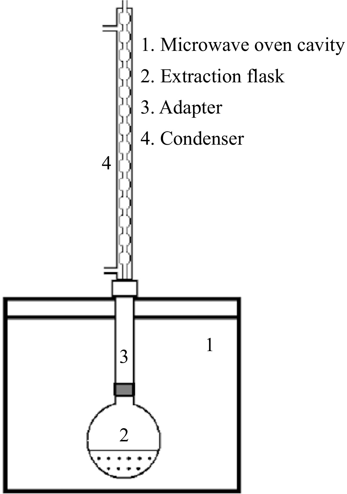
      Figure 1. 

      Diagram of the microwave-assisted extraction equipment.

    • Total phenolic contents of liquid extracts obtained from CE and MAE methods were determined by Folin-Ciocalteu method[23]. The extracts were diluted to a ratio of 1:20 by using water or ethanol-water mixture (50:50, v:v). 2.5 ml Folin-Ciocalteu reagent (0.2 N) was added to 0.5 ml sample. The mixture was vortexed for 5 s and kept in the dark for 5 min. Two ml sodium carbonate solution (75 g/L) was added to the mixture, and it was vortexed. This last mixture was kept in dark for 1 h. Then the absorbance was measured in a spectrophotometer at 760 nm. A calibration curve of gallic acid for different concentrations was prepared and the results were expressed as mg GAE/L (GAE : gallic acid equivalent).

    • This analysis was applied according to the method described by Akdeniz et al.[23]. The extracts were diluted to a ratio of 1:20 by using water or ethanol-water mixture (50:50, v:v). 3.9 ml DPPH solution (25 ppm) was added to 100 μl sample. The mixture was vortexed for 5 s and kept in the dark for 1 h. Then the absorbance was measured in a spectrophotometer at 517 nm. A calibration curve of DPPH for different concentrations (5, 10, 15, 20, and 25 ppm) was prepared and the results (TACDPPH) were expressed as mg DPPH/L.

    • This analysis was applied according to Özyürek et al.[24]. Firstly, 0.2 M copper (II) chloride solution, 1 M and pH 7.0 ammonium acetate solution and 7.5 × 10⁻³ M neocuproin solution were prepared. The extracts were diluted to a ratio of 1:60 by using water or ethanol-water mixture (50:50, v:v). One ml copper (II) chloride solution, 1 ml neocuproin solution, 1 ml ammonium acetate solution and 1.1 ml water were mixed with 10 ml diluted solid sample, and this was vortexed for 5 s. It was kept in the dark for 1 h and then, then the absorbance was measured in a spectrophotometer at 450 nm. A calibration curve of Trolox for different concentrations (50, 100, 150, 200, and 250 ppm) was prepared and the results (TACCUPRAC) were expressed as mmol TE/L (TE : Trolox equivalent).

    • The microstructure of red cabbage extracts from conventional and microwave-assisted extractions was investigated using a scanning electron microscope (SEM) (Gaia 3, Tescan). The extracts were lyophilized before the analysis to remove the moisture inside. The samples were coated with a thin layer of gold and mounted on stubs with the help of a double-sided adhesive tape before the visualization[25]. The SEM images were taken at 100×, and 2,500× magnifications.

    • Response surface methodology (RSM) was applied to optimize the MAE of phenolic compounds from red cabbage and Box-Behnken experimental design was employed for the experimental design. Extraction time (X1), solid/solvent ratio (X2), and microwave power (X3) were chosen as independent variables. The range and center point values of three independent variables presented in Table 1 are chosen according to preliminary experiments. The complete experimental design consisted of 17 experimental points containing 12 factorial points and five center points are shown in Supplementary Tables S1 & S2 for both solvent types, which are water and ethanol-water (50:50, v:v). Experimental runs were randomized to minimize the unexpected variability.

      Table 1.  Coded and actual values of the independent variables.

      Independent variables Code levels
      −1 0 1
      Extraction time (X1, min) 5 10 15
      Solid : solvent ratio (X2, 1/X) 20 30 40
      Microwave power (X3, W) 200 400 600

      The mathematical model corresponding to the Box-Behnken design (Eqn 1) was partitioned into linear, quadratic, and interactive components.

      Y=b0+ki=1biXi+ki=1biiX2i+k1i=1kj>1bijXiXj (1)

      where, Y is the predicted response (TPC, TACCUPRAC and TACDPPH), b0 is a constant and bi, bii and bij are the model coefficients. They represent the linear, quadratic and interaction effects of variables. The adequacy of the model was determined by evaluating the lack of fit, coefficient of determination (R2), and the Fischer test value (F-value) obtained from the analysis of variance (ANOVA) that was generated by Design Expert 12.0 statistical software. One-way ANOVA and Tukey analysis were also performed.

    • The maximum total phenolic content (TPC), and total antioxidant capacity values (TACDPPH and TACCUPRAC) obtained from MAE (solvent:water) were 2154.1 mg GAE/L (15 min, 1:20 solid : solvent ratio, 400 watt), 136.1 mg DPPH/L (15 min, 1:20 solid : solvent ratio, 400 watt), and 22.3 mmol TE/L (5 min, 1:20 solid : solvent ratio, 400 watt), respectively (Supplementary Table S1). The minimum values were 651 mg GAE/L (5 min, 1:40 solid : solvent ratio, 400 watt), 32.5 mg DPPH/L (5 min, 1:40 solid : solvent ratio, 400 watt), and 4.36 mmol TE/L (5 min, 1:40 solid : solvent ratio, 400 watt) for the same solvent (Supplementary Table S1). Supplementary Table S2 shows the experimental results for the ethanol-water mixture solvent. According to these results, the maximum values for TPC, TACDPPH, and TACCUPRAC were 1,106.4 mg GAE/L (10 min, 1:20 solid : solvent ratio, 200 watt), 134.4 mg DPPH/L (10 min, 1:20 solid : solvent ratio, 600 watt), and 17.3 mmol TE/L (10 min, 1:20 solid : solvent ratio, 600 watt), respectively. On the other hand, the minimum values for the same solvent were 634.5 mg GAE/L (5 min, 1:40 solid : solvent ratio, 400 watt), 60.8 mg DPPH/L (15 min, 1:40 solid : solvent ratio, 400 watt), and 6.6 mmol TE/L (10 min, 1:40 solid : solvent ratio, 600 watt).

    • RSM was applied for the optimization of MAE process conditions for obtaining the maximum yields for total phenolics (TPC) and total antioxidant capacities (TACDPPH and TACCUPRAC). Two identical Box-Behnken designs, each containing 17 experiments, were performed for two types of solvents, which were water and ethanol-water mixture (50:50, v:v). Extraction time (X1), solid/solvent ratio (X2), and microwave power (X3) were the independent variables chosen for maximizing the yields.

      Design matrices of the experimental conditions with their corresponding response values for both types of solvents were subjected to regression analysis, and the significance of each coefficient in the models was calculated (Supplementary Tables S1 & S2). Design Expert 12.0 software recommended the quadratic models with regression for all responses, and the adequacies of the models were confirmed using variance analysis (ANOVA) for TPC, TACDPPH, and TACCUPRAC. Tables 2 & 3 summarize ANOVA analysis for these responses obtained from MAE with water and water-ethanol mixture solvents, respectively.

      Table 2.  ANOVA for the quadratic equations of Design Expert 12.0.1 for MAE of TPC, TACDPPH, and TACCUPRAC in red cabbage and statistical indicators for each response (solvent : water).

      Source Sum of squares df Mean square F-value p-value
      Model (TPC) 2.34E+06 8 2.92E+05 67.92 0.0001 Significant
      X1 20274.94 1 20274.94 4.72 0.0819
      X2 1.69E+06 1 1.69E+06 392.13 < 0.0001
      X3 22967.4 1 22967.4 5.34 0.0688
      X1X2 22734.61 1 22734.61 5.29 0.0698
      X1X3 12778.04 1 12778.04 2.97 0.1453
      X2X3 3159.87 1 3159.87 0.7352 0.4304
      X12 26836.11 1 26836.11 6.24 0.0546
      X22 2.33E+05 1 2.33E+05 54.26 0.0007
      X32 0 0
      Residual 21488.64 5 4297.73
      Lack of fit 14143.94 1 14143.94 7.7 0.0501 Not significant
      Pure error 7344.7 4 1836.18
      Cor total 2.36E+06 13
      Model (TACDPPH) 9307.92 8 1163.49 4871.42 < 0.0001 Significant
      X1 193.06 1 193.06 808.33 < 0.0001
      X2 6939.72 1 6939.72 29055.95 < 0.0001
      X3 29.43 1 29.43 123.22 0.0001
      X1X2 306.43 1 306.43 1282.97 < 0.0001
      X1X3 262.93 1 262.93 1100.85 < 0.0001
      X2X3 16.56 1 16.56 69.31 0.0004
      X12 14.59 1 14.59 61.1 0.0005
      X22 708.01 1 708.01 2964.36 < 0.0001
      X32 0 0
      Residual 1.19 5 0.2388
      Lack of fit 0.5408 1 0.5408 3.31 0.143 Not significant
      Pure error 0.6534 4 0.1633
      Cor total 9309.12 13
      Model (TACCUPRAC) 184.87 8 23.11 16.62 0.0033 Significant
      X1 5.4 1 5.4 3.88 0.106
      X2 109.62 1 109.62 78.82 0.0003
      X3 1.45 1 1.45 1.04 0.3537
      X1X2 55.2 1 55.2 39.7 0.0015
      X1X3 1.78 1 1.78 1.28 0.309
      X2X3 1.23 1 1.23 0.8834 0.3904
      X12 0.7006 1 0.7006 0.5038 0.5096
      X22 1.42 1 1.42 1.02 0.3587
      X32 0 0
      Residual 6.95 5 1.39
      Lack of fit 3.91 1 3.91 5.13 0.0863 Not significant
      Pure error 3.05 4 0.7619
      Cor total 191.83 13

      Table 3.  ANOVA for the quadratic equations of Design Expert 12.0.1 for MAE of TPC, TACDPPH, and TACCUPRAC in red cabbage and statistical indicators for each response (solvent : ethanol-water, 50:50, v:v).

      Source Sum of squares df Mean square F-value p-value
      Model (TPC) 2.80E+05 9 31099.63 7.02 0.0088 Significant
      X1 19499.18 1 19499.18 4.4 0.0741
      X2 1.93E+05 1 1.93E+05 43.66 0.0003
      X3 6063.76 1 6063.76 1.37 0.2803
      X1X2 2.36 1 2.36 0.0005 0.9822
      X1X3 7959.32 1 7959.32 1.8 0.222
      X2X3 359.1 1 359.1 0.0811 0.7841
      X12 42236.73 1 42236.73 9.53 0.0176
      X22 9986.68 1 9986.68 2.25 0.1769
      X32 2963.88 1 2963.88 0.669 0.4403
      Residual 31011.78 7 4430.25
      Lack of fit 25791.29 3 8597.1 6.59 0.0501 Not significant
      Pure error 5220.5 4 1305.12
      Cor total 3.11E+05 16
      Model (TACDPPH) 8898.31 9 988.7 21.78 0.0003 Significant
      X1 0.2048 1 0.2048 0.0045 0.9483
      X2 7871.99 1 7871.99 173.4 < 0.0001
      X3 7.09 1 7.09 0.1561 0.7045
      X1X2 19.98 1 19.98 0.4401 0.5283
      X1X3 3.96 1 3.96 0.0872 0.7763
      X2X3 3.55 1 3.55 0.0783 0.7877
      X12 432.95 1 432.95 9.54 0.0176
      X22 533.11 1 533.11 11.74 0.011
      X32 75.26 1 75.26 1.66 0.2388
      Residual 317.79 7 45.4
      Lack of fit 66.7 3 22.23 0.3542 0.7899 Not significant
      Pure error 251.08 4 62.77
      Cor total 9216.09 16
      Model (TACCUPRAC) 167.95 9 18.66 25.72 0.0001 Significant
      X1 0.4512 1 0.4512 0.622 0.4562
      X2 137.7 1 137.7 189.81 < 0.0001
      X3 2.59 1 2.59 3.57 0.1009
      X1X2 1.24 1 1.24 1.71 0.2318
      X1X3 0.0462 1 0.0462 0.0637 0.808
      X2X3 5.62 1 5.62 7.74 0.0272
      X12 3.09 1 3.09 4.26 0.0779
      X22 16.41 1 16.41 22.63 0.0021
      X32 0.1155 1 0.1155 0.1592 0.7018
      Residual 5.08 7 0.7254
      Lack of fit 3.32 3 1.11 2.51 0.1973 Not significant
      Pure error 1.76 4 0.44
      Cor Total 173.03 16

      The models obtained for total phenols (TPC) in terms of mg GAE/L were significant with p-value = 0.0001, with R2 value of 0.9909 and adjusted R2 value of 0.9763 for water and p-value = 0.0088, with R2 value of 0.9003 and adjusted R2 value of 0.7720 for ethanol-water mixture. Sin et al.[26] reported that an R2 value greater than 0.8 is desirable for a developed regression model. The lack of fit values was not significant (p > 0.05), showing that the models were well-fitted and could be used to predict the TPC of the extracts. By applying multiple regression analysis to the experimental data of TPC for both solvents, the following second-order polynomial equations (Eqns 2 & 3) were found:

      TPC(water)=1016.63649.09X2+341.46X22 (2)
      TPC(ethanol-water)=858.95155.50X2100.16X21 (3)

      The models obtained for total antioxidant capacity (TACDPPH) in terms of mg DPPH equivalent/L were significant with p-value < 0.0001, with R2 value of 0.999 and adjusted R2 value of 0.999 for water and p-value = 0.0003, with R2 value of 0.966 and adjusted R2 value of 0.921 for ethanol-water mixture. The lack of fit values was not significant (p > 0.05), showing that the models were well-fitted and could be used to predict TACDPPH of the extracts. By applying multiple regression analysis to the experimental data of TACDPPH for both solvents, the following second-order polynomial equations (Eqns 4 & 5) were found:

      TACDPPH(water)=66.67+4.91X141.65X22.71X3+8.75X1X2+8.11X1X36.04X2X3+2.56X21+18.82X22 (4)
      TACDPPH(ethanol-water)=96.2831.37X210.14X21+11.25X22 (5)

      Total antioxidant capacity (TACCUPRAC) models in terms of mmol TE/L were significant with p-value = 0.0033, with R2 value of 0.9638 and adjusted R2 value of 0.9058 for water and p-value = 0.0001, with R2 value of 0.9707, and adjusted R2 value of 0.9329 for ethanol-water mixture. The lack of fit values was not significant (p > 0.05). By applying multiple regression analysis to the experimental data of TACCUPRAC for both solvents, the following second-order polynomial equations (Eqns 6 & 7) were found:

      TACCUPRAC(water)=10.395.24X2+3.72X1X2 (6)
      TACCUPRAC(ethanolwater)=9.064.15X21.18X2X3+1.97X22 (7)
    • The response surface graphs for TPC results for the solvent types of water (Fig. 2a & b) and ethanol-water (Fig. 2c & d) were illustrated in Fig. 2. Figures 3 & 4 show the response surfaces for TACDPPH and TACCUPRAC, respectively. In all these figures, the effects of extraction parameters on dependent variables can be seen, compared, and the interaction of different parameters can be discussed.
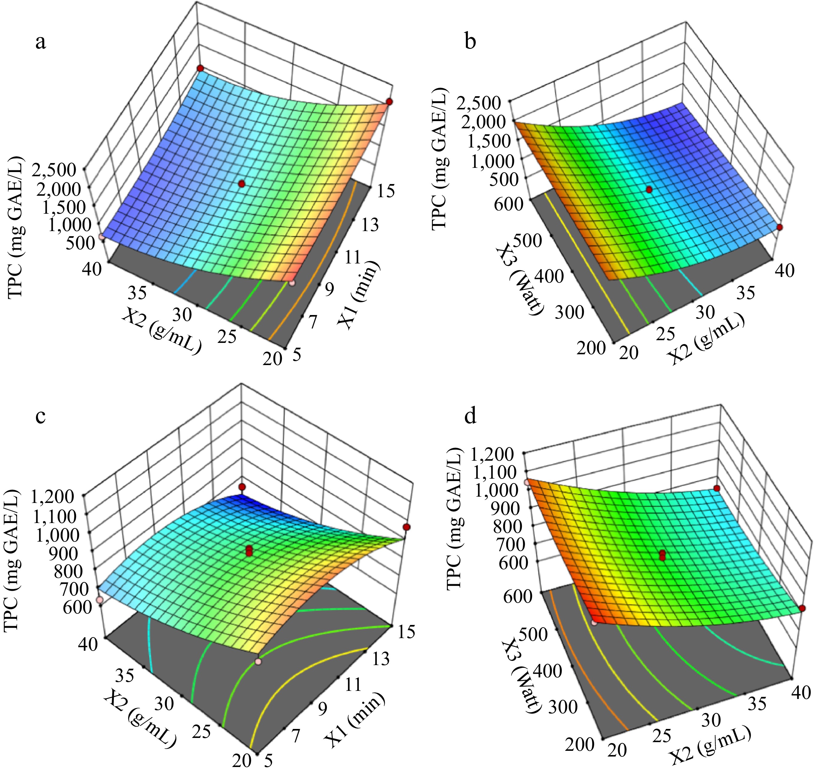
      Figure 2. 

      Response surfaces for TPC graphs (a) and (b) are for solvent : water; graphs (c) and (d) are for solvent : ethanol-water.
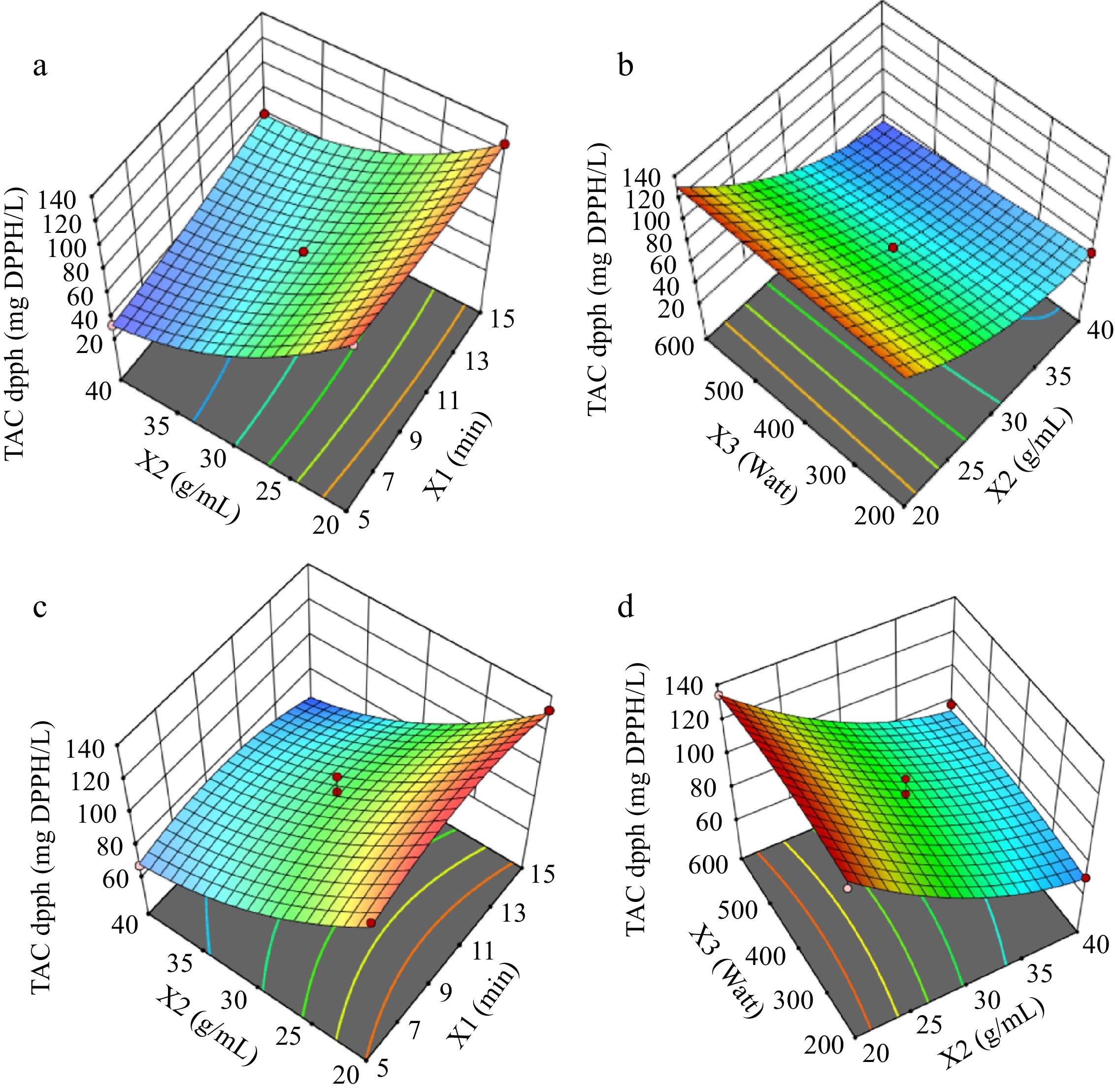
      Figure 3. 

      Response surfaces for TACDPPH, graphs (a) and (b) are for solvent:water; graphs (c) and (d) are for solvent:ethanol-water.
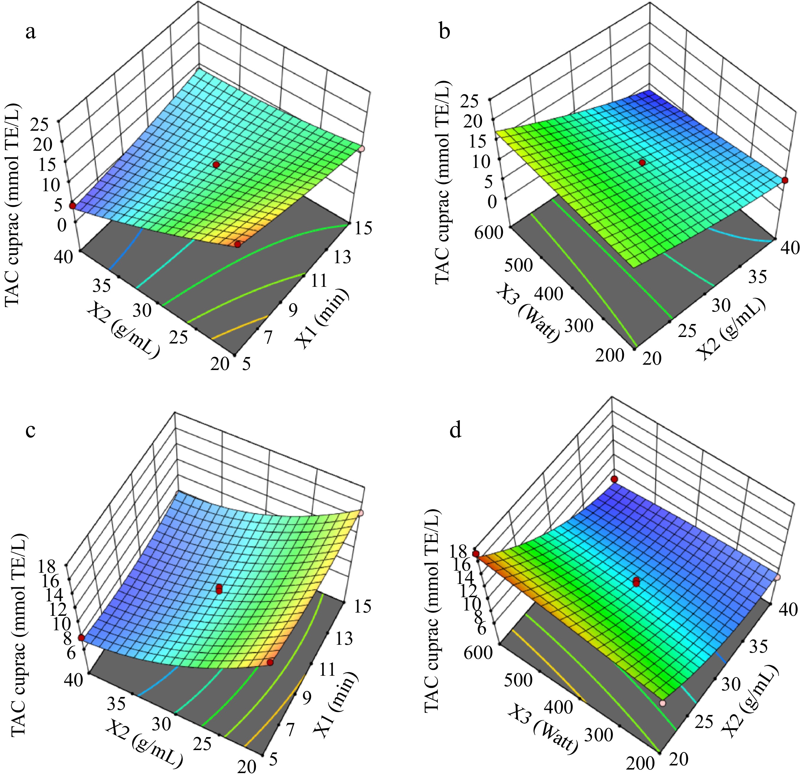
      Figure 4. 

      Response surfaces for TACCUPRAC graphs (a) and (b) are for solvent : water; graphs (c) and (d) are for solvent : ethanol-water.

      These figures generally corroborate previous research and the models of this work given in the previous section emphasizing the significance of solid : solvent ratio and its interactions with other variables for optimizing the extraction of bioactive compounds. Studies have shown that fine-tuning the solid : solvent ratio can significantly enhance phenolic compound yield[27]. The interaction effects underscore the complexity of the extraction process, where multiple factors interact to influence phenolic compound recovery efficiency[28].

    • The conventional extraction (CE) of phenolic compounds from red cabbage using water and an ethanol-water mixture at varying temperatures (40 and 70 °C) and extraction times (4 and 6 h) showed distinct patterns. At 40 °C with water, TPC increased with longer extraction times, while at 70 °C, TPC decreased after 6 h, suggesting thermal degradation[16,27]. Ethanol-water mixtures provided higher TPC values, but a decrease was observed at 70 °C for longer extraction times. Similarly, antioxidant capacity (TACCUPRAC and TACDPPH) values were higher at 40 °C for 4 h and decreased at higher temperatures and longer times, indicating potential degradation[14,29].

      In comparison, microwave-assisted extraction (MAE) demonstrated superior efficiency over CE. MAE significantly reduced extraction times while yielding higher TPC and antioxidant capacity values. Using ethanol-water, MAE achieved 1,062.6 mg GAE/L TPC in just over 9 min, whereas CE required 4−6 h to reach its maximum of 1,677.3 mg GAE/L. With water, MAE yielded 2,146 mg GAE/L TPC in 5 min, surpassing CE. The same trend was observed in antioxidant capacities, where MAE achieved higher TACCUPRAC and TACDPPH values in shorter times. The rapid and uniform heating provided by MAE contributed to this efficiency[28,30].

    • SEM images in Fig. 5a & b show red cabbage extract samples obtained from optimal conditions at CE. Rough and uneven surface morphology with a wide range of particle sizes can be seen in Fig. 5a. This heterogeneity in surface texture may indicate the insufficient breakdown of cell walls and incomplete release of intracellular compounds. At higher magnification (2,500×), Fig. 5b displays a more granular surface, indicating that the CE approach may leave residual cellular structures and bound phenolics. CE usually involves prolonged heating and solvent penetration, which can lead to incomplete extraction and degradation of sensitive bioactives.
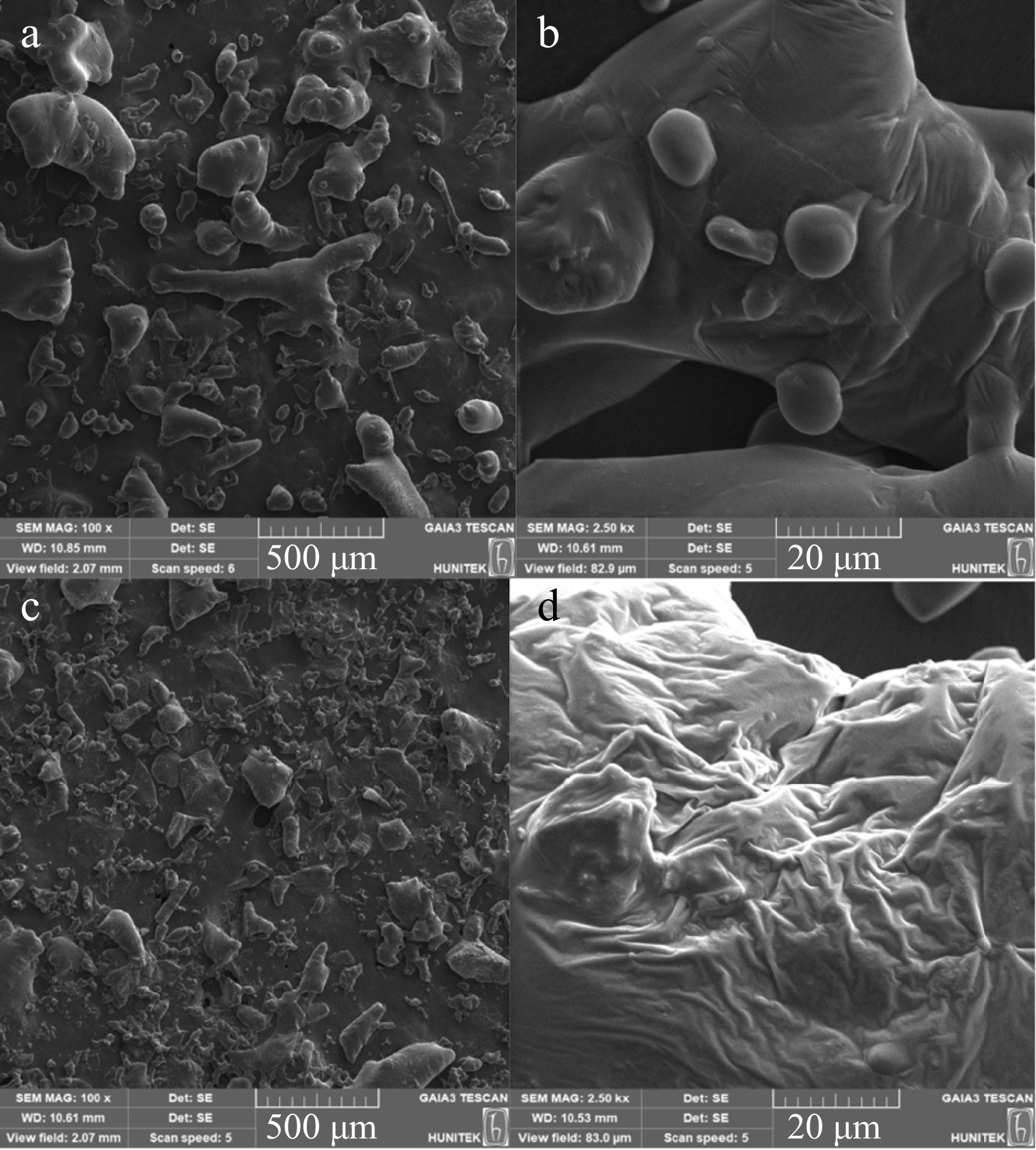
      Figure 5. 

      SEM images of red cabbage extracts. (a) Red cabbage extract from CE (100×), (b) red cabbage extract from CE (2,500×), (c) red cabbage extract from MAE (100×), (d) red cabbage extract from MAE (2,500×)

      In contrast, Fig. 5c & d depict red cabbage extracts obtained through optimal conditions at MAE. Figure 5c demonstrates a more uniform particle distribution with smaller and more evenly broken particles than CE. These results suggest that MAE is more efficient at cell wall destruction and releasing phenolic compounds. MAE improves mass transfer rate and extraction efficiency by rapid heating and high energy input. Figure 5d further illustrates the effect of MAE at a magnification of 2,500×, where the surface seems more fibrillar. MAE can cause significant structural changes at the cellular level, leading to more efficient extraction. The dielectric heating effect during MAE causes the fibrillar structure of the extract surface because of the breakdown of the plant cell matrix and the release of bound compounds. The differences in microstructure between CE and MAE extracts, as shown in Fig. 5, indicate the superior efficiency of MAE in breaking down cell walls and enhancing extraction.

    • The extraction of phenolic compounds from plant material is influenced by various factors, including the nature of the plant material, type and concentration of solvent, sample:solvent ratio, particle size, extraction time, and temperature[8]. The interaction of the solvent with microwaves can also significantly impact the extraction process. Ethanol, methanol, and acetone are commonly used as organic solvents due to their high solubility and polarity characteristics[31]. In this study, acidified water (pH 3.0−3.3) and an ethanol-water mixture (50:50, v:v) were used. The solid : solvent ratio was included as an independent variable due to its impact on extraction yield (Table 1). Additionally, microwave irradiation power (watt) and extraction time (min) were considered crucial variables because they influence the temperature inside the extraction vessel.

    • ANOVA results (Table 2) indicated that the solid:solvent ratio (X2) and its quadratic term (X22) were the most significant factors affecting TPC during MAE when water was used as the solvent. An increase in the solid:solvent ratio had a negative linear impact on TPC, while the quadratic effect was positive. Neither extraction time (X1) nor microwave power (X3) significantly influenced TPC within the studied ranges (5−15 min, 200−600 watt). Similar trends have been reported in other studies, where the solid : solvent ratio was a critical factor for phenolic extraction, while microwave power had a limited effect[27]. Therefore, the solvent type and ratio were two significant effects for phenolic compound extraction[29]. For the ethanol-water mixture (Table 3), the solid : solvent ratio remained a significant factor, along with the quadratic term of extraction time (X12). This indicates that the solid : solvent ratio continues to dominate the process, whereas the role of the quadratic effect of extraction time becomes more influential.

      Table 4.  TPC, TACCUPRAC and TACDPPH yields of conventional extraction.

      Temperature (°C) Time (h) Solvent* TPC (mg GAE/L) TACCUPRAC (mmol TE/L) TACDPPH (mg DPPH/L)
      40 4 w 1018.5 ± 1.28D 8.9 ± 0.03B 33.1 ± 0.75B
      6 1165.9 ± 1.29C 11.2 ± 0.02A 54.52 ± 0.32A
      70 4 1324.4 ± 2.22A 11.6 ± 0.16A 48.0 ± 0.08A
      6 1192.6 ± 1.28B 11.6 ± 0.31A 50.2 ± 1.78A
      40 4 e-w 1677.3 ± 1.15a 16.77 ± 0.14a 66.3 ± 0.81a
      6 1515.3 ± 1.15b 14.2 ± 0.04b 58.5 ± 0.49b
      70 4 1268.0 ± 2.00c 10.0 ± 0.03d 36.1 ± 1.48d
      6 1202.7 ± 1.15d 10.7 ± 0.03c 44.5 ± 0.65c
      The results are given as the mean of three replicates with the standard deviations. Different letters represent the significant differences (p ≤ 0.05) between the results for each column. Upper case letters represent the solvent-water and lower case letters represent the solvent-ethanol-water. * w: water, e-w: ethanol-water.

      Regarding antioxidant capacity (TACDPPH), all independent variables (X1, X2, X3) significantly impacted the values when water was used as the solvent (Eqn 4). Extraction time (X1) had a positive effect on TACDPPH, whereas solid : solvent ratio (X2) and microwave power (X3) had negative effects. The quadratic terms for both extraction time and solid : solvent ratio were significant, suggesting a complex relationship between these variables and antioxidant capacity. Similar findings have been reported by Yiğit et al.[8] in a study on anthocyanin content. For the ethanol-water solvent, the TACDPPH model (Eqn 5) showed that the solid : solvent ratio was the only significant linear variable, while quadratic terms for extraction time (X1²) and solid : solvent ratio (X2²) were also significant. These results align with those from the TPC models. Studies such as those by Rodsamran & Sothornvit[32] similarly noted that microwave power does not significantly affect antioxidant capacity during MAE.

      ANOVA results (Table 2) for TACCUPRAC indicated that the solid:solvent ratio (X2) and its interaction with extraction time (X1X2) were significant. Increasing the solid : solvent ratio led to a decrease in TACCUPRAC, although this effect was mitigated when extraction time and solid : solvent ratio were optimized together. Similar trends were observed with the ethanol-water mixture, where the interaction between solid/solvent ratio and microwave power (X2X3), as well as the quadratic effect of solid : solvent ratio (X2²), were significant. The quadratic relationship highlights that increasing the solid : solvent ratio beyond a certain point can positively influence TACCUPRAC.

      In future microwave-assisted extraction studies, the microwave power that provides the best results for phenolic extraction may be kept constant, while focusing on optimizing the solid : solvent ratio, solvent type, and extraction time.

    • Optimal conditions for MAE were determined using the desirability function, with values of 0.838 for ethanol-water, and 0.924 for water. X1 = 9.2 min, X2 = 1:20, X3 = 501.8 watt for ethanol-water solvent, and X1 = 5 min, X2 = 1:20, X3 = 384.1 watt for water-only solvent were found as optimum conditions for MAE method. These results showed that lower extraction time and microwave power were required when water is used than when ethanol-water was used the solvent to obtain the optimum phenolic content and antioxidant capacity values. Therefore, this indicated that there were more water-soluble phenolic compounds in the samples. Moreover, higher extraction time or microwave power levels may also cause degradation of possible heat-labile phenolics in the extracts.

      The response surface plots for TPC under different conditions of water and ethanol-water solvents are illustrated in Fig. 2. Increasing the solid : solvent ratio significantly improves TPC, particularly at moderate extraction times. Moderate microwave power combined with an optimal solid : solvent ratio enhances TPC, as seen in Fig. 2b[33]. For the ethanol-water mixture, a lower solid : solvent ratio paired with moderate microwave power yields the highest TPC values[27]. These results align with findings indicating that extraction parameters, such as solvent polarity and extraction time, strongly influence phenolic content[6].

      Figure 3 shows the response surfaces for TACDPPH. In the water solvent system, a balanced solid : solvent ratio and longer extraction times optimize antioxidant capacity. In contrast, moderate microwave power combined with optimal solid : solvent ratios yields the best results with the ethanol-water mixture. These trends suggest that antioxidant compounds are sensitive to extraction conditions, with higher power settings potentially leading to thermal degradation[30,34]. Studies have similarly noted the importance of controlling microwave power to preserve antioxidant properties[28].

      For total antioxidant capacity measured by the CUPRAC assay (TACCUPRAC), Fig. 4 demonstrates that balanced extraction time and solid:solvent ratio maximize the yields in both solvent systems. Lower microwave power and optimal solid:solvent ratios consistently result in higher antioxidant capacity for ethanol-water extracts. These findings underscore the non-linear effects of variables like extraction time and microwave power, which must be carefully controlled to optimize both phenolic and antioxidant recovery[8,11].

    • The comparison of CE and MAE techniques revealed that MAE demonstrated superior efficiency in extracting phenolic compounds and antioxidants from red cabbage (Table 4). MAE not only reduced extraction times significantly but also enhanced the yield of phenolic compounds and antioxidants compared to CE. The use of an ethanol-water mixture generally provided better extraction yields than water alone in both methods, with the benefits being more pronounced in MAE[11]. One of the major advantages of MAE is its ability to rapidly heat the solvent and sample, which allows for shorter extraction times. This minimizes the exposure of sensitive phenolic compounds to high temperatures, reducing the risk of thermal degradation[15]. In contrast, CE methods, which typically involve longer extraction times at higher temperatures, may lead to a higher rate of phenolic compound degradation. Reducing microwave power and extraction time in MAE can further mitigate potential damage to sensitive phenolic compounds, providing a distinct advantage over CE in preserving compound integrity[34]. These findings align with previous studies that reported ethanol as an effective solvent for polyphenol extraction due to its polarity and ability to interact with microwaves[28]. Additionally, studies have shown that MAE often achieves better extraction efficiency and shorter processing times compared to ultrasound-assisted extraction (UAE)[32].

    • The SEM images revealed significant differences in the microstructure of the extracts obtained through CE and MAE. CE extracts exhibited rough and uneven surface morphology with a wide range of particle sizes, suggesting insufficient breakdown of cell walls and incomplete release of intracellular compounds. In contrast, MAE extracts demonstrated more uniform particle distribution with smaller and more evenly broken particles, indicating more efficient cell wall destruction and release of phenolic compounds.

      The fibrillar surface structure observed in MAE extracts, as compared to CE, can be attributed to the dielectric heating effect of microwaves, which causes significant structural changes at the cellular level. These findings suggest that MAE is a more effective method for obtaining high yields of phenolic compounds and antioxidants from red cabbage, as it enhances mass transfer rates and extraction efficiency through rapid heating and high energy input.

    • The effectiveness of microwave-assisted extraction (MAE) compared to conventional methods (CE) was demonstrated by this study for obtaining phenolic compounds and antioxidants from red cabbage. Response surface methodology was applied in the optimization of the key extraction parameters, including extraction time, solid/solvent ratio, and microwave power, for both water and ethanol-water solvents. Box-Behnken design was used in experimental design. MAE reduced the required time and enhanced the yield of total phenolic content and antioxidant capacities by improving extraction efficiency. This method proved to be superior to CE, which included longer extraction times and yielded lower concentrations of desired bioactive compounds. Scanning electron microscopy (SEM) images confirmed the structural differences between the two methods, and they showed that MAE-treated samples had more uniform particle distribution and higher porosity. These observations suggest that MAE can break down the cell wall in plants and release intracellular compounds more effectively compared to CE. Overall, the results of this study strongly support the utilization of MAE as an advanced method for the extraction of phenolic compounds and antioxidants from red cabbage. This technology not only reduces processing time but also preserves bioactive compounds, making it suitable for the food industry. Further studies could be recommended for the application of MAE in other plant materials and exploration of its scalability for industrial use. Additionally, investigation of the stability and bioavailability of the extract samples could provide valuable insights into their health benefits and potential applications in food products.

      • This research was supported by Hacettepe University-Scientific Research Projects Coordination (BAP) Unit (FBA-2017-12587).

      • The authors confirm contribution to the paper as follows: study conception and design: Turabi Yolaçaner E; data collection: Yiğit Ü; analysis and interpretation of results: Turabi Yolaçaner E, Yiğit Ü; draft manuscript. preparation: Turabi Yolaçaner E, Yiğit Ü. Both authors reviewed the results and approved the final version of the manuscript.

      • The datasets generated during and/or analyzed during the current study are available from the corresponding author on reasonable request.

      • The authors declare that they have no conflict of interest.

      • Supplementary Table S1 Experimental design by Box-Behnken and the results* of the experiments (Solvent: water).
      • Supplementary Table S2 Experimental design by Box-Behnken and the results* of the experiments (Solvent: ethanol-water, 50:50, v:v).
      • Copyright: © 2024 by the author(s). Published by Maximum Academic Press on behalf of Nanjing Agricultural University. This article is an open access article distributed under Creative Commons Attribution License (CC BY 4.0), visit https://creativecommons.org/licenses/by/4.0/.
    Figure (5)  Table (4) References (34)
  • About this article
    Cite this article
    Yiğit Ü, Turabi Yolaçaner E. 2024. Enhanced extraction of phenolic compounds from red cabbage utilizing microwave-assisted method: a Box-Behnken approach for optimization. Food Materials Research 4: e030 doi: 10.48130/fmr-0024-0024
    Yiğit Ü, Turabi Yolaçaner E. 2024. Enhanced extraction of phenolic compounds from red cabbage utilizing microwave-assisted method: a Box-Behnken approach for optimization. Food Materials Research 4: e030 doi: 10.48130/fmr-0024-0024

Catalog

  • About this article

/

DownLoad:  Full-Size Img  PowerPoint
Return
Return