REVIEW   Open Access    

Recent advances on surface enhanced Raman spectroscopy in safety assessment and quality control of meat and meat products

More Information
  • With the continuous development of spectroscopy technology, surface enhanced Raman spectroscopy (SERS) has been widely used as a fast and sensitive analysis method for the qualitative and quantitative analysis of trace analytes in foods. At present, SERS has been widely used in various fields such as food safety, materials, and biomedicine. However, the advances of SERS in meat safety and quality detection have not been summarized. In this review, the development history and detection principles of SERS are introduced and the advantages and potential of SERS application in the field of meat safety and quality detection evaluated. Then, two classical SERS detection modes were compared, namely labeled detection and label-free detection, in terms of the advantages, disadvantages, and application scopes. Furthermore, the specific applications of SERS in detecting bacteria, viruses, veterinary drug residues, food additives, illegal additives, and biotoxins in meat and meat products were presented. In addition, the development of SERS in meat adulteration and freshness identification are summarized. The prospects of the future development of SERS in meat safety and quality assessment will likely involve multiple method integrations, new material development, and artificial intelligence. It is expected that this review will not only provide a comprehensive summary and exploration of SERS in meat safety and quality assessment but also shed light on the future innovation and continued development of SERS in the food and meat industry.
  • Salvia rosmarinus L. (old name Rosmarinus officinalis), common name Rosemary thrives well in dry regions, hills and low mountains, calcareous, shale, clay, and rocky substrates[1]. Salvia rosmarinus used since ancient times in traditional medicine is justified by its antiseptic, antimicrobial, anti-inflammatory, antioxidant, and antitumorigenic activity[1,2]. The main objective of the study is to evaluate the antimicrobial activity of different extracts of Salvia rosmarinus in vitro, and its compounds related to in silico targeting of enzymes involved in cervical cancer. Since the start of the 20th century, some studies have shown that microbial infections can cause cervical cancers worldwide, infections are linked to about 15% to 20% of cancers[3]. More recently, infections with certain viruses like Human papillomaviruses (HPV) and Human immunodeficiency virus (HIV), bacteria like Chlamydia trachomatis, and parasites like schistosomiasis have been recognized as risk factors for cancer in humans[3]. Then again, cancer cells are a group of diseases characterized by uncontrolled growth and spread of abnormal cells. Many things are known to increase the risk of cancer, including dietary factors, certain infections, lack of physical activity, obesity, and environmental pollutants[4]. Some studies have found that unbalanced common flora Lactobacillus bacteria around the reproductive organ of females increases the growth of yeast species (like Candida albicans) and some studies have found that women whose blood tests showed past or current Chlamydia trachomatis infection may be at greater risk of cervical cancer. It could therefore be that human papillomavirus (HPV) promotes cervical cancer growth[3]. Salvia rosmarinus is traditionally a healer chosen as a muscle relaxant and treatment for cutaneous allergy, tumors, increases digestion, and the ability to treat depressive behavior; mothers wash their bodies to remove bacterial and fungal infections, promote hair growth, and fight bad smells[5] .

    The study of plant-based chemicals, known as phytochemicals, in medicinal plants is gaining popularity due to their numerous pharmacological effects[6] against drug resistance pathogens and cancers. The causes of drug resistance to bacteria, fungi, and cancer are diverse, complex, and only partially understood. The factors may act together to initiate or promote infections and carcinogenesis in the human body is the leading cause of death[7]. Antimicrobial medicines are the cornerstone of modern medicine. The emergence and spread of drug-resistant pathogens like bacteria and fungi threaten our ability to treat common infections and to perform life-saving procedures including cancer chemotherapy and cesarean sections, hip replacements, organ transplantation, and other surgeries[7]. On the other hand, information about the current magnitude of the burden of bacterial and fungal drug resistance, trends in different parts of the world, and the leading pathogen–drug combinations contributing to the microbial burden is crucial. If left unchecked, the spread of drug resistance could make many microbial pathogens much more lethal in the future than they are today. In addition to these, cancers can affect almost any part of the body and have many anatomies and molecular subtypes that each require specific management strategies to avoid or inhibit them. There are more than 200 different types of cancer that have been detected. The world's most common cancers affecting men are lung, prostate, colorectal, stomach, and liver cancers[8]. While breast, cervix, colorectal, lung, and stomach cancers are the most commonly diagnosed among women[8]. Although some cancers said to be preventable they seem to still be one of the causes of death to humans, for example cervical cancer. The need to fill the gap to overcome the problem of searching for antimicrobials and anticancers from one source of Salvia rosmarinus is of importance.

    Cervical cancer is a common cancer in women and a prominent cause of death[9]. In Ethiopia, cervical cancer is a big deal for women aged 15 to 44, coming in as the second most common cancer[9]. Globally, it's the fourth most common prevalent disease for women[10]. Aberrant methylation of tumor-suppressor genes' promoters can shut down their important functions and play a big role in causing cervical tumors[10]. There are various cervical cancer repressor genes (proteins turn off or reduce gene expression from the affected gene), such as CCNA1, CHF, HIT, PAX1, PTEN, SFRP4, and TSC1. The genes play a crucial role in causing cervical cancer by regulating transcription and expression through promoter hypermethylation, leading to precursor lesions during cervical development and malignant transformation[11]. The process of DNA methylation is primarily carried out by a group of enzymes known as DNA methyltransferases (DNMT1). It has been reported that DNMT1 (PDB ID: 4WXX), a protein responsible for DNA methylation can contribute to the development of cervical cancer. DNMT1 inhibits the transcription of tumor suppressor genes, facilitating tumorigenesis, which finally develops into cervical cancer. Tumor suppressor gene transcription is inhibited by DNMT1, which helps cancer grow and eventually leads to cervical cancer. Repressive genes' hypermethylation may be decreased, their expression can be increased, and the phenotype of malignant tumors can be reversed by inhibiting the DNMT1 enzyme.

    On the other hand, infection by the human papilloma virus (HPV) phenotype 16, enzyme 6 (PDB ID: 4XR8) has been correlated with a greatly increased risk of cervical cancer worldwide[12]. Based on variations in the nucleotide sequences of the virus genome, over 100 distinct varieties of the human papilloma virus (HPV) have been identified (e.g. type 1, 2 etc.). Genital warts can result from certain types 6 and 11 of sexually transmitted HPVs. Other HPV strains, still, that can infect the genitalia, do not show any symptoms of infection[8]. Persistent infection with a subset of approximately 13 so-called 'high-risk' sexually transmitted HPVs, including such as types 16, 18, 31, 33, 35, 39, 45, 51, 52, 56, 58, 59, and 68 different from the ones that cause warts may lead to the development of cervical intraepithelial neoplasia (CIN), vulvar intraepithelial neoplasia (VIN), penile intraepithelial neoplasia (PIN), and/or anal intraepithelial neoplasia (AIN). These are precancerous lesions and can progress to invasive cancer. Almost all occurrences of cervical cancer have HPV infection as a required component[13]. Superfluous infection by HPV type 16 E6 (PDB ID: 4XR8) has been correlated with a greatly increased genital risk of precursor cervical cancer worldwide[11]. Scholars more defined in major biochemical and biological activities of HPV type 16 E6 (PDB ID: 4XR8) in high-risk HPV oncogenes and how they may work together in the development of cervical disease and cancer[13].

    One potential approach to treat cervical cancer is to inhibit the activity of the DNMT1 and HPV type 16 E6 enzymes specifically[1316]. Over 50% of clinical drug forms worldwide originate from plant compounds[17]. In the past, developing new drugs was a lengthy and costly process. However, with the emergence of bioinformatics, the use of computer-based tools and methods have become increasingly important in drug discovery. One such method is molecular docking and ADMET profiling which involves using the structure of a drug to screen for potential candidates. This approach is known as structure-based drug design and can save both time and resources during the research process[15]. Structural-based drug designing addresses ligand binding sites with a known protein structure[15]. Using free binding energies, a computational method known as docking examines a large number of molecules and suggests structural theories for impeding the target molecule[17]. Nowadays, due to increasing antibiotic resistance like bacteria, fungi, and cancer cells, natural products remain an important source for discovering antimicrobial compounds and novel drugs for anti-cancers like cervical cancers. Therefore, the purpose of this research is to assess the antimicrobial activity of extracts, molecular docking, ADMET profiling in anticancer properties of compounds isolated from Salvia rosmarinus, on a targeting DNMT1 and HPV type 16 E6 in human cervical cancer. In the present study, various solvent crude extracts obtained from Salvia rosmarinus were used for antimicrobial activity and the isolated compounds 1 and 2 were submitted for in silico study to target the DNMT1 and HPV type 16 E6 enzymes to inhibit the growth of human cervical cancer cells.

    Healthy Salvia rosmarinus leaves were collected in Bacho district, Southwest Showa, Oromia, Ethiopia, during the dry season of November 2022. The plant materials were authenticated by Melaku Wondafrish, Natural Science Department, Addis Ababa University and deposited with a voucher number 3/2-2/MD003-80/8060/15 in Addis Ababa University's National Herbarium.

    The most common organic solvent used in extractions of medicinal plants is 2.5 L of petroleum ether, chloroform/methanol (1:1), and methanol. The test culture medium for microbes was used and performed in sterile Petri dishes (100 mm diameter) containing sterile Muller–Hinton Agar medium (25 mL, pH 7) and Sabouraud Dextrose Agar (SDA) for bacteria and fungi, respectively. A sterile Whatman filter paper (No. 1) disc of 6 mm diameter was used to determine which antibiotics an infective organism is sensitive to prescribed by a minimum zone of inhibition (MZI). Ciprofloxacin antibiotic reference (manufactured by Wellona Pharma Ciprofloxacin tablet made in India) and Ketoconazole 2% (made in Bangladesh) were used as a positive controls for antibacterial and antifungal, respectively and Dimethyl sulfoxide (DMSO) 98.9% was used as a negative control for antimicrobial tests. In the present study, the height of the column was 650 mm and the width was 80 mm. Several studies by previous researchers showed the acceptable efficiency of column chromatography (up to 43.0% w/w recovery) in the fractionation and separation of phenolic compounds from plant samples[18]. In column chromatography, the ideal stationary phase used silica gel 60 (0.200 mm) particles. The 1H-NMR spectrums of the compounds were analyzed using a 600 MHz NMR machine and 150 MHz for 13C NMR. The compounds were dissolved in MeOD for compound 1 and in DMSO for compound 2 for NMR analysis. On the other hand, UV spectroscopy (made in China) used 570 nm ultraviolet light to determine the absorbency of flavonoids (mg·g−1) phytochemicals.

    The samples (extracts) were analyzed to detect the presence of certain chemical compounds such as alkaloids (tested using Wagner's reagents), saponins (tested using the froth test), steroids (tested with Liebermann Burchard's tests), terpenoids (tested with Lidaebermann Burchard's tests), quinones, and flavonoids (tested using Shinoda tests)[19].

    The leaves of Salvia rosmarinus (500 g) were successively extracted using maceration using petroleum ether, chloroform/methanol (1:1), and methanol, every one 2.5 L for 72 h to afford 3.6, 6, and 53 g crude extracts, respectively. The methanol/chloroform (1:1) extract (6 g) was loaded to silica gel (150 g) column chromatography using the increasing polarity of petroleum ether, methanol/chloroform (1:1) solvent system to afford 80 fractions (100 mL each). The fraction obtained from chloroform/methanol 1:1 (3:2) after repeated column chromatography yielded compound 1 (18 mg). Fractions 56-65, eluted with chloroform/methanol (1:1) were combined and purified with column chromatography to give compound 2 (10 mg).

    The microorganisms were obtained from the Ethiopia Biodiversity Institution (EBI). Two gram-positive bacteria namely Staphylococcus aureus serotype (ATCC 25923) and Streptococcus epidermidis (ATCC14990); and three gram-negative bacteria, namely Escherichia coli (ATCC 25922), Pseudomonas aeruginosa (ATCC 5702), and Klebsiella pneumonia (ATCC e13883) were inoculated overnight at 37 °C in Muller–Hinton Agar/MHA culture medium and two fungus strains of Candida albicans (ATCC 16404) and Aspergillus niger (ATCC 11414) were inoculated overnight at 27−30 °C in Sabouraud Dextrose Agar/SDA culture medium[20].

    The antibacterial and antifungal activities of different crude extracts obtained from Salvia rosmarinus plant leaves were evaluated by the disk diffusion method (in accordance with the 13th edition of the CLSI M02 document on hardydiagnostics.com/disk-diffusion). Briefly, the test was performed in sterile Petri dishes (100 mm diameter) containing solid and sterile Muller–Hinton Agar medium (25 mL, pH 7) and Sabouraud Dextrose Agar (SDA) for bacteria and fungi, respectively. The extracts were placed on the surface of the media that had previously been injected with a sterile microbial suspension (one microbe per petri dish) after being adsorbed on sterile paper discs (5 μL per Whatman disc of 6 mm diameter). To prevent test samples from eventually evaporating, all Petri dishes were sealed with sterile laboratory films. They were then incubated at 37 °C for 24 h, and the zone diameter of the inhibition was measured and represented in millimeters. Ciprofloxacin antibiotic reference (manufactured by Wellona Pharma Ciprofloxacin tablet, India) was used as a positive control and DMSO was used as a negative control for antibacterial activity test while Ketoconazole 2% (Bangladesh) was used as a positive control and 10 μL of 0.2% agar as a negative control for antifungal activity tests[20]. The term 'inhibitory concentration' refers to the minimum sample concentration required to kill 99.9% of the microorganisms present[21]. Three repetitions of the crude extract sample were used to precisely measure the inhibitory halo diameter (in mm), which was then expressed as mean ± standard deviation to assess the anti-microbial activity.

    Cervical cancer-causing protein was identified through relevant literature. The protein molecule structure of DNA (cytosine-5)-methyltransferase 1 (DNMT1) (PDB ID: 4WXX)[21] and HPV type 16 E6 (PDB ID: 4XR8)[21] - a protein known to cause cervical cancer - were downloaded from the Protein Data Bank[22]. The stability of the protein molecule was assessed using Rampage[23].

    Phytochemical constituents of Salvia rosmarinus plant leaves were used to select a source of secondary metabolites (ligands). Ligand molecules were obtained through plant extraction, and isolation, and realized with PubChem (https://pubchem.ncbi.nlm.nih.gov/). The ligands were downloaded in Silver diamine fluoride format (SDF) and then converted to PDB format using an online SMILES translator (https://cactus.nci.nih.gov/translate/). The downloaded files were in PDB format, which was utilized for running various tools and software[24].

    The Biovia Discovery Studio Visualizer software was used to analyze the protein molecule. The protein molecule was converted into PDB format and its hierarchy was analyzed by selecting ligands and water molecules. Both the protein molecule and the water molecules lost their attached ligands during the analysis. Finally, the protein's crystal structure was saved in a PDB file[25].

    PyRx software was utilized to screen secondary metabolites and identify those ligands with the lowest binding energy to the protein target. The ligands with the lowest binding energy were further screened for their drug-likeliness property through analysis. It is worth noting that PyRx runs on PDBQT format. To begin using PyRx, it needs to load a protein molecule. This molecule should be converted from PDB to the protein data bank, partial charge (Q), and Atom Type (PDBQT) format. Once the protein molecule is loaded, it can import ligands from a specific folder in Silver diamine fluoride format. The ligand energy was minimized and changed to PDBQT format. The protein was docked with the ligand and screened based on minimum binding energy (https://cactus.nci.nih.gov/translate/).

    The optimal ligand was selected for final docking using AutoDock Vina and Biovia by modifying the reference of Discovery Studio Client 2021 (https://cactus.nci.nih.gov/translate/).

    The protein target from the Protein Data Bank (PDB) was loaded onto the graphical interface of AutoDock Vina. To prepare the protein for docking, water molecules were removed, hydrogen polar atoms were added, and Kollman charges were assigned to the protein molecule. Ultimately, PDBQT format was used to store the protein. After being imported in PDB format, the Ligand molecule was transformed to PDBQT format. Next, a grid box was chosen to represent the docked region. The command prompt was used to run AutoDock Vina and the outcomes were examined (https://cactus.nci.nih.gov/translate/).

    Docking the ligand with the protein target DNMT1(PDB ID: 4WXX)[22] and HPV type 16 E6 (PDB ID: 4XR8)[21] enzymes were performed using Biovia Discovery Studio Client 2021 by loading the protein target first followed by the ligand in PDB format. The charges were attached to the protein molecule, and the energy was minimized for the ligands. Both the protein and ligand molecules were prepared for docking. Once the docking process was complete, the results were analyzed based on several parameters, including absolute energy, clean energy, conf number, mol number, relative energy, and pose number. The interaction between the protein and ligand was analyzed using structure visualization tools, such as Biovia Discovery Studio Visualizer and PyMol (https://cactus.nci.nih.gov/translate/).

    The process of visualizing the structure was carried out using the PyMol tool. PyMol is a freely available software. Firstly, the protein molecule in PDBQT form was loaded on the PyMol graphical screen. Then, the output PDBQT file was added. The docked structure was visualized and the 'molecule' option was changed to 'molecular surface' under the 'shown as' menu (https://cactus.nci.nih.gov/translate/).

    Drug likeliness properties of the screened ligands were evaluated using the SwissADME online server. SMILE notations were obtained from PubChem and submitted to the SwissADME web server for analysis. The drugs were subjected to Lipinski's rule of five[20] for analysis. Lipinski's rules of five were selected for final docking through AutoDock Vina and Biovia Discovery Studio Client 2021. Ligands 1 and 2 were analyzed using Lipinski's rule of five for docking with AutoDock Vina and Biovia Discovery Studio Client 2021.

    The antimicrobial analysis data generated by triplicate measurements reported as mean ± standard deviation, and a bar graph also generated by GraphPad Prism version 8.0.1 (244) for Windows were used to perform the analysis. GraphPad Prism was used and combined with scientific graphing, comprehensive bar graph fitting (nonlinear regression), understandable statistics, and data organization. Prism allows the performance and modification of basic statistical tests commonly used and determined through the statistical applications in microbiology labs (https://graphpad-prism.software.informer.com/8.0/).

    Phytochemical screening of the different extracts for the presence (+) and absence (−) of alkaloids, steroids, glycosides, coumarins, terpenoids, flavonoids, carbohydrates, tannins, and saponins were done. The present study showed that alkaloids, terpenoids, flavonoids, and tannins tests in S. rosmarinus leaves of petroleum ether, chloroform/methanol (1:1), and methanol extracts were high whereas glycoside, coumarins, and carbohydrates had a moderate presence. The extract of S. rosmarinus leaves contain commonly bioactive constituents such as alkaloids, steroids, terpenoids, flavonoids, tannins, and saponins. These bioactive chemicals have active medicinal properties. Phytochemical compounds found in S. rosmarinus leaves have the potential to treat cancer cells and pathogens. The study also found that these flavonoids are related to natural phenolic compounds with anticancer and antimicrobial properties in the human diet (Table 1).

    Table 1.  Phytochemical screening tests result of petroleum ether, chloroform/methanol (1:1) and methanol extracts of Salvia rosmarinus leaves.
    Botanical name Phytochemicals Phytochemical screening tests Different extracts
    Petroleum ether Chloroform/methanol (1:1) Mehanol
    Salvia rosmarinus Alkaloids Wagner's test ++ ++ ++
    Steroids Libermann Burchard test ++ + ++
    Glycoside Keller-Killiani test +
    Coumarins Appirade test + +
    Terpenoids Libermann Burchard test ++ ++ ++
    Flavonoids Shinoda test ++ ++ ++
    Carbohydrate Fehling's test ++ ++
    Tannins Lead acetate test ++ ++ ++
    Saponins Foam test + + +
    + indicates moderate presence, ++ indicates highly present, − indicates absence.
     | Show Table
    DownLoad: CSV

    Two compounds were isolated and characterized using NMR spectroscopic methods (Fig. 1 & Supplementary Fig. S1ac). Compound 1 (10 mg) was isolated as yellow crystals from the methanol/chloroform (1:1) leaf extract of Salvia rosmarinus. The TLC profile showed a spot at Rf 0.42 with methanol/chloroform (3:2) as a mobile phase. The 1H-NMR spectrum (600 MHz, MeOD, Table 2, Supplementary Fig. S1a) of compound 1 showed the presence of one olefinic proton signal at δ 5.3 (t, J = 3.7 Hz, 1H), two deshielded protons at δ 4.7 (m, 1H), and 4.1 (m, 1H) associated with the C-30 exocyclic methylene group, and one O-bearing methine proton at δH 3.2 (m, 1H), and six methyl protons at δ 1.14 (s, 3H), 1.03 (d, J = 6.3 Hz, 3H), 1.00 (s, 3H), 0.98 (s, 3H), 0.87 (s, 3H), and 0.80 (s, 3H). A proton signal at δ 2.22 (d, J = 13.5 Hz, 1H) was attributed to methine proton for H-18. Other proton signals integrate for 20 protons were observed in the range δ 2.2 to 1.2. The proton decoupled 13C-NMR and DEPT-135 spectra (151 MHz, MeOD, Supplementary Fig. S1b & c) of compound 1 revealed the presence of 30 well-resolved carbon signals, suggesting a triterpene skeleton. The analysis of the 13C NMR spectrum displayed signals corresponding to six methyl, nine methylene, seven methine, and eight quaternary carbons. Among them, the signal observed at δ 125.5 (C-12) belongs to olefinic carbons. The methylene carbon showed signals at δC 39.9, 28.5, 18.1, 36.7, 23.9, 30.4, 26.5, 32.9, and 38.6. The quaternary carbons showed a signal at δC 39.4, 41.9, 38.4, 138.2, 41.8, and 47.8. The signals of exocyclic methylene carbon signals appeared at δ 153.1 and 103.9. The spectrum also showed sp3 oxygenated methine carbon at δ 78.3 and carboxyl carbon at δ 180.2. The spectrum revealed signals due to methyl groups at δC 27.4, 16.3, 15.0, 20.2, 22.7, and 16.4. The remaining carbon signals for aliphatic methines were shown at δC 55.3, 55.2, 53.0, and 37.1. The NMR spectral data of compound 1 is in good agreement with data reported for micromeric acid, previously reported from the same species by Abdel-Monem et al.[26]. (Fig. 1, Table 2).

    Figure 1.  Structure of isolated compounds from the leaves of Salvia rosmarinus.
    Table 2.  Comparison of the 13C-NMR spectral data of compound 1 and micromeric acid (MeOD, δ in ppm).
    Position NMR data of compound 1 Abdel-Monem
    et al.[26]
    1H-NMR 13C-NMR 13C-NMR
    1 38.60 39.9
    2 27.8 28.5
    3 3.2 (m, 1H) 78.3 80.3
    4 39.4 39.9
    5 55.3 56.7
    6 18.1 18.3
    7 36.7 34.2
    8 41.9 40.7
    9 53 48.8
    10 38.4 38.2
    11 23.9 24.6
    12 5.3 (t, J = 3.7 Hz, 1H) 125.5 127.7
    13 138.2 138
    14 41.8 43.3
    15 30.4 29.1
    16 26.5 25.6
    17 47.8 48
    18 δ 2.22 (d, J = 13.5 Hz, 1H) 55.2 56.1
    19 37.1 38.7
    20 153.1 152.8
    21 32.9 33.5
    22 39.0 40.1
    23 27.4 29.4
    24 16.3 16.9
    25 15.0 16.6
    26 20.2 18.3
    27 22.7 24.6
    28 180.2 177.8
    29 16.4 17.3
    30 4.7 (m, 1H), and 4.1 (m, 1H) 103.9 106.5
     | Show Table
    DownLoad: CSV

    Compound 2 (18 mg) was obtained as a white amorphous isolated from 40% methanol/chloroform (1:1) in petroleum ether fraction with an Rf value of 0.49. The 1H NMR (600 MHz, DMSO, Supplementary Fig. S2a) spectral-data showed two doublets at 7.79 (d, J = 8.7 Hz, 2H), and 6.90 (d, J = 8.7 Hz, 2H) which are evident for the presence of 1,4-disubstituted aromatic group. The oxygenated methylene and terminal methyl protons were shown at δ 4.25 (q, J = 7.1 Hz, 2H) and 1.29 (t, J = 7.1 Hz, 3H), respectively. The13C-NMR spectrum, with the aid of DEPT-135 (151 MHz, DMSO, Table 3, Supplementary Fig. S2b & c) spectra of compound 2 confirmed the presence of well-resolved seven carbon peaks corresponding to nine carbons including threee quaternary carbons, one oxygenated methylene carbon, one terminal methyl carbon, and two symmetrical aromatic methine carbons. The presence of quaternary carbon signals was shown at δ 120.9 (C-1), 148.2 (C-4), and ester carbonyl at δ 166.0 (C-7). The symmetry aromatic carbons signal was observed at δ 131.4 (C-2, 6), and 116.8 (C-3, 5). The oxygenated methylene and terminal methyl carbons appeared at δC 60.4 (C-8) and 14.7 (C-9), respectively. The spectral results provided above were in good agreement with those for benzocaine in the study by Alotaibi et al.[27]. Accordingly, compound 2 was elucidated to be benzocaine (4-Aminobenzoic acid-ethyl ester) (Table 3, Fig. 1, Supplementary Fig. S2ac), this compound has never been reported before from the leaves of Salvia rosmarinus.

    Table 3.  Comparison of the 1H-NMR, and 13C-NMR spectral data of compound 2 and benzocaine (DMSO, δ in ppm).
    Position NMR data of compound 2 Alotaibi et al.[27]
    1H-NMR 13C-NMR 1H-NMR 13C-NMR
    1 120.9 119
    2 7.79 (d, J = 8.7 Hz, 2H) 131.4 7.86 (d, J = 7.6 Hz) 132
    3 6.90 (d, J = 8.7 Hz, 2H) 116.8 6.83 (d, J = 7.6 Hz) 114
    4 148.2 151
    5 6.90 (d, J = 8.7 Hz, 2H) 116.8 6.83 (d, J = 7.6 Hz) 114
    6 7.79 (d, J = 8.7 Hz, 2H) 131.4 7.86 (d, J = 7.6 Hz) 132
    7 166.0 169
    8 4.3 (q, J = 7.1 Hz, 2H) 60.4 4.3 (q, J = 7.0 Hz) 61
    9 1.3 (t, J = 7.1 Hz, 3H) 14.7 1.36 (t, J = 7.0 Hz) 15
     | Show Table
    DownLoad: CSV

    The extracts and isolated compounds from Salvia rosmarinus were evaluated in vitro against microbes from gram-positive bacteria (S. aureus and S. epidermidis), gram-negative bacteria (E. coli, P. aeruginosa, and K. pneumoniae) and fungi (C. albicans and A. Niger) (Table 4). The petroleum ether extracts exhibited significant activity against all the present study-tested microbes at 100 μg·mL−1, resulting in an inhibition zone ranging from 7 to 21 mm. Chloroform/methanol (1:1) and methanol extracts demonstrated significant activity against all the present study-tested microbes at 100 μg·mL−1 exhibiting inhibition zones from 6 to 14 mm and 6 to 13 mm, respectively (Table 4). The chloroform/methanol (1:1) extracts were significantly active against bacteria of E. coli and K. pneumonia, and A. Niger fungi at 100 μg·mL−1. On the other hand, chloroform/methanol (1:1) extracts were significantly inactive against the S. rosmarinus and P. aeruginosa of bacteria and C. albicans of fungi, and again chloroform/methanol (1:1) extracts overall significantly active produced an inhibition zone of 12 to 14 mm (Table 4). Methanol extracts exhibited significant activity against S. aureus, E. coli bacteria, and A. Niger fungi at 100 μg·mL−1. The inhibition zone was recorded to be 11 to 13 mm. However, methanol extracts exhibited significant inactivity against K. pneumoniae (Table 4). The overall result of our studies shows that Salvia rosmarinus was extracted and evaluated in vitro, exhibiting significant antibacterial and antifungal activity, with inhibition zones recorded between 6 to 21 mm for bacteria and 5 to 21 mm for fungi. In our study, the positive control for ciprofloxacin exhibited antibacterial activity measured at 21.33 ± 1.15 mm, 15.00 ± 0.00 mm, and 14.20 ± 0.50 mm for petroleum ether, chloroform/methanol (1:1), and methanol extracts, respectively. Similarly, the positive control for ketoconazole demonstrated antifungal activity of 22.00 ± 1.00 mm, 13.67 ± 0.58 mm, and 15.00 ± 0.58 mm for petroleum ether, chloroform/methanol (1:1), and methanol extracts, respectively. Additionally, our findings indicated that the mean values of flavonoids (mg/g) tested were 92.2%, 90.4%, and 94.0% for petroleum ether, chloroform/methanol (1:1), and methanol extracts, respectively. This suggests that the groups of phenolic compounds evaluated play a significant role in antimicrobial activities, particularly against antibiotic-resistant strains.

    Table 4.  Comparison of mean zone of inhibition (MZI) leaf extracts of Salvia rosmarinus.
    Type of specimen, and standard antibiotics for
    each sample
    Concentration (μg·mL−1) of extract
    in 99.8% DMSO
    Average values of the zone of inhibition (mm)
    Gram-positive (+) bacteria Gram-negative (−) bacteria Fungai
    S. aurous S. epidermidis E. coli P. aeruginosa K. pneumoniae C. albicans A. niger
    Petroleum ether extracts
    S. rosmarinus 50 18.50 ± 0.50 15.33 ± 0.58 0.00 ± 0.00 0.00 ± 0.00 10.00 ± 0.00 15.93 ± 0.12 4.47 ± 0.50
    75 19.87 ± 0.06 17.00 ± 0.00 9.33 ± 0.29 10.53 ± 0.50 10.93 ± 0.12 18.87 ± 0.23 5.47 ± 0.50
    100 21.37 ± 0.78 17.50 ± 0.50 11.47 ± 0.50 13.17 ± 0.29 12.43 ± 0.51 20.83 ± 0.76 6.70 ± 0.10
    Standard antibiotics Cipro. 21.33 ± 1.15 18.33 ± 0.58 9.33 ± 0.58 12.30 ± 0.52 15.00 ± 0.00
    Ketocon. 22.00 ± 1.00 10.67 ± 0.58
    Chloroform/methanol (1:1) extracts
    50 5.47 ± 0.42 0.00 ± 0.00 10.33 ± 0.00 0.00 ± 0.00 9.70 ± 0.00 0.00 ± 0.12 8.47 ± 0.50
    S. rosmarinus
    75 5.93 ± 0.06 0.00 ± 0.00 11.33 ± 0.29 0.00 ± 0.50 12.50 ± 0.12 0.00 ± 0.23 10.67 ± 0.50
    100 6.47 ± 0.06 0.00 ± 0.00 14.17 ± 0.50 7.33 ± 0.29 14.17 ± 0.51 0.00 ± 0.76 12.67 ± 0.10
    Standard antibiotics Cipro. 15.00 ± 0.00 11.00 ± 1.00 11.33 ± 0.58 10.00 ± 0.52 12.67 ± 0.00
    Ketocon. 7.00 ± 1.00 13.67 ± 0.58
    Methanol extracts
    50 9.17 ± 0.29 5.50 ± 0.50 0.00 ± 0.00 7.50 ± 0.00 0.00 ± 0.00 6.57 ± 0.12 0.00 ± 0.50
    S. rosmarinus
    75 9.90 ± 0.10 6.93 ± 0.12 9.33 ± 0.29 8.50 ± 0.50 0.00 ± 0.00 8.70 ± 0.23 0.00 ± 0.50
    100 11.63 ± 0.55 7.97 ± 0.06 11.47 ± 0.50 9.90 ± 0.10 0.00 ± 0.00 10.83 ± 0.76 13.13 ± 0.10
    Standard antibiotics Cipro. 13.00 ± 0.00 11.50 ± 0.50 14.20 ± 0.58 13.33 ± 0.29 10.00 ± 0.00
    Ketocon. 12.00 ± 1.00 15.00 ± 0.58
    Mean values of flavonoids (mg·g−1) by 570 nm
    S. rosmarinus
    Petroleum ether extracts Chloroform/methanol (1:1) extracts Methanol extracts
    50 0.736 0.797 0.862
    75 0.902 0.881 0.890
    100 0.922 0.904 0.940
    Samples: Antibiotics: Cipro., Ciprofloxacin; Ketocon., ketoconazole (Nizoral); DMSO 99.8%, Dimethyl sulfoxide.
     | Show Table
    DownLoad: CSV

    Determining the three solvent extracts in S. rosmarinus plants resulted in relatively high comparable with positive (+) control. Especially, the S. rosmarinus petroleum ether leaf extracts against drug resistance human pathogenic bacteria S. aureus, S. epidermidis, E. coli, P. aeruginosa, and K. pneumoniae were minimum zone of inhibition (MZI) recorded that 21.37 ± 0.78, 17.50 ± 0.50, 11.47 ± 0.50, 13.17 ± 0.29, and 12.43 ± 0.51 mm, respectively and against human pathogenic fungi C. albicans and A. niger were minimum zone of inhibition (MZI) recorded that 20.83 ± 0.76 and 6.70 ± 0.10 mm, respectively which was used from bacteria against S. aureus MZI recorded that 21.37 ± 0.78 mm higher than the positive control (21.33 ± 1.15 mm). The S. rosmarinus of chloroform/methanol (1:1) extracts were found to be against E. coli (14.17 ± 0.50 mm) and K. pneumoniae (14.17 ± 0.51 mm) higher than the positive control 11.33 ± 0.58 and 12.67 ± 0.00 mm, respectively. The methanol extracts of leaves in the present study plants were found to have overall MZI recorded less than the positive control. The Salvia rosmarinus crude extracts showed better antifungal activities than the gram-negative (−) bacteria (Table 4, Fig 2, Supplementary Fig. S3). Therefore, the three extracts, using various solvents of different polarity indexes, have been attributed to specific biological activities. For example, the antimicrobial activities of Salvia rosmarinus extracts may be due to the presence of alkaloids, terpenoids, flavonoids, tannins, and saponins in natural products (Table 1).

    Figure 2.  Microbes' resistance with drugs relative to standard antibiotics in extracts of Salvia rosmarinus. The figures represent understudy of three extracts derived from Salvia rosmarinus. (a) Petroleum ether, (b) chloroform/methanol (1:1), and (c) methanol extracts tested in Salvia rosmarinus.

    Compounds 1 and 2 were isolated from chloroform/methanol (1:1) extract of Salvia rosmarinus (Fig. 1, Tables 2 & 3). The plant extract exhibited highest antibacterial results recorded a mean inhibition with diameters of 21 and 14 mm at a concentration of 100 mg·mL−1 against S. aureus and E. coli/K. pneumoniae, respectively. After testing, overall it was found that the highly active petroleum ether extract of Salvia rosmarinus was able to inhibit the growth of S. aureus and C. albicans, with inhibition zones of 21 and 20 mm, respectively. The petroleum ether extracts showed good efficacy against all tested microbes, particularly gram-positive bacteria and fungi (Table 4). This is noteworthy because gram-negative bacteria generally exhibit greater resistance to antimicrobial agents. Petroleum ether and chloroform/methanol (1:1) extracts of the leaves were used at a concentration of 100 mg·mL−1, resulting in impressive inhibition zone diameters of 11 and 14 mm for E. coli, 13 and 7 mm for P. aeruginosa, and 12 and 14 mm for K. pneumoniae, respectively.

    The present study found that at a concentration of 50 μg·mL−1, petroleum ether, chloroform/methanol (1:1), and MeOH extracts did not display any significant inhibition zone effects against the tested microbes. This implies that the samples have a dose-dependent inhibitory effect on the pathogens. The leaves of Salvia rosmarinus have been found to possess remarkable antimicrobial properties against gram-negative bacteria in different extracts such as E. coli, P. aeruginosa, and K. pneumoniae with 14.17 ± 0.50 in chloroform/methanol (1:1), 13.17 ± 0.29 in petroleum ether and 14.17 ± 0.51 in chloroform/methanol (1:1), respectively. However, in the present study, Salvia rosmarinus was found to possess remarkable high zones of inhibition with diameters of 21.37 ± 0.78 and 17.50 ± 0.50 mm antimicrobial properties against S. aureus, and S. epidermidis of gram-positive bacteria, respectively (Supplementary Fig. S3). The results are summarized in Fig. 2ac.

    The crystal structure of human DNMT1 (351-1600), classification transferase, resolution: 2.62 Å, PDB ID: 4WXX. Active site dimensions were set as grid size of center X = −12.800500 Å, center Y = 34.654981 Å, center Z = −24.870231 Å (XYZ axis) and radius 59.081291. A study was conducted to investigate the binding interaction of the isolated compounds 1 and 2 of the leaves of Salvia rosmarinus with the binding sites of the DNMT1 enzyme in human cervical cancer (PDB ID: 4WXX), using molecular docking analysis.

    The study also compared the results with those of standard anti-cancer agents Jaceosidin (Table 5 & Fig. 3). The compounds isolated had a final fixing energy extending from −5.3 to −8.4 kcal·mol−1, as shown in Table 4. It was compared to jaceosidin (–7.8 kcal·mol−1). The results of the molecular docking analysis showed that, compound 1 (−8.4 kcal·mol−1) showed the highest binding energy values compared with the standard drugs jaceosidin (–7.8 kcal·mol−1). Compound 2 has shown lower docking affinity (–5.3 kcal·mol−1) but good matching amino acid residue interactions compared to jaceosidin. After analyzing the results, it was found that the isolated compounds had similar residual interactions and docking scores with jaceosidin.

    Table 5.  Molecular docking results of ligand compounds 1 and 2 against DNMT1 enzyme (PDB ID: 4WXX).
    Ligands Binding affinity

    ( kcal·mol−1)
    H-bond Residual interactions
    Hydrophobic/electrostatic Van der Waals
    1 −8.4 ARG778 (2.85249), ARG778 (2.97417), VAL894 (2.42832) Lys-889, Pro-879, Tyr-865, His-795, Cys-893, Gly-760, Val-759, Phe-892, Phe-890, Pro-884, Lys-749
    2 −5.3 ARG596 (2.73996), ALA597 (1.84126), ILE422 (2.99493), THR424 (2.1965), ILE422 (2.93653) Electrostatic Pi-Cation-ARG595 (3.56619), Hydrophobic Alkyl-ARG595 (4.15839), Hydrophobic Pi-Alkyl-ARG595 (5.14967) Asp-423, Glu-428, Gly-425, Ile-427, Trp-464, Phe-556, Gln-560, Gln-594, Glu-559, Gln-598, Ser-563
    Jaceosidin −7.8 ASP571 (2.93566), GLN573 (2.02126), GLU562 (2.42376), GLN573 (3.49555), GLU562 (3.46629) Hydrophobic Alkyl-PRO574 (4.59409), Hydrophobic Alkyl-ARG690 (5.09748), Hydrophobic Pi-Alkyl-PHE576 (5.1314), Hydrophobic Pi-Alkyl-PRO574 (4.97072), Hydrophobic Pi-Alkyl-ARG690 (5.07356) Glu-698, Cys-691, Ala-695, Pro-692, Val-658, Glu-566, Asp-565
     | Show Table
    DownLoad: CSV
    Figure 3.  The 2D and 3D binding interactions of compounds against DNMT1 enzyme (PDB ID: 4WXX). The 2D and 3D binding interactions of compound 1 and 2 represent against DNMT1 enzyme, and jaceosidin (standard) against DNMT1 enzyme.

    Hence, compound 1 might have potential anti-cancer agents. However, anti-cancer in vitro analysis has not yet been performed. Promising in silico results indicate that further research could be beneficial. The 2D and 3D binding interactions of compounds 1 and 2 against human cervical cancer of DNMT1 enzyme (PDB ID: 4WXX) are presented in Fig. 3. The binding interactions between the DNMT1 enzyme (PDB ID: 4WXX), and compound 1 (Fig. 3) and compound 2 (Fig. 3) were displayed in 3D. Compounds and amino acids are connected by hydrogen bonds (green dash lines) and hydrophobic interactions (non-green lines).

    Crystal structure of the HPV16 E6/E6AP/p53 ternary complex at 2.25 Å resolution, classification viral protein, PDB ID: 4XR8. Active site dimensions were set as grid size of center X = −43.202782 Å, center Y = −39.085513 Å, center Z = −29.194115 Å (XYZ axis), R-value observed 0.196, and Radius 65.584122. A study was conducted to investigate the binding interaction of the isolated compounds 1 and 2 of the leaves of Salvia rosmarinus with the binding sites of the enzyme of human papilloma virus (HPV) type 16 E6 (PDB ID: 4XR8), using molecular docking analysis software. The study also compared the results with those of standard anti-cancer agents jaceosidin (Table 6 & Fig. 4). The compounds isolated had a bottom most fixing energy extending from −6.3 to −10.1 kcal·mol−1, as shown in Table 6. It was compared to jaceosidin (–8.8 kcal·mol−1). The results of the molecular docking analysis showed that, compound 1 (−10.1 kcal·mol−1) showed the highest binding energy values compared with the standard drugs jaceosidin (–8.8 kcal·mol−1). Compound 2 has shown lower docking affinity (–6.3 kcal·mol−1) but good matching amino acid residue interactions compared to jaceosidin. After analyzing the results, it was found that the isolated compounds had similar residual interactions and docking scores with jaceosidin.

    Table 6.  Molecular docking results of ligand compounds 1 and 2 against HPV type 16 E6 (PDB ID: 4XR8).
    Ligands Binding affinity
    (kcal·mol−1)
    H-bond Residual interactions
    Hydrophobic/electrostatic Van der Waals
    1 −10.1 ASN101 (2.25622), ASP228 (2.88341) Asp-148, Lys-176, Lys-180, Asp-178, Ile-179, Tyr-177, Ile-334, Glu-382, Gln-336, Pro-335, Gln-73, Arg-383, Tyr-100
    2 −6.5 TRP63 (1.90011), ARG67 (2.16075), ARG67 (2.8181) Hydrophobic Pi-Sigma-TRP341 (3.76182), Hydrophobic Pi-Pi Stacked-TYR156 (4.36581), Hydrophobic Pi-Pi T-shaped-TRP63 (5.16561), Hydrophobic Pi-Pi T-shaped-TRP63 (5.44632), Hydrophobic Alkyl-PRO155 (4.34691), Hydrophobic Pi-Alkyl-TRP341 (4.11391), Hydrophobic Pi-Alkyl-ALA64 (4.61525) Glu-154, Arg-345, Asp-66, Met-331, Glu-112, Lys-16, Trp-231
    Jaceosidin −8.8 ARG146 (2.06941), GLY70 (3.49991), GLN73 (3.38801) Electrostatic Pi-Cation-ARG67 (3.93442), Hydrophobic Pi-Alkyl-PRO49 (5.40012) Tyr-342, Tyr-79, Ser-338, Arg-129, Pro-335, Leu-76, Tyr-81, Ser-74, Tyr-71, Ser-80, Glu-46
     | Show Table
    DownLoad: CSV
    Figure 4.  The 2D and 3D binding interactions of compounds against HPV type 16 E6 (PDB ID: 4XR8). The 2D and 3D binding interactions of compound 1 and 2 represent against HPV type 16 E6 enzyme, and jaceosidin (standard) against HPV type 16 E6 enzyme.

    Hence, compounds 1 and 2 might have potential anti-cancer agents of HPV as good inhibitors. However, anti-cancer in vitro analysis has not been performed yet on HPV that causes cervical cancer agents. Promising in silico results indicate that further research could be beneficial. The 2D and 3D binding interactions of compounds 1 and 2 against human papilloma virus (HPV) type 16 E6 enzyme (PDB ID: 4XR8) are presented in Fig. 4. The binding interactions between the HPV type 16 E6 enzyme (PDB ID: 4XR8) and compound 1 (Fig. 4) and compound 2 (Fig. 4) were displayed in 3D. Compounds and amino acids are connected by hydrogen bonds (magenta lines) and hydrophobic interactions (non-green lines).

    In silico bioactivities of a drug, including drug-likeness and toxicity, predict its oral activity based on the document of Lipinski's Rule[25] was stated and the results of the current study showed that the compounds displayed conform to Lipinski's rule of five (Table 7). Therefore, both compounds 1 and 2 should undergo further investigation as potential anti-cancer agents. Table 8 shows the acute toxicity predictions, such as LD50 values and toxicity class classification (ranging from 1 for toxic, to 6 for non-toxic), for each ligand, revealing that none of them were acutely toxic. Furthermore, they were found to be similar to standard drugs. Isolated compound 1 has shown toxicity class classification 4 (harmful if swallowed), while 2 showed even better toxicity prediction giving results of endpoints such as hepatotoxicity, mutagenicity, cytotoxicity, and irritant (Table 8). All the isolated compounds were predicted to be non-hepatotoxic, non-irritant, and non-cytotoxic. However, compound 1 has shown carcinogenicity and immunotoxicity (Table 9). Hence, based on ADMET prediction analysis, none of the compounds have shown acute toxicity, so they might be proven as good drug candidates.

    Table 7.  Drug-likeness predictions of compounds computed by Swiss ADME.
    Ligands Formula Mol. Wt. (g·mol−1) NRB NHA NHD TPSA (A°2) Log P (iLOGP) Log S (ESOL) Lipinski's rule of five
    1 C30H46O3 454.68 1 3 2 57.53 3.56 −6.21 1
    2 C 9H11NO2 165.19 3 2 1 52.32 1.89 −2.21 0
    Jaceosidin C17H14O7 330.3 3 7 3 105 1.7 1 0
    NHD, number of hydrogen donors; NHA, number of hydrogen acceptors; NRB, number of rotatable bonds; TPSA, total polar surface area; and log P, octanol-water partition coefficients; Log S, turbid metric of solubility.
     | Show Table
    DownLoad: CSV
    Table 8.  Pre ADMET predictions of compounds, computed by Swiss ADME.
    Ligands Formula Skin permeation value
    (logKp - cm·s−1)
    GI
    absorption
    Inhibitor interaction
    BBB permeability Pgp substrate CYP1A2 inhibitor CYP2C19 inhibitor CYP2C9 inhibitor CYP2D6 inhibitor
    1 C30H46O3 −4.44 Low No No No No No No
    2 C 9H11NO2 −5.99 High Yes No No No No No
    Jaceosidin C17H14O7 −6.13 High No No Yes No Yes Yes
    GI, gastrointestinal; BBB, blood brain barrier; Pgp, P-glycoprotein; and CYP, cytochrome-P.
     | Show Table
    DownLoad: CSV
    Table 9.  Toxicity prediction of compounds, computed by ProTox-II and OSIRIS property explorer.
    Ligands Formula LD50
    (mg·kg−1)
    Toxicity
    class
    Organ toxicity
    Hepatotoxicity Carcinogenicity Immunotoxicity Mutagenicity Cytotoxicity Irritant
    1 C30H46O3 2,000 4 Inactive Active Active Inactive Inactive Inactive
    2 C 9H11NO2 NA NA Inactive Inactive Inactive Inactive Inactive Inactive
    Jaceosidin C17H14O7 69 3 Inactive Inactive Inactive Inactive Inactive Inactive
    NA, not available.
     | Show Table
    DownLoad: CSV

    Rosemary is an evergreen perennial plant that belongs to the family Lamiaceae, previously known as Rosmarinus officinalis. Recently, the genus Rosmarinus was combined with the genus Salvia in a phylogenetic study and became known as Salvia rosmarinus[28,29] and it has been used since ancient times for various medicinal, culinary, and ornamental purposes. In the field of food science, rosemary is well known as its essential oil is used as a food preservative, thanks to its antimicrobial and antioxidant properties, rosemary has many other food applications such as cooking, medicinal, and pharmacology uses[30]. According to the study, certain phytochemical compounds found in Salvia rosmarinus leaves have the potential to halt the growth of cancer cells, and pathogens or even kill them[31]. In literature, alkaloids are found mostly in fungi and are known for their strong antimicrobial properties, which make them valuable in traditional medicine[32,33]. However, in the present study, S. rosmarinus species have been shown to possess alkaloids. Most alkaloids have a bitter taste and are used to protect against antimalarial, antiasthma, anticancer, antiarrhythmic, analgesic, and antibacterial[33] also some alkaloids containing nitrogen such as vincristine, are used to treat cancer.

    Steroids occur naturally in the human body. They are hormones that help regulate our body's reaction to infection or injury, the speed of metabolism, and more. On the other hand, steroids are reported to have various biological activities such as chronic obstructive pulmonary disease (COPD), multiple sclerosis, and imitate male sex hormones[34]. It is a natural steroid compound occurring both in plants and animals[35]. Thus, were found in the present study. Terpenoids are derived from mevalonic acid (MVA) which is composed of a plurality of isoprene (C5) structural units. Terpenoids, like mono-terpenes and sesquiterpenes, are widely found in nature and more than 50,000 have been found in plants that reduce tumors and cancers. Many volatile terpenoids, such as menthol and perillyl alcohol, are used as raw materials for spices, flavorings, and cosmetics[36]. In the present study, high levels of these compounds were found in Salvia rosmarinus leaves.

    Flavonoids are a class of phenolic compounds commonly found in fruits and vegetables and are considered excellent antioxidants[37]. Similarly, the results of this study revealed that S. rosmarinus contain flavonoids. According to the literature, these flavonoids, terpenoids, and steroid activities include anti-diabetic, anti-inflammatory, anti-cancer, anti-bacterial, hepatic-protective, and antioxidant effects[36]. Tannins are commonly found in most terrestrial plants[38] and have the potential to treat cancer, and HIV/AIDS as well as to treat inflamed or ulcerated tissues. Similarly, in the present study, tannins were highly found in the presented plant. On the other hand, due to a sudden rise in the number of contagious diseases and the development of antimicrobial resistance against current drugs, drug development studies are vital to discovering novel medicinal compounds[30] and add to these cancer is a complex multi-gene disease[39] as in various cervical cancer repressor genes[11] that by proteins turn off or reduce gene expression from the affected gene to cause cervical cancer by regulating transcription and expression through promoter hypermethylation (DNMT1), leading to precursor lesions during cervical development and malignant transformation.

    In a previous study[40], a good antibacterial result was recorded at a median concentration (65 μg·mL−1). Methanol extract showed a maximum and minimum zone antibacterial result against negative bacteria E. coli 14 + 0.71 and most of the petroleum ether tests show null zone of inhibition. However, in the present study at a concentration of 100 μg·mL−1, the methanol extract demonstrated both maximum and minimum antibacterial zones against E. coli 11.47 ± 0.50. Conversely, the test conducted with petroleum ether exhibited a good zone of inhibition by increasing concentration. Further research may be necessary to determine the optimal concentration for this extract to maximize its efficacy. The results obtained in gram-negative bacteria such as E. coli, P. aeruginosa, and K. pneumoniae are consistent with previous research findings[41]. However, in the present study, Salvia rosmarinus has been found to possess high zones of inhibition with diameters of 21.37 ± 0.78 and 17.50 ± 0.50 mm antimicrobial properties against S. aureus, and S.epidermidis of gram-positive bacteria, respectively (Table 4 & Fig. 2, Supplementary Fig. S3). According to a previous study[42], the ethanolic leaf extract of Salvia rosmarinus did exhibit activity against C. albicans strains. In the present study, the antifungal activity of petroleum ether extracts from Salvia rosmarinus were evaluated against two human pathogenic fungi, namely C. albicans and A. niger. The findings showed that at a concentration of 100 μg·mL−1, the extracts were able to inhibit the growth of C. albicans 20.83 ± 0.76 resulting in a minimum zone of inhibition.

    Antimicrobial agents can be divided into groups based on the mechanism of antimicrobial activity. The main groups are: agents that inhibit cell wall synthesis, depolarize the cell membrane, inhibit protein synthesis, inhibit nucleic acid synthesis, and inhibit metabolic pathways in bacteria. On the other hand, antimicrobial resistance mechanisms fall into four main categories: limiting the uptake of a drug; modifying a drug target; inactivating a drug; and active drug efflux. Because of differences in structure, etc., there is a variation in the types of mechanisms used by gram-negative bacteria vs gram-positive bacteria. Gram-negative bacteria make use of all four main mechanisms, whereas gram-positive bacteria less commonly use limiting the uptake of a drug[43]. The present findings showed similar activity in chloroform/methanol (1:1) and methanol extracts of leaves of Salvia rosmarinus than gram-negative bacteria like P. aeruginosa and Klebsiella pneumoniae. However, Staphylococcus epidermidis of gram-positive bacteria under chloroform/methanol (1:1) extracts have similarly shown antimicrobial résistance. This occurred due to intrinsic resistance that may make use of limiting uptake, drug inactivation, and drug efflux that need further study. The structure of the cell wall thickness and thinners of gram-negative and gram-positive bacteria cells, respectively when exposed to an antimicrobial agent, there happen two main scenarios may occur regarding resistance and persistence. In the first scenario, resistant cells survive after non-resistant ones are killed. When these resistant cells regrow, the culture consists entirely of resistant bacteria. In the second scenario, dormant persistent cells survive. While the non-persistent cells are killed, the persistent cells remain. When regrown, any active cells from this group will still be susceptible to the antimicrobial agent.

    Ferreira et al.[44] explained that with molecular docking, the interaction energy of small molecular weight compounds with macromolecules such as target protein (enzymes), and hydrophobic interactions and hydrogen bonds at the atomic level can be calculated as energy. Several studies have been conducted showing natural products such as epigallocatechin-3-gallate-3-gallate (EGCG), curcumin, and genistein can be used as an inhibitor of DNMT1[4547] . In the literature micromenic (1) is used for antimicrobial activities and for antibiotic-resistance like methicillin-resistant Staphylococcus aureus (MRSA)[48], and benzocaine (2) is used to relieve pain and itching caused by conditions such as sunburn or other minor burns, insect bites or stings, poison ivy, poison oak, poison sumac, minor cuts, or scratches[49]. However, in the present study, Salvia rosmarinus was used as a source of secondary metabolites (ligands) by using chloroform/methanol (1:1) extract of the plant leaves yielded to isolate micromeric (1) and benzocaine (2) in design structure as a candidate for drugs as inhibitors of the DNMT1 enzyme by inhibiting the activity of DNMT1 that prevent the formation of cervical cancer cells.

    Cervical cancer is one of the most dangerous and deadly cancers in women caused by Human papillomaviruses (HPV). Some sexually transmitted HPVs (type 6 owner of E6) may cause genital warts. There are several options for the treatment of early-stage cervical cancer such as surgery, nonspecific chemotherapy, radiation therapy, laser therapy, hormonal therapy, targeted therapy, and immunotherapy, but there is no effective cure for an ongoing HPV infection. In the present study, Salvia rosmarinus leaves extracted and isolated compounds 1 and 2 are one of the therapeutic drugs design structure as a candidate drug for inhibiting HPV type 16 E6 enzyme. Similarly, numerous researchers have conducted studies on the impact of plant metabolites on the treatment of cervical cancer. Their research has demonstrated that several compounds such as jaceosidin, resveratrol, berberin, gingerol, and silymarin may be active in treating the growth of cells[47].

    Small-molecule drugs are still most commonly used in the treatment of cancer[50]. Molecular docking in in silico looks for novel small-molecule (ligands) interacting with genes or DNA or protein structure agents which are still in demand, newly designed compounds are required to have a specific even multi-targeted mechanism of action to anticancer and good selectivity over normal cells. In addition to these, in the literature, anti-cancer drugs are not easily classified into different groups[51]. Thus, drugs have been grouped according to their chemical structure, presumed mechanism of action, and cytotoxic activity related to cell cycle arrest, transcription regulation, modulating autophagy, inhibition of signaling pathways, suppression of metabolic enzymes, and membrane disruption[52]. Another problem for grouping anticancers often encountered is the resistance that may emerge after a brief period of a positive reaction to the therapy or may even occur in drug-naïve patients[50]. In recent years, many studies have investigated the molecular mechanism of compounds affecting cancer cells and results suggest that compounds exert their anticancer effects by providing free electron charge inhibiting some of the signaling pathways that are effective in the progression of cancer cells[53] and numerous studies have shown that plant-based compounds such as phenolic acids and sesquiterpene act as anticancer agents by affecting a wide range of molecular mechanisms related to cancer[53]. The present investigations may similarly support molecular mechanisms provided for the suppression of metabolic enzymes of cervical cancer.

    The main aim of the study was to evaluate the antimicrobial activity of different extracts of Salvia rosmarinus in vitro, and its compounds related to in silico targeting of enzymes involved in cervical cancer. The phytochemical screening tests indicated the presence of phytochemicals such as alkaloids, terpenoids, flavonoids, and tannins in its extracts. The plant also exhibited high antimicrobial activity, with varying efficacy in inhibiting pathogens in a dose-dependent manner (50−100 μg·mL−1). However, this extract exhibited a comparatively high inhibition zone in gram-positive and gram-negative bacteria had lower inhibition zones against E. coli, P. aeruginosa, and K. pneumoniae, respectively, and stronger antifungal activity 20.83 ± 0.76 mm inhibition zone against C. albicans fungi. Molecular docking is a promising approach to developing effective drugs through a structure-based drug design process. Based on the docking results, the in silico study predicts the best interaction between the ligand molecule and the protein target DNMT1 and HPV type 16 E6. Compound 1 (–8.3 kcal·mol−1) and 2 (–5.3 kcal·mol−1) interacted with DNMT1 (PDB ID: 4WXX) and the same compound 1 (–10.1 kcal·mol−1) and 2 (–6.5 kcal·mol−1) interacted with HPV type 16 E6 (PDB ID: 4XR8). Compounds 1 and 2 may have potential as a medicine for treating agents of cancer by inhibiting enzymes DNMT1 and HPV type 16 E6 sites, as well as for antimicrobial activities. None of the compounds exhibited acute toxicity in ADMET prediction analysis, indicating their potential as drug candidates. Further studies are required using the in silico approach to generate a potential drug through a structure-based drug-designing approach.

  • The authors confirm contribution to the paper as follows: all authors designed and comprehended the research work; plant materials collection, experiments performing, data evaluation and manuscript draft: Dejene M; research supervision and manuscript revision: Dekebo A, Jemal K; NMR results generation: Tufa LT; NMR data analysis: Dekebo A, Tegegn G; molecular docking analysis: Aliye M. All authors reviewed the results and approved the final version of the manuscript.

  • All data generated or analyzed during this study are included in this published article.

  • This work was partially supported by Adama Science and Technology University under Grant (ASTU/SP-R/171/2022). We are grateful for the fellowship support from Adama Science and Technology University (ASTU), the identification of plants by Mr. Melaku Wendafrash, and pathogenic strain support from the Ethiopian Biodiversity Institute (EBI). We also thank the technical assistants of the Applied Biology and Chemistry departments of Haramaya University (HU) for their help.

  • The authors declare that they have no conflict of interest.

  • [1]

    Organization for Economic Co-operation and Development/Food and Agriculture Organization of the United Nations (OECD/FAO). 2023. OECD-FAO Agricultural Outlook 2023-2032. Paris: OECD Publishing. doi: 10.1787/08801ab7-en

    [2]

    Xie Y, Cai L, Zhou G, Li C. 2024. Global research landscape and trends of plant-based meat analogues: a bibliometric analysis. Food Materials Research 4:e020

    doi: 10.48130/fmr-0024-0011

    CrossRef   Google Scholar

    [3]

    Kaur L, Mao B, Beniwal AS, Abhilasha, Kaur R, et al. 2022. Alternative proteins vs animal proteins: The influence of structure and processing on their gastro-small intestinal digestion. Trends in Food Science & Technology 122:275−86

    doi: 10.1016/j.jpgs.2022.02.021

    CrossRef   Google Scholar

    [4]

    Yin Z, Liu F, Gu X, Zhang L, Ma Y, et al. 2022. A comparison of hepatic lipid metabolism and fatty acid composition in muscle between Duroc × Landrace × Yorkshire and Tibetan pigs from three regions. Food Materials Research 2:7

    doi: 10.48130/fmr-2022-0007

    CrossRef   Google Scholar

    [5]

    Xie Y, Ma Y, Cai L, Jiang S, Li C. 2022. Reconsidering meat intake and human health: a review of current research. Molecular Nutrition & Food Research 66:2101066

    doi: 10.1002/mnfr.202101066

    CrossRef   Google Scholar

    [6]

    Su G, Yu C, Liang S, Wang W, Wang H. 2024. Multi-omics in food safety and authenticity in terms of food components. Food Chemistry 437:137943

    doi: 10.1016/j.foodchem.2023.137943

    CrossRef   Google Scholar

    [7]

    Esteki M, Shahsavari Z, Simal-Gandara J. 2019. Food identification by high performance liquid chromatography fingerprinting and mathematical processing. Food Research International 122:303−17

    doi: 10.1016/j.foodres.2019.04.025

    CrossRef   Google Scholar

    [8]

    Masiá A, Suarez-Varela MM, Llopis-Gonzalez A, Picó Y. 2016. Determination of pesticides and veterinary drug residues in food by liquid chromatography-mass spectrometry: a review. Analytica Chimica Acta 936:40−61

    doi: 10.1016/j.aca.2016.07.023

    CrossRef   Google Scholar

    [9]

    Yang Y, Lu X, Yang F, Jia Z, Xie X, et al. 2023. Analysis of dipeptides in Chinese liquors based on dansylation combined with liquid chromatography-mass spectrometry. Food Chemistry-X 20:100933

    doi: 10.1016/j.fochx.2023.100933

    CrossRef   Google Scholar

    [10]

    Okada A, Tsuchida M, Aoyagi K, Yoshino A, Rahman M, et al. 2023. Detection of Campylobacter spp. in chicken meat using culture methods and quantitative PCR with propidium monoazide. Poultry Science 102:102883

    doi: 10.1016/j.psj.2023.102883

    CrossRef   Google Scholar

    [11]

    Lifshitz Z, Adler A, Carmeli Y. 2016. Comparative study of a novel biochemical assay, the Rapidec Carba NP test, for detecting carbapenemase-producing enterobacteriaceae. Journal of Clinical Microbiology 54:453−56

    doi: 10.1128/JCM.02626-15

    CrossRef   Google Scholar

    [12]

    Zhang C, Huang L, Pu H, Sun DW. 2021. Magnetic surface-enhanced Raman scattering (MagSERS) biosensors for microbial food safety: Fundamentals and applications. Trends in Food Science & Technology 113:366−81

    doi: 10.1016/j.jpgs.2021.05.007

    CrossRef   Google Scholar

    [13]

    Di Nardo F, Chiarello M, Cavalera S, Baggiani C, Anfossi L. 2021. Ten years of lateral flow immunoassay technique applications: trends. Challenges and Future Perspectives. Sensors 21:5185

    doi: 10.3390/s21155185

    CrossRef   Google Scholar

    [14]

    Dong X, Qi S, Khan IM, Sun Y, Zhang Y, et al. 2023. Advances in riboswitch-based biosensor as food samples detection tool. Comprehensive Reviews in Food Science and Food Safety 22:451−72

    doi: 10.1111/1541-4337.13077

    CrossRef   Google Scholar

    [15]

    Zhang X, Zhu D, Yang X, Man C, Jiang Y, et al. 2024. Nanozyme-enabled microfluidic biosensors: A promising tool for on-site food safety analysis. Trends in Food Science & Technology 148:104486

    doi: 10.1016/j.jpgs.2024.104486

    CrossRef   Google Scholar

    [16]

    Meza Ramirez CA, Greenop M, Ashton L, Rehman IU. 2021. Applications of machine learning in spectroscopy. Applied Spectroscopy Reviews 56:733−63

    doi: 10.1080/05704928.2020.1859525

    CrossRef   Google Scholar

    [17]

    Pinto R, Vilarinho R, Carvalho AP, Moreira JA, Guimarães L, et al. 2021. Raman spectroscopy applied to diatoms (microalgae, Bacillariophyta): Prospective use in the environmental diagnosis of freshwater ecosystems. Water Research 198:117102

    doi: 10.1016/j.watres.2021.117102

    CrossRef   Google Scholar

    [18]

    Zhang W, Ma J, Sun DW. 2021. Raman spectroscopic techniques for detecting structure and quality of frozen foods: principles and applications. Critical Reviews in Food Science and Nutrition 61:2623−39

    doi: 10.1080/10408398.2020.1828814

    CrossRef   Google Scholar

    [19]

    Jones RR, Hooper DC, Zhang L, Wolverson D, Valev VK. 2019. Raman techniques: fundamentals and frontiers. Nanoscale Research Letters 14:231

    doi: 10.1186/s11671-019-3039-2

    CrossRef   Google Scholar

    [20]

    Ma H, Pan SQ, Wang WL, Yue X, Xi XH, et al. 2024. Surface-enhanced Raman spectroscopy: current understanding, challenges, and opportunities. ACS Nano 18:14000−19

    doi: 10.1021/acsnano.4c02670

    CrossRef   Google Scholar

    [21]

    Gu Y, Li Y, Qiu H, Yang Y, Wu Q, et al. 2023. Recent progress on noble-free substrates for surface-enhanced Raman spectroscopy analysis. Coordination Chemistry Reviews 497:215425

    doi: 10.1016/j.ccr.2023.215425

    CrossRef   Google Scholar

    [22]

    Nilghaz A, Mahdi Mousavi S, Amiri A, Tian J, Cao R, et al. 2022. Surface-enhanced Raman spectroscopy substrates for food safety and quality analysis. Journal of Agricultural and Food Chemistry 70:5463−76

    doi: 10.1021/acs.jafc.2c00089

    CrossRef   Google Scholar

    [23]

    Hua Z, Yu T, Liu D, Xianyu Y. 2021. Recent advances in gold nanoparticles-based biosensors for food safety detection. Biosensors & Bioelectronics 179:113076

    doi: 10.1016/j.bios.2021.113076

    CrossRef   Google Scholar

    [24]

    Kutsanedzie FYH, Agyekum AA, Annavaram V, Chen Q. 2020. Signal-enhanced SERS-sensors of CAR-PLS and GA-PLS coupled AgNPs for ochratoxin A and aflatoxin B1 detection. Food Chemistry 315:126231

    doi: 10.1016/j.foodchem.2020.126231

    CrossRef   Google Scholar

    [25]

    Hussain N, Pu H, Sun DW. 2021. Core size optimized silver coated gold nanoparticles for rapid screening of tricyclazole and thiram residues in pear extracts using SERS. Food Chemistry 350:129025

    doi: 10.1016/j.foodchem.2021.129025

    CrossRef   Google Scholar

    [26]

    Ge K, Hu Y, Li G. 2022. Recent Progress on Solid substrates for surface-enhanced Raman spectroscopy analysis. Biosensors 12:941

    doi: 10.3390/bios12110941

    CrossRef   Google Scholar

    [27]

    Xie T, Cao Z, Li Y, Li Z, Zhang FL, et al. 2022. Highly sensitive SERS substrates with multi-hot spots for on-site detection of pesticide residues. Food Chemistry 381:132208

    doi: 10.1016/j.foodchem.2022.132208

    CrossRef   Google Scholar

    [28]

    Scarabelli L, Coronado-Puchau M, Giner-Casares JJ, Langer J, Liz-Marzán LM. 2014. Monodisperse gold nanotriangles: size control, large-scale self-assembly, and performance in surface-enhanced Raman scattering. ACS Nano 8:5833−42

    doi: 10.1021/nn500727w

    CrossRef   Google Scholar

    [29]

    Bell SEJ, Charron G, Cortés E, Kneipp J, de la Chapelle ML, et al. 2020. Towards reliable and quantitative surface-enhanced Raman scattering (SERS): from key parameters to good analytical practice. Angewandte Chemie 59:5454−62

    doi: 10.1002/anie.201908154

    CrossRef   Google Scholar

    [30]

    Chong NS, Donthula K, Davies RA, Ilsley WH, Ooi BG. 2015. Significance of chemical enhancement effects in surface-enhanced Raman scattering (SERS) signals of aniline and aminobiphenyl isomers. Vibrational Spectroscopy 81:22−31

    doi: 10.1016/j.vibspec.2015.09.002

    CrossRef   Google Scholar

    [31]

    Jiang L, Hassan MM, Ali S, Li H, Sheng R, et al. 2021. Evolving trends in SERS-based techniques for food quality and safety: a review. Trends in Food Science & Technology 112:225−40

    doi: 10.1016/j.jpgs.2021.04.006

    CrossRef   Google Scholar

    [32]

    Song D, Yang R, Long F, Zhu A. 2019. Applications of magnetic nanoparticles in surface-enhanced Raman scattering (SERS) detection of environmental pollutants. Journal of Environmental Sciences 80:14−34

    doi: 10.1016/j.jes.2018.07.004

    CrossRef   Google Scholar

    [33]

    Yin Z, Xu K, Jiang S, Luo D, Chen R, et al. 2021. Recent progress on two-dimensional layered materials for surface enhanced Raman spectroscopy and their applications. Materials Today Physics 18:100378

    doi: 10.1016/j.mtphys.2021.100378

    CrossRef   Google Scholar

    [34]

    Liu Y, Zhou H, Hu Z, Yu G, Yang D, et al. 2017. Label and label-free based surface-enhanced Raman scattering for pathogen bacteria detection: a review. Biosensors & Bioelectronics 94:131−40

    doi: 10.1016/j.bios.2017.02.032

    CrossRef   Google Scholar

    [35]

    Deng D, Yang H, Liu C, Zhao K, Li J, et al. 2019. Ultrasensitive detection of diclofenac in water samples by a novel surface-enhanced Raman scattering (SERS)-based immunochromatographic assay using AgMBA@SiO2-Ab as immunoprobe. Sensors and Actuators B-Chemical 283:563−70

    doi: 10.1016/j.snb.2018.12.076

    CrossRef   Google Scholar

    [36]

    Wu L, Yan H, Li G, Xu X, Zhu L, et al. 2019. Surface-imprinted gold nanoparticle-based surface-enhanced Raman scattering for sensitive and specific detection of patulin in food samples. Food Analytical Methods 12:1648−57

    doi: 10.1007/s12161-019-01498-4

    CrossRef   Google Scholar

    [37]

    Song C, Li J, Sun Y, Jiang X, Zhang J, et al. 2020. Colorimetric/SERS dual-mode detection of mercury ion via SERS-Active peroxidase-like Au@AgPt NPs. Sensors and Actuators B: Chemical 310:127849

    doi: 10.1016/j.snb.2020.127849

    CrossRef   Google Scholar

    [38]

    Han XX, Ozaki Y, Zhao B. 2012. Label-free detection in biological applications of surface-enhanced Raman scattering. Trac-Trends in Analytical Chemistry 38:67−78

    doi: 10.1016/j.trac.2012.05.006

    CrossRef   Google Scholar

    [39]

    Hickey ME, Gao S, He L. 2020. Comparison of label-free and label-based approaches for surface-enhanced Raman microscopic imaging of bacteria cells. Analytical Science Advances 1:245−53

    doi: 10.1002/ansa.202000088

    CrossRef   Google Scholar

    [40]

    Wang C, Wang C, Li J, Tu Z, Gu B, et al. 2022. Ultrasensitive and multiplex detection of four pathogenic bacteria on a bi-channel lateral flow immunoassay strip with three-dimensional membrane-like SERS nanostickers. Biosensors & Bioelectronics 214:114525

    doi: 10.1016/j.bios.2022.114525

    CrossRef   Google Scholar

    [41]

    He H, Sun DW, Pu H, Huang L. 2020. Bridging Fe3O4@Au nanoflowers and Au@Ag nanospheres with aptamer for ultrasensitive SERS detection of aflatoxin B1. Food Chemistry 324:126832

    doi: 10.1016/j.foodchem.2020.126832

    CrossRef   Google Scholar

    [42]

    Zheng XS, Jahn IJ, Weber K, Cialla-May D, Popp J. 2018. Label-free SERS in biological and biomedical applications: Recent progress, current challenges and opportunities. Spectrochimica Acta Part A: Molecular and Biomolecular Spectroscopy 197:56−77

    doi: 10.1016/j.saa.2018.01.063

    CrossRef   Google Scholar

    [43]

    Reymond-Laruinaz S, Saviot L, Potin V, Marco de Lucas MdC. 2016. Protein-nanoparticle interaction in bioconjugated silver nanoparticles: a transmission electron microscopy and surface enhanced Raman spectroscopy study. Applied Surface Science 389:17−24

    doi: 10.1016/j.apsusc.2016.07.082

    CrossRef   Google Scholar

    [44]

    Singhal K, Kalkan AK. 2010. Surface-enhanced Raman scattering captures conformational changes of single photoactive yellow protein molecules under photoexcitation. Journal of the American Chemical Society 132:429−31

    doi: 10.1021/ja9028704

    CrossRef   Google Scholar

    [45]

    Arabi M, Ostovan A, Zhang Z, Wang Y, Mei R, et al. 2021. Label-free SERS detection of Raman-Inactive protein biomarkers by Raman reporter indicator: toward ultrasensitivity and universality. Biosensors & Bioelectronics 174:112825

    doi: 10.1016/j.bios.2020.112825

    CrossRef   Google Scholar

    [46]

    Xu LJ, Lei ZC, Li J, Zong C, Yang CJ, et al. 2015. Label-free surface-enhanced Raman spectroscopy detection of DNA with single-base sensitivity. Journal of the American Chemical Society 137:5149−54

    doi: 10.1021/jacs.5b01426

    CrossRef   Google Scholar

    [47]

    Wang S, Dong H, Shen W, Yang Y, Li Z, et al. 2021. Rapid SERS identification of methicillin-susceptible and methicillin-resistant Staphylococcus aureus via aptamer recognition and deep learning. RSC Advances 11:34425−31

    doi: 10.1039/D1RA05778B

    CrossRef   Google Scholar

    [48]

    Wang X, Zeng J, Sun Q, Yang J, Xiao Y, et al. 2021. An effective method towards label-free detection of antibiotics by surface-enhanced Raman spectroscopy in human serum. Sensors and Actuators B: Chemical 343:130084

    doi: 10.1016/j.snb.2021.130084

    CrossRef   Google Scholar

    [49]

    Zhang WS, Wang YN, Wang Y, Xu ZR. 2019. Highly reproducible and fast detection of 6-thioguanine in human serum using a droplet-based microfluidic SERS system. Sensors and Actuators B: Chemical 283:532−37

    doi: 10.1016/j.snb.2018.12.077

    CrossRef   Google Scholar

    [50]

    Fraire JC, Sueldo Ocello VN, Allende LG, Veglia AV, Coronado EA. 2015. Toward the design of highly stable small colloidal SERS substrates with supramolecular host–guest interactions for ultrasensitive detection. The Journal of Physical Chemistry C 119:8876−88

    doi: 10.1021/acs.jpcc.5b01647

    CrossRef   Google Scholar

    [51]

    Su X, Liu X, Xie Y, Chen M, Zhong H, et al. 2023. Quantitative label-free SERS detection of trace fentanyl in biofluids with a freestanding hydrophobic plasmonic paper biosensor. Analytical Chemistry 95:3821−29

    doi: 10.1021/acs.analchem.2c05211

    CrossRef   Google Scholar

    [52]

    Zhang R, Li L, Guo Y, Shi Y, Li JF, et al. 2023. Confined-Enhanced Raman Spectroscopy. Nano Letters 23:11771−77

    doi: 10.1021/acs.nanolett.3c03734

    CrossRef   Google Scholar

    [53]

    Xu J, Mishra P. 2022. Combining deep learning with chemometrics when it is really needed: A case of real time object detection and spectral model application for spectral image processing. Analytica Chimica Acta 1202:339668

    doi: 10.1016/j.aca.2022.339668

    CrossRef   Google Scholar

    [54]

    Cui F, Yue Y, Zhang Y, Zhang Z, Zhou HS. 2020. Advancing biosensors with machine learning. ACS Sensors 5:3346−64

    doi: 10.1021/acssensors.0c01424

    CrossRef   Google Scholar

    [55]

    Lussier F, Thibault V, Charron B, Wallace GQ, Masson JF. 2020. Deep learning and artificial intelligence methods for Raman and surface-enhanced Raman scattering. TrAC Trends in Analytical Chemistry 124:115796

    doi: 10.1016/j.trac.2019.115796

    CrossRef   Google Scholar

    [56]

    Hu Q, Sellers C, Kwon JSI, Wu HJ. 2022. Integration of surface-enhanced Raman spectroscopy (SERS) and machine learning tools for coffee beverage classification. Digital Chemical Engineering 3:100020

    doi: 10.1016/j.dche.2022.100020

    CrossRef   Google Scholar

    [57]

    Diao X, Li X, Hou S, Li H, Qi G, et al. 2023. Machine learning-based label-free SERS profiling of exosomes for accurate fuzzy diagnosis of cancer and dynamic monitoring of drug therapeutic processes. Analytical Chemistry 95:7552−59

    doi: 10.1021/acs.analchem.3c00026

    CrossRef   Google Scholar

    [58]

    Zong C, Xu M, Xu LJ, Wei T, Ma X, et al. 2018. Surface-enhanced Raman spectroscopy for bioanalysis: reliability and challenges. Chemical Reviews 118:4946−80

    doi: 10.1021/acs.chemrev.7b00668

    CrossRef   Google Scholar

    [59]

    Gong T, Das CM, Yin MJ, Lv TR, Singh NM, et al. 2022. Development of SERS tags for human diseases screening and detection. Coordination Chemistry Reviews 470:214711

    doi: 10.1016/j.ccr.2022.214711

    CrossRef   Google Scholar

    [60]

    Jiang C, Wang Y, Song W, Lu L. 2019. Delineating the tumor margin with intraoperative surface-enhanced Raman spectroscopy. Analytical and Bioanalytical Chemistry 411:3993−4006

    doi: 10.1007/s00216-019-01577-9

    CrossRef   Google Scholar

    [61]

    Liu H, Gao X, Xu C, Liu D. 2022. SERS Tags for Biomedical Detection and Bioimaging. Theranostics 12:1870−903

    doi: 10.7150/thno.66859

    CrossRef   Google Scholar

    [62]

    Wang Y, Schlücker S. 2013. Rational design and synthesis of SERS labels. Analyst 138:2224−38

    doi: 10.1039/c3an36866a

    CrossRef   Google Scholar

    [63]

    Lin J, Shang Y, Li X, Yu J, Wang X, et al. 2017. Ultrasensitive SERS Detection by Defect Engineering on Single Cu2O Superstructure Particle. Advanced Materials 29:1604797

    doi: 10.1002/adma.201604797

    CrossRef   Google Scholar

    [64]

    Wang Y, Liu S, Hu Y, Fu C, Chen W. 2023. Ultrasensitive detection of thiram based on surface-enhanced Raman scattering via Au@Ag@Ag core/shell/shell bimetallic nanorods. Analyst 148:5435−44

    doi: 10.1039/D3AN00821E

    CrossRef   Google Scholar

    [65]

    Pham XH, Seong B, Hahm E, Huynh KH, Kim YH, et al. 2021. Glucose detection of 4-mercaptophenylboronic acid-immobilized gold-silver core-shell assembled silica nanostructure by surface enhanced Raman scattering. Nanomaterials 11:948

    doi: 10.3390/nano11040948

    CrossRef   Google Scholar

    [66]

    Ando J, Asanuma M, Dodo K, Yamakoshi H, Kawata S, et al. 2016. Alkyne-Tag SERS Screening and Identification of Small-Molecule-Binding Sites in Protein. Journal of the American Chemical Society 138:13901−10

    doi: 10.1021/jacs.6b06003

    CrossRef   Google Scholar

    [67]

    Zhou T, Lu D, She Q, Chen C, Chen J, et al. 2021. Hypersensitive detection of IL-6 on SERS substrate calibrated by dual model. Sensors and Actuators B: Chemical 336:129597

    doi: 10.1016/j.snb.2021.129597

    CrossRef   Google Scholar

    [68]

    Wang H, Pu G, Devaramani S, Wang Y, Yang Z, et al. 2018. Bimodal electrochemiluminescence of G-CNQDs in the presence of double coreactants for ascorbic acid detection. Analytical Chemistry 90:4871−77

    doi: 10.1021/acs.analchem.8b00517

    CrossRef   Google Scholar

    [69]

    Tan H-S, Wang T, Han J-M, Liu M, Li S-S. 2024. Dual-signal SERS biosensor based on spindle-shaped gold array for sensitive and accurate detection of miRNA 21. Sensors and Actuators B: Chemical 403:135157

    doi: 10.1016/j.snb.2023.135157

    CrossRef   Google Scholar

    [70]

    Liu H, Dai E, Xiao R, Zhou Z, Zhang M, et al. 2021. Development of a SERS-based lateral flow immunoassay for rapid and ultra-sensitive detection of anti-SARS-CoV-2 IgM/IgG in clinical samples. Sensors and Actuators B-Chemical 329:129196

    doi: 10.1016/j.snb.2020.129196

    CrossRef   Google Scholar

    [71]

    Jiang S, Li Q, Wang C, Pang Y, Sun Z, Xiao R. 2021. In situ exosomal microRNA determination by target-triggered SERS and Fe3O4@TiO2-based exosome accumulation. ACS Sensors 6:852−62

    doi: 10.1021/acssensors.0c01900

    CrossRef   Google Scholar

    [72]

    Zhu T, Hu Y, Yang K, Dong N, Yu M, et al. 2018. A novel SERS nanoprobe based on the use of core-shell nanoparticles with embedded reporter molecule to detect E. coli O157:H7 with high sensitivity. Microchimica Acta 185:30

    doi: 10.1007/s00604-017-2573-9

    CrossRef   Google Scholar

    [73]

    Peng R, Qi W, Deng T, Si Y, Li J. 2024. Development of surface-enhanced Raman scattering-sensing Method by combining novel Ag@Au core/shell nanoparticle-based SERS probe with hybridization chain reaction for high-sensitive detection of hepatitis C virus nucleic acid. Analytical and Bioanalytical Chemistry 416:2515−25

    doi: 10.1007/s00216-024-05219-7

    CrossRef   Google Scholar

    [74]

    Lenzi E, Jimenez de Aberasturi D, Liz-Marzán LM. 2019. Surface-enhanced Raman scattering tags for three-dimensional bioimaging and biomarker detection. ACS Sensors 4:1126−37

    doi: 10.1021/acssensors.9b00321

    CrossRef   Google Scholar

    [75]

    Rodal-Cedeira S, Vázquez-Arias A, Bodelón G, Skorikov A, Núñez-Sánchez S, et al. 2020. An expanded surface-enhanced Raman scattering tags library by combinatorial encapsulation of reporter molecules in metal nanoshells. ACS Nano 14:14655−64

    doi: 10.1021/acsnano.0c04368

    CrossRef   Google Scholar

    [76]

    WHO. 2022. Food safety. www.who.int/news-room/fact-sheets/detail/food-safety

    [77]

    Xue J, Zhang W. 2013. Understanding China's food safety problem: An analysis of 2387 incidents of acute foodborne illness. Food Control 30:311−17

    doi: 10.1016/j.foodcont.2012.07.024

    CrossRef   Google Scholar

    [78]

    Danielski GM, Evangelista AG, Luciano FB, de Macedo REF. 2022. Non-conventional cultures and metabolism-derived compounds for bioprotection of meat and meat products: a review. Critical Reviews in Food Science and Nutrition 62:1105−18

    doi: 10.1080/10408398.2020.1835818

    CrossRef   Google Scholar

    [79]

    Warmate D, Onarinde BA. 2023. Food safety incidents in the red meat industry: A review of foodborne disease outbreaks linked to the consumption of red meat and its products, 1991 to 2021. International Journal of Food Microbiology 398:110240

    doi: 10.1016/j.ijfoodmicro.2023.110240

    CrossRef   Google Scholar

    [80]

    Cao C, Wang M, Zhang D, Yu S, Xie H, et al. 2023. Portable ATP bioluminescence sensor with high specificity for live Escherichia coli O157: H7 strain synergistically enhanced by orientated phage-modified stir bar extraction and bio-proliferation. Biosensors & Bioelectronics 220:114852

    doi: 10.1016/j.bios.2022.114852

    CrossRef   Google Scholar

    [81]

    Shan S, Liu D, Guo Q, Wu S, Chen R, et al. 2016. Sensitive detection of Escherichia coli O157:H7 based on cascade signal amplification in ELISA. Journal of Dairy Science 99:7025−32

    doi: 10.3168/jds.2016-11320

    CrossRef   Google Scholar

    [82]

    Dhital R, Bosilevac JM, Schmidt JW, Mustapha A. 2024. Multiplex high resolution melt curve real-time PCR for detection of Shiga-toxin producing and blaCTX-M-harboring E. coli in beef products. Food Control 157:110173

    doi: 10.1016/j.foodcont.2023.110173

    CrossRef   Google Scholar

    [83]

    Nassarawa SS, Luo Z, Lu Y. 2022. Conventional and emerging techniques for detection of foodborne pathogens in horticulture crops: a leap to food safety. Food and Bioprocess Technology 15:1248−67

    doi: 10.1007/s11947-021-02730-y

    CrossRef   Google Scholar

    [84]

    Yang Y, Zeng C, Huang J, Wang M, Qi W, et al. 2022. Specific and quantitative detection of bacteria based on surface cell imprinted SERS mapping platform. Biosensors & Bioelectronics 215:114524

    doi: 10.1016/j.bios.2022.114524

    CrossRef   Google Scholar

    [85]

    Cho IH, Bhandari P, Patel P, Irudayaraj J. 2015. Membrane filter-assisted surface enhanced Raman spectroscopy for the rapid detection of E-coli O157:H7 in ground beef. Biosensors & Bioelectronics 64:171−76

    doi: 10.1016/j.bios.2014.08.063

    CrossRef   Google Scholar

    [86]

    Wang J, Chen Q, Belwal T, Lin X, Luo Z. 2021. Insights into chemometric algorithms for quality attributes and hazards detection in foodstuffs using Raman/surface enhanced Raman spectroscopy. Comprehensive Reviews in Food Science and Food Safety 20:2476−507

    doi: 10.1111/1541-4337.12741

    CrossRef   Google Scholar

    [87]

    Wu X, Xu C, Tripp RA, Huang YW, Zhao Y. 2013. Detection and differentiation of foodborne pathogenic bacteria in mung bean sprouts using field deployable label-free SERS devices. Analyst 138:3005−12

    doi: 10.1039/c3an00186e

    CrossRef   Google Scholar

    [88]

    Leong SX, Tan EX, Han X, Luhung I, Aung NW, et al. 2023. Surface-enhanced Raman scattering-based surface chemotaxonomy: combining bacteria extracellular matrices and machine learning for rapid and universal species identification. ACS Nano 17:23132−43

    doi: 10.1021/acsnano.3c09101

    CrossRef   Google Scholar

    [89]

    Eady M, Setia G, Park B, Wang B, Sundaram J. 2023. Biopolymer encapsulated silver nitrate nanoparticle substrates with surface-enhanced Raman spectroscopy (SERS) for Salmonella detection from chicken rinse. International Journal of Food Microbiology 391−393:110158

    doi: 10.1016/j.ijfoodmicro.2023.110158

    CrossRef   Google Scholar

    [90]

    Zheng S, Xiao J, Zhang J, Sun Q, Liu D, et al. 2024. Python-assisted detection and photothermal inactivation of Salmonella typhimurium and Staphylococcus aureus on a background-free SERS chip. Biosensors & Bioelectronics 247:115913

    doi: 10.1016/j.bios.2023.115913

    CrossRef   Google Scholar

    [91]

    Gallo M, Ferrara L, Calogero A, Montesano D, Naviglio D. 2020. Relationships between food and diseases: What to know to ensure food safety. Food Research International 137:109414

    doi: 10.1016/j.foodres.2020.109414

    CrossRef   Google Scholar

    [92]

    Han S, Hyun SW, Son JW, Song MS, Lim DJ, et al. 2023. Innovative nonthermal technologies for inactivation of emerging foodborne viruses. Comprehensive Reviews in Food Science and Food Safety 22:3395−421

    doi: 10.1111/1541-4337.13192

    CrossRef   Google Scholar

    [93]

    Aladhadh M. 2023. A Review of Modern Methods for the Detection of Foodborne Pathogens. Microorganisms 11:1111

    doi: 10.3390/microorganisms11051111

    CrossRef   Google Scholar

    [94]

    Fiers J, Tignon M, Cay AB, Simons X, Maes D. 2022. Porcine reproductive and respiratory syndrome virus (PRRSv): a cross-sectional study on ELISA seronegative, multivaccinated sows. Viruses 14:1944

    doi: 10.3390/v14091944

    CrossRef   Google Scholar

    [95]

    Wang Z, Wei P. 2024. Shifting the paradigm in RNA virus detection: integrating nucleic acid testing and immunoassays through single-molecule digital ELISA. Frontiers in Immunology 14:1331981

    doi: 10.3389/fimmu.2023.1331981

    CrossRef   Google Scholar

    [96]

    Fu X, Wang Q, Ma B, Zhang B, Sun K, et al. 2023. Advances in Detection Techniques for the H5N1 Avian Influenza Virus. International Journal of Molecular Sciences 24:17157

    doi: 10.3390/ijms242417157

    CrossRef   Google Scholar

    [97]

    Wang X, Li S, Qu H, Hao L, Shao T, et al. 2023. SERS-based immunomagnetic bead for rapid detection of H5N1 influenza virus. Influenza and Other Respiratory Viruses 17:e13114

    doi: 10.1111/irv.13114

    CrossRef   Google Scholar

    [98]

    Sun Y, Xu L, Zhang F, Song Z, Hu Y, et al. 2017. A promising magnetic SERS immunosensor for sensitive detection of avian influenza virus. Biosensors & Bioelectronics 89:906−12

    doi: 10.1016/j.bios.2016.09.100

    CrossRef   Google Scholar

    [99]

    Wang C, Wang C, Wang X, Wang K, Zhu Y, et al. 2019. Magnetic SERS strip for sensitive and simultaneous detection of respiratory viruses. ACS Applied Materials & Interfaces 11:19495−505

    doi: 10.1021/acsami.9b03920

    CrossRef   Google Scholar

    [100]

    van Asselt ED, Jager J, Jansen LJM, Hoek-van den Hil EF, Barbu I, et al. 2023. Prioritizing veterinary drug residues in animal products for risk-based monitoring. Food Control 151:109782

    doi: 10.1016/j.foodcont.2023.109782

    CrossRef   Google Scholar

    [101]

    Al Tamim A, Alzahrani S, Al-Subaie S, Almutairi MA, Al Jaber A, et al. 2022. Fast simultaneous determination of 23 veterinary drug residues in fish, poultry, and red meat by liquid chromatography/tandem mass spectrometry. Arabian Journal of Chemistry 15:104116

    doi: 10.1016/j.arabjc.2022.104116

    CrossRef   Google Scholar

    [102]

    Lin S, Zhao Z, Lv YK, Shen S, Liang SX. 2021. Recent advances in porous organic frameworks for sample pretreatment of pesticide and veterinary drug residues: a review. Analyst 146:7394−417

    doi: 10.1039/D1AN00988E

    CrossRef   Google Scholar

    [103]

    Chen Y, Cao J, Zhang J, Qi Z, Yan H. 2024. Functionalized nanofibers mat prepared through thiol-ene "click" reaction as solid phase extraction adsorbent for simultaneous detection of florfenicol and paracetamol residues in milk. Food Chemistry 437:137830

    doi: 10.1016/j.foodchem.2023.137830

    CrossRef   Google Scholar

    [104]

    Khaled A, Gionfriddo E, Acquaro V Jr, Singh V, Pawliszyn J. 2019. Development and validation of a fully automated solid phase microextraction high throughput method for quantitative analysis of multiresidue veterinary drugs in chicken tissue. Analytica Chimica Acta 1056:34−46

    doi: 10.1016/j.aca.2018.12.044

    CrossRef   Google Scholar

    [105]

    Peng Y, Liu M, Zhao J, Yuan H, Li Y, et al. 2016. Determination of benzylpenicillin potassium residues in duck meat using surface enhanced Raman spectroscopy with Au nanoparticles. Journal of Spectroscopy 2016:7086723

    doi: 10.1155/2016/7086723

    CrossRef   Google Scholar

    [106]

    Zhao J, Liu P, Yuan H, Peng Y, Hong Q, et al. 2016. Rapid detection of tetracycline residues in duck meat using surface enhanced Raman spectroscopy. Journal of Spectroscopy 2016:1845237

    doi: 10.1155/2016/1845237

    CrossRef   Google Scholar

    [107]

    Zhao R, Bi S, Shao D, Sun X, Li X. 2020. Rapid determination of marbofloxacin by surface-enhanced Raman spectroscopy of silver nanoparticles modified by β-cyclodextrin. Spectrochimica Acta Part A: Molecular and Biomolecular Spectroscopy 229:118009

    doi: 10.1016/j.saa.2019.118009

    CrossRef   Google Scholar

    [108]

    Shi S, Yu H, Yang F, Yao W, Xie Y. 2022. Simultaneous determination of 14 nitroimidazoles using thin-layer chromatography combined with surface-enhanced Raman spectroscopy (TLC-SERS). Food Bioscience 48:101755

    doi: 10.1016/j.fbio.2022.101755

    CrossRef   Google Scholar

    [109]

    Tu J, Wu T, Yu Q, Li J, Zheng S, et al. 2023. Introduction of multilayered magnetic core-dual shell SERS tags into lateral flow immunoassay: A highly stable and sensitive method for the simultaneous detection of multiple veterinary drugs in complex samples. Journal of Hazardous Materials 448:130912

    doi: 10.1016/j.jhazmat.2023.130912

    CrossRef   Google Scholar

    [110]

    Wu L, Zhang C, Long Y, Chen Q, Zhang W, Liu G. 2022. Food additives: from functions to analytical methods. Critical Reviews in Food Science and Nutrition 62:8497−517

    doi: 10.1080/10408398.2021.1929823

    CrossRef   Google Scholar

    [111]

    Hu J, Chen R, Xu Z, Li M, Ma Y, et al. 2021. Research on enhanced detection of benzoic acid additives in liquid food based on terahertz metamaterial devices. Sensors 21:3238

    doi: 10.3390/s21093238

    CrossRef   Google Scholar

    [112]

    Kim G, Lee H, Baek I, Cho BK, Kim MS. 2022. Short-Wave Infrared Hyperspectral Imaging System for Nondestructive Evaluation of Powdered Food. Journal of Biosystems Engineering 47:223−32

    doi: 10.1007/s42853-022-00141-1

    CrossRef   Google Scholar

    [113]

    Logue C, Dowey LRC, Strain JJ, Verhagen H, McClean S, et al. 2017. Application of liquid chromatography–tandem mass spectrometry to determine urinary concentrations of five commonly used low-calorie sweeteners: a novel biomarker approach for assessing recent intakes? Journal of Agricultural and Food Chemistry 65:4516−25

    doi: 10.1021/acs.jafc.7b00404

    CrossRef   Google Scholar

    [114]

    Li L, Zhang M, Chen W. 2020. Gold nanoparticle-based colorimetric and electrochemical sensors for the detection of illegal food additives. Journal of Food and Drug Analysis 28:641−53

    doi: 10.38212/2224-6614.3114

    CrossRef   Google Scholar

    [115]

    Martins FCOL, Sentanin MA, De Souza D. 2019. Analytical methods in food additives determination: Compounds with functional applications. Food Chemistry 272:732−50

    doi: 10.1016/j.foodchem.2018.08.060

    CrossRef   Google Scholar

    [116]

    Dies H, Siampani M, Escobedo C, Docoslis A. 2018. Direct detection of toxic contaminants in minimally processed food products using dendritic surface-enhanced Raman scattering substrates. Sensors 18:2726

    doi: 10.3390/s18082726

    CrossRef   Google Scholar

    [117]

    Zhang Y, Zhang Y, Jia J, Peng H, Qian Q, et al. 2023. Nitrite and nitrate in meat processing: Functions and alternatives. Current Research in Food Science 6:100470

    doi: 10.1016/j.crfs.2023.100470

    CrossRef   Google Scholar

    [118]

    Zhang H, Lai H, Li G, Hu Y. 2020. 4-Aminothiophenol capped halloysite nanotubes/silver nanoparticles as surface-enhanced Raman scattering probe for in-situ derivatization and selective determination of nitrite ions in meat product. Talanta 220:121366

    doi: 10.1016/j.talanta.2020.121366

    CrossRef   Google Scholar

    [119]

    Yang Q, Sun DW, Pu H. 2023. Porous materials nanohybridized with metal nanoparticles as substrates for enhancing SERS detection in food safety applications. Trends in Food Science & Technology 141:104202

    doi: 10.1016/j.jpgs.2023.104202

    CrossRef   Google Scholar

    [120]

    Zhang Y, Yang Z, Zou Y, Farooq S, Li Y, et al. 2023. Novel Ag-coated nanofibers prepared by electrospraying as a SERS platform for ultrasensitive and selective detection of nitrite in food. Food Chemistry 412:135563

    doi: 10.1016/j.foodchem.2023.135563

    CrossRef   Google Scholar

    [121]

    Liang F, Huang Y, Miao J, Lai K. 2024. A simple and efficient alginate hydrogel combined with surface-enhanced Raman spectroscopy for quantitative analysis of sodium nitrite in meat products. Analyst 149:1518−26

    doi: 10.1039/D3AN01771K

    CrossRef   Google Scholar

    [122]

    Gu C, Xiang Y, Guo H, Shi H. 2016. Label-free fluorescence detection of melamine with a truncated aptamer. Analyst 141:4511−17

    doi: 10.1039/C6AN00537C

    CrossRef   Google Scholar

    [123]

    Shen T, Zhou T, Wan Y, Su Y. 2018. High-precision and low-cost wireless 16-channel measurement system for malachite green detection. Micromachines 9:646

    doi: 10.3390/mi9120646

    CrossRef   Google Scholar

    [124]

    Li G, Zhang X, Zheng F, Liu J, Wu D. 2020. Emerging nanosensing technologies for the detection of β-agonists. Food Chemistry 332:127431

    doi: 10.1016/j.foodchem.2020.127431

    CrossRef   Google Scholar

    [125]

    Rajkumar M, Li YS, Chen SM. 2013. Electrochemical detection of toxic ractopamine and salbutamol in pig meat and human urine samples by using poly taurine/zirconia nanoparticles modified electrodes. Colloids and Surfaces B: Biointerfaces 110:242−47

    doi: 10.1016/j.colsurfb.2013.03.038

    CrossRef   Google Scholar

    [126]

    Vass M, Hruska K, Franek M. 2008. Nitrofuran antibiotics: a review on the application, prohibition and residual analysis. Veterinární Medicína 53:469−500

    doi: 10.17221/1979-VETMED

    CrossRef   Google Scholar

    [127]

    Xie Y, Chen T, Guo Y, Cheng Y, Qian H, et al. 2019. Rapid SERS detection of acid orange II and brilliant blue in food by using Fe3O4@Au core-shell substrate. Food Chemistry 270:173−80

    doi: 10.1016/j.foodchem.2018.07.065

    CrossRef   Google Scholar

    [128]

    Xia Z, Cai W, Shao X. 2015. Rapid discrimination of slimming capsules based on illegal additives by electronic nose and flash gas chromatography. Journal of Separation Science 38:621−25

    doi: 10.1002/jssc.201400941

    CrossRef   Google Scholar

    [129]

    Fu Y, Zhou Z, Kong H, Lu X, Zhao X, et al. 2016. Nontargeted screening method for illegal additives based on ultrahigh-performance liquid chromatography–high-resolution mass spectrometry. Analytical Chemistry 88:8870−77

    doi: 10.1021/acs.analchem.6b02482

    CrossRef   Google Scholar

    [130]

    Oplatowska M, Stevenson PJ, Schulz C, Hartig L, Elliott CT. 2011. Development of a simple gel permeation clean-up procedure coupled to a rapid disequilibrium enzyme-linked immunosorbent assay (ELISA) for the detection of Sudan I dye in spices and sauces. Analytical and Bioanalytical Chemistry 401:1411−22

    doi: 10.1007/s00216-011-5185-y

    CrossRef   Google Scholar

    [131]

    Yan B, Sun K, Chao K, Alharbi NS, Li J, et al. 2018. Fabrication of a novel transparent SERS substrate comprised of Ag-nanoparticle arrays and its application in rapid detection of ractopamine on meat. Food Analytical Methods 11:2329−35

    doi: 10.1007/s12161-018-1216-z

    CrossRef   Google Scholar

    [132]

    Wu H, Wang J, Yang Q, Qin S, Li Z, et al. 2023. Ultrasensitive and stable SERS detection by defect engineering constructed Ag@Ga-doped ZnO core-shell nanoparticles. Applied Surface Science 621:156873

    doi: 10.1016/j.apsusc.2023.156873

    CrossRef   Google Scholar

    [133]

    Majhi SM, Rai P, Yu YT. 2015. Facile approach to synthesize Au@ZnO core-shell nanoparticles and their application for highly sensitive and selective gas sensors. ACS Applied Materials & Interfaces 7:9462−68

    doi: 10.1021/acsami.5b00055

    CrossRef   Google Scholar

    [134]

    Su L, Hu H, Tian Y, Jia C, Wang L, et al. 2021. Highly Sensitive Colorimetric/Surface-Enhanced Raman Spectroscopy Immunoassay Relying on a Metallic Core-Shell Au/Au Nanostar with Clenbuterol as a Target Analyte. Analytical Chemistry 93:8362−69

    doi: 10.1021/acs.analchem.1c01487

    CrossRef   Google Scholar

    [135]

    Tang X, Zuo J, Yang C, Jiang J, Zhang Q, et al. 2023. Current trends in biosensors for biotoxins (mycotoxins, marine toxins, and bacterial food toxins): principles, application, and perspective. TrAC Trends in Analytical Chemistry 165:117144

    doi: 10.1016/j.trac.2023.117144

    CrossRef   Google Scholar

    [136]

    Petropoulos K, Bodini SF, Fabiani L, Micheli L, Porchetta A, et al. 2019. Re-modeling ELISA kits embedded in an automated system suitable for on-line detection of algal toxins in seawater. Sensors and Actuators B: Chemical 283:865−72

    doi: 10.1016/j.snb.2018.12.083

    CrossRef   Google Scholar

    [137]

    Ahuja V, Singh A, Paul D, Dasgupta D, Urajová P, et al. 2023. Recent advances in the detection of food toxins using mass spectrometry. Chemical Research in Toxicology 36:1834−63

    doi: 10.1021/acs.chemrestox.3c00241

    CrossRef   Google Scholar

    [138]

    Gupta R, Raza N, Bhardwaj SK, Vikrant K, Kim KH, et al. 2021. Advances in nanomaterial-based electrochemical biosensors for the detection of microbial toxins, pathogenic bacteria in food matrices. Journal of Hazardous Materials 401:123379

    doi: 10.1016/j.jhazmat.2020.123379

    CrossRef   Google Scholar

    [139]

    Subekin A, Alieva R, Kukushkin V, Oleynikov I, Zavyalova E. 2023. Rapid SERS detection of botulinum neurotoxin type A. Nanomaterials 13:2531

    doi: 10.3390/nano13182531

    CrossRef   Google Scholar

    [140]

    Kim K, Choi N, Jeon JH, Rhie GE, Choo J. 2019. SERS-Based Immunoassays for the Detection of Botulinum Toxins A and B Using Magnetic Beads. Sensors 19:4081

    doi: 10.3390/s19194081

    CrossRef   Google Scholar

    [141]

    Jia X, Wang K, Li X, Liu Z, Liu Y, et al. 2022. Highly sensitive detection of three protein toxins via SERS-lateral flow immunoassay based on SiO2@Au nanoparticles. Nanomedicine-Nanotechnology Biology and Medicine 41:102522

    doi: 10.1016/j.nano.2022.102522

    CrossRef   Google Scholar

    [142]

    Abbas O, Zadravec M, Baeten V, Mikuš T, Lešić T, et al. 2018. Analytical methods used for the authentication of food of animal origin. Food Chemistry 246:6−17

    doi: 10.1016/j.foodchem.2017.11.007

    CrossRef   Google Scholar

    [143]

    Du J, Gan M, Xie Z, Zhou C, Li M, et al. 2023. Current progress on meat food authenticity detection methods. Food Control 152:109842

    doi: 10.1016/j.foodcont.2023.109842

    CrossRef   Google Scholar

    [144]

    Cheubong C, Sunayama H, Takano E, Kitayama Y, Minami H, et al. 2023. A rapid abiotic/biotic hybrid sandwich detection for trace pork adulteration in halal meat extract. Nanoscale 15:15171−78

    doi: 10.1039/D3NR02863A

    CrossRef   Google Scholar

    [145]

    Sezer B, Bjelak A, Velioglu HM, Boyaci IH. 2021. Protein based evaluation of meat species by using laser induced breakdown spectroscopy. Meat Science 172:108361

    doi: 10.1016/j.meatsci.2020.108361

    CrossRef   Google Scholar

    [146]

    Liu H, Cao T, Chen H, Zhang J, Li W, et al. 2023. Two-color lateral flow nucleic acid assay combined with double-tailed recombinase polymerase amplification for simultaneous detection of chicken and duck adulteration in mutton. Journal of Food Composition and Analysis 118:105209

    doi: 10.1016/j.jfca.2023.105209

    CrossRef   Google Scholar

    [147]

    Mansouri M, Fathi F, Jalili R, Shoeibie S, Dastmalchi S, et al. 2020. SPR enhanced DNA biosensor for sensitive detection of donkey meat adulteration. Food Chemistry 331:127163

    doi: 10.1016/j.foodchem.2020.127163

    CrossRef   Google Scholar

    [148]

    Schmutzler M, Beganovic A, Boehler G, Huck CW. 2015. Methods for detection of pork adulteration in veal product based on FT-NIR spectroscopy for laboratory, industrial and on-site analysis. Food Control 57:258−67

    doi: 10.1016/j.foodcont.2015.04.019

    CrossRef   Google Scholar

    [149]

    Kuswandi B, Gani AA, Ahmad M. 2017. Immuno strip test for detection of pork adulteration in cooked meatballs. Food Bioscience 19:1−6

    doi: 10.1016/j.fbio.2017.05.001

    CrossRef   Google Scholar

    [150]

    Windarsih A, Bakar NKA, Dachriyanus, Yuliana ND, Riswanto FDO, et al. 2023. Analysis of pork in beef sausages using LC-orbitrap HRMS Untargeted metabolomics combined with chemometrics for halal authentication study. Molecules 28:5964

    doi: 10.3390/molecules28165964

    CrossRef   Google Scholar

    [151]

    Li YC, Liu SY, Meng FB, Liu DY, Zhang Y, et al. 2020. Comparative review and the recent progress in detection technologies of meat product adulteration. Comprehensive Reviews in Food Science and Food Safety 19:2256−96

    doi: 10.1111/1541-4337.12579

    CrossRef   Google Scholar

    [152]

    Perez IMN, Badaró AT, Barbon S, Barbon A, Pollonio MAR, Barbin DF. 2018. Classification of chicken parts using a portable near-infrared (NIR) spectrophotometer and machine learning. Applied Spectroscopy 72:1774−80

    doi: 10.1177/0003702818788878

    CrossRef   Google Scholar

    [153]

    Velásquez L, Cruz-Tirado JP, Siche R, Quevedo R. 2017. An application based on the decision tree to classify the marbling of beef by hyperspectral imaging. Meat Science 133:43−50

    doi: 10.1016/j.meatsci.2017.06.002

    CrossRef   Google Scholar

    [154]

    Kumar A, Kumar RR, Sharma BD, Gokulakrishnan P, Mendiratta SK, Sharma D. 2015. Identification of species origin of meat and meat products on the DNA basis: a review. Critical Reviews in Food Science and Nutrition 55:1340−51

    doi: 10.1080/10408398.2012.693978

    CrossRef   Google Scholar

    [155]

    Seddaoui N, Amine A. 2020. A sensitive colorimetric immunoassay based on poly(dopamine) modified magnetic nanoparticles for meat authentication. LWT - Food Science and Technology 122:109045

    doi: 10.1016/j.lwt.2020.109045

    CrossRef   Google Scholar

    [156]

    Liu J, Chen J, Wu D, Huang M, Chen J, et al. 2021. CRISPR-/Cas12a-mediated liposome-amplified strategy for the surface-enhanced Raman scattering and naked-eye detection of nucleic acid and application to food authenticity screening. Analytical Chemistry 93:10167−74

    doi: 10.1021/acs.analchem.1c01163

    CrossRef   Google Scholar

    [157]

    Khalil I, Yehye WAA, Muhd Julkapli N, Sina AAI, Rahmati S, et al. 2020. Dual platform based sandwich assay surface-enhanced Raman scattering DNA biosensor for the sensitive detection of food adulteration. Analyst 145:1414−26

    doi: 10.1039/C9AN02106J

    CrossRef   Google Scholar

    [158]

    Khalil I, Yehye WA, Muhd Julkapli N, Ibn Sina AA, Islam Chowdhury F, et al. 2021. Simultaneous detection of dual food adulterants using graphene oxide and gold nanoparticle based surface enhanced Raman scattering duplex DNA biosensor. Vibrational Spectroscopy 116:103293

    doi: 10.1016/j.vibspec.2021.103293

    CrossRef   Google Scholar

    [159]

    Zhao J, Ni Y, Tan L, Zhang W, Zhou H, et al. 2024. Recent advances in meat freshness "magnifier": fluorescence sensing. Critical Reviews in Food Science and Nutrition 4:11626−42

    doi: 10.1080/10408398.2023.2241553

    CrossRef   Google Scholar

    [160]

    Su L, Nian Y, Li C. 2023. Microencapsulation to improve the stability of natural pigments and their applications for meat products. Food Materials Research 3:10

    doi: 10.48130/fmr-2023-0010

    CrossRef   Google Scholar

    [161]

    Johnson J, Atkin D, Lee K, Sell M, Chandra S. 2019. Determining meat freshness using electrochemistry: are we ready for the fast and furious? Meat Science 150:40−46

    doi: 10.1016/j.meatsci.2018.12.002

    CrossRef   Google Scholar

    [162]

    Duan X, Li Z, Wang L, Lin H, Wang K. 2023. Engineered nanomaterials-based sensing systems for assessing the freshness of meat and aquatic products: A state-of-the-art review. Comprehensive Reviews in Food Science and Food Safety 22:430−50

    doi: 10.1111/1541-4337.13074

    CrossRef   Google Scholar

    [163]

    Ye H, Koo S, Zhu B, Ke Y, Sheng R, et al. 2022. Real-Time Fluorescence Screening Platform for Meat Freshness. Analytical Chemistry 94:15423−32

    doi: 10.1021/acs.analchem.2c03326

    CrossRef   Google Scholar

    [164]

    Qu F, Ren D, He Y, Nie P, Lin L, et al. 2018. Predicting pork freshness using multi-index statistical information fusion method based on near infrared spectroscopy. Meat Science 146:59−67

    doi: 10.1016/j.meatsci.2018.07.023

    CrossRef   Google Scholar

    [165]

    Lin X, Li N, Xiao Q, Guo Y, Wei J, et al. 2022. Polyvinyl alcohol/starch-based film incorporated with grape skin anthocyanins and metal-organic framework crystals for colorimetric monitoring of pork freshness. Food Chemistry 395:133613

    doi: 10.1016/j.foodchem.2022.133613

    CrossRef   Google Scholar

    [166]

    Qu C, Fang H, Yu F, Chen J, Su M, et al. 2024. Artificial nose of scalable plasmonic array gas sensor for multi-dimensional SERS recognition of volatile organic compounds. Chemical Engineering Journal 482:148773

    doi: 10.1016/j.cej.2024.148773

    CrossRef   Google Scholar

    [167]

    Sun J, Zhang Z, Li H, Yin H, Hao P, et al. 2022. Ultrasensitive SERS analysis of liquid and gaseous putrescine and cadaverine by a 3D-rosettelike nanostructure-decorated flexible porous substrate. Analytical Chemistry 94:5273−83

    doi: 10.1021/acs.analchem.1c05013

    CrossRef   Google Scholar

    [168]

    Kim H, Trinh BT, Kim KH, Moon J, Kang H, et al. 2021. Au@ZIF-8 SERS paper for food spoilage detection. Biosensors & Bioelectronics 179:113063

    doi: 10.1016/j.bios.2021.113063

    CrossRef   Google Scholar

  • Cite this article

    Wu M, He H. 2024. Recent advances on surface enhanced Raman spectroscopy in safety assessment and quality control of meat and meat products. Food Materials Research 4: e029 doi: 10.48130/fmr-0024-0018
    Wu M, He H. 2024. Recent advances on surface enhanced Raman spectroscopy in safety assessment and quality control of meat and meat products. Food Materials Research 4: e029 doi: 10.48130/fmr-0024-0018

Figures(7)  /  Tables(1)

Article Metrics

Article views(1965) PDF downloads(405)

Other Articles By Authors

REVIEW   Open Access    

Recent advances on surface enhanced Raman spectroscopy in safety assessment and quality control of meat and meat products

Food Materials Research  4 Article number: e029  (2024)  |  Cite this article

Abstract: With the continuous development of spectroscopy technology, surface enhanced Raman spectroscopy (SERS) has been widely used as a fast and sensitive analysis method for the qualitative and quantitative analysis of trace analytes in foods. At present, SERS has been widely used in various fields such as food safety, materials, and biomedicine. However, the advances of SERS in meat safety and quality detection have not been summarized. In this review, the development history and detection principles of SERS are introduced and the advantages and potential of SERS application in the field of meat safety and quality detection evaluated. Then, two classical SERS detection modes were compared, namely labeled detection and label-free detection, in terms of the advantages, disadvantages, and application scopes. Furthermore, the specific applications of SERS in detecting bacteria, viruses, veterinary drug residues, food additives, illegal additives, and biotoxins in meat and meat products were presented. In addition, the development of SERS in meat adulteration and freshness identification are summarized. The prospects of the future development of SERS in meat safety and quality assessment will likely involve multiple method integrations, new material development, and artificial intelligence. It is expected that this review will not only provide a comprehensive summary and exploration of SERS in meat safety and quality assessment but also shed light on the future innovation and continued development of SERS in the food and meat industry.

    • Meat and meat products occupy a prominent place in the human diet. With the economic recovery from the COVID-19 pandemic, meat consumption is constantly increasing. A report jointly released by the Organization for Economic Cooperation and Development (OECD) and the Food and Agriculture Organization of the United Nations (FAO) shows that meat supply will continue to increase over the next ten years. During this period, it is expected that global per capita meat demand will increase by 2% until 2032, especially, the production of pork and poultry is expected to increase significantly[1]. At present, plant-based meat analogs are continuously expanding their market share, but further exploration of their nutritional functions is needed[2]. Meat and meat products have high nutritional value and unique flavor characteristics, compared with plant-based proteins, animal-derived proteins have higher bioavailability, and their types and proportions of amino acids are closer to human needs[3]. The balanced intake of polyunsaturated fatty acids and saturated fatty acids can maintain the nutritional balance of the body[4]. Therefore, they are more easily absorbed by the human body and meet the nutritional and health needs of consumers. Reasonable intake of meat and meat products is crucial for the growth, development, and health of the body[5]. However, richer nutrients of meat and meat products also cause several safety issues, including foodborne pathogens (bacteria, viruses, parasites), chemical substances (heavy metal elements, illegal additives, etc.)[6]. In addition, the authenticity identification and freshness evaluation of meat are also important owing to growing requirements for meat quality. Therefore, monitoring hazard residues, authenticity, and freshness is necessary to ensure meat safety and quality.

      Meat and meat products are highly susceptible to contamination during processing, transportation, storage, and sale. For small molecule pollutants, traditional detection methods include gas chromatography (GC), high-performance liquid chromatography (HPLC) and liquid chromatography-mass spectrometry (LC-MS)[79]. For microbial contamination, cultivation methods combined with molecular biology techniques or biochemical reactions are adopted[10,11]. Although these methods are of high accuracy, restrictive experimental conditions and long experimental cycles cannot meet the needs of the meat industry[12]. With the continuous development of detection technology, immunological techniques based on specific binding between antigen and antibody, including lateral flow immunochromatography assay (LFIA), enzyme linked immunosorbent assay (ELISA) and electrochemical biosensor technologies using nucleic acids, enzymes, and other recognition elements, are developing rapidly[1315]. In recent years, with the application of chemometrics and machine learning technology, the spectroscopy technology for food safety detection is becoming increasingly widespread, which can achieve the goals of predicting and classifying food samples[16]. Among them, Raman spectroscopy, as a representative technique for detecting trace molecules can quickly achieve non-destructive testing of biological samples, promoting rapid detection in food safety analysis[17]. Raman scattering is inelastic scattering of object molecules under light radiation, in which the frequency of light waves would shift compared with incident light waves. By measuring this deviation, molecular information about the vibration and rotation of relevant molecules can be obtained, thereby achieving the detection and identification of the target. Although Raman spectroscopy technology has achieved simple, rapid, and fingerprinting detection, it still faces problems such as weak Raman signals[18,19]. Researchers have always been committed to enhancing Raman detection signals and exploiting other strengths. Surface-enhanced Raman spectroscopy (SERS) has been emerging as a predominant strategy to solve such problems by virtue of surface plasmon-resonance induced high signal enhancement effects and various enhancement manners[20]. The application of SERS in the detection of meat and meat products is shown in Fig. 1.
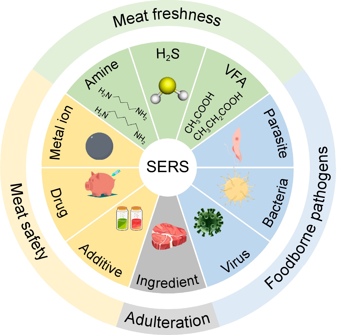
      Figure 1. 

      Schematic application of SERS analysis in meat and meat products.

      The complexity of matrices often leads to poor repeatability of Raman signals. To improve the repeatability and sensitivity of this technology, it is necessary to develop SERS substrates with stable plasmonic enhancement. Various SERS substrates have been developed. If classified by substrate properties, they can be divided into noble metal substrates and noble-free metal substrates[21]. From the view of practicability, the existing SERS substrates can be divided into colloidal substrates and solid substrates[22]. Colloidal substrates generally include single or multiple metal nanoparticles[2325], showing better signal stability, while solid substrates exhibit higher signal amplification ability due to good morphology controllability[26]. Common forms of solid substrates include membrane substrate and self-assembled substrate[27,28]. The size, morphology, and material of SERS substrates have a significant impact on the enhancement of Raman signals[29]. For any SERS substrate, the widely accepted mechanisms for SERS enhancement effects are electromagnetic enhancement (EM) and chemical enhancement (CM), as shown in Fig. 2. The EM is excited by the strong electromagnetic field generated by surface plasmon resonance on ultrathin or nanostructured surfaces, thus enhancing the electromagnetic signal. The CM is induced by the formation of charge transfer complexes between adsorbed molecules and metal substrates, thereby achieving enhancement effects[30]. Due to its fast response speed, high sensitivity, and non-destructive detection ability, SERS has been widely applied in fields such as food safety analysis[31], environmental monitoring[32], and material science[33].
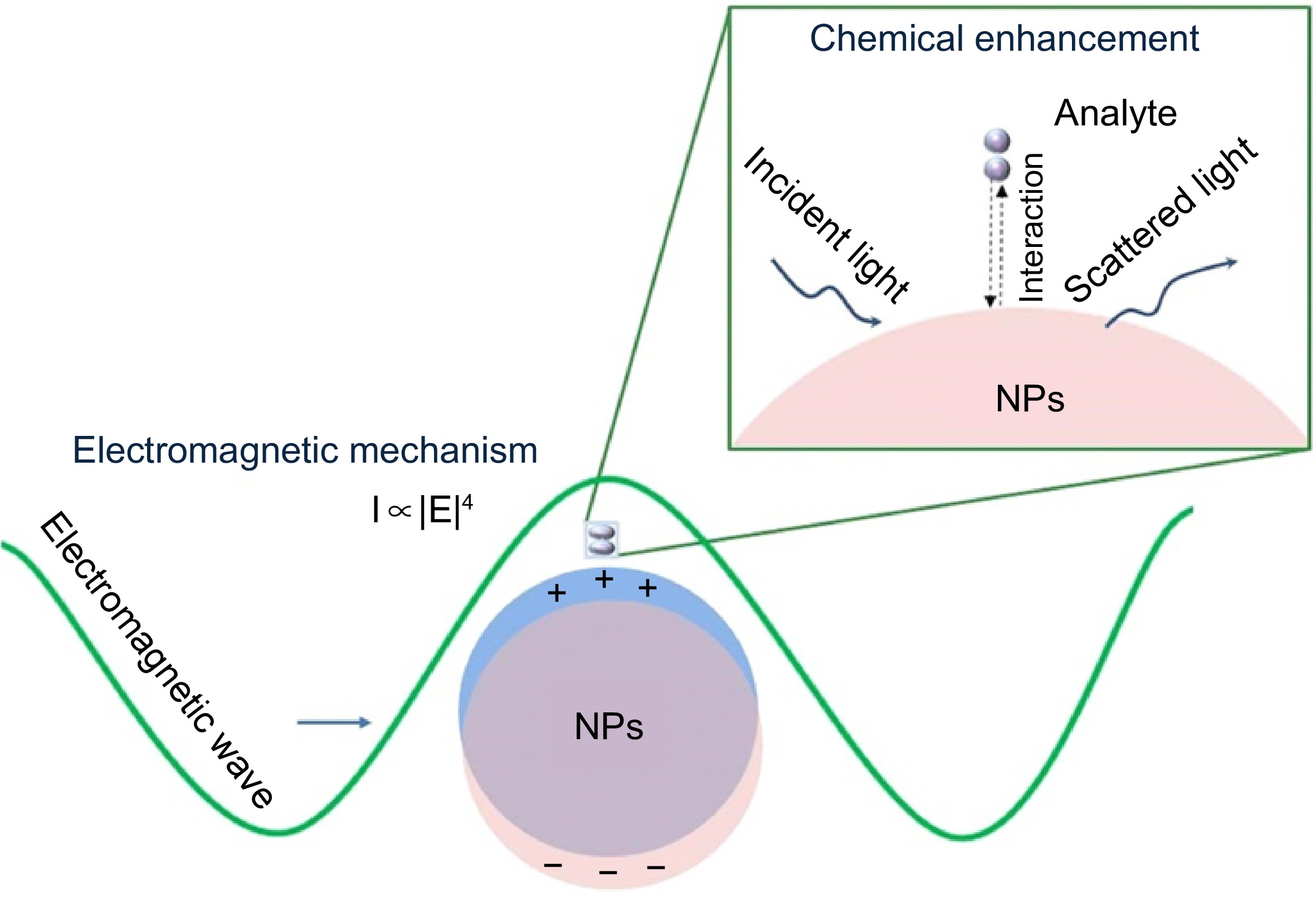
      Figure 2. 

      Enhancement mechanisms of SERS[34].

      SERS can be used in combination with various technologies, such as immunochromatography[35], molecular imprinting technology[36], colorimetric technology[37], etc., to improve selectivity, sensitivity, and detection efficiency. General SERS detection strategies are labeled and label-free detection modes in terms of detection of indirect and direct detection of the target. The former can detect Raman signals of targets without labeling or special processing, not only providing the inherent molecular information but also making the detection process simpler. However, it is limited by the concentration and complexity of the detection system[38,39]. The latter can reflect the analyte by strong Raman characteristic signals of SERS tags. With the aid of recognition elements like antibodies[40], aptamers[41], and molecularly imprinted polymers (MIPs)[36], the labeled method can achieve the detection of various targets.

      Different from other spectroscopy techniques, SERS can provide a highly sensitive fingerprint analysis, amplify the Raman signal of the analyte without fluorescence background interference, and match various laser conditions and different types of instruments including large high-resolution workstations and portable devices. Therefore, SERS has now developed into a powerful platform for quickly detecting trace extrinsic or harmful substances in meat and meat products. Compared with previous reviews, this review focuses on two SERS detection modes and their related applications in meat and meat products. SERS detection is divided into labeled detection and label-free detection to compare their advantages and disadvantages. Then, the research progress of SERS in detecting several hazards such as foodborne pathogens and veterinary drug residues are summarized, and the practicability of this technology in identifying meat adulteration and spoilage discussed. Finally, a perspective of SERS to more efficiently and sensitively meet the requirements of rapid and high-throughput detection in the meat industry is provided.

    • In the label-free SERS detection mode, the active substrate of SERS can directly bind to the analyte without additional signal indicators to assist detection[42]. The spectral information provided by this method can not only be used for the detection of the substance under test, but also for analyzing the structural information or fingerprinting of biomolecules[43,44]. Arabi et al.[45] proposed a mussel-inspired surface imprinted capillary sensor that can quickly and sensitively detect proteins. The universal sensor was not limited by pre-processing and operator skills. Xu et al.[46] used iodide-modified Ag nanoparticles (Ag IMNPs) to achieve label-free detection of single stranded DNA molecules. This detection strategy not only significantly improved Raman signals, but also reduced the probability of biological molecule denaturation during the detection process. Wang et al.[47] combined chemometric methods to achieve label-free detection of methicillin-sensitive Staphylococcus aureus (MSSA) and methicillin-resistant Staphylococcus aureus (MRSA), which played an important role in the detection of bacterial resistance and identification of resistant strains. To detect antibiotic residues in serum, Wang et al.[48] modified nanoparticles with bromide ions and used peak intensity changes as a basis for distinguishing different antibiotic molecules. This method is of great significance in drug detection. Zhang et al.[49] designed a SERS microfluidic chip for drug detection, providing a new platform for efficient detection of 6-thioguanine (6-TG) in human serum.

      In label-free SERS detection, the binding mode and interaction mechanism between the tested substance and the SERS substrate are worth exploring in depth, which often determines the sensitivity and signal of the detection system[42] The strategies for anchoring the tested molecule mainly include biomolecular recognition[47], non covalent bonding[50], and electrostatic and hydrophobic interactions[51]. Zhang et al.[52] used single-molecule surface-enhanced Raman spectroscopy (SM-SERS) to investigate the phenomenon of signal fluctuations caused by the adsorption and desorption of molecules near hot spots. They utilized active nanoshells to confine and anchor molecules onto the surface of plasmon nanoparticles, significantly improving the sensitivity and reproducibility of single-molecule detection (Fig. 3a). Meanwhile, combining the detected spectral data with chemometrics and machine learning methods enables more effective data analysis[53]. Raman spectroscopy data is rich and complex, with the assistance of machine learning and chemometric methods, data processing, including noise reduction and interference elimination, can be quickly achieved[54,55] (Fig. 3b). There are a wide range of applications in food analysis[56], and biomedicine[57].

      Figure 3. 

      Label-free SERS detection. (a) Schematic diagram for the detection of in-situ encapsulated active shells[52]. (b) Application of machine learning in Raman spectroscopy analysis[55].

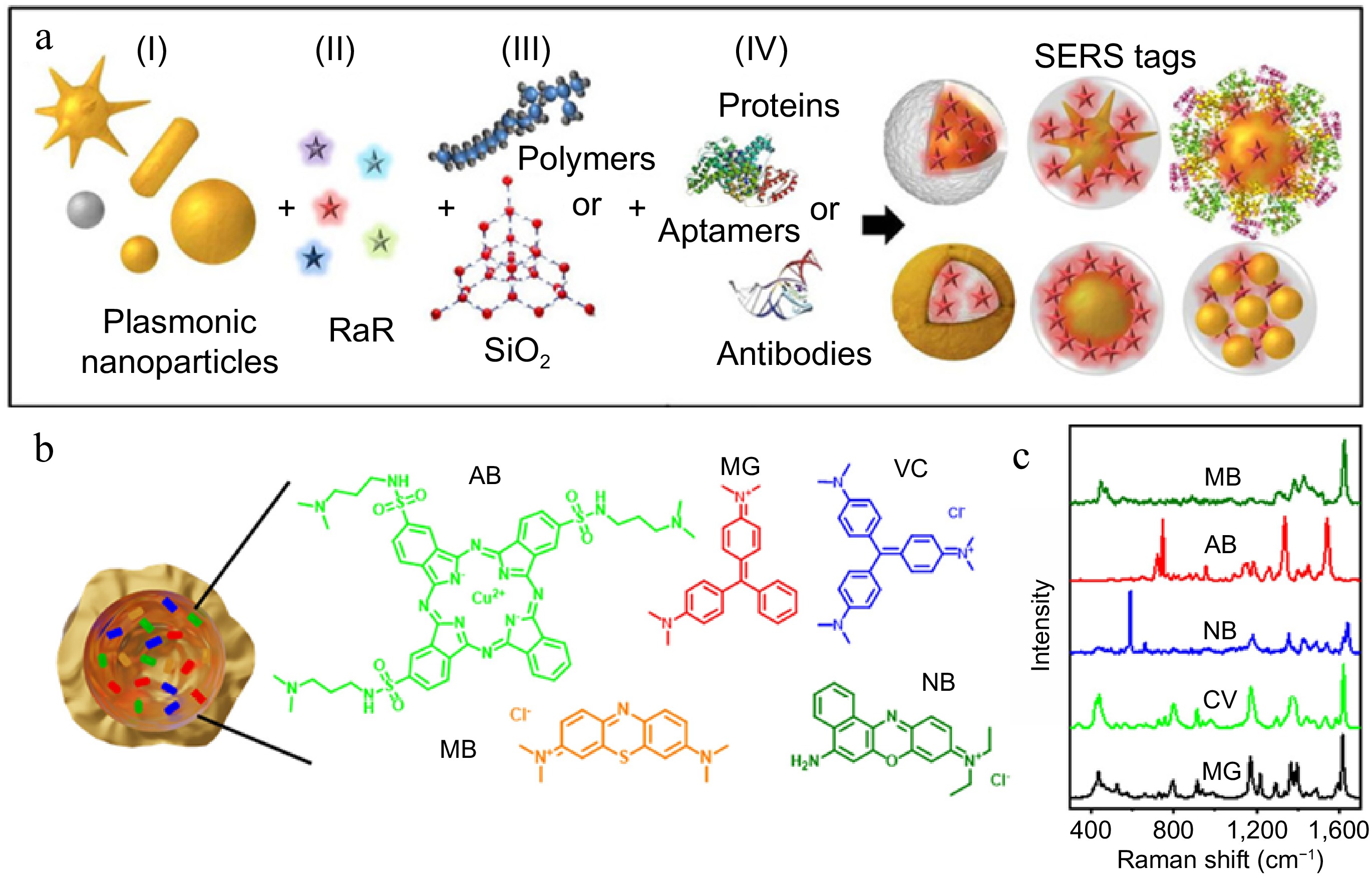
    • When the composition of the matrix to be tested is complex or physical characteristics such as temperature and pH need to be monitored, label-free detection has significant limitations compared to labeled detection[58]. The labeled SERS detection method relies on functionalizing Raman reporters with high sensitivity, specificity, and selectivity. By observing the Raman shift and intensity changes of characteristic peaks in the Raman spectrum, the presence and amount of the tested substance can be reflected by Raman reporters[59]. Although the labeled mode cannot reflect rich intrinsic biological information, multiple SERS tags might have a potential in multiplex analysis[34].

      SERS tags need to have ultra-high sensitivity, specificity, and photostability[60]. Typically, SERS tags consist of four parts: plasmonic nanoparticles, Raman reporters, coating layers, and targeting ligands[61] (Fig. 4a). As SERS substrates, plasmonic nanoparticles are activated by localized surface plasmon resonance (LSPR) to enhance the signal. Raman reporters with excellent properties are adsorbed on the surface of the SERS substrate, and then encapsulated with a protective layer to make the particles more stable. Finally, targeting ligands such as antibodies and aptamers are connected to form SERS tags[62]. Raman reporters can be mainly divided into three categories, specifically including dye molecules containing nitrogen or sulfur-like crystal violet (CV)[63] (Fig. 4b & c), thiol molecules like 4-mercaptobenzoic acid (4-MBA)[64], and 4-mercaptophenylboronic acid (4-MPBA)[65]. Alkyne molecules possessing unique peaks in Raman silent regions that attract emerging attention on SERS due to largely reduced background interference[66]. To enhance the stability and signal strength of SERS tags, dual signal molecules for the detection of biomolecules are used[67]. The dual signal method can not only reduce the influence of external interference and improve the repeatability of detection but is also suitable for the detection of low concentration analytes in complex samples[68]. In terms of dual signal, one serves as an internal standard signal and the other as a response signal, which can reduce detection errors and have higher detection accuracy compared with single signal systems. Tan et al.[69] used 5,5'-dithiobis (2-nitrobenzoic acid) (DTNB) as the internal standard signal, 6-carboxyl-Xrhodamine (ROX) as the response signal, and the double signal based SERS sensor detected the miR-21 of human serum samples, with a detection limit of 0.046 pM. It has broad application prospects in the early diagnosis of breast cancer.

      Figure 4. 

      Labeled SERS detection. (a) The structure of SERS tags[74]. (b) Capsules encoded with malachite green (MG), crystal violet (CV), Nile blue (NB), Astra blue (AB), and methylene blue (MB)[75]. (c) Raman spectra of reporters[75].

      Liu et al.[70] combined SERS with LFIA and proposed a biosensor for detecting anti-SARS-CoV-2 IgM/IgG. This sensor used DTNB as a Raman reporter modified on silica nanosphere coated with an Ag shell, to provide a sensitive detection strategy for rapid screening of SARS-CoV-2 infection. Jiang et al.[71] synthesized Fe3O4@TiO2-based SERS tags using DTNB as the Raman reporter, achieving in situ detection of exosomal miRNAs. Zhu et al.[72] embedded 4,4'- dipyridyl (DP) into AuNPs and silica shell to prepare SERS probes with excellent stability and specificity, achieving ultrasensitive detection of E. coli O157:H7. Combined with hybridization chain reaction (HCR), Peng et al.[73] used 4-ethynylbenzaldehyde (EBA) and two different structures of HCR sequences as SERS tags, developing a novel SERS sensing method and achieving sensitive detection of hepatitis C virus (HCV) nucleic acid.

    • According to statistics from the World Health Organization (WHO), approximately 600 million people are infected with foodborne diseases[76], largely increasing the burden on the healthcare system. Meat and meat products are important sources of high-quality protein for the human body, and due to its rich nutritional content, meat is often contaminated by foodborne pathogens and bacteria[77]. The bacteria pollution sources of meat products can be divided into two categories: endogenous and exogenous pollution. The former usually refers to pollution caused by microorganisms carried by livestock and poultry, while the latter often refers to microbial pollution present in the processing and circulation process[78]. Common foodborne pathogenic bacteria in meat and meat products include L. monocytogenes, Salmonella, E. coli, etc.[79]. Consuming meat contaminated with foodborne pathogenic bacteria poses a serious threat to human life and health. Therefore, to control the occurrence of foodborne diseases and protect the development of the meat industry, it is crucial to establish sensitive and rapid methods for detection.

      At present, foodborne pathogenic bacteria can be detected through the following three strategies. Physiological and biochemical testing can indicate the presence of pathogens through chemical signals, such as the ATP bioluminescence method[80]. Immunological testing is based on specific binding of bacterial antigens followed by signal amplification, such as ELISA[81]. Molecular testing relies on nucleic acid-based hybrid and amplification, such as PCR[82]. Compared with traditional plate culture methods, these methods have achieved sensitive and accurate detection of pathogens, however, they still face several drawbacks including slower detection speed, longer detection cycles, and more operation steps[83]. Due to the multiple advantages of SERS, the technology has been widely used to detect foodborne pathogens. Yang et al.[84] reported a surface cell imprinting (SCIS) method to capture the target pathogens followed by SERS mapping detection with a nanosilver modified by 4-MPBA (4-MPBA@AgNPs) as the SERS tag. It has achieved specific and quantitative determination of E. coli in chicken breast samples, with a linear range of 102−108 CFU/mL and a detection limit as low as 1.35 CFU/mL. By changing the bacterial cell imprinting substrate, this platform can also be used for the detection of other bacteria. Cho et al.[85] proposed using membrane filtration and immunomagnetic separation techniques to capture and enrich target bacteria. Using 4-MBA modified AgNPs as SERS tags, 10 CFU/mL of E. coli O157:H7 was detected in ground beef within 1 h. In label-free detection mode, the Raman signal of the analyte mainly comes from the surface chemical composition and metabolites, but some bacteria have similar cell wall components, resulting in high similarity in their SERS fingerprint spectra. On the other hand, the amount of spectral information data is too complex to distinguish. In this case, mathematical-statistical analysis methods and chemometrics methods should be combined to eliminate signal interference during the detection process and achieve efficient detection of foodborne pathogens[86,87]. Leong et al.[88] used a SERS-based surface chemistry classification method, in combination with machine learning, to classify six types of bacteria by layering surface charges, biochemical features, and the types and quantities of functional groups. The accuracy was up to 98%, and the relationship between bacterial extracellular matrices (ECMs) surface features and SERS fingerprint spectra were successfully made. Eady et al.[89] compared traditional plate culture and PCR methods and confirmed that combining SERS with support vector machine (SVM) could realize rapid detection and accurate classification of Salmonella typhimurium in chicken rinse. In addition, Zheng et al.[90] utilized python assisted SERS chips to achieve photothermal inactivation of Salmonella typhimurium and Staphylococcus aureus in blood samples, avoiding the problem of secondary contamination during the detection process (Fig. 5). The related analytical peformances of these methods were compared in Table 1.
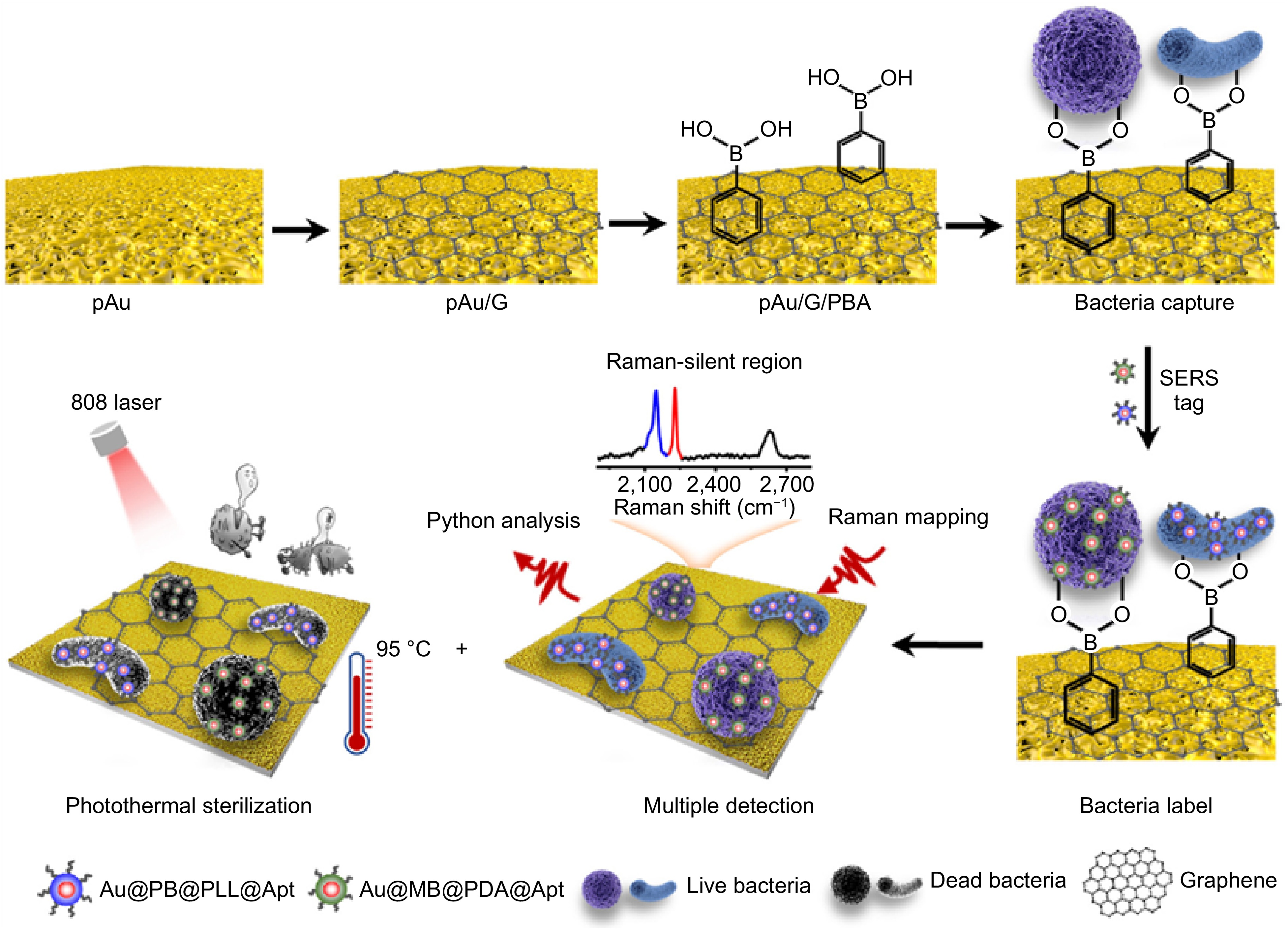
      Figure 5. 

      Schematic diagram of capturing, detecting, and inactivating bacteria[90].

    • Foodborne viruses that exist in various foods might cause diseases such as viral gastroenteritis and hepatitis in humans. Patients often suffer from acute vomiting and diarrhea due to ingestion of contaminated water or food[91]. Common foodborne viruses in meat and meat products include avian influenza virus, norovirus, hepatitis E virus, and rotavirus[92]. PCR is a classical technique for virus infection identification with high sensitivity and accuracy but requires complex sample pretreatment and expensive equipment[93]. In addition to PCR, immunological methods such as ELISA are also commonly used for virus detection[94]. However, the sensitivity and accuracy of this method are not as good as nucleic acid amplification technology[95]. Therefore, SERS-based methods were developed for the efficient detection of foodborne viruses. H5N1 is a highly pathogenic and deadly subtype of avian influenza virus[96]. Wang et al.[97] used an unlabeled SERS method, using AgNPs as the substrate, to achieve rapid detection of influenza A (H5N1) subtype influenza virus in chicken embryos, by forming specific sandwich immunocomplexes, with high accuracy and strong specificity. This provided a reference basis for the simple and rapid detection of various infectious viruses. Sun et al.[98] proposed a magnetic immunosensor labeled with 4-MBA for the detection of avian influenza virus H3N2. The sensor had the advantages of high sensitivity and rapid detection. Therefore, this method had the potential to be applied to the detection of avian influenza virus in other real biological samples. Wang et al.[99] designed and synthesized a novel magnetic tag with excellent signal amplification performance. The SERS-LFIA system was used to detect HAdV and H1N1, and it was found that this method had high sensitivity with detection limits of 10 and 50 PFU/mL, respectively, and could be used in real biological samples such as human whole blood, serum, and sputum. The related analytical peformances of these methods were compared in Table 1.

    • To prevent and control animal diseases, veterinary drugs, mainly include antibiotics, antiparasitic, and antifungal drugs, hormones, and anti-inflammatory drugs, have long been applied during livestock feeding[100]. The use of veterinary drugs often bring profits to animal producers and reduce losses. However, residual veterinary drugs in animal bodies may have many negative impacts on the animals themselves and consumers who consume them, including the development of drug resistance in both animal and human bodies, affecting the functioning of the immune system and the diversity of gut microbiota[101]. Before detecting veterinary drug residues, it is necessary to perform sample pretreatment to reduce external interference, which is closely related to the accuracy and precision of the detection[102]. Currently, common methods such as solid-phase extraction (SPE) and solid-phase microextraction (SPME) are used to separate the analyte from the complex sample matrices[103,104]. Even so, the sample solution after enrichment is still so complex as to direct detection and identification, thus gas/liquid chromatography (GC/LC) followed by mass spectrometry (MS) is necessary.

      It is worth mentioning that SERS has outstanding potential in detecting residual harmful chemical components. Peng et al.[105] used AuNPs as substrates to detect benzylpenicillin potassium (PG) and explored the effects of Au substrates and reporter adsorption on SERS intensity. Finally, the established method was applied to the detection of PG in duck meat. Zhao et al.[106] used OTR202 (AuNPs) and OTR103 (gold colloid enhancement reagent) as SERS substrates, combined with adaptive iteratively reweighted penalized least squares (air-PLS) to remove fluorescence and background signals, and the detection limit was down to 1.120 mg/L, achieving rapid detection of tetracycline residues in duck meat. Zhao et al.[107] established a method for determining marbofloxacin using SERS based on β-cyclodextrin-modified silver nanoparticles (β-CD-AgNPs) with a detection limit of 1.7 nmol/L. In chicken and duck samples, the spiked recovery rate of marbofloxacin ranged from 101.3% to 103.1%, providing a solution for reliable on-site detection in the future. To reduce spectral interference from other substances in food matrices, combining SERS with other separation techniques is a good choice. Shi et al.[108] used thin-layer chromatography combined with SERS, namely TLC-SERS, to achieve simultaneous and rapid (< 10 min) detection of 14 nitroimidazole compounds in pork with a detection limit of 0.1 mg/L. Based on the magnetic SERS-LFA system, Tu et al.[109] synthesized SERS tags using DTNB and 4-MBA as dual Raman reporters, combined with specific antibodies. They utilized the dual signal amplification effect of numerous stable hotspots and magnetic enrichment to detect the residues of four veterinary drugs in pork. This method achieved trace detection at the pg/mL level within 35 min, effectively improving the sample detection signal and sensitivity, and had great prospects in the detection of harmful small molecules (Fig. 6). The related analytical peformances were compared in Table 1.
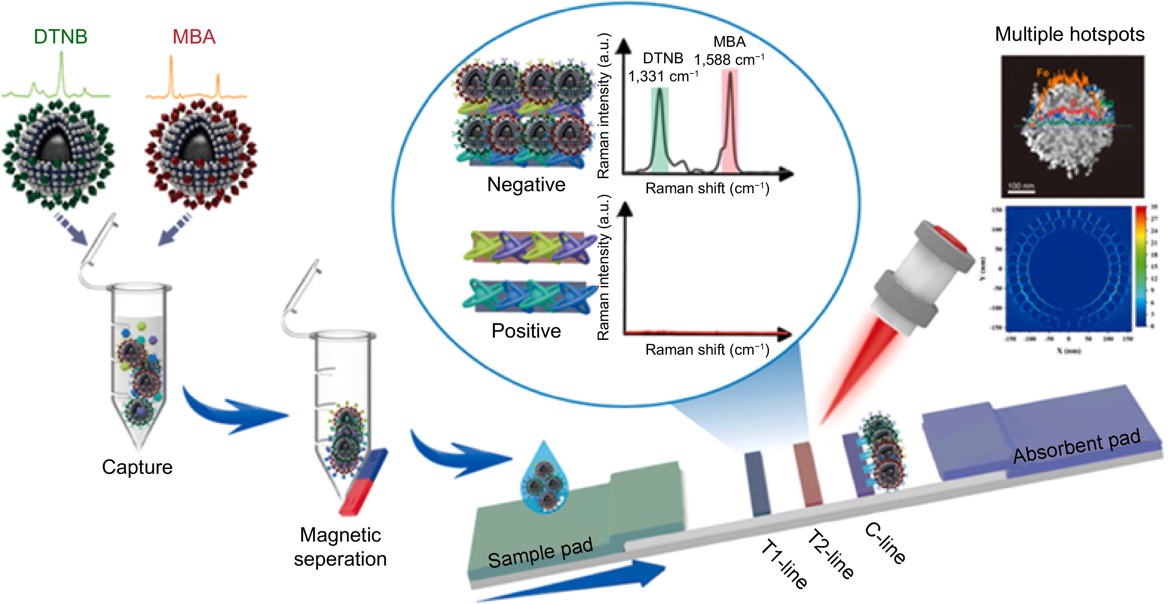
      Figure 6. 

      Schematic diagram of using the SERS-LFA system to detect multiple veterinary drugs[109].

    • Food additives are artificially synthesized or natural substances that can improve the sensory characteristics and quality of food. The application of food additives has played a great role in the development of the food industry. To improve the flavor, texture, nutrition, and extend the shelf life of meat products, several food additives include antioxidants, preservatives, colorants, and acidity regulators are applied during meat processing and storage[110]. However, unscrupulous retailers, in pursuit of commercial interests, abuse food additives, and even engage in the illegal use of preservatives, colorants, and other substances with maximum amount limits in meat product production, such as the abuse of nitrite, composite phosphates, and sodium benzoate. Unreasonable uses of food additives have also brought about a series of food safety issues, posing a great threat to the life and property safety of consumers[111]. At present, the main detection methods for food additives include spectroscopy[112], chromatography[113], and electroanalysis[114]. Meanwhile, the sampling and pretreatment steps have a considerable effect on detection accuracy[115].

      For several common food additives, SERS also exhibits satisfactory potential[116]. Nitrite, as an essential food additive in meat processing, is often used as a preservative and coloring agent, but it might cause certain health risks, thus being necessary to develop a rapid SERS detection method[117]. Zhang et al.[118] enhanced the SERS signal of nitrite by introducing 4-aminothiopenol capped AgNPs decorated halloysite nanotubes (HNTs-AgNPs4−ATP), thereby achieving the detection of nitrite ions in sausages and pork luncheon meat. This method achieved in-situ derivatization and selective determination of nitrite ions in meat products through labeled SERS. The effective dispersion and deposition of metal nanoparticles play an important role in maintaining substrate stability and improving SERS performance[119]. Zhang et al.[120] developed a SERS platform for the rapid detection of nitrite using electrospinning-assisted electrospray technology. The use of this technology is of great significance for the effective deposition of certain shaped metal nanoparticles into SERS layers. The platform had good selectivity, stability, and anti-interference ability, and the detection limit was about 15.29 ng/L, realizing the detection of nitrite in chicken sausage, canned pork, bacon, and ham. Liang et al.[121] combined hydrogel materials with SERS technology to prepare a sensor for detecting the concentration of sodium nitrite, and introduced machine learning to analyze data and predict results. The minimum detection limit reaches 3.75 mg/kg, realizing the quantitative determination of sodium nitrite in the extracts of bacon, lunch meat, and ham slices. The analytical peformances were compared in Table 1.

    • In recent years, food safety accidents caused by illegal additives have aroused public attention to food quality and safety. Compared with the abuse of food additives, illegal additives have more serious implications owing to their severe toxicity to both livestock and the human body. Illegal additives mainly include melamine[122], malachite green[123], receptor agonist[124], and other substances, which are usually used to fraudulently increase nutrient content or preserve freshness[23]. The most common illegal additives in meat and meat products include β-adrenergic receptor agonists (clenbuterol hydrochloride[123], ractopamine[125], etc.) in pork, beef, mutton, and animal liver, nitrofuran drugs in pork and poultry[126], and synthetic pigments such as acid orange in meat products[127]. At present, commonly used detection methods for illegal additives include gas chromatography[128], mass spectrometry[129], ELISA[130], etc. With the continuous development of SERS substrates, Yan et al.[131] prepared transparent SERS substrates using anodic aluminum oxide (AAO) template method for direct detection of residual ractopamine on pork without the need for pretreatment. This method achieved the detection of trace amounts of ractopamine in meat samples with a detection limit of 10−8 M, and also opened up a new way for the direct measurement of other trace chemical substances on the surface of food. The uniform core-shell structure of nanomaterials significantly improves the SERS performance in signal enhancement and stability by increasing loading and reducing aggregation, which can improve detection efficiency and reliability[132,133]. Su et al.[134] designed a core-shell structure as a multifunctional tag and used the dual model colorimetric/SERS-LFIA for the detection of clenbuterol. This method increased the sensitivity of the detection system and stronger colorimetric reaction through antigen antibody specific binding, achieving quantitative detection of clenbuterol in pork, chicken, and sausages with a detection limit as low as 0.05 ng/mL. Xie et al.[127] realized the rapid detection of acid orange II in braised pork by synthesizing new core-shell nanomaterials including SERS substrates of Fe3O4@Au. In combination with machine learning methods, they verified the correctness of the detection results and compared with the results of HPLC, showing that this method can be used as an alternative to conventional HPLC methods for the detection and analysis of acid orange in food. The related analytical peformances of these methods were compared in Table 1.

    • Biotoxins are a class of toxic substances produced by various organisms such as Clostridium, E. coli, Staphylococcus aureus, and the common biotoxins in meat and meat products are botulinum toxin and shiga toxin. Biotoxins often cause acute or chronic poisoning in the human body and have become a major threat in fields such as food and medicine[135]. The detection of food biotoxins typically involves quantitative analysis using ELISA[136], MS[137], and HPLC[138]. Nowadays, biosensors based on SERS have become an important analytical method for biotoxin detection. Subekin et al.[139] developed an aptasensor based on silver nanoislands as a SERS substrate for rapid detection of type A botulinum toxin. Due to its ability to recognize molecules and serve as Raman tags, the sensor has high specificity and good reproducibility, with a detection limit of 2.4 ng/mL, and can achieve rapid detection of botulinum toxin in complex matrices. Kim et al.[140] synthesized three-dimensional magnetic beads modified with gold nanoparticles and developed a SERS-based magnetic immunoassay for rapid and sensitive detection of botulinum toxin. The detection limit of this technology for type A and type B botulinum toxin reached 5.7 ng/mL (type A) and 1.3 ng/mL (type B). The proposed method is a low-cost and efficient detection technology for botulinum toxin and promising for other trace biotoxin detection in meat. Jia et al.[141] developed a biosensor with SiO2@Au/DTNB as the SERS tag that can simultaneously detect ricin, staphylococcal enterotoxin B (SEB), and type A botulinum toxin (BoNT/A) by combining SERS with LFIA. This technology achieved rapid on-site detection of three toxins with good repeatability and specificity, and was capable of application in clinical medicine. The analytical peformances were compared in Table 1.

      Table 1.  Applications of SERS in detection of meat hazards and additives.

      Detection object SERS substrate Method LOD Ref.
      Foodborne pathogens
      E. coli O157:H7 AgNPs SERS-SCIS 1.35 CFU/mL [84]
      E. coli O157:H7 AuNPs SERS 10 CFU/mL [85]
      Salmonella typhimurium AgNPs SERS-SVM / [89]
      Salmonella typhimurium and Staphylococcus aureus pAu/G SERS-Python / [90]
      Foodborne viruses
      H5N1 AgNPs SERS 5.0 × 10⁻⁶ TCID50/mL [97]
      H3N2 AuNPs SERS 102 TCID50/mL [98]
      HAdV, H1N1 AgNPs SERS-LFIA 10, 50 PFU/mL [99]
      Veterinary drug residues
      Benzylpenicillin potassium AuNPs SERS / [105]
      Tetracycline OTR202-OTR103 SERS-air PLS 1.120 mg/L [106]
      Marbofloxacin AgNPs SERS 1.7 nmol/L [107]
      Nitroimidazoles AuNPs SERS-TLC 0.1 mg/L [108]
      Multiple veterinary drugs Au@AgNPs SERS-LFA 0.52–6.2 pg/mL [109]
      Food additives
      Nitrite ions AgNPs SERS 0.51 μg/L [118]
      Nitrite AgNPs SERS 15.29 ng/L [120]
      Sodium nitrite AuNPs SERS 3.75–8.11 mg/kg [121]
      Illegal additives
      Acid orange II AuNPs SERS-DFT 1 μg/mL [127]
      Ractopamine AgNPs SERS 10−8 mol/L [131]
      Clenbuterol Au/AuNS SERS-LFIA 0.05 ng/mL [134]
      Biotoxin
      Botulinum neurotoxin type A AgNPs SERS 2.4 ng/mL [139]
      Botulinum toxins A and B AuNPs SERS 5.7 ng/mL (A), 1.3 ng/mL (B) [140]
      BoNT/A AuNPs SERS-LFIA 0.1 ng/mL [141]
    • Meat adulteration is a fraudulent behavior of unscrupulous merchants who mix low-quality meat or non-meat substances into high-priced meat or its products to seek extra profits. Such behavior usually includes species or variety adulteration, production source adulteration, and production process adulteration[142,143]. Meat adulteration might be related to the changes in supply and demand, as well as the cost of animal husbandry and processing. However, this trickery has had a serious negative effect on the meat industry and hidden unknown safety issues. The food safety issues caused by meat adulteration are worrying. The adulterated meat might contain unknown species, pathogens, and veterinary drugs, which may not only directly affect the life and property safety of consumers but also involve religious issues and affect market stability[144,145]. The existing detection methods for meat adulteration mainly include nucleic acid detection technology[146], biosensors[147], spectroscopic detection technology[148], immunological detection technology[149], and mass spectrometry technology[150]. Currently, the detection methods for meat adulteration can be divided into non-destructive and destructive testing[151]. Non-destructive testing techniques include near-infrared spectroscopy[152], hyperspectral imaging[153], etc. Destructive testing techniques include detection based on nucleic acid[154], and protein[155]. As a non-destructive detection method, SERS has been applied in meat fraud detection. Liu et al.[156] proposed a novel detection strategy mediated by CRISPR/Cas12a followed by SERS, which could convert the target nucleic acid concentration into a visual signal for meat adulteration detection, achieving the detection of low adulteration rate samples in complex food matrices. Khalil et al.[157] designed an ultrasensitive dual nano platform SERS biosensor, namely the graphene oxide gold nanorod (GO AuNR) and gold nanoparticles (AuNPs), which could qualitatively and quantitatively detect DNA from any source. This sensor could replace traditional pork DNA detection methods, helping to more efficiently address the issues of authenticity and species origin in meat products. Khalil et al.[158] also developed a DNA biosensor for SERS that could quantitatively detect two types of meat simultaneously. The detection principle lies in the covalent conjugation between the signal probe and the capture probe. When the target sequences of two species are fixed simultaneously, the hybridization between the probe and the target achieves signal enhancement. This sensor used a SERS activity dual platform to increase detection sensitivity, with strong selectivity and specificity. As an emerging technology, SERS is still lacking in research on meat adulteration detection.

      Existing research mainly focus on the detection of species sources, and further development is needed for cases of determining the authenticity of production areas and processes. To fully explore Raman spectrometry, new data mining technologies such as machine learning should be introduced to identify adulteration. Besides, the development of portable detection devices to achieve real-time on-site assessment is another future research trend.

    • Meat contains abundant nutrients, making it an ideal place for microbial reproduction. The presence of microorganisms and some endogenous enzymes often leads to a decrease in the freshness of meat and eventual spoilage. The decrease in freshness of meat can be judged by changes in color, which not only leads to the disposal of meat and resource waste but also poses health risks such as pathogens and toxins[159,160]. With the improvement in disposable income, consumers are paying increasing attention to the freshness of foods. The traditional methods for determining freshness include sensory evaluation, chemical index detection, and microbial detection[161]. However, these methods all have some limitations. Sensory evaluation methods require specialized evaluators and the judgment results are always subjective. For chemical indicators such as total volatile base nitrogen (TVBN) and microbial detection, the detection methods are cumbersome and time-consuming, and cannot meet the needs of contemporary food industry[162]. Therefore, it is crucial to develop an efficient and sensitive non-destructive testing method that can measure the freshness of products. At present, several advanced methods for determining freshness include fluorescence spectroscopy[163], near-infrared spectroscopy[164], and visual intelligent packaging technology[165]. Notably, food freshness detection technology based on SERS has been widely reported. Qu et al.[166] achieved the detection of volatile organic compounds (VOCs) in chicken samples, including bacterial metabolites, H2S, aldehydes, and biogenic amines, by integrating array sensors and combining machine learning, thereby achieving real-time determination of their freshness (Fig. 7). Given that cadaverine and putrescine are toxic biogenic amines produced by microorganisms, posing a significant threat to human health and food security, Sun et al.[167] utilized p-MBA functionalized SERS substrates to capture the analyte molecules through amide reactions, enabling trace detection of amine substances in pork samples, which is of great significance for detecting the degree of food spoilage. Kim et al.[168] designed a SERS paper platform coated with a metal-organic framework (MOF) that could effectively recognize volatile amine molecules, and this paper's sensory has been successfully applied to the freshness determination of chicken, beef, and pork samples.

      Figure 7. 

      Schematic diagram of a scalable plasma array gas sensor for multi-dimensional SERS recognition[166].

    • With the rise in public awareness of food safety and human health, meat safety, and quality have received unprecedented attention. SERS, as an ultrasensitive detection technique, can achieve trace analysis of target analytes. Besides, the rapid and high-resolution detection characteristics of SERS make it suitable for on-site detection and multiplex analysis. This review focused on the development of the application of SERS technology in safety detection and quality assessment of meat and meat products. First, both label-free and labeled SERS detection modes were summarized, and labeled mode was predominant owing to higher sensitivity and various forms. Then, the application of SERS in foodborne pathogens, veterinary drug residues, food additive abuse, illegal additive use, and detection of biotoxin in meat and meat products were introduced. Meanwhile, the progress of SERS-based meat adulteration and freshness identification were discussed. However, SERS also faced some existing problems, such as poor reproducibility of detection results, weak resistance to external interference, and difficulty in enriching analytes. For future research, the following points can be underlined to promote the further development and application of SERS technology in the detection of meat and meat products: (1) Combining SERS with other technologies. SERS, as a sensitive detection method, relies on appropriate pretreatment operations to improve the accuracy and precision of detection; (2) Expanding SERS research on heavy metal ion pollution detection in meat and meat products by combining elemental analysis techniques, and boosting research on adulteration identification and freshness detection of meat and meat products; (3) Combining machine learning and chemometrics methods to mining Raman spectroscopy data and improving automatic detection and smart sensing. With the continuous development of material and data science, researchers will further study novel SERS substrates, SERS tags, and SERS instruments to improve the stability and repeatability of this technology, paving the foundation for safety and quality assessment in the meat and food industry.

      • This review was funded by the National Natural Science Foundation of China (22327804 and 22004064).

      • The authors confirm contribution to the paper as follows: study conception: Wu M, He H; draft manuscript preparation: Wu M; manuscript revision: He H. Both the authors read and approved the final manuscript.

      • Data sharing is not applicable to this article as no new data were created or analyzed in this study.

      • The authors declare that they have no conflict of interest.

      • Copyright: © 2024 by the author(s). Published by Maximum Academic Press on behalf of Nanjing Agricultural University. This article is an open access article distributed under Creative Commons Attribution License (CC BY 4.0), visit https://creativecommons.org/licenses/by/4.0/.
    Figure (7)  Table (1) References (168)
  • About this article
    Cite this article
    Wu M, He H. 2024. Recent advances on surface enhanced Raman spectroscopy in safety assessment and quality control of meat and meat products. Food Materials Research 4: e029 doi: 10.48130/fmr-0024-0018
    Wu M, He H. 2024. Recent advances on surface enhanced Raman spectroscopy in safety assessment and quality control of meat and meat products. Food Materials Research 4: e029 doi: 10.48130/fmr-0024-0018

Catalog

  • About this article

/

DownLoad:  Full-Size Img  PowerPoint
Return
Return